Photospintronics: Magnetic Field-Controlled Photoemission and ...
6
Photospintronics: Magnetic Field-Controlled Photoemission and Light-Controlled Spin Transport in Hybrid Chiral Oligopeptide- Nanoparticle Structures Prakash Chandra Mondal, † Partha Roy, † Dokyun Kim, ‡ Eric E. Fullerton, ‡ Hagai Cohen, § and Ron Naaman* ,† † Department of Chemical Physics, Weizmann Institute of Science, Rehovot 76100, Israel ‡ Center for Memory and Recording Research, University of California, San Diego, California 92093, United States § Chemical Research Support, Weizmann Institute of Science, Rehovot 76100, Israel * S Supporting Information ABSTRACT: The combination of photonics and spintronics opens new ways to transfer and process information. It is shown here that in systems in which organic molecules and semiconductor nanoparticles are combined, matching these technologies results in interesting new phenomena. We report on light induced and spin-dependent charge transfer process through helical oligopeptide− CdSe nanoparticles’ (NPs) architectures deposited on ferromagnetic substrates with small coercive force (∼100−200 Oe). The spin control is achieved by the application of the chirality-induced spin- dependent electron transfer effect and is probed by two different methods: spin-controlled electrochemichemistry and photoluminescence (PL) at room temperature. The injected spin could be controlled by excitation of the nanoparticles. By switching the direction of the magnetic field of the substrate, the PL intensity could be alternated. KEYWORDS: Charge transfer, chirality, spin transport, oligopeptide, spin selectivity, ferromagnet T he concept of controlling magnetism by light has attracted much attention in recent years. 1−3 Specifically, the controlling of spin transport by light combines two interesting technologies, photonics and spintronics. 4,5 Having organic material makes the coupling of the two technologies easier 6−8 because the light-absorbing properties of organic molecules can be modified relatively easily. 9,10 Recently, it was found that the transmission of electrons through chiral molecules depends on the electrons’ spin orientation; this effect is referred to as chiral- induced spin selectivity (CISS). 11,12 The CISS effect makes it possible to construct spintronic devices without ferromagnetic spin injectors because the chiral molecules themselves serve to select a specific spin to transport across the molecules. 13−16 In the hybrid system studied herein, helical oligopeptide molecules were attached on one end to CdSe nanoparticles (NPs) and on the other end to ferromagnetic substrates. Two experimental configurations were applied; in the first, the photoemission intensity from the NPs was monitored as a function of the magnetization direction of the substrate (Figure 1A). In addition, electrochemical measurements were conducted (Figure 1B) when the ferromagnetic working electrode was coated with the oligopeptide−CdSe NP system. The faradic current through the adsorbed layer was measured as a function of the direction of the magnetic field when the system was illuminated. It was established that when an electron is transmitted through a chiral molecule, a specific spin orientation is preferred. Here, we utilized the effect to control the photoluminescence (PL) intensity from the NPs by controlling the direction of the magnetic field of the substrate. We also demonstrated control by light of the preferred spin transmitted through the structures of hybrid chiral molecules− NPs. ■ RESULTS AND DISCUSSION The hybrid system studied is based on a self-assembled monolayer (SAM) of a helical oligopeptide, 17 HS−CH 2 −CH 2 − NHCO(Ala-AiB) 8 −NH 2 , (known as Ala8, where Ala = Alanine, AiB = 2-aminoisobutyric acid) adsorbed on a specially prepared ferromagnetic (FM) crystalline multilayer (Co−Pt) (see Figure 1). The soft ferromagnetic multilayers with perpendicular magnetization were prepared either on glass or on a 300 nm thermally grown SiO 2 substrate. In all cases, a 5 nm Ta adhesive layer was deposited on the substrates, followed by a 2 nm Pt layer on it. Co and Pt were repetitively (three or five times) deposited onto the substrates. The substrate exhibits soft magnetization with a perpendicular easy axis (Figure S1). The FM materials become readily oxidized under aerobic conditions and specifically in an electrochemical cell having an aqueous solution as the electrolyte. 18 To address this problem, we Received: February 10, 2016 Revised: March 24, 2016 Published: March 30, 2016 Letter pubs.acs.org/NanoLett © 2016 American Chemical Society 2806 DOI: 10.1021/acs.nanolett.6b00582 Nano Lett. 2016, 16, 2806−2811 This is an open access article published under an ACS AuthorChoice License, which permits copying and redistribution of the article or any adaptations for non-commercial purposes.
Transcript of Photospintronics: Magnetic Field-Controlled Photoemission and ...

Photospintronics: Magnetic Field-Controlled Photoemission andLight-Controlled Spin Transport in Hybrid Chiral Oligopeptide-Nanoparticle StructuresPrakash Chandra Mondal,† Partha Roy,† Dokyun Kim,‡ Eric E. Fullerton,‡ Hagai Cohen,§
and Ron Naaman*,†
†Department of Chemical Physics, Weizmann Institute of Science, Rehovot 76100, Israel‡Center for Memory and Recording Research, University of California, San Diego, California 92093, United States§Chemical Research Support, Weizmann Institute of Science, Rehovot 76100, Israel
*S Supporting Information
ABSTRACT: The combination of photonics and spintronics opens new ways to transfer andprocess information. It is shown here that in systems in which organic molecules and semiconductornanoparticles are combined, matching these technologies results in interesting new phenomena. Wereport on light induced and spin-dependent charge transfer process through helical oligopeptide−CdSe nanoparticles’ (NPs) architectures deposited on ferromagnetic substrates with small coerciveforce (∼100−200 Oe). The spin control is achieved by the application of the chirality-induced spin-dependent electron transfer effect and is probed by two different methods: spin-controlledelectrochemichemistry and photoluminescence (PL) at room temperature. The injected spin couldbe controlled by excitation of the nanoparticles. By switching the direction of the magnetic field ofthe substrate, the PL intensity could be alternated.
KEYWORDS: Charge transfer, chirality, spin transport, oligopeptide, spin selectivity, ferromagnet
The concept of controlling magnetism by light has attractedmuch attention in recent years.1−3 Specifically, the
controlling of spin transport by light combines two interestingtechnologies, photonics and spintronics.4,5 Having organicmaterial makes the coupling of the two technologies easier6−8
because the light-absorbing properties of organic molecules canbe modified relatively easily.9,10 Recently, it was found that thetransmission of electrons through chiral molecules depends onthe electrons’ spin orientation; this effect is referred to as chiral-induced spin selectivity (CISS).11,12 The CISS effect makes itpossible to construct spintronic devices without ferromagneticspin injectors because the chiral molecules themselves serve toselect a specific spin to transport across the molecules.13−16 Inthe hybrid system studied herein, helical oligopeptide moleculeswere attached on one end to CdSe nanoparticles (NPs) and onthe other end to ferromagnetic substrates. Two experimentalconfigurations were applied; in the first, the photoemissionintensity from the NPs was monitored as a function of themagnetization direction of the substrate (Figure 1A). Inaddition, electrochemical measurements were conducted(Figure 1B) when the ferromagnetic working electrode wascoated with the oligopeptide−CdSe NP system. The faradiccurrent through the adsorbed layer was measured as a functionof the direction of the magnetic field when the system wasilluminated. It was established that when an electron istransmitted through a chiral molecule, a specific spin
orientation is preferred. Here, we utilized the effect to controlthe photoluminescence (PL) intensity from the NPs bycontrolling the direction of the magnetic field of the substrate.We also demonstrated control by light of the preferred spintransmitted through the structures of hybrid chiral molecules−NPs.
■ RESULTS AND DISCUSSION
The hybrid system studied is based on a self-assembledmonolayer (SAM) of a helical oligopeptide,17 HS−CH2−CH2−NHCO(Ala-AiB)8−NH2, (known as Ala8, where Ala = Alanine,AiB = 2-aminoisobutyric acid) adsorbed on a specially preparedferromagnetic (FM) crystalline multilayer (Co−Pt) (see Figure1). The soft ferromagnetic multilayers with perpendicularmagnetization were prepared either on glass or on a 300 nmthermally grown SiO2 substrate. In all cases, a 5 nm Ta adhesivelayer was deposited on the substrates, followed by a 2 nm Ptlayer on it. Co and Pt were repetitively (three or five times)deposited onto the substrates. The substrate exhibits softmagnetization with a perpendicular easy axis (Figure S1). TheFM materials become readily oxidized under aerobic conditionsand specifically in an electrochemical cell having an aqueoussolution as the electrolyte.18 To address this problem, we
Received: February 10, 2016Revised: March 24, 2016Published: March 30, 2016
Letter
pubs.acs.org/NanoLett
© 2016 American Chemical Society 2806 DOI: 10.1021/acs.nanolett.6b00582Nano Lett. 2016, 16, 2806−2811
This is an open access article published under an ACS AuthorChoice License, which permitscopying and redistribution of the article or any adaptations for non-commercial purposes.
coated the FM substrates with a very thin and smooth overlayerof Au (1 nm). More details on the layer preparation andcharacterization are given in Methods and SupportingInformation.CdSe NPs of 6−7 nm diameter are covalently attached to the
SAM through its free primary amine (−NH2) of Ala8. The NPsabsorb light at 514 nm, whereas the excited NPs emit at about650 nm.19 The Ala8−CdSe NP assembly was characterizedusing polarization modulation-infrared reflection−absorptionspectra (PM-IRRAS), atomic force microscopy (AFM) images,and X-ray photoelectron spectroscopy (XPS) spectroscopy. Itwas found to be stable under ambient conditions and did notdeteriorate upon mild sonication (see Figures S2−S6 andTables S1−S3 in the Supporting Information). An externalstatic magnetic field of 0.35 T was applied to switch themagnetization of the ferromagnetic substrate. Two differentexperimental setups were employed. The first one is based onmeasuring fluorescence from the NPs under ambientconditions (Figure 1A), whereas in the second one electro-
chemical measurements were performed in a custom-madeelectrochemical cell (Figure 1B).In the fluorescence setup, upon photoexcitation of the NPs
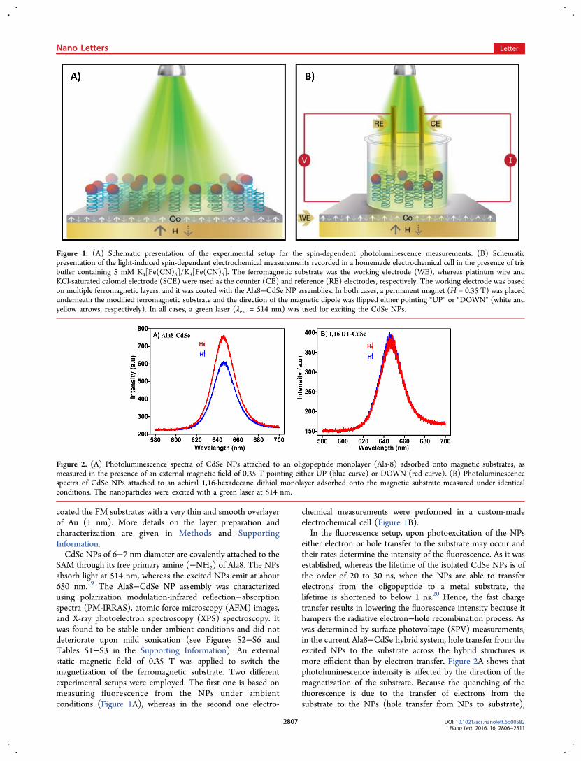
either electron or hole transfer to the substrate may occur andtheir rates determine the intensity of the fluorescence. As it wasestablished, whereas the lifetime of the isolated CdSe NPs is ofthe order of 20 to 30 ns, when the NPs are able to transferelectrons from the oligopeptide to a metal substrate, thelifetime is shortened to below 1 ns.20 Hence, the fast chargetransfer results in lowering the fluorescence intensity because ithampers the radiative electron−hole recombination process. Aswas determined by surface photovoltage (SPV) measurements,in the current Ala8−CdSe hybrid system, hole transfer from theexcited NPs to the substrate across the hybrid structures ismore efficient than by electron transfer. Figure 2A shows thatphotoluminescence intensity is affected by the direction of themagnetization of the substrate. Because the quenching of thefluorescence is due to the transfer of electrons from thesubstrate to the NPs (hole transfer from NPs to substrate),
Figure 1. (A) Schematic presentation of the experimental setup for the spin-dependent photoluminescence measurements. (B) Schematicpresentation of the light-induced spin-dependent electrochemical measurements recorded in a homemade electrochemical cell in the presence of trisbuffer containing 5 mM K4[Fe(CN)6]/K3[Fe(CN)6]. The ferromagnetic substrate was the working electrode (WE), whereas platinum wire andKCl-saturated calomel electrode (SCE) were used as the counter (CE) and reference (RE) electrodes, respectively. The working electrode was basedon multiple ferromagnetic layers, and it was coated with the Ala8−CdSe NP assemblies. In both cases, a permanent magnet (H = 0.35 T) was placedunderneath the modified ferromagnetic substrate and the direction of the magnetic dipole was flipped either pointing “UP” or “DOWN” (white andyellow arrows, respectively). In all cases, a green laser (λexc = 514 nm) was used for exciting the CdSe NPs.
Figure 2. (A) Photoluminescence spectra of CdSe NPs attached to an oligopeptide monolayer (Ala-8) adsorbed onto magnetic substrates, asmeasured in the presence of an external magnetic field of 0.35 T pointing either UP (blue curve) or DOWN (red curve). (B) Photoluminescencespectra of CdSe NPs attached to an achiral 1,16-hexadecane dithiol monolayer adsorbed onto the magnetic substrate measured under identicalconditions. The nanoparticles were excited with a green laser at 514 nm.
Nano Letters Letter
DOI: 10.1021/acs.nanolett.6b00582Nano Lett. 2016, 16, 2806−2811
2807
when the electron’s spin is parallel to the adsorbed moleculeaxis (when the magnet was pointing “UP”), its transfer is moreefficient and as a result photoluminescence intensity getquenched. This observation is consistent with former datashowing the same spin preference also in spin-dependentphotoelectron transmission through oligopeptides.21 Theaverage ratio of the PL intensity, as estimated by the areaunder the curve measured for two different directions (themagnet pointing “DOWN” vs “UP”), was found to be 1.4 ±0.2. These results are highly reproducible among five differentsamples prepared under identical conditions. Several controlexperiments were performed where Ala-8 was replaced byachiral 1,16-hexadecane dithiol (DT), biphenyl-4,4′-dithiol(BD), and 4-aminothiophenol; an external magnetic field wasapplied, keeping the experimental setup identical. The achiralSAM−CdSe NP assembles did not exhibit any effect of theexternal magnetic field on the PL intensity, as shown in Figure2B and Figure S7, corroborating that the CISS effect arisesexclusively from chiral structures.4
Spin-dependent electrochemical measurements were per-formed using both the oligopeptide and oligopeptide-NPstructures assembled on the ferromagnetic substrates in thepresence of a chemically robust redox probe such as
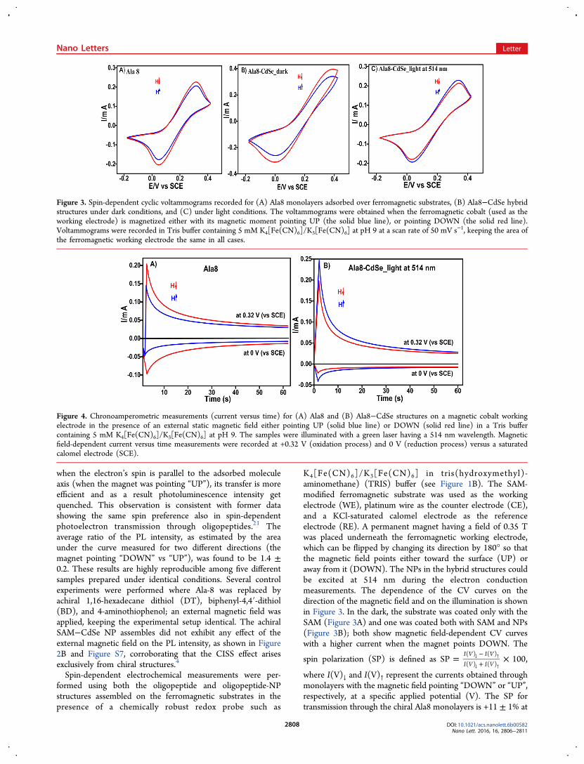
K4[Fe(CN)6]/K3[Fe(CN)6] in tris(hydroxymethyl)-aminomethane) (TRIS) buffer (see Figure 1B). The SAM-modified ferromagnetic substrate was used as the workingelectrode (WE), platinum wire as the counter electrode (CE),and a KCl-saturated calomel electrode as the referenceelectrode (RE). A permanent magnet having a field of 0.35 Twas placed underneath the ferromagnetic working electrode,which can be flipped by changing its direction by 180° so thatthe magnetic field points either toward the surface (UP) oraway from it (DOWN). The NPs in the hybrid structures couldbe excited at 514 nm during the electron conductionmeasurements. The dependence of the CV curves on thedirection of the magnetic field and on the illumination is shownin Figure 3. In the dark, the substrate was coated only with theSAM (Figure 3A) and one was coated both with SAM and NPs(Figure 3B); both show magnetic field-dependent CV curveswith a higher current when the magnet points DOWN. The
spin polarization (SP) is defined as = ×−+
↓ ↑
↓ ↑SP 100
I V I V
I V I V
( ) ( )
( ) ( ),
where I(V)↓ and I(V)↑ represent the currents obtained throughmonolayers with the magnetic field pointing “DOWN” or “UP”,respectively, at a specific applied potential (V). The SP fortransmission through the chiral Ala8 monolayers is +11 ± 1% at
Figure 3. Spin-dependent cyclic voltammograms recorded for (A) Ala8 monolayers adsorbed over ferromagnetic substrates, (B) Ala8−CdSe hybridstructures under dark conditions, and (C) under light conditions. The voltammograms were obtained when the ferromagnetic cobalt (used as theworking electrode) is magnetized either with its magnetic moment pointing UP (the solid blue line), or pointing DOWN (the solid red line).Voltammograms were recorded in Tris buffer containing 5 mM K4[Fe(CN)6]/K3[Fe(CN)6] at pH 9 at a scan rate of 50 mV s−1, keeping the area ofthe ferromagnetic working electrode the same in all cases.
Figure 4. Chronoamperometric measurements (current versus time) for (A) Ala8 and (B) Ala8−CdSe structures on a magnetic cobalt workingelectrode in the presence of an external static magnetic field either pointing UP (solid blue line) or DOWN (solid red line) in a Tris buffercontaining 5 mM K4[Fe(CN)6]/K3[Fe(CN)6] at pH 9. The samples were illuminated with a green laser having a 514 nm wavelength. Magneticfield-dependent current versus time measurements were recorded at +0.32 V (oxidation process) and 0 V (reduction process) versus a saturatedcalomel electrode (SCE).
Nano Letters Letter
DOI: 10.1021/acs.nanolett.6b00582Nano Lett. 2016, 16, 2806−2811
2808

+50 mV and +7 ± 0.5% at +0.32 V. These values are similar tothose obtained before with different chiral molecules.10,22
Figure 3B,C presents CV curves obtained with the CdSe NPsattached to SAM. In addition to the PMIRRAS, AFM images,and XPS data, the attachment of the NPs was also confirmed bythe voltammetry measurements, which showed an increase inthe oxidation potential of the redox probe, Fe2+, by 60 mV. Inthe dark (Figure 3B), the hybrid system has an SP value of +9± 1% at the reduction potential (at 0 V) and +8 ± 0.5% at theoxidation potential (+0.38 V). Upon illumination of the CdSeNPs, the sign of the SP is reversed and it is −3 ± 0.5% at +25mV (Figure 3C). Namely, illumination causes a flip in the signand a reduction in the absolute value of the spin polarization.To verify the observations, we performed chronoamperom-
etry experiments in which the current at a given potential isrecorded as a function of time. The electron conduction acrossthe SAMs (of Ala8) themselves and through illuminatedoligopeptide−CdSe structures was measured (Figure 4A,B,respectively). Whereas for the SAMs themselves or for theSAM−CdSe NPs in the dark (data not shown), the Faradaiccurrent was higher when the magnet was pointing DOWN;however, in the illuminated sample the current was higherwhen the magnetic field direction was pointing UP. This effectwas observed for both the oxidation or reduction processes. Forthe chiral SAMs, only when the SAM−CdSe NPs were in thedark the SP was +17 ± 1% and +35 ± 2% at 0.32 and 0 V,respectively. When the SAM−CdSe NP sample was illuminatedat 514 nm, the SP is −11 ± 1% at +0.32 V, and −30 ± 2% atthe reduction potential, 0 V. The voltammograms recorded ona bare ferromagnetic substrate as a working electrode did notexhibit any magnetic field effect on the Faradaic current whenthe working electrode was magnetized either with its magneticmoment pointing UP or DOWN (Figure S8).It is known that in the CISS effect, the alignment of the
preferred spin in the electron transport process depends onseveral factors, among them (i) the handedness of themolecules, (ii) the electric field along them, and (iii) theelectrons’ velocity.12 For example, electrons moving from rightto left along molecules with a preferred spin for a given electricfield will move with the opposite spin if the external field isreversed. However, if the electrons move from left to right with
the opposite sign of field on the molecule then the same spinsof the electrons will be preferred in both directions. In theferromagnetic substrate cobalt, the high density of state belowand above the Fermi level is of the same spin. Hence, this iswhy in the oxidation and reduction part of the electrochemicalcycle, the same spin is preferred.Figure 5 presents a scheme that explains the experimental
observations. Before photoexcitation (Figure 5A), the NPs arepositively charged, as observed by the XPS measurements thatindicate that the work function of the surface decreases by 105meV upon the NPs attachment to the functional polypeptidemonolayers (see Table S3). The PL studies are consistent withthe electrons being transferred with their spin aligned parallel totheir velocity. This observation is consistent with former studieson spin-dependent electron transmission through the sameoligopeptides.10 In the reduction process of the electrochemicalmeasurements, electrons move from the cobalt substrate (Co)through the helical molecule to the NPs and thereafter to theredox couple in solution. In the oxidation process a reverse fieldis applied and the electrons are transferred in the oppositedirection from the redox couple to the NPs and from there toCo; as a result, the same spin is preferred for electronstransferred from the NPs to the Co. When the NPs arephotoexcited, a hole is transferred from the NPs to the Cosubstrate and the NPs are now negatively charged (Figure 5B).Chemically resolved electrical measurements (CREM) datashow a prominent response of the peptide−CdSe NP structuresto light illumination at 630 nm. A decrease in the bindingenergies of the Cd line by 120 meV under light, as comparedwith the same value recorded in the dark, results in a clearmanifestation of the excited state hole transfer to the substrateand creates a negative charge over the NPs (Figure S6 andTable S3). Hence, the electric field on the chiral molecule is ofthe opposite direction and now electrons with spin antiparallelto their velocity are transferred. The electrochemical process issimilar as in the dark with the electrons transmitted betweenthe substrate and the redox couple in solution through the NPs.
■ CONCLUSIONS
The present study demonstrates the interaction between lightand electrons’ spin as manifested when light controls the spin-
Figure 5. A scheme describing the effect of the light on the spin selectivity. Before excitation (A), the NPs are positively charged and electrons aretransferred with their spin aligned parallel to their velocity. In the reduction part of the electrochemical process electrons move from the FM cobaltsubstrate (Co) through the helical molecule to the NPs and from there to the redox couple present in solution. In the oxidation stage, the electronsare transferred from the redox couple to the NPs and from there to the Co. When NPs are photoexcited (B), because of the hole being transferredfrom the NPs to the Co the NP is now negatively charged. Hence, the electric field on the chiral molecule is in the opposite direction and nowelectrons with spin antiparallel to their velocity are transferred preferentially. The electrochemical process is similar to the electrons transmittedthrough the NPs to the redox couple in solution.
Nano Letters Letter
DOI: 10.1021/acs.nanolett.6b00582Nano Lett. 2016, 16, 2806−2811
2809

selective electron transport through chiral molecules. Byattaching a fluorophore, the NPs, to the chiral molecule theactual spin orientation being transmitted can be flipped withsignificant efficiency. Thus, when combining light and amagnetic field the chirality-induced electron transfer processmay find increased interest in the field of light-controlledbiospintronic research.23 It is important to appreciate that theeffect observed here relates to weak magnetic fields applied onthe ferromagnetic substrate and hence differ significantly fromthe known magneto-chiral anisotropy in charge transfer, inwhich very high fields (many Tesla) are applied.24 Thecombination of systems, like those presented here, andmolecular magnets25,26 may result is a new type of photo-spintronics devices that are all molecular.
■ METHODS
Preparation of Soft Ferromagnetic Substrates. Theferromagnetic films with a perpendicular easy axis wereprepared on soda lime glass slides and on silicon substrate⟨100⟩ on which 5 nm of tantalum and 2 nm of platinum weredeposited using a magnetron sputtering system with a basepressure of <3 × 10⁻⁸ Torr. Cobalt and platinum depositionwere repeated thrice on top of the 2 nm platinum layer. A thinlayer of cobalt, followed by 1 nm gold, was grown on top. Thestacking structure of the magnetic substrates was Si/SiO2/Ta5/Pt 2/[Co 0.28/Pt 0.3]3/Co 0.28/Au 1 (in nm). All metallayers were deposited at a working pressure of Ar 1.5 mTorr atroom temperature.Preparation of Oligopeptide Monolayer and Oligo-
peptide−CdSe Nanoparticle Structures. Magnetic sub-strates with a 1 nm Au overlayer were cleaned with boilingacetone followed by ethanol for 15 min in each. The substrateswere dried carefully under N2. Subsequently, the substrateswere plasma cleaned in argon (at ∼0.4 Torr) for 1 min andimmediately immersed into a solution containing 0.1 mMpolyalanine (Ala8) in TFE/H2O (6:4, v/v, deoxygenated withAr for 45 min) for 48 h in order to achieve maximum surfacecoverage. The functionalized substrates were rinsed with thesame solvent mixture and mildly sonicated for 5 s to removeany unreacted materials. Consequently, the monolayers weredried carefully under N2 and used for IR, XPS, andelectrochemical measurements.Self-assembled monolayers of Ala8, added onto the magnetic
substrates, were immersed into a solution containing CdSe inanhydrous toluene for 4 h under dark conditions. In order toremove unreacted NPs, the substrates were lightly sonicated for5 s in toluene and dried gently under N2 flow and immediatelyused for measurements.
■ CHARACTERIZATION
The magnetic properties of perpendicular magnetized [Co/Pt]films were characterized by using a vibrating sample magneto-meter (VSM, VersaLab Quantum Design). Saturation magnet-ization (Ms) is 827 ± 96.7 emu/cm3 (measured moment/volume of Co and Pt) and the coercively is 145 and 270 Oe forsubstrate 1 and substrate 2, respectively (Figure S1).The formation of monolayers in the polypetide and its CdSe
NPs structures were characterized by recording the polarizationmodulation-infrared reflection−absorption spectra (PM-IRRAS) using a Nicolet 6700 FTIR equipped with a PEM-90photoelastic modulator (Hinds Instruments, Hillsboro, OR) atan incidence angle of 80°. In all cases, vibrational stretching
frequencies of amide I and amide II bonds in the SAMs weremonitored.
X-ray Photoelectron Spectroscopy (XPS) and Chemi-cally Resolved Electrical Measurements (CREM). Freshlyprepared SAMs of oligopeptide (Ala8) and peptide−CdSearchitectures on the ferromagnetic substrates were analyzed byXPS measurements using a Kratos Axis Ultra DLDspectrometer equipped with a monochromatic Al Kα X-raysource (hν = 1486.6 eV) operating at a power of 75 W. Twoemission angles (65° and 0° with respect to the surface normal)were compared. Detection pass energies ranged between 20and 80 eV. Elemental concentrations and layer thicknesses werededuced from the relative intensities of overlayer (N, C, O, andS 2p) versus substrate (Ta, Pt, Co, Au, and O) signals. Specialcare was devoted to eliminate beam-induced effects and damageto molecules in particular. This was achieved by conductingrepeated measurements, starting with very fast scans at freshspots, in order to evaluate the exposure-dependent spectralchanges.The coverage by CdSe nanoparticles (NPs) was estimated
based on the NP average size, 6−7 nm in diameter, which wasdetermined independently. Work function measurements wereperformed prior to any XPS scan, by inspecting thephotoelectron onset at low kinetic energies under a sourcepower of 0.3W. Site-specific photovoltages were extracted byevaluating the energy shifts of representative lines (Cd, N, C,and Au) under a 630 nm diode source.27 The advantage ofthese measurements over standard SPV experiments stemsfrom the fact that one can resolve local electrostatic potentialchanges and in particular those developing specifically on theCdSe NPs.The formation of Ala8−CdSe nanoparticle structures on
ferromagnetic substrates was further confirmed by AFM imagesrecorded on a Multimode/Nanoscope (Bruker-Nano, SantaBarbara, CA, U.S.A.). The AFM images were acquired innoncontact mode and a Si probe having a resonance frequencyof 70−90 kHz was used. The topographic images wererecorded at a scan rate of 1 Hz. Several images (at least atfour different points) of each sample were taken from differentfields of view (0.5−2.0 μm) to confirm the uniformity andreproducibility of the samples. The formation of homogeneousdistributions of CdSe nanoparticles in the monolayer wasconfirmed by AFM imaging.The PL measurements were carried out by using a LabRam
HR800-PL spectrofluorimeter microscope (Horiba Jobin-Yivon). Typically, 514 nm laser light (argon-ion CW laser at∼15 mW/cm2) has been used for excitation of CdSe NPs. Theincident light was impinged on the surface at an angle 90° tothe sample surface. Prior to collecting the PL spectra, an area(typically, 20 au × 20 au) and number points (25 points)within this area were defined in order to map the surface.Afterward, the PL spectra were collected using a microscope(with a 5× high working distance lens). During themeasurement, a confocal aperture (1100 μm) was fully openedand the integration time was maintained at 15 s. Finally, thespectra were presented after averaging out the PL of individualpoints within the defined area at two different magneticorientations.Cyclic voltammetry (CV) and chronoamperometric meas-
urements were carried out using a Bio-Logic potentiostat SAS(Model SP-200) with an inbuilt software EC Lab (V 10.36) byemploying a typical three-electrode electrochemical cell. ASAM-modified Co substrate, a Pt wire, and KCl-saturated
Nano Letters Letter
DOI: 10.1021/acs.nanolett.6b00582Nano Lett. 2016, 16, 2806−2811
2810

calomel electrodes were used as the working, counter, andreference electrodes, respectively, in the presence of Tris buffercontaining 5 mM K4[Fe(CN)6]/K3[Fe(CN)6] taken into acustom-made electrochemical cell. Spin-dependent electro-chemical measurements were carried out in the presence of apermanent magnet having a magnetic field strength of 0.35 T,which was placed underneath the Co working electrode,followed by changing the magnet’s direction (UP/DOWN).
■ ASSOCIATED CONTENT*S Supporting InformationThe Supporting Information is available free of charge on theACS Publications website at DOI: 10.1021/acs.nano-lett.6b00582.
Sample preparation and characterization and theexperimental setup. (PDF)
■ AUTHOR INFORMATIONCorresponding Author*E-mail: [email protected] ContributionsP.C.M. fabricated and characterized the monolayers, performedthe electrochemical measurements, and analyzed the data. P.R.conducted the photoluminescence experiments. D.K. fabricatedthe soft magnetic substrates, designed the film structure, andcharacterized it. E.E.F. guided the preparation of the magneticsubstrates and the scientific discussion. H.C. carried out XPSand CREM measurements. R.N. conceived the research plan.All authors contributed to the writing of the manuscript.NotesThe authors declare no competing financial interest.
■ ACKNOWLEDGMENTSP.C.M., P.R., and R.N. acknowledge financial assistance fromthe ERC-Adv grant, the VW Foundation, and the Israel ScienceFoundation.
■ REFERENCES(1) Lambert, C.-H.; Mangin, S.; Varaprasad, B. S. D. C. S.; Takahashi,Y. K.; Hehn, M.; Cinchetti, M.; Malinowski, G.; Hono, K.; Fainman,Y.; Aeschlimann, M.; Fullerton, E. E. Science 2014, 345, 1337−1340.(2) Koopmans, B.; Malinowski, G.; Dalla Longa, F.; Steiauf, D.;Fahnle, M.; Roth, T.; Cinchetti, M.; Aeschlimann, M. Nat. Mater.2009, 9, 259−265.(3) Kirilyuk, A.; Kimel, A. V.; Rasing, T. Rep. Prog. Phys. 2013, 76,026501.(4) Bliokh, K. Y.; Smirnova, D.; Nori, F. Science 2015, 348, 1448−1451.(5) Dediu, V. A.; Hueso, L. E.; Bergenti, I.; Taliani, C. Nat. Mater.2009, 8, 707−716.(6) Sun, D.; Ehrenfreund, E.; Vardeny, Z. V. Chem. Commun. 2014,50, 1781−1793.(7) Sanvito, S. Nat. Mater. 2007, 6, 803−804.(8) Lee, S.-Y.; Paik, S.-Y.; McCamey, D. R.; Yu, J.; Burn, P. L.;Lupton, J. M.; Boehme, C. J. Am. Chem. Soc. 2011, 133, 2019−2021.(9) Vardeny, Z. V.; Heeger, A. J.; Dodabalapur, A. Synth. Met. 2005,148, 1−3.(10) Sugawara, T.; Matsushita, M. M. J. Mater. Chem. 2009, 19,1738−1753.(11) Gohler, B.; Hamelbeck, V.; Markus, T. Z.; Kettner, M.; Hanne,G. F.; Vager, Z.; Naaman, R.; Zacharias, H. Science 2011, 331, 894−897.(12) Naaman, R.; Waldeck, D. H. Annu. Rev. Phys. Chem. 2015, 66,263−281.
(13) Dor, O. B.; Yochelis, S.; Mathew, S. P.; Naaman, R.; Paltiel, Y.Nat. Commun. 2013, 4, 2256.(14) Mathew, S. P.; Mondal, P. C.; Moshe, H.; Mastai, Y.; Naaman,R. Appl. Phys. Lett. 2014, 105, 242408.(15) Ravi, S.; Sowmiya, P.; Karthikeyan, A. SPIN 2013, 3, 1350003.(16) Alam, K. M.; Pramanik, S. Adv. Funct. Mater. 2015, 25, 3210−3218.(17) Pawlowski, J.; Juhaniewicz, J.; Tymecka, D.; Sek, S. Langmuir2012, 28, 17287−17294.(18) Mondal, P. C.; Fontanesi, C.; Waldeck, D. H.; Naaman, R. ACSNano 2015, 9, 3377−3384.(19) Gotesman, G.; Waldeck, D. H.; Naaman, R. J. Phys. Chem. A2009, 113, 7213−7217.(20) Odoi, M. Y.; Hammer, N. I.; Early, K. T.; McCarthy, K. D.;Tangirala, R.; Emrick, T.; Barnes, M. D. Nano Lett. 2007, 7, 2769−2773.(21) Kettner, M.; Gohler, B.; Zacharias, H.; Mishra, D.; Kiran, V.;Naaman, R.; Fontanesi, C.; Waldeck, D. H.; Sek, S.; Pawlowski, J.;Juhaniewicz, J. J. Phys. Chem. C 2015, 119, 14542−14547.(22) Mondal, P. C.; Kantor-Uriel, N.; Mathew, S. P.; Tassinari, F.;Fontanesi, C.; Naaman, R. Adv. Mater. 2015, 27, 1924−1927.(23) Einati, H.; Mishra, D.; Friedman, N.; Sheves, M.; Naaman, R.Nano Lett. 2015, 15, 1052−1056.(24) Krstic, V.; Roth, S.; Burghard, M.; Kern, K.; Rikken, G. L. J. A. J.Chem. Phys. 2002, 117, 11315−11319.(25) Ishikawa, N.; Sugita, M.; Ishikawa, T.; Koshihara, S.-y.; Kaizu, Y.J. Am. Chem. Soc. 2003, 125, 8694−8695.(26) Milios, C. J.; Vinslava, A.; Wernsdorfer, W.; Moggach, S.;Parsons, S.; Perlepes, S. P.; Christou, G.; d Brechin, E. K. J. Am. Chem.Soc. 2007, 129, 2754−2755.(27) Cohen, H.; Sarkar, S. K.; Hodes, G. J. Phys. Chem. B 2006, 110,25508−25513.
Nano Letters Letter
DOI: 10.1021/acs.nanolett.6b00582Nano Lett. 2016, 16, 2806−2811
2811