Photoluminescent and antimicrobial properties of silver...
10
ORIGINAL ARTICLE Photoluminescent and antimicrobial properties of silver‐doped indium hydroxide synthesized by one‐step microwave‐assisted hydrothermal method Nivaldo Freire Andrade Neto 1 | Mara T. S. Tavares 2 | Erik A. C. Ferreira 1 | Ana I. L. Sales 3 | Lucymara F. A. Lima 3 | Elson Longo 4 | Mauricio R. D. Bomio 1 | Fabiana V. Motta 1 1 LSQM, DEMAT, UFRN, Natal, Brazil 2 IFBA, Feira de Santana, Feira de Santana, Brazil 3 LBMG, UFRN, Natal, Brazil 4 LIEC, UFSCar, São Carlos, Araraquara, Brazil Correspondence Nivaldo Freire Andrade Neto, LSQM, DEMAT, UFRN, Natal, Brazil. Email: [email protected] Funding information FAPESP-CDMF, Grant/Award Number: 2013/07296-2; CNPq; CAPES Abstract Silver‐doped indium hydroxide In (1-x) (OH) 3 : x Ag (with x = 0, 1, 2, 4, and 8 mol %) of Ag nanoparticles were synthesized by the microwave‐assisted hydrothermal (MAH) method at 140°C for 30 minutes. These nanoparticles were characterized by X‐ray diffraction (XRD), fourier transformed infrared (FT‐IR) spectroscopy, and optical diffuse reflectance. Photoluminescence (PL) spectra were acquired with a 350 nm beam of a krypton ion laser as an excitation source. The antibacte- rial activities of the samples were evaluated against gram negative Escherichia coli bacteria and gram positive Staphylococcus aureus bacteria using the disc dif- fusion method. The results showed that all diffraction peaks present in XRD pat- terns could be indexed to the cubic lattice related to the In(OH) 3 phase. Broadband photoluminescence behavior in visible range spectra was observed for all samples with a maximum peak centered in the blue and green regions. The antibacterial activities showed that In (1-x) (OH) 3 : x Ag nanoparticles have a promis- ing bactericide that can be used for deactivating microbes. KEYWORDS bactericide activity, doping, indium hydroxide, microwave, silver 1 | INTRODUCTION In recent years, several papers report that different types of food and environmental sources harbor bacteria that are resistant to one or more antibiotics. 1,2 Therefore, any search of new methods that minimize or delete food contamination will have a significant impact on the incidence of food- borne diseases. 3 A possible approach is using nanoparticle materials to reduce the microbial contamination on the food surfaces and in the food preparation environment. Nanoma- terials are efficient, since they are able to attach more copies of microbial assemblage and cells. 4 Nanostructures presented unique properties, which are different from bulk materials, having enhanced properties and technological applications. 5–7 Silver nanoparticles have shown their efficacy as an alternative antimicrobial material over a wide diversity of bacteria. Dai et al 8 showed that nanoparticles of AgCl/Ag have a good antibacterial effect against Escherichia coli (ATCC 25922), Staphylococcus aureus (ATCC 29213), Staphylococcus epidermidis (ATCC 12228), Pseudomonas aeruginosa (ATCC 15692), and one fungal species Candida albicans (CAF2/1). Due to these proper- ties, they can be added to medical materials aimed at preventing infections. 9 Ag particles at a nanometer scale have a higher chemical activity due to their greater area Received: 6 June 2018 | Revised: 21 September 2018 | Accepted: 5 October 2018 DOI: 10.1111/ijac.13127 Int J Appl Ceram Technol. 2019;16:471–480. wileyonlinelibrary.com/journal/ijac © 2018 The American Ceramic Society | 471
Transcript of Photoluminescent and antimicrobial properties of silver...
OR I G I N A L AR T I C L E
Photoluminescent and antimicrobial properties of silver‐dopedindium hydroxide synthesized by one‐step microwave‐assistedhydrothermal method
Nivaldo Freire Andrade Neto1 | Mara T. S. Tavares2 | Erik A. C. Ferreira1 |
Ana I. L. Sales3 | Lucymara F. A. Lima3 | Elson Longo4 | Mauricio R. D. Bomio1 |
Fabiana V. Motta1
1LSQM, DEMAT, UFRN, Natal, Brazil2IFBA, Feira de Santana, Feira deSantana, Brazil3LBMG, UFRN, Natal, Brazil4LIEC, UFSCar, São Carlos, Araraquara,Brazil
CorrespondenceNivaldo Freire Andrade Neto, LSQM,DEMAT, UFRN, Natal, Brazil.Email: [email protected]
Funding informationFAPESP-CDMF, Grant/Award Number:2013/07296-2; CNPq; CAPES
AbstractSilver‐doped indium hydroxide In(1-x)(OH)3:xAg (with x = 0, 1, 2, 4, and 8 mol
%) of Ag nanoparticles were synthesized by the microwave‐assisted hydrothermal
(MAH) method at 140°C for 30 minutes. These nanoparticles were characterized
by X‐ray diffraction (XRD), fourier transformed infrared (FT‐IR) spectroscopy,
and optical diffuse reflectance. Photoluminescence (PL) spectra were acquired
with a 350 nm beam of a krypton ion laser as an excitation source. The antibacte-
rial activities of the samples were evaluated against gram negative Escherichia
coli bacteria and gram positive Staphylococcus aureus bacteria using the disc dif-
fusion method. The results showed that all diffraction peaks present in XRD pat-
terns could be indexed to the cubic lattice related to the In(OH)3 phase.
Broadband photoluminescence behavior in visible range spectra was observed for
all samples with a maximum peak centered in the blue and green regions. The
antibacterial activities showed that In(1-x)(OH)3: xAg nanoparticles have a promis-
ing bactericide that can be used for deactivating microbes.
KEYWORD S
bactericide activity, doping, indium hydroxide, microwave, silver
1 | INTRODUCTION
In recent years, several papers report that different types offood and environmental sources harbor bacteria that areresistant to one or more antibiotics.1,2 Therefore, any searchof new methods that minimize or delete food contaminationwill have a significant impact on the incidence of food-borne diseases.3 A possible approach is using nanoparticlematerials to reduce the microbial contamination on the foodsurfaces and in the food preparation environment. Nanoma-terials are efficient, since they are able to attach morecopies of microbial assemblage and cells.4 Nanostructurespresented unique properties, which are different from bulk
materials, having enhanced properties and technologicalapplications.5–7
Silver nanoparticles have shown their efficacy as analternative antimicrobial material over a wide diversity ofbacteria. Dai et al8 showed that nanoparticles of AgCl/Aghave a good antibacterial effect against Escherichia coli(ATCC 25922), Staphylococcus aureus (ATCC 29213),Staphylococcus epidermidis (ATCC 12228),Pseudomonas aeruginosa (ATCC 15692), and one fungalspecies Candida albicans (CAF2/1). Due to these proper-ties, they can be added to medical materials aimed atpreventing infections.9 Ag particles at a nanometer scalehave a higher chemical activity due to their greater area
Received: 6 June 2018 | Revised: 21 September 2018 | Accepted: 5 October 2018
DOI: 10.1111/ijac.13127
Int J Appl Ceram Technol. 2019;16:471–480. wileyonlinelibrary.com/journal/ijac © 2018 The American Ceramic Society | 471
ratio by its volume, and have drawn an increasing atten-tion to it,10,11 resulting in the appearance of newmechanical, chemical, electrical, optical, magnetic, elec-tro‐optical, and magneto‐optical properties of thenanoparticles, which are different from their bulk proper-ties.12 Studies reported the antibacterial activity of Agnanoparticles with different modifications.11,13 Theantimicrobial activity increases as the size of the Agnanoparticles decreases.14 However, silver nanoparticleswith diameters <200 nm have high surface energy, con-sequently they tend to aggregate spontaneously to reducethis energy, making them unstable for long‐term applica-tions in air, water or sunlight.15 To solve this problem, awide range of materials, such as ZnO,16,17 TiO2,
18,19
and SiO220,21 have been employed as a support for Ag
nanoparticles, so that the ultra fine Ag nanoparticles canbe homogeneously formed without aggregation.22 Amongthe numerous semiconductors, the use of In(OH)3 is notreported in literature. This semiconductor has a wide(Eg) gap estimated to be 5.15 eV,23,24 especially atnanostructure sizes, and has created intensive interestbecause of its semiconducting and optical properties.
In summary, this study reports the synthesis ofIn(1-x)(OH)3:xAg structures doped with x = 0, 1, 2, 4, and8 mol% of Ag using the MAH method at the low tempera-ture of 140°C for 30 minutes. In addition, the antimicrobialactivity and photoluminescent properties of Ag‐doped In(OH)3 particles were investigated to enlarge our under-standing of their fundamental properties (Table 1).
2 | EXPERIMENTAL
2.1 | Materials
Indium nitrate (In(NO3)3·H2O, 99%; Sigma‐Aldrich, St.Louis, MO), silver nitrate (AgNO3, 99%; Fluka Mumbai,India), potassium hydroxide (KOH, Fluka, 99%), and dis-tilled water were used as received to prepare silver‐dopedindium hydroxide nanoparticles.
2.2 | Preparation of In(OH)3 nanoparticles
The experimental procedure is as follows: 7 mmol ofindium nitrate and silver nitrate were dissolved into 80 mLof deionized water under constant stirring for 20 minutes.Ag was added to it in the following percentages: 0, 1, 2, 4,and 8 mol%. The pH of the solution was adjusted to 11 byadding potassium hydroxide (6 mol/L). The mixture wastransferred into a teflon autoclave which was sealed, andthe reaction system was heated under hydrothermal condi-tions at 140°C for 30 minutes (the heating rate was fixedat 25C°/min) using microwave irradiation (2.45 GHz and amaximum power of 800 W). The pressure in the autoclave
was stabilized at 3.0 atm. The white product obtained bythe microwave‐assisted hydrothermal (MAH) treatment wascentrifuged, washed with distilled water and ethanol andfinally dried at room temperature.
2.3 | Characterization of In(OH)3nanoparticles
The powders were characterized by X‐ray diffraction(XRD) using a Shimadzu diffractometer (Model XRD‐7000, CuKα radiation [λ = 1.54 Å], 40 kV and 30 mA and2θ from 5° to 80°; Shimadzu, Tokyo, Japan). FT‐IR spectrawere taken in the range from 4000 to 450 cm−1 using KBrpellets as a reference using a Perkin–Elmer FT‐IR 1000spectrophotometer in its transmittance mode. The In(OH)3morphology and particle size were observed by FEG‐SEM(Supra 35‐VP Model; Carl Zeiss, Jena, Germany). UV–visreflectance spectra of In(OH)3 pure and doped powderswere taken using Shimadzu (model UV‐2600) spectropho-tometer. All measurements were taken at room temperature.
2.4 | Photoluminescence
Photoluminescence (PL) spectra were acquired with an AshMonospec 27 monochromator (Thermal Jarrel, Franklin,MA) and a R4446 photomultiplier (Hamamatsu Photonics,Hamamatsu, Japan). The 350 nm beam of a krypton ionlaser (90 K; Coherent Innova, Santa Clara, CA) was usedas an excitation source while maintaining its maximum out-put power at 200 mW. All measurements were performedat room temperature.
2.5 | Antimicrobial tests
The in vitro antibacterial activity of Ag‐doped In(OH)3nanoparticles were tested against the bacterial speciesS. aureus and E. coli by the agar disc diffusion method.25
Initially, the stock cultures of bacteria were revived byinoculating in broth media and grown at 37°C for18 hours. The agar plates of media (10 g peptone‐, 10 g
TABLE 1 Lattice constants (a, Å), cell volumes (V, Å3) and theaverage crystallite size (Dcrys, Å) of the cubic structure of In(OH)3:xAg powders prepared by the microwave‐assisted hydrothermalmethod
In(OH)3:xAg a (Å) V (Å3) D (nm)
x = 0 7.97 505.9 3.20
x = 1% 7.95 503.7 2.94
x = 2% 7.94 503.1 1.93
x = 4% 7.93 501.5 1.85
x = 8% 7.92 499.8 1.81
472 | ANDRADE NETO ET AL.
NaCl and 5 g yeast extract, 20 g agar in 1000 mL of dis-tilled water) were prepared and wells were made on theplate. Each plate was inoculated with 18‐hour‐old cultures(100 μL) and spread evenly on the plate. After the samplesIn(1-x)(OH)3:xAg (x = 0, 0.01, 0.02, 0.04, 0.08) were placedonto well plates in triplicate. All the plates were incubatedat 37°C for 24 hours and the diameters of the inhibitionzone were noted.
3 | RESULTS AND DISCUSSION
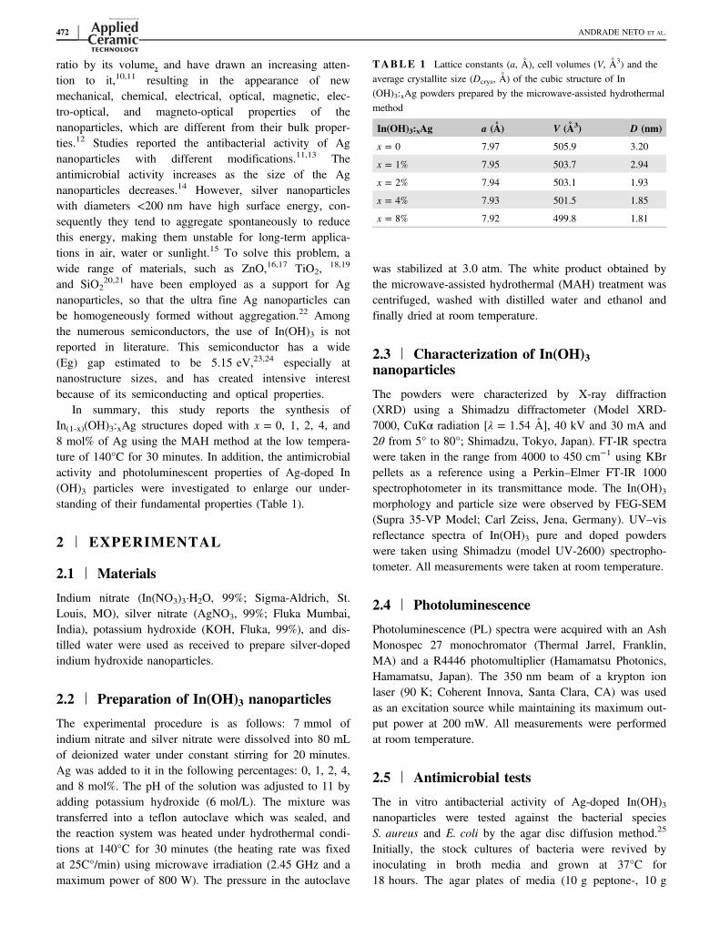
X‐ray diffraction patterns of In(OH)3 particles with differ-ent percentages of Ag prepared by the MAH method at140°C for 30 minutes are shown in Figure 1A‐E, respec-tively. All diffraction peaks in XRD patterns could beindexed to the cubic lattice related to the In(OH)3 phase(PDF card No. 16‐161). With the increase in percentages ofsilver (see Figure 1B–D), all phases obtained by the MAHtreatment remained as In(OH)3 with a cubic structure whichindicates that Ag ions were incorporated into In(OH)3nanocrystals. The presence of secondary phases of AgOand Ago was not observed, which indicates that the finalparticles are a pure phase compound.
The crystalline sizes were estimated by the Scherrerequation and full‐width half maximum (FWHM) of anobserved peak, respectively. The strongest peak (200) wasused to calculate the average crystallite size (D) of In(OH)3particles. The intensity of the peaks was decreased(Figure 1) and the position of the peaks slightly changed tothe right (higher angles) with the increase in the silver
concentration, which indicates that Ag+ doping can cause aslight decrease in the lattice parameters with the In(OH)3
26
doping. The addition of silver promotes the reduction ofthe parameter a for the In(OH)3 powders, where this reduc-tion is evident when comparing the samples of pure In(OH)3 and doped with 8% Ag, which have parameters a =7.97 and 7.92Å, respectively. The decrease in the latticeparameters of doped samples is a strong indication that theions are incorporated into the sites of In(OH)3. Theseresults are in agreement with the investigation realized bySena et al27 using the density functional theory (DFT).Sena et al27 showed in their work that the lattice parame-ters obtained from the fit for samples of In2O3 doped withFe, Co, and Ni at room temperature are smaller than thelattice parameters for the pure In2O3 samples.
The crystallite size decreased with Ag doping level,from 3.20 nm for pure In(OH)3 to 1.81 nm for 8 mol% Ag.These changes are associated with increased disorder in thecrystal structure of In(OH)3 by defects caused due to theincorporation of ions with larger ionic radius (In3+ = 0.81Å; Ag+ = 1.15 Å) suggesting that the peaks have agreater width according to the Scherrer equation. Byincreasing the dopant content of Ag+, the growth of thecrystal size was inhibited, this reduction in the crystal sizemay also be attributed to the formation of In–O–Ag prod-ucts on the surface of the In(OH)3:Ag, which inhibits thecrystal growth.28
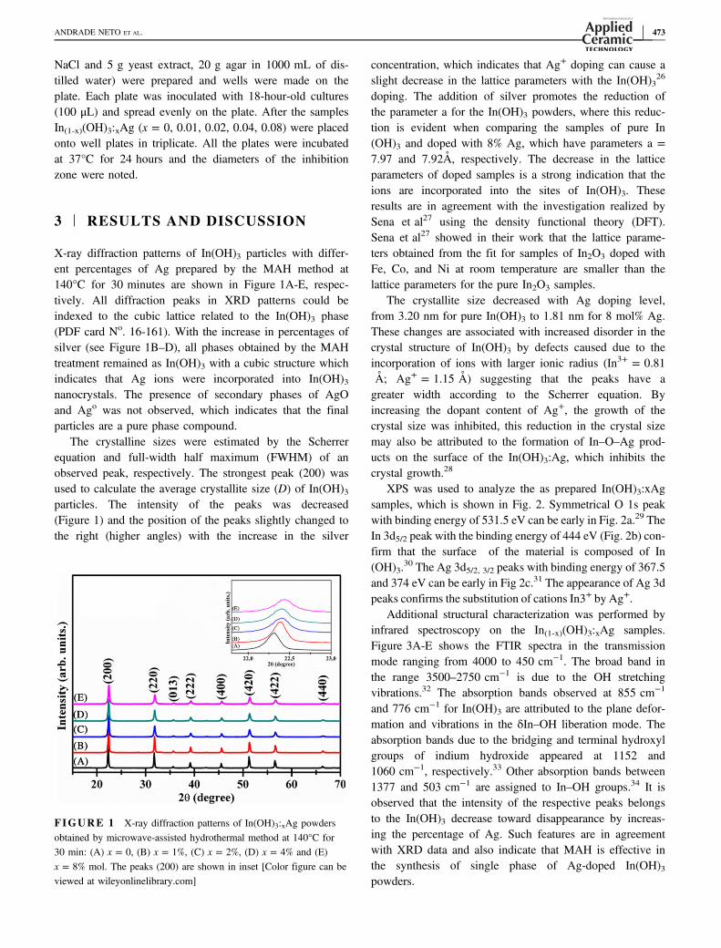
XPS was used to analyze the as prepared In(OH)3:xAgsamples, which is shown in Fig. 2. Symmetrical O 1s peakwith binding energy of 531.5 eV can be early in Fig. 2a.29 TheIn 3d5/2 peak with the binding energy of 444 eV (Fig. 2b) con-firm that the surface of the material is composed of In(OH)3.
30 The Ag 3d5/2, 3/2 peaks with binding energy of 367.5and 374 eV can be early in Fig 2c.31 The appearance of Ag 3dpeaks confirms the substitution of cations In3+ by Ag+.
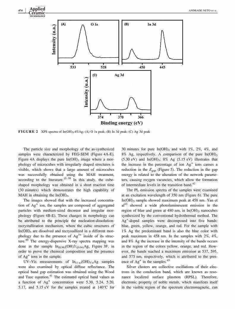
Additional structural characterization was performed byinfrared spectroscopy on the In(1-x)(OH)3:xAg samples.Figure 3A‐E shows the FTIR spectra in the transmissionmode ranging from 4000 to 450 cm−1. The broad band inthe range 3500–2750 cm−1 is due to the OH stretchingvibrations.32 The absorption bands observed at 855 cm−1
and 776 cm−1 for In(OH)3 are attributed to the plane defor-mation and vibrations in the δIn–OH liberation mode. Theabsorption bands due to the bridging and terminal hydroxylgroups of indium hydroxide appeared at 1152 and1060 cm−1, respectively.33 Other absorption bands between1377 and 503 cm−1 are assigned to In–OH groups.34 It isobserved that the intensity of the respective peaks belongsto the In(OH)3 decrease toward disappearance by increas-ing the percentage of Ag. Such features are in agreementwith XRD data and also indicate that MAH is effective inthe synthesis of single phase of Ag‐doped In(OH)3powders.
FIGURE 1 X‐ray diffraction patterns of In(OH)3:xAg powdersobtained by microwave‐assisted hydrothermal method at 140°C for30 min: (A) x = 0, (B) x = 1%, (C) x = 2%, (D) x = 4% and (E)x = 8% mol. The peaks (200) are shown in inset [Color figure can beviewed at wileyonlinelibrary.com]
ANDRADE NETO ET AL. | 473
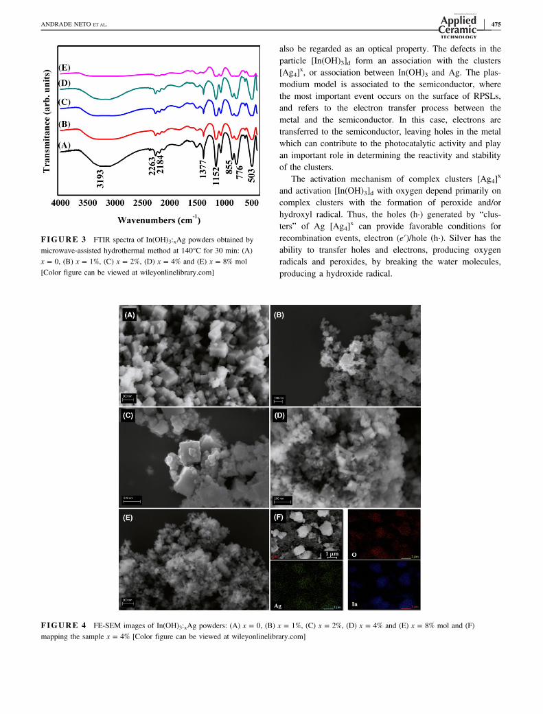
The particle size and morphology of the as‐synthesizedsamples were characterized by FEG‐SEM (Figure 4A‐E).Figure 4A displays the pure In(OH)3 image where a mor-phology of microcubes with irregularly shaped structures isvisible, which shows that a large amount of microcubeswas successfully obtained using the MAH treatment,according to the literature.35–39 In this study, the cube‐shaped morphology was obtained in a short reaction time(30 minutes) which demonstrates the high capability ofMAH in obtaining the In(OH)3.
The images showed that with the increased concentra-tion of Ag+ ion, the samples are composed of aggregatedparticles with medium‐sized decrease and irregular mor-phology (Figure 4B‐E). These changes in morphology canbe attributed to the principle the nucleation‐dissolution‐recrystallization mechanism, where the cubic structures ofIn(OH)3 are dissolved and recrystallized in a different mor-phology due to the presence of Ag3+ inside of its struc-ture.40 The energy‐dispersive X‐ray spectra mapping wasdone in the sample In(0.96)(OH)3:(0.04)Ag, Figure 3F, inorder to prove the chemical composition and the presenceof Ag+ ions in the sample.
UV–Vis measurements of In(1-x)(OH)3:xAg sampleswere also examined by optical diffuse reflectance. Theoptical band gap estimation was obtained using the Woodand Tauc equation.41 The estimated optical band values asa function of Ag+ concentration were 5.30, 5.24, 5.20,5.17, and 5.15 eV for the samples treated at 140°C for
30 minutes for pure In(OH)3 and with 1%, 2%, 4%, and8% Ag, respectively. A comparison of the pure In(OH)3(5.30 eV) and In(OH)3: 8% Ag (5.15 eV) illustrates thatthe increase in the percentage of ion Ag+ ions causes areduction in the Egap (Figure 5). The reduction in the gapenergy is related to the alteration of the network parame-ters, causing oxygen vacancies, which allow the formationof intermediate levels in the transition band.42
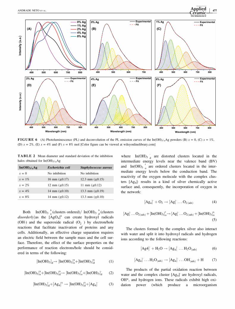
The PL emission spectra of the samples were examinedat an excitation wavelength of 350 nm (Figure 6). The pureIn(OH)3 sample showed maximum peak at 458 nm. Yan etal43 showed a wide photoluminescent emission in theregion of blue and green at 480 nm, in In(OH)3 nanocubessynthesized by the conventional hydrothermal method. TheAg+‐doped samples were decomposed into five bands:blue, green, yellow, orange, and red. For the sample with1% Ag the predominant band is also the blue color withpeak maximum in 458 nm. In the samples with 2%, 4%,and 8% Ag the increase in the intensity of the bands occursin the region of the colors yellow, orange, and red. How-ever, the bands reached a maximum emission at 537, 595,and 575 nm, respectively, which is attributed to the pres-ence of Ag+ in the samples.43
Silver clusters are collective oscillations of their elec-trons in the conduction band, which are known as reso-nance localized surface plasmon (RPSL). Therefore,electronic property of noble metals, which manifests itselfin the visible region of the spectrum electromagnetic, can
FIGURE 2 XPS spectra of In(OH)3:4%Ag; (A) O 1s peak; (B) In 3d peak; (C) Ag 3d peak
474 | ANDRADE NETO ET AL.
also be regarded as an optical property. The defects in theparticle [In(OH)3]d form an association with the clusters[Ag4]
x, or association between In(OH)3 and Ag. The plas-modium model is associated to the semiconductor, wherethe most important event occurs on the surface of RPSLs,and refers to the electron transfer process between themetal and the semiconductor. In this case, electrons aretransferred to the semiconductor, leaving holes in the metalwhich can contribute to the photocatalytic activity and playan important role in determining the reactivity and stabilityof the clusters.
The activation mechanism of complex clusters [Ag4]x
and activation [In(OH)3]d with oxygen depend primarily oncomplex clusters with the formation of peroxide and/orhydroxyl radical. Thus, the holes (h·) generated by “clus-ters” of Ag [Ag4]
x can provide favorable conditions forrecombination events, electron (e′)/hole (h·). Silver has theability to transfer holes and electrons, producing oxygenradicals and peroxides, by breaking the water molecules,producing a hydroxide radical.
FIGURE 3 FTIR spectra of In(OH)3:xAg powders obtained bymicrowave‐assisted hydrothermal method at 140°C for 30 min: (A)x = 0, (B) x = 1%, (C) x = 2%, (D) x = 4% and (E) x = 8% mol[Color figure can be viewed at wileyonlinelibrary.com]
FIGURE 4 FE‐SEM images of In(OH)3:xAg powders: (A) x = 0, (B) x = 1%, (C) x = 2%, (D) x = 4% and (E) x = 8% mol and (F)mapping the sample x = 4% [Color figure can be viewed at wileyonlinelibrary.com]
ANDRADE NETO ET AL. | 475
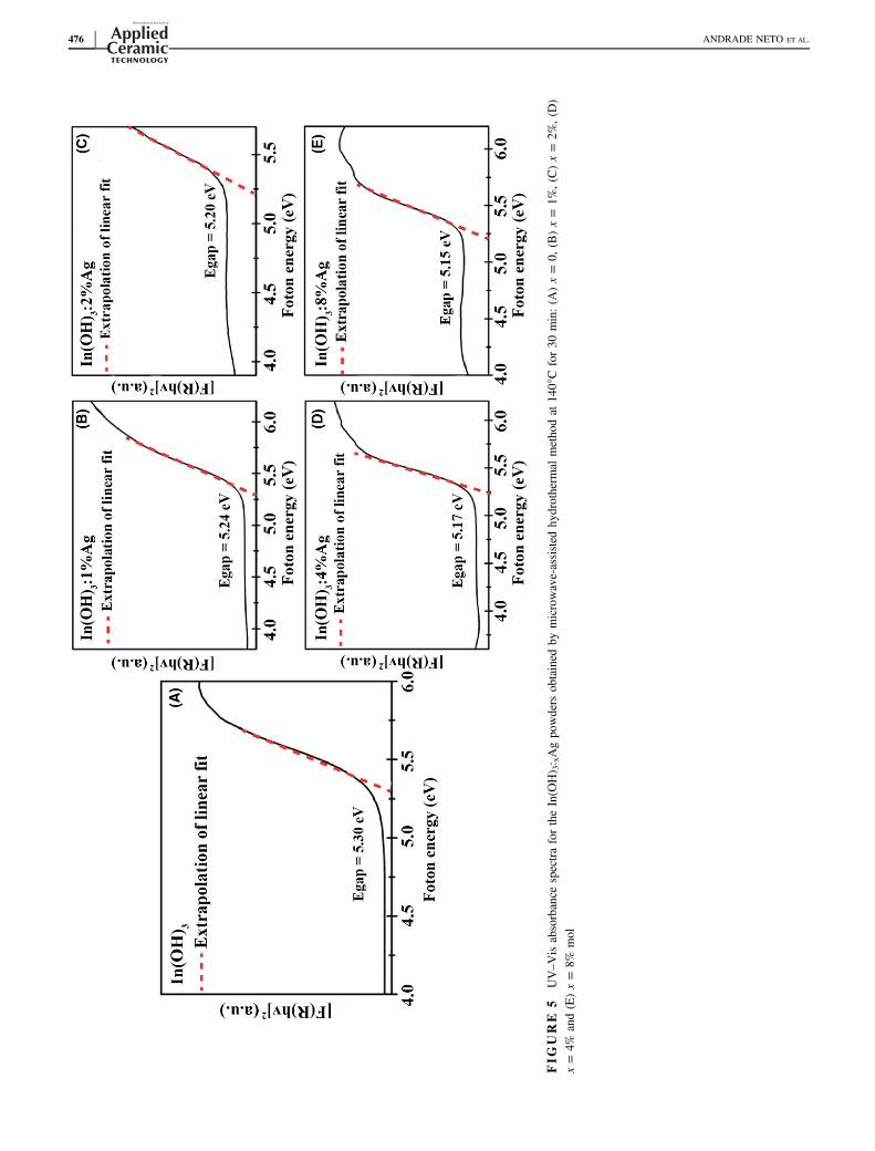
FIG
URE
5UV–V
isabsorbance
spectrafortheIn(O
H) 3: xAgpo
wders
obtained
bymicrowave‐assisted
hydrotherm
almetho
dat
140°C
for30
min:(A
)x=
0,(B)x=
1%,(C)x=
2%,(D)
x=
4%and(E)x=
8%mol
476 | ANDRADE NETO ET AL.
Both�In(OH)3
�xo (clusters ordered)=
�In(OH)3
�xd (clusters
disorderlyÞas the [AgO4]x can create hydroxyl radicals
(OH·) and the superoxide radical (O�2 ) by electron/hole
reactions that facilitate inactivation of proteins and anycells. Additionally, an effective charge separation requiresan electric field between the sample mass and the cell sur-face. Therefore, the effect of the surface properties on theperformance of reaction electrons/hole should be consid-ered in terms of the following:
In(OH)3½ �d! In(OH)3½ �xoþ In(OH)3½ �xd (1)
In(OH)3½ �xoþ In(OH)3½ �xd! In(OH)3½ �0oþ In(OH)3½ ��d (2)
In(OH)3½ ��dþ½Ag4�x ! In(OH)3½ �xdþ½Ag4�� (3)
where�In(OH)3
��d are distorted clusters located in the
intermediate energy levels near the valence band (BV)and
�In(OH)3
�0o are ordered clusters located in the inter-
mediate energy levels below the conduction band. Thereactivity of the oxygen molecule with the complex clus-ters [Ag4]· results in a kind of silver chemically activesurface and, consequently, the incorporation of oxygen inthe network:
½Ag4�� þ O2 ! ½Ag�� . . .O2 ðadsÞ (4)
½Ag�� . . .O2ðadsÞ þ In(OH)3½ �0o!½Ag�� . . .O2ðadsÞ þ In(OH)3½ �xo(5)
The clusters formed by the complex silver also interactwith water and split it into hydroxyl radicals and hydrogenions according to the following reactions:
½Ag4�� þ H2O ! ½Ag4�� . . .H2OðadsÞ (6)
½Ag4�� . . .H2OðadsÞ ! ½Ag4�� . . .OH�ðadsÞ þ H (7)
The products of the partial oxidation reaction betweenwater and the complex cluster [Ag4]
· are hydroxyl radicals,OH*, and hydrogen ions. These radicals exhibit high oxi-dation power (which produce a microorganism
TABLE 2 Mean diameter and standard deviation of the inhibitionhalos obtained for In(OH)3:xAg
In(OH)3:xAg Escherichia coli Staphylococcus aureus
x = 0 No inhibition No inhibition
x = 1% 16 mm (±0.17) 12.3 mm (±0.15)
x = 2% 12 mm (±0.15) 11 mm (±0.12)
x = 4% 14 mm (±0.10) 13.3 mm (±0.19)
x = 8% 14 mm (±0.12) 13.3 mm (±0.10)
FIGURE 6 (A) Photoluminescence (PL) and deconvolution of the PL emission curves of the In(OH)3:xAg powders (B) x = 0, (C) x = 1%,(D) x = 2%, (E) x = 4% and (F) x = 8% mol [Color figure can be viewed at wileyonlinelibrary.com]
ANDRADE NETO ET AL. | 477
mineralization in water (anodic oxidation; Equation (5)).The primary cathodic reaction is the formation of superox-ide species [AgO4]·…O0
2. Then, these species react with H·and produce hydrogen peroxide radicals (O2H·) via the fol-lowing reactions:
½AgO4�� . . .O02 ðadsÞ þ H� ! ½AgO4�� þ O2H� (8)
The radicals OH· and O2H· continue to occur while
reacting with the bacterial cells that ultimately result inoxidation.
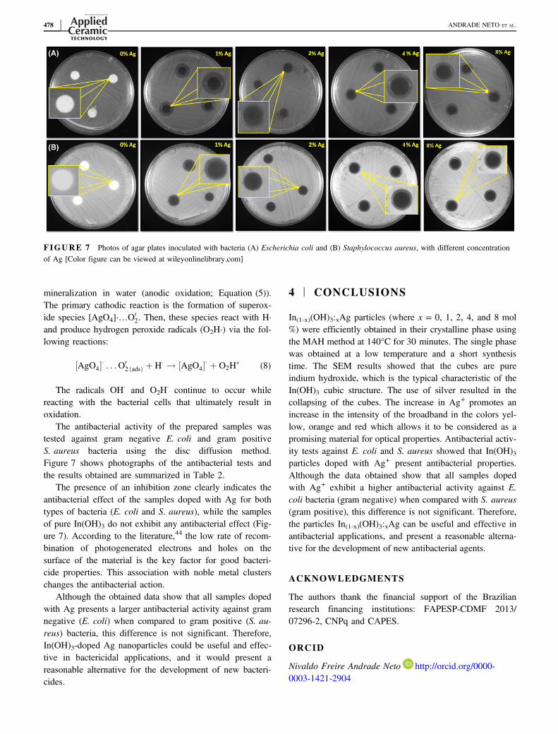
The antibacterial activity of the prepared samples wastested against gram negative E. coli and gram positiveS. aureus bacteria using the disc diffusion method.Figure 7 shows photographs of the antibacterial tests andthe results obtained are summarized in Table 2.
The presence of an inhibition zone clearly indicates theantibacterial effect of the samples doped with Ag for bothtypes of bacteria (E. coli and S. aureus), while the samplesof pure In(OH)3 do not exhibit any antibacterial effect (Fig-ure 7). According to the literature,44 the low rate of recom-bination of photogenerated electrons and holes on thesurface of the material is the key factor for good bacteri-cide properties. This association with noble metal clusterschanges the antibacterial action.
Although the obtained data show that all samples dopedwith Ag presents a larger antibacterial activity against gramnegative (E. coli) when compared to gram positive (S. au-reus) bacteria, this difference is not significant. Therefore,In(OH)3‐doped Ag nanoparticles could be useful and effec-tive in bactericidal applications, and it would present areasonable alternative for the development of new bacteri-cides.
4 | CONCLUSIONS
In(1-x)(OH)3:xAg particles (where x = 0, 1, 2, 4, and 8 mol%) were efficiently obtained in their crystalline phase usingthe MAH method at 140°C for 30 minutes. The single phasewas obtained at a low temperature and a short synthesistime. The SEM results showed that the cubes are pureindium hydroxide, which is the typical characteristic of theIn(OH)3 cubic structure. The use of silver resulted in thecollapsing of the cubes. The increase in Ag+ promotes anincrease in the intensity of the broadband in the colors yel-low, orange and red which allows it to be considered as apromising material for optical properties. Antibacterial activ-ity tests against E. coli and S. aureus showed that In(OH)3particles doped with Ag+ present antibacterial properties.Although the data obtained show that all samples dopedwith Ag+ exhibit a higher antibacterial activity against E.coli bacteria (gram negative) when compared with S. aureus(gram positive), this difference is not significant. Therefore,the particles In(1-x)(OH)3:xAg can be useful and effective inantibacterial applications, and present a reasonable alterna-tive for the development of new antibacterial agents.
ACKNOWLEDGMENTS
The authors thank the financial support of the Brazilianresearch financing institutions: FAPESP‐CDMF 2013/07296‐2, CNPq and CAPES.
ORCID
Nivaldo Freire Andrade Neto http://orcid.org/0000-0003-1421-2904
FIGURE 7 Photos of agar plates inoculated with bacteria (A) Escherichia coli and (B) Staphylococcus aureus, with different concentrationof Ag [Color figure can be viewed at wileyonlinelibrary.com]
478 | ANDRADE NETO ET AL.
REFERENCES
1. Anderson AD, Nelson JM, Rossiter S, Angulo FJ. Public healthconsequences of use of antimicrobial agents in food animals inthe United States. Microb Drug Resist. 2003;9(4):373–9.
2. Schroeder CM, White DG, Meng J. Retail meat and poultry as areservoir of antimicrobial‐resistant Escherichia coli. Food Micro-biol. 2004;21(3):249–55.
3. Byrne CM, Bolton DJ, Sheridan JJ, McDowell DA, Blair IS. Theeffects of preslaughter washing on the reduction of Escherichiacoli O157:H7 transfer from cattle hides to carcasses duringslaughter. Lett Appl Microbiol. 2000;30(2):142–5.
4. Luo PG, Stutzenberger FJ. Nanotechnology in the detection andcontrol of microorganisms. Adv Appl Microbiol. 2008;63:145–81.
5. Zinatloo-Ajabshir S, Salavati-Niasari M. Novel poly(ethylenegly-col)‐assisted synthesis of praseodymium oxide nanostructures viaa facile precipitation route. Ceram Int. 2015;41(1, Part A):567–75.
6. Mortazavi-Derazkola S, Zinatloo-Ajabshir S, Salavati-Niasari M.Novel simple solvent‐less preparation, characterization and degra-dation of the cationic dye over holmium oxide ceramic nanostruc-tures. Ceram Int. 2015;41(8):9593–601.
7. Mortazavi-Derazkola S, Zinatloo-Ajabshir S, Salavati-Niasari M.New sodium dodecyl sulfate‐assisted preparation of Nd2O3 nanos-tructures via a simple route. RSC Adv. 2015;5(70):56666–76.
8. Dai L, Liu R, Hu L-Q, Si C-L. Simple and green fabrication ofAgCl/Ag‐cellulose paper with antibacterial and photocatalyticactivity. Carbohydr Polym. 2017;174:450–5.
9. Yao X, Zhang X, Wu H, Tian L, Ma Y, Tang B. Microstructureand antibacterial properties of Cu‐doped TiO2 coating on titaniumby micro‐arc oxidation. Appl Surf Sci. 2014;292:944–7.
10. Wu T-S, Wang K-X, Li G-D, Sun S-Y, Sun J, Chen J-S. Mont-morillonite‐supported Ag/TiO2 nanoparticles: an efficient visible‐light bacteria photodegradation material. ACS Appl Mater Inter-faces. 2010;2(2):544–50.
11. Park MVDZ, Neigh AM, Vermeulen JP, de la Fonteyne LJJ, Ver-haren HW, Briedé JJ, et al. The effect of particle size on thecytotoxicity, inflammation, developmental toxicity and genotoxic-ity of silver nanoparticles. Biomaterials. 2011;32(36):9810–7.
12. Whitesides GM. Nanoscience, nanotechnology, and chemistry.Small. 2005;1(2):172–9.
13. Devi P, Patil SD, Jeevanandam P, Navani NK, Singla ML. Synthe-sis, characterization and bactericidal activity of silica/silver core–shell nanoparticles. J Mat Sci Mater Med. 2014;25(5):1267–73.
14. Baker C, Pradhan A, Pakstis L, Pochan DJ, Shah SI. Synthesisand antibacterial properties of silver nanoparticles. J NanosciNanotechnol. 2005;5(2):244–9.
15. Cao S, Chen J, Hu J. The fabrication and progress of core‐shellcomposite materials. Aust J Chem. 2009;62(12):1561–76.
16. Zhang Y, Gao X, Zhi L, Liu X, Jiang W, Sun Y, et al. The syn-ergetic antibacterial activity of Ag islands on ZnO (Ag/ZnO)heterostructure nanoparticles and its mode of action. J Inorg Bio-chem. 2014;130:74–83.
17. Motshekga SC, Ray SS, Onyango MS, Momba MNB. Micro-wave‐assisted synthesis, characterization and antibacterial activityof Ag/ZnO nanoparticles supported bentonite clay. J HazardMater. 2013;262:439–46.
18. Bokare A, Sanap A, Pai M, Sabharwal S, Athawale AA. Antibac-terial activities of Nd doped and Ag coated TiO2 nanoparticles
under solar light irradiation. Colloids Surf B Biointerfaces.2013;102:273–80.
19. Zhang F-J, Chen M-L, Oh W-C. Photoelectrocatalytic propertiesand bactericidal activities of silver‐treated carbon nanotube/titaniacomposites. Compos Sci Technol. 2011;71(5):658–65.
20. Wei LQ, Chen XL, Gao XH, Guo RJ, Xu BS. Preparation of Ag/SiO2 powder with light color and antibacterial performance. Pow-der Technol. 2014;253:424–8.
21. Baheiraei N, Moztarzadeh F, Hedayati M. Preparation andantibacterial activity of Ag/SiO2 thin film on glazed ceramic tilesby sol–gel method. Ceram Int. 2012;38(4):2921–5.
22. Kim YH, Lee DK, Cha HG, Kim CW, Kang YS. Synthesis andcharacterization of antibacterial Ag−SiO2 nanocomposite. J PhysChem C. 2007;111(9):3629–35.
23. Avivi S, Mastai Y, Gedanken A. Sonohydrolysis of In3 + Ions:formation of needlelike particles of indium hydroxide. ChemMater. 2000;12(5):1229–33.
24. Ishida T, Kuwabara K, Koumoto K. Formation and characteriza-tion of indium hydroxide films. J Ceram Soc Jpn. 1998;106(1232):381–4.
25. Bauer AW, Kirby WM, Sherris JC, Turck M. Antibiotic suscepti-bility testing by a standardized single disk method. Am J ClinPathol. 1966;45(4):493–6.
26. Tubtimtae A, Lee M-W. ZnO nanorods on undoped and indium‐doped ZnO thin films as a TCO layer on nonconductive glass fordye‐sensitized solar cells. Superlattices Microstruct. 2012;52(5):987–96.
27. Sena C, Costa MS, Muñoz EL, Cabrera-Pasca GA, Pereira LFD,Mestnik-Filho J, et al. Charge distribution and hyperfine interac-tions in the vicinity of impurity sites in In2O3 doped with Fe Co,and Ni. J Magn Magn Mater. 2015;387:165–78.
28. Anandan S, Vinu A, Mori T, Gokulakrishnan N, Srinivasu P,Murugesan V, et al. Photocatalytic degradation of 2,4,6‐trichloro-phenol using lanthanum doped ZnO in aqueous suspension. CatalCommun. 2007;8(9):1377–82.
29. Zhu W, Zhai J, Sun Z, Jiang L. Ammonia Responsive SurfaceWettability Switched on Indium Hydroxide Films with Micro-and Nanostructures. J Phys Chem C. 2008;112(22):8338–42.
30. Li Y, Lei R, Xu S. Effect of pH on the synthesis of In(OH)3 andIn(OH)3:Ce3+/Dy3+ nanocrystals by a fast, mild microwavemethod. J Solid State Chem. 2018;258:117–23.
31. Mikhlin YL, Pal'yanova GA, Tomashevich YV, VishnyakovaEA, Vorobyev SA, Kokh KA. XPS and Ag L3-edge XANEScharacterization of silver and silver–gold sulfoselenides. J PhysChem Solids. 2018;116:292–8.
32. Kőrösi L, Papp S, Dékány I. Preparation of transparent conduc-tive indium tin oxide thin films from nanocrystalline indium tinhydroxide by dip‐coating method. Thin Solid Films. 2011;519(10):3113–8.
33. Ho WH, Yen SK. Preparation and characterization of indiumoxide film by electrochemical deposition. Thin Solid Films.2006;498(1):80–4.
34. Nakamoto K. Theory of normal vibrations. Infrared and RamanSpectra of Inorganic and Coordination Compounds. Hoboken,NJ: John Wiley & Sons; 2008:1–147.
35. Motta FV, Lima RC, Marques APA, Leite ER, Varela JA, LongoE. In2O3 microcrystals obtained from rapid calcination in domes-tic microwave oven. Mater Res Bull. 2010;45(11):1703–6.
ANDRADE NETO ET AL. | 479
36. Motta FV, Lima RC, Marques APA, Li MS, Leite ER, VarelaJA, et al. Indium hydroxide nanocubes and microcubes obtainedby microwave‐assisted hydrothermal method. J Alloy Compd.2010;497(1):L25–8.
37. Zhu H, Wang Y, Wang N, Li Y, Yang J. Hydrothermalsynthesis of indium hydroxide nanocubes. Mater Lett. 2004;58(21):2631–4.
38. Lin S-E, Wei W-CJ. Synthesis and growth kinetics of monodis-persive indium hydrate particles. J Am Ceram Soc. 2006;89(2):527–33.
39. Shi Z, Wang W, Zhang Z. Synthesis and characterization ofindium hydroxide truncated polyhedral microcrystals. Mater Lett.2008;62(27):4293–5.
40. Motta FV, Marques APA, Li MS, Abreu MFC, Paskocimas CA,Bomio MRD, et al. Preparation and photoluminescence character-istics of In(OH)3:xTb3 + obtained by Microwave‐AssistedHydrothermal method. J Alloy Compd. 2013;553:338–42.
41. Wood DL, Tauc J. Weak absorption tails in amorphous semicon-ductors. Phys Rev B. 1972;5(8):3144–51.
42. Li H, Meng F, Gong J, Fan Z, Qin R. Structural, morphologicaland optical properties of shuttle‐like CeO2 synthesized by a facilehydrothermal method. J Alloy Compd. 2017;722:489–98.
43. Zhou Z, Liu G, Wei Q, Yan H, Liu Q. Luminescence propertiesof Ag nanoclusters doped SiO2–PbF2 oxyfluoride glasses.J Lumin. 2016;169:695–700.
44. Zhang A, Zhang J. Visible‐light activities of Gd2O3/BiVO4 com-posite photocatalysts. J Mater Sci. 2010;45(15):4040–5.
How to cite this article: Andrade Neto NF, TavaresMTS, Ferreira EAC, et al. Photoluminescent andantimicrobial properties of silver‐doped indiumhydroxide synthesized by one‐step microwave‐assistedhydrothermal method. Int J Appl Ceram Technol.2019;16:471–480. https://doi.org/10.1111/ijac.13127
480 | ANDRADE NETO ET AL.