Photo-induced formation of size-selective Ag nanoparticles and their interactions with Escherichia...
9
Journal of Photochemistry and Photobiology A: Chemistry 222 (2011) 25–33 Contents lists available at ScienceDirect Journal of Photochemistry and Photobiology A: Chemistry journa l h o me pa g e: www .elsevier.com/locate/jphotochem Photo-induced formation of size-selective Ag nanoparticles and their interactions with Escherichia coli Subrata Kundu ∗ , Hong Liang Materials Science and Mechanical Engineering, Texas A&M University, College Station, TX 77843-3123, USA a r t i c l e i n f o Article history: Received 6 January 2011 Received in revised form 15 March 2011 Accepted 27 March 2011 Available online 13 June 2011 Keywords: ATP Ag NPs CTAB UV-irradiation Escherichia coli a b s t r a c t A new synthetic route was utilized for the formation of size-selective Ag nanoparticles (NPs) exploiting UV-irradiation techniques. The one step process exclusively generates Ag NPs of smaller sizes, larger sizes and aggregated nanostructures. The reduction of Ag(I) was done in the presence of adenosine tri-phosphate (ATP) under 30 min of UV-irradiation. ATP served as a dual role, a reducing agent and a stabilizing agent for the formation of Ag NPs. The mechanisms of the particle formation and the effects of different reaction parameters were studied in details. The synthesized particles were stable for more than a month under ambient condition without indication of oxide formation. Moreover, the method did not need any harsh reduction conditions. The synthesized negatively charged ATP–Ag NPs has been mixed with bacteria Escherichia coli and studied their interaction. Finally a comparative study was made with the ATP–Ag NPs with positively charged CTAB–Ag NPs. The results indicated that positively charged Ag NPs deposited better with the bacterium surface compared to the negatively charged. The present research might find important applications for the synthesis of other nanomaterials as well as to understand the interactions of NPs with microorganisms. © 2011 Elsevier B.V. All rights reserved. 1. Introduction In recent years, synthesis and characterization of colloidal metal particles in the ‘neglected dimension’ have shown tremendous importance due to their promising application in chemistry, biol- ogy, physics, materials science, medicine, and electronics. Metal nanoparticles exhibit special properties when compared to their bulk element. This is possible because of their high surface-to- volume ratio and unique optical [1], electronic [2], magnetic [3] and catalytic [4] properties. Most of these properties are strongly depend upon the size and shape of the particles as well as the surrounding medium. Among the different colloidal metal nanoparticles (NPs), novel metal NPs have shown particular inter- est due to their close lying valence and conduction bands in which the electron moves freely. As the properties of novel metallic nanoparticles change with their size and shape, an effective route is required for synthesis of NPs with control of the same. Among the various synthetic routes, chemical synthesis methods are superior to physical methods for controlling the size and shapes. ∗ Corresponding author. Current address: Electrochemical Materials Science (ECMS) Division, Central Electrochemical Research Institute (Council of Scientific and Industrial Research), Karaikudi - 630 006, Tamil Nadu, India. Tel.: +91 4565 227550 559x264/224; fax: +91 4565 227779/227713. E-mail addresses: subrata [email protected] (S. Kundu), [email protected] (H. Liang). It is proven that Ag NPs are superior to other metals due to their high electric conductivity [5], optical properties [6], antimi- crobial effect [7], use as oxidative catalyst [8] and spatial Raman spectroscopic [9] behavior. Moreover, Ag NPs have strong surface plasmon resonance (SPR) band in the visible region and that helps the study of the evolution and characterization of the particles in solution. In the literature there are a number of chemical meth- ods reported for the synthesis of Ag NPs [10–16]. The easiest and well-known method is the reduction of silver ions in the presence of NaBH 4 or tri-sodium citrate to obtain negatively charged Ag NPs [10,11]. Several other methods include polyol process [12], - radiolytic process [13], sonochemical process [14], laser irradiation techniques [15] and microwave heating [16]. Liu et al. synthesized size-selective Ag NPs in poly-vinyl-pyrrolidone solution in the pres- ence of metallic seed particles [17]. Tan et al. synthesized Ag NPs by the reduction of Ag salt using potassium bi-tartrate as a reduc- ing agent and stabilized the particles in a polymeric solution [18]. Kundu et al. synthesized Ag nanocubes using microwave heating method for a very short time in poly (styrene) sulfonate (PSS) solu- tion [19]. A photochemical method was used for the synthesis of SERS (surface enhanced Raman scattering) active Ag NPs in sil- ica matrix [20]. Gedanken’s group synthesized Ag NPs on silica spheres using a sonochemical deposition method [21]. Recently, our group synthesized Ag NPs in a non-ionic surfactant media using alkaline 2,7-dihydroxy naphthalene (2,7-DHN) as reducing agent under microwave irradiation techniques [16]. Most of the above methods were either time consuming, needing multiple steps for 1010-6030/$ – see front matter © 2011 Elsevier B.V. All rights reserved. doi:10.1016/j.jphotochem.2011.03.024
-
Upload
subrata-kundu -
Category
Documents
-
view
214 -
download
1
Transcript of Photo-induced formation of size-selective Ag nanoparticles and their interactions with Escherichia...

Pa
SM
a
ARRAA
KAACUE
1
pionbvadtnetnrvt
(aT
(
1d
Journal of Photochemistry and Photobiology A: Chemistry 222 (2011) 25– 33
Contents lists available at ScienceDirect
Journal of Photochemistry and Photobiology A:Chemistry
journa l h o me pa g e: www .e lsev ier .com/ locate / jphotochem
hoto-induced formation of size-selective Ag nanoparticlesnd their interactions with Escherichia coli
ubrata Kundu ∗, Hong Liangaterials Science and Mechanical Engineering, Texas A&M University, College Station, TX 77843-3123, USA
r t i c l e i n f o
rticle history:eceived 6 January 2011eceived in revised form 15 March 2011ccepted 27 March 2011vailable online 13 June 2011
eywords:TP
a b s t r a c t
A new synthetic route was utilized for the formation of size-selective Ag nanoparticles (NPs) exploitingUV-irradiation techniques. The one step process exclusively generates Ag NPs of smaller sizes, largersizes and aggregated nanostructures. The reduction of Ag(I) was done in the presence of adenosinetri-phosphate (ATP) under 30 min of UV-irradiation. ATP served as a dual role, a reducing agent anda stabilizing agent for the formation of Ag NPs. The mechanisms of the particle formation and the effectsof different reaction parameters were studied in details. The synthesized particles were stable for morethan a month under ambient condition without indication of oxide formation. Moreover, the method did
g NPsTABV-irradiationscherichia coli
not need any harsh reduction conditions. The synthesized negatively charged ATP–Ag NPs has been mixedwith bacteria Escherichia coli and studied their interaction. Finally a comparative study was made with theATP–Ag NPs with positively charged CTAB–Ag NPs. The results indicated that positively charged Ag NPsdeposited better with the bacterium surface compared to the negatively charged. The present researchmight find important applications for the synthesis of other nanomaterials as well as to understand the
icro
interactions of NPs with m. Introduction
In recent years, synthesis and characterization of colloidal metalarticles in the ‘neglected dimension’ have shown tremendous
mportance due to their promising application in chemistry, biol-gy, physics, materials science, medicine, and electronics. Metalanoparticles exhibit special properties when compared to theirulk element. This is possible because of their high surface-to-olume ratio and unique optical [1], electronic [2], magnetic [3]nd catalytic [4] properties. Most of these properties are stronglyepend upon the size and shape of the particles as well ashe surrounding medium. Among the different colloidal metalanoparticles (NPs), novel metal NPs have shown particular inter-st due to their close lying valence and conduction bands in whichhe electron moves freely. As the properties of novel metallicanoparticles change with their size and shape, an effective route is
equired for synthesis of NPs with control of the same. Among thearious synthetic routes, chemical synthesis methods are superioro physical methods for controlling the size and shapes.∗ Corresponding author. Current address: Electrochemical Materials ScienceECMS) Division, Central Electrochemical Research Institute (Council of Scientificnd Industrial Research), Karaikudi - 630 006, Tamil Nadu, India.el.: +91 4565 227550 559x264/224; fax: +91 4565 227779/227713.
E-mail addresses: subrata [email protected] (S. Kundu), [email protected]. Liang).
010-6030/$ – see front matter © 2011 Elsevier B.V. All rights reserved.oi:10.1016/j.jphotochem.2011.03.024
organisms.© 2011 Elsevier B.V. All rights reserved.
It is proven that Ag NPs are superior to other metals due totheir high electric conductivity [5], optical properties [6], antimi-crobial effect [7], use as oxidative catalyst [8] and spatial Ramanspectroscopic [9] behavior. Moreover, Ag NPs have strong surfaceplasmon resonance (SPR) band in the visible region and that helpsthe study of the evolution and characterization of the particles insolution. In the literature there are a number of chemical meth-ods reported for the synthesis of Ag NPs [10–16]. The easiest andwell-known method is the reduction of silver ions in the presenceof NaBH4 or tri-sodium citrate to obtain negatively charged AgNPs [10,11]. Several other methods include polyol process [12], �-radiolytic process [13], sonochemical process [14], laser irradiationtechniques [15] and microwave heating [16]. Liu et al. synthesizedsize-selective Ag NPs in poly-vinyl-pyrrolidone solution in the pres-ence of metallic seed particles [17]. Tan et al. synthesized Ag NPsby the reduction of Ag salt using potassium bi-tartrate as a reduc-ing agent and stabilized the particles in a polymeric solution [18].Kundu et al. synthesized Ag nanocubes using microwave heatingmethod for a very short time in poly (styrene) sulfonate (PSS) solu-tion [19]. A photochemical method was used for the synthesis ofSERS (surface enhanced Raman scattering) active Ag NPs in sil-ica matrix [20]. Gedanken’s group synthesized Ag NPs on silicaspheres using a sonochemical deposition method [21]. Recently,
our group synthesized Ag NPs in a non-ionic surfactant media usingalkaline 2,7-dihydroxy naphthalene (2,7-DHN) as reducing agentunder microwave irradiation techniques [16]. Most of the abovemethods were either time consuming, needing multiple steps for
26 S. Kundu, H. Liang / Journal of Photochemistry and Photobiology A: Chemistry 222 (2011) 25– 33
Table 1Details concentrations, size of the particles, UV-irradiation time for the formation of ATP stabilized Ag NPs.
Set number Final conc. of ATPsolution (M)
Final conc. ofAgNO3 solution (M)
Time ofUV-irradiation (min)
Color of thefinal product
Shape of theparticles
Diameter of theparticles (nm)
1 9.09 × 10⁻² 9.09 × 10⁻³ 30 Yellowish Spherical ∼3.5 ± 1.52 9.09 × 10⁻² 9.09 × 10⁻⁴ 30 Light orange Spherical ∼37 ± 0.5
td
thnetbafplepcc[[hstAr
awostAtccNattbsiTwe
2
2
tShdw
3 9.09 × 10⁻³ 9.09 × 10⁻⁴ 30
he synthesis, or generated various shapes with uncontrollable sizeistributions.
Recently, different biological molecules like proteins [22], pep-ides [23], amino acids [24] and DNA (deoxyribo-nucleic acid) [25]ave been used for the synthesis and fabrication of metal NPs. Theucleic acid or nucleotide mediated synthesis of metal NPs is anmerging area. The major advantage of nucleotide mediated syn-hesis is that the size and the chemical property of the NPs cane tuned according to the structure of the nucleic acid. The otherdvantages are, (a) they can bind with different metal ions via dif-erent well established binding modes, (b) nucleic acid in solutionrovide well defined 3-D shape which can be used for molecu-
ar building blocks like squares [26], cubes [27], T-junctions [28],tc., (c) nucleic acid sequences are much more cost-effective thanolypeptide sequences. Kelley and Eaton reported earlier that thehemical properties of the NPs could be changed according to theonstituent nucleotide or nucleotide tri-phosphate building blocks29,30]. Recently, we synthesized metal [31] and semi-conductor2] nanowires using DNA as template either using microwaveeating or UV-irradiation method. Adenine coated Ag NPs wereynthesized by Wei et al. by the reduction of Ag ions in NaBH4 solu-ion [32]. In the present work, we use UV-irradiation to synthesizeg NPs using adenosine-tri-phosphate (ATP) as a stabilizing andeducing agent.
Here, we report the size-selective synthesis of Ag NPs using simple photochemical approach. The reduction of Ag(I) ionsas done in the presence of ATP under 30 min of continu-
us UV-irradiation. ATP acted as a reducing agent during theynthesis as well as a stabilizing agent after the formation ofhe NPs. The method produced various sized and aggregatedg NPs via tuning the metal ion-to-ATP-molar ratio. The syn-
hesized Ag NPs were deposited on the bacterium Escherichiaoli (E. coli) to study their interactions. Moreover, positivelyharged CTAB (cetyl trimethylammonium bromide) stabilized AgPs were also synthesized and deposited on the bacteria. Finally
comparative study was conducted. It was found that posi-ively charged Ag NPs (CTAB stabilized) interacted better withhe bacteria surface compared to the negatively charged (ATP sta-ilized) Ag NPs. To the best of our knowledge, the synthesis ofize-selective Ag NPs using ATP under UV-irradiation and theirnteractions with bacteria E. coli have not been reported before.he present synthesis processes as well as the interactions studyith bacteria are simple, straightforward, reproducible, and cost
ffective.
. Experimental
.1. Reagents
Adenosine tri-phosphate (ATP), silver nitrate (99%), and cetylrimethyl ammonium bromide (CTAB, 99%) were purchased from
igma–Aldrich and used without further purification. Sodium boro-ydride (NaBH4) was also purchased from Sigma–Aldrich. Thee-ionized (DI) water was used for the entire synthesis process asell as in the NPs–bacteria interaction study.Dark orange Honey comb like aggregated ∼12 ± 0.5
2.2. Instruments
UV–visible (UV–vis) absorption spectra were recorded in aHitachi (model U-4100) UV-VIS-NIR spectrophotometer equippedwith a 1 cm quartz cuvette holder for liquid samples. A high-resolution transmission electron microscope (HR-TEM) (JEOL JEM2010) was used at an accelerating voltage of 200 kV. The energydispersive X-ray spectrum (EDS) was recorded with an OxfordInstruments INCA energy system connected with the TEM. The XRDanalysis was done with a scanning rate of 0.020 S−1 in the 2� range35–85◦ using a Bruker-AXS D8 Advanced Bragg-Brentano X-rayPowder Diffractometer with Cu K� radiation (� = 0.154178). TheX-ray photoelectron spectroscopy (XPS) analysis was carried outusing a Kratos Axis Ultra Imaging X-ray photoelectron spectrom-eter with monochromatic Al K� line (1486.7 eV). The instrumentintegrates a magnetic immersion lens and charge neutralizationsystem with a spherical mirror analyzer, which provides real-timechemical state and elemental imaging using a full range of passenergies. The emitted photoelectrons were detected by the ana-lyzer at a passing energy of 20 eV with energy resolution of 0.1 eV.The incident X-ray beam was normal to the sample surface andthe detector was 45◦ away from the incident direction. The analy-sis spot on the sample was 0.4 mm by 0.7 mm. A xenon lamp fromNewport Corporation at a wavelength of 260 nm on the samplewas used for UV-photoirradiation. The approximate intensity was20 ± 0.5 �W and the bandwidth of irradiation was 260 ± 8 nm. Thedistance of the sample from the light source was 15 cm. The samplewas placed over a wooden box with a stand to make the light shineon it directly.
2.3. Size-selective synthesis of ATP stabilized Ag NPs
Size-selective Ag NPs were synthesized by varying the concen-trations of AgNO3 with the concentrations of aqueous ATP solution.The solution mixture containing ATP and AgNO3 are mixed wellwith a magnetic stirrer and then placed under UV-irradiation forabout 30 min. For a typical synthesis, 6 mL of 0.1 (M) ATP solutionwas mixed with 0.6 mL of 0.1 (M) AgNO3 solution and the solu-tion mixture was UV-irradiated for 30 min. The formation of silverparticles started within 5 min of UV-irradiation as light yellowishcolor is observed in the solution mixture. Finally after 30 min ofUV-irradiation the color changed to deep reddish yellow. This solu-tion contains spherical Ag NPs with smaller sizes. For other twosizes like larger spheres and aggregated Ag particles we varied theconcentration of AgNO3 and ATP keeping the UV-irradiation timefixed for 30 min. The details concentrations, size of the particles,UV-irradiation time is given in Table 1. The colors of the synthe-sized Ag NPs were stable for more than a month in the dark underambient condition without change in any optical properties.
2.4. Synthesis of CTAB-stabilized Ag NPs
Positively charged CTAB stabilized Ag NPs are synthesizedaccording to the previous report by Yonezawa et al. [33]. In thesynthesis we mixed 10 mL of 5 × 10⁻³ (M) AgNO3 with 50 mL of10−2 (M) CTAB solution and stirred well. Initially, after mixing the

and Photobiology A: Chemistry 222 (2011) 25– 33 27
mAd1ct
2a
XgipovwTasdm8
2N
Tmgatasifdr
3
3
tAigaewaonrcasaAdtt4
in curve D, Fig. 2. Curves E and F in Fig. 2 shows the SPR bands forlarger sizes and aggregated Ag NPs, respectively. With the increasein particles size, the absorption maxima were also shifted towards
Fig. 2. UV–vis absorption spectra at various stages of ATP–Ag NP synthesis. Curve Ashows the absorption spectra of aqueous AgNO3 solution. Curve B shows the absorp-tion spectra of aqueous ATP solution having a band at ∼258 nm. Curve C shows the
S. Kundu, H. Liang / Journal of Photochemistry
ixed solution shows white turbidity due to the formation ofgBr. Finally, 5 mL of 0.4 (M) ice-cold NaBH4 solution was addedrop wise while stirring vigorously. Stirring was continued another5–20 min after complete mixing of NaBH4. The solution colorhanged to dark yellow confirmed the formation of Ag NPs andhe solution was stable for more than a week in ambient condition.
.5. Preparation of sample for UV–vis, TEM, EDS, XRD, and XPSnalysis
The Ag NPs were characterized using UV–vis, TEM, EDS, XRD, andPS measurements. The Ag NPs solution after successive centrifu-ation was re-dispersed in DI water and used for the measurementn UV-vis spectrophotometer. The samples for TEM and EDS wererepared by placing a drop of the corresponding Ag NP solutionnto a carbon-coated Cu grid followed by slow evaporation of sol-ent at ambient condition. For XRD and XPS analysis, silicon (Si)afers were used as the substrates for the thin film preparation.
he wafers were cleaned thoroughly in acetone and sonicated forbout 20 min. The cleaned substrates were covered with the Ag NPolution and dried in a vacuum chamber. After the first layer waseposited, subsequent layers were deposited by repeatedly addingore Ag NP solution and drying. Final samples were obtained after
–10 depositions and then analyzed using XRD and XPS techniques.
.6. Preparation of bacteria samples and their incubation with AgPs
Bacteria, E. coli strain DH5� was used in our current research.hese bacteria strains were generously collected from the depart-ent of Biology at the Texas A&M University. The E. coli was
rown in the liquid LB (Luria–Bertani) media at 37 ◦C and stirredt 250 rpm for overnight (∼16 h). After completely grown, the bac-eria solution containing E. coli was taken in a small centrifuge tubend varied amounts of Ag NPs (both ATP and CTAB stabilized) mixedeparately. The mixed solution containing Ag NPs and bacteria wasncubated for different times and then spotted over a TEM Cu gridor analysis. The bacteria Ag NPs after spotting on TEM grid were air-ried overnight and then analyzed using TEM and EDS. The detailedesults are given in later part of Section 3.
. Results and discussion
.1. UV–vis spectroscopy study
Monodispersed Ag NPs with different sizes were synthesized byhe reduction of AgNO3 with ATP under 30 min of UV-irradiation.TP not only acted as a stabilizing agent but also as a reduc-
ng agent for the reduction of Ag(I) to Ag(0). ATP directed therowth of Ag NPs and produced different sizes. The ATP is
multifunctional nucleotide generally used in cells as a co-nzyme. One molecule of ATP consists of a three phosphate grouphich is attached with ribose sugar and that sugar binds with
denine base. Fig. 1A shows the chemical structures of nucle-side, nucleoside-monophosphate, nucleoside-diphosphate, anducleoside-triphosphate. Fig. 2 shows the UV–vis spectrum of theeaction mixture at different stages of the synthesis process. Theolorless aqueous solution of AgNO3 has a very small intense peakt ∼302 nm in the visible region (curve A, Fig. 2). The aqueous ATPolution has an absorption band at ∼258 nm due to the presence ofromatic base molecule (curve B, Fig. 2). The mixing of ATP withgNO3 (curve C, Fig. 2) resulted a small peak shift as well as a
ecrease in absorbance value. This was probably due to interac-ions of ATP with Ag(I) ions. After UV-irradiation for 30 min withhe solution containing ATP and AgNO3, a new peak appeared at18 nm due to the SPR of spherical Ag NPs [10–16,19,20]. The peakFig. 1. (A) Chemical structure of the different nucleosides. (B) Schematic repre-sentation how the different parts of ATP having different specific roles for Ag-NPssynthesis.
at 418 nm is due to the formation of smaller sized Ag NPs as shown
absorption spectra of the mixture of ATP with AgNO3. Curves D, E and F show thesurface plasmon resonance (SPR) band for the formation of different sizes Ag NPs.The inset shows three different color images indicated with S1, S2, and S3 containingAg NPs of different sizes. (For interpretation of the references to color in this figurelegend, the reader is referred to the web version of this article.)

28 S. Kundu, H. Liang / Journal of Photochemistry and Photobiology A: Chemistry 222 (2011) 25– 33
Fig. 3. Transmission electron microscopy (TEM) images for the synthesis of ATP–Ag NPs after 30 min of UV irradiation with variation of reaction conditions. (A and B) Showsthe low and high magnified TEM images of spherical Ag NPs having average particles diameter ∼3.5 ± 1.5 nm. The inset of (A) shows its corresponding higher magnifiedimage. (C and D) Shows the low and high magnified image of bigger size spherical Ag NPs having average particles sizes ∼37 ± 5 nm. The inset of (B and D) shows theirc ted As their c
haF5gooawam
orresponding SAED patterns. (E and F) Shows the low magnified images of aggregaingle Ag particle from the aggregates are ∼12 ± 5 nm. The inset of (E and F) shows
igher wavelength region which was well matched with the liter-ture report previously. For larger size spherical particles (curve E,ig. 2), we observed a broad absorption band ranged from 370 to70 nm with an absorption maxima at 470 nm. Similarly for aggre-ated Ag NPs (curve F, Fig. 2), we observed a broad peak in the rangef 370 and 600 nm with an absorption maxima at 490 nm. The insetf Fig. 2 shows three different color images indicated with S1, S2,
nd S3 containing Ag NPs of different sizes. S1 signifies the Ag NPsith smaller size, S2 signifies the Ag NPs with larger size, and S3 theggregated Ag NPs. The synthesized Ag NP solutions were stable forore than a month under dark in the ambient condition.
g NPs and the average lengths are more than 10 �m and the average diameters of aorresponding higher magnified images.
3.2. Transmission electron microscopy (TEM) analysis
Fig. 3 shows the transmission electron microscopy images ofvarious sized and ATP stabilized Ag NPs. Those were synthesizedwith the above method (details given in experimental section) after30 min of UV irradiation. Fig. 3A and B shows the low and highmagnified TEM image corresponding to the curve D in Fig. 2. Parti-
cles are spherical, separated with an average size of ∼3.5 ± 1.5 nm.The inset of Fig. 3A shows its corresponding high magnified image.The inset of Fig. 3B shows the corresponding selected area elec-tron diffraction (SAED) pattern confirming that the ATP stabilized
S. Kundu, H. Liang / Journal of Photochemistry and Photobiology A: Chemistry 222 (2011) 25– 33 29
Fp
AmtsatteFhtctsfiA
3
rtdcaTr
3
l(noslmfort
ig. 4. Energy dispersive X-ray spectrum (EDS) of the ATP–Ag NPs showing theeaks for C, O, Cu, P, and Ag.
g NPs are single crystal. Fig. 3C and D shows the low and highagnified images corresponding to curve E in Fig. 2. Here the par-
icles are bigger in size than previous one and the average particlesizes are ∼37 ± 5 nm. Fig. 3D shows the image of a single particlesnd the inset shows the corresponding SAED pattern. It confirmshat the single crystal nature of the particles. Fig. 3E and F showshe low magnified images of aggregated Ag NPs taken from differ-nt parts of the same samples (corresponding to curve F, Fig. 2).rom the image it is clear that small Ag NPs aggregated to form theoney-comb like structure. The lengths of the aggregated struc-ures are more than 10 �m. The inset of Fig. 3E and F shows theirorresponding high magnified images. From those, it is clear thathe average diameters of a single Ag particle from the aggregate-is ∼12 ± 5 nm. These smaller particles are aggregated together toorm the honey comb-like structure. From the above TEM analysist is clear that different reaction condition produces different sizesg NPs.
.3. Energy dispersive X-ray spectroscopy (EDS) analysis
Fig. 4 shows the results obtained from the energy dispersive Xay spectroscopy (EDS) analysis. EDS analysis is used to determinehe elements present in the synthesis. The spectrum consisted ofifferent peaks from the C, O, Cu, P, and Ag. The C and Cu peaksame from the C-coated Cu TEM grid used for the analysis. The Pnd O peaks came from the ATP used for the synthesis of Ag NPs.he high intense Ag peak came from the Ag NPs. The above EDSesult confirms the presence of Ag NPs in the sample.
.4. X-ray diffraction (XRD) analysis
Fig. 5 shows the powder X-ray diffraction pattern of ATP stabi-ized Ag NPs. The peaks were assigned to diffraction from the (1 1 1),2 0 0), (2 2 0), (3 1 1), and (2 2 2) planes of silver NPs (JCPDS cardumber 4-0783) [24]. These diffraction peaks confirm the presencef a face-centered cubic (fcc) structure of crystalline Ag NPs. Theizes of the Ag NPs were measured by the X-ray diffraction peak’sine width using the Debye formula for small crystal spheres. The
ean diameters of the Ag particles are consisted with the result
rom the TEM image. According to the literature, the specific shapef any fcc nano crystal mainly depends by the ratio between growthates along the <1 0 0> and <1 1 1> directions [34]. Here in our syn-hesis, we used ATP as a stabilizing agent so the selective interactionFig. 5. Powder X-ray diffraction (XRD) pattern of the ATP–Ag NPs having the diffrac-tion from the (1 1 1), (2 0 0), (2 2 0), (3 1 1), and (2 2 2) planes of silver NPs.
between the ATP and different crystal facets of Ag NPs alters thegrowth rate along the different crystal directions.
3.5. X-ray photoelectron spectroscopy (XPS) analysis
Fig. 6 shows the X-ray photoelectron spectroscopy (XPS) imagesof a self-assembled Ag NPs film. The sensitivity of XPS in chemicalanalysis stems mostly from its ability to resolve the chemical iden-tity of the atoms from the measured binding energies. Fig. 6A showsthe overall survey and Fig. 6B the Ag (3d) XPS spectra. The surveyspectrum consists of the characteristic peaks of O(1s) at 529.5 eV,C(1s) at 282 eV and Ag(3d) peaks. The Ag(3d) region is characterizedby a doublet which arises from the spin–orbit coupling (Ag 3d5/2and Ag 3d3/2) as shown in Fig. 6B. The two intense Ag peaks appearat a binding energy of 374.1 (for Ag 3d3/2) and 368 (for Ag 3d5/2) eVwhich clearly indicates the formation of Ag NPs. The peak positionsare in agreement with the previous literature [16,35]. In the surveyspectrum we did not observe peaks for the oxide formation whichindicates the stability of Ag NPs. The O(1s) peak came from the ATPwhich is bound to the surface of the Ag NPs.
3.6. Effects of other reaction parameters
To standardize the reduction process we examined the variationof reaction parameters like concentration of ATP and Ag(I) ions andthe UV-irradiation time. The best result with size selective Ag NPsand aggregated Ag NPs are formed in the concentrations given inthe experimental section and in Table 1. We have seen that whenAg(I) concentration is very high (≥10−1 M), the formation of AgNPs is very fast (within 5–6 min of UV-irradiation) but particlesgets precipitated after few hours of the synthesis. Similarly, whenthe concentration of Ag(I) is low (≤10−4 M), the formation of AgNPs took longer time (>3 h) and formed an un-uniform particlessizes. We also varied the concentrations of ATP and observed that ahigher concentration mostly small size spherical particles formedand at lower ATP concentration either bigger or aggregated Ag NPsformed. We also varied the UV-irradiation time and observed that30 min is sufficient for the formation of size-selective Ag NPs. The
Ag NPs started formation within 3–5 min of UV-irradiation andcompleted after 30 min of UV-irradiation. A long irradiation time(more than 2 h) results the precipitation of the Ag NPs. We testedour reaction with normal conventional heating procedure using
30 S. Kundu, H. Liang / Journal of Photochemistry and P
Fs
ctaw
3
rpAoansUMoaNoeacc
we have seen that smaller spheres were formed at a high concen-tration of both Ag(I) and ATP. At a high Ag(I) concentration oncethe nucleation started, a huge number of Ag nuclei were formed.Due to high concentration of ATP, the pre-formed Ag nuclei did
ig. 6. X-ray photoelectron spectrum (XPS) of the ATP–Ag NPs. (A) is the overallurvey spectra and (B) is the spectra for Ag (3d) regions.
onventional heater or using water bath but none of them resultedhe formation of Ag NPs in the experimental time scale. Thus allbove control experiment proved that the size-selective Ag NPsere formed only at a specific concentration as given in Table 1.
.7. Reaction mechanism of Ag NPs formation
The formation of size-selective Ag NPs was initiated by theeduction of Ag(I) ions in the presence of ATP under UV-hotoirradiation. In our proposed synthesis both the presence ofTP and the UV-irradiation is extremely necessary for the formationf Ag NPs. In the absence of ATP, no Ag particles formed due to thebsence of reducing agent while in the absence of UV-irradiationo particles formed due to lack of formation of radical species in theolution mixture. So a proper concentration of ATP and a sufficientV irradiation is necessary for the formation of Ag NPs. Kumar andital [36], and Green et al. [37] group reported earlier that nucle-
sides like ATP, guanosine triphosphate (GTP) acted as a cappinggent and templates for the formation of metal and semi-conductorPs. In their study they used these nucleosides only for cappingf templating agents but they did not reveal their reducing prop-
rty. This nucleotide or nucleic acid based NPs synthesis is a newpproach because one could tune the NPs size and their physico-hemical property according to the nucleic acid structure and theirompositions. Earlier Kelley [29] and Eaton [30] group reportedhotobiology A: Chemistry 222 (2011) 25– 33
earlier that properties of NPs could be tuned according to the con-stituent RNA sequence or nucleotide tri-phosphate building block.During discussion of reaction mechanisms, one important issue tobe discussed is about which group acted as reducing agent for Ag(I)reduction. The ATP molecule contains hydroxyl groups on its sugarpart as shown in Fig. 1. It has been reported that molecules contain-ing hydroxyl group could act as a reducing agent in NPs synthesisunder UV-irradiation [38–42]. Different hydroxyl compounds likeascorbic acid [38], TX-100 [39], dendrimer [40], 2,7-DHN [41], andpoly (vinyl) alcohol (PVA) [42] could act as a reducing agent inNPs synthesis in the presence of UV-irradiation. These compoundshave hydroxyl group on their skeleton which undergoes hydroxyliccleavage to a hydroxyl radical in the presence of UV-irradiation. TheUV light generated hydroxyl radicals that could act as a reducingagent for reduction of metal ions to metal (0). Our group reportedearlier that hydroxylic compounds could reduce Au(III) [43], Pd(II)[44], Pt(IV) [45] and Rh(III) [42] ions to produce their correspond-ing NPs with different morphology. We also reported previouslythat DNA having hydroxyl groups in its sugar could act as a reduc-ing agent in the synthesis of metal [31] and semi-conductor [2]nanowires. These hydroxyl groups probably acts as a reducing agentand are oxidized to ketones after the reduction. So here it is rea-sonable to assume that the hydroxyl radicals were formed duringUV-irradiation of the solution mixture containing Ag ions and ATPpromoted the reduction of Ag(I) to Ag(0). Once the nucleation ofAg(0) started to grow, it would involve in multiple steps and even-tually generate Ag NPs in different sizes. In the first step of reductionof Ag(I) to Ag(0), Ag atoms were formed. The Ag atoms subsequentlygrew to form small Ag nuclei and finally the crystalline Ag particles.The ATP molecule having different groups were adsorbed onto thesurface of these crystal particles and slowed down the growth rateof different crystal facets. Finally after 30 min of UV-irradiation, dif-ferent sized Ag NPs were formed which in turn depended uponthe concentrations of both Ag(I), ATP and UV-irradiation time. Weassume that different parts of ATP have some specific roles in NPssynthesis (as shown in Fig. 1B). The three phosphate group and ‘N’group assisted NP growth, the amine group helped for NP stabi-lization, and the hydroxyl group in the sugar part helps the radicalgeneration. Similar types of results were shown earlier with GTP byBerti and Burley for the synthesis of inorganic NPs [46]. In our study,
Fig. 7. Transmission electron microscopy (TEM) images of positively charged CTABstabilized Ag NPs having average particles diameter ∼45 ± 5 nm. Inset shows thecorresponding higher magnified image.

S. Kundu, H. Liang / Journal of Photochemistry and Photobiology A: Chemistry 222 (2011) 25– 33 31
Fig. 8. Transmission electron microscopy (TEM) images of the bacteria E. coli with the Ag NPs at different reaction conditions. (A and B) Shows the image of only bacteriaE ows tN nd F)
3
nWofocdpOt
. coli at different magnifications having average diameter is ∼4 ± 1 �m. (C and D) ShPs for 30 min. The inset of (C) shows the corresponding high magnified image. (E a0 min of incubation.
ot get much free space to grow thus formed mall size Ag NPs.hen the Ag(I) concentration was relatively low, lesser number
f Ag nuclei were formed and received much space to grow and toorm a bigger sized Ag NPs. The aggregated Ag nanostructures werebserved both at a lower concentrations of Ag(I) and ATP. At loweroncentration the interaction of the cationic part or the ATP with
ifferent crystal facets was slow and growth of the particles tooklace in all directions and produced big size aggregated particles.nce a particle was formed, they tried to bind with the nearest par-icles in order to lower the surface energy which was facilitate to
he low and high magnified TEM image of the bacteria after incubation with ATP–AgShows the low and high magnified TEM image of the E. coli with CTAB–Ag NPs after
low concentration of ATP. So the aggregated spherical Ag nanos-tructures were formed in the solution. It is well known that theformation of definite size and shape depends mostly on two fac-tors, firth the faceting tendency of stabilizing agents and second thegrowth kinetics of Ag that is rate of supply of Ag(0) to the differentcrystallographic planes. The similar growth mechanism of the Ag
particles formation was reported [16,21]. The present method ofAg NPs formation can find various important applications as ATPon its surface. They can act as a molecular spacer for SERS study orother biological applications. We also tested our study with other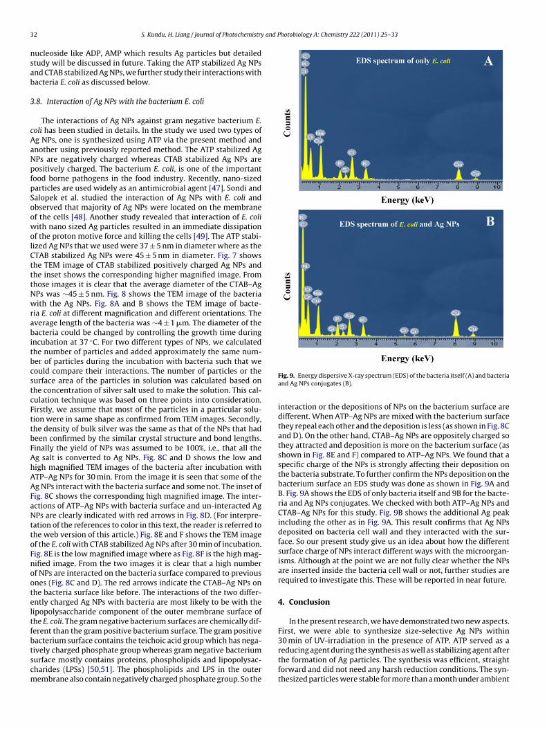
3 and Photobiology A: Chemistry 222 (2011) 25– 33
nsab
3
cAaNpfpSoowolCtttNwrabitbcstcFttbFAhAAFaNttoFnooteltfbtscm
2 S. Kundu, H. Liang / Journal of Photochemistry
ucleoside like ADP, AMP which results Ag particles but detailedtudy will be discussed in future. Taking the ATP stabilized Ag NPsnd CTAB stabilized Ag NPs, we further study their interactions withacteria E. coli as discussed below.
.8. Interaction of Ag NPs with the bacterium E. coli
The interactions of Ag NPs against gram negative bacterium E.oli has been studied in details. In the study we used two types ofg NPs, one is synthesized using ATP via the present method andnother using previously reported method. The ATP stabilized AgPs are negatively charged whereas CTAB stabilized Ag NPs areositively charged. The bacterium E. coli, is one of the importantood borne pathogens in the food industry. Recently, nano-sizedarticles are used widely as an antimicrobial agent [47]. Sondi andalopek et al. studied the interaction of Ag NPs with E. coli andbserved that majority of Ag NPs were located on the membranef the cells [48]. Another study revealed that interaction of E. coliith nano sized Ag particles resulted in an immediate dissipation
f the proton motive force and killing the cells [49]. The ATP stabi-ized Ag NPs that we used were 37 ± 5 nm in diameter where as theTAB stabilized Ag NPs were 45 ± 5 nm in diameter. Fig. 7 showshe TEM image of CTAB stabilized positively charged Ag NPs andhe inset shows the corresponding higher magnified image. Fromhose images it is clear that the average diameter of the CTAB–AgPs was ∼45 ± 5 nm. Fig. 8 shows the TEM image of the bacteriaith the Ag NPs. Fig. 8A and B shows the TEM image of bacte-
ia E. coli at different magnification and different orientations. Theverage length of the bacteria was ∼4 ± 1 �m. The diameter of theacteria could be changed by controlling the growth time during
ncubation at 37 ◦C. For two different types of NPs, we calculatedhe number of particles and added approximately the same num-er of particles during the incubation with bacteria such that weould compare their interactions. The number of particles or theurface area of the particles in solution was calculated based onhe concentration of silver salt used to make the solution. This cal-ulation technique was based on three points into consideration.irstly, we assume that most of the particles in a particular solu-ion were in same shape as confirmed from TEM images. Secondly,he density of bulk silver was the same as that of the NPs that hadeen confirmed by the similar crystal structure and bond lengths.inally the yield of NPs was assumed to be 100%, i.e., that all theg salt is converted to Ag NPs. Fig. 8C and D shows the low andigh magnified TEM images of the bacteria after incubation withTP–Ag NPs for 30 min. From the image it is seen that some of theg NPs interact with the bacteria surface and some not. The inset ofig. 8C shows the corresponding high magnified image. The inter-ctions of ATP–Ag NPs with bacteria surface and un-interacted AgPs are clearly indicated with red arrows in Fig. 8D. (For interpre-
ation of the references to color in this text, the reader is referred tohe web version of this article.) Fig. 8E and F shows the TEM imagef the E. coli with CTAB stabilized Ag NPs after 30 min of incubation.ig. 8E is the low magnified image where as Fig. 8F is the high mag-ified image. From the two images it is clear that a high numberf NPs are interacted on the bacteria surface compared to previousnes (Fig. 8C and D). The red arrows indicate the CTAB–Ag NPs onhe bacteria surface like before. The interactions of the two differ-ntly charged Ag NPs with bacteria are most likely to be with theipopolysaccharide component of the outer membrane surface ofhe E. coli. The gram negative bacterium surfaces are chemically dif-erent than the gram positive bacterium surface. The gram positiveacterium surface contains the teichoic acid group which has nega-
ively charged phosphate group whereas gram negative bacteriumurface mostly contains proteins, phospholipids and lipopolysac-harides (LPSs) [50,51]. The phospholipids and LPS in the outerembrane also contain negatively charged phosphate group. So theFig. 9. Energy dispersive X-ray spectrum (EDS) of the bacteria itself (A) and bacteriaand Ag NPs conjugates (B).
interaction or the depositions of NPs on the bacterium surface aredifferent. When ATP–Ag NPs are mixed with the bacterium surfacethey repeal each other and the deposition is less (as shown in Fig. 8Cand D). On the other hand, CTAB–Ag NPs are oppositely charged sothey attracted and deposition is more on the bacterium surface (asshown in Fig. 8E and F) compared to ATP–Ag NPs. We found that aspecific charge of the NPs is strongly affecting their deposition onthe bacteria substrate. To further confirm the NPs deposition on thebacterium surface an EDS study was done as shown in Fig. 9A andB. Fig. 9A shows the EDS of only bacteria itself and 9B for the bacte-ria and Ag NPs conjugates. We checked with both ATP–Ag NPs andCTAB–Ag NPs for this study. Fig. 9B shows the additional Ag peakincluding the other as in Fig. 9A. This result confirms that Ag NPsdeposited on bacteria cell wall and they interacted with the sur-face. So our present study give us an idea about how the differentsurface charge of NPs interact different ways with the microorgan-isms. Although at the point we are not fully clear whether the NPsare inserted inside the bacteria cell wall or not, further studies arerequired to investigate this. These will be reported in near future.
4. Conclusion
In the present research, we have demonstrated two new aspects.First, we were able to synthesize size-selective Ag NPs within30 min of UV-irradiation in the presence of ATP. ATP served as a
reducing agent during the synthesis as well as stabilizing agent afterthe formation of Ag particles. The synthesis was efficient, straightforward and did not need any harsh reduction conditions. The syn-thesized particles were stable for more than a month under ambient
and P
csNNntt
A
DaYDLw
R
[
[[[
[
[
[[[[
[
[
[
[
[
[
[[[[[
[[[[[[[[[[
[[
[[[[[
S. Kundu, H. Liang / Journal of Photochemistry
onditions without indication of oxide formation. Second, we havetudied the interactions of positively and negatively charged AgPs with bacterium E. coli. We observed that positively charged AgPs deposited better with the bacteria surface compared with theegatively charged. The present research could find new applica-ions for a fast synthesis process of other nanomaterials as well aso understand the interactions of NPs with microorganisms.
cknowledgements
This research was in part sponsored by the NSF-0506082; theepartment of Mechanical Engineering, Texas A&M University;nd the Texas Engineering Experiments Station. We wish to thankan Zhao for supplying bacteria samples from Biological Scienceepartment in TAMU. Supports for TEM and EDS by Dr. Zhipinguo at the Microscopy Imaging Center (MIC), Texas A&M Universityere greatly appreciated.
eferences
[1] A.P. Alivisatos, Science 271 (1996) 933–937.[2] S. Kundu, H. Liang, Adv. Mater. 20 (2008) 826–831.[3] M. Mandal, S. Kundu, T.K. Sau, S.M. Yusuf, T. Pal, Chem. Mater. 15 (2003)
3710–3715.[4] H. Fang, Y. Wu, J. Zhao, J. Zhu, Nanotechnology 17 (2006) 3768–3774.[5] B.T. Anto, S. Sivaramakrishnan, L.L. Chua, P.K.H. Ho, Adv. Funct. Mater. 20 (2010)
296–303.[6] V. Wong, M.A. Ratner, J. Phys. Chem. B 110 (2006) 19243–19253.[7] D. Lee, R.E. Cohen, M.F. Rubner, Langmuir 21 (2005) 9651–9659.[8] M.J. Beier, T.W. Hansen, J.D. Grunwaldt, J. Catal. 266 (2009) 320–330.[9] S. Nie, S.R. Emory, Science 275 (1997) 1102–1106.10] M. Prochazka, J. Stepanek, P.Y. Turpin, J. Bok, J. Phys. Chem. B 106 (2002)
1543–1549.11] G. Frens, Nature 241 (1972) 20–22.12] P.V. Silvert, R.H. Urbina, K.T. Elhsissen, J. Mater. Chem. 7 (1997) 293–299.13] J.L. Marignier, J. Belloni, M.O. Delcourt, J.P. Chevalier, Nature 317 (1985)
344–345.14] R.A. Salkar, P. Jeevanandam, S.T. Aruna, Y. Koltypin, A. Gedanken, J. Mater. Chem.
9 (1999) 1333–1335.15] T. Tsuji, T. Mizuki, S. Ozono, M. Tsuji, J. Photochem. Photobiol. A: Chem. 206
(2009) 134–139.
[[[[
hotobiology A: Chemistry 222 (2011) 25– 33 33
16] S. Kundu, K. Wang, H. Liang, J. Phys. Chem. C 113 (2009) 134–141.17] F.K. Liu, P.W. Huang, T.C. Chu, F.H. Ko, Mater. Lett. 59 (2005) 940–944.18] Y. Tan, X. Dai, Y. Li, D. Zhu, J. Mater. Chem. 13 (2003) 1069–1075.19] S. Kundu, V. Maheshwari, S. Niu, R.F. Saraf, Nanotechnology 19 (2008)
65604–65609.20] S. Kundu, M. Mandal, S.K. Ghosh, T. Pal, J. Colloid Interface Sci. 272 (2004)
134–144.21] V.G. Pol, D.N. Srivastava, O. Palchik, V. Palchik, M.A. Slifkin, A.M. Weiss, A.
Gedanken, Langmuir 18 (2002) 3352–3357.22] S. Behrens, A. Heyman, R. Maul, S. Essig, S. Steigerwald, A. Quintilla, W. Wenzel,
J. Buerck, O. Dgany, O. Shoseyov, Adv. Mater. 21 (2009) 3515–3519.23] M.J. Kogan, I. Olmedo, L. Hosta, A.R. Guerrero, L.J. Cruz, F. Albericio,
Nanomedicine 2 (2007) 287–306.24] B. Hu, S.-B. Wang, K. Wang, M. Zhang, S.-H. Yu, J. Phys. Chem. C 112 (2008)
11169–11174.25] G. Wei, H. Zhou, Z. Liu, Y. Song, L. Wang, L. Sun, Z. Li, J. Phys. Chem. B 109 (2005)
8738–8743.26] J. Zhang, Y. Liu, Y. Ke, H. Yan, Nano Lett. 6 (2006) 248–251.27] N. Seeman, Curr. Opin. Struct. Biol. 6 (1996) 519–526.28] M.C. SanMartin, C. Gruss, J.M. Carazo, J. Mol. Biol. 268 (1997) 15–20.29] N. Ma, C.J. Dooley, S.O. Kelley, J. Am. Chem. Soc. 128 (2006) 12598–12599.30] L.A. Gugliotti, D.L. Feldheim, B.E. Eaton, J. Am. Chem. Soc. 127 (2005)
17814–17818.31] S. Kundu, H. Liang, Langmuir 24 (2008) 9668–9674.32] H. Wei, Y. Wang, E. Wang, Nanotechnology 18 (2007) 175610–175615.33] T. Yonezawa, S. Ya, Onoue, N. Kimizuka, Langmuir 16 (2000) 5218–5220.34] Z.L. Wang, J. Phys. B: At. Mol. Opt. Phys. 104 (2000) 1153–1175.35] C. Weiping, Z. Huicai, L. Zhang, J. Appl. Phys. 83 (1998) 1705–1710.36] A. Kumar, S. Mital, J. Colloid Interface Sci. 240 (2001) 459–466.37] M. Green, R. Taylor, G. Wakefield, J. Mater. Chem. 13 (2003) 1859–1861.38] A. Pal, T. Pal, J. Raman Spectrosc. 30 (1999) 199–204.39] A. Pal, Talanta 46 (1998) 583–587.40] K. Esumi, A. Suzuki, N. Aihara, K. Usui, K. Torigoe, Langmuir 14 (1998)
3157–3159.41] S. Kundu, K. Wang, H. Liang, J. Phys. Chem. C 113 (2009) 18570–18577.42] S. Kundu, D. Huitink, K. Wang, H. Liang, J. Colloid Interface Sci. 344 (2010)
334–342.43] S. Kundu, L. Peng, H. Liang, Inorg. Chem. 47 (2008) 6344–6352.44] S. Kundu, K. Wang, D. Huitink, H. Liang, Langmuir 25 (2009) 10146–10152.45] S. Kundu, H. Liang, Langmuir 26 (2010) 6720–6727.46] L. Berti, G.A. Burley, Nat. Nanotechnol. 3 (2008) 81–87.47] M. Raffi, F. Hussain, T.M. Bhatti, J.I. Akhter, A. Hameed, M.M. Hasan, J. Mater.
Sci. Technol. 24 (2008) 192–196.48] I. Sondi, B. Salopek-Sondi, J. Colloid Interface Sci. 275 (2004) 117–182.49] V.K. Sharma, R.A. Yngard, Y. Lin, Adv. Colloid Interface Sci. 145 (2009) 83–96.50] T.J. Beveridge, J. Bacteriol. 181 (1999) 4725–4733.51] T.J. Beveridge, Int. Rev. Cytol. 72 (1981) 229–317.