Phospholipids in equine summer eczema and its therapy
60
Department of Equine and Small Animal Medicine Faculty of Veterinary Medicine University of Helsinki Finland Phospholipids in equine summer eczema and its therapy Raija Hallamaa Academic dissertation To be presented, with the permission of the Faculty of Veterinary Medicine of the University of Helsinki, for public examination in Auditorium XII of Helsinki University, Unioninkatu 34, Helsinki, on 29 th September, 2017, at 12 noon. Helsinki 2017
Transcript of Phospholipids in equine summer eczema and its therapy

3
P h o s P h o l i P i d s i n e q u i n e s u m m e r e c z e m a a n d i t s t h e r a P y
Department of Equine and Small Animal MedicineFaculty of Veterinary Medicine
University of HelsinkiFinland
Phospholipids in equine summer eczema and its therapy
Raija Hallamaa
Academic dissertation
To be presented, with the permission of the Faculty of Veterinary Medicine of the University of Helsinki, for public examination in Auditorium XII of Helsinki University,
Unioninkatu 34, Helsinki, on 29th September, 2017, at 12 noon.
Helsinki 2017
väikkäri_sisus_17.indd 3 28.8.2017 16.40

P h o s P h o l i P i d s i n e q u i n e s u m m e r e c z e m a a n d i t s t h e r a P y
4
Supervisors: Professor Outi Vainio Faculty of Veterinary Medicine University of Helsinki Finland
Docent Marja Raekallio Faculty of Veterinary Medicine University of Helsinki Finland
Reviewers: Docent Matti Jauhiainen Minerva Foundation Institute for Medical Research and National Institute for Health and Welfare (THL) Biomedicum, Helsinki Finland
Docent Peter Mattjus Biochemistry Faculty of Science and Engineering Åbo Akademi University Finland
Opponent: Docent Petteri Nieminen Faculty of Health Sciences School of Medicine Institute of Biomedicine/Anatomy University of Eastern Finland Finland
©Raija Hallamaa 2017Graphic design: Satu SalmivalliISBN 978-951-51-3651-0 (Paperback)ISBN 978-951-51-3652-7 (PDF, http.ethesis.helsinki.fi)Helsinki University Print (Unigrafia)Helsinki 2017
To the memory of Thomas Tallberg
väikkäri_sisus_17.indd 4 28.8.2017 16.40

5
P h o s P h o l i P i d s i n e q u i n e s u m m e r e c z e m a a n d i t s t h e r a P y
To the memory of Thomas Tallberg
väikkäri_sisus_17.indd 5 28.8.2017 16.40

P h o s P h o l i P i d s i n e q u i n e s u m m e r e c z e m a a n d i t s t h e r a P y
6
C O N T E N T S
ABSTRACTLIST OF ORIGINAL PUBLICATIONSABBREVIATIONS
1. INTRODUCTION
2. REVIEW OF THE LITERATURE Features of equine summer eczema........................................................................................Immune responses in allergic reactions of the skin and in equine summer eczema...............Mechanisms of pruritus..........................................................................................................General aspects of phospholipids and their roles in allergy...................................................
3. AIMS OF THE STUDY
4. MATERIALS AND METHODS
Horses.....................................................................................................................................Data of horses.........................................................................................................................Autoserum preparation and therapy........................................................................................Blood samplings for lipid analyses........................................................................................Phospholipid analyses............................................................................................................Statistical analyses..................................................................................................................
5. RESULTS
Characteristics of summer eczema.........................................................................................Autoserum therapy................................................................................................................. Phospholipids.........................................................................................................................
6. DISCUSSION
General aspects of equine summer eczema.............................................................................Autoserum therapy..................................................................................................................Phospholipids..........................................................................................................................Limitations of the thesis..........................................................................................................Clinical implications...............................................................................................................
7. CONCLUSIONS
8. ACKNOWLEDGEMENTS
9. REFERENCES
Appendix I...............................................................................................................................Appendix II.............................................................................................................................
789
10
11
11131616
20
21
212123232424
26
262729
39
3940414545
46
47
49
6061
väikkäri_sisus_17.indd 6 28.8.2017 16.40

7
P h o s P h o l i P i d s i n e q u i n e s u m m e r e c z e m a a n d i t s t h e r a P y
A B S T R A C T
Summer eczema is one of the most common diseases that causes discomfort and impairs the quality of life of horses worldwide. This recurrent, insect hypersensitivity-linked allergic pruritus affects horses typically during the summer months, when horses are predisposed to insect bites. Although horses of various breeds may be affected, this disorder has been demonstrated to be more common among some horse breeds than others. The lack of a feasible treatment has made equine summer eczema a challenge for veterinary medicine.
The main aims of this study were to examine serum phospholipids and their use in the therapy of summer eczema. The hypotheses were that the profiles of the major serum phospho-lipids differ between affected and healthy horses and these phospholipids are concentrated in the autologous serum preparations applied in therapy depending on the clinical status of the horse. Other aims were to delineate clinical features of summer eczema among Finnhorses and the other breeds affected in Finland.
The efficacy of the autoserum preparation in therapy was investigated in 28 horses in a ran-domized, placebo-controlled and double-blinded study, and the usefulness of this treatment was also evaluated according to long-term information collected from the owners of the 343 horses treated with this therapy over 12 years. Serum phospholipids and their changes after autoserum therapy were analysed in 10 horses with summer eczema and 10 matched healthy controls by liquid chromatography coupled with a triple-quadrupole mass spectrometry. Content of phospho-lipids in autoserum preparations made from the sera of 10 affected and 6 healthy horses were analysed by electrospray ionization mass spectrometry. The autoserum preparation was made from the horse’s own serum by serial washings to separate water-soluble molecules from water-insol-uble lipids. Finally, the extracted lipids were mixed with ethanol and absorbed in sugar granules for oral administration.
Horses in the placebo group showed significant aggravation in their clinical signs compared with horses treated with autoserum therapy at the same time (P=0.0329). According to long-term data from the owners, 70% of the horses treated with an autoserum preparation benefited from this therapy (95% CI 0.64-0.75, P<0.0001) and 16% did not, and 14% of the owners did not provide a clear opinion. No harmful side effects related to this therapy were observed over the 12-year period.
Horses with summer eczema displayed significantly lower concentrations of phosphati-dylcholine (P<0.0001) and sphingomyelin (P=0.0115) in their sera than healthy horses. After a 4-week autoserum therapy, no significant difference in the concentrations of these phospholipids between affected horses and their matched controls could be demonstrated. The change in clinical signs correlated significantly with the alterations in sphingomyelin concentrations (P=0.0047). Of the specific molecular species, sphingomyelin 15:0 showed a significant association with the change in clinical status (P=0.0268).
Analysis of autoserum preparations revealed that these preparations contained major serum phospholipids, however, in significantly differing concentrations between horses with summer eczema and healthy controls. Affected horses showed more abundant concentrations of phosphati-dylcholine (P=0.042) and sphingomyelin (P=0.0017) than healthy horses. In addition, concentra-tions of these phospholipids correlated significantly with the clinical status of the horse (both P values <0.001).
Finnhorses formed the largest group of horses enrolled in this study. Most Finnhorses had be-come affected with summer eczema before the age of 5 years and showed moderate clinical signs. Features of the disease were mainly uniform between Finnhorses and the other affected breeds. Severity of the signs was not related to age at onset. No significant correlation existed between duration and severity of the disease.
This study showed that an autoserum preparation containing serum phospholipids was a favourable method to treat equine summer eczema. Horses with summer eczema displayed signifi-cant differentiations in their serum phospholipid profiles and these alterations seemed to change according to the clinical status of the horse. Lipids as signalling molecules may become targets of intense research also in veterinary medicine, and applications of this autoserum therapy for other allergic manifestations of horses should be explored.
väikkäri_sisus_17.indd 7 28.8.2017 16.40

P h o s P h o l i P i d s i n e q u i n e s u m m e r e c z e m a a n d i t s t h e r a P y
8
L I S T O F O R I G I N A L P U B L I C A T I O N S
This thesis is based on the following publications, referred to in the text by their Roman numerals:
I Hallamaa, R.E. (2009) Characteristics of equine summer eczema with emphasis on differences between Finnhorses and Icelandic horses in a 11-year study. Acta Veterinaria Scandinavica 51, 29.
II Hallamaa, R.E, Lepistö, R.L. & Tallberg, T. (2001) Treatment of Equine Summer Eczema with an Autogenous Serum Preparation, possible effected by Inductional Lipid Signals. Deutsche Zeitschrift fűr Onkologie 33, 57-62.
III Hallamaa, R.E. (2010) Autoserum preparation in the treatment of equine summer eczema: Findings over 12 years. Equine Veterinary Education 22, 610-615.
IV Hallamaa, R.E., Batchu, K.C. & Tallberg, T. (2014) Phospholipids in sera of horses with summer eczema: Lipid analysis of the autoserum preparation used in therapy. Equine Veterinary Journal 46, 322-327.
V Hallamaa, R. & Batchu, K. (2016) Phospholipid analysis in sera of horses with allergic dermatitis and in matched healthy controls. Lipids in Health and Disease 15:45, 1-9, doi: 10.1186/s12944-016-0209-4.
The copyright holders of these original publications kindly gave their permission to reproduce them in this thesis. In addition, some unpublished material is presented.
väikkäri_sisus_17.indd 8 28.8.2017 16.40
9
P h o s P h o l i P i d s i n e q u i n e s u m m e r e c z e m a a n d i t s t h e r a P y
A B B R E V I A T I O N S
AA arachidonic acidAPC antigen-presenting cellApoM apolipoprotein M CD cluster of differentiationCETP cholesteryl ester transfer protein DC dendritic cellELA equine leucocyte antigen ELISA enzyme-linked immunosorbent assay ESI-MS electrospray ionization mass spectrometryEV extracellular vesicle FOXP3 forkhead box protein 3GPCR G-protein coupled receptorGWAS genome-wide association studyHDL high-density lipoproteinHL hepatic lipaseIBH insect bite hypersensitivity IL interleukin LC Langerhans cellLCAT lecithin-cholesterol acyltransferase LC-MS liquid chromatography-mass spectrometryLDL low-density lipoproteinLIR leucocyte immunoglobulin-like receptorLPL lipoprotein lipaseLTP lipid-transfer proteinlysoPA lysophosphatidic acid lysoPC lysophosphatidylcholineMHC major histocompatibility complex MR1 protein MHC class I-related proteinm/z mass-to-charge ratio PA phosphatidic acid PAR2 protease-activated receptor-2PBMC peripheral blood mononuclear cellPC phosphatidylcholine PE phosphatidylethanolamine PI phosphatidylinositol PL phospholipidPLTP phospholipid transfer protein PS phosphatidylserineRCT reverse cholesterol transport S1P sphingosine-1-phosphate SM sphingomyelinSP substance PSRM selective reaction monitoringTGF-β transforming growth factor betaTh1 cell T helper 1 cellTh2 cell T helper 2 cell Treg cell regulatory T cellTRP transient receptor potential TSLP thymic stromal lymphopoietinVLDL very low-density lipoprotein
väikkäri_sisus_17.indd 9 28.8.2017 16.40

P h o s P h o l i P i d s i n e q u i n e s u m m e r e c z e m a a n d i t s t h e r a P y
10
1 . I N T R O D U C T I O N
Equine summer eczema is the most common allergic skin disease in horses (Barbet 1992, Scott & Miller 2003), manifesting recurrently during the summer months, when animals are exposed to biting insects, especially species of Culicoides (Halldorsdottir et al. 1989, Barbet 1992). There-fore, this disease is also known as insect hypersensitivity or insect bite hypersensitivity (Kurotaki et al. 1994, van Grevenhof et al. 2007), although it was firstly named Queensland itch according to the place where its relation to these midges was initially described and introduced (Riek 1953, Wilson 2014). Currently, equine summer eczema has been documented worldwide in locations where Culicoides exist. Iceland is the only country in which summer eczema does not afflict horses due to absence of these insects (Broström et al. 1987). However, equine summer eczema is commonly found in Icelandic horses exported from Iceland (Halldorsdottir et al. 1989, Björnsdot-tir et al. 2006), and therefore, this breed has been a target for various investigations related to this disorder. Besides Icelandic horses, summer eczema affects horses of multiple breeds, especially many native horse breeds (Kurotaki et al. 1994, van Grevenhof et al. 2007, Velie et al. 2016). Although the disease has been recognized in Finnhorses for decades, its features have not thus far been thoroughly delineated in this breed.
Pruritus is the main clinical sign, usually beginning at the mane and tail (Halldorsdottir & Larsen 1991, Björnsdottir et al. 2006). Local alopecia, self-excoriation and secondary skin infections follow depending on the intensity of scratching, and horses with severe itch may have large lesions over the body. Treatment is challenging and no effective therapy exists. Therefore, the main aim is to minimize contacts with biting insects by stabling horses during the time when insects are active or by using insecticides or special protective blankets. Antihistamines, gluco-corticoids and diverse skin ointments have been used to relieve clinical signs (Barbet 1992, Scott & Miller 2003). However, all available treatments have limitations concerning their efficiency, feasibility, side effects or cost. Euthanasia is not an unusual alternative in horses with severe signs of summer eczema. In addition, this disease causes great financial loss to owners since the sum-mer seasons are the best time for riding and other outdoor activities with horses. Equine summer eczema is one of the diseases most commonly impairing the quality of life of horses.
This study originated from the well-known difficulties encountered in the therapy of equine summer eczema. The main purpose was to develop and apply a new treatment for this harmful disease and to evaluate the efficacy of this therapy according to a randomized, placebo-controlled double-blinded study and a long-standing follow-up. In this therapy, a horse’s own serum was specifically prepared and used for oral administration. This autoserum therapy is based on the hy-pothesis that certain serum phospholipids could be involved as signalling molecules in the patho-genesis of equine summer eczema, and these lipid molecules could also be key players in the treatment. Upon specific processing of serum, these phospholipid molecules may be concentrated in autoserum preparations.
Research on lipids has been activated during the past two decades. Advanced methods to analyse and monitor lipids have created pivotal possibilities to unravel the multitude of lipid mol-ecules involved in various metabolic reactions. At the same time, lipid profiling has become an im-portant tool to delineate relevant fingerprints typical of distinct human diseases. However, lipids and their associations with various disorders have thus far been poorly investigated in horses. The development of mass spectrometry combined with modern computer systems has enabled serum phospholipids, even when present at minute levels, to be analysed. Here, lipids were analysed both in the serum and in the autoserum preparations of horses with summer eczema. In addition, chang-es in serum lipid profiles were assessed according to alterations in the clinical status of horses.
The aims of this study were to examine clinical features of summer eczema in native Finn-horses, to assess efficacy of autoserum therapy and to delineate phospholipids in the serum and au-toserum preparations and to evaluate their association with the clinical course of summer eczema.
väikkäri_sisus_17.indd 10 28.8.2017 16.40

11
P h o s P h o l i P i d s i n e q u i n e s u m m e r e c z e m a a n d i t s t h e r a P y
2 . R E V I E W O F T H E L I T E R A T U R E
Features of equine summer eczema
Equine summer eczema, also known as insect hypersensitivity or insect bite hypersensitivity (IBH) especially in the scientific literature, is the most common allergic skin disease of the horse (Barbet 1992, Scott & Miller 2003). It is found worldwide, however, regional differences exist (Riek 1953, Broström et al. 1987, Steinman, et al. 2003a, van Grevenhof et al. 2007). Although records of this disorder have been found dating back over one hundred years, its relation to biting insects was not suggested until 1953 by Riek in Australia. Later, several studies focused particularly on antibodies against Culicoides species have supported this aetiology (Halldorsdottir et al. 1989, Hellberg et al. 2006, Wagner et al. 2006, Langner et al. 2009, Meulenbroeks et al. 2013). The genus of Culicoides comprises over one thousand species found worldwide (Huldén et al. 2008). Iceland is the only country in which these insects have not been observed (Broström et al. 1987). Different species of IBH-linked Culicoides prevail in different parts of the world, C. obsoletus, nubeculosus and sonorensis being the main ones (Langner et al. 2008, van der Meide et al. 2013).
In Finland, this pruritic disorder has been a well-recognized problem in the Finnhorse, a native Finnish coldblood breed that has been officially bred over 100 years. Summer eczema has been known as “jouhikutka” or “harja-ja häntäkutka” in the early Finnish veterinary literature. It was supposed to be associated with the neglected cleaning of the mane and tail, and later, with unspecified hypersensitivity reactions (Westermarck 1949, Ora 1963). In Finland, 25 species of Culicoides have been recorded (Huldén & Huldén 2014), and of these, particularly Culicoides ob-soletus has spread throughout the country (Huldén et al. 2008). The major species affecting horses have not yet been determined.
Although horses worldwide are exposed to Culicoides, not all horses develop this allergic dermatitis. However, there is a clear tendency for some horse breeds to be more affected than oth-ers. Usually these horses represent native or local breeds or populations, e.g. Friesian horses and Shetland ponies in the Netherlands (van Grevenhof et al. 2007, Schurink et al. 2009) and Exmoor ponies in the United Kingdom (Velie et al. 2016). Icelandic horses imported from Iceland are particularly prone to this allergy (Halldorsdottir & Larsen 1991). Over the decades, the hereditary component of sensitivity to summer eczema has been researched and special interest has been targeted to equine leucocyte antigens (ELAs) encoded by genes in chromosome 20 (Halldorsdot-tir et al. 1991, Lazary et al. 1994, Andersson et al. 2012) and to genetic regions on several other chromosomes (Schurink et al. 2012, 2013, Shrestha et al. 2015, Velie et al. 2016). Recent genome-wide association studies (GWASs) have revealed that most of these regions are linked to immune defence reactions (Andersson et al. 2012, Schurink et al. 2013, Shrestha et al. 2015). However, no clear and purely hereditary-based background to the outbreak of this disease has emerged. Therefore, it is commonly thought that equine summer eczema is a multifactor disease resulting from a harmful combination of hereditary susceptibility and environmental influence (Steinman, et al. 2003a, van Grevenhof et al. 2007, Andersson et al. 2012, Velie et al. 2016), as in humans with atopic dermatitis (Elias & Wakefield 2011, Novak & Leung 2011, Rutkowski et al. 2014).
The main clinical sign of summer eczema is pruritus (Scott & Miller 2003). Severity of secondary skin lesions depends on the intensity of scratching and these lesions are typically found in the mane and tail (Halldorsdottir & Larsen 1991), but in severely affected horses also on large areas all over the body (Figure 1). Due to an allergic reaction, a variable amount of swelling is usually found in the mane, particularly with more advanced disease. Horses show clinical signs recurrently during the summer season when insects are active, and these periods vary between dif-ferent countries. Culicoides have their breeding season when the mean 24-h temperature is 10ºC or above (Halldorsdottir & Larsen 1991, Barbet 1992), and in Finland this period, known as thermal summer, usually extends from May to September in Turku, or from June to August in Sodankylä, the more northern part of the country (Finnish Meteorological Institute 2016).
väikkäri_sisus_17.indd 11 28.8.2017 16.40

P h o s P h o l i P i d s i n e q u i n e s u m m e r e c z e m a a n d i t s t h e r a P y
12
Figure 1. Finnhorse with lesions of summer eczema on large areas in the head.
Diagnosis of equine summer eczema is based on clinical examination and typical, seasonally recurring signs (Broström et al. 1987, Barbet 1992, Scott & Miller 2003, Hellberg et al. 2006, van Grevenhof et al. 2007, Olsén et al. 2011). Serological assays for identification of specific aller-gens have not shown uniform results with intradermal testing and are thus regarded as unreliable (Morgan et al. 2007, Langner et al. 2008). Horses with allergic dermatitis usually display positive reactions to various antigens both in skin tests (Jose-Cunilleras et al. 2001) and in serological as-says (Frey et al. 2008) and may also show non-specific inflammatory skin reactions at the site of administration (Langner et al. 2008). Neither total serum IgE (Wagner et al. 2003) nor allergen-specific IgE analysis (Frey et al. 2008) with enzyme-linked immunosorbent assay (ELISA) have proven useful for diagnosis since no significant differences have been found between healthy and affected horses with these methods. Moreover, concentrations of free serum IgE do not correlate with the severity of clinical signs, although higher levels have been detected from the sera of af-fected horses (Meulenbroeks et al. 2013). Langner et al. (2008) demonstrated that tests based on the histamine release from basophils or mast cells are diagnostically more reliable than serological assays. However, current techniques with the use of recombinant Culicoides antigens produced in E. coli or baculovirus expression systems, instead of crude extracts, have provided new and more accurate diagnostic tools to distinguish healthy and affected horses (Langner et al. 2009, van der Meide et al. 2013). The diversity of Culicoides species and the cross-reactivity between these antigens (Langner et al. 2009, van der Meide et al. 2013) render execution of these studies some-what challenging. The main differential diagnoses of summer eczema are parasitic skin diseases, dermatophytosis and other allergic disorders (Barbet 1992, Scott & Miller 2003).
Treatment of this allergy is difficult. The main principle is to avoid contact with biting in-sects. However, total isolation is impossible to achieve since the summer months are otherwise the best time for outdoor activities with horses. Special blankets, insecticides, glucocorticoids, antihistamines and various kinds of skin ointments are the most commonly used treatments (Bar-bet 1992, Scott & Miller 2003, Björnsdottir et al. 2006, Schaffartzik et al. 2012, Wilson 2014). Unfortunately, none of the available therapies have been uniformly beneficial, and all have limita-tions relating to efficacy, feasibility, side effects or cost. Special blankets have been designed for protection from bites, and their use in combination with overnight stabling has been demonstrated to be a significantly more favourable measure than either a blanket or overnight stabling alone (Olsén et al. 2011). Insecticides have been used for decades and their disadvantage is a rather short evaporation time (Barbet 1992). Medical treatment has been based on the use of glucocorticoids
väikkäri_sisus_17.indd 12 28.8.2017 16.40

13
P h o s P h o l i P i d s i n e q u i n e s u m m e r e c z e m a a n d i t s t h e r a P y
and antihistamines. However, both of these medications have disadvantages; glucocorticoids due to detrimental side effects (Barbet 1992, Foster et al. 1998, Schaffartzik et al. 2012) and antihis-tamines due to low bioavailability after oral administration (Barbet 1992, Foster et al. 1998, Di-rikolu et al. 2008, Olsén et al. 2008, Kuroda et al. 2013) and poor efficacy relative to placebo treat-ment (Olsén et al. 2011). Immunotherapy has been used occasionally, but without marked success (Barbet 1992, Ginel et al. 2014), although Anderson et al. (1996) did describe strongly positive outcomes in six of the 10 severely affected horses that were subcutaneously treated with whole-body Culicoides extracts combined with a mycobacterial cell wall adjuvant. Besides these crude whole-body preparations (Barbet 1992, Anderson et al. 1996), commercial Culicoides extracts that are currently available, have been used. Ginel et al. (2014) conducted a placebo-controlled study by using these commercial Culicoides antigens combined with other environmental aller-gens adjusted according to horses’ positive reactions to the commercial ELISA, but they observed no significant benefit from this therapy. Preventive immunization schedules with recombinant antigens have been recently introduced in both humans (Valenta et al. 2012) and horses (Jonsdottir et al. 2015). The idea of this approach for horses is to immunize Icelandic horses already in Iceland before export, thereby probably managing to avoid the outbreak of summer eczema. This process is in its pilot phase (Jonsdottir et al. 2015).
Immune responses in allergic reactions of the skin and in equine summer eczema
The skin is an important organ for sensing various environmental antigens and together with the gastrointestinal tract represents the first sentinel against foreign intruders and substances (Scott & Miller 2003, Chinthrajah et al. 2016). Cells assembling in allergic reactions are situated in both the epidermis and the dermis (Scott & Miller 2003, Kendall & Nicolaou 2013), and immune cells are also recruited from the circulation when necessary (Galli et al. 2008). In the epidermis, Langer-hans cells (LCs) are the main cell group participating in immune responses (Ginhoux & Merad 2010). Keratinocytes collaborate by producing a variety of cytokines and lipid mediators (Scott & Miller 2003, Kawakami et al. 2009, Kendall & Nicolaou 2013). LCs are classified as a subgroup of dendritic cells (DCs) (Clausen & Kel 2010, Ginhoux & Merad 2010, Otsuka & Kabashima 2015), and despite many similarities with classical DCs, LCs have features that distinguish them from DCs, which predominate in the dermis (Ginhoux & Merad 2010, Haniffa et al. 2015). For example, proliferation and differentiation of these cells follow different routes (Ginhoux & Merad 2010, Haniffa et al. 2015). However, both of these cell types are known as antigen-presenting cells (APCs) (Clausen & Kel 2010, Ginhoux & Merad 2010). LCs recognize antigens in the epidermis and transport these captured molecules to regional lymph nodes for presentation to naive T cells (Clausen & Kel 2010, Dubrac et al. 2010). It has been demonstrated that migrated APCs are de-tected in the draining lymph nodes 18 hours after antigen challenge (Sokol et al. 2008). Both LCs (Clausen & Kel 2010, Ginhoux & Merad 2010) and DCs (Steinman et al. 2003b) express major histocompatibility complex (MHC) class II molecules, through which protein antigens are pre-sented. Depending on the amount of antigen, either LCs or DCs will be activated for presentation, LCs being more sensitive against low antigen doses (Bacci et al. 1997). There is evidence that DCs regulate the balance of immune responses, especially deleting extraneous T cells and/or promot-ing the expansion of regulatory T (Treg) cells, after antigen presentation has been accomplished (Steinman et al. 2003b, Chinthrajah et al. 2016). In addition to antigen-presenting MHC mol-ecules, APCs express CD1 (cluster of differentiation) molecules that are specialized in presenting lipid antigens (Jayawardena-Wolf & Bendelec 2001, Leslie et al. 2008, De Libero & Mori 2010, Salio et al. 2010, Girardi & Zajonc 2012, Adams 2014), and distinct MHC class I-related (MR)1 proteins, which in turn are associated with small antigen molecules for T cell presentation (Layre et al. 2014, Pierce et al. 2014, Birkinshaw et al. 2015). Lipid and glycolipid antigens are firstly sorted by extra- and intracellular lipid-transfer proteins, LTPs (Mori & De Libero 2008), and de-pending on, for example the length and unsaturation degree of acyl chains these lipid antigens are presented via different types of CD1 molecules on the plasma membrane of APCs (Jayawardena-Wolf & Bendelec 2001, De Libero & Mori 2010, Layre et al. 2014, Birkinshaw et al. 2015). These
väikkäri_sisus_17.indd 13 28.8.2017 16.40

P h o s P h o l i P i d s i n e q u i n e s u m m e r e c z e m a a n d i t s t h e r a P y
14
targets for lipids were not described until the late 1980s by Porcelli et al. (Porcelli et al. 1989, De Libero & Mori 2010), and in recent years comprehension of CD1 molecules and their function has grown enormously (Jayawardena-Wolf & Bendelec 2001, De Libero & Mori 2010, Salio et al. 2010, Jyonouchi et al. 2011, Birkinshaw et al. 2015).
In addition to APCs, mast cells have an important role in dermal immune defences, espe-cially in allergic responses (Olivera & Rivera 2005, Silveira e Souza et al. 2011). Mast cells are stimulated when allergens bind to IgE, leading to crosslinking of FcεRI receptors, the high-affinity receptors for IgE on the plasma membrane of mast cells (Kawakami & Galli 2002, Price et al. 2008, Galli et al. 2011). This crosslinking of captured FcεRI receptors induces a degranulation of mast cells, followed by leakage of histamine and other inflammatory mediators, including pros-taglandins and leukotrienes (Boyce 2007, Metcalfe et al. 2016). These substances are released within minutes after FcεRI activation (Gould et al. 2003, Kulinski et al. 2015) and are responsible for the clinical signs, such as oedema, erythema and pruritus, associated with an allergic reaction (Galli et al. 2008). Sphingosine-1-phosphate (S1P) is an important lipid mediator of mast cells that is produced later after FcεRI aggregation (Kulinski et al. 2015). This bioactive sphingolipid derivative acts in an autocrine and paracrine fashion not only by regulating mast cells (Price et al. 2008, Olivera & Rivera 2011) but also by controlling migration of APCs to lymph nodes (Reines et al. 2009) and promoting the egress of lymphocytes from lymphoid organs (Cyster & Schwab 2012, Maceyka & Spiegel 2014). In an allergy-focused milieu, S1P modifies T-cell functions, es-pecially towards responses of T helper 2 (Th2) cells (Olivera & Rivera 2011), but also has a role in the maturation and behaviour of Treg cells (Cyster & Schwab 2012).
After antigen presentation by APCs and release of mediators by mast cells and the other in-flammatory cells involved, T cells are induced towards a Th2 phenotype, and these T cells in turn stimulate B lymphocytes to produce more allergen-specific IgE, leading to further crosslinking with IgE-loaded FcεRI receptors and the antigen on mast cells (Kawakami & Galli 2002, Galli et al. 2008). Simultaneously, other leucocytes, such as basophils and eosinophiles, are recruited from the circulation at the site of inflammation (Galli et al. 2008), and, additionally, basophils from the circulation migrate to the draining lymph nodes, where they promote Th2 skewing after allergen challenge (Sokol et al. 2008). The role of basophils has long been ignored (Falcone et al. 2006, Min & Paul 2008); however, these cells have recently been demonstrated to participate in various pivotal phases during allergic responses (Mukai et al. 2005, Falcone et al. 2006, Schneider et al. 2010). Besides mast cells, basophils express FcεRI receptors, but also leucocyte immunoglobulin-like receptors (LIRs) through which basophils are either activated (LIR7) or inhibited (LIR3) to release histamine, cysteinyl leukotrienes and interleukin (IL)-4 (Sloane et al. 2004). Thus, being stimulated via LIRs, basophils are able to act independently from IgE-mediated crosslinking with FcεRI receptors (Sloane et al. 2004). Moreover, LIR3 is able to link with LIR7 and FcεRI, result-ing in inhibition of these receptors and their related mediators (Sloane et al. 2004). Taken together, populations of both innate and adaptive immune cells are implicated in the later stages of allergic reactions (Galli et al. 2008, Schneider et al. 2010).
Of the cytokines assembling in allergic responses, IL-4 and IL-13 are the most important for promoting inflammation (Akdis et al. 2005, Galli et al. 2008, Matsuoka et al. 2013), while IL-10, transforming growth factor (TGF)-β (Akdis et al. 2005, Sakaguchi et al. 2009, Schmet-terer et al. 2012, Chinthrajah et al. 2016) and IL-2 are associated with suppression, IL-2 actually being involved in the differentiation of Treg cells (Sakaguchi et al. 2009, Josefowicz et al. 2012). Matsuoka et al. (2013) have recently introduced an additional inhibitory cytokine, IL-27, which is produced by DCs and targeted for Th2 suppression. Of the promoting cytokines, IL-4 is particu-larly linked to Th2 skewing (Falcone et al. 2006, Sokol et al. 2008), likewise thymic stromal lym-phopoietin (TSLP) produced by basophils (Sokol et al. 2008, Otsuka & Kabashima 2015). Various cell types assembling in allergic inflammations are able to secrete these substances depending on the status of responses (Galli et al. 2008, Schmetterer et al. 2012). Studies on cultured peripheral blood mononuclear cells (PBMCs) from horses with and without summer eczema have shown that the concomitant expressions of IL-10 and TGF-β down-regulate IL-4-producing cells and these cells are more pronounced in cultures of horses with summer eczema (Hamza et al. 2008).
Treg cells are a specific lineage of T cells that participate in the regulation of immune re-sponses (Akdis et al. 2005, Sakaguchi et al. 2009, Schmetterer et al. 2012, Kratzer & Pickl 2016).
väikkäri_sisus_17.indd 14 28.8.2017 16.40

15
P h o s P h o l i P i d s i n e q u i n e s u m m e r e c z e m a a n d i t s t h e r a P y
These cells are differentiated from the other T cell types according to their CD molecules and an expression of the transcription factor, forkhead box protein 3 (FOXP3) (Sakaguchi et al. 2009, Schmetterer et al. 2012). The phenotype of CD4+CD25+FOXP3+CD127- is currently considered to represent an exact definition for naturally occurring Treg cells (Schmetterer et al. 2012). Treg cells produce IL-10 and TGF-β (Akdis et al. 2005), and these cytokines in turn prevent activation of LCs and DCs (Sakaguchi et al. 2009, Schmetterer et al. 2012), which is essential for the pro-motion of Th2 responses and sustenance of allergic disorders (Clausen & Kel 2010, Dubrac et al. 2010). Additionally, Treg cells express various cell-surface molecules that promote their suppres-sive actions by, for example, abrogating effector T cells from interactions with APCs (Sakaguchi et al. 2009, Schmetterer et al. 2012).
Normally, the immune responses occurring after contact with various allergens are self-lim-iting without noxious consequences (Gould et al. 2003, Wilson 2014) and are controlled by inter-actions between APCs, mast cells and Treg cells, leading to tolerance against innocuous antigens (Steinman, et al. 2003b, Price et al. 2008, Reines et al. 2009, Sakaguchi et al. 2009, Chinthrajah et al. 2016). Responses to bites of insects are usually local and transient and are classified as type I or immediate hypersensitivity reactions, resulting mainly from mast cell activation (Kurotaki et al. 1994, Scott & Miller 2003, Wagner et al. 2006, Wilson 2014). However, in horses with summer eczema these responses are prolonged as horses are exposed to allergens (Wilson 2014), leading to high IgE levels, continuous antigen presentation to T cells with subsequent Th2-cell polarizations (Hellberg et al. 2006, Heimann et al. 2011, Meulenbroeks et al. 2015) and finally a possible switch towards a delayed, type IV hypersensitivity state (Kurotaki et al. 1994, Scott & Miller 2003, Schaffartzik et al. 2012). Actually, both of these hypersensitivity types seem to exist simultaneously (Meulenbroeks et al. 2015).
It has been demonstrated recently that in affected horses the balance between Th2 and Treg cells is disturbed, and this asymmetry may be pivotal in the pathogenesis of summer eczema (Hei-mann et al. 2011, Hamza et al. 2012), as has been observed also in allergic humans (Akdis et al. 2005). Although healthy horses and horses with summer eczema have not shown differences in the amounts of Treg cells in the circulation (Hamza et al. 2012) or in skin biopsies (Meulenbroeks et al. 2013), affected horses are incapable of elevating the number of Treg cells after antigen stimula-tion, while healthy horses develop a significant increase of these cells (Hamza et al. 2012). This was suggested to be the result of the excessive IL-4 secretion detected in eczema horses (Hamza et al. 2012). A more recent study has demonstrated that also T helper 1(Th1)-cell polarization seems to prevent IBH since healthy horses respond more commonly with Th1 skewing to allergen chal-lenge (Meulenbroeks et al. 2015).
Kurotaki et al. (1994) distinguished three stages in the progression of histopathological changes related to insect hypersensitivity. The initial phase comprises epidermal intercellular oedema, an increase in the number of LCs and perivascular infiltrations of eosinophils and mast cells. In the more advanced stage, the number of mast cells and eosinophils increase further, si-multaneously with infiltrating T cells and DCs in the dermo-epidermal junction, and finally, when clinical signs start to regress along with cooling weather conditions, hyperkeratosis and epidermal hyperplasia present (Kurotaki et al. 1994). Van der Haegen et al. (2001) described similar acute-phase changes with an abundance of eosinophils, mast cells and lymphocytes, and Meulenbroeks et al. (2015) demonstrated that these reactions were more pronounced in affected horses, even in wintertime when IBH-affected horses were subjected to the intradermal challenge of Culicoides antigens.
Atopic dermatitis of humans has many features in common with equine summer eczema. Intense pruritus, excessive IgE-mediated sensitization and Th2 polarization are typical findings of both disorders (Jose-Cunilleras et al. 2001, Dubrac et al. 2010, Elias & Wakefield 2011, Heimann et al. 2011, Novak & Leung 2011, Schaffartzik et al. 2012). In addition, their histopathological pictures have many similarities (Kurotaki et al. 1994, Kawakami et al. 2009). In humans, disturbed epidermal barrier function due to inherited defects of filaggrin protein is one of the main predis-posing factors making the skin more sensitive to external intruders (Proksch et al. 2003, Elias & Wakefield 2011, Novak & Leung 2011). In horses, this has not yet been investigated (Schaffartzik et al. 2012).
väikkäri_sisus_17.indd 15 28.8.2017 16.40

P h o s P h o l i P i d s i n e q u i n e s u m m e r e c z e m a a n d i t s t h e r a P y
16
Mechanisms of pruritus
Pruritus is the main clinical manifestation of equine summer eczema. Therefore, it is important to understand the mechanisms underlying itch. Pruritus that originates purely from the skin and affects only the skin is categorized as pruritoceptive itch, as opposed to neurogenic, psychogenic and neuropathic itch, the initial causes of which are unrelated to skin disorders (Patel & Dong 2011, Garibyan et al. 2013). Pruritus is transmitted via sensory nerves that protrude their fibres into the epidermis, while the bodies of these cells are situated in the dorsal root ganglia in the vicinity of the spinal cord (Steinhoff et al. 2006, Garibyan et al. 2013). The endings of these un-myelinated C-fibres express receptors and ion channels that convey itch signals following interac-tions with pruritogens, the mediators of pruritus (Garibyan et al. 2013). In the next phase, nerve impulses are conducted with spinal neurons and finally received in the brain (Steinhoff et al. 2006, Garibyan et al. 2013). Debate exists as to whether itch and pain stimuli possess the same nerve routes or whether there are specific neurons for each impulse (Patel & Dong 2011, Garibyan et al. 2013). However, current understanding supports the theory that both types of neurons exist, and itch-sensitizing neurons are a subset of pain neurons that are activated when the stimulus does not reach the threshold of the pain stimulus (Oude Elferink et al. 2011, Patel & Dong 2011, Garibyan et al. 2013).
Various cells and pruritogens are able to communicate with nerve endings and to contribute to the sense of pruritus (Steinhoff et al. 2006). Histamine is the main cause of itching by stimu-lating cutaneous sensory nerves (Gould et al. 2003, Ohsawa & Hirasawa 2014), although mast cell tryptase is also suggested to have pruritogenic effects (Kawakami et al. 2009). In addition, substance P (SP) of mast cells and IL-31 of Th2 cells are mediators of pruritus (Garibyan et al. 2013). Tryptase has been detected also in mast cells from horses with IBH (van der Haegen et al. 2001). All of these pruritogens have their own targets on the nerve endings, e.g. G-protein coupled receptors (GPCR) H1 and H4 for histamine (Garibyan et al. 2013, Ohsawa & Hirasawa 2014), protease-activated receptor (PAR)-2 for tryptase, neurokinin receptors for SP and IL-31 receptors for IL-31 (Steinhoff et al. 2006, Patel & Dong 2011, Garibyan et al. 2013). The exact mode for IL-31 action is thus far obscure since IL-31 has receptors both on the sensory nerves and in the plasma membrane of keratinocytes (Steinhoff et al. 2006). In addition to specific receptors, stimuli to nerve cells are mediated via the ion channels of the transient receptor potential (TRP) family (Gar-ibyan et al. 2013). When the receptors or ion channels are activated, sensory nerve endings release neuromediators, transferring itch signals to the spinal cord and the brain (Steinhoff et al. 2006).
Keratinocytes seem to be additional key players in the circuit of pruritus. These cells are in a close contact with sensory nerve endings and express receptors and ion channels involved in con-duction of itch stimulus (Steinhoff et al. 2006, Garibyan et al. 2013). Moreover, keratinocytes are able to release both enhancing and suppressing mediators (Steinhoff et al. 2006). Keratinocytes are currently the focus of intense research (Garibyan et al. 2013). Taken together, various cell types and their mediators are involved in the mechanism of pruritus. Therefore, therapies based only on the use of antihistamines have usually shown limited effects on both horses and humans with allergic skin diseases (Olsén et al. 2011, Garibyan et al. 2013).
General aspects of phospholipids and their roles in allergy
Lipids and phospholipidsLipids are a diverse group of molecules that act as the main structural components of cellular membranes (van Meer 2011, Holthuis & Menon 2014). Lipids also participate in various intra- and extracellular reactions, including inflammation, apoptosis, cell signalling and a plethora of hormo-nal and enzymatic activities (Subbaiah & Liu 1996, van Meer 2005, Fadeel & Xue 2009, Blom et al. 2015). The defining feature of lipids is their insolubility in water, an ability that enables them to form bilayer membranes (Nelson & Cox 2008, Holthuis & Menon 2014) and serve as a corner-stone for the existence of uni- and multicellular organisms. To date, biological lipids are divided into eight main groups, including fatty acids, glycerolipids, glycerophospholipids, sphingolipids, sterol lipids, prenol lipids, saccharolipids and polyketides, of which glycerophospholipids and
väikkäri_sisus_17.indd 16 28.8.2017 16.40

17
P h o s P h o l i P i d s i n e q u i n e s u m m e r e c z e m a a n d i t s t h e r a P y
sphingolipids are also known as phospholipids (PLs) (Nelson & Cox 2008, Bublin et al. 2014). The cellular lipidome – the total set of lipid molecules in the specific cell types – comprises more than 1000 distinct lipids, and in the plasma, there are thousands of lipid species (Quehenberger et al. 2010, van Meer & de Kroon 2011). Of the content in human plasma, lipids represent the major-ity of biological molecules (Quehenberger & Dennis 2011).
PLs are composed of a polar head group, including a phosphate group and its substitute, two acyl chains and a glycerol backbone in glycerophospholipids or one acyl chain and a sphingosine backbone in sphingolipids (Nelson & Cox 2008). The polar head group defines the class of PLs and based on their acyl chain composition, i.e. the degree of unsaturation and the length of acyl chains, PLs are distinguished into different molecular species (Nelson & Cox 2008). The nomen-clature of species is based on the total number of carbon atoms and double bonds in acyl chains (Nelson & Cox 2008), i.e. PC 36:2 has 36 carbon atoms with 2 double bonds in acyl chains. The main PL classes found in mammalian cells and circulation are phosphatidylcholine (PC), phos-phatidylethanolamine (PE), phosphatidylserine (PS), phosphatidylinositol (PI), phosphatidic acid (PA) and sphingomyelin (SM) (van Meer 2005, Quehenberger & Dennis 2011). PLs constitute the second largest group of lipids in human plasma; only sterol lipids, in major part as cholesterol existing in esterified or non-esterified forms (Nelson & Cox 2008), are found more abundantly (Quehenberger & Dennis 2011). Of the PLs, PC and SM are the most abundant in horse serum, while PE and PI have been detected at minor levels (Fuchs et al. 2009).
Phospholipids as structural components of membranes and circulating lipoproteinsPLs are enriched in the plasma membrane, where they form a bilayer barrier between extra- and intracellular spaces (Kierszenbaum 2002). In this bilayer, lipids are settled so that their hydro-phobic acyl chains are towards the inner side of this bilayer, while the polar head groups are at the outer side (Kierszenbaum 2002, Holthuis & Menon 2014). As a result, the plasma membrane has two leaflets, outer and inner, and these leaflets have their own specific lipid compositions (Kierszenbaum 2002, van Meer 2011). This lipid asymmetry is crucial for the transmembrane trafficking of the cell (Fadeel & Xue 2009, van Meer 2011). PC and SM are the major PLs in the outer leaflet, whereas PS, PE, PI and PA prefer the inner leaflet (Kierszenbaum 2002, Nelson & Cox 2008, Silveira e Souza et al. 2011). In addition to the plasma membrane, PLs are enriched in intracellular membranes, such as endoplasmic reticulum, where their compositions and amounts depend on the organelles and activities involved (Holthuis & Menon 2014).
In the mammalian circulation, lipids are carried mainly as a part of lipoprotein particles (Fig-ure 2) (Shepherd 1991, Watson et al. 1993), whereas free fatty acids are bound to albumin (Watson et al. 1993). Lipoprotein particles (Figure 2) consist of surface-amphipathic apolipoproteins and specific compositions of lipids, including cholesterol, cholesteryl esters, triglycerides and PLs (Watson et al. 1991, Watson et al. 1993). Four classes of plasma lipoproteins have been identified in horses (Shepherd 1991, Watson et al. 1993). High-density lipoprotein (HDL) is the major class in horses, accounting about 60% of the total lipoprotein mass, and with low-density lipoprotein (LDL) they are the main carriers of cholesterol and PLs (Watson et al. 1991). In contrast to HDL or LDL, very low-density lipoproteins (VLDLs) and chylomicrons are triglyceride-rich lipoproteins, mostly involved in energy metabolism (Watson et al. 1991, 1993). Chylomicrons carry dietary lipids supplied from the intestine, whereas VLDL transports endogenous triglycerides derived from the liver (Watson et al. 1993). Equine VLDL contains both small and large molecular apoli-poproteins (Watson et al. 1993). In addition to lipoproteins, blood contains lipid molecules that are bound in vesicular structures such as extracellular vesicles (EVs); however, their lipid content is low in comparison with that in lipoproteins (Caby et al. 2005, Bobrie et al. 2011, Chaput & Théry 2011, Raposo & Stoorvogel 2013).
Phospholipid transfer protein (PLTP) and cholesteryl ester transfer protein (CETP) regulate trafficking between circulating lipoproteins (Guyard-Dangremont et al. 1998, Zannis et al. 2015). Unlike humans, horses have not shown CETP activity (Watson et al. 1993, Guyard-Dangremont et al. 1998). In addition to PLTP, equine lipid homeostasis is controlled by lecithin-cholesterol acyl-transferase (LCAT) and lipolytic enzymes such as lipoprotein lipase (LPL) and hepatic lipase (HL) (Watson et al. 1993). Breed, gender or a postprandial stage has shown no relation to the activity of LCAT, LPL or HL in adult horses (Watson et al. 1993).
väikkäri_sisus_17.indd 17 28.8.2017 16.40

P h o s P h o l i P i d s i n e q u i n e s u m m e r e c z e m a a n d i t s t h e r a P y
18
In intracellular spaces, lipids are transported via both vesicular structures and specific LTPs (Mori & De Libero 2008, Wong et al. 2017). LTPs organize intracellular trafficking of PLs by ex-tracting lipid molecules from membranes and by transporting them between organelles (Holthuis & Menon 2014, Wong et al. 2017). Some LTPs are specialised for particular lipids, while others carry various types of lipid molecules (Wong et al. 2017).
Figure 2. Simplified model of lipid transport between the intestinal lumen, circulation and tissues (modified from Shepherd 1991, Watson et al. 1991, Zannis et al. 2015). Chylomicrons derived from dietary lipids are postprandially hydrolysed by lipoprotein lipase (LPL) in the circulation. Generated free fatty acids are direct-ed to adipose tissue for storage or to muscles to be used for energy production, while chylomicron remnants are cleared by the liver. Very low-density lipoproteins (VLDLs) are synthesized in the liver and in the circulation, they are converted to low-density lipoproteins (LDLs) in a process catalysed by LPL and hepatic lipase (HL). High-density lipoproteins (HDLs), which are mainly synthesized in the liver, recycle cholesterol from peripher-al tissues back to the liver where extra cholesterol is secreted in the bile. This is known as the reverse cholesterol transport (RCT) process. Lipoproteins are the principal lipid carriers in the circulation. The major lipids carried in lipoprotein particles are cholesteryl esters, free cholesterol, triglycerides and various phospholipids. Apolipo-proteins are major structural components on the particle surface and due to their amphipathic nature they are re-sponsible for solubilisation of lipids. A detailed structure of the lipoprotein particle is presented.
Phospholipids in allergic inflammationMembrane PLs are a prime source of arachidonic acid (AA), the precursor for both various pro- and anti-inflammatory lipid mediators such as eicosanoids, endocannabinoids and lipoxins (Boyce 2007, Bannenberg & Serhan 2010, Serhan & Petasis 2011, Kendall & Nicolaou 2013, Demetz et al. 2014). In inflammatory conditions, these bioactive lipids act in a paracrine or autocrine fashion, usually at nanomolar concentrations (Boyce 2007, Bannenberg & Serhan 2010, Stables & Gilroy 2011). In inflammation and its resolution, lipid mediators co-operate iteratively to restore homeo-stasis (Serhan & Petasis 2011, Stables & Gilroy 2011, Fanning & Boyce 2013).
Of the major PLs, PC is usually profiled as a structural lipid and a source for production of other lipids, whereas SM has essential functions related to epidermal barrier metabolism and sig-
Tissues
HDL
cholesterol
LiverVLDL
LDL
Bloodvessel
Bloodvessel
bileAdipose tissueMuscles
Intestinallumen
lipids
chylomicrons
lipoprotein lipase
lipoprotein particle
free fatty acidschylomicronremnants
phospholipidscholesterolcholesteryl estersapolipoproteinstriglyserides
väikkäri_sisus_17.indd 18 28.8.2017 16.40

19
P h o s P h o l i P i d s i n e q u i n e s u m m e r e c z e m a a n d i t s t h e r a P y
nalling in allergic reactions (Proksch et al. 2003, Price et al. 2008). SMs are important precursors of S1P, the pivotal lipid mediator produced by mast cells in allergic responses (Price et al. 2008, Olivera & Rivera 2011, Kulinski et al. 2015). After activation of FcεRI receptors, S1P is generated by sphingosine kinases in mast cells and released for the engagement with G-protein coupled S1P receptors on the membrane of the parent cells and nearby cells (Olivera & Rivera 2005, Price et al. 2008, Kolter 2011, Hanson et al. 2012, Maceyka & Spiegel 2014, Kulinski et al. 2015). Five types of S1P receptors, numbered from S1P1 to S1P5, have been identified (Reines et al. 2009, Chun et al. 2010). These receptors comprise differing numbers of amino acids and their engagements result in distinct influences (Chun et al. 2010). For example, the binding site on the S1P1 receptors is highly sensitive to hydrophobicity of the ligand (Hanson et al. 2012). The concentration gradient of S1P between blood and tissues is continuously maintained since decreased blood levels subject one to lymphopenia (Price et al. 2008, Kulinski et al. 2015). Erythrocytes are the main source of blood S1P (Price et al. 2008, Cyster & Schwab 2012). In the circulation, S1P is bound to albumin and apolipoprotein M (apoM) in HDL, apoM being the main and specific carrier for S1P (Christof-fersen et al. 2011). The ability of S1P to affect APC and lymphocyte trafficking has created new therapeutic options for use of S1P receptors (Reines et al. 2009, Cyster & Schwab 2012), particu-larly in the treatment of autoimmune disorders (Maceyka & Spiegel 2014).
Since lipids participate in various metabolic reactions at different steps in mammalian bi-ology, these molecules are involved in a wide range of disorders. As already mentioned, PL de-rivatives are key players in inflammation and its resolution, and therefore, disturbances in their metabolism are associated especially with immune-mediated afflictions, of which allergies and autoimmune responses are the most common (Kolter 2011, Jovanovic et al. 2013, Korematsu et al. 2014, Lis-Swiety et al. 2014, Maceyka & Spiegel 2014). In skin allergies, particularly in at-opic disorders, PL derivatives have roles as both structural components of skin and regulators of allergic responses. Ceramides are the main components of the epidermal lipid barrier and active metabolites derived from SM (Proksch et al. 2003, Kolter 2011, Olivera & Rivera 2011, Maceyka & Spiegel 2014). Production of ceramides and SM is linked, since ceramides can be synthesized from SM and vice versa (Kolter 2011, Maceyka & Spiegel 2014). Ceramides protect skin from excess water loss (Proksch et al. 2003, Kolter 2011) and ceramides detected in the epidermis have typically long acyl chains (Kolter 2011). SM is a precursor for two of the seven types of ceramides identified in the skin (Proksch et al. 2003). In atopic disorders, an altered SM metabolism leads to disturbed production of ceramides, resulting in defects in barrier integrity (Proksch et al. 2003). In the atopic skin, the amount of ceramide has been demonstrated to be lower than in the healthy skin (Proksch et al. 2003). Apart from the skin, ceramides have an important role in the endothelium by responding to various inflammatory signals (Maceyka & Spiegel 2014). Intriguingly, SM seems to be involved in allergic reactions also as a part of allergens, especially in dairy products, where they promote the secretion of Th2 skewing cytokines (Jyonouchi et al. 2011).
Although lipids play an essential role in many biological reactions (van Meer 2005, Nelson & Cox 2008, Inouye et al. 2010, Serhan & Petasis 2011) and PLs form a marked constituent of lipids present in mammalian serum lipoproteins (Quehenberger & Dennis 2011), involvements of lipids and their changes under various pathological conditions are still poorly understood (Sub-baiah & Liu 1996, Quehenberger et al. 2010, van Meer 2011, Lin et al. 2016). This is mainly due to the wide diversity of lipids in living organisms (Quehenberger et al. 2010) and the earlier deficient methods to identify this multitude of specific lipid species (Hirvisalo & Renkonen 1970, Nelson & Cox 2008, Hammad et al. 2010). However, the recent development of mass spectrom-etry combined with modern software applications has made it possible not only to differentiate and quantify lipid compositions in various cell organelles and biochemical reactions (Hermansson et al. 2005, van Meer 2005, Shaner et al. 2009, Hammad et al. 2010, Harkewicz & Dennis 2011, Kainu 2012, Batchu 2016) but also to monitor lipids as biomarkers in health and disease (Fuchs et al. 2005, Qu et al. 2012, Quintana et al. 2012, González-Dominguez et al. 2014, Li et al. 2014, Patel et al. 2014, Vinding et al. 2015).
väikkäri_sisus_17.indd 19 28.8.2017 16.40

P h o s P h o l i P i d s i n e q u i n e s u m m e r e c z e m a a n d i t s t h e r a P y
20
3 . A I M S O F T H E S T U D Y
There were two major aims in the present thesis. The first aim was to examine serum PLs and their therapeutic use in horses with summer eczema. The preliminary hypotheses were that profiles of the major serum PLs differ between affected and healthy horses and these PLs typical of the horse are present in autologous serum preparations used in therapy of the disease. The second major aim was to delineate clinical features of summer eczema in Finnhorses. Specific aims were as follows:
1. To delineate characteristics of summer eczema among Finnhorses and other breeds affected in Finland (I).
2. To evaluate feasibility of autoserum therapy based on the results of the placebo- controlled double-blind study (II) and long-term information collected for this treatment (III).
3. To analyse PL compositions both in autologous serum preparations (IV) and in sera of horses with summer eczema and healthy controls and to assess whether these lipid profiles change after therapy (V).
väikkäri_sisus_17.indd 20 28.8.2017 16.40

21
P h o s P h o l i P i d s i n e q u i n e s u m m e r e c z e m a a n d i t s t h e r a P y
4 . M A T E R I A L S A N D M E T H O D S
Horses
Altogether 363 horses with clinical signs of summer eczema and 16 healthy controls were in-cluded in this work comprising a series of studies performed in 1997-2014. Features of the disease were delineated in 275 horses between 1997 and 2007 (Study I), whereas studies on autoserum therapy comprised 343 horses and were carried out in 1997-2008 (Studies II and III). Of these 343 horses, 28 participated in the randomized, placebo-controlled and double-blinded study performed in 1997-1998 (Study II), and additionally, Study II included 39 horses treated with autoserum preparation but not placebo controlled. Besides the 67 horses participating in Study II, there were 276 horses in Study III that had not participated in the earlier studies. Taken together, Study III comprised 343 horses and this study was carried out to obtain long-term clinical experience of autoserum therapy. Serum PLs were analysed from autoserum preparations of 10 affected and 6 healthy horses in 2012 (Study IV). Differences in serum PLs and changes after therapy were examined between the 10 horses with summer eczema and their 10 matched healthy controls in 2014 (Study V).
Horses that had taken part in the first clinical study on autoserum therapy (II) were recruited through active advertising in the main horse magazines in Finland, while subjects in the other studies (I, III-V) consisted of horses whose owners or local veterinarians had contacted the author due to summer eczema over the years. The diagnosis was based on a clinical examination per-formed by the author or by local veterinarians. All horses with typical clinical signs of summer eczema during thermal summer (mean 24-h temperature 10ºC or above) were included. Horses with pruritus before or after this summer season were excluded. Furthermore, horses that had been simultaneously medicated with antihistamines or glucocorticoids were excluded, while all other treatments presented in detail later (Table 3) were permitted. The additional inclusion criterion for eczema horses in Study V was that a matched control horse could be obtained from the same stable where the affected horse was kept.
Healthy horses without a history of summer eczema were used as controls in Studies IV and V. In Study IV, all controls were Finnhorses and they were collected from stables that si-multaneously housed horses suffering from summer eczema, while Study V comprised matched healthy controls that lived on the same farms and were fed with a similar fodder as their affected counterparts.
The studies on therapy were approved by the Animal Care and Use Committee of the Uni-versity of Helsinki (25.04.1997), and the use of healthy horses in Study V was approved by the Regional State Administrative Agency of Southern Finland (ESAVI/1016/04.10.07/2014). Own-ers’ signed informed consent for inclusion was obtained.
Data of horses
Information about the horses was acquired by questionnaire (Appendix I) and supplemented with interviews when necessary. For all horses, data were collected on breed, gender, age and clinical signs. Additionally, age at onset, duration of disease and previous treatments were recorded. Clini-cal signs were categorized by the author as mild, moderate or severe depending on the size of skin lesions (Figure 3). Signs were regarded as mild if pruritus was the only clinical sign. Horses with pruritus and mild skin lesions in the mane, tail and/or body were classified as having moderate signs, while horses with pruritus and large skin lesions were graded as having severe signs. Own-ers’ opinions about aggravating factors were also requested.
Information about the autoserum therapy was collected yearly in 1997-2008, and the ques-tionnaires were sent to owners in late autumn of the year that therapy had been started (II, III). The mode of the questionnaire (Appendix II) was similar over the study period. However, it was not possible to acquire full data for all horses in Studies I and III, and thus, the total numbers of horses vary for the individual variables presented in the tables.
väikkäri_sisus_17.indd 21 28.8.2017 16.40

P h o s P h o l i P i d s i n e q u i n e s u m m e r e c z e m a a n d i t s t h e r a P y
22
Figure 3. Clinical signs of summer eczema were classified as mild (a, b), moderate (c, d) or severe (e, f) depending on the severity of skin lesions in the mane, tail and body (I-V).
a. b.
c. d.
e. f.
väikkäri_sisus_17.indd 22 28.8.2017 16.40

23
P h o s P h o l i P i d s i n e q u i n e s u m m e r e c z e m a a n d i t s t h e r a P y
Autoserum preparation and therapy
Horses in Studies II, III and V were treated with an autologous serum preparation that was intro-duced in Study II. A blood sample was collected into a 10 ml plain serum tube when a horse had shown typical clinical signs of summer eczema for at least 2 weeks. The sample was kept at room temperature and sheltered from sunshine until harvesting. Serum was harvested without using a centrifuge when blood cells were clotted and clear serum was visible. A sample of 0.05-0.1 ml was taken from the superficial layer of the serum. This aliquot was washed twice by shaking with sterile water (1:100), and the same volume was again taken from the superficial layer of the dilu-tion. After the second washing, the same volume was taken again from the superficial layer and the final dilution was made in 40-48% ethanol. Ten drops of this solution were absorbed into 20 g of ordinary sugar granules. These granules were stored in plastic or glass bottles at room temperature and sheltered from sunshine. The administered dose consisted of 10-20 sugar granules and it was given orally once a day for 2 weeks, followed by a 1-week pause, after which horses were treated again for one week. After this basic period, treatment was repeated as 1-week courses whenever horses showed clinical signs (II, III).
In Study II, 28 of the total of 67 horses participated in the randomized, placebo-controlled and double-blinded part of the study, and these horses were randomly divided into placebo and autose-rum therapy groups. Horses in the placebo group were treated with a preparation that was made otherwise similarly, except that no serum was added.
The efficacy of the autoserum therapy in the randomized, placebo-controlled and double-blinded part of Study II was evaluated after a 4-week treatment by the author according to the same scale that was used to grade the clinical signs. The treatment was regarded as beneficial when clinical signs became milder on this scale. After this 4-week study period, all horses in the placebo group were treated also with autoserum preparation.
Results of the horses without placebo controlling and blinding in Studies II and III were based on owners’ opinions of the therapy acquired by questionnaires and interviews. Their opin-ions were expressed as none, little or considerable benefit. If the owner could not express clear opinion, the answer was recorded as no opinion. In addition, information about the time needed for possible alleviation of clinical signs, frequency of medication and any related side effects was acquired. Owners whose horses participated in the original placebo-controlled study (II) were interviewed 5 years later. Information regarding the use of autoserum preparation and their horses’ clinical condition over the 5-year period was collected (III).
Although Study V did not focus on the clinical efficacy of autoserum therapy per se, the same scale used in Study II was applied in Study V to assess changes in clinical signs after therapy.
Blood samplings for lipid analyses
PL contents were analysed from both autologous serum preparations (IV) and horses’ sera (V). In Study IV, autoserum preparations were made as introduced earlier in this text, however, the amount of serum used was 0.065 ml in all samples and the final dilution was made in 48% ethanol. Lipid analysis of autoserum preparations was performed using these final dilutions, and they were stored at room temperature until analysed. Sera from healthy controls were collected at the same time as affected horses showed clinical signs and were prepared and stored similarly.
The first blood samples for serum PL analyses (V) were drawn when eczema horses had shown typical clinical signs of summer eczema for at least 2 weeks, and the second samples when the eczema horses had been on autoserum therapy for 4 weeks. Blood samples were taken using 10 ml plain vacuum tubes that were filled fully and kept at room temperature for at least 3 hours before harvesting. Sera were harvested without using centrifugation. Of the serum, 0.065 ml was taken from the superficial layer of the serum and added to 4.2 ml of 48% ethanol and stored at room temperature until analysed. Samples were collected from the matched controls similarly and simultaneously and also handled in the same manner.
väikkäri_sisus_17.indd 23 28.8.2017 16.40

P h o s P h o l i P i d s i n e q u i n e s u m m e r e c z e m a a n d i t s t h e r a P y
24
Phospholipid analyses
PLs were analysed from both autologous serum preparations and sera of affected and healthy horses. Lipid analyses were performed in the laboratory of Docent Pentti Somerharju, Department of Biochemistry and Developmental Biology, Faculty of Medicine, Helsinki University. Autose-rum preparations (IV) were analysed by electrospray ionization mass spectrometry (ESI-MS) with direct injection into the mass spectrometer, while liquid chromatography mass spectrometry (LC-MS) with selective reaction monitoring (SRM) was used to detect lipids in the serum (V). The main PL classes analysed were PC, PE, PS, PI, PA and SM.
Lipid analysis of autoserum preparation: The samples in 48% ethanol were dried under a N2 stream and the residue was reconstituted in 500 µl of chloroform/methanol (1:2) and stored at -20ºC. Half of each sample was taken for preliminary mass spectrometry analysis to ascertain the constitution of the lipids in it, and unlabelled external standards were added to the remainder in or-der to quantify the amount of lipids. After addition of aqueous NH4OH (4%, i.e. 1% of NH4OH), the sample was infused at 6 µl/min to a Micromass Quattro Micro triple-quadrupole mass spec-trometer (Waters, Milford, MA, USA) (Hermansson et al. 2005). Specific scans in both positive and negative ion modes for the detection of individual PL classes based on their head groups and species based on their acyl chain compositions were performed. Spectra were acquired for 5 min over a mass range of m/z (mass-to-charge ratio) 400-950 at a frequency of 4 scans/min. Lipid standards, di-22:1 PC, lysophosphatidylcholine (lysoPC) 20:0 and di-18:1 PA were each added to all samples to quantify the concentrations of the lipid species in these classes. The spectra obtained were exported to Microsoft Excel (Microsoft Corporation, Redmond, WA, USA) and the detected lipids were quantified by using LIMSA software (University of Helsinki, Helsinki, Finland).
Lipid analysis of serum: All samples stored in 48% ethanol were subjected to Folch’s method (Folch et al. 1957) for lipid extraction, dried under a N2 stream , reconstituted in 500 μl of chloroform/methanol (1:2) and further spiked with the following labelled standards (Avanti Polar Lipids, Alabaster, AL, USA) corresponding to each head group: D9(di-44:2) and D9(di-40:2) for PC, D4(di-40:2) and D4(di-20:0) for PE, D3(di-40:2) and D3(di-44:2) for PS, D6(di-36:2) and D6(di-28:0) for PI and finally unlabelled 25:0-SM. LC-MS with SRM was used for the analyses. Waters ACQUITY Ultra Performance LC system (Waters, Milford, MA, USA) equipped with a Waters ACQUITY BEH C18 column (1.0 × 100 mm) was used to separate the molecular species using gradient elution. Solvent A was acetonitrile/H2O (60:40) with 10 mM ammonium formate and 1% NH4OH, while solvent B was isopropanol/acetonitrile (90:10) containing 10 mM am-monium formate and 1% NH4OH. The flow rate was 0.13 ml/min and the column temperature 60°C. Solvent B was set to 40% at injection and increased linearly to 100% in 14 min, remained at this value for 3 min, decreased back to 40% in 1 min and then remained there until the end of the gradient at 20 min. The eluent was directed to the ESI source of Waters Quattro Premier triple-quadrupole mass spectrometer (Waters, Milford, MA, USA) operated in the positive ion mode. For SRM transitions, proton adducts of the PC, PE, PS and PI species were selected as the precursors, while the product ion was either the head group (PC, SM, PI) or the diacylglycerol fragment (PE, PS). For quantification purposes, the SRM chromatograms were integrated and the relative con-centrations of the individual molecular species were calculated using QuanLynx software (Waters, Milford, MA, USA).
väikkäri_sisus_17.indd 24 28.8.2017 16.40

25
P h o s P h o l i P i d s i n e q u i n e s u m m e r e c z e m a a n d i t s t h e r a P y
Statistical analyses
Characteristics of summer eczema (I) were evaluated by Fisher’s exact test for qualitative and the t test for quantitative variables. One-way analysis of variance was used to assess the relation between the age of onset or duration of disease and the severity of clinical signs.
Results of autoserum therapy were analysed by Fisher’s exact test in the placebo-controlled study (II). Binomial test was used to compare proportions of the horses with or without benefit from this treatment (II, III). Changes in clinical signs before the therapy and after a 5-year follow-up (III) were evaluated by the exact McNemar’s test in the horses that participated in the original placebo controlled study (II).
Differences between PL concentrations detected from autoserum preparations of healthy and eczema horses were assessed by using the Mann-Whitney test due to non-parametric distribution of the data verified by the Shapiro-Wilk test, and the correlation between PL concentrations and clinical signs by using Spearman’s correlation test (IV). The Friedman test was used for pairwise comparisons of PL concentrations in sera between the matched groups and over time (V). The re-lationship between the change in PL concentrations and clinical signs after therapy was evaluated by Spearman’s correlation test (V).
Analyses were performed using statistical software StatsDirect (StatsDirect Ltd., Sale, Cheshire, UK) and PASW Statistics 18.0 [(SPSS) IBM Corporation, Armonk, NY, USA]. Two-sided P values <0.05 were considered significant.
väikkäri_sisus_17.indd 25 28.8.2017 16.40

P h o s P h o l i P i d s i n e q u i n e s u m m e r e c z e m a a n d i t s t h e r a P y
26
5 . R E S U L T S
Characteristics of summer eczema
Of the 275 horses in Study I, 139 were Finnhorses, 71 Icelandic horses, 45 ponies and 12 other breeds, while breed was unspecified in 8 horses. All Finnhorses were born in Finland, as were 11 of the Icelandic horses, whereas 56 horses were imported from Iceland. The origin of 4 Icelandic horses remained unknown. Detailed information on affected horses and their disease is presented in Table 1.
Horses Gender (%) Age at onset Clinical signs (%) Duration of disease ♀ ♂ mean±sd (years) mild moderate severe mean±sd (years)All horsesn=275 48 52 6.1±4.1 9 75 16 3.3±2.4
Finnhorses 49 51 4.6±3.6 14 75 11 3.6±2.7n=107
Icelandichorses 46 54 9.0±2.9 3 72 25 3.0±1.9n=60 imported 45 55 9.6±2.0 4 69 27 3.0±2.0n=49born in Finland 50 50 6.9±4.4 0 82 18 2.8±1.9n=11
New Forestponies 53 47 4.5±3.6 0 55 45 3.7±2.5n=15
Shetlandponies 69 31 3.8±3.1 0 90 10 3.5±1.2n=15
Male and female horses were equally affected, and no significant differences existed between their clinical symptoms. Most of the horses showed moderate clinical signs (Table 1). However, New Forest ponies (n=15) and imported Icelandic horses suffered significantly more often from severe clinical signs than Finnhorses (P=0.0082 and P=0.0183, respectively), while no significant differ-ence was found between Finnhorses and Icelandic horses born in Finland (Table 1). Severity of clinical signs was not associated with age at onset (Table 2). This was uniformly observed in both all horses and Finnhorses. Neither was there any significant relation between the duration of the disease and the severity of summer eczema (Table 2).
Table 1. Characteristics of summer eczema in 275 horses in Finland between 1997 and 2007 with detailed information on four breeds most afflicted by summer eczema (I).
väikkäri_sisus_17.indd 26 28.8.2017 16.40
27
P h o s P h o l i P i d s i n e q u i n e s u m m e r e c z e m a a n d i t s t h e r a P y
Table 2. Age at onset and duration of summer eczema in 207 horses with different types of clinical signs (I).
Clinical signs mild moderate severe
Age at onset (years) All horses (n=207) 5.4±3.9 6.2±4.0 6.3±4.5Finnhorses (n=107) 4.7±3.6 4.6±3.6 4.1±4.2 Duration of disease (years) All horses (n=207) 2.6±1.8 3.5±2.5 2.8±1.8
When the horses entered Study I, 81% had shown clinical signs in two consecutive summers or more. Most of the horses showed clinical signs from May to October. Yet, the time of onset varied; some horses had signs already in the spring, others not until autumn. Of the owners, 241 answered the question dealing with aggravating factors. Most (64%) regarded biting insects as one of the aggravating factors. In addition, grass fodder was recorded by 19% and sunlight by 12%, while 22% of owners were unable to specify particular factor.
Autoserum therapy
The 28 horses participating in the placebo-controlled and double-blinded study had suffered from summer eczema for 4.3±2.5 years (mean±sd, range 1-10). Of these horses, 6 (21%) had been af-fected with mild, 14 (50%) with moderate and 8 (29%) with severe clinical signs during the years before this study. Of the horses enrolled without placebo-controlling but treated with autoserum preparation in 1997-2008, 10% had shown mild, 76% moderate and 14% severe clinical signs before enrollment (III).
Most of the horses had been treated with various methods presented in Table 3. Special blan-kets designed for eczema horses were not in common use when Study II started, while many of the horses enrolled in the later study (III) used these coveralls (Table 3). In 8 horses, the beginning of blanket use and autoserum therapy coincided.
Table 3. Most common treatments used in horses with equine summer eczema before treatment with autoserum. Horses in a placebo-controlled part of Study II are indicated as A and the other horses as B. Horses participat-ing in Study III, but not in Study II are indicated as C.
Treatment Number of horses (%) Study II Study II Study III Total horses enrolled horses enrolled horses enrolled in 1997-98, A in 1997-98, B in 1999-08, C n=28 n=33 n=212 n=273
Blanket 0 (0%) 0 (0%) 103 (49%) 103 (38%)Glucocorticoids 10 (34%) 11 (33%) 34 (16%) 55 (20%)Antihistamines 5 (18%) 2 (6%) 10 (5%) 17 (6%)Ointments 17 (61%) 24 (73%) 133 (63%) 174 (64%)Insecticides 8 (29%) 10 (30%) 60 (28%) 78 (29%)No treatments 1 (4%) 1 (3%) 21 (10%) 23 (8%)
väikkäri_sisus_17.indd 27 28.8.2017 16.40

P h o s P h o l i P i d s i n e q u i n e s u m m e r e c z e m a a n d i t s t h e r a P y
28
The overall evaluation of this autoserum treatment was based on three components: a placebo-controlled study (II), long-term information on clinical experience acquired from questionnaires by owners (III) and a 5-year follow-up of horses (III) originally participating in the placebo-controlled study (II). Results of these studies are provided in Table 4.
Clinical signs after a 4-week treatment
not aggravated (milder, same) aggravated Placebo-controlled study, n=28 autoserum group, n=14 13 (8, 5) 1 93% (57%, 36%) 7% placebo group, n=14 7 (5, 2) 7 P=0.0329 50% (36%, 14%) 50%
benefit no benefit no opinionOwners’ opinion oftherapy, n=300 209 (70%) 48 (16%) 42 (14%) P<0.0001 95% CI 0.64-0.75
Clinical signs
mild moderate or severe5-year follow-up, n=26* before therapy 6 20 5 years after 12** 14*** P=0.0313
* 2 of the original 28 horses underwent euthanasia soon after the placebo-controlled study, one of these due to another disease and one due to summer eczema and another disease **including 5 horses without clinical signs***including 4 horses euthanized due to summer eczema in 1998-2003
In the placebo-controlled and double-blinded study (II), horses were treated for 4 weeks, after which their clinical status was evaluated by the author. Signs were recorded as relieved, unchanged or aggravated. Of autoserum-treated horses, significantly fewer horses showed aggravated clinical signs (Table 4).
Data of new horses on autoserum therapy were collected continuously in 1997-2008 and the total response rate by owners to the questionnaires was 88%, corresponding to information on 301 of the 343 horses in total. One owner had not started the therapy at all, thus only opinions of 300 owners are listed in Table 4. Based on the information from owners, most of the horses had benefited from this treatment (Table 4). Some of the owners could not express clear opinion, e.g. if therapy has commenced in late autumn, a horse had been recently bought or the circumstances of the horse had simultaneously changed. According to owners’ answers, clinical signs usually became milder during the first 4 weeks of therapy; only 13% reported positive responses later (III).
Table 4. Clinical efficacy of autoserum therapy (II, III).
väikkäri_sisus_17.indd 28 28.8.2017 16.40

29
P h o s P h o l i P i d s i n e q u i n e s u m m e r e c z e m a a n d i t s t h e r a P y
Accelerated healing of skin lesions without the development of new damage was an initial sign of alleviation reported by the owners (III). Oral administration and a single daily dosage for one-week courses, usually twice in every summer month, were regarded as convenient to perform. No harmful side effects were reported during the 12-year period when data were collected.
Of the 28 horses that were originally grouped in the placebo-controlled study (II), 26 were followed for 5 years (Table 4). After 5 years, significantly fewer horses suffered from severe or moderate signs than before autoserum therapy (Table 4). These horses had received autoserum preparation mainly for two summer seasons (III), mean being 2.1±1.1 seasons (range 1-5). Some of the horses with mild signs (Table 4) had also been treated with locally applied ointments during these years. Only one horse had been medicated with glucocorticoids due to simultaneous respira-tory signs. However, a total of 5 of the original 28 horses had been euthanized due to summer eczema (Table 4).
Of the 300 horses treated by autoserum therapy in 1997-2008 (III), 16% had received no benefit from this therapy according to owners’ answers (Table 4). Features of these 48 horses were analysed in detail (III). No significant association with breed, gender or age could be found. Neither were clinical results significantly linked to age at disease onset or duration of disease. Ad-ditionally, the use of special blankets showed no significant relation to better outcome of therapy. Severity of clinical signs was the only feature that had a significant association with the efficacy of this therapy; significantly fewer horses with severe signs had benefited from the treatment than horses with milder signs (P=0.0245). A similar trait was also observed among the 28 horses that had participated in the first placebo-controlled study (II); all 5 horses that were euthanized had suffered from moderate or severe clinical signs before autoserum therapy (Table 4).
Phospholipids
Serum PLs were analysed from both the autoserum preparations (IV) and the sera (V) and were compared between healthy control horses and horses with summer eczema.
Phospholipids in autoserum preparationOf the PL classes (PC, PE, PS, PI, PA and SM) analysed, PC, SM, lysoPC and PA were detected from the preparations. Of these, PC was found most abundantly, and of the PC species, 34:0, 34:1, 34:2, 36:0, 36:1 and 36:2 displayed the highest concentrations. In addition, traces of many other PC species were detected and all of the PC species are presented in Table 9. The detected SM species were 16:0, 18:0 and 21:0, SM 16:0 showing the most abundant concentrations. Traces of lysolipids (lysoPC 16:0, 18:0, 18:1 and 18:2) were also found in all samples. PA was the only negative-charged lipid that was detected across all samples and comprised species of 32:0 and 34:1. Relative contents of the major PLs detected in autoserum preparations of healthy and af-fected horses are presented in Table 5.
Horses with summer eczema showed significantly different concentrations of PC, SM and PA in their autoserum preparations compared with preparations from healthy controls (Figures 4, 5 and 6). Both PC and SM were found more abundantly in the preparations of affected than healthy horses (P=0.042 and P=0.0017, respectively), while concentrations of PA were significantly higher in healthy horses (P=0.0075). Concentrations of lysoPC did not differ between affected horses and controls.
Comparisons of clinical signs and concentrations of PC, SM and PA displayed significant associations. Both PC and SM increased according to the severity of clinical signs (both P values <0.001), while concentrations of PA had a negative relationship with the severity of summer ec-zema (P<0.001).
väikkäri_sisus_17.indd 29 28.8.2017 16.40

P h o s P h o l i P i d s i n e q u i n e s u m m e r e c z e m a a n d i t s t h e r a P y
30
Table 5. Relative contents of the major phospholipids detected in autoserum preparations of horses with sum-mer eczema (n=10) and healthy control horses (n=6). Phospholipid classes: phosphatidylcholine (PC), sphingo-myelin (SM), phosphatidic acid (PA) (IV).
Phospholipids Relative content mean±sd (%)
Eczema horses Healthy horses
PC 77.7±25.9 46.2±13.3SM 2.1±0.7 1.1±0.3PA 20.2±26.3 52.7±13.4
Phosphatidylcholine, pmol/0.5 ml
Phosphatidylcholine, pmol/0.5 ml
Figure 4. Phosphatidylcholine (PC) concentrations in autoserum preparations. Serum sample of 65 µl derived from horses with summer eczema (n=10) and healthy control horses (n=6) was first diluted 1:100 in distilled water, mixed and from top of this dilution, another 65 µl was further treated at 1:100 dilution with distilled wa-ter. From this, 65 µl superficial aliquot was dissolved 1:100 in 48% ethanol. Ethanol was evaporated and finally the residue was dissolved in 0.5 ml of chloroform/methanol for ESI-MS analysis. The concentrations of lipids are expressed as pmol/0.5 ml and not per original serum volume. The figure displays minimum, lower quartile, median, upper quartile and maximum PC levels. PC concentrations differed significantly between eczema and healthy horses (P=0.042) (IV).
väikkäri_sisus_17.indd 30 28.8.2017 16.40

31
P h o s P h o l i P i d s i n e q u i n e s u m m e r e c z e m a a n d i t s t h e r a P y
Sphingomyelin, pmol/0.5 ml
Figure 5. Sphingomyelin (SM) concentrations in autoserum preparations. Serum sample of 65 µl derived from horses with summer eczema (n=10) and healthy control horses (n=6) was first diluted 1:100 in distilled water, mixed and from top of this dilution, another 65 µl was further treated at 1:100 dilution with distilled water. From this, 65 µl superficial aliquot was dissolved 1:100 in 48% ethanol. Ethanol was evaporated and finally the resi-due was dissolved in 0.5 ml of chloroform/methanol for ESI-MS analysis. The concentrations of lipids are ex-pressed as pmol/0.5 ml and not per original serum volume. The figure displays minimum, lower quartile, me-dian, upper quartile and maximum SM levels. SM concentrations differed significantly between eczema and healthy horses (P=0.0017) (IV).
Figure 6. Phosphatidic acid (PA) concentrations in autoserum preparations. Serum sample of 65 µl derived from horses with summer eczema (n=10) and healthy control horses (n=6) was first diluted 1:100 in distilled water, mixed and from top of this dilution, another 65 µl was further treated at 1:100 dilution with distilled wa-ter. From this, 65 µl superficial aliquot was dissolved 1:100 in 48% ethanol. Ethanol was evaporated and finally the residue was dissolved in 0.5 ml of chloroform/methanol for ESI-MS analysis. The concentrations of lipids are expressed as pmol/0.5 ml and not per original serum volume. The figure displays minimum, lower quartile, median, upper quartile and maximum PA levels. PA concentrations differed significantly between eczema and healthy horses (P=0.0075) (IV).
Phosphatidic acid, pmol/0.5 ml
väikkäri_sisus_17.indd 31 28.8.2017 16.40

P h o s P h o l i P i d s i n e q u i n e s u m m e r e c z e m a a n d i t s t h e r a P y
32
Phospholipids in serumOf the PL classes analysed, all except PS were detected in the sera of healthy and affected horses, although the major classes were present in differing relative contents (Table 6). In addition to those classes presented in Table 6, PA 36:2 and lysoPC species of 16:0, 18:0 and 18:2 were detected. PC was the most abundant of the PL classes found, and its total concentrations in horses with summer eczema and in matched healthy controls are provided in Figure 7. The major molecular species of PC are presented in Figure 8, and all of the detected species are listed in Table 9. The most abun-dant species was PC 36:2, in both healthy and affected horses. The total serum concentrations of SM in affected horses and matched healthy controls are illustrated in Figure 9, and the detected SM species are presented in Figure 10. The most abundant species in all samples was SM 20:0 (Figure 10). Total concentrations of PC and SM were significantly more abundant in healthy than in affected horses (Table 7), despite the higher relative content of SM in the sera of horses with summer eczema (Table 6). Differences in specific molecular species between healthy and affected horses are provided in Figures 8 and 10. Concentrations of PC and SM increased in the samples that were collected later in the summer, both in affected horses and in their matched controls (horses numbered 7-10 in Figures 8 and 10). PI and PE were found in lower relative contents than PC or SM (Table 6). Concentrations of these minor PLs showed no significant difference between healthy horses and horses with summer eczema (Table 7).
Table 6. Relative contents of the major phospholipids detected in sera of horses with summer eczema (n=10) and their matched healthy controls (n=10). Phospholipid classes: phosphatidylcholine (PC), sphingomyelin (SM), phosphatidylinositol (PI), phosphatidylethanolamine (PE) (V).
Phospholipids Relative content mean±sd (%)
Eczema horses Healthy horses
PC 76.0±4.8 83.3±9.8SM 21.9±3.8 14.8±8.5PI 1.9±1.4 1.7±1.5PE 0.2±0.2 0.2±0.2
Figure 7. Total serum phosphatidylcholine (PC) concentrations in horses with summer eczema and in their matched healthy controls (a) be-fore therapy and (b) after a 4-week autoserum therapy of the eczema horses. A horse and its matched control are presented consecutively. Serum sample of 65 μl was first diluted in 4.2 ml of 48% ethanol, which was evaporated and finally the residue was dissolved in 0.5 ml of chloroform/methanol for LC-MS lipid analysis. Lipid concentrations are expressed as pmol/0.5 ml and not per original serum volume. Sta-tistical comparisons between the groups at both time points are presented in Table 7 (V).
0
1000
2000
3000
4000
5000
6000
7000
8000
1 2 3 4 5 6 7 8 9 10 11 12 13 14 15 16 17 18 19 20
Horses, numbered 1-20
PC c
once
ntra
tion/
pmol
in to
tal o
f 0.
5 m
l
Eczema horsesControl horses
0
1000
2000
3000
4000
5000
6000
7000
8000
1 2 3 4 5 6 7 8 9 10 11 12 13 14 15 16 17 18 19 20
Horses, numbered 1-20
PC c
once
ntra
tion/
pmol
in to
tal o
f 0.
5 m
l
Eczema horsesControl horses
a. b.
väikkäri_sisus_17.indd 32 28.8.2017 16.40

33
P h o s P h o l i P i d s i n e q u i n e s u m m e r e c z e m a a n d i t s t h e r a P y
a.
0,000
1,000
2,000
3,000
4,000
5,000
1 2 3 4 5 6 7 8 9 10Eczema horsesnumbered 1-10
Con
cent
ratio
n/pm
ol in
tota
l of
0.5
ml
PC-34:2
PC-34:1
PC-36:4
PC-36:2
PC-36:1
PC-40:8
0,000
1,000
2,000
3,000
4,000
5,000
1 2 3 4 5 6 7 8 9 10Control horsesnumbered 1-10
Con
cent
ratio
n/pm
ol in
tota
l of
0.5
ml
0,000
1,000
2,000
3,000
4,000
5,000
1 2 3 4 5 6 7 8 9 10Eczema horsesnumbered 1-10
Con
cent
ratio
n/pm
ol in
tota
l of
0.5
ml
PC-34:2
PC-34:1
PC-36:4
PC-36:2
PC-36:1
PC-40:8
0,000
1,000
2,000
3,000
4,000
5,000
1 2 3 4 5 6 7 8 9 10Control horsesnumbered 1-10
Con
cent
ratio
n/pm
ol in
tota
l of
0.5
ml
b.
Figure 8. Concentrations of major species of phosphatidylcholine (PC) detected in sera of horses with summer eczema (n=10) and their matched healthy controls (n=10, matched pairs numbered similarly from 1 to 10) (a) before and (b) after a 4-week autoserum therapy of the affected horses. Samples were treated and concentrations of species are expressed as presented in Figure 7. All species showed significantly lower concentrations in affected horses before therapy (P<0.0001). After therapy, concentrations of PC 34:1 and PC 36:1 were significantly lower in affected horses, while PC 40:8 was significantly lower in healthy controls (P<0.05) (V).
Figure 9. Total serum sphingomyelin (SM) concentrations in horses with summer eczema and in their matched healthy controls (a) before therapy and (b) after a 4-week autoserum therapy of the eczema horses. A horse and its matched control are presented consecutively. Serum sample of 65 μl was first diluted in 4.2 ml of 48% ethanol, which was evaporated and finally the residue was dissolved in 0.5 ml of chloro-form/methanol for LC-MS lipid analysis. Lipid concentrations are expressed as pmol/0.5 ml and not per original serum volume. Statistical comparisons between the groups at both time points are presented in Table 7 (V).
a. b.
0
100
200
300
400
500
600
700
800
1 2 3 4 5 6 7 8 9 10 11 12 13 14 15 16 17 18 19 20
Horses, numbered 1-20
SM c
once
ntra
tion/
pmol
in to
tal o
f 0.
5 m
l
Eczema horsesControl horses
0
100
200
300
400
500
600
700
800
900
1000
1 2 3 4 5 6 7 8 9 10 11 12 13 14 15 16 17 18 19 20
Horses, numbered 1-20
SM c
once
ntra
tion/
pmol
in to
tal o
f 0.
5 m
l
Eczema horsesControl horses
väikkäri_sisus_17.indd 33 28.8.2017 16.40

P h o s P h o l i P i d s i n e q u i n e s u m m e r e c z e m a a n d i t s t h e r a P y
34
Figure 10. Concentrations of sphingomyelin (SM) species detected in sera of horses with summer eczema (n=10) and their matched healthy controls (n=10, matched pairs numbered similarly from 1 to 10) (a) before and (b) after a 4-week autoserum therapy of the affected horses. Samples were treated as presented in Figure 9. Concentrations of species are expressed as nmol/0.5 ml and not per original serum volume. SM 15:0 and SM 24:1 showed significantly lower concentrations in affected horses before and SM 18:0 and SM 21:0 after therapy than in their healthy controls (P<0.05) (V).
a.
b.
0,0000
0,1000
0,2000
0,3000
0,4000
0,5000
0,6000
1 2 3 4 5 6 7 8 9 10Eczema horsesnumbered 1-10
Con
cent
ratio
n/nm
ol in
tota
l of
0.5
ml
SM-15:0SM-16:0SM-18:0SM-20:0SM-21:0SM-24:1SM-24:0
0,0000
0,1000
0,2000
0,3000
0,4000
0,5000
0,6000
0,7000
1 2 3 4 5 6 7 8 9 10Control horsesnumbered 1-10
0,0000
0,1000
0,2000
0,3000
0,4000
0,5000
0,6000
0,7000
0,8000
0,9000
1 2 3 4 5 6 7 8 9 10Eczema horsesnumbered 1-10
Con
cent
ratio
n/nm
ol in
tota
l of 0
.5 m
l SM-15:0
SM-16:0
SM-18:0
SM-20:0
SM-21:0
SM-24:1
SM-24:0
0,0000
0,1000
0,2000
0,3000
0,4000
0,5000
0,6000
0,7000
0,8000
0,9000
1 2 3 4 5 6 7 8 9 10Control horsesnumbered 1-10
väikkäri_sisus_17.indd 34 28.8.2017 16.40

35
P h o s P h o l i P i d s i n e q u i n e s u m m e r e c z e m a a n d i t s t h e r a P y
Table 7. Statistical pairwise analyses, according to the Friedman test, of differences in serum concentrations of the major phospholipid classes detected between horses with summer eczema (n=10) and their matched healthy controls (n=10, controls indicated with c). The samples collected before therapy of horses with summer eczema are indicated with 0 in both horses with summer eczema and their matched healthy controls, and the samples collected when the horses with summer eczema have been on autoserum therapy for 4 weeks are indicated with 4 in both horses with summer eczema and their matched healthy controls. Phospholipid classes: phosphatidyl-choline (PC), sphingomyelin (SM), phosphatidylinositol (PI), phosphatidylethanolamine (PE) (V).
PC 0 PC 4 PC 0 c PC 4 cPC 0 p=0.0007 p<0.0001 p<0.0001PC 4 p=0.0007 ns nsPC 0 c p<0.0001 ns nsPC 4 c p<0.0001 ns ns SM 0 SM 4 SM 0 c SM 4 cSM 0 ns p=0.0115 nsSM 4 ns p=0.0005 nsSM 0 c p=0.0115 p=0.0005 p=0.0186SM 4 c ns ns p=0.0186 PI 0 PI 4 PI 0 c PI 4 cPI 0 ns ns nsPI 4 ns ns nsPI 0 c ns ns nsPI 4 c ns ns ns PE 0 PE 4 PE 0 c PE 4 cPE 0 p=0.0058 ns p<0.0001PE 4 p=0.0058 ns nsPE 0 c ns ns p=0.0018PE 4 c p<0.0001 ns p=0.0018
Changes in phospholipids after therapy After a 4-week autoserum therapy, sera of affected horses and their matched healthy controls were analysed again and the relative contents of the main PL classes detected are provided in Table 8. In healthy horses, relative contents of the PL classes remained virtually unchanged, while in affected horses, concentrations of PC and SM had changed, and the resulting relative contents hardly differed from the values of healthy horses (Table 8). No significant differences could be found in the total concentrations of PC or SM between healthy and affected horses after therapy (Table 7, Figures 7 and 9). In the horses with summer eczema, the total PC concentration had increased significantly (Table 7), whereas healthy horses showed a slightly decreased total con-centration (Figure 7). Concentrations of all the major PC molecular species in the affected horses shown in Figure 8 increased significantly (P<0.05) after therapy, while in the healthy horses the only significant change found was a decrease of PC 40:8 (P=0.0007). The total concentrations of SM decreased in both groups (Figure 9), and this change was significant in healthy horses (Table 7). Of the specific molecular species, concentrations of SM 16:0, 18:0, 24:0 and 24:1 decreased significantly (P<0.05) in both healthy and affected horses during the 4 weeks (Figure 10). Concen-trations of PI or PE showed no significant differences between healthy and affected horses after therapy (Table 7).
väikkäri_sisus_17.indd 35 28.8.2017 16.40

P h o s P h o l i P i d s i n e q u i n e s u m m e r e c z e m a a n d i t s t h e r a P y
36
Table 8. Relative contents of the major phospholipids detected in sera of horses with summer eczema (n=10) and matched healthy control horses (n=10) after the affected horses have been on autoserum therapy for 4 weeks. Phospholipid classes: phosphatidylcholine (PC), sphingomyelin (SM), phosphatidylinositol (PI), phos-phatidylethanolamine (PE) (V).
Phospholipids Relative content mean±sd (%)
Eczema horses Healthy horses
PC 87.4±1.7 85.6±2.7SM 11.0±1.6 12.4±1.9PI 1.4±0.7 1.8±1.3PE 0.2±0.2 0.2±0.2
Changes in phospholipid concentrations and associations with clinical signsAlterations in PL concentrations were evaluated in 10 eczema horses according to amelioration or aggravation of clinical signs after autoserum therapy (V). Five of the horses had mild and 5 moderate signs before autoserum therapy. After a 4-week therapy, clinical signs became milder in 6 horses and remained unchanged in 3, whereas one horse suffered from an aggravated disease. Horses with positive clinical outcome displayed a milder decrease in the total SM concentrations than horses with poorer response or controls (Figure 11). The change in these concentrations correlated significantly with alterations in clinical status (P=0.0047). Of the specific molecular species, SM 15:0 showed a significant association with the change of clinical signs (P=0.0268), and actually, SM 15:0 exhibited also a significantly lower concentration in affected horses before therapy than in healthy horses, but not afterwards (Figure 10).
väikkäri_sisus_17.indd 36 28.8.2017 16.40

37
P h o s P h o l i P i d s i n e q u i n e s u m m e r e c z e m a a n d i t s t h e r a P y
Change in SM concentration, pmol in total of 0.5 ml
Figure 11. Change in SM concentrations after a 4-week autoserum therapy of horses with summer eczema. A) Affected horses with positive clinical response (n=6) and B) without positive clinical response (n=4) and their matched healthy controls, Ac and Bc, respectively. Changes in concentrations are calculated from the values presented in Figure 9 (a and b) and are expressed as pmol in total of 0.5 ml. The figure displays minimum, low-er quartile, median, upper quartile and maximum values of SM changes (V).
Differences in phospholipid profiles between serum and autoserum preparationsThe main PLs that differed in both the serum (V) and autoserum preparations (IV) between healthy horses and horses with summer eczema were PC and SM. Healthy horses showed significantly more abundant concentrations of PC and SM in their sera (V), while in autoserum preparations concentrations of these PLs were significantly lower in healthy horses than in affected horses (IV). Specific molecular species of PC and SM detected in the sera and concentrated in autoserum preparations are provided in Table 9. Autoserum preparations contained more species of PC than present in serum. Most of these species in the autoserum preparations seemed to have two double bonds or fewer; this was especially evident in the species with shorter acyl chains (Table 9). Ac-cordingly, in autoserum preparations those species with more double bonds were found only in species with long acyl chains such as 38:5, 40:5 or 40:6 (IV). In addition, these highly unsaturated species were detected in minor quantities, while in serum (V) they were also found in abundant concentrations, e.g. 36:4 or 40:8 (Figure 8). In contrast to PC, more molecular species of SM were detected in serum than in autoserum preparations (Table 9). In serum, SM 20:0 was the most abun-dant in all horses (V), whereas SM 16:0 prevailed in autoserum preparations (IV).
väikkäri_sisus_17.indd 37 28.8.2017 16.40

P h o s P h o l i P i d s i n e q u i n e s u m m e r e c z e m a a n d i t s t h e r a P y
38
PCSera Autoserum preparations Same species in both
26:0, 28:0, 31:1, 33:1, 33:2, 28:0, 30:0, 30:1, 31:0, 31:1, 28:0, 31:1, 33:1, 33:2, 33:333:3, 34:1, 34:2, 36:1, 36:2, 32:0, 32:1, 33:0, 33:1, 33:2 34:1, 34:2, 36:1, 36:2, 36:3,36:3, 36:4, 36:6, 38:8, 40:8 33:3, 34:0, 34:1, 34:2, 34:3, 36:4 35:0, 35:1, 35:2, 35:3, 36:0, 36:1, 36:2, 36:3, 36:4, 37:0, 37:1, 37:2, 38:0, 38:1, 38:2, 38:3, 38:4, 38:5, 39:0, 40:0, 40:1, 40:2, 40:4, 40:5, 40:6
SMSera Autoserum preparations Same species in both
15:0, 16:0, 18:0, 20:0, 21:0, 16:0, 18:0, 21:0 16:0, 18:0, 21:024:0, 24:1
Table 9. Phosphatidylcholine (PC) and sphingomyelin (SM) species detected in sera (n=20), autoserum prepa-rations (n=16) or both in healthy horses and horses with summer eczema (IV, V).
väikkäri_sisus_17.indd 38 28.8.2017 16.40

39
P h o s P h o l i P i d s i n e q u i n e s u m m e r e c z e m a a n d i t s t h e r a P y
6 . D I S C U S S I O N
General aspects of equine summer eczema
According to this thesis, Finnhorses and Icelandic horses were the breeds most afflicted by equine summer eczema in Finland, the vast majority of horses comprising Finnhorses (I, III). During the 11-year period of Study I the average number of Finnhorses in Finland was 19 500 and the number of Icelandic horses 1300, while the total number of other breeds was continuously about 10 000 greater than the number of Finnhorses over those years (Hippos 2016). Noteworthy is that only 23% of the eczema horses in Study I comprised breeds other than Finnhorses or Icelandic horses, while in many countries this disorder is typical in warmblood horses (Kurotaki et al. 1994, Stein-man et al. 2003a).
Icelandic horses imported from Iceland are known to be prone to insect hypersensitivity since they have not been predisposed to bites of Culicoides (Broström et al. 1987, Björnsdottir et al. 2006), indicating that horses exposed since birth are less commonly affected than horses with-out early contacts with insects (Broström et al. 1987, Björnsdottir et al. 2006). In Finland, virtually all horses, even in northern Finland, are exposed to biting insects since birth, and thus, unknown factors are responsible for the susceptibility of Finnhorses to develop summer eczema. Andersson et al. (2012) suggested that in horses not born versus born in Iceland genetic susceptibility may play a more marked role in the outbreak than purely environmental factors. However, genetic stud-ies on Finnhorses have not yet been performed. Due to strict official breeding orders and accurate records maintained for Finnhorses for over a century, this breed could be an interesting target for such studies, as has been shown in the recent study on crib-biting in Finnhorses (Hemmann 2014).
Study I revealed that summer eczema usually manifests by the age of 5 years and seldom after 12 years in Finnhorses. It is therefore a disease of young animals, although it tends to fol-low a horse for years (Barbet 1992). Findings of the typical age of onset in other breeds are controversial since in some studies summer eczema has been concentrated in grazing seasons and in others the prevalence of the disease has been compared between age groups (Broström et al. 1987, Halldorsdottir & Larsen 1991, Steinman et al. 2003a). However, Icelandic horses not born in Iceland were found to develop summer eczema by the age of 5 years (Halldorsdottir & Larsen 1991, Schurink et al. 2013), which is consistent with the findings in Finnhorses. Interestingly, Wagner et al. (2003) demonstrated that Icelandic horse foals younger than six months did not show any serum IgE when they were exposed to biting insects, in contrast to their simultaneously IBH-affected or healthy dams. Furthermore, in atopic dermatitis of humans, 85% of the outbreaks are found before the age of 5 years, but rarely after adolescence (Kawakami et al. 2009, Dubrac et al. 2010, Rutkowski et al. 2014). This implies that there may be some common immune-mediated mechanisms that predispose to allergies or atopic disorders in youth. Notably, age at onset seemed not to predict severity of clinical signs neither in all horses nor in Finnhorses (I).
Most of the horses participating in this study had moderate clinical signs, and this was also the case among Finnhorses (I, III, IV). The subgroups of horses that significantly more often showed severe signs than Finnhorses were New Forest ponies and Icelandic horses imported from Iceland (I). The latter finding agrees with the observation that imported Icelandic horses show more severe disease than horses not born in Iceland (Broström et al. 1987). Despite the seasonally typical recurrences, clinical signs seemed not to be aggravated over the years, with no significant difference found between duration of the disease in horses with mild, moderate and severe signs (I). This result differs from an earlier cross-sectional survey on Icelandic horses (Broström et al. 1987) in which clinical signs tended to worsen with time, but is in accordance with the later find-ings of Schurink et al. (2012, 2013), who showed no increase in severity over the years.
The rather small number of horses with mild signs may be partly explained by the strict limits in classification of signs and perhaps owners’ unwillingness to contact veterinarians when their horses suffered only from pruritus without skin lesions, i.e. mild disease (I, II, III). This is sup-ported by the data of van Grevenhof et al. (2007) showing that Shetland ponies suffer more often
väikkäri_sisus_17.indd 39 28.8.2017 16.40

P h o s P h o l i P i d s i n e q u i n e s u m m e r e c z e m a a n d i t s t h e r a P y
40
from mild than severe signs when mild signs also include signs of dermatitis. In the present study, there were no Shetland ponies with only pruritus without skin lesions (I). Thus, accurate classifica-tion of clinical signs when results between various clinical studies are evaluated is critical.
Autoserum therapy
The idea to use autoserum therapy in the treatment of equine summer eczema stemmed from our earlier studies on equine sarcoid and its treatment with an autogenous polymerized tumour vaccine prepared from excised sarcoid material and supplemented with serum proteins (Tallberg et al. 1979, Kinnunen et al. 1999, Hallamaa 2007). However, in summer eczema our original hypothesis focused on lipids that were supposed to be excessively produced and thereby released into the circulation, leading to abnormal aggregate complexes of lipid particles (II). According to this hypothesis, these clusters would be incapable of binding their corresponding receptors on the plasma membrane, thus preventing the target signalling pathways. Additionally, lipids as a part of floating lipoproteins (Watson et al. 1991, 1993, Hammad et al. 2012) constitute the main fraction that could manage the serial dilutions with water and finally with highly concentrated ethanol during the processing of serum since water-soluble molecules are dissolved during washings and more insoluble molecules, such as proteins, will be denatured by alcohol (Nelson & Cox 2008).
The results of the first placebo-controlled and double-blinded clinical study on this therapy were encouraging (II). Horses in the placebo group showed significant aggravation during the 4-week follow-up compared with horses on autoserum therapy (II). In general, it is important to explicitly state the criteria for the evaluation of clinical results, especially between the different studies and therapies. In the present study, horses were assessed according to the same 3-graded scale over the entire study period from 1997 to 2014. Based on the long-term information col-lected about the disease and autoserum therapy (I, II, III and V), this scale proved to be a practical and sufficient method for classifying clinical signs and evaluating therapeutic responses, espe-cially in Finnhorses (I).
The majority of the horses treated by autoserum therapy (III) had suffered from moderate or severe clinical signs before enrolling. Interestingly, horses entering this study after 1998 showed significantly less severe signs than horses included earlier (III). The use of special blankets had simultaneously become more common in Finland (III), which could explain this difference since there were no blanket users when the first placebo-controlled study was carried out. However, no significant difference in the clinical response to autoserum therapy was found between horses with and without a blanket (III). These special blankets may shield horses from developing large skin lesions, although this relationship was not statistically assessed here. In any case, the beneficial effect of the use of a blanket combined with overnight stabling has been demonstrated by Olsén et al. (2011).
After a 5-year follow-up, significantly fewer horses showed moderate or severe signs than before treatment and some of the horses were actually symptom-free (III). According to earlier research, equine summer eczema does not typically improve with age (Scott & Miller 2003, Schaf-fartzik et al. 2012), although Schurink et al. (2009) did report unexpectedly low repeatability among a Shetland mare population. However, they speculated that these results were probably due to ambiguity in data collection (Schurink et al. 2009), and in their later studies this trait of low repeatability could not be demonstrated (Schurink et al. 2012, 2013). Therefore, this follow-up result of amelioration in 12 horses was a favourable outcome, especially as most of the horses had been affected with summer eczema for several years before enrolment. However, 5 horses of the original 28 horses had been euthanized due to severe clinical signs of summer eczema, which had not improved with autoserum therapy (III). When horses without benefit from this therapy were assessed in detail, severity of initial clinical signs had a significant association with failure of autoserum treatment (III). This is consistent with earlier studies on allergen-specific immuno-therapy in both horses and humans; poorer responses were found in the treatment of disorders with more severe signs (Anderson et al. 1996, Ginsberg & Eichenfield 2016, Slavyanakaya et al. 2016). It has been demonstrated recently that various cell surface proteins, especially those linked to eosinophils, were expressed differently depending on the type and severity of allergic disorders
väikkäri_sisus_17.indd 40 28.8.2017 16.40

41
P h o s P h o l i P i d s i n e q u i n e s u m m e r e c z e m a a n d i t s t h e r a P y
(Metcalfe et al. 2016). Some of those proteins were up-regulated when patients showed mild or moderate signs, whereas others only in severe clinical manifestations (Metcalfe et al. 2016). Therefore, unknown immunological/biochemical factors may be related to the extent of equine summer eczema, rendering the most affected horses refractory to therapies, including autoserum treatment.
Owners’ opinions of the therapy were mainly positive. Approximately 70% of horses had benefited from this autoserum treatment according to their owners (III). However, the lack of placebo-controlling makes the assessment of this result somewhat unreliable. Owners may evalu-ate findings without reasonable objectivity or may be willing to give positive answers to please the researcher. Nevertheless, most owners in this study had been involved with this disorder for years, thus being experienced and fairly sceptical of various treatments. In addition, responses of owners who had recently purchased their horses and were unfamiliar with the manifestation of the disease were recorded as ‘no opinion’ by the author. The rather high proportion of horses receiving considerable benefit from this treatment, about 28%, relative to horses receiving some or no ben-efit, 42% and 16%, respectively (III), suggests that most of the positive answers by owners were based on realistic evaluations. Interestingly, in most of the horses receiving benefit from therapy, clinical signs started to regress within 2-4 weeks of commencing autoserum therapy (III), a time when allergen-induced late cutaneous responses have been observed to be suppressed in humans after allergen-specific immunotherapy (Matsuoka et al. 2013).
The long-term study made it possible to attenuate the impact of annual weather conditions on clinical results since various climatic changes influence the amount of Culicoides (Björnsdottir et al. 2006, van Grevenhof et al. 2007). Furthermore, over the years of this study, horses were en-rolled from many areas of Finland, thus minimizing the potential impact of geographical variation (Broström et al. 1987), such as seaside (Riek 1953), altitude (Steinman et al. 2003a) or vegetation (van Grevenhof et al. 2007), on the number of insects.
In general, oral administration was regarded as easy to perform, and no adverse effects related to this therapy were recorded during the series of studies carried out in 1997-2014. The route of administration plays an important role in compliance, especially when we are working with animals. Oral administration is considered more convenient than, for instance, serial injec-tions (Anderson et al. 1996, Valenta et al. 2012, Jonsdottir et al. 2015, Slavyanakaya et al. 2016). One disadvantage of autoserum therapy, besides the poor clinical outcome in some horses, was the 2-week wait period before the blood sample for autoserum preparation could be collected. This delay was especially stressful when a horse showed severe signs soon after onset. However, this time was needed since the prerequisite for blood sampling (II, III, V) was that the allergic reaction be fully initiated, which occurs within 2 weeks of exposure to insect bites (Barbet 1992). None-theless, autoserum therapy was continued the following summer, when necessary, with the former preparation stored at room temperature, without renewed collection of blood (III).
Phospholipids
Findings in serumOf the major PLs in the mammalian serum (Fuchs et al. 2009, Quehenberger & Dennis 2011), PC and SM were detected in healthy and affected horses. However, horses with summer eczema showed significantly lower total concentrations of PC and SM in their sera than healthy con-trols (V). Due to matching of these horses, environmental circumstances, such as the fodder and feeding regimen, were similar between the affected horses and their counterparts. Moreover, the samples from the affected horse and its matched healthy control were collected at the same time. Therefore, it is unlikely that nutritional factors had markedly impacted on these results, although the composition of serum lipids (Subbaiah & Liu 1996, Vesper et al. 1999, O’Connor et al. 2007, Fuchs et al. 2009, Paassilta et al. 2014) and even of milk lipids (Laitinen et al. 2006, Thijs et al. 2011, Kuhnt et al. 2015) is strongly dependent on the type of diet, and certain diets may per se de-crease the risk for allergies (Magnusson et al. 2015). It has been suggested that PC is consumed as a source of arachidonic acid that is further converted to leukotrienes in cells assembling in allergic responses (Metcalfe et al. 2016). Thus, the lower PC levels in the serum of eczema horses could
väikkäri_sisus_17.indd 41 28.8.2017 16.40
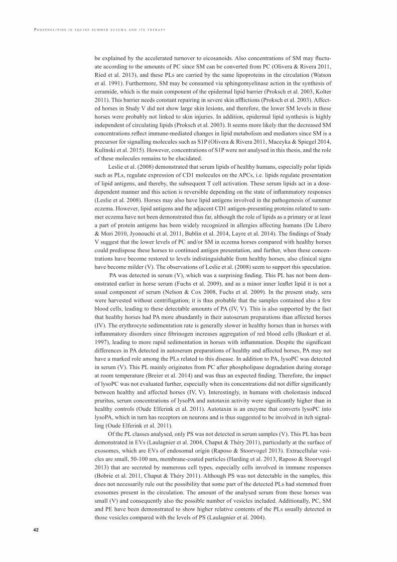
P h o s P h o l i P i d s i n e q u i n e s u m m e r e c z e m a a n d i t s t h e r a P y
42
be explained by the accelerated turnover to eicosanoids. Also concentrations of SM may fluctu-ate according to the amounts of PC since SM can be converted from PC (Olivera & Rivera 2011, Ried et al. 2013), and these PLs are carried by the same lipoproteins in the circulation (Watson et al. 1991). Furthermore, SM may be consumed via sphingomyelinase action in the synthesis of ceramide, which is the main component of the epidermal lipid barrier (Proksch et al. 2003, Kolter 2011). This barrier needs constant repairing in severe skin afflictions (Proksch et al. 2003). Affect-ed horses in Study V did not show large skin lesions, and therefore, the lower SM levels in these horses were probably not linked to skin injuries. In addition, epidermal lipid synthesis is highly independent of circulating lipids (Proksch et al. 2003). It seems more likely that the decreased SM concentrations reflect immune-mediated changes in lipid metabolism and mediators since SM is a precursor for signalling molecules such as S1P (Olivera & Rivera 2011, Maceyka & Spiegel 2014, Kulinski et al. 2015). However, concentrations of S1P were not analysed in this thesis, and the role of these molecules remains to be elucidated.
Leslie et al. (2008) demonstrated that serum lipids of healthy humans, especially polar lipids such as PLs, regulate expression of CD1 molecules on the APCs, i.e. lipids regulate presentation of lipid antigens, and thereby, the subsequent T cell activation. These serum lipids act in a dose-dependent manner and this action is reversible depending on the state of inflammatory responses (Leslie et al. 2008). Horses may also have lipid antigens involved in the pathogenesis of summer eczema. However, lipid antigens and the adjacent CD1 antigen-presenting proteins related to sum-mer eczema have not been demonstrated thus far, although the role of lipids as a primary or at least a part of protein antigens has been widely recognized in allergies affecting humans (De Libero & Mori 2010, Jyonouchi et al. 2011, Bublin et al. 2014, Layre et al. 2014). The findings of Study V suggest that the lower levels of PC and/or SM in eczema horses compared with healthy horses could predispose these horses to continued antigen presentation, and further, when these concen-trations have become restored to levels indistinguishable from healthy horses, also clinical signs have become milder (V). The observations of Leslie et al. (2008) seem to support this speculation.
PA was detected in serum (V), which was a surprising finding. This PL has not been dem-onstrated earlier in horse serum (Fuchs et al. 2009), and as a minor inner leaflet lipid it is not a usual component of serum (Nelson & Cox 2008, Fuchs et al. 2009). In the present study, sera were harvested without centrifugation; it is thus probable that the samples contained also a few blood cells, leading to these detectable amounts of PA (IV, V). This is also supported by the fact that healthy horses had PA more abundantly in their autoserum preparations than affected horses (IV). The erythrocyte sedimentation rate is generally slower in healthy horses than in horses with inflammatory disorders since fibrinogen increases aggregation of red blood cells (Baskurt et al. 1997), leading to more rapid sedimentation in horses with inflammation. Despite the significant differences in PA detected in autoserum preparations of healthy and affected horses, PA may not have a marked role among the PLs related to this disease. In addition to PA, lysoPC was detected in serum (V). This PL mainly originates from PC after phospholipase degradation during storage at room temperature (Breier et al. 2014) and was thus an expected finding. Therefore, the impact of lysoPC was not evaluated further, especially when its concentrations did not differ significantly between healthy and affected horses (IV, V). Interestingly, in humans with cholestasis induced pruritus, serum concentrations of lysoPA and autotaxin activity were significantly higher than in healthy controls (Oude Elferink et al. 2011). Autotaxin is an enzyme that converts lysoPC into lysoPA, which in turn has receptors on neurons and is thus suggested to be involved in itch signal-ling (Oude Elferink et al. 2011).
Of the PL classes analysed, only PS was not detected in serum samples (V). This PL has been demonstrated in EVs (Laulagnier et al. 2004, Chaput & Théry 2011), particularly at the surface of exosomes, which are EVs of endosomal origin (Raposo & Stoorvogel 2013). Extracellular vesi-cles are small, 50-100 nm, membrane-coated particles (Harding et al. 2013, Raposo & Stoorvogel 2013) that are secreted by numerous cell types, especially cells involved in immune responses (Bobrie et al. 2011, Chaput & Théry 2011). Although PS was not detectable in the samples, this does not necessarily rule out the possibility that some part of the detected PLs had stemmed from exosomes present in the circulation. The amount of the analysed serum from these horses was small (V) and consequently also the possible number of vesicles included. Additionally, PC, SM and PE have been demonstrated to show higher relative contents of the PLs usually detected in those vesicles compared with the levels of PS (Laulagnier et al. 2004).
väikkäri_sisus_17.indd 42 28.8.2017 16.40

43
P h o s P h o l i P i d s i n e q u i n e s u m m e r e c z e m a a n d i t s t h e r a P y
Findings in autoserum preparations in comparison with serum The presence of lipids in autoserum preparations was the original hypothesis for the treatment of horses with summer eczema in Studies II and IV. Additionally, the finding that affected horses showed more abundant concentrations of PC and SM in these preparations (IV) was in accordance with our preliminary suggestion that horses with summer eczema have abnormally large amounts of lipids in the circulation (II). Therefore, there was a discrepancy between the results from serum and autoserum preparations. In contrast to our preliminary hypothesis (II), affected horses showed significantly lower concentrations of these major serum PLs in their sera than healthy horses (V). This contradictory finding may be related to the changes in hydrophobic/hydrophilic interactions between lipid molecules and their lipoprotein carriers (Watson et al. 1993) among horses with summer eczema. Lipid composition of autoserum preparations showed that hydrophobic lipid classes and their specific species had been concentrated in those preparations (IV). Although mi-nor amounts of PI and PE were detected in serum (V), they were not found in the preparations (IV). Evidently, this resulted not only from their usually low relative content in mammalian sera (Fuchs et al. 2009), but also from their more hydrophilic nature compared with PC or SM (Nelson & Cox 2008). In autoserum preparations, the less water-soluble PC species were prevalent and the most hydrophilic species were found only in low concentrations (IV), whereas in the serum these more water-soluble species were abundantly present (V). This supports the interpretation that the most hydrophilic species were dissolved in water during the processing of serum. However, spe-cies of SM seemed to act in a slightly different way. SM 16:0 was the most abundant in autoserum preparations, while SM 20:0 predominated in serum, although the former should be more soluble in water than the latter (Nelson & Cox 2008). This suggests that there are at present some obscure factors influencing hydrophobic/hydrophilic properties of certain PLs and their specific species in horses with summer eczema. Mineral salt interactions have been found to affect the solubility of lipids and make lipids even more insoluble in water (Folch et al. 1957). Moreover, lipid-mineral salt interactions dissociate, but only after several washings (Folch et al. 1957). This could be a potential explanation for eczema horses having lower concentrations of PC and SM in serum but higher concentrations in autoserum preparations than healthy horses, and for certain more water-soluble species being concentrated in autoserum preparations.
Phospholipids and clinical status In horses with summer eczema, serum concentrations of PC and SM were significantly lower than in healthy control horses, although they per se showed no significant associations with the severity of clinical signs before therapy (V). On the other hand, concentrations of PC and SM in autoserum preparations displayed a significant relation to severity of signs (IV). Based on these findings, PL profiles in serum seemed to reflect “a rough picture” of the clinical status, i.e. healthy or affected, while in autoserum preparations they provided a more detailed fingerprint. This implies that lipid particles linked to the severity of this allergic disorder could have been concentrated in autoserum preparations. However, this interpretation requires further studies with a greater number of horses than here.
Changes in the total SM concentrations correlated significantly with the alterations in clinical status in response to therapy (V). Of the specific SM species, SM 15:0 showed a significant as-sociation with the alterations in clinical signs after therapy and also was exhibited at significantly lower levels in eczema horse sera than healthy horse sera before therapy, but not afterwards. Therefore, SM seems to play a dynamic role in the course of this disease.
Mode of action in autoserum therapy The underlying mechanism of this therapy remains unknown. A rational explanation for the ac-tion is probably related to cell signalling pathways. The small amounts of various PLs, detected mainly in pico- or nanomolar concentrations, in the autoserum preparations could be sufficient only as a part of signalling cascades, particularly participating in downstream pathways. On signal transduction, minute amounts of molecules are able to interact not only as inducers but also as amplifiers of the corresponding cascades (Nelson & Cox 2008, van Meer & de Kroon 2011). Na-nomolar concentrations are typical for lipid mediators, with these substances being formed during inflammation and its resolution (Serhan & Petasis 2011, Stables & Gilroy 2011); S1P receptors,
väikkäri_sisus_17.indd 43 28.8.2017 16.40

P h o s P h o l i P i d s i n e q u i n e s u m m e r e c z e m a a n d i t s t h e r a P y
44
for instance, are sensitive to nanomolar affinities (Chun et al. 2010). Sometimes, higher amounts of lipid mediators even decrease the expression of target substances, while lower concentrations accelerate their production (Demetz et al. 2014). Intriguingly, sphingolipids have been detected in other biological substances, e.g. dairy products such as milk and butter, usually in micromolar concentrations (Vesper et al. 1999).
The site of action and the target cells involved could not be established based on the infor-mation here. However, some cautious suggestions can be made. In autoserum preparations, lipid particles were absorbed into sugar granules for oral administration. Recently, it has been demon-strated that taste receptors are able to regulate innate immune defences and these taste receptors are also expressed in various tissues beyond the oral cavity, thus supporting their additional func-tion in immune responses (Laffitte et al. 2014, Lee & Cohen 2015, Workman et al. 2015). Of these receptors, bitter and sweet taste receptors are the most important (Lee & Cohen 2015, Workman et al. 2015). Therefore, it seems possible that sweet taste receptors could perceive lipid molecules embedded in sugar granules and provide the primary step for the ongoing action. In the next phase, immune cells, probable basophils, might interact with taste receptors, potentially leading to a modification of FcεRI receptors. Mast cells and basophils are the main cell types assembling in allergic reactions by releasing histamine and a plethora of lipid mediators and cytokines when activated via FcεRI receptors (Kawakami & Galli 2002, Wagner et al. 2006, Kawakami et al. 2009, Cromheecke et al. 2014, Galli et al. 2016, Metcalfe et al. 2016). However, of these cells only basophils are detected in the circulation (Galli et al. 2016, Kratzer & Pickl 2016, Metcalfe et al. 2016). Basophils have been implicated as one of the major cell populations expressing IgE in the peripheral blood of IBH-affected horses (Wagner et al. 2003) and cell response to specific al-lergen challenge in horses with summer eczema (Langner et al. 2008). Therefore, basophils could be one of the messengers participating in the response during autoserum therapy. This suggestion is based also on the following investigations dealing with basophils and allergic inflammation. Lo-cal antigen challenge has been shown to activate circulating basophils, i.e. inducing also systemic allergic influence (Saini et al. 2004). In addition, basophils are able to promote CD4+ T helper cell polarization (Sokol et al. 2008, Kim et al. 2009, Otsuka & Kabashima 2015) and also to act as antigen-presenting cells to CD8+ T cells (Kim et al. 2009). Moreover, Mukai et al. (2005) demon-strated that basophils are essential for the development of IgE-mediated chronic allergic reactions without the intervention of T cells or mast cells. Recently, basophils have been linked to the devel-opment of severe cutaneous hypersensitivity to mosquito bites in humans (Sakakibara et al. 2015). Because basophils interact in various phases of immune responses and they possess capacities not only in innate immune responses but also in adaptive immune responses (Schneider et al. 2010), they might be involved in the course of summer eczema and its resolution after autoserum therapy. Further basic research, including cell culture approaches, is needed.
In mast cells, ensuing activation of FcεRI receptors promotes synthesis of S1P (Olivera & Rivera 2005, 2011, Price et al. 2008, Kulinski et al. 2015), and this lipid mediator has certain two-dimensional functions by enhancing or suppressing allergic responses, possibly depending on type of allergen and site of action (Kulinski et al. 2015). An interesting finding was made in the study of Hamza et al. (2008); a supernatant collected from cultured PBMCs of healthy horses and added to cultures from horses with summer eczema abrogated IL-4 production in these cell cultures. They observed that IL-10 and TGF-β, the major cytokines produced by Treg cells (Ak-dis et al. 2005), possessed this down-regulating effect on IL-4 secretion when these cytokines were simultaneously expressed. But, as stated by Hamza et al. (2008), there may be other factors present in the supernatant that also promote or affect IL-4 down-regulation. Notably, they added autologous serum to these cell cultures, and accordingly, the supernatants probably contained signalling molecules, such as S1P, that are normally present in serum (Christoffersen et al. 2011, Hammad et al. 2012). Afterwards, Hamza et al. (2012) showed that horses with summer eczema were incapable in response allergen challenge of promoting expression of Treg cells, in contrast to healthy horses. Intriguingly, S1P plays a pivotal role in the maturation and function of Treg cells (Cyster & Schwab 2012). Galli et al. (2016) demonstrated that serum collected from animals exposed to honey bee venom prevented detrimental allergic reactions in naive animals, although injected in minute amounts. When binding to the FcεRI receptor was blocked, the beneficial ef-fect of the donor serum disappeared (Galli et al. 2016). This cautiously supports the speculation
väikkäri_sisus_17.indd 44 28.8.2017 16.40

45
P h o s P h o l i P i d s i n e q u i n e s u m m e r e c z e m a a n d i t s t h e r a P y
that the autoserum preparation could interpose via FcεRI receptors and the changes found in the SM concentrations of the horses with summer eczema may reflect or influence alterations in S1P production. The findings of significantly higher SM concentrations in the preparations of affected horses than in healthy horses and the significant correlation between the changed SM concentra-tions and clinical status after therapy are in agreement with this assumption. However, S1P was not analysed in this thesis. Even though alterations in SM concentrations correlated with clinical signs, no conclusions can be drawn from the causality.
Although many research projects associated with S1P signalling are ongoing (Chun et al. 2010), various basic mechanisms between lipid mediators and allergic responses are still unre-solved, not only in relation to the present thesis, but also at a more general level (Christoffersen et al. 2011, Cyster & Schwab 2012, Maceyka & Spiegel 2014, Kulinski et al. 2015, Galli et al. 2016). Advanced methods to analyse detailed information on cell biochemistry (Shaner et al. 2009, Ham-mad et al. 2010, Harkewic & Dennis 2011, Ried et al. 2013, Maceyka & Spiegel 2014, Metcalfe et al. 2016) will help to illuminate the answers.
Limitations of the thesis
In addition to the limitations related to autoserum therapy and discussed earlier in that section, there are limitations in the analysis of the blood samples. By low speed centrifugation for autose-rum preparation, red blood cells could have been avoided in serum samples, without compromis-ing lipid recovery. In addition, lipoprotein particles were not analysed in detail. Therefore, water-insoluble components other than PLs carried by lipoproteins remained unidentified in autoserum preparations, likewise their possible therapeutic effect.
Clinical implications
This was the first series of studies focused on serum PLs and their use in treating horses with sum-mer eczema. Autoserum treatment showed beneficial effects on this allergic skin disease and was convenient to apply in clinical practice since serum PLs could be easily extracted and prepared for oral administration. Based on the findings of this thesis and recent literature in human medi-cine, serum lipoproteins with specific PL species could be an interesting target for further stud-ies on immune-mediated disorders in horses, especially respiratory tract allergies or autoimmune diseases. Accordingly, the utilization of autoserum preparation in the therapy of other allergic manifestations should be explored.
väikkäri_sisus_17.indd 45 28.8.2017 16.40

P h o s P h o l i P i d s i n e q u i n e s u m m e r e c z e m a a n d i t s t h e r a P y
46
7 . C O N C L U S I O N S
1. This study demonstrated that in Finnhorses summer eczema usually manifests at a young age, and most horses in contact with veterinarians suffer from moderate clinical signs. Severity of clinical signs was not related to either age at onset or duration of disease.
2. Autologous serum preparation containing the major serum phospholipids was shown to be a favourable method to treat equine summer eczema, with no harmful side effects.
3. Horses with summer eczema had an altered phospholipid profile in their sera compared with healthy horses, and these profiles seemed to change according to the clinical status of the horse. In affected horses, phosphatidylcholine and sphingomyelin were more concentrated in autoserum preparations than they were in healthy horses. Of the major serum phospholipids, sphingomyelin might be a potential link between equine summer eczema and its therapy; however, the causal relation has to be verified in future studies.
väikkäri_sisus_17.indd 46 28.8.2017 16.40

47
P h o s P h o l i P i d s i n e q u i n e s u m m e r e c z e m a a n d i t s t h e r a P y
8 . A C K N O W L E D G E M E N T S
“Sometimes crazy ideas prove to be useful”. These were the encouraging words of Docent Tho-mas Tallberg when I first introduced my idea of autoserum therapy to him in 1996. That was the start of my studies on equine summer eczema, and over the next twenty years, I have met some wonderful people, visited numerous stables and seen, of course, many horses. I sincerely thank everyone who has helped me on this journey and contributed to my academic dissertation, not forgetting the owners of the horses and the practising vets. Special thanks go to the following persons and institutions:
My supervisors, Professor Outi Vainio and Docent Marja Raekallio, at the Faculty of Veterinary Medicine have been the main promoters of my dissertation and without their encouragement and positive attitude this work would never have been finished. I am deeply grateful for their scientific help and guidance and also appreciate the good atmosphere that they created for students in their department.
I am indebted to Docent Thomas Tallberg and his kind staff, especially Eeva Lönnqvist, at the Institute of Bio-Immunotherapy, where we collaborated on several long-term research projects. They gave me important support in organizing these studies, and Docent Tallberg had a crucial role in our studies focusing on lipids. Unfortunately, he did not live to see the fruits of our labour.
I warmly thank Krishna Batchu for his valuable work with lipid analyses and for his co-authorship. He always had time for me even though he was writing his own PhD thesis. I am also grateful to Tarja Grundström for extensive laboratory work with the horse samples. I gratefully acknowledge Docent Pentti Somerharju for the opportunity to conduct the lipid analyses at the Department of Developmental Biology and Biochemistry, Institute of Biomedicine, University of Helsinki.
I am sincerely grateful to Docent Petteri Nieminen for kindly accepting the invitation to be my opponent.
My deep gratitude is owed to the official reviewers of this thesis, Docent Peter Mattjus and Do-cent Matti Jauhiainen, for the expert comments and valuable criticism that greatly improved the manuscript.
I am also thankful to University Lecturer Sami Junnikkala for his contribution to this thesis, espe-cially for his excellent comments to the section dealing with immunology.
My author-editor Carol Ann Pelli revised the language of the thesis, and I deeply appreciate her help.
I am grateful to Docent Reijo Käkelä and all members of the Functional Lipidomics Group for the time spent at Lipidomics seminars. These seminars were worthwhile not only for the abundant breakfasts but also for the intriguing lectures from the diverse field of lipids and the fresh ideas that emerged from our discussions. I also thank Reijo for his advice and useful comments on my thesis.
Professor Seppo Sarna has provided assistance with statistical analyses and Docent Jan Dabek with language issues, for which I am most grateful. I also thank Docent Mirja Ruohoniemi for her advice and attentive comments when I was writing my publications and Karin Hemmann, PhD, for her help with many practical matters.
Docent Lena Huldén and Larry Huldén, PhD, are thanked for cooperation and many interesting discussions about insects, especially Culicoides, in recent years.
väikkäri_sisus_17.indd 47 28.8.2017 16.40

P h o s P h o l i P i d s i n e q u i n e s u m m e r e c z e m a a n d i t s t h e r a P y
48
This study was supported by a grant from the Albert Lindsay von Julin Foundation, for which I am sincerely grateful.
I deeply thank Ritva Nyman, my friend and one of the co-authors of the first study on autoserum therapy, for her help and advice in preparing the serum. Our inspiring discussions and sometimes even intense arguments during our wanderings in Lapland gave me impetus for this work.
I am grateful to my assistant Leena for her skilful work with autoserum preparations over the years; she has really been my “right hand” with her laboratory and secretarial assistance. My warm thanks also go to Leila and Minna at our veterinary clinic for helping me with day-to-day problems.
Finally, I thank my kind husband Raimo for his support, understanding and love, always.
väikkäri_sisus_17.indd 48 28.8.2017 16.40
49
P h o s P h o l i P i d s i n e q u i n e s u m m e r e c z e m a a n d i t s t h e r a P y
9 . R E F E R E N C E S
Adams, E.J. (2014) Lipid presentation by human CD1 molecules and the diverse T cell populations that re-spond to them. Curr. Opin. Immunol. 26, 1-6, doi:10.1016/j.coi.2013.09.005.
Akdis, M., Blaser, K. & Akdis, C.A. (2005) T regulatory cells in allergy: Novel concepts in the pathogen-esis, prevention, and treatment of allergic diseases. J. Allergy Clin. Immunol. 116, 961-968, doi: 10.1016/j.jaci.2005.09.004.
Anderson, G.S., Belton, P., Jahren, E., Lange, H. & Kleider, N. (1996) Immunotherapy Trial for Horses in Brit-ish Columbia with Culicoides (Diptera: Ceratopogonidae) Hypersensitivity. J. Med. Entomol. 33, 458-466.
Andersson, L.S., Swinbune, J.E., Meadows, J.R.S., Broström, H., Eriksson, S., Fikse, W.F., Frey, R., Sundquist, M., Tseng, C.T., Mikko, S. & Lindgren, G. (2012) The same ELA class II risk factors confer equine insect bite hypersensitivity in two distinct populations. Immunogenetics 64, 201-208, doi: 10.1007/s00251-011-0573-1.
Bacci, S., Alard, P., Dai, R., Nakamura, T. & Streilein, J.W. (1997) High and low doses of haptens dictate whether dermal or epidermal antigen-presenting cells promote contact hypersensitivity. Eur. J. Immunol. 27, 442-448.
Bannenberg, G. & Serhan, C.N. (2010) Specialized Pro-Resolving Lipid Mediators in the Inflammatory Re-sponse: An Update. Biochim. Biophys. Acta 1801(12), 1260-1273, doi: 10.1016/j.bbalip.2010.08.002.
Barbet, J. (1992) Culicoides Hypersensitivity. In: Current Therapy in Equine Medicine, Ed: L. Mills, W.B. Saunders, Philadelphia. pp 693-696.
Baskurt, O.K., Farley, R.A. & Meiselman, H.J. (1997) Erythrocyte aggregation tendency and cellular proper-ties in horse, human, and rat: a comparative study. Am. J. Physiol. 273, 2604-2612.
Batchu, K.C. (2016) Factors regulating the substrate specificity of A-type phospholipases – a mass-spectromet-ric study. Academic Diss., Faculty of Biological and Environmental Sciences, University of Helsinki, 17-35.
Birkinshaw, R.W., Pellicci, D.G., Cheng, T-Y., Keller, A.N., Sandoval-Romero, M., Gras, S., de Jong, A., Ul-drich, A.P., Moody, D.B., Godfrey, D.I. & Rossjohn, J. (2015) αβ T cell antigen receptor recognition of CD1a presenting self lipid ligands. Nature Immunology 16(3), doi: 10.1038/ni.3098.
Björnsdottir, S., Sigvaldadottir, J., Broström, H., Langvad, B. & Sigurosson, A. (2006) Summer eczema in exported Icelandic horses: influence of environmental and genetic factors. Acta Vet. Scand. 48, 3.
Blom, T., Li, S., Dichlberger, A., Bäck, N., Ah Kim, Y., Loizides-Mangold, U., Riezman, H., Bittman, R. & Ikonen, E. (2015) LAPTM4B facilitates late endosomal ceramide export to control cell death pathways. Na-ture Chemical Biology doi: 10.1038/nchembio.1889.
Bobrie, A., Colombo, M., Raposo, G. & Théry, C. (2011) Exosome Secretion: Molecular Mechanisms and Roles in Immune Responses. Traffic 12, 1659-1668, doi: 10.1111/j.1600-0854.2011.01225.x.
Boyce, J.A. (2007) Mast cells and eicosanoid mediators: a system of reciprocal paracrine and autocrine regula-tion. Immunological Reviews 217, 168-185.
Breier, M., Wahl, S., Prehn, C., Fugmann, M., Ferrari, U., Weise, M., Banning, F., Seissler, J., Grallert, H., Adamski, J. & Lechner, A. (2014) Targeted Metabolomics Identifies Reliable and Stable Metabolites in Human Serum and Plasma Samples. PloS One 9, 1-11, e89728, doi:10.1371/journal.pone.0089728.
Broström, H., Larsson, Å. & Troedsson M. (1987) Allergic dermatitis (sweet itch) of Icelandic horses in Swe-den: An epidemiological study. Equine vet. J. 19, 229-236.
Bublin, M., Eiwegger, T. & Breiteneder, H. (2014) Do lipids influence the allergic sensitization process? J. Allergy Clin. Immunol. 134, 521-9, doi: 10.1016/j.jaci.2014.04.015.
väikkäri_sisus_17.indd 49 28.8.2017 16.40

P h o s P h o l i P i d s i n e q u i n e s u m m e r e c z e m a a n d i t s t h e r a P y
50
Caby, M.-P., Lankar, D., Vincendeau-Scherrer, C., Raposo, G. & Bonnerot, C. (2005) Exosomal-like vesicles are present in human blood plasma. International Immunology 17, 879-887, doi: 10.1093/intimm/dxh267.
Chaput, N. & Théry, C. (2011) Exosomes: immune properties and potential clinical implementations. Semin. Immunopathol. 33, 419-440, doi: 10.1007/s00281-010-0233-9.
Chinthrajah, R.S., Hernandez, J.D., Boyd, S.D., Galli, S.J. & Nadeau, K.C. (2016) Molecular and cellu-lar mechanisms of food allergy and food tolerance. J. Allergy Clin. Immunol. 137, 984-997, doi: 10.1016/jaci.2016.02.004.
Christoffersen, C., Obinata, H., Kumaraswamy, S.B., Galvani, S., Ahnström, J., Sevvana, M., Egerer-Sieber, C., Muller, Y.A., Hla, T., Nielsen, L.B. & Dahlbäck, B. (2011) Endothelium-protective sphingosine-1-phos-phate provided by HDL-associated apolipoprotein M. PNAS 108, 9613-9618, doi: 10.1073/pnas.1103187108.
Chun, J., Hla, T., Lynch, K.R., Spiegel, S. & Moolenaar, W.H. (2010) International Union of Basic and Clini-cal Pharmacology. LXXVIII. Lysophospholipid Receptor Nomenclature. Pharmacol. Rev. 62, 579-87, doi: 10.1124/pr.110.003111.
Clausen,B.E. & Kel, J.M. (2010) Langerhans cells: critical regulators of skin immunity? Immunology and Cell Biology 88, 351-360, doi: 10.1038/icb.2010.40.
Cromheecke, J.L., Nguyen, K.T. & Huston, D.P. (2014) Emerging Role of Human Basophil Biology in Health and Disease. Curr. Allergy Asthma Rep. 14, 408, doi: 10.1007/s11882-013-0408-2.
Cyster, J.G. & Schwab, S.R. (2012) Sphingosine-1-Phosphate and Lymphocyte Egress from Lymphoid Or-gans. Annu. Rev. Immunol. 30, 69-94, doi: 10.1146/annurev-immunol-020711-075011.
De Libero G, Mori L. (2010) How immune system detects lipid antigens? Prog. Lipid Res. 49,120-127; doi:10.1016/j.plipres.2009.10.002.
Demetz, E., Schroll, A., Auer, K., Heim, C., Patsch, J.R., Eller, P., Theurl, M., Theurl, I., Theurl, M., Seifert, M., Lener, D., Stanzl, U., Haschka, D., Asshoff, M., Dichtl, S., Nairz, M., Huber, E., Stadlinger, M., Moschen, A.R., Li, X., Pallweber, P., Scharnagl, H., Stojakovic, T., März, W., Kleber, M.E., Garlaschelli, K., Uboldi, P., Catapano, A.L., Stellaard, F., Rudling, M., Kuba, K., Imai, Y., Arita, M., Schuetz, J.D., Pramstaller, P.P., Tietge, U.J.F., Trauner, M., Norata, G.D., Claudel, T., Hicks, A.A., Weiss, G. & Tancevski, I. (2014) The Ara-chidonic Acid Metabolome Serves as a Conserved Regulator of Cholesterol Metabolism. Cell Metabolism 20, 1-12, doi.org/10.1016/j.cmet.2014.09.004.
Dirikolu, L., Lehner, A.F., Harkins, J.D., Woods, W.E., Karbiesiuk, W., Gates, R.S., Fisher, M. & Tobin, T. (2008) Pyrilamine in the horse: detection and pharmacokinetics of pyrilamine and its major urinary metabolite O-desmethylpyrilamine. J. vet. Pharmacol. Therap. 32, 66-78, doi: 10.1111/j.1365-2885.2008.01005.x.
Dubrac, S., Schmuth, M. & Ebner, S. (2010) Atopic dermatitis: the role of Langerhans cells in disease patho-genesis. Immunology and Cell Biology 88, 400-409, doi: 10.1038/icb.2010.33.
Elias, P.M. & Wakefield, J.S. (2011) Therapeutic Implications of a Barrier-Based Pathogenesis of Atopic Der-matitis. Clin. Rev. Allergy Immunol. 41, 282-295, doi: 10.1007/s12016-010-8231-1.
Fadeel, B. & Xue, D. (2009) The ins and outs of phospholipid asymmetry in the plasma membrane: roles in health and disease. Crit. Rev. Biochem. Mol. Biol. 44, 264-277.
Falcone, F.H., Zillikens, D. & Gibbs, B.F. (2006) The 21st century renaissance of the basophil? Current in-sights into its role in allergic responses and innate immunity. Experimental Dermatology 15, 855-864, doi: 10.1111/j.1600-0625.2006.00477.x.
Fanning, L.B. & Boyce, J.A. (2013) Basic Science for the Clinician: Lipid Mediators. Ann. Allergy Asthma Immunol. 111, 155-162, doi: 10.1016/j.anai.2013.06.031.
Finnish Meteorological Institute (2016) Termiset vuodenajat. www.ilmatieteenlaitos.fi/termiset-vuodenajat.
väikkäri_sisus_17.indd 50 28.8.2017 16.40

51
P h o s P h o l i P i d s i n e q u i n e s u m m e r e c z e m a a n d i t s t h e r a P y
Folch, J., Lees, M. & Sloane Stanley, G.H. (1957) A simple method for the isolation and purification of total lipides from animal tissues. J. Biol. Chem. 226(1), 497-509.
Foster, A.P., McKelvie, J. & Cunningham, F.M. (1998) Inhibition of antigen-induced cutaneous responses of ponies with insect hypersensitivity by the histamine-1 receptor antagonist chlorpheniramine. Vet. Rec. 143, 189-193, doi: 10.1136/vr.143.7.189.
Frey, R., Bergvall, K. & Egenvall, A. (2008) Allergen-specific IgE in Icelandic horses with insect bite hyper-sensitivity and healthy controls, assessed by FcεR1α-based serology. Vet. Immunol. Immunopathol. 126, 102-109, doi: 10.1016/j.vetimm.2008.06.010.
Fuchs, B., Schiller, J., Wagner, U., Häntzschel, H. & Arnold, K. (2005) The phosphatidylcholine / lysophos-phatidylcholine ratio in human plasma is an indicator of the severity of rheumatoid arthritis: Investigations by 31P NMR and MALDI-TOF MS. Clin. Biochem. 38, 925-933, doi:10.1016/j.clinbiochem.2005.06.006.
Fuchs, B., Bondzio, A., Wagner, U. & Schiller, J. (2009) Phospholipid compositions of sera and synovial fluids from dog, human and horse: a comparison by 31P-NMR and MALDI-TOF MS. J. Animal. Physiol. Anim. Nutr. 93, 410-422.
Galli, S.J., Tsai, M. & Piliponsky, A.M. (2008) The development of allergic inflammation. Nature 454, 445-454, doi: 10.1038/nature07204.
Galli, S.J., Borregaard, N. & Wynn, T.A. (2011) Phenotypic and functional plasticity of cells of innate im-munity: macrophages, mast cells and neutrophils. Nature Immunology 12, 1035-1044, doi: 10.1038/ni.2109.
Galli, S.J., Starkl, P., Marichal, T. & Tsai, M. (2016) Mast cells and IgE in defense against venoms: Possible “good side” of allergy? Allergology International 65, 3-15, doi: 10.1016/j.alit.2015.09.002.
Garibyan, L., Rheingold, C.G. & Lerner, E.A. (2013) Understanding the pathophysiology of itch. Dermato-logic Therapy 26, 84-91.
Ginel, P.J., Hernandez, E., Lucena, R., Blanco, B., Novales, M. & Mozos, E. (2014) Allergen-specific immu-notherapy in horses with insect bite hypersensitivity: a double-blind, randomized, placebo-controlled study. Vet. Dermatol. 25, 29-e10, doi:10.1111/vde.12092.
Ginhoux, F. & Merad, M. (2010) Ontogeny and homeostasis of Langerhans cells. Immunology and Cell Biol-ogy 88, 387-392, doi: 10.1038/icb.2010.38.
Ginsberg, D.N. & Eichenfield, L.F. (2016) Debates in allergy medicine: Specific immunotherapy in children with atopic dermatitis, the “con” view. World Allergy Organization Journal 9, 16, doi: 10.1186/s40413-016-0107-2.
Girardi, E. & Zajonc, D.M. (2012) Molecular basis of lipid antigen presentation by CD1d and recognition by natural killer T cells. Immunol. Rev. 250, 167-179, doi: 10.1111/j.1600-065X.2012.01166.x.
González-Dominguez, R., Garcia-Barrera, T. & Gómez-Ariza, J.L. (2014) Metabolomic study of lipids in serum for biomarker discovery in Alzheimer’s disease using direct infusion mass spectrometry. J. Pharm. Biomed. Anal. 98, 321-326 doi.org/10.1016/jpba.2014.05.023.
Gould, H.J., Sutton, B.J., Beavil, A.J., Beavil, R.L., McCloskey, N., Coker, H.A., Fear, D. & Smurthwaite, L. (2003) The Biology of IgE and the Basis of Allergic Disease. Annu. Rev. Immunol. 21, 579-628, doi: 10.1146/annurev.immunol.21.120601.141103.
Guyard-Dangremont, V., Desrumaux, C., Gambert, P., Lallemant, C. & Lagrost, L. (1998) Phospholipid and cholesteryl ester transfer activities in plasma from 14 vertebrate species. Relation to atherogenesis susceptibil-ity. Comp. Biochem. Physiol. B 120, 517-525.
Hallamaa, R.E. (2007) Bio-immunotherapy in the treatment of equine sarcoid, the commonest tumour of the horse. Cancer Therapy 5, 143-150.
väikkäri_sisus_17.indd 51 28.8.2017 16.40

P h o s P h o l i P i d s i n e q u i n e s u m m e r e c z e m a a n d i t s t h e r a P y
52
Halldorsdottir, S., Larsen, H.J. & Mehl, R. (1989) Intradermal challenge of Icelandic horses with extracts of four species of the genus Culicoides. Res. Vet. Sci. 47, 283-287.
Halldorsdottir, S. & Larsen, H.J. (1991) An epidemiological study of summer eczema in Icelandic horses in Norway. Equine vet. J. 23, 296-299.
Halldorsdottir, S., Lazary, S., Gunnarsson, E. & Larsen, H.J. (1991) Distribution of leucocyte antigens in Icelandic horses affected with summer eczema compared to non-affected horses. Equine vet. J. 23, 300-302.
Hammad, S.M., Pierce, J.S., Soodavar, F., Smith, K.J., Al Gadban, M.M., Rembiesa, B., Klein, R. L., Hannun, Y.A., Bielawski, J. & Bielawska, A. (2010) Blood sphingolipidomics in healthy humans: impact of sample collection methodology. J. Lipid Res. 51(10), 3074-3087, doi: 10.1194/jlr.D008532.
Hamza, E.,Wagner, B., Jungi, T.W., Mirkovitch, J. & Marti, E. (2008) Reduced incidence of insect-bite hy-persensitivity in Icelandic horses is associated with a down-regulation of interleukin-4 by interleukin-10 and transforming growth factor-β1. Vet. Immunol. Immunopathol. 122, 65-75, doi: 10.1016/j.vetimm.2007.10.018.
Hamza, E., Steinbach, F. & Marti, E. (2012) CD4+CD25+T cells expressing FoxP3 in Icelandic hors-es affected with insect bite hypersensitivity. Vet. Immunol. Immunopathol. 148, 139-144, doi:10.1016/j.vetimm.2011.05.033.
Haniffa, M., Gunawan, M. & Jardine, L. (2015) Human skin dendritic cells in health and disease. J. Dermatol. Sci. 77, 85-92, doi: 10.1016/j.jdermsci.2014.08.012.
Hanson, M.A., Roth, C.B., Jo, E., Griffith, M.T., Scott, F.L., Reinhart, G., Desale, H., Clemons, B., Calahan, S.M., Schuerer, S.C., Sanna, M.G., Han, G.W., Kuhn, P., Rosen, H. & Stevens, R.C. (2012) Crystal Structure of a Lipid G Protein-Coupled Receptor. Science 335, 851-855, doi: 10.1126/science.1215904.
Harding, C.V., Heuser, J.E. & Stahl, P.D. (2013) Exosomes: Looking back three decades and into the future. J. Cell Biol. 200, 367-371, doi: 10.1083/jcb.201212113.
Harkewicz, R. & Dennis, E.A. (2011) Applications of Mass Spectrometry to Lipids and Membranes. Annu. Rev. Biochem. 80, 301-325, doi: 10.1146/annurev-biochem-060409-092612.
Heimann, M., Janda, J., Sigurdardottir, O.G., Svansson, V., Klukowska, J., von Tscharner, C., Doherr, M., Broström, H., Andersson, L.S., Einarsson, S., Marti, E. & Torsteinsdottir, S. (2011) Skin-infiltrating T cells and cytokine expression in Icelandic horses affected with insect bite hypersensitivity: A possible role for regulatory T cells. Vet. Immunol. Immunopathol. 140, 63-74.
Hellberg, W., Wilson, A.D., Mellor, P., Doherr, M.G., Torsteinsdottir, S., Zurbriggen, A., Jungi, T. & Marti, E. (2006) Equine insect bite hypersensitivity: Immunoblot analysis of IgE and IgG subclass responses to Culicoides nubeculosus salivary gland extract. Vet. Immunol. Immunopathol. 113, 99-112, doi: 10.1016/j.vetimm.2006.04.009.
Hemmann, K. (2014). Crib-biting in Horses: A Physiological and Genetic Study of Candidate Causative Fac-tors. Academic Diss., Faculty of Veterinary Medicine, University of Helsinki.
Hermansson, M., Uphoff, A., Käkelä, R., Somerharju, P. (2005) Automated Quantitative Analysis of Complex Lipidomes by Liquid Chromatography/Mass Spectrometry. Anal. Chem. 77, 2166-2175.
Hippos (2016) Hevoskannan kehitys. www.hippos.fi/files/13835/hevoskannan_kehitys_.
Hirvisalo, E.L. & Renkonen, O. (1970) Composition of human serum sphingomyelins. J. Lipid Res. 11, 54-59.
Holthuis, J.C.M. & Menon, A.K. (2014) Lipid landscapes and pipelines in membrane homeostasis. Nature 510, 48-57, doi:10.1038/nature13474.
Huldén, L. & Huldén, L. (2014) Checklist of the family Ceratopogonidae (Diptera) of Finland. ZooKeys 441, 53-61, doi: 10.3897/zookeys.441.7742.
väikkäri_sisus_17.indd 52 28.8.2017 16.40

53
P h o s P h o l i P i d s i n e q u i n e s u m m e r e c z e m a a n d i t s t h e r a P y
Huldén, L., Huldén, L. & Lahtinen, T. (2008) Bluetongue-viruksen vektorilajit Suomessa [Vectors of blue-tonguevirus in Finland]. Suom. Eläinlääkäril. 114, 158-161.
Inouye, M., Silander, K., Hamalainen, E., Salomaa,V., Harald, K., Jousilahti, P., Männistö, S., Eriksson, J.G., Saarela, J., Ripatti, S., Perola, M.,van Ommen, G-J.B., Taskinen, M-R., Palotie, A., Dermitzakis, E.T. & Peltonen, L. (2010) An Immune Response Network Associated with Blood Lipid Levels. PLoS Genet. 6(9): e1001113., doi: 10.1371/journal.pgen.1001113.
Jayawardena-Wolf, J. & Bendelac, A. (2001) CD1 and lipid antigens: intracellular pathways for antigen pres-entation. Curr. Opin. Immunol. 13, 109-113.
Jonsdottir, S., Hamza, E., Janda, J., Rhyner, C., Meinke, A., Marti, E., Svansson, V. & Torsteinsdottir, S. (2015) Developing a preventive immunization approach against insect bite hypersensitivity using recombinant aller-gens: A pilot study. Vet. Immunol. Immunopathol. 166, 8-21, doi: 10.1016/j.vetimm.2015.05.002.
Jose-Cunilleras, E., Kohn, C.W., Hillier, A., Saville, W.J.A. & Lorch, G. (2001) Intradermal testing in healthy horses and horses with chronic obstructive pulmonary disease, recurrent urticaria, or allergic dermatitis. JAV-MA 219,1115-1121.
Josefowicz, S.Z., Lu, L.-F. & Rudensky, A.Y. (2012) Regulatory T Cells: Mechanisms of Differentiation and Function. Annu. Rev. Immunol. 30, 531-564, doi: 10.1146/annurev.immunol.25.022106.141623.
Jovanovic, V., Abdul Aziz, N., Lim, Y.T., Ng Ai Poh, A., Jin Hui Chan, S., Ho Xin Pei, E., Lew, F.C., Shui, G., Jenner, A.M., Bowen, L., McKinney, E.F., Lyons, P.A., Kemeny, M.D., Smith, K.G.C., Wenk, M.R. & MacAry, P.A. (2013) Lipid Anti-Lipid Antibody Responses Correlate with Disease Activity in Systemic Lupus Erythematosus. PLoS One 8, e55639, doi:10.1371/journal.pone.0055639.
Jyonouchi, S., Abraham, V., Orange, J.S., Spergel, J.M., Gober, L., Dudek, E., Saltzman, R., Nichols, K.E. & Cianferoni, A. (2011) Invariant natural killer T cells from children with versus without food allergy ex-hibit differential responsiveness to milk-derived sphingomyelin. J. Allergy Clin. Immunol. 128, 102-109, doi: 10.1016/j.jaci.2011.02.026.
Kainu, V. (2012) Metabolism and Translocation of Aminophospholipids in Mammalian Cells. Academic Diss., Faculty of Medicine, University of Helsinki.
Kawakami, T. & Galli, S.J. (2002) Regulation of mast-cell and basophil function and survival by IgE. Nature Reviews Immunology 2, 773-786.
Kawakami, T., Ando, T., Kimura, M., Wilson, B.S. & Kawakami, Y. (2009) Mast cells in atopic dermatitis. Curr. Opin. Immunol. 21, 666-678, doi:10.1016/j.coi.2009.09.006.
Kendall, A. & Nicolaou, A. (2013) Bioactive lipid mediators in skin inflammation and immunity. Prog. Lipid Res. 52, 141-164, doi.org/10.1016/j.plipres.2012.10.003.
Kierszenbaum, A.L. (2002) Epithelial glands, cytomembranes. In: Histology and Cell Biology, Mosby, Inc., Elsevier Science, St. Louis, Missouri. pp 56-75.
Kim, S., Shen, T. & Min, B. (2009) Basophils Can Directly Present or Cross-Present Antigen to CD8 Lympho-cytes and Alter CD8 T Cell Differentiation into IL-10-Producing Phenotypes. The Journal of Immunology 183, 3033-3039, doi: 10.4049/jimmunol.0900332.
Kinnunen, R.E., Tallberg, Th., Stenbäck, H. & Sarna, S. (1999) Equine sarcoid tumour treated by autogenous tumour vaccine. Anticancer Res. 19, 3367-3374.
Kolter, T. (2011) A view on sphingolipids and disease. Chem. Phys. Lipids 164, 590-606, doi: 10.1016/j.chemphyslip.2011.04.013.
Korematsu, S., Miyahara, H., Kakita, A. & Izumi, T. (2014) Elevated serum anti-phosphatidylcholine IgG antibodies in patients with influenza vaccination-associated optic neuritis. Vaccine 32, 6345-6348, doi.org/10.1016/j.vaccine.2014.09.053.
väikkäri_sisus_17.indd 53 28.8.2017 16.40

P h o s P h o l i P i d s i n e q u i n e s u m m e r e c z e m a a n d i t s t h e r a P y
54
Kratzer, B. & Pickl, W. (2016) Years in Review: Recent Progress in Cellular Allergology. Int. Arch. Allergy Immunol. 169, 1-12, doi: 10.1159/000444753.
Kuhnt, K., Degen, C. & Jahreis, G. (2015) Evaluation of the Impact of Ruminant trans Fatty acids on Human Health: Important Aspects to Consider. Crit. Rev. Food Sci. Nutr. doi:10.1080/10408398.2013.808605.
Kulinski, J.M., Munoz-Cano, R. & Olivera, A. (2015) Sphingosine-1-phosphate and other lipid mediators gen-erated by mast cells as critical players in allergy and mast cell function. Eur. J. Pharmacol. doi.org/10.1016/j.ejphar.2015.02.058.
Kuroda, T., Nagata, S.-I., Takizawa, Y., Tamura, N., Kusano, K., Mizobe, F. & Hariu, K. (2013) Pharmacoki-netics and pharmacodynamics of d-chlorpheniramine following intravenous and oral administration in healthy Thoroughbred horses. The Vet. J. 197, 433-437, doi.org/10.1016/j.tvjl.2013.02.003.
Kurotaki, T., Narayama, K., Oyamada, T., Yoshikawa, H. & Yoshikawa, T. (1994) Immunopathological Study on Equine Insect Hypersensitivity (“Kasen”) in Japan. J. Comp. Pathol. 110, 145-152.
Laffitte, A., Neiers, F. & Briand, L. (2014) Functional roles of the sweet taste receptor in oral and extraoral tissues. Curr. Opin. Clin. Nutr. Metab. Care 17, 379-385, doi: 10.1097/MCO.0000000000000058.
Laitinen, K., Sallinen, J., Linderborg, K. & Isolauri, E. (2006) Serum, cheek cell and breast milk fatty acid compositions in infants with atopic and non-atopic eczema. Clin. Exp. Allergy 36, 166-173.
Langner, K.F.A., Darpel, K.E., Drolet, B.S., Fischer, A., Hampel, S., Heselhaus, J.E., Mellor, P.S., Mertens, P.P.C. & Leibold, W. (2008) Comparison of cellular and humoral immunoassays for the assessment of summer eczema in horses. Vet. Immunol. Immunopathol. 122, 126-137, doi: 10.1016/j.vetimm.2007.11.001.
Langner, K.F.A., Jarvis, D.L., Nimtz, M., Heselhaus, J.E., McHolland, L.E., Leibold, W. & Drolet, B.S. (2009) Identification, expression and characterisation of a major salivary allergen (Cul s 1) of the biting midge Cu-licoides sonorensis relevant for summer eczema in horses. Int. J. Parasitol. 39, 243-250, doi: 10.1016/j.ij-para.2008.06.008.
Laulagnier, K., Motta, C., Hamdi, S., Roy, S., Fauvelle, F., Pageaux, J.-F., Kobayashi, T., Salles, J.-P., Perret, B., Bonnerot, C. & Record, M. (2004) Mast cell- and dendritic cell-derived exosomes display a specific lipid composition and an unusual membrane organization. Biochem. J. 380, 161-171.
Layre, E., de Jong, A. & Moody, D.B. (2014) Human T cells use CD1 and MR1 to recognize lipids and small molecules. Curr. Opin. Chem. Biol. 23, 31-38, doi:10.1016/j.cbpa.2014.09.007.
Lazary, S., Marti, E., Szalai, G., Gaillard, C. & Gerber, H. (1994) Studies on the frequency and associations of equine leucocyte antigens in sarcoid and summer dermatitis. Anim. Genet. 25, 75-80.
Lee, R.J. & Cohen, N.A. (2015) Taste Receptors in Innate Immunity. Cell Mol. Life Sci. 72, 217-236, doi: 10.1007/s00018-014-1736-7.
Leslie, D.S., Dascher, C.C., Cembrola, K., Townes, M.A., Hava, D.L., Hugendubler, L.C., Mueller, E., Fox, L., Roura-Mir, C., Moody, D.B., Vincent, M.S., Gumperz, J.E., Illarionov, P.A., Besra, G.S., Reynolds, C.G. & Brenner, M.B. (2008) Serum lipids regulate dendritic cell CD1 expression and function. Immunology 125, 289-301, doi: 10.1111/j.1365-2567.2008.02842.x.
Li, J.-F., Qu, F., Zheng, S.-J., Ren, J.-Y., Wu, H.-L., Liu, M., Liu, H., Ren, F., Chen, Y., Zhang, J.-L. & Duan, Z.-P. (2014) Plasma Sphingolipids as Potential Indicators of Hepatic Necroinflammation in Patients with Chronic Hepatitis C and Normal Alanine Aminotransferase Level. Plos One 9(4), e95095, doi:10.1371/journal.pone.0095095.
Lin, N., Shi, J.-J., Li, Y.-M., Zhang, X.-Y., Chen, Y., Calder, P.C. & Tang, L.-J. (2016) What is the impact of n-3 PUFAs on inflammation markers in Type 2 diabetic mellitus populations?: a systemic review and meta-anal-ysis of randomized controlled trials. Lipids in Health and Disease 15, 133, doi: 10.1186/s12944-016-0303-7.
väikkäri_sisus_17.indd 54 28.8.2017 16.40

55
P h o s P h o l i P i d s i n e q u i n e s u m m e r e c z e m a a n d i t s t h e r a P y
Lis-Swiety, A., Brzezinska-Wcislo, L., Arasiewicz, H. & Bergler-Czop, B. (2014) Antiphospholipid antibodies in localized scleroderma: the potential role of screening tests for the detection of antiphospholipid syndrome. Postep. Derm. Alergol. 2, 65-70.
Maceyka, M. & Spiegel, S. (2014) Sphingolipid metabolites in inflammatory disease. Nature 510, 58-67, doi: 10.1038/nature13475.
Magnusson, J., Kull, I., Westman, M., Håkansson, N., Wolk, A., Melén, E., Wickman, M. & Bergström, A. (2015) Fish and polyunsaturated fat intake and development of allergic and nonallergic rhinitis. J. Allergy Clin. Immunol. 136(5), doi: 10.1016/j.jaci.2015.05.030.
Matsuoka, T., Shamji, M.H. & Durham, S.R. (2013) Allergen Immunotherapy and Tolerance. Allergology International 62, 403-413, doi: 10.2332/allergolint.13-RAI-0650.
Metcalfe, D.D., Pawankar, R., Ackerman, S.J., Akin, C., Clayton, F., Falcone, F.H., Gleich, G.J., Irani, A.-M., Johansson, M.W., Klion, A.D., Leiferman, K.M., Levi-Schaffer, F., Nilsson, G., Okayama, Y., Prussin, C., Schroeder, J.T., Schwartz, L.B., Simon, H.-U., Walls, A.F. & Triggiani, M. (2016) Biomarkers of the involve-ment of mast cells, basophils and eosinophils in asthma and allergic diseases. World Allergy Organization Journal doi: 10.1186/s40413-016-0094-3.
Meulenbroeks, C., van der Meide, N.M.A., Zaiss, D.M.W., Sloet van Oldruitenborgh-Oosterbaan, M.M., van der Lugt, J.J., Smak, J., Rutten, V.P.M.G. & Willemse, T. (2013) Seasonal differences in cytokine expression in the skin of Shetland ponies suffering from insect bite hypersensitivity. Vet. Immunol. Immunopathol. 151, 147-156, doi: 10.1016/j.vetimm.2012.11.007.
Meulenbroeks, C., van der Lugt, J.J., van der Meide, N.M.A., Willemse, T., Rutten, V.P.M.G. & Zaiss, D.M.W. (2015) Allergen-Specific Cytokine Polarization Protects Shetland Ponies against Culicoides obsoletus-Induced Insect Bite Hypersensitivity. Plos One 10, e0122090, doi: 10.1371/journal.pone.0122090.
Min, B. & Paul, W.E. (2008) Basophils: in the spotlight at last. Nature Immunology 9, 223-225.
Morgan, E.E., Miller Jr, W.H. & Wagner, B. (2007) A comparison of intradermal testing and detection of allergen-specific immunoglobulin E in serum by enzyme-linked immunosorbent assay in horses affected with skin hypersensitivity. Vet. Immunol. Immunopathol. 120, 160-167, doi: 10.1016/j.vetimm.2007.08.007.
Mori, L. & De Libero, G. (2008) Presentation of lipid antigens to T cells. Immunol. Lett. 117, 1-8, doi: 10.1016/j.imlet.2007.11.027.
Mukai, K., Matsuoka, K., Taya, C., Suzuki, H., Yokozeki, H., Nishioka, K., Hirokawa, K., Etori, M., Yamash-ita, M., Kubota, T., Minegishi, Y., Yonekawa, H. & Karasuyama, H. (2005) Basophils Play a Critical Role in the Development of IgE-Mediated Chronic Allergic Inflammation Independently of T Cells and Mast Cells. Immunity 23, 191-202, doi: 10.1016/j.immuni.2005.06.011.
Nelson, D.L. & Cox, M.M. (2008) Lipids, Biological Membranes and Transport, Biosignaling, Lipid Biosyn-thesis, Hormonal Regulation and Integration of Mammalian Metabolism. In: Principles of Biochemistry, 5th edn., Ed: K. Ahr, W.H. Freeman, New York. pp 343-380, 805-850, 901-912.
Novak, N. & Leung, D.Y.M. (2011) Advances in atopic dermatitis. Curr. Opin. Immunol. 23, 778-783, doi:10.1016/j.coi.2011.09.007.
O’Connor, C.I., Lawrence, L.M. & Hayes, S.H. (2007) Dietary fish oil supplementation affects serum fatty acid concentrations in horses. J. Anim. Sci. 85, 2183-2189.
Ohsawa, Y. & Hirasawa, N. (2014) The Role of Histamine H1 and H4 Receptors in Atopic Dermatitis: From Ba-sic Research to Clinical Study. Allergology International. 63, 533-542, doi: 10.2332/allergolint.13-RA-0675.
Olivera, A. & Rivera, J. (2005) Sphingolipids and the Balancing of Immune Cell Function: Lessons from the Mast Cell. J. Immunol. 174, 1153-1158, doi:10.4049/jimmunol.174.3.1153.
väikkäri_sisus_17.indd 55 28.8.2017 16.40

P h o s P h o l i P i d s i n e q u i n e s u m m e r e c z e m a a n d i t s t h e r a P y
56
Olivera, A. & Rivera, J. (2011) An emerging role for the lipid mediator sphingosine-1-phosphate in mast cell effector function and allergic disease. Adv. Exp. Med. Biol. 716, 123-142.
Olsén, L., Bondesson, U., Broström, H., Tjälve, H. & Ingvast-Larsson, C. (2008) Cetirizine in horses: Phar-macokinetics and pharmacodynamics following repeated oral administration. The Vet. J. 177, 242-249, doi: 10.1016/j.tvjl.2007.03.026.
Olsén, L., Bondesson, U., Broström, H., Olsson, U., Mazogi, B., Sundqvist, M., Tjälve, H. & Ingvast-Larsson, C. (2011) Pharmacokinetics and effects of cetirizine in horses with insect bite hypersensitivity. The Vet. J. 187, 347-351, doi: 10.1016/j.tvjl.2009.12.030.
Ora, T. (1963) Kotieläinten yleisimmät sairaudet ja ensiapu. In: Maanviljelijän tietokirja 2, Ed: P. Kajanoja, Werner Söderström Osakeyhtiö, Porvoo. pp 746-747.
Otsuka, A. & Kabashima, K. (2015) Contribution of basophils to cutaneous immune reactions and Th2-medi-ated allergic responses. Frontiers in Immunology 6, 393, doi: 10.3389/fimmu.2015.00393.
Oude Elferink, R.P.J., Kremer, A.E. & Beuers, U. (2011) Mediators of pruritus during cholestasis. Curr. Opin. Gastroenterol. 27, 289-293, doi: 10.1097/MOG.0b013e32834575e8.
Paassilta, M., Kuusela, E., Korppi, M., Lemponen, R., Kaila, M. & Nikkari, S.T. (2014) Food allergy in small children carries a risk of essential fatty acid deficiency, as detected by elevated serum mead acid proportion of total fatty acids. Lipids in Health and Disease 13, 180, doi: 10.1186/1476-511X-13-180.
Patel, K.N. & Dong, X. (2011) Itch: Cells, Molecules, and Circuits. ACS Chem. Neurosci. 2, 17-25, doi: 10.1021/cn100085g.
Patel, N., Vogel, R., Chandra-Kuntal, K., Glasgow, W. & Kelavkar, U. (2014) A Novel Three Serum Phos-pholipid Panel Differentiates Normal Individuals from Those with Prostate Cancer. Plos One 9, 1-9, e88841, doi:10.1371/journal.pone.0088841.
Pierce, B.G, Vreven, T. & Weng, Z. (2014) Modeling T cell receptor recognition of CD1-lipid and MR1-metabolite complexes. BMC Bioinformatics 15:319, doi: 10.1186/1471-2105-15-319.
Price, M.M., Oskeritzian, C.A., Milstien, S. & Spiegel, S. (2008) Sphingosine-1-phosphate synthesis and func-tions in mast cells. Future Lipidol. 3, 665-674, doi:10.2217/17460875.3.6.665.
Porcelli, S., Brenner, M.B., Greenstein, J.L., Terhorst, C., Balk, S.P. & Bleicher, P.A. (1989) Recognition of cluster of differentiation 1 antigens by human CD4¯CD8¯ cytolytic T lymphocytes. Nature 341, 447-450.
Proksch, E., Jensen, J.-M. & Elias, P.M. (2003) Skin Lipids and Epidermal Differentiation in Atopic Dermati-tis. Clin. Dermatol. 21, 134-144.
Qu, F., Wu, C.-S., Hou, J.-F., Jin, Y. & Zhang, J.-L. (2012) Sphingolipids as New Biomarkers for Assess-ment of Delayed-Type Hypersensitivity and Response to Triptolide. PLoS One 7, e52454, doi:10.1371/journal.pone.0052454.
Quehenberger, O., Armando, A.M., Brown, A.H., Milne, S.B., Myers, D.S., Merrill, A.H., Bandyopadhyay, S., Jones, K.N., Kelly, S., Shaner, R.L., Sullards, C.M., Wang, E., Murphy, R.C., Barkley, R.M., Leiker, T.J., Raetz, C.R.H., Guan, Z., Laird, G.M., Six, D.A., Russell, D.W., McDonald, J.G., Subramaniam, S., Fahy, E. & Dennis, E.A. (2010) Lipidomics reveals a remarkable diversity of lipids in human plasma. J. Lipid Res. 51, 3299-3305, doi: 10.1194/jlr.M009449.
Quehenberger, O. & Dennis, E.A. (2011) The Human Plasma Lipidome. N. Engl. J. Med. 365(19), 1812-1823, doi: 10.1056/NEJMra1104901.
Quintana, F., Yeste, A., Weiner, H. & Covacu, R. (2012) Lipids and lipid-reactive antibodies as biomarkers for multiple sclerosis. J. Neuroimmunol. 248, 53-57, doi:10.1016/j.jneuroim.2012.01.002.
väikkäri_sisus_17.indd 56 28.8.2017 16.40

57
P h o s P h o l i P i d s i n e q u i n e s u m m e r e c z e m a a n d i t s t h e r a P y
Raposo, G. & Stoorvogel, W. (2013) Extracellular vesicles: Exosomes, microvesicles, and friends. J. Cell Biol. 200, 373-383, doi: 10.1083/jcb.201211138.
Reines, I., Kietzmann, M., Mischke, R., Tschernig, T., Lüth, A., Kleuser, B. & Bäumer, W. (2009) Topical Application of Sphingosine-1-Phosphate and FTY720 Attenuate Allergic Contact Dermatitis Reaction through Inhibition of Dendritic Cell Migration. Journal of Investigative Dermatology 129, 1954-1962, doi: 10.1038/jid.2008.454.
Ried, J.S., Baurecht, H., Stückler, F., Krumsiek, J., Gieger, C., Heinrich, J., Kabesch, M., Prehn, C., Peters, A., Rodriguez, E., Schulz, H., Strauch, K., Suhre, K., Wang-Sattler, R., Wichmann, H.-E., Theis, F.J., Illig, T., Adamski, J. & Weidinger, S. (2013) Integrative genetic and metabolite profiling analysis suggests altered phosphatidylcholine metabolism in asthma. Allergy 68, 629-636, doi: 10.1111/all.12110.
Riek, R.F. (1953) Studies on allergic dermatitis (Queensland itch) of the horse: the aetiology of the disease. Aust. J. Agric. Res. 5, 109-129.
Rutkowski, K., Sowa, P., Rutkowska-Talipska, J., Sulkowski, S. & Rutkowski, R. (2014) Allergic diseases: the price of civilisational progress. Postep. Derm. Alergol. 2, 77-83, doi: 10.5114/pdia.2014.40936.
Saini, S., Bloom, D.C., Bieneman, A., Vasagar, K., Togias, A. & Schroeder, J. (2004) Systemic effects of al-lergen exposure on blood basophil IL-13 secretion and FcεRIβ. J. Allergy Clin. Immunol. 114, 768-774, doi: 10.1016/j.jaci.2004.06.015.
Sakaguchi, S., Wing, K., Onishi, Y., Prieto-Martin, P. & Yamaguchi, T. (2009) Regulatory T cells: how do they suppress immune responses? International Immunology 21, 1105-1111, doi: 10.1093/intimm/dxp095.
Sakakibara, Y., Wada, T., Muraoka, M., Matsuda, Y., Toma, T. & Yachie, A. (2015) Basophil activation by mosquito extracts in patients with hypersensitivity to mosquito bites. Cancer Sci. 106, 965-971, doi: 10.1111/cas.12696.
Salio, M., Silk, J.D. & Cerundolo, V. (2010) Recent advances in processing and presentation of CD1 bound lipid antigens. Curr. Opin. Immunol. 22, 81-88, doi:10.1016/j.coi.2009.12.008.
Schaffartzik, A., Hamza, E., Janda, J., Crameri, R., Marti, E. & Rhyner, C. (2012) Equine insect bite hypersen-sitivity: What do we know? Vet. Immunol. Immunopathol. 147, 113-126.
Schmetterer, K.G., Neunkirchner, A. & Pickl, W. (2012) Naturally occuring regulatory T cells: markers, mech-anisms, and manipulation. FASEB J. 26, 2253-2276, doi: 10.1096/fj.11-193672.
Schneider, E., Thieblemont, N., Leite De Moraes, M. & Dy, M. (2010) Basophils: new players in the cytokine network. Eur. Cytokine Netw. 21,142-153, doi: 10.1684/ecn.2010.0197.
Schurink, A., van Grevenhof, E.M., Ducro, B.J. & van Arendonk, J.A.M. (2009) Heritability and repeatability of insect bite hypersensitivity in Dutch Shetland breeding mares. J. Anim. Sci. 87, 484-490, doi: 10.2527/jas.2008-1129.
Schurink, A., Ducro, B.J., Bastiaansen, J.W.M., Frankena, K. & van Arendonk, J.A.M. (2012) Genome-wide association study of insect bite hypersensitivity in Dutch Shetland pony mares. Anim. Genet. 44, 44-52, doi: 10.1111/j.1365-2052.2012.02368.x.
Schurink, A., Wolc, A., Ducro, B.J., Frankena, K., Garrick, D.J. & Dekkers, J.C.M. (2013) Genome-wide as-sociation study of insect bite hypersensitivity in two horse populations in the Netherlands. Genet. Sel. Evol. 44, 44-52.
Scott, D.W. & Miller, W.H., Jr. (2003) Structure and Function of the Skin, Insect Hypersensitivity. In: Equine Dermatology, Eds: R. Kersey and D. LeMelledo, Saunders, Elsevier Science, St. Louis, Missouri. pp 1-58, 458-474.
Serhan, C.N. & Petasis, N.A. (2011) Resolvins and Protectins in Inflammation-Resolution. Chem. Rev. 111(10), 5922-5943, doi:10.1021/cr100396c.
väikkäri_sisus_17.indd 57 28.8.2017 16.40

P h o s P h o l i P i d s i n e q u i n e s u m m e r e c z e m a a n d i t s t h e r a P y
58
Shaner, R.L., Allegood, J.C., Park, H., Wang, E., Kelly, S., Haynes, C.A., Sullards, M.C. & Merrill, A.H., Jr. (2009) Quantitative analysis of sphingolipids for lipidomics using triple quadrupole and quadrupole linear ion trap mass spectrometers. J. Lipid Res. 50, 1692-1707, doi: 10.1194/jlr.D800051-JLR200.
Shepherd, J. (1991) Equine plasma lipoproteins: comparative lessons. Equine vet. J. 23, 329-330.
Shrestha, M., Eriksson, S., Schurink, A., Andersson, L.S., Sundquist, M., Frey, R., Broström, H., Bergström, T., Ducro, B. & Lindgren, G. (2015) Genome-Wide Association Study of Insect Bite Hypersensitivity in Swed-ish-Born Icelandic Horses. J. Hered. 366-374, doi: 10.1093/jhered/esv033.
Silveira e Souza, A.M.M., Mazucato, V.M., Jamur, M.C. & Oliver, C. (2011) Lipid Rafts in Mast Cell Biology. Journal of Lipids doi:10.1155/2011/752906.
Slavyanakaya, T.A., Derkach, V.V. & Sepiashvili, R.I. (2016) Debates in allergy medicine: specific immuno-therapy efficiency in children with atopic dermatitis. World Allergy Organization Journal 9, 15, doi:10.1186/s40413-016-0106-3.
Sloane, D.E., Tedla, N., Awoniyi, M., MacGlashan, D.W., Jr., Borges, L., Austen, K.F. & Arm, J.P. (2004) Leu-kocyte immunoglobulin-like receptors: novel innate receptors for human basophil activation and inhibition. Blood 104, 2832-2839, doi: 10.1182/blood-2004-01-0268.
Sokol, C.L., Barton, G.M., Farr, A.G. & Medzhitov, R. (2008) A mechanism for the initiation of allergen-induced T helper type 2 responses. Nat. Immunol. 9, 310-318, doi: 10.1038/ni1558.
Stables, M. & Gilroy, D. (2011) Old and new generation lipid mediators in acute inflammation and resolution. Prog. Lipid Res. 50, 35-51, doi:10.1016/j.plipres.2010.07.005.
Steinhoff, M., Bienenstock, J., Schmelz, M., Maurer, M., Wei, E. & Bíró, T. (2006) Neurophysiological, Neu-roimmunological, and Neuroendocrine Basis of Pruritus. Journal of Investigative Dermatology 126, 1705-1718, doi: 10.1038/sj.jid.5700231.
Steinman, A., Peer, G. & Klement, E. (2003a) Epidemiological study of Culicoides hypersensitivity in horses in Israel. Vet. Rec. 152, 748-751.
Steinman, R.M., Hawiger, D. & Nussenzweig, M.C. (2003b) Tolerogenic Dendritic Cells. Annu. Rev. Immu-nol. 21, 685-711, doi: 10.1146/annurev.immunol.21.120601.141040.
Subbaiah, P.V. & Liu, M. (1996) Comparative studies on the substrate specificity of lecithin:cholesterol acyl-transferase towards the molecular species of phosphatidylcholine in the plasma of 14 vertebrates. J. Lipid Res. 37, 113-122.
Tallberg, Th., Tykkä, H., Halttunen, P., Mahlberg, K.L., Uusitalo, R., Carlsson, O., Sandstedt, B., Oravisto, K., Lehtonen, T., Sarna, S. & Strandström, H. (1979) Cancer immunity. The effect in cancer-immunotherapy of polymerised autologous tumour tissue and supportive measures. Scand. J. Clin. Lab. Invest. 39, 1-35.
Thijs, C., Müller, A., Rist, L., Kummeling, I., Snijders, B.E.P., Huber, M., van Ree, R., Simoes-Wüst, A.P., Dagnelie, P.C. & van den Brandt, P.A. (2011) Fatty acids in breast milk and development of atopic eczema and allergic sensitisation in infancy. Allergy 66, 58-67.
Valenta, R., Campana, R., Marth, K. & van Hage, M. (2012) Allergen-specific immunotherapy: from therapeu-tic vaccines to prophylactic approaches. J. Intern. Med. 272, 144-157, doi: 10.1111/j.1365-2796.2012.02556.x.
van der Haegen, A., Griot-Wenk, M., Welle, M., Busato, A., von Tscharner, C., Zurbriggen, A. & Marti, E. (2001) Immunoglobulin-E-bearing cells in skin biopsies of horses with insect bite hypersensitivity. Equine vet. J. 33, 699-706.
van der Meide, N.M.A., Roders, N., Sloet van Oldruitenborgh-Oosterbaan, M.M., Schaap, P.J., van Oers, M.M., Leibold, W., Savelkoul, H.F.J. & Tijhaar, E. (2013) Cloning and expression of candidate allergens from Culicoides obsoletus for diagnosis of insect bite hypersensitivity in horses. Vet. Immunol. Immunopathol. 153, 227-239, doi:10.1016/j.vetimm.2013.03.005.
väikkäri_sisus_17.indd 58 28.8.2017 16.40

59
P h o s P h o l i P i d s i n e q u i n e s u m m e r e c z e m a a n d i t s t h e r a P y
van Grevenhof, E.M., Ducro, B., Heuven, H.C.M. & Bijma, P. (2007) Identification of environmental factors affecting the prevalence of insect bite hypersensitivity in Shetland ponies and Friesian horses in the Nether-lands. Equine vet. J. 39, 69-73.
van Meer G. (2005) Cellular lipidomics. EMBO J. 24, 3159-3165.
van Meer, G. (2011) Dynamic Transbilayer Lipid Asymmetry. Cold Spring Harb. Perspect. Biol. 3:a004671.
van Meer, G. & de Kroon, A.I.P.M. (2011) Lipid map of the mammalian cell. J. Cell Science 124, 5-8.
Velie, B.D., Shrestha, M., Francois, L., Schurink, A., Tesfayonas, Y.G., Stinckens, A., Blott, S., Ducro, B.J., Mikko, S., Thomas, R., Swinburne, J.E., Sundqvist, M., Eriksson, S., Buys, N. & Lindgren, G. (2016) Using an Inbred Horse Breed in a High Density Genome-Wide Scan for Genetic Risk Factors of Insect Bite Hyper-sensitivity (IBH). PloS One 12, doi: 10.1371/journal.pone.0152966.
Vesper, H., Schmelz, E.-M., Nikolova-Karakashian, M.N., Dillehay, D.L., Lynch, D.V. & Merrill, A.H., Jr. (1999) Sphingolipids in Food and the Emerging Importance of Sphingolipids to Nutrition. J. Nutr. 129, 1239-1250. Vinding, R.K., Stokholm, J., Chawes, B.L.K. & Bisgaard, H. (2015) Blood lipid levels associate with child-hood asthma, airway obstruction, bronchial hyperresponsiveness, and aeroallergen sensitization. J. Allergy Clin. Immunol. doi: 10.1016/j.jaci.2015.05.033.
Wagner, B., Radbruch, A., Rohwer, J. & Leibold, W. (2003) Monoclonal anti-equine IgE antibodies with speci-ficity for different epitopes on the immunoglobulin heavy chain of native IgE. Vet. Immunol. Immunopathol. 92, 45-60, doi: 10.1016/S0165-2427(03)00007-2.
Wagner, B., Miller, W.H., Morgan, E.E., Hillegas, J.M., Erb, H.N., Leibold, W. & Antczak, D.F. (2006) IgE and IgG antibodies in skin allergy of the horse. Vet. Res. 37, 813-825.
Watson, T.D.G., Burns, L., Love, S., Packard, C.J. & Shepherd, J. (1991) The isolation, characterisation and quantification of the equine plasma lipoproteins. Equine vet. J. 23, 353-359.
Watson, T.D.G., Packard, C.J. & Shepherd, J. (1993) Plasma lipid transport in the horse (Equus Caballus). Comp. Biochem. Physiol. 106B, 27-34.
Westermarck, H. (1949) Ihotaudit. In: Maatalouden eläinlääkärikirja, Eds: R. Stenius and S. Simonen, Werner Söderström Osakeyhtiö, Porvoo. pp 365-366.
Wilson, A.D. (2014) Immune responses to ectoparasites of horses, with a focus on insect bite hypersensitivity. Parasite Immunol. 36, 560-572, doi: 10.1111/pim.12142.
Wong, L.H., Čopič, A. & Levine, T.P. (2017) Advances on the Transfer of Lipids by Lipid Transfer Proteins. Trends Biochem. Sci. article in press, doi: 10.1016/j.tibs.2017.05.001.
Workman, A.D., Palmer, J.N., Adappa, N.D. & Cohen, N.A. (2015) The Role of Bitter and Sweet Taste Recep-tors in Upper Airway Immunity. Curr. Allergy Asthma Rep. 15, 72, doi: 10.1007/s11882-015-0571-8.
Zannis, V.I., Fotakis, P., Koukos, G., Kardassis, D., Ehnholm,C., Jauhiainen, M. & Chroni, A. (2015) High Density Lipoproteins. In: HDL Biogenesis, Remodeling, and Catabolism, Eds: A. von Eckardstein and D. Kardassis, Springer International Publishing, vol. 224, pp 53-111.
väikkäri_sisus_17.indd 59 28.8.2017 16.40

P h o s P h o l i P i d s i n e q u i n e s u m m e r e c z e m a a n d i t s t h e r a P y
60
APPENDIX I
Date__________
Owner
Address
Telephone
Name of the horse
Breed
Gender
Date of birth, country
Colour
Use of the horse
Anamnesis:Onset of clinical signs
Severity of signs before autoserum treatment:
Itching in the maneItching in the tail
Mild skin lesions in the maneMild skin lesions in the tail Mild skin lesions in the body
Large skin lesions in the maneLarge skin lesions in the tail Large skin lesions in the body
Aggravating factors
Hereditary background
Treatments given and their influence
väikkäri_sisus_17.indd 60 28.8.2017 16.40

61
P h o s P h o l i P i d s i n e q u i n e s u m m e r e c z e m a a n d i t s t h e r a P y
APPENDIX II
FOLLOW-UP Date__________
Owner
Name of the horse
Clinical signs after start of autoserum therapy:
Pruritus: same milder worse
Skin lesions: same milder worse
If pruritus has become milder, when has this occurred? during the first 2 weeks of therapy during the 1-week pause during the third week of therapy later
Has the horse been tired during therapy? no yes
If yes: during the first 2 weeks of therapy during the 1-week pause during the third week of therapy later all the time
Has appetite changed? no yes
Has the horse been exposed to? sun insects grass fodder
Use of autoserum preparation:
Autoserum preparation has been administered: about 1 week a month about 1-2 weeks a month about 2-3 weeks a month about daily
väikkäri_sisus_17.indd 61 28.8.2017 16.40

P h o s P h o l i P i d s i n e q u i n e s u m m e r e c z e m a a n d i t s t h e r a P y
62
Indicate the months of medication: May June July August September October
Do you think that your horse has benefited from this therapy relative to the severity of clinical signs in earlier summers? no benefit some benefit much benefit
How convenient was this therapy to perform? easy rather easy difficult
Has your horse had some adverse effects from this therapy? yes no
If yes, what kind of effects?
väikkäri_sisus_17.indd 62 28.8.2017 16.40