Pheochromocytoma and Paraganglioma: An Endocrine...
28
Pheochromocytoma and Paraganglioma: An Endocrine Society Clinical Practice Guideline Jacques W. M. Lenders, Quan-Yang Duh, Graeme Eisenhofer, Anne-Paule Gimenez-Roqueplo, Stefan K. G. Grebe, Mohammad Hassan Murad, Mitsuhide Naruse, Karel Pacak, and William F. Young, Jr Radboud University Medical Center (J.W.M.L.), 6500 HB Nijmegen, The Netherlands; VA Medical Center and University of California, San Francisco (Q.-Y.D.), San Francisco, California 94121; University Hospital Dresden (G.E.), 01307 Dresden, Germany; Assistance Publique-Hôpitaux de Paris, Hôpital Européen Georges Pompidou, Service de Génétique, (A.-P.G.-R.), F-75015 Paris, France; Université Paris Descartes (A.-P.G.-R.), F-75006 Paris, France; Mayo Clinic (S.K.G.G., M.H.M.), Rochester, Minnesota 55905; National Hospital Organisation Kyoto Medical Center (M.N.), Kyoto 612-8555; Japan; Eunice Kennedy Shriver National Institute of Child Health & Human Development (K.P.), Bethesda, Maryland 20892; and Mayo Clinic (W.F.Y.), Rochester, Minnesota 55905 Objective: The aim was to formulate clinical practice guidelines for pheochromocytoma and para- ganglioma (PPGL). Participants: The Task Force included a chair selected by the Endocrine Society Clinical Guidelines Subcommittee (CGS), seven experts in the field, and a methodologist. The authors received no corporate funding or remuneration. Evidence: This evidence-based guideline was developed using the Grading of Recommendations, Assessment, Development, and Evaluation (GRADE) system to describe both the strength of rec- ommendations and the quality of evidence. The Task Force reviewed primary evidence and com- missioned two additional systematic reviews. Consensus Process: One group meeting, several conference calls, and e-mail communications enabled consensus. Committees and members of the Endocrine Society, European Society of En- docrinology, and Americal Association for Clinical Chemistry reviewed drafts of the guidelines. Conclusions: The Task Force recommends that initial biochemical testing for PPGLs should include measurements of plasma free or urinary fractionated metanephrines. Consideration should be given to preanalytical factors leading to false-positive or false-negative results. All positive results require follow-up. Computed tomography is suggested for initial imaging, but magnetic resonance is a better option in patients with metastatic disease or when radiation exposure must be limited. 123 I-metaio- dobenzylguanidine scintigraphy is a useful imaging modality for metastatic PPGLs. We recommend consideration of genetic testing in all patients, with testing by accredited laboratories. Patients with paraganglioma should be tested for SDHx mutations, and those with metastatic disease for SDHB mutations. All patients with functional PPGLs should undergo preoperative blockade to prevent peri- operative complications. Preparation should include a high-sodium diet and fluid intake to prevent postoperative hypotension. We recommend minimally invasive adrenalectomy for most pheochro- mocytomas with open resection for most paragangliomas. Partial adrenalectomy is an option for selected patients. Lifelong follow-up is suggested to detect recurrent or metastatic disease. We suggest personalized management with evaluation and treatment by multidisciplinary teams with appropriate expertise to ensure favorable outcomes. (J Clin Endocrinol Metab 99: 1915–1942, 2014) ISSN Print 0021-972X ISSN Online 1945-7197 Printed in U.S.A. Copyright © 2014 by the Endocrine Society Received February 19, 2014. Accepted April 24, 2014. Abbreviations: CT, computed tomography; 18 F-FDG, 18 F-fluorodeoxyglucose; 18 F-FDOPA, 18 F-fluo- rodihydroxy-phenylalanine; LC-ECD, liquid chromatography with electrochemical detection; LC- MS/MS, liquid chromatography with tandem mass spectrometry; MEN2, multiple endocrine neo- plasia type 2; MIBG, metaiodobenzylguanidine; MRI, magnetic resonance imaging; NF1, neurofibromatosis type 1; PET, positron emission tomography; PPGL, pheochromocytoma and paraganglioma; ROC, receiver operating characteristic; SDH, succinate dehydrogenase; VHL, von Hippel-Lindau; VMA, vanillylmandelic acid; VUS, variant of unknown significance. SPECIAL FEATURE Clinical Practice Guideline doi: 10.1210/jc.2014-1498 J Clin Endocrinol Metab, June 2014, 99(6):1915–1942 jcem.endojournals.org 1915 The Endocrine Society. Downloaded from press.endocrine.org by [Luis Cardoso] on 07 November 2014. at 02:13 For personal use only. No other uses without permission. . All rights reserved.
Transcript of Pheochromocytoma and Paraganglioma: An Endocrine...

Pheochromocytoma and Paraganglioma: AnEndocrine Society Clinical Practice Guideline
Jacques W. M. Lenders, Quan-Yang Duh, Graeme Eisenhofer,Anne-Paule Gimenez-Roqueplo, Stefan K. G. Grebe, Mohammad Hassan Murad,Mitsuhide Naruse, Karel Pacak, and William F. Young, Jr
Radboud University Medical Center (J.W.M.L.), 6500 HB Nijmegen, The Netherlands; VA Medical Center andUniversity of California, San Francisco (Q.-Y.D.), San Francisco, California 94121; University Hospital Dresden (G.E.),01307 Dresden, Germany; Assistance Publique-Hôpitaux de Paris, Hôpital Européen Georges Pompidou, Service deGénétique, (A.-P.G.-R.), F-75015 Paris, France; Université Paris Descartes (A.-P.G.-R.), F-75006 Paris, France; MayoClinic (S.K.G.G., M.H.M.), Rochester, Minnesota 55905; National Hospital Organisation Kyoto Medical Center(M.N.), Kyoto 612-8555; Japan; Eunice Kennedy Shriver National Institute of Child Health & Human Development(K.P.), Bethesda, Maryland 20892; and Mayo Clinic (W.F.Y.), Rochester, Minnesota 55905
Objective: The aim was to formulate clinical practice guidelines for pheochromocytoma and para-ganglioma (PPGL).
Participants: The Task Force included a chair selected by the Endocrine Society Clinical GuidelinesSubcommittee (CGS), seven experts in the field, and a methodologist. The authors received nocorporate funding or remuneration.
Evidence: This evidence-based guideline was developed using the Grading of Recommendations,Assessment, Development, and Evaluation (GRADE) system to describe both the strength of rec-ommendations and the quality of evidence. The Task Force reviewed primary evidence and com-missioned two additional systematic reviews.
Consensus Process: One group meeting, several conference calls, and e-mail communicationsenabled consensus. Committees and members of the Endocrine Society, European Society of En-docrinology, and Americal Association for Clinical Chemistry reviewed drafts of the guidelines.
Conclusions: The Task Force recommends that initial biochemical testing for PPGLs should includemeasurements of plasma free or urinary fractionated metanephrines. Consideration should be givento preanalytical factors leading to false-positive or false-negative results. All positive results requirefollow-up. Computed tomography is suggested for initial imaging, but magnetic resonance is a betteroption in patients with metastatic disease or when radiation exposure must be limited. 123I-metaio-dobenzylguanidine scintigraphy is a useful imaging modality for metastatic PPGLs. We recommendconsideration of genetic testing in all patients, with testing by accredited laboratories. Patients withparaganglioma should be tested for SDHx mutations, and those with metastatic disease for SDHBmutations. All patients with functional PPGLs should undergo preoperative blockade to prevent peri-operative complications. Preparation should include a high-sodium diet and fluid intake to preventpostoperative hypotension. We recommend minimally invasive adrenalectomy for most pheochro-mocytomas with open resection for most paragangliomas. Partial adrenalectomy is an option forselectedpatients. Lifelongfollow-up is suggestedtodetect recurrentormetastaticdisease.Wesuggestpersonalizedmanagementwithevaluationandtreatmentbymultidisciplinaryteamswithappropriateexpertise to ensure favorable outcomes. (J Clin Endocrinol Metab 99: 1915–1942, 2014)
ISSN Print 0021-972X ISSN Online 1945-7197Printed in U.S.A.Copyright © 2014 by the Endocrine SocietyReceived February 19, 2014. Accepted April 24, 2014.
Abbreviations:CT,computedtomography;18F-FDG,18F-fluorodeoxyglucose;18F-FDOPA,18F-fluo-rodihydroxy-phenylalanine; LC-ECD, liquid chromatography with electrochemical detection; LC-MS/MS, liquid chromatography with tandem mass spectrometry; MEN2, multiple endocrine neo-plasia type 2; MIBG, metaiodobenzylguanidine; MRI, magnetic resonance imaging; NF1,neurofibromatosis type 1; PET, positron emission tomography; PPGL, pheochromocytoma andparaganglioma; ROC, receiver operating characteristic; SDH, succinate dehydrogenase; VHL, vonHippel-Lindau; VMA, vanillylmandelic acid; VUS, variant of unknown significance.
S P E C I A L F E A T U R E
C l i n i c a l P r a c t i c e G u i d e l i n e
doi: 10.1210/jc.2014-1498 J Clin Endocrinol Metab, June 2014, 99(6):1915–1942 jcem.endojournals.org 1915
The Endocrine Society. Downloaded from press.endocrine.org by [Luis Cardoso] on 07 November 2014. at 02:13 For personal use only. No other uses without permission. . All rights reserved.

SUMMARY OF RECOMMENDATIONS
1.0 Biochemical Testing for Diagnosis ofPheochromocytoma and Paraganglioma (PPGL)
1.1 We recommend that initial biochemical testingfor PPGLs should include measurements of plasma freemetanephrines or urinary fractionated metanephrines.(1�QQQQ)
1.2 We suggest using liquid chromatography withmass spectrometric or electrochemical detection methodsrather than other laboratory methods to establish a bio-chemical diagnosis of PPGL. (2�QQEE)
1.3 For measurements of plasma metanephrines, wesuggest drawing blood with the patient in the supine po-sition and use of reference intervals established in the sameposition. (2�QQEE)
1.4 We recommend that all patients with positive testresults should receive appropriate follow-up according tothe extent of increased values and clinical presentation.(1�QQEE)
2.0 Imaging Studies2.1 We recommend that imaging studies to locate PPGL
should be initiated once there is clear biochemical evidenceof a PPGL. (1�QQEE)
2.2 We suggest computed tomography (CT) rather thanmagnetic resonance imaging (MRI) as the first-choice im-aging modality because of its excellent spatial resolutionfor thorax, abdomen, and pelvis. (2�QQQE)
2.3 We recommend MRI in patients with metastaticPPGL, for detection of skull base and neck paraganglio-mas, in patients with surgical clips that cause artifactswhen using CT, in patients with an allergy to CT contrast,and in patients in whom radiation exposure should belimited (children, pregnant women, patients with knowngermline mutations, and those with recent excessive radi-ation exposure). (1�QQQE)
2.4 We suggest the use of 123I-metaiodobenzylguani-dine (MIBG) scintigraphy as a functional imaging modal-ity in patients with metastatic PPGL detected by otherimaging modalities when radiotherapy using 131I-MIBG isplanned, and occasionally in some patients with an in-creased risk for metastatic disease due to large size of theprimary tumor or to extra-adrenal, multifocal (exceptskull base and neck PPGLs), or recurrent disease.(2�QEEE)
2.5 We suggest the use of 18F-fluorodeoxyglucose (18F-FDG) positron emission tomography (PET)/CT scanningin patients with metastatic disease. 18F-FDG PET/CT is thepreferred imaging modality over 123I-MIBG scintigraphyin patients with known metastatic PPGL. (2�QQQE)
3.0 Genetic Testing3.1 We recommend that all patients with PPGLs should
be engaged in shared decision making for genetic testing.(1�QQQE)
3.2 We recommend the use of a clinical feature-drivendiagnostic algorithm to establish the priorities for specificgenetic testing in PPGL patients with suspected germlinemutations. (1�QQQE)
3.3 We suggest that patients with paraganglioma un-dergo testing of succinate dehydrogenase (SDH) muta-tions and that patients with metastatic disease undergotesting for SDHB mutations. (2�QQQE)
3.4 We recommend that genetic testing for PPGL bedelivered within the framework of health care. Specifi-cally, pretest and post-test counseling should be available.All tests for PPGL genetic testing should be performed byaccredited laboratories. (Ungraded recommendation)
4.0 Perioperative Medical Management4.1 We recommend that all patients with a hormonally
functional PPGL should undergo preoperative blockadeto prevent perioperative cardiovascular complications. (1/QQEE) We suggest �-adrenergic receptor blockers as thefirst choice. (2�QQEE)
4.2 We recommend preoperative medical treatment for7 to 14 days to allow adequate time to normalize bloodpressure and heart rate. Treatment should also include ahigh-sodium diet and fluid intake to reverse catechol-amine-induced blood volume contraction preoperativelyto prevent severe hypotension after tumor removal.(1�QQEE)
4.3 We recommend monitoring blood pressure, heartrate, and blood glucose levels with adjustment of associ-ated therapies in the immediate postoperative period.(1�QQEE)
4.4 We suggest measuring plasma or urine levels ofmetanephrines on follow-up to diagnose persistent dis-ease. We suggest lifelong annual biochemical testing toassess for recurrent or metastatic disease. (2�QQEE)
5.0 Surgery5.1 We recommend minimally invasive adrenalectomy
(eg, laparoscopic) for most adrenal pheochromocytomas.(1�QQEE) We recommend open resection for large (eg,�6 cm) or invasive pheochromocytomas to ensure com-plete tumor resection, prevent tumor rupture, and avoidlocal recurrence. (1�QEEE) We suggest open resection forparagangliomas, but laparoscopic resection can be per-formed for small, noninvasive paragangliomas in surgi-cally favorable locations. (2�QEEE)
1916 Lenders et al Guidelines on Pheochromocytoma and Paraganglioma J Clin Endocrinol Metab, June 2014, 99(6):1915–1942
The Endocrine Society. Downloaded from press.endocrine.org by [Luis Cardoso] on 07 November 2014. at 02:13 For personal use only. No other uses without permission. . All rights reserved.
5.2 We suggest partial adrenalectomy for selected pa-tients, such as those with hereditary pheochromocytoma,with small tumors who have already undergone a con-tralateral complete adrenalectomy to spare adrenal cortexto prevent permanent hypocortisolism. (2�QEEE)
6.0 Personalized Management6.1 In recognition of the distinct genotype-phenotype
presentations of hereditary PPGLs, we recommend a per-sonalized approach to patient management (ie, biochem-ical testing, imaging, surgery, and follow-up). (Ungradedrecommendation)
6.2 We recommend that patients with PPGLs should beevaluated and treated by multidisciplinary teams at cen-ters with appropriate expertise to ensure favorable out-come. In particular, patients should be referred to suchcenters should there be pregnancy, metastatic disease, orissues concerning the complexity or difficulty in biochem-ical diagnosis; localization; performance and interpreta-tion of genetic testing; preoperative preparation; surgicaltreatment; and follow-up. (Ungraded recommendation)
METHOD OF DEVELOPMENT OF EVIDENCE-BASED CLINICAL PRACTICE GUIDELINES
The Clinical Guidelines Subcommittee (CGS) of the En-docrine Society deemed the diagnosis of pheochromocy-toma and paraganglioma a priority area in need of practiceguidelines and appointed a Task Force to formulate evi-dence-based recommendations. The Task Force followedthe approach recommended by the Grading of Recom-mendations, Assessment, Development, and Evaluation(GRADE) group, an international group with expertise inthe development and implementation of evidence-basedguidelines (1). A detailed description of the gradingscheme has been published elsewhere (2). The Task Forceused the best available research evidence to develop therecommendations. The Task Force also used consistentlanguage and graphic descriptions of both the strength ofa recommendation and the quality of evidence. In terms ofthe strength of the recommendation, strong recommen-dations use the phrase “we recommend” and the number1, and weak recommendations use the phrase “we sug-gest” and the number 2. Cross-filled circles indicate thequality of the evidence, such that QEEE denotes very lowquality evidence; QQEE, low quality; QQQE, moderatequality; andQQQQ, high quality. The Task Force has con-fidence that persons who receive care according to thestrong recommendations will derive, on average, moregood than harm. Weak recommendations require more
careful consideration of the person’s circumstances, val-ues, and preferences to determine the best course of action.Linked to each recommendation is a description of theevidence and the values that panelists considered in mak-ing the recommendation; in some instances, there are re-marks, a section in which panelists offer technical sugges-tions for testing conditions, dosing, and monitoring.These technical comments reflect the best available evi-dence applied to a typical person being treated. Often thisevidence comes from the unsystematic observations of thepanelists and their values and preferences; therefore, theseremarks should be considered suggestions.
The Endocrine Society maintains a rigorous conflict-of-interest review process for the development of clinicalpractice guidelines. All Task Force members must declareany potential conflicts of interest, which are reviewedbefore the members are approved to serve on the TaskForce and periodically during the development of theguideline. The conflict-of-interest forms are vetted by theCGS before the members are approved by the Society’sCouncil to participate on the guideline Task Force. Par-ticipants in the guideline development must include a ma-jority of individuals without conflict of interest in the mat-ter under study. Participants with conflicts of interest mayparticipate in the development of the guideline, but theymust have disclosed all conflicts. The CGS and the TaskForce have reviewed all disclosures for this guideline andresolved or managed all identified conflicts of interest.
Conflicts of interest are defined by remuneration in anyamount from the commercial interest(s) in the form ofgrants; research support; consulting fees; salary; owner-ship interest (eg, stocks, stock options, or ownership in-terest excluding diversified mutual funds); honoraria orother payments for participation in speakers’ bureaus, ad-visory boards, or boards of directors; or other financialbenefits. Completed forms are available through the En-docrine Society office.
Funding for this guideline was derived solely from theEndocrine Society, and thus the Task Force received nofunding or remuneration from commercial or otherentities.
Definition, Prevalence, and ClinicalSignificance of Pheochromocytomaand Paraganglioma
Definition of pheochromocytoma andparaganglioma (PPGL)
A pheochromocytoma is a tumor arising from adreno-medullary chromaffin cells that commonly produces one
doi: 10.1210/jc.2014-1498 jcem.endojournals.org 1917
The Endocrine Society. Downloaded from press.endocrine.org by [Luis Cardoso] on 07 November 2014. at 02:13 For personal use only. No other uses without permission. . All rights reserved.
or more catecholamines: epinephrine, norepinephrine,and dopamine. Rarely, these tumors are biochemically si-lent. A paraganglioma is a tumor derived from extra-ad-renal chromaffin cells of the sympathetic paravertebralganglia of thorax, abdomen, and pelvis. Paragangliomasalso arise from parasympathetic ganglia located along theglossopharyngeal and vagal nerves in the neck and at thebase of the skull (3); these do not produce catecholamines.These last paraganglioma in the neck and at the base of theskull receive minimal coverage in this guideline. About 80to 85% of chromaffin-cell tumors are pheochromocyto-mas, whereas 15 to 20% are paragangliomas (4). Togetherthey will be referred to here as PPGL.
The prevalence of PPGLThe prevalence of PPGL in patients with hypertension
in general outpatient clinics varies between 0.2 and 0.6%(5–8). Diagnosis of PPGL may be missed during life; au-topsy studies demonstrate undiagnosed tumors in 0.05–0.1% of patients (9–11). In children with hypertension,the prevalence of PPGL is approximately 1.7% (12).Nearly 5% of patients with incidentally discovered adre-nal masses on anatomical imaging prove to have a pheo-chromocytoma (13, 14).
At least one-third of all patients with PPGLs havedisease-causing germline mutations (inherited muta-tions present in all cells of the body). The prevalence ofPPGL in individuals carrying a germline mutation inPPGL susceptibility genes may be around 50%. Patientswith hereditary PPGLs typically present with multifocaldisease and at a younger age than those with sporadicneoplasms (15, 16).
The clinical importance of PPGLIt is important to suspect, confirm, localize, treat, and
resect these tumors for several reasons. Most of these tu-mors hypersecrete catecholamines, and if untreated, car-diovascular morbidity and mortality are high (17–21).Also, PPGLs enlarge with time and may cause mass-effectsymptoms by encroaching upon or extending into adja-cent tissues and organs.
Another reason to encourage case detection is that, forfamilial disease, detection of a tumor in the proband mayresult in earlier diagnosis and treatment in other familymembers. Finally, some PPGLs have malignant potential.Malignancy is defined as the presence of metastases innonchromaffin tissue; the prevalence varies between 10and 17% (22). Mutations in the gene encoding SDH sub-unit B (SDHB) can lead to metastatic disease in 40% ormore of the patients (23, 24).
Reasons to suspect PPGLThe most important step for diagnosis of PPGL is to
first recognize the possibility of the tumor. As reviewed indetail elsewhere (4, 7, 25, 26), it is key to recognize thesigns and symptoms and other manifestations or clinical
Table 1. Clinical Settings for Testing for PPGL
Signs and symbols of PPGL, in particular if paroxysmalPPGL symptoms provoked by use of medications associated
with adverse effects (see Table 2)Adrenal incidentaloma, with or without hypertensionHereditary predisposition or syndromic features suggesting
hereditary PPGLPrevious history of PPGL
Table 2. Medications That Are Implicated in Adverse Reactions in Patients with Pheochromocytoma and That CanPrecipitate a Crisis
Class of Drugs Examples
Dopamine D2 receptor antagonists (including someantiemetic agents and antipsychotics)
Metoclopramide, sulpiride, amisulpride,tiapride, chlorpromazine, prochlorperazine,droperidol
�-Adrenergic receptor blockersa Propranolol, sotalol, timolol, nadolol, labetalolSympathomimetics Ephedrine, pseudoephedrine, fenfluramine,
methylphenidate, phentermine,dexamfetamine
Opioid analgesics Morphine, pethidine, tramadolNorepinephrine reuptake inhibitors (including tricyclic
antidepressants)Amitriptyline, imipramine,
Serotonin reuptake inhibitors (rarely reported) Paroxetine, fluoxetineMonoamine oxidase inhibitors Tranylcypromine, moclobemide, phenelzineCorticosteroids Dexamethasone, prednisone, hydrocortisone,
betamethasonePeptides ACTH, glucagonNeuromuscular blocking agents Succinylcholine, tubocurarine, atracurium
a Although most case reports on �-adrenergic receptor blockers pertain to nonselective blockers, selective �1-blockers may also precipitate a crisisbecause at higher doses they may lose �1-selectivity.
1918 Lenders et al Guidelines on Pheochromocytoma and Paraganglioma J Clin Endocrinol Metab, June 2014, 99(6):1915–1942
The Endocrine Society. Downloaded from press.endocrine.org by [Luis Cardoso] on 07 November 2014. at 02:13 For personal use only. No other uses without permission. . All rights reserved.

settings that might signal a need for biochemical testing forPPGL (Tables 1 and 2). Biochemical testing is also war-ranted in syndromic forms of PPGL, which may be indi-cated by specific clinical stigmata (Table 3).
1.0 Biochemical Testing for Diagnosis ofPheochromocytoma and Paraganglioma
Available tests and test performance
Recommendation1.1 We recommend that initial biochemical testing
for PPGLs should include measurements of plasma freemetanephrines or urinary fractionated metanephrines.(1QQQQ)
1.1 EvidenceThere is compelling evidence that measurements of
plasma free or urinary fractionated metanephrines are su-perior to other tests of catecholamine excess for diagnosisof PPGLs; the theoretical basis for this is provided by im-proved understanding of catecholamine metabolism (27–29). According to this understanding, the free metaneph-rines are produced within adrenal chromaffin cells (or thetumors derived from these cells) by membrane-bound cat-echolamine O-methyltransferase. Lack of this enzyme insympathetic nerves, the major site of initial norepineph-rine metabolism, means that the O-methylated metabo-lites are relatively specific markers of chromaffin tumors.Most importantly, these metabolites are produced contin-uously within tumors by a process that is independent ofexocytotic catecholamine release, which for some tumorsoccurs at low rates or is episodic in nature.
The superior sensitivity of urine metanephrines overcatecholamines and vanillylmandelic acid (VMA) for di-agnosis of PPGLs was first suggested from a meta-analysisby Manu and Runge (30). This analysis was followed by
reports revealing false-negative results for measurementsof urine catecholamines and VMA and improved accuracywith measurements of urinary metanephrines (31–36).
Initial evidence that measurements of plasma free meta-nephrines provide advantages for diagnosis of PPGLs overother tests was first outlined by Lenders et al (37). Diag-nostic specificity was equivalent to other tests, but diag-nostic sensitivity was superior. A second National Insti-tutes of Health (NIH) study involving patients screenedfor hereditary PPGLs established excellent sensitivity of97%, well in excess of the 47 to 74% for other tests (38).The final NIH report, with cumulative experience in over800 patients, established that the superiority of plasmametanephrines for the diagnosis remained significant,even when compared with combinations of other tests(39).
The high diagnostic accuracy of measurements ofplasma free metanephrines has now been confirmed by 15independent studies (39–53) (Table 4).
Areas under receiver operating characteristic (ROC)curves reported in nine of these studies ranged from 0.965to 1. Among studies involving comparisons with otherbiochemical tests, all except two indicated both improvedsensitivity and specificity for plasma metanephrines thanfor plasma (n � 4) and urine (n � 7) catecholamines orVMA (n � 1). These exceptions included one study inwhich the combination of urinary catecholamines and to-tal metanephrines (normetanephrine and metanephrinemeasured in combined form by spectrophotometry) wasassessed by areas under ROC curves to offer similar di-agnostic accuracy to measurements of plasma metaneph-rines (41).
Five of the 15 studies involved comparisons of plasmafree with urine fractionated metanephrines (39, 42, 46, 48,53). The results suggest higher specificity of the plasmathan the urine test (Table 5); however, all five studies had
Table 3. Clinical Findings Associated with Syndromic PPGL
Multiple endocrine neoplasiatype 2A
Medullary thyroid cancer, primary hyperparathyroidism, and cutaneouslichen amyloidosis
Multiple endocrine neoplasiatype 2B
Medullary thyroid cancer, mucocutaneous neuromas, skeletaldeformities (eg, kyphoscoliosis or lordosis), joint laxity, myelinatedcorneal nerves, and intestinal ganglioneuromas (Hirschsprungdisease)
von Hippel-Lindau syndrome Hemangioblastoma (involving the cerebellum, spinal cord, orbrainstem), retinal angioma, clear cell renal cell carcinoma,pancreatic neuroendocrine tumors and serous cystadenomas,endolymphatic sac tumors of the middle ear, papillary cystadenomasof the epididymis and broad ligament
Neurofibromatosis type 1 Neurofibromas, multiple café-au-lait spots, axillary and inguinalfreckling, iris hamartomas (Lisch nodules), bony abnormalities,central nervous system gliomas, macrocephaly, and cognitive deficits
doi: 10.1210/jc.2014-1498 jcem.endojournals.org 1919
The Endocrine Society. Downloaded from press.endocrine.org by [Luis Cardoso] on 07 November 2014. at 02:13 For personal use only. No other uses without permission. . All rights reserved.

limitations, and none involved head-to-head comparisonsof mass spectrometric-based measurements.
As shown by Perry et al (54), measurements of urinefractionated metanephrines by mass spectrometry provideexcellent sensitivity (97%) and specificity (91%) for di-agnosis of PPGLs, with an area under the ROC curve of0.991 in par with measurements of plasma metanephrinesdetermined by other studies (Table 4). Thus, until there aredata available directly comparing plasma and urinarymeasurements by “gold standard” mass spectrometricmethods, there can be no recommendation that one test issuperior to the other. This includes measurements of uri-nary free fractionated metanephrines as an alternative test(55–57). Thus, all measurements of fractionated meta-nephrines remain recommended as initial screening tests.
1.1 Values and preferencesThe committee recognizes the importance of high di-
agnostic sensitivity as a primary consideration to avoidmissed diagnoses of potentially lethal tumors and the needto minimize additional testing (eg, imaging) when initialtest results are negative. Our recommendation that initial
testing should always include measurements of plasmafree or urinary fractionated metanephrines does not ex-clude the use of additional biochemical tests during initialtesting. Despite the convenience of a spot urine sample,there is no evidence to suggest that this should replace thestandardized 24-hour urine collection method.
1.1 RemarksWhen measuring the 24-hour urinary excretion of frac-
tionated metanephrines, urinary creatinine should bemeasured to verify completeness of the urine collection.
Measurement methods
Recommendation1.2 We suggest using liquid chromatography with mass
spectrometric or electrochemical detection methodsrather than other laboratory methods to establish a bio-chemical diagnosis of PPGL. (2�QQEE)
1.2 EvidenceFractionated metanephrines may be measured by liquid
chromatography with electrochemical or fluorometric de-
Table 4. Summary Characteristics of 15 Diagnostic Studies Involving Measurements of Plasma FreeNormetanephrine and Metanephrine for Diagnosis of PPGL
First Author, Year (Ref.)
Analytical
Method
Sampling
Position
URL NMN,
nmol/L URL MN, nmol/L
Diagnostic
Sensitivity
Diagnostic
Specificity
Area Under
ROC Curve
Analytical Test
Comparisons
Raber, 2000 (40) LC-ECD Supine 0.66 0.30 100% (17/17) 100% (14/14) nd UCLenders, 2002 (39) LC-ECD Supine 0.61 0.31 99% (211/214) 89% (575/644) 0.985 UFM, UTM, UC, UV, PCSawka, 2003 (41) LC-ECD Seated 0.90 0.50 97% (30/31) 85% (221/261) 0.965 UTM, UCUnger, 2006 (42) RIA Seated 0.69a 0.19a 96% (23/24) 79% (54/68) nd UFM, UC, PCGiovanella, 2006 (43) LC-ECD Not stated 0.50 Sum NMN & MN 95% (42/44) 94% (140/148) nd CgAVaclavik, 2007 (44) LC-ECD Supine 0.61 0.31 100% (25/25) 96.7% (1194/1235) nd NoneGao, 2008 (45) EIA Supine 0.73 0.47 97% (29/30) 86% (44/51) 0.965 NoneHickman, 2009 (46) LC-ECD Not stated 0.90 0.60 100% (22/22) 98% (40/41) 0.993 UFM, UC, UV, PCProcopiou, 2009 (47) EIA Not stated 1.09 0.46 91% (20/22) 100% (156/156) 0.987 UCGrouzmann, 2010 (48) LC-ECD Supine 1.39 0.85 96% (44/46) 89% (102/114) 0.993 UFM, PCPeaston, 2010 (49) LC-MS/MS Seated 1.18 0.51 100% (38/38) 96% (108/113) 1.000 PM by EIAMullins, 2011 (50) EIA Seated 0.98 0.46 100% (13/13) 88% (51/60) 0.969 PM by LC-MS/MSSarathi, 2011 (51) EIA Seated 0.98 0.46 94% (32/34) 94% (62/66) nd NoneChristensen, 2011 (52) EIA Seated 1.09 0.46 91% (10/11) 99% (172/174) 0.970 UCUnger, 2012 (53) EIA Seated 0.91a 0.13a 90% (17/19) 90% (54/60) nd UFM, CgA
Abbreviations: CgA, chromogranin A; EIA, enzyme immunoassay; MN, plasma free metanephrine; nd, no data; NMN, plasma freenormetanephrine; PC, plasma catecholamines; PM, plasma metanephrines; RIA, radioimmunoassay; UC, urine catecholamines; UFM, urinefractionated metanephrines; URL, upper reference limit; UTM, urine total metanephrines; UV, urine VMA.a URL determined from ROC curves, package inserts of EIA kits.
Table 5. Comparison of Diagnostic Performance of Plasma Free Versus Urinary Fractionated Metanephrines from 5Available Studies
First Author, Year (Ref.)
Sensitivity Specificity
Plasma Urine Plasma Urine
Lenders, 2002 (39) 98.6% (211/214) 97.1% (102/105) 89.3% (575/644) 68.6% (310/452)Unger, 2006 (42) 95.8% (23/24) 93.3% (14/15) 79.4% (54/68) 75.0% (39/52)Hickman, 2009 (46)a 100.0% (14/14) 85.7% (12/14) 97.6% (40/41) 95.1% (39/41)Grouzmann, 2010 (48) 95.7% (44/46) 95.0% (38/40) 89.5% (102/114) 86.4% (121/140)Unger, 2012 (53) 89.5% (17/19) 92.9% (13/14) 90.0% (54/60) 77.6% (38/49)
a Data restricted to that available from Table 4 of those studies where all measurements were made.
1920 Lenders et al Guidelines on Pheochromocytoma and Paraganglioma J Clin Endocrinol Metab, June 2014, 99(6):1915–1942
The Endocrine Society. Downloaded from press.endocrine.org by [Luis Cardoso] on 07 November 2014. at 02:13 For personal use only. No other uses without permission. . All rights reserved.

tection (LC-ECD), liquid chromatography with tandemmass spectrometry (LC-MS/MS), or immunoassay meth-ods. Accumulating evidence indicates that the diagnosticperformance of these methods varies to an extent that thisshould be considered in the choice of diagnostic tests. Thisevidence includes comparisons of the results of the eightstudies to date employing LC-ECD or LC-MS/MS with theseven other studies employing immunoassays (Table 4).These data reveal lower diagnostic sensitivity of the latterthan the former measurement methods.
Other evidence includes results of interlaboratory qual-ity assurance programs establishing that immunoassaysnot only suffer from imprecision compared with LC-ECDand LC-MS/MS, but also substantially underestimateplasma concentrations of metanephrine and normeta-nephrine (58, 59). Poorer diagnostic performance of im-munoassay than LC-MS/MS measurements has been fur-ther indicated by two other studies (49, 50). The firstconfirmed the lower measured values of plasma normetaneph-rine by immunoassay than by LC-MS/MS and highlighted intwo patients with pheochromocytoma repeatedly false-nega-tive measurements by immunoassay compared with elevatedlevels by LC-MS/MS (49).
1.2 Values and preferencesThe committee recognizes that the availability of dif-
ferent measurement methods varies regionally. Therefore,our recommendation that the choice of biochemical testfor diagnosis of PPGL should take into account themethod of measurement pertains mainly to locationswhere there is a choice in available methods. Where choiceis limited, consideration should be given to upgrading tomore accurate and precise measurement methods or re-ferring patients or specimens to specialist centers wheresuch methods are available.
Preanalytical sampling conditions and referenceintervals
Recommendation1.3 For measurements of plasma metanephrines, we
suggest drawing blood with the patient in the supine po-sition and use of reference intervals established in the sameposition. (2�QQEE)
1.3 EvidenceMeasurements of plasma metanephrines for diagnosis
of PPGLs were established using blood samples collectedin the supine position; this recognizes the rapid circulatoryclearances of the metabolites, the strong influence of sym-pathetic activation and upright posture to stimulate re-lease of norepinephrine and metabolism to normetaneph-
rine, and likely the lack of response in patients with PPGLs(37–39, 60, 61). Lack of response of plasma normeta-nephrine to upright posture in patients with PPGLs wasconfirmed by Raber et al (40), but was misinterpreted tosupport sampling without consideration of postural orother influences on sympathetic outflow and plasmanormetanephrine (62, 63).
Recognizing the problem of seated sampling, Lenders etal (64) took blood samples from 60 patients with primaryhypertension in the seated position and after 30 minutes ofsupine rest, at which stage consistent decreases in plasmanormetanephrine were noted. Using data from a further872 patients tested for PPGLs, it was calculated that draw-ing blood in the seated position would result in a 2.8-foldincrease in false-positive results.
Higher concentrations of plasma metanephrines in up-right positions of blood sampling than in supine positionshave been confirmed in other studies (65, 66), explainingwhy upper cutoffs of reference intervals determined fromblood collected in the seated position (48, 62, 63, 67) areup to 2-fold higher than those determined in the supineposition (38). Thus, in the study by Lenders et al (64), itwas estimated that the use of upper limits of referenceintervals determined from samples collected in the seatedinstead of the supine position would result in a drop indiagnostic sensitivity associated with a 3-fold increase infalse-negative results. Because patients with PPGLs do notshow significant posture-associated increases in meta-nephrines (40), the associated danger of missing the diag-nosis with seated reference intervals applies equally to pa-tients sampled in both supine and seated positions.
The potential for misdiagnoses associated with seatedrather than supine sampling is evident from examinationof data available from five and seven respective studiesinvolving supine and seated sampling (Table 4), in whichseated sampling is associated with reduced diagnostic ac-curacy. It is therefore suggested that for diagnosis ofPPGLs, blood should be preferably taken with the patientin the supine position; when blood taken in the seatedposition yields a positive result, the test should be repeatedin the supine position. Furthermore, for interpretation ofresults, reference intervals should be utilized that do notcompromise diagnostic sensitivity. Age adjustments forupper cutoffs to both maintain diagnostic sensitivity andminimize false-positives associated with higher plasmaconcentrations of normetanephrine in older patients pro-vide one approach (68).
1.3 Values and preferencesThe committee recognizes that at most clinical centers,
phlebotomists routinely sample blood with patients in the
doi: 10.1210/jc.2014-1498 jcem.endojournals.org 1921
The Endocrine Society. Downloaded from press.endocrine.org by [Luis Cardoso] on 07 November 2014. at 02:13 For personal use only. No other uses without permission. . All rights reserved.

seated position. Sampling in the supine position takes ex-tra time and effort and entails additional cost. Thus, bloodmay be taken in the seated position, but with recognitionthat this entails an increased likelihood of false-positiveresults and a need for follow-up with sampling in the su-pine position. In situations where this requirement cannotbe followed, measurements of urinary fractionated meta-nephrines provide a useful alternative, or patients may bereferred to specialist centers experienced with recom-mended procedures.
The committee also recognizes that reference intervalsfor plasma free metanephrines are often reported fromblood samples taken from seated subjects or according tothe package inserts of commercial kits (Table 4). For bothsituations, clinicians should be aware of the increased like-lihood of false-negative results.
1.3 RemarksFor drawing blood in the supine position for measure-
ment of plasma metanephrines, patients should be fullyrecumbent for at least 30 minutes before sampling.
Interpretation of test results and follow-up
Recommendation1.4 We recommend that all patients with positive test
results should receive appropriate follow-up according tothe extent of increased values and clinical presentation.(1�QQEE)
1.4 EvidenceAlthough the high diagnostic sensitivity of plasma free
or urine fractionated metanephrines means that almost allcases of symptomatic catecholamine-producing tumorscan be detected by positive results, this does not imply thatall positive results indicate the presence of a tumor. Theusually less than 1% pretest prevalence of PPGLs com-bined with suboptimal diagnostic specificity means thatfalse-positive results far outnumber true-positive results.As reported in a retrospective analysis of laboratory re-sults from 1896 patients by Yu and Wei (69), false-positiveresults are common, with a rate of 19–21% for bothplasma free and urine fractionated metanephrines. How-ever, in an audit of patients with positive test results, it wasshown that only 28% of patients received appropriatefollow-up (70).
More than 75% of all PPGLs can be easily recognizedfrom the extent and nature of increased results (39, 71).For example, elevations of both normetanephrine andmetanephrine are rare as false-positives but occur in atleast half of all patients with adrenal pheochromocyto-mas. Such findings should therefore be treated with a high
level of suspicion. Similarly, findings of solitary increasesin either normetanephrine or metanephrine elevated3-fold or more above upper cutoffs are also rare as false-positives and should be followed up in most cases by im-aging to locate the tumors.
The larger problem for interpreting positive test re-sults concerns those that are borderline, which involvesa quarter of all patients with PPGLs, hidden among amuch larger proportion of patients without tumors andsimilarly elevated test results. In most situations this isdue to inappropriate sampling and is easily dealt with byrepeat sampling in the supine position. If results remainelevated, the clonidine suppression test with measure-ments of plasma normetanephrine provides one methodto distinguish true-positive from false-positive border-line elevations of that metabolite (71, 72). This test hasa purported diagnostic specificity of 100% with a sen-sitivity of 97%, but as yet it has not been validated inany prospective study (Table 6). Others have proposedthe combination of measurements of chromogranin andurinary fractionated metanephrines as follow-up testsfor elevations of plasma metanephrines (73). In situa-tions of borderline positive test results and low proba-bility of a tumor, a wait-and-retest approach can illu-minate increased likelihood of an enlarging small tumorwhen mild initial elevations are followed by continuedincreases after 6 months or more.
Medications that directly interfere with measurementmethods (eg, acetaminophen, mesalamine, sulfasalazinein LC-ECD methods) or interfere with the disposition ofcatecholamines (eg, tricyclic antidepressants) can result inmildly to markedly raised values for biochemical test re-sults (Table 7) (74–77). Physiological stress associatedwith extreme illness, as in intensive care settings, and lab-oratory error are examples that should be considered ininterpreting marked elevations of plasma or urine meta-nephrines (69, 78). In such situations, confirmatory test-ing after exclusion of these sources of false-positives isuseful.
For plasma free metanephrines, dietary considerationsare only relevant when measurements include the dopa-mine metabolite 3-methoxytyramine (65). For such mea-surements, sampling should be done after an overnightfast.
1.4 Values and preferencesThe committee recommends that all positive results
should be followed up. However, the nature of this andwhether to first follow-up with additional comprehensiveor involved biochemical testing procedures, adopt a wait-
1922 Lenders et al Guidelines on Pheochromocytoma and Paraganglioma J Clin Endocrinol Metab, June 2014, 99(6):1915–1942
The Endocrine Society. Downloaded from press.endocrine.org by [Luis Cardoso] on 07 November 2014. at 02:13 For personal use only. No other uses without permission. . All rights reserved.
and-retest approach, or proceed directly to imaging stud-ies remains a matter of clinical judgment based on thepretest probability of the tumor and the extent and patternof increases in test results in relation to the presentation ofpatients and other preanalytical considerations impactingtest interpretation.
2.0 Imaging Studies
Recommendation2.1 We recommend that imaging studies to locate
PPGLs should be initiated once there is clear biochemicalevidence of a PPGL. (1�QQEE)
2.1. EvidenceThere are no randomized controlled studies to support
restricting the use of imaging to patients with clear bio-chemical evidence of PPGLs. Rather the strength of thisrecommendation is based on the high diagnostic sensitiv-ity of modern biochemical tests when correctly imple-mented, as outlined in the preceding sections.
There are nevertheless situations that clinicians shouldbe aware of where PPGL can be biochemically negative,even when collections of specimens and biochemical mea-surements are correctly employed: 1) skull base and neckparagangliomas, often biochemically silent and for whichimaging represents the principal means for diagnosis; and2) paragangliomas in patients with SDHx mutations.Emerging evidence indicates that some of these paragan-gliomas lack the biosynthetic machinery for catechol-amine production and may present with biochemicallysilent features (80, 81). The tumors consequently canreach a large size. Thus, only imaging studies can detect thepresence of these tumors.
2.1. Values and preferencesFor a cost-effective approach and to avoid unnecessary
radiation, there is a need for biochemical proof of PPGLbefore imaging studies are performed. The committee rec-
Table 7. Major Medications That May Cause FalselyElevated Test Results for Plasma and UrinaryMetanephrines
Plasma Urine
NMN MN NMN MN
Acetaminophena �� � �� �Labetalola � � �� ��Sotalola � � �� ���-Methyldopaa �� � �� �Tricyclic antidepressantsb �� � �� �Buspironea � �� � ��Phenoxybenzamineb �� � �� �MAO-inhibitorsb �� �� �� ��Sympathomimeticsb � � � �Cocaineb �� � �� �Sulphasalazinea �� � �� �Levodopac � � �� �
Abbreviations: MAO, monoamine oxidase; MN, metanephrine; NMN,normetanephrine; ��, clear increase; �, mild increase; �, noincrease.a Analytical interference for some but not all methods employingLC-ECD.b Pharmacodynamic interference leading to increased levels affectingall analytical methods.c Analytical interference with some LC-ECD assays, and alsopharmacodynamic interference increase the dopamine metabolite3-methoxytyramine affecting all analytical methods.
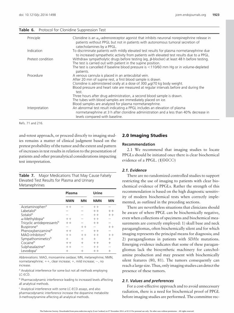
Table 6. Protocol for Clonidine Suppression Test
Principle Clonidine is an �2-adrenoreceptor agonist that inhibits neuronal norepinephrine release inpatients without PPGL but not in patients with autonomous tumoral secretion ofcatecholamines by a PPGL.
Indication To discriminate patients with mildly elevated test results for plasma normetanephrine dueto increased sympathetic activity from patients with elevated test results due to a PPGL.
Pretest condition Withdraw sympatholytic drugs before testing (eg, �-blocker) at least 48 h before testing.The test is carried out with patient in the supine position.The test is cancelled if baseline blood pressure is �110/60 mm Hg or in volume-depleted
patients.Procedure A venous cannula is placed in an antecubital vein.
After 20 min of supine rest, a first blood sample is drawn.Clonidine is administered orally at a dose of 300 �g/70 kg body weight.Blood pressure and heart rate are measured at regular intervals before and during the
test.Three hours after drug administration, a second blood sample is drawn.The tubes with blood samples are immediately placed on ice.Blood samples are analyzed for plasma normetanephrine.
Interpretation An abnormal test result indicating a PPGL includes an elevation of plasmanormetanephrine at 3 h after clonidine administration and a less than 40% decrease inlevels compared with baseline.
Refs. 71 and 216.
doi: 10.1210/jc.2014-1498 jcem.endojournals.org 1923
The Endocrine Society. Downloaded from press.endocrine.org by [Luis Cardoso] on 07 November 2014. at 02:13 For personal use only. No other uses without permission. . All rights reserved.

ognizes that currently there is insufficient evidence to for-mulate guidelines about when and how to perform imag-ing studies in patients at risk for biochemically silentPPGL.
Recommendation2.2 We suggest CT rather than MRI as the first-choice
imaging modality because of its excellent spatial resolu-tion for thorax, abdomen, and pelvis. (2�QQQE)
2.2 EvidenceCT with contrast provides an excellent initial method
for the localization of PPGLs, with sensitivity between 88and 100% (82–90). CT has excellent tomographical res-olution but, as with MRI, lacks specificity. On CT, PPGLsmay be homogeneous or heterogeneous, necrotic withsome calcifications, solid, or cystic. Although between 87and 100% of PPGLs exhibit a mean attenuation of morethan 10 Hounsfield units on unenhanced CT, PPGLs canoccasionally have more than 60% washout of contrastagents on 15-minute delayed scanning (91–94). A highsignal intensity (bright) T2-weighted MRI image may beof value for the detection of PPGLs; however, a recentstudy showed that in pheochromocytomas this finding isrelatively uncommon (95).
The use of nonionic contrast is safe, and therefore, con-trast CT can be performed in patients without adrenergicreceptor blockade (96, 97). Modern CT scans can detecttumors 5 mm or larger. Because most PPGLs are locatedin the abdomen, a CT scan of the abdomen and pelvisshould be the first option. Some studies showed that sen-sitivity of CT for extra-adrenal, residual, recurrent, ormetastatic tumors can be as low as 57% and inferior toMRI (83, 86, 98–102). CT is preferred to MRI for detec-tion of lung metastatic lesions (84). For skull base and neckparaganglioma, the sensitivity of MRI is between 90 and95% (81). Use of ultrasound is usually not recommendeddue to its suboptimal sensitivity.
2.2 Values and preferencesThe committee recognizes that the results of current
and previous studies should be interpreted with caution,taking into consideration the type of CT or MRI scans andthe design of the study, including its criteria, selection ofpatients, and controls. The committee recognizes thatrarely there are PPGLs not detected by any anatomicalimaging studies due to their small size or location, thepresence of surgical clips, or postoperative changes, andthat such tumors can only be detected by functional im-aging modalities.
Recommendation2.3 We recommend MRI in patients with metastatic
PPGLs, for detection of skull base and neck paraganglio-mas, in patients with surgical clips causing artifacts whenusing CT, in patients with an allergy to CT contrast, andin patients in whom radiation exposure should be limited(children, pregnant women, patients with known germlinemutations, and those with recent excessive radiation ex-posure). (1�QQQE)
2.3 EvidenceSee previous Section 2.2.
2.3 Values and preferencesMRI should not be performed in patients who have
intracranial aneurysm clips.See previous Section 2.2.
Recommendation2.4 We suggest the use of 123I-metaiodobenzylguani-
dine (MIBG) scintigraphy as a functional imaging modal-ity in patients with metastatic PPGLs detected by otherimaging modalities when radiotherapy using 131I-MIBG isplanned and occasionally in some patients with an in-creased risk for metastatic disease due to large size of theprimary tumor or to extra-adrenal, multifocal (exceptskull base and neck PPGLs), or recurrent disease.(2�QEEE)
2.4 EvidenceBecause 123I-MIBG has better sensitivity than 131I-
MIBG for detection of PPGLs (103- 106), only the formeragent is recommended for imaging. Another advantageof 123I- over 131I-labeled MIBG is its utility for imagingby SPECT. Because up to 50% of normal adrenal glandsdemonstrate physiological uptake of 123I-MIBG, false-positive results can be a problem (104, 107). Asymmet-ric uptake in normal adrenal glands can further lead tomisinterpretation.
Sensitivity of 123I-MIBG ranges between 85 and 88%for pheochromocytomas and between 56 and 75% forparagangliomas, whereas specificity ranges from 70–100% and 84–100%, respectively (108–111). Sensitivityfor metastatic PPGLs is between 56 and 83% (109, 112),whereas for recurrent PPGLs it is approximately 75%(113). Results from a meta-analysis showed 90% sensi-tivity and specificity for pheochromocytomas, whereassensitivities were 98% for paragangliomas, falling to 79%for malignant PPGLs (114). In another meta-analysis of 15studies of 123I-MIBG scintigraphy, sensitivity was 94%and specificity 92% (115). For SDHx-related PPGLs, and
1924 Lenders et al Guidelines on Pheochromocytoma and Paraganglioma J Clin Endocrinol Metab, June 2014, 99(6):1915–1942
The Endocrine Society. Downloaded from press.endocrine.org by [Luis Cardoso] on 07 November 2014. at 02:13 For personal use only. No other uses without permission. . All rights reserved.

in particular for SDHB-related PPGLs, overall sensitivityof 123I-MIBG is less than 50% (81, 116). Similar results ofsuboptimal sensitivity have also been reported for the de-tection of skull base and neck, thoracic, bladder, or re-current paragangliomas (107, 108, 111, 113, 117, 118).
123I-MIBG SPECT is widely available, and recent stud-ies suggest that its performance is similar to PET scanningusing 18F-fluorodopamine, 18F-fluorodihydroxy-phenyl-alanine (18F-FDOPA), or 18F-FDG in the detection ofpheochromocytoma (112, 119). For paragangliomas ormetastatic disease including SDHx-related tumors, 123I-MIBG is inferior to 18F-FDG-PET, 18F-FDOPA, or soma-tostatin receptor imaging with 111In-diethylene triaminepentaacetic acid-pentetreotide (81, 118, 120–123).
2.4 Values and preferencesIn making this recommendation, the committee has
taken into account the findings of the Endocrine Society-sponsored systematic review of functional imaging inPPGL as well as the evidence outlined above that 123I-MIBG scintigraphy has limited use due to relatively sub-optimal sensitivity, particularly in patients with metastaticPPGLs and those with SDHx-related PPGLs (Brito, J. P.,N. Asi, C. Undavali, et al., submitted for publication).Nevertheless, in patients with metastatic PPGLs in whomsurgery is not an option, 123I-MIBG scintigraphy is usefulbecause, if positive, treatment with 131I-MIBG may beconsidered. The recommendation for restricted use ofMIBG to patients with or at risk for metastatic disease thusrecognizes this therapeutic need, the widespread availabil-ity of this functional imaging modality, as well as its lim-ited utility for identifying lesions not detected by conven-tional imaging.
2.4 RemarksAccumulation of 123I-MIBG can be decreased by sev-
eral drugs: 1) sympathomimetics; 2) agents that block cat-echolamine transport via the norepinephrine transporter,such as cocaine and tricyclic antidepressants; and 3) agentssuch as calcium channel blockers and some combined �-and �-adrenergic receptor blockers such as labetalol(125). Therefore, most of these drugs should be withheldfor about 2 weeks before 123I-MIBG scintigraphy. Accu-mulation of 123I-MIBG is also profoundly decreased innecrotic tumors (89). Use of 123I-MIBG scintigraphy iscontraindicated in pregnant women. The committee rec-ommends that 123I-MIBG scintigraphy be performed byand results be assessed by experienced nuclear medicinephysicians.
Recommendation2.5 We suggest the use of 18F-FDG PET/CT scanning in
patients with metastatic disease. 18F-FDG PET/CT is the
preferred imaging modality over 123I-MIBG scintigraphyin patients with known metastatic PPGLs. (2�QQQE)
2.5 EvidenceIn the initial study of Shulkin et al (126), the overall
sensitivity of 18F-FDG PET was 76%, but it was higher inpatients with metastatic (88%) than benign (58%) PPGLs.This study and several subsequent studies showed a supe-riority of 18F-FDG PET compared with 131I-MIBG scin-tigraphy for detection of metastatic PPGLs (119, 120,127–131). Overall, the sensitivity of 18F-FDG PET wasshown to be between 74 and 100%, with the highest per-formance for metastatic, particularly SDHB-related,PPGLs (120, 122, 126, 128–130, 132–134).
2.5 Values and preferencesThe committee recognizes that some studies indicated
that 18F-FDG PET is complementary with other func-tional imaging studies in some patients. It is also recog-nized that there are limited data regarding the use of var-ious PET imaging modalities in patients with differentgenetic mutations.
2.5 RemarksThe use of PET imaging modalities is contraindicated in
pregnant women. There are also several drugs that mayprofoundly decrease the uptake of PET radiopharmaceu-ticals by PPGL, but the data are limited, and further studiesare needed.
3.0 Genetic Testing
Recommendation3.1 We recommend that all patients with PPGLs should
be engaged in shared decision making for genetic testing.(1�QQQE)
3.1 EvidenceSince 1990, 14 different PPGL susceptibility genes have
been reported: NF1 (135), RET (136), VHL (137), SDHD(138), SDHC (139), SDHB (140), EGLN1/PHD2 (141,142), KIF1� (143), SDH5/SDHAF2 (144), IDH1 (145),TMEM127 (146), SDHA (147), MAX (148), and HIF2�
(149). The roles of EGLN1/PHD2 (150, 151), KIF1�, andIDH1 (152) have not been confirmed by other studies,suggesting that mutations in these genes are an infrequentcause of hereditary PPGLs. The role of somatic or germlinemutations in HIF2�, reported in a few patients (149, 153–156), awaits validation in a larger series.
There are several reasons to consider genetic testing inall patients who present with PPGLs: 1) at least one-third
doi: 10.1210/jc.2014-1498 jcem.endojournals.org 1925
The Endocrine Society. Downloaded from press.endocrine.org by [Luis Cardoso] on 07 November 2014. at 02:13 For personal use only. No other uses without permission. . All rights reserved.

of all patients with PPGLs have disease-causing germ-line mutations (157); 2) mutations of SDHB lead tometastatic disease in 40% or more of affected patients(23, 24); and 3) establishing a hereditary syndrome inthe proband may result in earlier diagnosis and treat-ment of PPGLs and other syndromic manifestations inrelatives (159 –161).
In clinical practice, patients with PPGLs can presentwith features that indicate a high likelihood of a hereditarycause. Such features include a positive family history(based on family pedigree or identification of a PPGL-susceptibility gene mutation in a relative), syndromic fea-tures, and multifocal, bilateral, or metastatic disease (157,162, 163). Many patients with PPGLs, however, do nothave the above-mentioned features.
Following the initial report of Neumann et al (15),genotyping of the main PPGL susceptibility genes (SDHB,SDHD, VHL, RET) has been performed in eight studies,each comprising more than 200 patients and encompass-ing 3694 subjects harboring 1250 germline mutations(33.8%) (162, 163, 165–170) (Table 8). This high fre-quency justifies consideration of genetic testing in eachpatient with a PPGL. The highest frequencies of germlinemutations are for SDHB (10.3%), SDHD (8.9%), VHL(7.3%), RET (6.3%), and NF1 (3.3%). Germline muta-tion frequencies of less than 2% are found for SDHC,SDHA, MAX, and TMEM127 (169, 171–173). No germ-line SDHAF2 mutations were found in a series of 315apparently sporadic PPGLs (174).
A mutation rate of 11.6% was revealed from a system-atic review of the literature featuring an analysis that in-cluded only patients who fulfilled at least three of fourcriteria: 1) a negative family history of PPGL; 2) absenceof syndromic features; 3) absence of bilateral disease; and4) absence of metastatic disease (Brito, J. P., N. Asi, C.Undavalli, L. Prokop, V. M. Montori, and N. H. Murad,submitted for publication).
3.1 Values and preferencesThe committee’s recommendation that genetic testing
should be considered in each patient does not imply thatgenetic testing should be done in each patient. In partic-ular, in view of the financial costs, genetic testing has lim-ited incremental value in patients with unilateral pheo-chromocytoma and no syndromic or malignant featuresand no positive family history. The importance of the di-agnosis of an inherited disease for at-risk families must bebalanced against any negative impacts and financial costsof genetic testing. The costs of genetic testing will probablydecrease with adoption of next-generation sequencingmethods. This may shift the balance in favor of more wide-spread genetic testing than currently practiced accordingto the variable country insurance-specific limitations ofhealth care coverage.
Recommendation3.2 We recommend the use of a clinical feature-driven
diagnostic algorithm to establish the priorities for specificgenetic testing in PPGL patients with suspected germlinemutations. (1�QQQE)
3.2 EvidenceThe committee proposes a decisional algorithm for se-
quential genetic testing, with selection of genes to be testedprioritized according to a syndromic or metastatic pre-sentation (Figure 1). Considerations of young age at PPGLpresentation, positive family history, and presentation ofmultifocal PPGLs or bilateral adrenal tumors are also rec-ommended for prioritizing patients for testing. Thereafter,considerations of tumor location and catecholamine bio-chemical phenotype may further guide selection of genesfor testing when justified.
A positive family history or syndromic presentation inpatients with PPGLs not only indicates a high priority forgenetic testing, but also may direct targeted germline mu-tation testing. Six different familial autosomal dominantdiseases can be suspected clinically: neurofibromatosis
Table 8. Detected Germline Mutations in All PPGL Patients
First Author, Year (Ref.) No. of Cases
Mutations
SDHB SDHD SDHC VHL RET NF1 SDHA SDHAF2 TMEM127 MAX n %
Lefebvre, 2012 (170) 269 21 12 6 ND ND ND ND 0 5 ND 44 16.3Amar, 2005 (165);
Burnichon, 2009 (166)
721 99 131 16 25 16 13 ND ND ND ND 300 41.6
Mannelli, 2009 (162) 501 24 47 4 48 27 11 ND ND ND ND 161 32.1Cascón, 2009 (163) 237 25 11 1 20 36 ND ND ND ND ND 93 39.2Jafri, 2012 (167) 501 121 44 ND 19 ND ND ND ND ND ND 184 36.7Erlic, 2009 (168) 1149 73 28 2 120 80 43 ND ND ND ND 346 30.1Korpershoek, 2011 (169) 316 16 26 2 19 26 21 5 5 2 ND 122 38.6Total n 3694 379 299 31 251 185 88 5 5 7 1250 33.8Mutation rate 10.3 8.9 1.0 (31/3193) 7.3 (251/3425) 6.3 (185/2924) 3.3 (88/2687)
ND, not determined.
1926 Lenders et al Guidelines on Pheochromocytoma and Paraganglioma J Clin Endocrinol Metab, June 2014, 99(6):1915–1942
The Endocrine Society. Downloaded from press.endocrine.org by [Luis Cardoso] on 07 November 2014. at 02:13 For personal use only. No other uses without permission. . All rights reserved.

type 1 (NF1), multiple endocrine neoplasia type 2(MEN2), von Hippel-Lindau (VHL) syndrome (see Table3), renal cell carcinoma with SDHB mutation (176), Car-ney triad (paragangliomas, gastric stromal tumors, pul-monary chondromas), and Carney-Stratakis syndrome(paragangliomas and gastric stromal sarcomas) (177).The MEN2 and VHL syndromes are usually characterizedby distinct clinical stigmata that directs targeted testing ofRET and VHL genes. Detection of mutations in the NF1gene is complex, and although testing is available in spe-cialized laboratories (178), the diagnosis of NF1 can beinvariably established by clinical findings alone (179).Nevertheless, some patients with NF1 and an apparentlysporadic PPGL presentation have been reported, all withmild features of the disease (180, 181); these findings il-lustrate the importance of careful clinical investigation ofpossible clinical stigmata of an underlying mutation in allpatients with PPGL.
Since 2003, several studies have reported that the iden-tification of a germline SDHB mutation is an importantrisk factor for malignancy for patients affected by PPGLs(165, 182) and of poor prognosis for patients affected bymetastatic PPGLs (24). Conversely, pathogenic SDHBmutations were reported in 30% of patients with meta-
static PPGLs (23). In a later review,Pasini and Stratakis (184) docu-mented a prevalence of 36% ofSDHB mutations in malignantPPGLs. Moreover, this higher riskwas observed in a pediatric series(185). Furthermore, a meta-analysisof 12 studies showed that the pooledrisk of malignant PPGL for SDHB-mutation carriers in incidence andprevalence studies was 17% and13%, respectively (186). The aboveevidence justifies SDHB genetic test-ing in patients with metastaticPPGLs (Figure 1).
In the absence of a syndromic, fa-milial, or metastatic presentation, se-lection of genes for testing may beguided by tumor location and bio-chemical phenotype (Figure 1) butprioritized according to age or pre-sentation of multiple tumors. Lowerage at PPGL presentation among pa-tients with germline mutations thanthose without mutations is well es-tablished (15, 162, 163, 165, 168,185, 187). Although there is noagreement upon age cutoff for ge-netic testing, the likelihood of a mu-
tation in patients with nonsyndromic PPGLs younger than45 years is 5-fold higher than in patients older than 45years (168). Prevalence of germline mutations among chil-dren with PPGLs is particularly high (185, 188–191), jus-tifying mutation screening in all such cases.
Prevalence of germline mutations is also high amongpatients with bilateral or multifocal PPGLs (162, 163,165, 166, 168, 192). SDHB mutations result mainly inextra-adrenal tumors (182). In a large study of patientswithnonsyndromicPPGLs(168), the prevalence of germlinemutations associated with multiple PPGLs was 5-foldhigher than for solitary PPGLs (54 vs 11.5%). In the samestudy, an extra-adrenal tumor location was shown tocarry a 4-fold higher risk of a germline mutation than anadrenal location, with mutations confined to SDHx genes.This high risk associated with extra-adrenal tumors hasbeen confirmed by numerous studies (159, 162, 163, 166,193), justifying the recommendation for screening ofSDHx gene mutations in affected patients.
As reviewed by Pasini and Stratakis (184), SDHx-re-lated genotype-phenotype correlations have been assessedby several studies based on international registries. Mul-
PPGL diagnosis
Targeted genetic testing Syndromic presentation
Dopaminergic
Extra-adrenal
SDHB Metastatic
Nonmetastatic
SDHD, SDHB, SDHC
Adrenal Noradrenergic
Noradrenergic
Dopaminergic
SDHB, SDHD, SDHC
SDHB, SDHD, SDHC, VHL, MAX
SDHD, SDHB, SDHC Skull base and neck
VHL SDHD, SDHB, SDHC, MAX
RET TMEM127, MAX Adrenergic
SDHD, SDHC, VHL, MAX
Figure 1. Decisional algorithm for genetic testing in patients with a proven PPGL.Dopaminergic, noradrenergic, and adrenergic phenotypes are defined as significant productionsof respective 3-methoxytyramine, normetanephrine, and metanephrine relative to combinedproduction of all three metabolites.
doi: 10.1210/jc.2014-1498 jcem.endojournals.org 1927
The Endocrine Society. Downloaded from press.endocrine.org by [Luis Cardoso] on 07 November 2014. at 02:13 For personal use only. No other uses without permission. . All rights reserved.

tiple skull base and neck tumors or a family history ofPPGL in the paternal branch suggests an SDHD-relatedPPGL (159, 166, 192). In contrast, SDHB-related PPGLsare often diagnosed as a single extra-adrenal tumor with-out any family history. SDHC mutation carriers are rarebut may develop all the stigmata of the disease. Mutationsof SDHA and SDHAF2 were described in only a few pa-tients. Negative SDHB immunohistochemistry in tumoraltissue suggests the presence of a mutation in one of theSDHx genes (194). Hormonally functional SDHx-relatedPPGLs are best detected by measurements of normeta-nephrine and methoxytyramine (195). Increases in me-thoxytyramine are particularly prevalent in patients withPPGLs due to SDHx mutations, justifying targeted testingof these mutations associated with this biochemical pre-sentation (Figure 1).
For nonsyndromic tumors with adrenal locations, mu-tations are far less common than for tumors at extra-ad-renal locations and encompass all established tumorsusceptibility genes. When justified by young age at pre-sentation or bilaterality, mutation testing should followthe decisional algorithm (Figure 1).
Hereditary PPGL due to TMEM127 or MAX muta-tions are infrequent, typically diagnosed later in life, andpreferentially adrenal in location; patients frequently havea positive family history (Table 8). TMEM127-relatedPPGLs typically produce epinephrine, whereas the bio-chemical phenotype of MAX-related tumors is interme-diate between adrenergic and noradrenergic (173).
In the absence of more common germline mutations,rare cases such as SDHA germline mutations may be con-sidered. For example, consider SDHA in patients withskull base and neck or thoracic-abdominal-pelvic para-ganglioma associated with negative SDHB and SDHA im-munohistochemistry on tumoral tissue.
3.2 Values and preferencesIn recommending a sequential algorithm with selec-
tive testing prioritized according to risk of mutations,the committee has considered the systematic review,indicating that the current level of evidence does notsupport indiscriminate genetic testing of PPGL suscep-tibility genes. It is also recognized that the recom-mended selective approach to genetic testing will likelybe made obsolete by development of next-generationsequencing methods allowing rapid and low-cost anal-ysis of all PPGL susceptibility genes. Interpretations ofpathogenicity, particularly associated with the greaternumber of variants of unknown significance (VUSs)generated by this technology, will nevertheless also in-creasingly require accurate knowledge of the genotype-
phenotype relationships that provide the basis for thecurrent sequential algorithm.
Recommendation3.3 We suggest that patients with paraganglioma un-
dergo testing of SDH mutations and that patients withmetastatic disease undergo testing for SDHB mutations.(2�QQQE)
3.3. EvidenceSee previous Section 3.2.
3.3 Values and preferencesSee previous Section 3.2.
Recommendation3.4 We recommend that genetic testing for PPGL be
delivered within the framework of health care. Specifi-cally, pretest and post-test counseling should be available.All tests for PPGL genetic testing should be performed byaccredited laboratories. (Ungraded recommendation).
3.4 EvidenceIn 2002, the Organization for Economic Cooperation
and Development (OECD) produced guidelines for qual-ity assurance in molecular genetic testing (196). TheOECD guidelines encompassed the general principles andbest practices for molecular genetic testing, the qualityassurance systems and proficiency testing programs, thequality of result reporting, and the education and trainingstandards for laboratory personnel. All molecular genetictesting services should be provided and practiced under aquality assurance framework by accredited laboratories.A required signed informed consent to test should be ob-tained according to national applicable standards (197).Pretest and post-test counseling should be available. Mo-lecular genetic testing laboratories should have policiesand procedures to document the analytical validity of alltests performed.
The European Molecular Genetics Quality Networkprovides external quality assessment schemes for VHL dis-ease and MEN2 (see www.emqn.org). The application ofthese guidelines was recommended in the United Statesafter the realization of a survey comparing data on themolecular genetic testing in laboratories in the UnitedStates with those in 18 others countries (198). Certifica-tion and accreditation, based on ISO 9001 and ISO 15189,respectively, are widely encouraged in human moleculargenetic testing laboratories (199).
Patients with PPGLs benefit from genetic counselingbefore and after germline mutation testing in order to beinformed about the different suspected inherited diseases
1928 Lenders et al Guidelines on Pheochromocytoma and Paraganglioma J Clin Endocrinol Metab, June 2014, 99(6):1915–1942
The Endocrine Society. Downloaded from press.endocrine.org by [Luis Cardoso] on 07 November 2014. at 02:13 For personal use only. No other uses without permission. . All rights reserved.

and their diagnosis and treatment, the diagnostic perfor-mance of corresponding genetic testing, and the familialrisk of transmission. Access to national/international spe-cialized networks/referral centers and patient supportgroups should be facilitated.
Except for NF1 and SDHA, all known PPGL suscep-tibility genes can be routinely sequenced, and large dele-tions can be searched for with commercial multiplexligation-dependent probe amplification kits and/or byquantitative PCR procedures by specialized genetic labo-ratories (157). A misinterpretation of genetic testing orincorrect results can lead to deleterious consequences forthe patient and his or her family (187). Every identifiedvariant should be cautiously interpreted. The PPGL ge-netic test may be positive (when the identified mutationclearly disrupts gene function), negative (when no varia-tion or a known nonfunctional polymorphism is found inDNA sequence), or uncertain (when a sequence VUS isdetected). The prediction of the clinical impact of a VUS isbased on variant classification systems using the clinical,biological, and familial context of the patient; the presenceof the VUS in general and/or specific polymorphisms/mu-tations databases; in silico prediction tools; and functionaltests, when available (200–202).
3.4 Values and preferencesIn making this recommendation, the committee has ad-
opted OECD recommendations in recognition of the im-portance of the quality of the methods applied for genetictesting and for associated genetic counseling.
3.4 RemarksSeveral quality assurance index items for molecular ge-
netic testing exist and should be applied for PPGL genetictesting, such as the use of negative and positive controls inanalyses and the confirmation of a positive test result ona second aliquot of germline DNA. Accredited laborato-
ries should analyze sequence VUSs with robust methodsfor interpretation. The interpretation of reporting shouldbe adapted to the individual patient and clinical situation.Genetic test results should be reported back to qualifiedhealthcare professionals to enable healthcare decision-making and to facilitate delivery of clear, well-informedinterpretation of the consequences to the patient and fam-ily members, when appropriate.
4.0 Perioperative Medical Management
Recommendation4.1 We recommend that all patients with a hormonally
functional PPGL should undergo preoperative blockadeto prevent perioperative cardiovascular complications. (1/QQEE) We suggest �-adrenergic receptor blockers as thefirst choice. (2�QQEE)
4.1 EvidenceEvidence from randomized controlled clinical studies
regarding the comparable effectiveness of nonselective �-vs �1-selective adrenergic receptor blockers is unavailable(203, 204). However, retrospective studies support the useof �-adrenergic receptor blockers as the first-choice drugclass to minimize perioperative complications (17, 101,204–206, 209). Retrospective studies demonstrated that�1-selective adrenergic receptor blockers were associatedwith lower preoperative diastolic pressure, a lower intra-operative heart rate, better postoperative hemodynamicrecovery (210), and fewer adverse effects such as reactivetachycardia and sustained postoperative hypotensionthan nonselective adrenergic blockers (211). Anotherstudy did not show any difference between selective andnonselective �-adrenergic receptor blockers (212).
Calcium channel blockers are the most often usedadd-on drug class to further improve blood pressure con-
Table 9. Presurgical Medical Preparation
Drug Starting Time Starting Dose Final Doseb
Preparation 1Phenoxybenzamine 10–14 d before surgery 10 mg b.i.d. 1 mg/kg/dor Doxazosine 10–14 d before surgery 2 mg/d 32 mg/d
Preparation 2Nifedipinea As add-on to preparation 1 when needed 30 mg/d 60 mg/dor Amlodipinea As add-on to preparation 1 when needed 5 mg/d 10 mg/d
Preparation 3Propranolol After at least 3–4 d of preparation 1 20 mg t.i.d. 40 mg t.i.d.or Atenolol After at least 3–4 d of preparation 1 25 mg/d 50 mg/d
Abbreviations: b.i.d., twice daily; t.i.d., three times daily.a Add when blood pressure cannot be controlled by �-adrenoceptor blockade (preparation 1).b Higher doses usually unnecessary.
doi: 10.1210/jc.2014-1498 jcem.endojournals.org 1929
The Endocrine Society. Downloaded from press.endocrine.org by [Luis Cardoso] on 07 November 2014. at 02:13 For personal use only. No other uses without permission. . All rights reserved.

trol in patients already treated with �-adrenergic receptorblockers (213–215) (Table 9); however, some studies havesuggested that this drug class can be used as the first choice(216). Monotherapy with calcium channel blockers is notrecommended unless patients have very mild preoperativehypertension or have severe orthostatic hypotension with�-adrenergic receptor blockers.
Preoperative coadministration of �-adrenergic recep-tor blockers is indicated to control tachycardia only afteradministration of �-adrenergic receptor blockers. Use of�-adrenergic receptor blockers in the absence of an �-ad-renoceptor blocker is not recommended because of thepotential for hypertensive crisis due to unopposed stimu-lation of �-adrenergic receptors. There is no evidence tosupport the preference of �1-selective adrenergic receptorblockers over nonselective �-adrenergic receptor blockers.Labetalol with its fixed but more potent � than � antag-onistic activities (�:� of 1:5) should not be used as theinitial therapy because it can result in paradoxical hyper-tension or even hypertensive crisis (217).
�-Methyl-paratyrosine (metyrosine) inhibits catechol-amine synthesis and may be used in combination with�-adrenergic receptor blockers for a short period beforesurgery to further stabilize blood pressure to reduce bloodloss and volume depletion during surgery (218, 219).
There has been one report that preoperative �1-adren-ergic receptor blockade offers no benefit in maintainingintraoperative hemodynamics of normotensive PPGL pa-tients (79). Nevertheless, it is the view of the committeethat for such patients �-adrenergic receptor blockersand/or calcium channel blockers remain recommended toprevent unpredictable increases in blood pressure duringsurgery (124).
4.1 Values and preferencesOur recommendation to initiate adrenergic blockade
before surgery places a higher value on the potential ben-efit of these drugs by preventing unpredictable instabilityin blood pressure during surgery and a relatively lowervalue on the potential for medication-related adverseeffects.
Recommendation4.2 We recommend preoperative medical treatment for
7 to 14 days to allow adequate time to normalize bloodpressure and heart rate. Treatment should also include ahigh-sodium diet and fluid intake to reverse catechol-amine-induced blood volume contraction preoperativelyto prevent severe hypotension after tumor removal.(1�QQEE)
4.2 EvidenceEvidence from randomized, controlled studies is un-
available. Retrospective studies report that �-adrenergicreceptor blockers should be started at least 7 days preop-eratively to normalize blood pressure and reverse bloodvolume contraction (124, 158). Intravenous infusion ofphenoxybenzamine for 5 hours per day for 3 days beforesurgery has been reported as one effective approach (164,175).
Evidence from randomized, controlled studies thattreatment should also include a high-sodium diet and fluidintake is not available (203). Retrospective studies reportthat initiation of a high-sodium diet a few days after thestart of �-adrenergic receptor blockade reverses blood vol-ume contraction, prevents orthostatic hypotension beforesurgery, and reduces the risk of significant hypotensionafter surgery (26, 209). Continuous administration of sa-line (1–2 L) is also helpful if started the evening beforesurgery. Treatment with �-adrenergic receptor blockersalone was shown to reverse blood volume contraction inonly about 60% of patients (183). Caution is required forvolume loading in patients with heart or renal failure.
There is no evidence from randomized controlled stud-ies to determine the optimal target blood pressure. Basedon retrospective studies and institutional experience, a tar-get blood pressure of less than 130/80 mm Hg while seatedand greater than 90 mm Hg systolic while standing seemsreasonable, with a target heart rate of 60–70 bpm seatedand 70–80 bpm standing (26, 209). These targets shouldbe modified in each patient according to age and accom-panying cardiovascular diseases (26, 216, 207). It shouldbe noted that complete prevention of intraoperative hy-pertension and tachycardia cannot be achieved by anydoses and combinations of antihypertensive and otherdrugs.
4.2 Values and preferencesThe recommendation to use �-adrenergic receptor
blockers at least 7 days preoperatively places a highervalue on the potential benefit of preventing unpredictableinstability in blood pressure during surgery and a rela-tively lower value on the potential for medication-relatedadverse effects. The recommendation for preoperativevolume loading places a higher value on preventing severeand sustained hypotension after removal of the tumor anda lower value on the potential for adverse effects such asblood pressure increase.
Recommendation4.3 We recommend monitoring blood pressure, heart
rate, and blood glucose levels with adjustment of associ-
1930 Lenders et al Guidelines on Pheochromocytoma and Paraganglioma J Clin Endocrinol Metab, June 2014, 99(6):1915–1942
The Endocrine Society. Downloaded from press.endocrine.org by [Luis Cardoso] on 07 November 2014. at 02:13 For personal use only. No other uses without permission. . All rights reserved.

ated therapies in the immediate postoperative period.(1�QQEE)
4.3 EvidenceThe major potential postoperative complications are
hypertension, hypotension, and rebound hypoglycemia.Our recommendation that blood pressure, heart rate. andplasma glucose levels should be closely monitored for24–48 hours is mainly based on retrospective studies andinstitutional experience (208, 209). Because of potentialadrenal insufficiency, particular attention needs to be paidto patients who undergo: 1) bilateral adrenalectomy; 2)bilateral cortical-sparing adrenalectomy; or 3) unilateralcortical-sparing adrenalectomy of a sole remaining adre-nal gland. There are numerous case reports of postsurgicalhypoglycemia but no studies documenting its exactprevalence.
4.3 Values and preferencesThe committee’s recommendation places a high value
on preventing blood pressure and heart rate instability andpostoperative hypoglycemia.
Recommendation4.4 We suggest measuring plasma or urine levels of
metanephrines on follow-up to diagnose persistent dis-ease. We suggest lifelong annual biochemical testing toassess for recurrent or metastatic disease. (2�QQEE)
4.4 EvidenceEvidence from randomized, controlled studies is un-
available. These recommendations depend on personaland institutional experiences. Several studies reportedhigh rates of recurrence or metastatic disease after surgicalresection (17, 22, 220, 221).
4.4 Values and preferencesOur recommendation to measure plasma or urine
metanephrines annually after surgery places a higher valuein detecting tumor recurrence or metastasis and a lowervalue in avoiding the incremental expenses of the bio-chemical testing.
4.4 RemarksTo document successful tumor removal, biochemical
testing should be performed upon recovery of the patientfrom surgery (eg, 2–4 wk after surgery).
5.0 Surgery
Recommendation5.1 We recommend minimally invasive adrenalectomy
(eg, laparoscopic) for most adrenal pheochromocytomas.
(1�QQEE) We recommend open resection for large (eg,�6 cm) or invasive pheochromocytomas to ensure com-plete tumor resection, prevent tumor rupture, and avoidlocal recurrence. (1�QEEE) We suggest open resection forparagangliomas, but laparoscopic resection can be per-formed for small, noninvasive paragangliomas in surgi-cally favorable locations. (2�QEEE)
5.1 EvidenceThere are no prospective randomized studies compar-
ing laparoscopic with open adrenalectomy for pheochro-mocytomas. Several large single-institution series (somewith historical controls) show laparoscopic adrenalec-tomy to be associated with less pain, less blood loss, fewerhospital days, and less surgical morbidity than open ad-renalectomy (222, 223). There are no data regarding anydifference in recurrence rate after open vs laparoscopicadrenalectomy. Mortality rate is about 1%, and the con-version rate and transfusion rate are about 5% (rate ofconversion to open resection is influenced by tumor sizeand surgeon experience). Because pheochromocytomasare rare, a prospective randomized study comparing openwith laparoscopic resection is unlikely.
The two most common laparoscopic approaches arethe lateral transabdominal/transperitoneal (Gagner) ap-proach and the posterior retroperitoneal (Walz) ap-proach. The former allows intra-abdominal evaluationand has more space for dissecting larger tumors (222,224). The latter may be preferable for patients who hadprior abdominal operation or those requiring bilateral ad-renalectomy (225, 226). Paragangliomas are more likelyto be malignant and are frequently found in areas difficultfor laparoscopic resection; thus, paragangliomas are morelikely than pheochromocytomas to require open resection,but some can be safely resected laparoscopically by expe-rienced surgeons (227).
5.1 Values and preferencesPatients prefer less pain, earlier recoveries, and shorter
hospitalizations, which are possible with laparoscopicsurgery.
5.1 RemarksSafe laparoscopic adrenalectomy requires surgeons
with skills and experience in advanced laparoscopicsurgery and centers with appropriate expertise in the pre-operative and postoperative management of pheochromo-cytoma, including anesthesia, endocrinology, and inten-sive care (228). Laparoscopic adrenalectomy can beperformed transperitoneally or retroperitoneally depend-ing on the surgeon’s preference and expertise (222, 225).
doi: 10.1210/jc.2014-1498 jcem.endojournals.org 1931
The Endocrine Society. Downloaded from press.endocrine.org by [Luis Cardoso] on 07 November 2014. at 02:13 For personal use only. No other uses without permission. . All rights reserved.

Seeding and recurrence of tumors in the adrenal bed orthroughout the abdominal cavity can occur if pheochro-mocytomas are fractured during dissection (229), man-dating precise and gentle dissection. Specimen bags usedfor tumor retrieval should not tear. The operation shouldbe converted to open resection if the laparoscopic ap-proach is difficult. Hand assistance or robot assistancemay be helpful in patients with large tumors that are dif-ficult to resect (230) and are used per the surgeon’sdiscretion.
Recommendation5.2 We suggest partial adrenalectomy for selected pa-
tients, such as those with hereditary pheochromocytoma,with small tumors who have already undergone a con-tralateral complete adrenalectomy to spare adrenal cortexto prevent permanent hypocortisolism. (2�QEEE)
5.2 EvidencePartial adrenalectomy is safe, with no increased surgi-
cal risks over complete adrenalectomy (231). Use of en-ergy devices such as ultrasonic shears and bipolar sealerslowers the risk of bleeding from the cut edges of the ad-renal gland. Selective removal of medullary tissue leavingonly cortical tissue is attempted, but usually some med-ullary tissue remains, which can cause tumor recurrence.
For patients with prior contralateral adrenalectomies,a successful partial adrenalectomy preserving sufficientadrenal cortex can prevent postoperative adrenal insuffi-ciency and requirements for glucocorticoid and mineralo-corticoid replacement (232–234). About 90% of patientscan remain steroid independent (235, 236). Larger tumorsresult in smaller remnants and a lower chance for steroidindependence. Partial adrenalectomy increases the risk fortumor recurrence from the remnant. Estimated recurrencerates are 10–15% over 10 years for VHL patients (233,235). The cumulative recurrence rate for MEN2 patientsafter adrenal-sparing surgery at 5 and 10 years is 38.5%,including ipsilateral and contralateral gland recurrence(237). In a recent series of 96 patients with hereditarybilateral pheochromocytomas, predominantly MEN2and VHL, the 3-year recurrence rate in the remnant ad-renal was 7%, and steroid independence was 78% (238).The risk of surgical complications when resecting a recur-rent tumor in a previously dissected area may be higherthan for primary resections; open adrenalectomy may beneeded for reoperation.
5.2 Values and preferencesSome surgeons advocate partial adrenalectomy even
for initial pheochromocytoma in patients who are at highrisk for subsequent contralateral adrenalectomy for pheo-
chromocytoma. The decision to perform partial adrenal-ectomy depends on the relative value placed on twocompeting problems. Complete bilateral adrenalectomyresults in hypocortisolism, with lifelong steroid depen-dence and the need to adjust steroid doses during physi-ological and pathological stress. Partial adrenalectomy in-evitably leaves some adrenal medullary tissues with riskfor recurrent pheochromocytoma. The resulting potentialfor reoperations (likely to be more difficult, with higherconversion and complication rates) must be balancedagainst the risks associated with chronically treated adre-nal cortical insufficiency. Unfortunately, the group of pa-tients who would benefit from partial adrenalectomy isexactly the same group who are at higher risk for recurrentpheochromocytoma from the remnant.
5.2 RemarksPartial adrenalectomy for smaller tumors, those in the
periphery, and those away from the main adrenal vein ismore likely to result in sufficient functioning of adrenalcortex. Usually one-third (if central vein is preserved) toone-half of one adrenal gland is needed to preserve corticalfunction and avoid hypocortisolism, although as little as15% of an adrenal gland has been found to be sufficient(239).
6.0 Personalized Management
Recommendation6.1 In recognition of the distinct genotype-phenotype
presentations of hereditary PPGLs, we recommend a per-sonalized approach to patient management (ie, biochem-ical testing, imaging, surgery, and follow-up). (Ungradedrecommendation).
6.1 EvidenceAccumulating evidence shows that hereditary PPGLs
are characterized by distinct clinical presentations and dif-ferences in biological behavior and mode of transmissionthat reflect underlying mutations (159, 195, 240–253).
Mutations of RET and NF1 genes are almost alwaysassociated with adrenal tumors that produce normeta-nephrine and metanephrine (28, 195). In contrast, tumorsdue to mutations of VHL and SDHx genes lack significantproduction of metanephrine (195). Additional increases inmethoxytyramine, the metabolite of dopamine, charac-terize 70% of tumors in patients with mutations of SDHxgenes (195). Biochemical screening and interpretation oftest results in these hereditary conditions can thereforebenefit from a personalized approach that considers gen-otype-biochemical phenotype relationships.
1932 Lenders et al Guidelines on Pheochromocytoma and Paraganglioma J Clin Endocrinol Metab, June 2014, 99(6):1915–1942
The Endocrine Society. Downloaded from press.endocrine.org by [Luis Cardoso] on 07 November 2014. at 02:13 For personal use only. No other uses without permission. . All rights reserved.

Whereas VHL-associated tumors occur principally atadrenal locations, tumors due to mutations of SDHx genesoccur mainly at extra-adrenal locations and include skullbase and neck paragangliomas with some differences de-pending on the particular SDH subunit affected (166, 241,245, 249). Patients with mutations of the SDHB gene de-serve special attention because they have a high risk ofmalignant disease that reflects both the typically large sizesand extra-adrenal location of associated tumors (165,182, 241, 254). Large tumor size and extra-adrenal loca-tion are both independent risk factors for malignantPPGLs that should be considered as part of the personal-ized management of any patient with PPGL (254, 255). Anincrease in plasma methoxytyramine is also a commonfeature of patients with metastatic PPGLs and is a prom-ising new biomarker to identify such patients (254, 256).
In addition to a personalized approach to biochemicaltesting and test interpretation, the above observations dic-tate a need for personalized approaches to tumor local-ization. This need is further strengthened by additionalfindings that the underlying mutation and associated bi-ological behavior impact the choice of functional imagingmodality (134, 182, 252) (Figure 2).
Although localization in patients with RET and NF1mutations or any patient with increased plasma or urineconcentrations of metanephrine should primarily focus onthe adrenals, localization in patients with mutations of
SDHx genes should involve appro-priate strategies for localizing extra-adrenal tumors.
Recent studies support the exis-tence of a genotype-specific imagingapproach in the localization ofPPGLs (134). 18F-FDOPA PET is su-perior to CT/MRI or any other func-tional imaging modalities for detec-tion of SDHx- and non-SDHx-related primary skull base and neckparagangliomas (117, 257). 18F-fluorodopamine PET is overall themost sensitive method in the evalu-ation of primary (except head andneck) PPGLs (112, 122), but it haslimited availability. 111In-diethylenetriamine pentaacetic acid-pentetreo-tide scintigraphy (Octreoscan) hasbeen found to be a very good imagingmethod for the detection of PPGLs inSDHx mutation carriers, althoughinferior to anatomical imaging(182).
The surgical approach shouldalso be personalized according not
only to tumor size and location but also to any underlyingmutation; adrenal cortical-sparing surgery is a consider-ation for bilateral adrenal disease, whereas patients at riskfor malignancy due to SDHB mutations should be con-sidered for approaches that minimize the possibility ofrecurrent or metastatic disease (237, 258).
Finally, all mutation carriers should receive consider-ation for annual biochemical surveillance for PPGLs. Thenature of this surveillance should, however, take into ac-count the particular gene affected according to the geno-type-phenotype relationships described above, as well asconsiderations of penetrance and potential severity of dis-ease. For example, because the penetrance of PPGLs inNF1 is low, screening for these tumors need not be con-sidered unless indicated by signs or symptoms. At the otherend of the spectrum, the high morbidity associated withundiagnosed PPGLs in patients with SDHB mutationsmandates closer attention; in addition to biochemical test-ing, periodic imaging with MRI should be considered todetect biochemically silent tumors. To avoid ionizing ra-diation, CT and nuclear medicine imaging modalitiesshould be reserved to further characterize detectedtumors.
6.1 Values and preferencesThecommittee recognizes that currently therearenostudies
firmly establishing that a personalized approach provides im-
PPGL diagnosis
Known germline mutation
18F-FDG or
18F-DOPA
or 111
In-Pentetreotide
18F-FDG or
111In-Pentetreotide*
(123
I-MIBG)†
No known or negative
germline mutation
Skull base neck
18F-DOPA or
111In-Pentetreotide
Extra- adrenal
18F-DOPA or
111In-Pentetreotide
Metastatic
VHL, RET, NF1
SDHx
18F-DOPA or
123I-MIBG or
18F-FDG
18F-DOPA or
111In-Pentetreotide
Adrenal 123I-MIBG or
18F-DOPA or
18F-FDG
Skull base /neck
Metastatic
Adrenal / extra-adr.
18F-FDG or
111In-Pentetreotide*
(123
I-MIBG)†
Figure 2. Decisional algorithm for functional imaging in patients with proven PPGL. *, Whentreatment with radiolabeled somatostatin analogs is considered. †, When treatment with 131I-MIBG is considered.
doi: 10.1210/jc.2014-1498 jcem.endojournals.org 1933
The Endocrine Society. Downloaded from press.endocrine.org by [Luis Cardoso] on 07 November 2014. at 02:13 For personal use only. No other uses without permission. . All rights reserved.

proved outcome. Nevertheless, it seems highly likely that suchapproaches will benefit patients, but they must also be consid-ered according to cost; as covered below, a related point to anycost-benefit analysis is that such personalized approaches canonly be feasible via specialist referral centers with appropriatemultidisciplinary expertise.
Recommendation6.2 We recommend that patients with PPGLs should
be evaluated and treated by multidisciplinary teams atcenters with appropriate expertise to ensure a favorableoutcome. In particular, patients should be referred tosuch centers should there be pregnancy, metastatic dis-ease, or issues concerning the complexity or difficulty inbiochemical diagnosis; localization; performance, andinterpretation of genetic testing; preoperative prepara-tion; surgical treatment; and follow-up. (Ungradedrecommendation)
6.2 EvidenceThere are no trials that demonstrate that the outcome
of patients who are diagnosed and treated for PPGLs inhigh-volume centers by high-volume surgeons/multidisci-plinary teams is superior to the outcome of patients man-aged in a low-volume hospital with no dedicated expertteam of different medical disciplines. However, severalcross-sectional studies showed that high-volume centershad lower postsurgical morbidity with shorter hospitalstay than low-volume centers (259–262). Some studiesfound higher rates of complications and conversions tolaparotomy in low-volume nonreferral centers than inhigh-volume centers (262). These studies, however, didnot focus on patients with pheochromocytoma.
The above differences are not unexpected becausethey have also been shown for other complex interven-tions such as vascular surgery (263). The strong inverserelation between case volume and postsurgical mortal-ity for esophageal cancer surgery was based on the hos-pital setting in which these complex interventions wereperformed (264).
6.2 Values and preferencesPPGL is a very rare disorder with fewer than five pa-
tients presenting per year, even in some larger medicalcenters. The clinical presentation and course of PPGLare widely variable and can be part of a multisystemsyndrome with many different organs affected. Mostphysicians are therefore unlikely to build sufficient spe-cific experience to deal with this disorder. For a correctdiagnosis, clinicians need to have appropriate experi-ence in interpreting clinical and laboratory results, in-cluding results of genetic testing. Other physicians such
as radiologists and nuclear medicine specialists alsoplay a crucial role for a reliable and accurate interpre-tation of imaging test results. Specialists such as cardi-ologists, anesthesiologists, and intensive care physi-cians must be involved in proper patient-tailoredtreatment. Therefore, the committee believes that amultidisciplinary team, experienced in dealing withthese patients, offers the best outcomes.
Financial Disclosures of the Task Force
Jacques W. M. Lenders, MD, PhD, FRCP (Chair)—Fi-nancial or Business/Organizational Interests: University ofDresden, Executive Committee member of PRESSOR(Pheochromocytoma and Paraganglioma Research Sup-port Organization); Significant Financial Interest or Lead-ership Position: none declared. Quan-Yang Duh, MD—Financial or Business/Organizational Interests: nonedeclared; Significant Financial Interest or Leadership Po-sition: none declared. Graeme Eisenhofer, PhD—Finan-cial or Business/Organizational Interests: GWT (Gesell-schaft fur Wissens and Technologie), European Society ofEndocrinology, ENSAT (European Network for the Studyof Adrenal Tumors), PRESSOR, Eli Lilly; Significant Fi-nancial Interest or Leadership Position: none declared.Anne-Paule Gimenez-Roqueplo, MD—Financial or Busi-ness/Organizational Interests: none declared; SignificantFinancial Interest or Leadership Position: none declared.Stefan K. G. Grebe, MD, PhD—Financial or Business/Or-ganizational Interests: none declared; Significant Finan-cial Interest or Leadership Position: none declared. M.Hassan Murad, MD*—Financial or Business/Organiza-tional Interests: KER (Knowledge and Evaluation Re-search) Unit (Mayo Clinic); Significant Financial Interestor Leadership Position: none declared. Mitsuhide Naruse,MD, PhD—Financial or Business/Organizational Inter-ests: Japan Endocrine Society Council Member; Signifi-cant Financial Interest or Leadership Position: none de-clared. Karel Pacak, MD, PhD, DSc—Financial orBusiness/Organizational Interests: none declared; Signif-icant Financial Interest or Leadership Position: none de-clared. William Young, Jr, MD, MSc—Financial or Busi-ness/Organizational Interests: The Endocrine Society;Significant Financial Interest or Leadership Position: nonedeclared.
* Evidence-based reviews for this guideline were pre-pared under contract with the Endocrine Society.
Cosponsoring Associations: American Association forClinical Chemistry, European Society of Endocrinology.
1934 Lenders et al Guidelines on Pheochromocytoma and Paraganglioma J Clin Endocrinol Metab, June 2014, 99(6):1915–1942
The Endocrine Society. Downloaded from press.endocrine.org by [Luis Cardoso] on 07 November 2014. at 02:13 For personal use only. No other uses without permission. . All rights reserved.

Acknowledgments
The Endocrine Society, 2055 L St, NW, Suite 600, Washington,DC 20036. E-mail: [email protected]. Telephone: 202-971-3636. Address all commercial reprint requests for orders101 and more to: http://www.endocrine.org/corporate-relations/commercial-reprints. Address all reprint requests fororders for 100 or fewer to Society Services, Telephone: 202-971-3636. E-mail: [email protected], or Fax:202-736-9705.
References
1. Atkins D, Best D, Briss PA, et al. Grading quality of evidence andstrength of recommendations. BMJ. 2004;328:1490.
2. Swiglo BA, Murad MH, Schünemann HJ, et al. A case for clarity,consistency, and helpfulness: state-of-the-art clinical practiceguidelines in endocrinology using the grading of recommendations,assessment, development, and evaluation system. J Clin EndocrinolMetab. 2008;93:666–673.
3. DeLellis RA, Lloyd RV, Heitz PU, Eng C. Pathology and Geneticsof Tumours of Endocrine Organs (IARC WHO Classification ofTumours). Lyon, France: World Health Organization; 2004.
4. Lenders JW, Eisenhofer G, Mannelli M, Pacak K. Phaeochromo-cytoma. Lancet. 2005;366:665–675.
5. Sinclair AM, Isles CG, Brown I, Cameron H, Murray GD, Rob-ertson JW. Secondary hypertension in a blood pressure clinic. ArchIntern Med. 1987;147:1289–1293.
6. Anderson GH Jr, Blakeman N, Streeten DH. The effect of age onprevalence of secondary forms of hypertension in 4429 consecu-tively referred patients. J Hypertens. 1994;12:609–615.
7. Ariton M, Juan CS, AvRuskin TW. Pheochromocytoma: clinicalobservations from a Brooklyn tertiary hospital. Endocr Pract.2000;6:249–252.
8. Omura M, Saito J, Yamaguchi K, Kakuta Y, Nishikawa T. Pro-spective study on the prevalence of secondary hypertension amonghypertensive patients visiting a general outpatient clinic in Japan.Hypertens Res. 2004;27:193–202.
9. Platts JK, Drew PJ, Harvey JN. Death from phaeochromocytoma:lessons from a post-mortem survey. J R Coll Physicians Lond.1995;29:299–306.
10. Lo CY, Lam KY, Wat MS, Lam KS. Adrenal pheochromocytomaremains a frequently overlooked diagnosis. Am J Surg. 2000;179:212–215.
11. McNeil AR, Blok BH, Koelmeyer TD, Burke MP, Hilton JM.Phaeochromocytomas discovered during coronial autopsies inSydney, Melbourne and Auckland. Aust N Z J Med. 2000;30:648 – 652.
12. Wyszynska T, Cichocka E, Wieteska-Klimczak A, Jobs K, Janusze-wicz P. A single pediatric center experience with 1025 children withhypertension. Acta Paediatr. 1992;81:244–246.
13. Mantero F, Terzolo M, Arnaldi G, et al. A survey on adrenal in-cidentaloma in Italy. Study Group on Adrenal Tumors of the Ital-ian Society of Endocrinology. J Clin Endocrinol Metab. 2000;85:637–644.
14. Mansmann G, Lau J, Balk E, Rothberg M, Miyachi Y, BornsteinSR. The clinically inapparent adrenal mass: update in diagnosis andmanagement. Endocr Rev. 2004;25:309–340.
15. Neumann HP, Bausch B, McWhinney SR, et al. Germ-line muta-tions in nonsyndromic pheochromocytoma. N Engl J Med. 2002;346:1459–1466.
16. Gimenez-Roqueplo AP, Dahia PL, Robledo M. An update on thegenetics of paraganglioma, pheochromocytoma, and associatedhereditary syndromes. Horm Metab Res. 2012;44:328–333.
17. Plouin PF, Duclos JM, Soppelsa F, Boublil G, Chatellier G. Factorsassociated with perioperative morbidity and mortality in patientswith pheochromocytoma: analysis of 165 operations at a singlecenter. J Clin Endocrinol Metab. 2001;86:1480–1486.
18. Khorram-Manesh A, Ahlman H, Nilsson O, Odén A, Jansson S.Mortality associated with pheochromocytoma in a large Swedishcohort. Eur J Surg Oncol. 2004;30:556–559.
19. Prejbisz A, Lenders JW, Eisenhofer G, Januszewicz A. Cardiovas-cular manifestations of phaeochromocytoma. J Hypertens. 2011;29:2049–2060.
20. Zelinka T, Petrák O, Turková H, et al. High incidence of cardio-vascular complications in pheochromocytoma. Horm Metab Res.2012;44:379–384.
21. Stolk RF, Bakx C, Mulder J, Timmers HJ, Lenders JW. Is the excesscardiovascular morbidity in pheochromocytoma related to bloodpressure or to catecholamines? J Clin Endocrinol Metab. 2013;98:1100–1106.
22. Plouin PF, Fitzgerald P, Rich T, et al. Metastatic pheochromocy-toma and paraganglioma: focus on therapeutics. Horm Metab Res.2012;44:390–399.
23. Brouwers FM, Eisenhofer G, Tao JJ, et al. High frequency of SDHBgermline mutations in patients with malignant catecholamine-pro-ducing paragangliomas: implications for genetic testing. J Clin En-docrinol Metab. 2006;91:4505–4509.
24. Amar L, Baudin E, Burnichon N, et al. Succinate dehydrogenase Bgene mutations predict survival in patients with malignant pheo-chromocytomas or paragangliomas. J Clin Endocrinol Metab.2007;92:3822–3828.
25. Manger WM. The protean manifestations of pheochromocytoma.Horm Metab Res. 2009;41:658–663.
26. Melmed S, Polonsky KS, Reed Larsen P, Kronenberg HM. WilliamsTextbook of Endocrinology. 12th ed. Philadelphia, PA: Elsevier;2011.
27. Eisenhofer G, Keiser H, Friberg P, et al. Plasma metanephrines aremarkers of pheochromocytoma produced by catechol-O-methyl-transferase within tumors. J Clin Endocrinol Metab. 1998;83:2175–2185.
28. Eisenhofer G, Huynh TT, Hiroi M, Pacak K. Understandingcatecholamine metabolism as a guide to the biochemical diag-nosis of pheochromocytoma. Rev Endocr Metab Disord. 2001;2:297–311.
29. Eisenhofer G, Kopin IJ, Goldstein DS. Catecholamine metabolism:a contemporary view with implications for physiology and medi-cine. Pharmacol Rev. 2004;56:331–349.
30. Manu P, Runge LA. Biochemical screening for pheochromocy-toma. Superiority of urinary metanephrines measurements. Am JEpidemiol. 1984;120:788–790.
31. Peaston RT, Lai LC. Biochemical detection of phaeochromocy-toma: should we still be measuring urinary HMMA? J Clin Pathol.1993;46:734–737.
32. Stewart MF, Reed P, Weinkove C, Moriarty KJ, Ralston AJ. Bio-chemical diagnosis of phaeochromocytoma: two instructive casereports. J Clin Pathol. 1993;46:280–282.
33. Gerlo EA, Sevens C. Urinary and plasma catecholamines and uri-nary catecholamine metabolites in pheochromocytoma: diagnosticvalue in 19 cases. Clin Chem. 1994;40:250–256.
34. Shawar L, Svec F. Pheochromocytoma with elevated metaneph-rines as the only biochemical finding. J La State Med Soc. 1996;148:535–538.
35. Hernandez FC, Sánchez M, Alvarez A, et al. A five-year report onexperience in the detection of pheochromocytoma. Clin Biochem.2000;33:649–655.
36. Gardet V, Gatta B, Simonnet G, et al. Lessons from an unpleasantsurprise: a biochemical strategy for the diagnosis of pheochromo-cytoma. J Hypertens. 2001;19:1029–1035.
37. Lenders JW, Keiser HR, Goldstein DS, et al. Plasma metanephrinesin the diagnosis of pheochromocytoma. Ann Intern Med. 1995;123:101–109.
doi: 10.1210/jc.2014-1498 jcem.endojournals.org 1935
The Endocrine Society. Downloaded from press.endocrine.org by [Luis Cardoso] on 07 November 2014. at 02:13 For personal use only. No other uses without permission. . All rights reserved.

38. Eisenhofer G, Lenders JW, Linehan WM, Walther MM, GoldsteinDS, Keiser HR. Plasma normetanephrine and metanephrine fordetecting pheochromocytoma in von Hippel-Lindau disease andmultiple endocrine neoplasia type 2. N Engl J Med. 1999;340:1872–1879.
39. Lenders JW, Pacak K, Walther MM, et al. Biochemical diagnosis ofpheochromocytoma: which test is best? JAMA. 2002;287:1427–1434.
40. Raber W, Raffesberg W, Bischof M, et al. Diagnostic efficacy ofunconjugated plasma metanephrines for the detection of pheochro-mocytoma. Arch Intern Med. 2000;160:2957–2963.
41. Sawka AM, Jaeschke R, Singh RJ, Young WF Jr. A comparison ofbiochemical tests for pheochromocytoma: measurement of frac-tionated plasma metanephrines compared with the combination of24-hour urinary metanephrines and catecholamines. J Clin Endo-crinol Metab. 2003;88:553–558.
42. Unger N, Pitt C, Schmidt IL, et al. Diagnostic value of variousbiochemical parameters for the diagnosis of pheochromocy-toma in patients with adrenal mass. Eur J Endocrinol. 2006;154:409 – 417.
43. Giovanella L, Squin N, Ghelfo A, Ceriani L. Chromogranin A im-munoradiometric assay in diagnosis of pheochromocytoma: com-parison with plasma metanephrines and 123I-MIBG scan. Q J NuclMed Mol Imaging. 2006;50:344–347.
44. Václavík J, Stejskal D, Lacnák B, et al. Free plasma metanephrinesas a screening test for pheochromocytoma in low-risk patients.J Hypertens. 2007;25:1427–1431.
45. Gao YC, Lu HK, Luo QY, Chen LB, Ding Y, Zhu RS. Comparisonof free plasma metanephrines enzyme immunoassay with (131)I-MIBG scan in diagnosis of pheochromocytoma. Clin Exp Med.2008;8:87–91.
46. Hickman PE, Leong M, Chang J, Wilson SR, McWhinney B.Plasma free metanephrines are superior to urine and plasma cat-echolamines and urine catecholamine metabolites for the investi-gation of phaeochromocytoma. Pathology. 2009;41:173–177.
47. Procopiou M, Finney H, Akker SA, et al. Evaluation of an enzymeimmunoassay for plasma-free metanephrines in the diagnosis ofcatecholamine-secreting tumors. Eur J Endocrinol. 2009;161:131–140.
48. Grouzmann E, Drouard-Troalen L, Baudin E, et al. Diagnosticaccuracy of free and total metanephrines in plasma and fraction-ated metanephrines in urine of patients with pheochromocytoma.Eur J Endocrinol. 2010;162:951–960.
49. Peaston RT, Graham KS, Chambers E, van der Molen JC, Ball S.Performance of plasma free metanephrines measured by liquidchromatography-tandem mass spectrometry in the diagnosis ofpheochromocytoma. Clin Chim Acta. 2010;411:546–552.
50. Mullins F, O’Shea P, FitzGerald R, Tormey W. Enzyme-linkedimmunoassay for plasma-free metanephrines in the biochemicaldiagnosis of phaeochromocytoma in adults is not ideal. Clin ChemLab Med. 2012;50:105–110.
51. Sarathi V, Pandit R, Jagtap V, et al. Performance of plasma frac-tionated free metanephrines by enzyme immunoassay in the diag-nosis of pheochromocytoma and paraganglioma. Endocr Pract.2011;17:759–765.
52. Christensen TT, Frystyk J, Poulsen PL. Comparison of plasmametanephrines measured by a commercial immunoassay and uri-nary catecholamines in the diagnosis of pheochromocytoma. ScandJ Clin Lab Invest. 2011;71:695–700.
53. Unger N, Hinrichs J, Deutschbein T, et al. Plasma and urinarymetanephrines determined by an enzyme immunoassay, but notserum chromogranin A for the diagnosis of pheochromocytoma inpatients with adrenal mass. Exp Clin Endocrinol Diabetes. 2012;120:494–500.
54. Perry CG, Sawka AM, Singh R, Thabane L, Bajnarek J, Young WFJr. The diagnostic efficacy of urinary fractionated metanephrinesmeasured by tandem mass spectrometry in detection of pheochro-mocytoma. Clin Endocrinol (Oxf). 2007;66:703–708.
55. Davidson DF. Phaeochromocytoma with normal urinary cat-echolamines: the potential value of urinary free metadrenalines.Ann Clin Biochem. 2002;39:557–566.
56. Boyle JG, Davidson DF, Perry CG, Connell JM. Comparison ofdiagnostic accuracy of urinary free metanephrines, vanillyl man-delic acid, and catecholamines and plasma catecholamines for di-agnosis of pheochromocytoma. J Clin Endocrinol Metab 2007;92:4602–4608.
57. Peitzsch M, Pelzel D, Glöckner S, et al. Simultaneous liquid chro-matography tandem mass spectrometric determination of urinaryfree metanephrines and catecholamines, with comparisons of freeand deconjugated metabolites. Clin Chim Acta. 2013;418:50–58.
58. Pillai D, Ross HA, Kratzsch J, et al. Proficiency test of plasma freeand total metanephrines: report from a study group. Clin ChemLab Med. 2009;47:786–790.
59. Pillai D, Callen S. Pilot quality assurance programme for plasmametanephrines. Ann Clin Biochem. 2010;47:137–142.
60. Eisenhofer G, Rundquist B, Aneman A, et al. Regional release andremoval of catecholamines and extraneuronal metabolism to meta-nephrines. J Clin Endocrinol Metab. 1995;80:3009–3017.
61. Eisenhofer G, Lenders J. Rapid circulatory clearances and half-livesof plasma free metanephrines. Clin Endocrinol (Oxf). 2012;77:484–485.
62. Lagerstedt SA, O’Kane DJ, Singh RJ. Measurement of plasma freemetanephrine and normetanephrine by liquid chromatography-tandem mass spectrometry for diagnosis of pheochromocytoma.Clin Chem. 2004;50:603–611.
63. Heider EC, Davis BG, Frank EL. Nonparametric determination ofreference intervals for plasma metanephrine and normetanephrine.Clin Chem. 2004;50:2381–2384.
64. Lenders JW, Willemsen JJ, Eisenhofer G, et al. Is supine rest nec-essary before blood sampling for plasma metanephrines? ClinChem. 2007;53:352–354.
65. de Jong WH, Eisenhofer G, Post WJ, Muskiet FA, de Vries EG,Kema IP. Dietary influences on plasma and urinary metanephrines:implications for diagnosis of catecholamine-producing tumors.J Clin Endocrinol Metab. 2009; 94:2841–2849.
66. Deutschbein T, Unger N, Jaeger A, Broecker-Preuss M, Mann K,Petersenn S. Influence of various confounding variables and stor-age conditions on metanephrine and normetanephrine levels inplasma. Clin Endocrinol (Oxf). 2010;73:153–160.
67. de Jong WH, Graham KS, van der Molen JC, et al. Plasma freemetanephrine measurement using automated online solid-phaseextraction HPLC tandem mass spectrometry. Clin Chem. 2007;53:1684–1693.
68. Eisenhofer G, Lattke P, Herberg M, et al. Reference intervals forplasma free metanephrines with an age adjustment for normeta-nephrine for optimized laboratory testing of phaeochromocytoma.Ann Clin Biochem. 2013;50:62–69.
69. Yu R, Wei M. False positive test results for pheochromocytomafrom 2000 to 2008. Exp Clin Endocrinol Diabetes. 2010;118:577–585.
70. Anas SS, Vasikaran SD. An audit of management of patients withborderline increased plasma-free metanephrines. Ann ClinBiochem. 2010;47:554–558.
71. Eisenhofer G, Goldstein DS, Walther MM, et al. Biochemical di-agnosis of pheochromocytoma: how to distinguish true- from false-positive test results. J Clin Endocrinol Metab. 2003;88:2656–2666.
72. Därr R, Lenders JW, Stange K, et al. Diagnosis of pheochromocy-toma and paraganglioma: the clonidine suppression test in patientswith borderline elevations of plasma free normetanephrine [in Ger-man]. Dtsch Med Wochenschr. 2013;138:76–81.
73. Algeciras-Schimnich A, Preissner CM, Young WF Jr, Singh RJ,Grebe SK. Plasma chromogranin A or urine fractionated meta-nephrines follow-up testing improves the diagnostic accuracy ofplasma fractionated metanephrines for pheochromocytoma. J ClinEndocrinol Metab. 2008;93:91–95.
1936 Lenders et al Guidelines on Pheochromocytoma and Paraganglioma J Clin Endocrinol Metab, June 2014, 99(6):1915–1942
The Endocrine Society. Downloaded from press.endocrine.org by [Luis Cardoso] on 07 November 2014. at 02:13 For personal use only. No other uses without permission. . All rights reserved.

74. Lenders JW, Eisenhofer G, Armando I, Keiser HR, Goldstein DS,Kopin IJ. Determination of metanephrines in plasma by liquidchromatography with electrochemical detection. Clin Chem.1993;39:97–103.
75. Ito T, Imai T, Kikumori T, et al. Adrenal incidentaloma: review of197 patients and report of a drug-related false-positive urinarynormetanephrine result. Surg Today. 2006;36:961–965.
76. Bouhanick B, Fauvel J, Pont F. Biochemical misdiagnosis of pheo-chromocytoma in patients treated with sulfasalazine. JAMA. 2010;304:1898–1901.
77. Neary NM, King KS, Pacak K. Drugs and pheochromocytoma–don’t be fooled by every elevated metanephrine. N Engl J Med.2011;364:2268–2270.
78. Leow MK, Loh KC, Kiat Kwek T, Ng PY. Catecholamine andmetanephrine excess in intracerebral haemorrhage: revisitingan obscure yet common “pseudophaeochromocytoma.” J ClinPathol. 2007;60:583–584.
79. Shao Y, Chen R, Shen ZJ, et al. Preoperative � blockade for nor-motensive pheochromocytoma: is it necessary? J Hypertens. 2011;29:2429–2432.
80. Timmers HJ, Pacak K, Huynh TT, et al. Biochemically silent ab-dominal paragangliomas in patients with mutations in the succi-nate dehydrogenase subunit B gene. J Clin Endocrinol Metab.2008;93:4826–4832.
81. Gimenez-Roqueplo AP, Caumont-Prim A, Houzard C, et al. Im-aging work-up for screening of paraganglioma and pheochromo-cytoma in SDHx mutation carriers: a multicenter prospective studyfrom the PGL.EVA Investigators. J Clin Endocrinol Metab. 2013;98:E162–E173.
82. Ganguly A, Henry DP, Yune HY, et al. Diagnosis and localizationof pheochromocytoma. Detection by measurement of urinary nor-epinephrine excretion during sleep, plasma norepinephrine con-centration and computerized axial tomography (CT-scan). Am JMed. 1979;67:21–26.
83. Welch TJ, Sheedy PF 2nd, van Heerden JA, Sheps SG, Hattery RR,Stephens DH. Pheochromocytoma: value of computed tomogra-phy. Radiology. 1983;148:501–503.
84. van Gils AP, van der Mey AG, Hoogma RP, et al. Iodine-123-metaiodobenzylguanidine scintigraphy in patients with chemod-ectomas of the head and neck region. J Nucl Med. 1990;31:1147–1155.
85. Maurea S, Cuocolo A, Reynolds JC, et al. Iodine-131-metaiodo-benzylguanidine scintigraphy in preoperative and postoperativeevaluation of paragangliomas: comparison with CT and MRI.J Nucl Med. 1993;34:173–179.
86. Jalil ND, Pattou FN, Combemale F, et al. Effectiveness and limitsof preoperative imaging studies for the localisation of pheochro-mocytomas and paragangliomas: a review of 282 cases. FrenchAssociation of Surgery (AFC), and The French Association of En-docrine Surgeons (AFCE). Eur J Surg. 1998;164:23–28.
87. Berglund AS, Hulthén UL, Manhem P, Thorsson O, Wollmer P,Törnquist C. Metaiodobenzylguanidine (MIBG) scintigraphy andcomputed tomography (CT) in clinical practice. Primary and sec-ondary evaluation for localization of phaeochromocytomas. J In-tern Med. 2001;249:247–251.
88. Hoegerle S, Nitzsche E, Altehoefer C, et al. Pheochromocytomas:detection with 18F DOPA whole body PET–initial results. Radi-ology. 2002;222:507–512.
89. Lumachi F, Tregnaghi A, Zucchetta P, et al. Sensitivity and positivepredictive value of CT, MRI and 123I-MIBG scintigraphy in lo-calizing pheochromocytomas: a prospective study. Nucl MedCommun. 2006;27:583–587.
90. Luster M, Karges W, Zeich K, et al. Clinical value of 18F-fluoro-dihydroxyphenylalanine positron emission tomography/com-puted tomography (18F-DOPA PET/CT) for detecting pheochro-mocytoma. Eur J Nucl Med Mol Imaging. 2010;37:484–493.
91. Ramsay JA, Asa SL, van Nostrand AW, Hassaram ST, de Harven
EP. Lipid degeneration in pheochromocytomas mimicking adrenalcortical tumors. Am J Surg Pathol. 1987;11:480–486.
92. Caoili EM, Korobkin M, Francis IR, et al. Adrenal masses: char-acterization with combined unenhanced and delayed enhanced CT.Radiology. 2002;222:629–633.
93. Blake MA, Krishnamoorthy SK, Boland GW, et al. Low-densitypheochromocytoma on CT: a mimicker of adrenal adenoma. AJRAm J Roentgenol. 2003;181:1663–1668.
94. Motta-Ramirez GA, Remer EM, Herts BR, Gill IS, Hamrahian AH.Comparison of CT findings in symptomatic and incidentally dis-covered pheochromocytomas. AJR Am J Roentgenol. 2005;185:684–688.
95. Jacques AE, Sahdev A, Sandrasagara M, et al. Adrenal phaeochro-mocytoma: correlation of MRI appearances with histology andfunction. Eur Radiol. 2008;18:2885–2892.
96. Mukherjee JJ, Peppercorn PD, Reznek RH, et al. Pheochromocy-toma: effect of nonionic contrast medium in CT on circulatingcatecholamine levels. Radiology. 1997;202:227–231.
97. Baid SK, Lai EW, Wesley RA, et al. Brief communication: radio-graphic contrast infusion and catecholamine release in patientswith pheochromocytoma. Ann Intern Med. 2009;150:27–32.
98. Quint LE, Glazer GM, Francis IR, Shapiro B, Chenevert TL. Pheo-chromocytoma and paraganglioma: comparison of MR imagingwith CT and I-131 MIBG scintigraphy. Radiology. 1987;165:89–93.
99. Maurea S, Cuocolo A, Reynolds JC, et al. Role of magnetic reso-nance in the study of benign and malignant pheochromocytomas.Quantitative analysis of the intensity of the resonance signal [inItalian]. Radiol Med Torino. 1993;85:803–808.
100. Maurea S, Cuocolo A, Reynolds JC, Neumann RD, Salvatore M.Diagnostic imaging in patients with paragangliomas. Computedtomography, magnetic resonance and MIBG scintigraphy compar-ison. Q J Nucl Med. 1996;40:365–371.
101. Goldstein RE, O’Neill JA Jr, Holcomb GW 3rd, et al. Clinicalexperience over 48 years with pheochromocytoma. Ann Surg.1999;229:755–764.
102. Sahdev A, Sohaib A, Monson JP, Grossman AB, Chew SL,Reznek RH. CT and MR imaging of unusual locations of extra-adrenal paragangliomas (pheochromocytomas). Eur Radiol.2005;15:85–92.
103. Shulkin BL, Shapiro B, Francis IR, Dorr R, Shen SW, Sisson JC.Primary extra-adrenal pheochromocytoma: positive I-123 MIBGimaging with negative I-131 MIBG imaging. Clin Nucl Med. 1986;11:851–854.
104. Furuta N, Kiyota H, Yoshigoe F, Hasegawa N, Ohishi Y. Di-agnosis of pheochromocytoma using [123I]-compared with[131I]-metaiodobenzylguanidine scintigraphy. Int J Urol. 1999;6:119 –124.
105. Nakatani T, Hayama T, Uchida J, Nakamura K, Takemoto Y,Sugimura K. Diagnostic localization of extra-adrenal pheochro-mocytoma: comparison of (123)I-MIBG imaging and (131)I-MIBG imaging. Oncol Rep. 2002;9:1225–1227.
106. Lev I, Kelekar G, Waxman A, Yu R. Clinical use and utility ofmetaiodobenzylguanidine scintigraphy in pheochromocytoma di-agnosis. Endocr Pract. 2010;16:398–407.
107. Mozley PD, Kim CK, Mohsin J, et al. The efficacy of iodine-123-MIBG as a screening test for pheochromocytoma. J Nucl Med.1994;35:1138–1144.
108. Bhatia KS, Ismail MM, Sahdev A, et al. 123I-metaiodobenzylgua-nidine (MIBG) scintigraphy for the detection of adrenal and extra-adrenal phaeochromocytomas: CT and MRI correlation. Clin En-docrinol (Oxf). 2008;69:181–188.
109. Wiseman GA, Pacak K, O’Dorisio MS, et al. Usefulness of 123I-MIBG scintigraphy in the evaluation of patients with known orsuspected primary or metastatic pheochromocytoma or para-ganglioma: results from a prospective multicenter trial. J NuclMed. 2009;50:1448 –1454.
110. Fiebrich HB, Brouwers AH, Kerstens MN, et al. 6-[F-18]Fluoro-
doi: 10.1210/jc.2014-1498 jcem.endojournals.org 1937
The Endocrine Society. Downloaded from press.endocrine.org by [Luis Cardoso] on 07 November 2014. at 02:13 For personal use only. No other uses without permission. . All rights reserved.

L-dihydroxyphenylalanine positron emission tomography is supe-rior to conventional imaging with (123)I-metaiodobenzylguani-dine scintigraphy, computer tomography, and magnetic resonanceimaging in localizing tumors causing catecholamine excess. J ClinEndocrinol Metab. 2009;94:3922–3930.
111. Milardovic R, Corssmit EP, Stokkel M. Value of 123I-MIBG scin-tigraphy in paraganglioma. Neuroendocrinology. 2010;91:94–100.
112. Ilias I, Chen CC, Carrasquillo JA, et al. Comparison of 6–18F-fluorodopamine PET with 123I-metaiodobenzylguanidine and111in-pentetreotide scintigraphy in localization of nonmetastaticand metastatic pheochromocytoma. J Nucl Med. 2008;49:1613–1619.
113. Rufini V, Treglia G, Castaldi P, et al. Comparison of 123I-MIBGSPECT-CT and 18F-DOPA PET-CT in the evaluation of patientswith known or suspected recurrent paraganglioma. Nucl MedCommun. 2011;32:575–582.
114. van der Horst-Schrivers AN, Kerstens MN, Wolffenbuttel BH.Preoperative pharmacological management of phaeochromocy-toma. Neth J Med. 2006;64:290–295.
115. Jacobson AF, Deng H, Lombard J, Lessig HJ, Black RR. 123I-meta-iodobenzylguanidine scintigraphy for the detection of neu-roblastoma and pheochromocytoma: results of a meta-analysis.J Clin Endocrinol Metab 2010;95:2596–2606.
116. Fonte JS, Robles JF, Chen CC, et al. False-negative 123I-MIBGSPECT is most commonly found in SDHB-related pheochromo-cytoma or paraganglioma with high frequency to develop meta-static disease. Endocr Relat Cancer. 2012;19:83–93.
117. King KS, Chen CC, Alexopoulos DK, et al. Functional imaging ofSDHx-related head and neck paragangliomas: comparison of 18F-fluorodihydroxyphenylalanine, 18F-fluorodopamine, 18F-fluoro-2-deoxy-D-glucose PET, 123I-metaiodobenzylguanidine scintig-raphy, and 111In-pentetreotide scintigraphy. J Clin EndocrinolMetab. 2011;96:2779–2785.
118. Fottner C, Helisch A, Anlauf M, et al. 6–18F-fluoro-L-dihydroxy-phenylalanine positron emission tomography is superior to 123I-metaiodobenzyl-guanidine scintigraphy in the detection of extraa-drenal and hereditary pheochromocytomas and paragangliomas:correlation with vesicular monoamine transporter expression.J Clin Endocrinol Metab. 2010;95:2800–2810.
119. Timmers HJ, Chen CC, Carrasquillo JA, et al. Staging and func-tional characterization of pheochromocytoma and paragangliomaby 18F-fluorodeoxyglucose (18F-FDG) positron emission tomog-raphy. J Natl Cancer Inst. 2012;104:700–708.
120. Timmers HJ, Kozupa A, Chen CC, et al. Superiority of fluorode-oxyglucose positron emission tomography to other functional im-aging techniques in the evaluation of metastatic SDHB-associatedpheochromocytoma and paraganglioma. J Clin Oncol. 2007;25:2262–2269.
121. Koopmans KP, Jager PL, Kema IP, Kerstens MN, Albers F, Dul-laart RP. 111In-octreotide is superior to 123I-metaiodobenzylgua-nidine for scintigraphic detection of head and neck paraganglio-mas. J Nucl Med. 2008;49:1232–1237.
122. Timmers HJ, Chen CC, Carrasquillo JA, et al. Comparison of18F-fluoro-L-DOPA, 18F-fluoro-deoxyglucose, and 18F-fluo-rodopamine PET and 123I-MIBG scintigraphy in the localizationof pheochromocytoma and paraganglioma. J Clin EndocrinolMetab. 2009;94:4757–4767.
123. Gabriel S, Blanchet EM, Sebag F, et al. Functional characterizationof nonmetastatic paraganglioma and pheochromocytoma by (18)F-FDOPA PET: focus on missed lesions. Clin Endocrinol (Oxf).2013;79:170–177.
124. Williams DT, Dann S, Wheeler MH. Phaeochromocytoma–views on current management. Eur J Surg Oncol. 2003;29:483–490.
125. Solanki KK, Bomanji J, Moyes J, Mather SJ, Trainer PJ, Britton KE.A pharmacological guide to medicines which interfere with the bio-
distribution of radiolabelled meta-iodobenzylguanidine (MIBG).Nucl Med Commun. 1992;13:513–521.
126. Shulkin BL, Thompson NW, Shapiro B, Francis IR, Sisson JC.Pheochromocytomas: imaging with 2-[fluorine-18]fluoro-2-de-oxy-D-glucose PET. Radiology. 1999;212:35–41.
127. Mamede M, Carrasquillo JA, Chen CC, et al. Discordant local-ization of 2-[18F]-fluoro-2-deoxy-D-glucose in 6-[18F]-fluorodo-pamine- and [(123I)]-metaiodobenzylguanidine-negative meta-static pheochromocytoma sites. Nucl Med Commun. 2006;27:31–36.
128. Mann GN, Link JM, Pham P, et al. [11C]metahydroxyephedrineand [18F]fluorodeoxyglucose positron emission tomography im-prove clinical decision making in suspected pheochromocytoma.Ann Surg Oncol. 2006;13:187–197.
129. Takano A, Oriuchi N, Tsushima Y, et al. Detection of metastaticlesions from malignant pheochromocytoma and paragangliomawith diffusion-weighted magnetic resonance imaging: comparisonwith 18F-FDG positron emission tomography and 123I-MIBGscintigraphy. Ann Nucl Med. 2008;22:395–401.
130. Taïeb D, Sebag F, Barlier A, et al. 18F-FDG avidity of pheochro-mocytomas and paragangliomas: a new molecular imaging signa-ture? J Nucl Med. 2009;50:711–717.
131. Timmers HJ, Eisenhofer G, Carrasquillo JA, et al. Use of 6-[18F]-fluorodopamine positron emission tomography (PET) as first-lineinvestigation for the diagnosis and localization of non-metastaticand metastatic phaeochromocytoma (PHEO). Clin Endocrinol(Oxf). 2009;71:11–17.
132. Taïeb D, Tessonnier L, Sebag F, et al. The role of 18F-FDOPAand 18F-FDG-PET in the management of malignant and mul-tifocal phaeochromocytomas. Clin Endocrinol (Oxf). 2008;69:580 –586.
133. Fathinul F, Nordin AJ, Zanariah H, et al. Localization and pre-diction of recurrent phaeochromocytoma/paraganglioma (PCC/PGL) using diagnostic 18[F]FDG-PET/CT. Cancer Imaging. 2011;11:S114–S115.
134. Timmers HJ, Taieb D, Pacak K. Current and future anatomical andfunctional imaging approaches to pheochromocytoma and para-ganglioma. Horm Metab Res. 2012;44:367–372.
135. Viskochil D, Buchberg AM, Xu G, et al. Deletions and a translo-cation interrupt a cloned gene at the neurofibromatosis type 1locus. Cell. 1990;62:187–192.
136. Mulligan LM, Kwok JB, Healey CS, et al. Germ-line mutations ofthe RET proto-oncogene in multiple endocrine neoplasia type 2A.Nature. 1993;363:458–460.
137. Latif F, Tory K, Gnarra J, et al. Identification of the von Hippel-Lindau disease tumor suppressor gene. Science. 1993;260:1317–1320.
138. Baysal BE, Ferrell RE, Willett-Brozick JE, et al. Mutations inSDHD, a mitochondrial complex II gene, in hereditary paragan-glioma. Science. 2000;287:848–851.
139. Niemann S, Müller U. Mutations in SDHC cause autosomal dom-inant paraganglioma, type 3. Nat Genet. 2000;26:268–270.
140. Astuti D, Latif F, Dallol A, et al. Gene mutations in the succinatedehydrogenase subunit SDHB cause susceptibility to familial pheo-chromocytoma and to familial paraganglioma. Am J Hum Genet.2001;69:49–54.
141. Lee S, Nakamura E, Yang H, et al. Neuronal apoptosis linked toEglN3 prolyl hydroxylase and familial pheochromocytomagenes: developmental culling and cancer. Cancer Cell. 2005;8:155–167.
142. Ladroue C, Carcenac R, Leporrier M, et al. PHD2 mutation andcongenital erythrocytosis with paraganglioma. N Engl J Med.2008;359:2685–2692.
143. Schlisio S, Kenchappa RS, Vredeveld LC, et al. The kinesin KIF1B�
acts downstream from EglN3 to induce apoptosis and is a potential1p36 tumor suppressor. Genes Dev. 2008;22:884–893.
144. Hao HX, Khalimonchuk O, Schraders M, et al. SDH5, a gene
1938 Lenders et al Guidelines on Pheochromocytoma and Paraganglioma J Clin Endocrinol Metab, June 2014, 99(6):1915–1942
The Endocrine Society. Downloaded from press.endocrine.org by [Luis Cardoso] on 07 November 2014. at 02:13 For personal use only. No other uses without permission. . All rights reserved.
required for flavination of succinate dehydrogenase, is mutated inparaganglioma. Science. 2009;325:1139–1142.
145. Gaal J, Burnichon N, Korpershoek E, et al. Isocitrate dehydroge-nase mutations are rare in pheochromocytomas and paraganglio-mas. J Clin Endocrinol Metab. 2010;95:1274–1278.
146. Qin Y, Yao L, King EE, et al. Germline mutations in TMEM127confer susceptibility to pheochromocytoma. Nat Genet. 2010;42:229–233.
147. Burnichon N, Brière JJ, Libé R, et al. SDHA is a tumor suppressorgene causing paraganglioma. Hum Mol Genet. 2010;19:3011–3020.
148. Comino-Méndez I, Gracia-Aznárez FJ, Schiavi F, et al. Exome se-quencing identifies MAX mutations as a cause of hereditary pheo-chromocytoma. Nat Genet. 2011;43:663–667.
149. Zhuang Z, Yang C, Lorenzo F, et al. Somatic HIF2A gain-of-func-tion mutations in paraganglioma with polycythemia. N EnglJ Med. 2012;367:922–930.
150. Astuti D, Ricketts CJ, Chowdhury R, et al. Mutation analysis ofHIF prolyl hydroxylases (PHD/EGLN) in individuals with featuresof phaeochromocytoma and renal cell carcinoma susceptibility.Endocr Relat Cancer. 2011;18:73–83.
151. Ladroue C, Hoogewijs D, Gad S, et al. Distinct deregulation ofthe hypoxia inducible factor by PHD2 mutants identified ingermline DNA of patients with polycythemia. Haematologica.2012;97:9 –14.
152. Yao L, Barontini M, Niederle B, Jech M, Pfragner R, Dahia PL.Mutations of the metabolic genes IDH1, IDH2, and SDHAF2 arenot major determinants of the pseudohypoxic phenotype of spo-radic pheochromocytomas and paragangliomas. J Clin EndocrinolMetab 2010;95:1469–1472.
153. Lorenzo FR, Yang C, Ng Tang Fui M, et al. A novel EPAS1/HIF2Agermline mutation in a congenital polycythemia with paragangli-oma. J Mol Med Berl. 2013;91:507–512.
154. Favier J, Buffet A, Gimenez-Roqueplo AP. HIF2A mutations inparaganglioma with polycythemia. N Engl J Med. 2012;367:2161–2162.
155. Comino-Méndez I, de Cubas AA, Bernal C, et al. Tumoral EPAS1(HIF2A) mutations explain sporadic pheochromocytoma andparaganglioma in the absence of erythrocytosis. Hum Mol Genet.2013;22:2169–2176.
156. Pacak K, Jochmanova I, Prodanov T, et al. New syndrome of para-ganglioma and somatostatinoma associated with polycythemia.J Clin Oncol. 2013;31:1690–1698.
157. Buffet A, Venisse A, Nau V, et al. A decade (2001–2010) of genetictesting for pheochromocytoma and paraganglioma. Horm MetabRes. 2012;44:359–366.
158. Mannelli M. Management and treatment of pheochromocytomasand paragangliomas. Ann NY Acad Sci. 2006;1073:405–416.
159. Benn DE, Gimenez-Roqueplo AP, Reilly JR, et al. Clinical presen-tation and penetrance of pheochromocytoma/paraganglioma syn-dromes. J Clin Endocrinol Metab. 2006;91:827–836.
160. Ong KR, Woodward ER, Killick P, Lim C, Macdonald F, MaherER. Genotype-phenotype correlations in von Hippel-Lindau dis-ease. Hum Mutat. 2007;28:143–149.
161. Gimenez-Roqueplo AP, Caumont-Prim A, Houzard C, et al. Im-aging work-up for screening of paraganglioma and pheochromo-cytoma in SDHx mutation carriers: a multicenter prospective studyfrom the PGL.EVA Investigators. J Clin Endocrinol Metab. 2013;98:E162–E173.
162. Mannelli M, Castellano M, Schiavi F, et al. Clinically guided ge-netic screening in a large cohort of Italian patients with pheochro-mocytomas and/or functional or nonfunctional paragangliomas.J Clin Endocrinol Metab. 2009;94:1541–1547.
163. Cascón A, Pita G, Burnichon N, et al. Genetics of pheochromo-cytoma and paraganglioma in Spanish patients. J Clin EndocrinolMetab. 2009;94:1701–1705.
164. Chew SL. Recent developments in the therapy of phaeochromo-cytoma. Expert Opin Investig Drugs. 2004;13:1579–1583.
165. Amar L, Bertherat J, Baudin E, et al. Genetic testing in pheochro-mocytoma or functional paraganglioma. J Clin Oncol. 2005;23:8812–8818.
166. Burnichon N, Rohmer V, Amar L, et al. The succinate dehy-drogenase genetic testing in a large prospective series of patientswith paragangliomas. J Clin Endocrinol Metab. 2009;94:2817–2827.
167. Jafri M, Whitworth J, Rattenberry E, et al. Evaluation of SDHB,SDHD and VHL gene susceptibility testing in the assessment ofindividuals with non-syndromic phaeochromocytoma, paragan-glioma and head and neck paraganglioma. Clin Endocrinol (Oxf).2013;78:898–906.
168. Erlic Z, Rybicki L, Peczkowska M, et al. Clinical predictors andalgorithm for the genetic diagnosis of pheochromocytoma pa-tients. Clin Cancer Res. 2009;15:6378–6385.
169. Korpershoek E, Favier J, Gaal J, et al. SDHA immunohistochem-istry detects germline SDHA gene mutations in apparently sporadicparagangliomas and pheochromocytomas. J Clin EndocrinolMetab. 2011;96:E1472–E1476.
170. Lefebvre S, Borson-Chazot F, Boutry-Kryza N, et al. Screeningof mutations in genes that predispose to hereditary paragan-gliomas and pheochromocytomas. Horm Metab Res. 2012;44:334 –338.
171. Yao L, Schiavi F, Cascon A, et al. Spectrum and prevalence ofFP/TMEM127 gene mutations in pheochromocytomas and para-gangliomas. JAMA. 2010;304:2611–2619.
172. Abermil N, Guillaud-Bataille M, Burnichon N, et al. TMEM127screening in a large cohort of patients with pheochromocytomaand/or paraganglioma. J Clin Endocrinol Metab. 2012;97:E805–E809.
173. Burnichon N, Cascón A, Schiavi F, et al. MAX mutations causehereditary and sporadic pheochromocytoma and paraganglioma.Clin Cancer Res. 2012;18:2828–2837.
174. Bayley JP, Kunst HP, Cascon A, et al. SDHAF2 mutations in fa-milial and sporadic paraganglioma and phaeochromocytoma.Lancet Oncol. 2010;11:366–372.
175. Jankovic RJ, Konstantinovic SM, Milic DJ, Mihailovic DS, StosicBS. Can a patient be successfully prepared for pheochromocytomasurgery in three days? A case report. Minerva Anestesiol. 2007;73:245–248.
176. Ricketts C, Woodward ER, Killick P, et al. Germline SDHB mu-tations and familial renal cell carcinoma. J Natl Cancer Inst. 2008;100:1260–1262.
177. Stratakis CA, Carney JA. The triad of paragangliomas, gastric stro-mal tumours and pulmonary chondromas (Carney triad), and thedyad of paragangliomas and gastric stromal sarcomas (Carney-Stratakis syndrome): molecular genetics and clinical implications.J Intern Med. 2009;266:43–52.
178. Pasmant E, Sabbagh A, Masliah-Planchon J, et al. Role of non-coding RNA ANRIL in genesis of plexiform neurofibromas inneurofibromatosis type 1. J Natl Cancer Inst. 2011;103:1713–1722.
179. Hersh JH, American Academy of Pediatrics Committee on Genet-ics. Health supervision for children with neurofibromatosis. Pedi-atrics. 2008;121:633–642.
180. Bausch B, Koschker AC, Fassnacht M, et al. Comprehensivemutation scanning of NF1 in apparently sporadic cases of pheo-chromocytoma. J Clin Endocrinol Metab. 2006;91:3478 –3481.
181. Burnichon N, Buffet A, Parfait B, et al. Somatic NF1 inactivationis a frequent event in sporadic pheochromocytoma. Hum MolGenet. 2012;21:5397–5405.
182. Gimenez-Roqueplo AP, Favier J, Rustin P, et al. Mutations in theSDHB gene are associated with extra-adrenal and/or malignantphaeochromocytomas. Cancer Res. 2003;63:5615–5621.
183. Grosse H, Schröder D, Schober O, Hausen B, Dralle H. The im-portance of high-dose �-receptor blockade for blood volume andhemodynamics in pheochromocytoma [in German]. Anaesthesist.1990;39:313–318.
doi: 10.1210/jc.2014-1498 jcem.endojournals.org 1939
The Endocrine Society. Downloaded from press.endocrine.org by [Luis Cardoso] on 07 November 2014. at 02:13 For personal use only. No other uses without permission. . All rights reserved.

184. Pasini B, Stratakis CA. SDH mutations in tumorigenesis and in-herited endocrine tumours: lesson from the phaeochromocytoma-paraganglioma syndromes. J Intern Med. 2009;266:19–42.
185. King KS, Prodanov T, Kantorovich V, et al. Metastatic pheochro-mocytoma/paraganglioma related to primary tumor developmentin childhood or adolescence: significant link to SDHB mutations.J Clin Oncol. 2011;29:4137–4142.
186. van Hulsteijn LT, Dekkers OM, Hes FJ, Smit JW, Corssmit EP.Risk of malignant paraganglioma in SDHB-mutation and SDHD-mutation carriers: a systematic review and meta-analysis. J MedGenet. 2012;49:768–776.
187. Eisenhofer G, Vocke CD, Elkahloun A, et al. Genetic screening forvon Hippel-Lindau gene mutations in non-syndromic pheochro-mocytoma: low prevalence and false-positives or misdiagnosis in-dicate a need for caution. Horm Metab Res. 2012;44:343–348.
188. Armstrong R, Sridhar M, Greenhalgh KL, et al. Phaeochromocy-toma in children. Arch Dis Child. 2008;93:899–904.
189. Hammond PJ, Murphy D, Carachi R, Davidson DF, McIntosh D.Childhood phaeochromocytoma and paraganglioma: 100% inci-dence of genetic mutations and 100% survival. J Pediatr Surg.2010;45:383–386.
190. Lahlou-Laforêt K, Consoli SM, Jeunemaitre X, Gimenez-Roque-plo AP. Presymptomatic genetic testing in minors at risk of para-ganglioma and pheochromocytoma: our experience of oncogeneticmultidisciplinary consultation. Horm Metab Res. 2012;44:354–358.
191. Cascón A, Inglada-Pérez L, Comino-Méndez I, et al. Genetics ofpheochromocytoma and paraganglioma in Spanish pediatric pa-tients. Endocr Relat Cancer. 2013;20:L1–L6.
192. Neumann HP, Erlic Z, Boedeker CC, et al. Clinical predictors forgermline mutations in head and neck paraganglioma patients: costreduction strategy in genetic diagnostic process as fall-out. CancerRes. 2009;69:3650–3656.
193. Ricketts CJ, Forman JR, Rattenberry E, et al. Tumor risks andgenotype-phenotype-proteotype analysis in 358 patients withgermline mutations in SDHB and SDHD. Hum Mutat. 2010;31:41–51.
194. van Nederveen FH, Gaal J, Favier J, et al. An immunohistochemicalprocedure to detect patients with paraganglioma and phaeochro-mocytoma with germline SDHB, SDHC, or SDHD gene mutations:a retrospective and prospective analysis. Lancet Oncol. 2009;10:764–771.
195. Eisenhofer G, Lenders JW, Timmers H, et al. Measurements ofplasma methoxytyramine, normetanephrine, and metanephrine asdiscriminators of different hereditary forms of pheochromocy-toma. Clin Chem. 2011;57:411–420.
196. Organisation for Economic Co-operation and Development. 2007OECD guidelines for quality assurance in molecular genetic testing.http://www.oecd.org/science/biotech/38839788.pdf. Accessed Janu-ary 3, 2014.
197. American Society of Clinical Oncology. American Society of Clin-ical Oncology policy statement update: genetic testing for cancersusceptibility. J Clin Oncol. 2003;21:2397–2406.
198. McGovern MM, Elles R, Ronchi E, Boone J, Lubin IM. Moleculargenetic testing in the United States: comparison with internationalpractice. Genet Test. 2008;12:187–193.
199. Berwouts S, Fanning K, Morris MA, Barton DE, Dequeker E.Quality assurance practices in Europe: a survey of molecular ge-netic testing laboratories. Eur J Hum Genet. 2012;20:1118–1126.
200. Plon SE, Eccles DM, Easton D, et al. Sequence variant classificationand reporting: recommendations for improving the interpretationof cancer susceptibility genetic test results. Hum Mutat. 2008;29:1282–1291.
201. Richards CS, Bale S, Bellissimo DB, et al. ACMG recommenda-tions for standards for interpretation and reporting of sequencevariations: Revisions 2007. Genet Med. 2008;10:294–300.
202. Houdayer C, Caux-Moncoutier V, Krieger S, et al. Guidelines forsplicing analysis in molecular diagnosis derived from a set of 327
combined in silico/in vitro studies on BRCA1 and BRCA2 variants.Hum Mutat. 2012;33:1228–1238.
203. Lentschener C, Gaujoux S, Tesniere A, Dousset B. Point of con-troversy: perioperative care of patients undergoing pheochromo-cytoma removal-time for a reappraisal? Eur J Endocrinol. 2011;165:365–373.
204. Weingarten TN, Cata JP, O’Hara JF, et al. Comparison of twopreoperative medical management strategies for laparoscopic re-section of pheochromocytoma. Urology. 2010;76:508.e6–e11.
204. Ross EJ, Prichard BN, Kaufman L, Robertson AI, Harries BJ. Pre-operative and operative management of patients with phaeochro-mocytoma. Br Med J. 1967;1:191–198.
205. Perry LB, Gould AB Jr. The anesthetic management of pheochro-mocytoma effect of preoperative adrenergic blocking drugs.Anesth Analg. 1972;51:36–40.
206. Young WF Jr. Pheochromocytoma: 1926–1993. Trends Endocri-nol Metab. 1993;4:122–127.
207. Malchoff CD, MacGillivray D, Shichman S. Pheochromocytomatreatment. In: Mansoor GA, ed. Secondary Hypertension. Totowa,NJ: Humana Press; 2004:235–249.
208. Kinney MA, Narr BJ, Warner MA. Perioperative management ofpheochromocytoma. J Cardiothorac Vasc Anesth. 2002;16:359–369.
209. Pacak K. Preoperative management of the pheochromocytoma pa-tient. J Clin Endocrinol Metab. 2007;92:4069–4079.
210. Prys-Roberts C, Farndon JR. Efficacy and safety of doxazosin forperioperative management of patients with pheochromocytoma.World J Surg. 2002;26:1037–1042.
211. Hoffman BB. Catecholamines, sympathomimetic drugs, and ad-renergic receptor antagonists. In: Hardman JG, Limbird LE, eds.Goodman & Gillman’s The Pharmacological Basis of Therapeu-tics. Philadelphia; McGraw-Hill; 2001:215–268.
212. Kocak S, Aydintug S, Canakci N. Alpha blockade in preoperativepreparation of patients with pheochromocytomas. Int Surg. 2002;87:191–194.
213. Combemale F, Carnaille B, Tavernier B, et al. Exclusive use ofcalcium channel blockers and cardioselective �-blockers in the pre-and per-operative management of pheochromocytomas. 70 cases[in French]. Ann Chir. 1998;52:341–345.
214. Ulchaker JC, Goldfarb DA, Bravo EL, Novick AC. Successful out-comes in pheochromocytoma surgery in the modern era. J Urol.1999;161:764–767.
215. Lebuffe G, Dosseh ED, Tek G, et al. The effect of calcium channelblockers on outcome following the surgical treatment of phae-ochromocytomas and paragangliomas. Anaesthesia 2005;60:439–444.
216. Bravo EL. Evolving concepts in the pathophysiology, diagnosis,and treatment of pheochromocytoma. Endocr Rev. 1994;15:356–368.
217. Briggs RS, Birtwell AJ, Pohl JE. Hypertensive response to labetalolin phaeochromocytoma. Lancet. 1978;1:1045–1046.
218. Perry RR, Keiser HR, Norton JA, et al. Surgical management ofpheochromocytoma with the use of metyrosine. Ann Surg. 1990;212:621–628.
219. Steinsapir J, Carr AA, Prisant LM, Bransome ED Jr. Metyrosineand pheochromocytoma. Arch Intern Med. 1997;157:901–906.
220. Wängberg B, Muth A, Khorram-Manesh A, et al. Malignant pheo-chromocytoma in a population-based study: survival and clinicalresults. Ann NY Acad Sci. 2006;1073:512–516.
221. Amar L, Fassnacht M, Gimenez-Roqueplo AP, et al. Long-termpostoperative follow-up in patients with apparently benign pheo-chromocytoma and paraganglioma. Horm Metab Res. 2012;44:385–389.
222. Shen WT, Grogan R, Vriens M, Clark OH, Duh QY. One hundredtwo patients with pheochromocytoma treated at a single institu-tion since the introduction of laparoscopic adrenalectomy. ArchSurg. 2010;145:893–897.
223. Agarwal G, Sadacharan D, Aggarwal V, et al. Surgical manage-
1940 Lenders et al Guidelines on Pheochromocytoma and Paraganglioma J Clin Endocrinol Metab, June 2014, 99(6):1915–1942
The Endocrine Society. Downloaded from press.endocrine.org by [Luis Cardoso] on 07 November 2014. at 02:13 For personal use only. No other uses without permission. . All rights reserved.

ment of organ-contained unilateral pheochromocytoma: compar-ative outcomes of laparoscopic and conventional open surgicalprocedures in a large single-institution series. Langenbecks ArchSurg. 2012;397:1109–1116.
224. Gagner M, Breton G, Pharand D, Pomp A. Is laparoscopic adre-nalectomy indicated for pheochromocytomas? Surgery. 1996;120:1076–1079.
225. Walz MK, Alesina PF, Wenger FA, et al. Laparoscopic and retro-peritoneoscopic treatment of pheochromocytomas and retroperi-toneal paragangliomas: results of 161 tumors in 126 patients.World J Surg. 2006;30:899–908.
226. Dickson PV, Alex GC, Grubbs EG, et al. Posterior retroperitoneo-scopic adrenalectomy is a safe and effective alternative to trans-abdominal laparoscopic adrenalectomy for pheochromocytoma.Surgery. 2011;150:452–458.
227. Goers TA, Abdo M, Moley JF, Matthews BD, Quasebarth M,Brunt LM. Outcomes of resection of extra-adrenal pheochromo-cytomas/paragangliomas in the laparoscopic era: a comparisonwith adrenal pheochromocytoma. Surg Endosc. 2013;27:428–433.
228. Scholten A, Cisco RM, Vriens MR, et al. Pheochromocytoma crisisis not a surgical emergency. J Clin Endocrinol Metab. 2013;98:581–591.
229. Li ML, Fitzgerald PA, Price DC, Norton JA. Iatrogenic pheochro-mocytomatosis: a previously unreported result of laparoscopic ad-renalectomy. Surgery. 2001;130:1072–1077.
230. Brunaud L, Ayav A, Zarnegar R, et al. Prospective evaluation of100 robotic-assisted unilateral adrenalectomies. Surgery. 2008;144:995–1001.
231. Iihara M, Suzuki R, Kawamata A, et al. Adrenal-preserving lapa-roscopic surgery in selected patients with bilateral adrenal tumors.Surgery. 2003;134:1066–1072.
232. Kaye DR, Storey BB, Pacak K, Pinto PA, Linehan WM, BratslavskyG. Partial adrenalectomy: underused first line therapy for smalladrenal tumors. J Urol. 2010;184:18–25.
233. Benhammou JN, Boris RS, Pacak K, Pinto PA, Linehan WM, Brat-slavsky G. Functional and oncologic outcomes of partial adrenal-ectomy for pheochromocytoma in patients with von Hippel-Lindau syndrome after at least 5 years of followup. J Urol. 2010;184:1855–1859.
234. Sanford TH, Storey BB, Linehan WM, Rogers CA, Pinto PA, Brat-slavsky G. Outcomes and timing for intervention of partial adre-nalectomy in patients with a solitary adrenal remnant and historyof bilateral phaeochromocytomas. BJU Int. 2011;107:571–575.
235. Volkin D, Yerram N, Ahmed F, et al. Partial adrenalectomy min-imizes the need for long-term hormone replacement in pediatricpatients with pheochromocytoma and von Hippel-Lindau syn-drome. J Pediatr Surg. 2012;47:2077–2082.
236. Alesina PF, Hinrichs J, Meier B, Schmid KW, Neumann HP, WalzMK. Minimally invasive cortical-sparing surgery for bilateralpheochromocytomas. Langenbecks Arch Surg. 2012;397:233–238.
237. Asari R, Scheuba C, Kaczirek K, Niederle B. Estimated risk ofpheochromocytoma recurrence after adrenal-sparing surgery inpatients with multiple endocrine neoplasia type 2A. Arch Surg.2006;141:1199–1205.
238. Grubbs EG, Rich TA, Ng C, et al. Long-term outcomes of surgicaltreatment for hereditary pheochromocytoma. J Am Coll Surg.2013;216:280–289.
239. Brauckhoff M, Gimm O, Thanh PN, et al. Critical size of residualadrenal tissue and recovery from impaired early postoperative ad-renocortical function after subtotal bilateral adrenalectomy. Sur-gery. 2003;134:1020–1027.
240. Eisenhofer G, Walther MM, Huynh TT, et al. Pheochromocyto-mas in von Hippel-Lindau syndrome and multiple endocrine neo-plasia type 2 display distinct biochemical and clinical phenotypes.J Clin Endocrinol Metab. 2001;86:1999–2008.
241. Neumann HP, Pawlu C, Peczkowska M, et al. Distinct clinical
features of paraganglioma syndromes associated with SDHB andSDHD gene mutations. JAMA. 2004;292:943–951.
242. Timmers HJ, Kozupa A, Eisenhofer G, et al. Clinical presentations,biochemical phenotypes, and genotype-phenotype correlations inpatients with succinate dehydrogenase subunit B-associated pheo-chromocytomas and paragangliomas. J Clin Endocrinol Metab.2007;92:779–786.
243. Eisenhofer G, Huynh TT, Elkahloun A, et al. Differential expres-sion of the regulated catecholamine secretory pathway in differenthereditary forms of pheochromocytoma. Am J Physiol EndocrinolMetab. 2008;295:E1223–E1233.
244. Timmers HJ, Gimenez-Roqueplo AP, Mannelli M, Pacak K. Clin-ical aspects of SDHx-related pheochromocytoma and paragangli-oma. Endocr Relat Cancer. 2009;16:391–400.
245. Srirangalingam U, Khoo B, Walker L, et al. Contrasting clinicalmanifestations of SDHB and VHL associated chromaffin tumours.Endocr Relat Cancer. 2009;16:515–525.
246. Welander J, Söderkvist P, Gimm O. Genetics and clinical charac-teristics of hereditary pheochromocytomas and paragangliomas.Endocr Relat Cancer. 2011;18:R253–R276.
247. Eisenhofer G, Timmers HJ, Lenders JW, et al. Age at diagnosisof pheochromocytoma differs according to catecholamine phe-notype and tumor location. J Clin Endocrinol Metab. 2011;96:375–384.
248. Eisenhofer G, Pacak K, Huynh TT, et al. Catecholamine metabo-lomic and secretory phenotypes in phaeochromocytoma. EndocrRelat Cancer. 2011;18:97–111.
249. Opocher G, Schiavi F. Functional consequences of succinate de-hydrogenase mutations. Endocr Pract. 2011;17(suppl 3):64–71.
250. Eisenhofer G, Tischler AS, de Krijger RR. Diagnostic tests andbiomarkers for pheochromocytoma and extra-adrenal paragangli-oma: from routine laboratory methods to disease stratification.Endocr Pathol. 2012;23:4–14.
251. Shah U, Giubellino A, Pacak K. Pheochromocytoma: implicationsin tumorigenesis and the actual management. Minerva Endocrinol.2012;37:141–156.
252. Baez JC, Jagannathan JP, Krajewski K, et al. Pheochromocytomaand paraganglioma: imaging characteristics. Cancer Imaging.2012;12:153–162.
253. Gimenez-Roqueplo AP, Caumont-Prim A, Houzard C, et al. Im-aging work-up for screening of paraganglioma and pheochromo-cytoma in SDHx mutation carriers: a multicenter prospective studyfrom the PGL.EVA investigators. J Clin Endocrinol Metab. 2013;98:E162–E173.
254. Eisenhofer G, Lenders JW, Siegert G, et al. Plasma methoxyty-ramine: a novel biomarker of metastatic pheochromocytoma andparaganglioma in relation to established risk factors of tumoursize, location and SDHB mutation status. Eur J Cancer. 2012;48:1739–1749.
255. Korevaar TI, Grossman AB. Pheochromocytomas and paragan-gliomas: assessment of malignant potential. Endocrine. 2011;40:354–365.
256. Peitzsch M, Prejbisz A, Kroiß M, et al. Analysis of plasma 3-me-thoxytyramine, normetanephrine and metanephrine by ultraper-formance liquid chromatography-tandem mass spectrometry:utility for diagnosis of dopamine-producing metastatic phaeochro-mocytoma. Ann Clin Biochem. 2013;50:147–155.
257. Hoegerle S, Ghanem N, Altehoefer C, et al. 18F-DOPA positronemission tomography for the detection of glomus tumours. EurJ Nucl Med Mol Imaging. 2003;30:689–694.
258. Machens A, Brauckhoff M, Gimm O, Dralle H. Risk-oriented ap-proach to hereditary adrenal pheochromocytoma. Ann NY AcadSci. 2006;1073:417–428.
259. Gallagher SF, Wahi M, Haines KL, et al. Trends in adrenalectomyrates, indications, and physician volume: a statewide analysis of1816 adrenalectomies. Surgery. 2007;142:1011–1021.
260. Park HS, Roman SA, Sosa JA. Outcomes from 3144 adrenalecto-
doi: 10.1210/jc.2014-1498 jcem.endojournals.org 1941
The Endocrine Society. Downloaded from press.endocrine.org by [Luis Cardoso] on 07 November 2014. at 02:13 For personal use only. No other uses without permission. . All rights reserved.
mies in the United States: which matters more, surgeon volume orspecialty? Arch Surg. 2009;144:1060–1067.
261. Villar JM, Moreno P, Ortega J, et al. Results of adrenal surgery.Data of a Spanish National Survey. Langenbecks Arch Surg. 2010;395:837–843.
262. Bergamini C, Martellucci J, Tozzi F, Valeri A. Complications inlaparoscopic adrenalectomy: the value of experience. Surg Endosc.2011;25:3845–3851.
263. Hernandez-Boussard T, McDonald KM, Morton JM, Dalman RL,Bech FR. Determinants of adverse events in vascular surgery. J AmColl Surg. 2012;214:788–797.
264. Wouters MW, Gooiker GA, van Sandick JW, Tollenaar RA. Thevolume-outcome relation in the surgical treatment of esophagealcancer: a systematic review and meta-analysis. Cancer. 2012;118:1754–1763.
1942 Lenders et al Guidelines on Pheochromocytoma and Paraganglioma J Clin Endocrinol Metab, June 2014, 99(6):1915–1942
The Endocrine Society. Downloaded from press.endocrine.org by [Luis Cardoso] on 07 November 2014. at 02:13 For personal use only. No other uses without permission. . All rights reserved.