PhD dissertation Robin Piron april 2014 jurymembers
179
Vaccine production and glycoengineering in Arabidopsis thaliana seeds Robin Piron April 2014 Promoters: Prof. Dr. Ann Depicker 1 Prof. Dr. Nico Callewaert 2 Ghent University, Faculty of Sciences: 1 Department of Plant Biotechnology and Bioinformatics 2 Department of Biochemistry and Microbiology 1 VIB – Department of Plant Systems Biology 2 VIB – Inflammation Research Center Thesis submitted in partial fulfillment of the requirements for the degree of Doctor (PhD) in Sciences: Biotechnology
Transcript of PhD dissertation Robin Piron april 2014 jurymembers

Vaccine production and glycoengineering in Arabidopsis
thaliana seeds
Robin Piron
April 2014
Promoters: Prof. Dr. Ann Depicker1
Prof. Dr. Nico Callewaert2
Ghent University, Faculty of Sciences:
1Department of Plant Biotechnology and Bioinformatics
2Department of Biochemistry and Microbiology
1VIB – Department of Plant Systems Biology
2VIB – Inflammation Research Center
Thesis submitted in partial fulfillment of the requirements for the degree of Doctor (PhD) in Sciences: Biotechnology

ii
This research was performed in the Plant-made Antibodies and Immunogens
research group led by Prof. Dr. Ann Depicker at the VIB-UGent Department of
Plant Systems Biology.
A part of the work was conducted in collaboration with the group of Prof. Dr.
Nico Callewaert at the VIB-UGent Inflammation Research Center, that of Prof.
Dr. Hans Nauwynck at the UGent Department of Virology, Parasitology and
Immunology and that of Prof. Dr. Johan Grooten at the UGent Department of
Biomedical Molecular Biology.
The research was funded by a PhD grant for Strategic Basic Research from the
agency of Innovation by Science and Technology.

iii

iv
Contents
Examination Board .......................................................................................................................... viii
Abbreviations .................................................................................................................................... ix
Scope & objectives .............................................................................................................................1
Summary ............................................................................................................................................3
Samenvatting .....................................................................................................................................7
Chapter I .......................................................................................................................................... 11
Towards a viral subunit vaccine produced in A. thaliana seeds, an introduction. ............................ 11
Vaccination ................................................................................................................................... 13
The early days ........................................................................................................................... 13
Attenuated, inactivated and subunit vaccines ........................................................................... 13
Recombinant vaccines ............................................................................................................... 14
Transgenic plants as biofactories ................................................................................................... 15
Seed production of PMPs .............................................................................................................. 18
The Arabidopsis seed platform ...................................................................................................... 19
The porcine reproductive and respiratory syndrome virus............................................................. 21
Chapter II ......................................................................................................................................... 27
Towards a viral subunit vaccine produced in A. thaliana seeds, results: “Boosting in planta
production of antigens derived from the porcine reproductive and respiratory syndrome virus
(PRRSV) and subsequent evaluation of their immunogenicity”. ...................................................... 27
Abstract ........................................................................................................................................ 29
Introduction .................................................................................................................................. 29
Results .......................................................................................................................................... 32
Antigen expression in A. thaliana seed ...................................................................................... 32
Glycosylation of antigens produced in A. thaliana seed ............................................................. 37
Accumulation levels of antigens produced in A. thaliana seed ................................................... 40
Purification of antigens produced in A. thaliana seed ................................................................ 41
Immunogenicity of antigens produced in A. thaliana seeds ....................................................... 43

v
Discussion ..................................................................................................................................... 47
Material and methods ................................................................................................................... 51
Ethics Statement ....................................................................................................................... 51
Cloning and generation of transgenic lines ................................................................................ 51
Protein extraction, protein concentration determination, SDS-PAGE, Coomassie staining and
western blot analysis ................................................................................................................. 53
Glycan analysis .......................................................................................................................... 54
Antigen purification by IMAC..................................................................................................... 54
Antigen purification by protein-A affinity chromatography ........................................................ 55
Antigen purification by gel filtration .......................................................................................... 55
ELISA to determine antigen accumulation in the seed ............................................................... 55
Immunization of mice ............................................................................................................... 56
Characterization of the antibody response in mice .................................................................... 57
Immunization of piglets ............................................................................................................. 57
Characterization of the antibody response in piglets ................................................................. 58
Acknowledgments......................................................................................................................... 58
Supplemental information ............................................................................................................ 59
Chapter III ........................................................................................................................................ 65
Glycoengineering in Arabidopsis seeds, an introduction. ................................................................. 65
Glycosylation ................................................................................................................................ 67
Plant N-glycosylation..................................................................................................................... 68
Immunogenicity of plant N-glycans ............................................................................................... 72
‘Humanizing’ the plant N-glycosylation pathway ........................................................................... 74
A key for each lock, towards the desired glycoform ....................................................................... 78
Engineering the Arabidopsis seed platform, Glycodelete TechnologyTM ......................................... 80
Rationale ................................................................................................................................... 80
The Trichoderma reesei Endo-N-acetyl-β-D-glucosaminidase .................................................... 81
The Arabidopsis thaliana complex-glycan-deficient mutant ....................................................... 83

vi
Chapter IV ........................................................................................................................................ 85
Glycoengineering in Arabidopsis seeds, results & discussion. .......................................................... 85
A reporter protein for the GlycoDelete technology, the activation-associated secretory protein 1 87
Results .......................................................................................................................................... 89
EndoT expression in Arabidopsis thaliana complex-glycan-deficient seeds ................................ 89
EndoT deglycosylates endogenous seed glycoproteins .............................................................. 93
ASP1 expression in wild type, cgl and glycodeleteTM Arabidopsis seed ....................................... 95
EndoT accumulation level and stability over generations........................................................... 99
Purification of recombinant ASP1 ............................................................................................ 100
Glycan analysis of ASP1 produced in wild type, cgl and GlycoDelete Arabidopsis seeds ............ 101
ASP1 carrying complex plant N-glycans, but not GlycoDelete ASP1 induces an anti-carbohydrate
response in rabbits .................................................................................................................. 110
Discussion ................................................................................................................................... 113
Material and methods ................................................................................................................. 118
Cloning and generation of transgenic lines .............................................................................. 118
Protein extraction, protein concentration determination, SDS-PAGE, Coomassie staining and
western blot analysis ............................................................................................................... 119
PNGaseF digestion of recombinant endoT ............................................................................... 120
ELISA to determine endoT accumulation level ......................................................................... 120
Recombinant ASP1 purification ............................................................................................... 121
Recombinant ASP1 glycan analysis by endoT digest ................................................................. 121
Recombinant ASP1 glycan analysis by glycosidase treatment and DSA-FACE ........................... 122
Recombinant ASP1 glycan analysis by MALDI-TOF spectrometry ............................................. 122
Immunization of rabbits .......................................................................................................... 123
Characterization of the antibody response in rabbits ............................................................... 124
Supplemental information .......................................................................................................... 126
Chapter V ....................................................................................................................................... 135
Conclusions & perspectives. ........................................................................................................... 135

vii
Vaccine production in Arabidopsis seeds ..................................................................................... 137
Glycoengineering Arabidopsis seeds............................................................................................ 140
References ..................................................................................................................................... 144
Addendum ..................................................................................................................................... 163
Acknowledgements ..................................................................................................................... 163
Curriculum Vitae ......................................................................................................................... 165
Publications ................................................................................................................................ 167

viii
Examination Board
Chair:
Prof. Dr. Geert De Jaeger UGent, Faculty of Sciences
Department of Plant Biotechnology and Bioinformatics
VIB, Department of Plant Systems Biology
Promoters:
Prof. Dr. Ann Depicker UGent, Faculty of Sciences
Department of Plant Biotechnology and Bioinformatics
VIB, Department of Plant Systems Biology
Prof. Dr. Nico Callewaert UGent, Faculty of Sciences
Department of Biochemistry and Microbiology
VIB, Inflammation Research Center
Examination commission:
Prof. Dr. Els Van Damme* UGent, Faculty of Bioscience Engineering
Department of Molecular Biotechnology
Prof. Dr. Peter Geldhof* UGent, Faculty of Veterinary Medicine
Department of Virology, Parasitology and immunology
Prof. Dr. Geert Angenon* VUB, Faculty of Sciences
Department of Applied Biological Sciences,
Dr. Stefaan De Koker* UGent, Faculty of Sciences
Department of Biomedical Molecular Biology
Dr. Wander Van Breedam* UGent, Faculty of Veterinary Medicine
Department of Virology, Parasitology and immunology
*member of the reading commitee

ix
Abbreviations
3’Arc 3’ end of the arceline5-I gene
3’ocs 3’ end of the octopine synthase gene
3’-UTR 3’-untranslated region
5’-UTR 5’-untranslated region
35S Constitutive promoter of the cauliflower mosaic virus
Ab Antibody
ADCC Antibody dependent cellular cytotoxicity
Apo Apoplast
APTS 8-amino-1,3,6-pyrenetrisulfonic acid
ASP1, 2 Activation-associated secreted protein 1 and 2
Α1,3-fucT Core α1,3-fucosyltransferase
Α1,4-fucT α1,4-fucosyltransferase
Bar Bar gene conferring phosphinothricin resistance
Bp Basepairs
BSA Bovine serum albumin
Β1,3-galT β1,3-galactosyltransferase
Β1,4-galT β1,4-galactosyltransferase
CCD Cross-reactive carbohydrate determinant
CDC Complement dependent cytotoxicity
Cgl1-1 Arabidopsis thaliana complex-glycan-deficient 1-1 mutant
Cgl1-2 Arabidopsis thaliana complex-glycan-deficient 1-2 mutant
CHAPS 3-[(3-Cholamidopropyl)dimethylammonio]-1-
propanesulfonate hydrate
CHCA α-cyano-4-hydroxycinnamic acid
Col-0 Arabidopsis thaliana ecotype Columbia
DSA-FACE DNA sequencer-assisted fluorofore-assisted carbohydrate
electrophoresis
DNA Deoxynucleic acid
EDTA Ethylenediaminetetraacetic acid

x
EndoT Trichoderma reesei endo-N-acetyl-β-D-glucosaminidase
ER Endoplasmic reticulum
ERAD ER-associated degradation pathway
ERV ER-derived vesicle
Fc Immunoglobulin ‘Fragment crystalizable’
Fuc Fucose
FucT β1,2-fucosyltransferase
Gal Galactose
GBP Green fluorescent protein binding protein
GBP-Fc GBP coupled at its C-terminus to the murine IgG3 Fc-domain
GFP Green fluorescent protein
GlcNAc N-acetylglucosamine
GMI - II Golgi-α-mannosidase I and II
GnTI - V N-acetylglucosaminyltransferase I to V
GOI Gene of interest
GP Glycoprotein
GP2 – 5 Glycoprotein 2 to 5
HCl Hydrochloric acid
HRP Horseradish peroxidase
Hex Trichoderma reesei β-N-acetylhexosaminidase
HEXO1, HEXO3 β-N-acetylhexosaminidase 1 and 3
IgA, E, G, M Immunoglobulin isotype A, E, G and M
IMAC Immobilized metal affinity chromatography
JB man Jack bean broad spectrum α1,2,3,6-mannosidase
KDa Kilodalton
LB T-DNA left border
LV Lelystad virus
Lsx A. thaliana β1,2-xylosyltransferase medial-Golgi targeting
signal
Lsx:endoT EndoT N-terminally fused to the lsx

xi
MAb Monoclonal antibody
MALDI-TOF Matrix-assisted laser desorption/ionisation–time of flight
analyzer
Man Mannose
NH4HCO3 Ammonium hydrogen carbonate
NptII Neomycin phosphotransferase II gene conferring kanamycin
resistance
OD280 Optical density measured at 280 nm
PMP Plant-made pharmaceutical
Pnos Promoter of the nopaline synthase gene
Pphas Promoter of the β-phaseolin gene
PPP Plant-geproduceerd pharmaceuticum
Ppt Phosphinothricin
PPV Plant-produced vaccine
PRRSV Porcine reproductive and respiratory virus
PSV Protein storage vacuole
RB T-DNA right border
RNA Deoxyribonucleic acid
ScFv Single-chain variable fragment
ScFv-Fc ScFv fused to the Fc-domain of an immunoglobulin
SDS Sodium dodecyl sulfate
SDS-PAGE SDS-polyacrylamide gel electrophoresis
SS Signal sequence of the Arabidopsis 2S albumin
T-DNA Transfer DNA
TFA Trifluoroacetic acid
Tm Transmembrane domain
TMB 3, 3’, 5, 5’-Tetramethylbenzidine
TMV Tobacco mosaic virus
TSP Total soluble protein
Vacc Vaccinated
WT Wild type

xii
Xyl Xylose
Ω Leader sequence of the ‘tobacco mosaic’ virus

1
Scope & objectives
The work presented in this thesis is framed within the larger ambition of the research group headed
by Prof. Dr. Depicker to develop a seed-based platform for the production of recombinant proteins.
Plants show distinct advantages over other systems for the production of recombinant proteins,
which nowadays constitute one of the fastests growing groups of therapeutic products for human
use. Plants are capable of complex protein production, lack human and animal pathogens and, for
most systems, production is easily scalable and might also be very cheap. Previously, a cassette was
developed in our research group for the high, seed-specific expression of a transgene based on
regulatory elements that were derived from seed storage proteins of the common bean Phaseolus
vulgaris. This cassette is used to transform the model organism Arabidopsis thaliana, whose small
stature facilitates its cultivation in a greenhouse, and promote high seed-specific accumulation of
heterologous proteins. The combination of both of these elements, i.e. the cassette and the
Arabidopsis host, constitutes the seed-platform that is our system of choice and has successfully
been used to express a multitude of recombinant proteins ranging from enzymes over single-chain
variable fragments to entire antibodies. During this PhD work, two research projects were
undertaken in parallel to amplify the capacity of the Arabidopsis seed-platform and these are
detailed in the following paragraphs.
A first project was aimed at extending the range of proteins produced in our system towards viral
antigens, which are complex proteins that are often difficult to express. If their efficacious
production could be managed in plants, this would allow plant-based platforms to consolidate a
unique position among the conventional, cell-culture based systems. Several proteins of the porcine
reproductive and respiratory virus, currently the most important pathogen of swine and for which
novel vaccines are highly needed, were selected, extensively optimized and expressed in our system
with the intention of evaluating the capacity of the seed-platform for subunit vaccine production.
Despite all the advantages offered by plants for the production of heterologous proteins, they suffer
from one drawback, namely that their N-glycosylation machinery differs from that of humans so that
therapeutics produced in plants often carry immunogenic glycans, which is potentially very
dangerous. This has sparked the development of a plethora of strategies to ‘humanize’ plant glycans
and avoid immunogenic glycan residues. An additional problem associated with complex glycans in
the context of therapeutic protein production is that these are heterogeneous and lead to the
generation of different ‘glycoforms’ of the same protein that show different characteristics, which is
highly undesireable from both a regulatory and commercial point of view. To avoid glycan
2
heterogeneity and immunogenicity, a straightforward method was conceived during the second
project, circumventing extensive modifications of the plant glycosylation pathway, to glycoengineer
the existing seed-platform so that therapeutic glycoproteins could be produced at high homogeneity
while lacking immunogenic plant glycans. The objectives of each project are summarized below.
Vaccine production in Arabidopsis seeds:
• Optimization and expression of viral antigens in the platform
• Assessment of the protective capacity of the seed-produced antigens via immunization trials
Glycoengineering in Arabidopsis seeds:
• Adapt the platform towards the production of glycoproteins lacking immunogenic plant
glycans
• Deliver proof-of-concept by expression of a reporter protein
• Deliver proof-of-functionality by assessment of the immunogenicity of the reporter protein

3
Summary
The discovery of vaccination, or the deliberate exposure to a pathogen or part thereof with the
intention of creating long lasting protective immunity, has given mankind a powerful tool to combat
infectious diseases. The advent of vaccination, together with widespread access to clean water, has
drastically reduced the incidence of these diseases during the last century. Nowadays, recombinant
technologies based on genetic engineering are readily at hand that allow the production of vaccines
in an array of cell-culture systems based on bacteria, yeasts, insect or mammalian cells. Alternatively,
also plants can be employed to make foreign proteins (on which vaccines are based), a concept called
‘molecular farming’, and they have a unique mix of features that is useful for the latter. Plants are
more evolved than bacteria and yeasts and as such are capable of producing complex proteins very
similar (but not identical) to those of humans, they are considered safe because they do not harbor
mammalian pathogens, production can easily be amplified by increasing the acreage of plants and no
high-tech equipment is needed for their cultivation so protein production is potentially very cheap.
In the research group of Prof. Dr. Depicker, a system was developed to stimulate the high
accumulation of heterologous proteins (proteins that are foreign to the host in which they are
introduced through genetic engineering) for various uses in the seeds of the model plant Arabidopsis
thaliana, which is well suited for both lab and commercial scale production of these plant-made
pharmaceuticals (PMPs). This system consists of gene elements of storage proteins that are present
in very high quantities in the seeds of the common bean Phaseolus vulgaris, which are used to drive
the expression of a foreign gene upon its introduction in Arabidopsis, resulting in high accumulation
of the corresponding protein in the seeds. An advantage about the production of PMPs in seeds is
that this does not interfere with the growth phase of the plant, the PMP is contained in a compact,
desiccated volume and the seeds can be stored for later processing of the PMP. Throughout the
years several different proteins have been successfully generated using this system such as enzymes
and antibodies and, in collaboration with the research group of Prof. Dr. Nauwynck at the Faculty of
Veterinary Medicine, we wanted to employ the benefits of the system for the production of a viral
subunit vaccine (a vaccine consisting of specific proteins of a pathogen called antigens). Viral
antigens often are complex proteins, which can be difficult to produce and plants might just be very
suited for this. The porcine reproductive and respiratory virus (PRRSV) was selected as a target
because it is currently the most important pathogen of swine and there is a continuous demand for
new vaccines to combat it.
PRRSV antigens had been made in plants before and turned out to be difficult to produce, only
accumulating to very low levels in the plant tissues, so we selected glycoprotein 3, 4 and 5 (GP3, 4

4
and 5) and extensively optimized these with the intention of increasing their accumulation in the
seeds of Arabidopsis. We removed their transmembrane domains that complicate production and
coupled the antigens to stabilizing protein domains such as the green fluorescent protein (GFP) and
immunoglobulin G ‘crystallizable fragments’ (Fc-domains). The resulting set of antigenic proteins, 17
in total, was produced in the seeds and several antigens accumulated to high levels. These could be
extracted and purified and when injected in mice, they generated antibodies, some of which had
virus-neutralizing capacity, which is considered to be an important parameter for protective
immunity against the PRRSV. However, these results could not be repeated during a pilot experiment
where three piglets received an antigen cocktail. The exact reason for this is currently not clear and
further investigation is required to clarify if the seed-produced antigens can protect piglets in vivo
from the PRRSV. All in all, we showed expression of the PRRSV antigens to levels far above those
achieved by other groups and this demonstrates the suitability, at least concerning accumulation and
purification, of our seed-system for the production of viral antigens. However, to assemble an
efficacious vaccine against the PRRSV, the seed-produced antigens also need to induce protective
immunity and their capacity to do so, remains unclear.
As mentioned before, plants show distinct advantages over other systems for the production of
recombinant proteins. The technology has been adapted by major pharmaceutical companies and
several products are in clinical trials on the way to be marketed. However, one major drawback is
that plant and human N-glycans (sugar structures attached to proteins) differ and although the plant
N-glycosylation pathway is to a large extent similar to that of humans, both pathways diverge during
the later stages were complex glycans are formed. Complex plant glycans can bear groups that are
absent (or present in other linkages) on human glycans and these are controversial because of their
immunogenic potential (propensity to provoke a reaction from the immune system). A large fraction
of the human population has antibodies in their blood directed against these groups. Although the
implications of this observation are not entirely clear and are currently under debate, it is generally
accepted that topical application of a PMP decorated with complex plant glycans is safe. However,
this picture changes when a therapeutic protein intended for injection in the blood stream is
envisaged. Here, binding of these antibodies to the glycans present on the protein might accelerate
the clearance of the protein from the blood, but in the worst case scenario it might also cause a
severe allergic reaction. Because of these potentially serious consequences a lot of research is
dedicated to ‘humanizing’ plant glycans to avoid these immunogenic glycan groups.
Another complication related to complex glycans in the context of therapeutic protein production is
that these constitute a diverse group of structures. A glycoprotein decorated with these structures
will thus exist as several ‘glycoforms’, having the same amino acid backbone but decorated with

5
different glycan structures. This heterogeneity is highly undesireable from both a commercial and
regulatory point of view. To tackle these issues, i.e. glycan heterogeneity and immunogenicity, we
devised a simple strategy in collaboration with the research group of Prof. Dr. Callewaert at the
Faculty of Sciences to engineer our system so that glycoproteins (proteins decorated with glycans,
which are sugar structures) could be produced at great uniformity without immunogenic plant
glycans and while maintaining their native fold (normal structure). To achieve this, we introduced a
fungal enzyme, the endoT, into the seeds of an Arabidopsis mutant, called the
complex-glycan-deficient (cgl) mutant. This mutant only makes ‘high-mannose’ glycans and although
these are non immunogenic by nature, they are still heterogenous and additionally, they can target a
protein to mannose receptor bearing cells which, except for specific applications, is detrimental.
However, these high-mannose glycans are also an excellent substrate for the endoT that cleaves
these glycans from proteins leaving only a minimal glycan structure (a single N-acetylglucosamine
(GlcNAc) residue) attached to the protein. Because this single GlcNAc is in direct contact with the
protein it is important to maintain the overall structure of the protein that, at the same time, will
display a completely homogenous glycan profile devoid of immunogenic plant glycans.
We produced the endoT in the seeds of the cgl mutant were it is enzymatically active and introduced
the activation-associated secretory protein 1 (ASP1), an antigenic protein from the parasitic
nematode Ostertagia ostertagi, as a reporter protein. The ASP1 was purified from both wild type
(unmodified) Arabidopsis seeds and endoT containing seeds, called wild type and GlycoDelete ASP1
respectively, and extensively characterized. This revealed that when the ASP1 is produced in our
unmodified system it is indeed decorated with plant specific glycans that are potentially
immunogenic, whereas in the endoT seeds these glycans are completely trimmed down to single
GlcNAcs. When rabbits were immunized with both ASP1 glycosylation variants, most, but not all
rabbits that had received the wild type ASP1 generated antibodies against the plant glycans thus
confirming their immunogenic potential. On the contrary, no such antibodies were present among
the rabbits that had received the GlycoDelete ASP1, thereby proving that our engineered system can
effectively be used to produce glycosylation variants of therapeutic proteins lacking immunogenic
glycans. The ASP1 apparently accumulated less in the endoT seeds than in the wild type seeds which
could be due to the presence of the endoT gene, the enzyme itself or be related to the integration of
the ASP1 gene in the Arabidopsis genome. This could, however, also be due to mere clonal variation
and further investigation is required to shed light on this matter.
In summary, we produced enzymatically active endoT in seeds of the cgl mutant. Upon introduction
of the antigenic reporter protein ASP1 in the endoT seeds, its glycan were completely trimmed down
to single GlcNAc residues that are in close contact with the protein. When rabbits were immunized

6
with purified ASP1, we showed that the ASP1 from endoT seeds did not provoke an antibody
response against plant glycans, whereas the ASP1 from unmodified, wild type seeds did, thereby
proving that the engineered seeds can effectively be used as a platform to produce PMPs with
minimal glycans that lack immunogenic groups, which has great economic potential.

7
Samenvatting
Met de ontdekking van vaccinatie, ofwel de bewuste blootstelling aan een pathogeen (of gedeelte)
daarvan met de intentie om langdurige beschermende immuniteit op te wekken tegen dat
pathogeen, heeft de mensheid een zeer doeltreffend middel in handen gekregen om besmettelijke
ziekteverwekkers te bestrijden. Gedurende de twintigste eeuw heeft vaccinatie, samen met
wijdverspreide toegang tot proper water, het voorkomen van deze ziektekiemen drastisch verlaagd.
Tegenwoordig laten allerhande recombinante technologieën gebaseerd op genetische modificatie
toe om vaccins aan te maken via verschillende systemen die beroep doen op bacteriële-, gist-, insect-
en zoogdiercellen. Planten vormen een alternatief voor deze klassieke productieplatformen en zijn
ook in staat eiwitten (waarop vaccins gebaseerd zijn) aan te maken, een concept dat men ‘molecular
farming’ noemt. Daarenboven vertonen planten een unieke combinatie van eigenschappen die
voordelig is voor de productie van eiwitten: ze zijn namelijk meer geëvolueerd dan bacteriën en
gisten wat maakt dat zij in staat zijn om complexe eiwitten aan te maken sterk gelijkend (maar niet
identiek, later meer daarover) op hoe deze in het menselijk lichaam aangemaakt worden. Verder is
de productie flexibel, ze kan namelijk eenvoudig verhoogd worden door het areaal aan planten uit te
breiden en is er voor de cultivatie van planten geen hoog-technologisch materiaal benodigd zodat de
productie mogelijks ook zeer goedkoop is.
In de onderzoeksgroep van Prof. Dr. Depicker is een systeem ontwikkeld om heterologe eiwitten
(eiwitten die vreemd zijn aan het organisme waar ze via genetische manipulatie in geïntroduceerd
worden), voor verscheidene doeleinden, aan te maken in de zaden van het modelorganisme
Arabidopsis thaliana, dat geschikt is om plant-geproduceerde pharmaceutica (PPPs) aan te maken
zowel op laboratorium- als op commerciële schaal. Dit systeem bestaat uit genelementen
overgenomen van opslageiwitten die in grote hoeveelheden aanwezig zijn in de zaden van de
gewone boon Phaseolus vulgaris en die gebruikt worden om de expressie van een vreemd gen te
sturen wanneer dit binnengebracht wordt in Arabidopsis, zodaning dat het heterologe eiwit sterk
accumuleert in de zaden. Voordelig aan de zaadspecifieke aanmaak van een PPP is dat dit de
vegetatieve fase (groeifase) van de plant niet beïnvloed, daarenboven accumuleert het PPP in een
droog en compact volume en kan het zaad opgeslagen worden zodat het PPP later verwerkt kan
worden. Doorheen de jaren zijn verscheidene eiwitten zoals enzymes en antilichamen met succes via
dit systeem tot expressie gebracht en wij wilden, in samenwerking met de onderzoeksgroep van Prof.
Dr. Nauwynck aan de Faculteit Diergeneeskunde, de voordelen die het zaadplatform ons biedt
aanwenden voor de productie van een vaccin. Meer bepaald een vaccin gebaseerd op virale eiwitten
(virale antigenen). Virale eiwitten zijn complexe, vaak moeilijk aan te maken eiwitten en planten

8
zouden wel eens uitermate geschikt kunnen zijn voor hun productie. Het porcien reproductief en
respiratoir syndroom virus (PRRSV) werd geselecteerd als doelorganisme, omdat dit momenteel de
belangrijkste varkenspathogeen is en er een grote nood is aan nieuwe vaccins om het virus te
bestrijden.
PRRSV antigenen werden reeds aangemaakt in verschillende planten met slechts lage
antigenopbrengsten als resultaat. De glycoproteinen (eiwitten waar suikerstructuren aan gekoppeld
zijn) 3, 4 en 5 (GP3, 4, 5) werden door ons geselecteerd en uitgebreid geoptimaliseerd met als doel
om hun accumulatie in de zaden te verhogen. De transmembraan domeinen van de antigenen, die
productie bemoeilijken, werden verwijderd en de antigenen werden gekoppeld aan stabiele
eiwitdomeinen zoals het ‘groen fluorescerende eiwit’ en de ‘kristalliseerbare fragmenten’ van
immunoglobulines. De set antigenische eiwitten die zo gevormd werd, 17 in totaal, werd aangemaakt
in de zaden en verscheidene antigenen vertoonden een hoge opbrengst. Deze konden geëxtraheerd
en opgezuiverd worden en na injectie in muizen waren deze in staat antilichamen op te wekken met
de capaciteit om het virus te neutralizeren, wat verondersteld wordt een belangrijke parameter te
zijn voor beschermende immuniteit tegen het PRRSV. Deze resultaten konden echter niet herhaald
worden tijdens een pilootexperiment waarbij drie biggen een antigencocktail toegediend kregen. De
precieze reden hiervoor blijft tot nu toe onduidelijk en verder onderzoek is nodig om uit te maken of
de antigenen aangemaakt in Arabidopsis zaad in staat zijn om biggen effectief bescherming te bieden
tegen het PRRSV. Alles bij elkaar genomen werd de productie van PRRSV antigenen in Arabidopsis
zaden aangetoond en werden opbrengsten bereikt die vele male hoger liggen dan voordien ooit
gerapporteerd werd, wat bewijst dat het zaadplatform geschikt is, wat betreft accumulatie en
zuivering, voor de aanmaak van virale antigenen. Vooralsnog blijft het echter onduidelijk of de
antigenen protectieve eigenschappen bezitten en effectief gebruikt kunnen worden om een
doeltreffend vaccin tegen het PRRSV samen te stellen.
Zoals eerder vermeld beschikken planten over specifieke voordelen ten opzichte van andere
systemen wat betreft de aanmaak van recombinante eiwitten. Deze technologie werd ondertussen
opgepikt door voorname pharmaceutische bedrijven en verscheidene PPPs bevinden zich in klinische
proeven op weg naar commercialisatie. Eén nadeel is echter dat plant en menselijke N-glycanen
(suikerstructuren vastgehecht aan eiwitten) verschillen en hoewel de plant N-glycosylatie machinerie
sterk gelijkt op de menselijke, verschillen beide processes in de afwerking van de glycanen, zodanig
dat complexe (afgewerkte) plant suikers structuren dragen die niet of anders voorkomen op
menselijke glycanen. Deze plant-specifieke structuren zijn controversieel omwille van hun potentiële
immunogeniciteit (hun intrinsieke vermogen om een reactie van het immuunsysteem uit te lokken).
Inderdaad, een groot deel van de menselijke populatie draagt antilichamen die tegen deze structuren

9
zijn gericht. Alhoewel de implicaties van deze observatie momenteel nog onduidelijk zijn en een bron
van discussie vormen, wordt algemeen aanvaard dat oppervlakkige behandeling met een PPP
gedecoreerd met plant glycanen veilig is. Dit beeld verandert echter totaal wanneer een PPP bedoeld
voor injectie in de bloedstroom beschouwd wordt. In dit geval zal de binding van antilichamen
gericht tegen plant glycanen namelijk kunnen leiden tot een versnelde verwijdering van het PPP uit
de bloedsomloop en in het slechtste geval, mogelijks een zeer ernstige allergische reactie teweeg
brengen. Omwille van deze potentiële levensbedreigende gevolgen wordt er dan ook veel onderzoek
verricht naar het ‘vermenselijken’ van plant glycanen met de intentie de immunogene structuren te
vermijden.
Een bijkomend probleem in de context van therapeutische proteinen is dat complexe glycanen een
verzameling van heterogene structuren vormen. Dit zorgt ervoor dat een eiwit zal voorkomen als een
set van ‘glycovormen’ (eenzelfde eiwit maar gedecoreerd met verschillende glycaanstructuren) die
verschillende eigenschappen zullen vertonen en dus elk apart gekarakteriseerd moeten worden,
hetgeen de commercialisatie en regularisatie van een therapeutisch glycoproteine verder
bemoeilijkt. Om een mouw te passen aan deze heterogeniteit en de eerder vermeldde
immunogeniciteit, werd in samenwerking met de onderzoeksgroep van Prof. Dr. Callewaert aan de
Faculteit Wetenschappen een eenvoudige strategie uitgedacht om het zaadplatform aan te passen,
zodanig dat glycoproteinen aangemaakt kunnen worden met een homogeen glycaanprofiel zonder
immunogene plant glycanen en met behoudt van hun originele structuur. Om dit te bereiken werd
een enzyme van de schimmel Trichoderma reesei, het endoT, geïntroduceerd in de zaden van de
Arabidopsis complex-glycan-deficient (cgl) mutant die enkel ‘hoog-mannose’ glycanen aanmaakt.
Hoewel deze glycanen van nature uit niet immunogeen zijn, is ook deze groep heterogeen.
Daarenboven kunnen hoog-mannose glycanen door hun interactie met de mannose receptor een
eiwit naar een specifieke set cellen sturen wat, op enkele gerichte applicaties na, liefst vermeden
wordt. Deze hoog-mannose structuren zijn dus niet wenselijk. Het endoT is echter in staat deze
glycanen af te splitsen van eiwitten waarbij slechts een minimale glycaanstructuur op het eiwit
achterblijft die bestaat uit 1 enkele N-acetylglucosamine suikergroep (GlcNAc). Aangezien deze in
direct contact staat met het eiwit is deze belangrijk om de structuur van het eiwit te behouden, maar
door de glycaanstructuur te reduceren tot enkel en alleen deze GlcNAc groep wordt tegelijkertijd ook
een compleet homogeen glycaanprofiel verkregen dat daarenboven ook nog eens geen immunogene
plant glycanen draagt.
Het endoT werd aangemaakt in de zaden van de cgl mutant waar werd aangetoond dat het enzyme
actief is. Vervolgens werd het ‘activatie-geassocieerd secretorisch eiwit 1’ (ASP1), een antigeen eiwit
van de parasitaire nematode Ostertagia ostertagi, ingebracht in de endoT zaden als reportereiwit.

10
Het ASP1 werd opgezuiverd uit zowel niet-gemodificeerd wild type Arabidopsis zaad als uit het endoT
zaad, wild type en GlycoDelete ASP1 respectievelijk, en extensief gekarakteriseerd, wat aan het licht
bracht dat het wild type ASP1 inderdaad potentieel immunogene plant glycanen draagt, daar waar
de glycanen aanwezig op het GlycoDelete ASP1 volledig gereduceerd waren tot GlcNAc residus. Beide
ASP1 varianten werden gebruikt om konijnen te immunizeren waarbij de meerderheid, maar niet alle
konijnen die geïnjecteerd waren met het wild type ASP1 antilichamen aanmaakten specifiek gericht
tegen plant glycanen, waardoor de immunogeniciteit van de plant glycanen nogmaals bevestigd
werd. Deze antilichamen konden echter niet gedetecteerd worden in de sera van de konijnen die het
GlycoDelete ASP1 hadden toegediend gekregen, waarbij we aantoonden dat het aangepaste
zaadplatform effectief gebruikt kan worden voor de aanmaak van PPPs zonder immunogene plant
glycanen. De opbrengst van het ASP1 eiwit in de endoT zaden bleek lager te liggen dan dit bereikt in
de wild type zaden en dit is mogelijks toe te schrijven aan aanwezigheid van het endoT gen, het
endoT enzyme zelf of dit is een artefact te wijten aan de integratie van het ASP1 gen in het genoom
van de Arabidopsis cgl mutant. Het zou echter ook kunnen dat deze observatie aan louter toeval toe
te schrijven is en verder onderzoek is nodig om deze kwestie verder uit te spitten.
Om samen te vatten kunnen we stellen dat actief endoT werd aangemaakt in de zaden van de cgl
mutant en dat dit in staat was om de glycanen van het antigenische reportereiwit ASP1 dat nadien
geïntroduceerd werd in de endoT zaden volledig te trimmen tot enkel de GlcNAc residus die in direct
contact staan met het eiwit. Toen konijnen geïnjecteerd werden met opgezuiverd ASP1
geproduceerd in de endoT zaden, induceerden deze structuren geen antilichamen specifiek voor
plant glycanen, daar waar de glycanen aanwezig op the ASP1 afkomstig uit de wild type zaden dat
wel deden, waarbij bewezen werd dat de endoT zaden weldegelijk gebruikt kunnen worden als
productieplatform voor de aanmaak van PPPs met minimale, niet-immunogene glycanen, wat een
groot commercieel potentieel heeft.

11
Chapter I
Towards a viral subunit vaccine produced in A. thaliana seeds,
an introduction.
Robin Piron and Ann Depicker

12
R.P. wrote the chapter and A.D. edited it.

Towards a viral subunit vaccine produced in A. thaliana seeds, an introduction.
13
Vaccination
The early days
Human history has been entangled with that of infectious diseases, outbreaks of which would
periodically decimate or even topple entire societies. It is estimated that during the 14th century the
plague, also known as “Black Death”, killed one out of three Europeans (Perry & Fetherston, 1997).
Two centuries later the arrival of Europeans in Central America, carrying with them diseases that had
been raging in Europe but were foreign to the Americas, initiated the demise of many great
Mesoamerican civilizations. Disease spread rapidly, killing thousands, crippling societies and paving
the way for the conquistadores that followed in its footsteps (Diamond, 1997). Mankind was left at
the mercy of these infectious diseases and their devastating consequences until vaccination, the
deliberate exposure to a pathogen or part of it to immunize against that pathogen, made its entry in
human history. It was Jenner’s observation that milkmaids exposed to cowpox were protected from
smallpox that sparked the first large scale vaccination program in 1798 (Bazin, 2011). This eventually
culminated in the eradication of the smallpox in 1979 (Fenner et al., 1988). At the end of the 19th
century the pioneer Louis Pasteur discovered that microorganism could be attenuated by exposure
to environmental stresses (Plotkin, 2005). This initiated a new era during which vaccinology, the
science behind vaccination, rapidly expanded. Pasteur’s principles of ‘isolate, inactivate and inject’
served as key concepts during this era and the initial discovery of attenuation was rapidly followed by
the development of methods to inactivate whole microbes and the discovery of bacterial toxins and
how these could be manipulated to produce toxoids, their harmless but immunogenic counterparts
(Plotkin & Plotkin, 2011; Rappuoli, 2007; Rinaudo et al., 2009) . The importance of the work of these
early scientists is reflected in the fact that the main vaccines in use nowadays are still based on the
three concepts of attenuation, inactivation and administration of components (subunits) of
pathogens to induce immunity.
Attenuated, inactivated and subunit vaccines
The purpose of vaccination is to induce long-lasting protective immunity from the exposure to
pathogens or parts thereof, with the intention of preventing clinically relevant infectious diseases
(Lambrecht et al., 2009). Until today, three major classes of vaccines have been in use to fight these
infectious diseases, i.e. attenuated, killed and subunit vaccines (Buonaguro & Butler-Ransohoff,
2010), each with its own merits and drawbacks. Attenuated vaccines consist of live pathogens (or
infectious virions in the case of viruses) that have been weakened or adapted to growth in a non-host

Chapter I
14
organism prior to their inclusion in the vaccine (Plotkin, 2005). As a consequence of this, the
pathogen will only be capable of suboptimal replication after injection and will cause a mild infection
instead of the full-blown disease. As soon as an adaptive immune response is mounted, the pathogen
is effectively eliminated from the host. The biggest advantage of this type of vaccines is that they
tend to induce long lasting immunological memory, with the obvious downside being that the
pathogen effectively replicates in the host organism and on occasion, this can cause serious disease.
This is why, whenever feasible, inactivated vaccines or subunit vaccines, are preferred over
attenuated vaccines. Neither inactivated vaccines, nor subunit vaccines contain infectious pathogen
and hence are safe, but they both have their own drawbacks. The procedures used to inactivate
pathogens can alter the structural elements that are important to mount an effective immune
response. Additionally, the induced immune response will be weaker because of lack of replicating
pathogen and the combination of these two factors causes inactivated vaccines to be less potent
than their attenuated counterparts. Subunit vaccines on the other hand, contain only those elements
of a pathogen that are known to be important to obtain protective immunity (Plotkin, 2005).
However, without replicating pathogen or the co-stimulatory molecules that are still present in killed
pathogens, the antigens used in subunit vaccines are generally very weak immunogens and need to
be administered with strong immune stimulating substances called adjuvants (Granell et al., 2010;
Lambrecht et al., 2009). Which type of vaccine is used against a specific pathogens depends on the
intrinsic characteristics of the pathogen but, given that they are sufficiently potent to induce lasting
immunological memory, the tendency is towards inactivated and subunit vaccines, with the latter
being favored by the technologies nowadays at hand for recombinant protein production.
Recombinant vaccines
The advent of molecular biology and genetic engineering in the 1980s revolutionized the production
of vaccines and especially that of subunit vaccines. Whereas previously tedious procedures had to be
followed to isolate specific components from pathogens, obviously with the risk of contamination by
live pathogen, genetic engineering now allowed the production of selected antigenic proteins in
bacteria, yeasts, animal and insect cells (Plotkin & Plotkin, 2011). One of the first successes of this
new technology was a hepatitis B vaccine based on a recombinant hepatitis B surface antigen
produced in yeast which substituted purification of the surface antigen from plasma of infected
individuals (Mcaleer et al., 1984). Another famous example attained through genetic engineering are
the human papilloma virus vaccines to protect against cervical cancer (Frazer, 2004). Here the major
capsid protein of the virus auto-assembles into highly immunogenic virus like particles when it is

Towards a viral subunit vaccine produced in A. thaliana seeds, an introduction.
15
produced in eukaryotic cells (Kirnbauer et al., 1992). By now numerous vaccines against a whole
range of pathogens have been developed and their distribution, together with widespread access to
purified water, have reduced the incidence of infectious diseases causing mortality among children
by 97 to 99% (Rappuoli et al., 2002). The world health organization estimates that vaccines avert two
to three million deaths a year making it the single most cost-effective tool to combat infectious
diseases. In this respect vaccination should be considered no less than one of the major
achievements of mankind.
Transgenic plants as biofactories
The demonstration in 1983 by Herrera-estrella et al. (1983) that a foreign resistance gene could be
inserted into tobacco plants by agrobacterium-mediated transformation and that these subsequently
made functional protein, opened up a whole new field for the production of therapeutic proteins.
Hopes were high as plants have some distinct features as compared to the more conventional
systems based on cultured bacteria, yeast or mammalian cells. Plants were considered to be safe as
they are not susceptible to human or animal pathogens and to be capable of complex protein
production, given that they correctly perform post-translational modification such as disulfide bond
formation and N-glycosylation (Gomord & Faye, 2004), a presumption that was soon confirmed when
Hiatt et al. (1989) showed the correct production of a recombinant antibody in tobacco plants.
Additionally it was expected that plants represented a cheaper system, given that less high-tech
equipment would be needed for their cultivation. Rapidly a future was envisaged in which plants
would both feed and heal mankind. Edible vaccines such as bananas or tomatoes with curative
properties would be grown cheaply were they were needed most, obviously with great benefit to the
poor and sick in the world (Ma et al., 2003; Penney et al., 2011). The field of ‘molecular farming’, or
the production of plant-made-pharmaceuticals (PMPs), expanded quickly and many of the proteins
previously produced in conventional cell culture systems were introduced into plants. Antigens from
major pathogens such as rabies, Yersinia pestis, the hepatitis B virus, human papilloma virus and
human immunodeficiency virus (HIV) were all produced in plants and complex structures such as
bacterial toxins, virus-like particles (VLPs) and antibodies were shown to assemble correctly (Alvarez
& Cardineau, 2010; Giorgi et al., 2010; Shchelkunov & Shchelkunova, 2010). Proof-of-concept of the
immunogenicity of several plant-produced antigens was also delivered in animal models and when
Thanavala et al. (2005) demonstrated that human immunity against hepatitis B virus could be
boosted by feeding transgenic potatoes, the dream of edible vaccines for the poor became almost
tangible. However, this initial enthusiasm was tempered by practical constraints and the same study

Chapter I
16
also highlights these shortcomings. Yields of antigen were low, a recurring issue for many PMPs
(Nagels et al., 2012b; Pelosi et al., 2012), requiring the human volunteers to consume quite large
volumes of potato pointing towards a second drawback, namely that of dose-standardization (Paul &
Ma, 2010). To receive the correct vaccine dose patients should ingest a defined quantity of
transgenic material, a prerequisite that would be difficult to implement outside of a laboratory
setting. This eventually led to the consensus that plant-produced-vaccines (PPVs) need to be
processed into a convenient formulation containing a standardized dose so they can be easily
administered (Rybicki, 2009).
Currently, different types of vaccines against a range of pathogens are being investigated and
developed. Oral (mucosal) vaccines are still a major research focus and formulations have been
devised with excellent thermo stable properties that allow the storage of the vaccine at ambient
temperature for prolonged periods of time (Penney et al., 2011). A property that has led to the
replacement of the term ‘edible vaccine’ with the more appropriate ‘heat stable oral vaccine’
(Rybicki, 2010). Another major research focus is on purified vaccines for parenteral delivery since
injection remains the most effective route of vaccination to date (Pelosi et al., 2012). Full purification
of a PPV provides the greatest control over the administered product and the immune response that
is induced is generally stronger so less antigen is needed (Paul & Ma, 2010; Salyaev et al., 2010). The
decades of research carried out ever since the conception of PMPs also tackled the initially recurring
issue of low protein yield. Although the optimal conditions for a given PMP still need to be identified
on a case-by-case basis, different transformation systems are now available for a whole range of
plant species and a variety of regulatory elements allow for tissue specific and subcellular targeted
accumulation of the PMP. This has led to reported record yields of up to 80% of total soluble protein
(TSP) for a transient viral based system (Marillonnet et al., 2004), 51% of TSP after stable chloroplast
transformation (Lentz et al., 2010) and 37% of TSP for a seed-produced PMP obtained via stable
genomic transformation (De Jaeger et al., 2002).
In 2006, the first PPV was allowed onto the market. The United States Department of Agriculture
granted market approval to Dow Agro Science for their veterinary vaccine against the Newcastle
disease affecting poultry1. Dow Agro Science never commercialized the vaccine but used it as proof-
of-concept for the suitability of their tobacco suspension cell system for vaccine production.
Together with several dedicated plant biotech companies most big pharmaceutical companies now
also have a division working on PMPs as exemplified by the acquirement of ICON genetics by Bayer.
1 http://www.aphis.usda.gov/animal_health/vet_biologics/publications/notice_06_10.pdf

Towards a viral subunit vaccine produced in A. thaliana seeds, an introduction.
17
Recently, the Israelian company Protalix was allowed to market its PMP Elelyso2 under an expanded
orphan disease program. Elelyso is the brand name of the human enzyme glucocerebrosidase
produced in carrot cell culture and it is used in enzyme replacement therapy to treat Gaucher’s
disease, a genetic lysosomal storage disease (Aviezer et al., 2009). With this milestone approval of a
non-topical PMP for parenteral use in humans and several PPVs in different stages of clinical trials,
both for oral as parenteral delivery, it is expected that PPVs are on the verge of breaking through as
valuable and useful alternatives for the current expensive and limited production of vaccines via the
classical bacteria, yeast and mammalian cell based culture systems (Penney et al., 2011).
2 http://www.fda.gov/NewsEvents/Newsroom/PressAnnouncements/ucm302549.htm

Chapter I
18
Seed production of PMPs
Over the years, many different platforms have been used for recombinant protein production. These
include leafy crops, cereal and legume seeds, oilseeds, fruits and vegetables, vegetative tissue of
higher plants, cell cultures, algae, mossess and aquatic plants (Ma et al., 2003; Obembe et al., 2011).
All these systems have their advantages and disadvantages, but seeds are of particular interest
because they have evolved to stockpile proteins for extended periods of time in a stable
environment (Stoger et al., 2005). Indeed, when recombinant antibodies and formats derived thereof
were produced in seeds they accumulated to high levels, were stable at room temperature for
extended periods and retained their binding capacity (reviewed in Virdi and Depicker (2013)).
Several factors drive these desirable traits. The abundance of chaperones and disulfide isomerases
during seed formation ensures that proteins reach their correct conformation, proteases are little
abundant and the compacted, dessicated nature of seeds stabilizes proteins (Muntz, 1998). Seed
water content is generally below 10% which is in stark contrast with leaves that in most cases contain
more than 90% of water (Boothe et al., 2010). The long term, stable storage of proteins in a small
volume eliminates the need for immediate processing of the harvested material. This decoupling of
the cultivation phase and further downstream processing gives seeds a marked advantage over other
more watery tissues, that need to be processed by freeze-drying upon harvesting or from which the
PMP has to be purified immediately. Downstream processing is further facilitated by the fact that the
seed proteome is relatively simple and that secondary metabolites known to interfere with
purification, such as alkaloids, are only present in small amounts (De Jaeger et al., 2002; Stoger et al.,
2005). Finally, seed-specific production of a PMP does not intervene with the vegetative phase of
plant growth.
A range of seeds are being employed for commercial PMP production, each with its own
characteristics. Maize has the highest seed biomass but has the undesirable trait of outcrossing,
increasing the risk for transgene spread. Barley and rice are both self-pollinating and have relative
high seed yields. Oilseeds such as safflower and rapeseed allow the PMP to be specifically purified by
targeting to the oil bodies that are then easily separated from the crude seed extract and the PMP
released, a technique used by the former Canadian company SemBioSys Genetic Inc. Although not
being used on a commercial scale, the legumes pea and soybean also have a high potential for PMP
production because of their massive protein content that can amount up to 40% of seed weight
(Boothe et al., 2010; Stoger et al., 2005).
Another interesting species, Arabidopsis thaliana, has some distinct features beneficial for PMP
production that are not encountered with the seed crops mentioned before. As a model organism

Towards a viral subunit vaccine produced in A. thaliana seeds, an introduction.
19
protocols for transformation and optimal growth are widely available. It is self-pollinating and its
small stature facilitates growing in greenhouses, thus reducing biosafety issues. It has a short life
cycle of three months that considerably reduces the timeline between transformation and PMP.
Methods for automatic sowing and harvesting of the seeds are under development and until today,
some the highest seed yields of PMP have been obtained in Arabidopsis (De Jaeger et al., 2002; Loos
et al., 2011a).
The Arabidopsis seed platform
To take full advantage of the favorable capacities of Arabidopsis for the production of PMPs, an
expression cassette was developed in our group designed for high seed-specific expression of
recombinant proteins. It was hypothesized that the regulatory elements that are responsible for the
massive accumulation of the endogenous seed storage proteins of the dicotyledonous plant
Phaseolus vulgaris would bestow the same characteristic onto a heterologous protein if placed under
control of these elements. Initially it was proven that the arcelin seed storage protein from the
common bean (Phaseolus vulgaris) also accumulated to high levels (15% of TSP) when it was
introduced in Arabidopsis (Goossens et al., 1999). This spurred the development of an expression
cassette based on the promoter and terminator from the arcelin5-I gene and indeed when
Arabidopsis was transformed with a murine single-chain variable fragment (scFv) cloned into this
cassette, the scFv accumulated up to 12.5% of TSP in the seeds of homozygous offspring (De Jaeger
et al., 2002). The cassette was further optimized by changing the promoter of the arcelin5-I gene for
that of the gene encoding β-phaseolin, another important seed storage protein from Phaseolus
vulgaris, and substituting the 5’-untranslated region (5’-UTR) of the arcelin5-I gene by that of the
tobacco mosaic virus (TMV). Accumulation levels doubled when this optimized cassette was used to
drive expression of the same murine scFv and it even established an all-time record yield for a
seed-produced PMP by reaching an accumulation level of no less than 36.5% of TSP in the seeds of
homozygous transformants (De Jaeger et al., 2002).
The importance of the regulatory elements that make up the expression cassette in achieving the
above mentioned yields can hardly be overestimated. Accumulation of scFv-Fcs in Arabidopsis seeds
under control of the strong, constitutive 35S promoter derived from the cauliflower mosaic virus was
considerably less than when the β-phaseolin promoter was used (De Jaeger et al., 2002; Loos et al.,
2011b). The scFv-Fcs under control of the latter were also structurally more integer, showing less
degradation than their 35S driven counterparts and this has been attributed to the different
spatiotemporal expression pattern of both promoters (Loos et al., 2011b). The 35S promoter is active

Chapter I
20
during the early stages of seed development only whereas the β-phaseolin promotor is expressed
during the entire period of seed filling and this might expose accumulating recombinant proteins to
different sets of proteases, with the expression pattern of the β-phaseolin promotor being more
beneficial for protein storage and integrity (Chen et al., 1986; Petruccelli et al., 2006; Virdi &
Depicker, 2013).
Ever since its conception, the cassette has been used to drive the seed specific expression of a
multitude of recombinant proteins in Arabidopsis such as scFvs (De Jaeger et al., 2002; Van
Droogenbroeck et al., 2007), fusions of scFvs with the “Fragment crystallizable” (Fc domain) of
immunoglobulins (IgGs) (Loos et al., 2011b), entire monoclonal antibodies (Loos et al., 2011a),
enzymes and auto-antigens (Morandini et al., 2011) and lately also a whole range of nanobodies
(VHHs), VHH-Fc fusions (De Buck et al., 2013) and even synthetic, dimeric secretory IgA (Virdi et al.,
2013). With some exceptions, the accumulation of the recombinant proteins produced in the seeds
of Arabidopsis ranged from <0.1 to 15% of TSP. Antibodies and synthetic variants thereof represent
the upper end of the range and the Arabidopsis seed platform seems to be exceptionally suited for
their production, as is exemplified by the record yield achieved by De Jaeger et al. (2002). The lower
end of the range is mainly occupied by enzymes and auto-antigens although also VHHs as such do not
accumulate very well and this has been attributed some instability at the protein level and/or
transcriptional inefficiency (De Buck et al., 2013).
Quite some exceptional yields have been achieved during the years using the seed-specific
expression cassette and although it was shown that the accumulation of recombinant antibodies to
as little as 1% of TSP is sufficient to induce an unfolded protein response, Arabidopsis seems rather
tolerant to the high accumulation of heterologous proteins in its seed, showing no dramatic impact
on seed germination and seedling growth (De Jaeger et al., 2002; De Wilde et al., 2013). This
excellent capacity for the production of recombinant proteins combined with its small statue, short
life cycle and its ease of transformation make the Arabidopsis seed-platform detailed here our
system of choice. Additionally, the seed-platform offers possibilities for the production of
recombinant proteins at a commercial scale. Automated sowing and harvesting of seeds is under
investigation and it has been estimated that Arabidopsis grown in a greenhouse facility would yield
between 180 and 240 grams of seeds per square meter on a yearly basis (He et al., 2013; Loos et al.,
2011a). Taking 10% of TSP as a realistic value for a product to be commercialized, these yield would
translate to (approximately one fifth of the dry seed weight consists of extractable protein) between
3.6 and 4.8 grams of recombinant protein per square meter.

Towards a viral subunit vaccine produced in A. thaliana seeds, an introduction.
21
With the excellent track record of the seed-platform as motivation, we wanted to extend the range
of recombinant proteins produced towards antigens and evaluate the possibilities the platform offers
for the production of subunit vaccines. More specifically, viral subunit vaccines were envisaged
because viral proteins are complex and hence, to truthfully reproduce these, an eukaryotic system is
needed. Plants could occupy a niche among the current available platforms by producing these
exigent proteins. To deliver proof-of-principle of the suitability of the seed-platform for the
production of antigenic proteins, the porcine reproductive and respiratory syndrome virus (PRRSV)
was chosen as a model organism and a set of its glycoproteins was selected to be expressed in the
seed-platform.
The porcine reproductive and respiratory syndrome virus
During the eighties a new disease emerged among swine typified by symptoms related to respiratory
distress and reproductive failure. Initially a multitude of names was used to describe the disease such
as the ‘swine infertility and respiratory syndrome’, ‘the mystery swine disease’ and the Dutch name,
‘abortus blauw’, based on the observation that infected sows would occasionally present a blue
discoloration of the ears (Collins et al., 1992; Wensvoort et al., 1991). Eventually porcine
reproductive and respiratory syndrome (PRRS) was adopted as a common name to describe the
disease and the causative organism, the porcine reproductive and respiratory syndrome virus
(PRRSV) was almost simultaneously isolated by researchers in North America and The Netherlands
(Collins et al., 1992; Terpstra et al., 1991; Wensvoort et al., 1991). The virus is classified among the
order of the Nidovirales in the family of the Arteriviridae, together with the simian haemorrhagic
fever virus, the lactate hydrogenase elevating virus and the equine arteritis virus, all of which are
positive stranded RNA viruses (Meulenberg et al., 1993b; Snijder & Meulenberg, 1998). These viruses
share many characteristics such as their genome organization, gene expression strategy, and the
tendency to induce persistent infections (Meulenberg et al., 1997b). They also primarily infect cells of
the monocyte/macrophage lineage and/or endothelial cells. Since its emergence, the PRRSV has
spread around the globe and is now endemic in most pig-rearing countries of the world. It is
considered to be one of the most important pig pathogens worldwide and it is estimated that in the
US alone economic losses inflicted by the PRRSV amount to over 500 million dollars yearly (Neumann
et al., 2005). Two genotypes exist, an American and North European, which in their turn can be
divided into several subtypes (An et al., 2010; Karniychuk et al., 2010).
The genome of the PRRSV is about 15 kilobases (kb) long and contains multiple open reading frames
(ORFs) that are transcribed as a nested set of subgenomic mRNAs, a typical feature among the

Chapter I
22
Nidovirales (Figure 1B). The PRRSV mRNAs are coterminal, sharing their 3’-end and also have a
common 5’-leader sequence that is derived from the genome 5’ end and is fused to the subgenomic
mRNAs by a discontinuous transcription mechanism (Meulenberg et al., 1993a). The first two ORFs
that comprise about three quarters of the entire genome, ORF 1a and 1ab, encode two polyproteins
that undergo auto-proteolytic processing to generate up to fourteen nonstructural proteins and
these are involved in genome replication and immunomodulation of the host organism (Fang &
Snijder, 2010; Kimman et al., 2009; Sun et al., 2010). The remaining ORFs code for the structural
proteins of the virion and several of these are embedded in the lipid envelope surrounding the
nucleocapsid (Figure 1A).
The glycoproteins 2, 3 and 4 (GP2, 3 and 4) that result from translation of the ORFs 2a, 3 and 4, are
the minor envelope proteins and are present in the lipid membrane as a non-covalently associated
heterotrimer (Meulenberg et al., 1995; Meulenberg & PetersendenBesten, 1996). The major
envelope proteins GP5 and the M proteins, encoded by ORF5 and 6 respectively, occur as disulfide
bridge-linked dimers (Mardassi et al., 1996). Translation of ORF7 generates the N protein that makes
up the nucleocapsid. The three main structural proteins, GP5, M and N are indispensable for viral
particle formation whereas GP2, GP3 and GP4 are essential only for virus infectivity (Wissink et al.,
2005). Two additional genes are present in the PRRSV genome and these are entirely embedded
within the existing ORF2 and 5. Translation of the former yields the small, non-glycosylated E protein,
postulated to function as an ion channel during release of the genome in the cytosol, whereas the
latter generates the ORF5a protein whose function is unknown (Johnson et al., 2011; Lee & Yoo,
2006; Snijder et al., 1999; Wu et al., 2001).
In nature, the PRRSV is primarily transmitted via the respiratory route but virions are also found in
the semen of infected boars which makes sexual transmission another important route of infection
(Snijder & Meulenberg, 1998). Additionally, the virus can cross the placenta during late gestation and
infect unborn piglets thereby inducing reproductive failure (Rowland, 2010). PRRSV has a tropism for
specific subsets of macrophages which it infects in the lungs, lymphoid tissue and placenta (Van
Breedam et al., 2010a). Attachment to macrophages is initiated by loose binding of the GP5-M dimer
to heparan sulfate moieties that are part of the O-linked glycans carried by glycoproteins anchored in
the cell membrane (Delputte et al., 2002). The dimer then stably engages the sialoadhesin receptor
via sialic acids present on the GP5 glycans and this triggers the clathrin mediated endocytosis of the
virion (Delputte & Nauwynck, 2004; Nauwynck et al., 1999; Van Breedam et al., 2010b). Once
internalized, a drop in pH is needed to uncoat the virion and deliver the genome to the cytosol so
productive infection can occur. This process is crucially mediated by the interaction of the GP2, 3
and 4 heterotrimer with the scavenger receptor CD163 and depends on the activity of the aspartic

Towards a viral subunit vaccine produced in A. thaliana seeds, an introduction.
23
protease cathepsin E, and an unidentified trypsin-like serine protease (Das et al., 2010; Misinzo et al.,
2008; Van Gorp et al., 2008).
Figure 1. Overview of the PRRSV virion, genome organization and expression. (A) The PRRSV virion3. The
singe-stranded RNA genome is enclosed in the nucleocapsid consisting of the N protein. The envelope
glycoproteins are depicted embedded in the lipid membrane surrounding the nucleocapsid. (B) PRRSV
genome organization and expression. The different ORFs and subgenomic mRNAs are depicted together
with the polyproteins that give rise to the nonstructural proteins. Genome expression starts with the
translation of the 1a and 1ab polyprotein (through a ribosomal frame shift), which are subsequently
proteolytically processed into the nonstructural proteins that modulate the host’s immunity and are
responsible for further viral replication. The different subgenomic mRNA’s that generate the structural
proteins (the corresponding ORF is given as a white, numbered box) are depicted on the right showing
their common 5’-leader sequence (black box). Once all the structural proteins are translated, they
associate with the genome into infectious virus particles that are subsequently released from the host
cell. Figure adapted from Snijder and Meulenberg (1998).
3 http://education.expasy.org/images/Arteriviridae_virion.jpg

Chapter I
24
PRRSV causes respiratory distress among pigs of all ages but is especially problematic when infecting
pregnant sows leading to abortion, early farrowing and birth of death or weakened piglets (Collins et
al., 1992; Terpstra et al., 1991). The situation is further aggravated by the fact that during recent
years also more virulent strains have been emerging with a high incidence of morbidity and mortality
among pigs of all ages, as is exemplified by the 2006 outbreak in China infecting two million pigs of
which 400.000 succumbed (An et al., 2010; Karniychuk et al., 2010; Tian et al., 2007; Zhou & Yang,
2010). When not lethal, infection is very persistent and can take several months before it is
completely resolved by the immune system that is apparently not able to cope efficiently with the
virus (Figure 2). The initial immune response is crippled and typified by the late appearance of
neutralizing antibodies and cell mediated immunity. Several mechanisms have been proposed to
explain the delayed immune response mounted against the PRRSV. The virus has immune
modulatory properties and interferes with the host’s interferon alpha production (Albina et al., 1998;
Kimman et al., 2009). Sensitive epitopes on the envelope proteins are protected by glycan shielding
(Ansari et al., 2006; Vu et al., 2011). The envelope proteins carry non-neutralizing, immunodominant
decoy B-cell epitopes that are targeted by the initial non-effective antibody response and the
neutralizing epitopes are often highly variable (Ostrowski et al., 2002; Vanhee et al., 2010).
Figure 2. The course of a PRRSV infection. Taken from Lopez and Osorio (2004). PRRSV induces a
defective immune response that is initially not able to cope with the virus and is characterized by the
delayed onset of an efficient adaptive immune response; it is only after several weaks that neutralizing
antibodies are generated and IFN-γ secreting cells arise. The latter two eventually reduce viremia, but the
virus nevertheless persists in peripheral lymphoid tissues and it takes months before it is completely
cleared from the host. Abbreviations used: mo(s), months; yr, year; IFN-γ, interferon gamma.

Towards a viral subunit vaccine produced in A. thaliana seeds, an introduction.
25
To combat PRRSV, prophylactic vaccines are administered that alleviate the clinical symptoms of a
subsequent infection. However, their production is hampered by the genetic drift of the virus and it
is generally accepted that no single vaccine will ever be capable of entirely controlling the PRRSV
(Nauwynck et al., 2012). Strategies are thus being devised that incorporate the use of different types
of vaccines to maximally profit from the specific benefit offered by each type. Naïve animals should,
for example, be vaccinated with attenuated vaccines that have been shown to provide good
protection, whereas inactivated or subunit vaccines should be used to boost the preexisting
immunity of sows. The latter are safer and would result in increased levels of neutralizing antibodies
in the blood and colostrum that protect sows against the transplacental spread of the PRRSV and the
birth of viremic piglets (Nauwynck et al., 2012). With the intention of producing such a subunit
vaccine, we selected the GP3, GP4 and GP5 as candidate antigens to be expressed in our
seed-platform. Neutralizing antibodies play an important role in clearance of cell-free virus whereas
cell-mediated immunity is needed to cope with PRRSV infected cells. The ideal vaccine should thus
contain important B and T-cell epitopes. Pepscan analysis of antibodies in the sera of pigs after
PRRSV infection revealed that both GP3 and GP4, but not GP5, carry neutralizing B-cell epitopes that
were recognized by the sera of multiple pigs and these thus constitute antigens with vaccine
potential (Vanhee et al., 2011). Although the GP5 selected in this study does not contain such
epitopes, it was shown that it carries multiple important T-cell epitopes and hence, was also retained
to be included in the experimental vaccine (Diaz et al., 2009; Vashisht et al., 2008).

26

27
Chapter II
Towards a viral subunit vaccine produced in A. thaliana seeds, results:
“Boosting in planta production of antigens derived from the porcine
reproductive and respiratory syndrome virus (PRRSV) and
subsequent evaluation of their immunogenicity”.
Robin Piron, Stefaan De Koker, Annelies De Paepe, Julie Goossens, Jonah Nolf, Els van Lerberge,
Janne Six, Johan Grooten, Hans Nauwynck and Ann Depicker.

28
Adapted from:
Boosting In Planta Production of Antigens Derived from the Porcine Reproductive and Respiratory
Syndrome Virus (PRRSV) and Subsequent Evaluation of Their Immunogenicity. Piron R, De Koker S,
De Paepe A, Goossens J, Grooten J, Nauwynck H, Depicker A (2014) PLoS ONE 9: e91386
R.P. wrote the manuscript and the corresponding chapter. R.P., S.D.K., A.D.P., J.G., H.N. and A.D.
conceived the experimental design. R.P., S.D.K., A.D.P., J.G., J.N., E.V.L. and J.S. performed the
experiments. A.D.P. generated the GP4, GP4(-Tm), GP5 and GP5(-Tm) constructs and corresponding
transgenic lines. E.V.L., J.G. and J.S. helped with the propagation of the transgenic lines and the
analysis of the antigen accumulation levels. J.N. assisted during the purification of the antigens.
S.D.K. carried out and analyzed the mouse immunization trial. The research group of professor
Nauwynck performed the piglet immunization trial and all the IPMA and serum-neutralisation assays.
S.D.K., A.D.P., J.G., H.N. and A.D. critically revised the manuscript and approved the final version.

Towards a viral subunit vaccine produced in A. thaliana seeds, results.
29
Abstract
Porcine reproductive and respiratory syndrome (PRRS) is a disease of swine, caused by an arterivirus,
the PRRS virus (PRRSV). This virus infects pigs worldwide and causes huge economic losses. Due to
genetic drift, current vaccines are losing their power. Adaptable vaccines could provide a solution to
this problem. This study aims at producing in planta a set of antigens derived from the PRRSV
glycoproteins (GPs) to be included in a subunit vaccine. We selected the GP3, GP4 and GP5 and
optimized these for production in an Arabidopsis seed platform by removing transmembrane
domains (Tm) and/or adding stabilizing protein domains, such as the green fluorescent protein (GFP)
and immunoglobulin ‘Fragment crystallizable’ (Fc) chains. Accumulation of the GPs with and without
Tm was low, reaching no more than 0.10% of total soluble protein (TSP) in homozygous seed.
However, addition of stabilizing domains boosted accumulation up to a maximum of 2.74% of TSP
when GFP was used, and albeit less effectively, also the Fc chains of the porcine IgG3 and murine
IgG2a increased antigen accumulation, to 0.96% and 1.81% of TSP respectively, while the murine
IgG3 Fc chain did not. Antigens with Tm were less susceptible to these manipulations to increase
yield. All the antigens were produced in the endoplasmic reticulum and accordingly, they carried
high-mannose N-glycans. The immunogenicity of several of those antigens was assessed and we
show that vaccination with purified antigen elicited antibodies with virus neutralizing activity in mice
but not in pigs.
Introduction
PRRSV is a pig pathogen that emerged in the late eighties in North America and North Europe (Collins
et al., 1992; Wensvoort et al., 1991). Since its emergence, PRRSV has spread across the globe. It is
considered one of the major pathogens affecting pig industries and causes severe economic losses
worldwide (Neumann et al., 2005; Zhou & Yang, 2010). It provokes respiratory distress in pigs of all
ages, but is especially problematic when infecting pregnant sows, leading to late abortion, early
farrowing and birth of dead or weakened piglets (Terpstra et al., 1991). Lately, more virulent strains
with a high incidence of pig mortality have been circulating (Karniychuk et al., 2010; Zhou & Yang,
2010).
PRRSV is an enveloped RNA virus belonging to the family of the Arteriviridae. Two genotypes, a
North American (NA-type) and a European (EU-type) have been identified, which again can be
divided in several subtypes (Meulenberg et al., 1993b; Stadejek et al., 2008). It has multiple open
reading frames embedded in its 15 kb long genome (Meulenberg et al., 1997b). Open reading frames

Chapter II
30
2 to 7 code for the glycoprotein (GP) 2, the E protein, GP3, GP4, GP5, the M protein and the
nucleocapsid protein N, respectively (Meulenberg et al., 1995; Meulenberg et al., 1997b; Wu et al.,
2001; Wu et al., 2005). Of these structural proteins, the GP5 and the M protein are the major
proteins found in the lipid envelope and occur as a disulfide-linked heterodimer (Mardassi et al.,
1996). The minor envelope proteins, i.e. GP2, GP3, GP4 and the E protein, are present as non-
covalently associated heteromultimers.
PRRSV infection induces a defective immune response with late appearance of neutralizing
antibodies and delayed cell mediated immunity (Kimman et al., 2009; Lopez & Osorio, 2004). This
allows the infection to be persistent and makes it difficult to eliminate PRRSV from an infected herd.
Control of PRRSV is focused on the administration of prophylactic vaccines to minimize the clinical
impact of an infection. The vaccines currently used in the field consist of killed and live, attenuated
vaccines. Commercial killed vaccines are safe and can provide some protection against homologous
viruses, but are totally ineffective against heterologous strains (Geldhof et al., 2012; Zuckermann et
al., 2007). The available attenuated vaccines, however, are very effective against homologous
challenges and can, to a certain degree, even cross protect against heterologous strains, but they
raise safety concerns because of their live viral content (Díaz et al., 2006; Geldhof et al., 2012; Key et
al., 2003; Nielsen et al., 2001; Zuckermann et al., 2007). The attenuated vaccine can revert to
virulence and cause infection in the vaccinated herd instead of preventing it. This has been
documented for PRRSV on several occasions (Key et al., 2003; Nielsen et al., 2001; Opriessnig et al.,
2002). There is thus an urgent need for an adaptable, safe and effective PRRSV vaccine, such as a
rationally engineered subunit vaccine. The latter is inherently safe because no virus is present and by
incorporating elements known to be important in immunity against PRRSV, could be very effective.
To this goal, we selected GP3, GP4 and GP5 of the European prototype PRRSV, the ‘Lelystad Virus’
(LV), to be included in a subunit vaccine. Since viral glycoproteins are complex proteins that cannot
be correctly produced in bacteria, we opted to express them in the seed of Arabidopsis thaliana, a
production platform that has previously been used to generate several heterologous proteins to
levels as high as 15% of total soluble protein (TSP) (De Jaeger et al., 2002; Goossens et al., 1999; Loos
et al., 2011a; Loos et al., 2011b; Van Droogenbroeck et al., 2007). Production of high-value
pharmaceuticals in plants has gained popularity during the last decades, due to intrinsic advantages
offered by this production platform, such as flexible scale up, the absence of human or animal
pathogens, and the potential for the cheap production and storage of complex proteins (Nagels et
al., 2012b; Paul & Ma, 2010; Rybicki, 2010). The technology has reached maturity and several plant
made pharmaceuticals are in clinical trials, on the road to the market (Yusibov et al., 2011). Different
plant species and tissues have been employed for the production of PRRSV vaccines. Banana leaves

Towards a viral subunit vaccine produced in A. thaliana seeds, results.
31
(Chan et al., 2013), potato tubers (Chen & Liu, 2011) and tobacco leaves (Chia et al., 2011; Chia et al.,
2010) were used to express the GP5 and recombinant variants thereof. The M protein was produced
in corn calli (Hu et al., 2012) and the N protein in soybean seed (Vimolmangkang et al., 2012).
Antigen accumulation was relatively low, ranging from 0.011% of TSP for GP5 expressed in tobacco to
0.65% of TSP for N protein expressed in soybean seed and none of the antigens were purified.
However, to overcome economic hurdles during vaccine production, it is essential that the antigens
accumulate to high levels and that an efficient purification protocol is in place. With this in mind, we
manipulated the GP3, GP4 and GP5 coding sequence to augment their production and to facilitate
purification by removing transmembrane domains (Tm), adding affinity tags and/or fusing them to
stabilizing protein domains, such as the ‘Fragment crystallizable’ (Fc) of immunoglobulins (IgGs) or
the green fluorescent protein (GFP), hence giving rise to an entire set of antigen formats derived
from the EU-prototype LV. All these different constructs were cloned in a seed-specific expression
cassette (Morandini et al., 2011), transformed into Arabidopsis thaliana and the resulting
transformants screened for antigen content. The antigens were characterized and evaluated for their
ease of production and purification. Further, the immunogenicity of the antigens and their potential
to raise neutralizing antibodies were investigated by vaccinating mice, and subsequently piglets, with
purified antigens.
This paper details the in planta production of a set of antigenic proteins derived from the
glycoproteins of the EU-prototype LV. We show that the antigens are correctly produced in the seed,
accumulate to levels that are economically feasible (1% of TSP or more; (Rybicki, 2009)) and can be
purified by single-step affinity chromatography. Additionally, we assayed the immunogenicity of
several antigens in both a murine and porcine model.

Chapter II
32
Results
Antigen expression in A. thaliana seed
The PRRSV envelope proteins GP3, GP4 and GP5 (Figure 1B) were selected and expressed as different
formats in A. thaliana seed (Figure 1C). Both full-length GP4 and GP5, as well as their truncated
formats without Tm (hereafter referred to as GP4-Tm and GP5-Tm) were cloned into an expression
cassette with regulatory sequences of Phaseolus vulgaris designed for high seed-specific expression
(De Jaeger et al., 2002; Goossens et al., 1999; Morandini et al., 2011) (Figure 1A). All antigens were
targeted to the endoplasmatic reticulum (ER) by N-terminal fusion to the signal peptide of a 2S2 seed
storage protein, and retained there via a C-terminal KDEL-tag. Little is known about the
transmembrane topology of the full-length GP4 and GP5. The C-terminal hydrophobic domain of GP4
is predicted to span the membrane once, whereas the central hydrophobic domain of GP5 is
predicted to span the membrane anywhere from one to three times (Meulenberg et al., 1995;
Wissink et al., 2005). In vivo, transit through the secretory pathway decorates the GPs with complex
mammalian N-glycans (Mardassi et al., 1996; Meulenberg et al., 1997a; Meulenberg et al., 1995).
However, retaining the GPs in the ER will cause their N-glycans to be of the high-mannose type
(Wissink et al., 2005). To evaluate the influence of these ER-associated high-mannose glycans on
antigen immunogenicity, a deglycosylated format of the GP4-Tm (named sGP4-Tm) was designed.
This was done by mutating the asparagine in the consensus sequence N-x-S/T (with x being any
amino acid except proline) into a glutamine for each of the four N-glycan attachment sites that was
present. For GP3, only a truncated format without Tm (named GP3-Tm) was cloned. Fusions were
also made between these proteins and stable protein domains, such as the GFP and the Fc chains of
the porcine IgG3 (pFc), the murine IgG3 (mFc3), and the murine IgG2a (mFc2a), generating a whole
set of antigenic PRRSV proteins (Figure 1C). All constructs except the murine Fc fusions, contain the
His6-tag. For every antigen construct, twenty transformants were generated, and the line with the
highest antigen content and in which the T-DNA was integrated at a single locus, as determined by
segregation analysis (see Material & methods and Supplemental Figures 1 to 3), was retained, selfed
and propagated to homozygosity.

Towards a viral subunit vaccine produced in A. thaliana seeds, results.
33
Figure 1. Schematic representation of the T-DNA bearing the seed-specific expression cassette (A), the
PRRSV glycoproteins selected to be included in a vaccine (B) and the antigen formats derived thereof
(C). (A) The T-DNA carrying the neomycin phosphotransferase II gene which confers resistance to the
antibiotic kanamycin and the cassette for high seed-specific expression driven by the β-phaseolin
promoter, the leader sequence of the tobacco mosaic virus and the arcelin5-I terminator. The cassette is
further supplemented with sequences encoding an ER-targeting and retention signal. (B) The full-length
PRRSV glycoproteins used in the current study and supplemented with a His6-tag (purple rod). The jagged
line depicts the Tm. The full-length GP3 is placed between brackets as it was never produced as such. Only
the GP3 without Tm was expressed, depicted as GP3-Tm in (C). (C) Overview of the manipulations
performed on the GPs and the resulting set of antigenic proteins whose coding sequences were cloned
into the seed-specific expression cassette. Abbreviations and symbols used: LB and RB, T-DNA left and
right border, respectively; 3’ocs, 3’end of the octopine synthase gene; nptII, the neomycin
phosphotransferase II gene; Pnos, promoter of the nopaline synthase gene; Pphas, promoter of the β-
phaseolin gene; Ω, leader sequence of the ‘tobacco mosaic virus’; SS, sequence coding for the signal
peptide of the 2S2 Arabidopsis seed storage protein; GOI, gene of interest; KDEL, coding sequence of the
ER-retention signal; 3’Arc, terminator of the arceline5-I gene; Tm, transmembrane domain, jagged line;
His6-tag, immobilized metal affinity chromatography tag, purple rod; pFc, porcine IgG3 Fc, chain of blue
ovals; mFc2a, murine IgG2a Fc, chain of orange ovals; mFc3, murine IgG3 Fc, chain of yellow ovals; GFP,
green fluorescent protein, bright green oval.

Chapter II
34
For all the GP3 derived constructs, except the GP3-Tm, transformants could be obtained. Extracts
from homozygous GP3-Tm:pFc, GP3-Tm:mFc2a and GFP:GP3-Tm seed stocks were analyzed by
western blotting with the anti- (α-) GP3 monoclonal antibody (mAb) VII2D that recognizes the linear
epitope MWCKIGHDRCEE (Van Breedam et al., 2011). This revealed several glycoforms migrating at
their expected theoretical molecular weight (Mw) ranging from 46.3 kDa (one of the six or seven N-
glycan attachment sites occupied) to 60.3 kDa (fully glycosylated) (Figure 2A; Table 1). Mainly for the
GFP:GP3-Tm also some non-reduced multimeric structures could be seen between 100 and 150 kDa,
together with some degradation products around 37 kDa.
The GP4 seed stocks were analyzed with the α-GP4 mAb XVI11C that recognizes the linear epitope
GVSAAQEKISFG (Costers et al., 2010) (Figure 2B). Since GP4 and GP4-Tm carry four N-glycan
attachment sites, four different glycoforms could be detected for both, with those of GP4 migrating
slower than those of GP4-Tm, due to the presence of the Tm (Figure 2B). However, a differential
glycosylation preference was observed, since GP4 showed a higher intensity of its upper band and a
very weak signal of the second lowest band, whereas GP4-Tm showed higher intensity for the second
band and equal intensities for the other three glycoforms. The signal of sGP4-Tm corresponded with
the lower band of GP4-Tm (Figure 2B), confirming that this represents the unglycosylated GP4-Tm.
Mutating the N-glycan sites seemed to have a deleterious effect on the accumulation of sGP4-Tm, as
shown by the low intensity of its corresponding band. All fusions of GP4, GP4-Tm and sGP4-Tm with
pFc, mFc3, mFc2a and GFP migrated at their expected theoretical Mw (Table 1), but with different
signal intensities (Figure 2B), pointing toward differences in antigen accumulation in the seed. For
GP4-Tm:mFc2a some non-reduced homodimers were visible around 100 kDa together with degraded
fragments at 25 kDa (Figure 2B). To a lesser extent, non-reduced multimers and degradation
products could also be observed for some of the other antigens (Figure 2B).
Western blot analysis of seed extracts containing the different GP5 antigens and detected with the α-
GP5 mAb II5D (which recognizes the linear epitope DSSTYQYIYNLT (Van Breedam et al., 2011)),
showed only for GP5-Tm:pFc a weak signal at 50 kDa (Figure 2C, left panel). In all samples however,
even in the wild-type extract, a strong background band appeared around 20-25 kDa, possibly
covering other signals. A similar pattern was observed when the α-GP5 mAbs II5A and VII2H were
used to detect the antigens (data not shown, it should however be noted that the mAbs II5D, II5A
and VII2H recognize the same linear epitope (Van Breedam et al., 2011)). Western blot analysis was
further performed with an antibody against the His6-tag, which was also present in the constructs
(Figure 1C) and enabled detection of all GP5 antigens (Figure 2C, right panel). As expected, GP5
migrates slower than GP5-Tm. The smaller, faint bands were hypothesized to represent partially or
unglycosylated proteins, since both GP5 and GP5-Tm carry two N-glycosylation sites, and this was

Towards a viral subunit vaccine produced in A. thaliana seeds, results.
35
confirmed by deglycosylation analyses (see next section). GP5:pFc migrates slower than GP5-Tm:pFc,
but is only very faintly detected, indicating low expression. GP5-Tm:pFc on the other hand
accumulates notably higher than the other GP5 antigens, but also shows a second band migrating at
a size smaller than the theoretical Mw of 49 kDa, which does not correspond with the partially or
unglycosylated proteins, but represents a cleavage product (see next section).
Figure 2. Characterization of the GP3, GP4 and GP5 antigens. Seed extracts of homozygous lines (unless
stated differently) producing the different antigen formats were analyzed by western blotting under
reducing conditions. Sixty µg of TSP was loaded. (A) The GP3 antigens detected with the α-GP3 mAb VII2D
(Van Breedam et al., 2011). (B) The GP4 antigens detected with the α-GP4 mAb XVI 11C (Costers et al.,
2010). For the sGP4-Tm, sGP4-Tm:pFc and the GFP:GP4, segregating T2 stocks were analyzed and for the
GFP:GP4-Tm and GP4-Tm:mFc2a, only 15 µg of TSP was loaded. (C) The GP5 antigens detected with the α-
GP5 mAb II5A (Van Breedam et al., 2011) (left panel) or an antibody against the His6-tag (right panel). For
the GP5-Tm:pFc, a T2 segregating stock was analyzed. Abbreviations used: WT, wild-type seed extract;
kDa, kilodalton.

Chapter II
36
In conclusion, all antigens, except the GP3-Tm for which no transformants could be obtained, are
present in the seed of the transgenic A. thaliana and accumulate to different levels. Also, aside from
the GP5-Tm:pFc that is partially cleaved, all antigens appear structurally integer and migrate
according to their expected size.

Towards a viral subunit vaccine produced in A. thaliana seeds, results.
37
Table 1. Physical parameters of the antigens
Antigen Amino acids N-glycosylation sites Predicted molecular weight (kDa)
Unglycosylated Fully glycosylated
GP3-Tm 166 6 19.2 31.2
GP3-Tm:pFc 407 7 46.3 60.3
GP3-Tm:mFc2a 392 7 44.6 58.6
GFP:GP3-Tm 410 6 46.6 58.6
GP4 176 4 19.5 27.5
GP4:pFc 417 5 46.7 56.7
GFP:GP4 420 4 46.9 54.9
GP4-Tm 158 4 17.6 25.6
GP4-Tm:pFc 399 5 44.7 54.7
GP4-Tm:mFc3 384 6 42.8 54.8
GP4-Tm:mFc2a 384 5 43.0 53.0
GFP:GP4-Tm 402 4 45.0 53.0
sGP4-Tm 158 0 17.6 17.6
sGP4-Tm:pFc 399 1 44.8 46.8
sGP4-Tm:mFc2a 384 1 43.0 45.0
GP5 179 2 19.9 23.9
GP5:pFc 420 3 47.1 53.1
GP5-Tm 119 2 13.6 17.6
GP5-Tm:pFc 381 3 43.1 49.1
Table 1. The molecular weight of the fully glycosylated antigens was obtained by taking the weight of the
unglycosylated antigens and adding to that the number of potential N-glycosylation sites multiplied by 2
kDa. Abbreviations used: kDa, kilodalton.
Glycosylation of antigens produced in A. thaliana seed
All antigens were translocated to the ER by an ER-targeting signal derived from the 2S2 albumin seed
storage protein of A. thaliana (Krebbers et al., 1988) and retained there via a C-terminal KDEL
sequence (Gomord et al., 1997a). If the antigens are indeed retained in the ER, they should carry N-
glycans of the high-mannose type (Pearse & Hebert, 2010). To verify their glycosylation status, all
antigens (18 in total) were deglycosylated by treatment with either endoglycosidase H (endoH) or
peptide-N-glycosidase F (PNGaseF), which differ in their substrate specificity. EndoH acts on
high-mannose N-glycans only (Maley et al., 1989), whereas PNGaseF processes all N-glycans except
core α1,3-fucosylated structures. In plants, this modification occurs in the late Golgi apparatus and
can be found on hybrid and complex N-glycans (Gomord et al., 2010). When the antigen carries

Chapter II
38
ER-typical high-mannose N-glycans, it will be deglycosylated by both enzymes. However, if the
antigen is present in other compartments of the secretory pathway and its glycans undergo
maturation into hybrid or complex type glycans, then treatment with endoH and PNGaseF will yield
differential deglycosylation, except for α1,3-fucosylated glycans that will not be processed by either
of both glycosidases. However, glycoproteins carrying these structures will be resilient to digestion
and hence, can be monitored as well.
All antigens are susceptible to deglycosylation by endoH and PNGaseF and no differences can be
discerned between treatment with either glycosidase, indicating that the proteins carry
high-mannose N-glycans (Figure 3A, B and C). As hybrid and/or complex glycans have been reported
on KDEL-tagged recombinant proteins before (He et al., 2012b; Loos et al., 2011a), it is possible that
minute quantities of these are present on the antigens as well and that the mobility shift assay did
not reveal these. However, the obtained results clearly identify high-mannose N-glycans as the
predominant glycoforms. For GFP:GP3-Tm some degradation was observed (Figure 3A). Figure 3B
shows that the banded pattern of both GP4 and GP4-Tm was reduced to a single band corresponding
to the fully deglycosylated antigen, confirming that it indeed represents different glycoforms. Figure
3B also shows some non-reduced multimers and degradation for GFP:GP4-Tm and GP4-Tm:mFc2a.
Similarly, for the GP5 antigens no differential behavior could be observed when treated with endoH
or PNGaseF (Figure 3C). Deglycosylation of GP5 and GP5-Tm showed that the faint bands present
before treatment correspond to partially and unglycosylated antigens, since both deglycosylation
treatments resulted in the reduction of this pattern to one single band (Figure 3C). Given that the
pattern of GP5-Tm:pFc (right panel, Figure 2C and 3C) was not reduced to one single band after
endoH or PNGaseF treatment, the lower band does not correspond to the unglycosylated protein,
but to a cleavage product (Figure 3C right panel).
To conclude, the antigens were post-translationally modified by N-glycosylation and glycosidase
treatment showed that all antigens are decorated with ER-typical high-mannose N-glycans, indicating
their correct production in the ER.

Towards a viral subunit vaccine produced in A. thaliana seeds, results.
39
Figure 3. Glycan analysis of the GP3, GP4 and GP5 antigens. Seed extracts from homozygous lines (unless
stated differently) producing the different antigen formats were treated with endoH or PNGaseF and
analyzed by western blotting under reducing conditions. Thirty µg of TSP was loaded. (A) Glycan analysis
of the GP3 antigens detected with the α-GP3 mAb VII2D (Van Breedam et al., 2011). (B) Glycan analysis of
the GP4 antigens detected with the α-GP4 mAb XVI 11c (Costers et al., 2010). For the sGP4-Tm, sGP4-
Tm:pFc and GFP:GP4, a segregating T2 stock was used. (C) Glycan analysis of the GP5 antigens detected
with an antibody against the His6-tag. For the GP5-Tm:pFc, a segregating T2 stock was analyzed.
Abbreviations used: -, no treatment; E, endoH treatment; P, PNGaseF treatment: WT, non-treated wild-
type seed extract; kDa, kilodalton.

Chapter II
40
Accumulation levels of antigens produced in A. thaliana seed
ELISAs were performed to determine the accumulation level of the antigens in the transgenic seed
and to assess the effect of fusing them to stabilizing protein domains (Table S1). The antigen content
was expressed as antigen mass per gram of dry seed and, after measuring the protein concentration
of the seed extracts, as percentage of TSP. For most antigens, T3 homozygous (Ho) lines were
analyzed. When not available, the T2 segregating (Se) seed stock was used (Table S1). Therefore, it
should be taken into account that, due to a gene dosage effect (De Jaeger et al., 2002), it is possible
that the accumulation of the antigens, for which a segregating seed stock was analyzed, could
increase when these lines are propagated to homozygosity. During the ELISA experiments, no signal
could be obtained for GP5 and GP5:pFc. However, western blotting clearly showed that they were
present in seed extracts (Figure 2C, right panel), and thus GP5 and GP5:pFc antigen accumulation was
determined by signal quantification after western blot analysis via the His6-tag (data not shown).
A graphical representation of the accumulation of the antigens (given as percentage of TSP, Figure 4)
shows that there was a large variation in accumulation between antigens, ranging from 0.005% of
TSP for sGP4-Tm to 2.74% of TSP for GFP:GP3-Tm. Some trends could however be observed. The full-
length antigens GP4 and GP5 did not accumulate well (0.08% and 0.10% of TSP, respectively) and
removing their Tm to generate GP4-Tm and GP5-Tm did not alter this considerably, but fusion to
stabilizing protein domains increased accumulation by several factors, up to 2.36% of TSP for
GFP:GP4-Tm. Fusions between GP3-Tm and the same stabilizing domains showed a similar increase,
resulting in percentages as those measured for the corresponding GP4-Tm fusions. It is noteworthy
that addition of an IgG Fc to the antigens without Tm can have a beneficial effect on their
accumulation, as described by De Buck et al. (2013), but that this is highly dependent of the Fc
added. Whereas the mFc3 did not enhance GP4-Tm accumulation, the pFc, and the mFc2a even more
so, notably did so, boosting accumulation from 0.05% to 0.67% and 1.59% of TSP, respectively. The
full-length antigens appeared to be less prone to manipulation by addition of stabilizing domains. In
contrast to GP4, where addition of the pFc and GFP had little impact on accumulation (from 0.08% to
0.14% and 0.15% of TSP, respectively), coupling of the pFc to GP5 actually reduced accumulation of
the resulting GP5:pFc (from 0.10% to 0.01% of TSP). Removing the four N-glycan attachment sites of
GP4-Tm in sGP4-Tm was also deleterious, since accumulation was diminished from 0.05% to 0.005%
of TSP. Again, addition of the pFc and mFc2a to sGP4-Tm slightly increased accumulation. Far
outperforming any other format, the N-terminal GFP fusions, i.e. GFP:GP3-Tm and GFP:GP4-Tm,
accumulated to 2.74% and 2.36% of TSP, respectively.

Towards a viral subunit vaccine produced in A. thaliana seeds, results.
41
Figure 4. Antigen accumulation in transgenic seeds as determined by ELISA and given as percentage of
TSP. Values are the mean of three biochemical repeats, error bars represent the standard deviation.
Accumulation of the GP3, GP4 and GP5 antigens is depicted as white, grey and black bars, respectively.
Abbreviation used: TSP, total soluble protein.
Purification of antigens produced in A. thaliana seed
Several techniques can be used for purification. Antigens fused to an IgG Fc are suitable for protein-A
affinity chromatography, whereas the formats carrying a His6-tag can be purified by immobilized
metal affinity chromatography (IMAC).
A high and low accumulating antigen, respectively GFP:GP4-Tm and GP4-Tm, were selected to be
purified by IMAC. The IMAC purification of GP4-Tm resulted in fractions enriched with GP4-Tm as
observed by western blot analysis (the banded pattern corresponds to the different glycoforms of
GP4-Tm, Figure 5B), but GP4-Tm co-eluted with contaminating proteins as shown on the
Coomassie-stained gel (Figure 5A). IMAC was thus efficient in capturing GP4-Tm, however, since the
eluted fractions were only semi-pure, additional steps are necessary to obtain pure GP4-Tm. No
degradation of the GP4-Tm was visible (data not shown). In contrast to GP4-Tm, the eluted
GFP:GP4-Tm was of high purity (80 – 82%, Figure 5C). Western blot analysis (Figure 5D) showed that
some GFP:GP4-Tm was still present in the flow through and after washing to remove imidazole, the
final yield was only 40%. Nonetheless, successive rounds of loading the flow through onto the
column and subsequent elution could increase the recovery up to 75% (data not shown). Also, small
quantities of degraded GFP:GP4-Tm were visible both in the crude extract as in the purified samples

Chapter II
42
(Figure 5D). These experiments suggest that when IMAC is used to purify proteins from crude A.
thaliana seed extracts, some endogenous proteins co-purify and that the level of contaminating
protein depends on the amount of target protein present in the extract. When the target protein is
abundant, then it is preferentially retained by the column. Low abundant proteins on the other hand
are co-purified with higher levels of endogenous proteins. In summary, IMAC is suitable to capture
GP4-Tm, but additional steps are necessary to obtain pure protein. Considering GFP:GP4-Tm and
taking a yield penalty into account, IMAC can be used to directly purify the antigen.
Figure 5. IMAC purification of selected antigens. One gram of homozygous seed stocks containing
0.14 mg GP4-Tm or 7.37 mg of GFP:GP4-Tm was extracted and subjected to IMAC. Purification of the GP4-
Tm was analyzed by 12% SDS-PAGE and Coomassie Blue staining (A) or western blotting (B). Purification of
the GFP:GP4-Tm was analyzed by SDS-PAGE (15%) and Coomassie Blue staining (C) or western blotting
(D). All gels were run under reducing conditions and an antibody against the His6-tag was used to detect
the antigens during western blot analysis. Abbreviations used: S, sample; F, flow through; W, wash; E,
elution; kDa, kilodalton.
The GP4-Tm antigens fused to pFc, mFc3 and mFc2a were selected to be purified by protein-A affinity
chromatography (Figure 6). The strong and specific interaction between protein-A derived from
Staphylococcus aureus and the Fc part of the antigens should allow for efficient purification (Goding,
1978). Coomassie and western blot analysis showed that all three Fcs used had affinity for the

Towards a viral subunit vaccine produced in A. thaliana seeds, results.
43
protein-A resin and that both low (GP4-Tm:mFc3 in Figure 6C) and high accumulating antigens
(GP4-Tm:pFc and GP4-Tm:mFc2a, Figure 6A and B, respectively) could be captured and purified by
single-step protein-A chromatography. The recovery and purity for GP4-Tm:pFc were 55 and 85%,
respectively, and for GP4-Tm:mFc2a 60 and 88%, respectively (they were not determined for
GP4-Tm:mFc3).
Figure 6. Protein-A affinity chromatography of selected antigens. For each antigen, one gram of
homozygous seeds (equivalent to 0.15 mg GP4-Tm:mFc3, 2.06 mg GP4-Tm:pFc and 5.15 mg
GP4-Tm:mFc2a) was extracted and subjected to protein-A chromatography. Purification of GP4-Tm:pFc
(A), GP4-Tm:mFc2a (B) and GP4-Tm:mFc3 (C) was analyzed by 12% SDS-PAGE and Coomassie Blue staining
(upper panels) or western blotting (lower panels). All gels were run under reducing conditions and the α-
GP4 mAb XVI 11C was used to detect the antigens during western blotting. Abbreviations used: S, sample;
F, flow through; W, wash; E, elution; kDa, kilodalton.
Immunogenicity of antigens produced in A. thaliana seeds
To study the immunogenicity of the plant-produced antigens, the GFP:GP4-Tm, GP4-Tm:mFc2a and
sGP4-Tm:mFc2a, all carrying the GP4-Tm, were selected to vaccinate mice. The GP4-Tm was chosen
as a model antigen as it contains a B-cell epitope that consistently induces neutralizing antibodies
upon infection and neutralizing antibodies are considered a good correlate for protection against
PRRSV (Lopez & Osorio, 2004; Meulenberg et al., 1997a). Mice were injected with 5 µg of purified
antigen mixed with an oil-in-water adjuvant following a prime-boost protocol with three weeks
between both vaccinations. Figure 7A shows that after six weeks high titers of GP4-Tm-specific
antibodies were induced by all three antigens as measured by ELISA. The sera collected at week six
were then subjected to an immuno peroxidase cell monolayer assay (IPMA) to check for viral antigen

Chapter II
44
recognition (Figure 7B). Despite the high antibody titers induced by all antigens, only GP4-Tm:mFc2a
generated antibodies capable of PRRSV recognition in IPMA. Subsequently these sera were shown to
have virus neutralizing capacity in a seroneutralization test (Figure 7C). All three antigens thus raised
high levels of antigen-specific antibodies in a mouse model, and those of the GP4-Tm:mFc2a
vaccinated group were also able to neutralize the PRRSV.
Figure 7. Characterization of the immunogenicity of selected GP4 antigens in a mouse model. Per
antigen, eight mice were vaccinated twice with 5 µg antigen mixed with an oil-in-water adjuvant. The
booster vaccination was administered at week three (both vaccinations are depicted as black arrows). (A)
Antigen-specific IgG titers induced by the GFP:GP4-Tm, GP4-Tm:mFc2a and sGP4-Tm:mFc2a at weeks
two, four, and six after vaccination as measured by ELISA. Per vaccinated group, the individual values for
each mouse and the mean (horizontal line) are given. (B) The titers of the sera collected at week six when
tested via IPMA. Per vaccinated group the number of mice with a given titer is indicated. (C) The sera at
week six of the GP4-Tm:mFc2a vaccinated mice were subjected to a seroneutralization assay to determine
their virus neutralizing activity. The mean percentage of virus reduction (n= eight mice) in function of the
dilution is given, error bars represent the standard deviation.

Towards a viral subunit vaccine produced in A. thaliana seeds, results.
45
Upon completion of the murine vaccination trial, a second study was initiated to assess the
immunogenicity of antigens carrying the pFc in pigs, the in vivo host of the PRRSV. During this
preliminary trial three piglets were vaccinated with an antigen cocktail consisting of 100 µg purified
GP3-Tm:pFc, 100 µg purified GP4-Tm:pFc and 100 µg purified GP5-Tm:pFc mixed with an oil-in-water
adjuvant following a prime-boost protocol with a four-week interval between both vaccinations. High
GP3-Tm, GP4-Tm and GP5-Tm-specific antibody titers were obtained after the booster vaccination
from week four onward (measured by ELISA; Figure 8A, B and C). When sera were tested via IPMA,
high titers were found for one piglet from week five onward, however, no response could be
detected for the other two piglets (Figure 8D). Sera from all three piglets collected at week five and
six were subjected to a seroneutralization test, but no response above the threshold could be
detected (data not shown).
Figure 8. Characterization of the immunogenicity of selected antigens carrying the pFc in a porcine
model. Three piglets were vaccinated twice with an antigen cocktail consisting of purified GP3-Tm:pFc,
GP4-Tm:pFc and GP5-Tm:pFc mixed with an oil-in-water adjuvant. The booster vaccination was
administered at week four (both vaccinations are depicted as black arrows). At different time points, the
titers of IgG antibodies specific for GP3-Tm (A), GP4-Tm (B) and GP5-Tm (C) was determined. The
collected sera were also tested via IPMA and titers of sera collected from week zero to six given (D).

Chapter II
46
In summary, all antigens vaccinated in mice and piglets generated high titers of antigen-specific
antibodies after the booster vaccination and the sera of mice vaccinated with GP4-Tm:mFc2a also
had virus neutralizing capacity. In contrast to this, no such activity could be detected in any of the
sera of the vaccinated piglets.

Towards a viral subunit vaccine produced in A. thaliana seeds, results.
47
Discussion
We designed, cloned and successfully expressed a set of antigenic proteins derived from the
glycoproteins of the PRRSV EU-prototype LV in the seed of A. thaliana. Analysis of antigen
glycosylation status showed that the antigens were post-translationally modified by N-glycosylation,
and that they carried high-mannose N-glycans, corresponding to their production in the ER and
possibly, also indicating their retention there. For GP4 this scenario would mean that its single-span
transmembrane domain (Meulenberg et al., 1995), predicted to leave the KDEL-tag in the cytosol out
of reach of the KDEL receptor, is not correctly inserted into the ER membrane or, that GP4 is retained
in the ER in a KDEL-independent manner. In animal cells, GP4 is withheld in the ER until it associates
with GP2 and GP3 to form a heterotrimer after which the complex is released (Wissink et al., 2005)
and a similar mechanism might function in the seed. For GP5, the exact topology of the Tm, which is
predicted to span the ER membrane anywhere from one to three times (Meulenberg et al., 1995), is
not known, but the presence of high-mannose glycans would strengthen the hypothesis that the
KDEL-tag is functional.
On the other hand, we cannot exclude that the antigens have migrated to other subcellular
compartments, since recombinant proteins with ER-typical glycosylation have been found in non-ER
compartments before in the seed of Arabidopsis. Using the same expression vector, the anti-HIV
monoclonal antibody 2G12 was found almost exclusively in protein storage vacuoles (Loos et al.,
2011a), whereas a synthetic antibody based on the 2G12 was deposited in newly formed ER-derived
vesicles (Loos et al., 2011b) and another synthetic antibody even in the periplasmic space (Van
Droogenbroeck et al., 2007). All these recombinant proteins carried the KDEL sequence and were
decorated to some extent with high-mannose glycans, showing that the mere presence of these
glycans is not sufficient to allocate a protein to the ER. Further localization studies are thus needed to
pinpoint the exact subcellular compartment in which the antigens reside.
Despite the advantages offered by the production of recombinant proteins in plants, such as safety,
flexible scalability and complex protein synthesis, low protein yields have been a major problem
during previous studies concerning PRRSV antigens (Chan et al., 2013; Chen & Liu, 2011; Chia et al.,
2011; Chia et al., 2010; Hu et al., 2012; Vimolmangkang et al., 2012). Antigen accumulation ranged
from 0.011% of TSP for recombinant GP5 expressed transiently in tobacco leaves (Chia et al., 2010)
to 0.65% of TSP for the N protein produced in soybean seed (Vimolmangkang et al., 2012). To
overcome this hurdle, we included in our set of antigenic proteins a number of antigen formats
carrying stable protein domains, such as GFP or an immunoglobulin Fc fragment, which are expected
to increase antigen accumulation. Indeed, a 6-fold increase (taking into account the similar molecular

Chapter II
48
weight of GP4-Tm and the pFc domain) in accumulation of the GP4-Tm component was observed
when comparing the GP4-Tm antigen (0.05% of TSP) to the GP4-Tm:pFc (0.67% of TSP), rising to over
15-fold for the GP4-Tm:mFc2a (1.59% of TSP) and even more than 20-fold when comparing to the
GFP:GP4-Tm (2.36% of TSP). Considering the GP5 antigens, both GP5 and GP5-Tm accumulated to
about 0.09% of TSP and this rose to 0.83% of TSP for GP5-Tm:pFc, corresponding to 2.3 mg/g of seed
weight. These numbers are considerably higher than those obtained for recombinant GP5 produced
in banana (0.037% of leaf TSP (Chan et al., 2013)), potato (4.7µg/g of leaf fresh weight (Chen & Liu,
2011)), tobacco (0.011% of TSP (Chia et al., 2010)) or for a fusion between GP5 and the B subunit of
Escherichia coli heat-labile enterotoxin produced in tobacco (0.015% of TSP (Chia et al., 2011)).
Further, our results are in line with those obtained by Obregon et al. (2006) who fused the HIV-1 p24
antigenic protein to the Fc of human IgA, resulting in a 13-fold increase in antigen accumulation and
those of De Buck et al. (2013) who could boost the accumulation of Nanobodies® (antigen-binding
domains of camelid heavy-chain only antibodies) in the seed of Arabidopsis by 10- to 100-fold up to
16% of TSP after fusion with Fcs of different origin. As a general tendency we observed that adding
stabilizing domains to the truncated antigens GP3-Tm, GP4-Tm and GP5-Tm had a positive influence
on antigen accumulation and that the extent of this influence depended on the domain added, with
GFP giving the highest accumulation followed by, in decreasing order, the mFc2a, pFc and mFc3.
GFP has a long history in the field of cell biology and GFP-fusion proteins have been made for
decades (Cubitt et al., 1995). The main goal of these fusions has always been to create reporter
proteins that could be followed at the cellular level. However, our data indicate that GFP can also
effectively be used to augment target protein accumulation and, taken into account its stability and
the fact that several GFP-based purification methods have been published (Peckham et al., 2006;
Rothbauer et al., 2008), fusion to GFP could represent an alternative strategy to boost target protein
production. However, caution should be taken in the context of vaccination where GFP might
interfere with the immune response raised against the GFP-tagged antigen.
Manipulation of the full-length antigens GP4 and GP5 by addition of stable protein domains was
much less effective, indicating that the Tm somehow compromises antigen accumulation. Mutating
the N-glycan attachment sites of the GP4-Tm profoundly disturbed accumulation of the resulting
deglycosylated sGP4-Tm. Both sGP4-Tm, as well all fusions herewith, underperformed when
compared to the corresponding glycosylated formats. It will be interesting to determine whether this
is due to the change in primary structure after amino acid substitution or because of the absence of
N-glycans, possibly resulting in a perturbation of interaction with chaperones and the quality control
mechanism in the ER. In summary, we observed that stable protein domains can be added to the

Towards a viral subunit vaccine produced in A. thaliana seeds, results.
49
antigens as a strategy to increase antigen accumulation with GFP being most beneficial, that the
effect of such fusions depends on both the antigen and the stable domain, and that for each antigen
the optimal manipulation should therefore be identified on a case by case basis.
In addition to boosting accumulation, the Fc domains also allow for protein-A affinity
chromatography. We showed that antigens carrying the pFc, mFc3 and mFc2a and varying in
accumulation between 0.15 and 5.15 mg per gram dry seed could all be purified by single-step
protein-A affinity chromatography with a recovery of about 60%. On the other hand, single-step
IMAC purification of antigen formats with a His6-tag was restricted to high abundant antigens such as
GFP:GP4-Tm. It seems that some seed proteins also show affinity for the resin and compete for
binding sites on the IMAC column. If the antigen is low abundant, then more endogenous proteins
will bind and co-elute, lowering the final antigen purity and increasing the need for an additional
purification step.
The platform to be chosen for subunit vaccine production should allow for high production and
correct folding and assembly of the antigens. Regarding the latter, it is doubtful that complex PRRSV
antigens can be produced correctly in bacteria. Indeed, disease enhancement was observed when
recombinant GP5 from E. coli was vaccinated (Prieto et al., 2011). PRRSV antigens were also
produced in baculovirus infected insect cells, but only low yields were achieved (Duran et al., 1997).
Virus-like particles containing GP5 or GP5 and M could be isolated from the insect cell culture
medium, but no yield parameter was reported (Nam et al., 2013; Wang et al., 2012). Also when
PRRSV antigens were produced in planta, accumulation was rather low, with a maximum of 0.65% of
TSP when the N protein was produced in soybean seeds (Vimolmangkang et al., 2012). In contrast to
previous studies, we managed to produce several PRRSV-derived antigens to levels of 1% of TSP and
higher, a level considered to be economically relevant (Rybicki, 2009), and extracted these to high
purity. Subsequently, the immunogenicity of the plant-produced antigens was assessed in a murine
model. The GFP:GP4-Tm, GP4-Tm:mFc2a and sGP4-Tm:mFc2a were vaccinated in mice and although
all antigens generated high titers of antigen-specific antibodies, only those induced by
GP4-Tm:mFc2a were virus neutralizing. Whereas both GFP and mFc2a served to increase antigen
accumulation in the seed, the latter also seemed to function as an immune enhancing tag, possibly
by targeting the antigen to antigen-presenting cells via interaction with Fc receptors (Czajkowsky et
al., 2012; Ye et al., 2011). Deletion of the N-glycans by amino acid substitution was detrimental for
antigen immunogenicity, as the sGP4-Tm:mFc2a did not induce virus neutralizing antibodies. The
ER-associated high-mannose glycans thus seem beneficial for antigen immunogenicity. Indeed,
mannosylation of antigens has been shown to selectively target antigen presenting cells thereby

Chapter II
50
enhancing antigen immunogenicity (Engering et al., 1997; Tan et al., 1997). In short, GP4-derived
antigen is capable of eliciting virus neutralizing antibodies in a murine model, confirming the
immunogenicity of the plant-produced antigens. Upon completion of the murine trial, antigen
immunogenicity was also preliminary assessed in a porcine model. Three piglets receiving an antigen
cocktail consisting of purified GP3-Tm:pFc, GP4-Tm:pFc and GP5-Tm:pFc developed high titers of
antigen-specific antibodies, but only the sera of one piglet recognized the PRRSV in the IPMA and no
virus neutralizing activity could be detected. In this respect it is interesting to mention the findings of
Plana-Durán et al. (1997) who observed that pigs having received an inactivated PRRSV vaccine were
partially protected against reproductive failure even though their sera were unresponsive in IPMA
before challenge. It has also been shown that some experimental, inactivated PRRSV vaccines confer
partial protection even without the presence of virus neutralizing antibodies prior to challenge
(Vanhee et al., 2009). Due to a priming effect these vaccinated piglets generate a faster and stronger
neutralizing antibody response after challenge. Taken these considerations into account, it becomes
clear that the antigens need to be further examined via a challenge experiment to study their
capacity to prime the antibody response, stimulate a cellular response and reduce viremia. This will
allow a final assessment of the protective capacity of a plant-produced PRRSV vaccine.
In conclusion, we generated a set of antigenic proteins derived from the PRRSV, incorporating
elements known to be important in immunity against PRRSV, in A. thaliana seed. The antigens were
correctly produced in the ER, accumulated to levels that are economically relevant and could be
purified by single-step affinity chromatography. The immunogenicity of the plant-produced antigens
was confirmed in a murine model and preliminary tested in a porcine model. It is now time to
perform a challenge experiment to assess the protective capacity of the plant-produced antigens and
continue on the road towards a safe and effective PRRSV vaccine.

Towards a viral subunit vaccine produced in A. thaliana seeds, results.
51
Material and methods
Ethics Statement
All animal experiments were approved by the Local Ethical Committee of the Faculty of Veterinary
Medicine, Ghent University.
Cloning and generation of transgenic lines
The original sequences of the GP3, GP4 and GP5 genes were derived from the complete LV-genome
(GenBank accession number M96262.2) and all the cloned genes described in the following
paragraphs were deposited at GenBank (accession numbers KF983786 to KF983804).
All the sequences coding for the antigens containing GP4, GP5 and their truncated counterparts GP4-
Tm, sGP4-Tm, and GP5-Tm were cloned in the seed-specific expression cassette of the gateway
compatible pPphasGW destination vector (Morandini et al., 2011) containing the β-phaesolin
promoter, the arcelin-5I terminator and the nptII gene for kanamycin resistance (De Jaeger et al.,
2002). The sequences of the antigens were further complemented at their 5’ end with the Kozak
motif (CCACC) plus the coding sequence of the ER-targeting signal peptide from the Arabidopsis 2S2
seed storage protein and at their 3’ end with the coding sequence of the KDEL ER-retention signal.
The genes of the GP4, GP5 and truncated formats GP3-Tm, GP4-Tm, sGP4-Tm and GP5-Tm were
chemically synthesized flanked by the gateway recombination sites attB1-B2. Additionally, a
sequence coding for six histidines (His6-tag) was inserted upstream of the KDEL coding sequence. A
BP recombination reaction with the pDON221 and LR reaction with the pPhasGW allowed transfer of
all genes to the destination vector, except those coding for the GP3-Tm and fusions thereof. The
latter were cloned into a multisite gateway destination vector using the aforementioned regulatory
elements and complemented with the same sequences encoding the ER-targeting and retention
signal, resulting in an expression vector bearing the bar gene as resistance marker (all Gateway
reactions were performed according to the Gateway manual protocol). The codon use of all cloned
sequences was optimized to resemble that displayed by Arabidopsis seed storage proteins and all
genes were sequence verified before transfer to the destination vectors.
Coupling of stabilizing domains to the GP4, GP5 and truncated formats GP3-Tm, GP4-Tm, sGP4-Tm
and GP5-Tm was done via classical cloning of the corresponding coding sequences. The sequence of
the porcine IgG3 Fc (accession no: EU372658; pFc) was added to that of the antigens using a Gateway
compatible pDON221 vector that contained the gene of a camel-like antibody (a fusion between the
antigen-binding domain of a heavy-chain camel antibody with an Fc fragment of choice) carrying the
porcine IgG3 Fc (Virdi et al., 2013). The sequence of the antigen binding domain was swapped by

Chapter II
52
restriction digest with the enzymes EcoRI and AvrII and replaced by that of the desired antigen. To
this goal the sequences of the GP4, GP5, and truncated counterparts, were amplified by polymerase
chain reaction (PCR) to include the EcoRI and AvrII restriction sites (Table S2 and S3). An LR reaction
transferred the resulting genes to the pPhasGW.
A similar strategy was followed to incorporate the sequences of the murine IgG3 Fc (NCBI accession
no X00915; UniProt accession no P03987; mFc3) and IgG2a Fc (derived from the full-length murine
IgG2a heavy chain; NCBI accession no AAB59660.1). The sequences of the antigen binding domains of
camel-like antibodies with murine IgG3 Fc and IgG2a Fc were swapped by restriction digest with
EcoRI and SacII and substituted by those of selected antigens that had been amplified by PCR (Table
S2 and S3). In contrast however to the cloning of the porcine IgG3 Fc, the sequence coding for the
His6-tag was no longer included in the final product. Subsequently, genes were transferred to the
destination vectors by LR reaction.
Concerning the GFP fusions, the sequence of the enhanced green fluorescent protein (derived from
the pFS7(GW)(Karimi et al., 2002)) was extended in silico at its 5’ end with the Kozak and 2S2
sequence. At its 3’ end the coding sequences of the his6-tag, the GP4-Tm plus KDEL and a BamHI
restriction site were added. The resulting GFP:GP4-Tm gene was embedded between the attL1-2
sites and chemically synthesized. By PCR it was extended to form the GFP:GP4 gene (Table S2 and
S3). The GP4-Tm sequence in the GFP:GP4-Tm gene was swapped by restriction digest with SacII and
BamHI for an amplicon of the GP3-Tm generating the GFP:GP3-Tm gene (Tabel S1 and S2). LR
reaction transferred all genes to the destination vectors.
Heat shock delivered all expression vectors to Agrobacterium strain C58C1RifR containing the pMP90
virulence plasmid (Koncz & Schell, 1986) and the resulting strains were used to transform Arabidopsis
thaliana (L.) Heyhn, ecotype Columbia 0, by floral dip (De Buck et al., 2012). Transformed seeds
containing the T-DNA were identified and lines propagated to homozygosity as detailed in (De Buck
et al., 2012). In short, T1 seeds obtained after floral dip were grown on selective Murashige and
Skoog medium for three to four weeks at 21 C° on a 16-h light / 8-h dark cycle. Twenty transformants
were then transferred to soil and grown under the same conditions. These were selfed and when
seed setting was complete, transferred to a room at 25 °C and low humidity until completely dry. The
dry seeds of each transformant were harvested to obtain twenty T2 seed stocks. These stocks were
screened for the presence of antigen by western blot analysis as detailed below and seven stocks
with high antigen accumulation were analyzed for the number of integrated T-DNA loci. As the T1
transformants are per definition hemizygous, the T-DNA locus number can be determined by the
segregation ratio in the T2 generation. To identify the single locus T-DNA integration lines, seeds of

Towards a viral subunit vaccine produced in A. thaliana seeds, results.
53
segregating T2 stocks were sown on selective medium and the observed ratio of germinated over
sensitive seeds was compared to the expected 3:1 segregation ratio for single locus lines using a
χ2-statistical test (De Buck et al., 2012). For each antigen the T2 stock with the highest antigen
content and the T-DNA integrated at a single locus in the genome was retained and propagated to
homozygosity.
Protein extraction, protein concentration determination, SDS-PAGE, Coomassie staining and
western blot analysis
To characterize the antigen production in transgenic seed, TSP was extracted as described elsewhere
(De Buck et al., 2012) with minor modifications: 1 ml of cold extraction buffer (50 mM Phosphate
buffer, pH 7.8, 0.3 M NaCl, 0.1% (w/v) 3-[(3-Cholamidopropyl)dimethylammonio]-1-
propanesulfonate hydrate (CHAPS) supplemented with an ethylenediaminetetraacetic acid (EDTA)
free, proteinase inhibitor cocktail (1 tablet per 50 ml; Roche)) was added to 10 mg of liquid nitrogen
(N2) frozen, crushed seeds. Samples were vortexed for 1 minute and centrifuged at approximately
20,000 g and 4°C for 30 minutes. 700 µl supernatant was removed, snap-frozen in liquid N2 and
stored at -20°C. Total protein concentration was determined by the Lowry method as described in
(De Buck et al., 2012).
Seed extracts containing TSP were separated by reducing sodium dodecyl sulfate–polyacrylamide gel
electrophoresis (SDS-PAGE). To visualize proteins by Coomassie staining, the gels were submerged in
InstantBlueTM (Expedeon) solution for an hour and then washed three times for 20 minutes with
double distilled water (ddH2O). Stained gels were photographed with the Molecular Imager®
ChemiDocTM XRS+ Imaging System (Biorad). For western blot analysis the proteins were transferred
electrophoretically from a polyacrylamide gel onto Immobilon-P polyvinylidene fluoride membrane
(Millipore, Billerica, MA). Blots were blocked in 4% (w/v) skimmed milk and 0.1% (v/v) Tween 20 in
phosphate-buffered saline (PBS). Several commercial secondary antibodies coupled to horse radish
peroxidase (HRP) were used to develop the western blots. Anti-mouse IgG HRP-linked whole
antibody (from sheep, GE healthcare) was used in combination with the α-GP4 mAb XVI11C and α-
GP5 mAbs II5D, II5A and VII2H at a dilution of 1:5000 in blocking buffer. The α-GP4 mAb XVIII5G and
α-GP3 mAb VII2D were detected with α-mouse IgG1 (gamma-chain)-HRP (from rabbit, Sigma Aldrich)
diluted 1:5000 in blocking buffer. As an antibody against the His6-tag, a penta-His HRP conjugate
(Qiagen) was used diluted 1:2000 in blocking buffer. Western blotting was further performed as
described in (De Buck et al., 2012) and the released chemiluminescent signal measured with the

Chapter II
54
abovementioned imaging system. Quantification of signal intensities was performed with the Image
Lab 3.0 software (Biorad).
Glycan analysis
Seed extracts containing TSP were treated with peptide N-glycosidase F (PNGaseF, New England
Biolabs) and recombinant endoglycosidase H (endoH, New England Biolabs) to analyze antigen
glycosylation status. Glycosidase reactions were performed according to the manufacturer’s
protocol. In short, to 9 µl of extract, 1 µl of 10x denaturing buffer was added and the sample was
incubated at 100°C for 10 minutes. Then, for the PNGaseF digest, 2 µl G7-buffer, 2 µl 10% NP-40 and
2 µl PNGaseF was added to the sample whereas for the endoH digest, 2 µl reaction buffer and 1 µl
endoH was added. Sample volume was adjusted to 20 µl with ddH2O, after which the sample was
incubated at 37°C for one hour and analyzed by western blotting.
Antigen purification by IMAC
The GP4-Tm and GFP:GP4-Tm antigens were purified by IMAC on an Äkta Explorer® purification
system fitted with a His Trap® Fast Flow column (1ml, GE healthcare). Seed extracts were prepared in
binding buffer (50 mM phosphate buffer pH 7.4, 0.3 M NaCl, 20 mM imidazole, 0.1% CHAPS
supplemented with an EDTA-free proteinase inhibitor cocktail (1 tablet per 50 ml; Roche)) by adding
frozen and pulverized seeds to cold buffer at a fixed w/v of 1 g seeds per 70 ml buffer. The extract
was vortexed and centrifuged for 30 minutes at 4°C and 40,000 g to remove cell debris. The
supernatant was removed, passed through a GF-prefilter (Sartorius) and a Millex®-HP filter unit
(Millipore) and then flowed over the IMAC column. After washing the column with binding buffer,
antigens were eluted with 0.5 M imidazole (in binding buffer). To remove the imidazole, the antigens
were washed with PBS on Amicon® Ultra centrifugal filters (Millipore, 3kDa molecular weight cutoff).
Antigen purity was determined by densitometric analysis of Coomassie-stained polyacrylamide gels
using the aforementioned imaging system and the Image Lab 3.0 software (Biorad). Antigen
concentration was derived from the sample optical density at 280 nm measured by a NanoDrop®
ND-1000 spectrophotometer.

Towards a viral subunit vaccine produced in A. thaliana seeds, results.
55
Antigen purification by protein-A affinity chromatography
To purify antigens carrying an Fc chain (mFc3, mFc2a or pFc) protein-A affinity chromatography was
performed as described in (De Buck et al., 2013) with minor modifications. Seed extracts were
prepared in binding buffer (50 mM phosphate buffer pH 7.4, 0.3 M NaCl, 0.1% CHAPS supplemented
with an EDTA-free proteinase inhibitor cocktail (1 tablet per 50 ml; Roche)) by adding frozen and
pulverized seeds to cold buffer at a fixed w/v of 1 g seeds per 70 ml buffer. The extract was vortexed
and centrifuged for 30 minutes at 4°C and 40,000 g to remove cell debris. The supernatant was
removed, passed through a GF-prefilter (Sartorius) and a Millex®-HP filter unit (Millipore) and then
flowed over the protein-A column (MabSelect SuRe 1ml; GE Healthcare) fitted onto Äkta Explorer®
purification system. After washing the column with binding buffer, antibodies were eluted with 1 M
arginine buffer (pH 2.7) and immediately neutralized with 1 M TrisHCl (pH 9.0). To remove the
arginine, the antigens were washed with PBS on Amicon® Ultra centrifugal filters (Millipore, 3kDa
molecular weight cutoff). Antibody concentration was derived from the sample optical density at
280 nm measured by a NanoDrop® ND-1000 spectrophotometer.
Antigen purification by gel filtration
After an initial chromatography step, antigens to be used in ELISA and in the vaccination trials were
further subjected to gel filtration to remove remaining impurities. To this goal, IMAC or protein-A
elution fractions were passed over a Superdex-200 column (GE Healthcare) fitted onto an Äkta
Explorer® purification system with lipopolysaccharide-free PBS as the mobile phase. Removal of
impurities and/or degradation products was assayed by Coomassie staining of polyacrylamide gels
and whenever necessary, pure, gel-filtrated antigen was concentrated using Amicon® Ultra
centrifugal filters (Millipore, 3kDa molecular weight cutoff). The antigen concentration was derived
from the sample optical density at 280 nm measured by a NanoDrop® ND-1000 spectrophotometer.
ELISA to determine antigen accumulation in the seed
The antigen content of the seed of high accumulating lines was analyzed by ELISA. Seed extracts
were prepared as detailed above, dilution series were made in duplicate with PBS as coating buffer,
loaded onto Maxisorp 96-well plates (Nunc, Sigma Aldrich) and coated overnight at 4°C. Wells were
washed three times with 270 µl washing buffer (PBS, 0.1% (v/v) Tween-20) and this was repeated
between all subsequent steps. Remaining protein-free binding sites were blocked with 270 µl
blocking buffer (PBS, 1% (w/v) bovine serum albumin (BSA, Sigma Aldrich), 0.1% (v/v) Tween-20) for

Chapter II
56
60 minutes at room temperature. 100 µl primary antibody solution was added and plates incubated
for 60 minutes at room temperature. Secondary antibody solution was added (100 µl) and plates
again incubated for 60 minutes at room temperature. Plates were then washed five times with
washing buffer and 100 µl 3, 3’, 5, 5’-Tetramethylbenzidine Liquid Substrate (TMB, Sigma Aldrich)
was added. After 10 minutes, color development was terminated by addition of 100 µl 1 M HCl and
plates were read at 450 nm in a VERSAmax tunable microplate reader. Standard curves were derived
from duplicate two-fold serial dilutions of purified antigen, measured values were plotted against
standard curves and antigen content of the seed calculated after correcting for antigen molecular
weight. For analysis of the GP3-Tm fusion antigens, the mAb α-GP3 VII2D was used (diluted 25:1000
in blocking buffer) as primary antibody together with α-mouse IgG1 (gamma-chain)-HRP (from rabbit,
Sigma Aldrich), diluted 1:5000 in blocking buffer, as the secondary antibody and protein-A purified
GP3-Tm:pFc as standard. The GP4, GP4-Tm and sGP4-Tm containing antigens were analyzed with the
α-GP4 mAb XVIII5G (diluted 25:1000 in blocking buffer) as primary antibody, the α-mouse IgG1
(gamma-chain)-HRP (from rabbit, Sigma Aldrich), diluted 1:5000 in blocking buffer, as the secondary
antibody and protein-A purified GP4-Tm:pFc as standard. For all antigens carrying GP5 or GP5-Tm, a
murine anti His6-tag (Serotec) primary antibody was used (diluted 1:1000 in blocking buffer),
together with an α-mouse IgG HRP-linked whole antibody (from sheep, GE healthcare) as secondary
antibody (diluted 1:5000) and protein-A purified GP5-Tm:pFc as standard.
Immunization of mice
Six- to eight-week-old female C57BL/6 mice were obtained from Janvier (Belgium). Mice were
housed under specific pathogen-free conditions. All animal experiments were approved by the Local
Ethical Committee of Ghent University. Mice were vaccinated twice with a 3-week interval by
subcutaneous injection with either 5 µg purified GFP:GP4-Tm, GP4-Tm:mFc2a or sGP4-Tm:mFc2a
mixed in a 1:1 ratio with the squalene-based oil-in-water adjuvant AddaVax (Invivogen). The antigens
were isolated and purified from crude seed extracts by IMAC (GFP:GP4-Tm) or protein-A affinity
chromatography (sGP4-Tm:mFc2a and GP4-Tm:mFc2a), followed by a gel filtration step as described
before. During the trial, blood samples were collected at week zero (preimmune), two, four and six
post immunization. Serum samples for IPMA and detection of virus neutralizing antibodies were
incubated 30 minutes at 56 °C prior to freezing. Mice were euthanized after the final bleeding at
week six.

Towards a viral subunit vaccine produced in A. thaliana seeds, results.
57
Characterization of the antibody response in mice
For detection of the α-GP4 antibodies, ELISA was performed on serum samples with purified GP4-
Tm:pFc as antigen. Maxisorp 96-well plates (Nunc, Sigma Aldrich) were coated overnight at 4°C with
1 µg antigen per well (in 100 µl PBS). Wells were washed three times with 270 µl washing buffer
(PBS, 0.1% v/v Tween-20) and this step was repeated between all following steps unless stated
differently. Wells were blocked with 270 µl blocking buffer (PBS, 0.1% Tween-20, 1% BSA) for two
hours at room temperature. 100 µl of four-fold serial dilutions (diluted in blocking buffer with an
initial dilution of 1:50) of sera were added and plates incubated for 90 minutes at room temperature.
Subsequently 100 µl of goat anti-mouse IgG-HRP (Southern Biotech) diluted 1:10.000 in blocking
buffer was added and plates incubated 90 minutes at room temperature. Wells were washed five
times with 270 µl blocking buffer and 100 µl TMB (Sigma Aldrich) was added. After 10 minutes color
development was terminated by addition of 100 µl 1M HCl and plates were read at 450 nm in a
VERSAmax tunable microplate reader.
To determine if the antibodies elicited by vaccination were able to recognize the PRRSV, the
collected sera were tested via an IPMA as described elsewhere (Labarque et al., 2000). Virus
neutralizing antibody titers were detected by a single-replication virus-neutralization test as
described in (Vanhee et al., 2009) with minor modifications. Two-fold serial dilutions of sera (starting
from a 1:10 dilution) were mixed with equal volumes of porcine alveolar macrophage-grown LV
resulting in a final titer of 105 TCID50/ml. Virus-sera mixtures were incubated for one hour at 37°C and
transferred to a 96-well plate (50 µl/well) with Marc-145 cells that were cultivated 48 hours prior to
use. The inoculum was removed after one hour and replaced by medium, after which the cells were
incubated for another 11 hours, fixed by drying and stored at -20°C. The cells were stained for PRRSV
infection with mAb 13E2 (Van Breedam et al., 2011) against the nucleocapsid protein of PRRSV and
peroxidase conjugated goat α-mouse polyclonal antibodies (Dako), followed by development with 3-
amino-9-ethylcarbazole. The number of infected cells in each well was counted in three fields at 200×
magnification, and expressed relative (%) to the mean number of infected cells for all mock
conditions within the same experiment.
Immunization of piglets
Three piglets four weeks of age were purchased from a PRRSV-negative farm, their
PRRSV-seronegative status was confirmed by IPMA (Labarque et al., 2000) upon arrival and animals
were housed in isolation units with HEPA-filtered air. Heat lamps were provided during the first few
weeks and piglets were fed grains sprinkled with milk powder and had access to water ad libitum.

Chapter II
58
The animal experiment was approved by the local ethical committee of the Faculty of Veterinary
Medicine, Ghent University. The three piglets were injected twice in the neck muscles behind the ear
(intramuscular) with an experimental vaccine following a prime – boost protocol. The vaccine
consisted of an antigen cocktail containing 100 µg purified GP3-Tm:pFc, 100 µg purified GP4-Tm:pFc
and 100 µg purified GP5-Tm:pFc (300 µg antigen in total) mixed with an oil-in-water adjuvant in a 1:1
ratio (Suvaxyn, Fort Dodge Animal Health). The antigens were isolated and purified from crude seed
extracts by protein-A affinity chromatography followed by a gel filtration step as described before.
The booster vaccination was administered at week four and blood samples (5 ml taken from the
jugular vein with disposable syringes) were collected at a weekly basis from week zero (preimmune)
onward. Serum samples for IPMA and detection of virus neutralizing antibodies were incubated 30
minutes at 56 °C prior to freezing. Piglets were euthanized at the end of the experiment by slow
injection of an overdose (15 ml) of a central nervous system depressant (Natrium Pentobarbital 20%)
in the jugular vein.
Characterization of the antibody response in piglets
Different ELISA protocols were used to measure the α-GP3, α-GP4 and α-GP5 antibodies in the piglet
sera. To detect the α-GP3 and α-GP4 antibodies, purified GP3-Tm:mFc2a or GP4-Tm:mFc2a was used
as antigen respectively. ELISA was further performed as described above except that 100 µl of
polyclonal α-porcine IgG-HRP diluted 1:40.000 in blocking buffer was added to the wells to detect the
α-GP3 or α-GP4 antibodies. To measure α-GP5 antibodies, the wells were coated with a seed extract
containing the GP5-Tm. To this goal one gram of homozygous GP5-Tm seed stock was extracted with
10 ml of PBS, the extract diluted 1:100 in PBS and 100 µl containing 0.3 µg GP5-Tm added to the
wells. Plates were incubated overnight at 4°C and protocol was followed as described for the α-GP3
and α-GP4 antibodies. To determine if the antibodies elicited by vaccination were able to recognize
the PRRSV, the collected sera were tested via an IPMA as described elsewhere (Labarque et al.,
2000). Virus neutralizing antibody titers were detected by a single-replication virus-neutralization
test as described above.
Acknowledgments
The authors thank Jonah Nolf, Els Van Lerberge and Janne Six for practical assistance with the
experiments, the research groups of J. Grooten and H. Nauwynck for assistance with the vaccination
trials and Annick Bleys for help in preparing the manuscript.

Towards a viral subunit vaccine produced in A. thaliana seeds, results.
59
Supplemental information
Supplemental figure S1. T2-analysis and inter-transformant variation in accumulation level of the GP3
derived antigens. For each antigen, 20 transformed T2 seed stocks were analyzed by western blot to
determine their antigen content, except for the GP3(-Tm):mFc2a, for which only 17 T2 seed stocks were
screened. After visual inspection of the western blots, seven stocks were selected, analyzed for single
locus T-DNA integration and the stock with the highest antigen content and the T-DNA integrated at a
single locus was retained, selfed and further propagated. A hollow arrow (and their corresponding
number) marks the propagated stocks. Seed extracts were prepared at a fixed w/v ratio, subjected to
western blotting and analyzed with (A) α-pig IgG-HRP to detect the GP3(-Tm):pFc, (B) an α-mouse IgG-HRP
conjugate to visualize the GP3(-Tm):mFc2a and (C) an antibody against the His-tag to probe for the
GFP:GP3(-Tm). 10 µl was loaded and gels were run under reducing conditions. The postion of the antigens
on the blots is indicated with a black arrow. Abbreviations used: kDa, kilodalton; wt, wild-type seed
extract.

Chapter II
60
Supplemental figure S2. T2-analysis
and inter-transformant variation in
accumulation level of the GP4
derived antigens. The GP4(-Tm) and
GP4 transgenic lines were
propagated to homozygousity by Dr.
A. De Paepe and provided as such.
Information on inter-transformant
variation was not at hand for these
lines. For each of the other antigens,
20 transformed T2 seed stocks were
analyzed by western blot to
determine their antigen content,
except for the sGP4(-Tm):pFc, for
which only 17 T2 seed stocks were
screened and the GFP:GP4 that was
screened by ELISA (data not shown).
After visual inspection of the
western blots, seven stocks were
selected, analyzed for single locus
T-DNA integration and the stock
with the highest antigen content
and the T-DNA integrated at a single
locus was retained, selfed and
further propagated. A hollow arrow
(and their corresponding number)
marks the propagated stocks. Seed
extracts were prepared at a fixed
w/v ratio, 10 µl was loaded onto the
gels that were run under reducing
conditions. The seven GP4(-Tm):pFc
(A) and GP4:pFc stocks (B) selected
for single locus determination and
detected with the α-GP4 mAb XVI
11C (Costers et al., 2010). (C) The
twenty screened GP4(-Tm):mFc2a
stocks visualized with α-mouse
IgG-HRP. (D) The twenty screened
GP4(-Tm):mFc3 stocks assayed with
goat α-mouse IgG3-HRP. (E) Twenty
screened GFP:GP4(-Tm) stocks
probed with an antibody against the
His6-tag. (F) The seven sGP4(-Tm)
stocks selected for single locus determination and detected with the α-GP4 mAb XVI 11C (Costers et al.,
2010). (G) Twenty screened sGP4(-Tm):pFc stocks probed with an antibody against the His6-tag. (H)
Twenty screened sGP4(-Tm):mFc2a stocks visualized with goat α-mouse IgG-HRP. Abbreviations used;
kDa, kilodaltion; wt, wild-type seed extract.

Towards a viral subunit vaccine produced in A. thaliana seeds, results.
61
Supplemental figure S3. T2-analysis and inter-transformant variation in accumulation level of the GP5
derived antigens. The GP5(-Tm) and GP5 transgenic lines were propagated to homozygousity by Dr. A. De
Paepe and provided as such. Information on inter-transformant variation was not at hand for these lines.
For the GP5:pFc and GP5(-Tm):pFc twenty transformed T2 seed stocks were analyzed by western blot to
determine their antigen content. After visual inspection of the western blots, seven stocks were selected,
analyzed for single locus T-DNA integration and the stock with the highest antigen content and the T-DNA
integrated at a single locus was retained, selfed and further propagated. A hollow arrow (and their
corresponding number) marks the propagated stocks. Seed extracts were prepared at a fixed w/v ratio, 10
µl was loaded onto the gels that were run under reducing conditions and detected with an antibody
against the His6-tag. (A) The twenty screened GP5(-Tm):pFc stocks. (B) The seven GP5:pFc stocks selected
for single locus determination.

Chapter II
62
Supplemental table 1. Antigen accumulation level
Antigen Line mg/g DS( +/- SD) % of TSP
GP3-Tm:pFc Ho 5-1-9 2.905 +/- 0.289 0.96 +/- 0.09
GP3-Tm:mFc2a Ho 1-1-8 6.185 +/- 2.235 1.81 +/- 0.36
GFP:GP3-Tm Ho 5-3-2 7.858 +/- 0.404 2.74 +/- 0.16
GP4 Ho 5/1-5 0.245 +/- 0.033 0.08 +/- 0.01
GP4:pFc Ho 1-5-3 0.485 +/- 0.048 0.14 +/- 0.01
GFP:GP4 Se 1-2 0.467 +/- 0.038 0.15 +/- 0.02
GP4-Tm Ho 5/3-4-1 0.170 +/- 0.015 0.05 +/- 0.01
GP4-Tm:mFc3 Ho 3-5-13 0.151 +/- 0.019 0.05 +/- 0.01
GP4-Tm:pFc Ho 3-1-4 2.064 +/- 0.086 0.67 +/- 0.04
GP4-Tm:mFc2a Ho 3-1-4 5.153 +/- 0.276 1.59 +/- 0.11
GFP:GP4-Tm Ho 5-4-5 7.377 +/- 0.334 2.36 +/- 0.30
sGP4-Tm Se 3-6 0.019 +/- 0.003 0.005 +/- 0.001
sGP4-Tm:pFc Se 4-15 0.183 +/- 0.014 0.06 +/- 0.002
sGP4-Tm:mFc2a Ho 2-3-1-1 0.133 +/- 0.011 0.04 +/- 0.01
GP5 Ho 3/2-5-1 0.273 +/- - 0.10 +/- -
GP5:pFc Ho 5-1-3-2 0.015 +/- - 0.01 +/- -
GP5-Tm Ho 4/9-E-7 0.258 +/- 0.059 0.09 +/- 0.005
GP5-Tm:pFc Se 1-15 2.333 +/- 0.077 0.83 +/- 0.022
Table S1. For each antigen given values are the mean of three biochemical repeats. Abbreviations used:
Ho, homozygous seed stock; Se, segregating seed stock; DS, dry seed; SD, standard deviation; TSP, total
soluble protein. Numbers in italic are derived from signal quantification after western blotting via the
His6-tag, since the GP5 and GP5:pFc antigen could not be detected by ELISA.

63
Supplemental table 2. Extension PCR data
Primer T ann
Project Target of amplification PCR reaction Forward Reverse 5x 20x
Cloning GP3(-Tm):pFc GP3(-Tm) 1 ropir1 ropir12 65 72
2 ropir1 ropir4 64 69
3 ropir1 ropir5 61 72
Cloning GP4(-Tm):pFc GP4(-Tm) 1 ropir9 ropir10 58 62
2 ropir1 ropir4 62 69
3 ropir1 ropir5 60 69
Cloning sGP4(-Tm):pFc sGP4(-Tm) 1 ropir1 ropir2 54 71
2 ropir1 ropir5 60 69
Cloning GP4:pFc GP4 1 ropir9 ropir10 58 62
2 ropir1 ropir4 62 69
3 ropir1 ropir5 60 69
Cloning GP5(-Tm):pFc GP5(-Tm) 1 ropir9 ropir10 58 62
2 ropir1 ropir4 62 69
3 ropir1 ropir5 60 69
Cloning GP5:pFc GP5 1 ropir9 ropir10 58 62
2 ropir1 ropir4 62 69
3 ropir1 ropir5 60 69
Cloning GP4(-Tm):mFc3 GP4(-Tm) 1 ropir1 mFc3R3 52 70
Cloning GP3(-Tm):mFc2a GP3(-Tm) 1 ropir9 mFc2aR2 54 63
Cloning GP4(-Tm):mFc2a GP4(-Tm) 1 ropir1 mFc3R3 52 70
Cloning sGP4(-Tm):mFc2a sGP4(-Tm) 1 ropir9 mFc3R3 52 63
Cloning GFP:GP4(-Tm) GFP:GP4(-Tm) 1 ropir1 JaR5 53 69
2 ropir1 JaR6 65 69
Cloning GFP:GP3(-Tm) GP3(-Tm) 1 JaF7 JaR8 58 71
Table S2. (Multistep) extension PCR data. Are given, the cloning project involved, the target of amplification, the forward and reverse primer used in each reaction, the number of
consecutive extension PCRs performed for each project and the annealing temperature of the first five and last twenty cycles. The Phusion® High-Fidelity DNA Polymerase (Finnzymes)
was used for all reactions according to the manufacturer’s guidelines. Abbreviations used; T ann, annealing temperature.

64
Supplemental table 3. Primer sequences
Primer name
Sequence (5' to 3')
ropir1 CCGGAATTCCGGCCACCATGGCAAACAAGCTTTTCCT
ropir2 AGATGGGTGTGGGGGGTTCGATGTCGCTTCCTCTTGGAACGAGGTGATGGTGATGGTGGTG
ropir4 AGCAAATTTCGGGACAGATGGGTGTGGGGGGTTCGATGTCGCTTCCTCTTGGAACGAGGTGATGG
ropir5 TACGCCTAGGACCTCTGCAGCTGGGCATGAGCAAATTTCGGGACAGATGGGTGT
ropir9 TGAATTCCCACCATGGCAAACAAGCTTTTCCT
ropir10 GCTTCCTCTTGGAACGAGGTGATGGTGATGGTGGTGGCTTGGTGTAAGGA
ropir12 GCTTCCTCTTGGAACGAGGTGATGGTGATGGTGGTGTTCCAAGTGGAACC
mFc2aR2 TGGACCCCGCGGTTCCAAGTGGAACCAGTTGCC
mFc3R3 GTATCCGCGGGCTTGGTGTAAGGAAGTGGA
JaF7 ATCACTCCGCGGACGAAGTTGATTCAAACTCCAGCTCTACGCTATGCTTT
JaR5 TGGCGAACAAGCAAGCAATGGTTGTAGCCCACCTCATTGCGCTTGGTGTAAGGAAGTGGA
JaR6 GGTGGATCCTTAAAGCTCGTCTTTGATTGCCAAGAGAATGGCGAACAAGCAAGCAATG
JaR8 GGGTGGATCCTCAAAGCTCGTCTTTTTCCAAGTGGAACCAGTTGCCACCG

65
Chapter III
Glycoengineering in Arabidopsis seeds, an introduction.
Robin Piron, Ann Depicker and Nico Callewaert.

66
R.P. wrote the chapter, A.D. and N.C. edited it.

Glycoengineering in Arabidopsis seeds, an introduction.
67
Glycosylation
Protein glycosylation, or the covalent linkage of an oligosaccharide side chain to the amino acid
backbone of a protein, represents the most widespread PTM. One third of all therapeutic proteins
approved for human use are glycosylated and it is estimated that this also holds true for 50 % of all
human proteins (Campbell & Yarema, 2005; Saint-Jore-Dupas et al., 2007; Walsh & Jefferis, 2006).
Glycosylation profoundly influences several critical characteristics of a protein such as solubility,
stability, susceptibility to proteases, serum half-life, function and activity (Jacobs & Callewaert, 2009;
Nagels et al., 2012b). Glycosylation is of such importance that the platform used for therapeutic
protein production is often chosen based on its capacity to decorate the therapeutic with those
glycans that are required for its optimal performance (Saint-Jore-Dupas et al., 2007). This is the main
reason why mammalian cells are preferred over organisms such as yeasts for the production of
therapeutic proteins. Indeed, the glycosylation performed by mammalian cells is very similar (but not
identical) to that of humans (Jacobs & Callewaert, 2009).
There are, however, several drawbacks associated with protein production in mammalian cells. A
glycoprotein that enters the ER will be decorated with a defined glycan structure at specific sites.
Once folded, the glycoprotein will pass through the secretory pathway were the attached glycan
structures will be modified by the sequential action of an array of glycosidases and
glycosyltransferases. Differences in accessibility of the glycans on the protein combined with the
non-template driven nature of the glycosylation machinery introduce a large heterogeneity in the
final glycan structures. One glycoprotein will thus exists as several ‘glycoforms’, sharing the same
protein backbone but decorated with different glycans (Jacobs & Callewaert, 2009). This
heterogeneity is highly undesirable from both a practical as a regulatory point of view. Different
glycoforms will show differential behavior during downstream processing and in vivo. This requires
that each glycoform is extensively characterized, which complicates therapeutic protein production.
Cultivation of mammalian cells is a costly process, expensive high-tech equipment is needed and the
cultivation process needs to be closely monitored to guarantee batch-to-batch reproducibility (Saint-
Jore-Dupas et al., 2007). Moreover, mammalian cells are prone to contamination with pathogens
such as viruses and prions and glycan heterogeneity is rather high (Bosch et al., 2013; Bosch &
Schots, 2010). To avoid these drawbacks, alternative production platforms are being investigated
such as, for example, plants. An advantage about plants in the context of therapeutic glycoprotein
production is that they are not susceptible to mammalian pathogens, production is scalable and
possibly cheap and last but not least, plant glycans are more homogeneous than mammalian glycans,
with all the associated benefits (Faye et al., 2005; Gomord & Faye, 2004). Also, plants seem to be

Chapter III
68
quite tolerant to alterations of their N-glycosylation machinery (Bosch et al., 2013). This sparked the
production of therapeutic proteins in several plant species and tissues, ranging from plant cell
cultures to entire plants and this via stable and/or transient expression, which has been covered in
more detail in the section ‘Transgenic plants as biofactories’.
Plant N-glycosylation
Proteins can be glycosylated by attachment of a glycan structure to an asparagine residue in the
consensus sequence Asn-X-Ser/Thr, with “X” being any amino acid except for proline, and this is
referred to as N-glycosylation. Additionally, glycans can be transferred to the hydroxyl groups of
serine, threonine and for plants, also hydroxyproline. This type of glycosylation, termed
O-glycosylation, will not be detailed here but is extensively reviewed by Gomord et al. (2010). In
many aspects, plant O- and N-glycosylation are alike; both have a profound influence on the
biochemical characteristics and biological function of proteins, they are considered to be different
between humans and plants and they can be immunogenic. As O-glycosylation is not within the
scope of the later presented work, we will here focus on N-glycosylation in plants and contrast this
with the N-glycans found on human proteins.
The entire N-glycosylation machinery is located in the secretory pathway (Figure 1) and starts with
the co-translational addition of the oligosaccharide precursor Glc3Man9GlcNAc2 onto the nascent
protein at N-x-S/T attachment sites. Once transferred to the glycoprotein, the precursor recruits
chaperones and oxidoreductases that aid the glycoprotein to reach its correct fold (Pearse & Hebert,
2010). During this process, the quality control mechanism in the ER trims the precursor down to
Man8GlcNAc2 (Figure 1, glycan 1) on correctly folded glycoproteins and these are subsequently
released to continue towards the Golgi apparatus (Pearse & Hebert, 2010). These initial steps are
shared between all higher eukaryots; it is only after transit through the Golgi that species-specific
N-glycans arise and these are known as ‘complex N-glycans’. When a glycoprotein reaches the
cis-Golgi the mannose residues on its glycans are further trimmed and an N-acetylglucosamine
residue (GlcNAc) is added to each branch. The resulting GlcNAc2Man3GlcNAc2 (Figure 1, glycan 5) is
basically the branching point from where plant and human N-glycosylation differ (Bosch et al., 2013).
In plants, further passage through the medial and trans-Golgi adds a bisecting β1,2-xylose to the core
mannose and an α1,3-fucose to the innermost GlcNAc, respectively (Figure 1). These modifications
(one or both) are omnipresent on complex plant N-glycans, but are absent in humans and are
potentially problematic (see later). Most recombinant glycoproteins that are produced in plants will

Glycoengineering in Arabidopsis seeds, an introduction.
69
thus predominantly carry GnGnXF N-glycans4 (Figure 1, glycan 7; (Bosch et al., 2013)). Nonetheless,
two additional modifications can be found on secretory glycoproteins. Proteins that are targeted to
the vacuole tend to carry MMXF glycans (Figure 1, glycan 10) and it is believed that they are the
product of vacuolar hexosaminidases that trim off terminal GlcNAc residues (Liebminger et al., 2011).
The resulting glycan structures are known as ‘paucimannosidic N-glycans’ and are considered to be a
hallmark of proteins with a vacuolar localization. These paucimannosidic N-glycans are also present
on some secreted glycoproteins and recently, a hexosaminidase associated with the plasma
membrane was discovered (Liebminger et al., 2011). Alternatively, although this generally occurs at
very low level on recombinant glycoproteins, the terminal GlcNAc residues of some secretory
proteins are further extended with both an α1,4-fucose and a β1,3-galactose (Fitchette-Laine et al.,
1997). This structure, known as the ‘Lewis A epitope’ (Figure 1, glycan 9) can also be found on some
human glycans (Sourrouille et al., 2008).
In humans, integration of additional GlcNAc residues in the Golgi can lead to further branching such
that the final complex N-glycan can be anywhere from bi- to tetra-antennary (Figure 2B). Another
major difference is that terminal GlcNAc residues can be further extended with galactose moieties
which are then capped with a sialic acid. Besides sialic acid, also a terminal ‘Lewis X epitope’ is
sometimes present. Substituting the plant β1,2-xylose, a bisecting β1,4-GlcNAc can be found
attached to the core mannose instead, and the innermost GlcNAc often carries a fucose in an α1,6
linkage as opposed to the α1,3-fucose encountered in plants.
To summarize, plants are capable of producing glycoproteins decorated with complex, bi-antennary
N-glycans that are mainly GlcNAc-terminated and rather homogeneous when compared to their
mammalian counterparts (compare Figure 2A and 2B). However, these glycans carry plant specific
fucose and xylose residues and lack some typical mammalian residues. Furthermore, these
characteristics seem to be conserved across the plant-kingdom (Bosch & Schots, 2010; Nagels et al.,
2011a).
4
Bi-antennary plant N-glycans are abbreviated in the tekst using the nomenclature proposed on
www.proglycan.org

Chapter III
70
Figure 1. The plant N-glycosylation pathway. Figure taken from Bosch et al. (2013). A correctly folded
glycoprotein will exit the ER carrying Man8GlcNAc2 glycans (1) and these are subsequently modified by the
sequential action of glycosidases and glycosyltransferases located in the different compartments of the
secretory pathway. Paucimannosidic glycans are a hallmark of vacuolar proteins but can also be found in
minor quantities on secreted proteins. Hexosaminidase processing is a likely mechanism for their
formation. Abbreviations used: ER, endoplasmic reticulum; ERV, ER-derived vesicle; PSV, protein storage
vacuole; Apo, apoplast; GMI, Golgi-α-mannosidase I; GntI and II, N-acetylglucosaminyltransferase I and II
respectively; GMII, Golgi-α-mannosidase II; XylT; β1,2-xylosyltransferase; α1,3-FucT, core
α1,3-fucosyltransferase; β1,3-GalT, β1,3-galactosyltransferase; α1,4-FucT, α1,4-fucosyltransferase; HEXO1
and HEXO3, β-N-acetylhexosaminidase 1 and 3 respectively. All glycan structures are depicted according
to the recommendations of the international consortium of functional glycomics5.
5 www.functionalglycomics.org

Glycoengineering in Arabidopsis seeds, an introduction.
71
Figure 2. Overview of the difference between plant and human complex N-glycans. (A) Typical glycan
structures found on therapeutic proteins produced in plants. Fucose and/or xylose bearing complex
glycans with one or two terminal GlcNAcs (1) are the predominant structures present on secretory
recombinant proteins expressed in planta. Paucimannosidic structures with fucose and/or xylose but
lacking terminal GlcNAcs (2) are sometimes also found, together with small quantities of high-mannose
glycans that can have from five to eight mannoses but never carry fucose and xylose (3). (B) Typical glycan
structures found on human secretory proteins. Anywhere from two (5) to four antennae (4) can be
present on a human glycans and these tend to carry terminal sialic acids. An exception are the glycans of
human IgG Fc domains that are predominantly biantennary with terminal galactoses (6) and only a minor
fraction of these glycans is further extended with sialic acid (5). Fucose is always attached through an
α1,6-linkage to the proximal GlcNAc and instead of β1,2-xylose, a bisecting GlcNAc can be present on
bi-antennary, but not tri- or tretra-antennary structures. All glycan structures are depicted according to
the recommendations of the international consortium of functional glycomics.

Chapter III
72
Immunogenicity of plant N-glycans
Plant N-glycosylation in the context of therapeutic protein production is a controversial topic, mainly
because of the two sugar residues, β1,2-xylose and α1,3-fucose, that are ubiquitously present on
complex plant N-glycans, but are absent in human glycans and thus can be immunogenic (Figure 2).
There are several observations to support their immunogenicity, namely that vaccination of lab
animals with plant glycoproteins raises antibodies specific for these glycans and that a whole range
of plant-glycan specific antibodies, covering almost all immunoglobulin isotypes, can be found in the
blood of humans. These observations are further detailed in the following paragraphs.
Firstly, immunization with proteins decorated with complex plant N-glycans consistently generates
antibodies specific for both β1,2-xylose and α1,3-fucose in goats, rabbits and rats, but not mice who,
depending on their genetic background (e.g. BALBc mice), do not seem to respond against these
residues (Bardor et al., 2003; Chargelegue et al., 2000; Jin et al., 2008a; Kurosaka et al., 1991).
Secondly, anti α1,3-fucose and β1,2-xylose IgG and IgM antibodies are present in the blood of 25 and
50% of non-allergic human blood donors respectively (Bardor et al., 2003). Thirdly, β1,2-xylose and
α1,3-fucose constitute carbohydrate epitopes that can be recognized by IgE antibodies and both are
present on a wide range of allergens, hence they are termed ‘cross-reactive carbohydrate
determinants’ (CCDs; (Aalberse & van Ree, 1996)) .
Apparently, IgE antibodies specific for plant N-glycans are quite widespread with a high prevalence
among the pollen-allergic and it is believed that they arise in response to insect stings and/or
exposure to pollen (van Ree, 2002). More than 20% of persons with an allergic respiratory disease
were shown to carry plant-specific CCD-IgE antibodies and this number rose to about 70% for
persons with multiple pollen allergies (Mari, 2002). Strikingly, plant-specific CCD-IgE could also be
found in 5% of the non-allergic persons. CCD-IgE could potentially be very dangerous as cross-linking
of receptors by IgE molecules on effector cells such as mast cells causes mediator release that can
result in an allergic reaction. It should be noted however that presence of an IgE showing biological
activity (mediator release) does not necessarily translate into a severe allergic reaction and this
apparent contradiction is reflected in the ongoing debate about the clinical relevance (role in causing
allergic symptoms) of these plant-specific CCD-IgE antibodies (van Ree, 2002).
Plant-specific CCD-IgE antibodies from the sera of allergic persons are capable of causing histamine
release from basinophils in vitro and although this is only true for a subset of the tested sera, this has
been regarded as evidence of their biological activity in vivo (Bublin et al., 2003; Foetisch et al.,
2003). However, an argument against the clinical relevance of CCD-specific IgE is that topical
application of plant glycoproteins carrying complex N-glycans is generally well tolerated, even in the
Glycoengineering in Arabidopsis seeds, an introduction.
73
allergic (Ma et al., 1998; Mari et al., 2008; Zeitlin et al., 1998). For example, when allergic patients
were screened for reactivity against a rice-produced human lactoferrin carrying complex plant
N-glycans, poor or no biological activity of the lactoferrin-binding IgE antibodies was found. Although
almost all the sera with plant-specific CCD-IgE antibodies could bind to the recombinant lactoferrin,
only half of the sera tested in vitro were found to be biologically active releasing histamine and when
compared to a major grass pollen allergen, the concentrations required for biological activity were 5
to 6 orders of magnitude higher. Eventually patients that tested positive for biological activity in vitro
were also submitted to a skin prick test and an oral challenge with the lactoferrin, but without any
adverse reactions. This led the authors to conclude that the plant-specific CCD-IgE antibodies had
poor or no biological activity and lacked any clinical relevance whatsoever (Mari et al., 2008).
Several explanations can be given for this apparent lack of clinical relevance. A prerequisite for
biological activity of the IgE is that it binds with sufficient affinity to its target and that this is at least
bivalent so that crosslinking occurs on the surface of the effector cell and mediator is released
(Altmann, 2007; Foetisch et al., 2003). However, it was recently reported that human IgE and IgG
specific for plant-CCDs have high affinities for their carbohydrate epitopes, debasing this affinity
argument (Jin et al., 2008b). This led Altmann (2007) to doubt the CCD-valency of the recombinant
lactoferrin used during the study performed by Mari et al. (2008). Nevertheless detailed glycan
analysis showed that two out of three N-glycan sites were occupied with MMXF and MUXF,
representing 83 % of the total glycan pool of which no less than 93 % carried xylose or fucose
(Fujiyama et al., 2004). Also, only about 21 % of patients carrying plant-specific CCD-IgE tested
positive in a skin-prick test when horse radish peroxidase was applied and these generally presented
very mild symptoms (Mari, 2002). Horse radish peroxidase carries six identical MMXF N-glycans
(Kurosaka et al., 1991). Given these results it seems reasonable to discard valency as an explanation
for low plant-glycan CCD-IgE activity. What is more probable is that an initial insect sting and/or
pollen exposure generates CCD-IgE, but humans are desensitized by the continuous contact with and
ingestion of plant proteins decorated with CCDs. In this respect, the high prevalence of plant-glycan
specific IgG and IgM in the blood of humans (Bardor et al., 2003) offers a plausible explanation for
this desensitization. The low titers of plant-glycan specific IgG and IgM, present in the blood because
of continuous exposure to plant glycoproteins, could compete for binding to complex plant glycans.
This would block interaction of CCD-IgE with its target thus reducing its biological activity and
concomitantly, its clinical relevance (Altmann, 2007).
To summarize, plant-specific CCD-IgE is quite widespread in the human population but seems of little
clinical relevance for the topical or oral application of a PMP carrying complex N-glycans.

Chapter III
74
Nonetheless, this picture changes when parenteral delivery of a PMP is envisaged, especially when
considering a therapeutic protein that needs to be administered repeatedly. One could argue that
many if not all persons with plant-specific CCD-IgE also have plant-specific CCD-IgG in their blood
stream that block the biological activity of the CCD-IgE and that plant-CCDs are thus rather
innocuous. However a recent incident serves to remind us that one should not thread lightly with
respect to CCDs. Severe hypersensitivity reactions were reported for cetuximab, a recombinant
monoclonal antibody approved for cancer therapy and delivered via the parenteral route. Most
subjects who had a hypersensitivity reaction were shown to have CCD-IgE antibodies capable of
binding to cetuximab in their serum before therapy. These antibodies were specific for
galactose-α1,3-galactose, a CCD that is present on glycoproteins from most non-primate mammals
and was introduced on cetuximab as an artefact of its production in the mouse myeloma cell line
SP2/0 (Chung et al., 2008).
The previous discussion clarifies that, in the context of therapeutic protein production for parenteral
delivery, plant-specific sugar residues should be avoided whenever possible. The widespread
presence of antibodies specific for plant glycans in the serum of patients might result in an
accelerated clearance of the therapeutic and, in a worst-case scenario, might provoke a severe
hypersensitivity reaction.
‘Humanizing’ the plant N-glycosylation pathway
Major efforts have been done to ‘humanize’ plant N-glycans, more specifically by avoiding the
addition of immunogenic glycans and/or introducing human glycan residues into the plant
N-glycosylation pathway. Several strategies have been pursued to obtain these goals. The
N-glycosylation pathway only differs from the Golgi apparatus onward, so the most straightforward
manner to avoid immunogenic plant glycans is to retain the therapeutic in the ER. This is in theory
easily achieved by adding the ER-retention sequence ‘H/KDEL’ to the C-terminus of the protein
(Gomord et al., 1997b). One should be aware that the KDEL sequence is actually a retrieval sequence
so the therapeutic can cycle between the ER and the cis-Golgi were it might come into contact with
mannosidases and glycosyltransferases. Hence if it is correctly retrieved it will present glycoforms
that can vary from Man9GlcNAc2 to Man5GlcNAc2 with only minor levels of Golgi typical
modifications. These are collectively termed ‘high-mannose’ N-glycans 6 and are typical for
ER-localized proteins. Overexpression and retention of a recombinant glycoprotein in the ER can give
6 These will further be depicted in the text as ‘Manx’, with ‘x’ being the number of mannose residues

Glycoengineering in Arabidopsis seeds, an introduction.
75
rise to ER-derived vesicles and glycoproteins present in these vesicles also carry typical high-mannose
N-glycans (Loos et al., 2011b). However, the functionality of ER-retention tags is not absolute and
some proteins can escape the retrieval mechanism, which results in increased glycan heterogeneity
and, worse, glycoforms carrying immunogenic complex N-glycans (He et al., 2012b). Also, for some
tagged proteins, complex N-glycans represent the majority of the glycoforms indicating that the
retention tag is non-functional and this supports the consensus that retention in the ER by a retrieval
sequence is protein specific (Gomord et al., 2010).
Another strategy to avoid the incorporation of β1,2-xylose and α1,3-fucose is to inactivate the
enzymes responsible for their transfer. This has been achieved by two methods. For Arabidopis
thaliana and the moss Physcomitrella patens, mutants were obtained defective in the genes coding
for xylosyl and fucosyltransferase, respectively by screening mutant libraries and by targeted
knockout via homologous recombination (Huether et al., 2005; Koprivova et al., 2004; Strasser et al.,
2004). Homologous recombination is exceptionally straightforward in the moss Physcomitrella.
However for other species these resources were not available and the fact that plant genes tend to
be found in multigene families further complicated matters. Hence, gene silencing was employed to
prevent the production of active enzymes and this method has proven more or less successful for a
range of plant species including Nicotiana benthamiana, Medicago sativa and Lemna minor (Cox et
al., 2006; Sourrouille et al., 2008; Strasser et al., 2008) and a number of the different lines lacking
fucose and xylose obtained by these methods were used for further manipulations of the plant
N-glycosylation pathway.
For example, a silenced N. benthamiana line was combined with human β1,4-galactosyltransferase
to produce the anti-HIV 2G12 monoclonal antibody devoid of xylose and fucose but with terminally
galactosylated N-glycans, a prerequisite for the subsequent addition of terminal sialic acids (Strasser
et al., 2009). The galactosyltransferase had been expressed in planta before, but only mixtures of
galactosylated complex glycans still bearing some fucose and xylose were obtained, highlighting the
importance of the fucosylation and xylosylation deficient lines (Bakker et al., 2001; Bakker et al.,
2006; Palacpac et al., 1999).
By now, most of the structures typical for human N-glycans have been introduced in planta (Figure
3). Often by tedious coordinated multigene expression as exemplified by the work of Castilho et al.
(2010) who managed to produce a sialylated version of the 2G12 anti-HIV monoclonal antibody in
silenced N. benthamiana plants. Genes coding for the biosynthesis, activation, transport and transfer
of sialic acid onto the N-glycan were transiently expressed in a coordinated manner resulting in no
less than six proteins that were targeted to different subcellular locations. Another major

Chapter III
76
achievement was the introduction of the transferases responsible for the addition of branching
GlcNAcs in silenced N. benthamiana lines. Both stable transformation and transient expression of the
two genes coding for the transferases resulted in the formation of bi-, tri- and tetra-antennary
N-glycans on the reporter protein erythropoietin C-terminally fused to an IgG Fc domain (Castilho et
al., 2011; Nagels et al., 2012a; Nagels et al., 2011b). However, it should be noted that in all cases the
introduced conversions were far from completely efficient, and more complex glycan mixtures were
formed than encountered in the wild type plants.
Besides sialylation and multi-antennary structures, other human N-glycan structures have been
introduced in planta, such as bisecting GlcNAc (Castilho et al., 2011; Rouwendal et al., 2007), core
fucose in a α1,6-linkage (Forthal et al., 2010) and the Lewis X epitope, which differs from the Lewis A
epitope by the linkages through which the galactose and fucose are attached to the terminal GlcNAc
(Lewis A: Fuc-α1,4(Gal-β1,3)GlcNAc, Lewis X: Fuc-α1,3(Gal-β1,4)GlcNAc; (Rouwendal et al., 2009)).
The only remaining feature of human N-glycans that has not been introduced in plants yet, are
multi-antennary, terminally sialylated N-glycans (Nagels et al., 2011b). As both components that
make up this structure (multiple antennae and sialylation) were individually successfully integrated in
the plant N-glycosylation machinery, it is plausible to assume that this is only a matter of time until
both elements are combined. Although theoretically feasible, this once more presents a practically
daunting task requiring the balanced expression of many genes whose products will need to be in the
correct subcellular compartment at the right moment and in the needed quantity. Moreover, the
hexosaminidase problem has not been satisfactorily solved, and this would require several more
gene knock-outs.
The previous paragraphs reflect the research effort that has been done up to today to ‘humanize’
plant glycans and although major advances have been made during the years, some unresolved
issues still remain. Firstly, most modification are not entirely effective and this can actually augment
glycan heterogeneity when compared to the wild type plant, which is highly undesirable from both a
regulatory as a commercial point of view. Secondly, some topics, e.g. the hexosaminidases have not
been touched yet, and doing so will only further enhance the complexity of the final ‘humanized’
plants. Thirdly, although proof-of-concept has been delivered for most of these modifications by
co-expression of reporterproteins, this has always been done on a lab scale and it will be extremely
difficult to translate these results into industrial applications. Taken into account that not all
commercially interesting crops are easily transformed, together with long regeneration times,
polyploidy and the need for multiple crosses to integrate all favorable attributes into one elite
cultivar, development times of ten years plus, are not exaggerated and actually, most efforts by
industry to achieve this have been halted due to not economically feasible.

Glycoengineering in Arabidopsis seeds, an introduction.
77
Figure 3. Engineering the plant N-glycosylation pathway. Overview of all the effort dedicated to
humanize plant N-glycans by knockout of endogenous transferases (hollow arrows, the inactivated
enzyme is preceded by the symbol ‘Δ’) and knock in of heterologous transferases (full arrows). A typical
immunogenic plant complex N-glycan structure is highlighted in green on the left. Inactivation of the
genes coding for β1,2-xylosyltransferase and core α1,3-fucosyltransferase yields a non-immunogenic
structure with terminal GlcNAcs, indicated by a blue circle, that forms the basic structure for almost all
further manipulations. Although not depicted here, also the Lewis X epitope has been introduced in
planta. The multiantennary, sialylated glycan structure is placed between brackets as it has not been
produced as such yet. Abbreviations used: XylT, β1,2-xylosyltransferase; FucT, core
α1,3-fucosyltransferase; α1,6-FucT, α1,6-fucosyltransferase; β1,4-GalT, β1,4-galactosyltransferase; GnT I
to V, N-acetylglucosaminyltransferase I to V; ST, set of enzymes responsible for the biosynthesis,
activation, transport and transfer of sialic acid onto N-glycans. All glycan structures are depicted according
to the recommendations of the international consortium of functional glycomics7.
7 www.functionalglycomics.org

Chapter III
78
A key for each lock, towards the desired glycoform
Ever since the conception of PMPs and the discovery of immunogenic plant N-glycans, major efforts
have been dedicated towards ‘humanizing’ the plant N-glycosylation pathway. At the very least this
meant silencing or knocking out the genes coding for fucosyl and xylosyltransferase combined with
the introduction of somewhere from one up to no less than six recombinant enzymes to obtain more
human-like N-glycans devoid of xylose and fucose (see previous section). All the work done to
engineer the plant N-glycosylation pathway is justified by the fact that different glycans are required
to bestow different characteristics onto a therapeutic protein. For example, glycoforms of
recombinant erythropoietin for human use with sialylated, multi-antennary N-glycans would be
highly desirable because these large structures would overall significantly increase the size of the
otherwise small erythropoietin (Nagels et al., 2012a). This would decrease the clearance of
erythropoietin by the kidneys and result in an increased circulation half-life and hence less frequent
administration would be needed.
Another good example are the immunoglobulins that carry two bi-antennary N-glycans on their Fc
domain (Figure 2B, glycan 5 and 6). These N-glycans do not interfere with antigen recognition but
determine the three-dimensional shape of the Fc domain and by doing so, modulate binding to
activating and inhibitory Fc receptors, thus influencing the biological activity of the IgG (Buck et al.,
2013; Shields et al., 2001; Umana et al., 1999). Most glycoforms of human IgG carry N-glycans with
terminal galactoses and it has been shown that removal of core fucose attached to the innermost
GlcNAc enhances antibody dependent cellular cytotoxicity (ADCC), an important effector function of
IgGs (Shields et al., 2002). Interestingly, this observation was made for both α1,3 as α1,6-fucose,
respectively from plant and mammalian origin (Forthal et al., 2010). Moreover, sialylated IgGs, which
represent less than 10 % of all glycoforms, were shown to have anti-inflammatory properties
(Anthony et al., 2008; Kaneko et al., 2006; Nimmerjahn & Ravetch, 2007). The N-glycans on the
Fc-domain are thus a crucial determinant of the biological activity of any given antibody.
All the above mentioned examples concern cases where the presence of specific glycan groups is
advantageous. The opposite also holds true and in some cases the absence of glycans, such as
immunogenic plant glycans, is beneficial. This idea can be further expanded to situations where it
would be interesting to avoid a glycan structure entirely. Exposed viral proteins can be heavily
glycosylated and it is believed that this serves to shield sensitive epitopes from the immune system, a
phenomenon known as ‘glycan shielding’ (Vu et al., 2011). Vaccines based on viral proteins lacking
these glycans might be more efficacious (Wang et al., 2009). In another interesting line, for some
monoclonal antibodies that bind and eliminate a factor circulating in the blood, effector functions are

Glycoengineering in Arabidopsis seeds, an introduction.
79
undesirable and could provoke unwanted side effects, such as is the case for e.g. the therapeutic
antibody infliximab that binds the potent proinflammatory cytokine tumor-necrosis-factor alpha
(TNF-α). Removing the N-glycans on the Fc domain changes the 3D conformation impeding
interaction with activating and inhibitory Fc receptors and hence abolishing effector functions (Buck
et al., 2013; Shields et al., 2001).
Taken together, there is a demand for deglycosylated variants of specific therapeutic glycoproteins.
This could simply be achieved by amino acid substitution of the asparagines to which the N-glycans
are attached. Nevertheless, this would change the primary structure of the recombinant protein,
possibly negatively influencing its desired therapeutic characteristics. Moreover, N-glycans play a
pivotal role in the correct folding of a glycoprotein in the ER by attracting chaperones and
oxidoreductases and interacting with the quality control mechanism (Pearse & Hebert, 2010). This
generates a paradox, where N-glycans on a therapeutic are both necessary and unwanted.
A solution to this problem would be to produce a therapeutic glycoprotein and then treat it
enzymatically with an endoglycosidase. However, this adds a number of steps to the entire
production process, which would further increase its complexity. Recently this process was simplified
by combining these steps in vivo via the transient co-expression of glycoproteins together with the
bacterial endoglycosidase PNGaseF in Nicotiana benthamiana (Mamedov et al., 2012; Mamedov &
Yusibov, 2013). The goal of these experiments was to produce a non-glycosylated version of a
vaccine candidate from the malaria parasite Plasmodium falciparum. The antigen carries several
putative N-glycosylation sites to which glycans are attached upon its recombinant expression in
plants. However, the native protein is not glycosylated and the presence of the N-glycans interferes
with its correct folding. Co-expression together with the PNGaseF effectively deglycosylated the
antigen that was significantly better detected by monoclonal antibodies raised against the native
antigen than its glycosylated counterpart (Mamedov et al., 2012).
Although this system is undoubtedly useful on specific occasions, for most proteins (those that are
glycosylated in their native form) the innermost GlcNAc, which is cleaved of by PNGaseF, is an
integral structural element, because of its direct contact with the peptide backbone, and is required
to maintain the protein’s native fold. Release of this GlcNAc by PNGaseF will alter the conformation
of the protein and this can result in loss of stability and degradation of the protein. For example, full
truncation of the glycans present on the Fc-domain of IgGs has been shown to increase susceptibility
to the protease papain, drastically lower the thermal stability and increase the propensity to
aggregate (Buck et al., 2013; Latypov et al., 2012; Mimura et al., 2000).

Chapter III
80
Engineering the Arabidopsis seed platform, Glycodelete TechnologyTM
Rationale
Up to date, no production platform exists that can efficiently cope with the removal of the glycans on
a native glycoprotein without affecting its overall fold. Therefore, we set out to glycoengineer the
Arabidopsis seed-platform to expand its capacities towards the production of glycoproteins with a
homogeneous glycoprofile that could show reduced immunogenicity and possibly enhanced
therapeutic efficacy when minimally glycosylated.
In an interesting line of work, mammalian cells were engineered to produce glycoproteins with short,
truncated glycans only. This ingenious strategy referred to as ‘GlycoDelete Technology’ was
conceived with the intention of reducing the heterogeneity that is associated with complex N-glycans
and that is detrimental for the downstream processing of a therapeutic protein, but without
interfering with the folding-catalyzing function of N-glycans in the ER (Meuris et al., manuscript in
press by Nature Biotechnology). The authors hypothesized that a single endoglycosidase delivered to
the Golgi would be able to hydrolyse the glycans present on therapeutic proteins between the two
GlcNAc residues of the chitobiose core. The single GlcNAc residue still attached to the protein
backbone would then serve as substrate for the endogenous galactosyltransferase and
sialyltransferase thus generating short trisaccharides with terminal sialic acids as is encountered on
the larger complex glycans. The trisaccharides mimic the the latter, but without the associated
heterogeneity and this is a desireable feature for most therapeutic proteins. Indeed, when a fungal
endoglycosidase was targeted to the trans-Golgi of a cell line producing high-mannose N-glycans, it
completely trimmed the glycans present on a set of therapeutic reporterproteins that were
coexpressed. These truncated glycans were then effectively further elongated with galactose and
sialic acid, albeit to various degrees. Despite the fact that some heterogeneity remained due to the
concurrent presence of mono-, di- and trisaccharides, the overall glycan heterogeneity had been
considerably reduced. Moreover, a neutralizing monoclonal antibody (anti-CD20) produced in the
‘GlycoDelete’ cells showed enhanced serum half-life and better safety profile because the truncated
glycans on its Fc-domain interfered with binding to activating an inhibitory Fc-receptors, hence
abolishing unwanted effector functions.
Based on these previous results validated in mammalian cells we implemented the GlycoDelete
concept in A. thaliana to tackle the issue of complex N-glycan heterogeneity and immunogenicity. To
achieve this goal, an Arabidopsis line has to be created that allows the addition of N-glycans to a
glycoprotein in the ER, so that it can reach its correct fold, and delivers the protein in a minimally
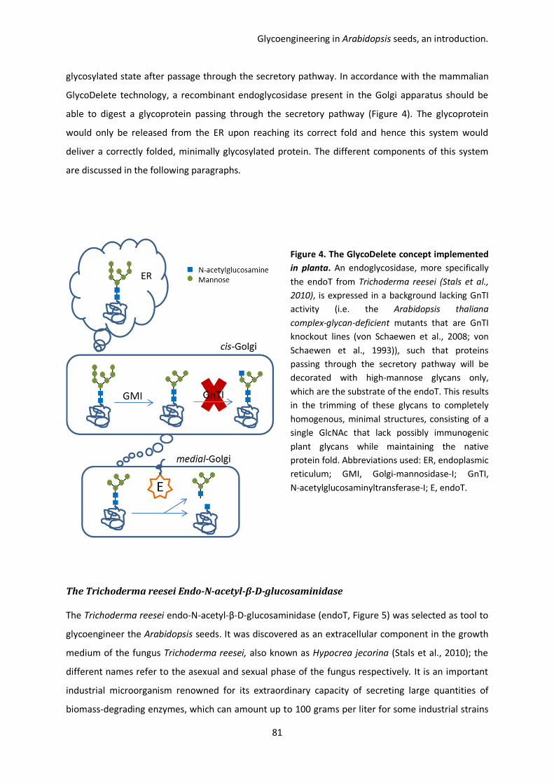
Glycoengineering in Arabidopsis seeds, an introduction.
81
glycosylated state after passage through the secretory pathway. In accordance with the mammalian
GlycoDelete technology, a recombinant endoglycosidase present in the Golgi apparatus should be
able to digest a glycoprotein passing through the secretory pathway (Figure 4). The glycoprotein
would only be released from the ER upon reaching its correct fold and hence this system would
deliver a correctly folded, minimally glycosylated protein. The different components of this system
are discussed in the following paragraphs.
Figure 4. The GlycoDelete concept implemented
in planta. An endoglycosidase, more specifically
the endoT from Trichoderma reesei (Stals et al.,
2010), is expressed in a background lacking GnTI
activity (i.e. the Arabidopsis thaliana
complex-glycan-deficient mutants that are GnTI
knockout lines (von Schaewen et al., 2008; von
Schaewen et al., 1993)), such that proteins
passing through the secretory pathway will be
decorated with high-mannose glycans only,
which are the substrate of the endoT. This results
in the trimming of these glycans to completely
homogenous, minimal structures, consisting of a
single GlcNAc that lack possibly immunogenic
plant glycans while maintaining the native
protein fold. Abbreviations used: ER, endoplasmic
reticulum; GMI, Golgi-mannosidase-I; GnTI,
N-acetylglucosaminyltransferase-I; E, endoT.
The Trichoderma reesei Endo-N-acetyl-β-D-glucosaminidase
The Trichoderma reesei endo-N-acetyl-β-D-glucosaminidase (endoT, Figure 5) was selected as tool to
glycoengineer the Arabidopsis seeds. It was discovered as an extracellular component in the growth
medium of the fungus Trichoderma reesei, also known as Hypocrea jecorina (Stals et al., 2010); the
different names refer to the asexual and sexual phase of the fungus respectively. It is an important
industrial microorganism renowned for its extraordinary capacity of secreting large quantities of
biomass-degrading enzymes, which can amount up to 100 grams per liter for some industrial strains

Chapter III
82
(Druzhinina et al., 2011; Martinez et al., 2008). Enzymes excreted by Trichoderma carry truncated
N-glycans consisting of a single GlcNAc attached to the amino acid backbone and it was discovered
that the endoT is responsible for this activity (Stals et al., 2010).
Purification of the enzyme from the growth medium and subsequent characterization revealed
several interesting features; the active form is both N and C-terminally processed releasing peptides
of respectively 9 and 46 amino acids long, endoT has a substrate specificity for high-mannose, but
not complex N-glycans and cleaves the bond between the innermost and second GlcNAc moieties of
the glycan leaving one GlcNAc residue attached to the protein backbone. Three N-glycan attachment
sites are present of which two are occupied with a single GlcNAc suggesting auto-cleavage. The
enzyme is both structurally and functionally related to other endoglycosidases (Stals et al., 2012;
Stals et al., 2010).
Figure 5. Structure of the Trichoderma reesei endoT. Figure taken from Stals et al. (2012). Crystallized
endoT was shown to have a complete (β/α)8-TIM barrel fold. The structure is rainbow colored according
to the residue number, starting with blue at the N-terminus and ending with red at the C-terminus. The
single GlcNAc residues at position N70 and N240 resulting from auto-deglycosylation are represented as
orange-colored stick models. (A) Upper view of the endoT with indication of the α-helices and β-sheets
that make up the barrel fold. (B) Side view of the endoT with an octasaccharide modeled into the active
site to indicate its position.
An advantage of endoT in the glycoengineering context is that, in contrast to other endoglycosidases
that completely remove the N-glycan, endoT releases the N-glycan while leaving the proximal GlcNAc

Glycoengineering in Arabidopsis seeds, an introduction.
83
residue attached to the protein. This is of considerable significance since this residue is in direct
contact with the protein backbone and forms an integral part of the overall structure of the protein.
EndoT activity thus eliminates the glycan structure while leaving the structurally important proximal
GlcNAc in its place, hence conserving the native protein fold.
The Arabidopsis thaliana complex-glycan-deficient mutant
To ensure optimal deglycosylation, the Arabidopsis thaliana complex-glycan-deficient (cgl) mutant
was selected as host for the glycoengineering. The cgl mutant was discovered during a chemical
mutagenesis screen and solely carries N-glycans of the high-mannose type, with a predominance of
Man5, which are an excellent substrate for the endoT (Stals et al., 2010; von Schaewen et al., 1993). A
deficiency in the N-acetylglucosaminyltransferase I (GnTI) that initiates the conversion of
high-mannose to complex N-glycans, lies at the base of this observed glycan profile (Gomez &
Chrispeels, 1994; von Schaewen et al., 1993). GnTI catalyzes the transfer of a terminal GlcNAc to the
α1,3 branch of Man5 in the cis-Golgi and its product is a prerequisite for all further maturation steps
(Wenderoth & von Schaewen, 2000).
Analysis showed that point mutations are responsible for the lack of GnTI function in both allelic cgl
variants (Frank et al., 2008). In the cgl1-1 mutant (Nottingham Arabidopsis stock centre number
N6192) this point mutation leads to the introduction of an additional N-glycan in the active site of the
enzyme, impeding it of reaching its correct fold (Strasser et al., 2005). The resulting enzyme is
inactive and retained in the ER, however, conditions that stimulate underglycosylation can partially
lift the block in conversion of high-mannose to complex N-glycans because in absence of the N-glycan
attached to it, the asparagine introduced by point mutation in the enzyme’s active site still allows for
residual GnTI activity (Frank et al., 2008; Strasser et al., 2005). The cgl1-1 mutant thus represents a
conditional mutant and it has recently been reported that recombinant glycoproteins expressed in its
seed carried predominantly high-mannose N-glycans, but with a significant fraction of complex
N-glycans, arguing for some GnTI activity during seed development (He et al., 2012a; He et al.,
2012b; He et al., 2013).
In contrast to the cgl1-1 mutant, the cgl1-2 is a true null mutant (Nottingham Arabidopsis stock
centre number N16366). Here, a point mutation at the end of an intron splice site creates a frame
shift that introduces a premature stop codon and no residual enzymatic activity can be found (Frank
et al., 2008). Both cgl mutants perform similar to wild type Arabidopsis (Col-0) under normal
conditions and specific heat, cold and light stress although they are possibly more susceptible to

Chapter III
84
stray pathogen infection and, except for longer root hairs, they display no obvious phenotype (Frank
et al., 2008; von Schaewen et al., 1993).
Although the glycans present on proteins produced in the cgl mutants are of the high-mannose type
and are non-immunogenic by nature, they are still considerably heterogeneous, ranging from Man5
to Man8, and give rise to different glycoforms, which is an undesireable feature for any therapeutic
protein. Additionally, high-mannose glycans can target a therapeutic protein to a specific subset of
cells that carry the mannose receptor, which results in the rapid clearance of the therapeutic from
the blood stream. All in all, the presence of these high-mannose glycans on a therapeutic protein
should, except for specific applications, be avoided.

85
Chapter IV
Glycoengineering in Arabidopsis seeds, results & discussion.
Robin Piron, Francis Santens, Annelies De Paepe, Julie Goossens, Els Van Lerberge, Jonah Nolf, Ann
Depicker and Nico Callewaert.

86
R.P. wrote the chapter. R.P., F.S., A.D. and N.C. conceived the experiments. R.P., F.S., A.D.P., J.G., J.N.
and E.V.L. performed the experiments. A.D.P. cloned the ASP1 gene constructs and introduced it into
wild type seeds. J.G. and E.V.L. helped with the screening and propagation of the transgenic lines.
J.N. assisted during ASP1 purification and F.S. during glycan analysis. A.D. and N.C. edited the
chapter.

Glycoengineering in Arabidopsis seeds, results.
87
A reporter protein for the GlycoDelete technology, the activation-associated
secretory protein 1
The activation-associated secretory protein 1 (ASP1, Figure 1) from the helminth Ostertagia ostertagi
was selected to serve as reporter glycoprotein to probe the functionality of the GlycoDelete
technology implemented in planta. The organism is the major parasitic nematode of cattle in the
temperate regions of the world and poses a serious problem (Geldhof et al., 2002). Treatment is
based on anti-helmintic drugs but because of concerns about resistance to these drugs and their
residual presence in consumer goods, e. g. milk and meat, alternative measures for control, such as
vaccination, are being investigated. So far, excretory/secretory proteins from adult larvae that were
purified by thiol-sepharose chromatography were shown to provide the strongest protective
immunity (Geldhof et al., 2002; Geldhof et al., 2000; Geldhof et al., 2004). Further analysis revealed
that ASP1 and ASP2 are the predominant components of this ES-thiol fraction and purified ASPs
elicited the same response as the crude fraction, identifying them as the main immunogens (Geldhof
et al., 2003; Meyvis et al., 2007).
However, isolation of large quantities for vaccination is difficult and there is great interest to produce
recombinant versions of these antigens. ASPs have been produced in the yeast Pichia pastoris and in
baculovirus-infected insect cells, but while very high antibody titers were elicited, neither
recombinant version was able to provide any protection whatsoever in vivo and the antibodies raised
in calves against these ASPs were not able to recognize the native ASP, pointing towards structural
differences between the recombinant and native ASPs (Geldhof et al., 2008; Van Coppernolle et al.,
2012). The crystal structure of Pichia-produced ASP1 has recently been elucidated and reveals that
the antigen consists of an N-terminal domain with an α-β-α fold, a typical feature for ASPs, and a
C-terminal hinge region that allows for dimerization, as occurs in vivo (Borloo et al., 2013; Meyvis et
al., 2007). Six internal disulfide bridges are present that stabilize the monomer and a peculiar feature
is that one of these links the C to the N-terminus, thus generating a cyclic structure. The seventh
bridge connects both monomers and it is postulated that its location in the less rigid hinge region
allows the dimer to be quite flexible. This is probably related to the in vivo function of the ASPs in the
host-parasite interaction although their exact role still awaits elucidation (Borloo et al., 2013).
The two N-glycan attachment sites of both ASPs are occupied in vivo with predominantly (90 %)
paucimannosidic and hybrid glycans that are core α1,6-fucosylated, but their function is still unclear
(Meyvis et al., 2008). Antibodies raised against the native ASP recognize the ASP in a
glycan-independent manner. However, reduction and denaturation of the ASP drastically reduced
antibody binding, revealing the importance of conformational epitopes of the protein backbone

Chapter IV
88
(Meyvis et al., 2008). As native ASP is purified through a thiol-sepharose chromatography step, the
purified product is a disulfide-scrambled mixture, in contrast to the recombinant proteins produced
in P. pastoris and insect cells. It is possible that this disulfide scrambling exposes epitopes to which
antibodies are raised that are important in the vaccine-induced protection. Anyhow, the
recombinant ASP1 from Pichia and baculovirus-infected insect cells apparently lack some structural
element, rendering them unprotective, and this spurred the production of recombinant ASP1 in the
seeds of Arabidopsis thaliana (unpublished results, Dr. A. De Paepe). It was hypothesized that the
seed platform might just be capable of correctly incorporating this enigmatic structural element into
the seed-produced ASP1 and bestow it with protective properties. The ASP1 could indeed be
produced in the seeds of Arabidopsis, however, competitive ELISA showed that the seed-produced
ASP1 is not recognized by neutralizing ASP1 antibodies, what seems to indicate that the
seed-produced ASP1 also lacks the structural element that is absent in the other recombinant ASP1s.
Figure 1. Structure of the
Osteragia ostertagi ASP1.
Figure adapted from Borloo
et al. (2013). Recombinant
ASP1 was produced in P.
pastoris, crystallized and its
structure elucidated. (A)
Cyclic ASP1 monomer with its
secondary structure
highlighted and six internal
cysteine bridges indicated.
Cys 195 is involved in
dimerization of the ASP1
monomers. Αlpha-helices and
β-sheets are depicted in red
and yellow respectively. (B)
ASP1 dimer connected by the
disulfide bridge formed by
both Cys 195 residues.

Glycoengineering in Arabidopsis seeds, results.
89
Results
EndoT expression in Arabidopsis thaliana complex-glycan-deficient seeds
A first step towards the generation of an Arabidopsis-seed platform capable of producing correctly
folded therapeutic proteins with truncated N-glycans is the coordinated integration of the endoT into
the secretory pathway of the seeds of the Arabidopsis cgl mutant. To avoid interference with the
folding of nascent proteins in the ER, the endoT needs to be targeted to the Golgi apparatus and this
in a seed-specific way. To this goal, the signal sequence of the endoT was swapped for that of the
Arabidopsis β1,2-xylosyltransferase which resides in the stacks of the medial-Golgi. The
xylosyltransferase is a type-II integral membrane protein, i.e. with an N-terminal cytoplasmic tail that
is 10 amino acids long, a single-span transmembrane domain and C-terminal catalytic domain facing
the lumen of the ER (Strasser et al., 2000). The first 32 amino acids that make up the cytoplasmic tail
and transmembrane domain were shown to be sufficient for correct localization in the medial-Golgi
(Pagny et al., 2003) and these were used to substitute the endoT signal sequence (Figure 2B). The
construct was cloned into a seed-specific expression cassette under control of the β-phaseolin
promoter the arcelin5-I terminator and used to transform the Arabidopsis cgl1-1 and 1-2 mutants
(Figure 2A). T1 transformants were selfed to obtain T2 segregating seed stocks which were analyzed
by western blot for the presence of endoT (Figure 3).
A similar pattern of endoT expression can be observed in both allelic mutant backgrounds (Figure 3A
and 3B), typified by a doublet band around 40 kDa and another major band around 30-33 kDa that is
surrounded by three additional bands of lower intensity of which one is situated higher at about 37
kDa and the remaining two lower in the 15 – 25 kDa range. The highest band of the doublet
corresponds to a glycoform of the lower band as it disappears after digestion with PNGaseF (see
later) and the latter probably represents the unprocessed endoT, as its observed size corresponds
with that of the full length, unglycosylated endoT (41.3 kDa, lsx:endoT; Table 1). The other major
band around 30 – 33 kDa seems to coincide with the size of the endoT catalytic domain (31.7 kDa) as
it is encountered in the extracellular medium of its host, the fungus Trichoderma reesei. In vivo, the
active extracellular endoT is the product of proteolytic processing at its N and C-terminus, releasing
peptides of 1.1 and 4.7 kDa respectively. These propeptides are present in the construct that was
used to transform the Arabidopsis cgl mutants and it is thus possible that the endoT is processed in a
similar way in Arabidopsis cgl seeds as occurs in vivo. In accordance with this hypothesis, the lower
doublet band could represent non glycosylated and unprocessed endoT (lsx:endoT, 41.3 kDa), the
highest low intensity band around 37 kDa may represent the C-terminally processed endoT (36.6
kDa) and the major band 30 – 33 kDa would then be the fully processed endoT catalytic domain (31.7

Chapter IV
90
kDa). The two remaining low intensity bands could be the result of degradation. An important remark
to be made is that if full processing indeed occurs in the seed, then the lsx will be released from the
endoT catalytic domain as an integral part of the N-terminal propeptide and this would convert the
endoT into a secretory protein instead of retaining it in the medial-Golgi.
Figure 2. T-DNA bearing the lsx:endoT gene and translation thereof. (A) The T-DNA from the binary
vector used to transform Arabidopsis, carrying the seed-specific expression cassette in which the endoT
construct was cloned and the bar gene to convey phosphinothricin (ppt) resistance to transformed
seedlings. The lsx:endoT gene is placed under control of the β-phaseolin promoter and the arcelin5-I
terminator, both derived from seed storage proteins of Phaseolus vulgaris (common bean), to promote
high, seed-specific expression. (B) The lsx:endoT fusion protein, consisting of the unprocessed endoT
without ER signal sequence coupled at its N-terminus to the medial-Golgi targeting signal of the
Arabidopsis β1,2-xylosyltransferase (lsx). Given are the fully processed endoT catalytic domain, both N
and C-terminal propeptides, the medial-Golgi targeting signal, the two sites that carry single GlcNAc
residues in vivo (red triangle) and the two remaining potential N-glycan attachment sites (pink triangles).
Abbreviations used: aa, amino acids; Pnos, nopaline synthase gene promoter; 3’ocs, octopine synthase
gene terminator; bar, bar gene conveying phosphinothricin resistance; Pphas, β-phaseoline gene
promoter; Ω, leader sequence of the ‘tobacco mosaic virus’; 3’Arc, 600 basepairs arceline5-I gene
terminator; lsx:endoT, gene construct coding for the lsx:endoT fusion protein; lsx:endoT, translational
fusion between the β1,2-xylosyltransferase medial-Golgi targeting signal and the N-terminus of the endoT
without signal sequence.

Glycoengineering in Arabidopsis seeds, results.
91
Figure 3. Expression analysis of endoT in the Arabidopsis cgl 1-1 and 1-2 mutant. Extracts of T2
segregating seed stocks of Arabidopsis cgl 1-1 (A) and cgl 1-2 (B) transformed with the endoT construct
were analyzed by western blotting using a polyclonal rabbit α-endoT serum (1:5000). Gels were run under
reducing conditions and 20 µg of protein was loaded. T-DNA single locus integration lines that were
selected to be propagated to homozygosity are marked with an arrow. A diamond indicates the doublet
band, whereas the major band around 30 kDa is represented by a star and the three additional bands of
minor intensity by hollow arrowheads. Abbreviations used: kDa, kilodalton; cgl, Arabidopsis
complex-glycan-deficient mutant; ColO, Arabidopsis thaliana ecotype Columbia.

Chapter IV
92
Table 1. Physical parameters of the recombinant endoT
Amino acids
Glycosylation sites
Predicted Mw (kDa)
min max
Lsx:endoT 374 3 41,3 47,3
EndoT propeptide 342 4 37,4 45,4
EndoT catalytic domain 290 3 31,7 37,7
lsx 32 0 3,9 3,9
N-terminal fragment 9 0 1,1 1,1
C-terminal fragment 43 1 4,7 6,7
Table 1. Characteristics of the recombinant endoT. For any peptide, the number of amino acids, number
of N-glycosylation sites and the minimum and maximum size are given (minimum size plus 2 kDa
multiplied by the number of potential glycosylation sites). EndoT propeptide and catalytic domain
correspond to the un- and fully processed endoT. Abbreviations used: Mw, molecular weight; kDa,
kilodalton; pI, isoelectric point; Lsx, Arabidopis β1,2-xylosyltransferase medial-Golgi targeting signal;
Lsx:endoT, endoT fused at its N-terminus to the lsx.
After segregation analysis the cgl1-1 endoT 2-2 and cgl1-2 endoT 5-3 were identified as the single
locus T-DNA integration lines with the highest endoT expression and these were subsequently
propagated to homozygosity. To further elucidate the endoT expression pattern, seed extracts of
both homozygous endoT lines were subjected to a PNGaseF treatment and analyzed via western
blotting (Figure 4). This revealed that only the most upper band is susceptible to glycan digest and
results in the collapse of the doublet band showing that it consists of two glycoforms of the same
protein (black diamond, Figure 4). Again, no difference could be observed between both allelic
mutant backgrounds. An interesting detail that is also revealed by this blot is that the intensity of the
endoT pattern is different for the extracts with and without the detergent dodecylmaltoside. The
latter is used to solubilize the endoT from the membrane in which it is anchored through its
N-terminal Golgi localization signal. Hence, the doublet band and the low intensity band right below
it that are only faintly stained in the extracts without detergent probably represent
membrane-anchored variants of the endoT. In accordance with the hypothesis postulated in the
previous section, the doublet band then represents two glycoforms of the unprocessed (lsx:endoT),
membrane-anchored endoT. The fainter band right below the doublet then corresponds to partially
processed endoT and its lower intensity in the detergent-free extract would suggest that it is
membrane-anchored and thus C-terminally, but not N-terminally processed. Finally, the equal
intensity of the band at around 33 kDa in both types of extract points towards loss of the membrane

Glycoengineering in Arabidopsis seeds, results.
93
anchor and hence, this represents the fully processed endoT catalytic domain only, which is
confirmed by comparison to the catalytic domain of yeast-produced endoT (see later).
Figure 4. Glycan analysis of the endoT produced in cgl seeds. Seed stocks of cgl lines T3 homozygous for
endoT (cgl 1-1 endoT 2-2-7 and cgl 1-2 endoT 5-3-1) were extracted at a fixed w/v ratio, with or without
the detergent dodecylmaltoside. The resulting extracts were digested with PNGaseF and analyzed via
western blot using a polyclonal rabbit α-endoT serum (1:5000). 10 µl of seed extract was incubated with
PNGaseF, loaded onto the gel and run under reducing conditions. The collapsed doublet band is indicated
with a black diamond. Abbreviations used: kDa, kilodalton; cgl, Arabidopsis complex-glycan-deficient
mutant.
EndoT deglycosylates endogenous seed glycoproteins
After transformation of the endoT in the cgl mutants and confirmation of its presence in the seeds,
the next step towards a functional GlycoDelete platform is to show enzymatic activity. EndoT has a
substrate specificity for high-mannose N-glycans, which are the predominant, if not only glycans
present on proteins in both allelic cgl backgrounds. It was hypothesized that if the seed-produced
endoT would show enzymatic activity, then it should also digest the endogenous seed glycoproteins.
To study the glycans present on these proteins, crude seed extracts were prepared from Arabidopsis
ecotype Columbia, both cgl mutants and low to high endoT expressing lines. These were separated
by SDS-PAGE and visualized by western blotting using the lectin Concanavalin-A (ConA) coupled to
horse radish peroxidase (HRP). ConA binds high-mannose N-glycans and together with the HRP
enzymatic moiety, this allows for chemiluminescent detection of high-mannose bearing
glycoproteins. A first western blot shows the endoT content of the selected lines ranging from

Chapter IV
94
non-detectable over intermediate to high (from ‘-‘ over ‘+’ to ‘++’ respectively, Figure 5A). The
second western blot reveals the high-mannose bearing proteins present in the extracts (Figure 5B). In
the Columbia extract, where N-glycans can mature into hybrid and complex glycans, only a few bands
are visible (Figure 5B). As expected, in the cgl mutants were high-mannose glycans are the sole
glycoforms present, much more proteins are stained. However, when comparing the profile of both
mutants to transformed lines expressing endoT, it immediately becomes clear that endoT expression
reduces this pattern and that there is an inverse relation between the endoT content of the extracts
and the number of bands detected with only two major bands remaining in the high and homozygous
endoT lines (‘++’ and ‘H’ respectively, Figure 5A & 5B). To confirm that the patterns revealed by ConA
detection (Figure 5B) truly represent high-mannose bearing glycoproteins, the western blot was
performed in duplicate and an excess of soluble ConA ligand (methyl-α-D-mannopyranoside) was
added during the incubation of the blot with ConA to specifically inhibit the glycan-binding site of
ConA (Figure 5C) showing that binding of the lectin was indeed through its mannoside binding site,
except for a remaining band around 20 kDa.
Figure 5. EndoT deglycosylates endogenous
seed proteins. Seed extracts from Arabidopsis
ecotype Columbia, both cgl mutants and
endoT expressing lines were prepared at a
fixed w/v ratio and analyzed by western blot
using (A) a polyclonal rabbit α-endoT serum to
assay the endoT content of the different lines,
(B) ConA to detect high-mannose bearing
glycoproteins and (C) ConA in the presence of
excess soluble ligand to reveal binding of
ConA through other sites than its mannoside
binding site. 10 µl of extract was loaded and
the gels were run under reducing conditions.
Abbreviations used: Col-0, Arabidopsis
ecotype Columbia; cgl, Arabidopsis
complex-glycan-deficient mutant; -/+/++,
non-detectable, intermediate and high endoT
content respectively; H, homozygous.
Glycoengineering in Arabidopsis seeds, results.
95
ASP1 expression in wild type, cgl and glycodeleteTM Arabidopsis seed
The reduction in ConA-sensitive seed glycoproteins in cgl lines expressing endoT delivered the
proof-of-concept of the activity of the recombinant endoT. This left one final question to be
addressed to demonstrate the full utility of the GlycoDelete seed-platform, namely that a foreign
glycoprotein would also be processed by the endoT upon coexpression in the seed. To tackle this
issue, the ASP1 from the parasitic nematode Ostertagia ostertagi was selected to be introduced in
the endoT expressing cgl lines. For both the cgl 1-1 as the cgl 1-2 mutant a line homozygous for
endoT was available (EndoT 2-2-7 and 5-3-1 respectively, propagated from line 2-2 and 5-3, Figure 3)
and although both mutants share the same phenotypical characteristics, they are genotypically
different and represent a conditional and a true null mutant respectively. To ensure complete
absence of GnTI functionality and complex plant N-glycans, we opted to continue only with the
Arabidopsis cgl 1-2 mutant homozygous for endoT (EndoT 5-3-1, propagated from line 5-3, Figure 3)
in further experiments. From here on we will refer to the latter as the GlycoDelete seed-platform.
A secretory version of ASP1 was N-terminally fused to the ER signal sequence of the Arabidopsis 2S2
seed storage protein and cloned into a seed-specific expression cassette under control of the same
regulatory elements that were used to drive expression of the endoT, except that the short version of
the arceline5-I 3’-UTR, 600 basepairs (bps), was substituted for the full length 3’-UTR (4000 bps;
unpublished results, Dr. A. De Paepe). The resulting construct was transformed into Arabidopsis
ecotype Columbia, the Arabidopsis cgl 1-2 mutant and the Arabidopsis cgl 1-2 line homozygous for
endoT (generation T3, line 5-3-1). The resulting T1 transformants were selfed to obtain twenty T2
segregating seed stocks per background. An acid buffer, that does not solubilizes the majority of the
endogenous seed proteins, facilitates selective extraction of the ASP1 from ground seeds, yielding
semi-pure extracts (see Material & methods). This allowed the T2 segregating seed stocks to be
visually screened for ASP1 content by SDS-PAGE and Coomassie staining of the corresponding acid
extracts (Supplemental Figure S1). Segregation analysis was subsequently performed to identify the
single locus T-DNA integration lines and for each background, the line with the highest ASP1
accumulation and the ASP1 T-DNA integrated at a single locus and was retained and propagated to
homozygosity.
Two patterns are observed when the recombinant ASP1 is produced in the different Arabidopsis
backgrounds. In Columbia, two predominant, similarly stained bands are visible with a molecular
weight just over 25 kDa and right below these, a third band of weak intensity (Figure 6). As ASP1 has
two potential N-glycosylation sites, we hypothesize that the three bands represent different ASP1
glycoforms and thus correspond to ASP1 carrying no, one and two N-glycans. Apparently, most ASP1

Chapter IV
96
is glycosylated, with an even distribution of the glycoforms with one and two N-glycans, whereas
only little ASP1 carries no N-glycans. The pattern of ASP1 from Columbia is identical to that of the
ASP1 expressed in the cgl1-2 mutant and this seems to indicate that the presence or absence of
complex plant glycosylation does not influence the production of ASP1 in Arabidopsis seed. When
looking at the ASP1 derived from the GlycoDelete lines, a strikingly different pattern is visible (Figure
6). Here, only one band is present around 25 kDa and it coincides with the lowest band of the ASP1
from Columbia and the cgl1-2 background. This confirms that the three bands of the Columbia and
cgl1-2 ASP1 indeed represent different glycoforms and that the lowest band corresponds to the
unglycosylated ASP1. The more important conclusion to be drawn, however, is that the ASP1 seems
fully susceptible to the endoT activity of the GlycoDelete line as no other glycoforms can be discerned
on the gel
.
Figure 6. Analysis of recombinant ASP1 expression in different Arabidopsis backgrounds. Seed stocks of
different lines producing recombinant ASP1 were extracted with an acid buffer at a fixed w/v ratio,
separated by SDS-PAGE and Coomassie-stained. 10 µl of extract was loaded and the gels were run under
reducing conditions. A BSA standard was included to allow densitometric quantitation of the ASP1 present
in the different extracts. Abbreviations used: BSA, bovine serum albumin; T2, single locus ASP1 T2 seed
stock; T3, single locus ASP1 T3 seed stock; T3 H, homozygous ASP1 T3 seed stock; Col-0, Arabidopis
ecotype Columbia; cgl1-2, Arabidopsis complex-glycan-deficient 1-2 mutant. EndoT cgl1-2, Arabidopsis
complex-glycan-deficient 1-2 mutant homozygous for endoT.
As the seed extracts are sufficiently pure, the signal intensity of the ASP1 bands was measured and
used to determine the ASP1 content of the different extracts via the reference BSA standards that
were also run on the gels. Together with the known w/v ratio at which the extracts were prepared,
this allowed to calculate the ASP1 accumulation for each line, which was expressed as milligram

Glycoengineering in Arabidopsis seeds, results.
97
extractable ASP1 per gram of dry seed weight (Table 2). The accumulation of ASP1 in the Columbia
and cgl1-2 background, with an average of 27.8 and 27.2 mg per gram of dry seed weight
respectively, is very similar for the three homozygous lines under study. This again supports the
notion that the presence/absence of complex N-glycosylation does not alter the production of ASP1
in Arabidopsis seed. However, this picture is quite different for the GlycoDelete lines, where the
average ASP1 accumulation for the three studied homozygous lines is only 7.1 mg per gram of dry
seed weight. When compared to the Columbia and cgl1-2 lines, this is approximately a four-fold
reduction. Nevertheless, the ASP1 produced in the GlycoDelete background still reaches an
accumulation of 3.5% of TSP, which is considerably higher than the 1% deemed the minimal treshold
for economic viability of a PMP (Rybicki, 2009). It should be noted that for the cgl and the
GlycoDelete ASP1 the three homozygous lines under study are derived from the same transformation
event and hence, caution should be taken when interpreting the observed differences in ASP1
accumulation. However, lower accumulation of the ASP1 in the GlycoDelete background when
compared to the cgl (and wild type) background seems a general tendency (compare Figure S1A and
B). An additional detail revealed by table 2 is that a clear dosage effect can be observed when the
seed stocks are propagated from heterozygous to homozygous and this characteristic is shared by all
backgrounds.
Table 2. ASP1 content of different transformed lines.
Line Background Generation mg/g seed % of TSP
ASP1 1-4-26 Col-0 T3 He 20,4 10,2%
ASP1 2-1-17 Col-0 T3 Ho 28,6 14,3%
ASP1 2-1-25 Col-0 T3 Ho 24,8 12,4%
ASP1 5-2-2 Col-0 T3 Ho 29,9 14,9%
ASP1 1-4 cgl1-2 endoT T2 He 5,9 2,9%
ASP1 1-4-5 cgl1-2 endoT T3 Ho 8,0 4,0%
ASP1 1-4-6 cgl1-2 endoT T3 Ho 7,1 3,6%
ASP1 1-4-8 cgl1-2 endoT T3 Ho 6,0 3,0%
ASP1 2-2 cgl1-2 T2 He 19,8 9,9%
ASP1 2-2-7 cgl1-2 T3 Ho 28,7 14,4%
ASP1 2-2-8 cgl1-2 T3 Ho 23,5 11,8%
ASP1 2-2-10 cgl1-2 T3 Ho 29,3 14,7%
Table 2. Seed extracts were prepared from all the different recombinant ASP1 producing lines at a fixed
w/v ratio and subjected to SDS-page. Densitometric analysis of Coomassie-stained gels, as shown in figure
5, allowed to calculate the ASP1 content of the different lines which was expressed as milligram ASP1 per
gram of dry seed weight and also as % of TSP (for 200 mg TSP per gram of dry seed). Abbreviations used:

Chapter IV
98
Col-0, Arabidopsis thaliana ecotype Columbia; cgl1-2, Arabidopsis complex-glycan-deficient 1-2 mutant;
cgl1-2 endoT, Arabidopsis cgl1-2 mutant homozygous for endoT; He, heterozygous; Ho, homozygous; TSP,
total soluble protein.
One possible explanation for the observed difference in accumulation of the ASP1 from the cgl
background and the GlycoDelete line would be that deglycosylation of the ASP1 alters its solubility in
the acid buffer used to extract the seeds. To check this the corresponding T3 seed stocks of both
ASP1 variants were extracted three consecutive times with the acid buffer and a final fourth time
with a buffer with a high concentration of detergent and reducing agent. The latter served to
solubilize the pellet of seed debris and extract any remaining protein. All the collected extracts were
analyzed by western blot to compare their ASP1 content and this shows that after three washing
steps with the acid buffer a considerable fraction of both ASP1 variants remain in the pellet (Figure
7). However visual inspection of the western blot reveals no major difference in extraction pattern
for both ASP1 variants and thus a difference in solubility can be discarded as an explanation for the
earlier observed four-fold difference in accumulation of both variants. An interesting observation is
made when looking at the pellet extraction of the GlycoDelete ASP1. Here, some faint bands of
slightly higher molecular mass appear that are not present in the preceding acid extracts. These
might correspond to intermediates of the ASP1 whose glycans have not yet been trimmed by endoT.
This might represent the nascent ASP1 population in the ER.
Figure 7. Solubility assay of the ASP1 produced in the
cgl1-2 and the GlycoDelete line. T3 seed stocks were
extracted three times with an acid buffer (numbers 1 to
3) and a final fourth time with a buffer to solubilize the
pellet (P). Extracts were prepared at a fixed w/v ratio and
analyzed by western blot using a polyclonal rabbit α-ASP1
serum (1:1000). 10 µl of extract was loaded and the gel
was run under reducing conditions. Abbreviations used:
cgl1-2, Arabidopsis complex-glycan-deficient 1-2 mutant;
EndoT cgl1-2, Arabidopsis cgl1-2 line homozygous for
endoT; P, pellet extraction; kDa, kilodalton.

Glycoengineering in Arabidopsis seeds, results.
99
EndoT accumulation level and stability over generations
We wanted to further characterize the seed-produced endoT by determining its accumulation level
in both homozygous cgl lines and assessing its stability over multiple generations. To this goal, seed
extracts were prepared from T3 seed stocks of both cgl lines homozygous for endoT, the endoT T3 2-
2-7 cgl1-1 and endoT T3 5-3-1 cgl1-2 respectively, and from a T5 seed stock that is derived from the
latter and co-expresses the ASP1 with the endoT. This line was created by supertransforming the
parental T3 5-3-1 endoT cgl1-2 line with the ASP1 construct and subsequent propagation to obtain
the T5 5-3-1/1-4 endoT cgl1-2 ASP1 seed stock that is homozygous for endoT and heterozygous for
ASP1. The seed extracts were analyzed via ELISA and the obtained OD values plotted against a
standard curve of purified fully processed endoT produced in the yeast Pichia pastoris to calculate
the endoT content of the corresponding lines (Figure 8A). The seed and yeast-produced endoT were
also subjected to western blot to assure recognition of both forms by the polyclonal α-endoT rabbit
serum used in the ELISA and compare the patterns revealed for both enzymes (Figure 8C).
Figure 8. Quantification of seed-produced endoT and comparison to yeast-produced endoT. For each
mutant background, cgl1-1 and cgl1-2, the T3 seed stock of the line homozygous for endoT was selected
to be analyzed (endoT 2-2-7 and 5-3-1, respectively), together with a T5 seed stock of the GlycoDelete line
(endoT cgl1-2) that co-expresses endoT and ASP1 and is heterozygous for the latter (ASP1 endoT 5-3-1/1-
4). Crude seed extracts were prepared and (A) subjected to ELISA using a polyclonal rabbit α-endoT
serum. Are given, the dilution at which the extracts were coated and the obtained OD values. Numbers #1
to #3 represent biochemical repeats from the same seed stock. (B) The measured values were plotted

Chapter IV
100
against an endoT standard curve and used to calculate the endoT content of the three analyzed lines. (C)
Western blot comparison of the yeast- and seed-produced endoT detected by a polyclonal rabbit α-endoT
serum (1:5000). Seed extracts were prepared at a fixed w/v ratio and 20 µl was loaded, whereas for the
purified Pichia-produced endoT 200 ng was loaded. The gel was run under reducing conditions and a black
arrow indicates the endoT catalytic domain.
The ELISA shows that the endoT content of the GlycoDelete line in generation T3 (Figure 8A, EndoT
cgl1-2) and generation T5 co-expressing ASP1 (Figure 8A, ASP1 endoT cgl1-2) is similar and, without
even reaching 0.5% of TSP, quite low (Figure 8B). Supertransformation of the GlycoDelete line with
the ASP1 construct under control of the same regulatory elements does not alter the endoT
accumulation in the offspring line, which in generation T5 is still identical to that of the parental line
(0.60 and 0.57mg, respectively) indicating stable inheritance of the endoT over several generations
(T3 to T5). When the seed-produced endoT is compared to the yeast-produced endoT on western
blot, the main signal of the catalytic domain of the endoT purified from yeast coincides with the
major band around 33 kDa in the seed extracts, hereby supporting the earlier formulated hypothesis
that this band indeed represents the fully processed endoT calatytic domain (Figure 8C).
Purification of recombinant ASP1
To obtain purified ASP1 for further in-depth analysis, the extraction of ASP1 by acid buffer from the
seed was combined with a three-step purification protocol consisting of two consecutive
ion-exchange chromatography steps followed by a final gel filtration step (Figure S2). When the
protocol was applied to the wild type, cgl, and GlycoDelete ASP1, this yielded recombinant ASP1 of
high purity (see Figure 9A and 9B). Under non-reducing conditions all the purified ASP1 variants are
present as dimers that run at an apparent molecular weight of 37 to 50 kDa (Figure 9B). When the
dimers are reduced the different ASP1 glycoforms can be discerned running at or right above 25 kDa
(Figure 9A and 9B). It should be noted that prior to purification, the ASP1 proteins are predominantly
present in the seed as dimers, but there is also a small fraction present as multimers and this is
independent of the Arabidopsis background in which the ASP1 was produced (Figure S2).
Nevertheless, as the ASP1 is encountered in vivo as a dimer of about 50 kDa (Meyvis et al., 2007), the
ASP1 dimers were selectively separated from the ASP1 multimers during the final gel filtration step
of the purification protocol and retained for further analysis. The presence of a fraction of the
seed-produced ASP1s as multimers might point to a structural difference between the latter and the
native ASP1 that occurs as dimers.

Glycoengineering in Arabidopsis seeds, results.
101
Figure 9. Characterization of purified recombinant ASP1. ASP1 produced in Col-0, cgl1-2 and GlycoDelete
seeds was purified by three consecutive chromatography steps and subjected to SDS-PAGE. Gels were
Coomassie-stained and run under reducing (A) and non-reducing conditions (B) or analyzed via western
blot using a polyclonal rabbit α-ASP serum (1:1000) and run under reducing conditions (C). 4 µg of purified
ASP1 was loaded onto the Coomassie-stained gels whereas 2 µg was loaded onto the gel analyzed by
western blot. Abbreviations used: Col-0, Arabidopsis ecotype Columbia; cgl1-2, Arabidopsis
complex-glycan-deficient 1-2 mutant; E cgl1-2, Arabidopsis cgl1-2 line homozygous for endoT.
Glycan analysis of ASP1 produced in wild type, cgl and GlycoDelete Arabidopsis seeds
ASP1 cgl1-2, but not ASP1 Col-0 is susceptible to in vitro endoT digestion
The profiles that the wild type and cgl ASP1 display under reducing conditions correspond to their
different glycoforms, i.e. carrying no, one or two N-glycans (Figure 9A), and are almost identical
pointing towards a similar N-glycan site occupancy in wild type and cgl Arabidopsis.
However, because of their production in the wild type and cgl Arabdidopsis, the ASP1 variants should
be decorated with complex and high-mannose N-glycans respectively, which cannot be distinguished
as such on the gel. Nevertheless, the presence of these glycans on the recombinant ASP1 variants
can be assayed by in vitro digest of both ASP1s by endoT, since it has substrate specificity for
high-mannose, but not complex glycans. To this goal, purified wild type and cgl ASP1 was incubated
with a dilution series of yeast-produced endoT subjected to SDS-PAGE and visualized via Coomassie
staining (Figure 10).

Chapter IV
102
As expected, the cgl ASP1 is susceptible to endoT treatment in vitro and the complete disappearance
overnight of the bands representing glycosylated cgl ASP1 shows that it solely carries high-mannose
N-glycans (Figure 10). On the other hand, the ASP1 wt is predominantly resistant to endoT digestion
although the slight increase in intensity of the lower band indicates that some endoT sensitive
glycans are also present. This points towards the acquisition of a majority of complex endoT
resistant glycans together with some remaining high-mannose endoT sensitive glycans, which is in
accordance with the glycan profile that is expected to be found on a secretory glycoprotein produced
in wild type Arabidopsis.
Figure 10. EndoT susceptibility of the ASP1 wt and cgl. Purified ASP1 wt and cgl was incubated overnight
with a serial dilution of yeast-produced endoT (the ratio of endoT over target protein is indicated above
the corresponding slots) and then subjected to SDS-PAGE and visualized by Coomassie staining. 5 µg of
protein was loaded onto the gel that was run under reducing conditions. For each sample the ratio of
endoT over the recombinant ASP1 is given. Abbreviations used: wt, Arabidopsis ecotype Columbia; cgl,
Arabidopsis complex-glycan-deficient 1-2 mutant; E, endoT.
DSA-FACE analysis of recombinant ASP1 and the plant glycan substrate specificity of
endoT
In concordance with the theoretically predicted profiles, the wt ASP1 carries endoT resistant complex
glycans, the cgl ASP1 endoT sensitive high-mannose glycans and the GlycoDelete ASP1 glycans that
are trimmed down to single GlcNAcs. To corroborate these findings and investigate the presence of

Glycoengineering in Arabidopsis seeds, results.
103
minor, previously undetected glycoforms, the ASP1s were further analyzed by DNA
sequencer-assisted, fluorophore-assisted carbohydrate electrophoresis (DSA-FACE) and/or
matrix-assisted laser desorption/ionization-time of flight (MALDI-TOF) mass spectrometry.
The precise glycan profiles of the wt and cgl ASP1, predicted to contain predominantly complex
glycans and high-mannose glycans respectively, were elucidated by releasing their glycans by
PNGaseA digestion and sequential treatment of the total glycan pools with different glycosidases.
DSA-FACE analysis of the glycan profile before and after each treatment then allowed annotation of
each of the glycans in the total glycan pool (Figure 11B and 12E). If a glycan is susceptible to
treatment with a specific glycosidase, its corresponding peak in the DSA-FACE electropherograms will
shift after the reaction to that of the smaller product and hence comparison of both
electropherograms can identify these glycans. After sequential treatments, eventually the exact
structure of the glycans present in the electropherograms can be deduced (Laroy et al., 2006).
Digestion with broad-spectrum mannosidase reveals glycans with terminal mannoses, whereas
α1,2-mannosidase identifies glycans with terminal α1,2 mannoses only (Man6 to Man9). Treatment
with hexosaminidase shows glycans with one or two terminal GlcNAcs (both complex and hybrid
glycans) and any of these enzymes can be combined to digest structures with both terminal GlcNAcs
and mannoses. To identify the peaks that represent complex α1,3-fucosylated glycans the
electropherogram of the total glycan pool released by PNGaseA can be compared to that of the
glycans released by PNGaseF, which does not digest core α1,3-fucosylated structures (Figure 12B and
12C).
When these techniques were applied to the wt ASP1 and its exact glycan profile was elucidated
(Figure 11B, 12B and S3), several interesting observations were made. All glycans, except the
high-mannose glycans that represent a small fraction of the total glycan pool, are core
α1,3-fucosylated and the majority of these also carry a bisecting β1,2-xylose. Among these
immunogenic plant glycans some paucimannosidic (Figure 11B peak 1 and 2) and hybrid structures
(peak 6 and 7) can be discerned, but the predominant glycoforms are complex glycans with one
terminal GlcNAc (and a fully processed α1,3 arm, peak 2 and 3) or two terminal GlcNAcs (peak 4 and
5). This confirms the previous observation that wt ASP1 is mostly decorated with complex, endoT
resistant, N-glycans. Indeed, all the glycans that are released by endoT treatment of wt ASP1 are
exclusively of the high-mannose type (Figure 12D). Production of the ASP1 in the Arabidopsis cgl
mutant drastically simplifies the glycan profile showing high-mannose glycans only with the structure
Man5 as the main glycoform (Figure 12E) and this is consistent with the lack of GnTI activity in the cgl
background that blocks conversion of high-mannose to complex glycans.

Chapter IV
104
Figure 11. DSA-FACE analysis of the wt ASP1 glycan pool and glycosidase treatments thereof. The total
glycan pool of wt ASP1 was released by PNGaseA digest, fluorescently labeled, treated with different
glycosidases and subjected to DSA-FACE. (A) Oligomaltose standard (glucose polymer), the number of
glucose units is indicated. (B) Total glycan pool released by PNGaseA and digested with (C) broad
spectrum jack bean α1,2,3,6-mannosidase, (D) hexosaminidase or (E) hexosaminidase and jack bean
mannosidase. (F) High-mannose glycan profile of RNAseB. Maltopentaose was added to all samples as an
internal standard. Abbreviations used: M5 to M9, Man5 to Man9; JB Man, jack bean
α1,2,3,6-mannosidase; Hex, jack bean β-N-acetylhexosaminidase.

Glycoengineering in Arabidopsis seeds, results.
105
Figure 12. DSA-FACE analysis of the wt and cgl ASP1 glycan pool and the substrate specificity of endoT
towards plant glycans. (A) Oligomaltose standard. (B) Glycans released from wt ASP1 by digestion with
PNGaseA, (C) PNGaseF and (D) endoT. (E) Cgl ASP1 total glycan pool released by PNGaseA digest. (F)
High-mannose glycan profile of RNAseB. Abbreviations used: M5 to M9, Man5 to Man9.

Chapter IV
106
Mass spectrometry analysis of GlycoDelete ASP1
The only remaining glycan profile to be characterized in detail was that of the GlycoDelete ASP1, which
could not be analyzed by DSA-FACE because of the small size of its trimmed glycans, which are not
susceptible to PNGaseA or PNGaseF release. Hence, the GlycoDelete ASP1 was treated with the
protease trypsin and the resulting tryptic peptides were subjected to MALDI-TOF spectrometry, which
should allow precise identification of the glycan structures still present on the GlycoDelete ASP1.
Initially the purified GlycoDelete ASP1 was trypsinized in solution following a two-step protocol to
ensure maximal proteolytic cleavage. The peptide mixture was then analyzed by MALDI-TOF
spectrometry and the resulting spectrum screened for the two glycopeptides that were predicted to
be released by tryptic digest of the GlycoDelete ASP1 (Figure 13A). All the peptides that could be
identified in the spectrum are depicted with their corresponding apparent masses in Figure 13B and
their peaks indicated on the spectrum (Figure 13C). Glycopeptide one and two are indeed present as
glycoforms carrying a single GlcNAc and for both peptides tryptic variants can be found that are
similarly decorated with a single GlcNAc. For glycopeptide one and its tryptic variant also the
unglycosylated amino acid backbones can be discerned. Interestingly, no other glycoforms were
present in the spectrum, pointing to complete digestion of the ASP1 glycans by endoT in the
Arabidopsis GlycoDelete background. In general, several tryptic variants are visible, both for the
glycopeptides as among the other peptides identified in the spectrum and these are the result of
incomplete proteolytic digestion. Trypsin cleaves after a lysine (K) or arginine (R), however, sequences
consisting of any two of the latter tend to be incompletely digested and apparently even the two-step
digestion protocol could not ensure complete cleavage. Also some minor, unidentified peaks remain in
the spectrum.
To further optimize the spectrum, the purified GlycoDelete ASP1 was run on a polyacrylamide gel prior
to mass spectrometric analysis. The area corresponding to single GlcNAc and possible larger
glycoforms was excised and the GlycoDelete ASP1 trypsinized in gel. The resulting peptides were then
solubilized and subjected to MALDI-TOF mass spectrometry. When comparing the spectra obtained
after both methods, an immediate and obvious difference is the reduction in number of tryptic
variants and smaller peaks (Figure 13C and 14C). No tryptic variants of the glycopeptides are
encountered and both glycopeptides are again present as glycoforms with single GlcNAcs although the
peak corresponding to glycopeptide 1 is of low intensity (Figure 14C). No other glycoforms could be
discerned confirming the previous observations that the ASP1 glycans are completely trimmed by the
endoT in the GlycoDelete Arabidopsis background. By comparing the masses of the predicted tryptic
peptides with those observed in the spectrum all major peaks could be annotated, except three peaks

Glycoengineering in Arabidopsis seeds, results.
107
at m/z 1975.02, 2185.15 and 2386.22. Nevertheless, aspecific cleavage of the GlycoDelete ASP1 in
silico yields several peptides with m/z values that are nearly identical to those of the observed peak
thus indicating that these probably also correspond to internal peptides of the GlycoDelete ASP1 (see
Table S1).

Chapter IV
108
Figure 13. MALDI-TOF analysis of in-solution trypsinized glycodelete ASP1. Purified GlycoDelete ASP1 was
carboxymethylated, trypsinized in-solution and subjected to MALDI-TOF spectrometry in the positive ion
mode [M+H]+. (A) The amino acid sequence of the GlycoDelete ASP1. Tryptic glycopeptide one and two are
depicted in blue and orange, respectively, and the N-glycan attachment sites underlined. Other tryptic
peptides found in the spectrum are indicated in green. (B) The different peptides identified in the spectrum
and their masses (in kilodalton) given as the molecular weight plus one proton. (C) Mass spectrum of the
trypsinized GlycoDelete ASP1. The peptide corresponding to a specific peak is indicated. A blue square
represent a glycopeptide carrying a single GlcNAc.

Glycoengineering in Arabidopsis seeds, results.
109
Figure 14. MALDI-TOF analysis of in-gel trypsinized glycodelete ASP1. Purified glycoDelete ASP1 was run
on gel, carbamidomethylated and the corresponding band area excised and digested with trypsin. The
resulting tryptic peptides were then solubilized and subjected to MALDI-TOF spectrometry in the positive
ion mode [M+H]+. (A) The amino acid sequence of the GlycoDelete ASP1. Tryptic glycopeptide one and two
are depicted in blue and orange, respectively, and the N-glycan attachment sites underlined. Other tryptic
peptides found in the spectrum are indicated in green. (B) The different peptides identified in the spectrum
and their mass (in kilodalton) given as their molecular weight plus one proton. (C) Mass spectrum of the
trypsinized GlycoDelete ASP1. The peptide corresponding to a specific peak is indicated. A blue square
represent a glycopeptide carrying a single GlcNAc.

Chapter IV
110
ASP1 carrying complex plant N-glycans, but not GlycoDelete ASP1 induces an
anti-carbohydrate response in rabbits
Two groups of five rabbits were injected subcutaneously with GlycoDelete or wild type ASP1 following
a prime-boost protocol. Rabbits received 35 µg of recombinant ASP1 mixed with complete freund’s
adjuvant at day zero and 35 µg mixed with incomplete freund’s adjuvant at days 14, 28 and 56. Blood
samples were taken on day zero prior to immunization (preimmune) and at days 28, 36 and 80 when
the rabbits were euthanized. The goal of the experiment was to assess the immunogenicity of the wild
type ASP1 with its fucose and xylose bearing complex plant glycans and compare this to the
GlycoDelete ASP1 that carries truncated glycans.
After the final vaccination, all rabbits had developed high titers of IgG antibodies specific for the
peptide backbone of the recombinant ASP1 (Figure 15A). These were measured by an direct ELISA in
which denatured, reduced and deglycosylated ASP1 was coated as the antigen (Figure S4). Variation in
titer was higher among the GlycoDelete ASP1 vaccinated group, but the average titer was similar and
high for both groups (Figure 15A). To evaluate the immunogenicity of the glycan structures of both
ASP1s, it was decided to study the presence/absence of IgG antibodies specific for plant glycans in the
sera of the vaccinated rabbits. To measure the latter, a recombinant glycoprotein not related to ASP1
was used as a neutral carrier of glycan structures that served as antigens in ELISA to allow selective
binding of glycan-specific IgGs. More specifically, a synthetic antibody was used that consisted of the
antigen-binding domain of a camelid heavy chain only antibody (nanobody), i.e. the GFP-binding
protein (GBP) coupled to the Fc domain of the murine IgG3 and this fusion protein is hereafter referred
to as GBP-Fc. The recombinant GBP-Fc has two N-glycosylation sites located in its Fc-domain and had
been expressed in different organisms, so different glycoforms were at hand. Production in Pichia
pastoris yielded high-mannose glycoforms (Man5 to Man8) whereas the GBP-Fc produced in
Arabidopsis seeds almost exclusively consisted of glycoforms with complex plant glycans bearing
immunogenic fucose and xylose residues (PhD dissertation 2013, Dr. T. De Meyer; Figure 15C). It
should be noted that approximately 30% of the Arabidopis-produced GBP-Fc is unglycosylated (Dr. T.
De Meyer, personal communication).
To probe the presence of plant-glycan specific IgGs, the sera of the vaccinated rabbits were subjected
to ELISAs, using the high-mannose and immunogenic GBP-Fc glycoforms as coated antigens. Prior to
the coating step, the purified glycoforms were reduced and denatured to expose their N-glycans that
otherwise might have been shielded from the sera because of their inward orientation due to the fold
of the Fc domain. The stripped GBP-Fc peptide backbone, obtained by reduction, denaturation and

Glycoengineering in Arabidopsis seeds, results.
111
digestion of the high-mannose glycoform with PNGaseF and monitored by mobility shift assay (Figure
15B), was also coated to measure non-glycan specific detection (background).
Three out of the five rabbits that had been injected with the recombinant ASP1 bearing complex plant
N-glycans had generated IgGs specific for β1,2-xylose and core α1,3-fucose, confirming that these are
indeed immunogenic (Figure 15A). Interestingly, the other two rabbits seemed unresponsive to the
plant glycans despite having high titers of IgG specific for the ASP1 peptide backbone in their sera. No
plant-glycan specific IgGs could be detected in the sera of the rabbits vaccinated with the GlycoDelete
ASP1. For all ten rabbits, no signal could be measured when the high-mannose GBP-Fc was coated as
antigen, pointing out that the detection of the fucose and/or xylose bearing GBP-Fc by three of the
wild type ASP1 vaccinated rabbits was specific for the latter two residues.

Chapter IV
112
Figure 15. Overview of the ASP1 and plant-glycan specific IgG titers after vaccination together with the
structure of the glycoforms of the GBP-Fc used during ELISAs. (A) Overview of the IgG titers measured in
the sera obtained after the final bleeding of the rabbits. Both vaccinated groups are indicated above the
figure. Are given, the IgG titers specific for the ASP1 peptide backbone and for both glycoforms of the
GBP-Fc decorated with high-mannose and plant glycans respectively. The mean titer is depicted by a
horizontal line. (B) Mobility shift assay to corroborate the deglycosylation of the ASP1 and GBP-Fc
high-mannose glycoforms by PNGaseF digestion. (C) DSA-FACE electropherogram of the glycans released by
PNGaseA from the GBP-Fc produced in yeast and Arabidopsis seed respectively. Abbreviations used: ‘–‘, no
PNGaseF; ‘+‘, active PNGaseF; ‘H’, heat-inactivated PNGaseF; Vacc, vaccinated; kDa, kilodalton; P.p., Pichia
pastoris; A.t., Arabidopsis thaliana; HM, high-mannose glycoform; X/F, complex glycoform bearing
immunogenic xylose and fucose residues.

Glycoengineering in Arabidopsis seeds, results.
113
Discussion
The endoT was transformed into the seeds of the Arabidopsis cgl mutant and targeted to the
medial-Golgi via the localization signal of the Arabidopsis β1,2-xylosyltransferase. Expression analysis
revealed that that the majority of the endoT is both C and N-terminally processed, as occurs in vivo.
Nonetheless, N-terminal processing would result in loss of the Golgi targeting signal as integral part of
the cleaved propeptide and release the endoT from the medial-Golgi. It will be interesting to study the
subcellular localization of the endoT in more detail to determine if the endoT, for example,
accumulates in the apoplast, which would confirm its secretion and argue for loss of the medial-Golgi
targeting signal. To characterize the engineered GlycoDelete platform, the endoT content of the
GlycoDelete line was determined and shown to be stable over several generations and upon
co-expression with a reporter glycoprotein. Despite the fact that the β-phaseolin promoter and
arcelin5-I terminator, regulatory sequences derived from the common bean Phaseolus vulgaris and
designed for high seed-specific expression of recombinant proteins, were used to drive endoT
expression in the cgl seeds, endoT did not accumulate to more than 0.3% of TSP (or 60 milligram per
gram of dry seed weight). A rather low number when compared to the levels, up to 15% and more of
TSP, that have been achieved for synthetic antibodies produced in wild type Arabidopsis seeds using
the same regulatory elements (De Buck et al., 2013; De Jaeger et al., 2002). Anyhow, massive endoT
production during co-expression of a therapeutic protein is undesirable as it would deviate resources
from the latter and might result being detrimental for its accumulation. Under the condition that
sufficient endoT is present in the seed to completely process any therapeutic protein that is expressed
in the GlycoDelete line, the low endoT content achieved actually represents an advantage rather than
disadvantage and apparently, the low endoT content of the GlycoDelete line was perfectly capable of
digesting a ten-fold excess of target protein (0.3 and 3.5% of TSP respectively), therefore
demonstrating the functionality of the GlycoDelete platform.
A peculiarity here were the accumulation levels observed for the different recombinant proteins
expressed in Arabidopsis seeds, with endoT showing the lowest number (0.3% of TSP), followed by the
GlycoDelete ASP1 (3.5% of TSP) and similar and high levels for both the cgl and wild type ASP1 (13.6%
and 13.9% of TSP respectively), despite sharing the same regulatory elements to drive their expression.
It must be noted that, for cloning purposes, a shorter version (600 basepairs (bp)) of the arcelin5-I
terminator was used to control endoT expression, whereas for the recombinant ASP1s the original
terminator was employed (4000 bp). Although according to Goossens et al. (1999) the first 600 bp of
the arcelin5-I terminator contain nearly all the necessary information for high expression, it was later
shown that a transgene under control of the β-phaseolin promoter and the 600 bp arcelin5-I
terminator did not achieve the same expression level as when the 4000 bp full-length terminator was

Chapter IV
114
used, indicating that the shortened terminator is less potent (Dr. K. De Wilde, PhD dissertation 2013).
Additionally, the lower endoT accumulation could also be a reflection of some of its intrinsic
characteristic, e.g. the fact that is a membrane-anchored protein which tend to more difficult to
express and it is probably the combination of these two factors that is responsible for the low endoT
accumulation observed, which is actually beneficial for the GlycoDelete platform.
Another interesting feature was that the Arabidopsis line used for expression of the recombinant
ASP1s seemed to influence the accumulation level with a four-fold reduction observed for the
GlycoDelete ASP1 when compared to the cgl and wild type ASP1 (3.5% versus 13.6% and 13.9% of TSP
respectively). The presence/absence of complex glycosylation did not seem to impact ASP1 expression
as the cgl and wild type ASP1 accumulated to nearly identical levels, but co-expression with endoT
apparently did, although it should be mentioned that the GlycoDelete ASP1 lines under study were
offspring of the same transformation event and this should be kept in mind when extrapolating the
observed differences between the GlycoDelete and the cgl and wild type ASP1. Nevertheless,
comparison of supplemental figure S1A and S1B seems to indicate a general tendency of the
recombinant ASP1 to accumulate less in the GlycoDelete background. As the only difference between
the GlycoDelete and cgl ASP1 resides in the presence/absence of the endoT gene construct and the
corresponding translated protein in its host, it is plausible to attribute the lower accumulation of the
GlycoDelete ASP1 to one of the latter two and several explanations are possible. Firstly, despite leaving
the proximal GlcNAc attached to the peptide backbone, the trimmed ASP1 might just be less stable
and hence, not accumulate to the same levels as its cgl and wild type counterparts. Secondly, although
the endoT is targeted to the medial-Golgi to avoid interference with protein folding in the ER, the
GlycoDelete ASP1 and endoT are under control of the same promoter and thus have an identical
temporal expression pattern what allows for contact between both proteins in the ER and this might
interfere with the correct folding of a fraction of the ASP1 leading to its degradation by the
ER-associated degradation pathway (ERAD). Thirdly, since both proteins share the same promoter they
might compete for resources during production lowering final GlycoDelete ASP1 accumulation,
although this seems little credible given that the endoT accumulation in the GlycoDelete line is low
(0.3% of TSP) and was identical before and after co-expression of the ASP1.
A final possibility that cannot be excluded, is that by mere chance only low GlycoDelete ASP1
accumulators were found among the 20 transformants that were screened (supplemental Figure S1A)
and that by amplifying the initial screen, transformants would have been picked up with accumulation
levels similar to those observed for the cgl and wild type ASP1. To exclude this chance factor, a well
characterized cgl ASP1 producing line should be supertransformed with the endoT construct, the
opposite operation of how the GlycoDelete ASP1 was generated during this work, and the

Glycoengineering in Arabidopsis seeds, results.
115
accumulation of the GlycoDelete ASP1 in the offspring should be compared to the parental line
without endoT. This will allow an exact determination of the influence of the endoT on the ASP1
accumulation and unveil the mechanism for lowering GlycoDelete ASP1 accumulation.
With 3.5, 13.6 and 13.9% of TSP the accumulation levels achieved for the GlycoDelete, cgl and wild
type ASP1, respectively, compare favorably to those obtained for other recombinant proteins that
were produced in Arabidopsis seeds using the same expression cassette and displayed accumulation
levels generally ranging from <0.1 up to 15% of TSP (Loos et al., 2011a; Loos et al., 2011b; Morandini
et al., 2011; Van Droogenbroeck et al., 2007). Apparently, the Arabidopsis seed platform seems to be
exceptionally suited for the production of antibodies and derivatives thereof, given that these make up
the higher end of the accumulation range with even an all-time record yield of 36.5% of TSP for a
murine single-chain variable fragment (De Buck et al., 2013; De Jaeger et al., 2002; Virdi et al., 2013).
On the lower end of the range, enzymes and antigenic proteins are found and these often need
extensive optimization to reach reasonable yields (Morandini et al., 2011; Piron et al., 2014). Despite
the fact that the cgl and wild type ASP1 are not structurally related to antibodies, they also rank
among the high accumulating recombinant proteins produced in Arabidopsis seeds. In fact, the 13.6%
of TSP reached by the cgl ASP1 is to our best knowledge the highest yield ever reported for a
recombinant protein expressed in this specific Arabidopsis background. The seeds of the cgl mutant
have been regularly used as a platform for the production of enzymes destined for enzyme
replacement therapy of lysosomal storage diseases because of the high-mannose glycans introduced
on the enzymes in this background that allow them to be specifically targeted to macrophages
(Downing et al., 2006; Downing et al., 2007; He et al., 2012a; He et al., 2012b). Several enzyme variants
have been produced in the cgl seeds and despite extensive optimization and the use of similar
regulatory elements to drive expression, i.e. the arcelin5-I promoter and terminator, the highest yield
reported for a recombinant variant of human α-L-iduronidase was only 5.7% of TSP (He et al., 2013). In
summary, recombinant ASP1 accumulated to high levels in both wild type and cgl Arabidopsis seeds
and although the accumulation of the GlycoDelete ASP1 was fourfold less, it still by far exceeded 1% of
TSP, a value that has been designated to be the minimum required for economic viability of a PMP
(Rybicki, 2009).
To study the immunogenic potential of the complex plant glycans present on the wild type ASP1 but
absent on the GlycoDelete ASP1, rabbits were immunized with purified GlycoDelete and wild type
ASP1. More specifically, this experiment was performed to determine if the plant specific β1,2-xylose
and core α1,3-fucose that were almost ubiquitously present on the glycans of the wild type ASP1, had
induced glycan-specific antibodies and in case this was true, to see if this response was lacking among
the GlycoDelete ASP1 immunized rabbits. Administration of the GlycoDelete and wild type ASP1

Chapter IV
116
resulted in high titers of IgG antibodies specific for the ASP1 peptide backbone in all immunized
animals and indeed, also three out of five rabbits that had received the wild type ASP1 had generated
IgG antibodies against the glycans carried by the wild type ASP1. The specificity of these antibodies for
the plant specific β1,2-xylose and core α1,3-fucose was demonstrated by the fact that only complex
plant glycans, but not high-mannose glycans were recognized by the sera with glycan specific IgGs.
Curiously, the remaining two rabbits from the wild type ASP1 immunized group had not mounted a
glycan specific antibody response and this probably reflects genetic differences in susceptibility to
these potential immunogenic plant glycans. As was expected, no glycan specific IgG antibodies could
be detected in the sera from the rabbits immunized with the GlycoDelete ASP1 confirming the lack of
immunogenic glycans on the latter. To sum up, this trial once more validates the immunogenic nature
of the structures β1,2-xylose and α1,3-fucose that are omnipresent on complex plant glycans but
absent on mammalian glycans (reviewed by Altmann (2007)). However it also identifies genetic
disposition as a key factor in determining the individual outcome of exposure to a potentially
immunogenic structures, i.e. if it is tolerated or if an antibody response is mounted against it. Most
importantly, It also demonstrates that the GlycoDelete platform can be effectively used for the
production of therapeutic proteins decorated with minimal, homogeneous glycans that are devoid of
β1,2-xylose and α1,3-fucose, structures whose presence could otherwise invoke an anti-carbohydrate
response upon parenteral administration of the therapeutic and could be potentially dangerous.
The GlycoDelete platform presented here is a very ingenious approach to tackle the problem that
immunogenic plant glycans (and glycan heterogeneity) pose. It does not suffer from the drawbacks
that other strategies aimed at ‘humanizing’ plant glycans display. It is based upon a single inactivated
endogenous glycosyltransferase and one introduced glycosidase, whereas other methods require the
knock-out and knock-in of multiple genes and these manipulations are in general not completely
efficient, which can actually even result in increased glycan heterogeneity. This is in sharp contrast
with the GlycoDelete seed platform that fully trimmed the glycans on a ten-fold excess of ASP1 to
homogeneity. None of the strategies for which proof-of-concept was delivered on a lab scale has so far
been introduced in a commercial crop and industrialized, because of the excessively large timescale it
would take to introduce all these traits to homozygousity, and then into an elite cultivar. In soybean,
for example, a year and half is needed between transformation and the generation of seeds
homozygous for the introduced trait (Dr. V. Virdi, personal communication). Stable integration of the
six genes needed for sialylation of plant glycans would then take about ten years, without even having
dealt with the introduced glycan heterogeneity and the inactivation of the genes responsible for the
immunogenic glycans, and this is were the GlycoDelete technology could mark a difference. Its
two-component setup would greatly reduce development time and facilitate its transfer to any crop

Glycoengineering in Arabidopsis seeds, results.
117
for which a GnTI knock-out can be obtained. A task that is considerably less cumbersome now
meganucleases and genome-editing technologies are making their entry into research and
development. The GlycoDelete technology has great potential and could be put to use in a whole
range of applications. Actually, any protein that is produced in plants, but should lack immunogenic
plant glycans and show great homogeneity, would benefit from the latter. One could envisage the
large scale production in soybean or pea, whose seeds have a remarkably high protein content, of
monoclonal antibodies (or Fc-fusion proteins) for which effector functions are unwanted, or vaccines
that show enhanced efficacy when minimally glycosylated. Needless to say, the options for the use of
the GlycoDelete technology are ample and it will be interesting to see the future development of the
technology in planta.
Immunization of calves with native ASP1 provides sufficient protection to allow them to build up
natural immunity against Ostertagia. However, because of difficulties to obtain large quantities of
native ASP1, there is great interest to produce recombinant ASP1s and despite the setbacks
experienced with the recombinant ASP1s from baculovirus-infected insect cells, Pichia and wild type
Arabidopsis, i.e. their non-protective nature, hopes are still high that at some point recombinant ASP1
can be produced for vaccine purposes. To achieve the latter, the structural element that embows the
native ASP1, but not the recombinant versions, with protective capacities must be identified and
analyses are ongoing to solve this issue. Although the glycans present on the native ASP1 did not
influence the binding of neutralizing antibodies, it will be interesting to analyze the effect of the
different glycans present on the wild type, cgl and GlycoDelete ASP1 upon immunization and this will
contribute to a better understanding of the characteristics of the ASP1, which will eventually lead to
the elucidation of the enigmatic ‘protective element’.

Chapter IV
118
Material and methods
Cloning and generation of transgenic lines
The coding sequences of the Trichoderma reesei endoT and the Ostertagia ostertagi ASP1 were
derived from those deposited at GenBank under accession number 4G2U_A and CAZ16624.1,
respectively. The recombinant endoT and ASP1 sequences that were cloned as described below and
used to transform various Arabidopsis lines are given in the supplemental information (SI1 and SI2).
The sequence coding for the endoT without signal peptide was extended in silico at its 5’-end with the
Kozak motif (CCACC) followed by a sequence encoding the first 32 amino acids that make up the
medial-Golgi targeting signal of the Arabidopsis β1,2-xylosyltransferase (Nagels et al., 2011b; Pagny et
al., 2003). The resulting gene was optimized to resemble the codon usage of endogenous Arabidopsis
seed storage proteins and chemically synthesized flanked by attL1-L2 sites. A multisite gateway
recombination reaction (all GatewayTM reactions were performed according to the manufacturer’s
protocol) transferred the gene to a destination vector that contains a T-DNA segment carrying the bar
gene for phosphinothricin resistance and a seed-specific expression cassette under control of the
β-phaseolin promoter and the arcelin5-I terminator.
The coding sequence of the ASP1 was extended in silico at its 5’-end with the Kozak motif (CCACC) plus
a sequence encoding the ER-targeting peptide of the Arabidopsis 2S albumin seed storage protein
(Krebbers et al., 1988). The resulting gene was codon optimized, flanked by attL1-L2 sites and
chemically synthesized. An LR recombination reaction transferred the gene to the gateway compatible
pPphasGW destination vector that contains a T-DNA segment carrying the nptII gene for kanamycin
resistance and a seed-specific expression cassette under control of the β-phaseolin promoter and the
arcelin5-I terminator (Morandini et al., 2011).
All the genes were sequence-verified prior to transfer to the destination vectors. Heat shock then
delivered all expression vectors to Agrobacterium strain C58C1RifR containing the pMP90 virulence
plasmid (Koncz & Schell, 1986) and the resulting strains were used to transform Arabidopsis thaliana
(L.) Heyhn, ecotype Columbia 0 and the allelic complex-glycan deficient 1-1 and 1-2 mutants, by floral
dip (De Buck et al., 2012; von Schaewen et al., 1993). The Arabidopsis thaliana cgl1-1 and 1-2 mutants,
stock number N6192 and N16366 respectively, were obtained from the European Arabidopsis Stock
Centre (Nottingham, UK). Transformed seeds containing the T-DNA were identified and lines
propagated to homozygosity as detailed in (De Buck et al., 2012). In short, T1 seeds obtained after
floral dip were grown on selective Murashige and Skoog medium for three to four weeks at 21 C° on a
16-h light / 8-h dark cycle. Twenty transformants were then transferred to soil and grown under the

Glycoengineering in Arabidopsis seeds, results.
119
same conditions. These were selfed and when seed setting was complete, transferred to a room at 25
°C and low humidity until completely dry. The dry seeds of each transformant were harvested to
obtain twenty T2 seed stocks. These stocks were screened for the presence of recombinant protein by
western blot analysis (for endoT) or Coomassie staining of polyacrylamide gels (for ASP1) as detailed
below and seven stocks with high accumulation of the recombinant protein were analyzed for the
number of integrated T-DNA loci. As the T1 transformants are per definition hemizygous, the T-DNA
locus number can be determined by the segregation ratio in the T2 generation. To identify the single
locus T-DNA integration lines, seeds of segregating T2 stocks were sown on selective medium and the
observed ratio of germinated over sensitive seeds was compared to the expected 3:1 segregation ratio
for single locus lines using a χ2 statistical test (De Buck et al., 2012). For each antigen, the T2 stock with
the highest antigen content and the T-DNA integrated at a single locus in the genome was retained
and propagated to homozygosity.
Protein extraction, protein concentration determination, SDS-PAGE, Coomassie staining and
western blot analysis
To characterize the endoT accumulation in transgenic seed, TSP was extracted as described elsewhere
(De Buck et al., 2012) with minor modifications: 1 ml of cold extraction buffer (50 mM Phosphate
buffer, pH 7.8, 0.3 M NaCl and supplemented with an EDTA-free, proteinase inhibitor cocktail (1 tablet
per 50 ml; Roche)) with or without the detergent n-Dodecyl β-D-maltoside (1% w/v) was added to 10
mg of liquid nitrogen (N2) frozen, crushed seeds. Samples were vortexed for 1 minute and centrifuged
at approximately 20,000 g and 4°C for 30 minutes. 700 µl supernatant was removed, snap-frozen in
liquid N2 and stored at -20°C. Total protein concentration was determined by the Lowry method as
described in De Buck et al. (2012). The ASP1 was extracted from the seed following the same protocol
that was applied to the endoT except that an acid extraction buffer was used (0.1 M sodium acetate at
pH 4.4 supplemented with CHAPS (0.1% w/v ) and an EDTA-free, proteinase inhibitor cocktail (1 tablet
per 50 ml; Roche)). During the solubility assay, ASP1 was extracted from the seeds three times with 0.1
m sodium acetate buffer at pH 4.4 and a final fourth time using the same buffer but supplemented
with 5% v/v β-mercaptoethanol and 8 M urea.
Seed extracts containing TSP were separated by reducing SDS-PAGE. To visualize proteins via
Coomassie staining, the gels were submerged in InstantBlueTM (Expedeon) solution for an hour and
then washed three times for 20 minutes with double distilled water (ddH2O). Stained gels were
photographed with the Molecular Imager® ChemiDocTM XRS+ Imaging System (Biorad) and
densitometric analysis of the visualized proteins was performed with the Image Lab 3.0 software

Chapter IV
120
(Biorad). For western blot analysis the proteins were transferred electrophoretically from a
polyacrylamide gel onto Immobilon-P polyvinylidene fluoride membrane (Millipore, Billerica, MA).
Blots were blocked in 4% (w/v) skimmed milk and 0.1% (v/v) Tween 20 in PBS. Polyclonal rabbit
α-endoT and α-ASP1 sera, diluted 1:10.000 and 1:5000 in blocking buffer, respectively, were used to
detect the recombinant proteins in combination with an anti-rabbit IgG HRP-linked whole antibody
diluted 1:5000 in blocking buffer (from sheep, GE healthcare). Western blotting was further performed
as described in De Buck et al. (2012) and the released chemiluminescent signal measured with the
abovementioned imaging system. Quantification of signal intensities was performed with the Image
Lab 3.0 software (Biorad). To identify high-mannose bearing glycoproteins western blots were
developed using ConA coupled to HRP: after transfer of the SDS-PAGE separated proteins onto the
blotting membrane, remaining free sites were blocked with 3% BSA and 0.05% (v/v) Tween 20 in PBS
and the membrane incubated with ConA-HRP (1 mg/ml in blockbuffer) with or without the ligand
methyl-α-D-mannopyranoside (100 mM, in blockbuffer). Western blotting was further performed as
described above.
PNGaseF digestion of recombinant endoT
Seed extracts containing TSP were treated with peptide N-glycosidase F (PNGaseF, New England
Biolabs) to analyze the endoT glycosylation status. Glycosidase reactions were performed according to
the manufacturer’s protocol. In short, to 9 µl of extract, 1 µl of 10x denaturing buffer was added and
the sample was incubated at 100°C for 10 minutes. Then, 2 µl G7-buffer, 2 µl 10% NP-40 and 2 µl
PNGaseF was added to the sample. Sample volume was adjusted to 20 µl with ddH2O, after which the
sample was incubated at 37°C for one hour and analyzed by western blotting.
ELISA to determine endoT accumulation level
The endoT content of specific seed stocks was analyzed by ELISA. Seed extract were prepared in
binding buffer with detergent as detailed above, dilution series were made in duplicate with sodium
carbonate as the buffer (50mM NaHCO3, pH 9.6), loaded onto Maxisorp 96-well plates (Nunc, Sigma
Aldrich) and coated overnight at 4°C. Plates were washed three times with Tween (0.1% v/v) in PBS
between all following steps unless stated differently. PBS with BSA (1% w/v) and Tween (0.1% v/v) was
used to block the wells after which 100 µl polyclonal rabbit α-endoT was added (diluted 1:2.500 in
blocking buffer). Subsequently 100 µl donkey α-rabbit IgG-HRP (Sigma Aldrich) was added as
secondary antibody (diluted 1:5000 in blocking buffer) and the wells were washed five times.

Glycoengineering in Arabidopsis seeds, results.
121
Incubation with 100 µl TMB (Sigma Aldrich) allowed color development that was stopped after 30
minutes by addition of 100 µl 1M HCl after which the plates were read at 450 nm in a VERSAmax
tunable microplate reader. Measured OD values were plotted against a standard curve derived from
duplicate dilution series of purified endoT and used to calculate the endoT content of the different
stocks.
Recombinant ASP1 purification
The acid extraction of the recombinant ASP1s from the seeds was coupled to a three-step purification
protocol consisting of cation and anion exchange chromatography followed by gel filtration. Seed
extracts were prepared in binding buffer (40mM sodium acetate, pH 4.4, supplemented with an EDTA
free proteinase inhibitor cocktail (1 tablet per 50 ml; Roche)) by adding frozen and pulverized seeds to
cold buffer at a fixed w/v of 1 g seeds per 70 ml buffer. The extract was vortexed and centrifuged for
30 minutes at 4°C and 40,000 g to remove cell debris. The supernatant was removed, passed through a
GF-prefilter (Sartorius) and a Millex®-HP filter unit (Millipore) and then flowed over the cation
exchange column (Hitrap SP HP 1 ml; GE Healthcare) fitted onto an Äkta Explorer® purification system.
Bound ASP1 was eluted by passing a gradient of elution buffer (40 mM sodium acetate, pH 4.4, 1M
NaCl) over the column. Subsequently the eluted fractions containing the majority of the ASP1 were
pooled, diluted in 20mM TrisHCl to lower the conductivity, adjusted to pH 8.0 and flowed over the
anion exchange column (Hitrap Q HP 1 ml; GE healthcare) fitted onto the same purification system.
Bound ASP1 was released from the column by a gradient of elution buffer (20 mM TrisHCl, pH 8.0, 1 M
NaCl). Again, the eluted fractions containing most ASP1 were retained, pooled and subjected to a final
gel filtration step by passage over a HiLoad Superdex 200 pg column (GE healthcare), with LPS-free PBS
as the mobile phase, fitted onto the same purification system. Removal of impurities and/or
degradation products was assayed by Coomassie staining of polyacrylamide gels and whenever
necessary, pure, gel filtrated ASP1 was concentrated using Amicon® Ultra centrifugal filters (Millipore,
3 kDa molecular weight cutoff). The ASP1 concentration was derived from the sample optical density
at 280 nm measured by a NanoDrop® ND-1000 spectrophotometer. Samples were snap-frozen in
liquid N2 and stocked at -20°C.
Recombinant ASP1 glycan analysis by endoT digest
To check the susceptibility of the glycan structures carried by wild type and cgl ASP1 to digestion with
endoT, purified ASP1 (in PBS) was incubated overnight at room temperature with serial dilutions of

Chapter IV
122
active, purified endoT produced in the yeast Pichia pastoris. Sample glycosylation status was
subsequently checked by a mobility shift assay on Coomassie stained polyacrylamide gels.
Recombinant ASP1 glycan analysis by glycosidase treatment and DSA-FACE
To identify the glycan structures present on wild type and cgl ASP1, purified recombinant ASP1 was
digested with the endoglycosidases PNGaseA and PNGaseF to release its glycan structures that were
then fluorescently labeled and analyzed by DSA-FACE via the plate method as described in (Laroy et al.,
2006). In short, reduced and denatured, and for PNGaseA digest also trypsinized, ASP1 was bound on
polyvynilidene fluoride membranes in a 96-well plate. The washed, carboxymethylated, protein was
digested with PNGaseF or PNGaseA, the supernatans transferred to a reaction tube and evaporated.
Glycans were fluorescently labeled with 8-amino-1,3,6-pyrenetrisulfonic acid (APTS) and gel filtrated
by passage over G10 Sephadex in 96-well plates to desalt and remove excess label. After evaporation
and resuspension in nanopure water, the glycans were eventually analyzed on a DNA-sequencer.
Sequential treatment and analysis of the released glycans with different exoglycosidases allowed
elucidation of the glycan structures present on the wild type and cgl ASP1.
Recombinant ASP1 glycan analysis by MALDI-TOF spectrometry
The truncation of the glycans on the ASP1 produced in the GlycoDelete Arabidopsis was analyzed by
MALDI-TOF mass spectrometry. Trypsinization of the GlycoDelete ASP1 prior to mass spectrometry
occurred in-solution or in-gel according to the protocols described in the following paragraphs.
For in-solution trypsinization, a reducing and denaturing buffer (50 mM TrisHCl, 10 mM DTT, 8 M urea)
was added to 5 µg of purified GlycoDelete ASP1 in PBS, samples were incubated at 37°C for 45
minutes, a 10x concentrated iodoacetic acid solution (200 mM in nanopure H2O) was added and
samples incubated in the dark for another 30 minutes after which samples were concentrated via 0.5
ml spin columns (10 kDa molecular weight cutoff, VS0102; Sartorius) to 50 µl. To ensure maximal
digestion the samples were treated with a mixture of trypsin and the endoproteinase Lys-C (200 ng,
1:25 ratio of protease to target protein, Trypsin/Lys-C mix Mass Spec Grade; Promega). Samples were
incubated 4 hours at 37°C to allow digestion by the Lys-C which is functional under highly denaturing
conditions. Subsequently, the samples were diluted (450 µl 50 mM TrisHCl, pH 8), lowering the ureum
concentration to activate the trypsin, incubated overnight at 37°C and dried in a speedvac. Samples
were finally reconstituted with 10 µL of 0.2% trifluoroacetic acid (TFA) (Sigma-Aldrich) and cleaned up
with C18 ZipTip® pipette tips (Millipore, Billerica, MA, USA) according to the manufacturer’s

Glycoengineering in Arabidopsis seeds, results.
123
instructions. Samples were analyzed with α-cyano-4-hydroxycinnamic acid (CHCA) matrix saturated in
50% acetonitrile containing 0.1% TFA, on a 4800 MALDI TOF/TOFTM Analyzer (Applied Biosystems by
Life Technologies) in the positive ion mode. The reported m/z values were observed in several
iterations of technical optimization of these experiments and the results of the fully optimized
experiments are shown. The m/z values observed in the mass spectra were compared to the
theoretical m/z values of ASP1 tryptic peptides that were calculated using the PeptideMass software8.
Values for different glycoforms of the tryptic glycopeptides were generated by the addition of glycan
mass increments to those of the naked glycopeptides and values for ASP1 peptides resulting from
nonspecific cleavage were calculated using the FindPept software9.
For in-gel tryptic digest, 8 µg purified GlycoDelete ASP1 was mixed with 5x concentrated SDS-PAGE
sample buffer (De Buck et al., 2012) and incubated for 8 minutes at 98°C. After the denaturation, an
excess of iodoacetamide was added (3 µl of 0.5 M iodoacetamide in nanopure H2O), samples were
incubated for one hour in the dark at room temperature, run on polyacrylamide gels and visualized via
Coomassie staining. An area containing the GlycoDelete ASP1 carrying trunctated glycans and possible
larger glycoforms was excised, destained and dried in a speed vac. Subsequent tryptic digest was
performed by reswelling the gel fragments with 500 ng trypsin / cLys-r mix resuspended in 25 mM
ammonium hydrogen carbonate (NH4HCO3) and incubating overnight at 37°C. The tryptic peptides
were extracted from the gel, dried in a speed vac, reconstituted with 10 µL of 0.2% trifluoroacetic acid
(TFA) (Sigma-Aldrich), cleaned up with C18 ZipTip® pipette tips and further analyzed as described
above.
Immunization of rabbits
Two groups of five rabbits were immunized subcutaneously with purified GlycoDelete or wild type
ASP1 (in LPS-free PBS) following a prime-boost protocol. Rabbits received a first injection of 35 µg
recombinant ASP1 mixed with complete Freund’s adjuvant in a 1:1 ratio at day one, followed by three
booster shots of 35 µg ASP1 mixed with incomplete Freund’s adjuvant in a 1:1 ratio at days 14, 28 and
56. Blood samples were taken on day zero (preimmune) and at days 28, 36 and 80 when the rabbits
were euthanized. Serum samples were incubated for 30 minutes at 56°C prior to freezing. The trial was
performed by the Health Department of the CER groupe (Marloie, Belgium).
8 http://web.expasy.org/peptide_mass/
9 http://web.expasy.org/findpept/

Chapter IV
124
Characterization of the antibody response in rabbits
ELISAs were performed to study the antibodies induced by immunization of rabbits with the wild type
and GlycoDelete ASP1. To measure the presence of IgG antibodies specific for the ASP1 peptide
backbone, high-mannose bearing cgl ASP1 was reduced, denatured and deglycosylated with PNGaseF,
and used as antigen in ELISA. To achieve the latter, PNGaseF buffer (50 mM Phosphate buffer, pH 7,
0.5% w/v SDS, 0.4 M β-mercapthoethanol) was added to purified cgl ASP1, samples were heated for 10
minutes at 98°C, Igepal was added (final concentration 1% w/v) together with in-house produced
PNGaseF and samples were incubated overnight at 37°C. Complete ASP1 deglycosylation was
monitored by mobility shift assay on Coomassie-stained polyacrylamide gels. A final 10 minute heating
step at 98°C inactivated the PNGaseF. Reduced, denatured and deglycosylated ASP1 was then diluted
in sodium carbonate buffer (50 mM, pH 9.6) and used to coat Maxisorp 96-well plates (Nunc, Sigma
Aldrich) overnight at 4°C (10 ng per well). Wells were washed three times with washing buffer (PBS,
0.1% v/v Tween 20) and this step was repeated between all following steps unless stated differently.
Wells were blocked for two hours (PBS, 250 mM glycine, 1% w/v BSA), emptied and allowed to dry
overnight at room temperature. Serial dilution of sera were made (in PBS, 0.1% v/v Tween 20, 0.1%
goat serum), 100 µl was added to each well and plates were incubated for two hours. Donkey α-rabbit
IgG-HRP (Sigma-Aldrich) was used as secondary antibody (diluted 1:5000 in PBS with 1% w/v BSA), 100
µl was added and plates incubated for one hour. Plates were washed five times and 100 µl TMB was
added to allow color development that was stopped after 30 minutes by addition of 100 µl 1M HCl
after which plates were read at 450 nm in a VERSAmax tunable microplate reader. The background
signal was subtracted from the measured values and an OD value of 0.3 was taken to calculate
antibody titers.
A similar strategy was followed to probe for the presence of plant-glycan specific IgG antibodies in the
sera of the immunized rabbits. Here, different glycoforms of GBP-Fc, a synthetic antibody, were used
as antigen in ELISA to identify sugar specific total IgG. High-mannose and complex plant-glycan bearing
glycoforms of the GBP-Fc had previously been purified from Pichia pastoris and wild type Arabidopsis
thaliana, respectively, and had been extensively characterized (Dr. T. De Meyer T, PhD dissertation
2013). The naked GBP-Fc peptide backbone was obtained by reduction, denaturation and
deglycosylation of the high-mannose GBP-Fc glycoform as described above for the cgl ASP1 and the
peptide backbone was used together with the high-mannose and complex-glycan bearing glycoforms
as antigen in ELISA. To avoid experimental variation due to differential antigen preparation, the
high-mannose and complex glycoform were also treated as described above with the exception that
heat-inactivated PNGaseF was added to the samples and this was checked by mobility shift assay on
Coomassie-stained polyacrylamide gels. ELISA was further performed as described in the previous

Glycoengineering in Arabidopsis seeds, results.
125
paragraph with the exception that 50 ng of GBP-Fc was coated per well. The background signal,
determined by coating the GBP-Fc peptide backbone as antigen, was subtracted from the measured
values and an OD value of 0.3 was taken to calculate antibody titers.

Chapter IV
126
Supplemental information
Figure S1. T2 analysis and inter-transformant variation in accumulation level of the seed-produced ASP1s.
The wild type ASP1 was propagated to homozygousity by Dr. A. De Paepe and provided as such.
Information on inter-transformant variation was not at hand for these lines. 20 GlycoDelete ASP1 and 19
cgl ASP1 T2 transformed seed stocks were screened for antigen content via Coomassie stained
polyacrylamide gels. After visual inspection, seven lines showing high antigen accumulation were selected
and analyzed for single locus T-DNA integration. Subsequently, the line with the highest antigen content
and the ASP1 T-DNA integrated at a single locus in the genome, was retained (indicated with a hollow
arrowhead) and propagated to homozygousity. Seed extracts were prepared at a fixed w/v ratio, 10 µl was
loaded onto the gels that were run under reducing conditions. (A) The 20 screened GlycoDelete ASP1 T2
seed stocks. (B) The 19 screened cgl ASP1 T2 seed stocks.

Glycoengineering in Arabidopsis seeds, results.
127
Figure S2. Recombinant ASP1 purification. Wild type ASP1 was extracted from one gram of a homozygous
seed stock and purified by three consecutive chromatography steps. ASP1 purity and integrity was
monitored by SDS-PAGE and Coomassie staining under reducing (left panels) and non-reducing conditions
(middel panels). 10 µl was loaded onto the gels. Right panels represent the corresponding chromatograms
showing the OD280. (A) Cation exchange chromatography step. Fractions pooled, diluted and subjected to
further purification are indicated with a black circle. (B) Anion exchange chromatography step. Eluted
fractions were separated into ASP1 dimers (green circle) and multimers (red circle) and independently gel
filtrated, (C) and (D), respectively. Abbreviations used: DC, Dual Color ladder; kDa, kilodalton; S, sample; FT,
flow through.

Chapter IV
128
Figure S3. DSA-FACE analysis of recombinant ASP1 glycan structures and α1,2-mannosidase (α1,2man)
digests. Glycans were released from purified recombinant ASP1 and treated with α1,2-mannosidase. (A)
Oligomaltose standard, the number of glucose units is indicated. (B) Glycans released from wild type ASP1
by PNGaseF and (C) digested with α1,2man. (D) Glycans released from cgl ASP1 by PNGaseA and (E)
digested with α1,2man. (F) EndoT released wild type ASP1 glycans and (G) digested with α1,2man. (H)
RNAseB high-mannose glycan standard. Abbreviations used: α1,2man, α1,2-mannosidase.

Glycoengineering in Arabidopsis seeds, results.
129
Figure S4. Overview of the α-ASP1 peptide backbone ELISAs. Sera obtained from the final bleeding of
GlycoDelete (left panel) and wild type ASP1 immunized rabbits (right panel) were subjected to ELISA with
reduced, denatured and deglycosylated ASP1 coated as the antigen. Values are the average of three
independent experiments, error bars represent the standard deviation. Background indicates the signal
detected when a pooled sample was loaded onto wells coated with inactivated PNGaseF only. Are given,
the serum dilution and the corresponding signal measured at 450 nm.

Chapter IV
130
Figure S5. Overview of the α-GBP-Fc ELISAs. Sera obtained from the final bleeding of GlycoDelete (left
panels) and wild type ASP1 immunized rabbits (right panels) were subjected to ELISA with the GBP-Fc
peptide backbone, the high-mannose (Hm) GBP-Fc and the xylose/fucose (X/F) bearing GBP-Fc coated as
antigens. The GBP-Fc peptide backbone was coated to determine the background signal. Are given, the
serum dilution (X-axis) and the corresponding signal measured at 450 nm (Y-axis). Dilution series were
made in triplicate and the average values are shown. No glycan-specific IgG could be detected in the sera of
the GlycoDelete ASP1 immunized rabbits, whereas three out of five wild type ASP1 immunized rabbits had
generated IgG specific for fucose and xylose.

Glycoengineering in Arabidopsis seeds, results.
131
Table S1. Matching peptides for unspecific cleavage of GlycoDelete ASP1
Observed m/z Predicted m/z Δmass peptide position
1.975.020 1.974.967 -0.053 (L)RNLQPDTEANIYNWQI(R) 116-131
1.975.020 1.974.967 -0.053 (R)NLQPDTEANIYNWQIR(P) 117-132
1.975.020 1.974.967 -0.053 (S)LRNLQPDTEANIYNWQ(I) 115-130
1.975.020 1.974.992 -0.028 (I)DTSIPQNLAQWLLFQNS(Q) 81-97
1.975.020 1.975.083 0.063 (F)HNQVRRDIAGASPLLNLT(G) 22-39
2.185.150 2.185.067 -0.082 (T)PWSWVTASLRNLQPDTEAN(I) 107-125
2.185.150 2.185.104 -0.046 (R)NLQPDTEANIYNWQIRPL(S) 117-134
2.185.150 2.185.129 -0.021 (L)PIDTSIPQNLAQWLLFQNS(Q) 79-97
2.386.220 2.386.179 -0.041 (R)NLQPDTEANIYNWQIRPLSN(I) 117-136
Supplemental table 1. In silico prediction of internal glycoDelete ASP1 peptides derived from nonspecific
cleavage that match the m/z values of unidentified peaks in the MALDI-TOF spectrum of in-gel trypsinized
GlycoDelete ASP1.
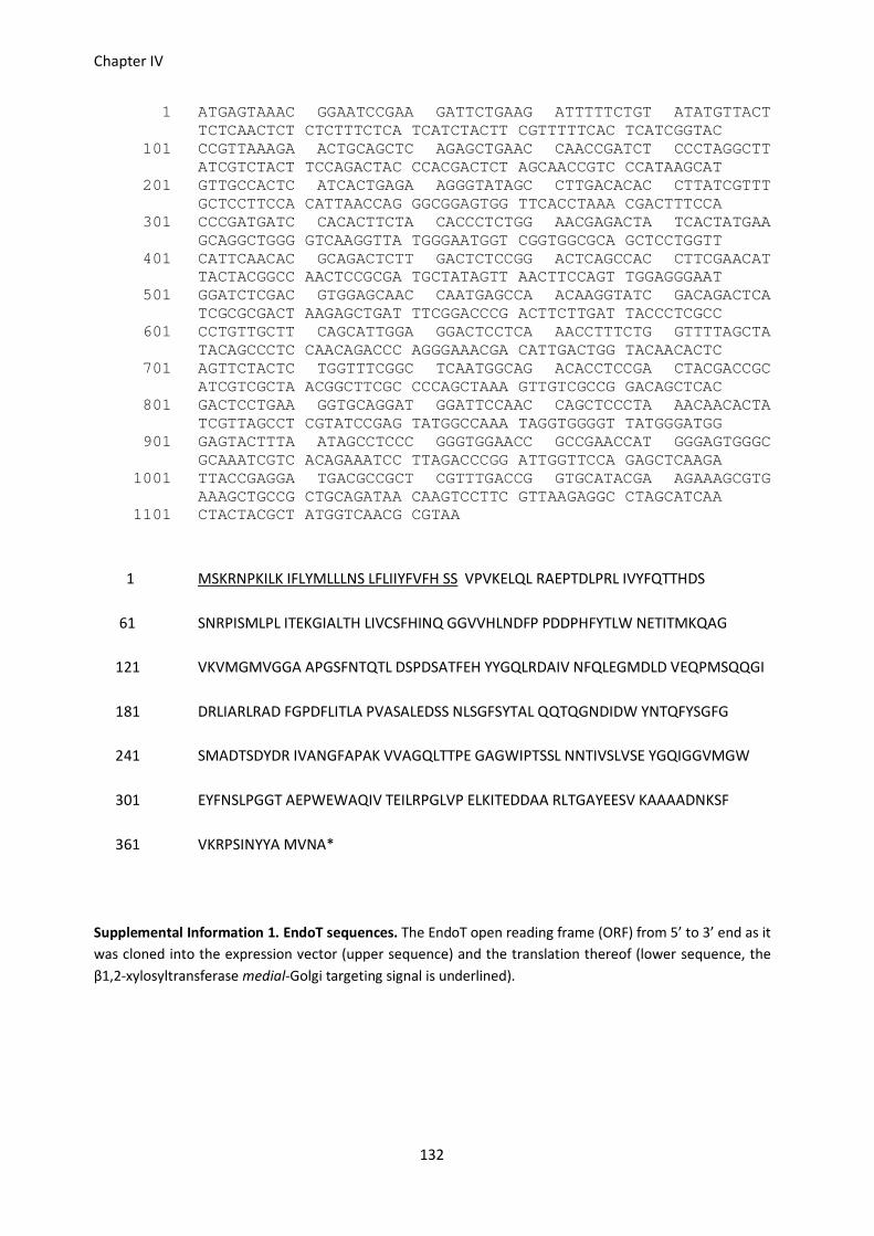
Chapter IV
132
1 ATGAGTAAAC GGAATCCGAA GATTCTGAAG ATTTTTCTGT ATATGTTACT TCTCAACTCT CTCTTTCTCA TCATCTACTT CGTTTTTCAC TCATCGGTAC
101 CCGTTAAAGA ACTGCAGCTC AGAGCTGAAC CAACCGATCT CCCTAGGCTT ATCGTCTACT TCCAGACTAC CCACGACTCT AGCAACCGTC CCATAAGCAT
201 GTTGCCACTC ATCACTGAGA AGGGTATAGC CTTGACACAC CTTATCGTTT GCTCCTTCCA CATTAACCAG GGCGGAGTGG TTCACCTAAA CGACTTTCCA
301 CCCGATGATC CACACTTCTA CACCCTCTGG AACGAGACTA TCACTATGAA GCAGGCTGGG GTCAAGGTTA TGGGAATGGT CGGTGGCGCA GCTCCTGGTT
401 CATTCAACAC GCAGACTCTT GACTCTCCGG ACTCAGCCAC CTTCGAACAT TACTACGGCC AACTCCGCGA TGCTATAGTT AACTTCCAGT TGGAGGGAAT
501 GGATCTCGAC GTGGAGCAAC CAATGAGCCA ACAAGGTATC GACAGACTCA TCGCGCGACT AAGAGCTGAT TTCGGACCCG ACTTCTTGAT TACCCTCGCC
601 CCTGTTGCTT CAGCATTGGA GGACTCCTCA AACCTTTCTG GTTTTAGCTA TACAGCCCTC CAACAGACCC AGGGAAACGA CATTGACTGG TACAACACTC
701 AGTTCTACTC TGGTTTCGGC TCAATGGCAG ACACCTCCGA CTACGACCGC ATCGTCGCTA ACGGCTTCGC CCCAGCTAAA GTTGTCGCCG GACAGCTCAC
801 GACTCCTGAA GGTGCAGGAT GGATTCCAAC CAGCTCCCTA AACAACACTA TCGTTAGCCT CGTATCCGAG TATGGCCAAA TAGGTGGGGT TATGGGATGG
901 GAGTACTTTA ATAGCCTCCC GGGTGGAACC GCCGAACCAT GGGAGTGGGC GCAAATCGTC ACAGAAATCC TTAGACCCGG ATTGGTTCCA GAGCTCAAGA
1001 TTACCGAGGA TGACGCCGCT CGTTTGACCG GTGCATACGA AGAAAGCGTG AAAGCTGCCG CTGCAGATAA CAAGTCCTTC GTTAAGAGGC CTAGCATCAA
1101 CTACTACGCT ATGGTCAACG CGTAA
1 MSKRNPKILK IFLYMLLLNS LFLIIYFVFH SS VPVKELQL RAEPTDLPRL IVYFQTTHDS
61 SNRPISMLPL ITEKGIALTH LIVCSFHINQ GGVVHLNDFP PDDPHFYTLW NETITMKQAG
121 VKVMGMVGGA APGSFNTQTL DSPDSATFEH YYGQLRDAIV NFQLEGMDLD VEQPMSQQGI
181 DRLIARLRAD FGPDFLITLA PVASALEDSS NLSGFSYTAL QQTQGNDIDW YNTQFYSGFG
241 SMADTSDYDR IVANGFAPAK VVAGQLTTPE GAGWIPTSSL NNTIVSLVSE YGQIGGVMGW
301 EYFNSLPGGT AEPWEWAQIV TEILRPGLVP ELKITEDDAA RLTGAYEESV KAAAADNKSF
361 VKRPSINYYA MVNA*
Supplemental Information 1. EndoT sequences. The EndoT open reading frame (ORF) from 5’ to 3’ end as it
was cloned into the expression vector (upper sequence) and the translation thereof (lower sequence, the
β1,2-xylosyltransferase medial-Golgi targeting signal is underlined).

Glycoengineering in Arabidopsis seeds, results.
133
1 ATGGCAAACA AGCTTTTCCT CGTCTGCGCA ACTTTCGCCC TCTGCTTCCT CCTCACCAAC GCCGGTTTTT GCTGCCCAGC CGATCTCAAC CAGACTGATG
101 AGGCAAGAAA GATCTTCCTC GATTTTCACA ACCAAGTTCG CCGTGATATC GCAGGTGCCA GCCCGTTGCT CAACCTCACC GGAGCTGTTC AGATGCGAAA
201 CGTTCTCGGT CCAGCTAAGA ACATGTACAG AATGGACTGG GACTGCAACC TCGAAGCCAA AGCAAAGGCC ATGATCTGGC CATGCACTAC CCCTCTCCCA
301 ATCGACACGA GCATTCCACA GAACCTCGCT CAGTGGCTAC TTTTCCAAAA CAGCCAGGAA AACGAGGTCT TGACCCAAAC GCCCTGGTCT TGGGTCACCG
401 CATCACTAAG AAATCTTCAA CCTGATACAG AGGCTAACAT CTACAACTGG CAGATTAGAC CACTATCCAA CATCGCTAAC TGGCAGAACC TAAAAGTTGG
501 ATGTGCTCAC AAGGTTTGCA AATTCCCCAC CGGGACAAAC ATGGTTGTGT CTTGCGCTTA CGGCGGCGAG GTCCTCCAGG ATAACGAAGT TGTCTGGGAC
601 AAGGGACCAA CTTGCATGTG CAACGCTTAC CCCAACAGCT TCTGCTGCAA CAATCTCTGT GACACAATCG CTGCTGCAAC ACTTCGCAAC CAGCCTTGCA
701 AGAGCACTTG A
1 MANKLFLVCA TFALCFLLTN A GFCCPADLN QTDEARKIFL DFHNQVRRDI AGASPLLNLT
61 GAVQMRNVLG PAKNMYRMDW DCNLEAKAKA MIWPCTTPLP IDTSIPQNLA QWLLFQNSQE
121 NEVLTQTPWS WVTASLRNLQ PDTEANIYNW QIRPLSNIAN WQNLKVGCAH KVCKFPTGTN
181 MVVSCAYGGE VLQDNEVVWD KGPTCMCNAY PNSFCCNNLC DTIAAATLRN QPCKST*
Supplemental information 2. Recombinant ASP1 sequences. The ASP1 ORF from 5’ to 3’ end as it was
cloned into the expression vector (upper sequence) and the translation thereof (lower sequence, the 2S
ER-signal peptide is underlined).

134

135
Chapter V
Conclusions & perspectives.
Robin Piron, Ann Depicker and Nico Callewaert.

136
R.P. wrote the chapter and A. D. and N.C. edited it.

Conclusions & perspectives
137
Vaccine production in Arabidopsis seeds
Since its discovery the PRRSV has spread rapidly, becoming endemic in almost all pig-rearing countries
around the world, and is nowadays considered to be the most important pathogen of swine. Whereas
older strains would mainly be problematic for pregnant sows and piglets, the situation is nowadays
further aggravated by the fact that lately also highly pathogenic strains have been appearing that
cause severe symptoms among pigs of all ages and have a high incidence of mortality. Vaccines are
being used to minimize the clinical impact of an infection but because of the genetic drift of the virus,
these constantly loose their power and it has been proposed that a whole battery of complementary,
adaptable vaccines will be needed to effectively combat the virus. A subunit vaccine, that is safe and
contains epitopes important in protective immunity, would be of good use in combination with the
exisiting attenuated vaccines.
We wanted to evaluate the capacity of the Arabidopsis seed-platform, previously used to obtain high
seed specific expression of a range of heterologous proteins, for vaccine production. To this goal we
selected the GP3, GP4 and GP5 of the PRRSV to be expressed in our system and included in a subunit
vaccine against the PRRSV. Viral antigens are complex, often difficult to express proteins and plants
could occupy a niche among the current platforms by producing these. However, antigens are
frequently plagued by low expressing levels that in their turn can obstruct further vaccine
development. Indeed, when PRRSV antigens were expressed in different plant species they showed
very low accumulation levels that did not even reach the 1% of TSP threshold. We set out to tackle
this issue by the rational design of antigen formats and fusions that were aimed at augmenting the
accumulation of the antigens in the seed.
Several strategies were tested and these included removal of transmembrane domains and/or
coupling the antigens to stabilizing protein domains such as the GFP and IgG Fc-domains. We showed
that the antigens as such do not accumulate very well in our system, hereby confirming their difficult
to produce nature, but that by select optimization their accumulation could be boosted by multiple
factors resulting in several antigens with yields that exceeded the 1% of TSP threshold, a feature that
hasn’t been attained by any previous study generating PRRSV antigens in planta. The results further
show that the viral antigens can correctly be produced in the ER, with the additional benefit that
high-mannose glycans are present on the antigens that might direct these to antigen presenting cells,
and that the added Fc-domains have a double role and also effectively serve as affinity handle besides
increasing accumulation.
We delivered proof-of-principle of the immunogenicity of purified seed-produced antigens by showing
that virus-neutralizing antibodies are generated in mice after injection of a GP4 derived antigen, which

Chapter V
138
is similar to the in vivo situation were GP4 is also an important target for neutralizing antibodies. Alas,
this could not be reproduced during a preliminary trial in piglets that had received an antigen cocktail.
An in-depth experiment with challenge is required to shed light on this matter and to dissect the role
of each antigen in more detail and evaluate if the antigens have, for example, a priming effect that
stimulates a faster immune reponse after challenge. Perhaps the presence of an immunodominant,
non-neutralizing B-cell epitope in the antigen cocktail skewed the induction of antibodies towards
non-neutralizing epitopes. Indeed, for the GP5 such a decoy epitope has been identified (Ostrowski et
al., 2002) and this might have affected the antibody response when it was administered together with
the GP3 and GP4 derived antigens. Again, a challenge experiment could assess the role of each antigen
alone and determine which antigens offer good protective immunity.
Another interesting venue is offered by the Fc-domain that is carried by the antigens that were
administered to the piglets and that is derived from the porcine IgG3. In silico prediction hints at a
good binding of the Fc-domain to the neonatal Fc receptor (FcRn) and experimental evidence seems to
support this statement (Butler et al., 2009; Virdi et al., 2013). An alternative route of antigen
administration could thus be envisaged where the antigens would be delivered intranasally and
associate with the FcRns present in the mucosa via binding of their IgG3 Fc-domain, resulting in their
effective uptake. The feasibility of this approach to mucosal vaccination was shown in mice by Ye et al.
(2011) who could generate both a mucosal as a systemic B and T-cell response against a model antigen
fused to an IgG Fc-domain when this was administered intranasally together with the adjuvant CpG. As
the PRRSV is primarily transmitted via the respiratory route, a mucosal immune response would be a
great first line of defence, which is a feature desirable for all PRRSV vaccines, and this merits further
investigation.
All in all, our results confirm that the PRRSV antigens are poor accumulators in planta, and show that
by mimicking the structures that do produce well in our system, i.e. antibodies, via fusion of the
antigens to Fc-domains (and the GFP) their accumulation could be considerably increased. For some,
but not all antigens, yields exceeded the 1% of TSP deemed necessary for economic viability of a PMP.
The results also show that no general optimization rule fits all antigens, however, most antigens do
have the tendency to accumulate better after fusion to stabilizing domains. Antigens could be
efficiently purified and were immunogenic, at least in mice, but their protective capacity in pigs
remains to be elucidated.
To formulate a straightforward conclusion concerning the suitability of the seed-platform for the
production of antigens with the final intention of generation a functional vaccine, is a more complex
matter. The obtained results show that, with respect to accumulation and purification, antigens can

Conclusions & perspectives
139
indeed be produced in the seeds. The optimized antigens could be purified via a straightforward
protocol and taking into consideration that downstream processing represents the bulk cost
associated with therapeutic protein production, this turns out favourable. However, the protective
capacity of the seed-produced antigens remains to be elucidated and this is as crucial as high yield and
efficient purification. It determines factors such as the dose that is required per immunization and this
in its turn directly influences the amount of antigen that needs to be produced and hence, the price
tag associated with the final vaccine. To conclude, antigen production in the seed and purification
from the latter is feasible, as shown by our results, but this not necessarily translates into an
efficacious and affordable vaccine and the suitability of the seed platform for the production of such a
vaccine must thus be evaluated on a case-by-case basis and will be determined by the outcome of the
interplay between the factors antigen immunogenicity, accumulation and downstream processing.

Chapter V
140
Glycoengineering Arabidopsis seeds
Plants have distinct advantages over other systems for the production of recombinant proteins: they
are capable of complex protein synthesis, they do not harbor animal and human pathogens,
production is easily scalable for most systems and possibly, also cheap. One major drawback however,
is that the plant and human N-glycosylation pathways diverge during the later stages of glycan
maturation in the Golgi apparatus and this results in the introduction of specific structures on complex
plant glycans that are not encountered on human glycans and hence, are possibly immunogenic.
Indeed, a large part of the human population has antibodies against the plant specific glycans
β1,2-xylose and core α1,3-fucose (Bardor et al., 2003; Mari, 2002) and this can prove problematic
when a therapeutic protein bearing these glycans is delivered into the blood stream. Binding of these
glycan specific antibodies can result in rapid clearance of the therapeutic, but in the worst case
scenario, this immunogenicity might provoke a life threatening anaphylactic shock.
Given the importance of these immunogenic glycans, a plethora of strategies have been employed to
‘humanize’ plant glycans, i.e. to avoid the synthesis of immunogenic glycans and to convert plant
glycosylation pathways to human-like synthesis. These glycoengineering techniques range from
relatively straightforward retention of a recombinant protein in the ER, to the more elaborate
inactivation of endogenous plant glycosyltransferases, which has often been coupled to the
concomitant introduction of heterologous transferases into the plant glycosylation machinery.
Needless to say, these are tedious strategies that require the coordinated expression of several genes
at the same time and at the right place. These endeavours are further complicated by the polyploidy of
many crop species, requiring decade long breeding strategies to obtain homozygous lines for all
knock-outs and transgenes. We, on the other hand, have developed a straightforward system that
bypasses extensive manipulations of the glycosylation pathway by seed-specific expression of a single
fungal gene in a selected Arabidopsis thaliana mutant lacking just one endogenous glycosyltransferase.
The resulting so-called ‘GlycoDelete’ seed-platform permits the production of any therapeutic
glycoprotein and delivers the latter as one glycoform, bearing N-glycans that are trimmed down to a
single GlcNAc, completely devoid of immunogenic β1,2-xylose and core α1,3-fucose.
To create such a platform, we selected the endoT, based on previous successes in mammalian cells,
which is an endoglycosidase from the fungus Trichoderma reesei that cleaves high-mannose and
hybrid-type N-glycans between the two protein-proximal β1,4-linked GlcNAc residues. We fused it to a
plant Golgi glycosyltransferase targeting signal and introduced it in the Arabidopsis cgl mutant whose
proteins carry high-mannose glycans only, which are an excellent substrate for the endoT. We show
that upon seed-specific expression, active endoT is produced that can digest the glycans of both

Conclusions & perspectives
141
endogenous seed proteins and those of a reporter protein that was also introduced in the cgl seeds
under control of the same regulary elements used to drive endoT expression. Although the endoT
content of the seeds was rather low, it was sufficient to entirely process a ten-fold excess of the
reporter protein ASP1. The yield of the recombinant ASP1, an immunogenic protein from the helminth
Ostertagia ostertagi, produced in the GlycoDelete seeds, was considerably lower than that of the ASP1
produced in seeds lacking endoT but the exact cause of this observation requires further in-depth
studies. It should be noted that the ASP1 expression cassette was introduced independently in the cgl
mutant and the Glycodelete background (cgl mutant plus endoT). Consequently, the lower expression
might well be due to a clonal variation. The ASP1 has a high vaccine potential to protect cattle from O.
ostertagi and because of difficulties in isolating large quantities of ASP1 from Ostertagia itself, there is
a great interest to produce recombinant ASP1. However, recombinant versions of the ASP1 seem to
lack some essential structural element, rendering them non-protective. Indications are at hand that
also the seed-produced ASP1s are structurally divergent from the native ASP1 (neutralizing antibodies
induced by the native ASP1 do not recognize recombinant ASP1 from wild type seeds). Further
investigation is needed to elucidate the precise nature of this critical element and to determine if the
seed-produced ASP1s also lack this element. One way to easily obtain additional information on this
topic would be to check if the antibodies present in the sera of the rabbits that have been immunized
with the wild type and the GlycoDelete ASP1 and bind to the peptide backbone of the seed-produced
ASP1, are also capable of recognizing the native ASP1, e.g. by competitive ELISA. In case that protective
capacity could be demonstrated for the seed-produced ASP1s, a recombinant vaccine against
Ostertagia ostertagi based on the seed-produced ASP1s can surely be envisaged, given that the high
accumulation of the ASP1s in the seed, combined with the optimized purification protocol would much
facilitate ASP1 production.
In-depth study of the glycans present on the different seed-produced ASP1s revealed that the glycan
profiles of the recombinant ASP1s are in complete accordance with the expected profiles: complex,
immunogenic plant glycans for the wild type ASP1, high-mannose glycans for the cgl ASP1 and
trimmed glycans consisting of a single GlcNAc on the GlycoDelete ASP1. Rabbits were injected with
wild type and GlycoDelete ASP1 to investigate the immunogenicity of the glycans present on the
ASP1s. Indeed, the majority, but not all of the rabbits immunized with wild type ASP1 had generated
plant glycan specific antibodies that were completely absent in the sera of the GlycoDelete ASP1
immunized group. These results confirm the immunogenic potential of the plant specific β1,2-xylose
and core α1,3-fucose so acclaimed in literature and validate the efforts dedicated to ‘humanizing’
plant glycans. It also, at least in rabbits, points to genetic predisposition as an important factor in
determining the outcome of exposure to these potentially immunogenic glycans. Last but not least, it

Chapter V
142
convincingly demonstrates that the GlycoDelete seed-platform can be used for the in planta
production of therapeutic proteins with a homogeneous glycan profile lacking immunogenic plant
glycans.
The GlycoDelete seed platform has several unique advantages. The seed specific expression of the
endoT (and the target protein) avoids interference with normal plant development and bestows the
platform with the additional benefits associated with seed production of PMPs, such as accumulation
of the PMP in a small volume and stable environment and the possibility to store the unprocessed
seeds for prolonged periods of time, which allows decoupling of PMP production and processing. The
platform is simple and only requires supertransformation of the GlycoDelete line with a gene of
interest under control of the same regulatory elements used to drive the endoT gene. The system is
robust and was shown to completely digest a recombinant protein present in ten-fold excess over the
endoT, which translates into a minimal processing capacity of 7 mg recombinant protein per gram dry
seed weight (3.5% of TSP) and possibly much more. The final and perhaps most important advantage is
that it fully accomplishes the goals that were set during its conception, namely that it produces a
therapeutic protein bearing short glycans, important to maintain its native conformation, that are
homogeneous and at the same time also completely lack the immunogenic β1,2-xylose and
α1,3-fucose.
The GlycoDelete seed-platform could be used as such for the commercial production of PMPs. The
small stature of the Arabidopsis host facilitates cultivation in greenhouses were it can be densely
packed and contained. Methods for the automatic harvesting and sowing of Arabidopsis seeds are
under investigation and it has been estimated that Arabidopsis grown in a greenhouse facility could
generate somewhere from 180 to 240 grams of dry seeds per square meter each year (He et al., 2013;
Loos et al., 2011a). Using its minimal capacity as a conservative estimate, this would correspond to 1.2
to 1.6 grams of endoT digested PMP produced in the GlycoDelete seed platform per square meter on a
yearly basis. Alternatively, the GlycoDelete technology could be transferred to another crop, such as
pea, whose seeds have an even higher protein content that can account for up to 40% of seed weight.
Work is underway in our lab to establish if expression patterns of synthetic IgAs produced in
Arabidopsis seeds using the seed-specific expression cassette can be translated to pea (Dr. V. Virdi,
personal communication). If the expression cassette functions similarly in pea then it would be feasible
to transfer the GlycoDelete technology as such to pea and although a mutant lacking GnTI activity is
not available, such a mutant could be created via meganuclease (or similar) technology.
Another interesting venue would be to check if the endoT could be transiently expressed in, for
example, Nicotiana benthamiana leaves, as has been tested for the bacterial PNGaseF that was able to

Conclusions & perspectives
143
completely deglycosylate co-expressed reporter proteins (Mamedov et al., 2012; Mamedov & Yusibov,
2013). PNGaseF has a broader substrate specificity than endoT, but it will nevertheless be interesting
to determine if the endoT can also function as an integral part of a transient expression system. The
latter represents another potential application of the endoT, especially because of the current
industrial interest in platforms based on transient production of PMPs. As becomes clear from this
discussion, there are several valid options available to continue with the application of the GlycoDelete
technology in planta.
In conclusion, we have designed and engineered a platform for the seed-specific production of
therapeutic proteins in planta that carry minimal glycans which are necessary to maintain the overall
protein fold but that at the same time are homogeneous and completely devoid of the immunogenic
plant specific structures β1,2-xylose and α1,3-fucose. We demonstrate the full functionality of the
system and prove that a minimally glycosylated version of a recombinant helminth antigen produced
in the platform does not provoke an anti-glycan response upon immunization of rabbits whereas its
recombinant counterpart produced in non-engineered, wild type seeds does, hereby confirming the
immunogenicity of these plant glycan structure and validating the potential of the GlycoDelete
seed-platform. The system has several advantages over other glycoengineered solutions to ‘humanize’
plant glycans and can be used as such for industrial applications. Alternatively, the GlycoDelete
technology can be tailored to meet specific needs and there are several options for further application
of the technology in planta.

144
References
Aalberse RC, van Ree R (1996) Cross-reactive carbohydrate determinants. Monogr Allergy 32: 78-83
Albina E, Carrat C, Charley B (1998) Interferon-alpha response to swine arterivirus (PoAV), the porcine reproductive and respiratory syndrome virus. J Interferon Cytokine Res 18: 485-490
Altmann F (2007) The role of protein glycosylation in allergy. Int Arch Allergy Immunol 142: 99-115
Alvarez ML, Cardineau GA (2010) Prevention of bubonic and pneumonic plague using plant-derived
vaccines. Biotechnol Adv 28: 184-196
An TQ, Tian ZJ, Xiao Y, Li R, Peng JM, Wei TC, Zhang Y, Zhou YJ, Tong GZ (2010) Origin of Highly
Pathogenic Porcine Reproductive and Respiratory Syndrome Virus, China. Emerg Infect Dis 16: 365-367
Ansari IH, Kwon B, Osorio FA, Pattnaik AK (2006) Influence of N-linked glycosylation of porcine
reproductive and respiratory syndrome virus GP5 on virus infectivity, antigenicity, and ability to induce
neutralizing antibodies. J Virol 80: 3994-4004
Anthony RM, Nimmerjahn F, Ashline DJ, Reinhold VN, Paulson JC, Ravetch JV (2008) Recapitulation of
IVIG anti-inflammatory activity with a recombinant IgG fc. Science 320: 373-376
Aviezer D, Brill-Almon E, Shaaltiel Y, Hashmueli S, Bartfeld D, Mizrachi S, Liberman Y, Freeman A,
Zimran A, Galun E (2009) A Plant-Derived Recombinant Human Glucocerebrosidase Enzyme-A
Preclinical and Phase I Investigation. PLoS ONE 4: e4792
Bakker H, Bardor M, Molthoff JW, Gomord V, Elbers I, Stevens LH, Jordi W, Lommen A, Faye L, Lerouge
P, Bosch D (2001) Galactose-extended glycans of antibodies produced by transgenic plants. Proc Natl
Acad Sci USA 98: 2899-2904
Bakker H, Rouwendal GJ, Karnoup AS, Florack DE, Stoopen GM, Helsper JP, van Ree R, van Die I, Bosch
D (2006) An antibody produced in tobacco expressing a hybrid beta-1,4-galactosyltransferase is essentially devoid of plant carbohydrate epitopes. Proc Natl Acad Sci USA 103: 7577-7582
Bardor M, Faveeuw C, Fitchette AC, Gilbert D, Galas L, Trottein F, Faye L, Lerouge P (2003)
Immunoreactivity in mammals of two typical plant glyco-epitopes, core alpha(1,3)-fucose and core
xylose. Glycobiology 13: 427-434
Bazin H (2011) Vaccination--A History: From Lady Montagu to Genetic Engineering: John Libbey
Eurotext.

145
Boothe J, Nykiforuk C, Shen Y, Zaplachinski S, Szarka S, Kuhlman P, Murray E, Morck D, Moloney MM
(2010) Seed-based expression systems for plant molecular farming. Plant Biotechnol J 8: 588-606
Borloo J, Geldhof P, Peelaers I, Van Meulder F, Ameloot P, Callewaert N, Vercruysse J, Claerebout E,
Strelkov SV, Weeks SD (2013) Structure of Ostertagia ostertagi ASP-1: insights into disulfide-mediated
cyclization and dimerization. Acta Crystallogr Sect D-Biol Crystallogr 69: 493-503
Bosch D, Castilho A, Loos A, Schots A, Steinkellner H (2013) N-Glycosylation of Plant-produced
Recombinant Proteins. Curr Pharm Design 19: 5503-5512
Bosch D, Schots A (2010) Plant glycans: friend or foe in vaccine development? Expert Rev Vaccines 9:
835-842
Bublin M, Radauer C, Wilson IB, Kraft D, Scheiner O, Breiteneder H, Hoffmann-Sommergruber K (2003)
Cross-reactive N-glycans of Api g 5, a high molecular weight glycoprotein allergen from celery, are
required for immunoglobulin E binding and activation of effector cells from allergic patients. FASEB J
17: 1697-1699
Buck PM, Kumar S, Singh SK (2013) Consequences of glycan truncation on Fc structural integrity. mAbs
5: 904-916
Buonaguro FM, Butler-Ransohoff JE (2010) PharmaPlant: the new frontier in vaccines. Forward. Expert
Rev Vaccines 9: 805-807
Butler JE, Wertz N, Deschacht N, Kacskovics I (2009) Porcine IgG: structure, genetics, and evolution.
Immunogenetics 61: 209-230
Campbell CT, Yarema KJ (2005) Large-scale approaches for glycobiology. Genome Biol 6: 236
Castilho A, Gattinger P, Grass J, Jez J, Pabst M, Altmann F, Gorfer M, Strasser R, Steinkellner H (2011)
N-glycosylation engineering of plants for the biosynthesis of glycoproteins with bisected and branched
complex N-glycans. Glycobiology 21: 813-823
Castilho A, Strasser R, Stadlmann J, Grass J, Jez J, Gattinger P, Kunert R, Quendler H, Pabst M, Leonard
R, Altmann F, Steinkellner H (2010) In planta protein sialylation through overexpression of the
respective mammalian pathway. J Biol Chem 285: 15923-15930
Chan HT, Chia MY, Pang VF, Jeng CR, Do YY, Huang PL (2013) Oral immunogenicity of porcine
reproductive and respiratory syndrome virus antigen expressed in transgenic banana. Plant Biotechnol
J 11: 315-324

146
Chargelegue D, Vine ND, van Dolleweerd CJ, Drake PMW, Ma JKC (2000) A murine monoclonal
antibody produced in transgenic plants with plant-specific glycans is not immunogenic in mice.
Transgenic Res 9: 187-194
Chen X, Liu J (2011) Generation and immunogenicity of transgenic potato expressing the GP5 protein
of porcine reproductive and respiratory syndrome virus. J Virol Methods 173: 153-158
Chen ZL, Schuler MA, Beachy RN (1986) Functional-Analysis of Regulatory Elements in a Plant Embryo-
Specific Gene. Proc Natl Acad Sci USA 83: 8560-8564
Chia M-Y, Hsiao S-H, Chan H-T, Do Y-Y, Huang P-L, Chang H-W, Tsai Y-C, Lin C-M, Pang VF, Jeng C-R
(2011) Evaluation of the immunogenicity of a transgenic tobacco plant expressing the recombinant
fusion protein of GP5 of porcine reproductive and respiratory syndrome virus and B subunit of
Escherichia coli heat-labile enterotoxin in pigs. Vet Immunol Immunopathol 140: 215-225
Chia M-Y, Hsiao S-H, Chan H-T, Do Y-Y, Huang PL, Chang HW, Tsai Y-C, Lin C-M, Pang VF, Jeng C-R
(2010) Immunogenicity of recombinant GP5 protein of porcine reproductive and respiratory syndrome
virus expressed in tobacco plant. Vet Immunol Immunopathol 135: 234-242
Chung CH, Mirakhur B, Chan E, Le Q, Berlin J, Morse M, Murphy BA, Satinover SM, Hosen J, Mauro D,
Slebos RJ, Zhou QW, Gold D, Hatley T, Hicklin DJ, Platts-Mills TAE (2008) Cetuximab-induced anaphylaxis and IgE specific for galactose-alpha-1,3-galactose. N Engl J Med 358: 1109-1117
Collins JE, Benfield DA, Christianson WT, Harris L, Hennings JC, Shaw DP, Goyal SM, McCullough S,
Morrison RB, Joo HS, Gorcyca D, Chladek D (1992) Isolation of swine infertility and respiratory
syndrome virus (isolate ATCC VR-2332) in North America and experimental reproduction of the disease
in gnotobiotic pigs. J Vet Diagn Invest 4: 117-126
Costers S, Lefebvre DJ, Van Doorsselaere J, Vanhee M, Delputte PL, Nauwynck HJ (2010) GP4 of porcine
reproductive and respiratory syndrome virus contains a neutralizing epitope that is susceptible to
immunoselection in vitro. Arch Virol 155: 371-378
Cox KM, Sterling JD, Regan JT, Gasdaska JR, Frantz KK, Peele CG, Black A, Passmore D, Moldovan-
Loomis C, Srinivasan M, Cuison S, Cardarelli PM, Dickey LF (2006) Glycan optimization of a human
monoclonal antibody in the aquatic plant Lemna minor. Nat Biotechnol 24: 1591-1597
Cubitt AB, Heim R, Adams SR, Boyd AE, Gross LA, Tsien RY (1995) Understanding, improving and using
green fluorescent proteins. Trends Biochem Sci 20: 448-455
Czajkowsky DM, Hu J, Shao Z, Pleass RJ (2012) Fc-fusion proteins: new developments and future
perspectives. EMBO Mol Med 4: 1015-1028

147
Das PB, Dinh PX, Ansari IH, de Lima M, Osorio FA, Pattnaik AK (2010) The Minor Envelope
Glycoproteins GP2a and GP4 of Porcine Reproductive and Respiratory Syndrome Virus Interact with
the Receptor CD163. J Virol 84: 1731-1740
De Buck S, Nolf J, De Meyer T, Virdi V, De Wilde K, Van Lerberge E, Van Droogenbroeck B, Depicker A
(2013) Fusion of an Fc chain to a VHH boosts the accumulation levels in Arabidopsis seeds. Plant
Biotechnol J 11: 1006-1016
De Buck S, Virdi V, De Meyer T, De Wilde K, Piron R, Nolf J, Van Lerberge E, De Paepe A, Depicker A
(2012) Production of camel-like antibodies in plants. Methods Mol Biol 911: 305-324
De Jaeger G, Scheffer S, Jacobs A, Zambre M, Zobell O, Goossens A, Depicker A, Angenon G (2002)
Boosting heterologous protein production in transgenic dicotyledonous seeds using Phaseolus vulgaris
regulatory sequences. Nat Biotechnol 20: 1265-1268
De Wilde K, De Buck S, Vanneste K, Depicker A (2013) Recombinant Antibody Production in
Arabidopsis Seeds Triggers an Unfolded Protein Response. Plant Physiol 161: 1021-1033
Delputte PL, Nauwynck HJ (2004) Porcine Arterivirus infection of alveolar macrophages is mediated by
sialic acid on the virus. J Virol 78: 8094-8101
Delputte PL, Vanderheijden N, Nauwynck HJ, Pensaert MB (2002) Involvement of the matrix protein in
attachment of porcine reproductive and respiratory syndrome virus to a heparinlike receptor on
porcine alveolar macrophages. J Virol 76: 4312-4320
Diamond J (1997) Guns, Germs and Steel: The Fates of Human Societies, NY: WW Norton & Company.
Díaz I, Darwich L, Pappaterra G, Pujols J, Mateu E (2006) Different European-type vaccines against
porcine reproductive and respiratory syndrome virus have different immunological properties and
confer different protection to pigs. Virology 351: 249-259
Diaz I, Pujols J, Ganges L, Gimeno M, Darwich L, Domingo M, Mateu E (2009) In silico prediction and ex
vivo evaluation of potential T-cell epitopes in glycoproteins 4 and 5 and nucleocapsid protein of
genotype-I (European) of porcine reproductive and respiratory syndrome virus. Vaccine 27: 5603-5611
Downing WL, Galpin JD, Clemens S, Lauzon SM, Samuels AL, Pidkowich MS, Clarke LA, Kermode AR
(2006) Synthesis of enzymatically active human alpha-L-iduronidase in Arabidopsis cgl (complex
glycan-deficient) seeds. Plant Biotechnol J 4: 169-181
Downing WL, Hu XK, Kermode AR (2007) Post-transcriptional factors are important for high-level expression of the human alpha-L-iduronidase gene in Arabidopsis cgl (complex-glycan-deficient) seeds.
Plant Sci 172: 327-334

148
Druzhinina IS, Seidl-Seiboth V, Herrera-Estrella A, Horwitz BA, Kenerley CM, Monte E, Mukherjee PK,
Zeilinger S, Grigoriev IV, Kubicek CP (2011) Trichoderma: the genomics of opportunistic success. Nat
Rev Microbiol 9: 749-759
Duran JP, Climent I, Sarraseca J, Urniza A, Cortés E, Vela C, Casal JI (1997) Baculovirus expression of
proteins of porcine reproductive and respiratory syndrome virus strain Olot/91. Involvement of ORF3
and ORF5 proteins in protection. Virus Genes 14: 19-29
Engering AJ, Cella M, Fluitsma D, Brockhaus M, Hoefsmit ECM, Lanzavecchia A, Pieters J (1997) The
mannose receptor functions as a high capacity and broad specificity antigen receptor in human
dendritic cells. Eur J Immunol 27: 2417-2425
Fang Y, Snijder EJ (2010) The PRRSV replicase: Exploring the multifunctionality of an intriguing set of
nonstructural proteins. Virus Res 154: 61-76
Faye L, Boulaflous A, Benchabane M, Gomord W, Michaud D (2005) Protein modifications in the plant
secretory pathway: current status and practical implications in molecular pharming. Vaccine 23: 1770-
1778
Fenner F, Henderson DA, Arita I, Jezek Z, Ladnyi ID (1988) Smallpox and its eradication, Vol. 6: World
Health Organization Geneva.
Fitchette-Laine AC, Gomord V, Cabanes M, Michalski JC, Saint Macary M, Foucher B, Cavelier B, Hawes
C, Lerouge P, Faye L (1997) N-glycans harboring the Lewis a epitope are expressed at the surface of
plant cells. Plant J 12: 1411-1417
Foetisch K, Westphal S, Lauer I, Retzek M, Altmann F, Kolarich D, Scheurer S, Vieths S (2003) Biological activity of IgE specific for cross-reactive carbohydrate determinants. J Allergy Clin Immunol 111: 889-
896
Forthal DN, Gach JS, Landucci G, Jez J, Strasser R, Kunert R, Steinkellner H (2010) Fc-Glycosylation
Influences Fc gamma Receptor Binding and Cell-Mediated Anti-HIV Activity of Monoclonal Antibody
2G12. J Immunol 185: 6876-6882
Frank J, Kaulfuerst-Soboll H, Rips S, Koiwa H, von Schaewen A (2008) Comparative Analyses of
Arabidopsis complex glycan1 Mutants and Genetic Interaction with staurosporin and temperature
sensitive3a. Plant Physiol 148: 1354-1367
Frazer IH (2004) Prevention of cervical cancer through papillomavirus vaccination. Nat Rev Immunol 4:
46-54
Fujiyama K, Sakai Y, Misaki R, Yanagihara I, Honda T, Anzai H, Seki T (2004) N-linked glycan structures
of human lactoferrin produced by transgenic rice. Biosci Biotechnol Biochem 68: 2565-2570

149
Geldhof MF, Vanhee M, Van Breedam W, Van Doorsselaere J, Karniychuk UU, Nauwynck HJ (2012)
Comparison of the efficacy of autogenous inactivated Porcine Reproductive and Respiratory Syndrome
Virus (PRRSV) vaccines with that of commercial vaccines against homologous and heterologous
challenges. BMC Vet Res 8: 182
Geldhof P, Claerebout E, Knox D, Vercauteren I, Looszova A, Vercruysse J (2002) Vaccination of calves
against Ostertagia ostertagi with cysteine proteinase enriched protein fractions. Parasite Immunol 24: 263-270
Geldhof P, Claerebout E, Knox DP, Agneessens J, Vercruysse J (2000) Proteinases released in vitro by
the parasitic stages of the bovine abomasal nematode Ostertagia ostertagi. Parasitology 121: 639-647
Geldhof P, Meyvis Y, Vercruysse J, Claerebout E (2008) Vaccine testing of a recombinant activation-associated secreted protein (ASP1) from Ostertagia ostertagi. Parasite Immunol 30: 57-60
Geldhof P, Vercauteren I, Gevaert K, Staes A, Knox DP, Vandekerckhove J, Vercruysse J, Claerebout E
(2003) Activation-associated secreted proteins are the most abundant antigens in a host protective
fraction from Ostertagia ostertagi. Mol Biochem Parasitol 128: 111-114
Geldhof P, Vercauteren I, Vercruysse J, Knox DP, Van den Broeck W, Claerebout E (2004) Validation of the protective Ostertagia ostertagi ES-thiol antigens with different adjuvantia. Parasite Immunol 26:
37-43
Giorgi C, Franconi R, Rybicki EP (2010) Human papillomavirus vaccines in plants. Expert Rev Vaccines 9:
913-924
Goding JW (1978) Use of staphylococcal protein A as an immunological reagent. J Immunol Methods
20: 241-253
Gomez L, Chrispeels MJ (1994) Complementation of an Arabidopsis-Thaliana Mutant That Lacks
Complex Asparagine-Linked Glycans with the Human Cdna-Encoding N-Acetylglucosaminyltransferase-
I. Proc Natl Acad Sci USA 91: 1829-1833
Gomord V, Denmat L-A, Fitchette-Lainé A-C, Satiat-Jeunemaitre B, Hawes C, Faye L (1997a) The C-
terminal HDEL sequence is sufficient for retention of secretory proteins in the endoplasmic reticulum
(ER) but promotes vacuolar targeting of proteins that escape the ER. Plant J 11: 313-325
Gomord V, Denmat LA, FitchetteLaine AC, SatiatJeunemaitre B, Hawes C, Faye L (1997b) The C-
terminal HDEL sequence is sufficient for retention of secretory proteins in the endoplasmic reticulum
(ER) but promotes vacuolar targeting of proteins that escape the ER. Plant J 11: 313-325

150
Gomord V, Faye L (2004) Posttranslational modification of therapeutic proteins in plants. Curr Opin
Plant Biol 7: 171-181
Gomord V, Fitchette AC, Menu-Bouaouiche L, Saint-Jore-Dupas C, Plasson C, Michaud D, Faye L (2010)
Plant-specific glycosylation patterns in the context of therapeutic protein production. Plant Biotechnol
J 8: 564-587
Goossens A, Dillen W, De Clercq J, Van Montagu M, Angenon G (1999) The arcelin-5 gene of Phaseolus
vulgaris directs high seed-specific expression in transgenic Phaseolus acutifolius and Arabidopsis
plants. Plant Physiol 120: 1095-1104
Granell A, Fernandez-del-Carmen A, Orzaez D (2010) In planta production of plant-derived and non-
plant-derived adjuvants. Expert Rev Vaccines 9: 843-858
He X, Galpin JD, Tropak MB, Mahuran D, Haselhorst T, von Itzstein M, Kolarich D, Packer NH, Miao YS,
Jiang LW, Grabowski GA, Clarke LA, Kermode AR (2012a) Production of active human
glucocerebrosidase in seeds of Arabidopsis thaliana complex-glycan-deficient (cgl) plants. Glycobiology
22: 492-503
He X, Haselhorst T, von Itzstein M, Kolarich D, Packer NH, Kermode AR (2012b) Influence of an ER-
retention signal on the N-glycosylation of recombinant human alpha-L-iduronidase generated in seeds of Arabidopsis. Plant Mol Biol Rep 79: 157-169
He X, Pierce O, Haselhorst T, von Itzstein M, Kolarich D, Packer NH, Gloster TM, Vocadlo DJ, Qian Y,
Brooks D, Kermode AR (2013) Characterization and downstream mannose phosphorylation of human
recombinant alpha-L-iduronidase produced in Arabidopsis complex glycan-deficient (cgl) seeds. Plant
Biotechnol J 11: 1034-1043
Herrera-estrella L, Depicker A, Vanmontagu M, Schell J (1983) Expression of Chimaeric Genes
Transferred into Plant-Cells Using a Ti-Plasmid-Derived Vector. Nature 303: 209-213
Hiatt A, Cafferkey R, Bowdish K (1989) Production of Antibodies in Transgenic Plants. Nature 342: 76-
78
Hu J, Ni Y, Dryman BA, Meng XJ, Zhang C (2012) Immunogenicity study of plant-made oral subunit
vaccine against porcine reproductive and respiratory syndrome virus (PRRSV). Vaccine 30: 2068-2074
Huether CM, Lienhart O, Baur A, Stemmer C, Gorr G, Reski R, Decker EL (2005) Glyco-engineering of
moss lacking plant-specific sugar residues. Plant Biol 7: 292-299
Jacobs PP, Callewaert N (2009) N-glycosylation Engineering of Biopharmaceutical Expression Systems.
Curr Mol Med 9: 774-800

151
Jin C, Altmann F, Strasser R, Mach L, Schahs M, Kunert R, Rademacher T, Glossl J, Steinkellner H
(2008a) A plant-derived human monoclonal antibody induces an anti-carbohydrate immune response
in rabbits. Glycobiology 18: 235-241
Jin C, Hantusch B, Hemmer W, Stadimann J, Altmann F (2008b) Affinity of IgE and IgG against cross-
reactive carbohydrate determinants on plant and insect glycoproteins. J Allergy Clin Immunol 121: 185-
190
Johnson CR, Griggs TF, Gnanandarajah J, Murtaugh MP (2011) Novel structural protein in porcine
reproductive and respiratory syndrome virus encoded by an alternative ORF5 present in all
arteriviruses. J Gen Virol 92: 1107-1116
Kaneko Y, Nimmerjahn F, Ravetch JV (2006) Anti-inflammatory activity of immunoglobulin G resulting
from Fc sialylation. Science 313: 670-673
Karimi M, Inzé D, Depicker A (2002) GATEWAYTM vectors for Agrobacterium-mediated plant
transformation. Trends Plant Sci 7: 193-195
Karniychuk UU, Geldhof M, Vanhee M, Van Doorsselaere J, Saveleva TA, Nauwynck HJ (2010)
Pathogenesis and antigenic characterization of a new East European subtype 3 porcine reproductive
and respiratory syndrome virus isolate. BMC Vet Res 6: 30
Key KF, Guenette DK, Yoon K-J, Halbur PG, Toth TE, Meng XJ (2003) Development of a heteroduplex
mobility assay to identify field isolates of porcine reproductive and respiratory syndrome virus with
nucleotide sequences closely related to those of modified live-attenuated vaccines. J Clin Microbiol 41:
2433-2439
Kimman TG, Cornelissen LA, Moormann RJ, Rebel JMJ, Stochofe-Zurwieden N (2009) Challenges for
porcine reproductive and respiratory syndrome virus (PRRSV) vaccinology. Vaccine 27: 3704-3718
Kirnbauer R, Booy F, Cheng N, Lowy DR, Schiller JT (1992) Papillomavirus L1 Major Capsid Protein Self-
Assembles into Virus-Like Particles That Are Highly Immunogenic. Proc Natl Acad Sci USA 89: 12180-
12184
Koncz C, Schell J (1986) The promoter of TL-DNA gene 5 controls the tissue-specific expression of
chimaeric genes carried by a novel type of Agrobacterium binary vector. Mol Gen Genet 204: 383-396
Koprivova A, Stemmer C, Altmann F, Hoffmann A, Kopriva S, Gorr G, Reski R, Decker EL (2004) Targeted
knockouts of Physcomitrella lacking plantspecific immunogenic N-glycans. Plant Biotechnol J 2: 517-
523

152
Krebbers E, Herdies L, De Clercq A, Seurinck J, Leemans J, Van Damme J, Segura M, Gheysen G, Van
Montagu M, Vandekerckhove J (1988) Determination of the processing sites of an Arabidopsis 2S
albumin and characterization of the complete gene family. Plant Physiol 87: 859-866
Kurosaka A, Yano A, Itoh N, Kuroda Y, Nakagawa T, Kawasaki T (1991) The Structure of a Neural
Specific Carbohydrate Epitope of Horseradish-Peroxidase Recognized by Anti-Horseradish Peroxidase
Antiserum. J Biol Chem 266: 4168-4172
Labarque GG, Nauwynck HJ, Van Reeth K, Pensaert MB (2000) Effect of cellular changes and onset of
humoral immunity on the replication of porcine reproductive and respiratory syndrome virus in the
lungs of pigs. J Gen Virol 81: 1327-1334
Lambrecht BN, Kool M, Willart MAM, Hammad H (2009) Mechanism of action of clinically approved
adjuvants. Curr Opin Immunol 21: 23-29
Laroy W, Contreras R, Callewaert N (2006) Glycome mapping on DNA sequencing equipment. Nat
Protoc 1: 397-405
Latypov RF, Hogan S, Lau H, Gadgil H, Liu D (2012) Elucidation of acid-induced unfolding and
aggregation of human immunoglobulin IgG1 and IgG2 Fc. J Biol Chem 287: 1381-1396
Lee C, Yoo D (2006) The small envelope protein of porcine reproductive and respiratory syndrome
virus possesses ion channel protein-like properties. Virology 355: 30-43
Lentz EM, Segretin ME, Morgenfeld MM, Wirth SA, Santos MJD, Mozgovoj MV, Wigdorovitz A, Bravo-
Almonacid FF (2010) High expression level of a foot and mouth disease virus epitope in tobacco
transplastomic plants. Planta 231: 387-395
Liebminger E, Veit C, Pabst M, Batoux M, Zipfel C, Altmann F, Mach L, Strasser R (2011) beta-N-
Acetylhexosaminidases HEXO1 and HEXO3 Are Responsible for the Formation of Paucimannosidic N-
Glycans in Arabidopsis thaliana. J Biol Chem 286: 10793-10802
Loos A, Van Droogenbroeck B, Hillmer S, Grass J, Kunert R, Cao J, Robinson DG, Depicker A, Steinkellner
H (2011a) Production of monoclonal antibodies with a controlled N-glycosylation pattern in seeds of Arabidopsis thaliana. Plant Biotechnol J 9: 179-192
Loos A, Van Droogenbroeck B, Hillmer S, Grass J, Pabst M, Castilho A, Kunert R, Liang M, Arcalis E,
Robinson DG, Depicker A, Steinkellner H (2011b) Expression of antibody fragments with a controlled N-
glycosylation pattern and induction of endoplasmic reticulum-derived vesicles in seeds of Arabidopsis.
Plant Physiol 155: 2036-2048
Lopez OJ, Osorio FA (2004) Role of neutralizing antibodies in PRRSV protective immunity. Vet Immunol
Immunopathol 102: 155-163

153
Ma JKC, Drake PMW, Christou P (2003) The production of recombinant pharmaceutical proteins in
plants. Nat Rev Genet 4: 794-805
Ma JKC, Hikmat BY, Wycoff K, Vine ND, Chargelegue D, Yu L, Hein MB, Lehner T (1998) Characterization
of a recombinant plant monoclonal secretory antibody and preventive immunotherapy in humans. Nat
Med 4: 601-606
Maley F, Trimble RB, Tarentino AL, Plummer TH, Jr. (1989) Characterization of glycoproteins and their
associated oligosaccharides through the use of endoglycosidases. Anal Biochem 180: 195-204
Mamedov T, Ghosh A, Jones RM, Mett V, Farrance CE, Musiychuk K, Horsey A, Yusibov V (2012)
Production of non-glycosylated recombinant proteins in Nicotiana benthamiana plants by co-
expressing bacterial PNGase F. Plant Biotechnol J 10: 773-782
Mamedov T, Yusibov V (2013) In vivo deglycosylation of recombinant proteins in plants by co-
expression with bacterial PNGase F. Bioengineered 4: 338-342
Mardassi H, Massie B, Dea S (1996) Intracellular synthesis, processing, and transport of proteins
encoded by ORFs 5 to 7 of Porcine Reproductive and Respiratory Syndrome Virus. Virology 221: 98-112
Mari A (2002) IgE to cross-reactive carbohydrate determinants: Analysis of the distribution and
appraisal of the in vivo and in vitro reactivity. Int Arch Allergy Immunol 129: 286-295
Mari A, Ooievaar-de Heer P, Scala E, Giani M, Pirrotta L, Zuidmeer L, Bethell D, van Ree R (2008)
Evaluation by double-blind placebo-controlled oral challenge of the clinical relevance of IgE antibodies
against plant glycans. Allergy 63: 891-896
Marillonnet S, Giritch A, Gils M, Kandzia R, Klimyuk V, Gleba Y (2004) In planta engineering of viral RNA
replicons: efficient assembly by recombination of DNA modules delivered by Agrobacterium. Proc Natl
Acad Sci USA 101: 6852-6857
Martinez D, Berka RM, Henrissat B, Saloheimo M, Arvas M, Baker SE, Chapman J, Chertkov O, Coutinho
PM, Cullen D, Danchin EGJ, Grigoriev IV, Harris P, Jackson M, Kubicek CP, Han CS, Ho I, Larrondo LF, de Leon AL, Magnuson JK, Merino S, Misra M, Nelson B, Putnam N, Robbertse B, Salamov AA, Schmoll M,
Terry A, Thayer N, Westerholm-Parvinen A, Schoch CL, Yao J, Barabote R, Nelson MA, Detter C, Bruce
D, Kuske CR, Xie G, Richardson P, Rokhsar DS, Lucas SM, Rubin EM, Dunn-Coleman N, Ward M, Brettin
TS (2008) Genome sequencing and analysis of the biomass-degrading fungus Trichoderma reesei (syn.
Hypocrea jecorina) (vol 26, pg 553, 2008). Nat Biotechnol 26: 1193-1193
Mcaleer WJ, Buynak EB, Maigetter RZ, Wampler DE, Miller WJ, Hilleman MR (1984) Human Hepatitis-B
Vaccine from Recombinant Yeast. Nature 307: 178-180

154
Meulenberg JJ, van Nieuwstadt AP, van Essen-Zandbergen A, Langeveld JP (1997a) Posttranslational
processing and identification of a neutralization domain of the GP4 protein encoded by ORF4 of
Lelystad virus. J Virol 71: 6061-6067
Meulenberg JJM, Demeijer EJ, Moormann RJM (1993a) Subgenomic Rnas of Lelystad Virus Contain a
Conserved Leader Body Junction Sequence. J Gen Virol 74: 1697-1701
Meulenberg JJM, Hulst MM, de Meijer EJ, Moonen PLJM, den Besten A, de Kluyver EP, Wensvoort G,
Moormann RJM (1993b) Lelystad virus, the causative agent of porcine epidemic abortion and
respiratory syndrome (PEARS), is related to LDV and EAV. Virology 192: 62-72
Meulenberg JJM, Petersen-Den Besten A, De Kluyver EP, Moormann RJM, Schaaper WMM, Wensvoort
G (1995) Characterization of proteins encoded by ORFs 2 to 7 of Lelystad virus. Virology 206: 155-163
Meulenberg JJM, Petersen den Besten A, de Kluyver E, van Nieuwstadt A, Wensvoort G, Moormann
RJM (1997b) Molecular characterization of Lelystad virus. Vet Microbiol 55: 197-202
Meulenberg JJM, PetersendenBesten A (1996) Identification and characterization of a sixth structural
protein of Lelystad virus: The glycoprotein GP(2) encoded by ORF2 is incorporated in virus particles.
Virology 225: 44-51
Meyvis Y, Callewaert N, Gevaert K, Timmerman E, Van Durme J, Schymkowitz J, Rousseau F, Vercruysse
J, Claerebout E, Geldhof P (2008) Hybrid N-glycans on the host protective activation-associated
secreted proteins of Ostertagia ostertagi and their importance in immunogenicity. Mol Biochem
Parasitol 161: 67-71
Meyvis Y, Geldhof P, Gevaert K, Timmerman E, Vercruysse J, Claerebout E (2007) Vaccination against Ostertagia ostertagi with subfractions of the protective ES-thiol fraction. Vet Parasitol 149: 239-245
Mimura Y, Church S, Ghirlando R, Ashton PR, Dong S, Goodall M, Lund J, Jefferis R (2000) The influence
of glycosylation on the thermal stability and effector function expression of human IgG1-Fc: properties
of a series of truncated glycoforms. Mol Immunol 37: 697-706
Misinzo GM, Delputte PL, Nauwynck HJ (2008) Involvement of proteases in porcine reproductive and respiratory syndrome virus uncoating upon internalization in primary macrophages. Vet Res 39: 55
Morandini F, Avesani L, Bortesi L, Van Droogenbroeck B, De Wilde K, Arcalis E, Bazzoni F, Santi L,
Brozzetti A, Falorni A, Stoger E, Depicker A, Pezzotti M (2011) Non-food/feed seeds as biofactories for
the high-yield production of recombinant pharmaceuticals. Plant Biotechnol J 9: 911-921
Muntz K (1998) Deposition of storage proteins. Plant Mol Biol 38: 77-99

155
Nagels B, Santens F, Weterings K, Van Damme EJM, Callewaert N (2011a) Improved sample
preparation for CE-LIF analysis of plant N-glycans. Electrophoresis 32: 3482-3490
Nagels B, Van Damme EJM, Callewaert N, Zabeau L, Tavernier J, Delanghe JR, Boets A, Castilho A,
Weterings K (2012a) Biologically active, magnICON (R)-expressed EPO-Fc from stably transformed
Nicotiana benthamiana plants presenting tetra-antennary N-glycan structures. J Biotechnol 160: 242-
250
Nagels B, Van Damme EJM, Pabst M, Callewaert N, Weterings K (2011b) Production of Complex
Multiantennary N-Glycans in Nicotiana benthamiana Plants. Plant Physiol 155: 1103-1112
Nagels B, Weterings K, Callewaert N, Van Damme EJM (2012b) Production of plant made
pharmaceuticals: from plant host to functional protein. Crit Rev Plant Sci 31: 148-180
Nam H-M, Chae K-S, Song Y-J, Lee N-H, Lee J-B, Park S-Y, Song C-S, Seo K-H, Kang S-M, Kim M-C, Choi I-
S (2013) Immune responses in mice vaccinated with virus-like particles composed of the GP5 and M
proteins of porcine reproductive and respiratory syndrome virus. Arch Virol 158: 1275-1285
Nauwynck HJ, Duan X, Favoreel HW, Van Oostveldt P, Pensaert MB (1999) Entry of porcine
reproductive and respiratory syndrome virus into porcine alveolar macrophages via receptor-mediated
endocytosis. J Gen Virol 80: 297-305
Nauwynck HJ, Van Gorp H, Vanhee M, Karniychuk U, Geldhof M, Cao A, Verbeeck M, Van Breedam W
(2012) Micro-dissecting the pathogenesis and immune response of PRRSV infection paves the way for
more efficient PRRSV vaccines. Transbound Emerg Dis 59: 50-54
Neumann EJ, Kliebenstein JB, Johnson CD, Mabry JW, Bush EJ, Seitzinger AH, Green AL, Zimmerman JJ (2005) Assessment of the economic impact of porcine reproductive and respiratory syndrome on
swine production in the United States. J Am Vet Med Assoc 227: 385-392
Nielsen HS, Oleksiewicz MB, Forsberg R, Stadejek T, Bøtner A, Storgaard T (2001) Reversion of a live
porcine reproductive and respiratory syndrome virus vaccine investigated by parallel mutations. J Gen
Virol 82: 1263-1272
Nimmerjahn F, Ravetch JV (2007) The antiinflammatory activity of IgG: the intravenous IgG paradox. J
Exp Med 204: 11-15
Obembe OO, Popoola JO, Leelavathi S, Reddy SV (2011) Advances in plant molecular farming.
Biotechnol Adv 29: 210-222
Obregon P, Chargelegue D, Drake PMW, Prada A, Nuttall J, Frigerio L, Ma JK-C (2006) HIV-1 p24-
immunoglobulin fusion molecule: a new strategy for plant-based protein production. Plant Biotechnol J
4: 195-207

156
Opriessnig T, Halbur PG, Yoon K-J, Pogranichniy RM, Harmon KM, Evans R, Key KF, Pallares FJ, Thomas
P, Meng XJ (2002) Comparison of molecular and biological characteristics of a modified live porcine
reproductive and respiratory syndrome virus (PRRSV) vaccine (Ingelvac PRRS MLV), the parent strain of
the vaccine (ATCC VR2332), ATCC-VR2385, and two recent field isolates of PRRSV. J Virol 76: 11837-
11844
Ostrowski M, Galeota JA, Jar AM, Platt KB, Osorio FA, Lopez OJ (2002) Identification of neutralizing and nonneutralizing epitopes in the porcine reproductive and respiratory syndrome virus GP5 ectodomain
(vol 76, pg 4241, 2002). J Virol 76: 6863-6863
Pagny S, Bouissonnie F, Sarkar M, Follet-Gueye ML, Driouich A, Schachter H, Faye L, Gomord V (2003)
Structural requirements for Arabidopsis beta1,2-xylosyltransferase activity and targeting to the Golgi.
Plant J 33: 189-203
Palacpac NQ, Yoshida S, Sakai H, Kimura Y, Fujiyama K, Yoshida T, Seki T (1999) Stable expression of
human beta1,4-galactosyltransferase in plant cells modifies N-linked glycosylation patterns. Proc Natl
Acad Sci USA 96: 4692-4697
Paul M, Ma JK-C (2010) Plant-made immunogens and effective delivery strategies. Expert Rev Vaccines
9: 821-833
Pearse BR, Hebert DN (2010) Lectin chaperones help direct the maturation of glycoproteins in the
endoplasmic reticulum. Biochim Biophys Acta 1803: 684-693
Peckham GD, Bugos RC, Su WW (2006) Purification of GFP fusion proteins from transgenic plant cell
cultures. Protein Expr Purif 49: 183-189
Pelosi A, Shepherd R, Walmsley AM (2012) Delivery of plant-made vaccines and therapeutics.
Biotechnol Adv 30: 440-448
Penney CA, Thomas DR, Deen SS, Walmsley AM (2011) Plant-made vaccines in support of the
Millennium Development Goals. Plant Cell Rep 30: 789-798
Perry RD, Fetherston JD (1997) Yersinia pestis - Etiologic agent of plague. Clin Microbiol Rev 10: 35-66
Petruccelli S, Otegui MS, Lareu F, Dinh OT, Fitchette AC, Circosta A, Rumbo M, Bardor M, Carcamo R,
Gomord V, Beachy RN (2006) A KDEL-tagged monoclonal antibody is efficiently retained in the
endoplasmic reticulum in leaves, but is both partially secreted and sorted to protein storage vacuoles
in seeds. Plant Biotechnol J 4: 511-527

157
Piron R, De Koker S, De Paepe A, Goossens J, Grooten J, Nauwynck H, Depicker A (2014) Boosting In
Planta Production of Antigens Derived from the Porcine Reproductive and Respiratory Syndrome Virus
(PRRSV) and Subsequent Evaluation of Their Immunogenicity. PLoS ONE 9: e91386
Plana-Durán J, Bastons M, Urniza A, Vayreda M, Vilà X, Mañé H (1997) Efficacy of an inactivated
vaccine for prevention of reproductive failure induced by porcine reproductive and respiratory
syndrome virus. Vet Microbiol 55: 361-370
Plotkin SA (2005) Vaccines: past, present and future. Nat Med 11: S5-11
Plotkin SA, Plotkin SL (2011) The development of vaccines: how the past led to the future. Nat Rev
Microbiol 9: 889-893
Prieto C, Martínez-Lobo FJ, Díez-Fuertes F, Aguilar-Calvo P, Simarro I, Castro JM (2011) Immunisation
of pigs with a major envelope protein sub-unit vaccine against porcine reproductive and respiratory
syndrome virus (PRRSV) results in enhanced clinical disease following experimental challenge. Vet J
189: 323-329
Rappuoli R (2007) Bridging the knowledge gaps in vaccine design. Nat Biotechnol 25: 1361-1366
Rappuoli R, Miller HI, Falkow S (2002) Medicine - The intangible value of vaccination. Science 297: 937-
939
Rinaudo CD, Telford JL, Rappuoli R, Seib KL (2009) Vaccinology in the genome era. J Clin Invest 119:
2515-2525
Rothbauer U, Zolghadr K, Muyldermans S, Schepers A, Cardoso MC, Leonhardt H (2008) A versatile
nanotrap for biochemical and functional studies with fluorescent fusion proteins. Mol Cell Proteomics
7: 282-289
Rouwendal GJ, Florack DE, Hesselink T, Cordewener JH, Helsper JP, Bosch D (2009) Synthesis of Lewis X
epitopes on plant N-glycans. Carbohydr Res 344: 1487-1493
Rouwendal GJA, Wuhrer M, Florack DEA, Koeleman CAM, Deelder AM, Bakker H, Stoopen GM, van Die
I, Helsper JPFG, Hokke CH, Bosch D (2007) Efficient introduction of a bisecting GlcNAc residue in
tobacco N-glycans by expression of the gene encoding human N-acetylglucosaminyltransferase III.
Glycobiology 17: 334-344
Rowland RRR (2010) The interaction between PRRSV and the late gestation pig fetus. Virus Res 154:
114-122
Rybicki EP (2010) Plant-made vaccines for humans and animals. Plant Biotechnol J 8: 620-637

158
Rybicki ER (2009) Plant-produced vaccines: promise and reality. Drug Discov Today 14: 16-24
Saint-Jore-Dupas C, Faye L, Gomord V (2007) From planta to pharma with glycosylation in the toolbox.
Trends Biotechnol 25: 317-323
Salyaev RK, Rigano MM, Rekoslavskaya NI (2010) Development of plant-based mucosal vaccines
against widespread infectious diseases. Expert Rev Vaccines 9: 937-946
Shchelkunov SN, Shchelkunova GA (2010) Plant-based vaccines against human hepatitis B virus. Expert
Rev Vaccines 9: 947-955
Shields RL, Lai J, Keck R, O'Connell LY, Hong K, Meng YG, Weikert SHA, Presta LG (2002) Lack of fucose
on human IgG1 N-linked oligosaccharide improves binding to human Fc gamma RIII and antibody-
dependent cellular toxicity. J Biol Chem 277: 26733-26740
Shields RL, Namenuk AK, Hong K, Meng YG, Rae J, Briggs J, Xie D, Lai J, Stadlen A, Li B, Fox JA, Presta LG
(2001) High resolution mapping of the binding site on human IgG1 for Fc gamma RI, Fc gamma RII, Fc
gamma RIII, and FcRn and design of IgG1 variants with improved binding to the Fc gamma R. Int J Dev
Biol 276: 6591-6604
Snijder EJ, Meulenberg JJM (1998) The molecular biology of arteriviruses. J Gen Virol 79: 961-979
Snijder EJ, van Tol H, Pedersen KW, Raamsman MJB, de Vries AAF (1999) Identification of a novel
structural protein of arteriviruses. J Virol 73: 6335-6345
Sourrouille C, Marquet-Blouin E, D'Aoust MA, Kiefer-Meyer MC, Seveno M, Pagny-Salehabadi S, Bardor
M, Durambur G, Lerouge P, Vezina L, Gomord V (2008) Down-regulated expression of plant-specific
glycoepitopes in alfalfa. Plant Biotechnol J 6: 702-721
Stadejek T, Oleksiewicz MB, Scherbakov AV, Timina AM, Krabbe JS, Chabros K, Potapchuk D (2008)
Definition of subtypes in the European genotype of porcine reproductive and respiratory syndrome
virus: nucleocapsid characteristics and geographical distribution in Europe. Arch Virol 153: 1479-1488
Stals I, Karkehabadi S, Kim S, Ward M, Van Landschoot A, Devreese B, Sandgren M (2012) High
resolution crystal structure of the endo-N-acetyl-β-D-glucosaminidase responsible for the
deglycosylation of Hypocrea jecorina cellulases. PLoS ONE 7: e40854
Stals I, Samyn B, Sergeant K, White T, Hoorelbeke K, Coorevits A, Devreese B, Claeyssens M, Piens K
(2010) Identification of a gene coding fora deglycosylating enzyme in Hypocrea jecorina. FEMS
Microbiol Lett 303: 9-17

159
Stoger E, Ma JKC, Fischer R, Christou P (2005) Sowing the seeds of success: pharmaceutical proteins
from plants. Curr Opin Biotechnol 16: 167-173
Strasser R, Altmann F, Mach L, Glossl J, Steinkellner H (2004) Generation of Arabidopsis thaliana plants
with complex N-glycans lacking beta1,2-linked xylose and core alpha1,3-linked fucose. FEBS Lett 561:
132-136
Strasser R, Castilho A, Stadlmann J, Kunert R, Quendler H, Gattinger P, Jez J, Rademacher T, Altmann F,
Mach L, Steinkellner H (2009) Improved virus neutralization by plant-produced anti-HIV antibodies
with a homogeneous beta1,4-galactosylated N-glycan profile. Int J Dev Biol 284: 20479-20485
Strasser R, Mucha J, Mach L, Altmann F, Wilson IB, Glossl J, Steinkellner H (2000) Molecular cloning and
functional expression of beta1, 2-xylosyltransferase cDNA from Arabidopsis thaliana. FEBS Lett 472:
105-108
Strasser R, Stadlmann J, Schahs M, Stiegler G, Quendler H, Mach L, Glossl J, Weterings K, Pabst M,
Steinkellner H (2008) Generation of glyco-engineered Nicotiana benthamiana for the production of
monoclonal antibodies with a homogeneous human-like N-glycan structure. Plant Biotechnol J 6: 392-
402
Strasser R, Stadlmann J, Svoboda B, Altmann F, Glossl J, Mach L (2005) Molecular basis of N-acetylglucosaminyltransferase I deficiency in Arabidopsis thaliana plants lacking complex N-glycans.
Biochem J 387: 385-391
Sun Z, Chen ZH, Lawson SR, Fang Y (2010) The Cysteine Protease Domain of Porcine Reproductive and
Respiratory Syndrome Virus Nonstructural Protein 2 Possesses Deubiquitinating and Interferon
Antagonism Functions. J Virol 84: 7832-7846
Tan MCAA, Mommaas AM, Drijfhout JW, Jordens R, Onderwater JJM, Verwoerd D, Mulder AA, van der
Heiden AN, Scheidegger D, Oomen LCJM, Ottenhoff THM, Tulp A, Neefjes JJ, Koning F (1997) Mannose
receptor-mediated uptake of antigens strongly enhances HLA class II-restricted antigen presentation
by cultured dendritic cells. Eur J Immunol 27: 2426-2435
Terpstra C, Wensvoort G, Pol JMA (1991) Experimental reproduction of porcine epidemic abortion and
respiratory syndrome (mystery swine disease) by infection with Lelystad vims: Koch's postulates
fulfilled. Vet Q 13: 131-136
Thanavala Y, Mahoney M, Pal S, Scott A, Richter L, Natarajan N, Goodwin P, Arntzen CJ, Mason HS
(2005) Immunogenicity in humans of an edible vaccine for hepatitis B. Proc Natl Acad Sci USA 102:
3378-3382
Tian K, Yu X, Zhao T, Feng Y, Cao Z, Wang C, Hu Y, Chen X, Hu D, Tian X, Liu D, Zhang S, Deng X, Ding Y,
Yang L, Zhang Y, Xiao H, Qiao M, Wang B, Hou L, Wang X, Yang X, Kang L, Sun M, Jin P, Wang S,

160
Kitamura Y, Yan J, Gao G (2007) Emergence of fatal PRRSV variants: unparalleled outbreaks of atypical
PRRS in China and molecular dissection of the unique hallmark. PLoS ONE 2: e526
Umana P, Jean-Mairet J, Moudry R, Amstutz H, Bailey JE (1999) Engineered glycoforms of an
antineuroblastoma IgG1 with optimized antibody-dependent cellular cytotoxic activity. Nat Biotechnol
17: 176-180
Van Breedam W, Costers S, Vanhee M, Gagnon CA, Rodríguez-Gómez IM, Geldhof M, Verbeeck M, Van
Doorsselaere J, Karniychuk U, Nauwynck HJ (2011) Porcine reproductive and respiratory syndrome
virus (PRRSV)-specific mAbs: supporting diagnostics and providing new insights into the antigenic
properties of the virus. Vet Immunol Immunopathol 141: 246-257
Van Breedam W, Delputte PL, Van Gorp H, Misinzo G, Vanderheijden N, Duan XB, Nauwynck HJ
(2010a) Porcine reproductive and respiratory syndrome virus entry into the porcine macrophage. J Gen
Virol 91: 1659-1667
Van Breedam W, Van Gorp H, Zhang JQ, Crocker PR, Delputte PL, Nauwynck HJ (2010b) The M/GP5
glycoprotein complex of porcine reproductive and respiratory syndrome virus binds the sialoadhesin
receptor in a sialic acid-dependent manner. PLoS Pathog 6: e1000730
Van Coppernolle S, Van Meulder F, Mihi B, Peelaers I, Vercruysse J, Claerebout E, Geldhof P (2012) Analysis of the vaccine induced immune response against the abomasal parasite Ostertagia ostertagi in
cattle suggests a pivotal role for NK cells. Immunology 137: 755-755
Van Droogenbroeck B, Cao J, Stadlmann J, Altmann F, Colanesi S, Hillmer S, Robinson DG, Van Lerberge
E, Terryn N, Van Montagu M, Liang M, Depicker A, De Jaeger G (2007) Aberrant localization and
underglycosylation of highly accumulating single-chain Fv-Fc antibodies in transgenic Arabidopsis
seeds. Proc Natl Acad Sci USA 104: 1430-1435
Van Gorp H, Van Breedam W, Delputte PL, Nauwynck HJ (2008) Sialoadhesin and CD163 join forces
during entry of the porcine reproductive and respiratory syndrome virus. J Gen Virol 89: 2943-2953
van Ree R (2002) Carbohydrate epitopes and their relevance for the diagnosis and treatment of allergic
diseases. Int Arch Allergy Immunol 129: 189-197
Vanhee M, Costers S, Van Breedam W, Geldhof MF, Van Doorsselaere J, Nauwynck HJ (2010) A variable
region in GP4 of European-type porcine reproductive and respiratory syndrome virus induces
neutralizing antibodies against homologous but not heterologous virus strains. Viral Immunol 23: 403-
413
Vanhee M, Delputte PL, Delrue I, Geldhof MF, Nauwynck HJ (2009) Development of an experimental
inactivated PRRSV vaccine that induces virus-neutralizing antibodies. Vet Res 40: 63

161
Vanhee M, Van Breedam W, Costers S, Geldhof M, Noppe Y, Nauwynck H (2011) Characterization of
antigenic regions in the porcine reproductive and respiratory syndrome virus by the use of peptide-
specific serum antibodies. Vaccine 29: 4794-4804
Vashisht K, Goldberg TL, Husmann RJ, Schnitzlein W, Zuckermann FA (2008) Identification of
immunodominant T-cell epitopes present in glycoprotein 5 of the North American genotype of porcine
reproductive and respiratory syndrome virus. Vaccine 26: 4747-4753
Vimolmangkang S, Gasic K, Soria-Guerra R, Rosales-Mendoza S, Moreno-Fierros L, Korban SS (2012)
Expression of the nucleocapsid protein of porcine reproductive and respiratory syndrome virus in
soybean seed yields an immunogenic antigenic protein. Planta 235: 513-522
Virdi V, Coddens A, De Buck S, Millet S, Goddeeris BM, Cox E, De Greve H, Depicker A (2013) Orally fed
seeds producing designer IgAs protect weaned piglets against enterotoxigenic Escherichia coli
infection. Proc Natl Acad Sci USA 110: 11809-11814
Virdi V, Depicker A (2013) Role of plant expression systems in antibody production for passive
immunization. Int J Dev Biol 57: 587-593
von Schaewen A, Frank J, Koiwa H (2008) Role of complex N-glycans in plant stress tolerance. Plant
Signal Behav 3: 871-873
von Schaewen A, Sturm A, O'Neill J, Chrispeels MJ (1993) Isolation of a mutant Arabidopsis plant that
lacks N-acetyl glucosaminyl transferase I and is unable to synthesize Golgi-modified complex N-linked
glycans. Plant Physiol 102: 1109-1118
Vu HLX, Kwon B, Yoon KJ, Laegreid WW, Pattnaik AK, Osorio FA (2011) Immune Evasion of Porcine Reproductive and Respiratory Syndrome Virus through Glycan Shielding Involves both Glycoprotein 5
as Well as Glycoprotein 3. J Virol 85: 5555-5564
Walsh G, Jefferis R (2006) Post-translational modifications in the context of therapeutic proteins. Nat
Biotechnol 24: 1241-1252
Wang CC, Chen JR, Tseng YC, Hsu CH, Hung YF, Chen SW, Chen CM, Khoo KH, Cheng TJ, Cheng YSE, Jan JT, Wu CY, Ma C, Wong CH (2009) Glycans on influenza hemagglutinin affect receptor binding and
immune response. Proc Natl Acad Sci USA 106: 18137-18142
Wang W, Chen X, Xue C, Du Y, Lv L, Liu Q, Li X, Ma Y, Shen H, Cao Y (2012) Production and
immunogenicity of chimeric virus-like particles containing porcine reproductive and respiratory
syndrome virus GP5 protein. Vaccine 30: 7072-7077

162
Wenderoth I, von Schaewen A (2000) Isolation and characterization of plant N-acetyl
glucosaminyltransferase I (GntI) cDNA sequences. Functional analyses in the Arabidopsis cgl mutant
and in antisense plants. Plant Physiol 123: 1097-1108
Wensvoort G, Terpstra C, Pol JMA, ter Laak EA, Bloemraad M, de Kluyver EP, Kragten C, van Buiten L,
den Besten A, Wagenaar F, Broekhuijsen JM, Moonen PLJM, Zetstra T, de Boer EA, Tibben HJ, de Jong
MF, van 't Veld P, Greenland GJR, van Gennep JA, Voets MT, Verheijden JHM, Braamskamp J (1991)
Mystery swine disease in the Netherlands: The isolation of Lelystad virus. Vet Q 13: 121-130
Wissink EHJ, Kroese MV, van Wijk HAR, Rijsewijk FAM, Meulenberg JJM, Rottier PJM (2005) Envelope
protein requirements for the assembly of infectious virions of porcine reproductive and respiratory
syndrome virus. J Virol 79: 12495-12506
Wu W-H, Fang Y, Farwell R, Steffen-Bien M, Rowland RRR, Christopher-Hennings J, Nelson EA (2001) A 10-kDa structural protein of porcine reproductive and respiratory syndrome virus encoded by ORF2b.
Virology 287: 183-191
Wu W-H, Fang Y, Rowland RRR, Lawson SR, Christopher-Hennings J, Yoon K-J, Nelson EA (2005) The 2b
protein as a minor structural component of PRRSV. Virus Res 114: 177-181
Ye L, Zeng R, Bai Y, Roopenian DC, Zhu X (2011) Efficient mucosal vaccination mediated by the neonatal Fc receptor. Nat Biotechnol 29: 158-163
Yusibov V, Streatfield SJ, Kushnir N (2011) Clinical development of plant-produced recombinant
pharmaceuticals: Vaccines, antibodies and beyond. Hum Vaccin 7: 313-321
Zeitlin L, Olmsted SS, Moench TR, Co MS, Martinell BJ, Paradkar VM, Russell DR, Queen C, Cone RA, Whaley KJ (1998) A humanized monoclonal antibody produced in transgenic plants for
immunoprotection of the vagina against genital herpes. Nat Biotechnol 16: 1361-1364
Zhou L, Yang H (2010) Porcine reproductive and respiratory syndrome in China. Virus Res 154: 31-37
Zuckermann FA, Garcia EA, Luque ID, Christopher-Hennings J, Doster A, Brito M, Osorio F (2007)
Assessment of the efficacy of commercial porcine reproductive and respiratory syndrome virus (PRRSV) vaccines based on measurement of serologic response, frequency of gamma-IFN-producing
cells and virological parameters of protection upon challenge. Vet Microbiol 123: 69-85

163
Addendum
Acknowledgements
A great many people have been involved in this work, all of which I would like to profoundly thank for
their contribution, and several have been of critical importance for its successful completion. None
more than my guiding professor Ann Depicker, who I met when I took one of her courses during the
Master after Master program in Plant Biotechnology which I followed at the Ghent University. After
having completed a Biochemistry career at the Catholic University of Leuven, filled with biophysics,
enzyme kinetics and chemistry, but very little molecular biology, I was fascinated by the world of
genetic engineering and realized that coming to Ghent, the cradle of plant biotechnology, had been a
good choice. Motivated to pursue a career in plant biotechnology I then contacted Ann to inquire
about the possibilities to start a PhD in her research group and yes indeed, a few conversations later I
was delighted to hear that she would accept me as her PhD student (under the condition that I’d
successfully apply for an IWT scholarship, but that’s another story). However, one issue kept creeping
up on me. Ever since reading the adventures of Tintin as a child, I had dreamt of one day venturing
abroad to get to know far away countries and the people who lived there. Not only had I not fulfilled
this dream, I was about to compromise myself to staying another four years in the land of chocolate,
fries and beer… Eventually, I contacted Ann to tell her that despite really appreciating her offer (and
having wasted her precious time), I had decided to travel to South America for some months instead.
Not sure how she would respond, I received an email that I remember to the letter until today and
that, to my astonishment, said the following: ‘Dear Robin, you should not feel obliged under any
societal pressure whatsoever to start a PhD; if you feel that at this point in time travelling will be more
enriching for you as a person, then you should do so. If you would still be interested in a PhD position
upon your return, feel free to contact me to discuss the possibilities. Ann.’ And indeed, about a year
later I followed her advice (again) and started in her group as a fresh and well-travelled PhD student.
Need I say more? Thank you Ann, for the patience, for the opportunities you have given me and for
always supporting me.
Let me extend my gratefulness to the research groups of Prof. Dr. Nauwynck, Prof. Dr. Callewaert and
Prof. Dr. Grooten for their contribution to the practical work described in this thesis, and also our own
research group, both the current lab members Jonah, Vikram, Els, Thomas and Veronique as our
formers members Sylvie, Kirsten, Annelies, Annick, Jolien and Joke. Thank you for the fun, nice lunch
conversations and ofcourse, the scientific discussions and helping hands during the practical work. I
would also like to thank my good friends Silke and Nathan for supporting me during the conception of

164
this thesis and keeping my mental health in check by organizing an occasional and unexpected evening
out. An additional honorary mentioning is in place here for the latter, who also served as a sparring
partner during martial arts training.
Now that I’ve touched upon the social theme, there are quite some more people that have colored my
life here in Ghent during the years I’ve spent working as a PhD student. I’ll kick off with my youth
friends from hometown Antwerp; great friends, great party! Then those that I’ve met here in Ghent, in
the lab, outside of the lab, during climbing, caving or related activities: Linde, Wannes & Wannes,
Hans, Lorin, François, my Spanish speaking friends, and many, many more, just too many to mention.
Thank you for being present in my life! I’d also like to express my exceptional gratitude to François Van
Damme who, besides being a friend, willingly designed the cover of my thesis.
Then there is this very special group of people without whom I would simply not be where I stand
now. I ofcourse refer to my family, my parents Nico and Ans, my sister Elena and brother Florian that
have always been there for me and supported me during whichever endeavor I embarked on: from
going to live alone in Leuven over choosing adventure above a PhD back to encouraging me to apply
for a PhD scholarship a year later and even watch me participate in martial arts competition. Thank
you, all of you (also my uncles, aunts, cousins ans grandparents), for your unquestionable support.
Finally, there is one person that merits a paragraph dedicated only to her: Patricia Alvarado, whom I
met during my sabbatical year in South America and who has been my girlfriend, companion, partner
in crime and the woman I love ever since. It is said that ‘behind every great man stands a strong
woman’, well, I am trying to live up to the expectations… Thank you for the five years we have shared
and thank you for your unconditional love.

165
Curriculum Vitae
Personal information
First & last name Robin Piron
Gender: Male
Date & place of birth: 18-09-1985, Wilrijk, Belgium
Nationality: Belgian
Social security number: 85091834590
Contact details
Permanent address: Joos Robijnslei 54, 2140, Borgerhout, Belgium
Email address: [email protected], [email protected]
Cellular phone no.: 0032 474 81 70 03
Phone no.: 0032 3 322 89 40
Education
PhD (2009 – 2014): Ghent University, Faculty of Sciences, Plant-made antibodies and
immunogens research group (Prof. Dr. A. Depicker). Title of the PhD
dissertation: ‘Vaccine production and Glycoengineering in Arabidopsis
thaliana seeds’.
Succesfully completed the Doctoral Training Programme (soft skills,
seminars and specialist courses) organized by the Doctoral School of
Life Sciences and Medicine.
Master after Master in Plant Biotechnology (2007 – 2008):
Ghent University, Faculty of Sciences, Title of the dissertation: ‘Hormonal crosstalk between the auxin and cytokinin signalling
pathway in the root meristem of Arabidopsis thaliana’. Obtained with
great distinction.
Master in Biochemistry (2005 – 2007):
Catholic University of Leuven, Faculty of Sciences, Title of the
dissertation: ‘Biosynthese van steviol, het aglycon van de zoetstof stevioside, in Stevia rebaudiana Bertoni’. Obtained with distinction.
Bachelor in Biology (2003 – 2005):
Catholic University of Leuven, Faculty of Sciences. Obtained with
distinction.

166
Training sessions attended
Laboratory animal courses I & II, Ghent University (Certified for working with Lab animals in
Belgium, 2011)
Effective scientific communication course, by Dr. Jean-luc Doumount, Ghent University (2011)
Tech transfer & Science based entrepreneurship course, VIB Research Training Course (2011)
Communication skills: conflict, negotiation and networking skills, Ghent University (2011)
Nanobody and protein production training, by Prof. Dr. S Muyldermans, VIB Research Training
Course, (2010)
Scientific activities
Attended the ‘Plant-based vaccines, antibodies and biologics’ (PBVA) conference in Verona,
Italy, 5-7 june 2013.
Participated from 15 august to 15 september 2012 in field work for the study ‘Diversidad de
tres tipos de simbiontes (parasitos sanguineos, acaros de las plumas y virus causante de lesiones cutaneous) de la avifauna Del bosque protector El Bosque, Loja, Ecuador’ in the
Podocarpus National Park, Ecuador, supervised by biologist M. Moens.
Attended the COST (European Cooperation in Science and Technology) molecular farming
action (FA0804) meeting in Vienna, Austria, 16-17 februari 2012.
Attended the COST (European Cooperation in Science and Technology) molecular farming
action (FA0804) meeting in Ghent, Belgium, 14-16 september 2011.
Language skills
Dutch Mother tongue
English In-depth reading, writing and conversational skills
Spanish In-depth reading and conversational skills, good writing skills
French Good reading and conversational skills, basic writing skills
Personal interests
Travelling, outdoor activities, development cooperation and cultural studies.

167
Publications
Piron R, De Koker S, De Paepe A, Goossens J, Grooten J, Nauwynck H, Depicker A (2014) Boosting In
Planta Production of Antigens Derived from the Porcine Reproductive and Respiratory Syndrome Virus
(PRRSV) and Subsequent Evaluation of Their Immunogenicity. PLoS ONE 9: e91386
De Buck S, Virdi V, De Meyer T, De Wilde K, Piron R, Nolf J, Van Lerberge E, De Paepe A, Depicker A
(2012) Production of camel-like antibodies in plants. Methods Mol Biol 911: 305-324
![Robin Sanders Dissertation Ppt[1]](https://static.fdocuments.us/doc/165x107/577d38431a28ab3a6b976fbd/robin-sanders-dissertation-ppt1.jpg)