Pharmacology of 8-aminoquinolines - Europe PubMed Central
10
Bulletin of the World Health Organization, 59 (3): 397-406 (1981) Pharmacology of 8-aminoquinolines R. S. GREWAL1 The 8-aminoquinolines were thefirst group of compounds to be synthesized specifically for their antimalarial activity. A large-scale research programme in the United States of America in the 1940s produced three new antimalarial drugs-pentaquine, isopentaquine, and primaquine -of which primaquine was the most effective. This article reviews know- ledge on the activity of these 8-aminoquinolines against all stages of the malaria parasite and suggests theirpossible modes ofaction. The toxic effects andpossible causal mechanisms are also outlined. The 8-aminoquinolines were the first group of com- pounds to be synthesized specifically for potential antimalarial activity. Initially, attempts were made to incorporate a diethylaminoalkylamino side-chain, which had been shown to enhance the activity of methylene blue, into the 6-methoxyquinoline moiety of quinine. The research of Schulmann, Schonhofer, and Roehl culminated in the introduction of Plas- mochin, later called pamaquine (1). In the 1940s, a large research programme was initi- ated in the United States of America, to develop more potent and less toxic antimalarial drugs. From this programme, three compounds-pentaquine, isopen- taquine, and primaquine-were selected for further study. Primaquine proved to be the most satisfactory compound (2-4). Quinocide, an isomer of prima- quine, is used in the USSR and East European coun- tries as a tissue schizontocide. The structures of the important 8-aminoquinolines are shown in Table 1. Many analogues of the 8-aminoquinolines have been assessed for their antimalarial activity, but few have been more effective than primaquine. Peters et al. (5) synthesized and tested a large number of deri- vatives with substitution on the 2-, 3-, 4-, 5-, and 6-position of the ring and also in the side-chain at position 8. The two most active compounds were WR 205439 (as maleate), which had a substitutiona at position 2 of the primaquine molecule, and WR 203608 (as P-diresorcylate), which had 4-methyl and 6-methoxy substitution, as well as substitutionb on the nitrogen atom of the ring. These derivatives displayed I Director, CIBA-GEIGY Research Centre, Goragon, Bombay, India. Present address: CIBA-GEIGY Ltd., K-125.15.10, 4002 Basle, Switzerland. CI b -(CH2)5 4 C2H5 far greater causal prophylactic activity than did primaquine against Plasmodium yoelii nigeriensis and probably had greater therapeutic indices. However, no other compound has shown similar activity, and primaquine remains the most important 8-amino- quinoline antimalarial drug. Primaquine is composed of equal parts of (+) and (-) forms because of the presence of an asymmetric carbon atom (6). The activity of the two forms against P. cynomolgi in rhesus monkeys was identical, but the toxicity of the (-) form was 3-5 times greater than that of (+)-primaquine, as judged by its hepatotoxic effects. These differences in toxicity would be impor- tant if the results were confirmed in man (7). Table 1. The structure of the important 8-aminoquinoline antimalarials Basic structure: CH30 7 N HNR Drug R Pamaquine CH(CH,)(CH2)3N(C2H,), Primaquine CH(CH3)(CH2)3NH, Isopentaquine CH(CH3)(CH2)3NHCH(CH3)2 Rhodoquine (Plasmocid) (CH2) 3NH(C2H5)2 Pentaquine (CH2),NHCH(CH3)2 Quinocide (CH2) 3CH(CH3 )NH2 4069 397-
Transcript of Pharmacology of 8-aminoquinolines - Europe PubMed Central

Bulletin of the World Health Organization, 59 (3): 397-406 (1981)
Pharmacology of 8-aminoquinolines
R. S. GREWAL1
The 8-aminoquinolines were thefirst group ofcompounds to be synthesized specificallyfor their antimalarial activity. A large-scale research programme in the United States ofAmerica in the 1940s produced three new antimalarial drugs-pentaquine, isopentaquine,and primaquine -of which primaquine was the most effective. This article reviews know-ledge on the activity ofthese 8-aminoquinolines against allstages ofthe malariaparasite andsuggests theirpossible modes ofaction. The toxic effects andpossible causal mechanisms arealso outlined.
The 8-aminoquinolines were the first group of com-pounds to be synthesized specifically for potentialantimalarial activity. Initially, attempts were made toincorporate a diethylaminoalkylamino side-chain,which had been shown to enhance the activity ofmethylene blue, into the 6-methoxyquinoline moietyof quinine. The research of Schulmann, Schonhofer,and Roehl culminated in the introduction of Plas-mochin, later called pamaquine (1).
In the 1940s, a large research programme was initi-ated in the United States of America, to develop morepotent and less toxic antimalarial drugs. From thisprogramme, three compounds-pentaquine, isopen-taquine, and primaquine-were selected for furtherstudy. Primaquine proved to be the most satisfactorycompound (2-4). Quinocide, an isomer of prima-quine, is used in the USSR and East European coun-tries as a tissue schizontocide.The structures of the important 8-aminoquinolines
are shown in Table 1.Many analogues of the 8-aminoquinolines have
been assessed for their antimalarial activity, but fewhave been more effective than primaquine. Peterset al. (5) synthesized and tested a large number of deri-vatives with substitution on the 2-, 3-, 4-, 5-, and6-position of the ring and also in the side-chain atposition 8. The two most active compounds were WR205439 (as maleate), which had a substitutiona atposition 2 of the primaquine molecule, and WR203608 (as P-diresorcylate), which had 4-methyl and6-methoxy substitution, as well as substitutionb on thenitrogen atom of the ring. These derivatives displayed
I Director, CIBA-GEIGY Research Centre, Goragon, Bombay,India. Present address: CIBA-GEIGY Ltd., K-125.15.10, 4002Basle, Switzerland.
CI
b -(CH2)5 4
C2H5
far greater causal prophylactic activity than didprimaquine against Plasmodium yoelii nigeriensis andprobably had greater therapeutic indices. However,no other compound has shown similar activity, andprimaquine remains the most important 8-amino-quinoline antimalarial drug.
Primaquine is composed of equal parts of (+) and(-) forms because of the presence of an asymmetriccarbon atom (6). The activity of the two forms againstP. cynomolgi in rhesus monkeys was identical, but thetoxicity of the (-) form was 3-5 times greater than thatof (+)-primaquine, as judged by its hepatotoxiceffects. These differences in toxicity would be impor-tant if the results were confirmed in man (7).
Table 1. The structure of the important 8-aminoquinolineantimalarials
Basic structure:
CH30 7
N
HNR
Drug R
Pamaquine CH(CH,)(CH2)3N(C2H,),Primaquine CH(CH3)(CH2)3NH,
Isopentaquine CH(CH3)(CH2)3NHCH(CH3)2
Rhodoquine (Plasmocid) (CH2) 3NH(C2H5)2
Pentaquine (CH2),NHCH(CH3)2
Quinocide (CH2) 3CH(CH3 )NH2
4069 397-

R. S. GREWAL
TISSUE SCHIZONTOCIDAL ACTIVITY
Tissue cultureTonkin (8) adapted the technique developed by
Hawking (9), for growing exoerythrocytic forms ofP. gallinaceum in tissue culture, in order to study theeffect of various antimalarials on the inhibition ofgrowth of these exoerythrocytic forms. In this testsystem, pamaquine dihydrochloride showed slightinhibitory activity at a dose of 0.5 mg/litre, which wasknown to be toxic to macrophages.
Avian malaria
Pamaquine administered orally at a dose of20.25 mg of base per day for 6 days, although notcurative, increased survival time in the P. gallin-aceum/Aedes aegypti model (10).
Fink (11) showed that primaquine, when givenintravenously several hours after an inoculation ofsporozoites, reduced P. cathemerium parasitaemia incanaries, giving an ED50 of 0.08 mg/kg of bodyweight. Under identical experimental conditions, pre-erythrocytic and erythrocytic forms of malaria res-ponded to primaquine in the same way.
Rodent malaria
Most et al. (12) showed that orally administeredprimaquine at a dose of 50 mg/kg of body weightexerted a causal prophylactic effect in mice and ratsinfected with P. berghei sporozoites. However, it wasnot clear whether the drug acted solely by inhibitingpre-erythrocytic schizogony, or whether it also had adamaging effect on the cryptozoites emerging into theperipheral circulation.
Primaquine prevented parasitaemia in rats infectedwith P. berghei sporozoites, obtained from the sali-vary glands of infected Anopheles stephensi. No exo-
erythrocytic forms were found in the livers of theseanimals and malaria did not develop after splenec-tomy, thereby indicating that primaquine had had acausal prophylactic effect (13).Primaquine diphosphate (40 mg/kg of body
weight), encapsulated in sonicated multilamellar lipo-somes, cured all mice infected with sporozoites ofP. berghei, with an LD50 of 139 mg/kg ofbody weightcompared with 39 mg/kg of body weight for the freecompound. Encapsulation of primaquine in lipo-somes therefore seems to increase efficacy and reducetoxicity (14).
Simian malaria
A causal prophylactic effect was seen in rhesusmonkeys infected with P. cynomolgi sporozoites andthen treated with a daily dose of 1 mg of primaquine
base/kg of body weight from day 1 to day 7 (15).Rossan et al. (16) showed that, inAotus trivirgatus
with sporozoite-induced P. vivax infection, chloro-quine (10, 10, and 5 mg/kg of body weight adminis-tered on 3 consecutive days) plus primaquine (1 mg/kg of body weight daily for 14 days) cured themonkeys and that splenectomy did not cause anyrelapse. Similar results were obtained in Saimirisciureus.
Human malaria
P. vivax. Jones et al. (17) evaluated the prophylacticeffects of pamaquine and pentaquine, and concludedthat they suppressed pre-erythrocytic schizogony.Pamaquine acted as a true causal prophylactic whenadministered as a daily dose of 90 mg of base. Arnoldet al. (18) demonstrated the complete effectiveness of30 mg of primaquine base in causal prophylaxis.
Since pamaquine and primaquine can eradicatesecondary exoerythrocytic schizont forms, they areused as antirelapse drugs in P. vivax and other relap-sing malarial infections.
P.falciparum. Pamaquine and primaquine cannotbe used as causal prophylactics against P.falciparuminfection, since the high doses required are toxic toman (19).
SPORONTOCIDAL ACTFION
Pamaquine, pentaquine, and primaquine showedno significant sporontocidal activity against P. gal-linaceum in Aedes aegypti (20, 21).Pamaquine, isopentaquine, and pentaquine when
fed to infected mosquitos at a concentration of 0.02/oproduced degenerative changes in oocyst development(22). While primaquine alone had no effect on oocystsor on the sporozoites of P. vivax in Anophelesstephensi or A. quadrimaculatus (23), in combinationwith quinine, chloroquine, or mepacrine, it showedmarked sporontocidal activity.
Terzian & Weatherby (24) showed that pamaquineinhibits the infectivity of mosquitos carrying P.fal-ciparum, possibly by destroying the sporozoites.
GAMETOCYTOCIDAL ACTION
The 8-aminoquinolines are very efficacious in des-troying gametocytes of P.falciparum and P. vivax. InMalaysia, pamaquine has been shown to render P.fal-ciparum gametocytes non-infective for mosquitos (see25). The gametocytes of the Papua New Guineastrains of P.falciparum and P. vivax lose theirinfectivity when exposed to pamaquine treatment
398

PHARMACOLOGY OF 8-AMINOQUINOLINES
(26). Walker (27) has reported that isopentaquinereduces the infectivity of P.falciparum gametocytes,while primaquine has been shown to be effectiveagainst the Chesson strain of P. vivax (28) and thePanama strain of P.falciparum (29). Burgess & Bray(30) have shown that primaquine treatment leads to aprogressive diminution and eventual disappearance ofthe gametocytes in P.falciparum infections in man.They also showed that, in the mosquito, infection ofthe salivary gland decreases rapidly, while that of thegut is more persistent. They concluded that prima-quine did not prevent ookinete formation, but inhibi-ted maturation and production of normal oocysts andsporozoites. In this way, it could be used to minimizethe transmission of chloroquine-resistant strains ofP.falciparum (31, 32).
ANTIMALARIAL ACTIVITY AGAINSTTHE ERYTHROCYTIC PHASE
A vian malaria
8-Aminoquinolines show marked schizontocidalactivity in canaries infected with P. relictum. Pama-quine was found to be 60 times more effective thanquinine against canary malaria (33). Also, several8-aminoquinolines showed marked activity (100 timesthat of quinine) against P. lophurae infection in theduck. However, these compounds did not show anycurative or prophylactic activity against sporozoite-induced avian infections (34), although they wereactive against P. gallinaceum infection in the chick.The simultaneous administration of primaquine
phosphate and chloroquine was effective in the treat-ment of African black-footed penguins infected withP. elongatum and P. relictum (3S).
Rodent malaria
8-Aminoquinolines show schizontocidal activityagainst P. berghei infection in albino mice. Prima-quine is much more potent than pamaquine with itseffective dose ranging from 1.25 mg of base/kg ofbody weight to 2.85 mg of base/kg of body weight(36-39). There was no observable difference in theprimaquine sensitivity of pyrimethamine-resistantP. berghei strains (40). Primaquine is also effectiveagainst P. chabaudi and P. vinckei infections in mice(41, 42).
Simian malaria
Davidson et al. (43) could not obtain curativeeffects with primaquine at doses of 31.6 mg/kg ofbody weight per day in trophozoite-induced P. cyno-molgi malaria in rhesus monkeys. However, a markedsuppression of parasitaemia was observed.
Human malaria
Primaquine and other 8-aminoquinolines are activeagainst asexual blood forms of human malaria, butonly at doses that are too toxic for general use (44).
EXPERIMENTAL DEVELOPMENT OF DRUG RESISTANCE
Fulton & Yorke (45) produced pamaquine resis-tance in a strain of P. knowlesi in rhesus monkeys, bydoubling the dose at each passage. By the fourth pass-age they had a strain of P. knowlesi that did notrespond to a dose of pamaquine (26.4 mg/kg of bodyweight) that was toxic to the monkey.
In contrast, more than 62 passages were necessaryto attain even a modest level of resistance to pama-quine in P. gailinaceum in chicks (46).Ramakrishnan & Prakash (47) and Prakash et al.
(48) were the first to develop primaquine-resistantstrains of P. berghei and P. knowlesi. For theP. knowlesi strain, 42 passages over 153 days wereneeded to produce an 8-fold increase in resistance toprimaquine (47). Prakash et al. (48) produced a12-fold increase in resistance to primaquine in P. ber-ghei, while Peters (49), using the same technique,developed a highly resistant strain of P. berghei.However, these strains show some loss of resistance ifpassaged in untreated mice (50).
Bishop (51) succeeded in developing a primaquine-resistant strain of P. gallinaceum that was able tosurvive in chicks receiving the maximum tolerateddose of primaquine diphosphate (5 mg of base/kg ofbody weight).
DRUG RESISTANCE IN HUMAN MALARIA PARASITES
Resistance in P. vivaxThere are differences in the innate susceptibilities of
various strains of P. vivax to primaquine. Primaquinewas less effective in preventing relapses in infectionswith the Papua New Guinea Chesson strain ofP. vivaxthan in those with the Korean strain (52).There have been no reports of resistance developing
as a direct consequence of the use of primaquine,pentaquine, or isopentaquine in man. However, resis-tance to primaquine has developed and cross-resis-tance to a new 8-aminoquinoline derivative, WIN5037, has been observed (50). Arnold et al. (53), inexperimental infections in human volunteers, man-aged to develop a resistant strain of P. vivax (Marvellstrain) that could survive the maximum tolerated doseof 120 mg of primaquine base. This was achieved after36 sequential passages during 250 days of continuousdrug selection pressure.
399
4(0 R. S. GREWAL
A few instances of primaquine resistance in the fieldhave been reported (54, 55). However, the develop-ment of primaquine-resistant strains of P. vivax hasnot caused any serious problem in the treatment ofinfected patients in Viet Nam (see 56).
Resistance in P. falciparumThere have been no reports of induced primaquine
resistance in P.falciparum. The drug has a relativelylow potency against the asexual blood forms, whichpractically precludes its use as a blood schizontocide.Therefore, it is incorrect to speak of primaquine resis-tance, when the normal tissue schizontocidal dose ofthe drug fails to eliminate the asexual blood forms.Consequently, reports of the occurrence of someprimaquine resistance in association with chloroquineresistance (57, 58) need to be interpreted with care.Powell et al. (59) showed that a weekly prophylacticdose of 45 mg of primaquine and 300 mg of chloro-quine base was not completely effective against theColombian strain of P.falciparum, and that it failedto produce a suppressive cure in volunteers. Infectionscaused by a strain of P.falciparum acquired at PortoVelho, were not cured by the usual doses of chloro-quine combined with the standard 14-day primaquinetreatment (60).
There are numerous reports of the failure of prima-quine to prevent outbreaks of infection due to chloro-quine-resistant Asian strains of P.falciparum,especially the Malayan (Camp.) and the Viet Nam CVstrain (60-63). Powell & Brewer (64) have demon-strated that primaquine is not completely effective inprotecting non-immune volunteers against sporo-zoite-induced infections with the chloroquine-resistant Thailand (JHK) strain of P.falciparum.
MECHANISM OF ACTION
The mechanism of action of 8-aminoquinolines hasnot been fully elucidated and more work is needed inthis area (65), but it is thought that their antimalarialactivity is attributable to their metabolites.
Greenberg et al. (66) demonstrated that pentaquineand primaquine were inactive against P. gallinaceumwhile their metabolites were effective. Smith (67), onthe basis of his studies with radiolabelled pentaquinein rhesus monkeys, concluded that this drug wasmetabolized to the 5,6-diquinone derivative. Quino-line-5,6-diquinone exists in an oxidation-reductionequilibrium with the 5,6-dihydroxy derivative. It hasbeen postulated that this product acts as an intermedi-ate in the biological oxidation-reduction systems anddisrupts their normal functions. Also, this effect maymake the erythrocyte more susceptible to haemolysisand explain the adverse haemolytic side-effects of
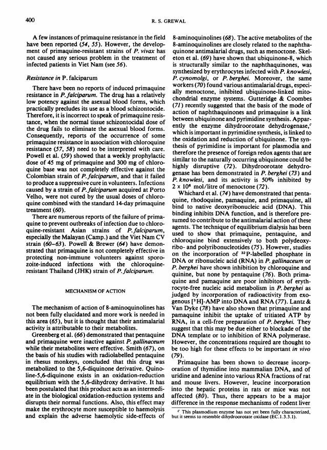
8-aminoquinolines (68). The active metabolites of the8-aminoquinolines are closely related to the naphtha-quinone antimalarial drugs, such as menoctone. Skel-eton et al. (69) have shown that ubiquinone-8, whichis structurally similar to the naphthaquinones, wassynthesized by erythrocytes infected with P. knowlesi,P. cynomolgi, or P. berghei. Moreover, the sameworkers (70) found various antimalarial drugs, especi-ally menoctone, inhibited ubiquinone-linked mito-chondrial enzyme systems. Gutteridge & Coombes(71) recently suggested that the basis of the mode ofaction of naphthaquinones and primaquine is a linkbetween ubiquinone and pyrimidine synthesis. Appar-ently the enzyme dihydroorotate dehydrogenase,cwhich is important in pyrimidine synthesis, is linked tothe oxidation and reduction of ubiquinone. The syn-thesis of pyrimidine is important for plasmodia andtherefore the presence of foreign redox agents that aresimilar to the naturally occurring ubiquinone could behighly disruptive (72). Dihydroorotate dehydro-genase has been demonstrated in P. berghei (73) andP. knowlesi, and its activity is 50% inhibited by2 x 106 mol/litre of menoctone (72).Whichard et al. (74) have demonstrated that penta-
quine, rhodoquine, pamaquine, and primaquine, allbind to native deoxyribonucleic acid (DNA). Thisbinding inhibits DNA function, and is therefore pre-sumed to contribute to the antimalarial action of theseagents. The technique of equilibrium dialysis has beenused to show that primaquine, pentaquine, andchloroquine bind extensively to both polydeoxy-ribo- and polyribonucleotides (75). However, studieson the incorporation of 32P-labelled phosphate inDNA or ribonucleic acid (RNA) in P. gallinaceum orP. berghei have shown inhibition by chloroquine andquinine, but none by pentaquine (76). Both prima-quine and pamaquine are poor inhibitors of eryth-rocyte-free nucleic acid metabolism in P. berghei asjudged by incorporation of radioactivity from exo-genous [3H]-AMP into DNA and RNA (77). Lantz&Van Dyke (78) have also shown that primaquine andpamaquine inhibit the uptake of tritiated ATP byRNA, in a cell-free preparation of P. berghei. Theysuggest that this may be due either to blockade of theDNA template or to inhibition of RNA polymerase.However, the concentrations required are thought tobe too high for these effects to be important in vivo(79).Primaquine has been shown to decrease incorp-
oration of thymidine into mammalian DNA, and ofuridine and adenine into various RNA fractions of ratand mouse livers. However, leucine incorporationinto the hepatic proteins in rats or mice was notaffected (80). Thus, there appears to be a majordifference in the response mechanisms of rodent liver
c This plasmodium enzyme has not yet been fully characterized,but it seems to resemble dihydroorotate oxidase (EC. 1.3.3.1).

PHARMACOLOGY OF 8-AMINOQUINOLINES
and Tetrahymena pyriformis (81) to theaminoquinolines. Conklin & Chou (82) found thatchloroquine, mepacrine, quinine, and primaquineblock amino acid uptake in Tetrahymena pyriformis.Working with P. lophurae, Sherman & Tangoshi (83)concluded that the inhibitory effects of chloroquine,quinine, and primaquine, on amino acid incorpor-ation were due to their influence on the energetics ofthe parasites and not to a direct blockade of aminoacid uptake.Whichard et al. (84) showed that 8-aminoquino-
lines and two hydroxylated derivatives inhibit certainbacterial andDNA polymerases. Also, the presence ofDNA-associated histone decreased the binding of8-aminoquinolines to isolated DNA (85).These experiments indicate that the 8-aminoquino-
lines probably affect several RNA functions as well asnucleic acid synthesis, but caution is needed inextrapolating these findings to the biological mech-anisms of the intact organism.
Morphological changes have been observed in vivoin the erythrocytic stages of P. berghei and P.fallax,following exposure to primaquine and menoctone(86). It has been shown (87, 88) that primaquine andother 8-aminoquinolines can induce mitochondriallesions in the exoerythrocytic stage of P.fallax intissue culture. The presence of [3 H]-primaquine (or itsmetabolite) in the mitochondria of the tissue stages ofP.fallax was shown by high resolution autoradio-graphy to coincide with the first observations of mito-chondrial swelling (89). Although mitochondrialrespiration is thought to be important in the tissuestages of P.fallax and other avian malarias, thesituation in the liver stages of mammalian species isless clear, and it is apparently not important for theerythrocytic stages of mammalian plasmodia (90).However, all three stages are affected by primaquine,especially the exoerythrocytic stages.
Primaquine, chloroquine, quinine, and quinacrineinhibit proteolysis in mouse kidney phagolysosomes.This phenomenon could be used for the screening ofdrugs that may be potentially useful for the treatmentof diseases caused by organisms that possess alysosome-like digestive system (91).Primaquine does not influence the folate pathway
in plasmodia. Peters (49) demonstrated that theaction of primaquine is not influenced by the adminis-tration of large doses of 4-aminobenzoic acid.
MECHANISM OF PRIMAQUINE RESISTANCE
Howells et al. (86) studied the ultrastructuralchanges produced by primaquine on the asexual bloodstages of P. berghei in mice. These workers found thatsome of the trophozoites that survived for 48 h after
exposure to the drug appeared to have an increasednumber of whirled organelles, which are believed toact as mitochondrial equivalents. They suggest thatthe parasites overcome the damaging effects of thedrug on their mitochondria by synthesizing more ofthese organelles to compensate for the functional loss.Also, the haemozoin of primaquine-resistant P. ber-ghei forms in larger vesicles than normal and appears,even at the light microscope level, as balls of pigmentthat are larger and darker than those seen in normaldrug-sensitive organisms (50). The importance ofthese observations to the mechanism by which theparasites develop primaquine resistance, is not clear.
HAEMOLYTIC EFFECTS
Primaquine and other 8-aminoquinolines have ahaemolytic effect on glucose-6-phosphate dehydro-genase (G6PD) -deficient erythrocytes, which areespecially sensitive to oxidizing agents. Cohen &Hochstein (92) suggested that the generation ofhydrogen peroxide in erythrocytes by the 8-amino-quinolines plays a major role in the induction ofhaemolysis. They were able to demonstrate the pres-ence of hydrogen peroxide in intact cells after theaddition of primaquine, but not after the addition ofthe 4-aminoquinoline, chloroquine. Similarly, hydro-gen peroxide was present after the addition of thehydroquinone-4-quinone redox system but not afterthe addition of non-autoxidizable resorcinol. Thegeneration of hydrogen peroxide from 8-aminoquino-lines required the presence of oxyhaemoglobin, andcould be blocked by the preliminary conversion ofoxyhaemoglobin to methaemoglobin.
G6PD-deficient erythrocytes are incapable ofsufficiently rapid regeneration of reduced nicotin-amide adenine dinucleotide phosphate (NADPH).Consequently all the NADPH-dependent reductiveprocesses in the cell are impaired. Metabolic processesare reduced to such an extent that normal vital func-tions can no longer proceed and the consequentalterations in the lipoprotein membrane of the cellresult in lysis (68, 93).Changes in the ultrastructure of the erythrocyte
membrane and vacuolization have been observed byGinn et al. (94) in red cells exposed to low concen-trations of primaquine. The authors suggest that thesechanges are related to its haemolytic effects.On the basis of studies on the haemolytic action of
primaquine on normal and G6PD-deficient red cells,Fraser & Vessel (95) produced evidence to suggest thatthe mechanism of methaemoglobin formation by suchcompounds may be unrelated to that which damagesthe red cell membrane.Methaemoglobinaemia occurs after administration
401

R. S. GREWAL
of high doses of 8-aminoquinolines. Observationsmade on military personnel in Viet Nam suggestedthat heterozygotes for NADPH-dependent meth-aemoglobin reductase are far more common than pre-viously recognized, and that the routine use of prima-quine-containing prophylactics will result in anincreased incidence of methaemoglobinaemia (96).
Quinacrine greatly enhances the toxicity of8-aminoquinolines, by increasing their plasma con-centration 5-10-fold, and extends their half-life byinterfering with their metabolic degradation. Sincequinacrine disappears very slowly from the body, thisphenomenon is observed if 8-aminoquinolines aregiven within 3 months of a dose of quinacrine (97).
ANTIBACTERIAL ACTIVITY
Primaquine has weak antibacterial activity againstEntamoeba coli, Staphylococcus aureus, Bacillussubtilis, and Candida albicans, and modest activityagainst Salmonella typhi, and Streptococcus pyo-genes. It also shows synergistic effects with most anti-biotics (98).
EFFECT ON IMMUNE MECHANISMS
Thong et al. (99) have shown that primaquineinhibits mitogen-induced, human lymphocyte prolif-erative responses. They used phytohaemagglutinin orpokeweed mitogen to induce a human lymphocyteproliferative response, which was measured by theincorporation of [3 H]-thymidine. They concludedthat doses of primaquine in the therapeutic range forvivax malaria pcssessed potent immunosuppressantactivity. However, it is not yet known whether patientson primaquine treatment are actually immuno-suppressed (100). Direct extrapolation from experi-mental findings to the clinical situation must be madewith care especially since the part of the immunesystem responsible for producing protective anti-bodies in malaria is unknown. To label primaquineas an immunosuppressant drug may unnecessarilydiscourage the use of this valuable antimalarial (101).
CARDIOVASCULAR PHARMACOLOGY
The frequent administration of high doses of penta-quine monophosphate has been shown to causemarked postural hypotension (102). Since the adrena-line responses were unaltered, the hypotension wasexplained in terms of an impairment of the central
sympathetic system. Moe et al. (103) found that largedoses of pamaquine, pentaquine, and isopentaquine,when administered over a long period to dogs andmonkeys, produced impairment of the sympatheticcardiovascular reflexes, which was believed to be dueto the destruction of medullary cell groups.
Fries et al. (104) administered pentaquine topatients suffering from essential or malignant hyper-tension. The drug produced a significant decrease inblood pressure but the toxic reactions were toofrequent and severe for its use as an antihypertensive.
EFFECTS ON THE MYOCARDIUM
Pamaquine and primaquine have been shown toexert quinidine-like effects on the myocardium. Thesecompounds are known to increase the refractoryperiod and conduction time in dogs, and have beenshown to be better than quinidine in protecting theheart against auricular fibrillation produced by theapplication of acetylcholine or aconitine, and againstauricular flutter from crush stimulation in anaesthe-tized dogs. They did not protect against epinephrine-induced arrhythmia (105).
Primaquine has been shown to possess an anti-arrhythmic effect in experiments where arterialfibrillation was induced by intravenous injection ofacetylcholine into neostigmine-pretreated animals.Primaquine was as active as quinidine, and did notaffect the ionic concentrations, or ionic fluxes, of themyocardial tissue (106).Pamaquine and primaquine inhibited stimulus initi-
ation, atrioventricular conduction, and intraven-tricular transmission of impulses.At low concentrations, primaquine produced a
transient positive chronotropic and ionotropic effectin isolated guinea-pig heart, which could be attributedto its ability to release adrenaline. Higher concen-trations of primaquine produced sustained negativechronotropic and ionotropic effects, which were stillpreceded by a transient sympathomimetic effect(107).
ANTIMUSCARINIC ACTIVITY
Primaquine has been shown to possess antimus-carinic activity on the guinea-pig bladder, and ongastrointestinal tract motility, as judged by the trans-port of a charcoal meal. These experiments indicatethat primaquine is a competitive antagonist of acetyl-choline, and that it acts synergistically with atropineon intestinal musculature in vivo (108).
402

PHARMACOLOGY OF 8-AMINOQUINOLINES
EFFECT ON CLOTTrING MECHANISMS
The administration of pamaquine to cattle infectedwith Thuleria sergenti caused a moderate extension ofclotting time, a decrease in platelet adhesiveness, clotretraction, and changes in the thromboelastogram.These changes persisted for 5-10 days after cessationof the treatment (109).
Primaquine has no antipyretic, analgesic, ormusculotropic effect (97). The only published reporton the effects of primaquine on the central nervoussystem is that by Schmidt & Schmidt (110), whichdeals with its neurotoxicity in rhesus monkeys.
TOXIC EFFECTS
Daily administration of an oral dose of 12-24 mgof primaquine/kg of body weight caused sublethal tolethal intoxication in rhesus monkeys. In addition tocentral nervous system lesions, the animals exhibitedcyanosis, anorexia, malaise, weight loss, markedmethaemoglobinaemia, anaemia, leukopenia, neutro-penia, reduction in myeloid elements of bone marrowand, in some cases, a yellow and grossly enlarged liver(110).The intramuscular administration of 4-6 mg of
primaquine/kg of body weight to rabbits producedchanges in liver function, including increases in ceph-alin-cholesterol flocculation, icterus index, and serumlevels of aspartate aminotransferase and alanineaminotransferase. Choline dihydrogen citrate wasvery effective in protecting the liver againstprimaquine toxicity (111). Intraperitoneal adminis-tration of 45 mg of primaquine/kg of body weightincreased liver tryptophane oxygenase activity in rats(112).Rhodoquine (Plasmocid) has been shown to
damage cardiac and skeletal muscle. Single dosesproduced cardiac necrosis, secondary inflammation,and calcification, which were preceded by adisturbance in cardiac glycogen metabolism, andinterference with 5'-nucleotidase activity in thecapillaries (113).
Price et al. (114) have shown by electron micros-copy that rhodoquine produces selective actin fila-ment and Z band degeneration in the muscle fibres ofthe rat diaphragm. It also produces sequentialmuscular necrosis, which is closely correlated withserum aminotransferase activity and the presence oflarge amounts of taurine in the urine (115).Rhodoquine injections (80 mg/kg of body weight)raised serum creatinine phosphokinase levels inrabbits, which closely paralleled the occurrence andthe degree of morphological degeneration (116, 117).
UMf
PHARMACOLOGIE DES AMINO-8 QUINOLEINES
Parmi les amino-8 quinoleines dont l'activite comme schi-zontocides tissulaires a ete eprouvee cliniquement, la prima-quine reste le meilleur compose. Elle est formee de partiesegales des formes ( + ) et (-) du fait de la presence d'un atomede carbone asymetrique. L'activite de ces stereoisomerescontre P. cynomolgi chez le singe est identique, mais la toxi-cite de la (-)-primaquine est trois A cinq fois superieure A cellede la (+ )-primaquine, si l'on en juge par ses effets hepato-toxiques. Ces differences pourraient etre importantes si ellesse confirmaient chez l'homme. Si la (+)-primaquine etaitreellement moins toxique chez l'homme, on pourrait aug-menter encore son indice therapeutique en la liant A despeptides.On postule que la primaquine agit par l'intermediaire de
son metabolite diquinone-5,6, qui est en equilibre d'oxydo-reduction avec le derive dihydroxy-5,6, mais il se peut que leseffets toxiques et les effets sur le plasmodium soient produitspar des composes differents; A l'heure actuelle, les re-sultats semblent favoriser I'hypothese d'une action desproduits metaboliques dans les deux cas. Les metabolitesagissent comme intermediaires dans les systemes biologiquesd'oxydoreduction, qu'ils rompent. Ils ressemblent A l'ubi-quinone-8 naturelle et inhibent les systemes mitochon-
driques lies A cette substance. On a propose comme base dumode d'action de la primaquine une liaison entre l'ubiqui-none et la synthese de la pyrimidine. La primaquine se lie Al'ADN isole et on presume que l'inhibition de la fonctionADN qui s'ensuit est liee au moins en partie A l'effet anti-paludique. La primaquine induit des lesions mitochon-driques dans les stades exoerythrocytaires de P.fallax enculture tissulaire, et la presence de primaquine tritiee ou deses metabolites a ete demontree par autoradiographie Ahaute resolution simultanement A l'observation d'ungonflement des mitochondries.La primaquine produit des effets hemolytiques sur les ery-
throcytes carences en G6PD, particulierement sensibles auxoxydants. Ces erythrocytes sont incapables de regenerersuffisamment vite leur NADPH du fait de leur deficit enG6PD.A dose moderee et forte, la primaquine provoque une
metheglobinemie, qui se manifeste par de la cyanose chez lessujets presentant une baisse de l'activite de la NADPHmethemoglobine reductase.La mepacrine augmente considerablement la toxicite des
amino-8 quinoleines, et cet effet peut persister pendant troismois.
403

404 R. S. GREWAL
II a ete demontre que la primaquine inhibe la reponselymphocytaire induite par des mitogenes chez I'homme,mais les constquences de cette observation pour 1'emploiclinique ne sont pas encore claires.La primaquine et les autres amino-8 quinoleines ont des
effets hypotenseurs chez I'animal. La primaquine a une acti-vite evoquant celle de la quinidine et augmente la periode
refractaire et le temps de conduction dans le cocur du chien.Elle possede une activite analogue a celle de l'atropine. II estnecessaire d'obtenir des donnees sur les effets de la prima-quine sur le systeme nerveux central et le systeme cardio-vasculaire au moyen de techniques modernes sophistiqueesde pharmacologie clinique.
REFERENCES
1. MOHLENS, P. Naturwissenschaften, 14: 1162 (1926).2. EDGCOMB, J. H. ET AL. Journal of the National
Malaria Society, 9: 285-292 (1950).3. ALVING, A. S. ET AL. Journal of the American Medi-
calAssociation, 149: 1558 (1952).4. COOPER, W. C. ET AL. American journal of tropical
medicine and hygiene, 2: 949 (1953).5. PETERS, W. ET AL. Annals of tropical medicine and
parasitology, 69: 311-328 (1975).6. CARROLL, F. I. ET AL. Chemistry and industry, 7: 477
(1975).7. SCHMIDT, L. H. ET AL. Antimicrobial agents and
chemotherapy, 12: 51 (1977).8. TONKIN, I. M. JR. British journal of pharmacology
and chemotherapy, 1: 163-173 (1946).9. HAWKING, F. Transactions of the Royal Society of
Tropical Medicine and Hygiene, 39: 245-263 (1945).10. DAVEY, D. G. Annals of tropical medicine and para-
sitology, 40: 453-471 (1946).11. FINK, E. Zeitschriftffitr Tropenmedizin undParasito-
logie, 21: 357-372 (1970).12. MOST, H. M. ET AL. American journal of tropical
medicine and hygiene, 16: 572-575 (1967).13. MOST, H. M. & MONTUORI, W. A. Americanjournal
of tropical medicine and hygiene, 24: 179-182 (1975).14. PIRSON, P. ET AL. Transactions oftheRoyal Society of
Tropical Medicine, 73: 347 (1979).15. SCHMIDT, L. H. ET AL. Bulletin of the World Health
Organization, 34: 783-788 (1966).16. ROSSAN, R. N. ET AL. American journal of tropical
medicine and hygiene, 24: 168-173 (1975).17. JONES, R. JR. ET AL. Journal of clinical investigation,
27: 5 1-55 (1948).18. ARNOLD, J. ET AL. Journal of laboratory and clinical
medicine, 46: 391-397 (1955).19. FELDMAN, H. A. ET AL. Federation proceedings, 5:
244 (1946).20. TERZIAN, L. A. Science, 106: 449-450 (1947).21. NARAYANDAS, M. G. & RAY, A. P. Indian journal of
malariology, 8: 138-141 (1954).22. TERZIAN, L. A. ET AL. Journal of infectious diseases,
90: 116-130 (1951).23. TERZIAN, L. A. ET AL. Experimentalparasitology, 23:
56-66 (1968).24. TERZIAN, L. A. & WEATHERBY, A. B. Americanjour-
nal of tropical medicine, 29: 19-22 (1949).25. FIELD, J. W. Bulletin of the First Medical Research
Federation ofMalaya, Kuala Lumpur, Federation ofMalay States Government Press, 1939.
26. MACKERRAS, M. J. & ERCOLE, Q. N. Transactions ofthe Royal Society of Tropical Medicine and Hygiene,42: 455-463 (1949).
27. WALKER, J. A. Transactions of the Royal Society ofTropical Medicine and Hygiene, 49: 351-355 (1955).
28. RAFFAELE, G. & CARRESCIA, P. M. Rivista dimalariologia, 40: 51-59 (1961).
29. JEFFERY, G. M. ET AL. American journal of hygiene,64: 1-11 (1956).
30 BURGESS, R. W. & BRAY, R. S. Bulletin of the WorldHealth Organization, 24: 451-456 (1961).
31. BERMAN, S. J. & HOLMES, K. K. Journal oftheAmer-ican Medical Association, 215: 291-292 (1971).
32. SCHULTZ, M. G. Journal of the American MedicalAssociation, 216: 519-529 (1971).
33. ROEHL, W. Archiv fir Schiffs- und Tropenhygiene,30: 311-318 (1926).
34. COATNEY, G. R. & COOPER, W. C. Journal ofpara-sitology, 34: 275 (1948).
35. STOSSKOFF, M. K. & BEIER, J. Journal of the Amer-ican Veterinary Medical Association, 175: 944-947(1979).
36. JACOBS, R. L. ET AL. Journal of parasitology, 49:920-925 (1963).
37. PETERS, W. Experimental parasitology, 17: 80-89(1965).
38. SCHNEIDER, J. ET AL. Bulletin de la Societe dePathologie exotique et de ses Filiales, 42: 449-452(1949).
39. SCHNEIDER, J. Indian journal of malariology, 8:275-279 (1954).
40. AGARWAL, K. ET AL. Indian journal of medicalresearch, 69: 577-582 (1979).
41. PETERS, W. Annals of tropical medicine andparasito-logy, 61: 52-56 (1967).
42. FINK, E. ET AL. Arzneimittel-Forschung, 20:1775-1777 (1970).
43. DAVIDSON, D. E. JR. ET AL. American journal of trop-ical medicine and hygiene, 25: 26-33 (1976).
44. SCHMIDT, L. H. Annual review of microbiology, 23:427-454 (1969).
45. FULTON, J. D. & YORKE, W. Annals of tropical med-icine andparasitology, 35: 223-239 (1941).
46. BISHOP, A. & BIRKETT, B. Nature (London), 159:884-885 (1947).
47. RAMAKRISHNAN, S. P. & PRAKASH, S. Bulletin of theNational Society of Indian Malariology, 9: 261-265(1961).

PHARMACOLOGY OF 8-AMINOQUINOLINES 405
48. PRAKASH, S. ET AL. Indian journal ofmalariology, 15:115-122 (1961).
49. PETERS, W. Annals oftropical medicine andparasito-logy, 60: 25-30 (1966).
SO. PETERS, W. Chemotherapy and drug resistance inmalaria, London, Academic Press, 1970.
51. BISHOP, A. Parasitology, 57: 755-770 (1967).52. ALVING, A. S. ET AL. In: Proceedings of the 6th Inter-
national Congress on Tropical Medicine and Malaria,Lisbon, 1959, vol. 7, pp. 203-209.
53. ARNOLD, J. ET AL. Transactions of the Royal Societyof Tropical Medicine and Hygiene, 55: 345 (1961).
54. HEATON, L. D. Military medicine, 128: 6-8 (1963).55. INMAN, M. ET AL. Federation proceedings, 26: 288
(1967).56. PETERS, W. Transactions of the Royal Society of
Tropical Medicine and Hygiene, 63: 24-45 (1969).57. MOORE, D. V. & LANIER, J. E. American journal of
tropical medicine and hygiene, 10: 5-9 (1961).58. GALVAO, A. L. A. ET AL. Arquivos da Faculdade de
higieneesatudeputblica da Universidade de Sto Paulo,15/16: 201-244 (1961).
59. POWELL, R. D. ET AL. American journal of tropicalmedicine and hygiene, 12: 509-512 (1963).
60. Box, E. D. ET AL. American journal oftropical medi-cine and hygiene, 12: 300-304 (1963).
61. RIECKMANN, K. H. ET AL. Bulletin of the WorldHealth Organization, 38: 625-632 (1968).
62. MCNAMARA, J. V. & RIECKMANN, K. H. In:Abstracts from the 8th International Congress onTropical Medicine and Malaria, Teheran, 1968, pp.1396-1398.
63. EPPES, R. B. ET AL. Military medicine, 132: 163-175(1967).
64. POWELL, R. D. & BREWER, G. J. Americanjournal oftropical medicine and hygiene, 16: 693-698 (1967).
65. WARHURST, D. C. Symposium of the British Societyfor Parasitology, 1973, vol. 2, pp. 1-28.
66. GREENBERG, J. ET AL. Journal of infectious diseases,88: 163 (1951).
67. SMITH, C. C. Pharmacology and experimental thera-peutics, 116: 67 (1956).
68. TARLOV, A. R. ET AL. Archives of internal medicine,109: 209 (1962).
69. SKELETON, F. S. ET AL. Journal of medicinal chem-istry, 13: 602-606 (1970).
70. SKELETON, F. S. ET AL. Journal of the AmericanChemical Society, 90: 5334-5446 (1968).
71. GUTrERIDGE, W. E. & COOMBES, G. H. Biochemistryofparasitic protozoa, London, Macmillan, 1977.
72. GUT-ERIDGE, W. E. ET AL. Biochimica et biophysicaacta, 582: 390-401 (1979).
73. KROOTH, R. S. ET AL. Science, 164: 1073-1075 (1969).74. WHICHARD, L. P. ET AL. Molecular pharmacology, 4:
630 (1968).75. MORRIS, C. R. & ANDREW, L. V. Federation proceed-
ings, 28: 361 (1969).76. SCHELLENBERG, K. A. & COATNEY, G. R. Biochem-
ical pharmacology, 6: 143-152 (1961).77. LANTZ, C. H. & VAN DYKE, K. Experimental para-
sitology, 31: 255-261 (1972).78. LANTZ, C. H. & VAN DYKE, K. Federation proceed-
ings, 29: 808 (1970).
79. CARTER, G & VAN DYKE, K. Proceedings of the Hel-minth Society, 39: 244-249 (1972) (Special issue).
80. FIELD, R. C. ET AL. British journal ofpharmacology,62: 159-164 (1978).
81. CONKLIN, K. A. ET AL. Molecular pharmacology, 9:304-310 (1973).
82. CONKLIN, K. A. & CHOU, S. C. Science, 166:1213-1214 (1970).
83. SHERMAN, I. W. & TANGOSHI, L. Proceedings of theHelminth Society, 39: 250-260 (1972) (Special issue).
84. WHICHARD, L. P. ET AL. Biochimica et biophysicaacta, 287: 52-67 (1972).
85. WASHINGTON, M. E. ET AL. Biochemical pharmaco-logy, 22: 477-484 (1973).
86. HOWELLS, R. E. ET AL. Annals of tropical medicineand parasitology, 64: 203-208 (1970).
87. BEAUDOIN, R. L. & AIKAWA, A. Science, 160:1233-1234 (1968).
88. AIKAWA, M. & BEAUDOIN, R. L. Military medicine,134: 987-999 (1969).
89. AIKAWA, M. & BEAUDOIN, R. L. Experimentalpara-sitology, 27: 454-463 (1970).
90. HOMEWOOD, C. A. Bulletin of the World HealthOrganization, 55: 229-235 (1977).
91. MEGO, J. L. & CHIN HA CHUNG, Biochemicalpharmacology, 28: 465-470 (1979).
92. COHEN, G. & HOCHSTEIN, P. Biochemistry, 3:895-900 (1964).
93. BEUTLER, E. & MITCHELL, M. Blood, 32: 816-818(1969).
94. GINN, F. L. ET AL. Science, 164: 843-845 (1969).95. FRASER, I. M. & VESSEL, E. S. Journal ofpharmaco-
logy and experimental therapeutics, 162: 152-165(1968).
96. COHEN, R. J. ET AL. New England journal of medi-cine, 279: 1127-1131 (1968).
97. GOODMAN, L. S. & GILLMAN, A. The pharmaco-logical basis of therapeutics, London, Macmillan,1970.
98. TOAMA, M. A. Journal ofpharmaceutical science, 67:23-25 (1978).
99. THONG, Y. H. ET AL. Transactions of the RoyalSociety of Tropical Medicine and Hygiene, 72:537-539 (1978).
100. THONG, Y. H. Transactions of the Royal Society ofTropical Medicine and Hygiene, 73: 474 (1979).
101. POONAPALAM, J. T. Transactions of the RoyalSociety of Tropical Medicine and Hygiene, 73:473-474 (1979).
102. RICHARDSON, A. P. ET AL. Proceedings of the Societyfor Experimental Biology and Medicine, 65: 258-261(1947).
103. MOE, G. K. ET AL. Journal of pharmacology andexperimental therapeutics, 95: 407-414 (1949).
104. FRIES, E. D. ET AL. Proceedings of the Society ofExperimental Biology, 64: 455-458 (1947).
105. ARORA, R. B. & MADAN, B. R. Archives internatio-nales de pharmacodynamie et de therapie, 107:215-222 (1956).
106. HOLLAND, E. L. ET AL. Journal of pharmacologicalscience, 51: 791-793 (1962).
107. MARSHALL, R. J. & OJEWOLE, J. A. 0. Toxicologyand applied pharmacology, 46: 759-768 (1978).

406 R. S. GREWAL
108. SEIDEL, E. R. ET AL. Toxicology and appliedpharma-cology, 45: 258 (1978).
109. HAYASHI, T. ET AL. Bulletin of the Faculty ofAgricul-ture, Tottori University, 30: 82-88 (1979).
110. SCHMIDT, I. G. & SCHMIDT, L. H. Journal of neuro-pathology and experimental neurology, 10: 231(1951).
111. EL-DENSHARY ET AL. Journal of the Egyptian MedicalAssociation, 52: 552-560 (1969).
112. MAGUS, R. D. ET AL. Biochemicalpharmacology, 20:486-489 (1971).
113. BAJUSZ, E. & JASMINE, G. Vitamin hormone-fermen-tation, 14: 28-42 (1965).
114. PRICE, H. M. ET AL. Laboratory investigation, 2:549-562 (1962).
115. BOWDIN, D. H. Archives of pathology, 74: 137-141(1962).
116. HENSON, J. B. & RAO, R. R. Canadian journal ofcomparative medicine and veterinary science, 30:157-160 (1966).
117. RAO, R. R. & HENSON, J. B. Research in veterinaryscience, 8: 369-374 (1967).