Pharmacokinetics of Methanol and Formate Michele A ... · PDF fileblood concentrations of...
48
Pharmacokinetics of Methanol and Formate in Female Cynomolgus Monkeys Exposed to Methanol Vapors Michele A. Medinsky, David C. Dorman, James A. Bond, Owen R. Moss, Derek B. Janszen, and Jeffrey I. Everitt Chemical Industry Institute of Toxicology, Research Triangle Park, NC Includes the Commentary of the Institute's Health Review Committee Research Report Number 77 June 1997
Transcript of Pharmacokinetics of Methanol and Formate Michele A ... · PDF fileblood concentrations of...

Pharmacokinetics of Methanol and Formate in Female Cynomolgus Monkeys Exposed to Methanol Vapors
Michele A. Medinsky, David C. Dorman, James A. Bond, Owen R. Moss, Derek B. Janszen, and Jeffrey I. Everitt Chemical Industry Institute of Toxicology, Research Triangle Park, NC
Includes the Commentary of the Institute's Health Review Committee
Research Report Number 77 June 1997

m: HEALTH EFFECTS INSTITUTE
The Health Effects Institute, established in 1980, is an independent and unbiased source
of information on the health effects of motor vehicle emissions. HEI studies all major pollutants, including regulated pollutants (such as carbon monoxide, ozone, nitrogen dioxide, and particulate matter), and unregulated pollutants (such as diesel engine exhaust,
methanol, and aldehydes). To date, HEI has supported more than 150 projects at institutions in North America and Europe.
Typically, HEI receives half its funds from the U.S. Environmental Protection Agency and
half from 28 manufacturers and marketers of motor vehicles and engines in the United States.
Occasionally, funds from other public or private organizations either support special projects or provide resources for a portion of an HEI study. This study was part of a larger methanol
program that received initial support from the American Petroleum Institute. Regardless of funding sources, HEI exercises complete autonomy in setting its research priorities and in reaching its conclusions. An independent Board of Directors governs HEI. The Institute's Research and Review Committees serve complementary scientific purposes and draw distinguished scientists as members. The results of HEI-funded studies are made available as Research Reports, which contain both the Investigators' Report and the Review Committee's evaluation of the work's scientific quality and regulatory relevance.

!£(Statement Synopsis of Research Report Number 77
Metabolic Studies in J\llonkeys Exposed to lVIethanol Vapors
BACKGROUND
In an effort to improve air quality and decrease dependence on petroleum, the U.S. government has encouraged the development of alternative fuels, one of which is methanol. Although the proposed use of methanol as an alternative motor vehicle fuel should decrease air pollution by reducing carbon monoxide and hydrocarbon emissions, expanding its use will increase emissions of other pollutants, especially formaldehyde and methanol vapors. Methanol is a natural constituent of plant and animal tissues and some foods, but can be highly poisonous when ingested (for example, as wood alcohol). The well-recognized symptoms of methanol poisoning (nervous system dysfunction, damage to the visual system, and even death) are thought to be due to the buildup offormate, a metabolite produced when methanol is broken down by the body. In small amounts, formate is essential for DNA and protein synthesis; however, excess blood levels are highly toxic. Converting formate to nontoxic substances requires adequate stores of one form of the vitamin folic acid, and proceeds efficiently as long as the body's metabolic systems are not overwhelmed by excess methanol. Most analysts think that inhaling low concentrations of methanol is not a risk factor for healthy people. However, there is concern about potentially susceptible populations, especially those who might be deficient in folic acid. These include pregnant women, the developing fetus, and patients with alcoholism and other chronically debilitating illnesses. The HEI funded this study to determine how rapidly formate is formed and removed in monkeys after they have been exposed to methanol vapors, and whether monkeys that are deficient in folic acid accumulate more formate than normal animals. Monkeys were used in this study because rodents are not susceptible to methanol-induced poisoning.
APPROACH
Dr. Medinsky and colleagues of the Chemical Industry Institute of Toxicology exposed female cynomolgus monkeys to environmentally relevant concentrations (10, 45, or 200 parts per million [ppm]) of methanol vapors and to one high dose (900 ppm) for two hours. The methanol was administered while the animals were under general anesthesia. They also exposed monkeys that had been fed a diet deficient in folic acid to the highest methanol concentration. In order to increase the sensitivity of their measurements, and to differentiate between the formate that naturally occurs in the body (endogenous formate) and that which results from methanol exposure, the investigators used methanol tagged with radiolabeled carbon.
RESULTS AND IMPLICATIONS
Blood levels of radiolabeled methanol increased in a dose-dependent manner in monkeys exposed for two hours to methanol vapors. The blood levels of formate also increased; however, the levels of formate derived from the radiolabeled methanol were 10 to 1000 times lower than endogenous blood formate levels reported from other research, and much lower than the levels known to be toxic. Even monkeys that had moderate folic acid deficiency and were exposed to 900 ppm methanol had peak concentrations of methanol-derived formate that were only one percent of the background levels. Because the exposure conditions used in this study are not the same as those experienced by people, the absolute blood methanol and formate levels cannot be directly extrapolated to humans. Nevertheless, this report provides reassuring data that single exposures to methanol vapors are unlikely to produce hazardous increases in blood methanol or formate concentrations in the general population or even in individuals with moderate folic acid deficiency. However, some workers, such as garage mechanics, tunnel workers, and underground parking lot attendants, who could be exposed to levels of methanol vapors higher than 200 ppm for longer than two hours, and fetuses, whose development can be affected by methanol exposure and folic acid deficiency, could still be at risk. Further research using animals with more pronounced folate deficiency and longer exposures is critical for any risk assessment of methanol.
This Statement, prepared by the Health Effects Institute and approved by its Board of Directors, is a summary of a research project sponsored by HEI from 1991 to 1993. This study was conducted by Dr. Michele A. Medinsky of the Chemical Industry Institute of Toxicology, ReseRrch Triangle Park, NC. The following Research Report contains both the detailed Investigators' Report and a Commentary on the study prepared by the Institute's Health Review Committee.

Copyright© 1997 Health Effects Institute. Printed at The Graphic Supervisors, Topsfield, MA. Library of Congress Catalog Number for the HEI Research Report Series: WA 754 R432. The paper in this publication meets the minimum standard requirements of the ANSI Standard Z39.48-1984 (Permanence of Paper) effective with Report Number 21, December 1988, and with Report Numbers 25, 26, 32, 51, and 65 Parts IV, VIII, and IX excepted. These excepted Reports are printed on acid-free coated paper.

TABLE OF CONTENTS Research Report Number 77
Pharmacokinetics of Methanol and Formate Monkeys Exposed to Methanol Vapors
Female Cynomolgus
Michele A. Medinsky, David C. Dorman, James A. Bond, Owen R. Moss, Derek B. Janszen, and Jeffrey L Everitt
I. STATEMENT Health Effects Institute . . . . . . . . . . . . . . . . . . . . . . . . . . . . . . . . . . . . . . . . . . . . i This Statement, prepared by the HEI and approved by the Board of Directors, is a nontechnical summary of the Investigators' Report and the Health Review Committee's Commentary.
H. INVESTIGATORS' REPORT . . ................................................ 1 When an HEI-funded study is completed, the investigators submit a final report. The Investigators' Report is first examined by three outside technical reviewers and a biostatistician. The Report and the reviewers' comments are then evaluated by members of the HEI Health Review Committee, who had no role in selecting or managing the project. During the review process, the investigators have an opportunity to exchange comments with the Review Committee and, if necessary, revise the report.
Abstract. .............................. 1 Introduction ........................... 1
Toxicity of Methanol .................. 2 Species Differences in Methanol Toxicity
and Metabolism ..................... 2 Methanol Uptake and Distribution ....... 3 Metabolism of Methanol ............... 3 Metabolism of Formate ................. 4 Folate Status in Humans and Effects of
Folate Deficiency ................... 5 Background Body Burdens of Methanol
and Formate ....................... 5 Low-Level Methanol Exposure .......... 7
Specific Aims .......................... 7 Specific Aim 1 ....................... 7 Specific Aim 2 ....................... 7
Methods and Study Design ............... 8 Chemicals ........................... 8 Animals and Their Care . . . . . . . . . . . . . . . . 8 Production of Folate Deficiency ......... 8 Folate Analysis and Hematology ......... 9 Methanol Exposures ................... 9 Methanol Exposure System Design ...... 11 Gas Chromatographic Analysis of
Test Atmosphere ................... 11 Measurement of Respiratory Function ... 12 Quantification of Exhaled nc_ Methanol
and 14C-Carbon Dioxide ............. 12
Blood and Urine Sampling . . . . . . . . . . . . . . . . 12 Determination of Blood and Urine Methanol
and Formate Concentrations . . . . . . . . . . . . . 12 Liquid Scintillation Spectroscopy Methods. . . 13 Statistical Methods and Data Analysis. . . . . . . 13
Results . . . . . . . . . . . . . . . . . . . . . . . . . . . . . . . . . . 13 Methanol Atmosphere Generation . . . . . . . . . . 13 Pulmonary Function . . . . . . . . . . . . . . . . . . . . . 13 Folate Analysis, Hematology, and
Clinical Signs . . . . . . . . . . . . . . . . . . . . . . . . . 15 Methanol Kinetics . . . . . . . . . . . . . . . . . . . . . . . 15 Kinetics of Methanol-Derived Formate. . . . . . . 19
Discussion and Conclusions. . . . . . . . . . . . . . . . . 20 Methanol Exposure During Normal
Fuel Use . . . . . . . . . . . . . . . . . . . . . . . . . . . . . 20 Methanol Uptake, Distribution,
and Elimination . . . . . . . . . . . . . . . . . . . . . . . 22 Dietary, Endogenous, and Methanol-Derived
Formate .............................. 23 The Folate Pathway and Sensitive Populations 24 Developmental Neurotoxicity. . . . . . . . . . . . . . 24
Implications of Findings. . . . . . . . . . . . . . . . . . . . 25 Acknowledgments. . . . . . . . . . . . . . . . . . . . . . . . . 25 References . . . . . . . . . . . . . . . . . . . . . . . . . . . . . . . 26 Appendices Available on Request . . . . . . . . . . . . 29 About the Authors. . . . . . . . . . . . . . . . . . . . . . . . . 29 Publications Resulting from This Research. . . . . 30 Abbreviations . . . . . . . . . . . . . . . . . . . . . . . . . . . . 30
(Continued on next page)
iii

TABLE OF CONTENTS (Continued}
Research Report Number 77
HL COMMENTARY Health Review Committee .................................... 31 The Commentary about the Investigators' Report is prepared by the HEI Health Review Committee and staff. Its purpose is to place the study into a broader scientific context, to point out its strengths and limitations, and to discuss the remaining uncertainties and the implications of the findings for public health.
Introduction .......................... 31 Methods and Study Design ................. 33 Methanol Toxicity and Results and Interpretation .................. 34
Folate-Dependent Detoxification ....... 31 Implications for Future Research .............. 36 Objectives and Study Design ............ 33 Conclusions ............................... 36 Technical Evaluation ................... 33 Acknowledgments ......................... 36
Attainment of Study Objectives ........ 33 Refurences ................................ 37
IV. RELATED HEI PUBLICATIONS ................................................ 39

:Pharmar:ok:inetics rvletha.nol and Formate in Female Cynomolgus rv!onkeys Vapors
Michele l\. Dorman, James A. Bond, Owen R. Moss, Derek B. Janszen, and Jeffrey I. Everitt
ABSTRACT
The 1990 Clean Air Act Amendments contain mandates for reduced automotive emissions and add new requirements for the use of alternative fuels such as methanol to reduce certain automotive pollutants. Methanol is acutely toxic in humans at relatively low doses, and the potential for exposure to methanol will be increased if it is used in automotive fuel. Formate is the metabolite responsible for neurotoxic effects of acute methanol exposure. Since formate metabolism is dependent on folate, potentially sensitive folate-deficient subpopulations, such as pregnant women, may accumulate formate and be at higher risk from low-level methanol exposure.
Our objective was to determine the pharmacokinetics of 14C-methanol and 14C-formate in normal and folate-deficient monkeys after exposure to 14C-methanol vapors at environmentally relevant concentrations: below the threshold limit value (TL V) *, at the TL V of 200 parts per million (ppm), and above the TLV. Four normal adult female cynomolgus monkeys were individually anesthetized with isoflurane, and each was exposed by endotracheal intubation to 10, 45, 200, or 900 ppm 14C-methanol for 2 hours. Concentrations of the inhaled and exhaled 14C-methanol, blood concentrations of 14C-methanol and 14C-formate, exhaled 14C-carbon dioxide e4COz), and respiratory parameters were measured during exposure. After exposure, 14C-methanol and 14C02 exhaled, 14C-methanol and 14C-
* A list of abbreviations appears at the end of the Investigators' Report. This investigators' Report is one part of Health Effects Institute Research Report Number 77, which also includes a Commentary by the HE! Health Review Committee, and an HEI Statement about the research project. Correspondence concerning the Investigators' Report may be addressed to Dr. Michele A. Medinsky, Chemical Industry Institute of Toxicology, 6 Davis Drive, P.O. Box 12137, Research Triangle Park, NC 27709-2137.
This study was part of a larger methanol research program that received initial support from the American Petroleum Institute.
Although this document was produced with partial funding by the United States Environmental Protection Agency under Assistance Award R824835 to the Health Effects Institute, it has not been subjected to the Agency's peer and administrative review and therefore may not necessarily reflect the views of the Agency, and no official endorsement should be inferred. The contents of this document also have not been reviewed by private party institutions, including those that support the Health Effects Institute; therefore, it may not reflect the views or policies of these parties, and no endorsement by them should be inferred.
Health Effects Institute Research Report Number 77 © 1997
formate excreted in urine, and 14C-methanol and 14C-formate in blood were quantified. The amounts of exhaled 14C-methanol and 14COz, blood concentrations of 14Cmethanol and 14C-formate, and 14C-methanol and 14C-formate excreted in urine were linearly related to methanol exposure concentration. For all exposures, blood concentrations of 14C-methanol-derived formate were 10 to 1000 times lower than endogenous blood formate concentrations (100 to 200 mM) reported for monkeys and were several orders of magnitude lower than levels of formate known to be toxic.
Since the metabolism of formate in primates depends on the availability of tetrahydrofolate, the same four monkeys were next placed on a folate-deficient diet until folate concentrations in red blood cells consistent with moderate folate deficiency (29 to 107 ng/mL) were achieved. Monkeys were then reexposed to the highest exposure concentration, 900 ppm 14C-methanol, for a similar 2-hour period, and again the pharmacokinetic data described above were obtained. Even with a reduced folate status, monkeys exposed to 900 ppm methanol for 2 hours had peak concentrations of methanol-derived formate that were well below the endogenous levels of formate. Although these results represent only a single exposure and therefore preclude broad generalizations, they do suggest the body contains sufficient folate stores to effectively detoxify small doses of methanolderived formate from exogenous sources, such as those that might occur during normal use of automotive fueL
INTRODUCTION
The 1990 Clean Air Act Amendments contain mandates for reduced automotive emissions and add new requirements for the use of alternative fuels such as methanoL Thus, methanol has the potential to become an important automotive fuel in the United States in the next century (Kavet and Nauss 1990). The Clean Air Act requires that the implementation of new technology not degrade environmental quality or compromise pubic health. Substituting methanol for currently used fuels such as gasoline or diesel fuel should improve environmental quality by reducing ambient concentrations of criteria pollutants, which in-
1

Pharmacokinetics of Methanol and Formate in Female Cynomolgus Monkeys Exposed to Methanol Vapors
elude particulate matter, oxides of nitrogen, and ozone. However, use of methanol may increase emissions of two pollutants with known toxic effects: formaldehyde and methanol. Emissions of methanol can arise from its release as uncombusted fuel in the exhaust, from its evaporation during refueling, and from vaporization after the engine is stopped. Formaldehyde emissions result from incomplete combustion of methanol fuels.
TOXICITY OF METHANOL
Methanol is acutely toxic to humans at relatively low doses. Evidence of its toxicity has existed since the early 1900s, when human exposure to relatively large acute doses of methanol (commonly known as wood alcohol) occurred either through accidental or intentional ingestion, percutaneous absorption, or inhalation. In the early part of the century, painters used methanol as a dilution agent for shellac, varnish, and paint. Methanol was also used as a cleaning fluid; as a solvent by hatters, dyers, shoemakers, brass finishers; and in manufacturing rubber tires (Ziegler 1921). During this time, about 100 cases of vision impairment or death from inhalation of methanol vapors were reported. These cases showed that acute exposure of humans to methanol can result in blindness and metabolic acidosis. Metabolic acidosis is due to a reduced pH in the blood and can eventually result in death.
Most information on methanol toxicity in humans concerns the effects of acute exposures (Health Effects Institute 1987). Acute methanol toxicity in humans follows a wellrecognized pattern. Initially, exposure to a toxic methanol dose results in a transient central nervous system depression, which is characteristic of alcohol exposure. Uneven gait and lack of coordination in movements are often apparent. A latent period of compensated metabolic acidosis without evident symptoms follows,, during which time plasma bicarbonate levels decrease. This latent period may last from several hours to several days; the typical latent period is 12 to 24 hours. When compensatory mechanisms become exhausted and acidity increases further, the blood pH begins to drop, resulting in uncompensated metabolic acidosis (Tephly and McMartin 1984). Symptoms include headache, dizziness, nausea, and vomiting, followed by severe abdominal pain and difficult breathing. The symptoms may progress to coma and death, which is usually due to respiratory failure.
Metabolic acidosis is the result of formic acid production. Formic acid is an intermediate in the metabolism of methanol. At physiological pH, formic acid dissociates rapidly and almost completely to formate and hydrogen ions. The hydrogen ions accumulate in the body if the rate of formate production is faster than the rate of its removal.
2
The increase in hydrogen ions entering the blood eventually overwhelms the normal acid-base homeostasis. The time during which the acid-base imbalance develops is the latent period in methanol toxicity.
Toxicity to the visual system also can occur in parallel with metabolic acidosis. Affected individuals experience visual disturbances such as blurred or indistinct vision, altered visual fields, and impairment of the pupillary response to light. Some individuals become blind, even if they recover from uncompensated metabolic acidosis. A local increase in blood flow to the optic disc is the earliest change that occurs in the retina of a methanol-poisoned individual. Later an excess of aqueous fluid, or edema, appears, as well as a blurring of the margin of the optic disc. The edema may persist for up to 2 months. The severity of the retinal edema is predictive of permanent loss of vision. Mild optic disc edema results in restoration of vision and full recovery. Severe edema invariably leads to permanent effects. Pallor of the optic disc, indicating loss ofthe blood supply to the head of the optic nerve and atrophy of the optic nerve, is a sign of irreversible effects to the visual system.
Autopsies of victims of lethal methanol poisonings have revealed (among other abnormalities) damage to the putamen of the basal ganglia in the brain. The putamen controls gross intentional motor activities that are normally performed unconsciously. Survivors of methanol intoxication may suffer damage to the putamen and have associated motor disorders.
The oral dose of methanol required to produce such acute toxicity in humans is relatively modest. For example, 0.3 to 1 g methanol/kg of body weight is considered the range of a minimum lethal dose for untreated cases. Four ounces of pure methanol, approximately 95 g, is lethal to 40% of exposed individuals. The lowest lethal dose reported is about three teaspoons or 15 mL of moonshine, which is 40% methanol. However, individuals have been kno-wn to survive after drinking approximately 500 mL of moonshine. Variability in the metabolism of methanol and its toxic metabolite formate may form the basis for this variability in human response.
SPECIES DIFFERENCES IN METHANOL TOXICITY AND METABOLISM
Much is known about the chemical and biological behavior of methanol. Formate is the metabolic product that is thought to be responsible for the acute toxic effects of methanol. The identification of formate as the toxic metabolite of methanol was the result of research conducted by Tephly and associates in the 1970s and 1980s in rodents and nonhuman primates (Tephly and McMartin 1984). The

liti.A. Medinsky et al.
toxic effects of methanol are due either largely or completely to extreme elevations of blood formate (McMartin et al. 1975). These elevated levels lead to metabolic acidosis and ocular damage, resulting in blindness and death.
Species differences in the accumulation of formate in blood correlate well with susceptibility to methanol-induced toxicity (McMartin 1977). Monkeys, a sensitive species, have high concentrations of formate in the blood following acute doses of methanol (3 g methanol/kg body weight). Rats, a species insensitive to the toxic effects of methanol, do not develop high blood concentrations of formate even after doses of methanol (4 g methanol/kg body weight) larger than those that produce toxicity in monkeys. This correlation between blood formate concentrations and toxic effects of methanol was first noted by Tephly and coworkers (Tephly and McMartin 1984; McMartin 1977)
and provided the first clue as to the mechanism by which methanol was producing its neurotoxic effects. The work of these researchers pointed to the need to look at the internal concentrations of formate in addition to methanol when assessing potential toxicity.
METHANOL UPTAKE AND DISTRIBUTION
Methanol can be absorbed into the blood through inhalation of vapors, ingestion of liquid methanol, or dermal exposure to liquid methanol. The capacity for various animal species to absorb methanol is similar because simple diffusion is the basic mechanism underlying methanol transfer across epithelial membranes. The site for methanol uptake by inhalation is most likely the upper respiratory tract, based on studies conducted with inhaled ethanol, an alcohol with similar solubility properties (Gerde and Dahl 1991). Their breath-by-breath analysis of ethanol uptake demonstrated that on inhalation, ethanol is completely absorbed by the upper respiratory tract tissues of dogs. Although a large fraction of the absorbed ethanol diffuses into the blood, significant ethanol concentrations remain in the mucosa at the end of the inspiration. On expiration this ethanol diffuses back into the airways and is exhaled, resulting in an uptake of 70%. Studies with humans have shown that methanol is rapidly absorbed after inhalation, with net absorption ranging from 60 to 85%. Like ethanol, ingested methanol is rapidly absorbed from the gastrointestinal tract with peak absorption occurring in 30 to 60 minutes, depending on the presence or absence of food in the stomach. Ingestion of methanol has been the principal route of exposure in cases of acute poisoning.
An average of 0.192 mg/ cm2 /min of methanol is absorbed through direct contact of methanol with the skin of human volunteers. Exposure of one hand to liquid methanol for only 2 minutes would result in a body burden of as much
as 170 mg, similar to that resulting from inhaling approximately 40 ppm methanol for 8 hours (International Programme on Chemical Safety 1994).
Regardless ofthe route of exposure, methanol distributes readily and uniformly to all organs and tissues in direct relation to their water content (Health Effects Institute 1987). The largest amounts are found in muscles, blood, gastrointestinal tract, and liver (International Programme on Chemical Safety 1994). In clinical cases of methanol poisoning, consistently higher levels of methanol are present in the cerebrospinal fluid than in the blood.
Following uptake and distribution, methanol is eliminated from the body very slowly, especially compared with ethanol, in all species. Clearance half-times ranging from 3 hours to more than 1 day have been reported, depending on the dose of methanol administered. The primary routes for elimination of methanol are urinary excretion, exhalation, and biotransformation. Urinary elimination of methanol is not a quantitatively large pathway for methanol clearance under normal circumstances. Methanol's small size and nonpolar characteristics suggest it will be readily filtered by the kidney glomeruli and reabsorbed by the proximal tubules. Exhalation of methanol that has been absorbed into the blood also is a slow process. The large blood:air partition coefficient for methanol suggests an affinity of methanol for blood that is several thousand times greater than for air at equilibrium (Horton et al. 1992).
METABOLISM OF METHANOL
The accumulation of formate in monkeys and humans (but not in rats) after methanol exposure indicates that either methanol oxidation to formate occurs at a faster rate in monkeys and humans, or that formate oxidation to carbon dioxide proceeds at a slower rate in monkeys and humans (Figure 1). Methanol is metabolized, or enzymatically
Primates
Alcohol dehydrogenase
Formaldehyde dehydrogenase
Limited
H, Folate
CHpH Methanol
HCHO Formaldehyde
HCOO Formate
C02
Carbon dioxide
Rodents
Catalaseperoxidase
system
Formaldehyde dehydrogenase
Abundant
H, Folate
Figure 1. Metabolism of methanol and formate in rodents and primates. Adapted from Medinsky and Dorman (1994) with permission.
3

Pharmacokinetics of Methanol and Formate in Female Cynomolgl.IS Monkeys Exposed to Methanol Vapors
converted, to formaldehyde by the liver enzymes alcohol dehydrogenase and catalase-peroxidase (Tephly and McMartin 1984). Studies that used specific enzyme inhibitors for catalase and alcohol dehydrogenase evaluated the respective roles of the two systems in the metabolism of methanol to formaldehyde in vivo (reviewed in Tephly and McMartin 1984). Present evidence indicates that rodents metabolize methanol mainly by the catalase-peroxidase system while monkeys, and probably humans, utilize alcohol dehydrogenase (McMartin 1977). While the enzymes responsible for conversion of methanol to formaldehyde in primates (including humans) and rodents (including rats and mice) are different, the rates of conversion are about the same. Thus, species differences in sensitivity to methanol toxicity do not seem to be due to differences in conversion of methanol to formaldehyde.
Removal of formaldehyde produced from methanol oxidation can occur by several pathways. Formaldehyde is a highly reactive compound that has a very short life span and can combine with a variety of cellular constituents, forming relatively stable adducts. Formaldehyde also can be metabolized by a formaldehyde-specific, NAD-dependent formaldehyde dehydrogenase; this dehydrogenase requires reduced glutathione as a cofactor and is often isolated in association with the enzyme glutathione thiolase. Formaldehyde combines with glutathione to form S-formyl glutathione. In the presence of thiolase, the product hydrolyzes to form formic acid and reduced glutathione. Formaldehyde oxidation in liver mitochondria is mediated by aldehyde dehydrogenase. Formaldehyde dehydrogenase activity of human liver is higher than that of rat liver (Tephly and McMartin 1984). Thus, species differences in sensitivity to methanol toxicity are not due to differences in conversion of formaldehyde to formate.
The formic acid that is produced by the oxidation of formaldehyde dissociates into formate and hydrogen ions. It is the rate of metabolic detoxification, or removal, of formate that is vastly different between rodents and primates. Herein lies the basis for the dramatic differences in methanol toxicity between rodents and primates. Primates are more sensitive, and rodents are less sensitive, to methanol.
METABOLISM OF FORMATE
Formate is detoxified by a multistep pathway to C02 (Eells et al. 1983). In all species studied, this is achieved through a tetrahydrofolate-dependent pathway. Folate, or folic acid, an essential vitamin found in fresh fruits and vegetables, is the building block of tetrahydrofolate (THF). The rate at which methanol-derived formate accumulates to toxic levels following methanol exposure is primarily influenced by the rate of formate metabolism. Rodents are
4
more efficient in the metabolism of formate to C02 than are humans and nonhuman primates, because rodents have higher concentrations of liver THF than do primates. The faster rate of formate removal means that rodents do not accumulate formate above endogenous levels at any methanol dose (Eells et al. 1983). Therefore rodents are not susceptible to methanol-induced metabolic acidosis or ocular toxicity.
Formate metabolism is dependent upon the activities of formyltetrahydrofolate synthetase, methenyltetrahydrofolate dehydrogenase, and the cosubstrate THF (Black et al. 1985; Johlin et al. 1987, 1989). Susceptible species appear to have lower liver THF concentrations, slower formate metabolism, and thus increased sensitivity to methanol when compared to resistant species such as rats. For example, liver THF concentrations in humans (6.5 ± 0.3 nmol THF/g liver) and monkeys (7.4 ± 0.8 nmol THF/g liver) are lower than those observed in rats (11.4 ± 0.8 nmol THF/g liver) and mice (42.9 ± 1.2 nmol THF/g liver) (Johlin et al. 1987). The maximal observable rate of formate metabolism in the rat is approximately two-fold higher than in the monkey (McMartin et al. 1977).
Considerable variability may be observed within individuals with respect to the rate of formate metabolism. For example, total liver folate concentrations in young outbreed swine (5.1 ± 1.2 nmollg; Makar et al. 1990), micropigs (8.2 ± 0.6 nmollg of liver; Tephly et al. 1992}, and Yucatan minipigs (17.5 ± 2.2 nmol/g of liver; Dorman et al. 1993) vary considerably. This variability in totalliv ar folate concentration also translates into variable rates of formate metabolism. Dorman and coworkers (1993) reported that minipigs given formate directly had an initial rate of formate elimination (h/2 = 50 min) similar to that reported for rats (Johlin et al. 1987). Other minipigs had much slower initial rates of formate elimination (t112 = 112 min}, similar to those reported for young female swine (t112 = 87 ± 18 min; Makar et al. 1990) and micropigs (h/2 = 74.1 ± 6.0 min; Tephly et al. 1992). This difference in the rate of formate metabolism has been observed in other species including humans, in which the elimination rate of formate is variable (h/2 = 60 to 120 min; McMartin et al. 1980). These results suggest that strain differences as well as differences in formate metabolism among individual animals may account for individual differences in sensitivity to methanol toxicity.
As noted previously, the hepatic store of folate in the liver is important for predicting whether or not a species is sensitive to methanol-induced acute toxicity. In addition to the differences in total liver folate concentration normally observed in different species, artificial manipulation of body stores of folate can result in altered formate elimina-

tion and heightened sensitivity to methanol. Monkeys with reduced hepatic folate stores develop a heightened sensitivity to the adverse effects of methanol (McMartin et aL 1977; Eells et aL 1983). For example, these investigators noted that monkeys given an acute oral dose of methanol (0.5 g methanol/kg body weight) had peak formate concentrations in blood almost five times higher when fed a folate-deficient diet than did animals given a normal diet. Supplementation of folate increases the rate of metabolism of formate to C02 in monkeys, but not in rats (lVIakar and Tephly 1976; McMartin et al. 1977).
Increased sensitivity to methanol can be observed in folate-deficient rodents. Tephly and coworkers demonstrated that rats maintained on a folate-deficient diet could be made susceptible to the toxic effects of methanol (Makar and Tephly 1976). Lee and coworkers (1994) also described the development of a rat model made folate-deficient through dietary manipulation. Folate-deficient rats (total liver folate concentration< 5 mg/g liver) developed significant increases in blood formate concentrations (1 to 2 mmol/L) following a single 6-hour exposure to 1200 to 2000 ppm methanol. In contrast, no increase in blood formate concentration was observed in normal rats (total liver folate concentration> 25 mg/g liver) exposed similarly to methanol (Lee et al. 1994). Folate deficiency also increased the incidence of mortality in rats repeatedly exposed to methanol (3000 ppm, 20 hours/day for 14 days; Lee et al. 1994). These data indicate that even species that are considered highly resistant to formate accumulation can become sensiiive upon folate depletion. Thus, folate-deficient humans might be at greater risk than normal individuals if they inhale even low concentrations of methanol.
FOLATE STATUS IN HUMANS AND EFFECTS OF FOLATE DEFICIENCY
As shown in Table 1, some human populations are at high risk of folate deficiency. These include pregnant women, the elderly, individuals with poor-quality diets, individuals on certain medications, and those with diseases such as alcohol dependency (MacGregor and Christensen 1992). Folate deficiency may occur from any combination of inadequate ingestion, absorption, or utilization as well as from increased requirement, excretion, or destruction of folate (Herbert 1990). The highest prevalence of low serum folate levels ( < 3.0 ng/mL) and erythrocyte levels ( < 140 ng/mL) occurs among women aged 20 to 44 years (Senti and Pilch 1984, 1985). Between 15% to 30% of pregnant women have megaloblastic changes in bone marrow. Megaloblastic anemia is a hallmark of folate deficiency. Additionally, the requirement for folate increases during pregnancy due to an increased rate of folate breakdown (McPartlin et al. 1993).
Therefore to maintain sufficient folate stores, pregnant women may need to have a higher intake of folate compared to nonpregnant individuals.
Along with its critical function in formate metabolism, folate also plays an important role in normal fetal development, nervous system function, and hematopoiesis. Humans with severe depletion of folate stores (i.e., erythrocyte folate levels < 120 ng/mL; liver folate levels < 1.2 f.lg/g tissue) develop damaged folate-dependent metabolism (Herbert 1987). Slowed DNA synthesis resulting from this degree of folate deficiency may result in a granulocytic hypersegmentation of peripheral white blood cells (Herbert 1987). More severe folate deficiency (i.e., erythrocyte folate levels< 100 ng/mL; liver folate levels< 1.0 flglg tissue) is characterized clinically by megaloblastic anemia.
The relationship between the maternal dietary status (normal vs. folate-deficient) and developmental neurotoxicity has been recognized since at least the 1950s. Folate deficiency in rodents may result in an increased incidence of cleft palate, syndactyly, crooked tails, hydronephrosis, and other defects. Dietary folate deficiency in rats (from gestational day 12 through lactation) results in decreased brain myelin and brain weight when compared with folatesupplemented rats (Hirono and Wada 1978). Folate deficiency in humans is associated with an increased incidence of organic brain syndrome, pyramidal tract damage, and neuropathy (Reynolds et al. 1973).
Interest in this area has been heightened by several clinical studies in which dietary folate supplementation of women during pregnancy was associated with a decreased incidence of posterior neural tube defects (that is, spina bifida) (Milunsky et al. 1989; Smithells et al. 1989; MRC Vitamin Study Research Group 1991; Cziezel and Dudas 1992) and of craniofacial clefts (Tolarova and Harris 1995).
BACKGROUND BODY BURDENS OF METHANOL AND FORMATE
Assessment of the possible adverse health effects due to exposure to low concentrations of methanol should include two other important sources of methanol and formate, diet and natural metabolic processes (Kavet and Nauss 1990). Contributions from these sources can equal those resulting from exposure to methanol vapors from automotive fuel. Methanol is ingested when eating fresh fruits and vegetables which are, ironically, also important sources of folate. Aspartame, an artificial sweetener included in the diets of many people, is a source of methanol. When aspartame is hydrolyzed in the intestines, 10% is released as free methanol (Roak-Foltz and Le.veille 1984). Diet soft drinks, an important source of aspartame for many Americans, contain about 555 mg of aspartame/L (Hamler 1984). Thus
5

Pharmacokinetics of Methanol and Formate in F'emale Cynomolgus Monkeys Exposed to Methanol Vapors
drinking a 12-oz diet beverage (about 200 mg aspartame) is roughly equivalent to a total methanol intake of 20 mg. Additionally, methanol is generated metabolically by normal enzymatic processes. Formate, generally considered to be the toxic metabolite of methanol, is present in the blood at background, or endogenous, concentrations that range from 0.07 to 0.4 mM. Formate is an indispensable building
block for many biological molecules, including nucleic acid components of DNA. Thus formate plays a complex role in human metabolism and toxicology. Although formate is essential for survival, too much of it, as can occur after intake of large doses of methanol, can cause severe toxicity and even death.
Table 1. Human Subpopulations with a High Incidence of Folate Deficiency
Subpopulation
U.S. population (age 20 to 40)
Canadian population (children and adolescents)
U.S. population Low-income urban black, Hispanic
Females
Low-income elderly
Elderly mentally ill
U.S. population (women)
Mexican American
Cuban American
Puerto Rican
Pregnant Women
U.S. population
Canadian population
Gastrointestinal disorders
Celiac disease
Adult gluten enteropathy
Crohn disease
Pernicious anemia
Anticonvulsant therapy
Chronic alcoholism
Psychiatric disorders
Estimated Incidence of Folate Deficiencya
~10% Low RBC folate (140 ng/mL)
10% Low serum folate (2.5 ng/mL)
15% (3 ng/mL)
11.7% to 15% (4 ng/mL), 45% to 75% with marginal serum folate levels
Up to 60% with low serum folate or RBC folate levels
Up to 80% ofthose admitted to nursing homes
11.9%
10.1%
8.1%
20% Low serum folate, 20% anemia
16% Low serum folate (2.5 ng/mL)
10% to 40% of Cases
90% ofCases
18% to 39% (Folate and/or B1z deficiency)
0.1% of Scandinavian or English ancestry
30% to 75%
Up to 80% low serum folate, 40% low RBC folate, 20% megaloblastic anemia
Up to 80% of cases
Reference
Senti and Pilch 1984, 1985
Health and Welfare Canada 1973
Bailey et aL 1982
Bailey et aL 1982; Reiter et aL 1987; Daniel et aL 1975; Clark et aL 1987
Rosenberg et aL 1982; Grinblat et aL 1986; Bailey et aL 1979; Coleman 1977
Abou-Saleh and Coppen 1986; Young and Ghadirian 1989
Fanelli-Kuczmarski et aL 1990
Wintrobe et aL 1981
Health and Welfare Canada 1973
Halsted 1977
Wintrobe et aL 1981
Wintrobe et aL 1981; Everson et aL 1988; Mendeloff 1980
Pedersen and Mosbeck 1969
Klipstein 1964; Wintrobe et aL 1981
Young and Ghadirian 1989; Wintrobe et aL 1981; Halsted et aL 1973; Hoyumpa 1986; Dansky et aL 1987; Lieber 1988; Sauberlich 1990
Young and Ghadirian 1989
a Serum folate: low: 3 ng/mL; marginal: 3.0 to 5.9 ng/mL (data taken from MacGregor and Christensen 1992).
6

M.A. Medinsky et al.
LOW-LEVEL METHANOL EXPOSURE
Although much is known about the biological behavior and health effects oflarge doses of methanol in animals and humans, information on the health effects and behavior of methanol and formate at lower doses has not been available until recently. One source of useful information on lower doses of methanol is research on the artificial sweetener aspartame. For example, methanol concentrations in the blood of normal adults given an abusive dose of 200 mg aspartame/kg of body weight (20 mg methanol/kg) have been examined (Stegink et al. 1981). For a 70-kg human, this aspartame dose is equivalent to drinking about 70 cans of diet soft drink at one sitting. Individuals who ingested this dose of aspartame had concentrations of methanol in blood that were higher than background levels. The peak concentrations of methanol occurred about 2 hours after aspartame ingestion. However, concentrations offormate in the blood were not elevated, compared with endogenous formate concentrations measured prior to the administration of the aspartame. Thus in humans, oral doses of approximately 20 mg methanol/kg body weight do not result in formate concentrations above endogenous levels and therefore would not be expected to produce the adverse health effects typical of acute methanol intoxication.
The studies with aspartame involved ingestion. How do these results compare with inhalation of methanol vapors at levels expected during use as a fuel? The issue of blood methanol and formate concentrations following inhalation exposure to methanol vapors has been addressed by Horton and coworkers, who exposed monkeys to concentrations of methanol ranging from 200 ppm to 2000 ppm for 6 hours (Horton et al. 1 992). These levels can exceede those expected during normal fuel use (Harvey et al. 1984; Health Effects Institute 198 7), but nonetheless provide a perspective on the issue. The concentrations of methanol and formate in the blood of the primates were measured for up to 18 hours after the end of the 6-hour inhalation exposure. The highest blood methanol concentrations occurred at the end of the 6-hour inhalation exposure and declined steadily following the end of exposure. Paralleling results with humans exposed to aspartame, the concentrations of formate in the blood of monkeys exposed to methanol vapors were not elevated above the preexposure concentrations (Figure 2). Blood concentrations of formate varied considerably among the individual monkeys and at various times up to 18 hours after the end ofthe exposure. However, these formate concentrations did not show a pattern with either time or methanol exposure concentration, suggesting that the formate levels, although variable, were not elevated above endogenous levels by exposure to inhaled methanol.
The highest methanol exposure concentration, 2000 ppm, is 10 times higher than the time-weighted-average threshold limit value (TWA TL V) for methanol.
SPECIFIC AIMS
SPECIFIC AIM 1
The studies on humans and monkeys provided important information regarding the lack of effect of exposure to methanol vapors on formate blood concentrati0ns. As noted by Kavet and Nauss (1 990), however, the incremental increase in blood formate resulting from methanol exposure cannot be discriminated from background unless the methanol is labeled in some distinct way. Therefore we do not know the contribution of methanol-derived formate to the total formate body pool, although we suspect that blood formate concentrations will not be increased above endogenous levels following exposure to 200 ppm methanol.
Thus, the objective of Specific Aim 1 was to determine the contribution of methanol-derived formate to the total formate body pool resulting from exposure to methanol vapors. Female cynomolgus monkeys were exposed to methanol radiolabeled with 14C over a range of concentrations (10 to 900 ppm) that included both workplace and environmental exposures. The 14C label made it possible to measure concentrations of methanol and formate in blood and excretory products, both during and after exposure to the methanol vapors.
SPECIFIC AIM 2
To address the issue of detoxification of methanol-derived formate by sensitive subpopulations, Specific Aim 2
12
0 200 1200 2000 Methanol exposure concentration (ppm)
Figure 2. Blood concentrations of formate in male rhesus monkeys prior to (pre), immediately after (end), and 1, 2, 3, 6, 12, and 18 hours after exposure to 0 (control), 200, 1200, or 2000 ppm methanol for 6 hours. Each bar represents the average formate concentration for three monkeys exposed to air or methanol. Data are taken from Horton and coworkers (1992) and adapted from Medinsky and Dorman {1994) with permission.
7

Pharmacokinetics of Methanol and Formate in Female Cynomolgus Monkeys Exposed to Methanol Vapors
was conducted to determine if methanol metabolism was altered in monkeys with reduced stores of folate. The monkeys from Specific Aim 1 were placed on a folate-devoid diet. After a sufficient period of time on the diet to reduce the folate levels in the red blood cells (RBCs) of these monkeys, they were exposed to 900 ppm of 14C-methanol for 2 hours using a study protocol that was similar to the previous studies. Radiocarbon was used to aid in measuring both methanol and formate. Methanol and formate clearance during reduced-folate status were compared to clearance during normal folate status.
METHODS AND STUDY DESIGN
CHEMICALS
14C-Methanol (specific activity= 40 mCilmmol, > 98% pure by gas chromatography), methanol (HPLC grade), potassium hydroxide, sulfuric acid, acetonitrile (HPLC grade), and magnesium perchlorate were obtained from Sigma Chemical Co. (St. Louis, MO). Ecolume® (ICN Biomedical, Irvine, CA) was used as the scintillation cocktail.
ANIMALS AND THEIR CARE
All animal use was conducted in accordance with the recommendations listed in "Guide for the Care and Use of Laboratory Animals" (National Institutes of Health Publication No. 86-23). Four adult, 12-year-old, 3- to 5.5-kg, female cynomolgus monkeys (Macaca fascicularis) were obtained from Hazelton Laboratories (Alice, TX). All clinical studies (stool bacteriological culture and antibiotic sensitivities, parasite direct smear and fecal float, routine serum blood chemistries, and whole blood cell counts) and physical examinations were conducted before methanol exposure; all were considered normal. Funduscopic examinations were performed under ketamine restraint by indirect ophthalmoscopy after inducing pharmacologic mydriasis with topical tropicamide, both before the start ofthe study and after its completion. Funduscopic findings were recovered on high-speed color film using a hand-held fundus camera (Kowa RC 2, Kowa, Japan). Animals were housed individually in 48- x 32- x 36-inch stainless-steel cages with squeezeback assemblies. Monkeys were initially provided with Certified Primate Chow (Purina Mills Inc., St. Louis, MO) supplemented with fruits and treats (ResultsTM' PRIMA-Treats, Bio-Serv Inc., Frenchtown, NJ). Following completion ofthe initial methanol exposures and pharmacokinetic analyses, each monkey was placed on a folate-deficient purified diet for primates (folic acid-deficient Certified Primate Chow #5048, Purina Mills Inc., St. Louis,
8
MO) supplemented with 1% succinylsulfathiazole to minimize intestinal microbial sources of folate. Fruit-flavored folate-deficient treats (Jello® Gelatin Dessert, General Foods, Corp., White Plains, NY) were given to the monkeys each day. Food was withheld for 12 hours prior to methanol exposure and general anesthesia. Water was available at all times except during the 2-hour exposure to methanol.
PRODUCTION OF FOLATE DEFICIENCY
The objective of Specific Aim 2 was to determine the pharmacokinetics of methanol and formate in folate-deficient monkeys in order to mimic a potentially sensitive subpopulation, individuals with a dietary folate deficiency. To achieve this aim, we elected to decrease the folate pool through dietary manipulation, the preferred method for interfering with the THF cycle (Makar and Tephly 1976). Estimates of the time to produce folate deficiency, defined as serum levels of folate below 4 ng/mL, range from 4 weeks to 3 months in humans, depending on the prior dietary status of the individual (Hillman 1980). Individuals maintained on a folate-deficient diet for longer periods eventually develop megaloblastic erythroblastosis, which is characterized by an interruption in the maturation of cells in the erythrocyte series. Clinically, this manifests by a megaloblastic anemia, which can be measured by the packed cell volume, RBC count, and RBC indices. In general, the RBCs are decreased in number and larger in size.
Monkeys were monitored weekly for serum and RBC levels of folate after initiation of a folate-deficient diet. Red blood cell folate levels also were measured because serum folate levels generally decrease before tissue folate levels and, hence, before effects on formate metabolism. The concentration of folate in the RBC is a useful surrogate for hepatic folate stores (Herbert 1990). Liver folate stores have been reported to last approximately 4 months, the same duration as the life span of normal RBCs, which provides justification for using RBC folate levels to assess total folate levels (MacGregor and Christensen 1993).
Whole blood samples were analyzed also for various indices of megaloblastic anemia. Our intent was to produce low folate levels without producing megaloblastic anemia. We viewed megaloblastic anemia as an extreme condition with less relevance to the general population of individuals who have low folate levels and no clinical signs of disease. Since it has been shown that folate levels in people increase when sufficient levels of folate are provided in the diet, and that megaloblastic anemia reverses (Herbert 1990). we felt confident the monkeys used in these studies would not suffer clinically observable adverse health effects.

M.A. Medinsky et al.
A female cynomolgus monkey that was obtained from the same supplier and at the same time as the other monkeys in this study, but did not undergo any methanol exposures, was used as a control animal in assessing the effect of a folate-deficient diet on the production of folate deficiency and hematology parameters. This control monkey was maintained on normal monkey chow during the period that the other four monkeys were maintained on the folate-deficient diet. Blood and serum folate levels and RBC parameters were measured on blood taken from this monkey at the same time blood samples were taken from the four monkeys on the folate-deficient diet.
FOLATE ANALYSIS AND HEMA TO LOGY
Blood folate and hematologic analyses were performed weekly beginning 2 weeks prior to initiation of the folatereduced diet. Hematocrit, RBC count, leukocyte count, mean corpuscular volume (MCV), and mean corpuscular hemoglobin concentration (MCHC) were determined using an S Plus II Coulter Counter (Coulter Electronics Inc., Hialeah, FL). Total folate in RBCs and serum was measured using a commercially available FDA-approved enzyme immunoassay kit (Cedia®, Microgenics Corporation, Concord, CA) with a Roche Cobas Fara II chemical analyzer (Roche Diagnostics Systems, Branchburg, NJ) as described by Khanna and coworkers (1989). This assay system spectrophotometrically measures o-nitrophenol-~-D-galactopyranoside (ONPG) hydrolysis by active ~-galactosidase. Two ~-galactosidase fragments form the catalytically active enzyme required for ONPG hydrolysis. Folate in the sample binds with a specific folate-binding protein incorporated onto one inactive ~-galactosidase fragment. Folate binding prevents this receptor fragment from recombining with the second fragment, thus inhibiting activity. An FDA-approved method, provided to us by Microgenics, was used to prepare RBCs for folate analysis in combination with the Cedia folate assay kit. The procedure and reagents are comercially available from Microgenics (Cedia® Red Blood Cell Folate Reagent Pack). It is important to note that the assays for both serum and RBC folate have been approved by the FDA for determining human serum and RBC folate levels.
Radioassays for folate were first described in the early 1970s (Rothenberg et al. 1972; Dunn and Foster 1973). These tests commonly use 1251-folate radiolabeled tracers and milk-binding proteins. The various commercial assays differ in their free- versus bound-folate separation techniques and choice of specimen pretreatment. The Cedia folate assay kit used in our studies has been approved by the Food and Drug Administration. A comparative evaluation of the performance of the Cedia vitamin B12/folate kit
with a commercially available radio assay kit has been published (van der Weide et al. 1992). The results from the nonisotopic Cedia assay correlated closely (r = 0.97) with the conventional radioassay (van der Weide et al. 1992). This Cedia technology has received wide acceptance for the measurement of drugs of abuse, cortisol, digoxin, digitoxin, phenytoin, theophylline, and other analytes (GonzalesBuitrago et al. 1994).
The Cedia folate assay kit uses recombinant DNA technology to obtain a nonradioactive binding assay (Khanna et al. 1989). The enzyme ~-galactosidase has been cleaved into two biologically inactive fragments. One large fragment (approximately 95% of the native ~-galactosidase sequence), called the enzyme acceptor, can bind with the remaining fragment (the enzyme donor) to form the catalytically active enzyme. Folic acid has been covalently bound to the respective enzyme donor fragment in a way that does not interfere with the spontaneous reassociation of enzyme fragments. Folic acid attached to the enzyme donor fragment can bind bovine milk folate-binding protein, one of the test kit reagents. Thus bovine milk folate-binding protein regulates the amount of enzyme formed (Henderson et al. 1986). Serum, heparinized plasma, or pretreated erythrocyte samples containing folic acid further regulate the reassembly of ~-galactosidase by competing for the limited amount of bovine milk folate-binding protein. Sample concentrations of folic acid are directly proportional to the amount of ~-galactosidase formed, as monitored by the hydrolysis of ONPG. During specimen preparation, the samples are diluted with a dithiothreitol-glycine buffer and boiled to remove any endogenous binding proteins and anti-intrinsic factor antibody. The folate levels in each sample are determined from a linear calibration curve using folate-containing standards supplied with the test kit.
METHANOL EXPOSURES
A randomized 4 x 4 Latin square design was used to schedule the exposure sequence for the exposure ofthe four monkeys to each ofthe four methanol concentrations (Table 2). The exposure sequence for the primates with reduced folate status is presented in Table 3. Single 2-hour exposures of each monkey to methanol vapors were conducted at 10, 45, 200, and 900 ppm 14C-methanol (1 mCi 14C-methanol per animal per exposure). One additional2-hour exposure to 900 ppm 14C-methanol was performed under folate-deficient conditions (900-FD) induced as described above. All methanol exposures were separated by at least a 2-month time period. Monkeys were exposed to vapors of 14C-methanol by intracheal intubation while under isoflurane anesthesia (Figure 3). Monkeys were given 5 to 15 mg/kg ketamine im (Ketaset®, Bristol Veterinary Products,
9

Pharmacokinetics of Methanol and Formate in Female Cynomolgus Monkeys Exposed to Methanol Vapors
Syracuse, NY) and 0.05 to 0.075 mg/kg atropine sc (Anpro Pharmaceutical, Arcadia, CA) prior to anesthesia induction with Z% to 4% isoflurane (Aerrane®, Madison, WI) administered via a face mask. Following induction of anesthesia, animals were intubated with a 5-mm endotracheal tube and maintained on 0.5 to Z% isoflurane. During anesthesia, animals were monitored using a Cardia Display CD-ZOO cardiac monitor (Elmont, NY).
Since the exposure of the monkeys to methanol vapors involved intubation with an endotracheal tube, the upper respiratory tract of the animals, including the nasal passages, was bypassed. Evidence suggests that, at least during exposures of short duration, uptake into the blood of watersoluble vapors such as methanol occurs in the upper respiratory tract (Gerde and Dahl1991). Thus, it is likely that the use of an intratracheal tube shifted the site for methanol uptake into blood towards the lower airways and alveolar region. As a result, the exposure system used in the present studies may have produced a kinetic profile for methanol in blood different from what might have been observed during oral or nasal breathing of the vapors, such as might
Table 2. Randomized Latin Square Design for Determining the Sequence of Inhalation Exposures of the Monkeys to Methanol Vapors in Specific Aim 1 a
Exposure
A
B
c D
E
F
G
H
K
L
M
N
0
p
Monkey Number
1
5
3
z 1
5
3
z 1
5
3
z 1
5
3
z
Exposure Level (ppm)
10
900
zoo 45
45
zoo 10
900
900
10
45
zoo zoo
45
900
10
a Data for the individual exposures are presented in Appendices A through E (available on request).
10
occur with humans. Serious consideration was given to the choice of a lung-only exposure system during the experimental design phase of the study. The main purpose of the study was to differentiate methanol-derived formate from endogenous formate. Achievement ofthis purpose necessitated the use of significant amounts of radioactive methanol. Use of large amounts of radioactivity required that we attend to the health and safety of the personnel involved in the exposures. Exposure via an endotracheal tube was considered to be the most effective method of containing and controlling the radioactive material.
In order to deliver methanol vapors using an endotracheal tube, it was necessary to expose the monkeys while under general anesthesia. We evaluated a number of possible agents including injectable and inhalable anesthetics. Control of the level of anesthesia and potential interference with methanol biotransformation were our chief considerations. Control of anesthesia was essential from an animal
Table 3. Sequence of Inhalation Exposures of the Monkeys with Reduced-Folate Status to Methanol Vapors in Specific Aim za
Exposure Level Exposure Monkey Number (ppm)
F1 1 900
FZ 5 900
F3 3 900
F4 z 900
a Data for the individual exposures are presented in Appendices A through E (available on request).
Methanol
Pressure transducer
Respiratory! parameters
~ Figure 3. Simplified schematic diagram of the experimental system for exposing primates to methanol vapors, collecting respiratory parameter data, monitoring inhaled concentrations, and capturing exhaled methanol and COz.

M.A. Merlinsky et al.
care and use standpoint. We viewed as unacceptable the potential loss of a monkey due to an overdose of anesthetic. Thus, isoflurane was the anesthetic of choice since its volatile nature permitted fine control over anesthetic level. In addition, since the objective was to measure methanol metabolism to formate, an anesthetic that was also a substrate for alcohol dehydrogenase or catalase would be unsuitable. Isoflurane is not a substrate for alcohol dehydrogenase since it does not possess a functional hydroxyl group. Biotransformation of isoflurane only occurs to a minimal extent through dehalogenation, a reaction mediated by cytochrome P-450 2E1. In contrast, methanol is metabolized by alcohol dehydrogenase in primates. Tephly and McMartin (1984) cite two reports that suggest the mixed-function oxidase system as a possible pathway by which methanol is oxidized to formaldehyde in rats. However the effectiveness of ethanol in competitively inhibiting methanol metabolism in primates (Tephly and McMartin 1984) suggests the mixed-function oxidase pathway is not quantitatively significant. Thus, isoflurane would not be expected to interfere with the metabolism of methanol in our studies. It is most likely that the two substrates, methanol and isoflurane, were metabolized by two independent enzyme systems, with no interactive effects.
METHANOL EXPOSURE SYSTEM DESIGN
Methanol exposures were performed using an endotracheal tube and a nonrebreathing valve to separate airflow during inspiration and exhalation (Figure 3). All system components were purchased from Hans Rudolph, Inc. (Kansas City, MO) with the exception of the ball valves (Carolina Fluid Products, Inc., Charlotte, NC). The exposure system consisted of two systems working in unison: a total flow system from which the animal drew exposure air containing methanol and isoflurane, and an exhaled flow system. The exposure atmosphere in the total flow system consisted of methanol vapor, isoflurane, and a mixture of 95% Oz and 5% COz. The methanol vapor was generated by injecting 14C-methanol and nonradiolabeled HPLCgrade methanol into a 60-L Tedlar® bag (SKC, Inc., EightyFour, PA) filled with 55 L of air. A mixture of 95% Oz and 5% C02 was passed through an isoflurane vaporizer (Vapomatic®, A.M. Bickford, Inc., Wales Center, NY) at a flow rate of 4.05 Llmin. The methanol vapor flow was pulled from the bag at a flow rate of 0.45 Llmin via a peristaltic pump (Cole-Parmer Instrument Co., Chicago, IL), and then joined with the flow from the isoflurane vaporizer for a total system air flow of 4.50 L!min. A vacuum was pulled at a flow rate equal to the total air flow rate (approximately 4.50 Llmin) to obtain a total static pressure of 0.0 inHzO at the
point where the animal sampled the exposure air. After balancing these flow rates, the total system was opened to high-efficiency particulate air (HEPA)-filtered room air on
the backside of the monkey's exposure port, to allow makeup room air to replace the air lost during each inspiration (not shown in Figure 3). A gas chromatograph sampled the total flow rate prior to the monkey's exposure port. The system allowed the monkey to breathe the exposure atmosphere as needed without altering the total flow rates or the concentration of methanol in the exposure atmosphere. The exposure generation system was tested prior to initiation of monkey exposures using a Harvard Intermediate Animal Ventilator (Harvard Apparatus, South Natick, MA) as an artificial monkey.
The exhaled flow system operated similarly to the total flow system. A vacuum was drawn from the exhaled flow system that consisted of a sample flow rate (0.2 Llmin) and an excess flow rate (1.8 Llmin) (not shown in Figure 3). Positive make-up air (approximately 2.0 Llmin) was introduced into the exhaled flow system to obtain a total static pressure of 0.0 inHzO at the exhaust of the nome breathing valve. After balancing these inward and outward total flow rates, the exhaled system was opened to room air (exiting through a HEPA filter) to vent excess positive make-up air during the monkey's exhalations.
GAS CHROMATOGRAPHIC ANALYSIS OF TEST ATMOSPHERE
Methanol concentrations in the test atmosphere were determined every 5 minutes using a Shimadzu GC 8A gas chromatograph equipped with a DB-1 column (15 meter x 0.53 mm, 5-[lm film thickness, J&W Scientific, Folsom, CA) with a nitrogen-carrier flow rate of 13 mL!min. Temperatures were as follows: injector, 120°C; detector, 120°C; and oven-column, 25°C. Under these conditions, retention times were 0.62 minutes and 0.91 minutes for methanol and isoflurane, respectively. The gas chromatograph was calibrated before each exposure using appropriate (8.0, 45, 195, and 959 ppm) methanol certified gas standards in nitrogen (Matheson Gas Products).
Analysis of an exposure atmosphere at 5-minute intervals is more frequent than is typical for inhalation exposures. Due to logistical constraints, the usual protocol requires six measurements to be obtained. For example, a 6-hour inhalation exposure might have measurements made on an hourly basis. The fact that a single exposure atmosphere was generated for each experiment in our studies permitted a more detailed characterization of each exposure. The detailed characterization identified each exposure as unique. In some experiments the exposure concentration remained steady during the entire 2 hours. In others the exposure concentration was below the target concentration for a period of time at the beginning of the exposure. Occasionally there were unexpected excursions in methanol vapor concentration during the 2-hour period. Assessment of the methanol vapor concentration at fre-
11

Pharmacokinetics of Methanol and formate in female Cynomolgus Exposed to Methanol Vapors
quent intervals allowed for the opportunity to adjust the methanol flow rates as necessary to achieve average vapor concentrations approximating the target exposure concentrations. All of the individual measurements for methanol vapor concentration were used in calculating the average concentration for each exposure. Thus, while the Latin square study design describes 4 methanol exposure groups with 4 animals per group, our exposure analysis indicates that 16 unique vapor exposures were conducted, with the individual exposures clustering around 4 target values.
MEASUREMENT OF RESPIRATORY FUNCTION
Respiratory frequency, tidal volumes, and respiratory (inspiratory and expiratory) flow rates were continuously monitored using a pressure transducer connected to a pneumotachograph (Model 8300D, Hans Rudolph, Inc., Kansas City, MO). An AID converter (MacADIOS 8ain, GW Instruments, Inc., Somerville, MA) was used to digitize output voltage generated during 1-minute intervals by the pressure transducer. Flow rates were recorded on a Macintosh PC using commercially available software (Superscope, GW Instruments, Inc., Somerville, MA). The area under the curve (AUC) during each inspiratory and expiratory cycle was used to determine tidal volume. The product of the tidal volume (mL!breath) and the breathing frequency (breaths/min) was the calculated minute ventilation (mL/min). The results were reported as the mean for every 5-minute interval.
Despite the fact that the monkeys were under general anesthesia, ventilatory patterns throughout the exposure period were variable and, to a large extent, unique to each animal and exposure. Thus, the total volume of the exposure atmosphere inhaled was different for each ofthe monkeys at each concentration level and for each of the four concentration levels for each individual monkey. The fact that individuals have different respiratory patterns is not unknown to inhalation toxicologists, but is a potential confounder that is seldom taken into account when conducting inhalation toxicity studies. The exposure system used in these studies permitted detailed measurement of respiratory parameters. Coupled with detailed information on the exposure concentration, the respiratory data were used to estimate the total methanol dose inhaled by each animal. Each of the 16 exposures resulted in a unique dose with the individual doses clustering around the 4 target exposure values.
QUANTIFICATION OF EXHALED 14C-METHANOL AND 14C-CARBON DIOXIDE
Exhaled 14C-methanol and 14COz were continuously collected over 3D-minute intervals during the exposure. The
12
sampled air was passed through a 14C-methanol trap (magnesium perchlorate) and then through two 14C0 2 traps, each containing 20 mL of 1.0 N potassium hydroxide. After the end of the exposure, the monkey was placed in a custom-designed primate cage (28.25- x 30- x 20-inch, Lab Products, Maywood, NJ) contained within a Lab Products H-1000 inhalation chamber that was placed within a 15-m3
Hinners-style inhalation chamber. Representative exhaled air samples containing 14C-methanol and 14C02 were collected at the following hours after the start of the exposure: 2.5 to 3, 3.5 to 4.5, 5 to 6, 6.5 to 7.5, 8 to 10.5, 11 to 24, and 24.5 to 48 hours. The sampled air was passed at 1.5 Llmin through a 14C-methanol trap (approximately 20 g magnesium perchlorate) and then through two 14C0 2 traps (a series of two impingers containing 250 mL of 1.0 N potassium hydroxide) to trap 14COz. The activity in aliquots from all impingers and perchlorate traps was measured by liquid scintillation spectrometry.
BLOOD AND URINE SAMPLING
Heparinized (heparin, Elkins-Sinn, Inc., Cherry Hill, NJ) blood samples (0.5 to 1.0 mL) were collected during the exposure (at 0, 0.25, 0.5, 1, 1.5, and 2 hours) through indwelling saphenous or tarsal vein catheters (Jelco'", Critikon, Tampa, FL). Venous catheter patency was maintained by flushing with 1 to 3 mL of sterile heparinized saline. Catheters were removed after the exposure and prior to recovery from anesthesia. Blood samples were then collected at 3, 4.5, 6, and 7.5 hours after exposure from ketamine-restrained (5 to 10 mg, im) monkeys. Direct venipuncture of the femoral vein was used to collect blood after the end of exposure. The samples for blood methanol and formate concentration measurements were placed in heparinized vials with Teflon-silica septa (Tuf-Bond'", Pierce, Rockford, IL). Blood samples were immediately frozen using a mixture of acetone and dry ice to minimize methanol loss. All spontaneously voided urine was collected until 48 hours after exposure using an iced liquid trap at the base of the H-1000 inhalation chamber (nominal times were 0 to 3, 3 to 4.5, 4.5 to 6, 6 to 7.5, 7.5 to 10.5, 10.5 to 12, 12 to 24, 24 to 36, and 36 to 48 hours after start of exposure).
DETERMINATION OF BLOOD AND URINE METHANOL AND FORMATE CONCENTRATIONS
Methods for 14C-methanol and 14C-formate analysis were modified from Horton and coworkers (1992). Heparinized blood samples were flash-frozen in acetone and dry ice within sealed air-tight vials and then thawed (in warm water) twice to lyse the cells prior to analysis. Following hemolysis, up to 25-J.!L aliquots were analyzed by highpressure liquid chromatography (HPLC). Urine was centri-

lVI.A. Medinsky et al.
fuged to remove debris and then aliquots of up to 50 J..!L were injected directly onto the HPLC column. An aliquot containing approximately 5000 dpm of 14C radioactivity was injected onto the HPLC column at each time point. Concentrations of 14C-methaiwl and 14C-formate in blood and urine were determined using HPLC and liquid scintillation spectroscopy. The HPLC (Kratos Inc.) was equipped with a Rezex'M ROA-orga..11ic acid column (300 mm x 7.8 mm i.d.; Phenomenex, Rancho Palos Verdes, CA), a similarly packed precolumn (50 mm x 4.6 mm i.d.), and a Brownlee Crs (Applied Biosystems) guard column. The mobile phase (1 mL/min) was 0.043 N sulfuric acid with 10% acetonitrile. The eluant was collected at fractions of 0.5 minute. The recovery of 14C-methanol- and 14C-formate-spiked blood standards was greater than 95%. With this chromatographic system, 14C-methanol and 14C-formate have 10-minute and 13-minute retention times, respectively. The limit of detection for these analyses was considered to be a twofold increase above the baseline radioactivity immediately preceding the peak of interest.
LIQUID SCINTILLATION SPECTROSCOPY METHODS
Radiocarbon (14C) was measured using a Packard Tricarb 1900CA liquid scintillation counter (Packard Instruments, Downers Grove, IL). Scintillation fluid (6 mL) was added to each sample assayed for radioactivity. Each sample was counted until sufficient counts were accumulated to give ± 5% standard deviation of the total counts. Quench correction was performed by the automatic external standard method.
STATISTICAL METHODS AND DATA ANALYSIS
The study as performed was a series of 4 independent experiments, each of which was administered to 4 monkeys for a total of 16 experiments. The order of administration was randomized according to a 4 x 4 Latin square so as to minimize any potential effect due to treament order (Table 2). No effect was expected, due to the approximately 2-month-long period between exposures for a given animal. This design permitted the accumulation of a large quantity of data while minimizing the number of experimental units.
A regression analysis approach was used for analysis of pharmacokinetic data (AUC for blood 14C-methanol or 14Cformate concentration vs. time; exhaled 14COz; exhaled 14Cmethanol; end-of-exposure blood 14C-methanol concentration) because data were available at four concentration exposure levels for each animal. The pseudo-independent variable for these analyses was the methanol dose, defined as the amount of methanol inhaled by each animal. The methanol dose was estimated as the product of the mean methanol exposure concentration (ppm) and the total volume (L) of
exposure atmosphere inhaled during the 2-hour exposure. The dependent and independent variables both were logtransformed.
Data are reported as means± SD (unless otherwise noted). Peak plasrHa concentration (Cmaxl and time to peak plasma concentration Ctmaxl were determined by visual inspection of plots of the methanol or formate plasma concentration vs. time curves. Area under the curve for blood 14C-methanol or 14C-formate concentration vs. time was calculated between 0 and 7.5 hours using the linear trapezoidal rule (Sigma Plot, Jandel Scientific, San Rafael, CA). The elimination rate constant (kell was estimated by fitting the blood 14C-methanol concentration to an open one-compartment intravenous infusion model. The half-time for 14C-methanol elimination was calculated from 0.693/ kel· The paired t test was used to compare mean values for the 900 ppm methanol exposures conducted under normal and folatedeficient conditions at the p < 0.05 level of significance. The kel estimations and analyses for 14C-methanol kinetics were performed on a per-animal basis, kel was further subjected to analysis of variance to assess if there was a difference between exposure concentrations. For this analysis, animals were treated as blocks.
RESULTS
METHANOL ATMOSPHERE GENERATION
The generation system used in these experiments allowed for a generally stable methanol concentration to be maintained during the exposure period although there were instances in which marked excursions were noted. Detailed exposure data for the individual exposures are reported graphically in Appendix A (available on request from Health Effects Institute). Average methanol concentrations during the 10, 45, 200,900, and 900-FD ppm protocols were 10.5, 42.8, 208, 917, and 890 ppm, respectively (Table 4). No significant difference between exposure concentrations was found between the 900 and 900-FD ppm exposures.
PULMONARY FUNCTION
As noted in the Methods section, methanol exposures were performed using an endotracheal tube while the monkeys were under general anesthesia. Even under anesthesia, however, exposure-to-exposure variation was observed in both tidal volume (VT) and the respiratory rate (!J for each monkey, resulting in a respiratory pattern unique to each exposure. For example, monkey #1 displayed a pattern of slow, deep breathing during exposure to 45 ppm methanol, resulting in a low breathing frequency and a large tidal
13

Pharmacokinetics of Methanol and Formate in Female Cynomolgus Monkeys Exposed to Methanol Vapors
Table 4. Methanol Exposure Concentrations, Respiratory Parameters, and Total Methanol Inhaled for Four Adult Female Cynomolgus Monkeys While Exposed to Methanol Under Normal or Folate-Deficienta Conditionsb
Monkey Number
Target Methanol Parameter Concentration (ppm) 1 2 3 5
Mean methanol 10 10.2 11.4 12.8 7.42 concentration (ppm) 45 41.3 39.8 45.7 44.3
200 193 219 204 214 900 913 918 925 914
900-FD 835 903 914 908
Mean frequency 10 36 50 32 37 (f, breaths per minute) 45 10 31 31 24
200 24 30 30 67 900 39 32 28 34
900-FD 48 27 35 43
Mean tidal volume (Vr, mL) 10 13.9 15.0 22.8 17.1 45 40.8 22.0 12.0 17.8
200 21.1 25.2 21.2 5.43 900 14.7 15.1 18.4 19.4
900-FD 13.8 22.2 16.0 15.0
Mean minute volume (mL!min) 10 500 750 730 633 45 408 682 372 427
200 506 756 636 364 900 573 483 515 650
900-FD 662 599 560 645
Total volume of exposure 20 58.9 87.3 86.5 72.0 atmosphere inhaled (L) 45 46.5 79.2 44.7 68.5
200 59.7 90.8 73.3 42.6 900 67.5 56.7 60.6 77.6
900-FD 75.2 72.3 73.3 65.0
Total methanol inhaled (!Jmol) 10 25.1 42.0 45.9 23.1 45 82.7 133 83.5 92.9
200 480 813 637 382 900 2570 2178 2340 2961
900-FD 2716 2658 2514 2876
a Data from a single two-hour exposure to 900 ppm methanol under folate-deficient (FD) conditions.
h Individual data for methanol concentration, respiratory rate, tidal volume, and minute volume are in Appendices A and B. The total volume of the exposure atmosphere inhaled was determined by integrating the minute ventilation versus time curve (Appendix B). Total methanol inhaled was calculated as follows: iJ.L methanol 752.8 mmHg 273'K mol L 106 ).!mol
L (ppm) x H x
296,K x -
22 L x -
6- x --
1 - x L inhaled= )J.IDOl of methanol inhaled.
760 mm g .4 10 iJ.L mo
14
M.A. Medinsky et al.
volume (Table 4). In contrast, monkey #5's breathing pattern during exposure to 200 ppm methanol was characterized by rapid, shallow breaths with a mean frequency of 67 breaths/min and a mean tidal volume of 5.43 mL!breath. Both of these breathing patterns resulted in smaller minute volumes for these animals during these two exposures compared with other exposures ofthese monkeys.
Tidal volume, respiratory rate, and minute ventilation measured during each of the individual exposures are reported in tabular form in Appendix B (available on request). The changing minute volume as a function oftime is displayed graphically in Appendix B for each monkey at each exposure. The average minute ventilation of the monkeys ranged from 364 to 756 mL!min, and the total volume of exposure atmosphere inhaled by the monkeys during the 2-hour exposure ranged from 42.6 to 90.8 L (Table 4). The total volume of methanol inhaled was determined by integrating the curve for minute ventilation as a function oftime into exposure. There were no significant differences in either tidal volume or respiratory rate in monkeys in the 900 and 900-FD ppm exposures.
FOLATE ANALYSIS, HEMATOLOGY, AND CLINICAL SIGNS
Serum folate concentrations were reduced to below normal levels within 6 weeks of initiation of the folate-deficient diet (Figure 4A). Final serum ( < 3 ng/mL) and RBC ( < 120 ng/mL) folate levels developed within 6 to 9 weeks in all four monkeys maintained on the folate-reduced diet (Figure 4B). These folate levels were consistent with stage III or greater folate deficiency as defined for humans by Herbert (1990). Although the Herbert scale for folate deficiency was developed for humans, Blocker and Thenen (1987) found that the sequence of appearance of indicators of folic acid deficiency for female cynomolgus monkeys was consistent with the human sequence.
Serum folate concentrations at the time of exposure to methanol were 2.4, 2.2, 1.4, and 0.5 ng/mL for monkeys 1, 2, 3, and 5, respectively. Red blood cell folate levels are a good indicator of hepatic folate stores (Herbert 1990). At the time of methanol exposure, RBC folate concentrations were 50, 107, 78, and 29 ng/mL for monkeys 1, 2, 3, and 5, respectively. The one control monkey (#4) maintained on a folate-sufficient diet did not develop serum or RBC folate deficiency (Figure 4).
Although folate levels were reduced, the monkeys on the folate-deficient diet did not develop megaloblastic anemia. As noted previously, producing megaloblastic anemia was contrary to our study design, which was to model the significant human population with reduced folate stores but without clinical signs of anemia. Serum folate levels in
our study were consistent with stage III folate deficiency in this human population. There were no significant changes in hematocrit, RBC count, MCV, or MCHC in folate-deficient monkeys, compared with their individual values before dietary manipulation. Funduscopic examination ofthe monkeys' optic discs and retinas did not reveal any evidence of methanol-induced ocular toxicity.
METHANOL KINETICS
As expected, 14C-methanol blood concentration increased during exposure, reaching its peak at the end ofthe 2-hour exposure in most cases. Blood concentrations of 14C-methanol during and after each ofthe individual exposures are shown for each monkey in Figure 5, and the
10
7
-2 -1
350
300
250
:€ 5200
* 0 ~ 150
"' II
100
50
-2 -1
A
Time (weeks)
-D- Control Monkey
Time (weeks)
Figure 4. Serum (panel A) and RBC (panel B) folate concentrations for individual animals given a folate-deficient purified die!. Control animal was a single monkey simultaneously maintained on a folate-sufficient diet. Taken from Dorman (1994) with permission.
15

Pharmacokinetics of Methanol and formate in .Female Cynomolgus Monkeys Exposed to Methanol Vapors
tabular data are reported in Appendix C (available on request). It should be emphasized that the HPLC!liquid scintillation spectroscopy analysis method employed in these studies was specifically designed to measure 14C-methanol. Our objective was to differentiate the exogenously administered methanol (in the form of 14C-methanol vapors) from both endogenously produced methanol and methanol taken up by the monkeys from other sources such as diet. By tracking only 14C-methanol we could determine the methanol burden due to inhalation of methanol vapors. Unless otherwise noted, all discussion of methanol concentrations in this report refer to 14C-methanol derived from the inha-
1000
100
~ c 10 0
~ w g 0 u 0 c
"' ~ :;;: 0.1 '0 0 0 iii
0.01 ~ 200
-+- 900-N Monkey #1
-+- 900-FO
0.001 ' 0 2 3 4 5 7
Hours After Start of Exposure
1000
100
~ 2 c 10 0 ·~
c w g 0 u g "' £ w
::;: 0.1 '0 0 0 iii
0.01
--+-- 900-N
Monkey #3 -+- 900-FD
0.001 0 2 3 4 7
lation of methanol vapors radiolabeled with 14C. The blood 14C-methanol concentration declined rapidly at completion of the exposure, and was no longer detectable between 8 and 10.5 hours after exposure. The end-of-exposure blood 14C-methanol concentration was linearly related to the methanol dose with p values for the individual regressions ranging from 0.051 to 0.002 (Figure 6). The slopes, intercepts, and 95% intervals for those parameters are shown in Figure 7. Regression analysis suggested that the slopes and intercepts for the four monkeys were similar. The methanol dose was calculated as the product of the mean methanol exposure concentration (ppm) and the total volume (L) of
1000
100
~ c 10 0
~ c Q)
g 0 u 0 c
"' .c a; 2 0.1 '0 0 0 iii -+- 45
0.01 ~ 200
----..-..>- 900-N
Monkey #2 -+- 900-FO
0.001 0 2 3 4 8
Hours After Start of Exposure
1000
100
~ 2 c 10 0
~ c <D 0 c 0 u 0 c
"' ~ :;;: 0.1 '0 0 0 iii
0.01
--+-- 900-N
Monkey #5 --e-- 900-FD
0.001 0 2 3 4 6 7
Hours After Start of Exposure Hours After Start of Exposure
Figure 5. Blood 14C-melhanol concentration (11Ml in individual monkeys during and following a 2-hour inhalation exposure to 10, 45, 200, or 900 ppm methanol. Data from a 2-hour 900-ppm exposure under folate-deficient status (900-FD) have also been included.
16

1000
100
~ 2
" ;;; __j
"0 0 10 -" "' :" " ~ 0 cc X w 0 -o c w
0.1 1000
l = 0.901 (p = .051) to 0.996 (p = .002)
0 0
0
10000
0 0
oo
100000
0
0
10
0
0 Monl<ey i 0 Monkey 2 .6. Monl<ey 3 o Monkey 5
MeOH Inhaled Dose (mL-ppm/hr)
0
1000000
Figure 6. Relationship of em!-of-exposure hlood 14C-melhanol concentration to methanol dose calculated as a product of the mean minute ventilation (mL!min) and the methanol exposure concentration (ppm). The range of the coefficient of determination (r2
) and associated p values determined for each individual monkey are presented. Each symbol represents the data obtained for an individual monkey for methanol exposures performed under normal folate status.
3.0
2.5
2.0
1.5
" Q. 1.0 0 w 0.5
0.0
-0.5
-1.0
4
?
0
-2 Q_ CD 2 CD
-4
.E ·6
-8
-10
-12
l •• 1 -~
Monkey: 1235
MeOH AUC
'l I
Monkey: 1235
+ _L
1235
Exhaled
co? -
r+~
-
1235
~~ ! l
1235
Exhaled MeOH
' T 10T
1235
l il tl 'l
1235
End-of-Exposure MeOH
T T [f it
1 J.
1235
Figure 7. Slopes and intercepts from linear regression analysis of methanol AUC, exhaled COz, exhaled methanol, and end-of-exposure methanol on methanol dose for individual animals. Slopes (upper panel) and intercepts (lower panel) are shown with respective 95% confidence intervals. For a given response cluster, the parameter values are for animals 1, 2, 3, and 5.
exposure atrnosphere inhaled during the 2-hour exposure. This dose metric was used in the analysis to account for differences in the amount of methanol inhaled each rnonkey due to differences in average meth_anol exposUie concentrations a..11d average minute ventilations among arlimals.
The AUC for blood methanol concentration vs. time was related to the methanol dose
Regression analysis suggested that the slopes and for the four monkeys were similar. Figure 9 shows the kel
1000
:f 100
:::: 2 0 :::J <(
I 0 ())
:;;; 10
1' 1000
l ~ 0.939 (p ~ .031) to 0.997 (p ~ .003)
0
D
"" 00 0
% D Monkey 1 0 Monkey 2
0 10 Monkey 3 10 0 Monkey 5 0
0
D
10000 100000 1000000 MeOH Inhaled Dose (ml-ppm/hr)
Figure 8. Relationship of 14C-methanol AUC to methanol dose calculated as a product of the mean minute ventilation (mL!min) and the methanol exposure concentration (ppm). The range of the coefficient of determination (r') and associated p values determined for each individual monkey are presented. Each symbol represents data obtained for an individual monkey for methanol exposures performed under normal folate status.
3.0
I 2.5
95%conhdertce mterval for mean kel
2.0
~~
~ i.5 ,
~
10
0.5
0.0 Monkeyl Monkey2 Monkey 3 MonkeyS
Figure 9. ket and 95"/o confidence intervals for individual animals at different exposure concen!ratons. For a given animal, reading left to right, the nominal exposure concentration was 10, 45, 200, 900, and 900-FD ppm, respectively. The mean h1 for a given animal is represented by the heavy black line, and the 95% confidence interval for the mean k,J by the gray box.
17

Pharmacokinetics of Methanol and Formate in ~'emale Cynomolgus Monkeys Exposed to Methanol Vapors
Table 5. Pharmacokinetic Parameters Measured for Four Adult Female Cynomolgus Monkeys During and After Exposure to Methanol Under Normal or Folate-Deficienta Conditions
Inhalation Exposure AUC Methanol Methanol kelb Concentration (ppm) (J.!M ~hour) (hour-1
)
10 2.2 ± 0.8 1.22 ± 0.51 45 7.5 ± 2.4 0.79 ± 0.25 200 53.3 ± 32 1.11±0.69 900 270 ± 191 0.73 ± 0.16 900-FD 549 ± 158 0.53 ± 0.07
"Data from a single two-hour exposure to 900 ppm methanol under folate-deficient conditions.
Methanol h!zc (hour)
0.56 (0.40-Q.98) 0.88 (0.67-1.28) 0.62 (0.39-1.65) 0.95 (0.78-1.22) 1.31 (1.16-1.51)
AUC Formate (~tM hour)
0.18 ± 0.08 0.52 ± 0.24
5.2 ± 6.6 8.5 ± 6.1
NDd
b The elimination rate constant was estimated by fitting each animal's data to an open one-compartment. intravenous-infusion model.
c Half-time for methanol elimination was calculated from 0.693/ kei· Values are means with 66o/o confidence intervals.
d ND =not done.
Table 6. Pathways for Elimination of Methanol-Derived Radioactivity for Four Adult Female Cynomolgus Monkeys During and After Exposure to Methanol Under Normal or Folate-Deficienta Conditions
Monkey Number
Target Methanol Pathway Concentration (ppm) 1 2 3 5
Total 14C-methanol 10 15.4 12.0 16.4 11.5 exhaled (J.!mol) 45 16.6 55.6 48.5 47.8
200 202 246 438 342 900 564 1030 676 1766
900-FD 1894 853 1132 1466
Total 14COz 10 8.9 7.2 6.4 4.1 exhaled (J.!mol) 45 6.4 30.2 14.0 23.3
200 70.4 143 255 14.8 900 452 390 308 632
900-FD 1493 251 261 668
Total urinary 10 0.0092 0.0039 0.0029 0.001 14C-methanol excreted 45 0.0328 0.005 0.0031 0.001 (J.!mol) 200 0.0262 0.299 0.0252 0.125
900 0.760 0.665 0.0425 0.154 900-FD 0.187 0.281 0.271 0.281
Total urinary 14C-formate 10 0.0099 0.0124 0.0204 0.0097 excreted (JJ.mol) 45 0.0262 0.0081 0.0531 0.0309
200 0.111 0.620 0.228 0.116 900 0.842 2.314 1.131 0.318
900-FD 1.454 1.976 1.069 1.147
Total 14C-derived 10 24.3 19.2 22.8 5.6 radioactivity eliminated b 45 23.1 85.8 62.6 71.1 (J.!mol) 200 273 390 693 357
900 1018 1423 985 2398 900-FD 3389 1106 1394 2135
a Data from a single two-hour exposure to 900 ppm methanol under folate-deficient (FD) conditions.
b Determined from the sum of the micromoles of 14C-derived material eliminated in expired air as methanol and C02 or in urine as methanol and formate.
18
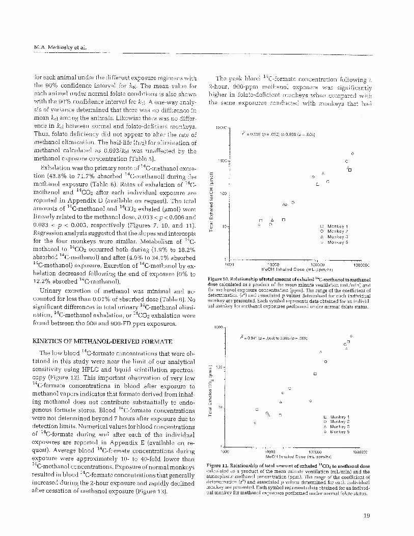
M.A. Medinsky et at
for each animal under the different exposure regimens with the 90% confidence interval for kel. The mean value for each animal under normal folate conditions is also shown with the 90% confidence interval for kel· A one-way analysis of variance determined that there was no difference in mean kel among the animals. Likewise there was no difference in kel between normal and folate-deficient monkeys. Thus, folate deficiency did not appear to alter the rate of methanol elimination. The half-life (fl/2) for elimination of methanol calculated as 0.693/kel was unaffected by the methanol exposure concentration (Table 5).
Exhalation was the primary route of 14C-methanol excretion (43.8% to 71.7% absorbed 14C-methanol) during the methanol exposure (Table 6). Rates of exhalation of 14Cmethanol and 14COz after each individual exposure are reported in Appendix D (available on request). The total amounts of 14C-methanol and 14COz exhaled ().lmol) were linearly related to the methanol dose, 0.033 < p < 0.006 and 0.083 < p < 0.003, respectively (Figures 7, 10, and 11). Regression analysis suggested that the slopes and intercepts for the four monkeys were similar. Metabolism of 14Cmethanol to 14COz occurred both during (1.9% to 18.2% absorbed 14C-methanol) and after (4. 9% to 34.1% absorbed 14C-methanol) exposure. Excretion of 14C-methanol by exhalation decreased following the end of exposure (0% to 12.2% absorbed 14C-methanol).
Urinary excretion of methanol was minimal and accounted for less than 0.01% of absorbed dose (Table 6). No significant differences in total urinary 14C-methanol elimination, 14C-methanol exhalation, or 14COz exhalation were found between the 900 and 900-FD ppm exposures.
KINETICS OF METHANOL-DERIVED FORMATE
The low blood 14C-formate concentrations that were obtained in this study were near the limit of our analytical sensitivity using HPLC and liquid scintillation spectroscopy (Figure 12). This important observation of very low 14C-formate concentrations in blood after exposure to methanol vapors indicates that formate derived from inhaling methanol does not contribute substantially to endogenous formate stores. Blood 14C-formate concentrations were not determined beyond 7 hours after exposure due to detection limits. Numerical values for blood concentrations of 14C-formate during and after each of the individual exposures are reported in Appendix E (available on request). Average blood 14C-formate concentrations during exposure were approximately 10- to 40-fold lower than 14C-methanol concentrations. Exposure of normal monkeys resulted in blood 14C-formate concentrations that generally increased during the 2-hour exposure and rapidly declined after cessation of methanol exposure (Figure 13).
The peak blood 14C-formate concentration following a 2-hour, 900-ppm methanol exposure was significantly higher in folate-deficient monkeys when compared with the same exposures conducted with monkeys that had
10000
1000
:r: ~ 100 2 -o Q)
a; .>:: X w a; 0 1-
10
1000
f- = 0.936 (p = .033) to 0.988 (p = .006)
IX> 0
D t:, D 0 0
0
D
0
0
D Monkey 1 0 Monkey 2 t:, Monkey 3 o Monkey 5
10000 1 00000 MeOH Inhaled Dose (ml-ppm/hr)
0
1000000
Figure 10. Relationship of total amount of exhaled 14C-methanol to methanol dose calculated as a product of the mean minute ventilation (mL!min) and the methanol exP.osure concentration (ppm). The range of the coefficient of determination (/) and associated p values determined for each individual monkey are presented. Each symbol represents data obtained for an individual monkey for methanol exposures performed under n,ormal folate status.
1000
~ 100 E
"' 0 0 Cl <D -;; .c X w
10
1000
l = 0.841 (p = 083) to 0.995 (p = .003)
D
0
D
0 0
0
D
1 0000 1 00000
0
D Monkey 1 0 Monkey 2 D. Monkey 3 o Monkey 5
MeOH Inhaled Dose (ml-ppm/hr)
0
1000000
Figure 11. Relationship of total amount of exhaled 14COz to methanol dose calculated as a product of the mean minute ventilation (mL!min) and the atmospheric methanol concentration (ppm). The range of the coefficient of determination (r2
) and associated p values determined for each individual monkey are presented. Each symbol represents data obtained for an individual monkey for methanol exposures performed under normal folate status.
19

normal folate levels. These peak levels of 14C eluting with the same retention time as formate represented levels of formate that were, however, still at least an order of magnitude lower than endogenous levels of formate and several orders of magnitude lower than formate blood levels associated with toxicity. In addition, HPLC profiles of various 14C-methanol-derived rr;etabolites in the blood of mon~zeys with reduced folate levels were at times very different from HPLC profiles of metabolites in the blood of monkeys with normal folate levels (Figure 12). This difference was especially apparent for blood samples taken at later times during the experiments. For example, the HPLC chromatograms in Figure 12 for blood samples taken from normal and folatedeficient monkeys at 2 hours after the start of the methanol exposure are very similar (top panels). The methanol and formate peaks are distinct and well resolved. In contrast, HPLC profiles of blood samples taken 7.5 hours after the start of the methanol exposure are very different for the normal monkey compared with the folate-deficient monkeys (bottom panels). In particular, the HPLC chromatograms for blood from folate-deficient monkeys contained large peaks of radioactivity that eluted earlier than those coeluting with methanol and formate standards. These earlier peaks contributed to a higher baseline in the chromatograms of blood from monkeys with reduced folate levels, resulting in an inability to resolve the formate peak \>vith sufficient accuracy. This difficulty can be seen most readily by comparing the chromatogram for a 2-hour sam-
500
400
100
75
~50
25
Time(min) 0 2 4
Folate-Deficient
14c-Formate
Time(min)
6 8 1,0 11
2 14 1,6 18 20 Time(min)
Figu~e 12. Representative HPLC chromatograms of heparinized blood samples collected from an individual monkey 2 hours or 7.5 hours after the start of a 2-hour inhalation exposure to 900 ppm methanol. Blood samples were taken under normal or folate-deficient conditions. With the chromatographic system used, 14C-formate and 14C-methanol had 10-minute and 12- to 13-minute retention times, respectively. The formate and methanol peaks are indicated by shaded and hatched areas, respectively. The limit of detection for h'1ese analyses was considered to be a 1.5-fold increase above Lhe baseline radioacti 1Iity immediately preceding the peak of interest.
to MethanDl Vapors
ple with that for a 7.5-hour sample from a folate-deficient monkey (right side, Figure 12). Although it is readily possible to detect a peak due to 14C-formate for the 2-hour sample (top right panel, shaded area), the chromatogram for the 7.5-hour sample has no peak in this area (bottom panel, shaded area). In contrast, the peak coeluting vvith 14C-formate was well resolved in blood taken from the normal monkey at both 2 and 7.5 hours after the start of the exposure (Figure 12, left panels), and thus quantifying formate at all time points was possible. In any event, even though formate could not be precisely quantified in samples from folate-deficient monkeys, the low counts associated with the area of the chromatogram in which the formate peak was located suggests that the 14C-formate concentrations had to be low.
The biochemical mechanism underlying the change in the pattern of metabolites observed after administration of methanol to normal and folate-deficient monkeys is not known. The presence of numerous peaks representing different methanol metabolites in the HPLC chromatograms is not surprising given that formate enters the one-carbon pool. Thus, 14C-formate-derived radioactivity can be incorporated into numerous metabolites in various metabolic pathways. Many of these metabolites can enter the blood. It is reasonable to expect that folate deficiency would alter intermediary metabolism of formate and formate metabolites resulting in a different HPLC profile in blood from normal monkeys compared with that from folate-deficient monkeys.
Some 14C-formate was eliminated in urine. However, urine was not a major route for removal of 14C-methanolderived formate (Figure 14). Urinary elimination is not considered an important route of elimination of formate in general (Karet and Nauss 1990).
DISCUSSION AND CONCLUSIONS
METHANOL EXPOSURE DURING NORMAL FUEL USE
The U.S. Environmental Protection Agency (EPA) has modeled methanol exposure levels that might occur under specific conditions of use (Kavet and Nauss 1990). For example, if 100% of all automobiles were powered by methanol-based fuels, models predict a range of methanol concentrations in street canyons, expressways, railroad tunnels, or parking garages from a low of 1 mg/m3 (0.77 ppm) to a high of 60 mg/m3
. Methanol concentrations in a personal garage during engine idle or hot-soak conditions are predicted to range from 2.9 mg/m3 to 50 mg/m3
. Those predicted during refueling of vehicles range from 30 to 50 mg/m3
. For reference purposes, the TLV of the American

M.A. Mediruky et al.
Conference of Governmental Industrial Hygienists (ACGIH) for exposure to methanol over an 8-hour workday is 260 mg/m3
(200 ppm). This value is for worker populations and not the general public.
The data presented in this study suggest that short-term inhalation of low levels (i.e., at or near the current TLV) of methanol will not result in elevation of blood formate concentrations above those present endogenously. The concentrations of methanol used in this study bracket estimates
100
__.,.__ 10
-~ 45 10
~ ~ 200
~ --+-- 900-N 3
c --+-- 900-FD 0
v~~~~ ~ c " '-' c: 0 0
* E 0.1 0 ~ lL -o 0 0
OJ
0.01
Monkey #1
0.001 --f-,---,------,------,-------,------.-----.----.---. 0 2 4 7
Hours After Start of Exposure
100
10
~ 3 c 0
~
(;.~-· (])
'-' c 0 0 (])
-ro E 0.1 0
~ __,_ 10 ll.
-o 0 ---<>- 45 0 (jj
--A- 200 0.01
-+--- 900-N
--+-- 900-FD
Monkey #3
0.001 0 2 3 4 5 6 7 8
Hours After Start of Exposure
of likely exposure scenarios to methanol vapors to its use as an automotive fuel (Health Effects Institute 1987). The highest methanol concentration (900 and expo-sure duration (2 hours) used in this study exceed conditions that are anticipated to occur under methanol automotive fuel use. For example, the estimated upper hurnan exposure limit occurs with hot-soak conditions (evaporative emissions from a malfunctioning vehicle turned off within a poorly ventilated, closed garage). Under this scenario, am-
100
-iJ- 10
10 --0>--- 45
~ -.r-- 200 3
~0 ---+- 900 N c 0
·~ ---e--- 900-FD
c " '-' c 0 0
~c-~.~ * E 0.1 0 lL -o 0 0 (jj
0.01
Monkey #2
0.001-+.---------r-----r-----;----,-----.---, 0 2 3 4 6 7
Hours After S1art of Exposure
100
10
~ /~-::-------'--_ ______________________ 3 c 0
~ c • (])
'-' c:
~-' 0 0 2 "' E 0.1 0
" ~J lL -o
r----~~ ~,~" 0 0 (jj
0.01
_,_ 10 ~ 200 --+- 900-FD
Monkey #5 -l>- 45 -+--- 900-N
0.001 0 2 3 4 6 7
Hours After Start of Exposure
Figure 13. Blood 14C-formate concentration (fiM) for individual animals during and following a 2-hmn inhalation exposure lo 10, 45, 200, or 900 ppm methanol. Data from a 2-hour 900 ppm exposure under folate-deficient status (900-FD) have also been included. Some data have been truncated beyond 3 hours because the higher baseline in the HPLC chromatograms at these later times resulted in an inability to resolve the formate peak with sufficient accuracy. Endogenous blood formate concentrations are reported to be between 100 and 200 flM (McMartin and coworkers 1979; Horton and coworkers 1992). Taken from Dorman and coworkers 1994 with permission.
21

Pharmacokinetics of Methanol a_qd Formate in female Cynomolgus Monkeys Exposed to Methanol Vapors
bient air methanol concentrations may reach 240 mg/m3
(185 ppm); but only short-term human exposures (< 15 minutes) will occur under these conditions. Similarly, a typical open-air automobile refueling with methanol will result in an approximately 3- to 4-minute exposure to 33 to 50 mg/m3 (25 to 38 ppm) of methanol (Harvey et al. 1984).
Other individuals who could be exposed to methanol vapors include tunnel workers, garage mechanics, and underground parking lot attendants. The exposure duration for these workers could be significantly longer than 2 hours, potentially approaching the 8-hour working day. Results of methanol-derived formate concentrations reported here could be extrapolated to this longer exposure duration. For example, the highest concentration used in the present study, 900 ppm, and the exposure duration, 2 hours, gives a concentration-time product of 1800 ppm-hours. Because the 8-hour TLV for the workplace is 200 ppm, the maximum concentration-time product that a worker could be exposed to is 1600 ppm-hours, a value approximately equal to the highest concentration-time product used in the present study. This calculation has an additional element of conservatism in that the methanol dose rate to the worker is four-fold slower than the dose rate used in our studies, since the total human dose is delivered over an 8-hour rather than a 2-hour period. This slower dose rate gives more time for detoxification of any formate that is produced. Thus, even for workers in garages and tunnels, methanol-derived formate will constitute a small fraction of the total formate pool.
1600
1400 f~-------1: 900-FD ppm
!1200 ---+------_____, 900 ppm
" ~ 1000
0 §
) 800
c ~ 600
:'5
~ 400 § 0 L--------J,-------.l. 200 ppm
200
45 ppm
10 ppm
24 48 72 Hours After Start of Methanol Exposure
Figure 1'1. Cumulative mean (± SEM) urine formate concentration (nmol) following a 2-hour inhalation exposure to 10, 45, 200, or 900 ppm methanol. Data from a 2-hour 900 ppm exposure under folate-deficient status (900-FD) have also been included (n = 4 monkeys per dose).
22
METHANOL UPTAKE, DISTRIBUTION, AND ELIMINATION
Methanol is an endogenous chemical with body bmdens arising from dietary somces and metabolic processes. Methanol is available in the ingestion of dietary fruits, fruit juices, alcoholic beverages, and certain vegetables, as well as from the gastrointestinal hydrolysis of the artificial sweetener aspartame. Metabolic sources for methanol production arise from the action of the methyltransferase enzyme system (Stegink et al. 1983).
Exposure of primates to 14C-methanol vapors resulted in only a small contribution to the reported endogenous blood methanol level. For example, the mean blood 14C-methanol concentrations (± SD) at the end of the 2-hour methanol exposure of 10, 45, 200, or 900 ppm were 0.65 ± 0.3, 3.0 ± 0.8, 21 ± 16, and 106 ± 84 ~M. respectively. Blood methanol concentrations obtained in our studies are equivalent to or below the endogenous blood methanol concentrations reported for healthy adult male humans (10 to 110 ~M; Sedivec et al. 1981; Lee et al. 1992). Similar background levels have also been observed in monkeys (Horton et al. 1992). Blood methanol concentrations determined at the end of our 200-ppm exposure also compare favorably with those reported by Horton and coworkers (1992), who used rhesus monkeys. In their study, a threefold longer inhalation exposure (6 hours) to 50 ppm methanol did not result in any increase in blood methanol above background levels. Similarly, a blood methanol concentration of 121 ~M was observed at the end of a 6-hour inhalation exposure to 200 ppm methanol (Horton et al. 1992). Likewise, exposure of humans to 190 ppm for 75 minutes (Cook et al. 1991) or to 200 ppm methanol for 6 hours (Lee et al. 1992) resulted in an average blood methanol concentration at the end of exposure of 58 ~M and 220 ~M, respectively. These results suggest that exposure of healthy humans to methanol at 200 ppm for 8 hours (the current permissible exposure limit set by the Occupational Safety and Health Administration) should not result in a toxicologically significant increase in the total blood methanol concentration.
Not unexpectedly, exhalation was the primary route of methanol excretion during exposure (44% to 72% absorbed methanol) and in the time immediately thereafter. The total amount of methanol exhaled was directly proportional to the methanol dose. As with other studies (Dutkiewicz et al. 1980; Kawai et al. 1991; Horton et al. 1992), urinary excretion of methanol was minimal and accounted for less than 0.01% of the absorbed dose. The elimination half-life of methanol observed in our study ( < 1 hour) is also similar to that reported for humans (1 to 3 hours) exposed by inhalation to 78 to 230 ppm for 8 hours (Sedivec et al. 1981). In our study, the half-life of methanol elimination was unaf-

M.A. Medinsky et al.
fected by the methanol dose. In addition, the blood methanol AUC was linearly related to the methanol dose, indicating that methanol metabolism and pharmacokinetics were independent of dose under the exposure conditions used in these studies. These results suggest that the pharmacokinetics of methanol is first-order at the very low levels of methanol exposure used in our studies. The methanol doses resulting from the lowest levels of exposure in our studies (10 and 45 ppm, 2 hours) are less than that arising from the ingestion of a single 355-mL diet beverage containing the artificial sweetener aspartame (Kavet and Nauss 1990). In comparison, Horton and coworkers (1992) observed nonlinear kinetics in rhesus monkeys that inhaled methanol vapors at 200, 1200, or 2000 ppm for a 6-hour exposure. Horton and coworkers observed a linear relationship between end-of-exposure blood concentrations and inhaled methanol vapor concentrations at 1200 and 2000 ppm, but the 200-ppm exposure concentration resulted in blood methanol levels less than those proportional to the two higher doses. This nonlinearity suggested to these investigators that methanol elimination proceeds by a saturable pathway and that monkeys demonstrate dose-dependent kinetics between 200 and 2000 ppm. Whether longer exposures to the lower levels of methanol used in our study would also have resulted in nonlinear methanol kinetics is unknown.
DIETARY, ENDOGENOUS, AND METHANOL-DERIVED FORMATE
Along with methanol, formate is also present in blood at low endogenous concentrations. Formate is naturally found in some foods (including honey, fruit syrups, and roasted coffee) and also has been used as a food preservative (Health Effects Institute 1987). Formate is also produced as a byproduct of several metabolic pathways, including histidine and tryptophan degradation (Health Effects Institute 1987). Formate's toxicologic significance, however, is related to its role as the toxic metabolite of methanol. Increased blood and tissue formate concentrations following high-dose methanol administration can account for the development of both metabolic acidosis and blindness in humans and nonhuman primates (McMartin et al. 1977, 1980; Roe 1982). Blood formate levels in excess of 10 mM are reported to occur in humans with neuro-ocular toxicosis following the ingestion of methanol (McMartin et al. 1980; Sejersted et al. 1983). Similarly, nonhuman primates given intravenous formate (approximately 3.1 mEq/kg/hr) for up to 50 hours developed maximal blood formate concentrations of 12 to 34 mM (Martin-Amat et al. 1978) with comparable cerebrospinal fluid formate concentrations (Martin-Amat et al. 1977). These monkeys subsequently developed metabolic
acidosis and optic nerve edema, characteristics of methanol poisoning. Permanent visual damage in methanol- and formate-poisoned monkeys (Hayreh et al. 1977) and in methanol-poisoned humans (Jacobsen and McMartin 1986) is reportedly associated with prolonged elevations (often greater than 24 hours) in blood formate concentration (> 7 to 10mM).
After inhalation of lower methanol concentrations, however, it appears unlikely that formate will accumulate to these toxicologically significant levels. Numerous studies support this conclusion. Exposure of nonhuman primates to 200 to 2000 ppm methanol for 6 hours likewise did not result in any increase in blood formate concentrations above endogenous levels (Horton et al. 1992). Studies conducted with the artificial sweetener aspartame, which is hydrolyzed to methanol in the gastrointestinal tract, further support these findings (Stegink et al. 1983, 1989). Our study has extended these previous findings by clearly identifying the direct contribution of 14C-methanol-derived 14C-formate to the endogenous blood formate pool following lowdose exposure to methanol. In our study, no significant accumulation of 14C-methanol-derived formate occurred in any of the 14C-methanol-exposed monkeys. Exposure of normal monkeys to 900 ppm for 2 hours resulted in a peak blood 14C-formate concentration of only 2.81 ± 1. 7 11M. This peak formate level represents only a small fraction of the endogenous formate blood concentration. For example, Buttery and Chamberlain (1988) reported plasma formate values ranging from 0.12 to 0.28 mM for 30 normal human subjects. Similar endogenous blood formate concentrations (0.1 to 0.2 mM) have also been reported for monkeys (McMartin et al. 1979; Horton et al. 1992), including our own (0.28 to 0.56 mM).
The studies described in this report were conducted using nonhuman primates, an acceptable experimental surrogate for humans. The question that always arises when using animal models is, can the results obtained in animals be extrapolated to humans? Recent experimental studies in humans are consistent with experiments in monkeys. Elevation of blood formate levels following 75-minute to 6-hour inhalation exposure to methanol at the TL V (200 ppm) did not occur in normal male human volunteers (Sedevic et al. 1981; Cook et al. 1991; Lee et al. 1992). For example, Lee and coworkers (1992) exposed healthy volunteers to methanol concentrations of 200 ppm for 6 hours. The subjects were either at rest or engaged in mild exercise. These investigators were able to observe a 3.5- to 4-fold increase in the peak methanol concentration in blood of the exposed volunteers compared to their preexposure levels, indicating that they had absorbed some of the methanol vapor. However, the investigators detected no change in the blood
23

formate concentration compared with the preexposure value. Similar results were observed when humans rested or exercised while exposed to methanol. Most d' Alessandro and coworkers ( 1994) measured fonnate levels in urine and serum after controlled methanol exposures of volunteers at the TLV (200 ppm). These investi-gators could not detect any increase in formate in urine or serum due to methanol exposure compared to the control values. Taken together, results of studies in humans or monkeys exposed to concentrations of methanol ranging from 10 to 2000 ppm suggest that exposure to methanol vapors during the normal use of methanol fuel will not pose an unacceptable rise in formate levels in healthy adults.
THE FOLATE PATHI!VAY AND SENSITIVE POPULATIONS
ll'1 both rats and monkeys, the major route of formate oxidation is through a folate-dependent pathway (McMartin et al.
1977). Formate oxidation by this pathway is dependent upon the activities of formyl THF synthetase and formaldehyde THF dehydrogenase and its co substrate THF (Black et al. 1985). Treatments that alter the level of hepatic THF (e.g., dietary manipulation, nitrous oxide, or methotrexate) can modify methanol toxicity in monkeys and rats (Makar and Tephly 1976; McMartin et al. 1977, 1979; Noker and Tephly 1980; Eells et al. 1983). The degree of folate deficiency achieved in our study has been associated with increased formate accumulation and enhanced acute methanol neuro-ocular toxicity (McMartin et al. 1977). De
crease in the folate metabolic pool prolongs formate blood levels by decreasing the rate at which formate combines with THF, the first step in the metabolism of formate to COz (McMartin et al. 1977). Our studies with moderately folatedeficient monkeys were conducted in order to determine whether metabolism of low doses of methanol-derived formate could be altered in a potentially sensitive folate-deficient subpopulation. The serum and RBC folate levels observed in our monkeys are consistent with a degree of folate deficiency associated with damaged folate-depend
ent metabolism (stage III offolate deficiency, Herbert scale, Herbert 1990). The sequence of biochemical changes that occurred in a normal human adult male continuously consurning a nearly folate-free diet has been described by Herbert (1962). Changes that occur in a folate-deficient human include reduced serum folate (3 weeks after start of
the folate-reduced diet), markedly elevated serum urinary forminoglutamic acid (FIGLU) at 18 weeks, and severely reduced RBC folate at 18 weeks. Male cynomologus monkeys fed a similar diet demonstrated comparable reductions in folate at 5 weeks, elevated FIGLU at 12 weeks,
and decreased RBC folate at 13 weeks (Blocker and Thenen 1987). Based on their work, the Herbert scale is appropriate for the cynomolgus monkey. This monkey is a particularly relevant animal model for human folic acid deficiency. When folate-deficient, these nonhuman primates show temporal development of the biochemical, hematological, and cytological changes characteristic of human megaloblastic anemia quite similarly to folate-deficient humans.
Folic acid deficiency is a common finding in subjects with chronic alcoholism, pregnant women, and patients with chronic debilitation (Herbert 1990). The average(± SD) peak blood 14C-methanol-derived formate concentrations were significantly higher in folate-deficient monkeys (9.54
± 4.65 11M) when compared with those obtained under folate-normal conditions (2.81 ± 1.7 ~tM). However, these blood 14C-formate concentrations were considerably lower than endogenous blood formate levels (100 to 200 !lM) observed in normal monkeys (Horton et al. 1992). Further, no significant differences in total urinary 14C-methanol elimination, 14C-methanol exhalation, or total metabolism of 14C-methanol to 14COz were observed between the 900
ppm exposures under either folate-deficient or normal conditions. These observations suggest that moderate folate deficiency should not result in significant formate accumulation following a single low-dose exposure to methanol.
DEVELOPMENTAL NEUROTOXICITY
The potential for exposure to methanol vapors generated during normal fuel use raises toxicological issues beyond those related to the occular toxicity and metabolic acidosis following acute methanol exposure. For example, teratogenicity studies conducted in rodents (Bolon et al. 1993,
1994; Rogers et al. 1993) have demonstrated that inhalation of methanol on the appropriate gestation day impairs neural tube closure. The concentrations of methanol used in the teratogenicity studies greatly exceed estimates of likely exposure scenarios to methanol vapors related to its use as an automotive fu.el. Extrapolation to humans is further confounded by the dramatic differences in the way rodents
and primates metabolize methanol and formate. Is the biochemical basis for the observed teratogenic effects in ro
dents relevant for humans? The answer to this question depends upon whether formate or methanol is involved in the teratogenic response.
Formate seems to play no apparent role in the development of methanol-induced exencephaly in mice (Dorman et al. 1995). Studies reported by Dorman and coworkers (1995) provide strong evidence that methanol, and not formate, is the proximate developmental toxicant and teratogen in pregnant CD-1 mice exposed to high concentra-

el al.
tions of methanol vapor. Mouse embryos display failure of the anterior neuropore to close (exencephaly) when dams inhale methanol concentrations of at least 10,000 ppm on gestation day 8 (copulation plug= gestation day O). Pharmacokinetic analyses in pregnant mice suggest that exencephaly is directly related to the high concentrations ofthe parent compound, methanol, in maternal blood and embryos rather than the accumulation offormate. Additionally, mice given a large single oral dose of sodium formate (750 mg/kg) on gestation day 8 did not develop exencephaly. In these animals, peak formate concentrations were also measured in the maternal plasma and the decidual complex (a group oftissues within the uterus, composed ofthe embryo and placental and embryonic fluids surrounded by the decidual membrane). Peak formate concentrations in plasma and decidual tissue of mice injected with sodium formate were similar to formate concentrations found in the same tissues of mice exposed to 15,000 ppm methanol. While the formate injections did not result in exencephaly, the 15,000-ppm methanol exposure did result in an increased incidence of exencephaly. Inhalation exposure to 15,000 ppm methanol over 6 hours also resulted in high concentrations of methanol (223 ± 23 mM) in maternal plasma of mice, suggesting that methanol itself may be the ultimate teratogen.
Finally, the relationship between folate status and developmental neurotoxicity is well recognized. Sakanashi and coworkers (1994) reported an increase in methanol-induced exencephaly and other terata in CD-1 mice placed on a folate-deficient diet and also reported that folate supplementation ameliorated those adverse developmental effects. Research on this important but unresolved aspect of methanol toxicity should help answer questions about the risk of exposure of the developing human fetus to low concentrations of methanol if it is used as an alternative fuel.
IMPLICATIONS OF FINDINGS
The objective of Specific Aim 1 of our study was to determine the contribution of methanol-derived formate (14C-labeled formate in these studies) to the total formate
body pool. The results of these experiments indicated that a 2-hour exposure to methanol concentrations up to 900 ppm produced a maximum, methanol-derived formate level that was only a fraction of the endogenous formate level and several orders of magnitude lower than levels of formate known to be toxic. These studies suggest that the maximum blood concentration of formate resulting from exposure to low concentrations of methanol, such as those that might be the result of methanol being used as an alternative fuel is insignificant compared with the endo-
genous formate levels. Our studies are consistent with results from recent studies conducted in humans in which increases in formate in urine or serum due to methanol exposure at the TL V could not be detected. Taken together, results of studies in people or monkeys suggest that brief exposure to methanol vapors during norm.al use of methanol fuel will not pose an rislc to healthy adults.
A significant fraction of the human population may be folate-deficient, and folate is pivotal in detoxifying formate, the toxic metabolite of methanol. Therefore, are folate-deficient individuals exposed to methanol at special risk? Our results in Specific Aim 2 indicate that even with a reducedfolate status, monkeys exposed to 900 ppm methanol for 2 hours still had peak concentrations of methanol-derived formate that were well below the endogenous levels of formate and orders of magnitude lower than levels that produce acute methanol toxicity. Although these results only represent a single exposure and therefore preclude broad generalizations, they do suggest that even the folatedeficient body contains sufficient folate stores to effectively detoxify small doses of methanol-derived formate from exogenous sources, such as those that might occur during average use of automotive fuel.
In summary, qualitative similarities in methanol metabolism between monkeys and humans (Roe 1982) suggest that the findings reported here for monkeys could be extrapolated to low-dose inhalation of methanol vapors by humans. Our data suggest that acute inhalation of methanol at concentrations relevant to human occupational and environmental exposures by healthy or moderately folate-deficient humans would not result in formate accumulation above endogenous levels. Whether methanol or formate may accumulate in target tissue (e.g., retina, optic nerve, or brain) or in blood following more prolonged methanol exposure is unknown, and these issues warrant further study.
ACKNOWLEDGMENTS
We would like to acknowledge Dr. Georgia Farris for conduct of hematology analyses. We also thank Dr. Bahman Asgharian, R. Arden James, Nancy Youtsey, Del Ponder, and Marianne Marshall for their assistance with the endotracheal intubation exposures. We would like to thank Nancy Youtsey and Horace Parkinson for methanol and formate blood analyses. Ophthalmoscopic examinations were performed by Drs. Mark Nasisse of the North Carolina State University School of Veterinary Medicine and Robert Peiffer Jr., at the University of North Carolina. We would also like to recognize the significant contributions made by Dr. Fred]. Miller to development of the experimental design and data analysis strategy. We would also like to acknow-
25

Pharmacokinetics of Methanol and Forma~e i;11 female Cynomolgus JExpnsed to Methanol Vapors
ledge Carol Bobbitt, Paul Ross, and the staff of the Animal Care Unit for their assistance with monkey handling and care.
REFERENCES
Abou-Saleh MT, Cop pen A. 1986. Psychiatric progress: The biology of folate in depression: Implications for nutritional hypotheses of the psychoses. J Psychiatr Res 20:91-101.
Bailey LB, Wagner PA, Christakis GJ, Araujo PE, Appledorf H, Davis CG, Masteryanni J, Dinning JS. 1979. Folacin and iron status and hematological findings in predominately
black elderly persons from urban low-income households.
Am J Clin Nutr 32:2346-2353.
Bailey LB, Wagner PA, Christakis GJ, Davis CG, Appledorf H, Araujo PE, Dorsey E, Dinning JS. 1982. Folacin and iron status and hematological findings in Black and SpanishAmerican adolescents from urban low-income households. Am J Clin Nutr 35:1023-1032.
Black KA, Eells JT, Noker PE, Hawtrey CA, Tephly TR. 1985. Role of hepatic tetrahydrofolate in species differences in methanol toxicity. Proc Natl Acad Sci (USA) 82:3854-3858.
Blocker DE, Thenen SW. 1987. Intestinal absorption, liver uptake, and excretion of 3H-folic acid in folic acid ·deficient, alcohol-consuming nonhuman primates. Am J Clin Nutr 46:503-510.
Bolon B, Welsch F, Morgan KT. 1994. Methanol-induced neural tube defects in mice: Pathogenesis during neurulation. Teratology 49:497-517.
Bolon B, Dorman DC, Janszen D, Morgan KT, Welsch F. 1993. Phase-specific developmental toxicity in mice following maternal methanol inhalation. Fundam Appl Taxicol 21:508-516.
Buttery JE, Chamberlain BR. 1988. A simple enzymatic method for the measurement of abnormal levels of formate in blood. J Anal Toxicol 12:292-294.
Clark AJ, Mossholder S, Gates R. 1987. Folacin status in adolescent females. Am J Clin Nutr 46:302-306.
Colman N. 1977. Folate deficiencies in humans. In: Advances in Nutritional Research, Vol. 1 (HH Draper, ed.) pp. 77-124. Plenum Press, New York, NY.
Cook MR, Bergman FJ, Cohen HD, Gerkovich MM, Graham C, Harris RK, Siemann LG. 1991. Effects of Methanol Vapor on Human Neurobehavioral Measures. Research Report Number 42. Health Effects Institute, Cambridge, MA.
26
Cziezel AE, Dudas I. 1992. Prevention ofthe first occurrence of neural-tube defects by periconceptional vitamin supplementation. N Engl J Med 327:1832-1835.
d'Alessandro A, Osterloh JD, Chuwers P, Quinlan PJ, Kelly TJ, Becker CE. 1994. Formate in serum and urine after controlled methanol exposure at the threshold limit value. Environ Health Perspect 102:178-181.
Daniel WA, Gaines EG, Bennett DL. 1975. Dietary intakes and plasma concentrations of folate in healthy adolescents. Am J Clin Nutr 28:363-370.
Dansky LV, Andermann E, Rosenblatt D, Sherwin AL, Andermann F. 1987. Anticonvulsants, folate levels, and pregnancy outcome: A prospective study. Ann Neural 21:176-182.
Dorman DC, Dye JA, Nassise MP, Ekuta J, Bolon B, Medinsky MA. 1993. Acute methanol toxicity in swine. Fundam Appl Toxicol20:341-347.
Dorman DC, Moss OR, Farris GA, Janszen D, Bond JA, Medinsky MA. 1994. Pharmacokinetics of inhaled 14Cmethanol and methanol-derived 14C-formate in normal and folate-deficient cynomolgus monkeys. Toxicol Appl Pharmacal 128(2):229-232.
Dutkiewicz B, Konczalik J, Karwacki W. 1980. Skin absorption and per os administration of methanol in men. Int Arch Occup Environ Health 47:81-88.
Dunn RT, Foster LB. 1973. Radioassay of serum folate. Clin Chern 19:1101-1105.
Eells JT, Black KA, Tedford CE, Tephly TR. 1983. Methanol toxicity in the monkey: Effects of nitrous oxide and methionine. J Pharmacal Exp Ther 227:349.
Everson RB, Wehr CM, Erexson GL, MacGregor JT. 1988. Association of marginal folate depletion with increased human chromosomal damage in vivo: Demonstration by analysis of micronucleated erythrocytes. JNCI 80:525-529.
Fanelli-Kuczmarski MT, Johnson CL, Elias L, Najjar MF. 1990. Folate status of Mexican American, Cuban, and Puerto Rican women. Am J Clin Nutr 52:358-372.
Gerde P, Dahl AR. 1991. A model for the uptake of inhaled vapors in the nose of the dog during cyclic breathing. Toxicol Appl Pharmacal 109:276-288.
Gonzales-Buitrago JM, Ruiz J, Garcia Bastos JL, Navajo JA, Barque L, Maside C, Rodriguez A, Balas D, Palet A. 1994. Multicentre evaluation of the Boehringer Mannheim Hitachi 911 system for homogenous immunoassays. Eur J Clin Chern Clin Biochem 32:489-493.

M.A. Medinsky et al.
Grinblat J, Marcus DL, Hernandez F, Freedman ML. 1986. Folate and vitamin B12 levels in an urban elderly population with chronic diseases: Assessment of two laboratory folate assays: Microbiologic and radioassay. J Am Geriatr Soc 34:627-632.
Halsted CH. 1977. Jejunal perfusion of simple and conjugated folates in celiac sprue. J Clin Invest 59:933940.
Harvey CA, Carey PM, Somers JH, Garbe RJ. 1984. Toxicologically acceptable levels of methanol and formaldehyde emissions from methanol-fueled vehicles. SAE Technical Paper Series #841357. Society of Automotive Engineers, Warrendale, PA.
Hayreh MS, Hayreh SS, Baumbach GL, Cancilla P, MartinAmat G, Tephly TR, McMartin KE, Makar AB. 1977. Methyl alcohol poisoning: III. Ocular toxicity. Arch Ophthalmol 95:1851-1858.
Health and Welfare Canada. 1973. Nutrition Canada National Survey, Information. Ottawa, Canada.
Health Effects Institute. 1987. Automotive Methanol Vapors and Human Health: An Evaluation of Existing Scientific Information and Issues for Future Research (A Special Report). Health Effects Institute, Cambridge, MA.
Henderson DR, Friedman SB, Harris JD, Manning WB, Zoccoli MA. 1986. CEDIA, a new homogeneous immunoassay system. Clin Chern 32:1637-1641.
Herbert V. 1962. Experimental nutritional folate deficiency in man. Trans Assoc Am Physicians 75:307-320.
Herbert V. 1987. The 1986 Herman Award Lecture: Nutrition as a continually unfolding story: The folate and vitamin B12 paradigm. Am J Clin Nutr 46:387-402.
Herbert V. 1990. Development of human folate deficiency. In: Folic Acid Metabolism in Health and Disease (Picciano MF, Stokstad ELR, Gregory JF, eds.) pp 195-210. WileyLiss, New York, NY.
Hillman RS. 1980. Vitamin B12, folic acid, and the treatment of megaloblastic anemias. In: The Pharmacological Basis of Therapeutics (Gilman AG, Goodman LS, Gilman AG, eds.). MacMillan Publishing Co., New York, NY.
Hirono H, Wada Y. 1978. Effects of dietary folate deficiency on developmental increase of myelin lipids in rat brain. J Nutr 108:766-772.
Homler BE. 1984. Aspartame: Implications for the food scientist. In: Aspartame: Physiology and Biochemistry (Stegink LD, Filer JR Jr, eds.) pp. 247-262. Marcel Dekker, New York, NY.
Horton VI, Higuchi MA, Rickert DE. 1992. Physiologically based pharmacokinetic model for methanol in rats, man
and humans. Toxicol Appl Pharmacol117:26-32.
Hoyumpa AJVL 1986. Mechanisms of vitamin deficiencies in alcoholism. Alcohol Clin Exp Res 10:573-581.
Jacobsen D, McMartin KE. 1986. IVIethanol and ethylene glycol poisonings: Mechanism of toxicity, clinical course, diagnosis, and treatment. Med Toxicol 1:309-334.
Johlin FC, Fortman CS, ]\Jgheim DD, Tephly TR. 1987. Studies on the role of folic acid and folate-dependent enzymes in human methanol poisoning. Mol Pharmacal 31:557-561.
Johlin FC, SwainS, Smith C, Tephly TR. 1989. Studies on the mechanism of methanol poisoning: Purification and comparison of rat and human liver 10-formyltetrahydrofolate dehydrogenase. Mol Pharmacal 35:745-750.
Kavet R, Nauss KM. 1990. The toxicity of inhaled methanol vapors. CRC Crit Rev Toxicol21:21-50.
Kawai T, Yasugi T, Mizunuma K, Horiguchi S, Hirase Y, Uchida Y, Ikeda M. 1991. Methanol in urine as a biological indicator of occupational exposure to methanol vapor. Int Arch Occup Environ Health 63:311-318.
Khanna PL, Dworschack RT, Manning WB, Harris JD. 1989. A new homogenous enzyme immunoassay using recombinant enzyme fragments. Clin Chim Acta 185:231-240.
Klipstein FA. 1964. Subnormal serum folate and macrocytosis associated with anticonvulsant drug therapy. Blood 23:68-86.
Lee EW, Terzo TS, D'Arcy JB, Gross KB, Schreck RM. 1992. Lack of blood formate accumulation in humans following exposure to methanol vapor at the current permissible exposure limit of 200 ppm. Am Ind Hyg Assoc J 53:99-104.
Lee EW, Garner CD, Terzo TS. 1994. Animal model for the study of methanol toxicity: Comparison of folate-reduced rat responses with published monkey data. J Toxicol Environ Health 41:71-82.
Lieber CS. 1988. The influence of alcohol on nutrition status. Nutr Rev 46:241-254.
MacGregor JT, Christensen CC. 1993. Study of the relationship between folate status and methanol toxicity. API Publication Number 4554. Health and Environmental Sciences Department, American Petroleum Institute, Washington, DC.
27

!Pharmacokinetics of Methanol and formate in Female Cynomolgus Monkeys Exposed to Methanol Vapors
Makar AB, Tephly TR. 1976. Methanol poisoning in the folate-deficient rat. Nature 261:715-717.
Makar AB, Tephly TR, Osweiler G. 1990. Formate metabolism in young swine. Toxicol ApplPharmacol105:315-320.
Martin-Amat G, McMartin KE, Hayreh SS, Hayreh MS, Tephly TR. 1978. Methanol poisoning: Ocular toxicity produced by formate. Toxicol Appl Pharmacol45:201-208.
Martin-Amat G, Tephly TR, McMartin KE, Makar AB, Hayreh MS, Hayreh SS, Baumbach G, Cancilla P. 1977. Methyl alcohol poisoning: II. Development of a model for ocular toxicity in methyl alcohol poisoning using the rhesus monkey. Arch Ophthalmol 95:1847-1850.
McMartin KE. 1977. The Role of Formic Acid in Methanol Poisoning in the Monkey. Ph.D. Dissertation. University of Iowa, Iowa City, IA.
McMartin KE, Ambre JJ, Tephly TR. 1980. Methanol poisoning in human subjects: Role for formic acid accumulation in the metabolic acidosis. Am J Med 68:414-418.
McMartin KE, Martin-Amat G, Makar AB, Tephly TR. 1977. Methanol poisoning: V. Role of formate metabolism in the monkey. J Pharmacal Exp Ther 201:564-572.
McMartin KE, Martin-Amat G, Noker PE, Tephly TR. 1979. Lack of a role for formaldehyde in methanol poisoning in the monkey. Biochem Pharmacal 28:645-649.
McMartin KE, Makar AB, Martin-Amat G, Palese M, Tephly TR. 1975. Methanol poisoning: I. The role of formic acid in the development of metabolic acidosis in the monkey and the reversal by 4-methylpyrazole. Biochem Med 13:319.
McPartlin J, Halligan A, Scott JM, Darling M, Weir DG. 1993. Accelerated folate breakdown in pregnancy. Lancet 341:148-149.
Medinsky MA, Dorman DC. 1994. Assessing risks of lowlevel methanol exposure. CIIT Activities 14(7):1-7.
Mendeloff AI. 1980. The epidemiology of inflammatory bowel disease. Clin Gastroenterol9:259-270.
Milunsky A, Jick H, Jick SS, Bruell CL, MacLaughlin DS, Rothman KJ, Willett W. 1989. Multivitamin/folic acid supplementation in early pregnancy reduces the prevalence of neural tube defects. JAMA 262:2847-2852.
MRC Vitamin Study Research Group. 1991. Prevention of neural tube defects: Results ofthe Medical Research Council Vitamin Study. Lancet 338:131-137.
Noker PE, Tephly TR. 1980. The role of folates in methanol toxicity. In: Alcohol and Aldehyde Metabolizing Systems, Vol IV (RG Thurman, ed.) pp. 305-315. Plenum Press, New York, NY.
Pedersen AB, Mosbeck J. 1969. Morbidity of pernicious anemia. Acta Med Scand 185:449-452.
Reiter LA, Boylan LM, Driskell J, Moak S. 1987. Vitamin B-12 and folate intakes and plasma levels of black adolescent females. JAm Diet Assoc 87:1065-1067.
Reynolds EH, Rothfield P, Pincus JH. 1973. Neurological disease associated with folate deficiency. Br Med J 2:398-400.
Roak-Foltz R, Leveille GA. 1984. Projected aspartame intake: Daily ingestion of aspartic acid, phenylalanine, and methanoL In: Aspartame: Physiology and Biochemistry (Stegink LD, Filer JR Jr, eds.) pp. 201-205. Marcel Dekker, New York, NY.
Roe 0. 1982. Species differences in methanol poisoning. CRC Crit Rev Toxicol10:275-286.
Rogers JM, Mole ML, Chernoff N, Barbee BD, Turner CI. Logsdon TR, Kavlock RJ. 1993. The developmental toxicity of inhaled methanol in the CD-1 mouse, with quantitative dose-response modeling for estimation of benchmark doses. Teratology 47:175-188.
Rosenberg IH, Bowman BB, Booper BA, Halsted CH, Lindenbaum J. 1982. Folate nutrition in the elderly. Am J Clin Nutr 36:1060-1066.
Rothenberg SP, DaCosta M, Rosenberg BS. 1972. A radioassay for serum folate: Use of a two-phase sequential incubation, ligand-binding system. N Engl J Med 285:1335-1339.
Sakanashi TM, Rogers JM, Keen CL. 1994. Influence offolic acid intake on the developmental toxicity of methanol in the CD-1 mouse. Teratology 49:368.
Sauberlich HE. 1990. Evaluation of folate nutrition in population groups. In: Folic Acid Metabolism in Health and Disease (MF Picciano, ELR Stokstad, JF Gregory III, eds.) pp. 211-236. Wiley & Sons, New York, NY.
Sedivec V, Mraz M, Flek J. 1981. Biological monitoring of persons exposed to methanol vapours. Int Arch Occup Environ Health 48:257-271.
Sejersted OM, Jacobsen D, Ovrebo S, Jansen H. 1983. Formate concentrations in plasma from patients poisoned with methanoL Acta Med Scand 213:105-110.

M.A. Medinsky ei al.
Senti FR, Pilch SM, eds. 1984. Assessment of the Folate Nutritional Status of the U.S. Population Based on Data Collected in the Second National Health and Nutrition Examination Survey, 1976-1980. Life Sciences Research Office, Federation of American Societies for Experimental Biology, Bethesda, MD.
Senti RR, Pilch SM. 1985. Analysis of folate data from Second National Health and Nutrition Examination Survey (NHANES II). J J\Tutr 115:1398-1402.
Smithells RW, Sheppard S, Wild J, Schorah CJ. 1989. Prevention of neural tube defect recurrences in Yorkshire: Final report. Lancet 2:489-499.
Stegink LD, Brummel MC, Filer LJ Jr, Baker GL. 1983. Blood methanol concentrations in one-year-old infants administered graded doses of aspartame. J Nutr 113:1600-1606.
Stegink LD, Filer LJ Jr, Bell EF, Ziegler EE, Tephly TR. 1989. Effect of repeated ingestion of aspartame-sweetened beverages on plasma amino acid, blood methanol, and blood formate concentrations in normal adults. Metabolism 38:357-363.
Stegink LD, Brummel MC, McMartin K, Martin-Amat G, Filer Jr LJ, Baker GL, Tephly TR. 1981. Blood methanol concentrations in normal adult subjects administered abuse doses of aspartame. J Toxicol Environ Health 7:281-290.
Tephly TR, Green MD, Gamble J. 1992. Formate metabolism in micropigs. Toxicol Appl Pharmacal 116:142-145.
Tephly TR, McMartin KE. 1984. Methanol metabolism and toxicity. In: Aspartame: Physiology and Biochemistry (Stegink LD, Filer JRJr, eds.) pp. 111-140. Marcel Dekker, New York, NY.
Tolarova M, Harris J. 1995. Reduced recurrence of oro facial clefts after periconceptional supplementation with highdose folic acid and multivitamins. Teratology 51:71-78.
van der Weide J, Homan HC, Cozijnsen-van Rheenen E, Vivie-Kipp Y, Poortman J, Kraaijenhagen RJ. 1992. Nonisotopic binding assay for measuring vitamin B12 and folate in serum. Clin Chern 38:766-768.
Wintrobe MM, Lee CR, Boggs DR, Bithell TC, Foerster J, Athens JW, Lukens JN. 1981. Clinical Hematology, 8th ed., pp. 559-604. Lea and Febiger, Philadelphia, PA.
Young SN, Ghadirian AM. 1989. Folic acid and psychopathology. Prog Neuropsychopharmacol Biol Psychiatry 13:841-863.
Ziegler SL. 1921. The ocular menace of wood alcohol poisoning. JAMA 77:1160.
APPENDICES AVAILABLE ON REQUEST
The following appendices may be obtained by writing the Health Effects Institue, 955 Massachusetts Avenue, Cambridge, MA 02139 or sending your request via e-mail to [email protected]. Please provide the first author's name, the Investigators' Report title, and the titles of the appendices you would like.
Appendix A. Methanol Exposure Concentration Measured During Individual Inhalation Exposures of Primates.
Appendix B. Respiratory Parameters Measured During Individual Inhalation Exposures of Primates.
~ Appendix C. Blood 14C-Methanol Concentrations Measured During and After Individual Inhalation Exposures of Primates.
3 Appendix D. Rates of Exhalation of 14C-Methanol and 14C-Carbon Dioxide Measured During and After Individual Inhalation Exposures of Primates.
• Appendix E. Blood 14C-Formate Concentrations Measured During and After Individual Inhalation Exposures of Primates.
ABOUT THE AUTHORS
Michele A. Medinsky received a Ph.D. in biology from the University of New Mexico in 1980. Following a 2-year postdoctoral fellowship at the Chemical Industry Institute of Toxicology (CIIT), she worked as a toxicologist at the Inhalation Toxicology Research Institute in Albuquerque, NM. She has been a staff scientist at CIIT since 1989. Dr. Medinsky's research interests are focused on the use of toxicokinetic data in assessing human health risks. David C. Dorman received a D.V.M. from Colorado State University in 1986 and a Ph.D. in veterinary biosciences from the University of Illinois in 1990. Upon completion of a postdoctoral fellowship at CIIT, he joined the CIIT staff as a neurotoxicologist in 1993. Dr. Dorman is certified by the American Board of Toxicology and the American Board of Veterinary Toxicology. Dr. Dorman's research interests are assessing the neurotoxicity of manganese, methanol, and other fuel additives using pharmacokinetic, neurobehavioral, and neurochemical methods. James A. Bond received a Ph.D. in pharmacology from the University of Washington in 1979. Following a 2-year postdoctoral fellowship at CUT, he worked at the Inhalation Toxicology Research Institute in Albuquerque, NM, first as a toxicologist and then as head of the Cell and Molecular Toxicology Group. Since 1989 he has been at CIIT, first as head of the Biochemical Toxicology Department and more
29

Pharmacokinetics of Methanol and Formate in Female Cynomolgus Monkeys Exposed to Metha."lol Vapors
recently as manager of the Cancer Program. Dr. Bond's research interests involve investigating the relationship between xenobiotic metabolism and toxicity, particularly relating to the respiratory tract.
Owen R. Moss received a Ph.D. in inhalation toxicology from the University of Rochester School of Medicine and Dentistry in 1976. From 1976 to 1989, he was employed as scientist and manager of the Inhalation Technology and Toxicology Section at Batelle Pacific Northwest Laboratories. In 1989, he joined CUT as a senior scientist and manager of the Inhalation Facilities. Dr. Moss' research interest is the application of aerosol physics towards a better understanding of true dose-response relationships in inhalation toxicology.
Derek B. Janszen did his baccalaureate work in chemistry, received an M.S. in toxicology from the University of Texas School of Public Health in Houston in 1985, and a Ph.D. in biostatistics from the Medical University of South Carolina in 1992. Prior to coming to CIIT in 1991, he held positions as a research technician at the Sloan Kettering Institute for Cancer Research, Rye, NY; as a medical technologist in clinical chemistry and toxicology at MetPath, Inc., Teterboro, NJ, and at Hermann Hospital, Houston, TX; as a toxicologist at Stillmeadow, Inc., Houston, TX; and as a statistical consultant on several projects during his doctoral training. His interests are statistical issues in toxicology and risk assessment, applications of statistical methodology to toxicological data, and the use of S-systems for physiologically based pharmacokinetic (PBPK) models.
Jeffrey I. Everitt received a D.V.M. from Cornell University in 1977 and completed a residency in pathology at the University of Pennsylvania in 1980. He has been a staff scientist at CIIT since 1985, where he is responsible for oversight ofthe institutional Animal Care and Use Program. Dr. Everitt is a diplomate of the American College of Veterinary Pathologists and of the American College of Laboratory Animal Medicine and serves as an ad hoc consultant in laboratory animal medicine to the American Association for the Accreditation of Laboratory Animal Care. He is a member of the American Association of Laboratory Animal Science, the American Society of Laboratory Animal Practitioners, and the Association of Primate Veterinarians. Among Dr. Everitt's research interests are the use of animal models in toxicology and carcinogenesis research and testing.
30
PUBLICATIONS RESULTING FROM THIS RESEARCH
Dorman DC, Moss OR, Farris GM, Janszen D, Bond JA, Medinsky MA. 1994. Pharmacokinetics of inhaled 14Cmethanol and methanol-derived 14C-formate in normal and folate-deficient cynomolgus monkeys. Toxicol Appl Pharmacal 128:229-238.
Medinsky MA, Dorman DC. 1995. Recent developments in methanol toxicity. Toxicol Lett 82/83:707-711.
ABBREVIATIONS
AUC
COz
Cmax
dpm
f FD
900-FD
FDA
FIGLU
GC
HEPA
HPLC
ket
MeOH
ONPG
ppm
RBC
tl/2
THF
TLV
tmax
TWA
TWATLV
area under the curve
carbon dioxide
peak plasma concentration
disintegrations per minute
respiratory rate
folate-deficient
900 ppm methanol exposure while monkey is folate-deficient
Food and Drug Administration
forminoglutamic acid
gas chromatography
high-efficiency particulate air [filter]
high-pressure liquid chromatography
rate constant for elimination
methanol
o-nitrophenol-P-D-galactopyranoside
parts per million
red blood cell, or erythrocyte
half life
tetrahydrofolate
threshold limit value
time to peak plasma concentration
time-weighted average
time-weighted-average threshold limit value
tidal volume

_C_O_~ ___ E_N_T_A_R_Y _______________________________________ ·~[ Health Review Committee LL
INTRODUCTION
In an effort to improve air quality and decrease dependence on petroleum, the U.S. government has encouraged the development of alternative fuels such as methanol by passing the Alternative Motor Fuels Act of 1988, the Clean Air Act Amendments of 1990, and the Energy Policy Act of 1992. As a result of these incentives, the petroleum industry has developed high-octane methanol fuels. Originally, proponents of methanol fuels envisioned their use as neat (or straight) methanol fuel. However, for technological, environmental, and safety reasons, most of today's methanol fuels are mixtures of 15% gasoline with 85% methanol, a blend known as M85. Methanol is also one of the fuel sources used to power fuel cells, an innovative electrochemical device that converts fuel combustion energy into electricity.
Although substituting methanol for petroleum-based fuels is predicted to lower the emissions of hydrocarbons (ozone precursors) and carbon monoxide from motor vehicles, expanding methanol use also will increase emissions of formaldehyde and methanol vapors. Because of the known toxicity of ingested methanol, concerns have arisen about the possible health effects of prolonged exposure to low concentrations of methanol vapors, especially for potentially susceptible populations. Such populations include the developing fetus and individuals who have low levels of tetrahydrofolate, a derivative of the vitamin folic acid and a necessary cofactor in the metabolic reactions that detoxify methanol. Because of concerns about its potential toxicity, the Clean Air Act Amendments of 1990, which established new procedures for regulating hazardous air pollutants, listed methanol as one of the 189 air pollutants that are assumed to be hazardous and that must be regulated by the U.S. Environmental Protection Agency (EPA)*.
In the late 1980s, the Health Effects Institute evaluated the potential health effects for the general public who might be exposed to methanol vapors as a result of introducing methanol-powered vehicles into the motor vehicle fleet (Health Effects Institute 1987). On the basis of available evidence, the HEI Special Report concluded that inhaling methanol vapors (at the exposure levels predicted to occur) would be unlikely to cause adverse health effects in healthy
* A list of abbreviations appears at the end of the Investigators' Report for your reference.
This document has not been reviewed by public or private party institutions, including those that support the Health Effects Institute; therefore, it may not reflect the views of these parties, and no endorsements by them should be inferred.
Health Effects Institute Research Report Number 77 © 1997
individuals. The report also identified a number ofresearch questions that needed to be addressed in order to reduce the uncertainties about the potential health effects associated with using methanol fuels. Following that assessment, the HEI Research Committee issued RFA 89-1, "Health Effects of Methanol Exposure: Metabolism and Pharmacokinetics, Fetal and Perinatal Neurotoxicity, Reproductive Toxicity." The RFA requested proposals on the metabolism of methanol following low-level exposures, especially in potentially susceptible populations, and its effects on neurologic and reproductive functions.
In response to RFA 89-1, Dr. Michele Medinsky and colleagues of the Chemical Industry Institute of Toxicology submitted an application entitled "Pharmacokinetics of Methanol and Formate in Female Cynomolgus Monkeys Exposed to Methanol Vapors."t In addition to studying the metabolism of methanol at environmentally relevant concentrations in a species closely related to humans, the investigators also proposed to evaluate the effects of folate deficiency on the accumulation offormate, a toxic metabolite of methanol. This study was part of a larger program of research that included studies on the uptake and disposition of methanol and its effects on fetal development in rodents and primates (Pollack and Brouwer 1996; Weiss et al. 1996; Burbacher et al. 1997).
METHANOL TOXICITY AND FOLATEDEPENDENT DETOXIFICATION
The adverse effects of acute methanol poisoning usually result from either accidental or intentional ingestion (Hunter 1975; Hayreh et al. 1980; LeWitt and Martin 1988). In those individuals who do not die from such exposures, the most severe effects are metabolic acidosis, blindness due to optic nerve damage (Hayreh et al. 1980), and motor dysfunction that results from basal ganglia damage (LeWitt and Martin 1988). The biochemical mechanisms responsible for methanol toxicity following short-term exposures by either inhalation or ingestion are well understood. In all mammalian species, the metabolic sequence leading from methanol
t The HE! Health Research Committee funded this two-year project, which began in January 1991 and had total expenditures of $432,300. The investigators submitted their draft final report in August 1995, and the HE! Health Review Committee accepted a revised report in April 1996. During the review process, the Review Committee and the investigators had an opportunity to exchange comments and to clarify issues in the Investigators' Report and in the Review Committee's Commentary. This Commentary is intended to aid the sponsors of HE! and the public by highlighting both the strengths and limitations of the study and by placing the investigators' findings into scientific perspective.
31

through intermediate metabolites to the end products is the same. Methanol is first converted to formaldehyde, which has a very short half-life, and then to formic acid, which under normal physiological conditions is converted to formate and a hydrogen ion (reviewed by Tephly and McMartin 1984; Health Effects Institute 1987; Kavet and Nauss 1990; Tephly 1991):
1 2 3
Met.c':lanol--'? Formaldehyde--'> Formate--'>--'> Carbon Dioxide+ HzO
(+H+)
In small quantities, formate is a normal physiological product that participates in a variety of one-carbon reactions. However, excess formate and its associated hydrogen ion are toxic and are generally considered to be responsible for the clinical signs observed in cases of acute methanol poisoning in humans and primates.
The toxicity of methanol and the basis of species susceptibility derive from the relative rates with which formate is generated (steps 1 and 2) and oxidized (step 3) carbon dioxide and water. In short, when formate is generated faster than it can be metabolized, excess formate sets off the toxic syndrome. The efficiency with which formate is oxidized depends on the metabolic rates of the folate-dependent enzymatic reactions that participate in step 3 and the availability of tetrahydrofolate, a reduced form of the vitamin folic acid. Rodents have relatively high levels of liver tetrahydrofolate, dispose of formate efficiently, and thus, escape toxicity. Therefore, normal rodents are generally considered not to be good models for studying acute methanol toxicity. However, humans and nonhuman primates have lower levels of liver tetrahydrofolate and metabolize formate more slowly. As a result, formate can accumulate and these species are at risk of methanol poisoning.
Most analysts predict that the levels of human exposure resulting from inhaling methanol vapors from motor vehicles would be orders of magnitude lower than those associated with acute methanol poisoning by ingestion. For examule, on the basis of limited emissions data, the projected exposure concentrations for methanol vapors from evaporative and exhaust motor vehicle emissions are estimated to be 1 to 10 parts per million (ppm) in typical traffic situations, and as high as 200 ppm in a worst-case scenario S'~ch as evaporative emissions from a malfunctioning vehicle in fu"1 enclosed garage (Health Effects Institute 1987;
Kavet and Nauss 1990). On the basis of what is known about the rate at which methanol is metabolized to carbon dioxide and water, formate should not accumulate in the blood of normal individuals, even at the highest estimated levels of exposure from auto emissions.
32
Although these projections suggest little risk of for healthy humans who inhale methanol vapors from motor vehicles, concern is still warranted about susceptible populations such as the developing fetLlS or individuals with folate deficiency. Folate is a vitamin that is essential for the one-carbon transfer reactions that synthesize amino acids and nucleotides; therefore it is needed for DNA, Rl'\TA, and protein synthesis. Populations at risk of folate deficiency are pregnant and lactating women and patients with chronic alcoholism. The suggestion that folate status is an important factor in determining the toxic response to ingested or inhaled methanol is supported by studies that show that reducing the liver stores of folate makes monkeys and rats more susceptible to the classic toxic syndrome that follows ingestion of high concentrations of methanol (Makar and Tephly 1976; McMartin et al. 1977; Eels et al. 1983; Lee et al. 1994). It is also well documented that inadequate folate intake is associated with an increased risk of neural tube defects (Scott et al. 1995) and that poor folate status may also be a risk factor for coronary disease (Stampfer et al. 1992).
Some animal studies suggest that at high concentrations, methanol itself may be a reproductive toxin. Exposing rodents to extremely high concentrations of methanol vapors (5 ,000 to 20,000 ppm) produces disturbances in fetal development, including exencephaly, neural tube defects, skeletal and limb abnormalities, and cleft palate (Nelson et al. 1985; Bolon et al. 1993, 1994; Rogers et al. 1993). These methanol-induced effects are dependent upon the species being studied and the stage of fetal development when exposure occurs. According to Dorman and coworkers (1995), some of these teratogenic effects of methanol reported in rodents may be related to high concentrations of the parent compound, methanol, rather than to the accumulation offormate. In their study, CD-1 mouse fetuses developed exencephaly after pregnant dams inhaled 15,000 ppm methanol on the eighth day of gestation; but exencephaly did not develop in pregnant dams given sodium formate orally at levels similar to those observed after the maternal inhalation of 15,000 ppm methanol. Although these data suggest that formate was not the primary teratogen, they do not distinguish among a direct effect of methanol, a methanol metabolite, or methanol and its metabolites. This unresolved feature of methanol toxicity is an important consideration for evaluating the risks associated with acute exposures to methanol.
In contrast, visual system toxicity in animals intoxicated with methanol, whether produced by short-term, longterm, or prolonged methanol poisoning, seems to share the common mechanism, described earlier, based on formate

Health Review Committee
production and lack of adequate formate metabolism (Eells et al. 1996). Consistent with this conclusion are the results of clinical cases and the limited number of epidemiologic studies of visual system toxicity induced by exposure to methanol. Further studies are warranted to provide additional information on the possible differences between teratogenic and visual system toxicity from methanol exposure.
As part of its methanol research program, the HEI Health Research Committee thought it important to study the metabolism of methanol following low-level exposures in nonhuman primates, which, like humans, are sensitive to the effects of ingested methanol, and to evaluate the effects of folate deficiency on the production of formate.
OBJECTIVES AND STUDY DESIGN
The primary objective of Dr. Medinsky's study was to determine the kinetics of uptake and clearance of methanol in monkeys exposed for two hours to methanol vapors at concentrations of 10, 45, 200, and 900 ppm. In order to investigate the detoxification of methanol-derived formate by sensitive subpopulations, the researchers also determined the kinetics of uptake and clearance of methanol in the same monkeys after they had been made folate-deficient and then exposed to methanol vapors for two hours at a concentration of 900 ppm.
Previous studies on humans and monkeys exposed to methanol have indicated that, unless ingested or inhaled methanol is labeled in some distinct way, it is difficult to distinguish the incremental increase in blood formate produced by low-level methanol exposures from endogenous or background levels. Therefore, to increase the analytical sensitivity of their measurements, the investigators used 14Cmethanol. They measured concentrations of 14C-methanol and 14COz in exhaled air and concentrations of 14C-methanol and 14C-formate in blood and urine both during and after the exposures.
In order to administer more precise doses of methanol and to decrease the potential for contaminating skin and leaking radioactivity into the environment, the investigators exposed the animals to methanol vapors by endotracheal intubation under general anesthesia. The exposure system consisted of an endotracheal tube and a nonrebreathing valve to separate airflow during inhalation and exhalation. It allowed the monkeys to inhale the exposure atmosphere as needed without altering the total flow rates or the concentration of methanol in the exposure atmosphere.
TECHNICAL EVALUATION
ATTAINMENT OF STUDY OBJECTIVES
The investigators conducted a valuable study of methanol vapor exposure that evaluated the disposition of methanol and the contribution of methanol-derived formate to total body formate in a methanol-sensitive primate under normal and folate-deficienct conditions. The investigators met the goals stated in their objectives and specific aims.
METHODS AND STUDY DESIGN
In general, Dr. Medinsky and colleagues performed welldesigned experiments using appropriate methods. Specifically, constructing the methanol exposure system, measuring respiratory function parameters (tidal volume, respiratory rate, and minute ventilation). quantifying exhaled and excreted radiolabeled methanol, and measuring methanol metabolites were all accomplished with state-ofthe-art procedures. However, there are three technical issues that need to be considered when interpreting the results of this study.
First, the monkeys were exposed to methanol vapors via an endotracheal tube, thus bypassing the nose, and the exposures were conducted while the animals were under general anesthesia. Because the upper respiratory tract contains important sites for absorption and metabolism of methanol (Pollack and Brouwer 1996), one would expect to find differences in delivered dose between inhalation exposure via the lungs only versus the lungs and nose. In addition, methanol uptake patterns and consequent metabolism might have been different if the animals had been exposed while breathing normally rather than under anesthesia. The degree of difference in delivered dose and the toxicologic impact of this difference is unknown. As discussed further in the following section, it appears that the delivered dose of methanol to the monkeys in this study was less than other investigators have reported for unanesthetized primates that inhaled methanol vapors via oronasal routes. Therefore, care must be taken when extrapolating the absolute levels of delivered methanol reported here to fully conscious animals breathing through the nose and mouth.
Second, serum folate and red blood cell folate levels were measured using a commercial folate assay kit (Cedia® Folate assay from Boehringer Mannheim) designed to quantify folate derivatives in human serum and plasma and later adapted for red blood cell folate determinations. (Folate is a generic term used to describe a family of coenzymes that includes folic acid and related compounds that exhibit folic
33

acid activity. The polyglutamate forms are the natural coenzymes and are the most abundant forms in tissues, except plasma, where all ofthe folate is present as the monogluta-mate.) l\'Ieasuring both serum and red blood cell folate generally provides a more accurate estimate of folate status than can be obtained from serum levels alone because red blood cell folate levels correlate with liver folate levels (Wagner 1984). The Cedia Folate assay depends on the folate derivatives in the samples competing with the folate conjugated to a diagnostic enzyme for a limited amount of the folate-binding protein available in the assay. This assay is complicated, and in this study, its results were not validated against any other assay. However, other folate assays are time-consuming and require high-performance liquid chromatography combined with microbiological assays of the chromatographically separated folate derivatives.
There is no reason to doubt the validity of the relative folate values reported in this study. However, because the folate-binding protein used in the assay has a greater affinity for some folate derivatives than others, and the ratios of these derivatives differ among tissues and species, the absolute serum and red blood cell folate values may not be comparable to those reported in humans. Although estimating serum folate levels by radioassay is generally accepted, measuring red blood cell folate using this assay kit has not been validated against the standard microbiologic assay for either human or monkey samples.
Third, for statistical analysis, the investigators used a Latin square design, a method for blocking treatment groups that generally provides more opportunity than randomized blocks for reducing errors. In this study, treatments were equal to methanol exposures and block effects were equal to subjects (individual monkeys) and the sequence oftreatments. Statistically significant differences were assessed by regression analysis for each individual monkey, using the methanol exposure concentration as an independent variable. Although the Latin square design was an excellent approach, the investigators did not take full advantage ofit. Their data analysis suffers from having used regression analysis on the data from each individual monkey. These individual regressions address the variability only within each monkey and do not quantify the between-subject variability; this gives the impression of more precision than actually exists. A review ofthe data clearly shows substantial variation among monkeys, none of which is quantified by the regressions on individual monkey data. In addition, the regression analysis supplies little information about the effect of methanol dose. At a minimum, it appears that the regressions should have been performed by using the logarithms of the dose as the independent variable.
A more informative regression analysis would have combined the data across monkeys, and incorporated variables in the analysis for the individual monkeys, or considered these variables as random effects, and used dose on the log scale. Furthermore, because the data show that variability increases as the dose increases, the preferred analysis would have included a transformation to stabilize the variance. Such an analysis would have not only been able to demonstrate the differences due to dose, but also would have quantified the dose-response relation and the variation across monkeys.
RESULTS AND INTERPRETATION
The investigators studied methanol and formate metabolism in normal and folate-deficient monkeys (Dorman et al. 1994). In animals given a folate-deficient diet for six weeks before they were exposed to methanol, serum folate levels ranged from 0.5 to 2.4 ng/mL. (In humans, values for serum folate less than 3 ng/mL are generally considered indicative of deficiency [Wagner 1984]). The reported red blood cell folate concentrations in monkeys fed the deficient diet for six or more weeks were less than 120 ng/mL, compared with 250 to 325 ng/mL before the diet began. Using a scale developed for human folate deficiency (Herbert 1990), the investigators note that these levels are consistent with early stage III or greater folate deficiency. However, given the uncertainty about the absolute red blood cell folate values, such interspecies comparisons should be made cautiously. As intended, the folate-deficient monkeys did not develop megaloblastic anemia, a clinical sign of folate deficiency in humans, nor did they show any additional clinically significant indications of blood abnormalities. Funduscopic examination of the optic nerves and retinas of the folate-deficient monkeys did not reveal any evidence of methanolinduced ocular toxicity.
Blood methanol concentrations in the normal and folatedeficient monkeys increased in a dose-dependent manner during exposure to methanol vapors and reached maximal levels by the end of the two-hour exposure period. In animals fed the complete diet, blood 14C-methanol concentrations ranged from approximately 0.2 to 0.8 [.!M following exposure to 10 ppm methanol vapors; 10 to 30 ~J.M following exposure to 200 ppm methanol; and 30 to 200 !J.M following exposure to 900 ppm methanol. In folate-deficient animals exposed to 900 ppm methanol vapors, the blood 14C-methanol concentrations were approximately 100 to 300 [.!M.
The investigators reported their blood methanol levels as micromolar methanol based on the specific radioactivity of administered 14C-methanol. These values represent the incremental increase in blood methanol derived from the 14C-labeled methanol vapors above background levels. The

_H_e_a_l_th __ R_e_v_i_evv __ C __ o_rn_rn __ it_t_e_e ______________________________________________________________________ ~[
investigators did not report total blood methanol levels; concentrations of endogenous blood methanol (from diet and metabolism) in humans and nonhuman primates are generally in the range of 1 to 3 11g/mL (Sedivec et al. 1981; Cook et al. 1991; Burbacher et al. 1997).
In this study, the increases in blood methanol observed following inhalation of methanol were smaller than those reported by other investigators. For example, the blood methanol levels in healthy human subjects increased from 0.6 to 1.9 11g/mL following inhalation of 200 ppm methanol vapors for two hours, or an average increase of 1.3 11g/mL (Cook et al. 1991). In another study, a six-hour exposure of human subjects to 200 ppm methanol resulted in a 5-11g/mL increase in blood methanol concentrations (Lee et al. 1992). Horton and colleagues (1992) reported that the blood methanol concentrations in male rhesus monkeys at the end of a six-hour exposure to 200 ppm methanol were 3.9 11g/mL; background levels were not reported. Burbacher and coworkers (1997) recently reported that blood methanol levels increased from 2.5 11g/mL to 5 11g/mL in female cynomolgus monkeys exposed to 200 ppm methanol for two hours. If the peak blood 14C-methanollevels reported in the present study (approximately 21 11M or 0.64 11g/mL) following a two-hour exposure to 200 ppm methanol are added to the estimated endogenous levels (1 to 3 11g/mL), the total blood methanol would be 1.64 to 3.64 11g/mL, which is lower than those reported by other investigators. Similar differences exist for the blood methanol levels reported in this study and those found in other studies of primates (Horton et al. 1992; Burbacher et al. 1997) following exposure to higher concentrations of methanol vapors. Thus, the anesthetized animals exposed to methanol vapors via an endotracheal tube may not have received the same delivered dose of methanol as humans and nonhuman primates who were awake and breathing normally.
Medinsky and coworkers reported that exhalation was the primary route of methanol elimination during methanol exposure. The total amounts of exhaled 14C-methanol and 14COz were linearly related to the exposure concentration. Urinary excretion of methanol and formate were minimal (less than 0.01% of the exposure dose). No significant differences were found in total urinary 14C-methanol elimination, 14C-methanol exhalation, or 14COz exhalation between the normal and folate-deficient animals exposed to 900 ppm methanol vapors. Similarly, no differences in pulmonary function were seen in normal and folate-deficient animals after exposure to 900 ppm methanol. In addition, the kinetic rate constants for elimination of 14Cmethanol and 14COz were identical for all methanol exposures in both normal and folate-deficient animals.
By using 14C-methanol vapors in their exposure studies, the investigators were able to determine the percentage of total formate derived from inhaled methanol. In animals fed a folate-sufficient diet, 14C-formate concentrations in the blood ranged from approximately 0.03 to 0.08 11M in animals exposed to 10 ppm methanol vapors to approximately 1 to 5 11M in those exposed to 900 ppm. In folate-deficient animals exposed to 900 ppm methanol vapors, the 14C-formate concentrations were approximately 4 to 10 11M. Endogenous blood formate levels in unexposed nonhuman primates are reported in the literature to be between 100 and 200 11M (McMartin et al. 1979; Horton et al. 1992). Thus, although 14C-formate concentrations in the blood of folatedeficient monkeys exposed to 900 ppm methanol were approximately threefold higher than those in unexposed monkeys, these concentrations were approximately 10- to 1000-fold lower than previously reported endogenous blood formate levels.
The information provided by these studies contributes greatly to our understanding of methanol metabolism and pharmacokinetics at environmentally relevant concentrations in an appropriate methanol-sensitive primate animal model. The qualitative similarities in methanol metabolism in monkeys and humans (McMartin et al. 1977; Roe 1982) and the results seen in recent experimental studies of humans exposed to methanol (Sedivic et al. 1981; Cook et al. 1991; Lee et al. 1992) suggest that monkeys are an acceptable experimental surrogate for humans. Given the small increases in blood formate levels relative to background levels observed in this study, it appears unlikely that the formate levels in humans exposed briefly to methanol vapors would be high enough to overwhelm detoxification mechanisms. This conclusion is based on a worst-case scenario predicted for evaporative emissions from the hot engine of a vehicle with neat methanol fuel parked in an enclosed garage (200 ppm for 15 minutes [Health Effects Institute 1987]). However, it applies to single exposures and should not be generalized to prolonged exposures. Some individuals (e.g., garage mechanics, tunnel workers, and underground parking lot attendants) could be exposed to levels of methanol vapors higher than 200 ppm for longer than the two-hour exposure period used in this study. Medinsky and coworkers estimated that even the latter exposure scenario would result in only minimally elevated blood formate levels. These models, however, need to be validated. Also, given the uncertainty about the actual dose of methanol delivered to the lungs in this study compared with that delivered to an animal breathing normally, one needs to be cautious in applying the absolute blood methanollevels reported here to humans.
35

IMPLICATIONS FOR FUTURE RESEARCH
As indicated by the lack of clinically significacrtt effects seen during the induction of folate deficiency and as aclmowledged by the investigators, they were able to induce only moderate folate deficiency in the test animals. Some susceptible human populations could have a more pronounced folate deficiency. Therefore, experiments in which the folate deficiency induced vvas greater than that achieved in this study would be useful in evaluating the limits of the effects of folate deficiency on methanol-induced toxicity. Also, notwithstanding the exposure equivalency calculations performed by the investigators, some individuals can be exposed to methanol vapors for periods of time longer than two hours. Therefore, further studies using monkeys made more folate-deficient and exposed for periods longer than two hours would be useful in assessing the possible effects of worst-case exposure scenarios in normal and folate-deficient human populations. Finally, as the investigators point out, the relationship between folate status and developmental toxicity is well recognized (Sakanashi et al. 1994). Research on the combined effects of methanol exposure and folate deficiency could help to determine the aggregate risk for the developing fetus due to the complex interactions among diet, environmental exposure to methanol, and pregnancy. Given the activities supported by the Energy Policy Act, methanol and other alcohol fuels are expected to contribute to the mix of fuels that will be used by the year 2000. Therefore, continued research to understand the consequences of human exposure to methanol vapors would be prudent for public health reasons.
CONCLUSIONS
The investigators determined the pharmacokinetics of methanol and formate in normal and folate-deficient monkeys following short-term exposure to environmentally relevant (10 to 200 ppm) and relatively high (900 ppm) concentrations of methanol vapors. In this study, the animals were exposed to methanol vapors by endotracheal intubation under anesthesia. Blood methanol concentrations in normal monkeys increased following exposure to methanol vapors in a dose-dependent manner. Exhalation was the primary route of methanol elimination during methanol exposure. No significant differences were found in pharmacokinetic parameters or pulmonary function in normal and folate-deficient animals.
35
Commentary
The report provides reassuring data indicating that single exposures to methanol vapors would be unlikely to produce hazardous increases in blood methanol or formate concentrations in people. This conclusion was expected based on previous data, but the labeled methanol studies provide further support for it. By using 14C-methanol in their exposure studies, the investigators were able to determine the amount of formate that was derived from the methanol deposited in the lungs relative to that derived from normal metabolism. Although 14C-formate levels in blood were elevated approximately three-fold in folate-deficient monkeys, these concentrations were approximately 10 to 1000 times lower than the endogenous blood formate concentrations reported in the literature.
Because the exposure conditions used in this study were not the same as those experienced by people, the absolute blood methanol and formate levels cannot be directly extrapolated to the human situation. Nevertheless, the small increases seen in methanol-derived blood formate concentrations observed in normal and folate-deficient monkeys following a two-hour exposure to methanol vapors as high as 900 ppm, especially when compared with background or endogenous levels, make it unlikely that healthy individuals exposed to methanol vapors resulting from the normal use of methanol as a vehicle fuel would be adversely affected. Even populations with moderate folate deficiency do not appear to be at greater risk from short-term exposures to methanol vapors. However, some workers, such as garage mechanics, tunnel workers, and underground parking lot attendants, who could be exposed to levels of methanol vapors greater than 200 ppm for longer than the two-hour exposure period used in these studies, and fetuses, whose development may be affected by both methanol exposure and folate deficiency, could still be at risk. Further research needs to be done using animals with more pronounced folate deficiency undergoing longer exposures. This information is critical for any risk assessment of methanol.
ACKNOWLEDGMENTS
The Health Review Committee wishes to thank the ad hoc reviewers for their help in evaluating the scientific merit of the Investigators' Report. The Committee is also grateful to Drs. Chester Bisbee and Kathleen M. Nauss for their assistance in preparing its Commentary, Ms. Virgi Hepner for her editorial assistance, Ms. Valerie Kelleher for overseeing the production of this report, and Ms. Malti Sharma and Ms. Mary Stilwell for their editorial and administrative support.
Health Review Committee
REFERENCES
Bolon B, Dorman DC, Janszen D, Morgan KT, Welsch F. 1993. Phase-specific developmental toxicity in mice following maternal methanol inhalation. Fundam Appl Taxicol 21:508-516.
Bolon B, Welsch F, Morgan KT. 1994. Methanol-induced neural tube defects in mice: Pathogenesis during neurulation. Teratology 49:497-517.
Burbacher T, Shen D, Grant K, Sheppard L, Damian D, Ellis S, Liberato N. 1997. Methanol Metabolism and Reproductive Toxicity in Nonhuman Primates (Macaca fascicularsis). [Investigators' Final Report currently under review.] Health Effects Institute, Cambridge, MA.
Cook MR, Bergman FJ, Cohen HD, Gerkovich MM, Graham C, Harris RK, Siemann LG. 1991. Effects of Methanol Vapor on Human Neurobehavioral Measures. Research Report Number 42. Health Effects Institute, Cambridge, MA.
Dorman DC, Moss OR, Farris GM, Janszen D, Bond JA, Medinsky MA. 1994. Pharmacokinetics of inhaled 14Cmethanol and 14C-formate in normal and folate-deficient cynomologus monkeys. Toxicol Appl Pharmacol128:229-238.
Dorman DC, Bolon B, Struve MF, LaPerle K, Wong BA, Elwick B, Welsch F. 1995. Role of formate in methanol-induced exencephaly in CD-1 mice. Teratology 52:30-40.
Eells JT, Black KA, Tedford CE, Tephly TR. 1983. Methanol toxicity in the monkey: Effects of nitrous oxide and methionine. J Pharmacal Exp Ther 227:349.
Eells JT, Salzman MM, Lewandowski MF, Murray TG. 1996. Formate-induced alterations in retinal function in methanol-intoxicated rats. Toxicol Appl Pharmacal 140:58-69.
Hayreh MS, Hayreh G, Baumbach P, Cancill G, MartinAmat K, Tephly T. 1980. Ocular toxicity of methanol: An experimental study. In: Neurotoxicity ofthe Visual System (Weiss B, Merigan WH, eds.) pp. 33-52. Raven Press, New York, NY.
Health Effects Institute. 1987. Automotive Methanol Vapors and Human Health: An Evaluation of Existing Scientific Information and Issues for Future Research (A Special Report). Health Effects Institute, Cambridge, MA.
Herbert V. 1990. Development of human folate deficiency. In: Folic Acid Metabolism in Health and Disease (Picciano MF, Stokstad ELR, Gregory JF, eds.) pp. 195-210. WileyLiss, New York, NY.
Horton VI, Higuchi MA, Rickert DE. 1992. Physiologically based pharmacokinetic model for methanol in rats, monkeys, ili'1d humans. Toxicol Appl Pharmacol117:26-32.
Hunter D. 1975. The Diseases of Occupations, 5th ed. English Universities Press, London, England.
Kavet R, Nauss KM. 1990. The toxicity of inhaled methanol vapors. Toxicology 21:21-50.
Lee EW, Garner CD, Terzo TS. 1994. Animal model for the study of methanol toxicity: Comparison of folate-reduced rat responses with published monkey data. J Toxicol Environ Health 41:71-82.
Lee EW, Terzo TS, D'Arcy JB, Gross KB, Schreck RM. 1992. Lack of blood formate accumulation in humans following exposure to methanol vapor at the current permissible exposure limit of 200 ppm. Am Ind Hyg Assoc J 53:99-104.
LeWitt PA, Martin SD. 1988. Dystonia, hypokinesis with putamina! necrosis after methanol intoxication. Clin Neuropharmacol 11:161-167.
Makar AB, Tephly TR. 1976. Methanol poisoning in the folate-deficient rat. Nature 261:715-717.
McMartin KE, Martin-Amat G, Makar AB, Tephly TR. 1977. Methanol poisoning: V. Role of formate metabolism in the monkey. Biochem Pharmacal 28:645-649.
Nelson BK, Brightwell WS, MacKenzie DR, Khan A, Burg JR, Weigel WW, Goad PT. 1985. Teratological assessment of methanol and ethanol at high inhalation levels in rats. Fundam Appl Toxicol5:727-736.
Pollack GM, Brouwer KLR. 1996. Maternal-Fetal Pharmacokinetics ofMethanol. Research Report Number 7 4. Health Effects Institute, Cambridge, MA.
Roe 0. 1982. Species differences in methanol poisoning. CRC Crit Rev Toxicol10:275-286.
Rogers JM, Mole ML, Chernoff N, Barbee BD, Turner CI, Logsdon TR, Kavlock RJ. 1993. The developmental toxicity of inhaled methanol in the CD-1 mouse, with quantitative dose-response modeling for estimation of benchmark doses. Teratology 47:175-188.
Sakanashi TM, Rogers JM, Keen CL. 1994. Influence of folic acid intake on the developmental toxicity of methanol in the CD-1 mouse. Teratology 49:368.
Scott JM, Weir DG, Kieke PN. 1995. Folate and neural tube defects. In: Folate in Health and Disease (Bailey LB, ed.) pp. 329-360. Marcel Dekker, New York, NY.
37

Lr[ Commentary £L -----------
Sedivec V, Mraz M, Flek J. 1981. Biological monitoring of persons exposed to methanol vapors. Int Arch Occup Environ Health 48:257-271.
Streissguth AP, Barr HM, Sampson PD, Parrish-Johnson JC, Kirchner GL, Martin DC. 1986. Attention, distraction, and reaction time at age 7 years and prenatal alcohol exposure. Neurobehav Toxicol Teratol8:717-725.
Tephly TR. 1991. The toxicity of methanol. Life Sci 48:1031-1041.
Tephly TR, McMartin KE. 1984. Methanol metabolism and toxicity. In: Aspartame (Stegink LD, Filer LJ Jr, eds.) pp. 111-140. Marcel Dekker, New York, NY.
Wagner C. 1984. Folic acid. In: Nutrition Reviews' Present Knowledge in Nutrition, 5th ed., pp. 332-346. The Nutrition Foundation, Washington, DC.
Weiss B, Stern S, Soderholm SC, Cox C, Sharma A, Inglis GB, Preston R, Balys M, Reuhl KR, Gelein R. 1996. Developmental Neurotoxicity of Methanol Exposure by Inhalation in Rats. Research Report Number 73. Health Effects Institute, Cambridge, MA.

RELATED HEI PUBLICATIONS: Methanol
Special Reports
Automotive Methanol Vapors and Human Health: An Evaluation of Existing Scientific Information and Issues for Future Research
The Potential Health Effects of Oxygenates Added to Gasoline: A Review of the Current Literature. A Special Report of the Institute's Oxygenates Evaluation Committee
Research Reports
Report No.
42
73
74
Title
Effects of Methanol Vapor on Human Neurobehavioral Measures
Developmental Neurotoxicity of Methanol Exposure by Inhalation in Rats
Maternal-Fetal Pharmacokinetics of Methanol
Principal Investigator
M.R. Cook
B. Weiss
G.M. Pollack
1987
1996
Publication Date
1991
1996
1996
Copies of these reports can be obtained by writing or calling the Health Effects Institute, 955 Massachusetts Avenue, Cambridge, MA 02139. Phone [617) 876-6700. FAX [617) 876-6709. E-Mail [email protected]
39


1£[--------The Board of Directors Archibald Cox Chairman Carl M. Loeb University Professor (Emeritus), Harvard Law School
Douglas Costle Chairman of the Board and Distinguished Senior Fellow, Institute for Sustainable Communities
Alice Huang Dean of Science, New York University
Donald Kennedy President (Emeritus) and Bing Professor of Biological Sciences, Stanford University
Health Research Committee Bernard D. Goldstein Chairman Director, Environmental and Occupational Health Sciences Institute
Glen R. Cass Professor of Environmental Engineering and Mechanical Engineering, California Institute of Technology
Seymour J. Garte Professor, Department of Environmental Medicine, New York University Medical Center
Leon Gordis Professor, Department of Epidemiology, Johns Hopkins University, School of Hygiene and Public Health
Health Review Committee Arthur Upton Chairman University of Medicine and Dentistry of New Jersey-Robert Wood Johnson Medical School and Environmental and Occupational Health Sciences Institute
John C. Bailar III Chair, Department of Health Studies, Biological Sciences Division, University of Chicago
A. Sonia Buist Professor of Medicine and Physiology, Oregon Health Sciences University
Ralph D' Agostino Professor of Mathematics/Statistics and Public Health, Boston University
Officers and Staff Daniel S. Greenbaum President
Richard M. Cooper Corporate Secretary Howard E. Garsh Director of Finance and Administration Kathleen M. Nauss Director for Scientific Review and Evaluation Robert M. O'Keefe Director of Program Strategy Jane Warren Director of Research Aaron J. Cohen Senior Scientist
Maria G. Costantini Senior Scientist Bernard Jacobson Staff Scientist Debra A. Kaden Staff Scientist
Martha E. Richmond Staff Scientist
Susan B. King Fellow, Sanford Institute of Public Policy, Duke University
Richard B. Stewart Professor, New York University School of Law
Robert M. White President (Emeritns), National Academy of Engineering, and Senior Fellow, University Corporation for Atmospheric Research
Meryl H. Karol Professor of Environmental and Occupational Health, University of Pittsburgh Graduate School of Public Health
Robert F. Sawyer Class of 1935 Professor of Energy (Emeritus), University of California at Berkeley
Frank E. Speizer Edward H. Kass Professor of Medicine, Channing Laboratory, Harvard Medical School, Department of Medicine, Brigham and Women's Hospital
Gerald van Belle Chairman, Department of Environmental Health, School of Public Health and Community Medicine, University of Washington
Donald J. Reed Professor and Director, Environmental Health Sciences Center, Oregon State University
David J. Riley Professor of Medicine, University of Medicine and Dentistry of New JerseyRobert Wood Johnson Medical School
Herbert Rosenkranz Chairman, Department of Environmental and Occupational Health, Graduate School of Public Health, University of Pittsburgh
Robert M. Senior Dorothy R. and Hubert C. Moog Professor of Pulmonary Diseases in Medicine, Washington University School of Medicine
Geoffrey H. Sunshine Staff Scientist Gail V. Allosso Senior Administrative Assistant Nyvia Colon Administrative Assistant
L. Virgi Hepner Managing Editor Valerie Kelleher Publications Production Coordinator Francine Marmen out Administrative Assistant Teresina McGuire Accounting Assistant Beverly Morse Receptionist
Jacqueline C. Rutledge Controller Malti Sharma Publications Assistant
Mary L. Stilwell Administrative Assistant

1-£( HEALTH EFFECTS INSTITUTE 955 Massachusetts Avenue, Cambridge, MA 02139 (617) 876-6700
Research Report Number 77 June 1997