PHACOEMULSIFICATION IN DIFFICULT SITUATIONS - Opthal
21
PHACOEMULSIFICATION IN DIFFICULT SITUATIONS Dr.Ganesh Balasubramaniam M.S., D.O.M.S., FMRF
Transcript of PHACOEMULSIFICATION IN DIFFICULT SITUATIONS - Opthal

PHACOEMULSIFICATION
IN
DIFFICULT
SITUATIONS
Dr.Ganesh Balasubramaniam
M.S., D.O.M.S., FMRF

ABSTRACT :
Purpose:
To evaluate the outcome of Phacoemulsification in cataracts with
pseudo exfoliation and subluxated lenses of varying etiology between
the period November 2008 and March 2009.
Materials and Methods:
25 cases of cataract with pseudo exfoliation and 10 cases of
cataract with subluxated lenses were chosen for this retrospective study.
Detailed preoperative assessment included slit lamp examination, visual
acuity, presence of phacodonesis,zonulolysis or vitreous in the anterior
chamber if the lens was subluxated, extent of subluxation , corneal
status, Intraocular pressure with applanation tonometry, detailed
fundus evaluation after dilatation, +78D biomicroscopic evaluation of
Macula and optic disc, intraoperative and postoperative complications,
postoperative observations including lens centration were noted.
All cases had foldable intraocular lenses and those cases which
did not have capsular support had rigid PMMA IOLs by scleral fixation
method. CTR was used whenever bag stability was found to be
inadequate in both types of cataract. Student t test was used for
statistical analysis.
Results:
Out of 25 cases with PEX cataract, 13 females and 12 males were
enrolled in this retrospective study. Age range was from 60 to 80 years.
Phacoemulsification was done for the right eye in 16 patients and left
eye in 9 patients. Mean Best corrected visual acuity was 6/9:N6. One

patient had 6/24P:N10 where fundus showed evidence of persistent
CME Two patients had 6/12P:N8, where vitreous loss occurred, and
anterior vitrectomy were done in 2 cases and rigid lenses were placed in
the sulcus. Postoperatively, increase in the IOP and inflammation
during early period were controlled with medications.
The etiology of subluxated cataracts was due to blunt trauma in 3 out of
the 10 cases. One case had subluxated lens associated with high myopia
and Ehler danlos syndrome. Most of them were in the age ranging from
60 to 80 years. 2-port anterior vitrectomy was done in 3 cases. Retinal
detachment of the other eye occurred in one myopic patient.
Postoperative Visual acuity ranged from 6/6:N6 to 6/18P:N6. Drop in
visual acuity was due to Cystoid macular edema and Age related
macular degeneration. Six eyes had endocapsular phacoemulsification.
One patient had anterior vitrectomy prior to capsulorhexis and the other
patient had it during phacoemulsification. CTR was implanted
(Morcher ring 10/12mm) in all cases. Two were aborted due to tear in
the anterior capsule and rigid single piece PMMA lenses were placed in
the sulcus by scleral fixation method.
Conclusion:
For safe and predictable outcome appropriate parameters have to
be used in subluxated cataracts. IOP and inflammation control during
early postoperative period will give good results cataracts with pseudo
exfoliation.

Scope of the study:
Phacoemulsfication is one of the finest procedures where
rehabilitation of the patient is very fast associated with good visual
outcomes if done properly. There are certain conditions like posterior
polar cataract, High myopia, vitrectomised eyes , pseudo exfoliation
cataracts , subluxated lenses, cataracts with rigid pupil pediatric
cataracts and brown cataracts which can pose challenge to even
experienced phaco surgeons.
I would like to discuss two most important situations where
phacoemulsification has to be handled with utmost skill to get the
desired postoperative results.
Pseudo exfoliation is characterized by abnormal production of a
fibrilllar extra cellular matrix material that deposits in the intraocular
and systemic tissues. It is well known that eyes with pseudo exfoliation
are at high risk of developing open and closed angle glaucomas along
with cataract. In my studies, cataract surgery in the presence of this
material has been associated with increased risk of intraoperative and
postoperative complications like zonular dialysis, vitreous loss,
prolonged corneal edema and inflammatory reaction. Lens decentration
has also been reported. Zonular weakness, rigid pupils, corneal
endothelial changes, breakdown of bloodaqueous barrier is some of the
leading causes increasing the risk of intraoperative and postoperative
complications, in eyes with pseudo exfoliation cataracts. We would like
to share our experiences in series of 25 cases.
Surgical management of subluxated cataracts presents a real
challenge to anterior segment surgeons. With recent advances in
equipment and instrumentation, better surgical techniques with perfect
understanding of fluidics, a surgeon should be able to perform relatively
safe cataract surgery in the presence of compromised zonules. I would

like to describe my experience regarding phaco in these cases. The sole
aim of this study with short series is to assess the intraoperative risks
and postoperative outcomes.
Those cases with peudoexfoliation present major challenge to
phaco surgeons especially during intraoperative and postoperative
period.
Materials and Methods:
Cataracts with pseudo exfoliation:
In our retrospective study conducted during November 2008 to
March 2009, we selected 25 eyes having cataract with pseudo
exfoliation. There were 13 females and 12 males in the age ranging from
60 to 80 years. Patients included in the study had no associated ocular
disease, previous surgery or trauma. All eyes were assessed under slit
lamp after dilatation, which showed presence of fibrilllin deposits on the
pupillary margin, anterior lens capsule or both, presence of
phacodonesis, type and grade of cataract were noted. Visual acuity,
intraocular pressure with applanation tonometry and A-Scan biometry
was done. Grade of Nuclear opacity was rated on a four-item scale:
a. +1=white yellow
b. +2= dark yellow
c. +3=orange
d. +4=Brown / red
On the day of surgery, patients were dilated with 1 drop of
Cyclopentolate. All surgeries were performed by a single surgeon. 1%,
Phenylephrine 10 % and diclofenac sodium 0.1% every fifteen minutes
was used for dilatation one hour before surgery. Tab .Diamox (250 mg)
and Tab. Valium was also given one hour prior to surgery. After
peribulbar anesthesia, a 2.75 mm superotemporal clear corneal incision

was done using a steel disposable keratome. Aqueous in the anterior
chamber was replaced with air and 0.5cc trypan blue dye was used to
stain the anterior capsule. Anterior chamber was filled with high
molecular weight cohesive viscoelastic. Side port was then created using
1mm 15 degree knife. An anterior continuous curvilinear capsulorhexis
was made with bent 26 gauge needle. Pupil dilatation was around 6
mm. Iris hooks or pupil expanders were not used. Vernier calipers
were used to measure the pupil size. Lens extraction was performed
with Allergen compact machine with OCE technology. Step-by-step
chop, direct chop, chop and stuff technique was used for nucleus
division and fragment consumption. After ensuring complete cortical
removal with bimanual irrigation and aspiration, in uneventful cases,
the capsular bag was reformed with dispersive viscoelastic and foldable
intraocular lenses were implanted in the bag. In cases, where posterior
capsule rupture or vitreous loss occurred intraoperatively a PMMA
single piece IOL was implanted. Anterior vitrectomy was done in two
cases. Endocapsular ring was used in all cases where zonules appeared
fragile after capsulorrhexis was done.
Cataracts with subluxated lens:
During the same period, a retrospective study was done in ten
eyes with subluxated cataract. Patients had subluxation either due to
trauma or nontrauma. One case had subluxated cataract congenitally
due to Ehler Dahnlos syndrome. Data included age, sex, refractive
error, preoperative visual acuity and cause for subluxation.
Slit lamp examination included anterior chamber depth, pupil
abnormality due to trauma, presence or absence of zonules and vitreous,
types and grades of cataract. Extent of subluxation was documented in
terms of quadrant involvement in clock hours. IOP was measured with
Applanation tonometry and none of the cases had corneal abnormality.
Dilated fundus examination was done along with slit lamp
biomicroscopic examination with +78D lens. Keratometry, A scan
biometry was done to get proper DBR values. B scan was not done as

all cases could be seen clearly with indirect ophthalmoscopy. Scleral
Indentation was done only if necessary.
All patients were given peribulbar anesthesia. No digital massage
or super pinky was used. Single surgeon performed all surgeries.
Maximum dilatation was achieved using cyclopentolate 1% eye drops
and phenylephrine 10% every 15 minutes one hour prior to surgery.
Topical flurbiprofen eye drops were used 30 mins and 15 mins before
surgery to maintain intraoperative mydriasis.
Phacoemulsification was done so that closed chamber could be
maintained in all cases. 3- Plane valvular clear corneal incision was
done with 2.75mm keratome. Temporal incision was done irrespective
of the zone of subluxation site. Anterior chamber was maintained with
adequate viscoelastics. Bimanual irrigation and aspiration and 2 port
anterior chamber vitrectomy was done when necessary. Anterior
chamber was filled with cohesive viscoelastic and a side port entry was
made about 3 clock hours from the main site with 15 degree paracentesis
knife. Anterior curvilinear capsulorhexis was done from the site where
zonules appeared firm with a bent 26 gauge needle. Utrata forceps were
used at times to complete the small rhexis which was enlarged as
required later. Flexile iris retractors were used in some cases with large
inadequate zonular support to hold the capsular bag. The stop on the
hook was adjusted to hold the rhexis edge to the scleral wall.
Multiquadrant gentle hydro dissection was performed in all cases.
Phacoemulsification was done using Allergan Compact machine
with ICE technology using appropriate low power, low vacuum and
aspiration with low bottle height. Slow motion phaco inside the
capsular bag was performed. Constant anterior chamber depth was
maintained using 2% HPMC during phaco and before removing any
instrument from the eye. The tip of phaco probe was directed to 6 0
clock position. Lens was fragmented with chop and stop or stop chop-
chop technique before fragment removal. A step down principle was
used to remove fragments.

In young patients in whom soft cataract was present, only
phacoaspiration was done using low parameters. Vitreous presence in
the anterior chamber when detected was cleared by 2 port anterior
vitrectomy. Probable entry of vitreous was checked using iris spatula at
the the points of entry to make sure that it was cleared with vitrectomy.
Cortical removal was done by gentle irrigation aspiration. Low
aspiration flow rate, minimum bottle height and appropriate vacuum.
Natural bag support for IOL placement was the main aim. CTR
(Morcher 10/12mm diameter ring) was inserted with forceps after
injecting sodium hyaluronate. CTR was inserted where the zonular
support was weak or deficient even more than 3 clock hours. Cortical
aspiration was done before inserting CTR through the main port.
Acrysof SA 60 AT single piece lenses were implanted in all
patients where the bag could be maintained with good capsular
support. In 2 cases, where there was total displacement of the capsular
bag, scleral fixation IOL was done using Hanita lenses with hooklets.
Anterior chamber was cleared of vitreous. In such cases, conjunctival
peritomy was done at 2 sites viz., 10 o clock and 4 o clock. 26 Gauge
bent needle was introduced 1 mm behind the limbus at one site and at
the opposite site ,a side cutting straight needle with 10-0 polypropylene
suture was threaded and guided into the 26 gauge need tip to guide the
suture outside from the opposite side.. The procedure was repeated
with straight needle 1 mm from the pulled out side so that 2 threads, a
superior and an inferior thread were seen in the pupillary axis. Both
inferior and superior sutures were pulled through the main site. The
mid portions of the sutures were cut after pulling adequate length. The
inferior suture was threaded through the hooklet of the haptic from
inferiorly and tied to the superior loop of a single piece Hanita IOL on
one side. Similarly the superior loop was threaded superiorly in the
opposite hooklet and then the knot tied to the inferior loop of the lens in
the opposite side. Both the loops were pulled through 10 and 4 0 clock
positions with careful insertion of the optic through the main incision
and under the iris. The straight arm needle and the adjacent loop were

pulled to anchor the lenses in the sulcus on either side. The sutures were
then fastened to sclera on either side. Conjunctival peritomy was closed
with 10-0 nylon. Residual viscoelastic was then thoroughly removed.
Main incision and paracentesis were sutured with 10-0 nylon sutures.
Subconjunctival decadron and garamycin 0.5 cc were given.
Postoperative assessment was done for all cases on day 1, day 4,
day 7 and four weeks later. Intraocular inflammation was graded +1 to
+4 depending on the number of cells in the anterior chamber visualized
with slit beam 1 mm width and 3 mm height. Standard postoperative
drug regime included a combination of prednisolone acetate 1% and
antibiotic drops for 4 to 6 weeks in a tapering fashion weekly. In cases
of raised IOP, antiglaucoma medication was added. X2 and students
test were used for statistical analysis.
Every follow up included a thorough slit lamp exam, IOP
recording, Best corrected visual acuity, centration of IOL and dilated
fundus exam. Systemic anti-inflammatory drugs along with oral
steroids 1 mg/kg body weight in tapering doses were given if indicted
after postoperative assessment Decentred IOL was defined by the
difference in distance from the optic edge to the limbus on both sides of
the IOL.
Results:
25 eyes were taken for this study with pseudo exfoliation. Out of
these 3 were females and 12 were males. Mean age was 70 (60 – 80
years).Differences in the ages between the two groups were not
statistically significant. Phacoemulsification was done for the right eye
in 16 patients and left eye in 9 patients. Mean Best corrected visual
acuity was 6/9:N6. One patient had 6/24P:N10 where fundus showed
evidence of persistent CME. Two patients had 6/12P:N8. Where vitreous
loss occurred, anterior vitrectomy was done in 2 cases and the lenses
were placed in the sulcus. With medications over 4 weeks there vision
improved to 6/9:N6. Two patients had zonular dehiscence during the

procedure and CTR was inserted with forceps. 56% of the eyes had
associated posterior sub capsular cataract and 10% had brown cataract.
Pupil diameter was only 6mm postoperatively also. Lens decentration,
Nucleus drop, iris prolapse or wound leak was not observed in any
patients. Corneal edema was seen in 3 patients with brown cataract
which resolved in a weeks time with medications. Iritis was noted in 10
patients which was resolved with topical anti-inflammatory
medications. Mean IOP was increased during the 1st week after surgery
in 18 patients. (Range 12-24 mm.Hg).
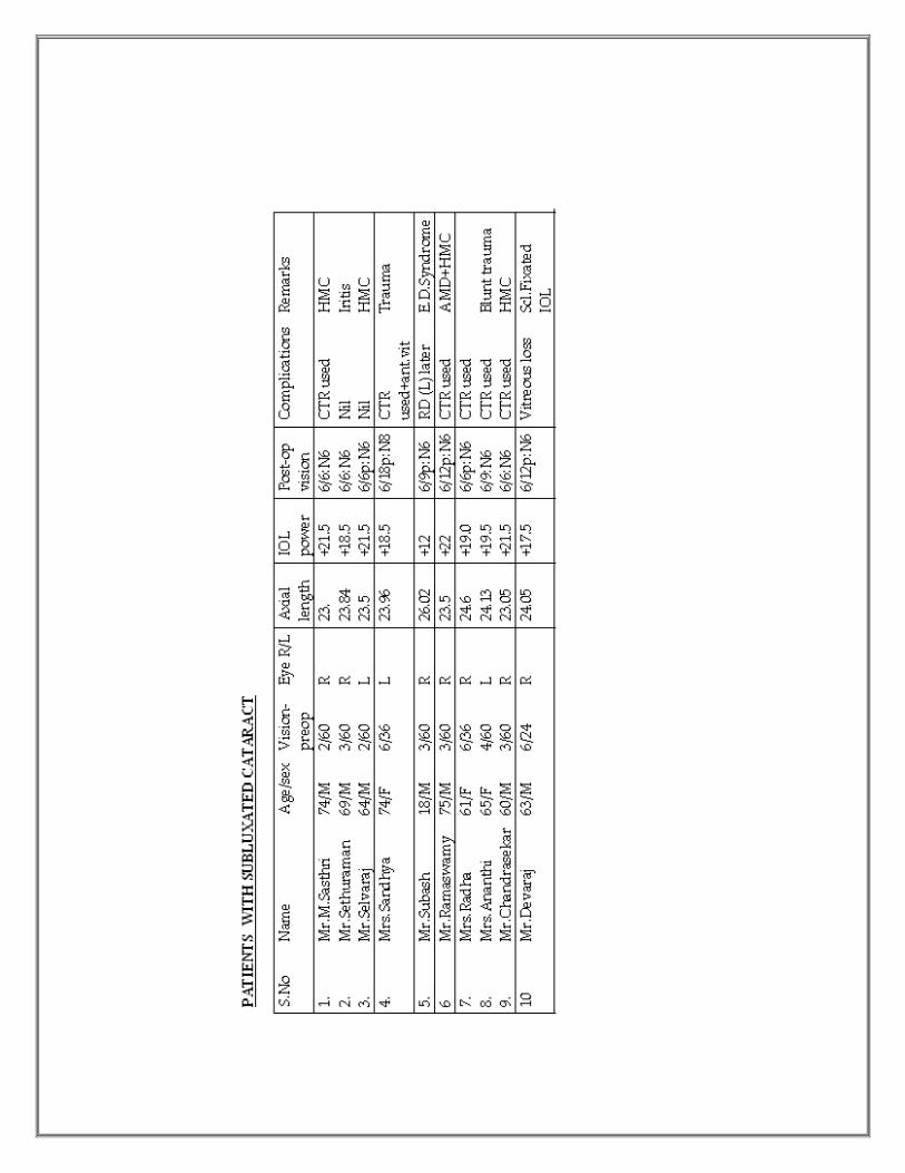
Out of the 10 eyes with subluxated cataract during the study
period, 3 patients had subluxation due to blunt trauma. One patient
who was highly myopic had subluxation secondary to EhlerDanlos
syndrome. 2 patients had subluxated lenses secondary to hyper mature
cataract. Traumatic mydriasis was noted in two patients and one had
angle recession glaucoma. Age group ranged from 18 years to 80 years.
There 3 females and 7 males in the study. One case secondary to trauma
had broken anterior vitreous face. Vitreous was present in the anterior
chamber. Two eyes had more than 180 degree subluxation. Zonules
were absent in them. Fundus examination showed lattice degeneration
with hole in two patients. They were advised to under laser barrage
before surgery. One young patient with high myopia and Ehler danlos
syndrome had retinal detachment in the other eye one month after
surgery. Postoperative Visual acuity ranged from 6/6:N6 to 6/18P:N6.
Drop in visual acuity was due to Cystoid macular edema and Age
related macular degeneration. Six eyes had endocapsular
phacoemulsification. One patient had anterior vitrectomy prior to
capulorhexis and the other patient had it during phacoemulsification.
CTR was implanted (Morcher ring 10/12mm) in all cases. Two were
aborted due to tear in the anterior capsule and rigid single piece PMMA
lenses were placed in the sulcus. No serious intraoperative
complications like vitreous loss, dropped nucleus or further zonular
dialysis were noted. IOP was elevated in 3 patients who were treated
with Timolol 0.5% eye drops bd.

Nine eyes had geometrically and clinically well centered IOL but
one lens was not in the capsular bag. It was found to be in the sulcus.
All patients were being followed for long term complications.
Discussions:
Pseudo exfoliation prevalence varies widely throughout the
world. It has been noted that PEX increases with increasing age. It has
been reported to be more in females than males because of longer life
expectancy. In this study, males were found to have Grade 3 nucleus.
Posterior sub capsular cataract was also seen in more than 50% of the
cases in this study which corroborates with the Ackinci turkey study.
There were no cases of gross phacodonesis, type or zonulolysis as
reported in journals. Moreno et al found higher incidence of
iridophacodonesis in eyes with PEX having light colored iris. Even in
Indian studies, no phacodonesis, type or zonulolysis was reported. Dark
irides have less damage which is prevalent among Indian eyes.
Poor Mydriasis is one of the main features in patients with PEX. We
were able to achieve 6 mm diameter only through viscoelastics or
stretches. There was no difficulty in performing anterior curvilinear
capsulorhexis. Trypan blue stain was used to stain the capsule in all the
cases.
In many case cataract surgery with pseudo exfoliation has been
found to be associated with increases risk of complications. One reason
could be the volume in those studies. In our study, we had only 25
cases with no phacodonesis, zonulolysis. Most important reason for
good vision could be the surgical experience and use of latest
equipments for decreased rate of complication.
It has been found that there was a definite increase in
postoperative irits and glaucoma which could be treated with medicines
in these eyes. The cause of rise in IOP could be partly due to
inflammation though the exact cause could not be ascertained. There

could be breakdown of blood aqueous barrier leading to leakage of
inflammatory materials and proteins. In two recent studies by various
authors there has been no significant increase in IOP.
The management of subluxated lenses has remained controversial
for many years. Jarrett has analyzed retrospectively surgical indications
for subluxated lenses. Techniques included discussion, ICCE, ECCE,
and cryoextraction. Recently, several authors have reported pars plana
lensectomy with anterior vitrectomy. Scleral fixated IOls have become a
good alternative to anterior chamber IOLs in eyes lacking capsular
support. Our surgical paradigm of closed chamber technique was
invaluable. Temporal clear corneal incision was preferred basically for
surgeons comfort and convenience. Most surgeons would prefer to
operate from the site where there is good zonular support. Care was
taken to stay away from the site of subluxation to prevent decentration
of the IOL with the capsular bag. Rhexis was small to restrict the
turbulence in the bag and vitreous aspiration. Iris retractors were used
to support the capsular bag and cause less stress on the remaining
zonules. Multiquadrant hydro dissection in small squirts helped us to
reduce stress on zonules and aid thorough cortical clean up, thereby less
chances for posterior capsule opacification. Nucleus rotation was not
attempted in all cases for fear of zonular stress and rupture.0 degree
phaco tip was used to bury the needle in the nucleus and not to cause
stress or shear on the capsular bag. Low flow parameters help to reduce
the turbulence in the chamber and bag. Repeated HPMC injection had
to be done to push the posterior capsule from coming forward.
Endocapsular technique helped us to fragment the nucleus and remove
the pieces from the central space without stress on the zonules. Using
the stop-chop-chop and stuff technique and step down technique with
smaller fragments helps us to keep the flow and vacuum at low levels.
Vitrectomy was performed to prevent traction on the vitreous base
which may lead to retinal detachment. Bimanual I/A was performed
with low bottle height to reduce turbulence within the capsular bag.
Use of CTR helps to stabilize and expand the capsular bag. The bag
equator was pushed to the periphery and IOL decentration was also

prevented. Less incidence of capsule contraction has also been reported.
Scleral fixation of IOL was done in cases where natural bag support
could not be retained. Inadvertent placement of one foldable lens could
be the reason for one of the lenses being placed in the sulcus rather than
haptic pop out .
In summary, we could achieve in the bag implantation in most
eyes due to closed chamber technique. IOLs could be implanted with
good CTR support and stretched bag. Decentration was not noted
probably because of less number of cases in this study. Cionni ring was
not used.
CONCLUSION:
Patients with cataracts associated with either pseudo exfoliation or
subluxated lenses can achieve good visual acuity following
phacoemulsification with intraocular implantation. Surgeon should
exercise cautious approach after careful preoperative evaluation. Strict
IOP and iritis control along with regular follow-ups can help patients to
have good visual acuity in the long run. Using appropriate phaco
power depending on the grades of cataract is essential. Cornea should
be sufficiently protected with good viscoelastics when one deals with
hard cataracts. In subluxated cataracts, low aspiration flow rate, low
vacuum and low bottle height will cause very minimal turbulence in the
anterior chamber. CTR and anterior vitrectomy has to be done
whenever necessary. Understanding the fluidics and the machine helps
the surgeon do the surgery with utmost confidence and ease. Keeping
the inflammation under control ensures that the chances of getting CME
are grossly reduced. Finally a safe and predictable outcome is what the
surgeon expects after a skillful surgery.


PSEUDOEXFOLIATION STATISTICS
SUBLUXATED LENS STATISTICS
Gender Distribution
Male, 7
Female, 3
Male Female
5
2 2
1
0
1
2
3
4
5
Numbers
6/6 6/9 6/12 6/18
Vision
Post Operative Visual Acuity
Gender Distribution
Male, 11
Female , 14
Male
Female
Age Distribution
8
11
3 3
0
2
4
6
8
10
12
60 -65 65-70 70-75 75-80
Age
Nu
mb
er
15
54
1
0
2
4
6
8
10
12
14
16
Number
6/6 6/9 6/12 6/18
Vision
Post Operative VIsual Acuity
Age Distribution
1
5
1
3
0
2
4
6
<60 60-65 65-70 70-75
A ge
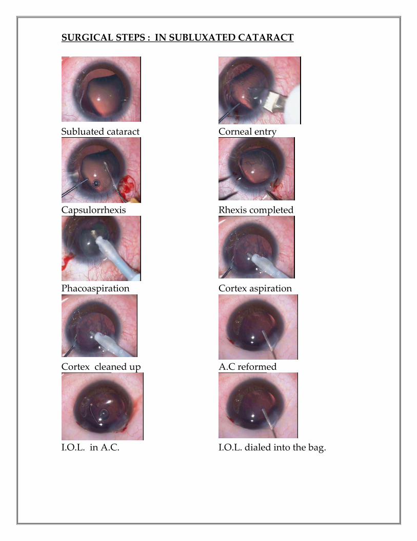
SURGICAL STEPS : IN SUBLUXATED CATARACT
Subluated cataract Corneal entry
Capsulorrhexis Rhexis completed
Phacoaspiration Cortex aspiration
Cortex cleaned up A.C reformed
I.O.L. in A.C. I.O.L. dialed into the bag.

PSEUDOEXFOLIATION CATARACT:
Pseudoexfoliation cataract Gonioscopic view – Sampolaesi Line
CTR
Crack and
Chop
Chop and Stuff

References:
1. Lu Lw, Fine I. H. Phacoemulsification in difficult and challenging cases: Phacoemulsification
in subluxated Cataract. New York, Stuttgart: Thieme, 1999. pp 99-110.
2. Gimbel H V. Two-stage capsulorhexis for endocapsular phacoemulsification. J Cataract
Refract Surg 1990;16:246-49.
3. Vasavada AR, Desai JP. Capsulorhexis: Its safe limits. Indian J Ophthalmol 1995;44:185-90.
4. Merriain JC, Zheng L. Iris hooks for phacoemulsification of the subluxated lens. J Cataract
Refract Surg 1997;23:1295-97.
5. Cionne RJ, Osher RH. Endocapsular ring approach to subluxated cataractous lens. J Cataract
Refract Surg 1995;21:245-49.
6. Vasavada AR, Desai JP. Stop, chop, chop and stuff. J Cataract Refract Surg 1996;22:526-29.
[PUBMED] [FULLTEXT]
7. Gimbel HV, Sun R, Heston JP. Management of zonular dialysis in phacoemulsification and
IOL implantation using the capsular tension ring. Ophthalmic Surg Lasers 1997;28:273-81.
[PUBMED] [FULLTEXT]
8. Cionni RJ, Osher RH. Management of profound zonular dialysis or weakness with a new
endocapsular ring designed for scleral fixation. J Cataract Refract Surg 1998;24:1299-306.
[PUBMED] [FULLTEXT]
9. Lam DSC, Young AL, Leung ATU, Rao SK, Fan DSP, Joan SK Ng. Scleral fixation of a
capsular tension ring for severe ectopia lentis. J Cataract Refract Surg 2000;26:609-12.
10. Gimbel HV, Sun R. Role of capsular tension rings in preventing capsule contraction. J
Cataract Refract Surg 2000;26:791-92. [PUBMED] [FULLTEXT]
11. Masket S, Crandall AS. Atlas of cataract surgery. Challenging phacoemulsification
procedures. Martin Dunitz Ltd. 1999. pp. 244-49.
12. Jensen AD, Cross HE. Surgical treatment of dislocated lenses in the Marfan syndrome and
homocystinuria. Trans Am Acad Ophthalmol Otolaryngol 1972;76:149-59. [PUBMED]
[FULLTEXT]
13. Sellyei LF, Barraquer J. Surgery of the ectopic lens. Ann Ophthalmol 1973;5:1127-33.
14. Vasavada AR, Singh R. Phacoemulsification in eyes with a small pupil. J Cataract Refract
Surg 2000;26:1210-18.
15. Mitra S, Ganesh A. Scleral Suspension pars plana lensectomy for ectopia lentis followed by
suture fixation of intraocular lens. Indian J Ophthalmol 2001;49:109-13. [PUBMED]
[FULLTEXT]
16. Moreno J, Duch S, Lajara J: Pseudoexfoliation syndrome: clinical factors related to capsular
rupture in cataract surgery. Acta Ophthalmol 1993; 71: 181–184.
17. Alfaite M, Leite E, Mira J, et al: Prevalence and surgical complications of pseudoexfoliation
syndrome in Portuguese patients with senile cataract. J Cataract Refract Surg 1996; 22: 972–976.
18. Dosso AA, Bonvin ER, Leuenberger PM: Exfoliation syndrome and phacoemulsification. J
Cataract Refract Surg 1997; 23: 122– 125.
19. Fine IH, Hoffman RS: Phacoemulsification in the presence of pseudoexfoliation: challenges
and options. J Cataract Refract Surg 1997; 23: 160–165.
20. Drolsum L, Haaskjold E, Sandvig K: Phacoemulsification in eyes with pseudoexfoliation. J
Cataract Refract Surg 1998; 24: 787– 792.
21. Wirbelauer C, Anders N, Pham DT, et al: Corneal endothelial cell changes in
pseudoexfoliation syndrome after cataract surgery. Arch Ophthalmol 1998; 116: 145–149.

22. Küchle M, Nguyen NX, Hannappel E, et al: The blood-aqueous barrier in eyes with
pseudoexfoliation syndrome. Ophthalmic Res 1995; 27(suppl 1):136–142.
23. Küchle M, Amberg A, Martus P, et al: Pseudoexfoliation syndrome and secondary cataract.
Br J Ophthalmol 1997; 81: 862–866.
24. Schumacher S, Nguyen NX, Küchle M, et al: Quantification of aqueous flare after
phacoemulsification with intraocular lens implantation in eyes with pseudoexfoliation
syndrome. Arch Ophthalmol 1999; 117: 733–735.
25. Helbig H, Schlötzer-Schrehardt U, Noske W, et al: Anterior chamber hypoxia and iris
vasculopathy in pseudoexfoliation syndrome. Ger J Ophthalmol 1994; 3: 148–153.
26. Vasavada AR, Singh R: Step-by-step, chop in situ and separation of very dense cataracts. J
Cataract Refract Surg 1998; 24: 156–159.
27. Vasavada AR, Desai JP, Singh R: Stop, chop, chop, and stuff. J Cataract Refract Surg 1996; 22:
526–529.
28. Repo LP, Naukkarinen A, Paljarvi L, et al: Pseudoexfoliation syndrome with poorly dilating
pupil: a light and electron microscopic study of the sphincter area. Graefes Arch Clin Exp
Ophthalmol 1996; 234: 171–176.
29. Nagashima RJ: Decreased incidence of capsule complications and vitreous loss during
phacoemulsification in eyes with pseudoexfoliation syndrome. J Cataract Refract Surg 2004; 30:
127–131.
30. Hiller R, Sperduto RD, Krueger DE: Pseudoexfoliation, intraocular pressure, and senile lens
changes in a population-based survey. Arch Ophthalmol 1982; 100: 1080–1082.
31. Mitchell P, Wang JJ, Smith W: Association of pseudoexfoliation syndrome with increased
vascular risk. Am J Ophthalmol 1997; 124: 685–687.
32. Puska P: Lens opacity in unilateral exfoliation syndrome with or without glaucoma. Acta
Ophthalmol 1994; 72: 290–296.
33. Bartholomew RS: Lens displacement associated with pseudocapsular exfoliation: a report on
19 cases in the Southern Bantu. Br J Ophthalmol 1970; 54: 744–750.
34. Ravalico G, Tognetto D, Baccara F: Heparinsurface modified intraocular lens implantation in
eyes with pseudoexfoliation syndrome. J Cataract Refract Surg 1994; 20: 543–549.
35. Assia EI, Apple DJ, Morgan RC, et al: The relationship between the stretching capability of
the anterior capsule and zonules. Invest Ophthalmol Vis Sci 1991; 32: 2835–2839.
36. Carpel EF: Pupillary dilation in eyes with pseudoexfoliation syndrome (letter). Am J
Ophthalmol 1988; 105: 692–694.
37. Zetterstrom C, Olivestedt G, Lundwall A: Exfoliation syndrome and extracapsular cataract
extraction with implantation of posterior chamber lens. Acta Ophthalmol 1992; 70: 85–90.
38. Shingleton BJ, Heltzer J, O’Donoghue MW: Outcomes of phacoemulsification in patients with
and without pseudoexfoliation syndrome.

ACKNOWLEDGEMENTS
I would like to thank God and my wife for giving me the strength to
complete the work in time. My heartfelt thanks to my staff, Dr.Uma ,
and my assistants for their support in selecting the patients for this
study.
I am grateful to all the patients who visited Jaya Eye Care Centre, 12,
Norton 3rd Lane, Mandavelipakkam, Chennai – 600 028 and graciously
consented to be a part of this study. I would also like to extend my
sincere thanks to the nursing staff and theatre staff for their kind
cooperation to carry out the surgeries successfully.
I would like to mention at this juncture that this work is original and
no copyright has been infringed upon.
Dr.Ganesh Balasubramaniam