Pertussis. Highly contagious respiratory infection Classic pertussis, the whooping cough syndrome,...
22
Pertussis
-
Upload
asher-walton -
Category
Documents
-
view
227 -
download
0
Transcript of Pertussis. Highly contagious respiratory infection Classic pertussis, the whooping cough syndrome,...

Pertussis
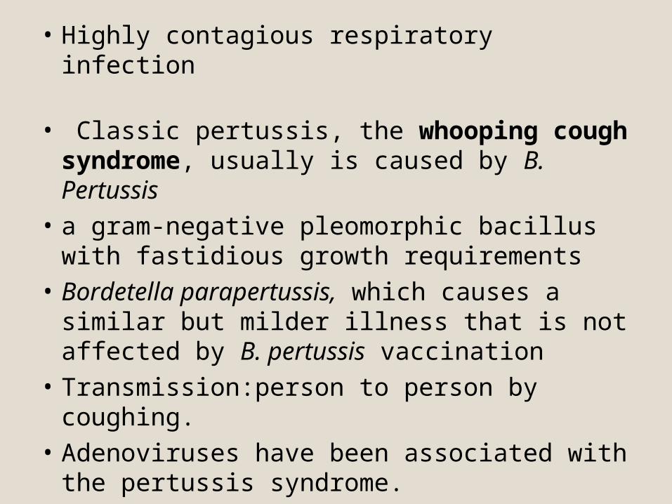
• Highly contagious respiratory infection
• Classic pertussis, the whooping cough syndrome, usually is caused by B. Pertussis
• a gram-negative pleomorphic bacillus with fastidious growth requirements
• Bordetella parapertussis, which causes a similar but milder illness that is not affected by B. pertussis vaccination
• Transmission:person to person by coughing.• Adenoviruses have been associated with the
pertussis syndrome.
EPIDEMIOLOGY
• Outbreaks first described in 16th century
• Bordetella pertussis isolated in 1906
• Estimated >500,000 deaths annually worldwide
Pertussis Epidemiology• Reservoir Human
Adolescents and adults
• TransmissionRespiratory dropletsAirborne rare
CLINICAL MANIFESTATIONS
• Incubation period 3-12 days (up to 21 days)
• Insidious onset, similar to minor upper respiratory infection with nonspecific cough
• Fever usually minimal throughout course
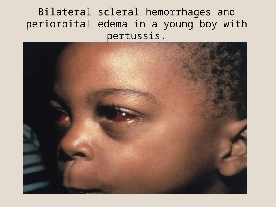
• Apnea & Cyanosis in infant

Pertussis Clinical Features• Catarrhal stage 1-2 weeks
• Paroxysmalcough stage 2-4 weeks
• Convalescence Weeks tomonths
Pertussis Clinical Features
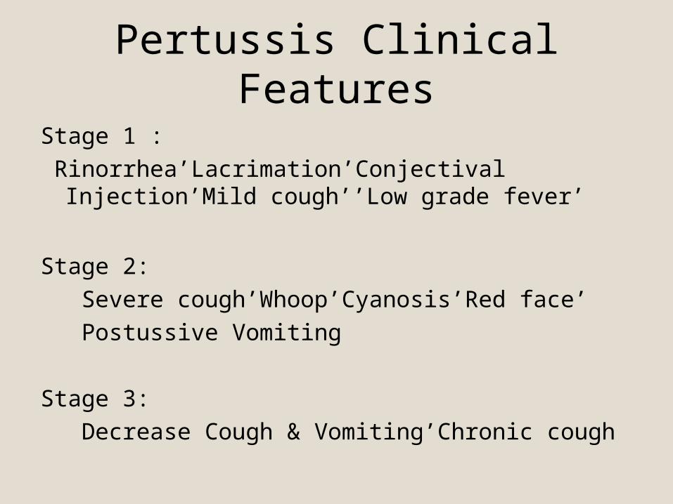
Stage 1 : Rinorrhea’Lacrimation’Conjectival Injection’Mild cough’’Low
grade fever’
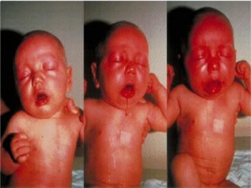
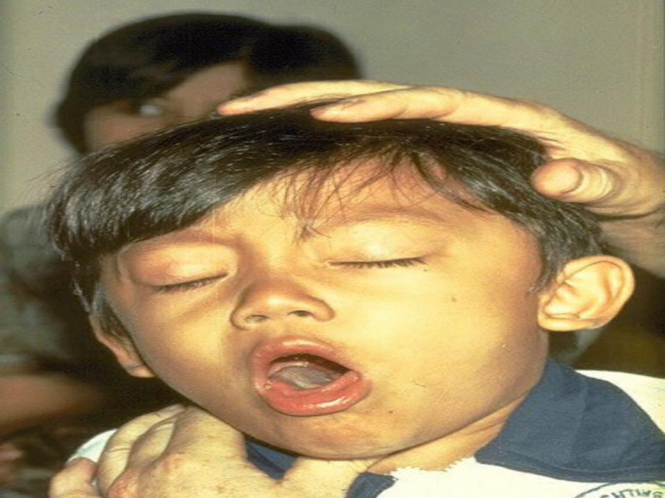
Stage 2: Severe cough’Whoop’Cyanosis’Red face’ Postussive Vomiting
Stage 3: Decrease Cough & Vomiting’Chronic cough
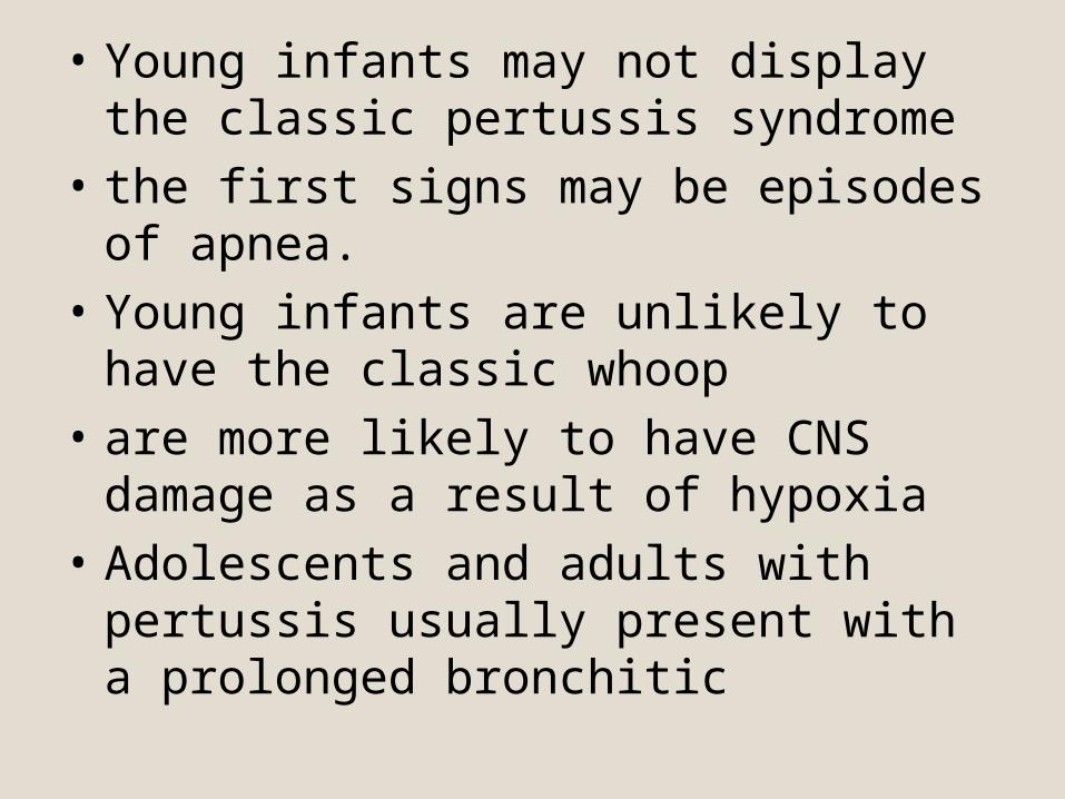
• Young infants may not display the classic pertussis syndrome
• the first signs may be episodes of apnea. • Young infants are unlikely to have the classic
whoop• are more likely to have CNS damage as a
result of hypoxia• Adolescents and adults with pertussis usually
present with a prolonged bronchitic
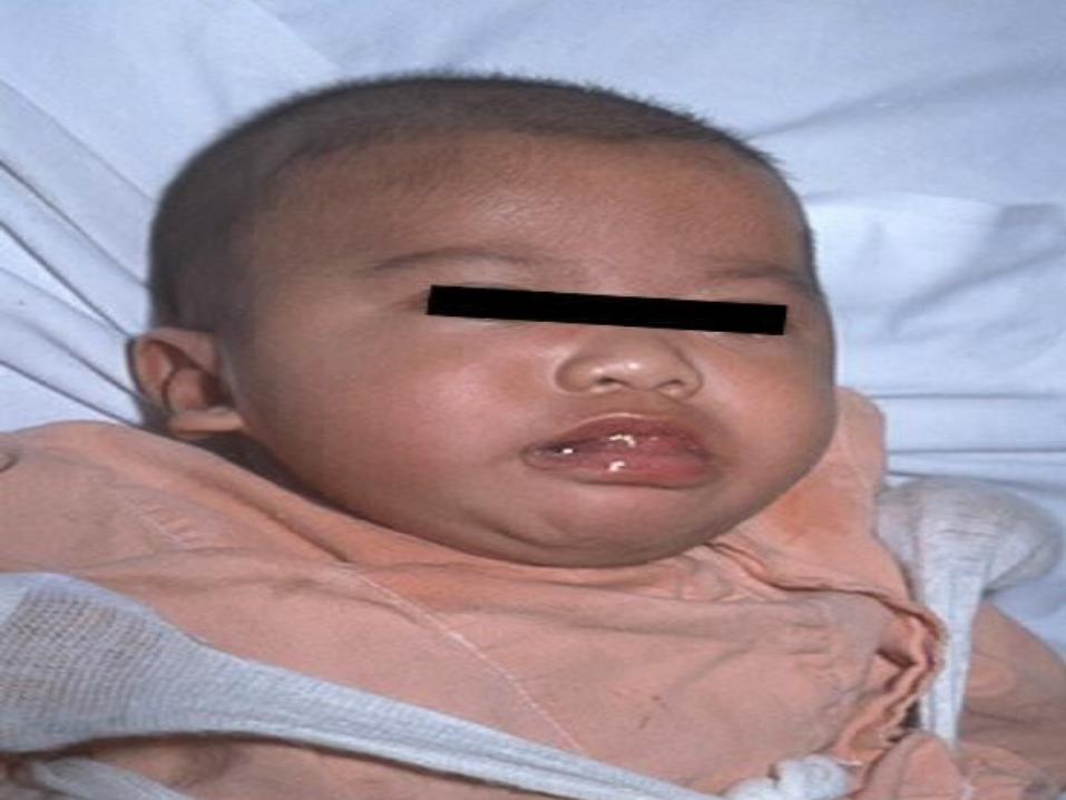
Bilateral scleral hemorrhages and periorbital edema in a young boy with pertussis.

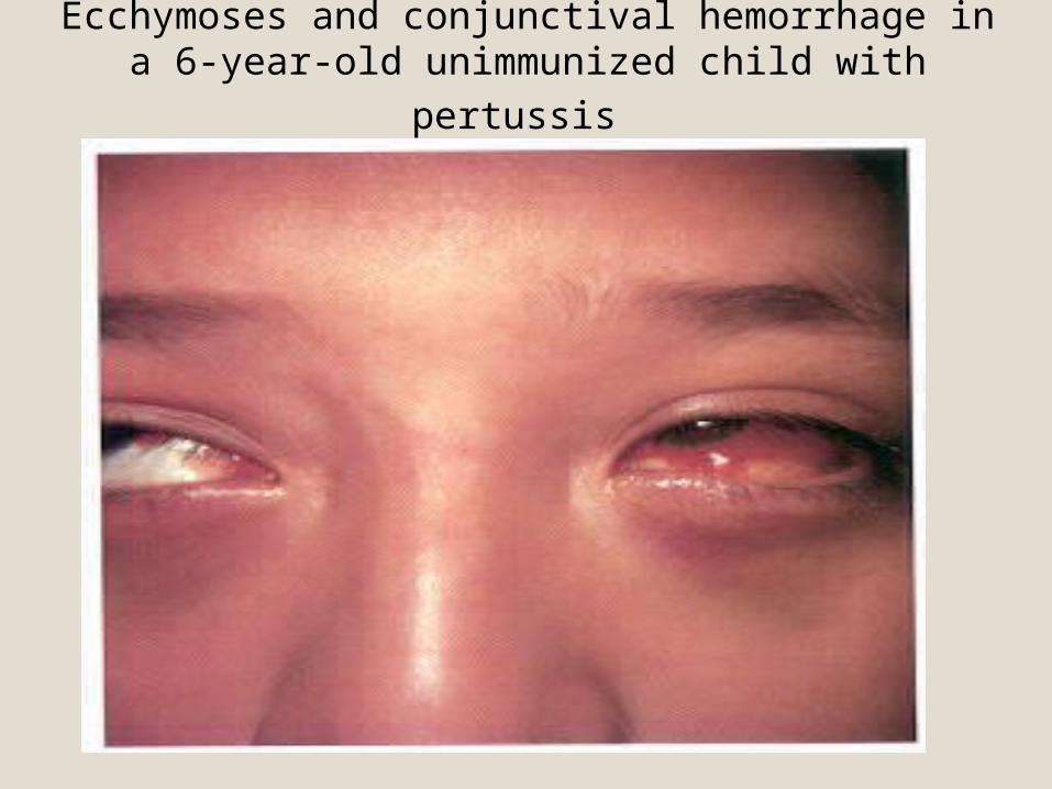
Ecchymoses and conjunctival hemorrhage in a 6-year-old unimmunized child with pertussis
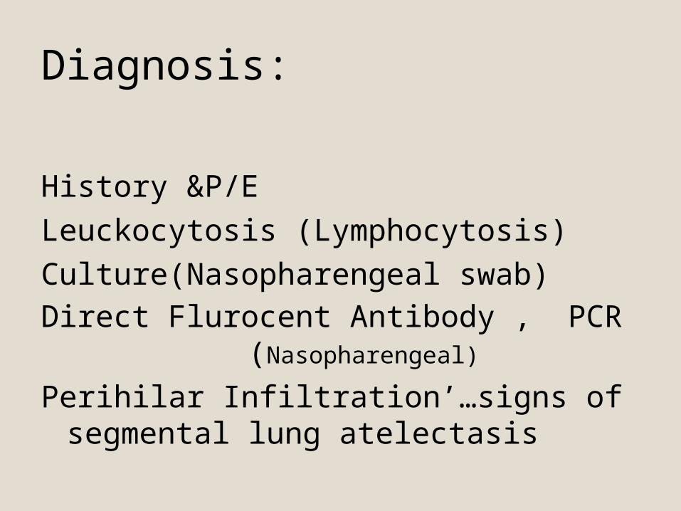
Diagnosis:
History &P/ELeuckocytosis (Lymphocytosis)Culture(Nasopharengeal swab)Direct Flurocent Antibody , PCR
(Nasopharengeal)
Perihilar Infiltration’…signs of segmental lung atelectasis
• who has pure or predominant complaint of cough, especially if the following are absent: fever, malaise or myalgia, exanthem or enanthem, sore throat, hoarseness, tachypnea, wheezes, and rales
• For sporadic cases, a clinical case definition of cough of ≥14 days' duration with at least 1 associated symptom of paroxysms, whoop, or post-tussive vomiting has a sensitivity of 81% and specificity of 58% for culture confirmation.
• Pertussis should be suspected in older children whose cough illness is escalating at 7–10 days and whose coughing episodes are not continuous.
• Pertussis should be suspected in infants <3 mo of age with apnea, cyanosis, or an acute life-threatening event (ALTE). B. pertussis is an occasional cause of sudden infant death.

Differential Diagnosis:
• Pneumonia• Asthma• RSV, parainfluenza virus, and C. pneumoniae• TB• CF• Foreign body • Bronchiolitis• Mediastinal Lymphadenopathy
COMPLICATIONS• Major complications are most common among infants
and young children: hypoxia, apnea, pneumonia, seizures, encephalopathy,
and malnutrition• The most frequent complication is pneumonia• Atelectasis may develop secondary to mucous plugs.• The force of the paroxysm may rupture alveoli and
produce pneumomediastinum pneumothorax, or interstitial or subcutaneous emphysema
• epistaxis; hernias; and retinal and subconjunctival hemorrhages

Treatment
• Erythromycin 50 mg/kg/D PO (14 DAY)• Azithromycin , clarithromycin ,TMP-SMZ • Salbutamol• Moist O2• Nasopharengeal suction• IV Fluid• No Immunoglobulin’No Antitussive drugs’No
Steroid
PREVENTION
• Pertussis vaccine has an efficacy of 70% to 90%• Erythromycin and other macrolides are effective in
preventing disease in contacts exposed to pertussis• Close contacts younger than 7 years should receive
a booster dose of DTaP also should be given a macrolide antibiotic.
• Close contacts older than age 7 should receive prophylactic macrolide antibiotic for 10 to 14 days, but not the vaccine