PERSPECTIVES IN DIABETES Incretin Action in the Pancreas:...
11
Incretin Action in the Pancreas: Potential Promise, Possible Perils, and Pathological Pitfalls Daniel J. Drucker Glucagon-like peptide-1 (GLP-1) and glucose-dependent insulinotropic polypeptide (GIP) are incretin hormones that control the secretion of insulin, glucagon, and somatostatin to facilitate glucose disposal. The actions of incretin hormones are termi- nated via enzymatic cleavage by dipeptidyl peptidase-4 (DPP-4) and through renal clearance. GLP-1 and GIP promote b-cell pro- liferation and survival in rodents. DPP-4 inhibitors expand b-cell mass, reduce a-cell mass, and inhibit glucagon secretion in pre- clinical studies; however, whether incretin-based therapies sus- tain functional b-cell mass in human diabetic subjects remains unclear. GLP-1 and GIP exert their actions predominantly through unique G protein-coupled receptors expressed on b-cells and other pancreatic cell types. Accurate localization of incretin receptor expression in pancreatic ductal or acinar cells in normal or diabetic human pancreas is challenging because antisera used for detection of the GLP-1 receptor often are neither sufficiently sensitive nor specific to yield reliable data. This article reviews recent advances and controversies in incretin hormone action in the pancreas and contrasts established mechanisms with areas of uncertainty. Furthermore, methodological challenges and pitfalls are highlighted and key areas requiring additional scientific in- vestigation are outlined. Diabetes 62:3316–3323, 2013 I ncretins are gut-derived circulating peptide hor- mones that potentiate glucose-dependent insulin secretion following ingestion of a meal. Glucose- dependent insulinotropic polypeptide (GIP) and glucagon-like peptide-1 (GLP-1) are the major incretin hormones. The insulinotropic actions of endogenously secreted GLP-1 and GIP are transient because both pep- tides are rapidly cleared by the kidney and inactivated by cleavage at the N-terminus by dipeptidyl peptidase-4 (DPP-4), a ubiquitous exopeptidase. Potentiation of incretin action underlines two therapeutic classes of glucose- lowering agents: GLP-1 receptor (GLP-1R) agonists and DPP-4 inhibitors (1). Original concepts of GIP and GLP-1 biology, which focused primarily on islet b-cells, have been expanded to include actions on other cell types within and outside the pancreas (2,3). There is now considerable in- terest in understanding how the potentiation of incretin action controls multiple facets of pancreatic biology, encompassing the regulation of glucose sensing, hormone secretion, and cell proliferation, differentiation, and sur- vival. Recent studies have suggested that incretin therapies promote pancreatic inflammation as well as aberrant cell proliferation within the endocrine and exocrine pancreas (4,5); substantial technical and methodological issues, however, limit the generalizability of these findings. This Perspectives in Diabetes evaluates the science supporting existing dogma and discusses new concepts, controversies, and uncertainties in the biology of incretin action in the pancreas. LOCALIZATION OF INCRETIN RECEPTOR EXPRESSION IN THE PANCREAS Several dozen commercial antisera are available for de- tection of GLP-1R and GIP receptor expression by immu- nohistochemical techniques and Western blotting, and real-time PCR is widely used to quantify expression of incretin receptor genes in pancreatic exocrine and endo- crine compartments. Most antisera used to detect GLP-1R expression (by immunohistochemistry or Western blot analysis) are neither sensitive nor specific (6,7). Important control experiments (absorption of the antibody with a peptide epitope, demonstration that the antibody rec- ognizes only a single protein, and failure to generate a signal in cells that do not express a full-length receptor mRNA transcript or in tissues from Glp1r 2/2 mice) are usually absent. Furthermore, multiple studies describe GLP-1R protein expression in cells or tissues that do not express full-length Glp1r mRNA. The widespread use of tightly cropped bands in Western blot analysis precludes accurate assessment of whether a putative band/protein detected by Western blotting is the correct size, the only GLP-1R immunoreactive protein visualized, or one of several unrelated immunoreactive proteins detected by the same antisera. Scientists who are interested in the expression of incretin hormone receptors face the challenging task of assessing how much, if any, of the data published with these antisera is correct. For example, immunoreactive GLP-1R protein expression or Glp1r mRNA transcripts have been detected throughout the heart and ventricle; however, we and others determined that cardiac Glp1r expression was restricted to the atria and absent from the ventricles in mice (8) and rats (9). How do the limitations of available reagents affect our understanding of incretin action in the pancreas? The pu- tative localization of incretin receptor expression in the exocrine pancreas provides an instructive example. Abun- dant immunohistochemical GLP-1R expression in ductal and acinar cells was reported in rodent and human pan- creas, papillary thyroid cancer, and pancreatic adenocarci- noma (10,11). Characterization of multiple GLP-1R antisera, including Abcam39072 (11), one of the reagents used in these studies, revealed major problems with sensitivity and specificity. These antisera detected multiple spurious bands in Western blot analyses of fibroblasts that do not express the GLP-1R and in cellular extracts from Glp1r 2/2 mice (6). We now extend these analyses to detection of the human GLP-1R. Western blot analysis using fibroblasts transfected From the Department of Medicine, Mt. Sinai Hospital, Samuel Lunenfeld Re- search Institute, University of Toronto, Toronto, Ontario, Canada. Corresponding author: Daniel J. Drucker, [email protected]. Received 23 May 2013 and accepted 19 June 2013. DOI: 10.2337/db13-0822 This article contains Supplementary Data online at http://diabetes .diabetesjournals.org/lookup/suppl/doi:10.2337/db13-0822/-/DC1. Ó 2013 by the American Diabetes Association. Readers may use this article as long as the work is properly cited, the use is educational and not for profit, and the work is not altered. See http://creativecommons.org/licenses/by -nc-nd/3.0/ for details. 3316 DIABETES, VOL. 62, OCTOBER 2013 diabetes.diabetesjournals.org PERSPECTIVES IN DIABETES
Transcript of PERSPECTIVES IN DIABETES Incretin Action in the Pancreas:...

Incretin Action in the Pancreas: Potential Promise,Possible Perils, and Pathological PitfallsDaniel J. Drucker
Glucagon-like peptide-1 (GLP-1) and glucose-dependentinsulinotropic polypeptide (GIP) are incretin hormones that controlthe secretion of insulin, glucagon, and somatostatin to facilitateglucose disposal. The actions of incretin hormones are termi-nated via enzymatic cleavage by dipeptidyl peptidase-4 (DPP-4)and through renal clearance. GLP-1 and GIP promote b-cell pro-liferation and survival in rodents. DPP-4 inhibitors expand b-cellmass, reduce a-cell mass, and inhibit glucagon secretion in pre-clinical studies; however, whether incretin-based therapies sus-tain functional b-cell mass in human diabetic subjects remainsunclear. GLP-1 and GIP exert their actions predominantlythrough unique G protein-coupled receptors expressed on b-cellsand other pancreatic cell types. Accurate localization of incretinreceptor expression in pancreatic ductal or acinar cells in normalor diabetic human pancreas is challenging because antisera usedfor detection of the GLP-1 receptor often are neither sufficientlysensitive nor specific to yield reliable data. This article reviewsrecent advances and controversies in incretin hormone action inthe pancreas and contrasts established mechanisms with areas ofuncertainty. Furthermore, methodological challenges and pitfallsare highlighted and key areas requiring additional scientific in-vestigation are outlined. Diabetes 62:3316–3323, 2013
Incretins are gut-derived circulating peptide hor-mones that potentiate glucose-dependent insulinsecretion following ingestion of a meal. Glucose-dependent insulinotropic polypeptide (GIP) and
glucagon-like peptide-1 (GLP-1) are the major incretinhormones. The insulinotropic actions of endogenouslysecreted GLP-1 and GIP are transient because both pep-tides are rapidly cleared by the kidney and inactivated bycleavage at the N-terminus by dipeptidyl peptidase-4(DPP-4), a ubiquitous exopeptidase. Potentiation of incretinaction underlines two therapeutic classes of glucose-lowering agents: GLP-1 receptor (GLP-1R) agonists andDPP-4 inhibitors (1). Original concepts of GIP and GLP-1biology, which focused primarily on islet b-cells, have beenexpanded to include actions on other cell types within andoutside the pancreas (2,3). There is now considerable in-terest in understanding how the potentiation of incretinaction controls multiple facets of pancreatic biology,encompassing the regulation of glucose sensing, hormonesecretion, and cell proliferation, differentiation, and sur-vival. Recent studies have suggested that incretin therapiespromote pancreatic inflammation as well as aberrant cell
proliferation within the endocrine and exocrine pancreas(4,5); substantial technical and methodological issues,however, limit the generalizability of these findings. ThisPerspectives in Diabetes evaluates the science supportingexisting dogma and discusses new concepts, controversies,and uncertainties in the biology of incretin action in thepancreas.
LOCALIZATION OF INCRETIN RECEPTOR EXPRESSION IN
THE PANCREAS
Several dozen commercial antisera are available for de-tection of GLP-1R and GIP receptor expression by immu-nohistochemical techniques and Western blotting, andreal-time PCR is widely used to quantify expression ofincretin receptor genes in pancreatic exocrine and endo-crine compartments. Most antisera used to detect GLP-1Rexpression (by immunohistochemistry or Western blotanalysis) are neither sensitive nor specific (6,7). Importantcontrol experiments (absorption of the antibody witha peptide epitope, demonstration that the antibody rec-ognizes only a single protein, and failure to generatea signal in cells that do not express a full-length receptormRNA transcript or in tissues from Glp1r2/2 mice) areusually absent. Furthermore, multiple studies describeGLP-1R protein expression in cells or tissues that do notexpress full-length Glp1r mRNA. The widespread use oftightly cropped bands in Western blot analysis precludesaccurate assessment of whether a putative band/proteindetected by Western blotting is the correct size, the onlyGLP-1R immunoreactive protein visualized, or one ofseveral unrelated immunoreactive proteins detected by thesame antisera.
Scientists who are interested in the expression of incretinhormone receptors face the challenging task of assessinghow much, if any, of the data published with these antiserais correct. For example, immunoreactive GLP-1R proteinexpression or Glp1r mRNA transcripts have been detectedthroughout the heart and ventricle; however, we and othersdetermined that cardiac Glp1r expression was restricted tothe atria and absent from the ventricles in mice (8) and rats(9). How do the limitations of available reagents affect ourunderstanding of incretin action in the pancreas? The pu-tative localization of incretin receptor expression in theexocrine pancreas provides an instructive example. Abun-dant immunohistochemical GLP-1R expression in ductaland acinar cells was reported in rodent and human pan-creas, papillary thyroid cancer, and pancreatic adenocarci-noma (10,11). Characterization of multiple GLP-1R antisera,including Abcam39072 (11), one of the reagents used inthese studies, revealed major problems with sensitivity andspecificity. These antisera detected multiple spurious bandsin Western blot analyses of fibroblasts that do not expressthe GLP-1R and in cellular extracts from Glp1r2/2 mice (6).We now extend these analyses to detection of the humanGLP-1R. Western blot analysis using fibroblasts transfected
From the Department of Medicine, Mt. Sinai Hospital, Samuel Lunenfeld Re-search Institute, University of Toronto, Toronto, Ontario, Canada.
Corresponding author: Daniel J. Drucker, [email protected] 23 May 2013 and accepted 19 June 2013.DOI: 10.2337/db13-0822This article contains Supplementary Data online at http://diabetes
.diabetesjournals.org/lookup/suppl/doi:10.2337/db13-0822/-/DC1.� 2013 by the American Diabetes Association. Readers may use this article as
long as the work is properly cited, the use is educational and not for profit,and the work is not altered. See http://creativecommons.org/licenses/by-nc-nd/3.0/ for details.
3316 DIABETES, VOL. 62, OCTOBER 2013 diabetes.diabetesjournals.org
PERSPECTIVES IN DIABETES

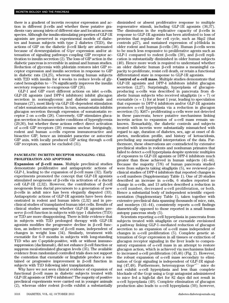
with human GLP-1R cDNA shows that Abcam39072 doesnot detect the human GLP-1R (Fig. 1). A second antiserum,distributed by Novus Biologicals (1940002), recognizes thehuman GLP-1R protein (Fig. 1) but also detects multiplespurious bands/proteins in control cells that do not expressthe Glp1r (Fig. 1). Similar problems with the sensitivity andspecificity of GLP-1R antisera have been described byothers (7). Hence the majority of published studies usingmultiple GLP-1R antisera must be discounted until the ex-perimental data are independently verified with validated,highly sensitive, and highly specific antisera.
Similar concerns relate to the interpretation of someexperiments using regular PCR or real-time PCR to detectexpression of the incretin receptor gene. Real-time PCRdetects Glp1r mRNA transcripts by generating an ampli-con of less than 100 base pairs, whereas regular PCR fre-quently uses primer pairs that generate Glp1r PCR productsthat are several hundred base pairs in length; both are farsmaller than the entire full-length GLP-1R open readingframe. However, cells may generate noncoding mRNAtranscripts detectable by regular or real-time PCR. Analy-sis of Gipr expression revealed ;64 possible Gipr mRNAsplice variants in RNA from human adipose tissue, onlytwo of which were predicted to contain an open readingframe sufficient to give rise to a fully functional, membrane-spanning GIP receptor protein (12). Whether one or moreof these variant Gipr RNA transcripts encodes a trun-cated GIP receptor protein that might exhibit dominantnegative signaling activity, as described in mouse b-cells(13), requires further investigation. Furthermore, usinga polyclonal antiserum, an immunoreactive GIP recep-tor protein was detected in human skeletal muscle (12),a tissue not previously reported to express full-lengthGipr mRNA transcripts (14). Despite reports describingthe detection of 1) partial Glp1r mRNA transcripts by PCRor 2) immunoreactive GLP-1R proteins by Western blotting
or immunohistochemistry in murine liver, macrophages, orventricular cardiomyocytes (2), we could not detect full-length Glp1r mRNA transcripts in the same cells and tis-sues (6,8).
Given the considerable limitations of commonly usedreagents and techniques, how should we interpret avail-able data reporting localization of GLP-1R expression inthe endocrine and exocrine pancreas? The difficulty inisolating pure ductal, acinar, or islet cell RNA that is freefrom contamination by other cell types renders use of suchcell fractions suboptimal for the analysis of cell-specificgene expression. Some groups have localized GLP-1R ex-pression in islet a-cells (15); however, analysis of Glp1rmRNA transcripts in RNA from purified murine a-cells andb-cells that were sorted using a fluorescence-activated cellsorter failed to detect Glp1r mRNA transcripts in a-cells(K. Furuyama, P. Herrera, personal communication). Simi-larly, Glp1r and Gcgr mRNA transcripts were not detectedby in situ hybridization in rat or mouse a-cells, respectively(16,17). Although Gipr mRNA transcripts were detectedin rodent a-cells (18), less information is available re-garding Glp1r or Gipr expression in human a-cells. GLP-1Ractivation stimulates secretion of islet somatostatin, butwhether some, most, or few somatostatin-producing d-cellsexpress the GLP-1R has not been established. DPP-4 ex-pression at the cell surface has been identified on murinea-cells and b-cells and even more strongly on ductal cells(19); however, whether DPP-4 activity locally regulatesbioactive incretin activity within these pancreatic cell typeshas not been determined.
Glp1r mRNA transcripts have been detected in pancre-atic ductal human pancreatic adenocarcinoma cell lines(20). However the GLP-1R agonist exendin-4 failed tostimulate growth or enhance cell survival in five differenthuman pancreatic cancer cell lines that express an en-dogenous Glp1r mRNA transcript. Whether Glp1r mRNAtranscripts are expressed in non-immortalized pancreaticductal or acinar cells remains uncertain. Tornehave et al.(16) were unable to demonstrate Glp1r mRNA transcriptsin pancreatic duct cells from mice and rats by in situ hy-bridization, despite detection of an immunoreactive pro-tein in ducts using a GLP-1R antibody that was subsequentlyshown to exhibit suboptimal specificity (6). Transcriptomeanalysis of human pancreatic endocrine and exocrine cellsdetected glucagon receptor (Gcgr) expression in ductalcells, but Glp1r expression was not reported (21). Despiteimmunohistochemical depiction of robust GLP-1R immu-nopositivity in human pancreatic cancer cells (22), usingtranscriptome analysis of publicly available databases(oncomine.com, version 4.4.3, and Genome ExpressionOmnibus, http://www.ncbi.nlm.nih.gov/geo/) we have beenunable to find evidence that Glp1r mRNA transcripts areoverexpressed in these tumors. Similarly, using in situ li-gand binding and autoradiography, Körner et al. (23) wereunable to detect GLP-1 binding sites in pancreatic adeno-carcinomas. New studies using individual endocrine oracinar cells purified by fluorescence-activated cell sorteranalysis or isolation of single pancreatic cells using laser-capture microdissection followed by the use of validatedantisera and/or PCR analysis using primers that span thefull-length Glp1r open reading frame should refine ourunderstanding of the direct cellular targets of GLP-1 actionin the pancreas.Incretin-mediated control of islet hormone secretion.The increasing realization that b-cells exhibit considerablefunctional heterogeneity begs the question of whether
FIG. 1. Characterization of the sensitivity and specificity of antiseraagainst the human GLP-1R. Baby hamster kidney cells were transientlytransfected with the vector pcDNA3.1 alone (lane 1) or with a humanGlp1r cDNA tagged at the C-terminus with green fluorescent protein(GFP) cloned into pcDNA3.1 (lane 2). Whole-cell extracts were pre-pared 48 h after transfection and analyzed by immunoblotting with theindicated commercial GLP-1R antibodies or with a rabbit polyclonalantibody against GFP (Abcam ab6556). Molecular mass standards areindicated on the right. Both the anti-GLP-1R antibody Novus1940002and the anti-GFP antibody detected similar immunoreactive proteins of;68 and;87 kDa, likely representing species of the GLP-1R-GFP fusionprotein that were glycosylated differently.
D.J. DRUCKER
diabetes.diabetesjournals.org DIABETES, VOL. 62, OCTOBER 2013 3317
there is a gradient of incretin receptor expression and ac-tion in different b-cells and whether these putative gra-dients vary among islets of different size and location acrossspecies. Although the insulin-stimulating properties of GLP-1Ragonists are preserved in experimental models of diabe-tes and human subjects with type 2 diabetes (T2D), theactions of GIP on the diabetic b-cell likely are attenuatedbecause of downregulation of Gipr expression and/or at-tenuation of signaling pathways coupling GIP receptor ac-tivation to insulin secretion (2). The loss of GIP action in thediabetic pancreas is reversible in animal and human studies.Reduction of glycemia with phlorizin restores islet GIP re-ceptor expression and insulin secretion in response to GIPin diabetic rats (24,25), whereas treating human subjectswith T2D with insulin for 4 weeks to reduce levels of gly-cated hemoglobin to ;7% significantly improves the insulinsecretory response to exogenous GIP (26).
GLP-1 and GIP exert different actions on islet a-cells.GLP-1R agonists (and DPP-4 inhibitors) inhibit glucagonsecretion in normoglycemic and diabetic animals andhumans (27), most likely via GLP-1R–dependent stimulationof islet somatostatin secretion. In turn, somatostatin inhibitsglucagon secretion through expression of somatostatin re-ceptor 2 on a-cells (28). Conversely, GIP stimulates gluca-gon secretion in humans under conditions of hyperglycemia(29,30), but whether these actions reflect direct activationof a-cell GIP receptors (29) remains unclear. Intriguingly,rodent and human a-cells express immunoreactive andbioactive GIP, hence an intraislet paracrine or autocrineGIP axis, with locally produced GIP acting through a-cellGIP receptors, cannot be excluded (31).
PANCREATIC INCRETIN RECEPTOR SIGNALING: CELL
PROLIFERATION AND APOPTOSIS
Expansion of b-cell mass. Multiple preclinical studiesdemonstrate proliferative and antiapoptotic actions ofGLP-1, leading to the expansion of b-cell mass (32). Earlyexperiments promoted the concept that GLP-1R agonistsstimulated neogenesis of b-cells via activation of a ductalcell GLP-1R (2,32). However, the contribution of b-cellneogenesis from ductal precursors to a generation of newb-cells in adult mice has been elegantly disputed (33).Antiapoptotic actions of GLP-1R agonists have been dem-onstrated in rodent and human islets (2,32) and in pre-clinical studies of transplanted human islet cells. Results ofclinical studies assessing whether GLP-1R agonists pre-serve b-cell function in subjects with type 1 diabetes (T1D)or T2D are more disappointing. There is little evidence thatin subjects with T2D prolonged therapy with GLP-1Ragonists modifies the progressive decline in b-cell func-tion, an indirect surrogate of b-cell mass, independent ofchanges in weight loss (34). Similarly, treatment withexenatide for 6–9 months in subjects with long-standingT1D who are C-peptide-positive, with or without immuno-suppression (daclizumab), did not enhance b-cell function orsuppress meal-stimulated glucagon levels (35). The availableevidence from randomized controlled trials does not supportthe contention that exenatide or liraglutide produce a sus-tained or progressive improvement in b-cell function insubjects with T1D following islet transplantation.
Why have we not seen clinical evidence of expansion offunctional b-cell mass in diabetic subjects treated withGLP-1R agonists or DPP-4 inhibitors? The majority of positivepreclinical experiments were carried out in younger animals(2), whereas older rodent b-cells exhibit a substantially
diminished or absent proliferative response to multipleregenerative stimuli, including GLP-1R agonists (36,37).The diminution in the replicative capacity of b-cells inresponse to GLP-1R agonists has been attributed to loss ofproteins that regulate the cell cycle, such as Skp2 (thatcontrols p27), and sustained expression of p16Ink4a inolder rodent and human b-cells (38). Human b-cells seemto be much less responsive to proliferative agents such asGLP-1 compared to rodent b-cells (39), and b-cell repli-cation is substantially diminished in older human subjects(40). Hence more work is required to understand whetheran older diabetic human b-cell retains a meaningful ca-pacity to proliferate, resist cell death, or retain a functionaldifferentiated state in response to GLP-1R agonists.Control of a-cell mass.Multiple studies demonstrate thatGLP-1R agonists and DPP-4 inhibitors inhibit glucagonsecretion (2,27). Surprisingly, hyperplasia of glucagon-producing a-cells was described in pancreata from di-abetic human subjects who received sitagliptin (n = 7) orexenatide (n = 1) for at least 1 year, leading to speculationthat exposure to DPP-4 inhibitors and/or GLP-1R agonistspromotes a-cell hyperplasia via a reduction in glucagonsecretion (5). Ki67+ proliferating a-cells were not detectedin these pancreata; hence putative mechanisms linkingincretin action to expansion of a-cell mass remain un-known. Remarkably, the diabetic controls and subjectstreated with incretin were substantially mismatched withregard to age, duration of diabetes, sex, age at onset of di-abetes, medication profile, and history of ketoacidosis,precluding any meaningful interpretation of the data. Fur-thermore, these observations are contradicted by extensivepreclinical studies in rodents and nonhuman primates thatfailed to detect a-cell hyperplasia despite systemic multiplesof exposures to GLP-1R agonists or DPP-4 inhibitors muchgreater than those achieved in human subjects (41–44).Because the majority (7/8) of human pancreata studiedwere from subjects taking sitagliptin (5), we reviewed pre-clinical studies of DPP-4 inhibitors that reported changes ina-cell numbers (Supplementary Table 1). One of 20 studiesdescribed an increase in a-cells, 6 studies reported nochange in a-cells, and 13 articles described a reduction ina-cell number, decreased a-cell proliferation, or both.Hence a substantial body of independent scientific experi-mentation (Supplementary Table 1), taken together withextensive preclinical data spanning thousands of mice, rats,and monkeys (41–44), consistently reports a-cell findingsdiametrically opposed to those reported in a small humanautopsy pancreas study (5).
Scientists reporting a-cell hyperplasia in pancreata fromsubjects treated with sitagliptin or exenatide envisioneda pathway linking GLP-1–mediated reduction of glucagonsecretion to an expansion of a-cell mass independent ofchanges in a-cell proliferation (5). Complete genetic at-tenuation of Gcgr expression in all tissues or extinction ofglucagon receptor signaling in the liver leads to compen-satory expansion of a-cell mass in an attempt to restoreglucagon action, which is achieved via mechanisms linkedto increased a-cell proliferation (45,46) (Fig. 2). However,the robust expansion of a-cell mass secondary to elimi-nation of Gcgr signaling is independent of GLP-1R signal-ing (47,48). Furthermore, heterozygous Gcgr+/2 mice donot exhibit a-cell hyperplasia and less than completeblockade of the Gcgr using a Gcgr antagonist administeredto mice fed a high-fat diet for 82 days did not result ina-cell hyperplasia (49). Complete elimination of glucagonproduction also leads to a-cell hyperplasia (50); however,
INCRETIN BIOLOGY AND THE PANCREAS
3318 DIABETES, VOL. 62, OCTOBER 2013 diabetes.diabetesjournals.org

DPP-4 inhibitors or GLP-1R agonists generally producea 20–50% reduction in plasma glucagon levels (27,51),a scenario that has never been shown to trigger a-cellhyperplasia. Hence a large amount of independent exper-imentation refutes the existence of a speculative pathway(5) linking partial reduction of glucagon secretion to ex-pansion of a-cell mass and neuroendocrine tumor forma-tion independent of changes in a-cell proliferation.Acinar and ductal cells. Notwithstanding the uncertaintyabout whether rodent or human acinar and ductal cellsexpress a functional GLP-1R, older rodent pancreaticductal cells retain the capacity to proliferate followingGLP-1R activation. Indeed, a threefold increase in ductalproliferation was observed after a 7-day course of exendin-4in three 7-month-old mice (38). Nevertheless, the hypoth-esis that sustained GLP-1R signaling and/or inhibition ofGcgr signaling (which also increases levels of GLP-1) willpromote exocrine cell proliferation, leading to the expan-sion of exocrine mass (5), has not been consistentlyreproduced in nonsensitized preclinical models. Treatmentwith exendin-4 for 12 weeks in transgenic mice expressingan activated K-ras oncogene increased the expression oflow-grade pancreatic intraepithelial neoplasia and enhancedductal cell proliferation, but acinar cell proliferation was notreported (10). The assertion that Gcgr2/2 mice or humanswith a Gcgr null mutation exhibit enhanced exocrineproliferation (5) is not supported by the published data(52,53) cited by the same authors. Although Gcgr2/2 miceexhibit pancreatic enlargement, increased acinar or duc-tal cell proliferation has not been detected by multiple
independent groups that have studied these animals(45,46,48,52).
Histological analyses of the pancreas have been carriedout after extensive chronic treatment (up to 2 years) withhigh doses of GLP-1R agonists in thousands of mice andrats and dozens of monkeys. None of the studies involvingmultiple doses of structurally distinct GLP-1R agonists hasreported expansion of the ductal or exocrine compart-ments in rodents or nonhuman primates (41,42). Similarly,the DPP-4 inhibitors vildagliptin or sitagliptin, which wereadministered to hundreds of mice and rats continuouslyfor 2 years (43,44) at doses producing high multiples ofsystemic drug exposure, did not result in acinar, ductal, orendocrine cell neoplasia. Although data from toxicologystudies of diabetic animals is limited, a 3-month treatmentregimen of exenatide twice daily at doses of 6, 40, and 250mg/kg/day produced no changes in pancreatic exocrinestructure or ductal proliferation (54). Similarly, no pro-liferative effects of exenatide or liraglutide were detectedin the exocrine pancreas of diabetic Zucker diabetic fattyrats after 13 weeks of drug administration (55). Sitagliptinwas administered for 3 months in monkeys, 12 monthsin dogs, and 24 months in mice and rats at doses pro-ducing levels of exposure considerably higher than thoseachieved clinically; no evidence of pancreatic abnormali-ties were detected on gross or histological analysis of thepancreas. However, precise details on the actual analysescarried out in these toxicology studies have not yet beenpublished (44). Each pharmaceutical sponsor of a DPP-4inhibitor or GLP-1R agonist is required to carry out 2-year
FIG. 2. GLP-1 and DPP-4 inhibitor action in the endocrine pancreas. GLP-1R agonists and DPP-4 inhibitors enhance insulin and reduce glucagonsecretion. In preclinical studies, these agents expand b-cell mass and reduce a-cell mass. Genetic mutations that disrupt glucagon receptor sig-naling or eliminate production of bioactive glucagon result in islet a-cell hyperplasia.
D.J. DRUCKER
diabetes.diabetesjournals.org DIABETES, VOL. 62, OCTOBER 2013 3319

carcinogenicity studies in 2 species; there have now beenthousands of animals exposed to DPP-4 inhibitors andGLP-1R agonists in addition to the studies reported above.However, reports of ductal or acinar proliferation or pan-creatic adenocarcinoma in preclinical studies, either in theform of toxicology reports submitted as part of new drugapplications to regulatory authorities or as publishedmanuscripts, have not yet been forthcoming.
Nevertheless, GLP-1R agonists increase the weight ofthe pancreas in some preclinical studies, most notably inyoung rodents (10,56), through mechanisms that are notcompletely understood. Selective restoration of the ex-pression of human GLP-1R under the control of the Pdx1promoter in b-cells and ducts normalized glucose homeo-stasis in Glp1r2/2 mice but was not sufficient to mediatean increase in pancreatic weight in response to exogenousexendin-4 (57). Therefore, although insulin secretion is notsufficient for the increase in pancreatic mass observedsecondary to GLP-1R activation, further studies are re-quired to elucidate the precise cell types and mechanismslinking GLP-1R activation to changes in pancreatic weight.GLP-1R signaling, DPP-4 inhibition, and pancreaticinflammation. The glucose reduction achieved with DPP-4inhibitors requires intact GLP-1R and GIP receptor sig-naling (58,59); however, non-glucoregulatory actions maybe mediated by other substrates, including stromal-derivedcell factor-1a (3,60). There are few data linking non-enzymatic signaling of DPP-4 to specific actions in the en-docrine or exocrine pancreas. The widespread expressionof GLP-1Rs on multiple immune cell populations (61), to-gether with the expression and activity of DPP-4(CD26) inthe immune system, provides a logical basis for exploringwhether GLP-1R agonists and/or DPP-4 inhibitors modulateimmune function. The majority of actions ascribed to DPP-4in immune cells are attributable to nonenzymatic actionsof the enzyme; hence DPP-4 signaling in immune cellsproceeds independent of its catalytic enzyme activity (62).Accordingly, partial inhibition of the catalytic activity ofDPP-4 using highly selective DPP-4 inhibitors would not bepredicted to perturb immune function (60). Indeed, T-cell–dependent immune responses are preserved in Dpp42/2
mice and in mice treated with a highly selective DPP-4 in-hibitor (63). Preclinical studies linking incretin action toenhanced pancreatic inflammation include the observationthat one of eight human islet amyloid polypeptide (hIAPP)transgenic rats treated with sitagliptin for 12 weeks de-veloped focal pancreatic inflammation (64).
In an attempt to reproduce abnormalities reported in theexocrine pancreas of hIAPP transgenic rats treated withsitagliptin for 12 weeks (64), Aston-Mourney et al. (65) fedhIAPP transgenic mice a high-fat diet and treated them withsitagliptin or metformin alone or in combination for 12months. In contrast to findings observed in hIAPP transgenicrats (64), islet amyloid deposition, ductal cell proliferation,and pancreatic mass were not increased by sitagliptin inhIAPP transgenic mice; however, b-cell mass was in-creased, consistent with the known actions of sitagliptin inmice (2). Furthermore, sitagliptin treatment was not as-sociated with pancreatic inflammation, necrosis, meta-plasia, neoplasia, or periductal fibrosis; pancreatic masswas increased in mice treated with metformin but not inmice treated with sitagliptin (65).
Two reports describe nondiabetic rats treated withexenatide that developed pancreatic damage and inflam-mation (66,67). Notably, in both experiments, rats treatedwith exenatide experienced profound weight loss (25–30%);
however, no pair-fed controls were included in theseanalyses, and mechanisms linking GLP-1R activation toincreased pancreatic inflammation were not identified(66,67). Rapid, profound weight loss is frequently associ-ated with a catabolic state, whereas more modest andgradual weight loss, particularly in the setting of preex-isting obesity, is generally associated with reduced tissueand systemic markers of inflammation.
Increased pancreatic inflammation has not been detectedin multiple preclinical studies examining chronic effects (upto 2 years) of administration of high doses of GLP-1R ago-nists or DPP-4 inhibitors in nondiabetic rodents or non-human primates (41–44). For example, treatment of diabeticrats with supratherapeutic doses of exenatide or liraglutidefor 13 weeks was not associated with histological orbiochemical evidence of pancreatic inflammation (54,55).Moreover, administration of GLP-1R agonists before orafter the induction of experimental pancreatitis did notenhance pancreatic inflammation in normal or diabeticrats and mice (68,69); GLP-1R agonists unexpectedly in-duced an anti-inflammatory gene expression profile in thepancreas of insulin-resistant mice fed a high-fat diet (68).Incretin-based therapies and inflammatory markersin humans. Small increases in plasma levels of amylaseand lipase have been reported in diabetic subjects treatedwith the DPP-4 inhibitors alogliptin and sitagliptin (70),and a separate observational study of diabetic subjectstreated with sitagliptin, saxagliptin, or exenatide reportedthat 35.6% of subjects exhibited increases in plasma levelsof amylase and/or lipase, with levels of lipase increasing toa relatively greater extent (71). Notably, elevated levels ofamylase and lipase also were observed, albeit less fre-quently, in diabetic control subjects who did not receivea DPP-4 inhibitor or GLP-1R agonist. Further study is re-quired to determine whether the increase in amylase andlipase reflects subclinical pancreatic inflammation ordysregulated synthesis, secretion, or clearance of theseenzymes. Administration of GLP-1R agonists or DPP-4 in-hibitors is associated with suppression of inflammation(72); however, many of these experiments do not controlfor concomitant reduction in glucose or body weight, whichmay also indirectly dampen inflammation. Exenatide ad-ministered twice daily for 12 weeks in subjects with T2Dreduced circulating markers of inflammation in circulatingmononuclear cells, independent of changes in body weight(73). A single, acute, 5-mg injection of exenatide signifi-cantly and rapidly reduced levels of reactive oxygen spe-cies, nuclear factor-kB binding activity, and expression oftumor necrosis factor-a, interleukin-1b, Jun NH2-terminalkinase-1, Toll-like receptor-4, and suppressor of cytokinesignaling-3 mRNA transcripts in RNA isolated from circu-lating mononuclear cells (73). Similarly, administration ofsitagliptin 100 mg once daily for 12 weeks to 12 subjectswith T2D reduced expression of proinflammatory markersin circulating mononuclear cells, whereas acute adminis-tration of 100 mg sitagliptin to fasting diabetic subjectssignificantly reduced mononuclear cell expression of Toll-like receptor-2, IkB kinase b, chemokine receptor type 2,cluster of differentiation-26 mRNA transcripts and decreasednuclear factor-kB binding activity (74). Hence the availabledata indicate that GLP-1R agonists and DPP-4 inhibitorsindependently exert anti-inflammatory actions in tissuessuch as the exocrine and endocrine pancreas, as well as incirculating blood cells from diabetic subjects, althoughthe mechanisms mediating these actions remain poorlyunderstood.
INCRETIN BIOLOGY AND THE PANCREAS
3320 DIABETES, VOL. 62, OCTOBER 2013 diabetes.diabetesjournals.org
SUMMARY AND PERSPECTIVE
The potential promise of incretin-based therapies has beenpartially realized in that we can now implement antidia-betic regimens associated with lower rates of hypoglyce-mia and weight gain. Although the first actions of GLP-1 onpancreatic islet cells were described more than 25 yearsago, we still have much to learn about how GLP-1R sig-naling regulates b-cell function. For example, the molecularmechanisms underlying glucose-sensitive GLP-1R signalinghave remained elusive. The precise cellular localization ofthe GLP-1R in islet and exocrine cells requires more carefulstudy, not only in animals, but also in pancreata from hu-man subjects over a broad range of ages and with andwithout preexisting diabetes or diseases of the pancreas.
Possible perils of incretin therapies include the de-velopment of complications, including pancreatitis andcancer. Although some studies combine groups of exper-imental subjects exposed to DPP-4 inhibitors and GLP-1Ragonists for pooled analyses of adverse events (5,75), thesetwo distinct drug classes exhibit mechanisms of action withmultiple fundamental differences (2,60). Therefore it is notscientifically justifiable to pool subjects exposed to DPP-4inhibitors and GLP-1R agonists. The hypothesis that acti-vation of GLP-1R signaling might promote increased cellproliferation and increase the incidence or detection of spe-cific neoplasms is reasonable. Indeed, rats and, to a lesserextent, mice exhibit C-cell hyperplasia and medullarythyroid cancer after prolonged, sustained exposure toGLP-1R agonists (76). Nevertheless, monkeys and humansexhibit major differences in GLP-1R expression in theirthyroid C-cells, and in the vast majority of subjects calci-tonin levels do not rise into the abnormal range followingprolonged exposure to GLP-1R agonists (77). Studiesassessing the pancreata of thousands and mice and ratshave not shown dysplasia or tumor formation followingtreatment with GLP-1R agonists or DPP-4 inhibitors forperiods up to 2 years. Furthermore, GLP-1 levels remainsubstantially elevated for years following many forms ofbariatric surgery, yet rates of pancreatitis, medullary thy-roid cancer, glucagonomas, and cancer of the pancreas arenot increased in this patient population, despite more than10 years of follow-up (78). Hence the hypothesis thatGLP-1R agonists or DPP-4 inhibitors will promote tumorformation (4) is not supported by the available preclinicalor clinical data.
Experimental evidence raising the possibility that incretin-based therapy may be associated with a predisposition todevelop pancreatitis or pancreatic cancer generates impor-tant hypotheses that require testing in mechanistic preclin-ical studies and independent validation in large randomized,controlled clinical trials. Pathological pitfalls of incretin-based science include the use of nonspecific antiseraand mismatched cases and controls and the generation ofnonvalidated hypotheses and irreproducible data. As mil-lions of patients with diabetes are being treated withincretin-based therapies, our collective responsibility to usehigher-quality science has never been greater. Underpow-ered studies using poorly validated reagents or analysis ofmismatched cases and controls (5) have a much greatercertainty of not being reproducible and do not advance ourunderstanding of incretin action in the pancreas. Emergingpharmacovigilance studies, such as the Safety Evaluation ofAdverse Reactions in Diabetes (SAFEGUARD) study shouldshed additional clarity on the risk-to-benefit ratio of medi-cations used to treat diabetes.
A great deal has been written about incretin action in thepancreas, including statements that are not substantiated orare contradicted by available data. For example, the claimthat “production of exendin-4 causes rapid proliferation ofintestinal tissue and a 50% increase in the size of the pan-creas” (79) inHeloderma suspectum is simply incorrect andis clearly refuted by the actual experimental data cited (80).The ongoing debate surrounding the mechanisms of actionand potential safety of incretin-based therapies remindsone of a quotation variably attributed to Daniel PatrickMoynihan, James Schlesinger, or Bernard Baruch: “Every-one is entitled to their own opinions, but they are not en-titled to their own facts.” The beauty of science is that it isself-correcting, and provocative experiments and observa-tions that are not highly reproducible are ultimately dis-carded. Over the next several years we will learn much moreabout the potential risks and benefits of incretin-basedtherapies from large, randomized, ongoing cardiovascularoutcome studies, with rigorous independent adjudication ofadverse events. Thoughtful scientists await the results ofthese and ongoing pharmacovigilance studies with great in-terest. The results of these trials will be extremely useful forincreasing our understanding of incretin action in not onlythe cardiovascular system but also the diabetic pancreas.
ACKNOWLEDGMENTS
Pancreatic incretin research in the Drucker laboratory issupported by grants 123391 and 82700 from the CanadianInstitute of Health Research.
D.J.D. is supported by appointments as a Canada Re-search Chair in Regulatory Peptides and the Banting andBest Diabetes Centre-Novo Nordisk Chair in IncretinBiology, and within the past 12 months has served asan advisor or consultant to Arisaph PharmaceuticalsInc., Eli Lilly Inc., GlaxoSmithKline, Intarcia Therapeu-tics, Merck Research Laboratories, Novo Nordisk Inc.,NPS Pharmaceuticals Inc., Sanofi, Takeda, and Transi-tion Pharmaceuticals Inc. D.J.D. receives operating sup-port for incretin research via grants to Mt. Sinai Hospitalfrom GlaxoSmithKline, Merck, and Novo Nordisk. D.J.D.is party to a DPP-4 inhibitor license agreement, togetherwith the University of Toronto, the University HealthNetwork, Tufts University, and Arisaph PharmaceuticalsInc. No other potential conflicts of interest relevant to thisarticle were reported.
The author thanks Laurie Baggio, Jackie Koehler, andBernardo Yusta (all from the Samuel Lunenfeld ResearchInstitute, Mt. Sinai Hospital, Toronto, Ontario, Canada) forconstructive comments. The author also thanks JackieKoehler for her assessment of Glp1r expression in publicdatabases, and Bernardo Yusta for performing Western blot-ting with the transfected GLP-1R cDNA and two GLP-1 re-ceptor antisera.
REFERENCES
1. Drucker DJ, Nauck MA. The incretin system: glucagon-like peptide-1 re-ceptor agonists and dipeptidyl peptidase-4 inhibitors in type 2 diabetes.Lancet 2006;368:1696–1705
2. Campbell JE, Drucker DJ. Pharmacology, physiology, and mechanisms ofincretin hormone action. Cell Metab 2013;17:819–837
3. Ussher JR, Drucker DJ. Cardiovascular biology of the incretin system.Endocr Rev 2012;33:187–215
4. Butler PC, Elashoff M, Elashoff R, Gale EA. A critical analysis of theclinical use of incretin-based therapies: are the GLP-1 therapies safe? Di-abetes Care 2013;36:2118–2125
D.J. DRUCKER
diabetes.diabetesjournals.org DIABETES, VOL. 62, OCTOBER 2013 3321

5. Butler AE, Campbell-Thompson M, Gurlo T, Dawson DW, Atkinson M,Butler PC. Marked expansion of exocrine and endocrine pancreas withincretin therapy in humans with increased exocrine pancreas dysplasiaand the potential for glucagon-producing neuroendocrine tumors. Di-abetes 2013;62:2595–2604
6. Panjwani N, Mulvihill EE, Longuet C, et al. GLP-1 receptor activationindirectly reduces hepatic lipid accumulation but does not attenuatedevelopment of atherosclerosis in diabetic male ApoE(-/-) mice. Endocri-nology 2013;154:127–139
7. Pyke C, Knudsen LB. The glucagon-like peptide-1 receptor—or not? En-docrinology 2013;154:4–8
8. Kim M, Platt MJ, Shibasaki T, et al. GLP-1 receptor activation and Epac2link atrial natriuretic peptide secretion to control of blood pressure. NatMed 2013;19:567–575
9. Wohlfart P, Linz W, Hübschle T, et al. Cardioprotective effects oflixisenatide in rat myocardial ischemia-reperfusion injury studies. J TranslMed 2013;11:84
10. Gier B, Matveyenko AV, Kirakossian D, Dawson D, Dry SM, Butler PC.Chronic GLP-1 receptor activation by exendin-4 induces expansion ofpancreatic duct glands in rats and accelerates formation of dysplastic le-sions and chronic pancreatitis in the Kras(G12D) mouse model. Diabetes2012;61:1250–1262
11. Gier B, Butler PC, Lai CK, Kirakossian D, DeNicola MM, Yeh MW. Glu-cagon like peptide-1 receptor expression in the human thyroid gland.J Clin Endocrinol Metab 2012;97:121–131
12. Ahlqvist E, Osmark P, Kuulasmaa T, et al. Link between GIP and osteo-pontin in adipose tissue and insulin resistance. Diabetes 2013;62:2088–2094
13. Harada N, Yamada Y, Tsukiyama K, et al. A novel GIP receptor splicevariant influences GIP sensitivity of pancreatic beta-cells in obese mice.Am J Physiol Endocrinol Metab 2008;294:E61–E68
14. Usdin TB, Mezey E, Button DC, Brownstein MJ, Bonner TI. Gastric in-hibitory polypeptide receptor, a member of the secretin-vasoactive in-testinal peptide receptor family, is widely distributed in peripheral organsand the brain. Endocrinology 1993;133:2861–2870
15. Heller TB, Kieffer TJ, Habener JF. Insulinotropic glucagon-like peptide Ireceptor expression in glucagon-producing alpha-cells of the rat endocrinepancreas. Diabetes 1997;46:785–791
16. Tornehave D, Kristensen P, Rømer J, Knudsen LB, Heller RS. Expressionof the GLP-1 receptor in mouse, rat, and human pancreas. J HistochemCytochem 2008;56:841–851
17. Kedees MH, Grigoryan M, Guz Y, Teitelman G. Differential expression ofglucagon and glucagon-like peptide 1 receptors in mouse pancreatic alphaand beta cells in two models of alpha cell hyperplasia. Mol Cell Endocrinol2009;311:69–76
18. Moens K, Heimberg H, Flamez D, et al. Expression and functional activityof glucagon, glucagon-like peptide I, and glucose-dependent insulinotropicpeptide receptors in rat pancreatic islet cells. Diabetes 1996;45:257–261
19. Dorrell C, Grompe MT, Pan FC, et al. Isolation of mouse pancreatic alpha,beta, duct and acinar populations with cell surface markers. Mol CellEndocrinol 2011;339:144–150
20. Koehler JA, Drucker DJ. Activation of glucagon-like peptide-1 receptorsignaling does not modify the growth or apoptosis of human pancreaticcancer cells. Diabetes 2006;55:1369–1379
21. Dorrell C, Schug J, Lin CF, et al. Transcriptomes of the major humanpancreatic cell types. Diabetologia 2011;54:2832–2844
22. Gier B, Butler PC. Glucagonlike peptide 1-based drugs and pancreatitis: clarityat last, but what about pancreatic cancer? JAMA Intern Med 2013;173:539–541
23. Körner M, Stöckli M, Waser B, Reubi JC. GLP-1 receptor expression inhuman tumors and human normal tissues: potential for in vivo targeting.J Nucl Med 2007;48:736–743
24. Xu G, Kaneto H, Laybutt DR, et al. Downregulation of GLP-1 and GIP re-ceptor expression by hyperglycemia: possible contribution to impairedincretin effects in diabetes. Diabetes 2007;56:1551–1558
25. Piteau S, Olver A, Kim SJ, et al. Reversal of islet GIP receptor down-regulation and resistance to GIP by reducing hyperglycemia in the Zuckerrat. Biochem Biophys Res Commun 2007;362:1007–1012
26. Højberg PV, Vilsbøll T, Rabøl R, et al. Four weeks of near-normalisation ofblood glucose improves the insulin response to glucagon-like peptide-1and glucose-dependent insulinotropic polypeptide in patients with type 2diabetes. Diabetologia 2009;52:199–207
27. Dunning BE, Gerich JE. The role of alpha-cell dysregulation in fasting andpostprandial hyperglycemia in type 2 diabetes and therapeutic im-plications. Endocr Rev 2007;28:253–283
28. de Heer J, Rasmussen C, Coy DH, Holst JJ. Glucagon-like peptide-1, butnot glucose-dependent insulinotropic peptide, inhibits glucagon secretionvia somatostatin (receptor subtype 2) in the perfused rat pancreas. Dia-betologia 2008;51:2263–2270
29. Chia CW, Carlson OD, Kim W, et al. Exogenous glucose-dependent in-sulinotropic polypeptide worsens post prandial hyperglycemia in type 2diabetes. Diabetes 2009;58:1342–1349
30. Lund A, Vilsbøll T, Bagger JI, Holst JJ, Knop FK. The separate and com-bined impact of the intestinal hormones, GIP, GLP-1, and GLP-2, on glu-cagon secretion in type 2 diabetes. Am J Physiol Endocrinol Metab 2011;300:E1038–E1046
31. Fujita Y, Wideman RD, Asadi A, et al. Glucose-dependent insulinotropicpolypeptide is expressed in pancreatic islet alpha-cells and promotes in-sulin secretion. Gastroenterology 2010;138:1966–1975
32. Brubaker PL, Drucker DJ. Minireview: Glucagon-like peptides regulate cellproliferation and apoptosis in the pancreas, gut, and central nervous sys-tem. Endocrinology 2004;145:2653–2659
33. Xiao X, Chen Z, Shiota C, et al. No evidence for b cell neogenesis in murineadult pancreas. J Clin Invest 2013;123:2207–2217
34. Bunck MC, Cornér A, Eliasson B, et al. Effects of exenatide on measures ofb-cell function after 3 years in metformin-treated patients with type 2 di-abetes. Diabetes Care 2011;34:2041–2047
35. Rother KI, Spain LM, Wesley RA, et al. Effects of exenatide alone and incombination with daclizumab on beta-cell function in long-standing type 1diabetes. Diabetes Care 2009;32:2251–2257
36. Rankin MM, Kushner JA. Adaptive beta-cell proliferation is severely re-stricted with advanced age. Diabetes 2009;58:1365–1372
37. Tschen SI, Dhawan S, Gurlo T, Bhushan A. Age-dependent decline in beta-cell proliferation restricts the capacity of beta-cell regeneration in mice.Diabetes 2009;58:1312–1320
38. Tschen SI, Georgia S, Dhawan S, Bhushan A. Skp2 is required for in-cretin hormone-mediated b-cell proliferation. Mol Endocrinol 2011;25:2134–2143
39. Parnaud G, Bosco D, Berney T, et al. Proliferation of sorted human and ratbeta cells. Diabetologia 2008;51:91–100
40. Perl S, Kushner JA, Buchholz BA, et al. Significant human beta-cell turn-over is limited to the first three decades of life as determined by in vivothymidine analog incorporation and radiocarbon dating. J Clin EndocrinolMetab 2010;95:E234–E239
41. Parkes DG, Mace KF, Trautmann ME. Discovery and development of ex-enatide: the first antidiabetic agent to leverage the multiple benefits of theincretin hormone, GLP-1. Expert Opin Drug Discov 2013;8:219–244
42. Nyborg NC, Mølck AM, Madsen LW, Knudsen LB. The human GLP-1 analogliraglutide and the pancreas: evidence for the absence of structural pan-creatic changes in three species. Diabetes 2012;61:1243–1249
43. Busch SJ, Hoffmann P, Sahota P, et al. Studies in rodents with thedipeptidyl peptidase-4 inhibitor vildagliptin to evaluate possible drug-induced pancreatic histological changes that are predictive of pancreatitisand cancer development in man. Diabetes Obes Metab 2013;15:72–76
44. Engel SS, Williams-Herman DE, Golm GT, et al. Sitagliptin: review ofpreclinical and clinical data regarding incidence of pancreatitis. Int J ClinPract 2010;64:984–990
45. Longuet C, Robledo AM, Dean ED, et al. Liver-specific disruption of themurine glucagon receptor produces a-cell hyperplasia: evidence for a cir-culating a-cell growth factor. Diabetes 2013;62:1196–1205
46. Vuguin PM, Kedees MH, Cui L, et al. Ablation of the glucagon receptorgene increases fetal lethality and produces alterations in islet developmentand maturation. Endocrinology 2006;147:3995–4006
47. Chen M, Mema E, Kelleher J, et al. Absence of the glucagon-like peptide-1receptor does not affect the metabolic phenotype of mice with liver-specific G(s)a deficiency. Endocrinology 2011;152:3343–3350
48. Ali S, Lamont BJ, Charron MJ, Drucker DJ. Dual elimination of the glu-cagon and GLP-1 receptors in mice reveals plasticity in the incretin axis.J Clin Invest 2011;121:1917–1929
49. Mu J, Jiang G, Brady E, et al. Chronic treatment with a glucagon receptorantagonist lowers glucose and moderately raises circulating glucagon andglucagon-like peptide 1 without severe alpha cell hypertrophy in diet-induced obese mice. Diabetologia 2011;54:2381–2391
50. Hayashi Y, Yamamoto M, Mizoguchi H, et al. Mice deficient for glucagongene-derived peptides display normoglycemia and hyperplasia ofislet alpha-cells but not of intestinal L-cells. Mol Endocrinol 2009;23:1990–1999
51. Dunning BE, Foley JE, Ahrén B. Alpha cell function in health and dis-ease: influence of glucagon-like peptide-1. Diabetologia 2005;48:1700–1713
52. Gelling RW, Du XQ, Dichmann DS, et al. Lower blood glucose, hyper-glucagonemia, and pancreatic alpha cell hyperplasia in glucagon receptorknockout mice. Proc Natl Acad Sci U S A 2003;100:1438–1443
53. Zhou C, Dhall D, Nissen NN, Chen CR, Yu R. Homozygous P86S mutationof the human glucagon receptor is associated with hyperglucagonemia,alpha cell hyperplasia, and islet cell tumor. Pancreas 2009;38:941–946
INCRETIN BIOLOGY AND THE PANCREAS
3322 DIABETES, VOL. 62, OCTOBER 2013 diabetes.diabetesjournals.org
54. Tatarkiewicz K, Belanger P, Gu G, Parkes D, Roy D. No evidence of drug-induced pancreatitis in rats treated with exenatide for 13 weeks. DiabetesObes Metab 2013;15:417–426
55. Vrang N, Jelsing J, Simonsen L, et al. The effects of 13 wk of liraglutidetreatment on endocrine and exocrine pancreas in male and female ZDFrats: a quantitative and qualitative analysis revealing no evidence of drug-induced pancreatitis. Am J Physiol Endocrinol Metab 2012;303:E253–E264
56. Baggio LL, Huang Q, Cao X, Drucker DJ. An albumin-exendin-4 conjugateengages central and peripheral circuits regulating murine energy andglucose homeostasis. Gastroenterology 2008;134:1137–1147
57. Lamont BJ, Li Y, Kwan E, Brown TJ, Gaisano H, Drucker DJ. PancreaticGLP-1 receptor activation is sufficient for incretin control of glucose me-tabolism in mice. J Clin Invest 2012;122:388–402
58. Flock G, Baggio LL, Longuet C, Drucker DJ. Incretin receptors for gluca-gon-like peptide 1 and glucose-dependent insulinotropic polypeptide areessential for the sustained metabolic actions of vildagliptin in mice. Di-abetes 2007;56:3006–3013
59. Hansotia T, Baggio LL, Delmeire D, et al. Double incretin receptorknockout (DIRKO) mice reveal an essential role for the enteroinsular axisin transducing the glucoregulatory actions of DPP-IV inhibitors. Diabetes2004;53:1326–1335
60. Drucker DJ. Dipeptidyl peptidase-4 inhibition and the treatment of type 2diabetes: preclinical biology and mechanisms of action. Diabetes Care2007;30:1335–1343
61. Hadjiyanni I, Siminovitch KA, Danska JS, Drucker DJ. Glucagon-likepeptide-1 receptor signalling selectively regulates murine lymphocyteproliferation and maintenance of peripheral regulatory T cells. Dia-betologia 2010;53:730–740
62. Kirby M, Yu DM, O’Connor S, Gorrell MD. Inhibitor selectivity in theclinical application of dipeptidyl peptidase-4 inhibition. Clin Sci (Lond)2010;118:31–41
63. Vora KA, Porter G, Peng R, et al. Genetic ablation or pharmacologicalblockade of dipeptidyl peptidase IV does not impact T cell-dependentimmune responses. BMC Immunol 2009;10:19
64. Matveyenko AV, Dry S, Cox HI, et al. Beneficial endocrine but adverseexocrine effects of sitagliptin in the human islet amyloid polypeptidetransgenic rat model of type 2 diabetes: interactions with metformin. Di-abetes 2009;58:1604–1615
65. Aston-Mourney K, Subramanian SL, Zraika S, et al. One year of sitagliptintreatment protects against islet amyloid-associated beta-cell loss and doesnot induce pancreatitis or pancreatic neoplasia in mice. Am J PhysiolEndocrinol Metab 2013;305:E475–E484
66. Yu X, Tang H, Huang L, Yang Y, Tian B, Yu C. Exenatide-induced chronicdamage of pancreatic tissue in rats. Pancreas 2012;41:1235–1240
67. Nachnani JS, Bulchandani DG, Nookala A, et al. Biochemical and histo-logical effects of exendin-4 (exenatide) on the rat pancreas. Diabetologia2010;53:153–159
68. Koehler JA, Baggio LL, Lamont BJ, Ali S, Drucker DJ. Glucagon-like pep-tide-1 receptor activation modulates pancreatitis-associated gene expres-sion but does not modify the susceptibility to experimental pancreatitis inmice. Diabetes 2009;58:2148–2161
69. Tatarkiewicz K, Smith PA, Sablan EJ, et al. Exenatide does not evokepancreatitis and attenuates chemically induced pancreatitis in normal anddiabetic rodents. Am J Physiol Endocrinol Metab 2010;299:E1076–E1086
70. Tokuyama H, Kawamura H, Fujimoto M, et al. A low-grade increase of se-rum pancreatic exocrine enzyme levels by dipeptidyl peptidase-4 inhibitor inpatients with type 2 diabetes. Diabetes Res Clin Pract 2013;100:e66–e69
71. Lando HM, Alattar M, Dua AP. Elevated amylase and lipase levels in patientsusing glucagonlike peptide-1 receptor agonists or dipeptidyl-peptidase-4inhibitors in the outpatient setting. Endocr Pract 2012;18:472–477
72. Drucker DJ, Rosen CF. Glucagon-like peptide-1 (GLP-1) receptor agonists,obesity and psoriasis: diabetes meets dermatology. Diabetologia 2011;54:2741–2744
73. Chaudhuri A, Ghanim H, Vora M, et al. Exenatide exerts a potent antiin-flammatory effect. J Clin Endocrinol Metab 2012;97:198–207
74. Makdissi A, Ghanim H, Vora M, et al. Sitagliptin exerts an antinflammatoryaction. J Clin Endocrinol Metab 2012;97:3333–3341
75. Singh S, Chang HY, Richards TM, Weiner JP, Clark JM, Segal JB. Gluca-gonlike peptide 1-based therapies and risk of hospitalization for acutepancreatitis in type 2 diabetes mellitus: a population-based matched case-control study. JAMA Intern Med 2013;173:534–539
76. Bjerre Knudsen L, Madsen LW, Andersen S, et al. Glucagon-like peptide-1receptor agonists activate rodent thyroid C-cells causing calcitonin releaseand C-cell proliferation [pulished correction appears in Endocrinology2012;153:1000]. Endocrinology 2010;151:1473–1486
77. Hegedüs L, Moses AC, Zdravkovic M, Le Thi T, Daniels GH. GLP-1 andcalcitonin concentration in humans: lack of evidence of calcitonin releasefrom sequential screening in over 5000 subjects with type 2 diabetesor nondiabetic obese subjects treated with the human GLP-1 analog,liraglutide. J Clin Endocrinol Metab 2011;96:853–860
78. Sjöström L, Gummesson A, Sjöström CD, et al.; Swedish Obese SubjectsStudy. Effects of bariatric surgery on cancer incidence in obese patients inSweden (Swedish Obese Subjects Study): a prospective, controlled in-tervention trial. Lancet Oncol 2009;10:653–662
79. Gale E. Incretin therapy: should adverse consequences have been antici-pated? BMJ 2013;346:f3617
80. Christel CM, DeNardo DF, Secor SM. Metabolic and digestive response tofood ingestion in a binge-feeding lizard, the Gila monster (Helodermasuspectum). J Exp Biol 2007;210:3430–3439
D.J. DRUCKER
diabetes.diabetesjournals.org DIABETES, VOL. 62, OCTOBER 2013 3323
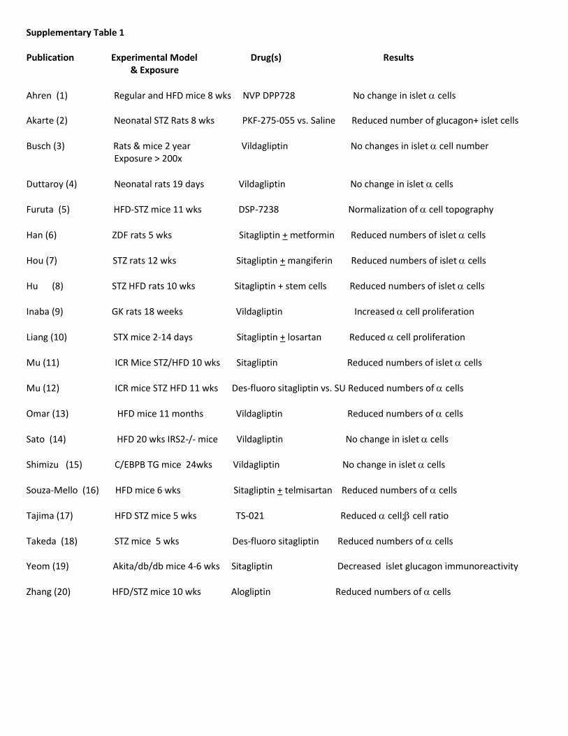
Supplementary Table 1 Publication Experimental Model Drug(s) Results & Exposure
Ahren (1) Regular and HFD mice 8 wks NVP DPP728 No change in islet cells Akarte (2) Neonatal STZ Rats 8 wks PKF-275-055 vs. Saline Reduced number of glucagon+ islet cells
Busch (3) Rats & mice 2 year Vildagliptin No changes in islet cell number Exposure > 200x
Duttaroy (4) Neonatal rats 19 days Vildagliptin No change in islet cells
Furuta (5) HFD-STZ mice 11 wks DSP-7238 Normalization of cell topography
Han (6) ZDF rats 5 wks Sitagliptin + metformin Reduced numbers of islet cells
Hou (7) STZ rats 12 wks Sitagliptin + mangiferin Reduced numbers of islet cells
Hu (8) STZ HFD rats 10 wks Sitagliptin + stem cells Reduced numbers of islet cells
Inaba (9) GK rats 18 weeks Vildagliptin Increased cell proliferation
Liang (10) STX mice 2-14 days Sitagliptin + losartan Reduced cell proliferation
Mu (11) ICR Mice STZ/HFD 10 wks Sitagliptin Reduced numbers of islet cells
Mu (12) ICR mice STZ HFD 11 wks Des-fluoro sitagliptin vs. SU Reduced numbers of cells
Omar (13) HFD mice 11 months Vildagliptin Reduced numbers of cells
Sato (14) HFD 20 wks IRS2-/- mice Vildagliptin No change in islet cells
Shimizu (15) C/EBPB TG mice 24wks Vildagliptin No change in islet cells
Souza-Mello (16) HFD mice 6 wks Sitagliptin + telmisartan Reduced numbers of cells
Tajima (17) HFD STZ mice 5 wks TS-021 Reduced cell; cell ratio
Takeda (18) STZ mice 5 wks Des-fluoro sitagliptin Reduced numbers of cells Yeom (19) Akita/db/db mice 4-6 wks Sitagliptin Decreased islet glucagon immunoreactivity
Zhang (20) HFD/STZ mice 10 wks Alogliptin Reduced numbers of cells
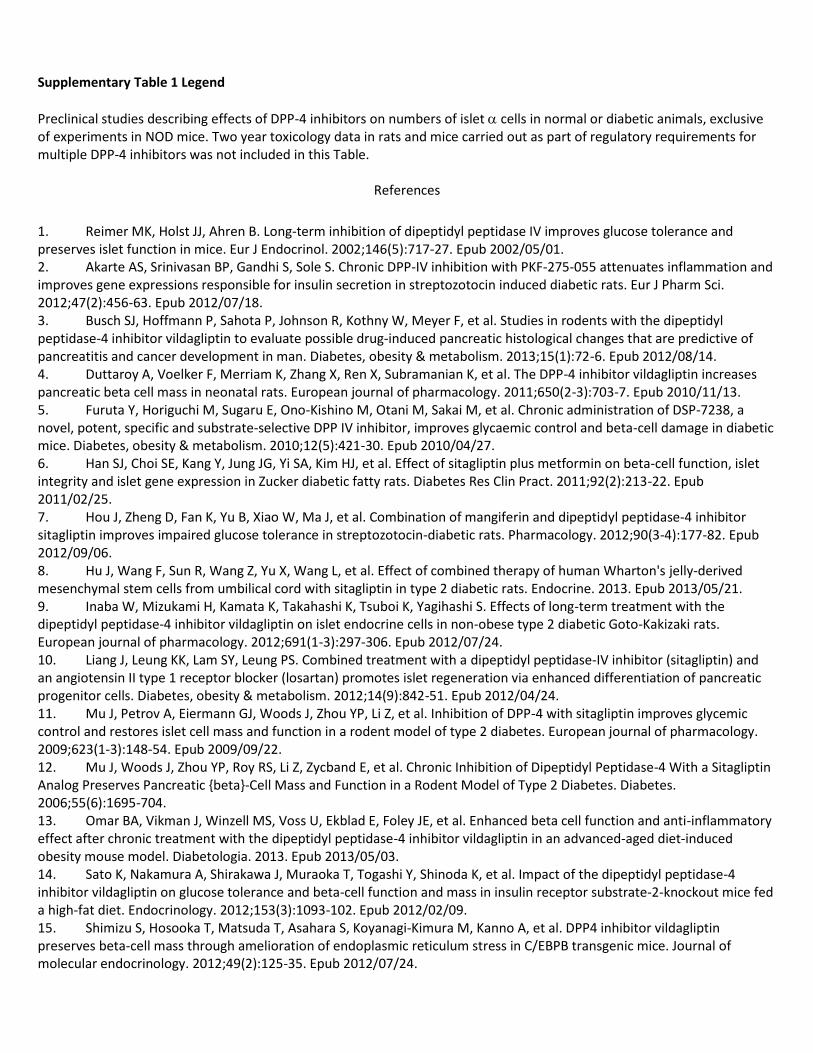
Supplementary Table 1 Legend
Preclinical studies describing effects of DPP-4 inhibitors on numbers of islet cells in normal or diabetic animals, exclusive of experiments in NOD mice. Two year toxicology data in rats and mice carried out as part of regulatory requirements for multiple DPP-4 inhibitors was not included in this Table.
References
1. Reimer MK, Holst JJ, Ahren B. Long-term inhibition of dipeptidyl peptidase IV improves glucose tolerance and preserves islet function in mice. Eur J Endocrinol. 2002;146(5):717-27. Epub 2002/05/01. 2. Akarte AS, Srinivasan BP, Gandhi S, Sole S. Chronic DPP-IV inhibition with PKF-275-055 attenuates inflammation and improves gene expressions responsible for insulin secretion in streptozotocin induced diabetic rats. Eur J Pharm Sci. 2012;47(2):456-63. Epub 2012/07/18. 3. Busch SJ, Hoffmann P, Sahota P, Johnson R, Kothny W, Meyer F, et al. Studies in rodents with the dipeptidyl peptidase-4 inhibitor vildagliptin to evaluate possible drug-induced pancreatic histological changes that are predictive of pancreatitis and cancer development in man. Diabetes, obesity & metabolism. 2013;15(1):72-6. Epub 2012/08/14. 4. Duttaroy A, Voelker F, Merriam K, Zhang X, Ren X, Subramanian K, et al. The DPP-4 inhibitor vildagliptin increases pancreatic beta cell mass in neonatal rats. European journal of pharmacology. 2011;650(2-3):703-7. Epub 2010/11/13. 5. Furuta Y, Horiguchi M, Sugaru E, Ono-Kishino M, Otani M, Sakai M, et al. Chronic administration of DSP-7238, a novel, potent, specific and substrate-selective DPP IV inhibitor, improves glycaemic control and beta-cell damage in diabetic mice. Diabetes, obesity & metabolism. 2010;12(5):421-30. Epub 2010/04/27. 6. Han SJ, Choi SE, Kang Y, Jung JG, Yi SA, Kim HJ, et al. Effect of sitagliptin plus metformin on beta-cell function, islet integrity and islet gene expression in Zucker diabetic fatty rats. Diabetes Res Clin Pract. 2011;92(2):213-22. Epub 2011/02/25. 7. Hou J, Zheng D, Fan K, Yu B, Xiao W, Ma J, et al. Combination of mangiferin and dipeptidyl peptidase-4 inhibitor sitagliptin improves impaired glucose tolerance in streptozotocin-diabetic rats. Pharmacology. 2012;90(3-4):177-82. Epub 2012/09/06. 8. Hu J, Wang F, Sun R, Wang Z, Yu X, Wang L, et al. Effect of combined therapy of human Wharton's jelly-derived mesenchymal stem cells from umbilical cord with sitagliptin in type 2 diabetic rats. Endocrine. 2013. Epub 2013/05/21. 9. Inaba W, Mizukami H, Kamata K, Takahashi K, Tsuboi K, Yagihashi S. Effects of long-term treatment with the dipeptidyl peptidase-4 inhibitor vildagliptin on islet endocrine cells in non-obese type 2 diabetic Goto-Kakizaki rats. European journal of pharmacology. 2012;691(1-3):297-306. Epub 2012/07/24. 10. Liang J, Leung KK, Lam SY, Leung PS. Combined treatment with a dipeptidyl peptidase-IV inhibitor (sitagliptin) and an angiotensin II type 1 receptor blocker (losartan) promotes islet regeneration via enhanced differentiation of pancreatic progenitor cells. Diabetes, obesity & metabolism. 2012;14(9):842-51. Epub 2012/04/24. 11. Mu J, Petrov A, Eiermann GJ, Woods J, Zhou YP, Li Z, et al. Inhibition of DPP-4 with sitagliptin improves glycemic control and restores islet cell mass and function in a rodent model of type 2 diabetes. European journal of pharmacology. 2009;623(1-3):148-54. Epub 2009/09/22. 12. Mu J, Woods J, Zhou YP, Roy RS, Li Z, Zycband E, et al. Chronic Inhibition of Dipeptidyl Peptidase-4 With a Sitagliptin Analog Preserves Pancreatic {beta}-Cell Mass and Function in a Rodent Model of Type 2 Diabetes. Diabetes. 2006;55(6):1695-704. 13. Omar BA, Vikman J, Winzell MS, Voss U, Ekblad E, Foley JE, et al. Enhanced beta cell function and anti-inflammatory effect after chronic treatment with the dipeptidyl peptidase-4 inhibitor vildagliptin in an advanced-aged diet-induced obesity mouse model. Diabetologia. 2013. Epub 2013/05/03. 14. Sato K, Nakamura A, Shirakawa J, Muraoka T, Togashi Y, Shinoda K, et al. Impact of the dipeptidyl peptidase-4 inhibitor vildagliptin on glucose tolerance and beta-cell function and mass in insulin receptor substrate-2-knockout mice fed a high-fat diet. Endocrinology. 2012;153(3):1093-102. Epub 2012/02/09. 15. Shimizu S, Hosooka T, Matsuda T, Asahara S, Koyanagi-Kimura M, Kanno A, et al. DPP4 inhibitor vildagliptin preserves beta-cell mass through amelioration of endoplasmic reticulum stress in C/EBPB transgenic mice. Journal of molecular endocrinology. 2012;49(2):125-35. Epub 2012/07/24.
16. Souza-Mello V, Gregorio BM, Relvas-Lucas B, da Silva Faria T, Aguila MB, Mandarim-de-Lacerda CA. Pancreatic ultrastructural enhancement due to telmisartan plus sitagliptin treatment in diet-induced obese C57BL/6 mice. Pancreas. 2011;40(5):715-22. Epub 2011/05/24. 17. Tajima A, Hirata T, Taniguchi K, Kondo Y, Kato S, Saito-Hori M, et al. Combination of TS-021 with metformin improves hyperglycemia and synergistically increases pancreatic beta-cell mass in a mouse model of type 2 diabetes. Life sciences. 2011;89(17-18):662-70. Epub 2011/08/30. 18. Takeda Y, Fujita Y, Honjo J, Yanagimachi T, Sakagami H, Takiyama Y, et al. Reduction of both beta cell death and alpha cell proliferation by dipeptidyl peptidase-4 inhibition in a streptozotocin-induced model of diabetes in mice. Diabetologia. 2012;55(2):404-12. Epub 2011/11/11. 19. Yeom JA, Kim ES, Park HS, Ham DS, Sun C, Kim JW, et al. Both sitagliptin analogue & pioglitazone preserve the beta-cell proportion in the islets with different mechanism in non-obese and obese diabetic mice. BMB reports. 2011;44(11):713-8. Epub 2011/11/29. 20. Zhang X, Wang Z, Huang Y, Wang J. Effects of chronic administration of alogliptin on the development of diabetes and beta-cell function in high fat diet/streptozotocin diabetic mice. Diabetes, obesity & metabolism. 2011;13(4):337-47. Epub 2011/01/06.