PerspectivesofStemCellsireneyan/Lab/papers/Pages from StemCellBook.pdf · Universidade de São...
26
Perspectives of Stem Cells
Transcript of PerspectivesofStemCellsireneyan/Lab/papers/Pages from StemCellBook.pdf · Universidade de São...

Perspectives of Stem Cells


Henning UlrichEditor
Perspectivesof Stem Cells
From Tools for StudyingMechanisms of NeuronalDifferentiation towards Therapy
123

EditorProf. Henning UlrichUniversidade de Sao PauloInstituto de QuimicaDepartamento de BioquimicaAv. Prof. Lineu Prestes 748Sao [email protected]
ISBN 978-90-481-3374-1 e-ISBN 978-90-481-3375-8DOI 10.1007/978-90-481-3375-8Springer Dordrecht Heidelberg London New York
Library of Congress Control Number: 2009940404
© Springer Science+Business Media B.V. 2010No part of this work may be reproduced, stored in a retrieval system, or transmitted in any form or byany means, electronic, mechanical, photocopying, microfilming, recording or otherwise, without writtenpermission from the Publisher, with the exception of any material supplied specifically for the purpose ofbeing entered and executed on a computer system, for exclusive use by the purchaser of the work.
Printed on acid-free paper
Springer is part of Springer Science+Business Media (www.springer.com)

Preface
Stem cells are fascinating cell types. They can replicate themselves forever whileretaining the potential to generate progeny with specific functions. Because of thesespecial properties, stem cells have been subjects of intensive investigation, fromunderstanding basic mechanisms underlying tissue generation, to modeling humandiseases, to application for cell replacement therapy. Stem cells come in differentforms. For example, mouse embryonic stem cells can general all cell types in a body,either in a dish or when put back into mouse embryos. On the other hand, neuralstem cells in the adult brain generate neurons and glia cells that contribute to thebrain’s plasticity. Rapid progress has been made in the stem cell field with discover-ies published in a record speed. A quick Pubmed search has returned 2789 hits for“embryonic stem cells” and 815 hits for “adult neural stem cells/neurogenesis” in theyear 2008 alone. It remains a taunting task for all who are interested in stem cells tokeep up with rapidly accumulating literatures. The “Perspectives of Stem Cells” by atruly international team of experts provides a timely and invaluable highlight of thestem cell field gearing toward future therapeutic applications in the nervous system.
Stem cells with neural potentials have attracted a lot of attention because of theirpromise for cell replacement therapy, ranging from degenerative neurological disor-ders to spinal cord injuries. Before such potentials to be realized, however, we needto understand the basic biology of these stem cells. For example, understanding howstem cell behaviors are controlled by intrinsic and extrinsic factors will help to directstem cells into a specific fate while avoiding undesired tumorigenesis. Equally impor-tant, we need to understand adult nervous system milieu where substitute neuronsneed to integrate into proper circuitry for maximal recovery. In this regard, the recentdiscovery of functional neurogenesis in discrete regions of the adult mammalianbrain, including humans, has a major impact on regenerative medicine. Not onlythere exist residual adult neural stem cells as endogenous cellular sources for neu-rogenesis, the adult nervous system itself exhibits surprising plasticity. They provideproper signals to support normal neurogenesis and, furthermore, additional signalsupon injuries to activate neural stem cells and guide their neuronal progeny to theright location. The dogma, “In the adult centers, the nerve paths are something fixed,ended, and immutable. Everything may die, nothing may be regenerated”, is now longgone.
The Prospective covers a broad spectrum of a fast evolving field of stem cells:from different model systems and stem cell types, to their cell biology and molecu-lar signaling mechanisms; from overview of protocols for directed neuronal subtypedifferentiation from embryonic stem cells, and even the latest induced pluripotent
v

vi Preface
stem cells, to specific considerations for the therapeutic application. The historicalview of neurogenesis since the time of Cajal was a delight retreat; the discussion ofretrotransposons in generating neuronal diversity through neurogenesis was fascinat-ing. The Prospective provides a much needed overview of the state of the art in thefield and a rich resource of updated information. More importantly, it sets up a stagefor flourishing of new ideas in stem cell biology and for fostering novel therapeuticapplications in the nervous system for years to come.
Clarksville, Maryland Hongjun SongAugust 2009

Editor Preface
The field of stem cell biology is geared towards translation into clinical practicethrough in vitro tissue production and regeneration therapy. The discovery of neu-rogenesis in selected areas of the adult brain has revolutionized neuroscience. Thisdiscovery has overturned the central assumption that no new neurons were origi-nated in the brain after birth, and provided the basis for understanding the molecularmechanisms of neural differentiation. Several in vitro models have been developedto investigate signalling pathway of neurogenesis regulation and cell fate specifica-tion. Massive propagation of embryonic cells into just the right type of phenotypeof a neural progenitor cell or strategies for mobilizing endogenous neural stem orprogenitor cells provide replacement therapies for brain injury resulting from strokeor neurodegenerative diseases. Such strategies are getting more important, since thedevelopment of ex vivo cultures of stem cells allows collection of multipotent cellsfrom patients, their differentiation and transplantation into diseased areas.
This book includes chapters on mechanisms of neural induction in early embryosas well as a detailed discussion of changes in paradigms in view of the discov-ery of adult neurogenesis. Neuronal differentiation is detailed using the olfactoryepithelium, one of the tissues bearing neurogenesis along life. In addition to chapterson therapeutic applicability of embryonic, very small-embryonic like, mesenchymalstem and neural progenitor cells, this book covers signalling mechanisms guidinginduction to differentiation and selective achievement of specific phenotypes. Celldiversification of the neuronal system is explained using the example of neural crestcell differentiation. Alterations in genetic material, such as loss of chromosomesand retrotransposition, are discussed as possible mechanisms for cell diversification.Furthermore, fundamental aspects of stem cell biology and neurogenesis, such as theimportance of proliferation induction, programmed cell death, as well as the func-tion of glia in differentiation of stem cells and development of neuronal circuits, arealso highlighted. The participation of cytoskeletal elements in cell polarization as pre-requisites of asymmetric division and differentiation induction is discussed. Furthertopics include the analysis of extracellular signals, such as neurotrophic factors andneurotransmitters, their receptor molecules and the propagation of these signals byintracellular signal transduction leading to activation of selective gene expression dur-ing differentiation. Rhythmic gene expression for activation and inhibition of Notchsignalling is discussed as a mechanism for regulating the progress of neurogenesis. Invitro cultures of embryonic, mesenchymal and neural stem cells as well as mobiliza-tion of endogenous stem and precursor cells for brain repair and replacement therapyin neurological disorders are important issues of this book.
vii

viii Editor Preface
Each chapter provides an invaluable resource for information on the most currentadvances in the field and possible therapeutic applications, with discussions of con-troversial issues and areas of emerging importance. By providing an up-to-date andcritical view of the state of Science, we hope that this book shall be a base for excit-ing scientific ideas regarding functions and therapeutic applications of stem cells inthe adult brain. The book is directed to neuroscientists, physicians, students and allwho are engaged and interested in the exciting and rapidly expanding field of modernneuroscience and stem cell biology.
São Paulo Henning UlrichJune 2009

Contents
1 Neural Induction . . . . . . . . . . . . . . . . . . . . . . . . . . . . 1Karla Loureiro Almeida, José Abreu, and C.Y. Irene Yan
2 Neurogenesis: A Change of Paradigms . . . . . . . . . . . . . . . . 11Luiz E. Mello and Beatriz M. Longo
3 Neurogenesis in the Olfactory Epithelium . . . . . . . . . . . . . . . 35Bettina Malnic and Lucia Armelin-Correa
4 Cell Diversification During Neural Crest Ontogeny:The Neural Crest Stem Cells . . . . . . . . . . . . . . . . . . . . . . 47Elisabeth Dupin, Giordano W. Calloni, and Nicole M. LeDouarin
5 Intermediate Filament Expression in Mouse EmbryonicStem Cells and Early Embryos . . . . . . . . . . . . . . . . . . . . . 59Zhigang Xue, Vivaldo Moura-Neto, Araksya Izmiryan,Sheila Cristina de Souza Martins, Jean Christophe Larcher,Denise Paulin, and Zhenlin Li
6 Aneuploidy in Embryonic Stem Cells . . . . . . . . . . . . . . . . . 73Rafaela C. Sartore, Priscila B. Campos,Michael J. McConnell, and Stevens K. Rehen
7 Retrotransposition and Neuronal Diversity . . . . . . . . . . . . . . 87Maria C. N. Marchetto, Fred H. Gage, and Alysson Muotri
8 Directing Differentiation of Embryonic Stem Cellsinto Distinct Neuronal Subtypes . . . . . . . . . . . . . . . . . . . . 97Noelle Ammon, Nathaniel Hartman, and Laura Grabel
9 Neurotransmitters as Main Players in the NeuralDifferentiation and Fate Determination Game . . . . . . . . . . . . 115Katia K. Yuahasi, Katia N. Gomes, Marcelo Campos, ArthurA. Nery, Ariane Nunes-Alves, Cleber A. Trujillo, and Henning Ulrich
10 Rhythmic Expression of Notch Signaling in NeuralProgenitor Cells . . . . . . . . . . . . . . . . . . . . . . . . . . . . . 135Hiromi Shimojo, Toshiyuki Ohtsuka, and Ryoichiro Kageyama
ix

x Contents
11 Neuron-Astroglial Interactions in Cell Fate Commitmentin the Central Nervous System . . . . . . . . . . . . . . . . . . . . . 145Joice Stipursky, Tânia Cristina Leite de Sampaio e Spohr,Luciana Ferreira Romão, and Flávia Carvalho Alcantara Gomes
12 The Origin of Microglia and the Development of the Brain . . . . . 171Flavia R.S. Lima, Anna Carolina C. da Fonseca,Giselle P. Faria, Luiz Gustavo F. Dubois, Tércia R. Alves,Jane Faria, and Vivaldo Moura Neto
13 Tissue Biology of Proliferation and Cell Death AmongRetinal Progenitor Cells . . . . . . . . . . . . . . . . . . . . . . . . 191Rafael Linden, Rodrigo A.P. Martins, Mariana S. Silveira,Helena L. Borges, Alfred Sholl-Franco, LucianneFragel-Madeira, and Ana Carolina Dudenhoeffer-Carneiro
14 Potential Application of Very Small Embryonic Like(VSEL) Stem Cells in Neural Regeneration . . . . . . . . . . . . . . 231Mariusz Z. Ratajczak, Ewa Zuba-Surma, Magda Kucia,Przemyslaw Nowacki, and Bogdan Machalinski
15 Embryonic Stem Cell Transplantation for the Treatmentof Parkinson’s Disease . . . . . . . . . . . . . . . . . . . . . . . . . 245Asuka Morizane and Jun Takahashi
16 Functional Multipotency of Neural Stem Cellsand Its Therapeutic Implications . . . . . . . . . . . . . . . . . . . 255Yang D. Teng, Serdar Kabatas, Jianxue Li,Dustin R. Wakeman, Evan Y. Snyder, and Richard L. Sidman
17 Dual Roles of Mesenchymal Stem Cells in Spinal CordInjury: Cell Replacement Therapy and as a Model Systemto Understand Axonal Repair . . . . . . . . . . . . . . . . . . . . . 271Cecile King, Shyam Patel, Treena Livingston Arinzeh,and Pranela Rameshwar
Index . . . . . . . . . . . . . . . . . . . . . . . . . . . . . . . . . . . . . . 285

Contributors
José Abreu Cellular and Developmental Biology Program, Institute of BiomedicalSciences, Universidade Federal do Rio de Janeiro, Centro de Ciências da Saúde –bloco F, Cidade Universitária, Rio de Janeiro, RJ 21949-590, Brazil,[email protected]
Karla Loureiro Almeida Prédio do Básico – CCS/UFES – Campus de MaruípeAv. Marechal Campos, 1468 Vitória, ES 29.043-900, Brazil,[email protected]
Ariane Nunes-Alves Departamento de Bioquímica, Instituto de Química,Universidade de São Paulo, São Paulo, Brazil, [email protected]
Tércia R. Alves Programa de Anatomia, Instituto de Ciências Biomédicas,Universidade Federal do Rio de Janeiro, Rio de Janeiro, Brazil,[email protected]
Noelle Ammon Hall-Atwater Laboratories, Biology Department, WesleyanUniversity, Middletown, CT, USA, [email protected]
Lucia Armelin-Correa Departamento de Bioquímica, Instituto de Química,Universidade de São Paulo, São Paulo, Brazil, [email protected]
Helena L. Borges Instituto de Ciências Biomédicas, Universidade Federal do Riode Janeiro, Cidade Universitaria, Rio de Janeiro, Brazil, [email protected]
Giordano W. Calloni CNRS UPR2197 Laboratoire Développement, Evolution etPlasticité du Système Nerveux, Institut de Neurobiologie Alfred Fessard, 91198Gif-sur-Yvette, France; Departamento de Biologia Celular, Embriologia e Genética,Centro de Ciências Biológicas, Universidade Federal de Santa Catarina,Florianópolis, Brazil, [email protected]
Priscila B. Campos Instituto de Ciências Biomédicas, Universidade Federal do Riode Janeiro, Rio de Janeiro, Brazil, [email protected]
Marcelo Campos Departamento de Bioquímica, Instituto de Química,Universidade de São Paulo, São Paulo, Brazil, [email protected]
Anna Carolina C. da Fonseca Programa de Anatomia, Instituto de CiênciasBiomédicas, Universidade Federal do Rio de Janeiro, Rio de Janeiro, Brazil,[email protected]
Luiz Gustavo F. Dubois Programa de Anatomia, Instituto de Ciências Biomédicas,Universidade Federal do Rio de Janeiro, Rio de Janeiro, Brazil, [email protected]
xi

xii Contributors
Ana Carolina Dudenhoeffer-Carneiro Instituto de Biofísica, UniversidadeFederal do Rio de Janeiro, Cidade Universitaria, Rio de Janeiro, Brazil,[email protected]
Elisabeth Dupin CNRS UPR2197 Laboratoire Développement, Evolution etPlasticité du Système Nerveux, Institut de Neurobiologie Alfred Fessard, 91198Gif-sur-Yvette, France, [email protected]
Giselle P. Faria Programa de Anatomia, Instituto de Ciências Biomédicas,Universidade Federal do Rio de Janeiro, Rio de Janeiro, Brazil,[email protected]
Jane Faria Programa de Anatomia, Instituto de Ciências Biomédicas, UniversidadeFederal do Rio de Janeiro, Rio de Janeiro, Brazil, [email protected]
Lucianne Fragel-Madeira Instituto de Biofísica, Universidade Federal do Rio deJaneiro, Cidade Universitaria, Rio de Janeiro, Brazil, [email protected]
Fred H. Gage Laboratory of Genetics, The Salk Institute for Biological Studies, LaJolla, CA, USA, [email protected]
Katia N. Gomes Departamento de Bioquímica, Instituto de Química, Universidadede São Paulo, São Paulo, Brazil, [email protected]
Flávia Carvalho Alcantara Gomes Laboratório de Neurobiologia Celular,Programa de Biologia Celular e do Desenvolvimento, Instituto de CiênciasBiomédicas, Centro de Ciências da Saúde, Universidade Federal do Rio de Janeiro,Rio de Janeiro, Brazil, [email protected]
Laura Grabel Hall-Atwater Laboratories, Biology Department, WesleyanUniversity, Middletown, CT, USA, [email protected]
Nathaniel Hartman Hall-Atwater Laboratories, Biology Department, WesleyanUniversity, Middletown, CT, USA, [email protected]
Araksya Izmiryan UPMC Univ Paris 6, UMR 7079, Paris, France,[email protected]
Serdar Kabatas Department of Neurosurgery, Baskent University IstanbulHospital, Istanbul, Turkey, [email protected]
Ryoichiro Kageyama Institute for Virus Research, Kyoto University, Kyoto, Japan;Japan Science and Technology Agency, CREST, Kyoto, Japan,[email protected]
Cecile King UMDNJ-New Jersey Medical School, Department of Medicine,Newark, NJ, USA, [email protected]
Magda Kucia Stem Cell Institute at James Graham Brown Cancer Center,University of Louisville, Louisville, KY, USA, [email protected]
Jean Christophe Larcher Laboratoire de Biologie du Développement, Paris,France, [email protected]
Nicole M. Le Douarin CNRS UPR2197 Laboratoire Développement, Evolution etPlasticité du Système Nerveux, Institut de Neurobiologie Alfred Fessard, 91198Gif-sur-Yvette, France, [email protected]

Contributors xiii
Jian xue Li Department of Neurology, Beth Israel Deaconess Medical Center,Harvard Medical School, Boston, MA, USA, [email protected]
Zhenlin Li UPMC Univ Paris 6, UMR 7079, Paris, France, [email protected]
Flavia R.S. Lima Programa de Anatomia, Instituto de Ciências Biomédicas,Universidade Federal do Rio de Janeiro, Rio de Janeiro, Brazil, [email protected]
Rafael Linden Instituto de Biofísica, Universidade Federal do Rio de Janeiro,Cidade Universitaria, Rio de Janeiro, Brazil, [email protected]
Treena Livingston Arinzeh New Jersey Institute of Technology, Department ofBiomedical Engineering, Newark, NJ, USA, [email protected]
Beatriz M. Longo Departamento de Fisiologia, Universidade Federal de São Paulo,São Paulo, Brazil, [email protected]
Bogdan Machalinski Department of Physiopathology Pomeranian MedicalUniversity, Szczecin, Poland, [email protected]
Bettina Malnic Departamento de Bioquímica, Instituto de Química, Universidadede São Paulo, São Paulo, Brazil, [email protected]
Maria C. N. Marchetto Laboratory of Genetics, The Salk Institute for BiologicalStudies, La Jolla, CA, USA, [email protected]
Sheila Cristina de Souza Martins Instituto de Ciências Biomédicas-UniversidadeFederal de Rio de Janeiro, Rio de Janeiro, Brazil, [email protected]
Rodrigo A.P. Martins Instituto de Biofísica, Universidade Federal do Rio deJaneiro, Cidade Universitaria, Rio de Janeiro, Brazil, [email protected]
Michael J. McConnell Crick-Jacobs Center for Theoretical and ComputationalBiology, Salk Institute for Biological Studies, La Jolla, California, USA,[email protected]
Luiz E. Mello Departamento de Fisiologia, Universidade Federal de São Paulo, SãoPaulo, Brazil, [email protected]
Asuka Morizane Department of Biological Repair, Institute for Frontier MedicalSciences, Kyoto University, Kyoto, Japan, [email protected]
Vivaldo Moura-Neto Instituto de Ciências Biomédicas-Universidade Federal deRio de Janeiro, Rio de Janeiro, Brazil, [email protected]
Alysson Muotri University of California at San Diego, School of Medicine,Department of Pediatrics/Rady Children’s Hospital San Diego and Department ofPediatrics/Cellular and Molecular Medicine, La Jolla, CA, USA, [email protected]
Arthur A. Nery Departamento de Bioquímica, Instituto de Química, Universidadede São Paulo, São Paulo, Brazil, [email protected]
Przemyslaw Nowacki Department of Physiopathology Pomeranian MedicalUniversity, Szczecin, Poland, [email protected]
Toshiyuki Ohtsuka Institute for Virus Research, Kyoto University, Kyoto, Japan;Japan Science and Technology Agency, CREST, Kyoto, Japan,[email protected]

xiv Contributors
Shyam Patel UMDNJ-New Jersey Medical School, Department of Medicine,Newark, NJ, USA, [email protected]
Denise Paulin UPMC Univ Paris 6, UMR 7079, Paris, France,[email protected]
Pranela Rameshwar UMDNJ-New Jersey Medical School, Department ofMedicine, Newark, NJ, USA, [email protected]
Mariusz Z. Ratajczak Stem Cell Institute at James Graham Brown Cancer Center,University of Louisville, Louisville, KY, USA; Department of PhysiopathologyPomeranian Medical University, Szczecin, Poland, [email protected]
Stevens K. Rehen Instituto de Ciências Biomédicas, Universidade Federal do Riode Janeiro, Rio de Janeiro, Brazil, [email protected]
Luciana Ferreira Romão Laboratório de Neurobiologia Celular, Programa deBiologia Celular e do Desenvolvimento, Instituto de Ciências Biomédicas, Centro deCiências da Saúde, Universidade Federal do Rio de Janeiro, Rio de Janeiro, Brazil,[email protected]
Rafaela C. Sartore Instituto de Ciências Biomédicas, Universidade Federal do Riode Janeiro, Rio de Janeiro, Brazil, [email protected]
Hiromi Shimojo Institute for Virus Research, Kyoto University, Kyoto, Japan;Japan Science and Technology Agency, CREST, Kyoto, Japan,[email protected]
Alfred Sholl-Franco Instituto de Biofísica, Universidade Federal do Rio deJaneiro, Cidade Universitaria, Rio de Janeiro, Brazil, [email protected]
Richard L. Sidman Department of Neurology, Beth Israel Deaconess MedicalCenter, Harvard Medical School, Boston, MA, USA,[email protected]
Mariana S. Silveira Instituto de Biofísica, Universidade Federal do Rio de Janeiro,Cidade Universitaria, Rio de Janeiro, Brazil, [email protected]
Evan Y. Snyder Stem Cell and Regeneration Program, The Burnham Institute, LaJolla, CA, USA, [email protected]
Tânia Cristina Leite de Sampaio e Spohr Laboratório de Neurobiologia Celular,Programa de Biologia Celular e do Desenvolvimento, Instituto de CiênciasBiomédicas, Centro de Ciências da Saúde, Universidade Federal do Rio de Janeiro,Rio de Janeiro, Brazil, [email protected]
Joice Stipursky Laboratório de Neurobiologia Celular, Programa de BiologiaCelular e do Desenvolvimento, Instituto de Ciências Biomédicas, Centro de Ciênciasda Saúde, Universidade Federal do Rio de Janeiro, Rio de Janeiro, Brazil,[email protected]
Jun Takahashi Department of Biological Repair, Institute for Frontier MedicalSciences, Kyoto University, Kyoto, Japan, [email protected]

Contributors xv
C.Y. Irene Yan Department of Cell and Developmental Biology, Institute ofBiomedical Sciences, Universidade de São Paulo, Av. Prof Lineu Prestes 1524, SãoPaulo, SP 05508-900, Brazil, [email protected]
Yang D. Teng Department of Neurosurgery, Harvard Medical School, Brigham andWomen’s Hospital, and Children’s Hospital Boston, Boston, MA, USA; Departmentof Physical Medicine and Rehabilitation, Harvard Medical School, SpauldingRehabilitation Hospital, Boston, MA, USA; Division of SCI Research, VeteransAffairs Boston Healthcare System, Boston, MA, USA, [email protected]
Cleber A. Trujillo Departamento de Bioquímica, Instituto de Química,Universidade de São Paulo, São Paulo, Brazil, [email protected]
Henning Ulrich Departamento de Bioquímica, Instituto de Química, Universidadede São Paulo, São Paulo, Brazil, [email protected]
Dustin R. Wakeman Stem Cell and Regeneration Program, The Burnham Institute,La Jolla, CA, USA; University of California at San Diego: Graduate Program inBiomedical Sciences, La Jolla, CA 92093, USA, [email protected]
Zhigang Xue UPMC Univ Paris 6, UMR 7079, Paris, France,[email protected]
Katia K. Yuahasi Programa de Pós-graduação em Neurologia, Departamento deNeurologia e Neurocirurgia, Universidade Federal de São Paulo, São Paulo, Brazil,[email protected]
Ewa Zuba-Surma Stem Cell Institute at James Graham Brown Cancer Center,University of Louisville, Louisville, KY, USA, [email protected]

Chapter 1
Neural Induction
Karla Loureiro Almeida, José Abreu, and C.Y. Irene Yan
Contents
1.1 Introduction . . . . . . . . . . . . . . . . . 11.2 Neural Induction in the Xenopus Embryo –
The Early Experiments . . . . . . . . . . . 21.3 Neural Default Model . . . . . . . . . . . . 31.4 BMP and the Neural Inducers . . . . . . . . 41.5 Challenges to the Neural Default Model . . . . 41.6 Neural Induction and the Avian Node . . . . . 41.7 Epiblast – The Responsive Tissue . . . . . . . 51.8 Inhibition of BMP in the Avian Context . . . . 61.9 FGF Signaling and Neural Induction . . . . . 7References . . . . . . . . . . . . . . . . . . . . 8
Abstract Neural induction, i.e. definition of theneural domain from the ectoderm, is a fundamentaltopic that has fascinated developmental biologists foryears. The concept was first proposed by Spemmanand Mangold after their classic experiment in theamphibian Xenopus laevis where transplantation ofthe embryo’s dorsal blastopore lip induced a com-plete neural axis from the acceptor embryo’s ectoderm.Since then, much effort has been applied into iden-tifying the signals that bias the ectoderm into neuralfate and the resulting picture clearly indicates thatneural induction is a multi-step process that requiresthe interplay of various pathways. A major part ofour current understanding of neural induction orig-inates from the original amphibian model Xenopuslaevis. Recently, the chick embryo has added another
C.Y.I. Yan (�)Department of Cell and Developmental Biology, Universidadede São Paulo, Av Prof. Lineu Prestes, 1524, São Paulo, SP,05508-900, Brazile-mail: [email protected]
layer of complexity to the interpretation of the resultsobtained from the amphibian model. Here, we willfocus on the landmark experiments that address theearliest step of neural induction in these two models.Specifically, we will discuss the Neural Default modelthat was generated from experiments in the amphib-ian embryo to explain the choice between epidermaland neural precursor fate and the modifications on thismodel based on conclusions derived from the chickembryo.
Keywords BMP signaling · Ectoderm · FGF · Neuralinduction · Smad · Xenopus · Chick
Abbreviations
BMP bone morphogenetic proteinTGF-β transforming growth factor β
FGF fibroblast growth factorMAPK mtogen activated protein kinase
1.1 Introduction
The induction of neural tissue is a fundamental ques-tion that has fascinated developmental biologists sincethe classic experiment by Spemmann and Mangold.In 1924, based on their results from grafting experi-ments performed in amphibian embryos, the authorsproposed for the first time the concept of neural induc-tion. At the time, it was known that the blastoporelip initial involution site during gastrulation markedthe dorsal region of the embryo, and that the futureneural plate arose from the dorsal ectoderm – the ven-tral ectoderm forms mainly epidermal tissue. Spemann
1H. Ulrich (ed.), Perspectives of Stem Cells,DOI 10.1007/978-90-481-3375-8_1, © Springer Science+Business Media B.V. 2010
2 K.L. Almeida et al.
and Mangold transplanted the blastopore lip of donorembryos to the ventral region of host embryos in gas-trula stage. The host embryos went on to developa second, ventral neuraxis and anterior nervous sys-tem. More strikingly, the duplicate nervous systemwas fully composed of host tissue, while the trans-plant gave rise to a second notochord (dorsal meso-derm) underlying it. This result suggested strongly thatthe grafted tissue’s “determinative influences on itssurroundings” converted the surrounding ventral ecto-derm into the second nervous system (Spemann andMangold, 1924). The authors named the dorsal blasto-pore lip the Organizer, and hypothesized that duringnormal development this region determined the choiceof a neural fate for the dorsal ectoderm. They alsoproposed that the effect of the Organizer on the respon-sive ectoderm necessarily would involve cell-to-cellcommunication.
In the ensuing years, much effort has been appliedfor identifying the exact signals that emanate from theOrganizer and activate the signaling pathways that biasthe ectoderm into neural fate in vertebrates. The result-ing picture, derived from data obtained by variousgroups, indicates that neural induction is a multi-stepprocess. The amphibian model, Xenopus laevis, hascontinued to be of major importance to our understand-ing of neural induction due to the ease of experimentalreadout of neural induction in ectoderm explants. Inrecent years, the chick embryo has added anotherlayer of complexity to the interpretation of the resultsobtained from the amphibian model. In the followingsections we will present the major results derived fromboth model systems and the model that is emergingfrom those results. For the purposes of this chapter, wewill focus our discussion on the earliest step of neuralinduction, which is the choice between epidermal andneural precursor fate.
1.2 Neural Induction in the XenopusEmbryo – The Early Experiments
In the decade of 1990–2000, the search for theOrganizer’s neural inducing factors intensified and wasmainly performed in the Xenopus embryo. Based onthe characteristic of the Organizer, it was agreed that
a bona fide candidate for direct neural inducer had tofulfill certain criteria: it should cause axis duplicationin whole embryos, it should be expressed in the dor-sal blastopore (Organizer) region and elimination of itsactivity should interfere with normal neural develop-ment. The experimental paradigm used to screen forcandidate neural inducers was based on the fact that,by definition, induction involves a signaling sourceand a responsive target. Based on Spemmans exper-iment, the endogenous source of neural inducers isthe Organizer and the responsive tissue the ectoderm.Thus, ectoderm explants assays were used as an initialscreen for candidates. Ectoderm explants (also knownas animal caps) are cultured from a piece of ecto-derm excised from the animal pole of late blastulas,the lower part of which constitutes the blastocoeleroof (Fig. 1.1a). At this stage, the ectoderm is notyet committed to an epidermal or neural fate andresponds to growth factors in the media or overex-pression of relevant mRNAs by adopting different cellfates, which are verified through the expression ofmarker genes. When cultured as an intact tissue insaline solution, ectoderm explants express genes char-acteristic of epidermal tissue (Kintner and Melton,1987). However, if the explant is co-cultured with adorsal blastopore lip, neural markers are expressedinstead (Kintner and Melton, 1987). Thus, a gene’sneural-inducing activity is identified if there is upregu-lation of the expression of neural markers and decreasein the expression of epidermal genes. Importantly,because the Organizer is part of the dorsal meso-derm, genes that increased neural marker expressionbut also induced mesoderm markers, were not con-sidered direct neural inducers, as their effect couldbe indirect, through additional factors secreted by themesoderm.
The first molecule to fulfill all of the above-mentioned criteria for direct neural induction wasNoggin, a secreted polypeptide first identified bySmith and Harland (1992) in the Xenopus. Afterwards,Follistatin (Hemmati-Brivanlou et al., 1994) andChordin (Sasai et al., 1994), were also isolated fromXenopus embryos on the basis of their neuraliz-ing activity. All of these factors fulfilled the above-mentioned conditions, including expression at theOrganizer. At the time, these molecules were thoughtto act by directly stimulating neural fate, albeit throughan as yet unidentified mechanism.

1 Neural Induction 3
Fig. 1.1 Epidermal defaultmodel versus neural defaultModel. (a) In the “epidermaldefault model” the normal fateof an ectodermic tissue wouldbe epidermal, unless thisectoderm is stimulated byexternal factors (such as thoseprovided by addition of thedorsal blastopore lip). (b) Inthe “neural default model”,the intact ectoderm secretesanti-neural/pro-epidermalfactors. Induction of neuralfate occurs either byco-culture with the dorsalblastopore lip, which secretesneuralizing factors ordissociation of the ectoderm.In the former, neuralizingfactors counteract the effect ofendogenous pro-epidermalfactors. In the latter case,dissociation of the ectodermdilutes these factors andgenerates neural fate.Addition of ectoderm extractrestores epidermal fate
1.3 Neural Default Model
Insight on the mode of action of these molecules camefrom a second series of experiments that exploredthe effect of cell dissociation on ectoderm cell fate.When ectoderm explants are dissociated into individ-ual cells and cultured as such for a set period of time,they express neural markers, instead of epidermal ones(Fig. 1.1b). Remarkably, this occurs in the absence ofthe dorsal blastopore lip and without the addition ofexogenous factors (Godsave and Slack, 1989; Grunzand Tacke, 1989; Sato and Sargent, 1989; Wilson et al.,1997). These data led to the hypothesis that neural-ization is the default fate for ectodermal cells, andthat the cell–cell interactions that occur in an intactectodermic tissue somehow inhibit this developmen-tal path, resulting in an epidermal fate (Fig. 1.1b).Once the tissue is dissociated, these “epidermal fac-tors” are sufficiently diluted so as to allow development
of neural fate (Godsave and Slack, 1989; Grunz andTacke, 1989; Sato and Sargent, 1989). Thus, it wasproposed that the ectoderm has “neural default” fate,which is revealed in the absence of exogenous sig-naling (reviewed by Muñoz-Sanjuán and Brivanlou,2002).
The addition of concentrated ectodermal super-natant to dissociated cell cultures prevented the expres-sion of neural markers after ectodermal dissociation(Grunz and Tacke, 1990). Thereafter, candidate pro-teins for the role of “epidermal factor” were addedonto dissociated cultures and tested for their ability torestore epidermal fate while suppressing neuralization.These screens identified Bone Morphogenetic Protein4 (BMP4), a member of the Transforming GrowthFactor beta (TGF-β) superfamily as a potent epidermalinducer. When BMP4 is added to a culture of cells dis-sociated from the ectoderm it induces the expression ofepidermal markers (Wilson and Hemmati-Brivanlou,1995). Moreover, the expression pattern of BMPs in
4 K.L. Almeida et al.
the Xenopus gastrula is consistent with the role of“epidermal factor”: BMP4 is found throughout theectoderm prior to gastrulation but, afterwards it isexcluded from the neural plate (Fainsod et al., 1994;Hemmati-Brivanlou and Thomsen, 1995). Finally,inhibition of BMP signaling in ectodermal cells withdominant-negative receptors or antisense BMP4 RNAneuralizes ectodermal cells (Sasai et al., 1995). Thislast set of data was consistent with the model thatinhibition of endogenous BMP signaling, throughdilution, directs dissociated ectodermal cells towardsneural fate.
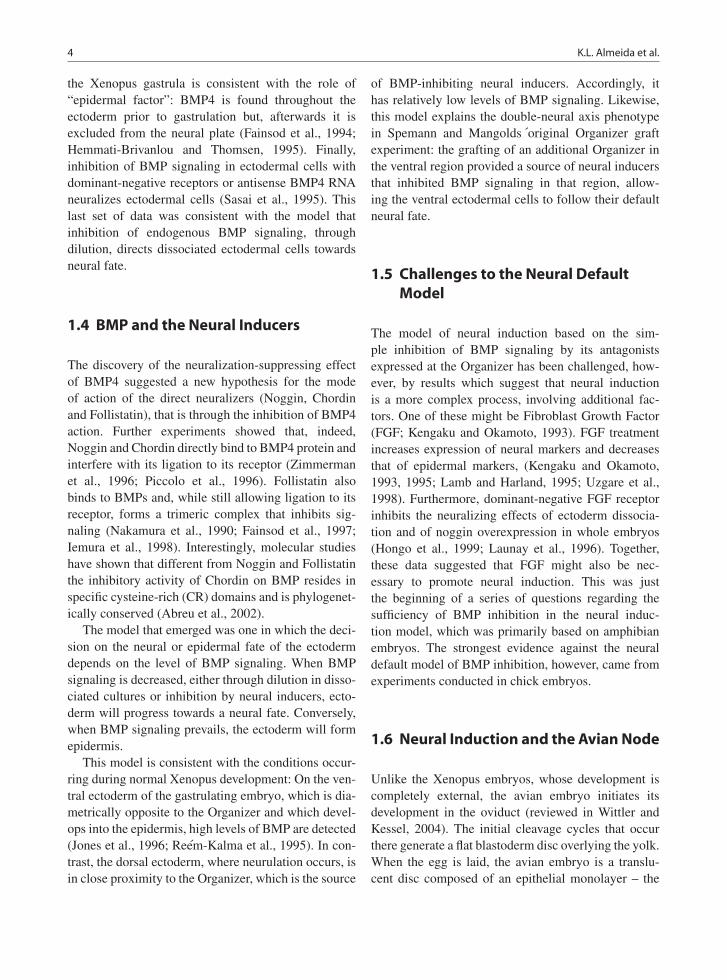
1.4 BMP and the Neural Inducers
The discovery of the neuralization-suppressing effectof BMP4 suggested a new hypothesis for the modeof action of the direct neuralizers (Noggin, Chordinand Follistatin), that is through the inhibition of BMP4action. Further experiments showed that, indeed,Noggin and Chordin directly bind to BMP4 protein andinterfere with its ligation to its receptor (Zimmermanet al., 1996; Piccolo et al., 1996). Follistatin alsobinds to BMPs and, while still allowing ligation to itsreceptor, forms a trimeric complex that inhibits sig-naling (Nakamura et al., 1990; Fainsod et al., 1997;Iemura et al., 1998). Interestingly, molecular studieshave shown that different from Noggin and Follistatinthe inhibitory activity of Chordin on BMP resides inspecific cysteine-rich (CR) domains and is phylogenet-ically conserved (Abreu et al., 2002).
The model that emerged was one in which the deci-sion on the neural or epidermal fate of the ectodermdepends on the level of BMP signaling. When BMPsignaling is decreased, either through dilution in disso-ciated cultures or inhibition by neural inducers, ecto-derm will progress towards a neural fate. Conversely,when BMP signaling prevails, the ectoderm will formepidermis.
This model is consistent with the conditions occur-ring during normal Xenopus development: On the ven-tral ectoderm of the gastrulating embryo, which is dia-metrically opposite to the Organizer and which devel-ops into the epidermis, high levels of BMP are detected(Jones et al., 1996; Reem-Kalma et al., 1995). In con-trast, the dorsal ectoderm, where neurulation occurs, isin close proximity to the Organizer, which is the source
of BMP-inhibiting neural inducers. Accordingly, ithas relatively low levels of BMP signaling. Likewise,this model explains the double-neural axis phenotypein Spemann and Mangolds ´original Organizer graftexperiment: the grafting of an additional Organizer inthe ventral region provided a source of neural inducersthat inhibited BMP signaling in that region, allow-ing the ventral ectodermal cells to follow their defaultneural fate.
1.5 Challenges to the Neural DefaultModel
The model of neural induction based on the sim-ple inhibition of BMP signaling by its antagonistsexpressed at the Organizer has been challenged, how-ever, by results which suggest that neural inductionis a more complex process, involving additional fac-tors. One of these might be Fibroblast Growth Factor(FGF; Kengaku and Okamoto, 1993). FGF treatmentincreases expression of neural markers and decreasesthat of epidermal markers, (Kengaku and Okamoto,1993, 1995; Lamb and Harland, 1995; Uzgare et al.,1998). Furthermore, dominant-negative FGF receptorinhibits the neuralizing effects of ectoderm dissocia-tion and of noggin overexpression in whole embryos(Hongo et al., 1999; Launay et al., 1996). Together,these data suggested that FGF might also be nec-essary to promote neural induction. This was justthe beginning of a series of questions regarding thesufficiency of BMP inhibition in the neural induc-tion model, which was primarily based on amphibianembryos. The strongest evidence against the neuraldefault model of BMP inhibition, however, came fromexperiments conducted in chick embryos.
1.6 Neural Induction and the Avian Node
Unlike the Xenopus embryos, whose development iscompletely external, the avian embryo initiates itsdevelopment in the oviduct (reviewed in Wittler andKessel, 2004). The initial cleavage cycles that occurthere generate a flat blastoderm disc overlying the yolk.When the egg is laid, the avian embryo is a translu-cent disc composed of an epithelial monolayer – the
1 Neural Induction 5
epiblast –, which is subdivided into a central area pel-lucida and an yolk-rich, extra-embryonic area opaca.The circumference where the pellucida and the opacameet is known as the Marginal Zone. After a few hours,a half-moon-shaped thickened region appears at theMarginal Zone. This structure is known as Kohler’ssickle and is the morphological landmark for the pos-terior end of the embryo and the site for initiation ofgastrulation. At the stage of its appearance, the epiblastcells migrate posteriorly in a bilaterally symmetricmovement and anteriorly at the midline, forming theprimitive streak through which epiblast cell ingress andform the definitive endoderm and mesoderm (Hatadaand Stern, 1994; Voiculescu et al., 2007; Joubin andStern, 1999 ). When sickle cells and the central epiblastcells meet at the anteriormost edge of the primi-tive streak, they form a thickened structure known asHensen’s Node, or simply the node (Fig. 2; Lawsonand Schoenwolf, 2001; Bachvarova et al., 1998 ). Asgastrulation continues, the primitive streak continuesexpanding anteriorly and bisects the embryo into leftand right regions (Fig. 1.2).
The node is considered the avian homologue of theamphibian dorsal blastopore lip. Its neural inductiveabilities and gene expression pattern are reminiscentof the Organizer: transplantation of the node to theextraembryonic area opaca induces a secondary neu-raxis (Waddington, 1932; Storey et al., 1992), withminimal participation of donor node cells (Storey
et al., 1992). Furthermore, the node expresses the avianhomologues of Goosecoid (Izpisua-belmonte et al.,1993), Goosecoid-like gene (Gsx, Lemaire et al., 1997)and Chordin (Streit et al., 1998), which are found in theXenopus Organizer.
1.7 Epiblast – The Responsive Tissue
The induction and patterning of the avian nervoussystem is a stepwise process that can be subdividedinto the ability of the epiblast to respond to neuraliz-ing signals (competence), the progressive stabilizationof this response (specification) and the subsequentpatterning of the neural region in its diverse axis.The initial experiments by Waddington (1932, 1993)showed that the avian blastula’s epiblast layer is com-petent to respond to neuralizing signals derived fromthe node. Indeed, fate mapping experiments show thatneural structures arise from a widespread region ofthe epiblast prior to gastrulation (Hatada and Stern,1994 García-Martínez et al., 1993). Waddington’s con-clusions were further refined by Storey et al. whotransplanted ectopic nodes to progressively older hostembryos and determined that the epiblast can generatea full antero-posterior neural axis up to early gas-trula stages (Storey et al., 1992; Streit et al., 1997).Thereafter, the epiblast cannot be induced to formanterior neural structures.
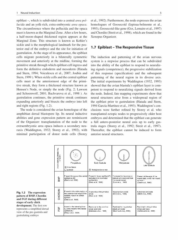
Fig. 1.2 The expressionpattern of BMP, Chordinand FGF during differentstages of early chickdevelopment. The first rowrepresents a simplified dorsalview of the pre-gastrula andgastrulating embryo

6 K.L. Almeida et al.
The precise stage at which the epiblast first demon-strates that it is competent to follow neural fate hasbeen progressively pushed back as more molecularmarkers have become available. For instance, the earlyneural marker Sox3 and late marker Sox2 have beenused as standard indicators of chick neural specifi-cation (Rex et al., 1997; Streit et al., 2000, 1997;Uchikawa et al., 2003). Sox3 is detected throughoutthe epiblast before neural induction in pre-gastrulaembryos and becomes restricted to the future neu-roectoderm as development progresses. Sox2 is firstdetected around the time when neural induction isbelieved to occur and its expression is limited to theneuroectoderm (Rex et al., 1997; Muhr et al., 1999).
Accordingly, immediately prior to gastrulation, thepotential of different regions of the epiblast differ.Cultures of explants derived from central epiblast gen-erated Sox2 and Sox3-positive cells whereas culturesderived from explants removed from regions closerto the marginal zone did not. Rather, these periph-eral explants express genes indicative of epidermalfate (Fig. 2, Wilson et al., 2000). Thus, by followingthe expression of Sox3 and Sox2 in cultured epi-blast explants, the earliest stage in which epiblast iscompartmentalized into neural and epidermal domainswas identified to be immediately prior to egg-laying(Wilson et al., 2000). At this stage, neural fate isrestricted to the central epiblast and epidermal fate tothe peripheral epiblast.
1.8 Inhibition of BMP in the AvianContext
The search for avian neural inducers that compartmen-talize the epiblast into neural or epidermal fate wasinitially based on a parallelism between the inductiveabilities of Hensen’s Node and Spemann’s Organizer.In support of this idea was the expression pattern ofBMPs and its inhibitors in late Primitive Streak stages:Prior to egg-laying, BMP is present throughout theepiblast but, when neuro-epidermal compartmentaliza-tion occurs, it becomes excluded from the prospectiveneural tissue (Wilson et al., 2000; Streit et al., 1998,Watanabe and Le Douarin, 1996; Streit et al., 1998).Likewise, the TGF-beta inhibitors chordin and noggin,which are expressed anterior to Kohler’s Sickle prior to
gastrulation, are found at the anterior tip of the prim-itive streak in early gastrulas and are restricted to thenotochord and the node in late gastrulas (Streit et al.,1998, Streit and Stern, 1999; Connolly et al., 1997).Altogether, these data suggested that in chick, simi-lar to Xenopus, BMP and its inhibitors are present incomplementary regions and that definition of a BMP-activity-free neural domain plays a crucial role inneural induction.
However, contrary to the results obtained inamphibian embryos, application of ectopic chordinonto early gastrula embryos cannot induce neural fatein non-neural ectoderm (Streit et al., 1998). Moreover,it would be expected, from the results in the frogmodel, that exposure to ectopic BMP would con-vert the presumptive neural domain into epidermal.Surprisingly, application of BMP onto early gastru-las’ neural domains does not inhibit Sox3 or Sox2expression (Streit et al., 1998). Inhibition of BMPsignaling through overexpression of Smad6 or dom-inant negative BMP receptor is also not sufficientfor neural induction (Linker and Stern, 2004). Theseresults, together with the findings that central epiblastis specified as neural prior to egg-laying (see previ-ous section), indicated that at early gastrula stagesthe neuro-ectodermal regions are already specified andthat the search for the initial neuralizing step shouldinclude earlier developmental stages.
Thus, Wilson and collaborators investigated theidentity of the signals that compartmentalized the cen-tral and peripheral epiblast into their respective neuraland epidermal fates in pre-gastrula embryos. At thisstage, the central epiblast is still susceptible to BMPand will respond to its presence by converting fromneural to epidermal fate (Streit et al., 1998; Wilsonet al. 2000). Thus, in early chick epiblasts, the Xenopusneural induction model holds true, in that BMP sig-naling confers an epidermal bias and that its absenceis necessary for neural fate. The dynamics of BMPexpression at this stage is consistent with its roleas the endogenous epidermalizing signal – BMP isdownregulated in central epiblast and maintained inperipheral epiblast (Streit et al., 1998; Wilson et al.,2000). This plasticity ends with the onset of gastrula-tion (HH4) (Fig. 2; Wilson et al., 2000). The neuraldomain’s progressive resistance to BMP reflects thegradual commitment to neural fate that occurs duringnormal embryonic development.

1 Neural Induction 7
1.9 FGF Signaling and Neural Induction
The question that remains is: what is the identity ofthe endogenous factor(s) that inhibit BMP signaling inthe pre-gastrula central epiblast? Contrary to expecta-tions, BMP signaling cannot be directly antagonizedby secreted BMP-inhibitors in pre-gastrula embryo.Although Chordin is expressed at the gastrula’s node,neither Chordin, Noggin, Follistatin or Caronte weredetected in central or peripheral epiblast in pre-gastrulaembryos (Levin, 1998; Wilson et al., 2000). Moreover,these inhibitors cannot induce neural markers by them-selves (Streit et al., 1998, 2000). In other words, analternative signaling mechanism must maintain thecentral epiblast BMP-free for the initial step in neuralinduction to occur.
The answer came from a series of elegant experi-ments that provided strong evidence that FGF meets allthe requirements for a role as an endogenous inhibitorof BMP in avian blastulas. Firstly, FGF3 is expressedin pre-gastrula central epiblast (Mahmood et al., 1995,Wilson et al., 2000, 2001). Furthermore, exogenouslyapplied FGF can induce the expression of early neuralmarkers (Streit et al., 2000). Blockade of endogenousFGF signaling inhibits expression of Sox3. Inhibitionof FGF signaling blocks neuralization and inductionof ectopic neural plate by a grafted organizer (Streitet al., 2000). Lastly, the FGF pathway is requiredfor downregulation of BMP levels in the central epi-blast, and absence of FGF signaling in the centralepiblast can be compensated for by the addition ofBMP inhibitors (Streit et al., 2000; Wilson et al., 2000,2001). Together, these data suggest that FGF is a puta-tive early neural inducer that acts by counteractingBMP signaling in the central epiblast.
These results agree with the previously mentionedeffects of FGF on Xenopus embryos. However, at thetime that those reports appeared, FGF was consideredmainly a posteriorizing signal that acted secondarily onthe neural domain generated by inhibition of BMP sig-naling. In light of the compelling data obtained fromchick embryos, the role of FGF as a primary neural-izing signal was revisited in the amphibian embryo aswell. This reassessment was done with ex vivo ectoder-mal explants and in vivo analysis of ventral ectodermfate in whole embryos. The results derived from invivo experiments differed somewhat from the clas-sical ex vivo experiments. While overexpression of
truncated TGF-beta receptor was sufficient to induceSox2 expression in amphibian ectodermal explants(Wilson and Hemmati-Brivanlou, 1995), it did notinduce a similar response in whole embryo ventralectoderm (Linker and Stern, 2004; Delaune et al.,2005). In this experimental paradigm, ectopic expres-sion of neural markers was achieved when there wasconcomitant inhibition of BMP and stimulation of FGFsignaling (Linker and Stern, 2004). Moreover, in theabsence of FGF signaling, the ectoderm cannot be neu-ralized by inhibition of BMP (Delaune et al., 2005).These results strongly suggest that, similar to theavian embryo, neuralization in the amphibian embryorequires interaction of the FGF and BMP pathways.
The interaction between both pathways has beenmapped to Smad1, a downstream nuclear effector ofthe BMP pathway. Smad1 nuclear translocation andtranscriptional activity are increased when it is phos-phorylated at the carboxy-terminal upon activation ofthe BMP receptor serine/threonine kinase (Massaguéand Chen, 2000). This activity is required for BMP-induced epidermal fate (Wilson et al., 1997; Nakayamaet al., 1998). In contrast, when Smad1 is phospho-rylated by MAPK in the central linker region, bothnuclear translocation and transcription are inhibited(Kretzschmar et al., 1997).
FGF signals through receptor tyrosine kinases thatultimately activate MAPK, which in turn phospho-rylates Smad1 (Pera et al., 2003). Underscoring theimportance of the MAPK pathway during Xenopusneural development, MAPK activity is required forneural induction by FGF and cell dissociation in ecto-derm explants (Uzgare et al., 1998; Kuroda et al.,2005). Thus, Smad1 integrates signals from the FGFand BMP pathway. Its activity results from the oppos-ing effects between FGF-induced linker region phos-phorylation versus the BMP-driven phosphorylation ofthe carboxy-region. Consistent with this idea, overex-pression of a MAPK-kinase insensitive Smad1 inhib-ited neural development in whole embryos, whereasmutation of both MAPK and BMP-sensitive regionsresulted in very mild phenotype (Pera et al., 2003).Thus, the final model that emerges places Smad1in the centre of the choice between neural and epi-dermal fate. In the presence of high levels of BMPsignaling, Smad1 is phosphorylated in the carboxyterminal, which activates its nuclear activity and cul-minates in epidermal fate. This epidermalizing effectcan be counteracted by FGF, which phosphorylates the

8 K.L. Almeida et al.
MH1N- -CLINKER MH2
P
FGF
MAPK
NEURAL EPIDERMAL
P
BMP
ChordinNoggin
Fig. 1.3 Neural andepidermal fate aredetermined by Smad1activity, which in turn isregulated byphosphorylation of itsserine/threonine residues.FGF-induced phosphorylationof the linker region retainsSmad1 in the cytoplasm andresults in neural fate, whereasBMP-inducedphosphorylation of thecarboxy terminal promotestranslocation of Smad1 to thenucleus and results inepidermal fate
Smad1 linker, inhibits its nuclear functions, resultingin adoption of neural fate (Fig. 1.3).
Although this model accounts for most of the resultsin the field, there are some points that must be consid-ered: firstly, besides FGF there are other growth factorsthat can activate MAPK activity, which raises the pos-sibility that additional secreted proteins can modulateneural induction (Linker and Stern, 2004). Second,MAPK has other target proteins, amongst them Smad2and Smad3, components of another TGF-beta pathway.Therefore, it is possible that FGF modulates additionalpathways for its neuralizing effect. Indeed, there isevidence that suppression of both Smad1 and Smad2activity are necessary for neural induction in ventralectoderm (Chang and Harland, 2007). Furthermore,the FGF pathway itself is modulated by other signalsthat are present during acquisition of neural compe-tence. For instance, in the chick embryo, the Wntpathway suppresses FGF signaling in the lateral epi-blast (Wilson et al., 2001). Lastly, as mentioned above,cell fate induction occurs in a continuous and progres-sive fashion. Therefore, the response of a target tissueto neuralizing or epidermalizing signals depends on itsdifferentiation state at the time of exposure. An exam-ple of this is neuralization through BMP inhibition inXenopus embryos. The response to BMP inhibitionis lost prior to the onset of gastrulation (Wawersiket al., 2005). Likewise, neural induction in Xenopusembryos is most sensitive to removal of FGF signalingduring mid-blastula transition (Delaune et al., 2005).Although these results are still under discussion (deAlmeida et al., 2008) and the exact period when eachidentified player is required for normal progression of
neural development is still unclear, it is the generalconsensus that the plasticity of the ectoderm decreaseswith time due to stabilization of cell fate (Streit et al.,1998; Wawersik et al., 2005; Linker and Stern, 2004;reviewed in Stern, 2005).
In conclusion, since the molecular identificationof direct neural inducers the development field hasproposed and refined models for the signaling thatunderlies the choice between epidermal and neural fatefrom the ectoderm. Even though the current modeldoes not account for all the complexity that occurs inthis process, the speed with which new findings are col-lected and incorporated into the most recent hypothesishas increased, and a more comprehensive panoramashould emerge in the next few years.
References
Abreu JG, Coffinier C, Larrain J, Oelgeschlager M, De RobertisEM (2002) Chordin-like CR domains and the regulationof evolutionarily conserved extracellular signaling systems.Gene 287:39–47.
Bachvarova RF, Skromne I, Stern CD (1998) Induction of primi-tive streak and Hensen’s node by the posterior marginal zonein the early chick embryo. Development 125:3521–3534.
Chang C, Harland RM (2007) Neural induction requires contin-ued suppression of both Smad1 and Smad2 signals duringgastrulation. Development 134:3861–3872.
Connolly DJ, Patel K, Cooke J (1997) Chick noggin is expressedin the organizer and neural plate during axial develop-ment, but offers no evidence of involvement in primary axisformation. Int J Dev Biol 41:389–396.
de Almeida I, Rolo A, Batut J, Hill C, Stern CD, Linker C (2008)Unexpected activities of Smad7 in Xenopus mesodermal andneural induction. Mech Dev 125:421–431.

1 Neural Induction 9
Delaune E, Lemaire P, Kodjabachian L (2005) Neural inductionin Xenopus requires early FGF signaling in addition to BMPinhibition. Development 132:299–310.
Fainsod A, Deissler K, Yelin R, Marom K, Epstein M, PillemerG, Steinbeisser H, Blum M (1997) The dorsalizing and neu-ral inducing gene follistatin is an antagonist of BMP-4. MechDev 63:39–50.
Fainsod A, Steinbeisser H, De Robertis EM (1994) On thefunction of BMP-4 in patterning the marginal zone of theXenopus embryo. EMBO J 13:5015–5025.
Garcia-Martinez V, Alvarez IS, Schoenwolf GC (1993)Locations of the ectodermal and nonectodermal subdivisionsof the epiblast at stages 3 and 4 of avian gastrulation andneurulation. J Exp Zool 267:431–446.
Godsave SF, Slack JM (1989) Clonal analysis of mesoderminduction in Xenopus laevis. Dev Biol 134:486–490.
Grunz H, Tacke L (1989) Neural differentiation of Xenopuslaevis ectoderm takes place after disaggregation anddelayed reaggregation without inducer. Cell Differ Dev 28:211–217.
Grunz H, Tacke L (1990) Extracellular matrix components pre-vent neural differentiation of disaggregated Xenopus ecto-derm cells. Cell Differ Dev 32:117–123.
Hatada Y, Stern CD (1994) A fate map of the epiblast of the earlychick embryo. Development 120:2879–2889.
Hemmati-Brivanlou A, Kelly OG, Melton DA (1994) Follistatin,an antagonist of activin, is expressed in the Spemannorganizer and displays direct neuralizing activity. Cell 77:283–295.
Hemmati-Brivanlou A, Thomsen GH (1995) Ventral mesoder-mal patterning in Xenopus embryos: expression patterns andactivities of BMP-2 and BMP-4. Dev Genet 17:78–89.
Hongo I, Kengaku M, Okamoto H (1999) FGF signaling andthe anterior neural induction in Xenopus. Dev Biol 216:561–581.
Iemura S, Yamamoto TS, Takagi C, Uchiyama H, NatsumeT, Shimasaki S, Sugino H, Ueno N (1998) Direct bindingof follistatin to a complex of bone-morphogenetic proteinand its receptor inhibits ventral and epidermal cell fatesin early Xenopus embryo. Proc Natl Acad Sci USA 95:9337–9342.
Izpisua-Belmonte JC, De Robertis EM, Storey KG, Stern CD(1993) The homeobox gene goosecoid and the origin of orga-nizer cells in the early chick blastoderm. Cell 74:645–659.
Jones CM, Dale L, Hogan BL, Wright CV, Smith JC (1996)Bone morphogenetic protein-4 (BMP-4) acts during gas-trula stages to cause ventralization of Xenopus embryos.Development 122:1545–1554.
Joubin K, Stern CD (1999) Molecular interactions continu-ously define the organizer during the cell movements ofgastrulation. Cell 98:559–571.
Kengaku M, Okamoto H (1993) Basic fibroblast growth fac-tor induces differentiation of neural tube and neural crestlineages of cultured ectoderm cells from Xenopus gastrula.Development 119:1067–1078.
Kengaku M, Okamoto H (1995) bFGF as a possible morphogenfor the anteroposterior axis of the cental nervous system inXenopus. Development 121:3121–3130.
Kintner CR, Melton DA (1987) Expression of Xenopus N-CAMRNA in ectoderm is an early response to neural induction.Development 99:311–325.
Kretzschmar M, Doody J, Massague J (1997) Opposing BMPand EGF signalling pathways converge on the TGF-betafamily mediator Smad1. Nature 389:618–622.
Kuroda H, Fuentealba L, Ikeda A, Reversade B, De Robertis EM(2005) Default neural induction: neuralization of dissociatedXenopus cells is mediated by Ras/MAPK activation. GenesDev 19:1022–1027.
Lamb TM, Harland RM (1995) Fibroblast growth factor is adirect neural inducer, which combined with noggin gen-erates anterior-posterior neural pattern. Development 121:3627–3636.
Launay C, Fromentoux V, Shi DL, Boucaut JC (1996) A trun-cated FGF receptor blocks neural induction by endogenousXenopus inducers. Development 122:869–880.
Lawson A, Schoenwolf GC (2001) Cell populations and mor-phogenetic movements underlying formation of the avianprimitive streak and organizer. Genesis 29:188–195.
Lemaire L, Roeser T, Izpisua-Belmonte JC, Kessel M (1997)Segregating expression domains of two goosecoid genes dur-ing the transition from gastrulation to neurulation in chickembryos. Development 124:1443–1452.
Levin M (1998) The roles of activin and follistatin signaling inchick gastrulation. Int J Dev Biol 42:553–559.
Linker C, Stern CD (2004) Neural induction requires BMP inhi-bition only as a late step, and involves signals other than FGFand Wnt antagonists. Development 131:5671–5681.
Mahmood R, Kiefer P, Guthrie S, Dickson C, Mason I (1995)Multiple roles for FGF-3 during cranial neural developmentin the chicken. Development 121:1399–1410.
Massague J, Chen YG (2000) Controlling TGF-beta signaling.Genes Dev 14:627–644.
Muhr J, Graziano E, Wilson S, Jessell TM, Edlund T (1999)Convergent inductive signals specify midbrain, hindbrain,and spinal cord identity in gastrula stage chick embryos.Neuron 23:689–702.
Muñoz-Sanjuan I, Brivanlou AH (2002) Neural induction, thedefault model and embryonic stem cells. Nat Rev Neurosci3:271–280.
Nakamura T, Takio K, Eto Y, Shibai H, Titani K, Sugino H(1990) Activin-binding protein from rat ovary is follistatin.Science 247:836–838.
Nakayama T, Gardner H, Berg LK, Christian JL (1998) Smad6functions as an intracellular antagonist of some TGF-betafamily members during Xenopus embryogenesis. GenesCells 3:387–394.
Pera E, Ikeda A, Eivers E, de Robertis EM (2003) Integration ofIGF, FGF and anti-BMP signals via Smad1 phosphorylationin neural induction. Genes Dev 17:3023–3028.
Piccolo S, Sasai Y, Lu B, De Robertis EM (1996) Dorsoventralpatterning in Xenopus: inhibition of ventral signals by directbinding of chordin to BMP-4. Cell 86:589–598.
Re’em-Kalma Y, Lamb T, Frank D (1995) Competition betweennoggin and bone morphogenetic protein 4 activities may reg-ulate dorsalization during Xenopus development. Proc NatlAcad Sci USA 92:12141–12145.
Rex M, Orme A, Uwanogho D, Tointon K, Wigmore PM, SharpePT, Scotting PJ (1997) Dynamic expression of chicken Sox2and Sox3 genes in ectoderm induced to form neural tissue.Dev Dyn 209:323–332.
Sasai Y, Lu B, Steinbeisser H, Geissert D, Gont LK, De RobertisEM (1994) Xenopus chordin: a novel dorsalizing factor

10 K.L. Almeida et al.
activated by orgaizerspecific homeobox genes. Cell 79:779–790.
Sasai Y, Lu B, Steinbeisser H, de Robertis EM (1995) Regulationof neural induction by the Chd and Bmp-4 antagonisticpatterning signals in Xenopus. Nature 376:333–336.
Sato SM, Sargent TD (1989) Development of neural induc-ing capacity in dissociated Xenopus embryos. Dev Biol134:263–266.
Smith WC, Harland RM (1992) Expression cloning of noggin, anew dorsalizing factor localized to the Spemann organizer inXenopus embryos. Cell 70:829–840.
Spemann H, Mangold H (1924) Induction of embryonic primor-dia by implantation of organizers from a different species.Roux’s Arch Entw Mech 100:599–638. Re-published in Int JDev Biol 45:13–38.
Stern CD (2005) Neural induction: old problem, new findings,yet more questions. Development 132:2007–2021.
Storey KG, Crossley JM, De Robertis EM, Norris WE, SternCD (1992) Neural induction and regionalisation in the chickembryo. Development 114:729–741.
Streit A, Berliner AJ, Papanayotou C, Sirulnik A, Stern CD(2000) Initiation of neural induction by FGF signallingbefore gastrulation. Nature 406:74–78.
Streit A, Lee KJ, Woo I, Roberts C, Jessell TM, Stern CD(1998) Chordin regulates primitive streak development andthe stability of induced neural cells, but is not sufficientfor neural induction in the chick embryo. Development 125:507–519.
Streit A, Sockanathan S, Perez L, Rex M, Scotting PJ, SharpePT, Lovell-Badge R, Stern CD (1997) Preventing the lossof competence for neural induction: HGF/SF, L5 and Sox-2.Development 124:1191–1202.
Streit A, Stern CD (1999) Establishment and maintenance of theborder of the neural plate in the chick: involvement of FGFand BMP activity. Mech Dev 82:51–66.
Uchikawa M, Ishida Y, Takemoto T, Kamachi Y, KondohH (2003) Functional analysis of chicken Sox2 enhancers
highlights an array of diverse regulatory elements that areconserved in mammals. Dev Cell 4:509–519.
Uzgare AR, Uzman JA, El-Hodiri HM, Sater AK (1998)Mitogen-activated protein kinase and neural specification inXenopus. Proc Natl Acad Sci USA 95:14833–14838.
Voiculescu O, Bertocchini F, Wolpert L, Keller RE, Stern CD(2007) The amniote primitive streak is defined by epithelialcell intercalation before gastrulation. Nature 449:1049–1052.
Waddington CH (1932) Experiments on the development ofchick and duck embryos cultivated in vitro. Philos Trans RSoc Lond B Biol Sci 221:179–230.
Waddington CH (1933) Induction by the primitive streak and itsderivatives in the chick. J Exp Biol 10:38–48.
Watanabe Y, Le Douarin NM (1996) A role for BMP-4 in thedevelopment of subcutaneous cartilage. Mech Dev 57:69–78.
Wawersik S, Evola C, Whitman M (2005) Conditional BMPinhibition in Xenopus reveals stage-specific roles for BMPsin neural and neural crest induction. Dev Biol 277:425–442.
Wilson SI, Graziano E, Harland R, Jessell TM, Edlund T (2000)An early requirement for FGF signalling in the acquisition ofneural cell fate in the chick embryo. Curr Biol 10:421–429.
Wilson PA, Hemmati-Brivanlou A (1995) Induction of epi-dermis and inhibition of neural fate by Bmp-4. Nature376:331–333.
Wilson PA, Lagna G, Suzuki A, Hemmati-Brivanlou A(1997) Concentration-dependent patterning of the Xenopusectoderm by BMP4 and its signal transducer Smad1.Development 124:3177–3184.
Wilson SI, Rydstrom A, Trimborn T, Willert K, Nusse R, JessellTM, Edlund T (2001) The status of Wnt signalling regu-lates neural and epidermal fates in the chick embryo. Nature411:325–330.
Wittler L, Kessel M (2004) The acquisition of neural fate in thechick. Mech Dev 121:1031–1042.
Zimmerman LB, De Jesus-Escobar JM, Harland RM (1996) TheSpemann organizer signal noggin binds and inactivates bonemorphogenetic protein 4. Cell 86:599–606.