Persistently Active Microbial Molecules Prolong Innate ... · Persistently Active Microbial...
16
Persistently Active Microbial Molecules Prolong Innate Immune Tolerance In Vivo Mingfang Lu 1 *, Alan W. Varley 2 , Robert S. Munford 1 1 Laboratory of Clinical Infectious Diseases, National Institute of Allergy and Infectious Diseases, National Institutes of Health, Bethesda, Maryland, United States of America, 2 Infectious Diseases Division, Department of Internal Medicine, University of Texas Southwestern Medical Center, Dallas, Texas, United States of America Abstract Measures that bolster the resolution phase of infectious diseases may offer new opportunities for improving outcome. Here we show that inactivation of microbial lipopolysaccharides (LPS) can be required for animals to recover from the innate immune tolerance that follows exposure to Gram-negative bacteria. When wildtype mice are exposed to small parenteral doses of LPS or Gram-negative bacteria, their macrophages become reprogrammed (tolerant) for a few days before they resume normal function. Mice that are unable to inactivate LPS, in contrast, remain tolerant for several months; during this time they respond sluggishly to Gram-negative bacterial challenge, with high mortality. We show here that prolonged macrophage reprogramming is maintained in vivo by the persistence of stimulatory LPS molecules within the cells’ in vivo environment, where naı ¨ve cells can acquire LPS via cell-cell contact or from the extracellular fluid. The findings provide strong evidence that inactivation of a stimulatory microbial molecule can be required for animals to regain immune homeostasis following parenteral exposure to bacteria. Measures that disable microbial molecules might enhance resolution of tissue inflammation and help restore innate defenses in individuals recovering from many different infectious diseases. Citation: Lu M, Varley AW, Munford RS (2013) Persistently Active Microbial Molecules Prolong Innate Immune Tolerance In Vivo. PLoS Pathog 9(5): e1003339. doi:10.1371/journal.ppat.1003339 Editor: Dario S. Zamboni, University of Sa ˜ o Paulo, Brazil Received September 4, 2012; Accepted March 18, 2013; Published May 9, 2013 This is an open-access article, free of all copyright, and may be freely reproduced, distributed, transmitted, modified, built upon, or otherwise used by anyone for any lawful purpose. The work is made available under the Creative Commons CC0 public domain dedication. Funding: This study was funded by the Division of Intramural Research, National Institute of Allergy and Infectious Diseases, Bethesda, MD, and by NIH Grant RO1 AI18188 to University of Texas Southwestern Medical Center, Dallas, TX. The funders had no role in study design, data collection and analysis, decision to publish, or preparation of the manuscript. Competing Interests: The authors have declared that no competing interests exist. * E-mail: [email protected] Introduction A great deal is known about how animals kill bacteria but very little about what happens to the corpses. Most bacterial structures seem to be broken down by phagocytes [1,2], yet much of the catabolic machinery is made by the bacteria themselves (phos- pholipases [3], peptidoglycan hydrolases [4], autolysins), which in some cases produce agonists that can elicit inflammation. Host enzymes may also participate, attacking peptidoglycan (lysozyme, peptidoglycan binding proteins [5,6]), lipids (phospholipases [3]), chitin [7], proteins (cathepsins, others), lipopolysaccharides (LPS) and, presumably, DNA. It is likely that the breakdown products are excreted, retained within phagocytes (e.g., in lymph nodes or arterial walls [8,9]) or in discrete extracellular deposits [10], or recycled by the host. The seminal studies of Cohn [1,11], Elsbach [2,3] and Schwab [12] established that degradation of ingested bacteria by phago- cytes can be incomplete. More recently, deposits of microbial antigens have been discovered in animals following infection with B. burgdorferii, the etiological agent of Lyme Disease [10]. A key question raised by these findings has remained unanswered to date: to what extent does the inactivation of stimulatory microbial molecules influence the outcome of infectious diseases or host reactions to microbe-rich environmental exposures? Here we show that for one important agonist, bacterial lipopolysaccharide (LPS), persistently active molecules can greatly delay immune recovery in vivo. Exposure to Gram-negative bacterial LPS induces humans and many other mammals to enter a transient state of altered immune responsiveness known as cellular reprogramming or tolerance [13–15]. The phenomenon has also been produced in animals by peritonitis [16], influenza virus infection [17] and bacterial lipopeptides [18,19] and in cultured macrophages using muramyl dipeptide [20], lipoteichoic acid [21], and flagella [22]. For a period that may last from a few hours to a few days after exposure to the microbial agonist, tolerant animals and cells respond to a second exposure by producing reduced amounts of many pro-inflammatory cytokines while maintaining, or even increasing, their production of certain anti-inflammatory and anti-infective molecules. Tolerance wanes as inflammation resolves and the animal regains normal immune responsiveness. Long considered a mechanism for preventing inflammation-induced injury, innate immune toler- ance may also be immunosuppressive [23]. Acyloxyacyl hydrolase (AOAH), an enzyme produced by macrophages, neutrophils, and dendritic cells, inactivates bioactive LPSs by removing two of the six fatty acyl chains that are present in the bioactive hexaacyl lipid A moiety [24]. In mice that lack AOAH, a single intraperitoneal exposure to as little as 80 ng of hexaacyl LPS induces tolerance that lasts for several weeks, much longer than that seen in wildtype animals that have received much larger doses [25]. Tolerant animals respond sluggishly to Gram- negative bacterial challenge and are unable to prevent bacterial multiplication in vivo [25]. PLOS Pathogens | www.plospathogens.org 1 May 2013 | Volume 9 | Issue 5 | e1003339
Transcript of Persistently Active Microbial Molecules Prolong Innate ... · Persistently Active Microbial...

Persistently Active Microbial Molecules Prolong InnateImmune Tolerance In VivoMingfang Lu1*, Alan W. Varley2, Robert S. Munford1
1 Laboratory of Clinical Infectious Diseases, National Institute of Allergy and Infectious Diseases, National Institutes of Health, Bethesda, Maryland, United States of
America, 2 Infectious Diseases Division, Department of Internal Medicine, University of Texas Southwestern Medical Center, Dallas, Texas, United States of America
Abstract
Measures that bolster the resolution phase of infectious diseases may offer new opportunities for improving outcome. Herewe show that inactivation of microbial lipopolysaccharides (LPS) can be required for animals to recover from the innateimmune tolerance that follows exposure to Gram-negative bacteria. When wildtype mice are exposed to small parenteraldoses of LPS or Gram-negative bacteria, their macrophages become reprogrammed (tolerant) for a few days before theyresume normal function. Mice that are unable to inactivate LPS, in contrast, remain tolerant for several months; during thistime they respond sluggishly to Gram-negative bacterial challenge, with high mortality. We show here that prolongedmacrophage reprogramming is maintained in vivo by the persistence of stimulatory LPS molecules within the cells’ in vivoenvironment, where naıve cells can acquire LPS via cell-cell contact or from the extracellular fluid. The findings providestrong evidence that inactivation of a stimulatory microbial molecule can be required for animals to regain immunehomeostasis following parenteral exposure to bacteria. Measures that disable microbial molecules might enhanceresolution of tissue inflammation and help restore innate defenses in individuals recovering from many different infectiousdiseases.
Citation: Lu M, Varley AW, Munford RS (2013) Persistently Active Microbial Molecules Prolong Innate Immune Tolerance In Vivo. PLoS Pathog 9(5): e1003339.doi:10.1371/journal.ppat.1003339
Editor: Dario S. Zamboni, University of Sao Paulo, Brazil
Received September 4, 2012; Accepted March 18, 2013; Published May 9, 2013
This is an open-access article, free of all copyright, and may be freely reproduced, distributed, transmitted, modified, built upon, or otherwise used by anyone forany lawful purpose. The work is made available under the Creative Commons CC0 public domain dedication.
Funding: This study was funded by the Division of Intramural Research, National Institute of Allergy and Infectious Diseases, Bethesda, MD, and by NIH GrantRO1 AI18188 to University of Texas Southwestern Medical Center, Dallas, TX. The funders had no role in study design, data collection and analysis, decision topublish, or preparation of the manuscript.
Competing Interests: The authors have declared that no competing interests exist.
* E-mail: [email protected]
Introduction
A great deal is known about how animals kill bacteria but very
little about what happens to the corpses. Most bacterial structures
seem to be broken down by phagocytes [1,2], yet much of the
catabolic machinery is made by the bacteria themselves (phos-
pholipases [3], peptidoglycan hydrolases [4], autolysins), which in
some cases produce agonists that can elicit inflammation. Host
enzymes may also participate, attacking peptidoglycan (lysozyme,
peptidoglycan binding proteins [5,6]), lipids (phospholipases [3]),
chitin [7], proteins (cathepsins, others), lipopolysaccharides (LPS)
and, presumably, DNA. It is likely that the breakdown products
are excreted, retained within phagocytes (e.g., in lymph nodes or
arterial walls [8,9]) or in discrete extracellular deposits [10], or
recycled by the host.
The seminal studies of Cohn [1,11], Elsbach [2,3] and Schwab
[12] established that degradation of ingested bacteria by phago-
cytes can be incomplete. More recently, deposits of microbial
antigens have been discovered in animals following infection with
B. burgdorferii, the etiological agent of Lyme Disease [10]. A key
question raised by these findings has remained unanswered to
date: to what extent does the inactivation of stimulatory microbial
molecules influence the outcome of infectious diseases or host
reactions to microbe-rich environmental exposures? Here we show
that for one important agonist, bacterial lipopolysaccharide (LPS),
persistently active molecules can greatly delay immune recovery
in vivo.
Exposure to Gram-negative bacterial LPS induces humans and
many other mammals to enter a transient state of altered
immune responsiveness known as cellular reprogramming or
tolerance [13–15]. The phenomenon has also been produced in
animals by peritonitis [16], influenza virus infection [17] and
bacterial lipopeptides [18,19] and in cultured macrophages
using muramyl dipeptide [20], lipoteichoic acid [21], and
flagella [22]. For a period that may last from a few hours to a
few days after exposure to the microbial agonist, tolerant
animals and cells respond to a second exposure by producing
reduced amounts of many pro-inflammatory cytokines while
maintaining, or even increasing, their production of certain
anti-inflammatory and anti-infective molecules. Tolerance
wanes as inflammation resolves and the animal regains normal
immune responsiveness. Long considered a mechanism for
preventing inflammation-induced injury, innate immune toler-
ance may also be immunosuppressive [23].
Acyloxyacyl hydrolase (AOAH), an enzyme produced by
macrophages, neutrophils, and dendritic cells, inactivates bioactive
LPSs by removing two of the six fatty acyl chains that are present
in the bioactive hexaacyl lipid A moiety [24]. In mice that lack
AOAH, a single intraperitoneal exposure to as little as 80 ng of
hexaacyl LPS induces tolerance that lasts for several weeks, much
longer than that seen in wildtype animals that have received much
larger doses [25]. Tolerant animals respond sluggishly to Gram-
negative bacterial challenge and are unable to prevent bacterial
multiplication in vivo [25].
PLOS Pathogens | www.plospathogens.org 1 May 2013 | Volume 9 | Issue 5 | e1003339

The mechanism(s) by which fully acylated LPS maintains
reprogramming in vivo have been uncertain. Here we considered
several possibilities. First, it seemed likely that bioactive LPS would
remain within or on Aoah2/2 macrophages for prolonged periods
and render them tolerant via cell-intrinsic signaling, despite the
existence of mechanisms that promote LPS efflux from macro-
phages [26,27]. Second, it was possible that extracellular bioactive
LPS, released from tolerant macrophages and/or other in vivo
reservoirs, could prevent tolerant macrophages from recovering
and induce tolerance in recruited naıve monocytes. A third
consideration was that mediators produced by tolerant cells, even
in the absence of LPS, could induce tolerance in themselves and
other cells in vivo [21]. Finally, it was possible that LPS-stimulated
Aoah2/2 macrophages might undergo long-lived, stable repro-
gramming that persisted even in the absence of bioactive LPS [28–
32]. A combination of these mechanisms was also considered.
The studies described here indicate that macrophage tolerance
can be maintained for long periods in vivo by the presence of small
amounts of fully acylated extracellular LPS. The source of the LPS
seems to be extrinsic to the tolerant cell, coming from the fluid in
which macrophages live or the LPS-containing cells they contact in
vivo. We found that cell-associated LPS can be released, bind to
other cells, and induce or maintain their tolerant state. Impor-
tantly, LPS-exposed, tolerant macrophages regained normal
responsiveness when transferred to a LPS-free environment.
Furthermore, in vivo inactivation of LPS by administering
recombinant AOAH partially prevented tolerance. These results
identify persistence of bioactive LPS in both cells and cell-extrinsic
reservoirs as a primary mechanism that drives prolonged
macrophage tolerance in vivo. They suggest that measures to
inactivate LPS in these reservoirs might shorten the period of
macrophage unresponsiveness that follows many Gram-negative
bacterial diseases. They also provide evidence that inactivation of
microbial molecules can be an essential element of the resolution/
recovery phase of infectious illnesses.
Results
LPS exposed, tolerant Aoah2/2 macrophages havedistinctive features
As previously reported [25], 14 days after LPS injection, Aoah+/+
mice have largely recovered from tolerance while Aoah2/2 mice
are still reprogrammed. To identify phenotypic markers of
tolerance, we injected Aoah2/2 and Aoah+/+ mice i.p. with LPS
or PBS and isolated their peritoneal macrophages 14 days later.
Peritoneal macrophages (F4/80+ cells as determined by flow
cytometry) from LPS-injected Aoah2/2 mice had lower SSC
(granularity), less surface F4/80 (macrophage marker, EGF-TM7
member of the adhesion-GPCR family), and less surface CD86
(costimulatory molecule, also reduced during innate immune
tolerance in humans [33]) when compared with their LPS-exposed
Aoah+/+ counterparts (Fig. 1 A–C). Each of these parameters
correlated with the cells’ responses to LPS ex vivo (Fig. 1 D–F,Fig. S1). When compared with naıve Aoah2/2 or LPS-exposed
Aoah+/+ macrophages, macrophages from LPS-exposed Aoah2/2
mice also had less surface CD11b (macrophage marker, Integrin
aM chain), CD69 (early activation marker), CD16/32, CD32 (Fccreceptors), and Ly6C/G (Gr1) and slightly higher CD40
(costimulatory molecule) expression (Table S1). They did not
have higher expression of two markers found in alternatively
activated macrophages, arginase or FIZZ 1 (Table S1). Based on
these findings, we used SSC and surface F4/80 and CD86
expression to identify the tolerant phenotype in subsequent
experiments.
Macrophages retain intracellular LPS for long periods invivo
We previously found that both Aoah+/+ and Aoah2/2 peritoneal
macrophages retain LPS for at least 10 days in vivo [25]. Whereas
the LPS in Aoah+/+ macrophages had been partially deacylated
(i.e., it had lost two of the six fatty acyl chains from lipid A), that in
Aoah2/2 macrophages was fully acylated [25]. The cells’ ability to
produce TNF in response to a second exposure to LPS was related
inversely to their LPS content; Aoah+/+ macrophages were almost
20-fold more responsive than were Aoah2/2 macrophages. It thus
seemed likely that cell-associated LPS, if fully acylated, could
maintain macrophage tolerance for long periods in vivo. To localize
the cell-associated LPS, we injected FITC-labeled LPS i.p. to
Aoah2/2 and Aoah+/+ mice and harvested their peritoneal
macrophages 10 days later. Anti-FITC antibodies were used to
detect cell-associated LPS. We found that the majority of the LPS
was intracellular (Fig. 2 A–C). Aoah+/+ macrophages contained
more LPS per cell than did Aoah2/2 macrophages, because there
were more macrophages in Aoah2/2 mouse peritoneum after i.p.
LPS injection [25]. Anticipating that there would be differences in
the intracellular localization of acylated and partially deacylated
LPS, we then studied the macrophages using immunofluorescence
microscopy. We found LPS co-localized with the lysosome marker,
LAMP1 (Fig. 2 D–H), but not with markers for ER (Calnexin), cis
and medial Golgi (Giantin), trans-Golgi (TGN46) or early
endosomes (Rab5a) (not shown) [34]. There was no evident
difference in the intracellular location of acylated LPS (in Aoah2/2
cells) and partially deacylated LPS (in Aoah+/+ cells). Thus, both
Aoah+/+ and Aoah2/2 peritoneal macrophages contain LPS in
endolysosomes for at least 10 days after i.p. injection; at this time
Aoah2/2 macrophages are tolerant and Aoah+/+ macrophages are
not, consistent with the tolerant state being determined by LPS
acylation status rather than by differential LPS localization within
the cells.
Author Summary
We showed previously that mice lacking acyloxyacylhydrolase (AOAH), the host enzyme that inactivatesGram-negative bacterial lipopolysaccharides (LPS), areunable to regain normal immune responsiveness for manyweeks/months after they are exposed in vivo to a smallamount of LPS or Gram-negative bacteria. The manypossible explanations for slow recovery included long-lasting epigenetic changes in macrophages or other hostcells, chronically stimulated cells that produce certainmediators, and persistent signaling by internalized LPSwithin macrophages. Using several in vivo techniques tostudy peritoneal macrophages, we found that none ofthese mechanisms was correct. Rather, prolonged recoveryis caused by intact LPS that remains in the environmentwhere macrophages live and can pass from one cell toanother in vivo. This is the first evidence that thepersistence of a bioactive microbial agonist, per se, canprevent resolution of inflammation in vivo. It also identifiesthe stimulatory microbial molecule as a realistic target forintervention – in further support, we found that providingrecombinant AOAH can be partially preventive. In a largersense, showing that chemical inactivation of one impor-tant microbial signaling molecule is required for fullrecovery should encourage efforts to find out whetherdisabling other microbial agonists (chitin, lipopeptides,flagella, others) also benefits infected animals.
Cell-Extrinsic LPS Prevents Tolerance Recovery
PLOS Pathogens | www.plospathogens.org 2 May 2013 | Volume 9 | Issue 5 | e1003339
The peritoneal environment maintains macrophagereprogramming
We then tested whether prolonged tolerance in vivo is due to
retention of bioactive LPS in Aoah2/2 peritoneal macrophages or
conferred by the peritoneal environment in which the macro-
phages reside. In these and subsequent transfer experiments,
donor and recipient macrophages were identified by their surface
expression of CD45.1 or CD45.2 using flow cytometry. We
transferred Aoah+/+ or Aoah2/2 peritoneal cells to Aoah+/+ or
Aoah2/2 recipient mice and injected LPS i.p. 24 hours later.
Fourteen days after injection, we harvested peritoneal cells and
stimulated them ex vivo with LPS while blocking protein secretion
with Brefeldin A. We then used flow cytometry to identify F4/80+macrophages, gated to distinguish CD45.1 cells from CD45.2
cells, and measured macrophage intracellular IL-6 and TNFa as
indices of LPS responsiveness (see example in Fig. S2). We found
that Aoah+/+ macrophages were tolerant 14 days after they were
transferred into Aoah2/2 mice, whereas Aoah2/2 macrophages
exhibited reduced tolerance after they were transferred into Aoah+/
+ mice (Fig. 3A). Aoah+/+ donor macrophages transferred into
LPS-injected Aoah2/2 mice also had lower surface levels of F4/80
and CD86 when studied 14 days after LPS injection, confirming
that Aoah+/+ macrophages gained the tolerant phenotype in LPS-
injected Aoah2/2 peritoneum (Fig. 3B, C). The findings were
similar whether we transferred CD45.1 donor cells to CD45.2
recipient mice or vice versa. The presence of reprogramming 14
days after i.p. LPS injection was thus determined by the recipient
environment, not by donor macrophage expression of AOAH or
the lack thereof.
If macrophage tolerance is maintained for prolonged periods by
environmental cues, removing tolerant Aoah2/2 macrophages
from an LPS-containing environment should allow them to regain
responsiveness to LPS. We injected Aoah2/2 mice i.p. with LPS to
produce tolerant macrophages. Fourteen days after injection, the
peritoneal cells were harvested, washed and transferred to the
peritoneal cavity of a naıve Aoah+/+ or Aoah2/2 mouse. Seven days
after transfer, we tested whether the donor Aoah2/2 macrophages
remained tolerant. We found that the donor Aoah2/2 macro-
phages recovered from tolerance after they were transferred into
either Aoah2/2 or Aoah+/+ mice (Fig. 3D–F); removal from an
LPS-containing environment thus allowed macrophage recovery
even in the absence of AOAH. The results again suggest that the in
vivo environment plays a pivotal role in determining the behavior
of peritoneal macrophages.
LPS exposure can induce prolonged macrophagereprogramming
We then asked how the peritoneal environment determines the
fate of macrophages. Although bioactive LPS might still be present
in the peritoneum, LPS also induces a broad array of inflamma-
tory mediators, some of which (e.g., IL-10, TGF-b, IL-1 receptor
antagonist) may promote macrophage reprogramming [15]. To
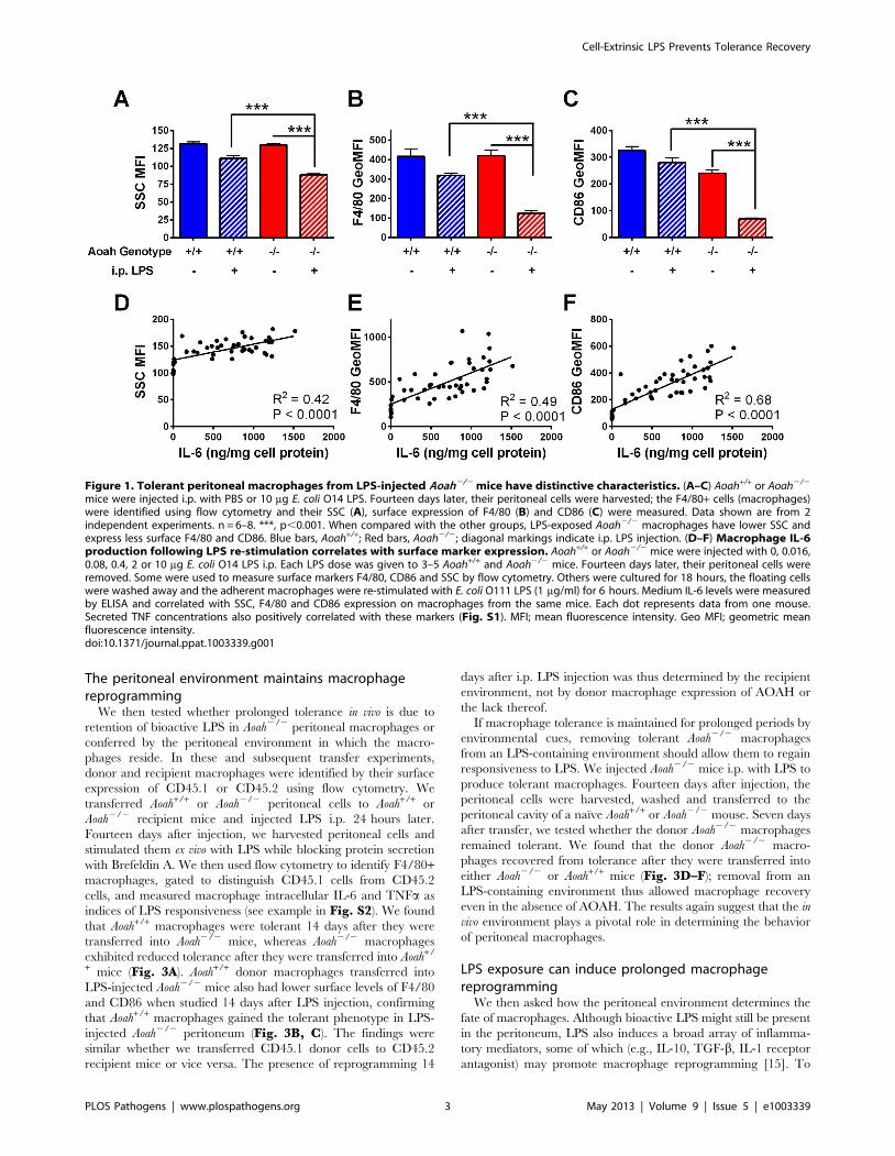
Figure 1. Tolerant peritoneal macrophages from LPS-injected Aoah2/2 mice have distinctive characteristics. (A–C) Aoah+/+ or Aoah2/2
mice were injected i.p. with PBS or 10 mg E. coli O14 LPS. Fourteen days later, their peritoneal cells were harvested; the F4/80+ cells (macrophages)were identified using flow cytometry and their SSC (A), surface expression of F4/80 (B) and CD86 (C) were measured. Data shown are from 2independent experiments. n = 6–8. ***, p,0.001. When compared with the other groups, LPS-exposed Aoah2/2 macrophages have lower SSC andexpress less surface F4/80 and CD86. Blue bars, Aoah+/+; Red bars, Aoah2/2; diagonal markings indicate i.p. LPS injection. (D–F) Macrophage IL-6production following LPS re-stimulation correlates with surface marker expression. Aoah+/+ or Aoah2/2 mice were injected with 0, 0.016,0.08, 0.4, 2 or 10 mg E. coli O14 LPS i.p. Each LPS dose was given to 3–5 Aoah+/+ and Aoah2/2 mice. Fourteen days later, their peritoneal cells wereremoved. Some were used to measure surface markers F4/80, CD86 and SSC by flow cytometry. Others were cultured for 18 hours, the floating cellswere washed away and the adherent macrophages were re-stimulated with E. coli O111 LPS (1 mg/ml) for 6 hours. Medium IL-6 levels were measuredby ELISA and correlated with SSC, F4/80 and CD86 expression on macrophages from the same mice. Each dot represents data from one mouse.Secreted TNF concentrations also positively correlated with these markers (Fig. S1). MFI; mean fluorescence intensity. Geo MFI; geometric meanfluorescence intensity.doi:10.1371/journal.ppat.1003339.g001
Cell-Extrinsic LPS Prevents Tolerance Recovery
PLOS Pathogens | www.plospathogens.org 3 May 2013 | Volume 9 | Issue 5 | e1003339

find out whether wildtype macrophages can become tolerant in
an Aoah2/2 environment that lacks LPS-induced mediators, we
transferred Aoah+/+ CD45.1 peritoneal cells into mice that lack
both AOAH and TLR4 (Aoah2/2Tlr42/2 CD45.2) and can
neither deacylate, nor respond to, LPS. Fourteen days after i.p.
LPS injection, the donor Aoah+/+ CD45.1 macrophages were
tolerant (Fig. 4A); since the recipient mice do not produce LPS-
induced mediators, this observation suggested strongly that such
mediators are not required to maintain prolonged tolerance in
vivo. In another approach, CD45.2 Aoah2/2Tlr4+/+ or Aoah2/
2Tlr42/2 mice were given LPS i.p. Fourteen days later, CD45.1
naıve Aoah+/+ peritoneal cells were introduced into the
peritoneal cavity. One day after transfer, the donor macro-
phages had become tolerant in both Aoah2/2Tlr4+/+ and Aoah2/
2Tlr42/2 recipients (Fig. 4B). These findings suggested that
bioactive LPS is present for a prolonged period in the LPS-
exposed Aoah2/2 peritoneum and that this LPS can induce and
maintain macrophage tolerance in the absence of LPS-induced
host mediators.
Bioactive LPS can be recovered from LPS-injectedAoah2/2 mice
To obtain direct evidence for the presence of LPS in the
peritoneum many days after i.p. injection, we injected 10 mg
[3H/14C]LPS into the peritoneal cavities of Aoah2/2 and Aoah+/+
mice and measured 14C and 3H in the peritoneal flush medium,
peritoneal cells, mesenteric membranes, and fat 10 days later. The
distribution of 14C dpm, a marker for the LPS carbohydrate
backbone, was similar in the presence and absence of AOAH:
about 0.3% of the injected LPS was found in cell-free peritoneal
flush fluid; from 6 to 9% of the injected 14C LPS was recovered
from intraperitoneal fat and 2 to 3% from the mesentery (Fig. 5A).
We reported previously that 1–4% of the injected LPS could be
recovered from peritoneal cells [25]. Based on the ratio of 3H to14C in the samples [35], 99% (Aoah+/+) and 19% (Aoah2/2) of the
recovered LPS had been deacylated (Fig. 5B), in keeping with the
results found for peritoneal cells [25].
Is the intraperitoneal acylated LPS bioactive? In other
experiments, we gave 10 mg LPS i.p. to Aoah+/+ and Aoah2/2
Figure 2. Cell-associated LPS. Aoah+/+ or Aoah2/2 mice were given 10 mg LPS-FITC i.p. Ten days later, their peritoneal cells were harvested, fixed,and either permeabilized (B) or not (A). Anti-FITC Ab conjugated with Phycoerythrin (PE) was added to measure macrophage (F4/80+) surface (A) ortotal (B) LPS-FITC. Dotted lines, F4/80+ cells from mice that received LPS i.p. (control for autofluorescence); solid lines, F4/80+ cells from mice thatwere given i.p. LPS-FITC. Red lines, Aoah2/2 ; blue lines, Aoah+/+. (C) The ratio of the GeoMFI of cell surface or total LPS-FITC to the GeoMFI of controlmacrophages. The dotted line indicates a ratio of 1.0 (no LPS-FITC association with cells). For both Aoah2/2 and Aoah+/+ cells, most of the FITC wasintracellular. (D–I) Mice were given 10 mg LPS-FITC i.p. Ten days after injection, peritoneal cells were harvested and cultured in plates with coverslipsfor 18 hours. Floating cells were washed away and adherent macrophages were fixed, permeabilized, and stained with anti-FITC Alexa fluor 488(green), anti-LAMP1 (CD107) Ab (red) and DAPI (blue). (D and E) Aoah2/2 macrophages. F, overlay of D and E. (G and H) Aoah+/+ macrophages. I,overlay of G and H. Much of the FITC-LPS was localized to LAMP1-positive vesicles in both Aoah2/2 and Aoah+/+ macrophages. Scale bar = 5 mm.doi:10.1371/journal.ppat.1003339.g002
Cell-Extrinsic LPS Prevents Tolerance Recovery
PLOS Pathogens | www.plospathogens.org 4 May 2013 | Volume 9 | Issue 5 | e1003339

Figure 3. The peritoneal environment confers macrophage phenotype. (A–C) CD45.1 (or CD45.2) Aoah+/+ or Aoah2/2 macrophages weretransferred to Aoah+/+ or Aoah2/2 recipient mice of the opposite CD45 genotype. 24 hours later, half of the mice in each group received 1 mg E. coliO14 LPS i.p. After 14 days, the peritoneal cells from the host mice were harvested. Half of the cells was treated with 1 mg/ml E. coli O111 LPS ex vivofor 4 hours in the presence of Brefeldin A. Intracellular IL-6 and TNF levels in F4/80+ donor macrophages (A) were measured using flow cytometry.Blue bars, Aoah+/+ donor macrophages; red bars, Aoah2/2 donor macrophages; diagonal markings indicate LPS exposure in vivo. The other half of thecells was used to measure F4/80+ macrophage surface expression of F4/80 (B) and CD86 without ex vivo stimulation (C). In B and C, only Aoah+/+
donor macrophages were studied; they acquired the ‘‘tolerant’’ surface phenotype (low F4/80 and CD86 surface expression) when transferred toAoah2/2 recipients. Tolerance tracked with the genotype of the host animal, not that of the donor macrophages. Data in A were combined from 4experiments (n = 6–13/group); data in B and C were combined from 2 experiments, n = 6–8/group. (D–F) Tolerant Aoah2/2 macrophages regainresponsiveness in naıve mice. CD45.1 (or CD45.2) Aoah2/2 mice were injected i.p. with 0.5 mg LPS. Fourteen days later, their peritoneal cells (includingtolerant macrophages) were harvested and transferred i.p. to naıve Aoah2/2 or Aoah+/+ mice of the opposite CD45 type. After 7 days, peritoneal cellswere harvested from the recipient mice and IL-6 and TNF responses were measured in the F4/80+ macrophages after re-challenging them with LPS exvivo (D). F4/80 and CD86 surface expression was also measured (E and F). Only results from donor macrophages are shown. ‘‘Tolerant donor’’macrophages were macrophages from Aoah2/2 mice that received 0.5 mg LPS i.p. 21 days earlier and were freshly isolated (in panel D) ormacrophages harvested from Aoah2/2 mice that received 0.5 mg LPS i.p. 14 days earlier and were preserved in 10% DMSO, 90% FBS at 280uC untilanalysis (in panels E and F). Data were combined from 3 experiments. n = 6–13. **, P,0.01; ***, P,0.001. Tolerance was lost by 7 days after LPS-exposed macrophages were transferred to either naıve Aoah2/2 or Aoah+/+ mice.doi:10.1371/journal.ppat.1003339.g003
Cell-Extrinsic LPS Prevents Tolerance Recovery
PLOS Pathogens | www.plospathogens.org 5 May 2013 | Volume 9 | Issue 5 | e1003339

mice and flushed their peritoneal cavities with culture medium 10
days later. The cell-free peritoneal flush medium from Aoah2/2
mice activated naıve Tlr4+/+ macrophages but not Tlr42/2
macrophages, suggesting that bioactive LPS was present in the
medium (Fig. 5C). When we re-challenged the macrophages with
LPS, we found that the flush medium from Aoah2/2 mice could
also render naıve Tlr4+/+ macrophages tolerant (Fig. 5D, E). We
conclude that (1) free LPS is present in the peritoneal fluid of
Aoah2/2 mice 10 days after i.p. injection; (2) this LPS is fully
acylated; and (3) the acylated LPS is bioactive, able to activate
naıve Tlr4+/+ macrophages and reprogram them.
Macrophages release bioactive LPS that can reprogramnaıve macrophages
If LPS that is taken up by macrophages can be released to act
on other cells, fully acylated LPS released from Aoah2/2 cells
should be able to activate naıve cells and reprogram them. We first
tested this idea in vitro. We injected Aoah2/2 mice with LPS i.p.
and harvested and washed their peritoneal cells 10 days later,
when almost all of the LPS was associated with macrophages [25].
We co-cultured the peritoneal cells (including tolerant macro-
phages) with naıve Tlr4+/+ or Tlr42/2 peritoneal cells (including
naıve macrophages) for 18 hours. Significantly higher IL-6 and IL-
10 (Fig. 6A, B) levels were found in the culture medium of Tlr4+/+
peritoneal cells co-cultured with tolerant Aoah2/2 cells than in the
medium of Tlr42/2 cells co-cultured with tolerant cells, suggesting
that LPS from tolerant cells stimulated naıve Tlr4+/+ cells to
produce these cytokines. After incubation for 18 hours, the co-
cultured cells were washed twice with cRPMI and the adherent
macrophages were treated with LPS for 6 hours (Fig. 6C–E).
Naıve macrophages co-cultured with tolerant cells produced less
TNF and IL-6, but similar amounts of IL-10, suggesting that they
had become reprogrammed during the 18 hour incubation period.
When we co-cultured naıve Tlr4+/+ peritoneal cells with Tlr42/2
Aoah2/2 peritoneal cells that had been exposed to LPS in vivo, the
naıve cells could also be activated, confirming that LPS from
previously exposed macrophages is sufficient to activate naıve
macrophages (data not shown). Co-culture of naıve Tlr4+/+
peritoneal cells with LPS-exposed Aoah+/+ cells (again, harvested
10 days after i.p. LPS injection, sufficient time for LPS inactivation
to occur in AOAH-sufficient animals) did not induce IL-6 or IL-10
production (data not shown).
To find out whether direct cell-cell contact is required for naıve
macrophage activation during co-culture, we separated naıve
peritoneal cells from LPS-exposed peritoneal cells in transwell
cultures. This significantly decreased IL-6 and IL-10 production
by the naıve macrophages (Fig. 6A, B), suggesting that direct cell-
cell contact enables optimal delivery of LPS from one macrophage
to another.
To test whether LPS-laden macrophages can release LPS to act
on other macrophages in vivo, we injected 20 mg LPS-FITC i.p. to
CD45.2 Aoah2/2Tlr42/2 mice. Seven days later, peritoneal cells
were harvested, washed, and transferred i.p. to CD45.1 Aoah2/2
naıve mice. After 7 days, we found that the transferred F4/80+macrophages had lost 2/3 of their LPS-FITC and that recipient
macrophages had gained small amounts of LPS-FITC, demon-
strating that LPS can be released from donor macrophages and
taken up by recipient macrophages in vivo (Fig. 7A, B). In
addition, we found that the recipient macrophages became
tolerant (Fig. 7C–E), indicating that they had been exposed to
bioactive LPS (the Tlr42/2 donor cells should not produce
tolerizing mediators).
Figure 4. Bioactive LPS in the peritoneum is sufficient to maintain macrophage tolerance in vivo. (A) CD45.1 Aoah+/+ peritoneal cellswere transferred to CD45.2 Aoah2/2Tlr4+/+ or Aoah2/2Tlr42/2 mice. Half of the mice in each group received 1 mg LPS i.p. Fourteen days later, theCD45.1 donor macrophages’ IL-6 and TNF responses to LPS were determined following ex vivo re-challenge. Naıve Aoah+/+ macrophages becametolerant when they were carried in LPS-injected Aoah2/2 mice, whether or not the host mice expressed TLR4. n = 4–7. (B) CD45.2 Aoah2/2Tlr4+/+ orAoah2/2Tlr42/2 mice received 1 mg LPS i.p. as indicated. Fourteen days later, CD45.1 naıve Aoah+/+ peritoneal cells were transferred i.p. to PBS- orLPS-injected mice. After 24 hours, the responses of the donor macrophages to LPS were measured ex vivo. Exposure to the LPS-containing Aoah2/2
peritoneal environment rendered naıve Aoah+/+ macrophages tolerant, whether or not the host mouse was able to respond to the i.p. dose of LPS.Data were combined from 2 experiments. n = 4–6. **, P,0.01; ***, P,0.001. Blue bars, Aoah2/2 Tlr4+/+ recipients; light blue bars, Aoah2/2Tlr42/2
recipients; diagonal markings represent i.p. LPS injection.doi:10.1371/journal.ppat.1003339.g004
Cell-Extrinsic LPS Prevents Tolerance Recovery
PLOS Pathogens | www.plospathogens.org 6 May 2013 | Volume 9 | Issue 5 | e1003339

Figure 5. Bioactive LPS is present in the peritoneum 10 days after i.p. injection. (A) Aoah+/+ or Aoah2/2 mice were given 10 mg 3H/14C LPSi.p. Ten days later, their peritoneal cavities were flushed with 5 ml PBS that contained 5 mM EDTA and the peritoneal fat and mesentery wereharvested. The amount of LPS in each specimen was determined by counting the 14C dpm in the LPS backbone. (B) The extent of deacylation wasdetermined by measuring the 3H/14C ratio. Deacylation was not calculated in peritoneal fluid because the flush fluids contained low amounts of 14Cand some free 3H- fatty acids. Blue striped bars, Aoah+/+; Red striped bars, Aoah2/2. (C) In other experiments, mice were injected i.p. with 10 mg LPS.Ten days later, the peritoneum was flushed with 2 ml cRPMI. The flush medium was centrifuged and the cell free supernatant was added to cultures ofnaıve Tlr4+/+ or Tlr42/2 macrophages. Eighteen hours later, the culture medium was harvested to measure IL-6. Only peritoneal flush fluid from Aoah2/2
mice elicited IL-6 production by naıve TLR4-expressing macrophages. (D, E) After incubation for 18 hours, the medium containing the flush fluid wasremoved, the macrophages were washed twice with cRPMI, and then they were treated with 1 mg/ml LPS for 6 hours. TNF and IL-6 in the culturemedium were measured. Flush fluid from LPS-exposed Aoah2/2 mice induced tolerance in naıve macrophages in vitro, whereas that from LPS-exposedAoah+/+ mice did not. Data are combined from 2 experiments. N = 6–8/group. *, P,0.05; **, P,0.01. Blue bars, Aoah+/+ peritoneal flush mediumoverlying TLR4+/+ cells; red bars, Aoah2/2 flush medium overlying TLR4+/+ cells; light blue bars, Aoah+/+ flush medium overlying TLR42/2 cells; pink bars,Aoah2/2 flush medium overlying TLR42/2 cells; diagonal markings indicate in vivo LPS exposure; orange bar, cRPMI medium overlying TLR4+/+ cells.doi:10.1371/journal.ppat.1003339.g005
Cell-Extrinsic LPS Prevents Tolerance Recovery
PLOS Pathogens | www.plospathogens.org 7 May 2013 | Volume 9 | Issue 5 | e1003339

These results show that macrophages can release the LPS that
they contain and that the released LPS can act on other cells in
vivo. If the LPS remains bioactive, it can induce tolerance in other
macrophages.
LPS-induced mediator(s) can induce tolerance in Tlr42/2
macrophagesBioactive LPS is thus present in the peritoneum of LPS-injected
Aoah2/2 mice, where it is sufficient to prevent macrophages from
regaining responsiveness. Do LPS-induced mediators also play a
role in maintaining tolerance? We transferred CD45.2 Aoah2/
2Tlr42/2 peritoneal cells to CD45.1 Aoah+/+ or Aoah2/2 (both
Tlr4+/+) recipient mice, injected LPS i.p., and asked whether the
Aoah2/2Tlr42/2 donor macrophages became tolerant in the
peritoneum of tolerant Aoah2/2 recipient mice. Fourteen days
after LPS injection, we harvested recipient peritoneal cells and
treated them ex vivo with Micrococcus luteus plus poly I:C (LPS-
exposed Aoah2/2 macrophages express hetero-tolerance [36] to
M. luteus [TLR2 agonist] and poly I:C [TLR3 agonist] [25]).
Aoah2/2Tlr42/2 donor macrophages (F4/80+) harvested from
LPS-injected Aoah2/2 recipients had significantly lower IL-6
and TNF responses to ex vivo re-challenge (Fig. 8A), and they
also had less surface F4/80 and CD86 expression (Fig. 8B, C).
Since Aoah2/2Tlr42/2 donor macrophages cannot sense LPS,
these results suggested that LPS-induced mediators in the
peritoneum are also important for prolonging tolerance in
Aoah2/2 mice. Aoah2/2Tlr42/2 donor cells were less tolerant
than were the recipient’s Aoah2/2Tlr4+/+ macrophages, again
Figure 6. Co-culture with tolerant cells induces tolerance in naıve macrophages in vitro. (A, B) Peritoneal cells from naıve Tlr4+/+ or Tlr42/2
mice were co-cultured for 18 hours at 37uC with washed peritoneal cells from Aoah2/2 mice that had been injected with 10 mg LPS i.p. 10 daysearlier. IL-6 and IL-10 were measured in culture medium. Only Tlr4+/+ cells co-cultured with tolerant peritoneal cells released IL-6 and IL-10. Separationof naıve Tlr4+/+ cells from LPS-exposed Aoah2/2 cells in Transwell cultures significantly decreased cytokine production, suggesting that cell-cellcontact is important for delivery of bioactive LPS. (C–E) In the same experiments, after incubation for 18 hours the Tlr4+/+ cells that had been co-cultured with tolerant cells were washed with cRPMI twice and then treated with 1 mg/ml LPS for 6 hours before TNF, IL-6 and IL-10 were measured inthe culture medium. Naıve macrophages co-cultured with tolerant cells became reprogrammed. Blue bars, Tlr4+/+ naıve macrophages; light blue bars,Tlr42/2 naıve macrophages; diagonal markings indicate co-culture with tolerant cells; checked markings represent Transwell cultures. Data werecombined from 3 experiments. n = 6–10. ***, P,0.001; NS, No significant difference.doi:10.1371/journal.ppat.1003339.g006
Cell-Extrinsic LPS Prevents Tolerance Recovery
PLOS Pathogens | www.plospathogens.org 8 May 2013 | Volume 9 | Issue 5 | e1003339

reinforcing the prominent direct role played by fully acylated
LPS.
rhAOAH prevents prolonged tolerance in vivoIFN-c can improve monocyte function in septic patients and
both IFN-c and GM-CSF can restore responses of LPS-
desensitized (tolerant) monocytes in vitro [37,38]. We next tested
whether Aoah2/2 tolerant macrophages can recover their
responses to LPS by treating them with rhAOAH (recombinant
human AOAH), IFN-c , GM-CSF or a combination of these
agents for 18 hours ex vivo (Fig. S3A–C). rhAOAH only slightly
increased the macrophages’ responses, possibly because deacyla-
tion occurs slowly and would not be expected to reach completion
within 18 hours [39]. GM-CSF preferentially restored the IL-6
response while IFN-c mainly boosted the TNF and RANTES
responses. Combining the three agents largely restored the
macrophages’ responses. Aoah2/2 tolerant macrophages, like
desensitized human monocytes [38], could thus be rescued by
treating them with IFN-c and GM-CSF in vitro.
To test whether providing AOAH to mice can prevent or
reverse prolonged tolerance in vivo, we gave Aoah2/2 mice LPS i.p.
on day 0, followed by rhAOAH or carrier protein BSA i.p. daily
from day 1 to day 13. Peritoneal cells were harvested on day 14,
plated, and adherent macrophages were rechallenged ex vivo with
LPS. LPS-stimulated RANTES and IL-6 were at naıve Aoah2/2
macrophage levels in macrophages from animals that had received
rhAOAH treatment (Fig. 9A–C). TNF production is the most
sensitively reprogrammed component of tolerance in these cells;
here the TNF level in culture medium overlying macrophages
from LPS-exposed rAOAH-treated mice was significantly lower
than the naıve macrophage level but 10-fold higher than the levels
produced by macrophages from mice that received carrier protein
BSA instead of rhAOAH. Recombinant AOAH was thus able to
ameliorate prolonged tolerance in vivo in Aoah2/2 animals.
Treatment with IFN-c or antibody to IL-10 receptor did not
rescue Aoah2/2 animals from prolonged endotoxin tolerance (data
not shown), in line with the conclusion that bioactive LPS plays a
dominant role in maintaining prolonged tolerance in vivo.
Figure 7. LPS can be released from macrophages and induce tolerance in other cells in vivo. (A) CD45.2 Aoah2/2 Tlr42/2 donor mice wereinjected i.p. with 20 mg LPS-FITC. Seven days later, their peritoneal cells were harvested, washed and transferred to CD45.1 Aoah2/2 recipient mice.After 7 days, the donor and recipient F4/80+ macrophages were analyzed by flow cytometry to measure the amount of cell-associated LPS-FITC. Darkgreen, donor macrophages before transfer; light green, donor macrophages recovered 7 days after transfer; red, recipient peritoneal macrophages 7days after transfer; Dotted black, LPS (unlabeled)-exposed Aoah2/2 macrophages (autofluorescence control). (B) Geometric mean fluorescenceintensity (GeoMFI) of cells in (A). (C–E) Naıve recipient macrophages become tolerant after exposure in vivo to donor macrophages thatcontain LPS-FITC. CD45.2 Aoah2/2 Tlr42/2 macrophages, either naıve or containing LPS-FITC, were transferred to naıve CD45.1 Aoah2/2 mice asdescribed in (A). Seven days after transfer, peritoneal cells were harvested from the recipient mice and half of the cells were re-challenged ex vivowith 1 mg/ml LPS in the presence of Brefeldin A. Intracellular IL-6 and TNF was measured in CD45.1 recipient macrophages (F4/80+) (C). Recipientmacrophage surface F4/80 (D) and CD86 (E) were measured in cells that were not re-stimulated ex vivo. TLR4-deficient donor cells bearing LPS-FITCinduced tolerance in recipient macrophages in vivo. Similar results were obtained in 2 additional experiments, each with n = 3. **, P,0.01; ***,P,0.001.doi:10.1371/journal.ppat.1003339.g007
Cell-Extrinsic LPS Prevents Tolerance Recovery
PLOS Pathogens | www.plospathogens.org 9 May 2013 | Volume 9 | Issue 5 | e1003339

Figure 8. Aoah2/2Tlr42/2 macrophages can become tolerant in LPS-injected Aoah2/2 mice. (A) CD45.2 Aoah2/2Tlr42/2 naıve peritonealcells were transferred to the peritoneal cavities of CD45.1 Aoah+/+Tlr4+/+ and Aoah2/2Tlr4+/+ mice. After 18 hours, half of the recipient mice in eachgroup were injected i.p. with 1 mg LPS. Fourteen days after injection, the peritoneal cells were harvested. Some of the cells were treated ex vivo with40 mg/ml Micrococcus luteus plus 2.5 mg/ml poly I:C for 8 hours in the presence of Brefeldin A. IL-6 and TNF production by donor (D) and recipient (R)F4/80+ macrophages was measured using flow cytometry. (B and C) The remainder of the peritoneal cells was not re-stimulated ex vivo and was usedto determine surface expression of F4/80 and CD86 on macrophages (F4/80+) by flow cytometry. **, P,0.01; ***, P,0.001. Data were combined from
Cell-Extrinsic LPS Prevents Tolerance Recovery
PLOS Pathogens | www.plospathogens.org 10 May 2013 | Volume 9 | Issue 5 | e1003339

Discussion
Patients who experience severe sepsis develop a state of immune
tolerance that may last for many weeks and is thought to be
immunosuppressive [40]. The phenomenon has been character-
ized principally by studying peripheral blood leukocytes, whose
responses to LPS and other microbial agonists are typically altered
to diminish pro-inflammatory cytokine production while main-
taining or increasing production of anti-inflammatory molecules
such as IL-10 and IL-1 receptor antagonist. LPS-induced
tolerance in mice mimics the human phenomenon in many ways
(reduced monocyte-macrophage CD86, reprogrammed cytokine
production) but not in others (e.g., murine macrophages do not
decrease class II molecule expression) [23].
Although some Gram-negative bacteria can modify the
acylation structure of their LPS in ways that may alter its ability
to trigger signaling via MD-2—TLR4 [41], LPS is also disabled by
host enzymes, either on mucosal surfaces (alkaline phosphatase)
[42] or in tissues (AOAH) [24]. In addition to experiencing
prolonged tolerance, mice that lack AOAH respond to small
subcutaneous or intravenous doses of LPS by producing high titers
of polyclonal antibodies [43] and developing massive, prolonged
hepatomegaly [44,45]. Animals have many other mechanisms for
neutralizing hexaacyl LPS in plasma and tissues [46], yet none of
these is able to prevent these striking reactions in animals that
cannot deacylate LPS. Transgenic mice that produce greater than
normal amounts of AOAH are protected from live E. coli challenge
[47], again emphasizing the importance of AOAH mediated LPS
inactivation in optimizing anti-bacterial immune responses.
Continued exposure to microbial agonists can prolong the
activation of cultured cells. Hume et al. reported that continuous
exposure to LPS induced a sustained activation state in a
macrophage cell line [48], and Hedl et al. found that prolonged
exposure to muramyl dipeptide, a ligand for Nod2, promoted
tolerance in human monocyte-derived macrophages [20]. Toler-
ance has also been described in human cells that may be exposed
repeatedly to LPS in vivo, such as the alveolar macrophages of
tobacco smokers [49] and blood monocytes from patients with
uncontrolled gram negative bacterial infection [50] or cystic
fibrosis [51]. Tolerance lasts for weeks in patients who have
chronic pyelonephritis with active bacterial urinary tract infection
[52], as well as in volunteers with typhoid fever [53]. Individuals
who inhale endotoxin-rich agricultural dusts may also develop
chronic macrophage activation or tolerance [54,55]. Here we
show that tolerance can be maintained in vivo for long periods by
the presence of small amounts of bioactive LPS, raising the
possibility that the rate and/or extent of LPS inactivation might
influence the rate of recovery from many Gram-negative bacterial
diseases.
The mammalian MD-2—TLR4 LPS receptor is activated most
sensitively by LPSs that have a lipid A moiety that contains 6 acyl
chains. Such ‘‘hexaacyl’’ LPSs are produced by many of the
Gram-negative bacterial commensals and pathogens that can
inhabit the mucosal surfaces of the upper respiratory and
gastrointestinal tracts [56,57]. Since AOAH inactivates these
LPSs, AOAH deficiency might be associated with greater
susceptibility to, or duration of, Gram-negative bacterial diseases
that involve mucosal surfaces. To date, genetic linkage studies
have found associations between polymorphisms in the AOAH
gene and rhinosinusitis (confirmed in 2 populations of different
ethnic composition [58,59]) as well as asthma [60] in humans. In
addition, two studies of large populations [61,62] have indepen-
dently found that AOAH mRNA expression is associated in trans
with polymorphisms in HLA-DRB1 that, in turn, have been
strongly linked to colitis and primary biliary cirrhosis. How
AOAH influences disease expression, if it does, remains uncertain.
The present studies identified the presence of cell-associated and
extracellular (cell-extrinsic) LPS as the primary determinant of
prolonged LPS tolerance in macrophages in vivo. This conclusion is
supported by the observations that 1) prolonged tolerance could be
induced in either Aoah2/2 or Aoah+/+ macrophages that had been
transferred into LPS-injected Aoah2/2 mice, and 2) Aoah2/2
macrophages did not exhibit prolonged tolerance when they were
transplanted into Aoah+/+ hosts, indicating that LPS inactivation by
AOAH in the host environment, not in the tolerant cell itself,
determines the tolerant phenotype. Tolerance could be induced in
naıve macrophages either by direct contact with cells that had
taken up LPS or by extracellular LPS acquired within the
peritoneal environment. In addition, administration of rAOAH to
Aoah2/2 mice partially prevented tolerance, and tolerance could
be induced in naıve macrophages when they were transferred into
LPS-exposed Aoah2/2Tlr42/2 mice or co-cultured with LPS-
exposed Aoah2/2Tlr42/2 peritoneal cells, which do not produce
LPS-induced mediators but can release fully acylated LPS into
their environment [26,63,64]. Although these studies identify
bioactive LPS as essential for maintaining tolerance, we also found
that LPS-induced paracrine mediators further promoted toler-
ance, perhaps in part by extending the phenotype to cells that do
not express TLR4. These results also do not exclude the possibility
that the epigenetic changes shown to be induced by short-term
exposure to LPS [28–32] contribute to maintaining prolonged
tolerance in macrophages, but these changes evidently do not
persist (or maintain dominance) in an environment that lacks
extracellular LPS; they can also be overcome by soluble mediators
such as interferon-c or GM-CSF.
Where does LPS deacylation occur in vivo? LPS may be
deacylated extracellularly, as was shown using purulent ascites
fluid [65]; both LPS-binding protein and CD14 can present LPS
to extracellular AOAH in a manner that promotes its deacylation
[66]. Alternatively, LPS may be taken up by macrophages,
neutrophils, or dendritic cells and inactivated by AOAH, or
conceivably LPS could be deacylated by AOAH in cells that have
acquired AOAH via mannose-6-phosphate receptors [67]. Recent
studies found that the LPS in circulating LPS-HDL (high density
lipoprotein) complexes undergoes deacylation in the liver [68],
where AOAH is produced by Kupffer and dendritic cells [44].
AOAH-deficient mice cannot deacylate LPS in ascites or in cells
within, or on the walls of, the peritoneal cavity, and these cells or
membranes may then become reservoirs that release small
amounts of bioactive LPS over time. This LPS then could
maintain macrophage tolerance or induce tolerance in monocytes
that newly arrive in the peritoneal fluid. The LPS ‘‘depot’’ includes
the peritoneal membrane and/or mesenteric fat, since we found
significant quantities of radiolabeled, fully acylated LPS in these
sites. Naıve macrophages may also acquire LPS from other
macrophages by direct cell-cell contact.
2 independent experiments. n = 5–7. Aoah2/2 Tlr42/2 macrophages, which are unable to respond to LPS, became tolerant to TLR2 and TLR3 ligands(A) and developed the tolerant surface phenotype (B and C) when they were transferred into LPS-primed Aoah2/2 mice, supporting a role for non-LPS stimuli in promoting tolerance in vivo. Pink bars, Aoah2/2Tlr42/2 donor macrophages; Blue bars, Aoah+/+ recipient macrophages; red bars,Aoah2/2 recipient macrophages; diagonal markings indicate in vivo LPS exposure.doi:10.1371/journal.ppat.1003339.g008
Cell-Extrinsic LPS Prevents Tolerance Recovery
PLOS Pathogens | www.plospathogens.org 11 May 2013 | Volume 9 | Issue 5 | e1003339

Figure 9. rhAOAH prevents prolonged endotoxin tolerance in vivo. Aoah2/2 mice were injected with 1 mg LPS i.p. on day 0. From days 1 to13, mice were given daily i.p. doses of 0.3 mg rhAOAH or placebo (carrier protein BSA in PBS) (diagonal marking red bars). Peritoneal cells wereexplanted on day 14 and the adherent macrophages were challenged ex vivo with LPS for 6 hrs before medium cytokine levels were measured. (A),IL-6; (B), TNF; (C), RANTES. Naıve Aoah2/2 peritoneal macrophages were used as controls (solid red bars). **, P,0.01; ***, P,0.001. rhAOAH largelyprevented prolonged tolerance in vivo.doi:10.1371/journal.ppat.1003339.g009
Cell-Extrinsic LPS Prevents Tolerance Recovery
PLOS Pathogens | www.plospathogens.org 12 May 2013 | Volume 9 | Issue 5 | e1003339

Innate immune reactions to microbes are typically short-lived.
Mobilizing an animal’s antimicrobial armamentarium usually
promotes microbial eradication and clearance within hours or a
few days. Potentially harmful inflammation then resolves as the
battlefield is cleared and defenses are restored [69]. Recovery is
thought to involve both anti-inflammation (preventing inflamma-
tion-induced damage) and resolution (clearing the battlefield and
promoting return of homeostasis). Known tissue resolution
mechanisms include neutrophil apoptosis, macrophage emigration
and efferocytosis of dead cells, and the production of lipoxins,
resolvins [and other lipids] [70], proteases, and gaseous signals that
promote restoration of homeostasis in tissues [69,71,72]. Here we
present evidence for another essential component of resolution:
inactivating the microbial molecules that tell the host that
microbes are present. A host’s ability to remove or disable
bioactive microbial molecules from an infected tissue may
influence the ultimate outcome of many host-microbe encounters,
since inactivating these molecules removes an important obstacle
to resolution of inflammation and restoration of innate host
defenses.
Materials and Methods
MiceAoah2/2 C57BL/6J mice were generated as described [35].
Tlr42/2 (B6.B10ScN-Tlr4lps-del/JthJ) mice were purchased from
Jackson Laboratory. These mice have a 7 kb deletion in the TLR4
gene; the mutation was backcrossed to C57Bl/6J for at least 6
generations. C57BL/6J CD45.1 (B6.SJL-Ptprca Pepcb/BoyJ)
mice were also from Jackson. Aoah2/2 Tlr42/2 and Aoah2/
2CD45.1 mice were obtained by crossing Aoah2/2 mice with
Tlr42/2 or B6 CD45.1 mice, respectively. All mice were housed in
a specific pathogen- and murine norovirus-free facility. All mice
were studied using protocols approved by the Institutional Animal
Care and Use Committee (IACUC). IACUC of the University of
Texas Southwestern Medical Center (permit number: A3472-01)
approved the Animal Protocol Number 0028-07-08-2. The
IACUC of the National Institutes of Allergy and Infectious
Diseases (permit number A4149-01) approved the Animal Study
Protocol LCID 11E. Both protocols adhered to the Guide for the
Care and Use of Laboratory Animals of the National Institutes of
Health.
ReagentsE. coli O14 LPS was prepared by the phenol-chloroform-
petroleum ether method [73]. E. coli O111 LPS was purchased
from Sigma. Salmonella typhimurium Rc LPS ([3H/14C]LPS; 3H-
labeled fatty acyl chains and 14C-labeled glucosamine backbone)
was prepared from S. typhimurium PR122 as described [74]; 1 mg
had ,150,000 dpm 3H and ,10,000 dpm 14C. Experiments
using radiolabeled LPS followed the requirements of the Radiation
Safety department, UT-Southwestern Medical Center. FITC-LPS
was prepared as described by Tobias et al. [25]. Micrococcus luteus
cells and poly I:C were obtained from Sigma. Recombinant
human AOAH was produced by ZymoGenetics, Inc. Recombi-
nant mouse IFN-c and recombinant mouse GM-CSF were from
R&D Systems.
AntibodiesAntibodies used for FACS were anti-F4/80 (clone BM8,
eBioscience), anti-CD86 (clone GL1, BD), anti-FITC (polyclonal
rabbit IgG, Invitrogen), anti-IL-6 (clone MP5-20F3, eBioscience),
anti-TNF (clone MP6-XT22, eBioscience), anti-CD45.1 (clone
A20, BD), and anti-CD45.2 (clone 104; BD). Antibodies used for
microscopy were anti-CD107a (LAMP-1) (clone 1D4B, BD), anti-
Rab5a (clone 15/Rab5, BD), anti-Giantin (rabbit polyclonal,
Abcam), anti-TGN46 (rabbit polyclonal, Abcam), anti-Calnexin
(rabbit polyclonal, Abcam).
Ex vivo tolerance experimentsMice were injected i.p. with various doses of LPS in 300 ml of
PBS. Fourteen days later, the mice were euthanized using CO2
and their peritoneal cells were harvested by flushing the
peritoneum with 5 ml of PBS containing 5 mM EDTA. Cells
were stained with antibodies and analyzed using flow cytometry.
To measure cell responses to re-challenge, peritoneal cells were
washed and resuspended in cRPMI medium (RPMI 1640
containing 10% heat-inactivated FBS (endotoxin ,0.06 EU/ml;
Hyclone), 100 mM nonessential amino acids, 100 U/ml penicillin,
0.1 mg/ml streptomycin, 2 mM L-glutamine, 10 mM sodium
pyruvate, 25 mM Hepes, pH 7.4, and 50 mM 2-mercaptoethanol)
and plated with 16106 cells/well in 12-well-plates. After
incubation for 18 hours (37uC, 5% CO2, 80% humidity), the
floating cells were washed away and the adherent macrophages
were treated with 1 mg/ml E. coli O111 LPS for 6 hours. The
culture medium was used for ELISA and the cells were washed
with PBS and lysed with PBS containing 0.1% Triton X-100 to
measure protein (Biorad).
Macrophage-associated LPS and imaging studiesMice were injected i.p. with 10 mg LPS-FITC. Ten days after
injection, peritoneal cells were harvested and stained with anti-F4/
80 Ab to identify macrophages. The cells were then washed and
fixed with 4% paraformaldehyde (PFA) or fixed and permeabilized
with cytofix/cytoperm (BD). Anti-FITC PE antibody was used to
detect cell-surface or total LPS-FITC by FACS. For microscopy,
peritoneal cells were plated in culture dishes for 18 hours, then the
floating cells were washed away and the adherent macrophages
were fixed with 4% PFA before being blocked and permeabilized
with 1% BSA, 25% goat serum, 0.05% saponin in PBS. Cells were
stained with primary antibodies at 4uC overnight and with the
secondary reagents at room temperature for 1 hour. 49,6-
diamidino-2-phenylindole (DAPI, Sigma) was used to stain nuclei.
After washing, FluorSave aqueous mounting medium (EMD
Chemicals) was applied and then coverslips were affixed. Stained
sections were examined using a Leica SP5 X-WLL confocal
microscope and analyzed using LAS AF Lite (Leica) software.
Transfer experimentsDonor mice were euthanized and their peritoneal cells were
harvested, washed, and resuspended in PBS. Cells from mice of
the same genotype/treatment were pooled and an aliquot
containing 26106 cells in 300 ml PBS was injected i.p to each
recipient mouse. Because naıve Aoah+/+, Aoah2/2, Aoah2/
2TLR42/2 , LPS-injected Aoah2/2 mice and Aoah2/
2TLR42/2 mice have similar per cent distribution of macro-
phages [25], all recipient mice received approximately the same
number of donor macrophages (86105). Twenty four hours after
transfer, half of the recipient mice from each group received 1 mg
LPS O14 i.p. Fourteen days later, recipient mice were euthanized
and their peritoneal cells were harvested, washed, and resus-
pended in cRPMI medium containing 1 mg/ml LPS 0111 at 37uCfor 4 hours (Tlr4+/+ macrophages) or 40 mg/ml Micrococcus luteus
plus 2.5 mg/ml poly I:C for 8 hours (Tlr42/2 macrophages) in the
presence of 3 mg/ml Brefeldin A (eBioscience). The ex vivo
stimulations were performed in non-tissue culture-treated V
bottom 96-well plates (Sarstedt) to minimize macrophage adhe-
sion. After stimulation, peritoneal cells were stained with anti-F4/
Cell-Extrinsic LPS Prevents Tolerance Recovery
PLOS Pathogens | www.plospathogens.org 13 May 2013 | Volume 9 | Issue 5 | e1003339

80 antibody to identify macrophages and CD45.1 and CD45.2
antibodies to differentiate donor and recipient cells (see Fig. S2).
The cells were then fixed and permeabilized (eBioscience) and
stained with antibodies to IL-6 and TNF a to measure intracellular
responses to ex vivo stimuli. We used the per cent of the total F4/
80+ cells that were IL-6, TNF double positive as a measure of
macrophage responsiveness. Approximately 1–106104 donor
macrophages were recovered from each recipient mouse. Fewer
donor macrophages were recovered from mice that had been
injected with LPS, especially Aoah2/2 mice; this reflected the
known migration of stimulated cells from the peritoneum [75]. In
cases when few donor macrophages were present, at least 100
donor macrophages were measured by flow cytometry and results
were compiled from at least 3 different mice in each group.
To exclude the possibility that macrophages became tolerant
when cultured ex vivo with tolerant macrophages or vice versa, we
harvested naıve (CD45.1) and tolerant (CD45.2) peritoneal cells,
mixed them at different ratios and stimulated them with LPS for
4 hours ex vivo. The responsiveness of macrophages did not change
during ex vivo co-culturing.
In some experiments (see Fig. 4B), we injected recipient mice i.p.
with 1 mg LPS. Fourteen days later, naıve peritoneal cells (including
naıve macrophages) were transferred i.p. to LPS-exposed mice or
naıve mice. After 24 hours, peritoneal cells were harvested and the
responsiveness of donor macrophages was analyzed ex vivo as
described above. We also obtained tolerant macrophages from
Aoah2/2 mice that had been injected i.p with 0.5 mg LPS and
transferred 26106 peritoneal cells (including approximately 86105
tolerant macrophages) to naıve Aoah2/2 or Aoah+/+ mice. Seven
days later, we analyzed whether the tolerant macrophages had
regained responsiveness in the naıve hosts ex vivo (Fig. 3D–E).
In other experiments (see Fig. 7), CD45.2 Aoah2/2TLR42/2
donor mice were injected with 20 mg LPS-FITC i.p. After 7 days,
peritoneal cells from LPS-FITC injected mice or control naıve
mice were harvested, washed, and 26106 peritoneal cells
(including approximately 86105 macrophages) were transferred
i.p. to CD45.1 Aoah2/2 mice. Seven days after transfer, peritoneal
cells were collected from recipient mice. The donor and recipient
macrophages’ LPS-FITC content, F4/80 and CD86 expression,
and ex vivo IL-6 and TNF responses were measured by flow
cytometry.
ELISA assaysIL-6, TNF and IL-10 ELISA kits were purchased from BD,
RANTES ELISA kit was from R&D system. Manufacturer
instructions were followed.
Quantitation of radiolabeled LPSWe injected 10 mg [3H/14C]LPS i.p. to Aoah2/2 or Aoah+/+
mice. Mice were euthanized 10 days later. Five ml PBS containing
5 mM EDTA was used to flush each peritoneal cavity. The
peritoneal fat, mesentery and livers were harvested and homog-
enized in PBS. Aliquots were solubilized in 1 ml 0.5% SDS with
25 mM EDTA and 5 ml Bio-safe II scintillation cocktail (Research
Products International Corp), and counted with quench and spill-
over correction (Packard Tri-Carb 2100TR; Perkin-Elmer).
Peritoneal flush medium and co-culture experimentsMice were given 10 mg LPS i.p. Ten days later, they were
euthanized using CO2 and 2 ml of cRPMI was used to flush the
peritoneal cavity. The flush medium was centrifuged and the cell-
free supernatant was collected. Peritoneal cells from naıve mice
were cultured in cRPMI medium for 4 hrs to allow macrophages
to adhere. The floating cells were washed away and the adherent
macrophages were cultured in flush medium for 18 hours (37uC, 5%
CO2, 80% humidity) before the medium was removed and saved for
ELISA. The cells were then washed with cRPMI twice and the
adherent macrophages were challenged with 1 mg/ml E. coli O111
LPS in cRPMI for 6 hours at 37uC. In co-culture experiments, 106
tolerant cells were mixed with 106 naıve cells in 12-well plates for
18 hours; the culture medium was collected for cytokine ELISA.
The co-cultured cells were then washed with cRPMI twice and the
adherent macrophages were stimulated with LPS for 6 hours. The
culture medium was used for ELISA. To measure cytokines
produced by naıve cells in the co-culture system and exclude a
contribution from tolerant cells, 106 tolerant cells were cultured in
separate wells and treated in the same manner as were co-cultured
cells. The low levels of cytokines produced by tolerant cells were
subtracted from those produced by co-cultured cells.
To separate tolerant cells from naıve cells, 106 tolerant cells
were cultured in permeable Transwell inserts (Corning) overlying
106 naıve cells in 12-well plates for 18 hours. The control was 106
naıve cells in permeable inserts co-cultured with 106 naıve cells.
The culture media were collected after 18 hours of co-culture.
In vivo rhAOAH treatmentAoah2/2 mice were given 1 mg LPS i.p. on day 0. From day 1 to
day 13, mice were injected i.p daily with 0.3 mg rhAOAH or
carrier protein BSA in 300 ml PBS. On day 14, the peritoneal cells
were harvested and macrophages were challenged with 1 mg/ml
E. coli O111 LPS for 6 hours at 37uC. Cytokine levels were
measured in the culture medium.
Flow cytometryFlow cytometry analysis was done on FACS Calibur or LS
Rortessa (BD). BD and FlowJo software was used to analyze data.
StatisticsUnpaired Student’s t test (two-tailed) was used for comparisons
between groups. Linear regression was used to perform correlation
analysis. In all figures, error bars indicate one SE.
Supporting Information
Figure S1 Macrophage TNF production after ex vivoLPS treatment correlates with cell SSC, F4/80 and CD86expression. Aoah+/+ or Aoah2/2 mice were injected i.p. with 0,
0.016, 0.08, 0.4, 2 or 10 mg E. coli O14 LPS. Each LPS dose was
given to 3–5 Aoah+/+ and Aoah2/2 mice. Fourteen days later, their
peritoneal cells were harvested. Some were used to measure
macrophage (F4/80+) surface markers F4/80, CD86 and SSC by
flow cytometry. Others were plated in culture dishes for 18 hours
and the adherent macrophages were re-stimulated with E. coli
O111 LPS (1 mg/ml) for 6 hrs. Medium TNF levels were
measured by ELISA and correlated with SSC (A), F4/80 (B)
and CD86 (C) surface expression on macrophages from the same
mice. Each dot represents data from one mouse.
(TIF)
Figure S2 Macrophage responses to ex vivo LPSstimulation. In this example, CD45.1 peritoneal cells were
transferred into peritoneum of CD45.2 recipient mice. Twenty-
four hours later, recipient mice were injected with LPS i.p. Two
weeks after injection, peritoneal cells were harvested from
recipient mice and re-stimulated with 1 mg/ml LPS ex vivo for
4 hours in the presence of Brefeldin A. Cell surface F4/80,
CD45.1, CD45.2 were stained first and then the cells were fixed
and their intracellular IL-6 and TNF were stained. F4/80+
Cell-Extrinsic LPS Prevents Tolerance Recovery
PLOS Pathogens | www.plospathogens.org 14 May 2013 | Volume 9 | Issue 5 | e1003339

macrophages were gated (A); donor and recipient macrophages
were differentiated by CD45.1 or CD45.2 expression (B); then
donor and recipient macrophage intracellular IL-6 and TNF
expression was plotted (C). We used the percentage of F4/80+cells that was positive for both IL-6 and TNF to measure the
macrophages’ responses.
(TIF)
Figure S3 Prolonged tolerance can be reversed in vitro.Peritoneal cells were harvested from 1 mg LPS i.p. injected Aoah2/
2 mice. Macrophages were purified by letting them adhere to 12-
well plates for 4 hrs and then they were treated with 400 ng/ml
rhAOAH, 10 ng/ml IFNc , 10 ng/ml GM-CSF or a combination
of these agents ex vivo (red bars). Eighteen hours later, the
macrophages were stimulated with LPS for 6 hours and cytokine
levels were measured in culture media. (A), IL-6; (B), TNF; (C),
RANTES. Peritoneal macrophages from 1 mg LPS-injected Aoah+/
+ mice were used as controls (blue bars). Tolerance reversal
required a combination of the 3 agents.
(TIF)
Methods S1 Microarray analysis. IFN-c, GM-CSF and
rhAOAH treatment.
(DOCX)
Table S1 Comparison of surface markers and mRNAexpression in peritoneal macrophages from LPS-inject-ed Aoah+/+ and Aoah2/2 mice.
(DOCX)
Acknowledgments
ZymoGenetics generously provided recombinant AOAH. We thank
Sandip Datta for improving the manuscript.
Author Contributions
Conceived and designed the experiments: ML RSM AWV. Performed the
experiments: ML. Analyzed the data: ML AWV RSM. Wrote the paper:
RSM ML AWV.
References
1. Cohn ZA (1963) The fate of bacteria within phagocytic cells. I. The degradation
of isotopically labeled bacteria by polymorphonuclear leucocytes and macro-
phages. J Exp Med 117: 27–42.
2. Elsbach P (1973) On the Interaction Between Phagocytes and Micro-organisms.
N Engl J Med 289: 846–852.
3. Elsbach P (1980) Degradation of Microorganisms by Phagocytic Cells. Rev
Infect Dis 2: 106–128.
4. Humann J, Lenz LL (2009) Bacterial peptidoglycan degrading enzymes and
their impact on host muropeptide detection. J Innate Immun 1: 88–97.
5. Wang H, Gupta D, Li X, Dziarski R (2005) Peptidoglycan recognition protein 2
(N-acetylmuramoyl-L-Ala amidase) is induced in keratinocytes by bacteria
through the p38 kinase pathway. Infect Immun 73: 7216–7225.
6. Royet J, Gupta D, Dziarski R (2011) Peptidoglycan recognition proteins:
modulators of the microbiome and inflammation. Nat Rev Immunol 11: 837–
851.
7. Seibold MA, Reese TA, Choudhry S, Salam MT, Beckman K, et al. (2009)
Differential Enzymatic Activity of Common Haplotypic Versions of the Human
Acidic Mammalian Chitinase Protein. Journal of Biological Chemistry 284:
19650–19658.
8. Renko J, Lepp PW, Oksala N, Nikkari S, Nikkari ST (2008) Bacterial signatures
in atherosclerotic lesions represent human commensals and pathogens.
Atherosclerosis 201: 192–197.
9. Koren O, Spor A, Felin J, Fak F, Stombaugh J, et al. (2011) Human oral, gut,
and plaque microbiota in patients with atherosclerosis. Proceedings of the
National Academy of Sciences 108: 4592–4598.
10. Bockenstedt LK, Gonzalez DG, Haberman AM, Belperron AA (2012)
Spirochete antigens persist near cartilage after murine Lyme borreliosis therapy.
J Clin Invest 122: 2652–2660.
11. Cohn ZA (1964) The fate of bacteria within phagocytic cells. III. Destruction of
an Escherichia coli agglutinogen within polymorphonuclear leucocytes and
macrophages. J Exp Med 120: 869–883.
12. Smialowicz RJ, Schwab JH (1977) Processing of streptococcal cell walls by rat
macrophages and human monocytes in vitro. Infect Immun 17: 591–
598.
13. Henricson BE, Manthey CL, Yu Perera P, Hamilton TA, Vogel SN (1993)
Dissociation of lipopolysaccharide (LPS)-inducible gene expression in murine
macrophages pretreated with smooth LPS versus monophosphoryl lipid A.
Infect Immun 61: 2325–2333.
14. Shnyra A, Brewington R, Alipio A, Amura C, Morrison DC (1998)
Reprogramming of lipopolysaccharide-primed macrophages is controlled by a
counterbalanced production of IL-10 and IL-12. Journal of Immunology 160:
3729–3736.
15. Medvedev AE, Sabroe I, Hasday JD, Vogel SN (2006) Tolerance to microbial
TLR ligands: molecular mechanisms and relevance to disease. J Endotoxin Res
12: 133–150.
16. Ellaban E, Bolgos G, Remick D (2004) Selective macrophage suppression during
sepsis. Cell Immunol 231: 103–111.
17. Didierlaurent A, Goulding J, Patel S, Snelgrove R, Low L, et al. (2008) Sustained
desensitization to bacterial Toll-like receptor ligands after resolution of
respiratory influenza infection. J Exp Med 205: 323–329.
18. Sato S, Takeuchi O, Fujita T, Tomizawa H, Takeda K, et al. (2002) A variety of
microbial components induce tolerance to lipopolysaccharide by differentially
affecting MyD88-dependent and - independent pathways. Int Immunol 14: 783–
791.
19. Wang JH, Doyle M, Manning BJ, Blankson S, Wu QD, et al. (2003) Bacterial
lipoprotein induces endotoxin-independent tolerance to septic shock. Journal of
Immunology 170: 14–18.
20. Hedl M, Li J, Cho JH, Abraham C (2007) Chronic stimulation of Nod2 mediates
tolerance to bacterial products. Proceedings of the National Academy of
Sciences 104: 19440–19445.
21. Lehner MD, Morath S, Michelsen KS, Schumann RR, Hartung T (2001)
Induction of cross-tolerance by lipopolysaccharide and highly purified
lipoteichoic acid via different Toll-like receptors independent of paracrine
mediators. J Immunol 166: 5161–5167.
22. Mizel SB, Snipes JA (2002) Gram-negative flagellin-induced self-tolerance is
associated with a block in interleukin-1 receptor-associated kinase release from
toll-like receptor 5. Journal of Biological Chemistry 277: 22414–22420.
23. Cavaillon JM, Adrie C, Fitting C, Adib-Conquy M (2003) Endotoxin tolerance:
is there a clinical relevance? J Endotoxin Res 9: 101–107.
24. Munford R, Lu M, Varley A (2009) Chapter 2: Kill the bacteria…and also their
messengers? Adv Immunol 103: 29–48.
25. Lu M, Varley AW, Ohta S, Hardwick J, Munford RS (2008) Host inactivation of
bacterial lipopolysaccharide prevents prolonged tolerance following gram-
negative bacterial infection. Cell Host Microbe 4: 293–302.
26. Thompson PA, Gauthier KC, Varley AW, Kitchens RL (2010) ABCA1
promotes the efflux of bacterial LPS from macrophages and accelerates recovery
from LPS-induced tolerance. Journal of Lipid Research 51: 2672–2685.
27. Kitchens RL, Thompson PA, Viriyakosol S, O’Keefe GE, Munford RS (2001)
Plasma CD14 decreases monocyte responses to LPS by promoting the transfer of
cell-bound LPS to plasma lipoproteins. J Clin Invest 108: 485–493.
28. Carson WF, Cavassani KA, Dou Y, Kunkel SL (2011) Epigenetic regulation of
immune cell functions during post-septic immunosuppression. Epigenetics 6:
273–283.
29. Foster SL, Hargreaves DC, Medzhitov R (2007) Gene-specific control of
inflammation by TLR-induced chromatin modifications. Nature 447: 972–978.
30. Liu TF, Yoza BK, El Gazzar M, Vachharajani VT, McCall CE (2011) NAD+-
dependent SIRT1 Deacetylase Participates in Epigenetic Reprogramming
during Endotoxin Tolerance. Journal of Biological Chemistry 286: 9856–9864.
31. Gazzar ME, Yoza BK, Hu JY-Q, Cousart SL, McCall CE (2007) Epigenetic
Silencing of Tumor Necrosis Factor {alpha} during Endotoxin Tolerance.
Journal of Biological Chemistry 282: 26857–26864.
32. Chan C, Li L, McCall CE, Yoza BK (2005) Endotoxin tolerance disrupts
chromatin remodeling and NF-kappaB transactivation at the IL-1beta promoter.
J Immunol 175: 461–468.
33. Wolk K, Hoflich C, Zuckermann-Becker H, Docke WD, Volk HD, et al. (2007)
Reduced monocyte CD86 expression in postinflammatory immunodeficiency.
Crit Care Med 35: 458–467.
34. Husebye H, Halaas O, Stenmark H, Tunheim G, Sandanger O, et al. (2006)
Endocytic pathways regulate Toll-like receptor 4 signaling and link innate and
adaptive immunity. EMBO J 25: 683–692.
35. Lu M, Zhang M, Kitchens RL, Fosmire S, Takashima A, et al. (2003) Stimulus-
dependent deacylation of bacterial lipopolysaccharide by dendritic cells. J Exp
Med 197: 1745–1754.
36. Dobrovolskaia MA, Medvedev AE, Thomas KE, Cuesta N, Toshchakov V, et
al. (2003) Induction of in vitro reprogramming by Toll-like receptor (TLR)2 and
TLR4 agonists in murine macrophages: effects of TLR ‘‘homotolerance’’ versus
‘‘heterotolerance’’ on NF-kappa B signaling pathway components. J Immunol
170: 508–519.
Cell-Extrinsic LPS Prevents Tolerance Recovery
PLOS Pathogens | www.plospathogens.org 15 May 2013 | Volume 9 | Issue 5 | e1003339

37. Docke WD, Randow F, Syrbe U, Krausch D, Asadullah K, et al. (1997)
Monocyte deactivation in septic patients: Restoration by IFN-gamma treatment.
Nature Med 3: 678–681.
38. Randow F, Docke WD, Bundschuh DS, Hartung T, Wendel A, et al. (1997) In
vitro prevention and reversal of lipopolysaccharide desensitization by IFN-
gamma, IL-l2, and granulocyte-macrophage colony-stimulating factor.
J Immunol 158: 2911–2918.
39. Munford RS, Hall CL (1985) Uptake and deacylation of bacterial lipopolysac-
charides by macrophages from normal and endotoxin-hyporesponsive mice.
Infect Immun 48: 464–473.
40. Hotchkiss RS, Coopersmith CM, McDunn JE, Ferguson TA (2009) The sepsis
seesaw: tilting toward immunosuppression. Nat Med 15: 496–497.
41. Reynolds CM, Ribeiro AA, McGrath SC, Cotter RJ, Raetz CRH, et al. (2006)
An Outer Membrane Enzyme Encoded by Salmonella typhimurium lpxR That
Removes the 39-Acyloxyacyl Moiety of Lipid A. Journal of Biological Chemistry
281: 21974–21987.
42. Bates JM, Akerlund J, Mittge E, Guillemin K (2007) Intestinal alkaline
phosphatase detoxifies lipopolysaccharide and prevents inflammation in
zebrafish in response to the gut microbiota. Cell Host Microbe 2: 371–382.
43. Lu M, Zhang M, Takashima A, Weiss J, Apicella MA, et al. (2005)
Lipopolysaccharide deacylation by an endogenous lipase controls innate
antibody responses to Gram-negative bacteria. Nat Immunol 6: 989–994.
44. Shao B, Lu M, Katz SC, Varley AW, Hardwick J, et al. (2007) A Host Lipase
Detoxifies Bacterial Lipopolysaccharides in the Liver and Spleen. Journal of
Biological Chemistry 282: 13726–13735.
45. Shao B, Kitchens RL, Munford RS, Rogers TE, Rockey DC, et al. (2011)
Prolonged hepatomegaly in mice that cannot inactivate bacterial endotoxin.
Hepatology 54: 1051–1062.
46. Munford RS (2005) Detoxifying endotoxin: time, place, and person. J Endotoxin
Res 11: 69–84.
47. Ojogun N, Kuang TY, Shao B, Greaves DR, Munford RS, et al. (2009)
Overproduction of acyloxyacyl hydrolase by macrophages and dendritic cells
prevents prolonged reactions to bacterial lipopolysaccharide in vivo. J Infect Dis
200: 1685–1693.
48. Hume DA, Underhill DM, Sweet MJ, Ozinsky AO, Liew FY, et al. (2001)
Macrophages exposed continuously to lipopolysaccharide and other agonists
that act via toll-like receptors exhibit a sustained and additive activation state.
BMC Immunol 2: 11.
49. Chen H, Cowan MJ, Hasday JD, Vogel SN, Medvedev AE (2007) Tobacco
Smoking Inhibits Expression of Proinflammatory Cytokines and Activation of
IL-1R-Associated Kinase, p38, and NF-kB in Alveolar Macrophages Stimulated
with TLR2 and TLR4 Agonists. J Immunol 179: 6097–6106.
50. Munoz C, Carlet J, Fitting C, Misset B, Bieriot J-P, et al. (1991) Dysregulation of
in vitro cytokine production by monocytes during sepsis. J Clin Invest 88: 1747–
1754.
51. del Fresno C, Garcia-Rio F, Gomez-Pina V, Soares-Schanoski A, Fernandez-
Ruiz I, et al. (2009) Potent Phagocytic Activity with Impaired Antigen
Presentation Identifying Lipopolysaccharide-Tolerant Human Monocytes:
Demonstration in Isolated Monocytes from Cystic Fibrosis Patients. The Journal
of Immunology 182: 6494–6507.
52. McCabe WR (1963) Endotoxin tolerance. II. Its occurrence in patients with
pyelonephritis. J Clin Invest 42: 618–625.
53. Greisman SE, Hornick RB, Carozza FA, Jr., Woodward TE (1963) The role of
endotoxin during typhoid fever and tularemia in man. I. Acquisition of tolerance
to endotoxin. J Clin Invest 42: 1064–1075.
54. Poole JA, Romberger DJ (2012) Immunological and inflammatory responses to
organic dust in agriculture. Curr Opin Allergy Clin Immunol 12: 126–132.
55. Poole JA, Alexis NE, Parks C, MacInnes AK, Gentry-Nielsen MJ, et al. (2008)
Repetitive organic dust exposure in vitro impairs macrophage differentiationand function. J Allergy Clin Immunol 122: 375–382, 382.
56. Munford RS (2008) Sensing Gram-negative bacterial lipopolysaccharides: a
human disease determinant? Infect Immun 76: 454-465.57. Munford RS, Varley AW (2006) Shield as signal: lipopolysaccharides and the
evolution of immunity to Gram-negative bacteria. PLoS Pathog 2: e67.58. Bosse Y, Bacot F, Montpetit A, Rung J, Qu HQ, et al. (2009) Identification of
susceptibility genes for complex diseases using pooling-based genome-wide
association scans. Human Genetics 125: 305-318.59. Zhang Y, Endam LM, Filali-Mouhim A, Zhao L, Desrosiers M, et al. (2012)
Polymorphisms in RYBP and AOAH Genes Are Associated with ChronicRhinosinusitis in a Chinese Population: A Replication Study. PLoS ONE 7:
e39247.60. Barnes KC, Grant A, Gao P, Baltadjieva D, Berg T, et al. (2006) Polymorphisms
in the novel gene acyloxyacyl hydroxylase (AOAH) are associated with asthma
and associated phenotypes. J Allergy Clin Immunol 118: 70–77.61. Fehrmann RSN, Jansen RC, Veldink JH, Westra HJ, Arends D, et al. (2011)
Trans-eQTLs Reveal That Independent Genetic Variants Associated with aComplex Phenotype Converge on Intermediate Genes, with a Major Role for
the HLA. PLoS Genet 7: e1002197.
62. Fairfax BP, Makino S, Radhakrishnan J, Plant K, Leslie S, et al. (2012) Geneticsof gene expression in primary immune cells identifies cell type-specific master
regulators and roles of HLA alleles. Nat Genet 44: 502–510.63. Duncan RL, Jr., Hoffman J, Tesh VL, Morrison DC (1986) Immunologic
activity of lipopolysaccharides released from macrophages after the uptake ofintact E. coli in vitro. J Immunol 136: 2924–2929.
64. Kitchens RL, Thompson PA, O’Keefe GE, Munford RS (2000) Plasma
constituents regulate LPS binding to, and release from, the monocyte cellsurface. J Endotoxin Res 6: 482.
65. Weinrauch Y, Katz SS, Munford RS, Elsbach P, Weiss J (1999) Deacylation ofpurified LPS by cellular and extracellular components of a sterile rabbit
peritoneal inflammatory exudate. Infect Immun 67: 3376–3382.
66. Gioannini TL, Teghanemt A, Zhang D, Prohinar P, Levis EN, et al. (2007)Endotoxin-binding proteins modulate the susceptibility of bacterial endotoxin to
deacylation by acyloxyacyl hydrolase. J Biol Chem 282: 7877–7884.67. Feulner JA, Lu M, Shelton JM, Zhang M, Richardson JA, et al. (2004)
Identification of acyloxyacyl hydrolase, a lipopolysaccharide-detoxifying en-zyme, in the murine urinary tract. Infect Immun 72: 3171–3178.
68. Shao B, Munford RS, Kitchens R, Varley AW (2012) Hepatic uptake and
deacylation of the LPS in bloodborne LPS-lipoprotein complexes. InnateImmun 18: 825–833.
69. Nathan C, Ding A (2010) Nonresolving Inflammation. Cell 140: 871–882.70. Serhan CN, Chiang N, Van Dyke TE (2008) Resolving inflammation: dual anti-
inflammatory and pro-resolution lipid mediators. Nat Rev Immunol 8: 349–361.
71. Rossi Ag, Sawatsky DA (2008) The Resolution of Inflammation. Basel:Birkhauser Verlag Ag.
72. Morris T, Stables M, Colville-Nash P, Newson J, Bellingan G, et al. (2010)Dichotomy in duration and severity of acute inflammatory responses in humans
arising from differentially expressed proresolution pathways. Proc Natl Acad SciUSA 107: 8842–8847.
73. Galanos C, Luderitz O, Westphal O (1969) A new method for the extraction of
R lipopolysaccharides. Eur J Biochem 9: 245–249.74. Hall CL, Munford RS (1983) Enzymatic deacylation of the lipid A moiety of
Salmonella typhimurium lipopolysaccharides by human neutrophils. Proc Natl AcadSci USA 80: 6671–6675.
75. Barth MW, Hendrzak JA, Melnicoff MJ, Morahan PS (1995) Review of the
macrophage disappearance reaction. J Leukoc Biol 57: 361–367.
Cell-Extrinsic LPS Prevents Tolerance Recovery
PLOS Pathogens | www.plospathogens.org 16 May 2013 | Volume 9 | Issue 5 | e1003339