Case Report Drug induced allergic glossitis – Report of a ...
1
Crest® Oral-B® at dentalcare.com Case Challenge ©2012
Persistent Oral Tenderness
The following Case Challenge is provided in conjunction with the American Academy of Oral and Maxillofacial Pathology.
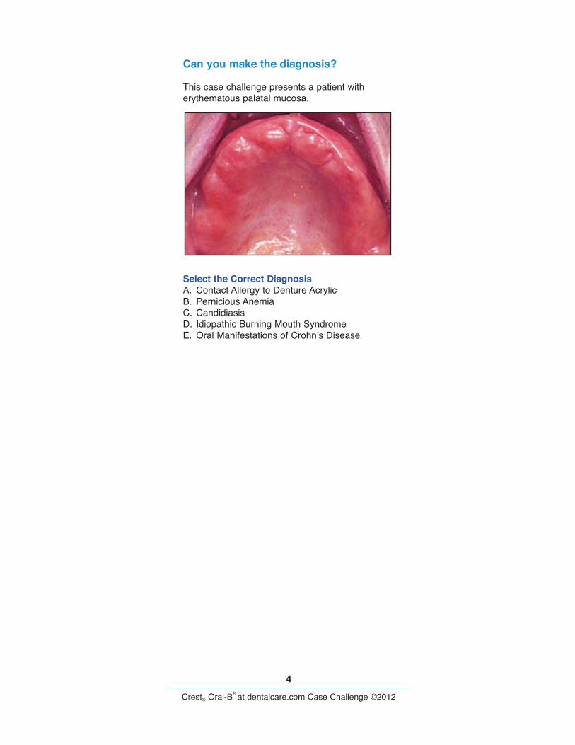
Case SummaryThis case challenge presents a patient with erythematous palatal mucosa.
An 81-year old male presents with a chief complaint of oral soreness, primarily involving the tongue and denture-bearing mucosa. Symptoms have been present for approximately two months and have not been relieved by denture adjustments.
After you have finished reviewing the available diagnostic information, make the diagnosis.
Cynthia L. Kleinegger, DDS, MS
2
Crest® Oral-B® at dentalcare.com Case Challenge ©2012
Diagnostic Information
Additional Clinical HistoryReview of the patient’s medical history reveals he had been diagnosed with Crohn’s disease 16 years previously. Two weeks ago he completed a course of prednisone prescribed to manage an exacerbation of Crohn’s disease. His current medications are dicyclomine hydrochloride, an anticholinergic agent, and mesalamine, an antiinflammatory agent, both for the management of this gastrointestinal disease. He also takes calcium and folic acid supplements.
Review of the patient’s dental history reveals he has worn a maxillary complete denture and a mandibular complete overdenture for 3 years. He has complained frequently of localized areas of soreness; however, in the past these have been relieved by minor denture adjustments.
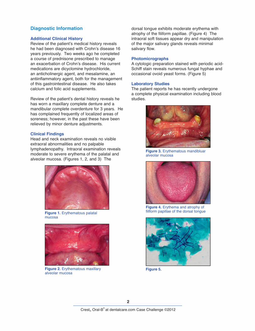
Clinical FindingsHead and neck examination reveals no visible extraoral abnormalities and no palpable lymphadenopathy. Intraoral examination reveals moderate to severe erythema of the palatal and alveolar mucosa. (Figures 1, 2, and 3) The
dorsal tongue exhibits moderate erythema with atrophy of the filiform papillae. (Figure 4) The intraoral soft tissues appear dry and manipulation of the major salivary glands reveals minimal salivary flow.
PhotomicrographsA cytologic preparation stained with periodic acid-Schiff stain reveals numerous fungal hyphae and occasional ovoid yeast forms. (Figure 5)
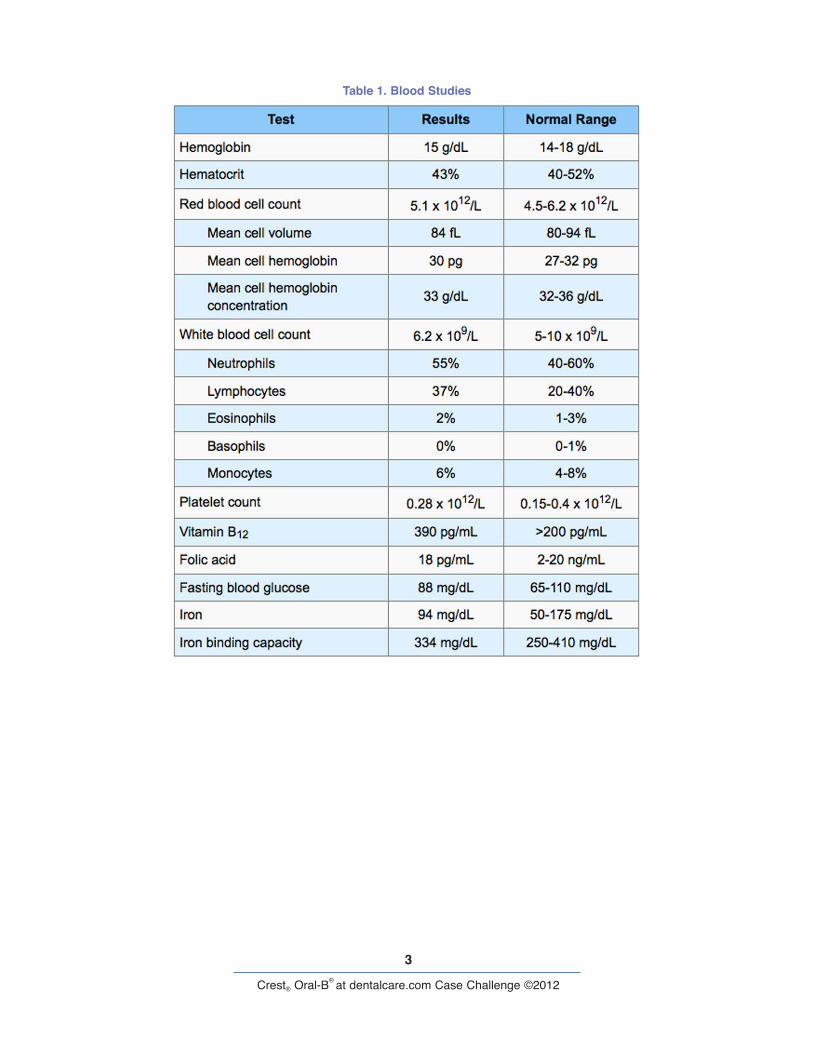
Laboratory StudiesThe patient reports he has recently undergone a complete physical examination including blood studies.
Figure 1. Erythematous palatal mucosa
Figure 2. Erythematous maxillary alveolar mucosa
Figure 3. Erythematous mandibluar alveolar mucosa
Figure 4. Erythema and atrophy of fillform papillae of the dorsal tongue
Figure 5.
3
Crest® Oral-B® at dentalcare.com Case Challenge ©2012
Table 1. Blood Studies
4
Crest® Oral-B® at dentalcare.com Case Challenge ©2012
Can you make the diagnosis?
This case challenge presents a patient with erythematous palatal mucosa.
Select the Correct DiagnosisA. Contact Allergy to Denture AcrylicB. Pernicious AnemiaC. CandidiasisD. Idiopathic Burning Mouth SyndromeE. Oral Manifestations of Crohn’s Disease

5
Crest® Oral-B® at dentalcare.com Case Challenge ©2012
Contact Allergy to Denture Acrylic
Choice A. Sorry, this is not the correct diagnosis.
Allergy to denture base acrylic is extremely uncommon. Irritation may occur due to the leaching of monomer from incompletely cured acrylic and, therefore, occurs immediately after denture delivery.1,2 The patient in this case had been wearing his current prostheses for over 3 years.
Please re-evaluate the information on this case and make another selection.

6
Crest® Oral-B® at dentalcare.com Case Challenge ©2012
Pernicious Anemia
Choice B. Sorry, this is not the correct diagnosis.
Although pernicious anemia often presents with atrophic glossitis and/or generalized mucositis and is a predisposing factor for candidiasis, the normal blood studies in this case would rule out that diagnosis.3,4 The diagnosis of pernicious anemia depends on an abnormally low serum vitamin B12level. Hemoglobin, hematocrit, and red blood cell counts are usually decreased and, unless there is a concomitant iron deficiency anemia, the mean red cell volume is usually
markedly elevated. Since pernicious anemia affects all blood cell lines, white blood cell and platelet counts may be decreased.5
In addition to glossitis or generalized mucositis, oral symptoms of pernicious anemia may include perioral paresthesia. Patients may also report tingling or numbness of the extremities or other systemic symptoms such as fatigue, weakness, shortness of breath, loss of appetite, and diarrhea.3,5
Please re-evaluate the information on this case and make another selection.
7
Crest® Oral-B® at dentalcare.com Case Challenge ©2012
Candidiasis
Choice C. Congratulations! You are correct.
This patient exhibited clinical, historical, and cytologic features consistent with candidiasis.
DiscussionCandidiasis (candidosis) is a common oral fungal infection, usually caused by Candida albicans. C albicans is present as part of the normal oral flora in 40% to 60% of people.6,7 Although less common, other Candida species including C glabrata, C tropicalis, C guilliermondi, and C krusei have been isolated from the oral cavity both as commensals and pathogens.8,9
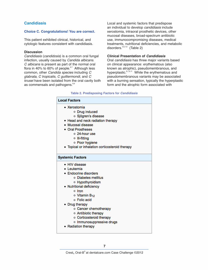
Local and systemic factors that predispose an individual to develop candidiasis include xerostomia, intraoral prosthetic devices, other mucosal diseases, broad-spectrum antibiotic use, immunocompromising diseases, medical treatments, nutritional deficiencies, and metabolic disorders.4,6,7,9 (Table 2)
Clinical Presentation of CandidiasisOral candidiasis has three major variants based on clinical appearance: erythematous (also known as atrophic), pseudomembranous, and hyperplastic.6,7,9,11 While the erythematous and pseudomembranous variants may be associated with a burning sensation, typically the hyperplastic form and the atrophic form associated with
Table 2. Predisposing Factors for Candidiasis
8
Crest® Oral-B® at dentalcare.com Case Challenge ©2012
denture stomatitis are asymptomatic. Other complaints that may be reported by patients with candidiasis include a scalded feeling, irritation from spicy or acidic foods and beverages, surface roughness, sore throat, and altered taste or smell.6
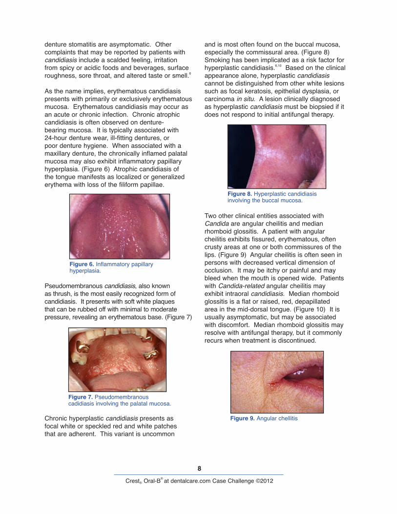
As the name implies, erythematous candidiasis presents with primarily or exclusively erythematous mucosa. Erythematous candidiasis may occur as an acute or chronic infection. Chronic atrophic candidiasis is often observed on denture-bearing mucosa. It is typically associated with 24-hour denture wear, ill-fitting dentures, or poor denture hygiene. When associated with a maxillary denture, the chronically inflamed palatal mucosa may also exhibit inflammatory papillary hyperplasia. (Figure 6) Atrophic candidiasis of the tongue manifests as localized or generalized erythema with loss of the filiform papillae.
Pseudomembranous candidiasis, also known as thrush, is the most easily recognized form of candidiasis. It presents with soft white plaques that can be rubbed off with minimal to moderate pressure, revealing an erythematous base. (Figure 7)
Chronic hyperplastic candidiasis presents as focal white or speckled red and white patches that are adherent. This variant is uncommon
and is most often found on the buccal mucosa, especially the commissural area. (Figure 8) Smoking has been implicated as a risk factor for hyperplastic candidiasis.6,10 Based on the clinical appearance alone, hyperplastic candidiasis cannot be distinguished from other white lesions such as focal keratosis, epithelial dysplasia, or carcinoma in situ. A lesion clinically diagnosed as hyperplastic candidiasis must be biopsied if it does not respond to initial antifungal therapy.
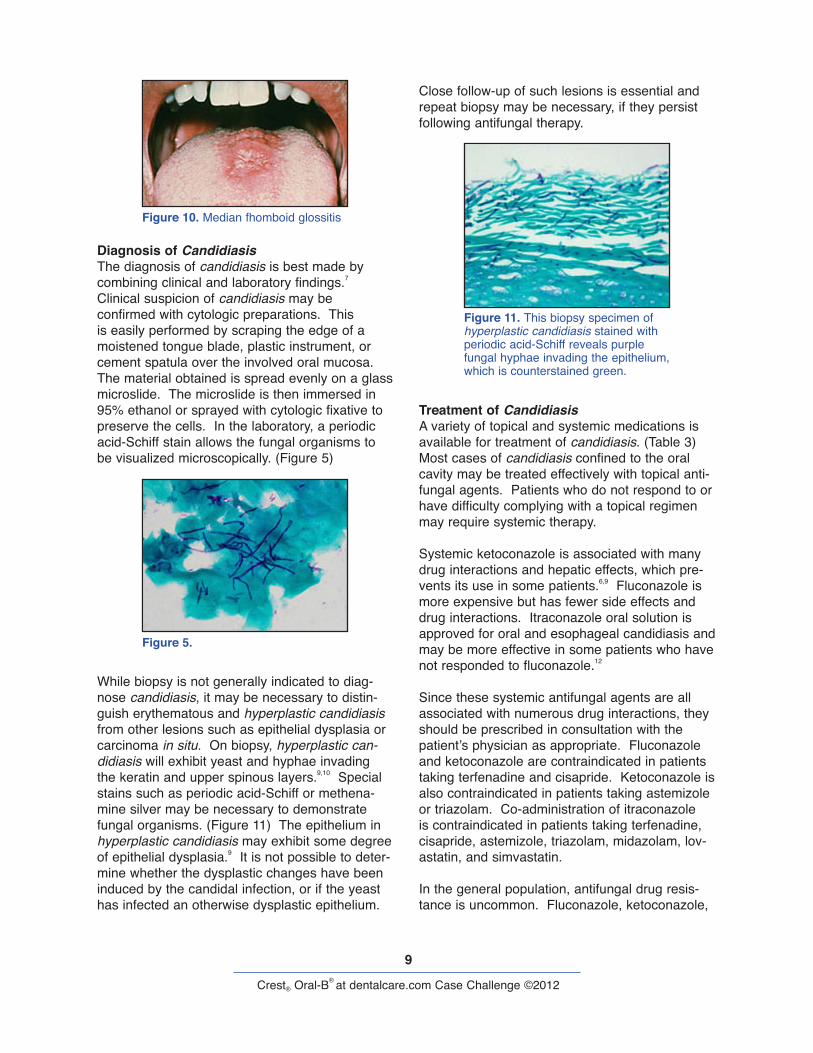
Two other clinical entities associated with Candida are angular cheilitis and median rhomboid glossitis. A patient with angular cheilitis exhibits fissured, erythematous, often crusty areas at one or both commissures of the lips. (Figure 9) Angular cheilitis is often seen in persons with decreased vertical dimension of occlusion. It may be itchy or painful and may bleed when the mouth is opened wide. Patients with Candida-related angular cheilitis may exhibit intraoral candidiasis. Median rhomboid glossitis is a flat or raised, red, depapillated area in the mid-dorsal tongue. (Figure 10) It is usually asymptomatic, but may be associated with discomfort. Median rhomboid glossitis may resolve with antifungal therapy, but it commonly recurs when treatment is discontinued.
Figure 6. Inflammatory papillary hyperplasia.
Figure 7. Pseudomembranous cadidiasis involving the palatal mucosa.
Figure 8. Hyperplastic candidiasis involving the buccal mucosa.
Figure 9. Angular chellitis
9
Crest® Oral-B® at dentalcare.com Case Challenge ©2012
Diagnosis of CandidiasisThe diagnosis of candidiasis is best made by combining clinical and laboratory findings.7 Clinical suspicion of candidiasis may be confirmed with cytologic preparations. This is easily performed by scraping the edge of a moistened tongue blade, plastic instrument, or cement spatula over the involved oral mucosa. The material obtained is spread evenly on a glass microslide. The microslide is then immersed in 95% ethanol or sprayed with cytologic fixative to preserve the cells. In the laboratory, a periodic acid-Schiff stain allows the fungal organisms to be visualized microscopically. (Figure 5)
While biopsy is not generally indicated to diag-nose candidiasis, it may be necessary to distin-guish erythematous and hyperplastic candidiasis from other lesions such as epithelial dysplasia or carcinoma in situ. On biopsy, hyperplastic can-didiasis will exhibit yeast and hyphae invading the keratin and upper spinous layers.9,10 Special stains such as periodic acid-Schiff or methena-mine silver may be necessary to demonstrate fungal organisms. (Figure 11) The epithelium in hyperplastic candidiasis may exhibit some degree of epithelial dysplasia.9 It is not possible to deter-mine whether the dysplastic changes have been induced by the candidal infection, or if the yeast has infected an otherwise dysplastic epithelium.
Close follow-up of such lesions is essential and repeat biopsy may be necessary, if they persist following antifungal therapy.
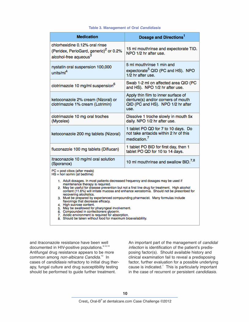
Treatment of CandidiasisA variety of topical and systemic medications is available for treatment of candidiasis. (Table 3) Most cases of candidiasis confined to the oral cavity may be treated effectively with topical anti-fungal agents. Patients who do not respond to or have difficulty complying with a topical regimen may require systemic therapy.
Systemic ketoconazole is associated with many drug interactions and hepatic effects, which pre-vents its use in some patients.6,9 Fluconazole is more expensive but has fewer side effects and drug interactions. Itraconazole oral solution is approved for oral and esophageal candidiasis and may be more effective in some patients who have not responded to fluconazole.12
Since these systemic antifungal agents are all associated with numerous drug interactions, they should be prescribed in consultation with the patient’s physician as appropriate. Fluconazole and ketoconazole are contraindicated in patients taking terfenadine and cisapride. Ketoconazole is also contraindicated in patients taking astemizole or triazolam. Co-administration of itraconazole is contraindicated in patients taking terfenadine, cisapride, astemizole, triazolam, midazolam, lov-astatin, and simvastatin.
In the general population, antifungal drug resis-tance is uncommon. Fluconazole, ketoconazole,
Figure 10. Median fhomboid glossitis
Figure 5.
Figure 11. This biopsy specimen of hyperplastic candidiasis stained with periodic acid-Schiff reveals purple fungal hyphae invading the epithelium, which is counterstained green.
10
Crest® Oral-B® at dentalcare.com Case Challenge ©2012
and itraconazole resistance have been well documented in HIV-positive populations.6,12,13 Antifungal drug resistance appears to be more common among non-albicans Candida.12 In cases of candidiasis refractory to initial drug ther-apy, fungal culture and drug susceptibility testing should be performed to guide further treatment.
An important part of the management of candidal infection is identification of the patient’s predis-posing factor(s). Should available history and clinical examination fail to reveal a predisposing factor, further evaluation for a possible underlying cause is indicated.7 This is particularly important in the case of recurrent or persistent candidiasis.
Table 3. Management of Oral Candidiasis

11
Crest® Oral-B® at dentalcare.com Case Challenge ©2012
When possible, predisposing factors should be removed or controlled. Since this is often not feasible, maintenance antifungal therapy may be required. (Table 3)
Patients with denture-related candidiasis should be educated regarding proper denture use. Prostheses should be evaluated and replaced if necessary. Soaking in 0.12% chlorhexidine glu-conate or 1:50 sodium hypochlorite has been rec-ommended to disinfect dentures.11,14 Denture dis-infection may also be accomplished by using the denture to deliver an antifungal cream or ointment to the infected denture-bearing mucosa. (Table 3) Even when asymptomatic, candidiasis of denture-bearing mucosa should be treated, as inflamed mucosa provides poor support and inflammation may contribute to resorption of underlying bone.4 In addition, untreated palatal candidiasis may progress to inflammatory papillary hyperplasia, which may require surgical removal.2
The patient in this case exhibited clinical and historical features of erythematous candidiasis, confirmed by the presence of fungal hyphae on cytologic preparations. The predisposing factors for candidiasis identified in this patient were xero-stomia secondary to the use of the anticholinergic drug dicyclomine hydrochloride and immunosup-pression due to a recent course of prednisone. The infection was treated with ketoconazole 2% cream applied to the inner surface of the dentures four times daily. (Table 3) Since continued use of dicyclomine hydrochloride was necessary for management of Crohn’s disease, the patient’s xerostomia could not be eliminated. Following resolution of signs and symptoms of candidiasis, the frequency of topical ketoconazole use was gradually decreased to a maintenance frequency of once daily. The patient was also educated in self-management of xerostomia.

12
Crest® Oral-B® at dentalcare.com Case Challenge ©2012
Idiopathic Burning Mouth Syndrome
Choice D. Sorry, this is not the correct diagnosis.
Burning mouth syndrome is a diagnosis of exclusion and would be inappropriate in view of this patient’s cytologic findings. Most patients with burning mouth syndrome exhibit no visible soft tissue changes.15,16
Please re-evaluate the information about this case.
13
Crest® Oral-B® at dentalcare.com Case Challenge ©2012
Oral Manifestations of Crohn’s Disease
Choice E. Sorry, this is not the correct diagnosis.
Although oral lesions of Crohn’s disease may correlate with exacerbation of intestinal symptoms, the mucosal changes exhibited by this patient were not consistent with the oral manifestations of this disease. Oral lesions of Crohn’s disease include a cobblestone pattern of the buccal mucosa, linear hyperplastic folds and ulcers of the vestibules, and diffuse firm swelling of the lips. The gingiva may exhibit granular, erythematous swellings and aphthous-type ulcerations may be present.17
Please re-evaluate the information about this case.
14
Crest® Oral-B® at dentalcare.com Case Challenge ©2012
References1. Jeganathan S, Lin CC. Denture stomatitis—a review of the aetiology, diagnosis and management.
Aust Dent J. 1992 Apr;37(2):107-14. Review.2. Wilson J. The aetiology, diagnosis and management of denture stomatitis. Br Dent J. 1998 Oct
24;185(8):380-4. Review.3. Kleinegger CL, Krolls SO. Severe pernicious anemia presenting with burning mouth symptoms.
Miss Dent Assoc J. 1996 Spring;52(1):12-4. Review. No abstract available.4. Budtz-Jorgensen E. Etiology, pathogenesis, therapy,and prophylaxis of oral yeast infections. Acta
Odontol Scand. 1990 Feb;48(1):61-9. Review.5. Toh BH, van Driel IR, Gleeson PA. Pernicious anemia. N Engl J Med. 1997 Nov 13;337(20):1441-8.
Review. No abstract available.6. Epstein JB, Polsky B. Oropharyngeal candidiasis: a review of its clinical spectrum and current
therapies. Clin Ther. 1998 Jan-Feb;20(1):40-57. Review.7. Fotos PG, Vincent SD, Hellstein JW. Oral candidosis. Clinical, historical, and therapeutic features of
100 cases. Oral Surg Oral Med Oral Pathol. 1992 Jul;74(1):41-9.8. Epstein JB. Antifungal therapy in oropharyngeal mycotic infections. Oral Surg Oral Med Oral Pathol.
1990 Jan;69(1):32-41. Review.9. Scully C, el-Kabir M, Samaranayake LP. Candida and oral candidosis: a review. Crit Rev Oral Biol
Med. 1994;5(2):125-57. Review.10. Lynch DP. Oral candidiasis. History, classification, and clinical presentation. Oral Surg Oral Med
Oral Pathol. 1994 Aug;78(2):189-93. Review.11. Rossie K, Guggenheimer J. Oral candidiasis: clinical manifestations, diagnosis, and treatment.
Pract Periodontics Aesthet Dent. 1997 Aug;9(6):635-41; quiz 642. Review.12. Rex JH, Walsh TJ, Sobel JD, Filler SG, Pappas PG, Dismukes WE, Edwards JE. Practice
guidelines for the treatment of candidiasis. Infectious Diseases Society of America. Clin Infect Dis. 2000 Apr;30(4):662-78.
13. Goff DA, Koletar SL, Buesching WJ, Barnishan J, Fass RJ. Isolation of fluconazole-resistant Candida albicans from human immunodeficiency virus-negative patients never treated with azoles. Clin Infect Dis. 1995 Jan;20(1):77-83.
14. Webb BC, Thomas CJ, Willcox MD, Harty DW, Knox KW. Candida-associated denture stomatitis. Aetiology and management: a review. Part 3. Treatment of oral candidosis. Aust Dent J. 1998 Aug;43(4):244-9. Review.
15. Ship JA, Grushka M, Lipton JA, Mott AE, Sessle BJ, Dionne RA. Burning mouth syndrome: an update. J Am Dent Assoc. 1995 Jul;126(7):842-53. Review.
16. Tourne LP, Fricton JR. Burning mouth syndrome. Critical review and proposed clinical management. Oral Surg Oral Med Oral Pathol. 1992 Aug;74(2):158-67. Review.
17. Bernstein ML, McDonald JS. Oral lesions in Crohn’s disease: report of two cases and update of the literature. Oral Surg Oral Med Oral Pathol. 1978 Aug;46(2):234-45.
About the AuthorNote: Bio information was provided at the time the case challenge was developed.
Cynthia L. Kleinegger, DDS, MSDr. Cynthia L. Kleinegger is Assistant Professor and Director of Advanced Education Programs in the Department of Oral Pathology, Radiology and Medicine at the University of Iowa College of Dentistry.
E-mail: [email protected]