Peritoneal dialysis part1
77
WWW.Montasser-kidney-center.com/ In Facebook :MontasserZeid2
-
Upload
melkholy -
Category
Health & Medicine
-
view
255 -
download
3
Transcript of Peritoneal dialysis part1

WWW.Montasser-kidney-center.com/ In Facebook :MontasserZeid2

WWW.Montasser-kidney-center.com/ In Facebook :MontasserZeid2
Prof. Dr. Montasser Zeid

WWW.Montasser-kidney-center.com/ In Facebook :MontasserZeid2
3
CAPD

WWW.Montasser-kidney-center.com/ In Facebook :MontasserZeid2
The first article on the use of the peritoneal cavity in experimental
uremia (induced in guinea pigs) was published in 1923 by Ganter.
In 1961, Boen described the use of intermittent peritoneal dialysis
(IPD) in patients with chronic renal failure
Historical Background 4 25/02/2012

WWW.Montasser-kidney-center.com/ In Facebook :MontasserZeid2
In1968 Tenckhoff developed an indwelling catheter, which led to
nightly dialysis. This intermittent method, using a cycler to infuse
and drain fluid while the patient slept, was the most common form
of peritoneal dialysis (PD)from the l960s to the late l970s.
In 1976, Popovich et al, introduced continuous ambulatory PD
(CAPD), where four to six exchanges are done each day with long
dwell times between exchanges.
Historical Background cont… 5 25/02/2012
WWW.Montasser-kidney-center.com/ In Facebook :MontasserZeid2
In 1978, this group described their early clinical experience using
bottled dialysis fluid.
Oreopoulos et al. in 1978 also described their experience with
CAPD. From this point on, use of CAPD increased and cycler-IPD
declined.
Historical Background cont… 6 25/02/2012

WWW.Montasser-kidney-center.com/ In Facebook :MontasserZeid2
In 1979 , the Seattle group used a combination of cyclic and
automated PD in two patients. This technique was called
continuous automated ambulatory PD (CAAPD) .
In1979 Baxter introduced to the market the first CAPD system that
included solution bags, tubing with a spike at one end, a titanium
luer lock to connect to the patient’s catheter, and an antiseptic
solution to clean the spike/bag connection.
Historical Background cont… 7 25/02/2012

WWW.Montasser-kidney-center.com/ In Facebook :MontasserZeid2
In 1981, Diaz-Buxo and coworkers described their more extensive
experience and called this technique continuous cycling PD
(CCPD)
Historical Background cont… 8 25/02/2012

WWW.Montasser-kidney-center.com/ In Facebook :MontasserZeid2
The peritoneal cavity is a potential space lined by the peritoneal
membrane , the total surface area of which approximates the
surface area of the skin in adults .The surface of the membrane is
lined by a layer of lubricated mesothelial cells, beneath which lies
the interstitium containing connective tissue and blood vessels, The
blood supply to the visceral peritoneum is derived from celiac,
superior, and inferior mesenteric arteries and their local branches,
Anatomy and Physiology 9 25/02/2012

WWW.Montasser-kidney-center.com/ In Facebook :MontasserZeid2
The parietal peritoneum receives blood from the lumbar,
intercostals and epigastria arteries and drains into inferior vena
cava.
Anatomy and Physiology 10 25/02/2012

WWW.Montasser-kidney-center.com/ In Facebook :MontasserZeid2
Sagittal section of the peritoneal cavity. 11 25/02/2012

WWW.Montasser-kidney-center.com/ In Facebook :MontasserZeid2
The exact blood flow to the membrane is unknown but is probably
from 60-70 ml/min . Venous blood drains ultimately into the
hepatic portal vein. Lymphatic drainage from the peritoneal cavity
is largely through the diaphragm to the lymphatics that are
associated with the internal mammary and anterior mediastinal
lymph nodes; ultimately draining into the right lymphatic duct.
Anatomy and Physiology 12 25/02/2012

WWW.Montasser-kidney-center.com/ In Facebook :MontasserZeid2
R1 : The stagnant capillary fluid overlying the endothelium
R2 : The capillary endothelium it sell.
R3 : The endothelial basement membrane.
R4 : The interstitium.
R5 : The mesothelium
R6 : The stagnant fluid film that overlies the peritoneal membrane.
Resistance to movement during peritoneal dialysis 13 25/02/2012

WWW.Montasser-kidney-center.com/ In Facebook :MontasserZeid2
Resistance to movement during peritoneal dialysis 14
Resistance to solute Movement During Peritoneal Dialysis
25/02/2012

WWW.Montasser-kidney-center.com/ In Facebook :MontasserZeid2
The three – pore model
This model suggests that the peritoneal capillary is the critical
barrier to peritoneal transport and that solute and water transport
across it is mediated by pores of three different.
1. Ultrasmall pores (0.8 nm in radius) that are transcellular
(aquaporin), these mostly transport water molecules.
Anatomy and Physiology cont… 15 25/02/2012

WWW.Montasser-kidney-center.com/ In Facebook :MontasserZeid2
2. intercellular small pores (4-6 nm), which mostly transport small
solutes.
3. large pores (>20 nm) transporting only large molecules such as
peptides and proteins.
Anatomy and Physiology cont… 16 25/02/2012

WWW.Montasser-kidney-center.com/ In Facebook :MontasserZeid2
The essential elements of peritoneal dialysis (PD) are:
A viable peritoneal cavity lined by a functional membrane.
Access to the peritoneal cavity, usually by means of an
indwelling catheter.
Dialysis fluid and delivery mechanism.
Technique of Peritoneal Dialysis 17 25/02/2012

WWW.Montasser-kidney-center.com/ In Facebook :MontasserZeid2
Long-term PD was made a reality by the development by Tenckhoff
of the first long-term indwelling catheter, created by adding
Dacron® cuffs to the earlier silicone-rubber catheters.
Modern catheters still use Tenckhoff’s design.
The material of the catheter is either silicone rubber or polyurethane
on which with Dacron cuffs are placed.
Peritoneal Dialysis Catheters 18 25/02/2012

WWW.Montasser-kidney-center.com/ In Facebook :MontasserZeid2
Each PD catheter is comprised of three parts
An intra-abdominal segment.
A subcutaneous tunnel segment.
An external segment.
Peritoneal Dialysis Catheters cont… 19 25/02/2012

WWW.Montasser-kidney-center.com/ In Facebook :MontasserZeid2
The intra-abdominal segment has multiple small holes and an
open terminal end.
The subcutaneous segment has two cuffs, the outer cuff is placed
just under the skin at the exit site, and the deep cuff is placed just
external to the fascia covering the parietal peritoneum.
The segment between the two cuffs lies in a tunnel, classically
curved in shape, and running from the skin exit site to the deeper
cuff.
Peritoneal Dialysis Catheters cont… 20 25/02/2012

WWW.Montasser-kidney-center.com/ In Facebook :MontasserZeid2
Table: Commonly used peritoneal catheters 21
Easy to place by percutaneous technique
Easy to remove and replace
Rectal discomfort is more common and outflow
problems are more common
Straight Tenckhoff
Same as above, but better patency and better outflow
Does not impinge on the rectum Curled Tenckhoff
Same as straight Tenckhoff
Tunnel must be curved Swan neck
Combination of Toronto Western and swan-neck
catheter
Can only be place and manipulated surgically
Missouri
Can only be placed and manipulated surgically Lifecath
25/02/2012

WWW.Montasser-kidney-center.com/ In Facebook :MontasserZeid2
Peritoneal Catheters ( lateral views ) 22 25/02/2012

WWW.Montasser-kidney-center.com/ In Facebook :MontasserZeid2
Three insertion techniques are used , surgical , blind ( bed side )
percutaneous, and peritoneoscope.
Prior to insertion of the catheter, the patient should be informed of
the potential complications, and the bladder and rectum should be
emptied. If the patient cannot empty their bladder, a urinary
catheter may be used to achieve this. This is to avoid puncturing a
full bladder, particularly a potential risk during the bedside
insertion.
Catheter Insertion Technique 23 25/02/2012

WWW.Montasser-kidney-center.com/ In Facebook :MontasserZeid2
Comparison of the Three Techniques 24
Disadvantages Advantages Technique
•More preparation is required
•More expensive
•Large incision
•Longer healing time
•Increased risk of fluid leak
Under direct vision:
•Lower risk of injury to viscera
•Catheter placed in desired place
•Adhesions or large omentum can be removed
surgically
•Any type of catheter can be inserted
Surgical
•Cannot be used if adhesions or
large omentum present
•Higher failure potential
•Simple, easy, and quick procedure
•Heals quickly and can be used immediately
•Performed in patient’s room with full sterile
precautions by the nephrologist
• Less expensive than the other two methods
Percutaneous
•Higher risk of injury to viscera
•Not all catheters can be placed
by this technique
•Expense of the scope Training
with scope.
•Faster healing
•Lower risk of fluid leak
•Simple procedure
•Useful even in presence of adhesions
Peritoneoscopic
Table : Comparison of catheter insertion techniques 25/02/2012

WWW.Montasser-kidney-center.com/ In Facebook :MontasserZeid2
Peritoneal Dialysis Catheters 25
Tenckhoff technique with curved tunnel
25/02/2012

WWW.Montasser-kidney-center.com/ In Facebook :MontasserZeid2
Peritoneal Dialysis Catheters 26
Common incision sites
25/02/2012

WWW.Montasser-kidney-center.com/ In Facebook :MontasserZeid2
Many of these are complications that are experienced with any
surgical procedure, including trauma to an abdominal organ (most
commonly the bowel) or to vessels, bleeding, infection, and bowel
perforation and specific complications :
Postoperative Complications 27 25/02/2012

WWW.Montasser-kidney-center.com/ In Facebook :MontasserZeid2
Postoperative Complications 28
Table : Common postoperative complications and their management
Management Complication
Decrease inflow volume
Switch to cycler PD in supine position
Keep abdomen empty when upright during day time
1. Fluid leak
For supine patient, change positions
Administer enema to move bowel loops
Increase the vertical distance between the abdomen and the
outflow bag mechanical obstruction, such as a clamp being left
on, or the Kinking/accidental suture of the catheter. If fibrin is
observed in the outflow,100-500 U heparin/l should be added
to the dialysate.
2. Outflow problems
(1) Systemic antibiotics
(2) If these fail or infection recurs, change the catheter and use
a fresh site for the tunnel
3. Tunnel infection
25/02/2012
WWW.Montasser-kidney-center.com/ In Facebook :MontasserZeid2
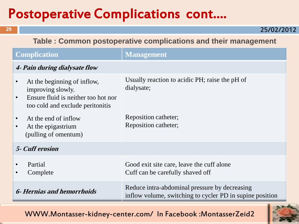
Postoperative Complications cont…. 29
Table : Common postoperative complications and their management
Management Complication
4- Pain during dialysate flow
Usually reaction to acidic PH; raise the pH of
dialysate;
Reposition catheter;
Reposition catheter;
• At the beginning of inflow,
improving slowly.
• Ensure fluid is neither too hot nor
too cold and exclude peritonitis
• At the end of inflow
• At the epigastrium
(pulling of omentum)
5- Cuff erosion
Good exit site care, leave the cuff alone
Cuff can be carefully shaved off
• Partial
• Complete
Reduce intra-abdominal pressure by decreasing
inflow volume, switching to cycler PD in supine position 6- Hernias and hemorrhoids
25/02/2012

WWW.Montasser-kidney-center.com/ In Facebook :MontasserZeid2
Postoperative Complications cont.. 30
If the catheter is blocked by a fibrin plug, urokinase or tPA may be
used in an attempt to dissolve the fibrin plug. Urokinase 5,000 IU
is diluted with 0.9% saline solution to 40 ml. this volume is
infused into the catheter and the catheter is clamped for 30-90 min
then drain .
25/02/2012

WWW.Montasser-kidney-center.com/ In Facebook :MontasserZeid2
Postoperative Complications cont.. 31
If unsuccessful, repeat with a higher dose of urokinase (10,000 IU
diluted to 40 ml with 0.9% saline ).Tissue plasminogen activator
(TPA) can also be used. A 1 mg/ml TPA concentration is infused
and left for 1-2 h, and has been reported to open up plugged PD
catheters.
25/02/2012

WWW.Montasser-kidney-center.com/ In Facebook :MontasserZeid2
Table : Composition of peritoneal dialysis fluid
Peritoneal Dialysis Fluid 32
Standard solution (mEq/l) Electrolytes
132
0*
2.5, 3.5
0.5, 1.5
96-102
35-40
1.5, 2.5. 3.5. 4.25
5.2-5.5
Sodium
Potassium
Calcium
Magnesium
Chloride
Lactate
Glucose ( g/dl)**
pH
25/02/2012

WWW.Montasser-kidney-center.com/ In Facebook :MontasserZeid2
Instead of lactate, bicarbonate solution or bicarbonate/lactate
solutions are now available. A two-compartment bag keeps the
bicarbonate solution separate from the rest of electrolytes,
including calcium and magnesium, to avoid precipitation of their
carbonate salts. Prior to use, the two solutions are mixed and
infused.
Peritoneal Dialysis Fluid cont … 33 25/02/2012

WWW.Montasser-kidney-center.com/ In Facebook :MontasserZeid2
Harmful, long-term effects of an acidic dialysis fluid (lactate
based) on the peritoneal membrane and on the function of
leukocytes and macrophages have been reported.
Peritoneal Dialysis Fluid cont … 34 25/02/2012

WWW.Montasser-kidney-center.com/ In Facebook :MontasserZeid2
Dextrose:
UF rates depends on the concentration of the dextrose in the solution
and duration of dwell in the peritoneal cavity and peritoneal
membrane characteristics.
Osmotic Agents 35 25/02/2012

WWW.Montasser-kidney-center.com/ In Facebook :MontasserZeid2
Amino Acid Solutions
A 1% amino acid solution is equivalent to 1.5% dextrose in terms of
UF volume, and a 2.0-2.76% amino acid solution is equivalent to
4.25% dextrose.
Some beneficial effects in patients with protein malnutrition. These
are usually used once a day, with more frequent use there is risk of
metabolic acidosis and an increase in urea concentration is seen.
Alternative Agents 36 25/02/2012

WWW.Montasser-kidney-center.com/ In Facebook :MontasserZeid2
Icodextrin is a glucose polymer that is derived from starch,
containing many glucose units bonded together. In the peritoneal
cavity, it slowly breaks down into smaller units thus causing
sustained osmolarity and UF.
This is particularly helpful in high transporters in removing fluid,
especially during the long dwell time (>6h).
Icodextrin Solution 37 25/02/2012

WWW.Montasser-kidney-center.com/ In Facebook :MontasserZeid2
In CCPD (Automated PD, APD) patients, icodextrin solutions
can be used during the daytime and in CAPD patients during the
night dwell to achieve adequate UF.
Ultrafiltration with icodextrin appears to be independent of
peritoneal membrane transport characteristics.
Icodextrin Solution cont …. 38 25/02/2012

WWW.Montasser-kidney-center.com/ In Facebook :MontasserZeid2
Delivery Mechanism 39
Contlnuous Ambulatory Peritoneal Dialysis Technique Using ‘Y’ Transfer Set
1. Keep patient side clamp (A) on
• Open clamps B and C
• Let small volume of fluid flush the tubes
2. Close clamp B
• Open clamp A
• Drain all fluid from the patient into the empty bag
3. Close clamp C
• Open clamp B and fill the peritoneal cavity
• Close clamp A and catheter clamp
• Disconnect the ‘Y’ set with bags
25/02/2012

WWW.Montasser-kidney-center.com/ In Facebook :MontasserZeid2
Continuous Ambulatory Peritoneal Dialysis (CAPD)
This requires only connecting tubes and bags of solutions (usually 2 L
of solution in 3-L bags), using gravity to fill and empty the
peritoneal cavity.
The most commonly used method uses four exchanges per day, but, in
some patients, five exchanges are necessary.
Peritoneal Dialysis Techniques 40 25/02/2012

WWW.Montasser-kidney-center.com/ In Facebook :MontasserZeid2
Automated Peritoneal Dialysis (APD)
This technique uses a cycler with or without added manual
exchanges. The two forms are continuous cycling PD (CCPD) and
nocturnal intermittent PD (NIPD). In CCPD, at night, a cycler is
used to have a few automated exchanges and, during daytime, there
are one or two manual exchanges.
Peritoneal Dialysis Techniques cont…. 41 25/02/2012

WWW.Montasser-kidney-center.com/ In Facebook :MontasserZeid2
Automated Peritoneal Dialysis (APD) cont ……
The NIPD involves automated nightly exchanges and during the
daytime the peritoneal cavity remains empty. the patient loads the
bags of solution on to a cycler and connects the catheter to this
cycler just before going to sleep. During the night, the cycler
makes four to five or more exchanges automatically.
Peritoneal Dialysis Techniques cont…. 42 25/02/2012

WWW.Montasser-kidney-center.com/ In Facebook :MontasserZeid2
Automated Peritoneal Dialysis (APD) cont ……
In the morning, 2 L of solution are left in the abdomen and the
patient disconnects from the cycler During the day, there is
usually no exchange and at night, the procedure is repeated .
Peritoneal Dialysis Techniques cont…. 43 25/02/2012

WWW.Montasser-kidney-center.com/ In Facebook :MontasserZeid2
TPD is a type of APD in which the dwell volume is not
completely drained before the next fill cycle is initiated . Typical
TPD regimens are programmed to leave 250, 500 or 1000 mls as a
reservoir and then subsequent infusions/drains occur into the
reservoir.
Tidal Peritoneal Dialysis (TPD) 44 25/02/2012

WWW.Montasser-kidney-center.com/ In Facebook :MontasserZeid2
TPD can be used in patients with pain at the beginning of the
infusion or in those who typically drain very slowly and
inefficiently at the end of the drain.
Tidal Peritoneal Dialysis (TPD) cont …. 45 25/02/2012

WWW.Montasser-kidney-center.com/ In Facebook :MontasserZeid2
Different types of peritoneal dialysis techniques. 46 25/02/2012

WWW.Montasser-kidney-center.com/ In Facebook :MontasserZeid2
Dose of Peritoneal Dialysis 47
Because the contribution of residual renal function to the total
dialysis dose is significant, it is important that renal contribution
and the total dose of dialysis are both measured frequently. If either
of these decrease significantly, the patient must be monitored very
closely and, at the first sign of under-dialysis, therapy must be
modified to avoid premature death from under-dialysis.
25/02/2012

WWW.Montasser-kidney-center.com/ In Facebook :MontasserZeid2
Total WC1Cr combines the glomerular filtration rate (GFR) with
the peritoneal creatinine clearance (WpClCr) and normalizes it for
a body surface area (BSA) of l.73m2:
1. Calculate residual GFR, convert it to weekly clearance.
2. Calculate peritoneal creatinine clearance and convert it to
weekly clearance (WpC1Cr).
Weekly Creatinine Clearance 48 25/02/2012

WWW.Montasser-kidney-center.com/ In Facebook :MontasserZeid2
3. Estimate BSA from height and weight
4. Normalize the WpC1Cr to a BSA of 1.73 m2
5. Weekly creatinine clearance =
GFR (from Step 1) + WpC1Cr per 1.73m2 (from Step 4)
Weekly Creatinine Clearance cont … 49 25/02/2012

WWW.Montasser-kidney-center.com/ In Facebook :MontasserZeid2
With declining GFR, the tubular secretion of creatinine increases;
creatinine clearance overestimates true GFR. To correct for the
tubular creatinine secretion, the average of urea and creatinine
clearance is used to estimate GFR.
GFR = (renal urea clearance + renal creatinine clearance)/2
Some investigators uses 50% of measured creatinine clearance as
GFR . GFR calculation using formula UV/P
Residual Glomerular Filtration Rate 50 25/02/2012

WWW.Montasser-kidney-center.com/ In Facebook :MontasserZeid2
Peritoneal creatinine clearance (pClCr) is measured by collecting
all peritoneal outflow for 24 h and measuring creatinine in the
dialysate and in the blood.
Peritoneal Creatinine Clearance 51 25/02/2012

WWW.Montasser-kidney-center.com/ In Facebook :MontasserZeid2
The WCICr should be normalized
for 1.73m2. BSA from weight
(kilograms) and height
(centimeters) can be either
estimated on a nomogram.
Correction for Body Surface Area 52
Surface area(m2) Weigh (kg)
Height(cm)
Body surface area as a function
of body weight and height
25/02/2012
WWW.Montasser-kidney-center.com/ In Facebook :MontasserZeid2
The second method of calculating dialysis dose is by the use of the
urea kinetics concept.
Urea Clearance Concept (Kt/Vurea) 53 25/02/2012

WWW.Montasser-kidney-center.com/ In Facebook :MontasserZeid2
The volume of distribution of urea (Vurea) is equivalent to the total
body water (TBW) and can be estimated by several methods.
1. Direct estimation from weight: Normally, water represents 58%
of body weight. Thus, the total body water = 0.58 x weight (0.58 x
70kg in our patient = 40.6L).
2. Use of specific formulae.
Volume of Distribution of Urea 54 25/02/2012

WWW.Montasser-kidney-center.com/ In Facebook :MontasserZeid2
Watson Formula : for adults
For men:
V (total body water in liters) = 2 447 + 0 3362 x weight (kg) + ) 0.174 x
height (cm) 0.09516 x age (years).
For women:
V (total body water in liters) = 2.097 + 0.2466 x weight +0.l069 x height
V ( by Watson formula ) :
V= 2.447 — 0.09516 A + 0.1704 H + 0.3362 W ( in males )
V=2.097 + 0.1069 H + 0.2466 W (in females )
where A = age (yr); H = height (cm), and W = weight (kg)
Volume of Distribution of Urea cont …. 55 25/02/2012

WWW.Montasser-kidney-center.com/ In Facebook :MontasserZeid2
Watson Formula : for adults
Mellits - Cheek Formula For Children:
For boys:
V (when height < 132.7 cm.) = -1.927 + 0.465 x weight + 0.045 x height;
V (when height >132.7 cm) = -21.993 + 0.406 x weight + 0.209 x height.
For girls:
V (when height < 110.8cm) = -0.076 + 0.507 x weight + 0.013 x height;
V (when height > 110.8 cm) = -10.3 13 + 0.252 x weight+ 0.154 x height
Volume of Distribution of Urea cont …. 56 25/02/2012

WWW.Montasser-kidney-center.com/ In Facebook :MontasserZeid2
(1) DOQI
NIPD CCPD CAPD Factor
2.2 2.1 2.0 Kt/V per wk
66 63 60 CrCl L per wk
(2) Canadian Society of Nephrology
Low/low-average transporters
(CAPD, CCPD, NIPD)
High/high-average
transporters
(CAPD, CCPD, NIPD)
Factor
2.0
50
2.0
60
Kt/V per wk
CrCl L per wk
Recommended Dose of Dialysis 57
Table :Clearance targets for peritoneal dialysis
25/02/2012

WWW.Montasser-kidney-center.com/ In Facebook :MontasserZeid2
Current KDOQI guideline:
1. For patients with residual kidney function (RKF) (considered to
be significant when urine volume is > 100 mL/d):
A. The minimal “delivered” dose of total small-solute clearance
should be a total (peritoneal and kidney) Kt/Vurea of at least 1.7
per week.
58
Recommended Dose of Dialysis 25/02/2012

WWW.Montasser-kidney-center.com/ In Facebook :MontasserZeid2
Current KDOQI guideline: cont….
2. For patients without RKF (urine volume is < l00 mL/d):
A. The minimal “delivered” dose of total small-solute clearance
should be a peritoneal Kt/Vurea of at least 1.7 per week
measured within the first month after starting dialysis therapy
and at least once every 4 months thereafter.
59
Recommended Dose of Dialysis cont …. 25/02/2012

WWW.Montasser-kidney-center.com/ In Facebook :MontasserZeid2
Current KDOQI guideline: cont….
The new guideline also does not recommend different targets for
different forms of the peritoneal dialysis techniques.
60
Recommended Dose of Dialysis cont …. 25/02/2012

WWW.Montasser-kidney-center.com/ In Facebook :MontasserZeid2
Solute and solvent movement across the peritoneal membrane.
Twardowski et al. first described a peritoneal equilibration test
(PET) to assess the peritoneal function in terms of the rate of
solute transport.
It is now routinely used to monitor membrane function in both
new and established patients.
Peritoneal Function Test 61 25/02/2012
WWW.Montasser-kidney-center.com/ In Facebook :MontasserZeid2
A 2 L volume of 2.5% dextrose solution is instilled in an empty
peritoneal cavity and dialysate samples are obtained at 0, 2, and
4h to measure creatinine and glucose concentrations.
At the end of the 4h dwell time, all of the fluid is drained out, the
total volume is measured, and the sample volumes (drawn during
the 2 and 4h) are added to the final volume.
Traditional Peritoneal Equilibration Test 62 25/02/2012

WWW.Montasser-kidney-center.com/ In Facebook :MontasserZeid2
A blood sample is also obtained at 4h to measure serum creatinine.
Two curves are then plotted.
One curve plots the 0-, 2-, and 4-h values of dextrose in the
dialysate outflow as a fraction of the initial dialysate dextrose
concentration (D/D0).
Traditional Peritoneal Equilibration Test cont ... 63 25/02/2012

WWW.Montasser-kidney-center.com/ In Facebook :MontasserZeid2
After emptying the peritoneal cavity completely, the patient instills
2L of 2.5% dextrose solution and arrives at the clinic within 4h.
The fluid is drained out completely, the volume is measured, and
blood and dialysate samples are sent for measurement of dextrose
and creatinine concentrations.
Fast Peritoneal Equilibration Test 64 25/02/2012

WWW.Montasser-kidney-center.com/ In Facebook :MontasserZeid2
Net Ultra-
filtrationa
Drainage Vol.
(mL)
Dialysate
Glucose
(mg/dL)
DIP
Creatinine
Transport
Classification
-470-35 1.580-2.084 230-501 0.82 -1.03 High
35-320 2.085-2.367 502-722 0.81- 0.66 High-average
320 2.368 723 0.64 Mean
320-600 2.369-2.650 724-944 0.50- 0.64 Low-average
600-1.276 2.651-3.326 945-1.214 0.43-0.49 Low
Table : 65
Classification of a patient’s peritoneal transport using fast peritoneal
equilibrium test
25/02/2012

WWW.Montasser-kidney-center.com/ In Facebook :MontasserZeid2
The PET results are useful in assessing membrane function and in
deciding which form of PD is most suitable for the patient.
Thus, a high transporter will have faster dissipation of dextrose and
a longer dwell time of CAPD would lead to a lower net UF and an
inability to control fluid excess.
Use of Fast Peritoneal Equilibration Test Results in Selecting a Peritoneal Dialysis Regimen
66 25/02/2012

WWW.Montasser-kidney-center.com/ In Facebook :MontasserZeid2
In a low-average transporter, rapid exchanges will not remove
enough solute and will lead to wastage of dialysate. a high
transporter will benefit from a shorter dwell time.
It must be stressed that PET does not give information on adequacy
of dialysis; adequacy must be assessed by calculation of Kt/Vurea
and weekly creatinine clearance .
Use of Fast Peritoneal Equilibration Test Resultsin Selecting a Peritoneal Dialysis Regimen cont ….
67 25/02/2012

WWW.Montasser-kidney-center.com/ In Facebook :MontasserZeid2
When the drainage volume fails to exceed the instilled volume by
at least 400 mL 4 hours after instillation of a 4.25% 2L test
exchange , some problem with UF is likely.
Interpretation of results 68 25/02/2012

WWW.Montasser-kidney-center.com/ In Facebook :MontasserZeid2
1. UF volume. Drainage volume in a PET with 2.5% dextrose
solution averages 2,370 mL. As 2-L bags of dialysate contain
about 2,050 mL of dialysis solution, this equals 320 mL of UF.
2. Drainage fluid glucose level. The mean 4-hour glucose level the
dialysate is 720 mg per dL. Values above 950 mg per dL. and
below 500 mg per dL are typically seen in low and high
transporters, respectively.
Interpretation of results cont ….. 69 25/02/2012

WWW.Montasser-kidney-center.com/ In Facebook :MontasserZeid2
3. Dialysate/plasma creatinine (D/P Cr). The mean D/P Cr ratio
at 4 hours is 0.65. High and low transporters typica1ly have
ratios above 0.82 and below 0.49, respectively.
Interpretation of results cont …. 70 25/02/2012

WWW.Montasser-kidney-center.com/ In Facebook :MontasserZeid2
Four major considerations in selection of the technique are:
1. Patient preference, life style, and practical considerations.
2. Medical considerations.
3. PET considerations.
4. Cost considerations.
Selection of Technique 71 25/02/2012

WWW.Montasser-kidney-center.com/ In Facebook :MontasserZeid2
Patients with GFR > 2m1/min:
For CAPD:
BSA < l.7m2 : 2L x four exchanges/day;
BSA l.7-2.0m2: 2.5L x four exchanges/day;
BSA > 2.0m2 : 3.0L x four exchanges/day
Empirical Approach to Deciding a Peritoneal Dialysis Regimen
72 25/02/2012

WWW.Montasser-kidney-center.com/ In Facebook :MontasserZeid2
Patients with GFR > 2m1/min:
For CCPD:
BSA < l.7m2 : 2Lx four exchanges (9 h/night) +2L/day;
BSA l.7-2.0m2 : 2.5 Lx four exchanges (9h/night)+2L/day
BSA> 2.0m2 : 3.0 Lx four exchanges (9 h/night) +2L/day
Empirical Approach to Deciding a Peritoneal Dialysis Regimen cont …
73 25/02/2012

WWW.Montasser-kidney-center.com/ In Facebook :MontasserZeid2
Patients with GFR < 2ml/min:
For CAPD:
BSA < l.7m2: 2.5 Lx four exchanges /day;
BSA l.7-2.0m2: 3.0Lx four exchanges/day;
BSA > 2,0m2 : 3.0Lx four exchanges/day
(added exchange at night should be considered)
Empirical Approach to Deciding a Peritoneal Dialysis Regimen cont ….
74 25/02/2012

WWW.Montasser-kidney-center.com/ In Facebook :MontasserZeid2
Patients with GFR < 2ml/min:
For CCPD:
BSA < l.7m2: 2.5L x four exchanges (9h /night) +2L/day;
BSA 1.7-2.0m2: 3.0L x four exchanges (9h/night) +2.5 L/day;
BSA > 2.0m2: 3.0L x 4 exchanges (l0h/night)+3Lx two
exchanges/ day.
Empirical Approach to Deciding a Peritoneal Dialysis Regimen cont ….
75 25/02/2012

WWW.Montasser-kidney-center.com/ In Facebook :MontasserZeid2
Patients with GFR < 2ml/min:
For NIPD:
The intermittent NIPD needs to be closely monitored for GFR
changes and adequacy of dialysis as well as membrane functions,
and the regimen must be modified accordingly.
Empirical Approach to Deciding a Peritoneal Dialysis Regimen cont ….
76 25/02/2012

WWW.Montasser-kidney-center.com/ In Facebook :MontasserZeid2
77
25/02/2012 WWW.Montasser-kidney-center.com/ In Facebook :MontasserZeid2