
Peristomal Complications - Winnipeg Regional Health · PDF file4/9/2014 2 Peristomal...
9
4/9/2014 1 Peristomal Complications Assessment & Management Mary Robertson RN BN CETN(C) Manitoba Ostomy Program Discussion Preamble - The stoma & peristomal area Peristomal complications & management The Stoma A surgically created opening for the elimination of bodily waste. The bowel is exteriorized & everted onto the surface of the abdomen, exposing the innermost lining, the mucosal layer of the bowel. The stoma: Has altered nerve sensation therefore needs protection from hot/cold, pressure & trauma. A healthy stoma will be shiny, red & moist (mucosal tissue). Quick proliferation of mucosal cells (2 to 5 d) This promotes quick healing & in a way is “self-cleaning”. The Stoma Often called an ostomy. A stoma can change shape & size. When the pouch is changed note the shape, size and colour of the stoma. Note also the location and function. The patient and the caregivers should know what type of stoma the individual has. 3 common types: • Colostomy • Ileostomy • Urostomy Peristomal Area The area of support for & around the stoma. Provides the surface area for the ostomy pouch to hold on to. The contour of the abdominal wall can change shape & size. Assess during the pouch change for skin integrity, colour , contour and patient comfort. Peristomal Complications Peristomal skin irritation is the most common early complication following stoma creation. (Kann, 2008) A 2010 study that followed 89 patients for 1 year after surgery, found 50% experienced peristomal skin complications and most were related to pouch leakages. (Jordan & Christian, 2013) Another study suggested that 85% of ostomy patients will experience a pouch leakage at some point in their lives. (Jordan & Christian, 2013)
Transcript of Peristomal Complications - Winnipeg Regional Health · PDF file4/9/2014 2 Peristomal...

4/9/2014
1
Peristomal
Complications Assessment & Management
Mary Robertson RN BN CETN(C)
Manitoba Ostomy Program
Discussion
Preamble - The stoma & peristomal area
Peristomal complications & management
The Stoma
A surgically created opening for the elimination of bodily waste.
The bowel is exteriorized & everted onto the surface of the abdomen, exposing the innermost lining, the mucosal layer of the bowel.
The stoma: Has altered nerve sensation therefore needs protection
from hot/cold, pressure & trauma.
A healthy stoma will be shiny, red & moist (mucosal tissue).
Quick proliferation of mucosal cells (2 to 5 d) This promotes quick healing & in a way is “self-cleaning”.
The Stoma
Often called an ostomy.
A stoma can change shape & size.
When the pouch is changed note the shape, size
and colour of the stoma. Note also the location
and function.
The patient and the caregivers should know what
type of stoma the individual has.
3 common types:
• Colostomy
• Ileostomy
• Urostomy
Peristomal Area
The area of support for & around the stoma.
Provides the surface area for the ostomy pouch
to hold on to.
The contour of the abdominal wall can change
shape & size.
Assess during the pouch change for skin
integrity, colour , contour and patient comfort.
Peristomal Complications
Peristomal skin irritation is the most common early
complication following stoma creation. (Kann, 2008)
A 2010 study that followed 89 patients for 1 year
after surgery, found 50% experienced peristomal
skin complications and most were related to pouch
leakages. (Jordan & Christian, 2013)
Another study suggested that 85% of ostomy
patients will experience a pouch leakage at some
point in their lives. (Jordan & Christian, 2013)

4/9/2014
2
Peristomal Complications
The problem may be well advanced by the time the
person seeks help.
Early hospital discharge & laparoscopic procedures
result in less time for teaching and supervised
practice with a new ostomy.
The best management of peristomal complications
is prevention.
(Jordan & Christian,2013)
Peristomal Complications
To the client:
↑↑’d pain &
discomfort
poor self image
social isolation
diminished QOL
What are the impacts ?
To the health care system?
↑↑’d costs & time associated
with individual’s care,
including supplies
Peristomal Complications
Chemical Irritation
Irritant Contact Dermatitis (common)
Allergic Contact Dermatitis (rare)
• < 0.6 % are true allergic reactions (Lyon,
2001) (Jordan & Christian, 2013)
Chemical Irritation
The most common cause of chemical irritation in the
peristomal area is the stoma output on the
abdominal skin.
Irritated skin may be at increased risk of allergic
reaction due to immune system overstimulation.
Once the sensitivity presents, it is usually permanent. Any
of the ostomy care products used on the skin could
potentially cause a reaction.
(Hampton & Bryant, 1992)
Irritant Contact Dermatitis Caused by stoma effluent in contact with the peristomal
skin.
It’s painful. The patient will complain of burning, stinging
& itching.
The skin will look reddened,
may have denuded/shallow
open areas.
The stoma output can be
seen on the skin when the
pouch is changed.
Complaints of the pouch
“falling off”.

4/9/2014
3
Irritant Contact Dermatitis
The irritation mirrors
where the effluent
was sitting on the
skin.
Gently cleanse the
area with warm tap
water. Pat dry.
Irritant Contact Dermatitis
Measure the stoma, and trace the area to get a fit that
includes only the stoma, +/or fistula if present.
Use fillers to help create a level surface to obtain a
reliable seal.
Dust the stomahesive
powder lightly, remove
excess. Dab with cavilon
wipe. Repeat. This will form
a protective crust.
Irritant Contact Dermatitis
Containment of the
effluent and protection of
the irritated skin will allow
the area to heal and the
dermatitis will resolve.
Once the area is healed
and intact –omit the
powder and film barrier
treatment.
Allergic Contact Dermatitis
Presents with:
Erythema
Burning, stinging
& itching
May have swelling
“Bull’s eye” target
appearance
Not related to
pouch leakages
Allergic Contact Dermatitis
Remove the offending
product.
Topical steroidal agent
may be needed.
Can use an interface
such as a Coloplast
barrier sheet if no other
pouching option
available.
Allergic Contact Dermatitis
Think prevention. Avoid use
of unnecessary products that
may be potential allergens
(Baby wipes, personal hygiene
wipes, soaps or cleansing
products)
Cleanse peristomal skin with
warm tap water.
Avoid the use of film barriers
(no sting, skin prep) or
adding additional tape.

4/9/2014
4
Papular Overgranulation
The number one cause is persistent or recurrent
leakage or seepage of the stoma output on the
skin.
Related to inadequate seal around the stoma.
?opening too large, ?wrong shape.
Also called:
Hyperplasia/Hypergranulation
Granulomas
Proudflesh
Papular Overgranulation
Lesions present as
red papules
occurring at the
mucocutaneous
junction.
The tissue is friable
and bleeds easily.
Cutting pouch larger
to accommodate,
makes it worse.
Papular Overgranulation
Treatment
Assess the shape & size
of the stoma
Evaluate fit of the pouch
Evaluate the emptying
rountine, is the pouch
being “rinsed” out?
Weekly application of
silver nitrate stick to
reduce the overgrowth
of tissue.
5 weeks of silver
nitrate treatment
Papular Overgranulation
Silver Nitrate:
• May sting temporarily.
• Clean area with warm tap water & pat dry.
• Roll the tip of the silver nitrate stick over the hypergranulation tissue avoiding any
contact with the stoma.
• If the treatment becomes too painful, rinse the area with normal saline. This will
deactivate the silver nitrate and the discomfort should quickly resolve. The tissue will
have a grayish/white appearance which will slough away.
• Apply appliance as to obtain a good seal.
2 weeks later
Pseudoverrucous Lesions
Wart-like lesions
occurring at the
mucocutaneous
junction.
May be numerous.
May be itchy and
tender.
Present in a variety of
discolorations from
white-grey to reddish-
brown.
Pseudoverrucous Lesions
The cause is chronic exposure to
urine in the peristomal area.
The shape & size of the
lesions can mimic the
difference between the
stoma base and the opening
of the pouch.
Also known as :
• Pseudoepithelia hyperplasia
• Chronic papillomatous dermatitis
4/9/2014
5
Pseudoverrucous Lesions
Treatment:
Correct the chronic exposure to the urine by
improving the seal and the fit of the appliance.
Re-measure the size & shape of the pouch
opening to fit 1/8” to 1/16” around the stoma.
Topical vinegar soaks of 25 to 50% strength will
help reduce the growths.
Ingestion of fruit juice or vit. C supplements to
acidify the urine.
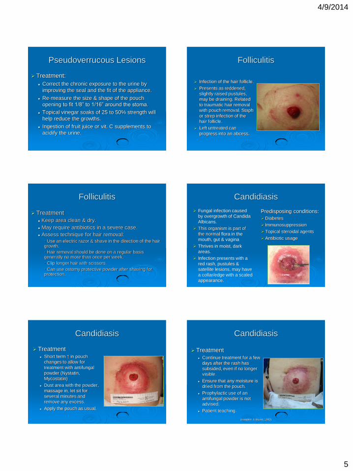
Folliculitis
Infection of the hair follicle.
Presents as reddened,
slightly raised pustules,
may be draining. Related
to traumatic hair removal
with pouch removal. Staph
or strep infection of the
hair follicle.
Left untreated can
progress into an abcess.
Folliculitis
Treatment
Keep area clean & dry.
May require antibiotics in a severe case.
Assess technique for hair removal:
• Use an electric razor & shave in the direction of the hair growth.
• Hair removal should be done on a regular basis generally no more than once per week.
• Clip longer hair with scissors.
• Can use ostomy protective powder after shaving for protection.
Candidiasis
Predisposing conditions:
Diabetes
Immunosuppression
Topical steroidal agents
Antibiotic usage
Fungal infection caused
by overgrowth of Candida
Albicans.
This organism is part of
the normal flora in the
mouth, gut & vagina
Thrives in moist, dark
areas.
Infection presents with a
red rash, pustules &
satellite lesions, may have
a collar/edge with a scaled
appearance.
Candidiasis
Treatment Short term ↑ in pouch
changes to allow for
treatment with antifungal
powder (Nystatin,
Mycostatin)
Dust area with the powder,
massage in, let sit for
several minutes and
remove any excess.
Apply the pouch as usual.
Candidiasis
Treatment Continue treatment for a few
days after the rash has
subsided, even if no longer
visible.
Ensure that any moisture is
dried from the pouch.
Prophylactic use of an
antifungal powder is not
advised.
Patient teaching.
(Hampton & Bryant, 1992)

4/9/2014
6
Caput Medusae
Associated with chronic
liver disease.
Presents as purplish
discoloration of the
peristomal skin.
Visible dilated cutaneous
veins.
Peristomal bleeding may
occur & can be severe.
Caput Medusae
Identify and manage the underlying disease.
Gentle pouch removal and care to the area, no
rubbing.
Cautious use of additional products such as
convexity, an ostomy belt or extended wear
adhesives.
Instruct the patient how to manage bleeding, apply
constant firm pressure X 10 mins, no peeking, if
still bleeding then seek medical attention, may
need cautery.
Pyoderma Gangrenosum
A painful ulcerating skin
disorder.
Sometimes associated
with inflammatory bowel
disease or cancer.
The ulcers may be
triggered by an injury to
the skin, such as trauma
from a tight appliance or
surgery.
Pyoderma Gangrenosum
The ulcers may be shallow or deep
They have a bluish undermined and ragged edge
Surrounding skin tends to be red and swollen
Healing ulcers result in cribriform scars (these
appear to have small holes like a seive).
Pyoderma Gangrenosum
Treatment
Topical steroidal agent
May require systemic steroid therapy
OD pouch changes for wound care
Reduce or eliminate convexity
Pain management
Time
Peristomal Fistula
A complication of
Crohn’s disease
Up to 10 % of those
with an ileostomy due
to Crohn’s will develop
a peristomal fistula.
Include the fistula in
the pouch opening.
Medical treatment for
IBD.
(Kann, 2008)
4/9/2014
7
Convexity How it works
The outward curve of the convex shape begins at the
opening of the barrier and extends to the edge of the
barrier.
The outward curve of the convex
barrier puts pressure around
the stoma.
This pressure will help the stoma
protrude into the pouch.
The effluent empties into the
pouch rather than under it.
The outward curve helps to flatten
out creases, indents or hollows.
Parastomal Hernia
Protrusion of loops of
bowel through a fascial
defect in the abdominal
wall near or around the
stoma.
Presents as a bulge on the
abdomen.
May asymptomatic.
May have a fullness,
pressure or dragging
feeling in the abdomen.
Parastomal Hernia
The hernia is noticeable
when the patient is in a
sitting or standing position.
Disappears in the supine
position.
Have the patient lift their
head, the hernia should
“pop up”.
Risk of incarcerated
hernia, loops of bowel
become trapped, leading
to bowel ischemia
Parastomal Hernia
This uneven contour
presents the patient with a
pouching challenge.
Difficult to maintain a reliable
seal.
Thin and stretched
peristomal skin at risk for
breakdown with pouch
leakages.
Increased pressure in the
peristomal area due to force
of hernia pushing out.
Parastomal Hernia
Prevention through maintenance of abdominal muscle
tone and the protection of the abdominal wall in the post
op recovery phase. No heavy lifting.
Use of a support belt or binder that fits around the
ostomy.
Flexible pouching system that can conform to the bulge.
Avoid constipation.
Stop irrigating if doing colostomy irrigation.
May require surgical repair.
Can recur.
Pressure Ulcer
Increased pressure in
the peristomal area
related to:
Use of convexity
Use of an ostomy belt
Presence of a hernia

4/9/2014
8
Pressure Ulcer
Treatment:
Remove cause
Re-evaluate pouch
Patient education
Gentle pouch removal
Pain management
Provide basic wound care
Pressure Ulcer
Cover the ulcer with a thin hydrocolloid.
Apply the pouch over the area.
Temporary ↑ in pouch change to allow wound
care
Fill the defect with
stomahesive powder
Use no sting barrier film
for peri-ulcer protection
Fill the ulcer with alginate
as ulcer exudate and
depth dictate.
Pressure Ulcer
1.
2.
Pressure Ulcer
4. 5.
Trauma
Other sources of trauma
Direct contact/trauma to the area
Skin stripping
• Rough or aggressive pouch removal.
• Abrasive cleansing technique.
• The addition of extra tape – whatever goes on, has
to come off.
• Too frequent pouch changes with an extended wear
barrier.
Treatment
Eliminate the cause, provide wound care
Patient education
Prevention of Peristomal
Complications Assess the fit of the pouch.
Re-assess stoma and peristomal area on a
regular basis or anytime something changes for
the person.
Cautious use of convexity and accessory items.
Avoid use of additional tape.
Avoid picking off old filler or paste, just wash over.
Patient education.
Avoid “washing out” the pouch, adds extra
moisture to the area.

4/9/2014
9
Thank you!
References
Alvey, B. & Beck, D. (2008). Peristomal Dermatology.
Clinics in Colon and rectal surgery, Vol. 21 No. 1.
Hampton, B. G. & Bryant, R. A. (1992). Ostomies and
continent diversions-Nursing Management. Mosby Inc. St.
Louis, Missouri.
Jordan, R. & Christian, M. (2013). Understanding peristomal
skin complications. Wound care advisor, Vol. 2, No. 3.
Kann,B. R. (2008). Early stomal complications. Clinics in
colon and rectal surgery. Vol. 21, No. 1
Lyon, C. C. & Smith, A. (2010). Abdominal stomas and their
skin disorders. An atlas of diagnosis and management.
Informa Healthcare. London.