Peripheral Neuroplasticity and Pain - AAU
26
Transcript of Peripheral Neuroplasticity and Pain - AAU


Peripheral Neuroplasticity and Pain
Brian E Cairns, PhD, DrMed, ACPR
April 28th, 2017

What is peripheral neuroplasticity?
Neuroplasticity refers to the ability of neurons to form new
connections to compensate for injury and disease and to adjust
their activities in response to new situations or to changes in their
environment.
Peripheral neuroplasticity is the compensation by primary afferent
fibers to injury or disease and can involve alterations in signalling
pathways, protein expression and interactions with autonomic
efferent fibers leading to prolonged functional, phenotypic and
structural changes.

Nociceptors can undergo neuroplastic changes that categorized
as
sensitization
desensitization
priming
Nociceptor sensitivity is modulated by
action of a great many neuroactive
substances (e.g. inflammatory soup)
that act on a wide variety of GPCRs
and ion channels expressed by
nociceptors.
Downstream signalling mechanisms
and the targets they alter to modifiy
nociceptor responsiveness are a focus
of active research. Gold & Gebhart 2010

Peripheral Sensitization and Pain
Complete Freund Adjuvant (CFA) induced inflammation results in
a persistant irregular low frequency discharge < 1 Hz in up to 25%
of nociceptors
Lowered activation thresholds
Xiao &Bennett 2007

Consequences of peripheral sensitization:
Ongoing (spontaneous) discharge = Spontaneous pain
Decreased activation threshold = Allodynia
Increased action potential firing & after discharges =
Hyperalgesia
Mechanisms:
Neurogenic inflammation
Alteration of receptor function
Phenotypic switch
Sensory-autonomic interaction
Priming

Neurogenic Inflammation
release of inflammatory
mediators causes vasodilation,
plasma leakage and mast cell
degranulation within the dura
mater
activation and sensitization of
dural afferent fibers
calcitonin gene-related peptide (CGRP) mediates meningeal
vasodilatation
substance P (SP) and neurokinin A (NKA) - plasma extravasation
of the dura mater
Prostaglandins, bradykinin, glutamate etc sensitize nociceptors
J Headache Pain (2012) 13:103–111

Alteration of Receptor Function
Nerve Growth Factor (NGF) released upon injury to peripheral
nerves
rapidly induces peripheral sensitization in humans and rats
Svensson et al 2008, 2010

Alteration of Receptor Function
Phosphorylation of TrpV1 and NMDA channels results in slowed
desensitization and increased current which increases nociceptor
responsiveness to heat and mechanical stimulation.
Chao 2003
NaV1.8/1.9
NR2B

Phenotypic switch
low threshold afferents acquire pain-provoking properties
after inflammation or nerve injury, Ab fibers start firing
spontaneously and selective activation of these fibers causes
after-discharges in nociceptive spinal cord neurons.
injury also increases the number of ganglion neurons that begin
to express neuropeptides (substance P, BDNF, NPY and CGRP)
Neuman et al 1996
NK1 antagonist
Substance P expression in DRG

Phenotypic switch (muscle afferents)
Intramuscular injection of NGF increases the expression of
peripheral NMDA receptors in larger diameter neurons.
Wong et al 2014
the number of afferent fibers expressing
substance P and CGRP is increased.
This change is greater in females than in
males.

Autonomic Sensory Interactions
4 weeks after mental nerve ligation, extensive sprouting of
sympathetic (DBH) and parasympathetic (VAChT) into skin
territory innervated by the damaged nerve.
Grelik et al 2005
Autonomic fibers come into close contact with sensory fibers
(CGRP positive), suggesting the possibility of an interaction.

Hyperalgesic Priming
Lowered threshold for sensitization
produced by an initial priming stimulus
(that is no longer altering function).
Carrageenan, NGF or GDNF induce
cutaneous mechanical sensitization.
after the end of the mechanical
sensitization, an injection of PGE2
produces up to 24 hours of mechanical
hyperalgesia than in animals give only
control injection.
GDNF-Induced Hyperalgesic
Priming
Ferrari et al 2010.

Hyperalgesic Priming
also shifts the dose response
relationship for PGE2-induced
hyperalgesia to the left – and 5-HT
adenosine
PGE2 normally activates the protein
kinase A (PKA) pathway in afferent
terminals.
Priming alters this receptor signalling
cascade (e.g. from inhibitory to
excitatory) to also recruit protein
kinase C epsilon (PKCe)
Is this a model of re-injury pain or
stress-induced pain?
Rechling and Levine 2009

Ectopic Action Potential Generation and Peripheral Neuroplasticity
There are normally no synapses in sensory ganglia (except for the
mesecephalic nucleus), but ganglion neurons and terminal
endings have many of the same receptors as central neurons.
Initiation of action potentials from a part of the primary afferent
fiber other than the terminal ending of the afferent fiber, e.g. from
a neuroma
Action potentials can be initiated along axons and by
depolarization of sensory ganglion neurons. This can be a
consequence of altered receptor expression or interactions
between the ganglion neuron and its supporting glial cells or
immune cells.
The brain interprets these signals as originating in the tissue that
the afferent fiber innervates, despite the fact that they are not
generated by a change in the tissue.

Sensory Ganglia:
Satellite Glial Cells surround monopolar neurons of sensory fibers
in ganglia and are similar to astrocytes
connected by gap junctions,
which allow communication
between adjacent cells
soma of sensory afferent fibers
can release neurotransmitters that
affect the function of SGCs
SGCs may release substances
which affect the activity of the
soma as well

Trigeminal Ganglion
In vitro, trigeminal ganglion neurons release glutamate.
Do SGCs store glutamate (like astrocytes)? Can activated SGCs
release glutamate? What would the consequence of elevated
glutamate be on the activity of ganglion neurons?
mGluR
NR
GluR
muscle brain
Glutamate
Antidromic Orthodromic
Ganglion neuron
Satellite Glial Cells
Trigeminal Ganglion

In the trigeminal
ganglion satellite glial
cells, which express
glutamine synthetase,
surround trigeminal
ganglion neurons.
Satellite glial cells and
trigeminal ganglion
neurons express
excitatory amino acid
transporters (EAATs)
and contain glutamate.
Trigeminal Satellite Glial Cells
Neuroscience 256 (2014) 23–35

Experimental Set-up
Masseter or
Temporalis
Muscle
Brainstem
Recording
Electrode
Stimulating
Electrode
Trigeminal
Ganglion
Trigeminal
Ganglion
V1
V2V3
Recording Electrode
Rostral Caudal
~ 2mm
Injection Needle
Neuroscience 256 (2014) 23–35

Glutamate
PBS 100 mM 250 mM 500 mM KCl
Cum
ula
tive
Dis
ch
arg
e (
spik
es)
0
10
20
30
40
50
60
Time (sec)
-600 0 600 1200
Dis
ch
arg
e (
Hz)
0
5
10
15
20
25
30
Intraganglionic injection
of glutamate (3 ml) into
the trigeminal ganglion
evokes discharge
Discharge increases
with increasing
glutamate
concentration
Increases in intraganglionic glutamate concentration excite
trigeminal ganglion neurons
Neuroscience 256 (2014) 23–35

Me
ch
an
ica
l T
hre
sh
old
(g)
*
*
- 31% - 32% - 9%
0
10
20
30
40
50
60
B Glu B Glu & APV
B Glu & TFB - TBOA
Me
ch
an
ica
l T
hre
sh
old
(g)
*
*
- 31% - 32% - 9%
0
10
20
30
40
50
60
B Glu B Glu & APV
B Glu & TFB - TBOA
Reproducible discharges can be evoked by intraganglionic
injection of glutamate and increased by EAAT inhibition.
Mechanical sensitization induced by intra-ganglionic injection of
glutamate
Discharge and sensitization attenuated by the NMDA receptor
antagonist APV
Glu
& TFB-
TBOA
Cu
mu
lative
Dis
ch
arg
e (
sp
ike
s)
*
0
20
40
60
80
100 *
Glu Glu
& APV
Glu Glu Glu

Dis
ch
arg
e (
Hz)
0
5
10
15
MT
(g)
0
25
50
75
100
10 min Glu Glu
Lidocaine 2%
0.0002
0.0000
-0.0002
vol
t
Spik
e A
c
5
6.724 6.726 6.728 6.730 6.732
s
0.0004
0.0002
0.0000
-0.0002
vol
t
Spik
e A
c
5
605.280 605.282 605.284 605.286 605.288
s
0.0002
0.0000
-0.0002
vol
t
Spik
e A
c
5
3966.004 3966.006 3966.008 3966.010 3966.012
s
0.0004
0.0002
0.0000
-0.0002
vol
t
Spik
e A
c
5
2405.668 2405.670 2405.672 2405.674 2405.676
s
0.0002
0.0000
-0.0002
vol
t
Spik
e A
c
5
23.164 23.166 23.168 23.170 23.172 23.174
s
200 mV
2 ms
Pre-lidocaine 10 min 40 min 60 min Recovery (85 min)
# #
Application of lidocaine to the brainstem blocked afferent input
but had no effect on the ability of intraganglionic injections of
glutamate to induce mechanical sensitization.
MT decreased 25%
Neuroscience 256 (2014) 23–35
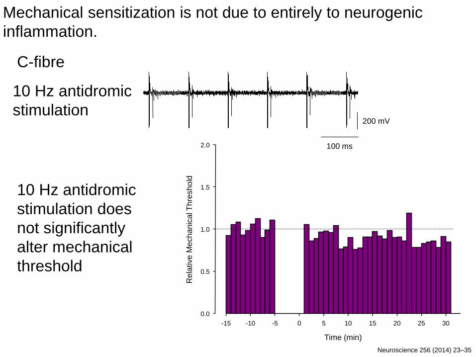
200 mV
100 ms
Time (min)
C-fibre
-15 -10 -5 0 5 10 15 20 25 30
Rela
tive
Me
ch
an
ical T
hre
sh
old
0.0
0.5
1.0
1.5
2.0
10 Hz antidromic
stimulation does
not significantly
alter mechanical
threshold
10 Hz antidromic
stimulation
Mechanical sensitization is not due to entirely to neurogenic
inflammation.
Neuroscience 256 (2014) 23–35

Intra-ganglionic Injection of Nitric Oxide (NO)
NO does not evoke action potential discharge but does induce
peripheral mechanical sensitization
NO causes SGCs to release PGE2. NO-induced sensitization is
blocked by NSAIDs and palmitoylethanolamide
Trigeminal Ganglion
muscle brain
Satellite
Glial
Cells
Antidromic Orthodromic
Ganglion neuron
PGE2
PGE2 NO
DETA/NO
BMT DETA/NO
Me
ch
an
ical T
hre
sh
old
(g
)
0
2
4
6
8
10
12
14
16
18
20
DETA/NO Contralateral
BMT DETA/NO
0
10
20
30
40
50
60
*
* *
Treatment Groups
Vehicle PEA 1 PEA 0.1 Ket 0.5
Re
lative
Me
ch
an
ica
l Th
resh
old
0.0
0.2
0.4
0.6
0.8
1.0
1.2
Cairns et al 2014

Summary
-SGCs contain glutamate and express EAATs, and play a role in
maintaining glutamate homeostasis in the ganglion.
-Artificial elevation of glutamate concentration in the ganglion
causes ectopic discharge that induces peripheral mechanical
sensitization.
-NO also induces peripheral mechanical sensitization, but by
inducing SGCs to release PGE2 onto ganglion neurons.
-A peripheral mechanism underlies mechanical sensitization, as
blockade of ascending sensory transmission does not affect the
induction of mechanical sensitization.
-Peripheral neuroplasticity can occur both at the terminal endings
and within sensory ganglia to induce long term changes in
nociceptive processing.

Aalborg University
•Parisa Gazerani
•Lars Arendt-Nielsen
University of British Columbia
•Xu-Dong Dong
•Hayes Wong
•Ujendra Kumar