Peripheral Nervous System_2
52
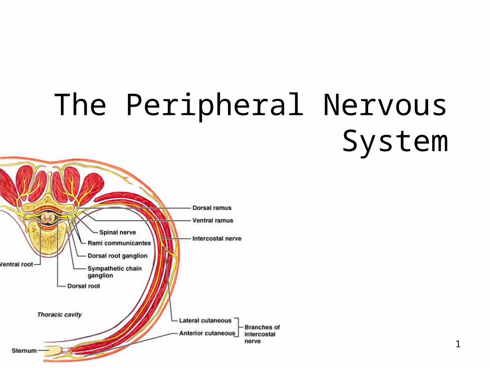
1 The Peripheral Nervous System
-
Upload
jendral-andi-wijaya-kusuma -
Category
Documents
-
view
226 -
download
0
description
m m m
Transcript of Peripheral Nervous System_2
1
The Peripheral Nervous System

2
Peripheral Nervous Systemways to categorize:
Motor or sensory General (widespread) or specialized (local) Somatic (outer tube) or visceral (inner tube)
3
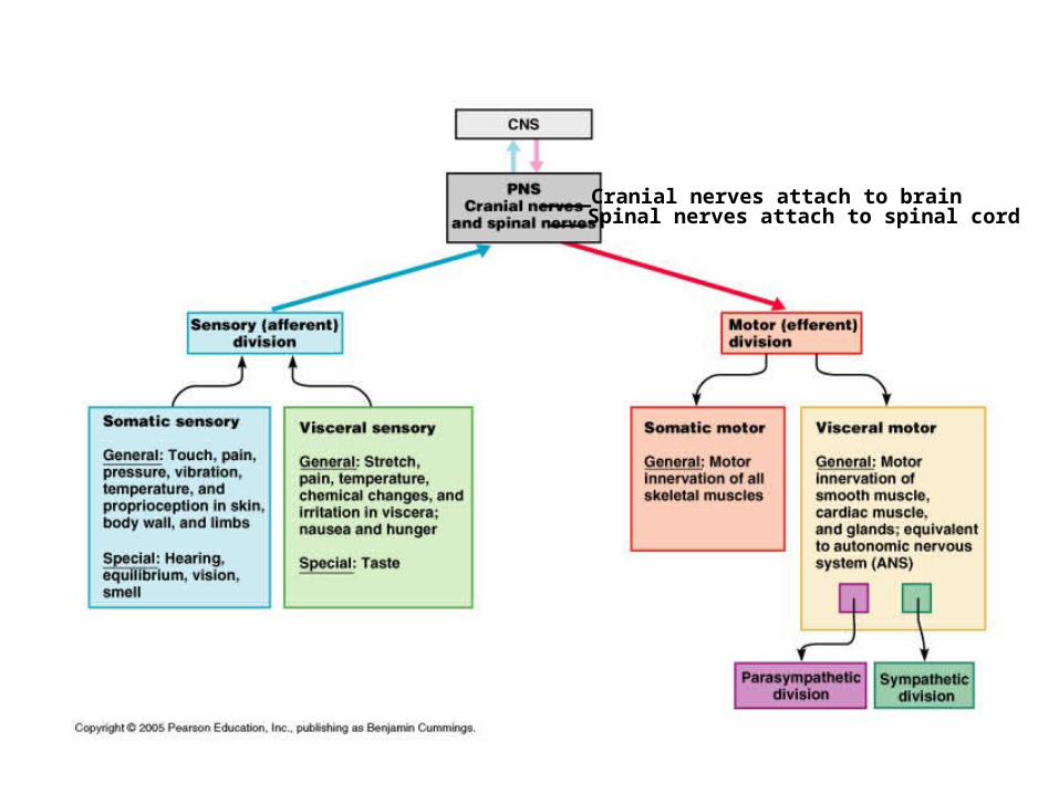
____Cranial nerves attach to brain___Spinal nerves attach to spinal cord
4
Peripheral sensory receptors
By location: Exteroceptors
Sensitive to stimuli arising from outside body
Interoceptors Or visceroreceptors, from internal viscera
Proprioceptors Monitor degree of stretch in skeletal muscles,
tendons, joints and ligaments
5
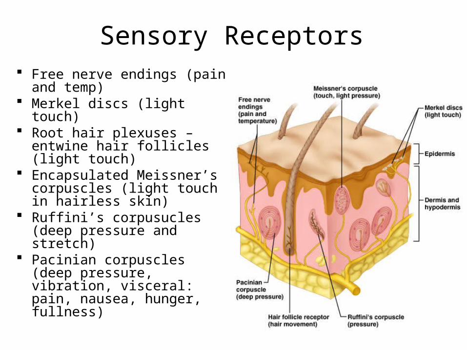
Sensory Receptors Free nerve endings (pain
and temp) Merkel discs (light touch) Root hair plexuses –
entwine hair follicles (light touch)
Encapsulated Meissner’s corpuscles (light touch in hairless skin)
Ruffini’s corpusucles (deep pressure and stretch)
Pacinian corpuscles (deep pressure, vibration, visceral: pain, nausea, hunger, fullness)
6
Proprioceptors
Skeletal muscles, joints, tendons, ligaments Degree of stretch, therefore information on body
movement: to cerebrum, cerebellum and spinal reflex arcs
Include: -Muscle spindles
-Golgi tendon organs
-Joint kinesthetic receptors
7
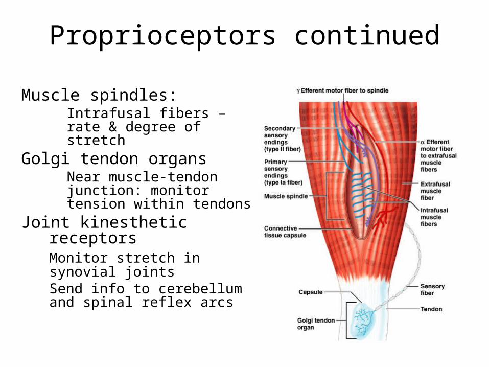
Proprioceptors continued
Muscle spindles:Intrafusal fibers – rate & degree of stretch
Golgi tendon organsNear muscle-tendon junction: monitor tension within tendons
Joint kinesthetic receptorsMonitor stretch in
synovial jointsSend info to
cerebellum and spinal reflex arcs
8
Peripheral motor endings
Innervation of skeletal muscle
Innervation of visceral muscles and glands

9
Motor axons innervate skeletal muscle fibers at neuromuscular junctions = motor end plates
Resemble nerve synapses between neurons, except for acetylcholinesterase:
breaks down acetylcholine so one twitch only
10
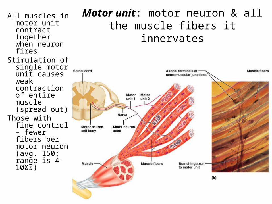
Motor unit: motor neuron & all the muscle fibers it innervates
All muscles in motor unit contract together when neuron fires
Stimulation of single motor unit causes weak contraction of entire muscle (spread out)
Those with fine control – fewer fibers per motor neuron (avg. 150: range is 4-100s)
11
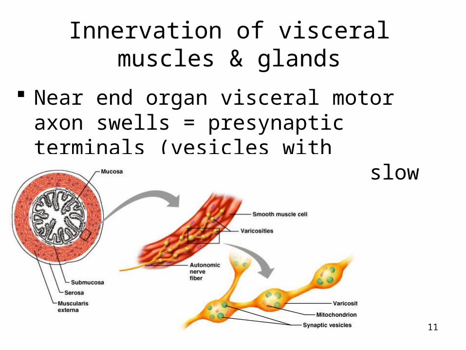
Innervation of visceral muscles & glands
Near end organ visceral motor axon swells = presynaptic terminals (vesicles with neurotransmitters): action slow (NT diffuses)
12
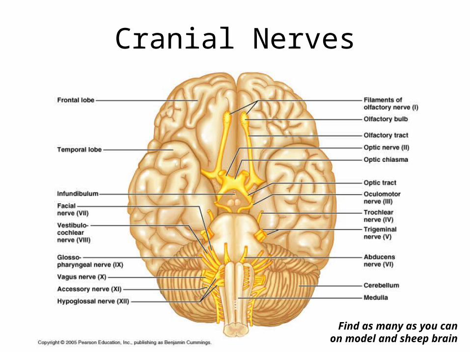
Cranial Nerves
Find as many as you can on model and sheep brain
13
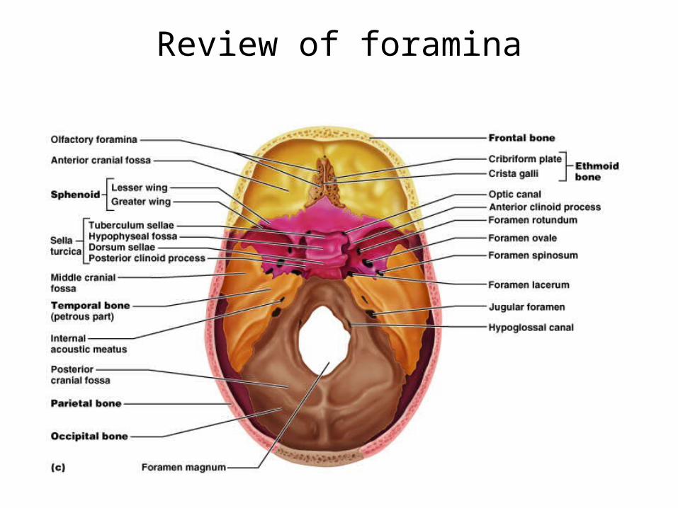
Review of foramina
14
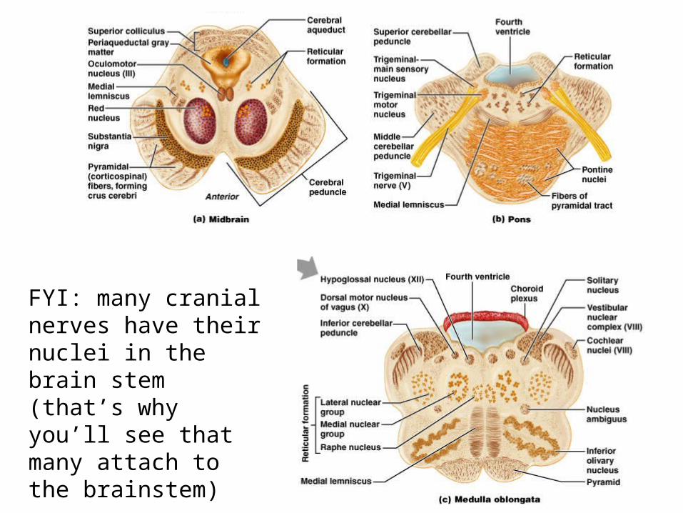
FYI: many cranial nerves have their nuclei in the brain stem (that’s why you’ll see that many attach to the brainstem)
15
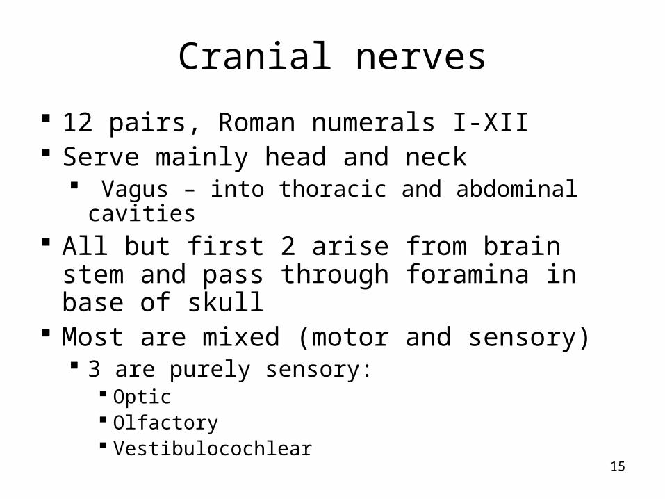
Cranial nerves
12 pairs, Roman numerals I-XII Serve mainly head and neck
Vagus – into thoracic and abdominal cavities All but first 2 arise from brain stem and
pass through foramina in base of skull Most are mixed (motor and sensory)
3 are purely sensory: Optic Olfactory Vestibulocochlear
16
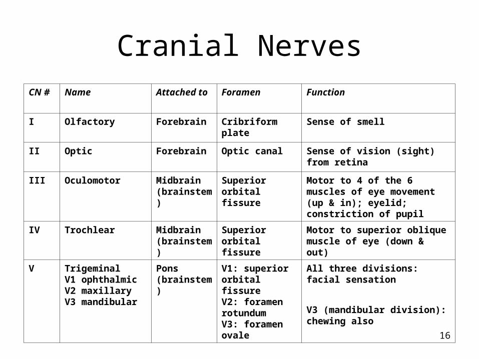
Cranial NervesCN # Name Attached to Foramen Function
I Olfactory Forebrain Cribriform plate Sense of smell
II Optic Forebrain Optic canal Sense of vision (sight) from retina
III Oculomotor Midbrain (brainstem)
Superior orbital fissure
Motor to 4 of the 6 muscles of eye movement (up & in); eyelid; constriction of pupil
IV Trochlear Midbrain (brainstem)
Superior orbital fissure
Motor to superior oblique muscle of eye (down & out)
V TrigeminalV1 ophthalmicV2 maxillaryV3 mandibular
Pons (brainstem)
V1: superior orbital fissureV2: foramen rotundumV3: foramen ovale
All three divisions: facial sensation
V3 (mandibular division): chewing also

17
VI Abducens Pons (brainstem)
Superior orbital fissure
Motor to lateral rectus muscle of eye (abducts outwards)
VII Facial Pons (brainstem)
Internal auditory canal
Facial expression (motor)Taste anterior 2/3 tongueSalivary & lacrimal glands (saliva and tears)
VIII Vestibulocochlear Pons (brainstem)
Internal auditorycanal
Equilibrium (vestibular)Hearing (cochlear)
IX Glossopharyngeal Medulla (brainstem)
Jugular foramen Taste & touch from posterior 1/3 tongue (sour, bitter); pharynx (throat) muscles of swallowing; parotid gland (saliva); senses carotid BP
X Vagus Medulla (brainstem)
Jugular foramen Senses aortic BP, slows heart rate, stimulates digestive organs; larynx (vocal cords), taste, swallowing
XI Accessory Medulla (brainstem)
Jugular foramen Sternocleidomastoid, trapezius, swallowing; part joins Vagus
XII Hypoglossal Medulla (brainstem)
Hypoglossal canal Innervation of tongue muscles
18
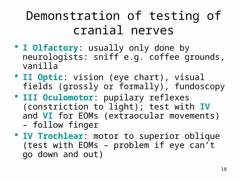
Demonstration of testing of cranial nerves
I Olfactory: usually only done by neurologists: sniff e.g. coffee grounds, vanilla
II Optic: vision (eye chart), visual fields (grossly or formally), fundoscopy
III Oculomotor: pupilary reflexes (constriction to light); test with IV and VI for EOMs (extraocular movements) – follow finger
IV Trochlear: motor to superior oblique (test with EOMs – problem if eye can’t go down and out)
19
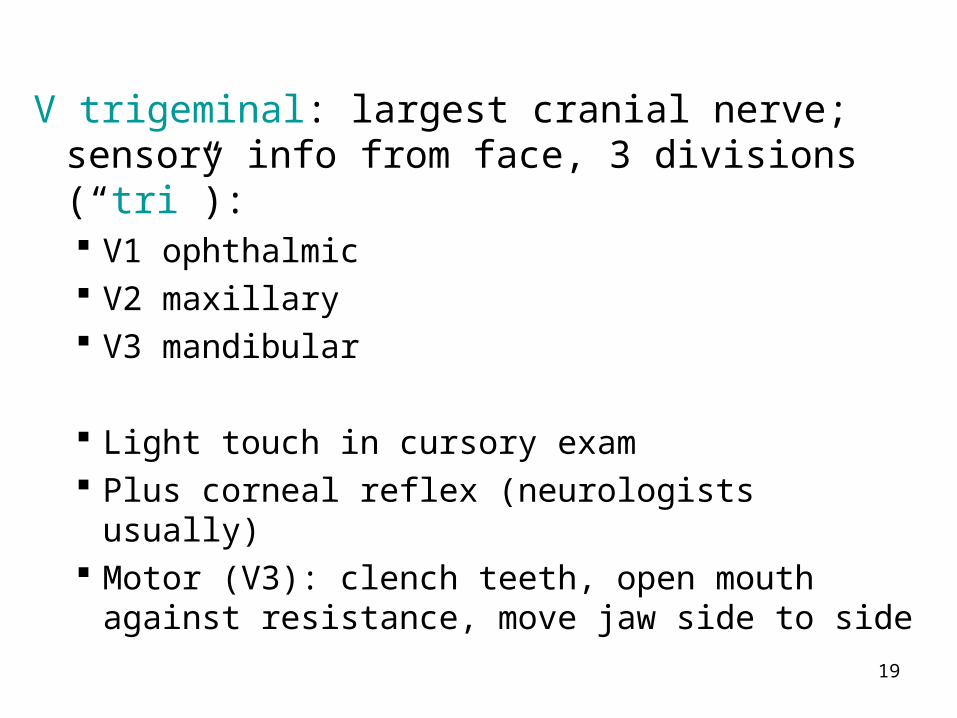
V trigeminal: largest cranial nerve; sensory info from face, 3 divisions (“tri”): V1 ophthalmic V2 maxillary V3 mandibular
Light touch in cursory exam Plus corneal reflex (neurologists usually) Motor (V3): clench teeth, open mouth against
resistance, move jaw side to side
20
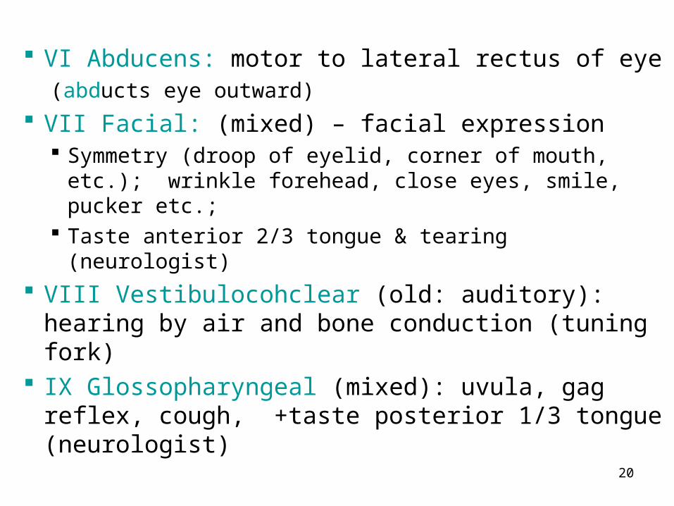
VI Abducens: motor to lateral rectus of eye(abducts eye outward)
VII Facial: (mixed) – facial expression Symmetry (droop of eyelid, corner of mouth, etc.);
wrinkle forehead, close eyes, smile, pucker etc.; Taste anterior 2/3 tongue & tearing (neurologist)
VIII Vestibulocohclear (old: auditory): hearing by air and bone conduction (tuning fork)
IX Glossopharyngeal (mixed): uvula, gag reflex, cough, +taste posterior 1/3 tongue (neurologist)
21
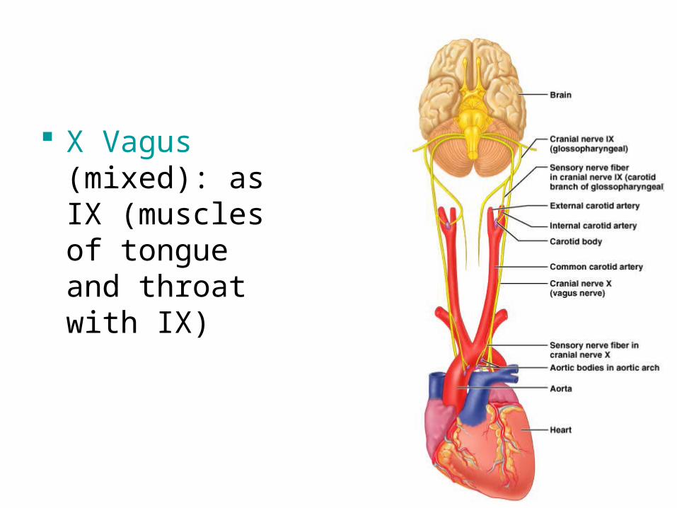
X Vagus (mixed): as IX (muscles of tongue and throat with IX)
22
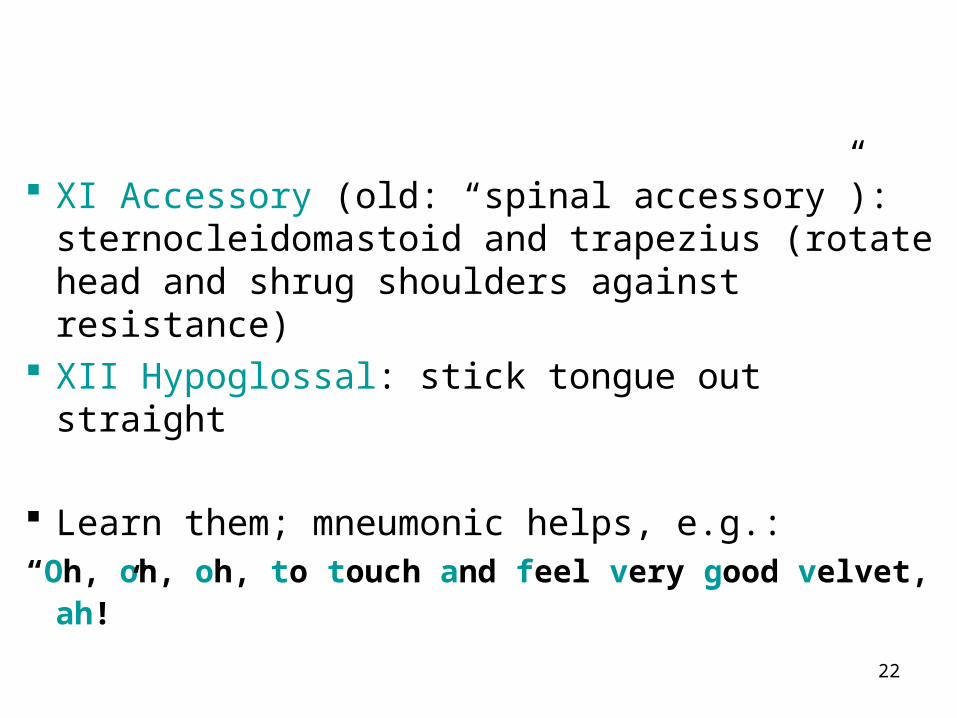
XI Accessory (old: “spinal accessory”): sternocleidomastoid and trapezius (rotate head and shrug shoulders against resistance)
XII Hypoglossal: stick tongue out straight
Learn them; mneumonic helps, e.g.:
“Oh, oh, oh, to touch and feel very good velvet, ah!”
23
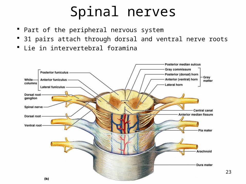
Spinal nerves Part of the peripheral nervous system 31 pairs attach through dorsal and ventral nerve roots Lie in intervertebral foramina
24
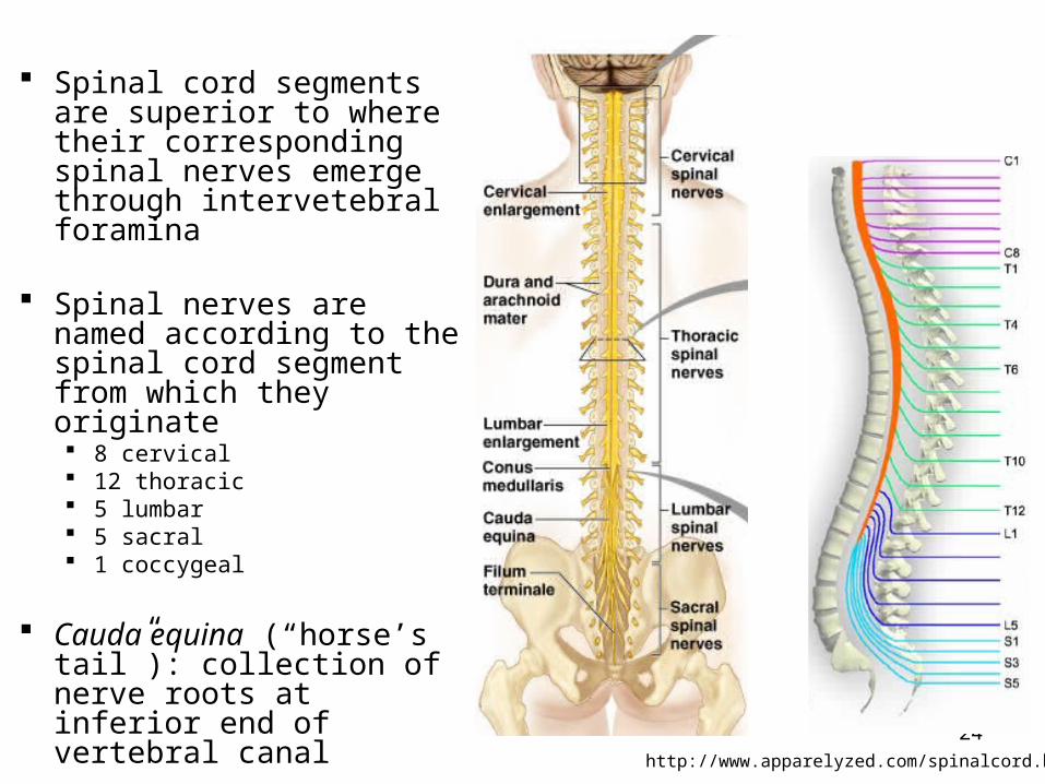
Spinal cord segments are superior to where their corresponding spinal nerves emerge through intervetebral foramina
Spinal nerves are named according to the spinal cord segment from which they originate 8 cervical 12 thoracic 5 lumbar 5 sacral 1 coccygeal
Cauda equina (“horse’s tail”): collection of nerve roots at inferior end of vertebral canal http://www.apparelyzed.com/spinalcord.html
25
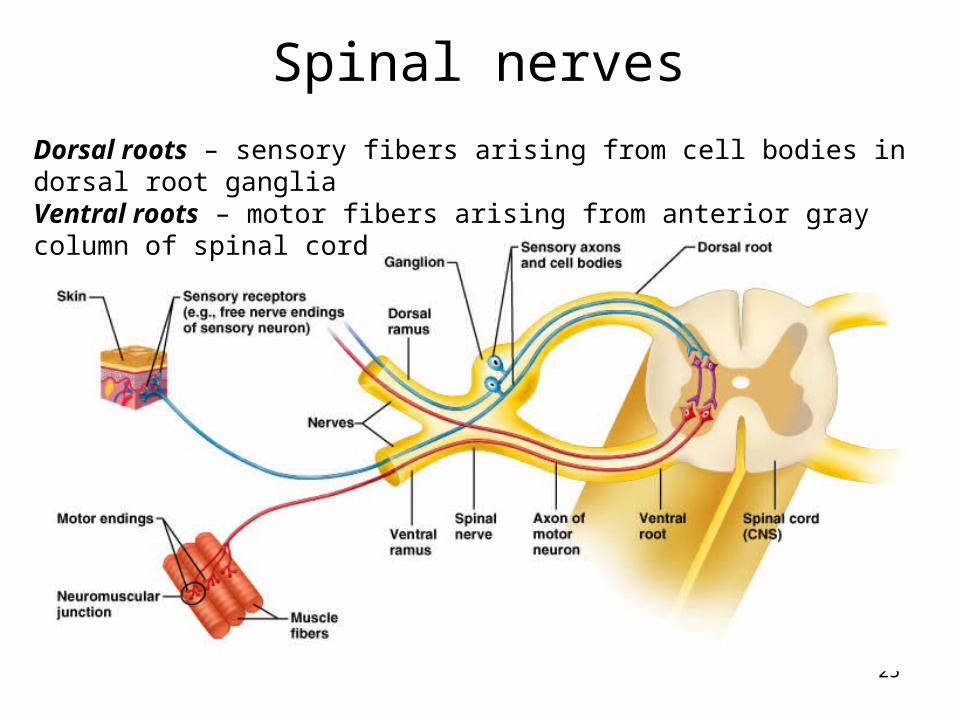
Spinal nerves
Dorsal roots – sensory fibers arising from cell bodies in dorsal root gangliaVentral roots – motor fibers arising from anterior gray column of spinal cord
26
Spinal nerves
Note: cervical spinal nerves exit from above the respective vertebra Spinal nerve root 1 from above C1 Spinal nerve root 2 from between C1 and C2, etc.
The remaining spinal nerve pairs emerge from the spinal cord below the same-numbered vertebra
Clinically, for example when referring to disc impingement, both levels of vertebra mentioned, e.g. C6-7 disc impinging on root 7
Symptoms usually indicate which level
27
Spinal nerves
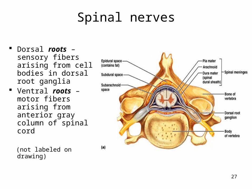
Dorsal roots – sensory fibers arising from cell bodies in dorsal root ganglia
Ventral roots – motor fibers arising from anterior gray column of spinal cord
(not labeled on drawing)
28
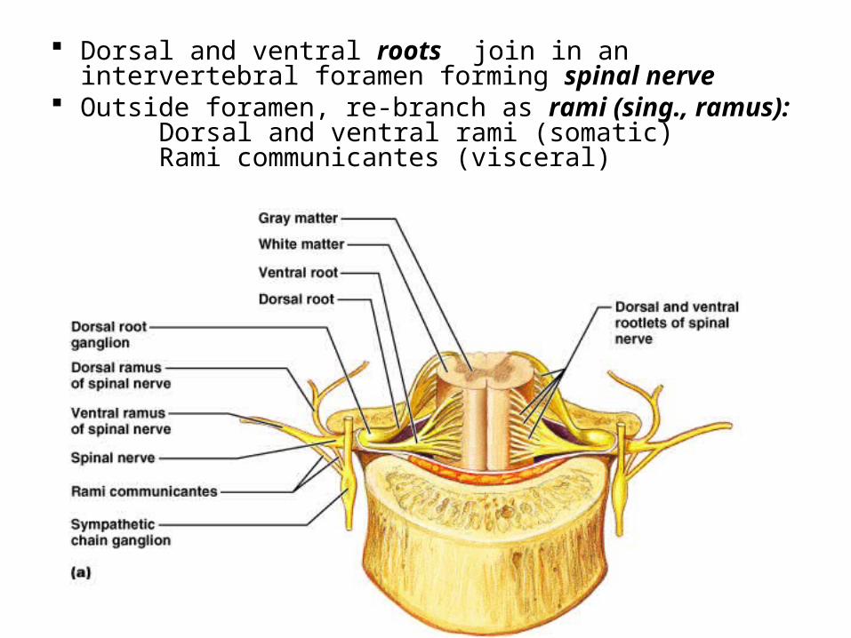
Dorsal and ventral roots join in an intervertebral foramen forming spinal nerve
Outside foramen, re-branch as rami (sing., ramus):Dorsal and ventral rami (somatic)Rami communicantes (visceral)
29
Dorsal rami serve the muscles and skin of the posterior trunk Back, from neck to sacrum, innervated in a
neatly segmented pattern: horizontal strip at same level as emergence from spinal cord
Ventral rami serve the muscles and skin of the lateral and anterior trunk In thorax only, a simple segmented pattern as
intercostal nerves Also serve the limbs
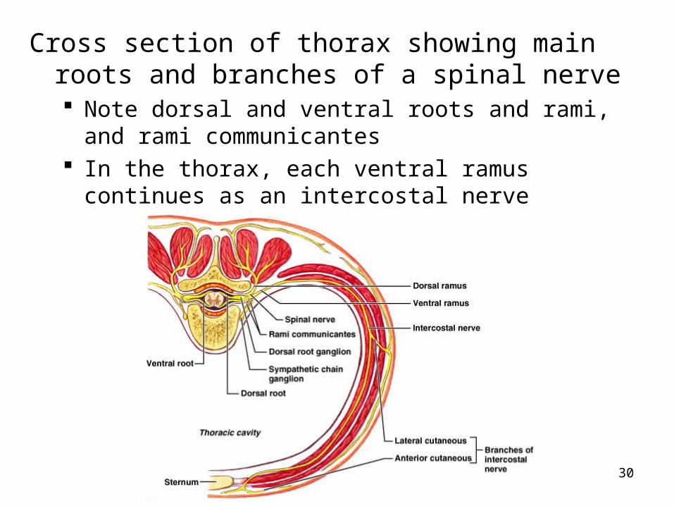
30
Cross section of thorax showing main roots and branches of a spinal nerve Note dorsal and ventral roots and rami, and rami
communicantes In the thorax, each ventral ramus continues as an
intercostal nerve
31
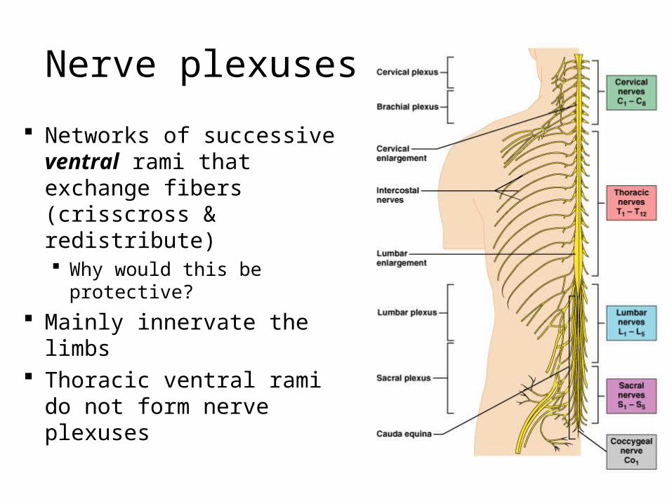
Nerve plexuses
Networks of successive ventral rami that exchange fibers (crisscross & redistribute) Why would this be
protective?
Mainly innervate the limbs
Thoracic ventral rami do not form nerve plexuses
32
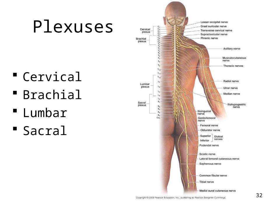
Plexuses
Cervical Brachial Lumbar Sacral
33
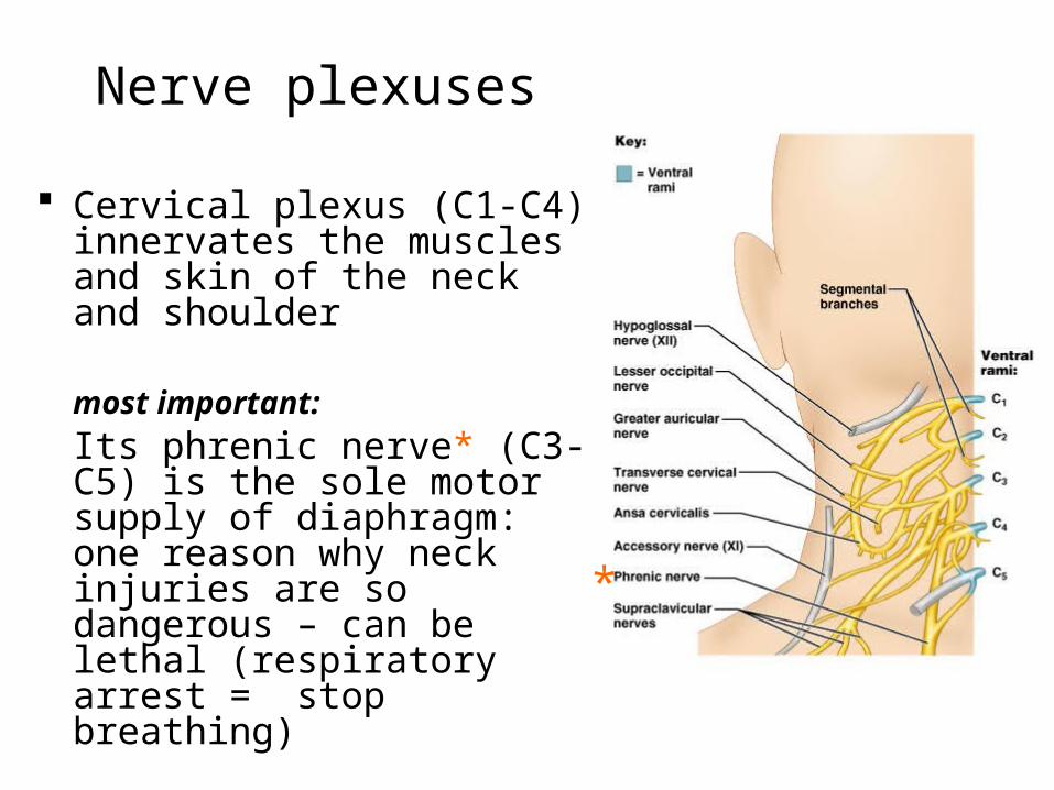
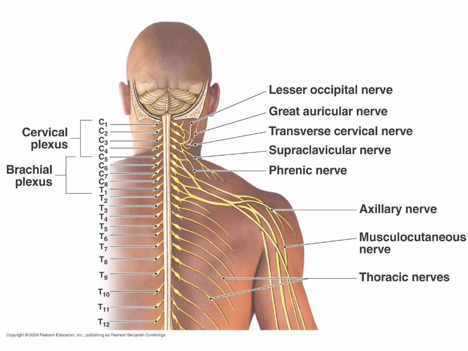
Nerve plexuses
Cervical plexus (C1-C4) innervates the muscles and skin of the neck and shoulder
most important:
Its phrenic nerve* (C3-C5) is the sole motor supply of diaphragm: one reason why neck injuries are so dangerous – can be lethal (respiratory arrest = stop breathing)
*
34
35
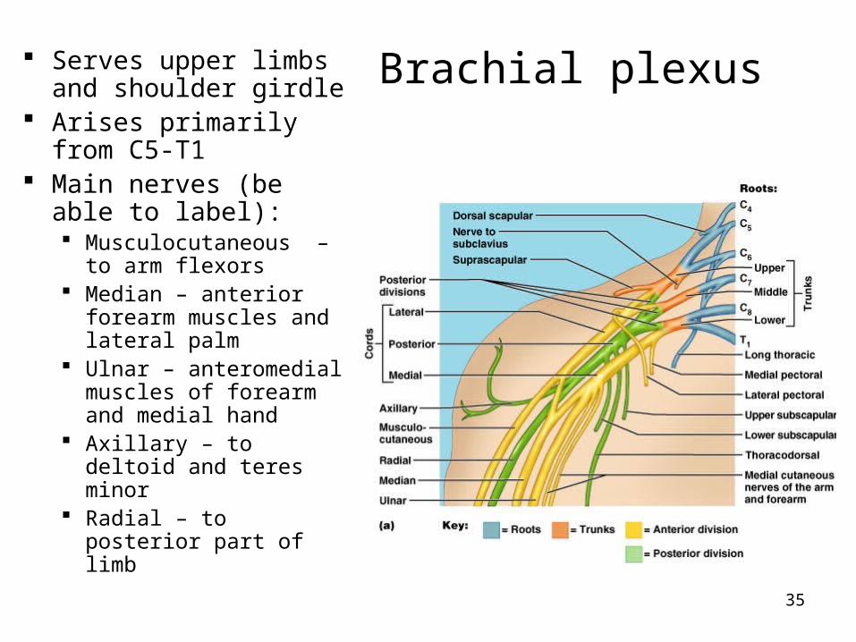
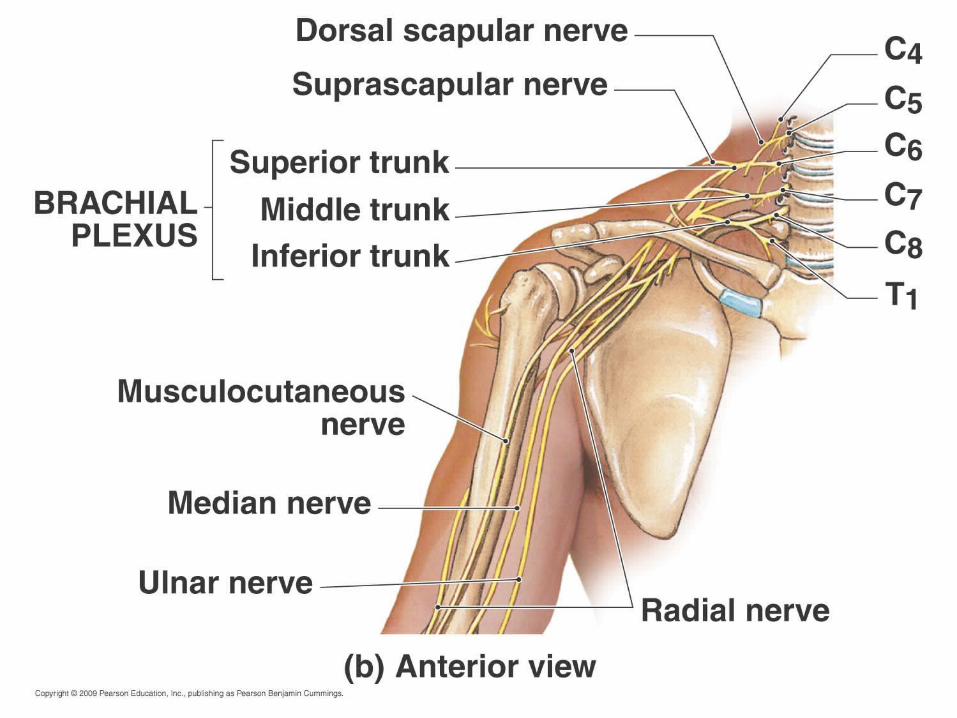
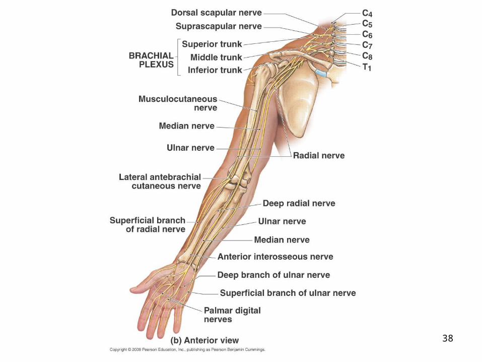
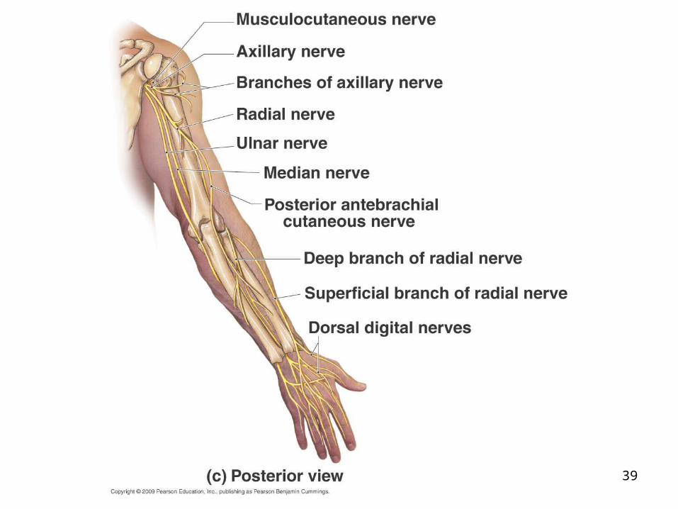
Brachial plexus Serves upper limbs and shoulder girdle
Arises primarily from C5-T1
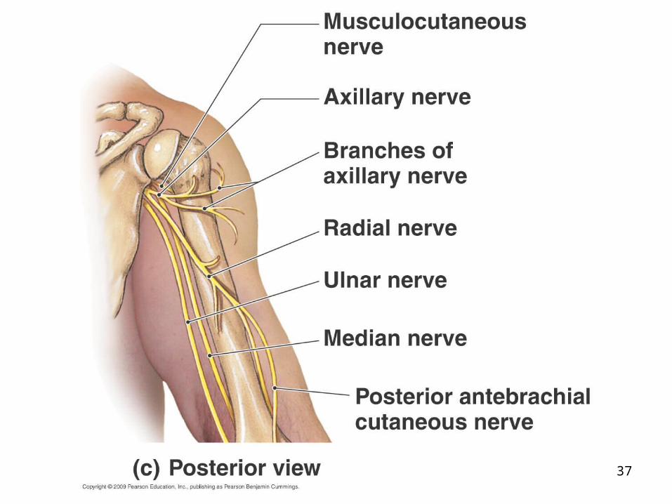
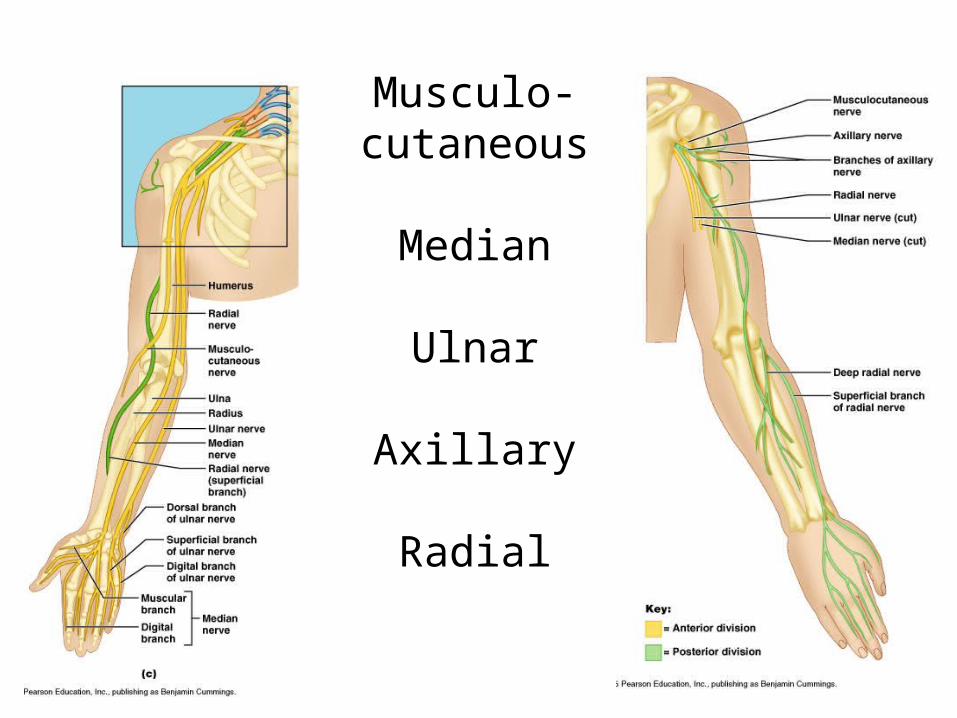
Main nerves (be able to label): Musculocutaneous – to
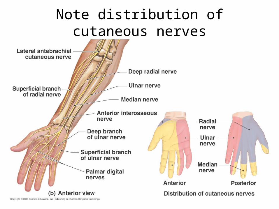
arm flexors Median – anterior
forearm muscles and lateral palm
Ulnar – anteromedial muscles of forearm and medial hand
Axillary – to deltoid and teres minor
Radial – to posterior part of limb
36
37
38
39
40
Musculo-cutaneous
Median
Ulnar
Axillary
Radial
41
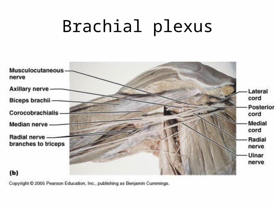
Brachial plexus
42
Note distribution of cutaneous nerves
43
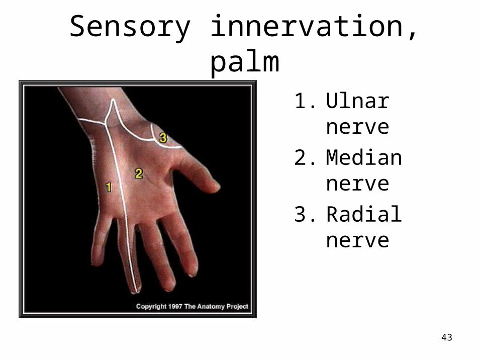
Sensory innervation, palm
1. Ulnar nerve
2. Median nerve
3. Radial nerve
44
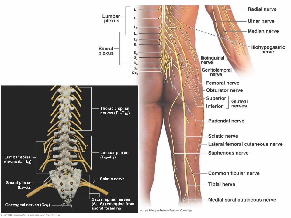
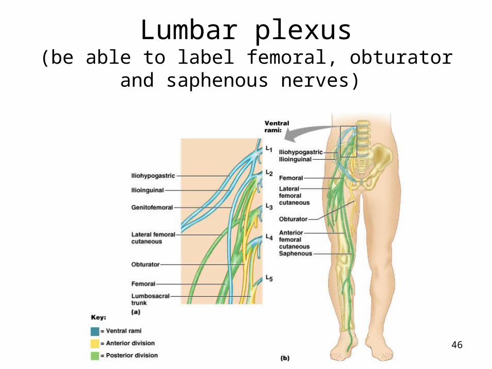
Lumbar plexus
L1-L4 Lies within the psoas major muscle Innervates anterior and medial muscles of
thigh through femoral and obturator nerves respectively
Femoral nerve also innervates skin on anterior thigh (including quads) and medial leg
45
46
Lumbar plexus(be able to label femoral, obturator and saphenous
nerves)

47
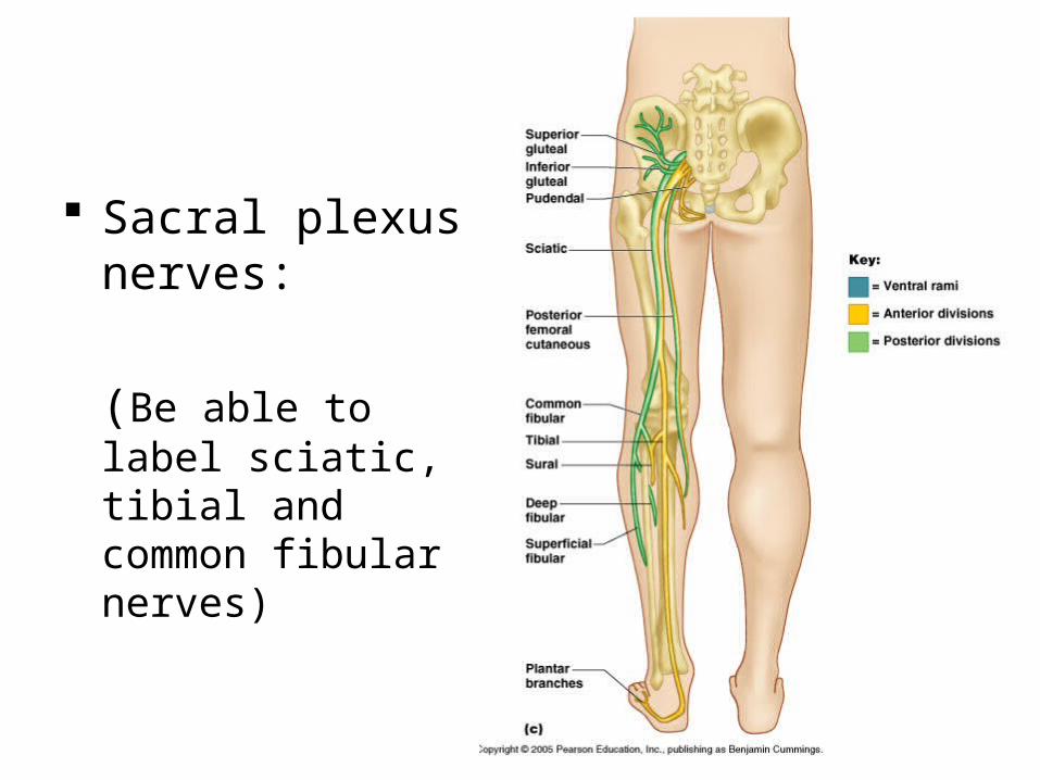
Sacral plexus L4-S4 Supplies muscles and
skin of posterior thigh and almost all of the leg
Main branch is the large sciatic nerve, which consists of: Tibial nerve – to most of
hamstrings, calf and sole
Common fibular nerve – to muscles of anterior and lateral leg and skin
Other branches supply pelvic girdle (gluteus muscles) and perineum (pudental nerve)
48
Sacral plexus nerves:
(Be able to label sciatic, tibial and common fibular nerves)
49
50
51
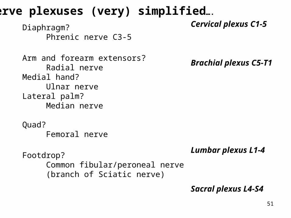
Diaphragm? Phrenic nerve C3-5
Arm and forearm extensors? Radial nerveMedial hand? Ulnar nerveLateral palm? Median nerve
Quad? Femoral nerve
Footdrop? Common fibular/peroneal nerve (branch of Sciatic nerve)
Cervical plexus C1-5
Brachial plexus C5-T1
Lumbar plexus L1-4
Sacral plexus L4-S4
Nerve plexuses (very) simplified….
52
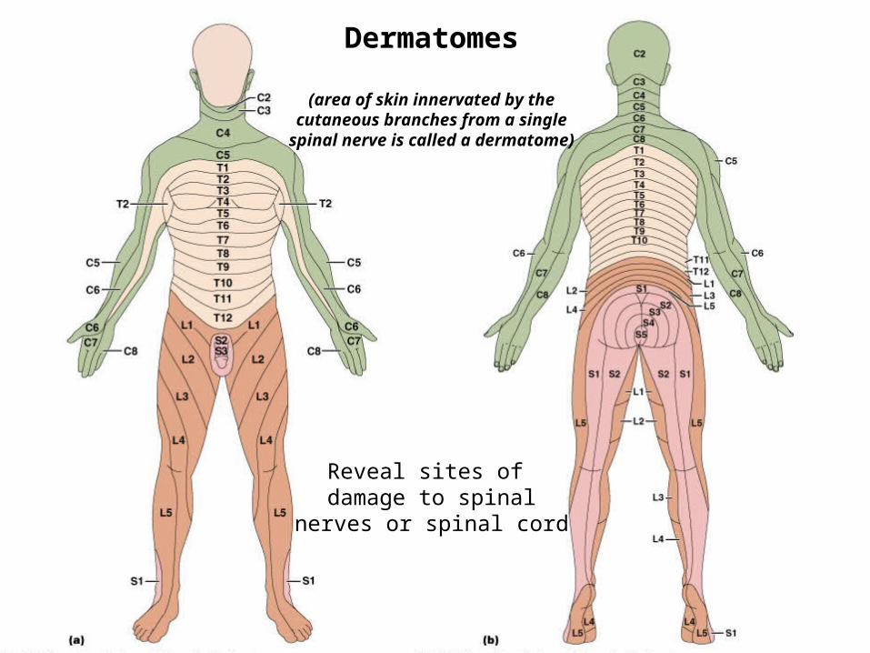
Dermatomes (innervation of skin) Dermatomes
(area of skin innervated by the cutaneous branches from a single
spinal nerve is called a dermatome)
Reveal sites of damage to spinal
nerves or spinal cord