Peripheral Nervous System. Human Anatomy and Physiology, 7e by Elaine Marieb & Katja Hoehn Copyright...
39
Peripheral Nervous System
-
Upload
prudence-barker -
Category
Documents
-
view
261 -
download
2
Transcript of Peripheral Nervous System. Human Anatomy and Physiology, 7e by Elaine Marieb & Katja Hoehn Copyright...
Peripheral Nervous System

Human Anatomy and Physiology, 7eby Elaine Marieb & Katja Hoehn
Copyright © 2007 Pearson Education, Inc.,publishing as Benjamin Cummings.
Place of the PNS in the structural organization of the nervous system
CNS
Sensory division Motor division
Autonomic nervous system
Sympathetic division
Parasympathetic division
PNS
Somatic nervous system
Human Anatomy and Physiology, 7eby Elaine Marieb & Katja Hoehn
Copyright © 2007 Pearson Education, Inc.,publishing as Benjamin Cummings.
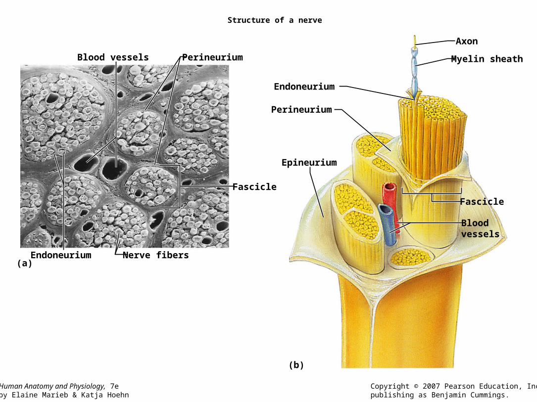
Structure of a nerve
(a)
(b)
Fascicle
Perineurium Blood vessels
Endoneurium Nerve fibers
Axon
Endoneurium
Perineurium
Epineurium
Myelin sheath
Bloodvessels
Fascicle

Spinal Nerves and Nerve Plexuses
• Characteristics:– Somatic sensation (conscious) and somatic motor control (voluntary
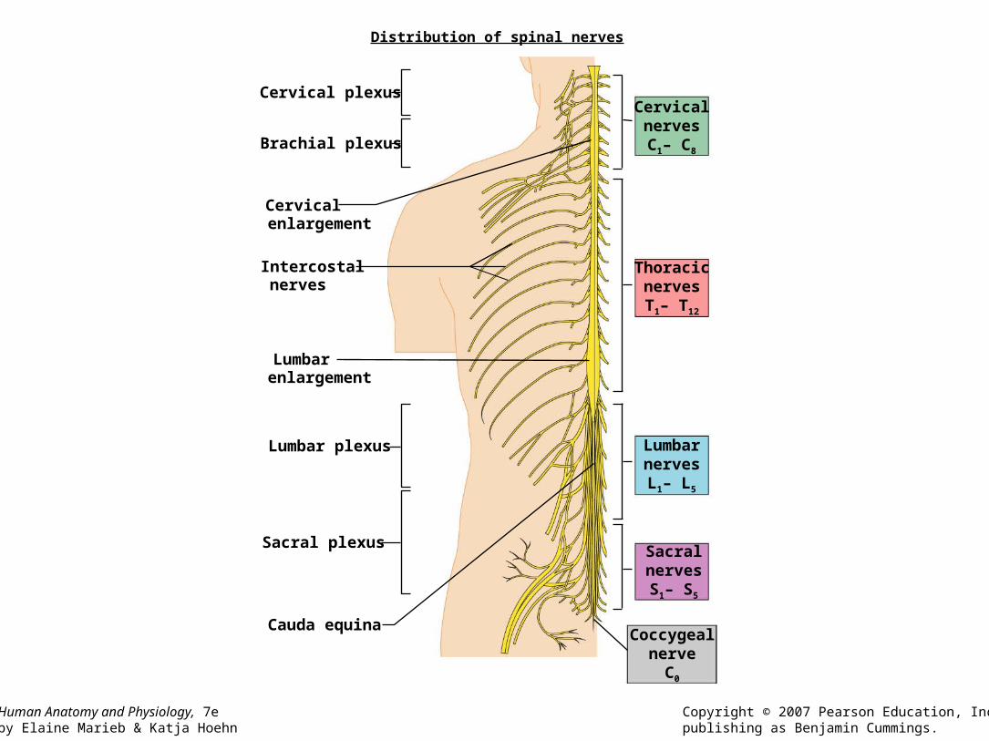
control) of skeletal muscles.– Includes cranial nerves: I, II, IV-VI, VIII, XI and XII.– Spinal nerves: 31
• Cervical: 8 (above C1, and below C1-C7)• Thoracic: 12 (below T1-T12)• Lumbar: 5 (below T1-T5)• Sacral: 5 ( below S1-S5)• Coccygeal: 1 exit coccyx
– Mixed nerves• Sensory • Motor
– Dorsal and ventral rami (nerve branches) plexuses (network of nerves)
Human Anatomy and Physiology, 7eby Elaine Marieb & Katja Hoehn
Copyright © 2007 Pearson Education, Inc.,publishing as Benjamin Cummings.
Distribution of spinal nerves
CervicalnervesC1– C8
ThoracicnervesT1– T12
LumbarnervesL1– L5
SacralnervesS1– S5
Coccygealnerve
C0
Cervical plexus
Intercostalnerves
Cervicalenlargement
Lumbarenlargement
Cauda equina
Brachial plexus
Lumbar plexus
Sacral plexus

Formation of spinal nerves and rami distribution
(b)
Dorsal ramus
Ventral ramus
Intercostal nerve
Lateral cutaneous
Anterior cutaneous
Spinal nerve
Rami communicantes
Dorsal root ganglion
Sympathetic trunk(chain) ganglion
Dorsal root
Ventral root
Sternum
Thoracic cavity
Branches ofintercostalnerve

The cervical plexus
Hypoglossalnerve (XII)
C1
C2
C3
C4
C5
Segmentalbranches
Lesser occipitalnerve
Greater auricularnerve
Ansa cervicalis
Phrenic nerve
Supraclavicularnerves
Accessory nerve (XI)
Transverse cutaneousnerve
Key:
= Ventral rami
Ventralrami:

The brachial plexus
(a)
= Roots
Key:
= Anterior division= Posterior division
= Trunks
Nerve tosubclavius
Suprascapular
Dorsal scapular
Posteriordivisions
Lateral
Cords
Axillary
Musculo-cutaneous
Radial
Median
Ulnar
Upper
Middle Trunks
Lower
C4
Roots:
C5
C6
C7
C8
T1
Upper subscapular
Lower subscapular
Thoracodorsal
Medial cutaneousnerves of the armand forearm
Long thoracic
Medial pectoral
Lateral pectoral
Posterior
Medial
Human Anatomy and Physiology, 7eby Elaine Marieb & Katja Hoehn
Copyright © 2007 Pearson Education, Inc.,publishing as Benjamin Cummings.
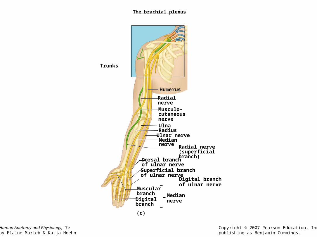
The brachial plexus
(c)
Mediannerve
Musculo-cutaneousnerve
Radialnerve
Humerus
Ulna
Ulnar nerveMediannerve
Radius
Radial nerve(superficialbranch)
Superficial branchof ulnar nerve
Dorsal branchof ulnar nerve
Digital branchof ulnar nerve
MuscularbranchDigitalbranch
Trunks
Human Anatomy and Physiology, 7eby Elaine Marieb & Katja Hoehn
Copyright © 2007 Pearson Education, Inc.,publishing as Benjamin Cummings.
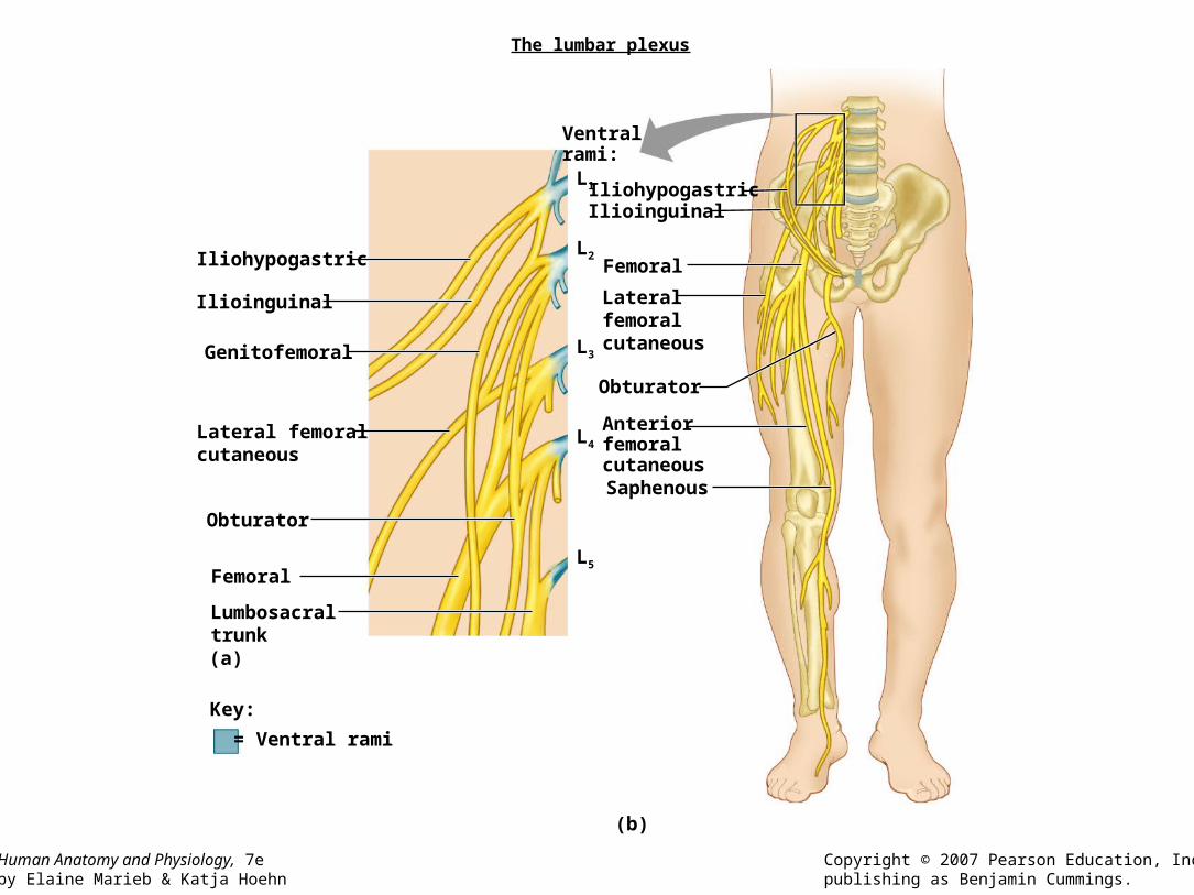
The lumbar plexus
(b)
(a)
Iliohypogastric
L1
L2
L3
L4
L5
Ilioinguinal
Genitofemoral
Lateral femoralcutaneous
Obturator
Femoral
Lumbosacraltrunk
Lateralfemoralcutaneous
AnteriorfemoralcutaneousSaphenous
Obturator
IliohypogastricIlioinguinal
Femoral
= Ventral rami
Key:
Ventral rami:

Human Anatomy and Physiology, 7eby Elaine Marieb & Katja Hoehn
Copyright © 2007 Pearson Education, Inc.,publishing as Benjamin Cummings.
The sacral plexus
(a)
(b)
Superiorgluteal
Lumbosacraltrunk
Inferiorgluteal
CommonfibularTibial
Posteriorfemoralcutaneous
Pudendal
Sciatic
Key:
Commonfibular
DeepfibularSuperficialfibular
Plantarbranches
TibialSural
Posteriorfemoralcutaneous
Pudendal
Sciatic
L4
L5
S1
S2
S3
S4
S5
C0
= Ventral rami
Ventralrami:
SuperiorglutealInferiorgluteal

• Activity 2– Identify spinal chord tracts
• Activity 3– Identify Major nerve plexuses and Peripheral
nerves
The Autonomic Nervous System(ANS)
Human Anatomy and Physiology, 7eby Elaine Marieb & Katja Hoehn
Copyright © 2007 Pearson Education, Inc.,publishing as Benjamin Cummings.
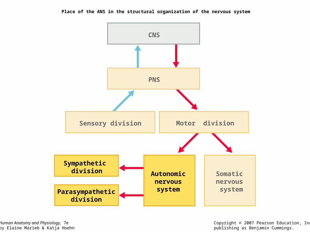
Place of the ANS in the structural organization of the nervous system
CNS
Sensory division Motor division
PNS
Autonomic nervous system
Sympathetic division
Parasympatheticdivision
Somatic nervous system

Characteristics of the ANS
• Regulates body function unconsciously.– Cardiac muscle, smooth muscle, glands.
• Consists of chains of 2 motor neurons.– Preganglion neuron: located in the CNS. – Ganglion neuron: synapses with pregalnglion, outside
the CNS and its axon synapses with the effector organ.
• Sympathetic (fight or flight) functions are antagonistic to the Parasympathetic (resting and digesting) functions.

Human Anatomy and Physiology, 7eby Elaine Marieb & Katja Hoehn
Copyright © 2007 Pearson Education, Inc.,publishing as Benjamin Cummings.
Comparison of somatic and autonomic nervous systems
Somatic nervous system Skeletal muscle
Centralnervous system Peripheral nervous system Effector organs
Acetylcholine
Smoothmuscle(e.g., in gut)
Acetylcholine
Ganglion
Adrenal medulla
GlandsBloodvessel
Cardiacmuscle
Sympatheticdivision
Autonomicnervoussystem
Acetylcholine
Acetylcholine
Norepinephrine
Epinephrine andnorepinephrine
Ganglion
Para-sympathetic
division
= Preganglionic axons (sympathetic)
= Postganglionic axons (sympathetic)
= Myelination = Preganglionic axons (parasympathetic)
= Postganglionic axons (parasympathetic)
Key:

Human Anatomy and Physiology, 7eby Elaine Marieb & Katja Hoehn
Copyright © 2007 Pearson Education, Inc.,publishing as Benjamin Cummings.
Overview of the subdivisions of the ANS
Salivaryglands
Eye
Skin*
Heart
Lungs
Liverand gall-bladder
Pancreas
Eye
Lungs
Bladder
Liver and gall-bladder
Pancreas
Stomach
Cervical
Sympatheticganglia
Cranial
Lumbar
Thoracic
Genitals
Heart
Salivaryglands
Stomach
Bladder
Adrenalgland
Parasympathetic Sympathetic
Sacral
Brain stem
L1
T1
Genitals

Human Anatomy and Physiology, 7eby Elaine Marieb & Katja Hoehn
Copyright © 2007 Pearson Education, Inc.,publishing as Benjamin Cummings.
Parasympathetic (craniosacral) division of the ANS
EyeLacrimalgland
Nasalmucosa
Ciliary ganglion
Pterygopalatineganglion
Submandibularganglion
Submandibularand sublingualglands
CN III
CN VIICN IXCN X
Otic ganglion
Parotid gland
Heart
Lung
Liver andgallbladder
Stomach
Pancreas
Urinary bladder and ureters
Smallintestine
Large intestine
S2
Pelvicsplanchnicnerves
Genitalia (penis, clitoris, and vagina)
Rectum
Celiacplexus
Inferiorhypogastric plexus
Cardiac and pulmonary plexuses
S4
III: Oculomotor
VII: Facial
IX: GlossopharyngealX: Vagus

Characteristics of the Parasympathetic Division
• Preganglion axons are located in the cranial nerves in the immediate area to be stimulated.
• Terminal or intramural ganglion, which emits a short axon to the organ, synapse with the preganglion ganglion.
• Sacral region ganglions synapse to pelvic splanchnic nerves that travel to the pelvic cavity.
Human Anatomy and Physiology, 7eby Elaine Marieb & Katja Hoehn
Copyright © 2007 Pearson Education, Inc.,publishing as Benjamin Cummings.
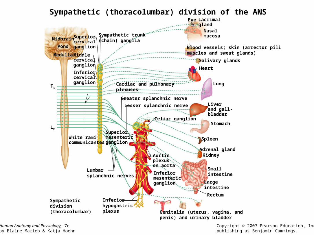
Sympathetic (thoracolumbar) division of the ANS
Midbrain Superiorcervicalganglion
Middlecervicalganglion
Inferiorcervicalganglion
Sympathetic trunk(chain) ganglia
Pons
Medulla
L2
T1
White ramicommunicantes
Liverand gall-bladder
Stomach
Spleen
KidneyAdrenal gland
Smallintestine
Largeintestine
Genitalia (uterus, vagina, andpenis) and urinary bladder
Celiac ganglion
Aorticplexuson aorta
Inferiormesenteric ganglion
Lesser splanchnic nerve
Greater splanchnic nerve
Superiormesentericganglion
Inferiorhypogastricplexus
Lumbarsplanchnic nerves
Eye Lacrimalgland
Nasalmucosa
Blood vessels; skin (arrector pilimuscles and sweat glands)
Salivary glands
Heart
Lung
Rectum
Cardiac and pulmonaryplexuses
Sympatheticdivision(thoracolumbar)

Characteristics of the Sympathetic Division
• Preganglion are located in the lateral ramus of the spinal chord T1 – L2. Axon leaves the chord via ventral root the spinal nerve the ventral ramus white ramus communicans paravertrebral ganglion in the sympathetic chain.
• Preganglion axon may:– Synapse with a same level sympathetic ganglion chain neuron.– Travel up or downward through the sympathetic chain in the paravertebral
region to another ganglion.• (Postganglionic reenter spinal nerve through gray ramus communicans to
travel in dorsal or ventral ramus to innervate organs).– Skip the ganglion and form part of the splanchnic nerves, which travele to
the organ to synapse with prevertebral or collateral ganglion. • Celiac, superior mesenteric, inferior mesenteric, hypogastric ganglia.
Human Anatomy and Physiology, 7eby Elaine Marieb & Katja Hoehn
Copyright © 2007 Pearson Education, Inc.,publishing as Benjamin Cummings.
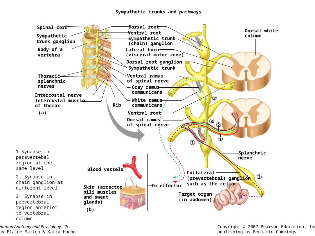
Sympathetic trunks and pathways
(a)
(b)
To effector
Blood vessels
Skin (arrectorpili musclesand sweatglands)
Dorsal whitecolumn
Spinal cord
Sympathetictrunk ganglion
Body of avertebra
Thoracicsplanchnicnerves
Dorsal root
Dorsal root ganglion
Ventral rootSympathetic trunk(chain) ganglionLateral horn(visceral motor zone)
Sympathetic trunk
Ventral ramusof spinal nerveGray ramuscommunicans
White ramuscommunicans
Intercostal nerve
Dorsal ramusof spinal nerve
Intercostal muscleof thorax
Splanchnicnerve
Collateral(prevertebral) ganglionsuch as the celiac
Target organ(in abdomen)
Ventral root
Rib
11
23
3
2
1.Synapse in paravertebal region at the same level
2. Synapse in chain ganglion at different level
3. Synapse in prevertebral region anterior to vertebral column
• Activity 4– Locate the ANS chains in the models.


Human Reflex Physiology
• Definition:– Rapid, predictable and involuntary motor
response to stimuli through pathways called reflex arcs.
• Two systems– Autonomic reflexes (unconscious): digestion,
sweating etc.
– Somatic reflexes: activate skeletal muscles.

Human Anatomy and Physiology, 7eby Elaine Marieb & Katja Hoehn
Copyright © 2007 Pearson Education, Inc.,publishing as Benjamin Cummings.
Hierarchy of motor control
FeedbackInternal
feedback
Cerebellum and basal nuclei
Highest (precommand)
Structures involved
Reflex activity
Segmental motorcontrols (CPG)
Interactions Control level
Middle
Motor cortex (pyramidal system)and brain stem nuclei (vestibular,red, reticular formation, etc.)
Lowest
Spinal cord
Motoroutput
Programs and instructions(modified by feedback)
Projection areas
Sensoryinput CNG: central pattern
generator

Human Anatomy and Physiology, 7eby Elaine Marieb & Katja Hoehn
Copyright © 2007 Pearson Education, Inc.,publishing as Benjamin Cummings.
The basic components of all human reflex arcs
Stimulus
Receptor
Skin
Sensory neuron
Spinal cord (in cross section)
Integration center
InterneuronMotor neuron
Effector
1
5
4
2 3

The reflex arc
• Characteristics: Structurally (number of neurons involved)– Monosynaptic arc: one synapse– Polysynaptic arc: one or more association neurons.
• Somatic Reflexes (skeletal muscle effectors)– Stretch reflexes: Postural and locomotion reflexes.
• Muscle spindle stimuli/Golgi organ in tendons (stretching) initiates reflex.
– Reciprocal inhibition: antagonistic efferent muscles are relaxed (damped).
– Patellar reflex (activity 1).

Human Anatomy and Physiology, 7eby Elaine Marieb & Katja Hoehn
Copyright © 2007 Pearson Education, Inc.,publishing as Benjamin Cummings.
The stretch reflex
(a)
(b)
Initialstimulus:musclestretch
Afferent impulsesfrom stretchreceptor tospinal cord
Efferentimpulses toalpha () motorneurons causecontractionof the stretchedmuscle thatresists/reversesthe stretch
Efferent impulsesto antagonistmuscles are damped(reciprocalinhibition)
Spinal cord(L2–L4)
Patella
Cell body ofsensory neuron
Key:+ Excitatory synapse– Inhibitory synapse
Motor neuronserving quadriceps
Motor neuronserving antagonistmuscle group(hamstrings)
Musclespindle
Quadriceps(extensors)
Hamstrings(flexors)
Musclespindle
Patellarligament
Interneuron
–
1
2
3

Human Anatomy and Physiology, 7eby Elaine Marieb & Katja Hoehn
Copyright © 2007 Pearson Education, Inc.,publishing as Benjamin Cummings.
The Golgi tendon reflex
++
+–
Quadriceps(extensor)
Golgitendonorgan
Hamstrings(flexor)
Afferent fiberfrom Golgitendon organ
Efferent fiberto muscleassociatedwith stretchedtendonEfferent fiberto antagonisticmuscle
Spinal cord
Interneurons
Key:+ Excitatory synapse– Inhibitory synapse
Human Anatomy and Physiology, 7eby Elaine Marieb & Katja Hoehn
Copyright © 2007 Pearson Education, Inc.,publishing as Benjamin Cummings.
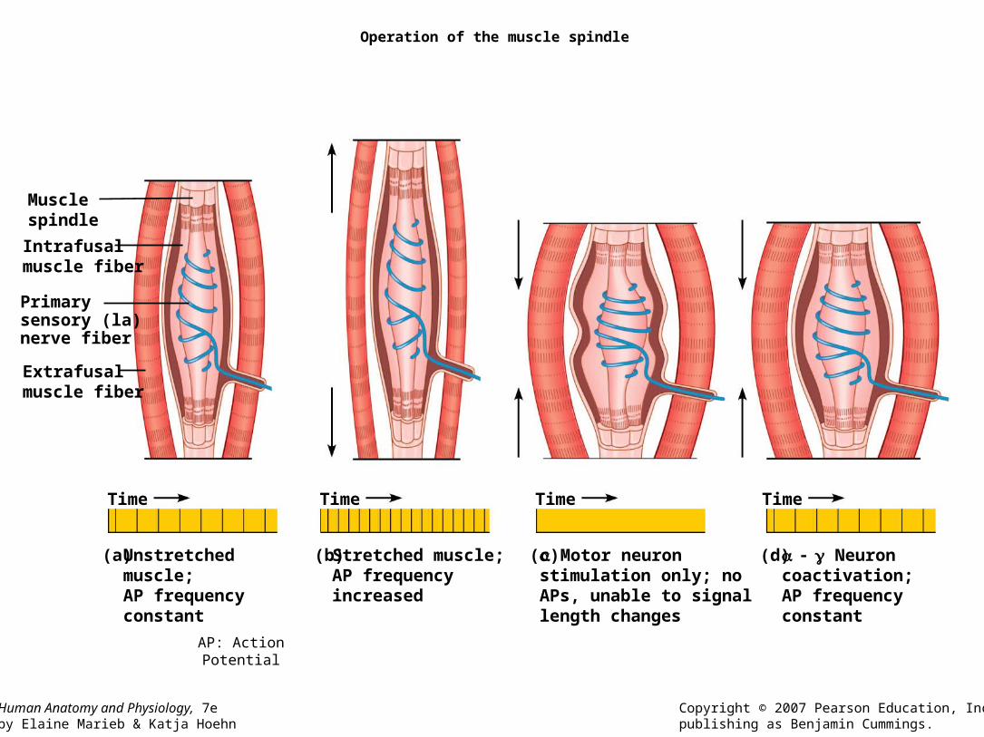
Operation of the muscle spindle
(a) (b) (d)(c)
Musclespindle
Intrafusalmuscle fiber
Primarysensory (la)nerve fiber
Extrafusalmuscle fiber
Time Time Time
Unstretched muscle;AP frequency constant
Stretched muscle;AP frequency increased
Neuron coactivation;AP frequency constant
Motor neuronstimulation only; noAPs, unable to signallength changes
Time
AP: Action Potential
Human Anatomy and Physiology, 7eby Elaine Marieb & Katja Hoehn
Copyright © 2007 Pearson Education, Inc.,publishing as Benjamin Cummings.
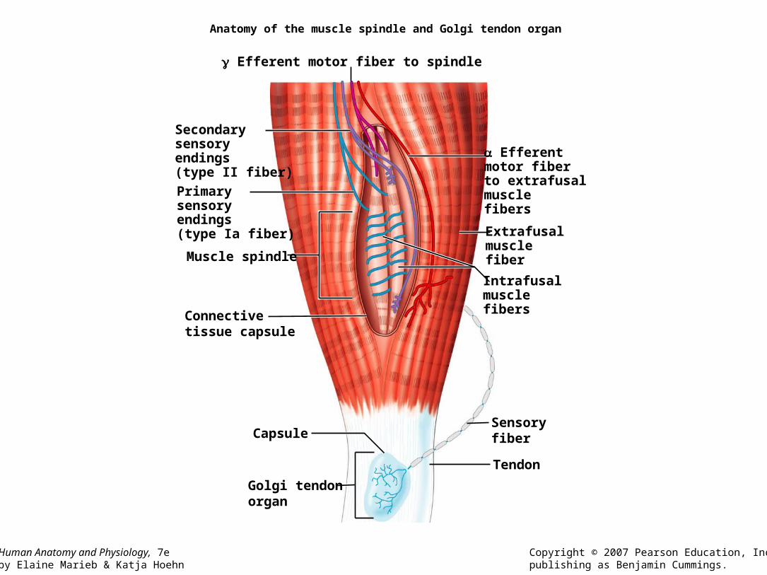
Anatomy of the muscle spindle and Golgi tendon organ
Primarysensoryendings(type Ia fiber)
Muscle spindle
Connectivetissue capsule
Efferentmotor fiberto extrafusalmusclefibers
Extrafusalmusclefiber
Intrafusalmusclefibers
Tendon
Efferent motor fiber to spindle
Secondarysensoryendings(type II fiber)
Golgi tendonorgan
CapsuleSensoryfiber
Somatic ReflexesSomatic Reflexes
• Crossed extensor reflex: Withdrawal reflex, followed by extension of the opposite limb.• Activity 2?

Human Anatomy and Physiology, 7eby Elaine Marieb & Katja Hoehn
Copyright © 2007 Pearson Education, Inc.,publishing as Benjamin Cummings.
The crossed-extensor reflex
Afferentfiber
Efferentfibers
Extensorinhibited
Flexorstimulated
Right arm(site of stimulus)
Left arm (site ofreciprocal activation)
Arm movements
Interneurons
Key:+ Excitatory synapse– Inhibitory synapse
Efferentfibers
FlexorinhibitedExtensorstimulated
+
–+
–
+
+
Flexes
Extends
Somatic ReflexesSomatic Reflexes
• Autonomic Reflexes– Pupillary reflexes
– Salivary reflex
• Reaction time of a reflex– Relative to the myelination of an axon andits
length relative to the interneuron or association center.
– Visual stimulus 150-300 ms.
Somatic ReflexesSomatic Reflexes
• Superficial cord reflex: Abdominal, cremaster and plantar reflexes.• Plantar reflex. Normal pyramidal activity, toes
flex and move close together. Activity 3.
– Cranial nerve reflex: optical (motor) nerves.• Corneal reflex (V). Activity 4

Autonomic reflexes
• Pupillary reflexes.– Cranial nerve II, III.
• Actvity 6: Contralateral response, ipsilateral response.
– Ciliospinal reflex. Pupilary.
– Salivary reflex. Smooth/skeletal muscles.
Human Anatomy and Physiology, 7eby Elaine Marieb & Katja Hoehn
Copyright © 2007 Pearson Education, Inc.,publishing as Benjamin Cummings.
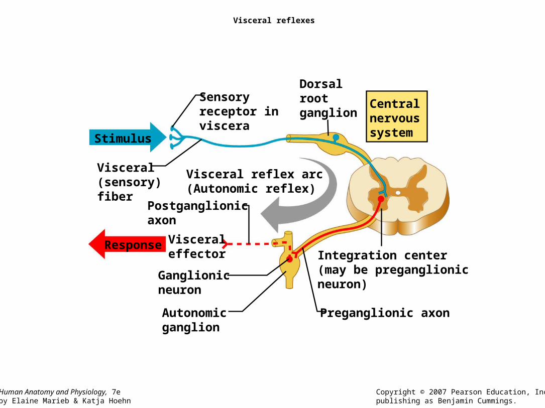
Visceral reflexes
Dorsalrootganglion
Sensoryreceptor inviscera
Centralnervoussystem
Visceral(sensory)fiber
Visceral reflex arc(Autonomic reflex)
Stimulus
Postganglionicaxon
Visceraleffector Integration center
(may be preganglionicneuron)
Preganglionic axonAutonomicganglion
Ganglionicneuron
Response

Human Anatomy and Physiology, 7eby Elaine Marieb & Katja Hoehn
Copyright © 2007 Pearson Education, Inc.,publishing as Benjamin Cummings.
Referred pain
Heart
Lungs anddiaphragm
Liver
Gallbladder
Stomach
Kidneys
Ovaries
Small intestine
Ureters
Urinarybladder
Colon
Pancreas
Liver
Heart
Appendix
Gallbladder