Peripheral and Central Mechanisms in Irritable Bowel Syndrome
102
Linköping University Medical Dissertations No. 1678 Peripheral and Central Mechanisms in Irritable Bowel Syndrome - in search of links Olga Bednarska Department of Medical and Health Sciences Linköping University, Sweden Linköping 2019
Transcript of Peripheral and Central Mechanisms in Irritable Bowel Syndrome

Linköping University Medical Dissertations No. 1678
Peripheral and Central Mechanisms in
Irritable Bowel Syndrome - in search of links
Olga Bednarska
Department of Medical and Health Sciences
Linköping University, Sweden
Linköping 2019
Olga Bednarska, 2019
Cover: schematic diagram of brain gut axis in IBS
Published article has been reprinted with the permission of the copyright
holder.
Printed in Sweden by LiU-Tryck, Linköping, Sweden, 2019
ISBN 978-91-7685-093-0
ISSN 0345-0082

To my Dad
Have no fear of perfection; you’ll never reach it.
Maria Skłodowska-Curie


Contents
CONTENTS
ABSTRACT ................................................................................................................ 1
SVENSK SAMMANFATTNING .................................................................................... 3
LIST OF PAPERS ........................................................................................................ 5
ABBREVIATIONS ....................................................................................................... 7
INTRODUCTION ........................................................................................................ 9
IRRITABLE BOWEL SYNDROME ............................................................................................... 9
THE BRAIN-GUT AXIS ........................................................................................................ 10
Mucosal barrier function ....................................................................................... 12
Transcellular and paracellular permeability ...................................................... 13
Neuroimmune regulation of mucosal barrier function ..................................... 16
Brain function ........................................................................................................ 19
Visceral pain pathways ...................................................................................... 22
Brain networks ................................................................................................... 24
Insula ................................................................................................................. 26
PSYCHOLOGICAL COMORBIDITIES ........................................................................................ 28
SEX DIFFERENCES IN IBS .................................................................................................... 29
SUMMARY ..................................................................................................................... 30
AIMS ...................................................................................................................... 31
METHOD ................................................................................................................ 33
SUBJECTS ....................................................................................................................... 33
QUESTIONNAIRES ............................................................................................................ 34
SIGMOIDOSCOPY ............................................................................................................. 36
BLOOD TESTS .................................................................................................................. 36
MUCOSAL BARRIER MEASUREMENTS ................................................................................... 37
Using chamber experiments .................................................................................. 37
Bacterial permeability ....................................................................................... 38
Paracellular permeability .................................................................................. 38
Blocking agents .................................................................................................. 38
Immunofluorescence of MC tryptase, VIP, VPACs ................................................ 39
MAGNETIC RESONANCE IMAGING DATA ACQUISITION AND ANALYSIS ......................................... 40
Functional Magnetic Resonance Imaging protocol ............................................... 40
Resting state functional MRI .................................................................................. 40
Diffusion tensor imaging ........................................................................................ 42

Peripheral and Central Mechanisms in Irritable Bowel Syndrome - in search of links
Quantitative Magnetic Resonance Spectroscopy .................................................. 42
STATISTICS ..................................................................................................................... 44
Paper I .................................................................................................................... 44
Paper II ................................................................................................................... 44
Path analysis ...................................................................................................... 45
Paper III .................................................................................................................. 46
ETHICAL APPROVAL .......................................................................................................... 46
RESULTS ................................................................................................................. 47
CLINICAL CHARACTERIZATION OF THE PATIENTS ...................................................................... 47
QUESTIONNAIRE DATA ...................................................................................................... 48
MUCOSAL BARRIER DATA .................................................................................................. 50
Bacterial and paracellular permeability ................................................................. 50
Regulation by VIP and MC ..................................................................................... 51
Questionnaire data and intestinal permeability .................................................... 52
RESTING STATE FUNCTIONAL CONNECTIVITY DATA .................................................................. 53
Paracellular permeability and DMN connectivity .................................................. 53
Bacterial permeability and DMN connectivity ....................................................... 54
Paracellular permeability, DMN-RVM functional connectivity and questionnaire data ........................................................................................................................ 56
QUANTITATIVE MAGNETIC RESONANCE SPECTROSCOPY DATA .................................................. 57
aINS neurotransmitter concentrations .................................................................. 57
aINS neurotransmitter concentrations and questionnaire data ........................... 57
DISCUSSION ........................................................................................................... 59
BRAIN-GUT AXIS: GUT ASPECTS ........................................................................................... 60
BRAIN-GUT AXIS: BRAIN ASPECTS ........................................................................................ 62
BRAIN-GUT AXIS: PSYCHOLOGICAL ASPECTS ........................................................................... 66
CONCLUSIONS ........................................................................................................ 69
FUTURE DIRECTIONS .............................................................................................. 71
ACKNOWLEDGEMENTS ........................................................................................... 73
REFERENCES ........................................................................................................... 75

Abstract
1
ABSTRACT
Irritable bowel syndrome (IBS) is a chronic visceral pain disorder with
female predominance, characterized by recurrent abdominal pain and
disturbed bowel habits in the absence of an identifiable organic cause.
This prevalent and debilitating disease, which accounts for a substantial
economic and individual burden, lacks exact diagnostic tools and effective
treatment, since its pathophysiology remains uncertain. The bidirectional
and multilayered brain-gut axis is a well-established disease model, how-
ever, the interactions between central and peripheral mechanisms along
the brain-gut axis remain incompletely understood. One of the well-
described triggering factors, yet accounting for only a fraction of IBS
prevalence, is bacterial gastroenteritis that affects mucosal barrier func-
tion. Altered gut microbiota composition as well as disturbed intestinal
mucosal barrier function and its neuroimmune regulation have been re-
ported in IBS, however, the impact of live bacteria, neither commensal
nor pathogenic, on intestinal barrier has not been studied yet. Further-
more, abnormal central processing of visceral sensations and psychologi-
cal factors such as maladaptive coping have previously been suggested as
centrally-mediated pathophysiological mechanisms of importance in IBS.
Brain imaging studies have demonstrated an imbalance in descending
pain modulatory networks and alterations in brain regions associated
with interoceptive awareness and pain processing and modulation, par-
ticularly in anterior insula (aINS), although biochemical changes puta-
tively underlying these central alterations remain poorly understood.
Most importantly, however, possible associations between these docu-
mented changes on central and peripheral levels, which may as complex
interactions contribute to disease onset and chronification of symptoms,
are widely unknown.
This thesis aimed to investigate the peripheral and central mecha-
nisms in women with IBS compared to female healthy controls (HC) and
to explore possible mutual associations between these mechanisms.
In Paper I, we studied paracellular permeability and passage of live
bacteria, both commensal and pathogenic through colonic biopsies
mounted in Ussing chambers. We explored the regulation of the mucosal
barrier function by mast cells and the neuropeptide vasoactive intestinal
polypeptide (VIP) as well as a correlation between mucosal permeability
and gastrointestinal and psychological symptoms. We observed increased
Peripheral and Central Mechanisms in Irritable Bowel Syndrome - in search of links
2
paracellular permeability and the passage of commensal and pathogenic
live bacteria in patients with IBS compared with HC, which was dimin-
ished by blocking the VIP receptors as well as after stabilizing mast cells
in both groups. Moreover, higher paracellular permeability was associated
with less somatic and psychological symptoms in patients.
In Paper II, we aimed to determine the association between colonic
mucosa paracellular permeability and structural and resting state func-
tional brain connectivity. We demonstrated different patterns of associa-
tions between mucosa permeability and functional and structural brain
connectivity in IBS patients compared to HC. Specifically, lower paracel-
lular permeability in IBS, similar to the levels detected in HC, was associ-
ated with more severe IBS symptoms and increased functional and struc-
tural connectivity between intrinsic brain resting state network and de-
scending pain modulation brain regions. Our findings further suggested
that this association between mucosa permeability and functional brain
connectivity was mainly mediated by coping strategies.
In Paper III, we investigated putative alterations in excitatory and in-
hibitory neurotransmission of aINS, as the brain’s key node of the sali-
ence network crucially involved in cognitive control, in IBS patients rela-
tive to HC and addressed possible connections with both symptoms and
psychological factors. We found decreased concentrations of the excitato-
ry neurotransmitter Glx in bilateral aINS in IBS patients compared to HC,
while inhibitory neurotransmitter GABA+ levels were comparable. Fur-
ther, we demonstrated hemisphere-specific associations between ab-
dominal pain, coping and aINS excitatory neurotransmitter concentra-
tion.
In conclusion, this thesis broadens the knowledge on peripheral and cen-
tral mechanisms in IBS and presents novel findings that bring together
the ends of brain-gut axis. Our results depict association between mucosal
permeability, IBS symptoms and functional and structural connectivity
engaging brain regions involved in emotion and pain modulation as well
as underlying neurotransmitter alterations.

Svensk sammanfattning
3
SVENSK SAMMANFATTNING
IBS (Irritable bowel syndrome) är en kronisk tarmsjukdom som känne-
tecknas av buksmärta i kombination med förändrade tarmvanor. Sjuk-
domen drabbar ungefär 11 % av befolkningen, övervägande kvinnor och
kan vara förenat med påtaglig försämrad livskvalitet för den drabbade
individen och ökade hälso- och sjukvårdskostnader. Förutom magtarm-
symptom lider patienter med IBS ofta av andra kroniska smärttillstånd
och olika psykologiska symtom, såsom nedstämdhet, ångest och bristande
förmåga att hantera kronisk smärta, så kallad coping. Orsaken till IBS är
ännu inte helt klarlagd, men störd kommunikation mellan tarmen och
hjärnan, via den så kallade ”hjärna-tarmaxeln”, anses vara väsentligt. Per-
soner med IBS kan skilja sig ganska mycket från varandra avseende
symptomens karaktär, vilket skulle kunna tyda på skillnader i de bakom-
liggande patofysiologiska faktorerna. I hjärna-tarmaxeln ingår bland an-
nat det centrala nervsystemet som även påverkas av psykologiska fak-
torer, delar av immun- och neuroendokrina systemet, tarmens slemhinna
samt tarmfloran. Störningar i dessa system har påvisats vid IBS. Däremot
är sambandet mellan störd hjärnfunktion, tarmbarriärfunktion och pati-
entens symptom fortfarande oklart. I denna avhandling undersöktes både
hjärnan och tarmbarriären hos kvinnor med IBS och friska kvinnor, samt
eventuella samband med magtarmsymptom och psykologiska symptom.
Kvinnor med IBS och friska kvinnor genomgick tjocktarmundersök-
ning med vävnadsprovtagning för att undersöka tarmbarriärfunktion
samt magnetkameraundersökning (MR) för att mäta struktur och aktivi-
tetsmönster i hjärnan vilka korrelerades till tarmbarriärfynden. En speci-
ell del av MR-undersökning, kallad MR-spektroskopi, genomfördes för att
mäta signalsubstanskoncentration i ett område i hjärnan, inblandad i den
känslomässiga upplevelsen och bearbetningen av smärtsignaler från tar-
men, kallat insula. Aktivitetsändringar i detta hjärnområde har tidigare
påvisats vid IBS. Alla deltagare fyllde också i frågeformulär om magtarm-
och psykologiska symptom.
I delstudie I undersökte vi tarmslemhinnans genomsläpplighet genom
att ta vävnadsbitar från tjocktarmen och undersöka dem i så kallade Us-
singkammarsystem. Vi undersökte passagen genom tarmslemhinnan för
små molekyler och för levande bakterier samt studerade regleringen av
denna passage. Resultaten visade att patienter med IBS hade tydligt ökad
genomsläpplighet både för små molekyler men också för bakterier. Vi
Peripheral and Central Mechanisms in Irritable Bowel Syndrom - in search of links
4
fann att genomsläppligheten regleras av tarmslemhinnas immunceller, så
kallade mastceller och ett neuroendokrint hormon vasoaktivt intestinalt
polypetid. Vi fann också att passagegraden för små molekyler var kopplad
till vissa kroppsliga och psykologiska symptom hos IBS patienterna.
I delstudie II studerade vi sambanden mellan hjärnans aktivitets-
mönster och struktur, tarmbarriärfunktion och symptom. Våra data vi-
sade att IBS patienterna med normal tarmbarriärfunktion hade liknande
avvikelser i hjärnan som man också kan se vid störda centrala smärtregle-
ringsmekanismer. De patienter med IBS som uppvisade ökad tarm-
genomsläpplighet hade mindre av dessa centrala störningar. Statistisk
analys visade att sambandet mellan tarmbarriärfunktionen och hjärn-
funktionen till viss del var förmedlad av en sämre copingförmåga.
I delstudie III undersökte vi sambanden mellan koncentrationen av
signalsubstanser i insula och symtom. Resultaten visade en minskad kon-
centration av den stimulerande signalsubstansen Glx i insula hos patien-
ter med IBS jämfört med friska. Dessutom indikerade våra data att den
vänstra och högra hjärnhalvan kan ha olika funktioner avseende smärt-
hantering, eftersom smärta var mer associerad till Glx koncentrationen i
högerinsula och copingförmåga mer till Glx koncentrationen i vänster in-
sula.
Sammanfattningsvis bidrar denna avhandling till ökad kunskap om tar-
mens perifera och hjärnans centrala mekanismer vid IBS samt presente-
rar nya resultat om en avvikande tarmbarriär och hjärnfunktion vid IBS.
Avhandlingen belyser vidare sambanden mellan förändringar i tarm och
hjärna och psykologiska symptom.

List of Papers
5
LIST OF PAPERS
I. Vasoactive Intestinal Polypeptide and Mast Cells Regulate
Increased Passage of Colonic Bacteria in Patients With Irritable
Bowel Syndrome
Olga Bednarska, Susanna Walter, Maite Casado-Bedmar, Magnus Ström, Elo-
isa Salvo-Romero, Maria Vicario, Emeran A. Mayer, Åsa V Keita
Gastroenterology, 2017 Oct;153(4):948-960.e3.
doi: 10.1053/j.gastro.2017.06.051.
II. Interactions between gut permeability and brain structure function
in health and irritable bowel syndrome
Suzanne T Witt, Olga Bednarska, Åsa V Keita, Adriane Icenhour, Michael P
Jones, Sigrid Elsenbruch, Johan D Söderholm, Maria Engström, Emeran A
Mayer, Susanna Walter
Neuroimage Clinical, 2019; 21: 101602. doi: 10.1016/j.nicl.2018.11.012.
III. Reduced excitatory neurotransmitter levels in anterior insulae are
associated with abdominal pain in irritable bowel syndrome.
Olga Bednarska*, Adriane Icenhour*, Sofie Tapper, Suzanne T Witt, Anders
Tisell, Peter Lundberg, Sigrid Elsenbruch, Maria Engström, Susanna Walter
*Both authors contributed equally
Accepted for publication in Pain

Peripheral and Central Mechanisms in Irritable Bowel Syndrome - in search of links
6

Abbreviations
7
ABBREVIATIONS
ACC anterior cingulate cortex
aINS anterior insula
ANS autonomic nervous system
BOLD blood-oxygen-level dependent
BPI Brief Pain Inventory
CAN central autonomic network
CEN central executive network
CNS central nervous system 51Cr-EDTA chromium labeled ethylenediaminetetraacetic acid
CRF corticotropin releasing factor
CRFR corticotropin releasing factor receptor
CSQ Coping Strategies Questionnaire
DMN default mode network
DTI diffusion tensor imaging
EAN emotional arousal network
ENS enteric nervous system
FA fractional anisotropy
FC functional connectivity
fMRI functional magnetic resonance imaging
GABA γ-aminobutyric acid
GABA+ γ-aminobutyric acid + co-edited macromolecular signals
GALT gut-associated lymphoid tissue
Gln glutamine
Glu glutamate
Glx glutamate + glutamine
GSD Gastrointestinal Symptom Diary
HADS Hospital Anxiety and Depression Scale
HC healthy controls
HPA hypothalamic-pituitary-adrenal axis
IBS irritable bowel syndrome
IBS-SSS IBS Severity Scoring System
ICA independent component analysis

Peripheral and Central Mechanisms in Irritable Bowel Syndrome - in search of links
8
INS insula
Isc short circuit current
MC mast cells
MEGA-PRESS Mescher-Garwood point resolved spectroscopy
mM millimolar
MRI magnetic resonance imaging
NSAID non-steroidal anti-inflammatory drugs
NTS nucleus tractus solitarius
PACAP pituitary adenylate cyclase polypeptide
PAG periaqueductal grey matter
PD potential difference
PHQ-15 Patient Health Questionnaire
PI-IBS postinfectious irritable bowel syndrome
pINS posterior insula
ppm parts per million
qMRS quantitative magnetic resonance spectroscopy
ROI region of interest
rs-fMRI resting-state functional magnetic resonance imaging
RVM rostroventral medulla
SMN sensorimotor network
SN salience network
TE echo time
TER transepithelial resistance
TR repetition time
VIP vasoactive intestinal polypeptide
vlPFC ventrolateral prefrontal cortex
VMb ventromedial thalamic nucleus basal portion
VMpo ventromedial thalamic nucleus posterior portion
VPAC vasoactive intestinal polypeptide receptor
VSI Visceral Sensitivity Index

Introduction
9
INTRODUCTION
Irritable bowel syndrome
Irritable bowel syndrome (IBS) is a widespread gastroenterological disor-
der with a pooled prevalence of around 11% [141] that ranges from 1.1% in
France and Iran to 35.5% in Mexico [225]. Although the term “IBS” first
appeared in 1950 [27], the disease was known long before, and had been
described by Jacob Mendes da Costa as early as in 1871 using the term –
“mucous colitis” [51]. Recurrent abdominal pain and disturbed bowel
habits are the hallmarks of the disease, which still lacks established dis-
ease-specific biomarkers. IBS is diagnosed based on diagnostic criteria in
the absence of alarm symptoms after the exclusion of some differential
diagnoses such as coeliac disease, inflammatory bowel diseases and colo-
rectal cancer, which can be arrived at after minimal laboratory tests and
colonoscopy when appropriate [159]. Over the last 40 years, since the first
release of the Manning criteria in 1978 [145], IBS diagnostic criteria have
been systematically changed and improved, with the publication of the
Rome criteria in 1989 [239], Rome II in 1999 [240], Rome III in 2006
[139] (see Box 1, these are the criteria used in this thesis) and Rome IV in
2016 [159]. The most recent Rome IV criteria sharpened the description
of pain as an IBS symptom, by removing the word “discomfort” since it
was considered to be imprecise in certain languages, and introducing the
overall relationship between abdominal pain and bowel movements, not
necessarily improvement of pain following bowel movements, as it was
defined in Rome III [204].
Although IBS diagnostic criteria may be fulfilled in individuals with
some organic diseases [157], the robustness of these criteria has been con-
firmed in studies showing that the prevalence of missed organic disease is
about 1.3% [60] when the IBS diagnosis is based on the criteria, which is
to be compared to approximately 44% when the diagnosis is based on
clinical impression only [59,234]. In the scarcity of effective IBS medica-
tion, the current treatment for IBS targeted for stool consistency altera-
tions is well established, well tolerated by the patients and easy to evalu-
ate, hence, it is widely used. To aid the clinician, IBS patients are sub-
grouped based on the prevalent stool consistency pattern, with groups
that include IBS-C with predominant constipation, IBS-D with predomi-
nant diarrhea, IBS-M with mixed type of stools and IBS-U for unsubtyped

Peripheral and Central Mechanisms in Irritable Bowel Syndrome - in search of links
10
IBS [159]. This subgrouping, however, has substantial flaws, as the stool
consistency fluctuates in the same individual in at least 80% of cases
[178]. Likewise, stool consistency itself poses negligible burden for some
of the patients in comparison to prevalent and often disabling extraintes-
tinal or psychological symptoms associated with the disease. In fact, the
plethora of extraintestinal symptoms such as fatigue, headache, insomnia,
musculoskeletal and urinary symptoms, along with psychological comor-
bidities, such as anxiety and depression [19] constitute the prevailing rea-
son that IBS patients seek care [55].
IBS, although not lethal, has a considerable detrimental effect on the
patients’ quality of life [217] and is a significant economic burden [33],
due not only to frequent work absenteeism [11], but also substantial
healthcare utilization [33]. This debilitating disease that constitutes a ma-
jor burden to individuals, the healthcare system and society, lacks exact
diagnostic tools and adequate treatment, since its pathophysiology re-
mains obscure. A fundamental comprehension of the heterogeneity of IBS
appears to be the only way to tackle it.
The brain-gut axis Probably due to its heterogeneity, the pathophysiology of IBS is still ob-
scure, but several predisposing factors (such as genetics, environmental
and psycho-social factors) have been identified, as have several triggering
factors (such as gastroenteritis, trauma, stress, and food intolerances).
These factors influence the physiological mechanisms that lead to altered
gut microbial flora, enhanced intestinal permeability, immunesystem acti-
Box 1 ROME III Diagnostic Criteria* for Irritable Bowel Syndrome
Recurrent abdominal pain or discomfort** at least 3 days per month in the last 3
months associated with 2 or more of the following:
1. Improvement with defecation
2. Onset associated with a change in frequency of stool
3. Onset associated with a change in form (appearance) of stool
*Criteria fulfilled for the last 3 months with symptom onset at least 6 months prior to
diagnosis.
**Discomfort means an uncomfortable sensation not described as pain. In patho-
physiology research and clinical trials, a pain/discomfort frequency of at least 2 days
a week during screening evaluation for subject eligibility.
(Longstreth et al, 2006)

Introduction
11
vation, gastrointestinal dysmotility, autonomic dysfunction and visceral
hypersensitivity. This complex multifactorial pathophysiology has been
summarized as a dysregulation of the brain-gut axis comprising all of the
abovementioned pathophysiological mechanisms (Figure 1) [153,165].
Explaining all the hallmark symptoms of IBS as disturbances in different
levels of this bi-directional system has gained increasing attention in re-
cent decades, resulting in a principal change of the view of all the func-
tional gastrointestinal disorders, that according to the latest Rome IV
terminology are now known as ”Disorders of gut-brain interaction” [159].
Particular components of the bottom-up and top-down model will be dis-
cussed in detail below.
Genetic factors
Sex and gender
Environment
Abuse history
Stress
Acute gastroenteritis
Psychological disturbances
Maladaptive coping
Abnormal central processing of sensations
Alterations in HPA axis function
Autonomic dysfunction
GI motility disturbances
Visceral hypersensitivity
Neuroimmune dysfunction
Increased intestinal permeability
Mast cell-nerve axis dysfunction
Abnormal peripheral processing of
sensations
Bile acids and short-chain fatty acids
disturbances
Altered microbiota
Food
Figure 1. The brain-gut axis and its role in the pathophysiology of IBS. Abbreviation: GI, gastrointestinal; HPA, hypothalamic-pituitary-adrenal axis.

Peripheral and Central Mechanisms in Irritable Bowel Syndrome - in search of links
12
Mucosal barrier function
The mucosa of the gastrointestinal tract is directly exposed to hazards
from the environment, while being responsible for the absorption of all
essential nutrients, electrolytes and water. To keep the balance between
protection and absorption, the mucosa in different parts of the digestive
tract has different properties, and these parts interact with each other in a
coordinated manner. The anatomy of the intestinal barrier in general con-
sists of two components: extracellular and cellular (Figure 2). The first
element is the lumen itself, where harmful substances and microorgan-
isms are destroyed in an unspecific manner by the acidic environment
and by digestive enzymes from the gastric, pancreatic and biliary fluids
[203]. The host luminal bacterial flora is also a part of the defence, com-
peting for space and nutrients with ingested pathogens [32]. The next ex-
tracellular component is the microclimate, i.e. unstirred water and the
mucus layer. In the colon, in order to oppose microbial invasion, the out-
er, stirred, part of the mucus layer contains immunoglobulins, phospho-
lipids, mucins and peptides with antimicrobial properties. This part of the
layer also prevents bacterial adhesion to the epithelium due to its hydro-
phobic properties. The inner non-stirred mucus layer is known as the
“glycocalyx”, and protects the epithelium from digestive enzymes, and de-
hydration, and facilitates nutrient absorption, epithelial renewal and oral
antigen tolerance. This part of mucus is mainly free from bacteria
[86,106]. Finally, intestinal motility and water excretion form the final
elements of the extracellular components of the intestinal barrier.
Figure 2. Intestinal barrier. Abbreviation: VIP, vasoactive intestinal pep-tide. Adapted from Keita et al. The intestinal barrier and its regulation by neuroimmune factors. Neurogastroenterology & Motility, 2010 with per-mission.

Introduction
13
The most important cellular component of the intestinal barrier is the
epithelium itself. It is a monolayer of enterocytes and several types of spe-
cialized cells, such as Goblet cells, producing mucus [117], Paneth cells,
secreting defensins and enterochromaffin cells, secreting hormones and
neuropeptides, located both in the small and the large intestine [201].
Membranous (M) cells, responsible for antigen uptake and presentation,
are located only in the small intestine [201]. All these specialized cells,
scattered among enterocytes, are the key cells of the epithelial lining. They
present digestive, metabolic and immune functions, and are involved in
the innate immune response, and the activation and recruitment of leuko-
cytes. The lamina propria, which lies underneath the epithelium, contains
immune cells that can be classified as organized GALT (gut-associated
lymphoid tissue), which comprises lymphoid follicles (in the colon), Pey-
er’s patches (in the ileum), and mesenteric nodes, which are responsible
for inducing immune response - and diffuse GALT, which comprises lym-
phocytes, eosinophils, dendritic cells, macrophages and mast cells, which
are general effectors of the immune response [201]. The lamina propria
harbors also connective tissue and cells of the enteric nervous system
(ENS). Neurons and enteric glial cells of the ENS, together with the cen-
tral nervous system (CNS), maintain the homeostasis in the gastrointesti-
nal tract, for example via secretion of neurotransmitters [111], as de-
scribed in the following section.
Transcellular and paracellular permeability
Epithelial cells that are lining the digestive tract create a monolayer of
semi-permeable, polarized cells connected to each other (and to basal
membrane) by protein complexes termed tight junctions (TJ), anchoring
junctions (adherens junctions and desmosomes connected to the cells cy-
toskeleton) and communication junctions (building communication
channels between neighboring cells) [201]. TJ are localized most apically
between the cells and control the passage of small water-soluble mole-
cules. They are built of four different protein complexes: occludin, clau-
dins, junctional adhesion molecules and tricellulin, that are connected to
the cells cytoskeleton via zonula occludens proteins [250]. These proteins
together regulate the paracellular route of the intestinal permeability,
accounting for 85% of the passive transepithelial transport. The transcel-
lular route across the epithelial membrane may occur both as diffusion
(passive transport of smaller lipophilic and hydrophilic molecules) and as
active transport by endocytosis: micropinocytosis, phagocytosis, receptor

Peripheral and Central Mechanisms in Irritable Bowel Syndrome - in search of links
14
mediated (clathrin-mediated) and clathrin-independent caveolar endocy-
tosis [32,110]. Micropinocytosis enables the fluid to enter the cells
through the formation of an invagination in the cell membrane. Phagocy-
tosis is a process, which internalizes larger particles that engage specific
membrane receptors. One of the most well-described endocytic routes
playing important role in cellular homeostasis is clathrin-mediated endo-
cytosis. In this process small vesicles are coated by cytosolic protein,
clathrin, and trap molecules that are ligands for clathrin receptors. Clath-
rin-independent endocytosis is still not fully understood, where small
flask-shaped lipid rafts termed caveolae form vesicles to internalize the
cargo. Interestingly, this pathway seems to be used by some pathogens,
such as Escherichia coli [40,110].
One of the strongest known trigger factor in IBS is gastroenteritis, ac-
counting for ca 9% of all IBS cases [14,210], however purely mathematical
modelling estimates that postinfectious IBS (PI-IBS) may constitute the
majority of IBS prevalence [210]. Bacterial infection (such as Campylo-
bacter jejuni, Salmonella enteridis [112,206], Salmonella thyphimurium
[114,158] and Escherichia coli [148]) poses higher risk for development of
PI-IBS than viral [186], or parasitic gastroenteritis [254]. This is probably
due to more severe mucosal damage, as a result of bacterial invasion and
cytotoxins [237]. This evident causality between gastroenteritis, impaired
mucosal barrier and development of PI-IBS resulted in studies on intesti-
nal permeability, initially done by using oral probe multi-sugar excretion
tests [147,227], that measured small bowel intestinal permeability. Subse-
quently mucosal barrier studies expanded to different types of IBS
[58,116,272,271] and both small and large bowel assessments [131,185],
however showing conflicting results [58,116,272].
The detrimental effect of non-steroidal anti-inflammatory drugs
(NSAID) on intestinal permeability is well investigated, and seems to be
enhanced in IBS in comparison to HC, supporting the notion of impaired
mucosal barrier in IBS [116]. The impact of NSAID on gut mucosa is man-
ifold, e.g. topical toxic, by damaging the mucus layer and inducing muco-
sal damage by inhibiting the prostaglandins; intracellular, by influencing
mitochondrial phosphorylation resulting in disruption of the TJ [116,121].
Thus, the exclusion of NSAID use is mandatory for the participants that
contribute to mucosal barrier studies.
Intestinal permeability can be measured using several techniques.
The most widely used assays are urine excretion of oral probe tests, which
use different (mostly indigestible) molecules of known absorption site,
like saccharides, polyethylene glycol (PEG) or radioactive chromium la-

Introduction
15
beled ethylenediaminetetraacetic acid (51Cr-EDTA). There are several
studies that employ these techniques exploring mainly PI-IBS and IBS-D
and show increased intestinal permeability [58,76,147,195,214,227,271].
Some of the pitfalls of those techniques, such as gastric emptying time
and dilution, bacterial degradation and intestinal motility, have been cir-
cumvented by using multi-sugar permeability test. Other confounders are
there to remain, such as renal dysfunction, saccharides-derived intestinal
motility changes or PEG high inter- and intra-individual variations
[82,93,172,175]. Oral probe tests, although non-invasive, are unable to
provide more detailed information about the mechanisms of the observed
disturbance in intestinal permeability.
The method that enables permeability studies ex vivo in real time on
viable human tissue in a diffusion chamber called Ussing chamber was
established in 1951 by zoologist Hans Henriksen Ussing and his colleague
chemist Karl Zehran [245]. Their work concerning active transport of so-
dium through isolated frog skin was a cornerstone of epithelial permeabil-
ity research, being nowadays, with some modifications, a golden standard
technique [79]. Ussing’s ”little chamber” consist of two half-chambers
filled with physiological buffered Krebs solution separated by the exam-
ined tissue. In this way the mucosal and serosal side of the tissue may be
exposed to different milieu. The solution in both half-chambers is stirred
by the gas flux of oxygen and carbon dioxide and kept at 37°C, i.e. the
temperature of human body. Active transport of ions produces a potential
difference (PD) measured by agar bridges. To determine this voltage, two
platinum electrodes, separated from the epithelium, transduce external
current nullifying PD. This current, termed short circuit current (Isc), to-
gether with PD, enables the calculatatiom of transepithelial resistance
(TER) in accordance with Ohm´s law (voltage= curent x resitance). TER
indicates electrical resistance mainly of the paracellular permeability
route via TJ.
To determine transcellular and paracellular permeability, different
substances of known transport route are used, such as 51Cr-EDTA for
paracellular permeability, and horseradish peroxidase for transcellular
permeability. Although this method is a hallmark of all epithelial permea-
bility studies, it carries several limitations, such as ex vivo environment,
where the biopsy taking per se constitutes tissue injury, following ische-
mia and denervation. However, meticulous monitoring of the viability of
the tissue diminishes this problem. Another limitation is the timing of the
experiment, as the viability of the tissue lasts no more than about 5-6
hours after the biopsy taking. Yet the possibility of subsequent microscop-

Peripheral and Central Mechanisms in Irritable Bowel Syndrome - in search of links
16
ic examination of the challenged tissue in order to explore, for instance, a
transport route of a so far unknown permeation marker, is unique for this
method.
Neuroimmune regulation of mucosal barrier function
Mast cells
Disruption in mucosal barrier function is known to go hand in hand with
low grade immune activation [252], which is also true in a proportion of
IBS patients [97,136]. The lamina propria, the final layer of intestinal bar-
rier, harbors an abundance of both innate and adaptive immune system
cells, whereof one of the most widely investigated innate immune cells are
the mast cells (MC). MC are of great importance in different aspects of the
function of intestinal tract, aside from host defense against microbes,
such as epithelial water and electrolyte secretion and mucosal barrier
function, blood circulation, neuroimmune interactions, pain mediation
and motility (Figure 3) [21]. MC distributed in the main host-barrier tis-
sues such as skin, respiratory, urinary and intestinal tract, show signifi-
cant heterogeneity and adaptability to different tissue conditions. Most
MC of the intestinal tract are located in lamina propria (ca 2-3% of all
cells [28]) and are of phenotype MCT (secreting only tryptase), while
those located in submucosa (about 1% of all cells) are termed MCTC phe-
notype (secreting both tryptase and chymase) [202,265,269]. The most
effective way to activate MC is via an IgE dependent high affinity receptor,
which plays a crucial role in allergic reactions. Other immune-dependent
(such as IgA, IgG, complements and Ig-free-light chains) as well as im-
mune-independent stimuli (such as neurotransmitters, neuropeptides
and hormones) may also trigger MC activation. Upon activation MC re-
lease an overabundance of mediators, that can be mainly divided into the
ones that are preformed in granules (such as histamine, serotonin, tryp-
tases, chymase, and to some degree TNFα and IL-6) and are secreted
within seconds from activation, as well as those synthesized de novo (such
as cytokines, chemokines, growth factors and some neuropeptides) and
are responsible for late phase reaction. Increased MC infiltration in both
the small bowel [83,149,150,249] and the colon
[3,15,16,42,50,49,180,263] in different IBS subtypes as well as MC activa-
tion [15,16,29,35,42,50,83,120,149,150,263] has been reported in many
studies, however conflicting data exist, showing comparable MC counts in
IBS and HC [17,25,35,36]. Positive correlation between MC infiltration

Introduction
17
and increased intestinal permeability in IBS has been implicated [235]
and MC mediators such as tryptase, TNFα or interleukins are known to
disrupt both paracellular [101,259] as well as transcellular permeability
[111].
Vasoactive intestinal polypeptide
One of the neuroimmune mediators released by MC is vasoactive intesti-
nal polypeptide (VIP) that consists of 28 aminoacids. It is produced in
various tissues, such as intestinal tract, pancreas and respiratory airways.
Nevertheless, VIP is primarily localized in neurons, both centrally- in su-
prachiasmatic nuclei of the hypothalamus, and peripherally- in the neu-
rons and other cells of peripheral nervous system and of ENS. VIP con-
centrations measured in serum are mainly of neural origin and are com-
Figure 3. The role of mast cells in IBS. Abbreviations: HPA, hypothalam-ic-pituitary-adrenal axis; SAM, sympathetic adrenal-medullary axis; DCs, dendritic cells; ENS, enteric nervous system; PNS, peripheral nervous system; CNS, central nervous system. Reprinted from Zhang et al Mast Cells and Irritable Bowel Syndrome: From the Bench to the Bedside. J Neurogastroenterology and Motility, 2016 with permission.

Peripheral and Central Mechanisms in Irritable Bowel Syndrome - in search of links
18
monly low, but may rise substantially in rare VIP producing tumors (VIP-
omas). They can occur in adults mainly in pancreas, although in children
VIPomas appear as tumors of the nervous system, ganglioneuromas or
ganglioneuroblastomas [73].
VIP and pituitary adenylate cyclase polypeptide (PACAP) belong to a
superfamily, together with other structurally related hormones (such as
glucagon, glucagon-like peptide, and secretin) and share some receptors,
termed PACAP type I and II receptors [87,127]. PACAP type I receptors
show mainly affinity for PACAP, whereas PACAP type II receptors are
sensitive to both PACAP and VIP, known as VIP receptors, and subdivid-
ed in VPAC1 and VPAC2. Both receptors are expressed in MC [87,127,224].
VPAC1 is mainly distributed in CNS (cortex and hippocampus), liver, lung,
intestine and T lymphocytes. VPAC2 receptor is also abundant in CNS
(thalamus, brainstem, spinal cord and dorsal root ganglia), and is found
in smooth muscles, intestines and in cardiovascular system [87]. Apart
from smooth muscle relaxation in the cardiovascular (vasodilatation),
gastrointestinal, urogenital and respiratory tract, VIP exerts a plethora of
different functions in various organs, such as: water and ion secretion in
the intestines and pancreas, stimulation of biliary secretion, endocrine
stimulation in adrenal glands, kidneys, pancreas, pituitary/hypothalamus
and thyroid gland as well as in CNS, regulation of cicardian rythm, neuro-
protection and neurodevelopment [87].
As far as inflammation and MC are concerned, VIP shows anti-
inflammatory properties by modulating T cell differentiation as well as
trafficking and stabilizing MC degranulation [87]. Nevertheless, evidence
on the impact of VIP on intestinal barrier is divergent, demonstrating
both stabilizing and disrupting effect on gut mucosa. The effect of VIP
may plausibly vary depending on different ongoing pathological processes
[34]. In fact, both VIP agonists and antagonists have been suggested as
promising anti-inflammatory agents in different inflammatory diseases,
such as rheumatoid arthritis (VIP agonists) or inflammatory bowel dis-
eases (both VIP agonists and antagonists) [34]. In IBS, increased amount
of VIP was detected in colonic biopsies in smaller studies [179,216], how-
ever data seem to be inconsistent [196].
Mast cell-nerve axis
The ENS is a part of the autonomic nervous system (ANS) and consists of
about 100 million nerve cells (intrinsic primary afferents [153]) organized
in three major plexuses: the myenteric (Auerbach plexus, located between
the longitudinal and circular muscle layers and regulating motility), the
Introduction
19
submucous (three ganglionated layers situated between the circular mus-
cle layer and mucosa) and the mucous plexus. The two latter ones, known
as Meissner plexus, are mainly engaged in interplexal communication and
secretion of about 30 different neurotransmitters [28]. ENS neurons
communicate with CNS through both afferent and efferent sympathetic
(via prevertebral ganglia) and parasympathetic neurons (via nervus vagus
and plexus pelvicus) that innervate the gut and, hence, contact ENS and
gut wall immune cells, such as MC [71]. In fact, MC have been shown to
be located in close proximity to extrinsic afferent nerves and enteric neu-
rons [12,91,202,230,231]. MC-nerve interaction is reciprocal [28,196], as
MC respond to neurotransmitters released by the neurons, while the bio-
active substances released by MC affect relevant neural receptors, result-
ing in visceral pain. However, this crosstalk requires activated MC, as only
then MC response to neuropeptides [202,246]. There is accumulating ev-
idence that MC activation and MC-nerve signaling may play crucial role in
peripheral sensitization and visceral hypersensitivity in IBS
[183,238,269]. However, data show both positive correlation between MC
activation/density and pain in IBS [15,185,214,263] as well as doubtful or
no correlation at all [15,150]. Nerve fiber overgrowth was observed in co-
lonic biopsies of IBS patients, due to release of nerve grow factor pro-
duced by MC [54]. MC stabilizers (ketotifen [120], sodium cromoglycate
[232]) as well as anti-inflammatory aminosalicylates (mesalazin [44])
demonstrated positive effect on visceral hypersensitivity and MC activa-
tion in several studies, although vague and doubtful effect on IBS symp-
toms [13].
Brain function
Brain is undoubtedly the ultimate instance for all the peripheral bodily
impulses, deciding which of them becomes conscious and creating the
feeling of pain. It is, however, not before the advancement of neuroimag-
ing techniques that the breakthrough in the appreciation of central as-
pects of pain modulation took place, even though some other techniques
(such as electroencephalography [109], computed tomography [26], posi-
tron emission tomography [132] or magnetoencephalography [43]) were
available long time before the dawning of magnetic resonance imaging
[129,146].
MRI utilizes the spin property of all the unbound protons or elec-
trons, in particular hydrogen protons abundant in the human body, creat-
ing small magnetic charge that aligns with a strong magnetic field intro-
Peripheral and Central Mechanisms in Irritable Bowel Syndrome - in search of links
20
duced by the MRI scanner. Thereafter high radiofrequency pulse is intro-
duced on the aligned protons that disrupts them and forces them to re-
align in the magnetic field. When the pulse is turned off, the protons re-
turn to the primary alignment along the magnetic field, releasing electro-
magnetic energy that is detected and differentiated by the scanner based
on how quickly the energy is released by different tissues. Grey and white
matter density as well as cortical thickness can be visualized, measured
and statistically compared by the means of high resolution structural MRI
of the whole brain or specific regions of interest (ROI). Regional grey mat-
ter density changes in IBS have been indicated, even though differences
between IBS and HC were less obvious and limited after controlling for
anxiety and depression [23,208,241].
Another type of structural MRI, diffusion tensor imaging (DTI),
measures movement of water molecules along white matter tracts. In case
of tissues with intrinsic fibrous structure such as brain white matter (neu-
ral axons) or muscles (muscle fibers), water molecules move easily along
those structures, which can be detected with DTI. The degree of anisotro-
py (direction-dependency) of the diffusion of water molecules is delineat-
ed using fractional anisotropy (FA). FA is a scalar value between zero (for
isotropic diffusion, similar in all directions as in a sphere, which depicts
no movement) and one (rarely in reality, ellipsoid reduced to a line when
diffusion is restricted to one direction). It depicts the microstructure of
the white matter, such as the white matter integrity, the diameter of the
axons, their density and myelinization. Changes in white matter micro-
structure of regions associated with nociception or innervating sen-
sorimotor cortex have been implicated in IBS [39,61,100].
In order to further investigate the function of particular brain areas
functional MRI (fMRI) utilizes changes in magnetic properties of oxygen-
ated and deoxygenated blood measured in real time. Almost a hundred
years ago Pauling determined that oxygenated hemoglobin was nonmag-
netic (diamagnetic) while deoxygenated hemoglobin had 4 unpaired elec-
trons not bound to oxygen, which made it paramagnetic [181]. Blood-
oxygen-level dependent (BOLD) contrast arises when neural activity
causes increase in cerebral blood flow and volume to optimize oxygen
supply with following higher oxygen consumption rate [138]. Due to high
spatial resolution (1-2mm) and satisfactory temporal resolution (of a few
seconds), this method has been widely adopted in a variety of tasked
based studies, such as brain responses to visceral stimulation in rectal dis-
tension protocols in IBS. Although inconsistent and difficult to compare
due to technological and procedural differences, those fMRI studies im-
Introduction
21
plicate activation changes in brain regions involved in salience and emo-
tional arousal networks [242]. That can be determined by examining
functional connectivity (FC) between different brain regions that express
neural activity simultaneously and build specific functional networks.
Even during rest, fMRI, called resting-state fMRI (rs-fMRI), can detect
slow fluctuations in the BOLD signal in functionally connected brain re-
gions, which provides evidence for task-negative default mode network
(DMN) as well as other task-positive intrinsic brain networks described
below.
Brain imaging studies provide knowledge about FC of different brain
regions; however these data are based on detection of changes in oxygen
utilization and blood flow and does not reflect underlying chemical pro-
cesses taking place in the synapses in specific brain regions. The most
abounded excitatory neurotransmitter in the central nervous system, glu-
tamate (Glu), accounts for more than 90% of the synaptic brain connec-
tions [160]. There is growing body of evidence implicating dysregulation
of both central and enteric glutamergic neurotransmission in gastrointes-
tinal diseases such as IBS [69]. Glu is transformed to glutamine (Gln) in
the glial cells and taken up by the neurons, where it is recycled again to
Glu, which is the precursor of γ-aminobutyric acid (GABA), the main in-
hibitory neurotransmitter produced in the neurons and released to the
synaptic space. From there, GABA is taken up by the glial cells and trans-
formed again to Glu, and that closes the cycle [182]. Specific technique,
existing in vitro even before MRI development, that explores chemistry
and cellular features in vivo, is known as quantitative magnetic resonance
spectroscopy (qMRS). It enables to detect and quantify Glu (reported with
its precursor Gln as a combined measure Glx) and GABA (reported with
coedited macromolecular signals as GABA+) in the human brain
[160,182]. As for MRI, qMRS is based on magnetic field proterties of 1H
hydrogen that depends on not only external magnetic field but also on lo-
cal magnetic field build by nearby electrons (so called shielding). It causes
a variation of resonance frequencies of 1H, called chemical shift (ex-
pressed in parts per million, ppm), that is specific for the structure of a
molecule and can be depicted as a spectral peak approximately propor-
tional to the number of nuclei that create this peak [182].
The most prevalent molecule in human tissue is water, with a concen-
tration about 1000x higher than other metabolites, which makes it impos-
sible to detect other chemicals in qMRS, unless water signal is sup-
pressed. Water suppression is done by applying a frequency selective
pulse at 4.7 ppm before the excitation phase and then dephasing water

Peripheral and Central Mechanisms in Irritable Bowel Syndrome - in search of links
22
spins with a crusher gradient. Lipids from the scalp resonate at about 1.3
ppm and may distort the spectra of other metabolites. The fat signal in
MRI and MRS is suppressed by using outer volume suppression tech-
niques, restraining the detected area to brain tissue only. GABA+ and Glx
are determined using MEGA-PRESS technique described in the Method
section [163,167].
Despite accumulating evidence of aberrant brain neurochemistry in
other chronic pain conditions [8,68,88], little is known about neuro-
transmitter levels in IBS [170].
Visceral pain pathways
In order to understand the brain’s role in visceral pain recognition and
modulation, basic knowledge about visceral pain circuitry is mandatory.
CNS and ENS interact in order to maintain homeostasis, for instance by
regulating MC and sharing the same neuropeptides and receptors, such as
substance P, vasopressin, somatostatin, and others [165,264]. Moreover,
ENS communicates with extrinsic vagal and spinal splanchnic and pelvic
afferents that project to the nucleus tractus solitarius (NTS) in the medul-
la and to lamina I of the dorsal horn, respectively. Both NTS and lamina I
project to parabrachial nucleus in the brainstem. Signals from the para-
brachial nucleus are routed to the thalamus (VMpo and VMb, ventrome-
dial thalamic nucleus posterior and basal portion) and thereafter both to
insula (INS) and anterior cingulate cortex (ACC), as well as to the hypo-
thalamus, amygdala and periaqueductal grey matter (PAG) [46,152,155].
Visceral afferent signals are processed in the brain in regions related to
brain networks, such as salience network (anterior INS - aINS, anterior
midcingulate cortex, amygdala), emotional arousal network (locus co-
eruleus, amygdala, subgenual and pregenual ACC, hippocampus), as well
as regions related to perception of gut homeostasis (brainstem nuclei,
thalamus, posterior INS - pINS) and descending pain modulation path-
way (PAG, rostroventral medulla.- RVM) [108].
The amygdala, attributable to both salience and emotional arousal
networks, is essential in decoding the emotional significance of afferent
stimuli, implicated in fear conditioning and processing of anxiety
[10,200]. It is an important center of pain modulation with a proven func-
tional lateralization, i.e. prevailing pro-nociceptive function of the right
central nucleus of amygdala, while excitation of the left central nucleus of
amygdala leads to decrease of pain perception [10,200]. Amygdalar neu-
Introduction
23
rons are able to switch off and on nociceptive pain perception by two op-
posite corticotropin releasing factor (CRF) receptors: CRFR1 mediating
hyperalgesia, and CRFR2 mediating analgesia [198]. Almost all amygdalar
subnuclei heavily project to aINS [92] . The parabrachial nucleus in the
pons contains the same CRF receptors and connects amygdala to spino-
parabrachial pain pathway, i.e. PAG and RVM [198].
PAG is a hub of descending opioid-mediated inhibition of nociceptive
stimuli, receiving input from cortical sites, amygdala, as well as ascending
spino-parabrachial nuclei [176]. PAG-driven descending pain modulation
occurs via antinociceptive, opioid-mediated effect on RVM’s on-cells and
activation of off-cells, resulting in analgesia. RVM modulates pain bidirec-
tionally, both inhibits pain through the off-cells and facilitates pain
through the on-cells. RVM is considered to be the final relay in descend-
ing modulation of noxious stimuli. Its activation is pivotal in development
and maintenance of central sensitization [176].
Output signals from the brain to the gut are led through ANS and the
hypothalamic-pituitary-adrenal (HPA) axis, both of them are simultane-
ously responsible for stress response regulation. Pain, as physiological
stressor, as well as psychological stress may increase ANS sympathetic
and decrease parasympathetic tones, upregulate the HPA axis and in-
crease the release of CRF. This results in amplifying peripheral inflamma-
tion, which in turn increases visceral afferent signaling and closes the vi-
cious circle [108].
The CNS has the ability to modulate the intensity and perception of
afferent nociceptive signals, via endogenous pain inhibitory pathways
(known as ”conditioned pain modulation”) or excitatory pathways (known
as ”central sensitization”) both on the spinal and supraspinal level [165].
The latest meta-analysis of 12 studies on conditioned pain modulation in
IBS populations showed diminished conditioned pain modulation in IBS
in comparison to HC with odds ratio of 4.84 (95%CI, 2.18-10.71, p<.0001)
[4]. However 88% of all the study participants were female, whereas pre-
vious meta-analysis performed on males reported less deficiency in condi-
tioned pain modulation [140]. Regulation of the fragile balance between
pain facilitation and inhibition takes place within RVM-PAG axis and cor-
tical and supraspinal centers such as medial prefrontal cortex, ACC,
midcingulate cortex, INS, amydala, hypothalamus and brainstem nuclei
[260]. Heterotopic stimulation, i.e. the phenomenon of diminished pain
perception due to application of another painful stimulus, has been widely
applicated in the studies of visceral pain modulation [4]. Graham et al
presented that in IBS, pain modulation by heterotopic stimulation acti-

Peripheral and Central Mechanisms in Irritable Bowel Syndrome - in search of links
24
vated brain areas connected to bottom-up control such as amygdala or
hippocampus, whereas in HC it activated areas of top-down control such
as ACC, INS, PAG and RVM [78]. Evidence of conditioned pain modula-
tion and central sensitization in visceral pain entities, such as IBS, is ac-
cumulating [4,155,260].
Brain networks
Already in the late 19th century, based mainly on studies on non-human
primates, scientists begun to understand that brain function, cognition in
particular, relays on complicated architectural network of different brain
regions (Figure 4). The breakthrough in brain function studies came with
the development of fMRI, especially BOLD signal technique, which gave
rise to exploration of functional connections of specific brain regions dur-
ing cognitive tasks, based on observed spatio-temporal patterns of slow
BOLD fluctuations. Those patterns corresponded to task-positive func-
tional networks described below. Up to the 1990-ties it was believed that
in health, brain activity is switched off when the person is awake and rest-
ing, not performing any mental or physical task [30]. However, other
techniques than fMRI (such as positron emission tomograph [193] or
electroencephalography [77]) provided evidence of coordinated spontane-
ous brain activity.
The only task-negative resting state network detected and widely re-
searched using FC fMRI, is known as default mode network (DMN), with
key nodes in ventrolateral prefrontal cortex (vlPFC) and posterior cingu-
late cortex. DMN is active at wakeful rest, such as remembering the past,
planning the future, daydreaming or mind-wandering and is usually deac-
tivated in goal-oriented tasks, which makes it negatively correlated to
task-positive networks. Decreased FC among DMN brain regions are
widely reported in chronic pain conditions [66,168,190] and implicated in
scarce IBS studies [84,98,133].
Salience network (SN) has been associated with the detection of sali-
ent stimuli (not limited to painful stimuli) and integration of sensory and
emotional stimuli in order to recruit relevant functional brain network.
The core brain regions of SN are aINS, vlPFC and ACC, particularly ante-
rior midcingulate cortex. Alteration in SN in IBS have been consistently
reported [96,95,156], confirming the key role of this network in IBS path-
ophysiology.

Introduction
25
Central executive network (CEN) is implicated in cognitive operating
on identified salience, i.e. direction of attention, planning, selection of re-
sponse against changing conditions and homeostatic contexts. Key nodes
include the posterior parietal cortex and dorsolateral prefrontal cortex.
Evidence suggests functional alterations in cognitive processes connected
with CEN in patients with IBS [2,115].
Sensorimotor network (SMN), that receives afferent input, is in-
volved in processing, integration and modulation of this sensory input.
Key nodes of SMN are basal ganglia, pINS, thalamus, primary and sup-
plementary motor cortices (located in precentral gyrus: M1 and M2 re-
spectively), primary and secondary somatosensory cortices (located in
postcentral gyrus: S1 and S2 respectively). Current evidence indicates in-
Figure 4. Brain networks. Abbreviations: Amyg, amygdala; aINS, anterior insula; aMCC, anterior midcingulate cortex; BG, basal ganglia; dlPFC, dorsolateral prefrontal cortex; Hipp, hippocampus; Hypo, hypothalamus; LCC, locus coeruleus complex; M1, primary motor cortex; M2, supple-mentary motor cortex; mPFC, medial prefrontal cortex; NTS, solitary nu-cleus; OFC, orbitofrontal cortex; PAG, periaqueductal grey; pgACC, pre-genual anterior cingulate cortex; pINS, posteria insula; PCC, posterior cingulate cortex; PPC, posterior parietal cortex; sgACC, subgenual anteri-or cingulate cortex; Thal, thalamus; vlPFC, ventrolateral prefrontal cor-tex. Adapted from Mayer et al, Towards a systems view of IBS. Nat Rev Gastroenterol Hepatol. 2015, with permission.

Peripheral and Central Mechanisms in Irritable Bowel Syndrome - in search of links
26
creased activation, along with neuroplastic alterations, in brain regions
associated with SMN in IBS patients [96,105,184].
Emotional arousal network (EAN) plays an important role in the as-
sessment of salient stimulus (both delivered and expected) and mediation
of ANS output via central autonomic network. EAN includes medial pre-
frontal cortex, vlPFC, pregenual (known also as rostral) ACC, subgenual
ACC, amygdala, hippocampus, locus coeruleus complex. Studies support
increased reactivity of EAN in response to both actual and perceived vis-
ceral stimuli in IBS [20,242,255].
Central autonomic network (CAN) plays essential role in visceromo-
tor, neuroendocrine, and pain control as well as behavioral responses
necessary to sustain homeostasis. It includes aINS, amygdala, hypothal-
amus, PAG, NTS, and ventrolateral medulla. Upregulation in CAN, in re-
sponse to chronic stress, has been presented in animal studies [52,94] and
in human brain imaging studies both in HC [199] and IBS patients [125].
The DMN, CEN and SN are the three canonical brain networks. Alter-
ations in salience detection in IBS, i.e. deciding about the importance of
the detected stimulus, are closely connected to increased cognition and
aberrant activity in DMN [85]. Proper evaluaton of both internal and ex-
ternal stimuli is vital for maintaining homeostasis and survival- aberra-
tions in salience circuits are known to contribute to chronic pain [24].
Insula
Growing body of evidence also supports the pivotal role of SN aberrations
in response and connectivity in IBS [96,95,156]. The key node of SN, insu-
la, is a portion of cerebral cortex hidden deep within the lateral sulcus, the
fissure between the frontal, temporal and parietal lobes. Anatomically,
insula is divided by the central sulcus into two sections, the bigger,
frontal, ventral and dorsal part- aINS and the smaller, posterior, dorsal
and ventral part- pINS. Regarding insular cytoarchitecture, the dysgranu-
lar medial part (mid-insula, mid-INS) is distinguished from the agranular
aINS and the granular pINS [215]. The current model of insular function
suggests that all the interoceptive impulses, including visceral pain are
collected in the pINS, integrated and processed in mid-INS. They are then
transferred to the aINS, which is the center of interoceptive awareness as
well as pain perception and cognitive and emotional pain modulation,
with heavy projections from amygdala, as mentioned before. It is aINS
that harbors large spindle shaped projection neurons, termed von Econ-

Introduction
27
Figure 5. Ascending pathways of the sympathetic and parasympathetic autonomic nervous system and the lateralization in the forebrain. Abbre-viations: VMpo, ventromedial thalamic nucleus posterior portion; VMb, ventromedial thalamic nucleus basal portion; NTS, nucleus tractus solita-rius; ACC, anterior cingulate cortex; dlPFC, dorsolateral prefrontal cor-tex; OFC, orbitofrontal cortex. Adapted from Craig, Forebrain emotional asymmetry: a neuroanatomical basis? Trends in Cognitive Sciences 2005.
omo neurons [164,171], that form the fast relay between limbic sensory
(aINS) and motor (ACC) cortices and are implied in self-awareness [47].
Increasing evidence supports crucial role of aINS in switching between
CEN and DMN in order to focus attention on salient stimuli
[161,228,257]. aINS is implicated in almost all kind of interoception, emo-
tional and bodily awareness, and displays strong functional lateralization
in line with well-recognized forebrain asymmetry of autonomic afferents
(Figure 5) [46]. In general, the right aINS is linked to energy spending,
survival processes and emotions, arousal and sympathetic activity, where-
as, on the contrary, the left aINS, is associated with energy saving activi-
ties, nourishment, affiliative emotions and parasympathetic activity [46-
48]. However, studies report sex differences in lateralization of emotional
processing involving aINS, with the prevailance of left aINS activation by
emotional stimuli in men, while similar activation of both aINS in women
[107].

Peripheral and Central Mechanisms in Irritable Bowel Syndrome - in search of links
28
Psychological comorbidities Stress and psychological factors are well-established and influential un-
derpinnings in the pathophysiology of IBS and the bio-psycho-social dis-
ease model. Numerous studies demonstrate that psychiatric comorbidi-
ties, such as anxiety and depression, chronic emotional stress, history of
abuse, specific personality traits and impaired coping play a prominent
role in peripheral and central responses to visceral stimuli [62]. The prev-
alence of psychiatric comorbidities may be as high as 60-90% in selected
IBS populations [126,233,256] in particular with overrepresentation of
anxiety and depression [130]. History of abuse, reported by over 40% of
patients seeking a gastroenterologist due to functional gastrointestinal
diseases including IBS [57], contributes to increased vulnerability and ex-
acerbation of IBS symptoms [151,258]. In fact, apart from type and severi-
ty of gastrointestinal infection, psychological factors, such as adverse life
events, increased stress susceptibility, anxiety, depression, and personali-
ty traits, such as neuroticism, are independent risk factors in the devel-
opment of postinfectious IBS [14,119,226].
Visceral hypersensitivity (painful perception of non-noxious stimuli-
allodynia or increased sensitivity to noxious stimuli- hyperalgesia) that 20
years ago was supposed to be “a reliable biological marker of IBS” [162],
now is proven to exist in only about half of the IBS patients [188]. Alt-
hough there is conflicting evidence on eventual correlation between hy-
persensitivity and symptom severity [63,128,218], positive association of
hypersensitivity and anxiety [81] and depression [63] is widely recog-
nized. Visceral hypersensitivity has been associated with hypervigilance
(increased attention to visceral stimuli) and cognitive bias due to an incli-
nation of negative categorization of visceral sensations commonly ob-
served in IBS [53,187].
Aforementioned dysregulation of ANS and HPA axis, known compo-
nent of IBS pathophysiology, is the well-documented way of the influence
of stress on living organisms, which substantiates the role of stress in IBS
symptomatology [151,177]. Evidence from clinical and experimental stud-
ies implicate prevailing impact of psychological stress on all the aspects of
IBS pathophysiology, such as mucosal immune activation, secretion and
permeability, intestinal sensitivity, alterations in peripheral and central
nervous system as well as gastrointestinal microbiota [191].
Individual susceptibility to stress is tightly coupled with the ability to
engage adaptive (in general: approaching the threat) or maladaptive (in
general: avoiding the threat) cognitive and behavioral strategies to deal

Introduction
29
with stressors, called coping. Maladaptive coping is widely associated with
both intestinal and extraintestinal symptom severity in IBS [261]. Particu-
larly catastrophizing, a prime maladaptive coping strategy, is overrepre-
sented in IBS and related to diminished quality of life [212,213] and in-
creased IBS symptom severity [56,247]. On the other hand one of the
most effective IBS treatments, cognitive behavioral therapy is mainly
based on implementing adaptive coping, in order to face and handle the
pain, and demonstrates long-term positive effects on both IBS symptoms
as well as quality of life [118,209].
Sex differences in IBS IBS is a disease with female predominance of varying reported female-to-
male ratios of 2-3:1, for different populations. The recent overall preva-
lence, however, is 67% higher in women (odds ratio 1.67 CI 1.53/1.82)
[37,140], which is probably biased by the selection of health care seeking
samples [37]. Sex differences in IBS have to be viewed from the perspec-
tive of known sex differences between healthy men and women, such as
slower gastrointestinal transit and lower pain thresholds to nociceptive
stimuli in women, increased cardiosympathetic tone in men and gender
differences in CNS activation (increased activation of sensory brain re-
gions in men while in ACC and INS in women) [37]. Although the role of
sex hormones in mediating sex differences in IBS is still incompletely un-
derstood, evidence indicates pronociceptive effect of estrogen in the CNS,
as well as estrogen-derived increase in MC activation, decrease in intesti-
nal permeability and motility and even modulation of gut microbiota
[166]. This is because estrogen receptors are widespread in the gastroin-
testinal tract (in the mucosa, smooth muscles, afferent sensory fibres) as
well as along the brain-gut axis (at spinal and supraspinal regions) [166].
Menstrual cycle increases IBS symptoms when estrogen och progesterone
concentrations increase, i.e. during the luteal phase and at the onset of
menses [90]. Women with IBS seem to report greater IBS symptom bur-
den such as nausea, bloating and constipation, increased extraintestinal
manifestations and worsened quality of life in comparison to male IBS
patients, however the differences in IBS symptoms between pre- and
postmenopausal women are still unclear [37,236]. Futhermore, women
with IBS exhibit reduced conditioned pain modulation (used as a proxy of
central sensitization), shown in some studies to vary during different
phases of menstrual cycle [102]. There is also a growing body of evidence
that indicates sex differences in central processing of visceral sensation in
IBS. Specifically, studies demonstrate increased activation in affective and

Peripheral and Central Mechanisms in Irritable Bowel Syndrome - in search of links
30
autonomic brain regions in women (amygdala, infragenual cortex) and in
corticolimbic pain inhibition regions in men (INS and midcingulate cor-
tex) during both cued and delivered aversive visceral stimuli [37,124,166].
There is also evidence on higher sensitivity to salient somatosensory and
interoceptive stimuli in women than in men [107].
Summary IBS is clearly a multifactorial disease of incompletely understood and def-
initely complex pathophysiology. Therefore, in line with the bio-psycho-
social model of IBS, the way to understand the disease is to deepen the
knowledge of the mechanisms behind the interplay between the peripher-
al and central processes in IBS.
Aims
31
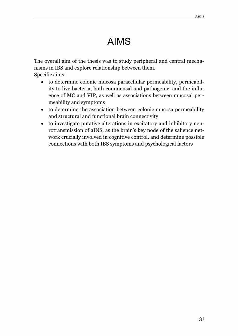
AIMS
The overall aim of the thesis was to study peripheral and central mecha-
nisms in IBS and explore relationship between them.
Specific aims:
to determine colonic mucosa paracellular permeability, permeabil-
ity to live bacteria, both commensal and pathogenic, and the influ-
ence of MC and VIP, as well as associations between mucosal per-
meability and symptoms
to determine the association between colonic mucosa permeability
and structural and functional brain connectivity
to investigate putative alterations in excitatory and inhibitory neu-
rotransmission of aINS, as the brain’s key node of the salience net-
work crucially involved in cognitive control, and determine possible
connections with both IBS symptoms and psychological factors

Peripheral and Central Mechanisms in Irritable Bowel Syndrome - in search of links
32

Method
33
METHOD
Subjects Patients aged 18-60 years, diagnosed with IBS, attending Gastroenterolo-
gy Department, University Hospital in Linköping, were screened for ful-
filling of Rome III criteria. HC aged 18-60 years, without any history of
gastrointestinal disease or complaints, were recruited by an advertise-
ment and offered monetary compensation (Figure 6). Exclusions criteria
for both groups were organic gastrointestinal disease, metabolic, neuro-
logical or psychiatric disorders and nicotine intake within 2 months be-
fore the study, as well as separate sigmoidoscopy exclusion criteria (self-
reported allergy, NSAID intake) and MR exclusion criteria (ferromagnetic
implants, claustrophobia or large tattoos). All participants were right-
handed.
83 patients were initially included in the study, whereof there were
only 12 men (13.6%). Because of the substantial underrepresentation of
the male patients, only female individuals were finally enrolled in the
study. 34 female healthy controls were initially recruited by an advertise-
ment. From the patients’ cohort, 32 women accomplished both sig-
moidoscopy and fMRI, while 34 women, who declined sigmoidoscopy,
underwent only fMRI examination. As for the HC, 15 individuals complet-
ed both fMRI and sigmoidoscopy, while 19 participants did
advertisement responders 115 persons
eligible 47 persons
25 aged matched women
15 (+5) fMRI+sigmoscopy
12 INS qMRS
10 only fMRI
10 INS qMRS
8 men
screened 202 patients
eligible 83 patients
71 women
32 (+5) fMRI+sigmoscopy
5 INS qMRS
34 only fMRI
34 INS qMRS
12 men
A B
Figure 6. Overview of inclusion process for both IBS patients (A) and healthy controls, HC (B). INS, insula.

Peripheral and Central Mechanisms in Irritable Bowel Syndrome - in search of links
34
only fMRI. Moreover, for the assessment of bacterial passage routes in
electron microscopy and immunofluorescence of TJ, five more patients
and five more HC were enrolled, who underwent both sigmoidoscopy and
fMRI.
Because the insula voxel in fMRI examination was added subsequent-
ly during the enrollment, INS qMRS data was finally available for 37 pa-
tients (drop out: 1- water data missing, 1- no left insula data) and 21 HC
(drop out 1-no left insula data). As neurotransmitter concentrations alter
with age [207], meticulous age-matching was performed with an age
range of ± 3 years.
Questionnaires An overview of the type of questionnaires used in each paper is depicted
in Table 1.
IBS Severity Scoring System (IBS-SSS) evaluated overall IBS symp-
tom severity by assessing 5 items: frequency and intensity of abdominal
pain, intensity of bowel distension, satisfaction with bowel habits and in-
terference with daily life [72]. Each item scores between 0 and 100 and
the sum score indicates mild (75-175), moderate (175-300) or severe
(>300) symptoms.
Table 1: Overview of the questionnaires used in each study of the thesis
Questionnaire Paper I Paper II Paper III
IBS-SSS X X X
VSI X X
HADS X X
CSQ X X
PHQ-15 X X
BPI X
GSD X
Mac Arthur X
IBS-SSS, IBS Severity Scoring System; VSI, Visceral Sensitivity index; HADS Hospital Anxiety and Depression Scale; CSQ, Coping Strategies Questionnaire; PHQ-15, Patient Health Ques-tionnaire; BPI, Brief Pain Inventory; GSD, Gastrointestinal Symptom Diary; Mac Arthur, Mac-Arthur Scale of Subjective Social Status.

Method
35
Visceral Sensitivity Index (VSI) was implemented to assess gastroin-
testinal symptom-specific anxiety [123]. The 15-item self-report question-
naire evaluates cognitive, emotional and behavioral responses to fear of
gastrointestinal sensations, symptoms and the context in which the symp-
toms are experienced. Items are scored on a reversed 6-point scale rang-
ing from 0 to 5, with sum scores between 0 and 75. Higher scores indicate
more severe symptom-specific anxiety.
Hospital Anxiety and Depression Scale (HADS) was applied to esti-
mate states of depression and anxiety [274]. The scale consists of seven
items for depression (HADS-D) and anxiety subscales (HADS-A), with
scores on each subscale ranging from 0 to 21. Cut-off values are indicated
as ≥8 for subclinical (suspicious) anxiety or depression and ≥11 as definite
cases on both the HADS-D and HADS-A, respectively [22].
Coping Strategies Questionnaire (CSQ) was used to evaluate cognitive
and behavioral strategies to cope with pain [197], involving six subscales
for cognitive strategies (ignoring pain, reinterpreting pain, diverting at-
tention, calming self-statements, catastrophizing, praying/hoping) and
two subscales for behavioral strategies (increasing activity, increasing
pain behavior). Sum scores between 0 and 36 for each subscale indicate
how frequently a coping strategy is used.
Patient Health Questionnaire (PHQ-15) was applied to assess the de-
gree of somatic symptom severity across 15 different somatic symptoms
[122]. Sum scores ranging from 0 to 30 indicate levels of somatic symp-
tom severity.
Brief Pain Inventory (BPI) is a validated pain assessment tool, that
measures both the intensity of pain (4 items, sensory dimension) and in-
terference of pain with the patient’s life (7 items, reactive dimension) [41].
Each item is scored on a 0-10 scale with sum scores ranging from 0-40 for
pain severity and from 0-70 for interference and higher scores indicating
higher levels of pain severity and interferences, respectively.
Gastrointestinal Symptom Diary (GSD) was used by IBS patients to
record their gastrointestinal symptoms on 14 consecutive days [192] (Fig-
ure 7). The symptoms (abdominal pain, nausea, bloating) as well as every
single bowel movement and stool consistency (defined by Bristol Stool
Chart [134]) were reported along a 24-hour time axis. The values were
manually scored, and the mean frequency of symptom episodes per week
and symptom duration per day was extracted from the diary data.

Peripheral and Central Mechanisms in Irritable Bowel Syndrome - in search of links
36
MacArthur Scale of Subjective Social Status (Mac Arthur) ladder, a
part of the MacArthur Socio-Economic Status (SES) questionnaire, aims
to assess the common sense of socio-economic status, measuring only
traditional, objective SES indicators [1,219]. The participant is requested
to mark the place on a "SES ladder” that corresponds to the person’s so-
cio-economic status, where the higher steps of the ladder, the higher in-
come, occupation, and education.
Sigmoidoscopy Colonic biopsies were collected during a flexible sigmoidoscopy per-
formed after 8 hours fasting and bowel cleansing with an enema before
the procedure. Biopsies, taken with a biopsy forceps without a central
lance, were directly put in ice-cold oxygenated Krebs buffer and trans-
ported to the laboratory for mounting on Ussing chambers for permeabil-
ity measurements.
Blood tests Prior to the sigmoidoscopy, blood samples (6ml fasting venous blood in
an EDTA tube) were collected for measurements of VIP plasma concen-
trations. To measure VIP levels in plasma a VIP- Enzyme Immunoassay
(EIA) kit was implemented according to the manufacturer’s instructions
(Phoenix Pharmaceuticals, Germany).
Figure 7: Gastrointestinal symptom diary card.

Method
37
Mucosal barrier measurements
Using chamber experiments
Mucosal barrier studies were performed in modified Ussing chambers
(Harvard apparatus Inc., Holliston, MA, USA; Figure 8) [79]. Mucosal
biopsies were carefully mounted to cover the 1.8mm² of exposed tissue
surface area. Thereafter 1.5 ml 10 mM mannitol in Krebs buffer was added
to the mucosal side of the chamber, while the serosal side was filled with
1,5ml 10 mM glucose in Krebs buffer. The system was continuously circu-
lated by gas flow, 95% O2/5% CO2, while simultaneously oxygenated and
kept warm at 37°C. Totally 40 min equilibration prior to the experiment
start was completed to obtain steady-state in transepithelial PD. The elec-
trophysiology measurements, Isc, TER and PD were monitored by the
means of two platinum electrodes for current passage and two silver chlo-
ride Ag/AgCl-electrodes for PD measurements, all in open circuit condi-
tions. Every second minute of the whole experiment period, sampling of
electrophysiology data were performed via LabVIEW software (LabVIEW,
National Instruments, Solna Sweden) by sending direct alternating cur-
rent pulses through the specimens and measuring the voltage response. A
current (I) – voltage (U) pair relationship linear-squares fit was done to
calculate the TER from the slope of the line and the PD from the intersec-
tion of the voltage (U=PD+TERxI). All parameters were calculated during
the 30-60-90-120 min period.
Figure 8: The drawing (A) and the photograph (B) of the Ussing chamber used in the thesis. Reprinted from Medical Dissertation No 1006, Linkö-ping University, with permission from Åsa Keita. Ag/AgCl, silver chloride electrodes.

Peripheral and Central Mechanisms in Irritable Bowel Syndrome - in search of links
38
Bacterial permeability
The aim of the study was to determine mucosal permeability to live bacte-
ria, both commensal and pathogenic. As mentioned before, the commen-
sal bacteria Escherichia coli seems to play a role in dysbiosis, lately asso-
ciated with IBS pathophysiology [174], and Salmonella typhimurium in-
fection of the gastrointestinal tract is widely recognized as a risk factor for
the subsequent development of IBS [114,158]. Therefore the choice of
these two bacterial strains seemed to be warranted. Escherichia coli HS
and Salmonella typhimurium were prepared according to routines de-
scribed previously [113]. Specifically, 5 ml of plasmid enhanced green flu-
orescent protein 1:100 was added to 50 ml of bacterial competent, careful-
ly shaken and incubated on ice for 30 min before heat shock at 40°C for
90 s. Subsequently, the bacteria were incubated added 450 ml SOC medi-
um (Super Optimal broth with Catabolite repression), at 37°C, 200
r.p.m.(revolutions per minute), for 1 hour, followed by 12 hours incuba-
tion at 37°C plated on LB (Lysogeny broth)+ ampicillin agar plate. After
20 min of Ussing chamber equilibration final concentrations of 108
CFU/ml of live green fluorescent protein (GFP) -labelled Escherichia coli
HS and Salmonella typhimurium were added to the mucosal side of sepa-
rate chambers. Bacterial passage was designated in a multileader plate
reader.
Paracellular permeability
51Cr-EDTA is a widely known marker for the paracellular permeability
used in Ussing chamber experiments that is proven to pass through the
mucosa between the cells. The firm binding of the radioactive Cr to the
EDTA molecule ensures equal Cr and EDTA passage and minimizes reac-
tion between the tissue Ca2+ and EDTA. After 20 min of Ussing chamber
equilibration mucosal side of each chamber was added 34μCi/ml 51Cr-
EDTA. At 0, 60 and 120 min of experiment, serosal samples were collect-
ed and replaced with an equal volume of Krebs solution. The paracellular
passage was calculated by gamma-counting (1282 Compugamma; LKB,
Bromma, Sweden).
Blocking agents
In order to explore the role of MC and VIP in intestinal barrier function
specific blocking agents were used. After initial 20 min equilibration, the
biopsies were treated on the serosal side with either 1μM of VIP receptor
blocker anti-VPAC, 1μM of MC stabilizer ketotifen in duplicate systems, or

Method
39
vehicle, and kept in equilibrium in additional 20 min before the bacteria
or 51Cr-EDTA were added. VIP receptor blocker is a fragment of VIP sig-
nal peptide containing only 23 amino acids (6-28) out of 30, thus it main-
tains binding affinity to both VIP receptors, while exerting no activation
(V4508 Sigma-Aldrich). Ketotifen is a MC degranulation stabilizer that
demonstrated some positive effect on IBS patients as described in the in-
troduction [120].
Immunofluorescence of MC tryptase, VIP, VPACs
Different processes of detecting particular antigens in tissue sections
based on the affiliation of these cellular antigens to specific antibodies are
termed immunohistochemistry. Immunofluorescence is an immunohisto-
chemical staining method of visualizing antigens after conjugating and
binding reaction with fluorescent dyed antibodies. Fluorescence is acti-
vated when an exciting light of a specific wavelength is applied on the pre-
treated specimen under a fluorescent microscope.
Colonic biopsies were fixed with 4% formaldehyde, dehydrated ac-
cording to standard procedures before embeded in paraffin blocks, micro-
tome sliced and mounted on a microscope slide. After heat fixation an in-
verse process of deparaffinisation and hydration took place, prior to treat-
ing with blocking reagent, in order to reduce nonspecific background
staining. Then, the samples were stained with primary antibodies and in-
cubated in a dark humidified chamber over night, at 4°C. After washing
with phosphate buffered saline (a water-based isotonic salt solution, PBS)
and incubating with secondary antibody, the samples were cover slipped
with anti-fade reagent and were ready for immunofluorescence examina-
tion the next day.
Intestinal MC contain huge amounts of tryptase in their granules, and
therefore anti-MC tryptase antibody was used to detect MC. As VIP is a
neuropeptide widely recognized to influence human digestive system in
various ways [169], for instance regulating intestinal permeability [112],
we stained the biopsies for both VIP and its receptors, VPAC1 and VPAC2
by using anti-VIP, anti-VPAC1 and anti-VPAC2 antibodies in immunoflu-
orescence and examined co-localization of VIP/VPAC1/VPAC2 and MC.
Expressions of MC, VIP/VPAC1/VPAC2 were evaluated in fluorescent
confocal microscopy.

Peripheral and Central Mechanisms in Irritable Bowel Syndrome - in search of links
40
Magnetic Resonance Imaging data acquisition and
analysis
Functional Magnetic Resonance Imaging protocol
In order not to influence the fMRI examination output following a poten-
tially unpleasant examination, such as sigmoidoscopy, according to the
study protocol, the participants underwent MR scan firstly, whereas sig-
moidoscopy was performed within 2 weeks’ time afterwards. This time-
period was set as a general recommendation and its protraction was not
an exclusion criterium, as individual intestinal permeability is supposed
to be quite stable over a period of at least 8 weeks, when repeating meas-
urements [144,248,273].
Participants were requested to fast for at least four hours (water was
acceptable) and refrain from consuming alcohol and taking any IBS-
related, pain, or sleep medication for at least 24 hours before the MRI
scan. Security protocol was collected from the participants to ensure no
contraindications to MR scanning. Images were collected on a 3 T Philips
Ingenia (Philips Healthcare, Best, The Netherlands) equipped with a 32-
channel head coil at the Center for Medical Image Science and Visualiza-
tion (CMIV) at Linköping University, Sweden. Prior to fMRI and qMRS, a
standard MR screening with T1-weight images was performed in all par-
ticipants to exclude substantial brain abnormalities, followed by qMRS
and thereafter fMRI and DTI examination.
Resting state functional MRI
Functional MRI data were obtained using the following parameters: repe-
tition time (TR) 2 seconds, echo time (TE) 37 milliseconds and voxel size
3.59 x 3.59 x 4mm3 in 28 slices. As magnetic resonance imaging is ex-
tremely sensitive to motion the rs-fMRI images were preprocessed follow-
ing the guidelines of the GIFT group independent component analysis
(ICA) toolbox, as described below [5].
fMRI uses BOLD as an indirect measure of neural activity encoded for
each voxel (volume element) of the brain, resulting in an abundance of
relevant and irrelevant signals. To organize and analyze the data mathe-
matical models are used to separate the signals of interest and these not
of interest. ICA is a blind source signal separation, which means that it

Method
41
isolates unknown signals in a single subject, grading them according to
most explained variance. It extracts independent patterns of comprehen-
sible fMRI activity that varies together over time, both in positive and
negative direction, and is significantly distinguishable from activities in
other sets of voxels. GIFT is an application allowing group assumption
from fMRI data using ICA, which is conducted in three main stages: data
compression, ICA and finally back reconstruction to recreate individual
subject and group components of the signals of interest.
Statistical parametric mapping SPM8 (http://www.fil.ion.ucl.ac.uk/
spm) was used to realign the images that were subsequently spatially
normalized using a standard brain atlas of the Montreal Neurologic Insti-
tute (MNI). They were additionally smoothed with an 8 mm full width
half maximum (FWHM) Gaussian kernel to diminish image noise and
further reduce the effects of individual anatomical variability. Head mo-
tion less than one voxel in any direction was set as a limit that has not
been exceeded in any of the examinations. Group ICA toolbox GIFT v4.0a
(http://icatb.sourceforge.net; [31]) was used to calculate functional con-
nectivity. After performing single ICA on all cases [18] with back recon-
Figure 9. Group independent DMN component. The one-sample t-test statistical map over all 47 subjects is thresholded at p < 0.05, Family Wise Error corrected for comparing across the whole brain. Color bar given in terms of t-statistic.
Peripheral and Central Mechanisms in Irritable Bowel Syndrome - in search of links
42
struction of every subject´s spatial maps from the raw data [65], 45 inde-
pendent components were calculated [135] and assured for stability using
ICASSO [135] with 500 iterations. ICASSO deals with the major ICA prob-
lem of reliability of the estimated components due to statistical error of
limited sample size as well as practical issues with ICA algorithm and the
real data that not always follow ICA model. ICASSO investigates ICA sta-
tistical reliability and stability by running the algorithm multiple times
with differently bootstrapped data sets in order to assess ICA estimates.
To identify the DMN components spatial regression with a template
of the DMN [222] was employed (Figure 9). The color brain maps use t-
statistics of the brain areas, presenting positive synchronous activation
marked in warm colors (red- low significance, yellow- high significance)
and negative simultaneous activation marked in cold colors (blue- low
significance, green- high significance), as shown in Figure 9.
Diffusion tensor imaging
DTI data were obtained using a 64-direction sequence with TR 9,339 sec-
onds, TE 82 milliseconds and voxel sixe 2 mm3 and diffusion weighting of
b=1000 seconds/mm2. The data were processed using standard proce-
dures (https://fsl.fmrib.ox.ac.uk/fsl/fslwiki/FSL; [104,223]) after being
reconstructed on the scanner. FLIRT toolbox [80,103] was employed to
correct for distortion and subjects movement due to eddy-current. Indi-
vidual FA images were spatially normalized to MNI template using tract-
based spatial statistics (TBSS; [221]) according to FNIRT toolbox [6] to
obtain group-level maps of FA.
Quantitative Magnetic Resonance Spectroscopy
qMRS measures were accomplished using MEGA-PRESS sequence (kind-
ly provided by R. Edden, Johns Hopkins University; [163,167]) with TR 2
seconds and TE 68 msec, edited pulses “ON” at 1.90 ppm and “OFF” at
7.46 ppm and water suppression MOIST (multiple optimizations insensi-
tive suppression train), in a voxel of 4.5x2.0x3.0 cm3 located in the right
and left aINS (Figure 10A, B). MEGA-PRESS sequence employs splitting
of the spectrum peak that varies in phase and amplitude depending on TE
due to J coupling (spin-spin coupling), occurring when protons in a mole-
cule are affected by other protons due to sharing of the electrons. MEGA-
PRESS is primarily optimized for evaluation of the weak GABA molecules

Method
43
spectrum, brain neurotransmitter with editing coupled signals at 1.9 and
3 ppm due to J coupling [173,205]. MEGA-PRESS exploits J-difference
editing by subtracting two data sets: “ON” and “OFF”. During “ON” acqui-
sition pulses at 1.9 ppm are applied, that selectively resonate some of the
GABA protons and simultaneously refocus the coupled protons at 3 ppm.
During “OFF” acquisition impulses applied have no influence on GABA
signals. Subtraction of “OFF” from “ON” spectra results in revealing only
those 1.9 ppm signals that are coupled with 3 ppm signals, which is specif-
Figure 10: Typical qMRS volume of interest (voxel size 4.5x2.0x3.0 cm3) placement in the (A) right and (B) left aINS. The yellow box illustrates the voxel targeting the GABA signal at 3.0 ppm, and the white box illus-trates the voxel the water residual at 4.7 ppm originates from, which shows the chemical shift displacement. Representative spectra with LCModel fitting depict (C) an averaged difference spectrum for the ex-traction of GABA+ and (D) an averaged MEGA-PRESS OFF spectrum for Glx extraction (labeled as 2 and 7, extracted solely from OFF-spectra) from a healthy volunteer (black line: post-processed spectra prior to fit-ting; red line: LCModel fit). Residuals are shown at the top of each pan-el. (Assignments: 1, Creatine (-2CH2-); 2, Glx (-2CH-); 3, Choline (-N(CH3)3); 4, Creatine (-N(CH3)); 5, GABA+ (-4CH2-); 6, tNA (-3CH2-); 7, Glx (-4CH2-); 8, GABA+ (-2CH2-); 9, tNA (-2CH3); 10. GABA+ (-3CH2-); 11-13, Macromolecules and lipids, (-CH2-). Abbreviations: Glx, glutamate+glutamine; GABA+, γ-Aminobutyric acid + coedited macro-molecular signals); tNA, total N-Acetyl compounds (NAA + NAAG).

Peripheral and Central Mechanisms in Irritable Bowel Syndrome - in search of links
44
ic for GABA. Linear combination of model spectra LCModel (Version 6.3-
1L; [189]) was applicated to calculate GABA+ from difference spectrum
after subtracting “OFF” from “ON” spectrum (Figure 10C). Glx was com-
puted from “OFF” spectrum (Figure 10D).
Statistics
Paper I
All parametric data, according to Shapiro-Wilk normality test, such as VIP
concentration in blood samples and biopsy lysates, MC density and ex-
pression of VPAC1 and VPAC2 receptors were shown as mean ± standard
error of mean (SEM) and comparisons between groups were performed
with Student t test (for comparing two groups) and ANOVA test (for more
than two groups).
Non-parametric data such as bacterial and paracellular permeability
were given as median (25th–75th interquartile range). Comparisons be-
tween groups were done with Mann–Whitney U tests for comparing of
two groups and Kruskal–Wallis test was used for comparing three or
more groups. Wilcoxon matched-pairs signed rank test was used for
paired comparisons of immunofluorescency measures of TJ with and
without Salmonella exposure. Differences with p<.05 were considered
significant. Correlation of non-parametric data were performed using
Spearman Correlation test. Analyses were calculated on GraphPad Prism
7 (https://www.graphpad.com/scientific-software/prism).
Paper II
Although not all questionnaire data passed the Shapiro-Wilk test, the re-
sults were presented as mean ± standard deviation (SD) in order to facili-
tate possible future interpretation and comparisons. Mann-Whitney U
test was used to calculate between group differences and Spearman’s rho
for the correlation between permeability and questionnaire data.
Analyses on the behavioral data were calculated using SPSS v24
(https://www.ibm.com/products/spss-statistics). GraphPad Prism 7
(https://www.graphpad.com/scientific-software/prism) was used to cre-
ate the graphs.
For correlations of permeability, DMN functional connectivity and
DTI data both between group (IBS vs HC) and within group (IBS and HC)

Method
45
permutation analyses using PALM software tool
(https://fsl.fmrib.ox.ac.uk/fsl/fslwiki/PALM; [262]) with 10,000 itera-
tions were performed. In general, a permutation is an alignment of all or
part of objects in a set, with regard to the sequence of the alignment. The
within-group analyses were restricted to the brain regions significant in
the initial between group analyses (masked at p<0.05, Family Wise Error
(FWE) correction for the whole brain comparisons). FWE is defined as
the probability of making at least one false conclusion in the series of hy-
pothesis tests. As depression and anxiety are known to influence func-
tional connectivity, both subscores from HAD questionnaire (HAD-A and
HAD-D) were included in all analyses as covariates of no interest, likewise
the time slot between the MR scan and sigmoscopy examination. Moreo-
ver, Threshold Free Cluster Enhancement (TFCE; [220]) was used to de-
fine the clusters with significance set at p < 0.05 and FWE correction. In a
fMRI analysis numerous statistical tests were used due to >80,000 voxels
included in the analysis, which makes it necessary for appropriate correc-
tion for multiple comparisons. TFCE is a well-established method for
multiple comparisons correction of statistical maps in fMRI studies. The
calculations were performed using the software Mango
(http://ric.uthscsa.edu/mango/ ; J.L. Lancaster and M.J. Martinez).
Path analysis
Path analysis is a type of multiple regression analysis, which is in general
a statistical analysis designated for normally distributed data sets. It is a
confirmatory statistical method that is based on a created by the re-
searcher theory about some associated regression relationships. Path
analysis tests the goodness of fit of this theory against a perfect model. In
our study the path analysis was done to test if the detected associations
between permeability and functional brain connectivity were related di-
rectly or they were mediated by some clinical characteristics from the
questionnaires. Prior to creating the model, significant correlations be-
tween the components had to be detected. As Spearman’s rho from barri-
er function data showed, only the paracellular permeability was signifi-
cantly correlated to some of the IBS questionnaire data, namely symptom
severity (IBS-SSS), gastrointestinal related anxiety (VSI) and coping
(CSQ-pain behavior). In order to define the association between 51Cr-
EDTA permeability and RVM functional connectivity permutation anal-
yses were used. These findings were confirmed by a simple calculation
with Pearson’s correlation coefficient. The path model was constructed

Peripheral and Central Mechanisms in Irritable Bowel Syndrome - in search of links
46
delineating both a direct relation between the gut and brain data as well
as indirect relations through the three clinical components mentioned
above. The path analysis was calculated using Mplus
(https://www.statmodel.com/) with all path coefficients presented in a
standardized form. As mentioned before, not all of the data were normally
distributed; therefore nonparametric bootstrapping with 2000 replica-
tions was also used. It is a method of inferential statistics based on the
principle of multiple sampling of a set of data while allowing for replace-
ment, and measuring a statistic of this sample. By replicating the proce-
dure numerous times, one can infer that the statistic measured on the
multiple samples corresponds with the statistic of the population.
Paper III
As not all questionnaire data passed the Shapiro-Wilk test, results were
presented as median (25th–75th interquartile range). Mann-Whitney U
test was used to calculate between group differences in Glx and GABA+
concentrations and questionnaire data. Chi Square test was applied for
categorical data group comparisons of medical journal data on previously
diagnosed anxiety and / or depression and medication use. Effect sizes
were indicated using Cohen’s d for results from U-test and as Cramér’s V
for Chi Square test.
Stepwise multiple linear regression analyses (criteria: probability to
enter ≤ .05, probability to remove ≥ .10) were carried out in IBS patients
to determine the significant predicting factors of aINS Glx and GABA+ at
p < .05. Statistical analyses were calculated using SPSS version 25 (IBM
Corporation, Armonk, NY) and GraphPad Prism 7
(https://www.graphpad.com/scientific-software/prism).
Ethical approval Ethical approval for the study was obtained from the Regional Ethical Re-
view Board in Linköping, Sweden (Dnrs. 2013/506-32; 2014/264-32). All
the participants gave written, informed consent according to the Helsinki
Declaration.

Results
47
RESULTS
Clinical characterization of the patients Demographic data for the female patients’ and the female HC’ cohorts in
all three Papers are depicted in Table 2. The cohort from Paper I and II
was identical except for the 5 additional patients and HC in Paper I, who
were included for the purpose of supplementary electron microscopy
studies. The patients and some of the HC from the Paper III cohort were
included subsequently, and thereby are not the same individuals as in Pa-
per I and II. There was no statistical difference between age of IBS and
HC in any of the cohorts. The vast majority of IBS patients had moderate
to severe disease intensity according to IBS-SSS questionnaire. The per-
centage of different types of IBS subgroups according to the Rome III cri-
teria is depicted in Table 2. This IBS subgrouping however, turned out not
to be significant in any of the main correlations explored in the thesis.
Table 2. Clinical characteristics of the cohorts included in Papers I, II and III
Paper I* Paper II* Paper III**
IBS Total 37 32 39
Age 32.2 (19-55) 32.6 (19-55) 32.1 (18-57)
IBS-SSS 347 (167–480) 343 (167-480) 336 (203-468)
IBS-D 8 (21.5%) 7 (22%) 10 (26%)
IBS-C 8 (21.5%) 8 (25%) 7(18%)
IBS-M 21 (57%) 17 (53%) 22(56%)
HC Total 20 15 21
Age 29.9 (20-48) 29.8 (21-48) 32.1 (20-55) Age and IBS-SSS given as mean (range). IBS-SSS, IBS Severity Scoring System; IBS-D, IBS with predominant diarrhea; IBS-C, IBS with predominant constipation; IBS-M, IBS with mixed type of stools *overlapping cohorts in Paper I and II; **separate cohort in Paper III
Severe psychiatric diseases such as schizophrenia, bi-polar disorder,
psychosis, severe depression etc, were exclusion criteria, as well as contra-
indications for MRI and structural neurological disorders. Roughly one
third of the patients as well as two HC (one HC in Paper I & II; two HC in
Paper III) had elevated HADS anxiety scores, and only a fraction of IBS
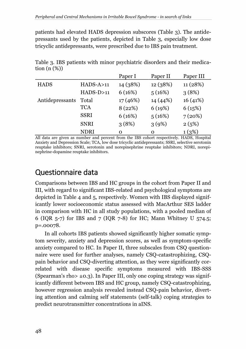
Peripheral and Central Mechanisms in Irritable Bowel Syndrome - in search of links
48
patients had elevated HADS depression subscores (Table 3). The antide-
pressants used by the patients, depicted in Table 3, especially low dose
tricyclic antidepressants, were prescribed due to IBS pain treatment.
Table 3. IBS patients with minor psychiatric disorders and their medica-tion (n (%))
Paper I Paper II Paper III
HADS HADS-A>11 14 (38%) 12 (38%) 11 (28%)
HADS-D>11 6 (16%) 5 (16%) 3 (8%)
Antidepressants Total 17 (46%) 14 (44%) 16 (41%)
TCA 8 (22%) 6 (19%) 6 (15%)
SSRI 6 (16%) 5 (16%) 7 (20%)
SNRI 3 (8%) 3 (9%) 2 (5%)
NDRI 0 0 1 (3%) All data are given as number and percent from the IBS cohort respectively. HADS, Hospital Anxiety and Depression Scale; TCA, low dose tricyclic antidepressants; SSRI, selective serotonin reuptake inhibitors; SNRI, serotonin and norepinephrine reuptake inhibitors; NDRI, norepi-nephrine-dopamine reuptake inhibitors.
Questionnaire data Comparisons between IBS and HC groups in the cohort from Paper II and
III, with regard to significant IBS-related and psychological symptoms are
depicted in Table 4 and 5, respectively. Women with IBS displayed signif-
icantly lower socioeconomic status assessed with MacArthur SES ladder
in comparison with HC in all study populations, with a pooled median of
6 (IQR 5-7) for IBS and 7 (IQR 7-8) for HC; Mann Whitney U 574.5;
p=.00078.
In all cohorts IBS patients showed significantly higher somatic symp-
tom severity, anxiety and depression scores, as well as symptom-specific
anxiety compared to HC. In Paper II, three subscales from CSQ question-
naire were used for further analyses, namely CSQ-catastrophizing, CSQ-
pain behavior and CSQ-diverting attention, as they were significantly cor-
related with disease specific symptoms measured with IBS-SSS
(Spearman's rho> ±0.3). In Paper III, only one coping strategy was signif-
icantly different between IBS and HC group, namely CSQ-catastrophizing,
however regression analysis revealed instead CSQ-pain behavior, divert-
ing attention and calming self statements (self-talk) coping strategies to
predict neurotransmitter concentrations in aINS.
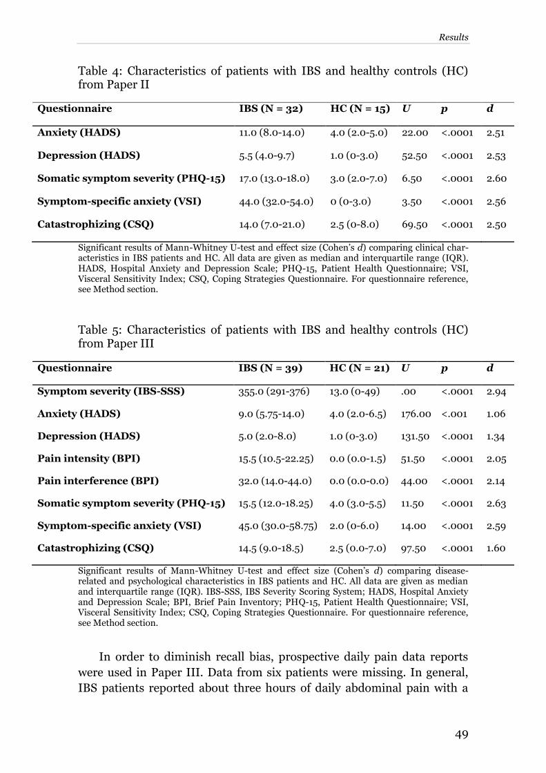
Results
49
Table 4: Characteristics of patients with IBS and healthy controls (HC) from Paper II
Questionnaire IBS (N = 32) HC (N = 15) U p d
Anxiety (HADS) 11.0 (8.0-14.0) 4.0 (2.0-5.0) 22.00 <.0001 2.51
Depression (HADS) 5.5 (4.0-9.7) 1.0 (0-3.0) 52.50 <.0001 2.53
Somatic symptom severity (PHQ-15) 17.0 (13.0-18.0) 3.0 (2.0-7.0) 6.50 <.0001 2.60
Symptom-specific anxiety (VSI) 44.0 (32.0-54.0) 0 (0-3.0) 3.50 <.0001 2.56
Catastrophizing (CSQ) 14.0 (7.0-21.0) 2.5 (0-8.0) 69.50 <.0001 2.50
Significant results of Mann-Whitney U-test and effect size (Cohen’s d) comparing clinical char-acteristics in IBS patients and HC. All data are given as median and interquartile range (IQR). HADS, Hospital Anxiety and Depression Scale; PHQ-15, Patient Health Questionnaire; VSI, Visceral Sensitivity Index; CSQ, Coping Strategies Questionnaire. For questionnaire reference, see Method section.
Table 5: Characteristics of patients with IBS and healthy controls (HC) from Paper III
Questionnaire IBS (N = 39) HC (N = 21) U p d
Symptom severity (IBS-SSS) 355.0 (291-376) 13.0 (0-49) .00 <.0001 2.94
Anxiety (HADS) 9.0 (5.75-14.0) 4.0 (2.0-6.5) 176.00 <.001 1.06
Depression (HADS) 5.0 (2.0-8.0) 1.0 (0-3.0) 131.50 <.0001 1.34
Pain intensity (BPI) 15.5 (10.5-22.25) 0.0 (0.0-1.5) 51.50 <.0001 2.05
Pain interference (BPI) 32.0 (14.0-44.0) 0.0 (0.0-0.0) 44.00 <.0001 2.14
Somatic symptom severity (PHQ-15) 15.5 (12.0-18.25) 4.0 (3.0-5.5) 11.50 <.0001 2.63
Symptom-specific anxiety (VSI) 45.0 (30.0-58.75) 2.0 (0-6.0) 14.00 <.0001 2.59
Catastrophizing (CSQ) 14.5 (9.0-18.5) 2.5 (0.0-7.0) 97.50 <.0001 1.60
Significant results of Mann-Whitney U-test and effect size (Cohen’s d) comparing disease-related and psychological characteristics in IBS patients and HC. All data are given as median and interquartile range (IQR). IBS-SSS, IBS Severity Scoring System; HADS, Hospital Anxiety and Depression Scale; BPI, Brief Pain Inventory; PHQ-15, Patient Health Questionnaire; VSI, Visceral Sensitivity Index; CSQ, Coping Strategies Questionnaire. For questionnaire reference, see Method section.
In order to diminish recall bias, prospective daily pain data reports
were used in Paper III. Data from six patients were missing. In general,
IBS patients reported about three hours of daily abdominal pain with a

Peripheral and Central Mechanisms in Irritable Bowel Syndrome - in search of links
50
frequency of about seven episodes per week (for details see Table 3 Paper
III).
Mucosal barrier data Electrophysiology, measured by TER after equilibration, was stable, as
well as Isc measured throughout the experiment.
There was a significantly decreased TER in biopsies from IBS com-
pared to HC in the beginning of the experiments, after 30 minutes, as well
as after 60 minutes from the start. At 90 minutes TER was still lower in
IBS, however it did not reach statistical significance.
Interestingly, only in biopsies from IBS added Salmonella typhimuri-
um compared to vehicle, TER was significantly decreased, both after 30,
60 and 90 min and this decrease was hampered by pretreating with Keto-
tifen but not with anti-VPACs.
Bacterial and paracellular permeability
Both bacterial and paracellular mucosal passage was significantly higher
in biopsies of IBS group compared to HC. Bacterial permeability was 2.79
times higher for Salmonella in biopsies from IBS than from HC and 1.88
times higher for Escherichia coli in biopsies from IBS than from HC (Fig-
ure 11). Bacterial passages were not intercorrelated, possibly implicating
different uptake mechanisms of the commensal and pathogenic bacteria
strains.
Figure 11. Bacterial passage through colonic biopsies mounted in Ussing chambers for 120 min. Comparisons were performed with Mann–Whitney U test, ***p < .0005.

Results
51
The differences were not that robust for paracellular permeability, but
still significantly increased in biopsies from IBS compared to HC (Figure
12). Despite substantial overlap between IBS and HC results, a small sub-
group of IBS patients showing elevated paracellular permeability could be
defined (Figure 12, red dots). Further analysis revealed that this small
subgroup also reported significantly less severe IBS symptoms and less
symptom-specific anxiety and somatization than the subgroup with more
”normal” paracellular permeability. 51Cr-EDTA permeability was not af-
fected by adding bacterial strains into chambers. There was no correlation
between 51Cr-EDTA and bacterial passages.
Regulation by VIP and MC
In chambers pre-treated with ketotifen or anti-VPACs bacterial permea-
bility was significantly decreased not only in IBS but also in HC (Figure 13
A, B).
There was higher plasma VIP concentration in IBS compared to HC,
although it did not correlate with any of the Ussing chamber measure-
ments.
Immunfluorescence examinations showed increased amount of MC in
colonic biopsies from IBS compared to HC as well as the percentage of
MC expressing VPAC1, but no significant difference neither for MC ex-
pressing VPAC2, nor for MC containing VIP.
Figure 12. 51Cr-EDTA passage through colonic biopsies of IBS and HC mounted in Ussing chambers at 120 min. Comparisons were performed with Mann–Whitney U test, *p < .05 vs vehicle. Red dots, a subgroup of IBS patients with elevated permeability (1.35-2.155cm/sx10-6).

Peripheral and Central Mechanisms in Irritable Bowel Syndrome - in search of links
52
MC density correlated with the percentage of MC expressing VPAC1
as well as the percentage of MC expressing VPAC1 was associated with the
percentage of MC expressing VPAC2.
Questionnaire data and intestinal permeability
Bacterial permeability revealed no significant correlation with any of the
symptoms assessed using the questionnaires.
Quite surprisingly, we found a negative correlation between paracellu-
lar permeability and some of the IBS-specific symptoms, such as intesti-
nal symptom severity (measured with IBS-SSS, Spearman's rho=−0.41, p
< 0.025), symptom-specific anxiety (measured with VSI, Spearman's
rho=−0.42, p < 0.024) as well as coping strategy, pain behavior (assessed
with CSQ, Spearman's rho=−0.502, p < 0.006) showed in Figure 14.
Figure 13. Influence of VIP and MC blockers on passage of E coli HS (A) and S typhimurium (B) through colonic biopsies of IBS and HC mounted in Ussing chambers at 120 min. Comparisons were performed with Mann–Whitney U test, **p < .005, ***p < .0005 vs vehicle.

Results
53
Figure 14. Graphs of questionnaires exhibiting significant correlations with levels of 51Cr-EDTA passage in the IBS participants only. From left to right, these questionnaires were IBS-SSS, IBS Severity Scoring System; VSI, Vis-ceral Sensitivity Index; and CSQ, Coping Strategies Questionnaire, pain be-havior subscale.
Resting state functional connectivity data
Paracellular permeability and DMN connectivity
All the within-group results both for paracellular permeability and bacte-
rial permeability (see below) were masked with the between-group results
thresholded at p < 0.05 with FWE correction.
Within-group and between-group results of correlation between para-
cellular permeability and DMN connectivity are depicted in Table 6.
Overall, brain features that correlated with paracellular permeability
in the two study groups were hardly overlapping, which would suggest
IBS-specific character of the alterations found in this group. In the main,
the correlations between paracellular permeability and FC in HC were
more various and more widely distributed than in IBS group.
Most interestingly, right amygdala exhibited negative correlation be-
tween FC with DMN and paracellular permeability in IBS group, whereas
in HC group changes in connectivity in this brain area were not detected
at all.
In HC, on the other hand, decreasing paracellular permeability was
associated with increased FC between DMN and PAG, which was not ob-
served in IBS group.
Correlation between paracellular permeability and RVM connectivity
with DMN was detected in both groups, however in HC the correlation
was positive (increased paracellular permeability - increased connectivity
RVM-DMN), while in IBS negative (increased paracellular permeability -

Peripheral and Central Mechanisms in Irritable Bowel Syndrome - in search of links
54
Table 6. Within-group and between-group results in correlation between
paracellular permeability and DMN connectivity
DMN connectivity with areas belonging to brain networks
PP Pain CEN SN EAN SMN
HC
pos RVM right middle frontal gyrus
bilateral pre-/postcentral gyri,
thalamus, SMA pa-racentral lobule
neg PAG ACC, right INS
bilateral precentral
gyri
IBS
pos
left inferior frontal gyrus,
right superior and medial frontal gyrus, caudate
ACC
SMA and medial paracentral lob-ule/post-central
gyrus
neg RVM
left middle and superior frontal gyrus, right infe-rior frontal gyrus
ACC
right amygdala/ parahippo-
campus
left precentral gyrus right post-central
gyrus
IBS vs HC
PAG RVM
bilateral middle and inferior frontal gyri
right amygdala, left orbital frontal cor-
tex
bilateral pre−/postcentral gyri, thalamus,
SMA, paracentral lobule
DMN, default mode network; PP, paracellular permeability; Pain, pain processing brain regions; CEN, central executive network; SN, salience network; EAN, emotional arousal network; SMN, sensorimotor network; IBSvsHC, between-group differences; RVM, rostroventral medulla; PAG, periaqueductal grey matter; ACC anterior cingulate cortex; INS, insula; SMA, supplementary motor area
- decreased connectivity RVM-DMN).
As far as DTI results are concerned, the association between paracel-
lular permeability and white matter microstructure would suggest that the
findings from rs-fMRI on FC between diverse brain regions and DMN in
relation to paracellular permeability correspond to increased white matter
fiber organization in the tracts subserving those areas.
Bacterial permeability and DMN connectivity
Mucosal permeability for Escherichia coli showed no specific correlation
with functional rs-fMRI connectivity of DMN, hence bacterial permeabil-
ity described below refers to mucosal permeability for Salmonella typhi-
murium.

Results
55
Within-group and between-group results in correlation between bac-
terial permeability for Salmonella typhimurium and DMN connectivity
are depicted in Table 7. Correlations between bacterial permeability for
Salmonella typhimurium and DMN connectivity in HC and IBS showed
more overlap than the findings for paracellular permeability did. Bacterial
permeability showed no correlation to RVM-DMN connectivity, as it was
the case for paracellular permeability, suggesting that probably paracellu-
lar permeability may represent more important source of central sensiti-
zation than the passage of live bacteria.
From DTI measurements, white matter fiber organization between
some of these brain regions was also increased, suggesting structural un-
derpinnings of observed functional connectivity.
Table 7. Within-group and between-group results in correlation between bacterial permeability and DMN connectivity
DMN connectivity with areas belonging to brain networks
BP Pain CEN SN EAN SMN
HC
pos bilateral or-bital frontal
cortices
neg RVM middle frontal gyri
ACC pre− and postcentral
gyri
IBS
pos pons medial frontal gyrus
SMA and medial pa-racentral lob-
ule/post-central gy-rus
neg left INS
left precentral gyrus
right post-central gyrus
IBS vs HC
RVM pons
middle and inferior
frontal gyri
ACC INS
orbital frontal cor-
tex
bilateral pre−/postcentral
gyri, SMA, paracen-tral lobule
DMN, default mode network; BP, bacterial permeability; Pain, pain processing brain regions; CEN, central executive network; SN, salience network; EAN, emotional arousal network; SMN, sensorimotor network; IBSvsHC, between-group differences; RVM, rostroventral medulla; ACC anterior cingulate cortex; INS, insula; SMA, supplementary motor area

Peripheral and Central Mechanisms in Irritable Bowel Syndrome - in search of links
56
Paracellular permeability, DMN-RVM functional connectivity
and questionnaire data
Given on one hand, that in IBS only paracellular permeability showed
correlation with clinical and behavioral parameters, and on the other
hand that in this group the correlation between paracellular permeability
and DMN-RVM functional connectivity was reversed compared to HC,
the question arised if and how the observed FC within RVM was related to
IBS and psychological symptoms. To explore this, a path analysis was per-
formed (Figure 15). Because RVM is known to be a key brain region in-
volved in descending pain modulation, we assumed in our path analysis
the unidirectional effect of paracellular permeability on RVM functional
connectivity and chose the questionnaires correlated with paracellular
permeability (IBS-SSS, VSI and CSQ-pain behavior) to model indirect ef-
fects on RVM (Figure 15). Path analysis indicated significant total effect of
paracellular permeability on RVM (−0.603 ± 0.134, p < 0.001), in line
with our resting state DMN connectivity findings, as well as significant
direct effect (−0.399 ± 0.174, p < 0.022). The other component of the to-
tal effect was a trend-level indirect effect (−0.204 ± 0.11, p < 0.062), al-
most solely driven by the CSQ-pain behavior (−0.203 ± 0.090, p <
0.024), suggesting that this coping strategy, instead for somatic symptom
severity or visceral specific anxiety, may modulate the correlation be-
tween barrier function and DMN-RVM connectivity.
Figure 15. Path model used to assess the mediating effects of the IBS-SSS, CSQ and VSI questionnaires on the effect of permeability on resting state functional connectivity in the RVM for the IBS patients only. Solid arrows indicate direct effects; dashed arrows indicate indirect effects. Significant and trend-level (p < 0.01) coefficients are shown in standardized form.
CSQ-Pain Behavior
-0.20±0.09
DMN-RVM Connectivity
Paracellular Permeability
-0.40±0.17
VSI n.s.
IBS-SSS n.s.

Results
57
Quantitative Magnetic Resonance Spectroscopy data
aINS neurotransmitter concentrations
qMRS data analysis showed significantly lower concentrations of excitato-
ry neurotransmitter Glx in aINS in IBS patients compared to HC. Differ-
ences were more robust for the right than the left hemisphere (right aINS
U = 170; p < .001, left aINS U = 228.5; p = .017; Figure 16) although sig-
nificant for both sides. However, no significant differences between IBS
and HC were found in GABA+ concentrations in either of the hemi-
spheres.
aINS neurotransmitter concentrations and questionnaire data
Stepwise regression analyses performed in IBS patients group showed
hemisphere-specific predictors of Glx concentrations. For the right aINS,
longer abdominal pain duration significantly predicted lower Glx concen-
trations (R2 = .354; F = 15.32; ß = -.624; t = -4.44; p < .001; Figure 17A);
as well as less frequently used coping strategy pain behaviors, however in
a less robust way (R2 = .117; F = 5.99; ß = .344; t = 2.45; p = .021; Figure
17B).
Figure 16. Results from group comparisons of Glx concentrations in (A) left and (B) right aINS in HC and IBS patients. All data are given as medi-an and interquartile range (IQR). Significantly lower bilateral aINS Glx concentrations were observed in patients with IBS compared to HC. *p < .05; ***p < .001. Glx, glutamate and glutamine.

Peripheral and Central Mechanisms in Irritable Bowel Syndrome - in search of links
58
For the left aINS, less frequent use of the coping strategy diverting at-
tention significantly predicted lower Glx concentrations (R2 = .228; F =
8.28; ß = .478; t = 2.88; p = .008; Figure 17C).
GABA+ concentrations were comparable in both groups. However,
regression analyses revealed that less frequently used pain coping strategy
calming self statements predicted higher concentrations of the inhibitory
neurotransmitter in the left aINS (R2 = .187; F = 6.46; ß = -.433; t = -
2.54; p = .017; Figure 17D). No model for the right aINS GABA+ was
found.
Figure 17: Scatterplots with regression curves and 95% confidence inter-vals depicting significant results from stepwise regression: (A) abdominal pain duration and (B) CSQ-pain behavior as predictors of Glx concentra-tion in the right aINS, (C) CSQ-diverting attention as a predictor of left aINS Glx and (D) CSQ-calming self statements (self-talk) as a significant predictor of GABA+ concentrations in the left aINS. CSQ, Coping Strate-gies Questionnaire; Glx, glutamate + glutamine; GABA+, γ-aminobutyric acid + co-edited macromolecular signals.

Discussion
59
DISCUSSION
Irritable bowel syndrome is a flagship disease of all the disorders of brain-
gut interaction, mostly due to its prevalence and substantial economic
and individual disease burden. The bidirectional and multilayered brain-
gut axis is a well-established paradigm in the still not completely under-
stood disease pathology. This thesis aimed to elucidate mechanisms that
contribute to both peripheral and central factors along the brain-gut axis
and to investigate possible interactions, which might be crucial to broad-
en our understanding of IBS pathophysiology (Figure 18).
i •Aim: to investigate putative alterations in excitatory and inhibitory neurotrans-mission of aINS, the hub of salience network and cognitive control, and address possible connections with both IBS symptoms and psychological factors.
•Results: Concentrations of the excitatory neurotransmitter Glx in bilateral aINS is decreased in IBS compared to HC, while inhibitory neurotransmitter GABA+ levels are comparable. Longer duration of pain predicts lower Glx levels in right aINS while less use of adaptive pain coping predicts lower Glx concentrations in the left aINS.
i •Aim: to determine association between colonic mucosa permeability and structural and functional brain connectivity.
•Results: The association between colonic permeability and functional and anatomical brain connectivity differs in IBS compared with HC. Specifically, lower permeability in IBS is correlated with more severe IBS symptoms and increased functional and structural connectivity between DMN and pain modulation brain regions.
•Aim: to determine colonic mucosa paracellular and bacterial permeability and the influence of MC and VIP, as well as associations between mucosal permeability and symptoms.
•Results: Colonic mucosa have increased paracellular permeability as well as passage of commensal and pathogenic live bacteria in IBS compared with HC, engaging MC and VIP as modulating factors. Paracellular permeability showed inverse correlation to some somatic and psychological IBS symptoms.
Paper II
Paper III
Paper I
BRAIN-GUT AXIS
Figure 18. Overview of the main findings of the thesis.

Peripheral and Central Mechanisms in Irritable Bowel Syndrome - in search of links
60
Brain-gut axis: gut aspects Disturbances in the intestinal mucosa function, as a result of gastrointes-
tinal infection and impaired mucosal barrier, are among the first findings
in IBS research. The idea of a ”leaky gut” has been discussed for several
decades and still, new studies confirming disruption of intestinal barrier
function in IBS arise, yet mainly considering specific subgroups of pa-
tients, i.e., in IBS-D or PI-IBS. Recently, however, evidence of intestinal
barrier dysfunction also in other IBS subgroups has been emerging
[58,76,147,195,214,227,271].
Our results confirm the alterations in the structure and the function of
colonic mucosa barrier in IBS patients, regardless of IBS subgroup. Since
activated MC are known to modify mucosal permeability [101,111,259], we
ascertained the absence of allergy and no use of NSAID in our cohort in
order to avoid bias. We found decreased levels of TJ protein occludin in
IBS irrespective of a consistency subgroup in comparison to HC at the
baseline, further diminished after adding Salmonella, but restored after
blocking VIP receptors or MC. This implicates the importance of immune
activation in intestinal barrier disruption. Moreover, we did not detect the
bacteria to enter the mucosa through the paracellular space, thus disrup-
tion of TJ appears not to be explained by a direct bacterial effect. Finally,
addition of bacteria did not influence the paracellular permeability meas-
ured as permeability to 51Cr-EDTA, despite the observed substantial effect
of Salmonella on TER.
Interactions between intestinal mucosa and microbiota have previ-
ously been demonstrated, however, these approaches have applied killed
bacteria strains, which likely does not mimic the in vivo pathological pro-
cesses [113,110,111]. Moreover, there are very few studies employing
Ussing chamber experiments for ex-vivo human studies [75,113,185], a
technique that gives the undeniable advantage of meticulous investigation
of live human tissue. Here, we studied mucosal permeability for live bac-
teria, both commensal and pathogenic, and found markedly increased
bacterial passage as well as significantly increased paracellular permeabil-
ity. Despite the absence of PI-IBS cases in our cohort, our findings show
almost no overlap between IBS and HC in bacterial passage for both bac-
terial strains, which highlights the significance of the well-established in-
teraction between intestinal mucosa and microbial flora in IBS. In fact,
the role of microbiota in IBS pathophysiology has lately been deemed of
interest. Namely, evidence supports the impact of bacteria and its me-
tabolites on mucosal barrier and immune function, motility, visceral sen-
sitivity and interactions with the enteric and central nervous system, in-
Discussion
61
cluding emotional behavior and pain modulation [154]. On the other
hand, gut microbiota composition is subject to parasympathetic modula-
tion of the ANS. The ANS can modify intestinal motility by either com-
promising ENS-generated migrating motor complex, which can lead to
bacterial overgrowth, or by mediating stress-induced transit acceleration,
which can result in diminished bacterial richness [154]. Furthermore,
ANS modulates mucus secretion, changing the thickness and quality of
the mucus layer, thus modifying the habitat of the gut biofilm [143]. The
lack of mucus on colonic biopsies, due to the circulation in Ussing cham-
bers, may contribute to the observed impactful susceptibility of examined
mucosa to bacterial passage, probably significantly tempered in vivo by
adequate mucus layer. Another important effect of the ANS on microbiota
is conducted through the interaction between the ANS and the intestinal
immune system. Neuronal and neuroendocrine signaling from neurons
and immune cells can change the microbial flora composition and activity
[154]. In our study, impaired intestinal permeability was restored both in
IBS and HC by blocking VIP receptors and stabilizing MC. The reciprocal
interaction between enteric neurons and MC requires previous MC activa-
tion, which is corroborated by our findings of enhanced MC degranulation
and VPAC1 expression in IBS, as well as by elevated levels of tryptase in
biopsy lysates from IBS patients. Despite the elevated MC counts in our
cohort, there was no association with IBS symptoms. The substantial in-
fluence of MC on intestinal barrier is well recognized
[15,16,29,35,42,50,83,120,149,150,263], and stabilizing of MC with keto-
tifen has proven not only to restore intestinal permeability, but also to al-
leviate IBS symptoms [120]. However, the decrease of bacterial passage
after blocking MC in our study was comparable to the effect obtained by
blocking VIP receptors. VIP is a neurotransmitter produced by different
cells, including MC, although mainly derived from neurons, and is also
released duringin response to stress [112], particularly chronic, psycho-
logical stress. Our group has previously demonstrated pro-inflammatory
effects of VIP both in animals and humans [34,112], in line with the ob-
servations made herein that showed tightening of mucosal barrier after
blocking VIP both in a physiological health situation and a disorder of
brain-gut axis strongly associated with chronic stress. We observed in-
creased VIP serum levels and numerically but not statistically higher con-
centrations of VIP in biopsy lysates in IBS. This could implicate increased
activity of the central (and probably the peripheral) nervous system in
IBS, as the CNS is known to release the majority of VIP measured in the
blood [87].

Peripheral and Central Mechanisms in Irritable Bowel Syndrome - in search of links
62
In order to understand the clinical relevance of the demonstrated al-
terations in colonic mucosal barrier function in IBS, we attempted to find
associations with either somatic or psychological symptoms reported by
the patients. Despite unprecedented findings on increased bacterial trans-
location in colonic mucosa in IBS, no correlation was found between bac-
terial passage and any of the IBS symptoms. Neither VIP nor MC density
were connected to any of the symptoms. Paracellular permeability, how-
ever, was negatively associated with IBS symptom severity, symptom-
specific anxiety and coping strategies. Our correlational findings clearly
indicate that IBS symptomatology cannot be explained merely with the
peripheral alterations and needs to be elucidated in a bigger perspective
of the whole brain gut axis.
Brain-gut axis: brain aspects Pain is the pivotal symptom of IBS [159]. Whatever type of pain in ques-
tion, the brain is definitely the last relay to expose the pain sensation.
Brain imaging indeed substantiates the patient’s subjective assessments
of pain and other psychological factors known to shape the disease. There
is accumulating evidence from fMRI studies pointing out altered neural
responses to expected or delivered rectal distensions in patients with IBS,
particularly involving brain regions forming the brain’s salience, emo-
tional arousal and central executive networks [128,137,211,242]. As we
observed association between mucosal barrier function and several so-
matic IBS symptoms and psychological factors, the question appeared if
and how brain imaging might support these findings. We aimed to link
the peripheral findings with the intrinsic brain activity during rest, focus-
ing on the DMN as the most important task-negative brain network,
known to be associated with self-related processing and tightly coupled
with monitoring of internal organs [9]. rs-fMRI FC, resembles momentary
brain activity changes, thus we expanded our observation using DTI, to
determine microstructural integrity of the white matter and its possible
correspondence with FC fMRI findings. Although scarce, IBS studies ap-
plying DTI demonstrate microstructural changes within sensory pro-
cessing brain regions [39,61]. While our peripheral findings of bacterial
passage were more robust than the ones referring to paracellular permea-
bility, it was the paracellular permeability that showed associations with
IBS symptoms. Also, associations between permeability and resting state
functional connectivity of the DMN were more pronounced for the para-
cellular permeability. Specifically, mucosal permeability for Escherichia
coli showed no association with rs-fMRI FC of the DMN, whereas the cor-
Discussion
63
relations for the passage of Salmonella demonstrated substantial overlap
between the findings in HC and IBS.
In HC, increasing paracellular permeability was related to increased
FC between the DMN and the RVM and decreased paracellular permea-
bility was associated with increased FC between the DMN and the PAG
and SN. That would indicate that in health, when the gut barrier is tight,
the ascending pain processing accords with the well-known visceral path-
way and employs the right insula to judge the salience of the afferent sig-
nals, as well as the PAG, the brain region of descending pain inhibition
[74,251,266], to subordinate visceral stimuli. On the other hand, when the
gut barrier is more disrupted, increasing afferent input comes up to the
brain. Then, the brainstem-derived pain inhibition occurs via the final
relay of pain output, the RVM, without engaging higher-order pain struc-
tures.
Nevertheless, that seems not to be the case in IBS. Here, we observed
a correlation between decreased paracellular permeability and increased
connectivity of the DMN with the RVM and the right amygdala.
In an attempt to explicate these associations, the paracellular findings
were placed under thorough scrutiny. When looking closely at the para-
cellular permeability, considerable overlap was seen between IBS and HC,
with a small subgroup of IBS patients showing higher paracellular perme-
ability than HC. Further analyses revealed that this subgroup with height-
ened parmeability reported less severe IBS symptoms and less symptom-
specific anxiety than the subgroup with paracellular permeability compa-
rable to that of HC. While inspecting the rs-fMRI – permeability data in
these subgroups, our findings suggested that in IBS patients with ”nor-
mal” gut barrier function, self-referential processing of visceral input en-
gages the hub of pain and emotion processing, the amygdala, as well as
the descending pain facilitation via the RVM. IBS patients with increased
permeability, however, displayed DMN connectivity corresponding with
the pattern seen in HC.
The amygdala has a pivotal role in decoding the emotional signifi-
cance of afferent stimuli and in shaping behavioral responses and emo-
tional memories [92]. It is also known to be an important center of pain
processing and modulation, being able to switch off and on nociceptive
pain perception [198]. We found the right amygdala to be activated, which
is in line with previous evidence of lateralization of the amygdala in so-
matic and visceral pain, indicating pronociceptive function of the right
amygdala [10,200].

Peripheral and Central Mechanisms in Irritable Bowel Syndrome - in search of links
64
On the other hand, none of the abovementioned IBS subgroups, nei-
ther the patients with ”normal” nor the ones with increased paracellular
permeability, exhibited significant correlations between mucosal function
and FC between the DMN and the PAG, a key hub of the descending pain
inhibition system [176]. Neither did the bacterial permeability show any
DMN-PAG correlations. This would suggest impaired central pain inhibi-
tion in IBS. As the RVM, however, may exert both pain facilitating and
inhibiting effects [176], it could be hypothesized that the IBS subgroup
with higher paracellular permeability activates more effectively RVM-
driven pain inhibition, due to increased peripheral input, while the sub-
group with more ”normal” paracellular permeability engages RVM-
derived pain facilitation, possibly prompted by emotional modulation in
the right amygdala.
In Paper II, the paracellular permeability correlations with brain FC
were not identical with the bacterial permeability- brain FC correlations
and much more robust for the paracellular findings, advocating perhaps
different mechanisms that support those associations. One could specu-
late that changes in paracellular permeability, as a more enduring process
than disruption in mucosal barrier due to bacterial invasion, exert a pre-
dominant impact on brain function, directly or indirectly. Moreover, both
for the bacterial and paracellular permeability, DTI findings confirmed
enhanced structural fiber organization among many of the abovemen-
tioned brain regions depicted using FC fMRI, implicating a persistent
character of the observed associations. Together, these findings suggest
that mucosal barrier function might be associated with self-referential
processing attributable to the DMN, as well as to compromised descend-
ing pain modulation pathways, known to contribute to central sensitiza-
tion [7,260].
The sensation of pain, however, is experienced only when considered
salient and made conscious. This process of switching between the self-
related DMN and the cognitive CEN depending on the saliency and emo-
tional load of the stimulus is conducted by the salience network’s core
node, aINS, constituting the interface between sensory input from poste-
rior insula and emotional input from the amygdala [161]. This brain re-
gion, one of the most often activated brain regions in functional neuroim-
aging studies [38], is widely recognized as a hub of interoceptive aware-
ness and cognitive and emotional pain modulation and thus a highly im-
portant component of brain-gut visceral pain pathways [45,48]. Altera-
tions of functional connectivity between SN, DMN, CEN and the INS have
been reported in IBS [95,98,253], however little is known about the un-
Discussion
65
derlying changes in brain neurotransmitters in this brain region. Glu, the
main excitatory neurotransmitter, is involved in modulation of visceral
sensitivity and motility at the level of the ENS, as well as in the regulation
of both the afferent and efferent pathways of the brain-gut axis [69].
qMRS enables measurement of both excitatory and inhibitory neuro-
transmitters in the brain tissue, however MEGA-PRESS technique used in
our study is specifically targeted for the quantification of GABA+ concen-
trations, hence allowing detection of Glx as a combined measure of Glu
and Gln [173,194]. We observed reduced Glx concentrations in bilateral
aINS with more robust results for the right aINS, which indicates altera-
tions in excitatory neurotransmission in this brain region. This may sup-
port the observed aberrations in cognitive and emotional pain processing
and the disability to engage descending pain inhibitory pathways in
chronic visceral pain conditions [243,244,267]. To our best knowledge
there is only one existing qMRS study in IBS, showing reduced hippo-
campal Glu level [170] interpreted as a potential hypofunction of hippo-
campal glutamatergic neurotransmission in IBS. Studies in other chronic
pain conditions are inconsistent, demonstrating unchanged Glu concen-
trations in aINS [68,67,89,88] or increased levels in pINS [8,88] in fi-
bromyalgia as well as increased Glu in aINS in endometriosis-associated
chronic pelvic pain [8]. That discordance may result from for instance dif-
ferences in applied MRS techniques, different distribution of grey matter
versus white matter and cerebro-spinal fluid in the voxel, varying disease
types, or intensity and duration of the disease.
In our study, we found that lower Glx levels in the right aINS were
predicted by more pain, while in the left aINS lower Glx levels were relat-
ed to less use of adaptive pain coping strategies, which is in line with a
presumed functional asymmetry of aINS [45,47,268]. Studies on func-
tional connectivity between aINS and subcortical and cortical areas sup-
port the notion of hemispheric lateralization both in health and chronic
pain conditions [107,268]. Kann and colleagues demonstrated in their FC
study that the right aINS, receiving input from sympathetic afferents, pro-
jects to cortical brain areas associated with attention and arousal, whereas
the left aINS, encoding parasympathetic input, projects to brain regions
involved in cognitive control and perspective taking [107]. Zhou et al con-
firmed a correlation between increased neural activity in the right aINS
and disease severity in functional dyspepsia patients [270]. Moreover,
Lowén et al showed that BOLD response to cued rectal distension in the
right aINS may be attenuated as a result of successful IBS treatment with
hypnotherapy or educational intervention [142]. On the other hand, aug-
mented parasympathetic input induced by slow breathing increased acti-

Peripheral and Central Mechanisms in Irritable Bowel Syndrome - in search of links
66
vation of the left aINS, which resulted in decreased acute pain intensity
and unpleasantness scores in HC, an effect that was also detected in vis-
ceral pain patients suffering from fibromyalgia, albeit to a lower extent
[268]. Altogether, there is mounting evidence that confirms hemisphere-
specific associations between aINS and visceral pain processing and regu-
lation, further corroborated by our findings, providing additional evi-
dence on altered brain biochemistry in aINS as a possible biochemical
underpinning of the observed aberrant neuroactivation.
Brain-gut axis: psychological aspects The impact of mood and emotional states on pain perception is widely
recognized, both for chronic and acute pain states [243]. Similarly, the
influence of adaptive and maladaptive coping is pivotal in IBS pathophys-
iology and treatment [56,118,209,212,213,247,261].
Although anxiety and depression are clearly overrepresented in IBS
and commonly proven to exert significant influence on disease severity
[62,126,130,256], we did not observe association between anxiety and de-
pression assessed with HADS and peripheral measures. We demonstrated
a negative correlation between paracellular permeability and disease spe-
cific anxiety, assessed with VSI, which however failed to show any signifi-
cant effect on modulating the correlation between the barrier function
and DMN-RVM connectivity.
Nevertheless, it is noteworthy that in all of our studies we observed
significant association between coping strategies assessed with CSQ and
both peripheral and central measures. Namely, we demonstrated a nega-
tive correlation between pain behaviour coping strategy and paracellular
permeability. This coping strategy had also a significant negative indirect
effect on the correlation between intestinal barrier function and DMN-
RVM connectivity. This suggests that the influence of paracellular perme-
ability on connectivity between DMN and top-down pain control might be
modulated by the utilization of pain coping strategies. On the other hand,
less use of diverting attention coping strategy proved to predict lower Glx
concentrations in the left aINS. One could speculate that engagement of
this coping strategy might influence the activation of the left aINS, com-
monly recognized to corroborate in attenuation of pain sensations. As
mentioned before, slow breathing, that activates parasympathetic affer-
ents, which terminate in left aINS, resulted in HC in decrease of affective
responses to acute pain stimulation [268].
The significance of cognitive and behavioral coping is well established
in IBS pathophysiology and its relevance in IBS treatment is well

Discussion
67
acknowledged [64] and fully corroborated by our findings. Indeed, treat-
ment strategies that target psychosocial factors in IBS, such as cognitive
behavioral therapy mindfulness-based therapy or gut directed hypnother-
apy, encorporating the facilitation of adaptive coping strategies, augment-
ing parasympathetic afferent input and modulating activation of cortico-
subcortical brain regions, have an overall NNT (number needed to treat)
of four patients (95% CI: 3–5), which is better than most of the drug ther-
apies available in IBS [70].

Peripheral and Central Mechanisms in Irritable Bowel Syndrome - in search of links
68
Conclusions
69
CONCLUSIONS
In the studies summarized in this thesis, we aimed to assess and bring
together some of the peripheral and central mechanisms in IBS and eluci-
date possible mutual associations.
Exploratory investigations of the peripheral mechanisms revealed
significantly increased paracellular permeability as well as passage of
commensal and pathogenic live bacteria in IBS compared with HC, engag-
ing MC and VIP as regulating factors. Despite the clearly altered barrier
function in IBS, no correlation was found between bacterial passage and
IBS symptoms; however, a negative association between paracellular
permeability and some somatic and psychological IBS symptoms was de-
tected.
Moreover, we aimed to determine associations between paracellular
colonic barrier and structural and resting-state functional brain connec-
tivity. This novel approach resulted in the identification of different pat-
terns of correlation between colonic mucosa permeability and functional
and anatomical brain connectivity in IBS compared to HC. Specifically, in
patients with permeability within the range of HC, an association was de-
tected not only with more severe IBS symptoms but also with increased
functional and structural connectivity between DMN and brain regions
involved in pain modulation.
Finally, using qMRS, we aimed to shed light on putative biochemical
underpinning of altered function of the aINS as a key node of the SN, piv-
otal in saliency switching, as well as in visceral pain processing and modu-
lation. We showed decreased concentrations of the excitatory neuro-
transmitter Glx in bilateral aINS in IBS compared to HC, while inhibitory
neurotransmitter GABA+ levels were comparable. More pain predicted
lower Glx levels in right aINS while less use of adaptive pain coping strat-
egies were correlated with lower Glx concentrations in the left aINS,
which is in line with a previously proposed functional asymmetry of aINS.
Our findings suggest that glutamatergic deficiency might be involved in a
failure to adaptively engage aINS in pain processing and its cognitive
modulation which could substantiate enhanced pain symptoms and in-
creased symptom burden in patients with IBS.
This thesis broadens the knowledge on peripheral and central mecha-
nisms in IBS, showing evidence on disturbed intestinal barrier function,

Peripheral and Central Mechanisms in Irritable Bowel Syndromem - in search of links
70
regulated by MC and VIP as well as altered biochemistry of the core brain
region of SN, correlated to somatic and psychological IBS symptoms in a
hemisphere-specific way. Moreover, we present novel findings that out-
line some of the mechanisms of the brain-gut axis, by showing association
between mucosal permeability, IBS symptoms and resting-state function-
al and structural connectivity that engage brain regions involved in emo-
tion and pain modulation. Our findings thereby complement and extend a
bio-psycho-social disease model of IBS, corroborating the essential role of
coping strategies in IBS pathophysiology and thus supporting the rele-
vance of psychological treatment strategies in IBS.

Future directions
71
FUTURE DIRECTIONS
Although IBS is definitely the most studied disorder of gut-brain interac-
tion, the complexity of this disease offers boundless possibilities for future
research. The unquestionably crucial role of psychological aspects in the
disease pathophysiology limits the validity of animal models in IBS re-
search. Instead, in order to study peripheral mechanisms, such as the en-
teric nervous system and its regulation by the gut microbiota, human or-
ganoid engineering to create better in vitro models could be employed.
The effect of microbiota on brain function has been lately deemed of in-
terest [154], however still little is known about the mechanisms of interac-
tion between the gut microflora, the intrinsic and the extrinsic primary
afferent neurons. On the other hand, the lateralization of the central find-
ings and the common knowledge of the termination of the parasympa-
thetic afferents in the left aINS, make it highly interesting to study the in-
fluence of vagal nerve stimulation on pain perception and modulation in
IBS.
Ultimately, it is the brain that is responsible for creating the feeling of
pain, thus clearly brain research is well established in IBS. BOLD fMRI as
well as functional connectivity are cornerstones in brain imaging; howev-
er, those techniques do not provide any information of the neurochemical
underpinnings of the observed blood flow changes. qMRS used in our
study enables the measurement of neurotransmitter concentrations
providing important insight in biochemistry potentially underlying aber-
rant brain function. Novel developments in brain imaging techniques are
emerging particularly in the field of spectoscopy, allowing a more fine
grained detection of neurotransmitters in much smaller voxels with high-
er resolution and also in deeper brain tissue, such as amygdala, which is
now inaccessible for qMRS studies. One of the new technologies is func-
tional 1H MRS (fMRS) [229] able to detect task-induced changes in Glu
concentrations, both in a single voxel and in multiple voxels. Multi-voxel
fMRS, called Magnetic Resonance Spectroscopy Imaging (MRSI) is a
technique, where a 2D or even 3D voxels can cover whole brain with a
high spatial resolution. In fact, proof-of-concept studies using simultane-
ous acquisition of both BOLD-fMRI and glutamate changes with MRSI
[99] demonstrate significant correlation and spatial accuracy between the
hemodynamic and neurochemical responses. Combining those techniques

Peripheral and Central Mechanisms in Irritable Bowel Syndromem - in search of links
72
with DTI could even provide data on sustainability of the observed altera-
tions.
I strongly believe that multimodal brain research may help to detect
new disease biomarkers, as well as different disease fingerprints, enabling
individualized treatment. Although the present IBS subgrouping based on
Rome criteria is still justifiable from a clinical point of view, it may not
fully reflect the disease pathophysiology, which we also confirmed in our
studies. However, what we did repeatedly observe in our studies was the
correlation of our findings with psychological aspects, specifically coping
strategies. As cognitive behavioral therapy constitutes an established type
of IBS treatment, this thesis further corroborates the importance of indi-
vidualized psychological interventions as an evidence-based instrument in
the management of IBS. A multimodal brain imaging study investigating
changes in brain responses before and after a successful psychological in-
tervention, as well as sustainability of these responses, would be highly
interesting.
Finally, future research should pay attention to two major aspects,
which are likely to significantly impact the generalizability. One of them is
the power, especially as far as brain imaging is concerned. In order to
avoid this issue, networking and centers of excellence may be a solution.
Next is the sex and gender aspect as it is well known that IBS sympto-
matology and pathophysiology differs substantially between women and
men [37,125]. Even in this case, collaboration between different second-
ary and tertiary care centers, as well as establishing quaternary centers,
may help to surpass this issue. Chronic pain is an epidemic worldwide.
We will never solve it unless we work together, beyond our urge to stand
out in the research world.

Acknowledgements
73
ACKNOWLEDGEMENTS
This thesis is a result of support and hard work of many people, to whom I
would like to express my deepest grattitude.
In particular I would like to thank all the patients and healthy volun-
teers for making this research possible, hopefully for future benefit.
To my main supervisor, Susanna Walter, a sincere thank you for not
only being my supervisor, but for being my mentor. Thank you for giving
me this opportunity and keeping believing in me even though I never did
myself. This thesis would never be completed without your endless posi-
tivity and creativity. Thank you for seeing me behind all the research and
work and thank you for your care. You are a true friend.
To my co-supervisor, Åsa Keita, for always being there and helping me
out with all my awkward questions and constant uncertainty. Thank you
for your never-ending support, enthusiasm and for your hard work with
Ussing studies and writing the Paper I, and making it a success.
To my co-supervisor, Maria Engström, for all your support and pa-
tience helping med understand the complicated world of magnetic reso-
nance imaging. Thank you for your valuable feedback and discussions.
To my co-supervisor, Magnus Ström, for all your support and sharing
your passion about scientific accuracy. The world – my world - will not be
the same without you sitting late at your room at the 14th floor. You have
truly deserved your retirement.
To Adriane Icenhour, for your everlasting support and boundless pa-
tience while teaching me the art of scietific writing and statistics. Thank
you for your enormous effort, creativity and inspiration during the writing
of the papers. Thank you for the time spent together and all your attempts
to remind me that life is not only work. And for answering emails 24/7.
To Suzanne Witt, for sharing you impressive knowledge and for all your
support and help you gave me to find my way through the labyrinth of
magnetic resonance imaging. Thank you for your hard work with the Pa-
per II, re-doing all the calculations thousands of times.
To Maite Casado-Bedmar, for your meticulous work at the lab and
while writing the papers. More importantly, I would like to thank you for

Peripheral and Central Mechanisms in Irritable Bowel Syndromem - in search of links
74
being my friend. Thank you and all the colleagues from the Bengt Ihre
research school for the great time spent together.
To Sofie Tapper, for your enormous work with the spectroscopy data
and for being a kind and caring person. Have fun with the pandas in USA!
To Sigrid Elsenbruch, for sharing your knowledge and for your invalu-
able inspiration and the enjoyable time spent togehther in Linköping.
To all my co-authors: Emeran A. Mayer, Maria Vicario, Peter
Lundberg, Anders Tisell, Michael P. Jones, Johan D. Söder-
holm, Eloísa Salvo-Romero, for sharing your vast knowledge and all
your hard work with the articles.
To Malin Olsson and Lena Svensson for all you help and support
while collecting data and performing the endoscopies.
To Martin Tinnerfelt Winberg, for your time and enthusiasm while
teaching med the complicated laboratory methods.
To all the staff at the surgical lab, the staff at CMIV, and the nurses
at the Forum Östergötland for the assistance during the research and
help with data collection.
To all my collegues at the Gastrointestinal Clinic, thank you for all
your support and help with clinical work whenever I needed more time
for the research. I really appreciate it.
To Linda Hermansen for your outstanding practical help. Thank you!
To my mother, Basia, and my sister, Gosia and family, for all your love
and endless support. I truly hope you will not be hearing my complaints
about the work load for some time now… To my father, Leszek, for inspi-
ration and believing in me – I made it, Dad! I miss you so much.
To my children, Zuzia and Jasiu, for all you love, patience and under-
standing when I was home late from work, day in, day out. Thank you,
Zuzanko, for your sparkling enthusiasm and never ending support.
Finally to my beloved husband, Krzyś, for your boundless love and un-
conditional support. You are the best that happened in my life. Nothing
would be possible without you.

References
75
REFERENCES
1. Adler NE, Epel ES, Castellazzo G, Ickovics JR. Relationship of subjective
and objective social status with psychological and physiological functioning:
preliminary data in healthy white women. Health psychology : official journal of
the Division of Health Psychology, American Psychological Association.
2000;19(6):586-92.
2. Aizawa E, Sato Y, Kochiyama T, Saito N, Izumiyama M, Morishita J,
Kanazawa M, Shima K, Mushiake H, Hongo M, Fukudo S. Altered cognitive
function of prefrontal cortex during error feedback in patients with irritable
bowel syndrome, based on FMRI and dynamic causal modeling.
Gastroenterology. 2012;143(5):1188-98.
3. Akbar A, Yiangou Y, Facer P, Walters JR, Anand P, Ghosh S. Increased
capsaicin receptor TRPV1-expressing sensory fibres in irritable bowel syndrome
and their correlation with abdominal pain. Gut. 2008;57(7):923-9.
4. Albusoda A, Ruffle JK, Friis KA, Gysan MR, Drewes AM, Aziz Q, Farmer
AD. Systematic review with meta-analysis: conditioned pain modulation in
patients with the irritable bowel syndrome. Alimentary pharmacology &
therapeutics. 2018;48(8):797-806.
5. Allen EA, Erhardt EB, Damaraju E, Gruner W, Segall JM, Silva RF,
Havlicek M, Rachakonda S, Fries J, Kalyanam R, Michael AM, Caprihan A,
Turner JA, Eichele T, Adelsheim S, Bryan AD, Bustillo J, Clark VP, Feldstein
Ewing SW, Filbey F, Ford CC, Hutchison K, Jung RE, Kiehl KA, Kodituwakku
P, Komesu YM, Mayer AR, Pearlson GD, Phillips JP, Sadek JR, Stevens M,
Teuscher U, Thoma RJ, Calhoun VD. A baseline for the multivariate comparison
of resting-state networks. Frontiers in systems neuroscience. 2011;5:2.
6. Andersson J, Smith S, Jenkinson M. FNIRT-FMRIB’s non-linear image
registration tool2008. 15-9 p.
7. Arendt-Nielsen L, Morlion B, Perrot S, Dahan A, Dickenson A, Kress HG,
Wells C, Bouhassira D, Mohr Drewes A. Assessment and manifestation of
central sensitisation across different chronic pain conditions. European journal of
pain (London, England). 2018;22(2):216-41.
8. As-Sanie S, Kim J, Schmidt-Wilcke T, Sundgren PC, Clauw DJ, Napadow
V, Harris RE. Functional Connectivity is Associated With Altered Brain
Chemistry in Women With Endometriosis-Associated Chronic Pelvic Pain. The
journal of pain : official journal of the American Pain Society. 2016;17(1):1-13.
9. Babo-Rebelo M, Richter CG, Tallon-Baudry C. Neural Responses to
Heartbeats in the Default Network Encode the Self in Spontaneous Thoughts.
The Journal of neuroscience : the official journal of the Society for Neuroscience.
2016;36(30):7829-40.
10. Baktay J, Neilan RM, Behun M, McQuaid N, Kolber B. Modeling Neural
Behavior and Pain During Bladder Distention using an Agent-based Model of the
Central Nucleus of the Amygdala. Spora : a journal of biomathematics. 2019;5:1-
13.

Peripheral and Central Mechanisms in Irritable Bowel Syndrome - in search of links
76
11. Ballou S, Keefer L. The impact of irritable bowel syndrome on daily
functioning: Characterizing and understanding daily consequences of IBS.
Neurogastroenterol Motil. 2017;29(4).
12. Barbara G, Stanghellini V, De Giorgio R, Corinaldesi R. Functional
gastrointestinal disorders and mast cells: implications for therapy.
Neurogastroenterol Motil. 2006;18(1):6-17.
13. Barbara G, Stanghellini V, Cremon C, De Giorgio R, Fronzoni L, Serra M,
Corinaldesi R. Aminosalicylates and other anti-inflammatory compounds for
irritable bowel syndrome. Digestive diseases (Basel, Switzerland). 2009;27 Suppl
1:115-21.
14. Barbara G, Grover M, Bercik P, Corsetti M, Ghoshal UC, Ohman L, Rajilić-
Stojanović M. Rome Foundation Working Team Report on Post-Infection
Irritable Bowel Syndrome. Gastroenterology. 2019;156(1):46-58.e7.
15. Barbara G, Stanghellini V, De Giorgio R, Cremon C, Cottrell GS, Santini D,
Pasquinelli G, Morselli-Labate AM, Grady EF, Bunnett NW, Collins SM,
Corinaldesi R. Activated mast cells in proximity to colonic nerves correlate with
abdominal pain in irritable bowel syndrome. Gastroenterology. 2004;126(3):693-
702.
16. Barbara G, Wang B, Stanghellini V, de Giorgio R, Cremon C, Di Nardo G,
Trevisani M, Campi B, Geppetti P, Tonini M, Bunnett NW, Grundy D,
Corinaldesi R. Mast cell-dependent excitation of visceral-nociceptive sensory
neurons in irritable bowel syndrome. Gastroenterology. 2007;132(1):26-37.
17. Bashashati M, Moossavi S, Cremon C, Barbaro MR, Moraveji S, Talmon G,
Rezaei N, Hughes PA, Bian ZX, Choi CH, Lee OY, Coeffier M, Chang L,
Ohman L, Schmulson MJ, McCallum RW, Simren M, Sharkey KA, Barbara G.
Colonic immune cells in irritable bowel syndrome: A systematic review and
meta-analysis. Neurogastroenterol Motil. 2018;30(1).
18. Bell AJ, Sejnowski TJ. An information-maximization approach to blind
separation and blind deconvolution. Neural computation. 1995;7(6):1129-59.
19. Bennett EJ, Piesse C, Palmer K, Badcock C-A, Tennant CC, Kellow JE.
Functional gastrointestinal disorders: psychological, social, and somatic features.
Gut. 1998;42:414–20.
20. Berman SM, Naliboff BD, Suyenobu B, Labus JS, Stains J, Ohning G,
Kilpatrick L, Bueller JA, Ruby K, Jarcho J, Mayer EA. Reduced brainstem
inhibition during anticipated pelvic visceral pain correlates with enhanced brain
response to the visceral stimulus in women with irritable bowel syndrome. The
Journal of neuroscience : the official journal of the Society for Neuroscience.
2008;28(2):349-59.
21. Bischoff SC, Kramer S. Human mast cells, bacteria, and intestinal immunity.
Immunological reviews. 2007;217:329-37.
22. Bjelland I, Dahl AA, Haug TT, Neckelmann D. The validity of the Hospital
Anxiety and Depression Scale. An updated literature review. J Psychosom Res.
2002;52(2):69-77.
23. Blankstein U, Chen J, Diamant NE, Davis KD. Altered brain structure in
irritable bowel syndrome: potential contributions of pre-existing and disease-
driven factors. Gastroenterology. 2010;138(5):1783-9.
References
77
24. Borsook D, Edwards R, Elman I, Becerra L, Levine J. Pain and analgesia:
the value of salience circuits. Prog Neurobiol. 2013;104:93-105.
25. Braak B, Klooker TK, Wouters MM, Welting O, van der Loos CM, Stanisor
OI, van Diest S, van den Wijngaard RM, Boeckxstaens GE. Mucosal immune
cell numbers and visceral sensitivity in patients with irritable bowel syndrome: is
there any relationship? The American journal of gastroenterology.
2012;107(5):715-26.
26. Brooks RA, Di Chiro G. Principles of computer assisted tomography (CAT)
in radiographic and radioisotopic imaging. Physics in medicine and biology.
1976;21(5):689-732.
27. Brown PW. The irritable bowel syndrome. Rocky Mountain medical journal.
1950;47(5):343-6.
28. Buhner S, Schemann M. Mast cell-nerve axis with a focus on the human gut.
Biochim Biophys Acta. 2012;1822(1):85-92.
29. Buhner S, Li Q, Vignali S, Barbara G, De Giorgio R, Stanghellini V,
Cremon C, Zeller F, Langer R, Daniel H, Michel K, Schemann M. Activation of
human enteric neurons by supernatants of colonic biopsy specimens from
patients with irritable bowel syndrome. Gastroenterology. 2009;137(4):1425-34.
30. Cabral J, Kringelbach ML, Deco G. Exploring the network dynamics
underlying brain activity during rest. Prog Neurobiol. 2014;114:102-31.
31. Calhoun VD, Adali T, Pearlson GD, Pekar JJ. A method for making group
inferences from functional MRI data using independent component analysis.
Human brain mapping. 2001;14(3):140-51.
32. Camilleri M, Lasch K, Zhou W. Irritable bowel syndrome: methods,
mechanisms, and pathophysiology. The confluence of increased permeability,
inflammation, and pain in irritable bowel syndrome. Am J Physiol Gastrointest
Liver Physiol. 2012;303(7):G775-85.
33. Canavan C, West J, Card T. Review article: the economic impact of the
irritable bowel syndrome. Alimentary pharmacology & therapeutics.
2014;40(9):1023-34.
34. Casado-Bedmar M, Heil SDS, Myrelid P, Soderholm JD, Keita AV.
Upregulation of intestinal mucosal mast cells expressing VPAC1 in close
proximity to vasoactive intestinal polypeptide in inflammatory bowel disease and
murine colitis. Neurogastroenterol Motil. 2019;31(3):e13503.
35. Cenac N, Andrews CN, Holzhausen M, Chapman K, Cottrell G, Andrade-
Gordon P, Steinhoff M, Barbara G, Beck P, Bunnett NW, Sharkey KA, Ferraz
JG, Shaffer E, Vergnolle N. Role for protease activity in visceral pain in irritable
bowel syndrome. J Clin Invest. 2007;117(3):636-47.
36. Chadwick VS, Chen W, Shu D, Paulus B, Bethwaite P, Tie A, Wilson I.
Activation of the mucosal immune system in irritable bowel syndrome.
Gastroenterology. 2002;122(7):1778-83.
37. Chang L, Heitkemper MM. Gender differences in irritable bowel syndrome.
Gastroenterology. 2002;123(5):1686-701.
38. Chang LJ, Yarkoni T, Khaw MW, Sanfey AG. Decoding the role of the
insula in human cognition: functional parcellation and large-scale reverse
inference. Cerebral cortex (New York, NY : 1991). 2013;23(3):739-49.

Peripheral and Central Mechanisms in Irritable Bowel Syndrome - in search of links
78
39. Chen JY, Blankstein U, Diamant NE, Davis KD. White matter abnormalities
in irritable bowel syndrome and relation to individual factors. Brain Res.
2011;1392:121-31.
40. Clark E, Hoare C, Tanianis-Hughes J, Carlson GL, Warhurst G. Interferon
gamma induces translocation of commensal Escherichia coli across gut epithelial
cells via a lipid raft-mediated process. Gastroenterology. 2005;128(5):1258-67.
41. Cleeland CS, Ryan KM. Pain assessment: global use of the Brief Pain
Inventory. Annals of the Academy of Medicine, Singapore. 1994;23(2):129-38.
42. Coeffier M, Gloro R, Boukhettala N, Aziz M, Lecleire S, Vandaele N,
Antonietti M, Savoye G, Bole-Feysot C, Dechelotte P, Reimund JM, Ducrotte P.
Increased proteasome-mediated degradation of occludin in irritable bowel
syndrome. The American journal of gastroenterology. 2010;105(5):1181-8.
43. Cohen D. Magnetoencephalography: detection of the brain's electrical
activity with a superconducting magnetometer. Science (New York, NY).
1972;175(4022):664-6.
44. Corinaldesi R, Stanghellini V, Cremon C, Gargano L, Cogliandro RF, De
Giorgio R, Bartesaghi G, Canovi B, Barbara G. Effect of mesalazine on mucosal
immune biomarkers in irritable bowel syndrome: a randomized controlled proof-
of-concept study. Alimentary pharmacology & therapeutics. 2009;30(3):245-52.
45. Craig AD. How do you feel? Interoception: the sense of the physiological
condition of the body. Nature reviews Neuroscience. 2002;3(8):655-66.
46. Craig AD. Forebrain emotional asymmetry: a neuroanatomical basis? Trends
Cogn Sci. 2005;9(12):566-71.
47. Craig AD. How do you feel--now? The anterior insula and human
awareness. Nature reviews Neuroscience. 2009;10(1):59-70.
48. Craig AD. Significance of the insula for the evolution of human awareness
of feelings from the body. Ann N Y Acad Sci. 2011;1225:72-82.
49. Cremon C, Gargano L, Morselli-Labate AM, Santini D, Cogliandro RF, De
Giorgio R, Stanghellini V, Corinaldesi R, Barbara G. Mucosal immune activation
in irritable bowel syndrome: gender-dependence and association with digestive
symptoms. The American journal of gastroenterology. 2009;104(2):392-400.
50. Cremon C, Carini G, Wang B, Vasina V, Cogliandro RF, De Giorgio R,
Stanghellini V, Grundy D, Tonini M, De Ponti F, Corinaldesi R, Barbara G.
Intestinal serotonin release, sensory neuron activation, and abdominal pain in
irritable bowel syndrome. The American journal of gastroenterology.
2011;106(7):1290-8.
51. Da Costa JM. Mucous enteritis. Am J Med Sci. 1871;89:321-35.
52. de Kloet ER, Joels M, Holsboer F. Stress and the brain: from adaptation to
disease. Nature reviews Neuroscience. 2005;6(6):463-75.
53. Dorn SD, Palsson OS, Thiwan SI, Kanazawa M, Clark WC, van Tilburg
MA, Drossman DA, Scarlett Y, Levy RL, Ringel Y, Crowell MD, Olden KW,
Whitehead WE. Increased colonic pain sensitivity in irritable bowel syndrome is
the result of an increased tendency to report pain rather than increased
neurosensory sensitivity. Gut. 2007;56(9):1202-9.
54. Dothel G, Barbaro MR, Boudin H, Vasina V, Cremon C, Gargano L,
Bellacosa L, De Giorgio R, Le Berre-Scoul C, Aubert P, Neunlist M, De Ponti F,

References
79
Stanghellini V, Barbara G. Nerve fiber outgrowth is increased in the intestinal
mucosa of patients with irritable bowel syndrome. Gastroenterology.
2015;148(5):1002-11 e4.
55. Drossman D, McKee D, Sandler R, Mitchell M, Cramer E, Lowman B,
Burger A. Psychosocial Factors in the Irritable Bowel Syndrome: A Multivariate
Study of Patients and Nonpatients With Irritable Bowel Syndrome.
Gastroenterology. 1988;95(3):701-8.
56. Drossman DA, Leserman J, Li Z, Keefe F, Hu YJ, Toomey TC. Effects of
coping on health outcome among women with gastrointestinal disorders.
Psychosomatic medicine. 2000;62(3):309-17.
57. Drossman DA, Leserman J, Nachman G, Li ZM, Gluck H, Toomey TC,
Mitchell CM. Sexual and physical abuse in women with functional or organic
gastrointestinal disorders. Annals of internal medicine. 1990;113(11):828-33.
58. Dunlop SP, Hebden J, Campbell E, Naesdal J, Olbe L, Perkins AC, Spiller
RC. Abnormal intestinal permeability in subgroups of diarrhea-predominant
irritable bowel syndromes. The American journal of gastroenterology.
2006;101(6):1288-94.
59. Eikelboom EM, Tak LM, Roest AM, Rosmalen JGM. A systematic review
and meta-analysis of the percentage of revised diagnoses in functional somatic
symptoms. J Psychosom Res. 2016;88:60-7.
60. El-Salhy M, Halwe J, Lomholt-Beck B, Gundersen D. The prevalence of
inflammatory bowel diseases, microscopic colitis, and colorectal cancer in
patients with irritable bowel syndrome. Gastroenterology Insights. 2011;3(1).
61. Ellingson BM, Mayer E, Harris RJ, Ashe-McNally C, Naliboff BD, Labus
JS, Tillisch K. Diffusion tensor imaging detects microstructural reorganization in
the brain associated with chronic irritable bowel syndrome. Pain.
2013;154(9):1528-41.
62. Elsenbruch S. Abdominal pain in Irritable Bowel Syndrome: a review of
putative psychological, neural and neuro-immune mechanisms. Brain Behav
Immun. 2011;25(3):386-94.
63. Elsenbruch S, Rosenberger C, Enck P, Forsting M, Schedlowski M,
Gizewski ER. Affective disturbances modulate the neural processing of visceral
pain stimuli in irritable bowel syndrome: an fMRI study. Gut. 2010;59(4):489-
95.
64. Enck P, Aziz Q, Barbara G, Farmer AD, Fukudo S, Mayer EA, Niesler B,
Quigley EM, Rajilic-Stojanovic M, Schemann M, Schwille-Kiuntke J, Simren M,
Zipfel S, Spiller RC. Irritable bowel syndrome. Nat Rev Dis Primers.
2016;2:16014.
65. Erhardt EB, Rachakonda S, Bedrick EJ, Allen EA, Adali T, Calhoun VD.
Comparison of multi-subject ICA methods for analysis of fMRI data. Human
brain mapping. 2011;32(12):2075-95.
66. Farmer MA, Baliki MN, Apkarian AV. A dynamic network perspective of
chronic pain. Neuroscience letters. 2012;520(2):197-203.
67. Fayed N, Andres E, Viguera L, Modrego PJ, Garcia-Campayo J. Higher
glutamate+glutamine and reduction of N-acetylaspartate in posterior cingulate

Peripheral and Central Mechanisms in Irritable Bowel Syndrome - in search of links
80
according to age range in patients with cognitive impairment and/or pain. Acad
Radiol. 2014;21(9):1211-7.
68. Fayed N, Andres E, Rojas G, Moreno S, Serrano-Blanco A, Roca M, Garcia-
Campayo J. Brain dysfunction in fibromyalgia and somatization disorder using
proton magnetic resonance spectroscopy: a controlled study. Acta psychiatrica
Scandinavica. 2012;126(2):115-25.
69. Filpa V, Moro E, Protasoni M, Crema F, Frigo G, Giaroni C. Role of
glutamatergic neurotransmission in the enteric nervous system and brain-gut axis
in health and disease. Neuropharmacology. 2016;111:14-33.
70. Ford AC, Quigley EM, Lacy BE, Lembo AJ, Saito YA, Schiller LR, Soffer
EE, Spiegel BM, Moayyedi P. Effect of antidepressants and psychological
therapies, including hypnotherapy, in irritable bowel syndrome: systematic
review and meta-analysis. The American journal of gastroenterology.
2014;109(9):1350-65; quiz 66.
71. Forsythe P. The parasympathetic nervous system as a regulator of mast cell
function. Methods in molecular biology (Clifton, NJ). 2015;1220:141-54.
72. Francis CY, Morris J, Whorwell PJ. The irritable bowel severity scoring
system: a simple method of monitoring irritable bowel syndrome and its
progress. Alimentary pharmacology & therapeutics. 1997;11(2):395-402.
73. Friesen SR. Update on the diagnosis and treatment of rare neuroendocrine
tumors. The Surgical clinics of North America. 1987;67(2):379-93.
74. Gao K, Kim YH, Mason P. SEROTONERGIC pontomedullary neurons are
not activated by antinociceptive stimulation in the periaqueductal gray. The
Journal of neuroscience : the official journal of the Society for Neuroscience.
1997;17(9):3285-92.
75. Gecse K, Roka R, Ferrier L, Leveque M, Eutamene H, Cartier C, Ait-
Belgnaoui A, Rosztoczy A, Izbeki F, Fioramonti J, Wittmann T, Bueno L.
Increased faecal serine protease activity in diarrhoeic IBS patients: a colonic
lumenal factor impairing colonic permeability and sensitivity. Gut.
2008;57(5):591-9.
76. Gecse K, Roka R, Sera T, Rosztoczy A, Annahazi A, Izbeki F, Nagy F,
Molnar T, Szepes Z, Pavics L, Bueno L, Wittmann T. Leaky gut in patients with
diarrhea-predominant irritable bowel syndrome and inactive ulcerative colitis.
Digestion. 2012;85(1):40-6.
77. Goldman RI, Stern JM, Engel J, Jr., Cohen MS. Simultaneous EEG and
fMRI of the alpha rhythm. Neuroreport. 2002;13(18):2487-92.
78. Graham S, Wilder-Smith CH, Song GH. Aberrant Connectivity of Brain
Centers During Somatic Pain and Activation of Endogenous Pain Modulation in
IBS Compared to Healthy Controls. Gastroenterology. 2008;134(275).
79. Grass GM, Sweetana SA. In vitro measurement of gastrointestinal tissue
permeability using a new diffusion cell. Pharmaceutical research. 1988;5(6):372-
6.
80. Greve DN, Fischl B. Accurate and robust brain image alignment using
boundary-based registration. NeuroImage. 2009;48(1):63-72.
81. Grinsvall C, Tornblom H, Tack J, Van Oudenhove L, Simren M.
Psychological factors selectively upregulate rectal pain perception in

References
81
hypersensitive patients with irritable bowel syndrome. Neurogastroenterol Motil.
2015;27(12):1772-82.
82. Grootjans J, Thuijls G, Verdam F, Derikx JP, Lenaerts K, Buurman WA.
Non-invasive assessment of barrier integrity and function of the human gut.
World J Gastrointest Surg. 2010;2(3):61-9.
83. Guilarte M, Santos J, de Torres I, Alonso C, Vicario M, Ramos L, Martinez
C, Casellas F, Saperas E, Malagelada JR. Diarrhoea-predominant IBS patients
show mast cell activation and hyperplasia in the jejunum. Gut. 2007;56(2):203-9.
84. Gupta A, Rapkin AJ, Gill Z, Kilpatrick L, Fling C, Stains J, Masghati S,
Tillisch K, Mayer EA, Labus JS. Disease-related differences in resting-state
networks: a comparison between localized provoked vulvodynia, irritable bowel
syndrome, and healthy control subjects. Pain. 2015;156(5):809-19.
85. Hall GB, Kamath MV, Collins S, Ganguli S, Spaziani R, Miranda KL,
Bayati A, Bienenstock J. Heightened central affective response to visceral
sensations of pain and discomfort in IBS. Neurogastroenterol Motil.
2010;22(3):276-e80.
86. Hansson GC, Johansson ME. The inner of the two Muc2 mucin-dependent
mucus layers in colon is devoid of bacteria. Gut microbes. 2010;1(1):51-4.
87. Harmar AJ, Fahrenkrug J, Gozes I, Laburthe M, May V, Pisegna JR, Vaudry
D, Vaudry H, Waschek JA, Said SI. Pharmacology and functions of receptors for
vasoactive intestinal peptide and pituitary adenylate cyclase-activating
polypeptide: IUPHAR review 1. Br J Pharmacol. 2012;166(1):4-17.
88. Harris RE, Sundgren PC, Craig AD, Kirshenbaum E, Sen A, Napadow V,
Clauw DJ. Elevated insular glutamate in fibromyalgia is associated with
experimental pain. Arthritis and rheumatism. 2009;60(10):3146-52.
89. Harris RE, Napadow V, Huggins JP, Pauer L, Kim J, Hampson J, Sundgren
PC, Foerster B, Petrou M, Schmidt-Wilcke T, Clauw DJ. Pregabalin rectifies
aberrant brain chemistry, connectivity, and functional response in chronic pain
patients. Anesthesiology. 2013;119(6):1453-64.
90. Heitkemper MM, Cain KC, Jarrett ME, Burr RL, Hertig V, Bond EF.
Symptoms across the menstrual cycle in women with irritable bowel syndrome.
The American journal of gastroenterology. 2003;98(2):420-30.
91. Heron A, Dubayle D. A focus on mast cells and pain. Journal of
neuroimmunology. 2013;264(1-2):1-7.
92. Hoistad M, Barbas H. Sequence of information processing for emotions
through pathways linking temporal and insular cortices with the amygdala.
NeuroImage. 2008;40(3):1016-33.
93. Hollander D, Vadheim CM, Brettholz E, Petersen GM, Delahunty T, Rotter
JI. Increased intestinal permeability in patients with Crohn's disease and their
relatives. A possible etiologic factor. Annals of internal medicine.
1986;105(6):883-5.
94. Holschneider DP, Bradesi S, Mayer EA. The role of experimental models in
developing new treatments for irritable bowel syndrome. Expert review of
gastroenterology & hepatology. 2011;5(1):43-57.
95. Hong JY, Kilpatrick LA, Labus JS, Gupta A, Katibian D, Ashe-McNalley C,
Stains J, Heendeniya N, Smith SR, Tillisch K, Naliboff B, Mayer EA. Sex and

Peripheral and Central Mechanisms in Irritable Bowel Syndrome - in search of links
82
disease-related alterations of anterior insula functional connectivity in chronic
abdominal pain. The Journal of neuroscience : the official journal of the Society
for Neuroscience. 2014;34(43):14252-9.
96. Hong JY, Kilpatrick LA, Labus J, Gupta A, Jiang Z, Ashe-McNalley C,
Stains J, Heendeniya N, Ebrat B, Smith S, Tillisch K, Naliboff B, Mayer EA.
Patients with chronic visceral pain show sex-related alterations in intrinsic
oscillations of the resting brain. The Journal of neuroscience : the official journal
of the Society for Neuroscience. 2013;33(29):11994-2002.
97. Hughes PA, Zola H, Penttila IA, Blackshaw LA, Andrews JM, Krumbiegel
D. Immune activation in irritable bowel syndrome: can neuroimmune
interactions explain symptoms? The American journal of gastroenterology.
2013;108(7):1066-74.
98. Icenhour A, Witt ST, Elsenbruch S, Lowen M, Engstrom M, Tillisch K,
Mayer EA, Walter S. Brain functional connectivity is associated with visceral
sensitivity in women with Irritable Bowel Syndrome. NeuroImage Clinical.
2017;15:449-57.
99. Ip IB, Berrington A, Hess AT, Parker AJ, Emir UE, Bridge H. Combined
fMRI-MRS acquires simultaneous glutamate and BOLD-fMRI signals in the
human brain. NeuroImage. 2017;155:113-9.
100. Irimia A, Labus JS, Torgerson CM, Van Horn JD, Mayer EA.
Altered viscerotopic cortical innervation in patients with irritable bowel
syndrome. Neurogastroenterol Motil. 2015;27(8):1075-81.
101. Jacob C, Yang PC, Darmoul D, Amadesi S, Saito T, Cottrell GS,
Coelho AM, Singh P, Grady EF, Perdue M, Bunnett NW. Mast cell tryptase
controls paracellular permeability of the intestine. Role of protease-activated
receptor 2 and beta-arrestins. The Journal of biological chemistry.
2005;280(36):31936-48.
102. Jarrett ME, Shulman RJ, Cain KC, Deechakawan W, Smith LT,
Richebe P, Eugenio M, Heitkemper MM. Conditioned pain modulation in
women with irritable bowel syndrome. Biological research for nursing.
2014;16(4):368-77.
103. Jenkinson M, Bannister P, Brady M, Smith S. Improved
optimization for the robust and accurate linear registration and motion correction
of brain images. NeuroImage. 2002;17(2):825-41.
104. Jenkinson M, Beckmann CF, Behrens TE, Woolrich MW, Smith
SM. FSL. NeuroImage. 2012;62(2):782-90.
105. Jiang Z, Dinov ID, Labus J, Shi Y, Zamanyan A, Gupta A, Ashe-
McNalley C, Hong JY, Tillisch K, Toga AW, Mayer EA. Sex-related differences
of cortical thickness in patients with chronic abdominal pain. PLoS One.
2013;8(9):e73932.
106. Johansson ME, Larsson JM, Hansson GC. The two mucus layers of
colon are organized by the MUC2 mucin, whereas the outer layer is a legislator
of host-microbial interactions. Proceedings of the National Academy of Sciences
of the United States of America. 2011;108 Suppl 1:4659-65.
107. Kann S, Zhang S, Manza P, Leung HC, Li CR. Hemispheric
Lateralization of Resting-State Functional Connectivity of the Anterior Insula:

References
83
Association with Age, Gender, and a Novelty-Seeking Trait. Brain connectivity.
2016;6(9):724-34.
108. Kano M, Dupont P, Aziz Q, Fukudo S. Understanding
Neurogastroenterology From Neuroimaging Perspective: A Comprehensive
Review of Functional and Structural Brain Imaging in Functional
Gastrointestinal Disorders. J Neurogastroenterol Motil. 2018;24(4):512-27.
109. Karbowski K. Sixty years of clinical electroencephalography.
European neurology. 1990;30(3):170-5.
110. Keita AV, Soderholm JD. The intestinal barrier and its regulation by
neuroimmune factors. Neurogastroenterol Motil. 2010;22(7):718-33.
111. Keita AV, Soderholm JD, Ericson AC. Stress-induced barrier
disruption of rat follicle-associated epithelium involves corticotropin-releasing
hormone, acetylcholine, substance P, and mast cells. Neurogastroenterol Motil.
2010;22(7):770-8, e221-2.
112. Keita AV, Carlsson AH, Cigehn M, Ericson AC, McKay DM,
Soderholm JD. Vasoactive intestinal polypeptide regulates barrier function via
mast cells in human intestinal follicle-associated epithelium and during stress in
rats. Neurogastroenterol Motil. 2013;25(6):e406-17.
113. Keita AV, Gullberg E, Ericson AC, Salim SY, Wallon C, Kald A,
Artursson P, Soderholm JD. Characterization of antigen and bacterial transport in
the follicle-associated epithelium of human ileum. Laboratory investigation; a
journal of technical methods and pathology. 2006;86(5):504-16.
114. Keithlin J, Sargeant JM, Thomas MK, Fazil A. Systematic review
and meta-analysis of the proportion of non-typhoidal Salmonella cases that
develop chronic sequelae. Epidemiol Infect. 2015;143(7):1333-51.
115. Kennedy PJ, Clarke G, O'Neill A, Groeger JA, Quigley EM,
Shanahan F, Cryan JF, Dinan TG. Cognitive performance in irritable bowel
syndrome: evidence of a stress-related impairment in visuospatial memory.
Psychological medicine. 2014;44(7):1553-66.
116. Kerckhoffs AP, Akkermans LM, de Smet MB, Besselink MG,
Hietbrink F, Bartelink IH, Busschers WB, Samsom M, Renooij W. Intestinal
permeability in irritable bowel syndrome patients: effects of NSAIDs. Digestive
diseases and sciences. 2010;55(3):716-23.
117. Kim YS, Ho SB. Intestinal goblet cells and mucins in health and
disease: recent insights and progress. Current gastroenterology reports.
2010;12(5):319-30.
118. Kinsinger SW. Cognitive-behavioral therapy for patients with
irritable bowel syndrome: current insights. Psychol Res Behav Manag.
2017;10:231-7.
119. Klem F, Wadhwa A, Prokop LJ, Sundt WJ, Farrugia G, Camilleri M,
Singh S, Grover M. Prevalence, Risk Factors, and Outcomes of Irritable Bowel
Syndrome After Infectious Enteritis: A Systematic Review and Meta-analysis.
Gastroenterology. 2017;152(5):1042-54 e1.
120. Klooker TK, Braak B, Koopman KE, Welting O, Wouters MM, van
der Heide S, Schemann M, Bischoff SC, van den Wijngaard RM, Boeckxstaens
GE. The mast cell stabiliser ketotifen decreases visceral hypersensitivity and

Peripheral and Central Mechanisms in Irritable Bowel Syndrome - in search of links
84
improves intestinal symptoms in patients with irritable bowel syndrome. Gut.
2010;59(9):1213-21.
121. Konig J, Wells J, Cani PD, Garcia-Rodenas CL, MacDonald T,
Mercenier A, Whyte J, Troost F, Brummer RJ. Human Intestinal Barrier
Function in Health and Disease. Clinical and translational gastroenterology.
2016;7(10):e196.
122. Kroenke K, Spitzer RL, Williams JB. The PHQ-15: validity of a new
measure for evaluating the severity of somatic symptoms. Psychosomatic
medicine. 2002;64(2):258-66.
123. Labus JS, Bolus R, Chang L, Wiklund I, Naesdal J, Mayer EA,
Naliboff BD. The Visceral Sensitivity Index: development and validation of a
gastrointestinal symptom-specific anxiety scale. Alimentary pharmacology &
therapeutics. 2004;20(1):89-97.
124. Labus JS, Naliboff BN, Fallon J, Berman SM, Suyenobu B, Bueller
JA, Mandelkern M, Mayer EA. Sex differences in brain activity during aversive
visceral stimulation and its expectation in patients with chronic abdominal pain: a
network analysis. NeuroImage. 2008;41(3):1032-43.
125. Labus JS, Hubbard CS, Bueller J, Ebrat B, Tillisch K, Chen M,
Stains J, Dukes GE, Kelleher DL, Naliboff BD, Fanselow M, Mayer EA.
Impaired emotional learning and involvement of the corticotropin-releasing
factor signaling system in patients with irritable bowel syndrome.
Gastroenterology. 2013;145(6):1253-61.e1-3.
126. Lackner JM, Ma CX, Keefer L, Brenner DM, Gudleski GD,
Satchidanand N, Firth R, Sitrin MD, Katz L, Krasner SS, Ballou SK, Naliboff
BD, Mayer EA. Type, rather than number, of mental and physical comorbidities
increases the severity of symptoms in patients with irritable bowel syndrome.
Clin Gastroenterol Hepatol. 2013;11(9):1147-57.
127. Langer I. Mechanisms involved in VPAC receptors activation and
regulation: lessons from pharmacological and mutagenesis studies. Frontiers in
endocrinology. 2012;3:129.
128. Larsson MB, Tillisch K, Craig AD, Engstrom M, Labus J, Naliboff
B, Lundberg P, Strom M, Mayer EA, Walter SA. Brain responses to visceral
stimuli reflect visceral sensitivity thresholds in patients with irritable bowel
syndrome. Gastroenterology. 2012;142(3):463-72.e3.
129. Lauterbur PC. Image formation by induced local interactions.
Examples employing nuclear magnetic resonance. 1973. Clinical orthopaedics
and related research. 1989(244):3-6.
130. Lee C, Doo E, Choi JM, Jang SH, Ryu HS, Lee JY, Oh JH, Park JH,
Kim YS. The Increased Level of Depression and Anxiety in Irritable Bowel
Syndrome Patients Compared with Healthy Controls: Systematic Review and
Meta-analysis. J Neurogastroenterol Motil. 2017;23(3):349-62.
131. Lee JW, Park JH, Park DI, Park JH, Kim HJ, Cho YK, Sohn CI,
Jeon WK, Kim BI. Subjects with diarrhea-predominant IBS have increased rectal
permeability responsive to tryptase. Digestive diseases and sciences.
2010;55(10):2922-8.

References
85
132. Lenzi GL, Jones T, Frackowiak RS. Positron emission tomography:
state of the art in neurology. Progress in nuclear medicine. 1981;7:118-37.
133. Letzen JE, Craggs JG, Perlstein WM, Price DD, Robinson ME.
Functional connectivity of the default mode network and its association with pain
networks in irritable bowel patients assessed via lidocaine treatment. The journal
of pain : official journal of the American Pain Society. 2013;14(10):1077-87.
134. Lewis SJ, Heaton KW. Stool form scale as a useful guide to
intestinal transit time. Scandinavian journal of gastroenterology. 1997;32(9):920-
4.
135. Li YO, Adali T, Calhoun VD. Estimating the number of independent
components for functional magnetic resonance imaging data. Human brain
mapping. 2007;28(11):1251-66.
136. Liebregts T, Adam B, Bredack C, Roth A, Heinzel S, Lester S,
Downie-Doyle S, Smith E, Drew P, Talley NJ, Holtmann G. Immune activation
in patients with irritable bowel syndrome. Gastroenterology. 2007;132(3):913-
20.
137. Liu X, Silverman A, Kern M, Ward BD, Li SJ, Shaker R, Sood MR.
Excessive coupling of the salience network with intrinsic neurocognitive brain
networks during rectal distension in adolescents with irritable bowel syndrome: a
preliminary report. Neurogastroenterol Motil. 2016;28(1):43-53.
138. Logothetis NK, Pfeuffer J. On the nature of the BOLD fMRI
contrast mechanism. Magnetic resonance imaging. 2004;22(10):1517-31.
139. Longstreth GF, Thompson WG, Chey WD, Houghton LA, Mearin F,
Spiller RC. Functional bowel disorders. Gastroenterology. 2006;130(5):1480-91.
140. Lovell RM, Ford AC. Effect of gender on prevalence of irritable
bowel syndrome in the community: systematic review and meta-analysis. The
American journal of gastroenterology. 2012;107(7):991-1000.
141. Lovell RM, Ford AC. Global prevalence of and risk factors for
irritable bowel syndrome: a meta-analysis. Clin Gastroenterol Hepatol.
2012;10(7):712-21.e4.
142. Lowen MB, Mayer EA, Sjoberg M, Tillisch K, Naliboff B, Labus J,
Lundberg P, Strom M, Engstrom M, Walter SA. Effect of hypnotherapy and
educational intervention on brain response to visceral stimulus in the irritable
bowel syndrome. Alimentary pharmacology & therapeutics. 2013;37(12):1184-
97.
143. Macfarlane S, Dillon JF. Microbial biofilms in the human
gastrointestinal tract. Journal of applied microbiology. 2007;102(5):1187-96.
144. Mahmood A, FitzGerald AJ, Marchbank T, Ntatsaki E, Murray D,
Ghosh S, Playford RJ. Zinc carnosine, a health food supplement that stabilises
small bowel integrity and stimulates gut repair processes. Gut. 2007;56(2):168-
75.
145. Manning AP, Thompson WG, Heaton KW, Morris AF. Towards
positive diagnosis of the irritable bowel. British medical journal.
1978;2(6138):653-4.
146. Mansfield P. Snapshot magnetic resonance imaging (Nobel lecture).
Angewandte Chemie (International ed in English). 2004;43(41):5456-64.

Peripheral and Central Mechanisms in Irritable Bowel Syndrome - in search of links
86
147. Marshall JK, Thabane M, Garg AX, Clark W, Meddings J, Collins
SM, Investigators WEL. Intestinal permeability in patients with irritable bowel
syndrome after a waterborne outbreak of acute gastroenteritis in Walkerton,
Ontario. Alimentary pharmacology & therapeutics. 2004;20(11-12):1317-22.
148. Marshall JK, Thabane M, Garg AX, Clark WF, Salvadori M, Collins
SM, Walkerton Health Study I. Incidence and epidemiology of irritable bowel
syndrome after a large waterborne outbreak of bacterial dysentery.
Gastroenterology. 2006;131(2):445-50; quiz 660.
149. Martinez C, Lobo B, Pigrau M, Ramos L, Gonzalez-Castro AM,
Alonso C, Guilarte M, Guila M, de Torres I, Azpiroz F, Santos J, Vicario M.
Diarrhoea-predominant irritable bowel syndrome: an organic disorder with
structural abnormalities in the jejunal epithelial barrier. Gut. 2013;62(8):1160-8.
150. Martinez C, Vicario M, Ramos L, Lobo B, Mosquera JL, Alonso C,
Sanchez A, Guilarte M, Antolin M, de Torres I, Gonzalez-Castro AM, Pigrau M,
Saperas E, Azpiroz F, Santos J. The jejunum of diarrhea-predominant irritable
bowel syndrome shows molecular alterations in the tight junction signaling
pathway that are associated with mucosal pathobiology and clinical
manifestations. The American journal of gastroenterology. 2012;107(5):736-46.
151. Mayer EA. The neurobiology of stress and gastrointestinal disease.
Gut. 2000;47(6):861-9.
152. Mayer EA. Gut feelings: the emerging biology of gut-brain
communication. Nature reviews Neuroscience. 2011;12(8):453-66.
153. Mayer EA, Tillisch K. The brain-gut axis in abdominal pain
syndromes. Annu Rev Med. 2011;62:381-96.
154. Mayer EA, Tillisch K, Gupta A. Gut/brain axis and the microbiota. J
Clin Invest. 2015;125(3):926-38.
155. Mayer EA, Gupta A, Kilpatrick LA, Hong JY. Imaging brain
mechanisms in chronic visceral pain. Pain. 2015;156 Suppl 1:S50-63.
156. Mayer EA, Labus JS, Tillisch K, Cole SW, Baldi P. Towards a
systems view of IBS. Nat Rev Gastroenterol Hepatol. 2015;12(10):592-605.
157. Mearin F, Lacy BE. Diagnostic criteria in IBS: useful or not?
Neurogastroenterol Motil. 2012;24(9):791-801.
158. Mearin F, Pérez-Oliveras M, Perelló A, Vinyet J, Ibañez A, Coderch
J, Perona M. Dyspepsia and Irritable Bowel Syndrome After a Salmonella
Gastroenteritis Outbreak: One-Year Follow-up Cohort Study. Gastroenterology.
2005;129(1):98-104.
159. Mearin F, Lacy BE, Chang L, Chey WD, Lembo AJ, Simren M,
Spiller R. Bowel Disorders. Gastroenterology. 2016.
160. Meldrum BS. Glutamate as a neurotransmitter in the brain: review of
physiology and pathology. The Journal of nutrition. 2000;130(4S Suppl):1007s-
15s.
161. Menon V, Uddin LQ. Saliency, switching, attention and control: a
network model of insula function. Brain structure & function. 2010;214(5-
6):655-67.

References
87
162. Mertz H, Naliboff B, Munakata J, Niazi N, Mayer EA. Altered rectal
perception is a biological marker of patients with irritable bowel syndrome.
Gastroenterology. 1995;109(1):40-52.
163. Mescher M, Merkle H, Kirsch J, Garwood M, Gruetter R.
Simultaneous in vivo spectral editing and water suppression. NMR in
biomedicine. 1998;11(6):266-72.
164. Morel A, Gallay MN, Baechler A, Wyss M, Gallay DS. The human
insula: Architectonic organization and postmortem MRI registration.
Neuroscience. 2013;236:117-35.
165. Mulak A, Bonaz B. Irritable bowel syndrome: a model of the brain-
gut interactions
Med Sci Monit. 2004(© Med Sci Monit, 2004; 10(4): RA55-62).
166. Mulak A, Tache Y, Larauche M. Sex hormones in the modulation of
irritable bowel syndrome. World journal of gastroenterology. 2014;20(10):2433-
48.
167. Mullins PG, McGonigle DJ, O'Gorman RL, Puts NA, Vidyasagar R,
Evans CJ, Edden RA. Current practice in the use of MEGA-PRESS spectroscopy
for the detection of GABA. NeuroImage. 2014;86:43-52.
168. Napadow V, LaCount L, Park K, As-Sanie S, Clauw DJ, Harris RE.
Intrinsic brain connectivity in fibromyalgia is associated with chronic pain
intensity. Arthritis and rheumatism. 2010;62(8):2545-55.
169. Neunlist M, Toumi F, Oreschkova T, Denis M, Leborgne J, Laboisse
CL, Galmiche JP, Jarry A. Human ENS regulates the intestinal epithelial barrier
permeability and a tight junction-associated protein ZO-1 via VIPergic pathways.
Am J Physiol Gastrointest Liver Physiol. 2003;285(5):G1028-36.
170. Niddam DM, Tsai SY, Lu CL, Ko CW, Hsieh JC. Reduced
hippocampal glutamate-glutamine levels in irritable bowel syndrome:
preliminary findings using magnetic resonance spectroscopy. The American
journal of gastroenterology. 2011;106(8):1503-11.
171. Nieuwenhuys R. The insular cortex: a review. Progress in brain
research. 2012;195:123-63.
172. Nikolaus S, Schreiber S. Diagnostics of inflammatory bowel disease.
Gastroenterology. 2007;133(5):1670-89.
173. O'Gorman RL, Michels L, Edden RA, Murdoch JB, Martin E. In
vivo detection of GABA and glutamate with MEGA-PRESS: reproducibility and
gender effects. Journal of magnetic resonance imaging : JMRI. 2011;33(5):1262-
7.
174. Ohman L, Tornblom H, Simren M. Crosstalk at the mucosal border:
importance of the gut microenvironment in IBS. Nat Rev Gastroenterol Hepatol.
2015;12(1):36-49.
175. Olaison G, Sjodahl R, Tagesson C. Decreased gastrointestinal
absorption of peroral polyethyleneglycols (PEG 1000) in Crohn's disease. A sign
of jejunal abnormality. Acta chirurgica Scandinavica. 1987;153(5-6):373-7.
176. Ossipov MH, Morimura K, Porreca F. Descending pain modulation
and chronification of pain. Current opinion in supportive and palliative care.
2014;8(2):143-51.

Peripheral and Central Mechanisms in Irritable Bowel Syndrome - in search of links
88
177. Overman EL, Rivier JE, Moeser AJ. CRF induces intestinal
epithelial barrier injury via the release of mast cell proteases and TNF-alpha.
PLoS One. 2012;7(6):e39935.
178. Palsson OS, Baggish JS, Turner MJ, Whitehead WE. IBS patients
show frequent fluctuations between loose/watery and hard/lumpy stools:
implications for treatment. The American journal of gastroenterology.
2012;107(2):286-95.
179. Palsson OS, Morteau O, Bozymski EM, Woosley JT, Sartor RB,
Davies MJ, Johnson DA, Turner MJ, Whitehead WE. Elevated vasoactive
intestinal peptide concentrations in patients with irritable bowel syndrome.
Digestive diseases and sciences. 2004;49(7-8):1236-43.
180. Park JH, Rhee PL, Kim HS, Lee JH, Kim YH, Kim JJ, Rhee JC.
Mucosal mast cell counts correlate with visceral hypersensitivity in patients with
diarrhea predominant irritable bowel syndrome. Journal of gastroenterology and
hepatology. 2006;21(1 Pt 1):71-8.
181. Pauling L, Coryell CD. The Magnetic Properties and Structure of
Hemoglobin, Oxyhemoglobin and Carbonmonoxyhemoglobin. Proceedings of
the National Academy of Sciences of the United States of America.
1936;22(4):210-6.
182. Petroff OA. GABA and glutamate in the human brain.
Neuroscientist. 2002;8(6):562-73.
183. Philpott H, Gibson P, Thien F. Irritable bowel syndrome - An
inflammatory disease involving mast cells. Asia Pacific allergy. 2011;1(1):36-42.
184. Piche M, Chen JI, Roy M, Poitras P, Bouin M, Rainville P. Thicker
posterior insula is associated with disease duration in women with irritable bowel
syndrome (IBS) whereas thicker orbitofrontal cortex predicts reduced pain
inhibition in both IBS patients and controls. The journal of pain : official journal
of the American Pain Society. 2013;14(10):1217-26.
185. Piche T, Barbara G, Aubert P, Bruley des Varannes S, Dainese R,
Nano JL, Cremon C, Stanghellini V, De Giorgio R, Galmiche JP, Neunlist M.
Impaired intestinal barrier integrity in the colon of patients with irritable bowel
syndrome: involvement of soluble mediators. Gut. 2009;58(2):196-201.
186. Porter CK, Gormley R, Tribble DR, Cash BD, Riddle MS. The
Incidence and gastrointestinal infectious risk of functional gastrointestinal
disorders in a healthy US adult population. The American journal of
gastroenterology. 2011;106(1):130-8.
187. Posserud I, Svedlund J, Wallin J, Simrén M. Hypervigilance in
irritable bowel syndrome compared with organic gastrointestinal disease. Journal
of Psychosomatic Research. 2009;66(5):399-405.
188. Posserud I, Syrous A, Lindstrom L, Tack J, Abrahamsson H, Simren
M. Altered rectal perception in irritable bowel syndrome is associated with
symptom severity. Gastroenterology. 2007;133(4):1113-23.
189. Provencher SW. Estimation of metabolite concentrations from
localized in vivo proton NMR spectra. Magnetic resonance in medicine.
1993;30(6):672-9.

References
89
190. Qi R, Ke J, Schoepf UJ, Varga-Szemes A, Milliken CM, Liu C, Xu
Q, Wang F, Zhang LJ, Lu GM. Topological Reorganization of the Default Mode
Network in Irritable Bowel Syndrome. Mol Neurobiol. 2016;53(10):6585-93.
191. Qin HY, Cheng CW, Tang XD, Bian ZX. Impact of psychological
stress on irritable bowel syndrome. World journal of gastroenterology.
2014;20(39):14126-31.
192. Ragnarsson G, Bodemar G. Division of the irritable bowel syndrome
into subgroups on the basis of daily recorded symptoms in two outpatients
samples. Scandinavian journal of gastroenterology. 1999;34(10):993-1000.
193. Raichle ME, MacLeod AM, Snyder AZ, Powers WJ, Gusnard DA,
Shulman GL. A default mode of brain function. 2001;98(2):676-82.
194. Ramadan S, Lin A, Stanwell P. Glutamate and glutamine: a review
of in vivo MRS in the human brain. NMR in biomedicine. 2013;26(12):1630-46.
195. Rao AS, Camilleri M, Eckert DJ, Busciglio I, Burton DD, Ryks M,
Wong BS, Lamsam J, Singh R, Zinsmeister AR. Urine sugars for in vivo gut
permeability: validation and comparisons in irritable bowel syndrome-diarrhea
and controls. Am J Physiol Gastrointest Liver Physiol. 2011;301(5):G919-28.
196. Rijnierse A, Nijkamp FP, Kraneveld AD. Mast cells and nerves
tickle in the tummy: implications for inflammatory bowel disease and irritable
bowel syndrome. Pharmacol Ther. 2007;116(2):207-35.
197. Rosenstiel AK, Keefe FJ. The use of coping strategies in chronic low
back pain patients: relationship to patient characteristics and current adjustment.
Pain. 1983;17(1):33-44.
198. Rouwette T, Vanelderen P, Roubos EW, Kozicz T, Vissers K. The
amygdala, a relay station for switching on and off pain. European journal of pain
(London, England). 2012;16(6):782-92.
199. Rubio A, Van Oudenhove L, Pellissier S, Ly HG, Dupont P, Lafaye
de Micheaux H, Tack J, Dantzer C, Delon-Martin C, Bonaz B. Uncertainty in
anticipation of uncomfortable rectal distension is modulated by the autonomic
nervous system--a fMRI study in healthy volunteers. NeuroImage. 2015;107:10-
22.
200. Sadler KE, McQuaid NA, Cox AC, Behun MN, Trouten AM, Kolber
BJ. Divergent functions of the left and right central amygdala in visceral
nociception. Pain. 2017;158(4):747-59.
201. Salvo Romero E, Alonso Cotoner C, Pardo Camacho C, Casado
Bedmar M, Vicario M. The intestinal barrier function and its involvement in
digestive disease. Revista espanola de enfermedades digestivas : organo oficial
de la Sociedad Espanola de Patologia Digestiva. 2015;107(11):686-96.
202. Santos J, Guilarte M, Alonso C, Malagelada JR. Pathogenesis of
irritable bowel syndrome: the mast cell connection. Scandinavian journal of
gastroenterology. 2005;40(2):129-40.
203. Sarker SA, Gyr K. Non-immunological defence mechanisms of the
gut. Gut. 1992;33(7):987-93.
204. Schmulson MJ, Drossman DA. What Is New in Rome IV. J
Neurogastroenterol Motil. 2017;23(2):151-63.

Peripheral and Central Mechanisms in Irritable Bowel Syndrome - in search of links
90
205. Schubert F, Gallinat J, Seifert F, Rinneberg H. Glutamate
concentrations in human brain using single voxel proton magnetic resonance
spectroscopy at 3 Tesla. NeuroImage. 2004;21(4):1762-71.
206. Schwille-Kiuntke J, Enck P, Zendler C, Krieg M, Polster AV,
Klosterhalfen S, Autenrieth IB, Zipfel S, Frick JS. Postinfectious irritable bowel
syndrome: follow-up of a patient cohort of confirmed cases of bacterial infection
with Salmonella or Campylobacter. Neurogastroenterol Motil. 2011;23(11):e479-
88.
207. Segovia G, Porras A, Del Arco A, Mora F. Glutamatergic
neurotransmission in aging: a critical perspective. Mechanisms of ageing and
development. 2001;122(1):1-29.
208. Seminowicz DA, Labus JS, Bueller JA, Tillisch K, Naliboff BD,
Bushnell MC, Mayer EA. Regional gray matter density changes in brains of
patients with irritable bowel syndrome. Gastroenterology. 2010;139(1):48-57.e2.
209. Seminowicz DA, Shpaner M, Keaser ML, Krauthamer GM,
Mantegna J, Dumas JA, Newhouse PA, Filippi CG, Keefe FJ, Naylor MR.
Cognitive-behavioral therapy increases prefrontal cortex gray matter in patients
with chronic pain. The journal of pain : official journal of the American Pain
Society. 2013;14(12):1573-84.
210. Shah ED, Riddle MS, Chang C, Pimentel M. Estimating the
contribution of acute gastroenteritis to the overall prevalence of irritable bowel
syndrome. J Neurogastroenterol Motil. 2012;18(2):200-4.
211. Sheehan J, Gaman A, Vangel M, Kuo B. Pooled analysis of brain
activity in irritable bowel syndrome and controls during rectal balloon distension.
Neurogastroenterol Motil. 2011;23(4):336-46, e158.
212. Sherwin L, Leary E, Henderson W. Effect of Illness Representations
and Catastrophizing on Quality of Life in Adults With Irritable Bowel
Syndrome2016. 44-53 p.
213. Sherwin LB, Leary E, Henderson WA. The association of
catastrophizing with quality-of-life outcomes in patients with irritable bowel
syndrome. Quality of life research : an international journal of quality of life
aspects of treatment, care and rehabilitation. 2017;26(8):2161-70.
214. Shulman RJ, Eakin MN, Czyzewski DI, Jarrett M, Ou CN. Increased
gastrointestinal permeability and gut inflammation in children with functional
abdominal pain and irritable bowel syndrome. The Journal of pediatrics.
2008;153(5):646-50.
215. Shura RD, Hurley RA, Taber KH. Insular cortex: structural and
functional neuroanatomy. The Journal of neuropsychiatry and clinical
neurosciences. 2014;26(4):276-82.
216. Simren M, Abrahamsson H, Bjornsson ES. An exaggerated sensory
component of the gastrocolonic response in patients with irritable bowel
syndrome. Gut. 2001;48(1):20-7.
217. Simren M, Svedlund J, Posserud I, Bjornsson ES, Abrahamsson H.
Health-related quality of life in patients attending a gastroenterology outpatient
clinic: functional disorders versus organic diseases. Clin Gastroenterol Hepatol.
2006;4(2):187-95.

References
91
218. Simren M, Tornblom H, Palsson OS, van Tilburg MAL, Van
Oudenhove L, Tack J, Whitehead WE. Visceral hypersensitivity is associated
with GI symptom severity in functional GI disorders: consistent findings from
five different patient cohorts. Gut. 2018;67(2):255-62.
219. Singh-Manoux A, Marmot MG, Adler NE. Does subjective social
status predict health and change in health status better than objective status?
Psychosomatic medicine. 2005;67(6):855-61.
220. Smith SM, Nichols TE. Threshold-free cluster enhancement:
addressing problems of smoothing, threshold dependence and localisation in
cluster inference. NeuroImage. 2009;44(1):83-98.
221. Smith SM, Johansen-Berg H, Jenkinson M, Rueckert D, Nichols TE,
Miller KL, Robson MD, Jones DK, Klein JC, Bartsch AJ, Behrens TE.
Acquisition and voxelwise analysis of multi-subject diffusion data with tract-
based spatial statistics. Nature protocols. 2007;2(3):499-503.
222. Smith SM, Fox PT, Miller KL, Glahn DC, Fox PM, Mackay CE,
Filippini N, Watkins KE, Toro R, Laird AR, Beckmann CF. Correspondence of
the brain's functional architecture during activation and rest. Proceedings of the
National Academy of Sciences of the United States of America.
2009;106(31):13040-5.
223. Smith SM, Jenkinson M, Woolrich MW, Beckmann CF, Behrens
TE, Johansen-Berg H, Bannister PR, De Luca M, Drobnjak I, Flitney DE, Niazy
RK, Saunders J, Vickers J, Zhang Y, De Stefano N, Brady JM, Matthews PM.
Advances in functional and structural MR image analysis and implementation as
FSL. NeuroImage. 2004;23 Suppl 1:S208-19.
224. Sohn W, Lee OY, Lee SP, Lee KN, Jun DW, Lee HL, Yoon BC,
Choi HS, Sim J, Jang KS. Mast cell number, substance P and vasoactive
intestinal peptide in irritable bowel syndrome with diarrhea. Scandinavian
journal of gastroenterology. 2014;49(1):43-51.
225. Sperber AD, Dumitrascu D, Fukudo S, Gerson C, Ghoshal UC,
Gwee KA, Hungin APS, Kang JY, Minhu C, Schmulson M, Bolotin A, Friger M,
Freud T, Whitehead W. The global prevalence of IBS in adults remains elusive
due to the heterogeneity of studies: a Rome Foundation working team literature
review. Gut. 2017;66(6):1075-82.
226. Spiller R, Garsed K. Postinfectious irritable bowel syndrome.
Gastroenterology. 2009;136(6):1979-88.
227. Spiller RC, Jenkins D, Thornley JP, Hebden JM, Wright T, Skinner
M, Neal KR. Increased rectal mucosal enteroendocrine cells, T lymphocytes, and
increased gut permeability following acute Campylobacter enteritis and in post-
dysenteric irritable bowel syndrome. Gut. 2000;47(6):804-11.
228. Sridharan D, Levitin DJ, Menon V. A critical role for the right
fronto-insular cortex in switching between central-executive and default-mode
networks. Proceedings of the National Academy of Sciences of the United States
of America. 2008;105(34):12569-74.
229. Stanley JA, Raz N. Functional Magnetic Resonance Spectroscopy:
The "New" MRS for Cognitive Neuroscience and Psychiatry Research. Frontiers
in psychiatry. 2018;9:76.

Peripheral and Central Mechanisms in Irritable Bowel Syndrome - in search of links
92
230. Stead RH. Innervation of mucosal immune cells in the
gastrointestinal tract. Regional immunology. 1992;4(2):91-9.
231. Stead RH, Dixon MF, Bramwell NH, Riddell RH, Bienenstock J.
Mast cells are closely apposed to nerves in the human gastrointestinal mucosa.
Gastroenterology. 1989;97(3):575-85.
232. Stefanini GF, Saggioro A, Alvisi V, Angelini G, Capurso L, di
Lorenzo G, Dobrilla G, Dodero M, Galimberti M, Gasbarrini G, et al. Oral
cromolyn sodium in comparison with elimination diet in the irritable bowel
syndrome, diarrheic type. Multicenter study of 428 patients. Scandinavian
journal of gastroenterology. 1995;30(6):535-41.
233. Surdea-Blaga T, Baban A, Dumitrascu DL. Psychosocial
determinants of irritable bowel syndrome. World journal of gastroenterology.
2012;18(7):616-26.
234. Tabibian N. Accuracy of clinical impressions: experience with
patients previously diagnosed to have functional bowel syndrome. The American
journal of gastroenterology. 1990;85(5):622-3.
235. Tache Y, Kiank C, Stengel A. A role for corticotropin-releasing
factor in functional gastrointestinal disorders. Current gastroenterology reports.
2009;11(4):270-7.
236. Tang YR, Yang WW, Wang YL, Lin L. Sex differences in the
symptoms and psychological factors that influence quality of life in patients with
irritable bowel syndrome. European journal of gastroenterology & hepatology.
2012;24(6):702-7.
237. Thabane M, Kottachchi DT, Marshall JK. Systematic review and
meta-analysis: The incidence and prognosis of post-infectious irritable bowel
syndrome. Alimentary pharmacology & therapeutics. 2007;26(4):535-44.
238. Theoharides TC. Mast cells in irritable bowel syndrome and
ulcerative colitis: function not numbers is what makes all the difference.
Digestive diseases and sciences. 2014;59(5):897-8.
239. Thompson WG, Dotevall G, Drossman D, Heaton KW, Kruis W.
Irritable bowel syndrome: Guidelines for the diagnosis. . Gastroenterology
International. 1989; 2 92-5.
240. Thompson WG, Longstreth GF, Drossman DA, Heaton KW, Irvine
EJ, Muller-Lissner SA. Functional bowel disorders and functional abdominal
pain. Gut. 1999;45 Suppl 2:Ii43-7.
241. Tillisch K, Labus JS. Advances in Imaging the Brain–Gut Axis:
Functional Gastrointestinal Disorders. Gastroenterology. 2011;140(2):407-11.e1.
242. Tillisch K, Mayer EA, Labus JS. Quantitative meta-analysis
identifies brain regions activated during rectal distension in irritable bowel
syndrome. Gastroenterology. 2011;140(1):91-100.
243. Tracey I, Mantyh PW. The cerebral signature for pain perception
and its modulation. Neuron. 2007;55(3):377-91.
244. Truini A, Tinelli E, Gerardi MC, Calistri V, Iannuccelli C, La Cesa
S, Tarsitani L, Mainero C, Sarzi-Puttini P, Cruccu G, Caramia F, Di Franco M.
Abnormal resting state functional connectivity of the periaqueductal grey in

References
93
patients with fibromyalgia. Clinical and experimental rheumatology. 2016;34(2
Suppl 96):S129-33.
245. Ussing HH, Zerahn K. Active transport of sodium as the source of
electric current in the short-circuited isolated frog skin. Acta physiologica
Scandinavica. 1951;23(2-3):110-27.
246. van der Kleij HP, Ma D, Redegeld FA, Kraneveld AD, Nijkamp FP,
Bienenstock J. Functional expression of neurokinin 1 receptors on mast cells
induced by IL-4 and stem cell factor. Journal of immunology (Baltimore, Md :
1950). 2003;171(4):2074-9.
247. van Tilburg MA, Palsson OS, Whitehead WE. Which psychological
factors exacerbate irritable bowel syndrome? Development of a comprehensive
model. J Psychosom Res. 2013;74(6):486-92.
248. van Wijck K, Verlinden TJ, van Eijk HM, Dekker J, Buurman WA,
Dejong CH, Lenaerts K. Novel multi-sugar assay for site-specific gastrointestinal
permeability analysis: a randomized controlled crossover trial. Clinical nutrition
(Edinburgh, Scotland). 2013;32(2):245-51.
249. Wang SH, Dong L, Luo JY, Gong J, Li L, Lu XL, Han SP.
Decreased expression of serotonin in the jejunum and increased numbers of mast
cells in the terminal ileum in patients with irritable bowel syndrome. World
journal of gastroenterology. 2007;13(45):6041-7.
250. Wang W, Uzzau S, Goldblum SE, Fasano A. Human zonulin, a
potential modulator of intestinal tight junctions. Journal of cell science. 2000;113
Pt 24:4435-40.
251. Waters AJ, Lumb BM. Inhibitory effects evoked from both the
lateral and ventrolateral periaqueductal grey are selective for the nociceptive
responses of rat dorsal horn neurones. Brain Res. 1997;752(1-2):239-49.
252. Wells JM, Brummer RJ, Derrien M, MacDonald TT, Troost F, Cani
PD, Theodorou V, Dekker J, Meheust A, de Vos WM, Mercenier A, Nauta A,
Garcia-Rodenas CL. Homeostasis of the gut barrier and potential biomarkers.
Am J Physiol Gastrointest Liver Physiol. 2017;312(3):G171-g93.
253. Weng Y, Qi R, Liu C, Ke J, Xu Q, Wang F, Zhang LJ, Lu GM.
Disrupted functional connectivity density in irritable bowel syndrome patients.
Brain imaging and behavior. 2017;11(6):1812-22.
254. Wensaas KA, Langeland N, Hanevik K, Morch K, Eide GE, Rortveit
G. Irritable bowel syndrome and chronic fatigue 3 years after acute giardiasis:
historic cohort study. Gut. 2012;61(2):214-9.
255. Verne GN, Himes NC, Robinson ME, Gopinath KS, Briggs RW,
Crosson B, Price DD. Central representation of visceral and cutaneous
hypersensitivity in the irritable bowel syndrome. Pain. 2003;103(1-2):99-110.
256. Whitehead WE, Palsson O, Jones KR. Systematic review of the
comorbidity of irritable bowel syndrome with other disorders: what are the
causes and implications? Gastroenterology. 2002;122(4):1140-56.
257. Vianna EP, Naqvi N, Bechara A, Tranel D. Does vivid emotional
imagery depend on body signals? International journal of psychophysiology :
official journal of the International Organization of Psychophysiology.
2009;72(1):46-50.

Peripheral and Central Mechanisms in Irritable Bowel Syndrome - in search of links
94
258. Videlock EJ, Adeyemo M, Licudine A, Hirano M, Ohning G, Mayer
M, Mayer EA, Chang L. Childhood trauma is associated with hypothalamic-
pituitary-adrenal axis responsiveness in irritable bowel syndrome.
Gastroenterology. 2009;137(6):1954-62.
259. Wilcz-Villega EM, McClean S, O'Sullivan MA. Mast cell tryptase
reduces junctional adhesion molecule-A (JAM-A) expression in intestinal
epithelial cells: implications for the mechanisms of barrier dysfunction in
irritable bowel syndrome. The American journal of gastroenterology.
2013;108(7):1140-51.
260. Wilder-Smith CH. The balancing act: endogenous modulation of
pain in functional gastrointestinal disorders. Gut. 2011;60(11):1589-99.
261. Wilpart K, Tornblom H, Svedlund J, Tack JF, Simren M, Van
Oudenhove L. Coping Skills Are Associated With Gastrointestinal Symptom
Severity and Somatization in Patients With Irritable Bowel Syndrome. Clin
Gastroenterol Hepatol. 2017;15(10):1565-71.e3.
262. Winkler AM, Ridgway GR, Webster MA, Smith SM, Nichols TE.
Permutation inference for the general linear model. NeuroImage. 2014;92:381-
97.
263. Vivinus-Nebot M, Dainese R, Anty R, Saint-Paul MC, Nano JL,
Gonthier N, Marjoux S, Frin-Mathy G, Bernard G, Hebuterne X, Tran A,
Theodorou V, Piche T. Combination of allergic factors can worsen diarrheic
irritable bowel syndrome: role of barrier defects and mast cells. The American
journal of gastroenterology. 2012;107(1):75-81.
264. Wood JD, Alpers DH, Andrews PL. Fundamentals of
neurogastroenterology. Gut. 1999;45 Suppl 2:Ii6-ii16.
265. Wouters MM, Vicario M, Santos J. The role of mast cells in
functional GI disorders. Gut. 2016;65(1):155-68.
266. Yeung JC, Yaksh TL, Rudy TA. Concurrent mapping of brain sites
for sensitivity to the direct application of morphine and focal electrical
stimulation in the production of antinociception in the rat. Pain. 1977;4(1):23-40.
267. Yu R, Gollub RL, Spaeth R, Napadow V, Wasan A, Kong J.
Disrupted functional connectivity of the periaqueductal gray in chronic low back
pain. NeuroImage Clinical. 2014;6:100-8.
268. Zautra AJ, Fasman R, Davis MC, Craig AD. The effects of slow
breathing on affective responses to pain stimuli: an experimental study. Pain.
2010;149(1):12-8.
269. Zhang L, Song J, Hou X. Mast Cells and Irritable Bowel Syndrome:
From the Bench to the Bedside. J Neurogastroenterol Motil. 2016;22(2):181-92.
270. Zhou G, Liu P, Wang J, Wen H, Zhu M, Zhao R, von Deneen KM,
Zeng F, Liang F, Gong Q, Qin W, Tian J. Fractional amplitude of low-frequency
fluctuation changes in functional dyspepsia: a resting-state fMRI study. Magnetic
resonance imaging. 2013;31(6):996-1000.
271. Zhou Q, Zhang B, Verne GN. Intestinal membrane permeability and
hypersensitivity in the irritable bowel syndrome. Pain. 2009;146(1-2):41-6.

References
95
272. Zhou Q, Souba WW, Croce CM, Verne GN. MicroRNA-29a
regulates intestinal membrane permeability in patients with irritable bowel
syndrome. Gut. 2010;59(6):775-84.
273. Zhou Q, Verne ML, Fields JZ, Lefante JJ, Basra S, Salameh H,
Verne GN. Randomised placebo-controlled trial of dietary glutamine
supplements for postinfectious irritable bowel syndrome. Gut. 2018.
274. Zigmond AS, Snaith RP. The hospital anxiety and depression scale.
Acta psychiatrica Scandinavica. 1983;67(6):361-70.

Peripheral and Central Mechanisms in Irritable Bowel Syndrome - in search of links
96