Peripheral and autonomic india
49
Peripheral and autonomic Nervous system Dr N. SATHYANARAYANA
-
Upload
eechendran-pillay -
Category
Science
-
view
58 -
download
2
Transcript of Peripheral and autonomic india

Peripheral and autonomic Nervous system
Dr N. SATHYANARAYANA

Nervous System• Has two divisions
– Central Nervous System– Peripheral Nervous System
• Central Nervous System– It Consists of brain and spinal cord– They are center of integration and control
• Peripheral Nervous System– It is nervous system outside brain and spinal cord
• Consists of:– 31 pairs of Spinal nerves– 12 pairs of cranial nerves – They carry information to and from the spinal
cord and brain
Peripheral Nervous System
Peripheral Nervous System
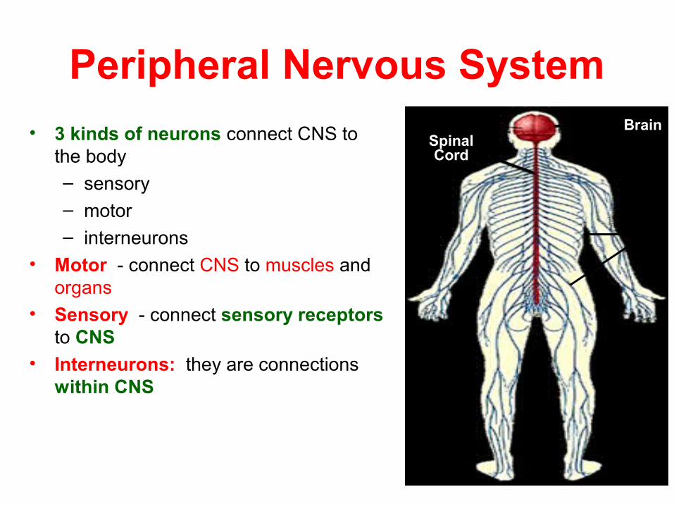
• 3 kinds of neurons connect CNS to the body– sensory– motor– interneurons
• Motor - connect CNS to muscles and organs
• Sensory - connect sensory receptors to CNS
• Interneurons: they are connections within CNS
SpinalCord
Brain
Nerves
• Sensory -Conducts impulses from receptors to the CNS
• There are two divisions :– Somatic (from skin, skeletal muscles and joints)– Autonomic (from organs in body cavity)
• Motor Division –conducts impulses from CNS to effectors
• There are two divisions :– Somatic – Muscles– Autonomic - Smooth muscle ,cardiac muscle Glands

Peripheral nervous system
Somatic Autonomic
Sympathetic Parasympathetic

1.Somatic System
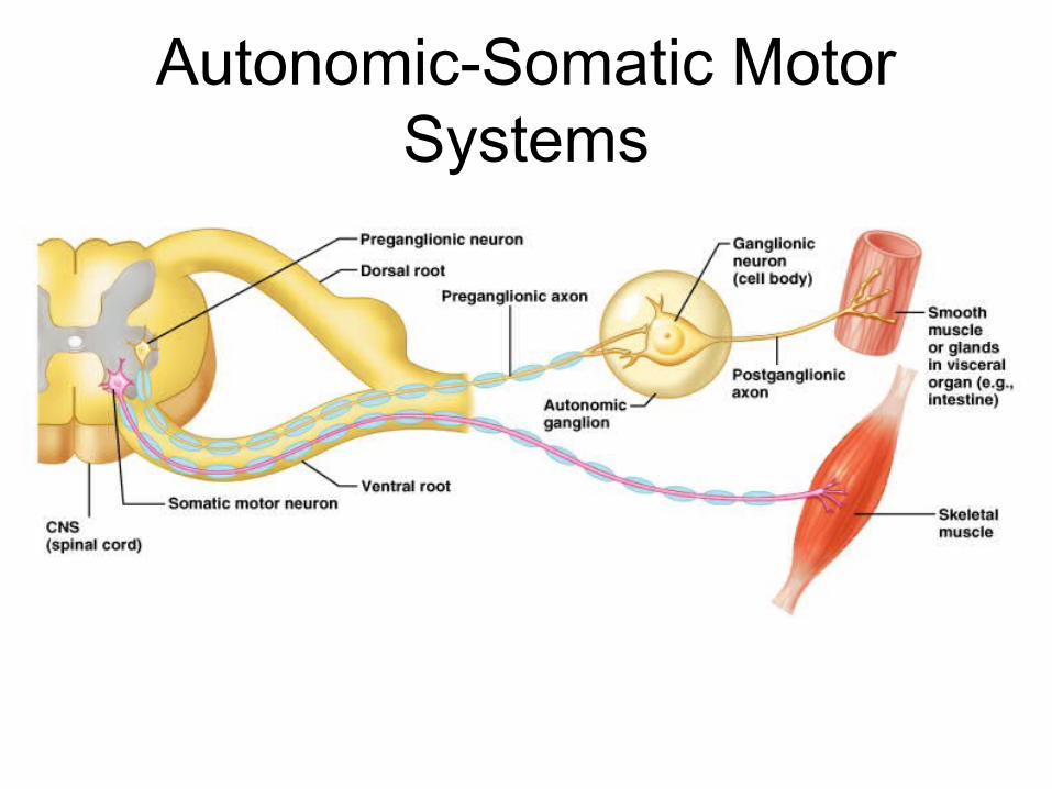
Autonomic-Somatic Motor Systems
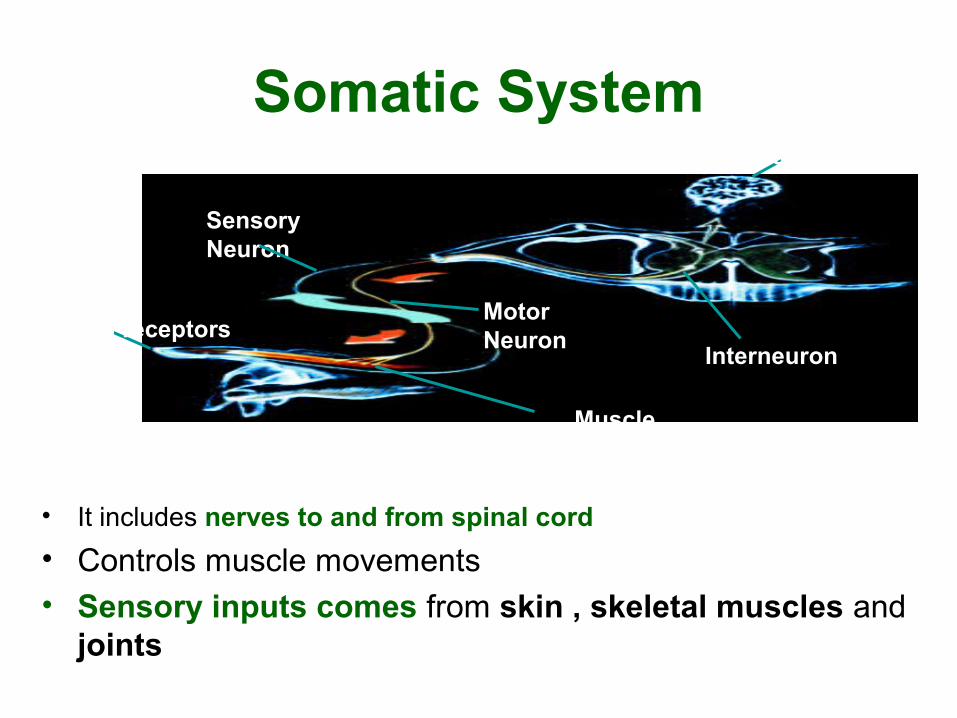
Somatic System
• It includes nerves to and from spinal cord
• Controls muscle movements• Sensory inputs comes from skin , skeletal muscles and
joints
Muscle
MotorNeuron
InterneuronSkin receptors
SensoryNeuron
Brain
2.Autonomic Nervous System
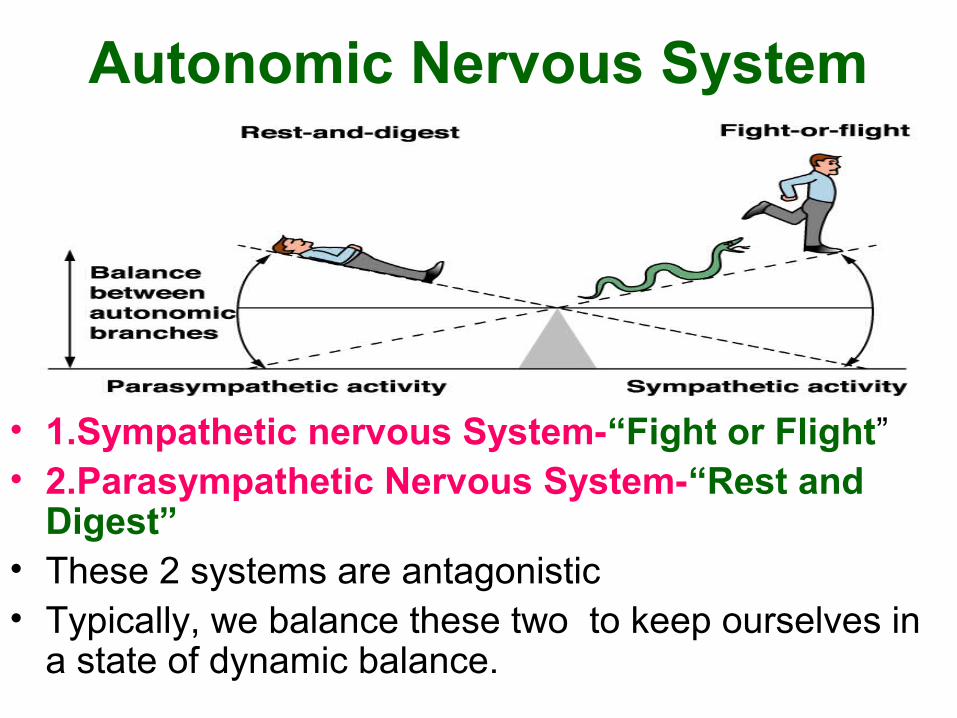
Autonomic Nervous System
• 1.Sympathetic nervous System-“Fight or Flight”• 2.Parasympathetic Nervous System-“Rest and
Digest” • These 2 systems are antagonistic• Typically, we balance these two to keep ourselves in
a state of dynamic balance.
Autonomic nervous system
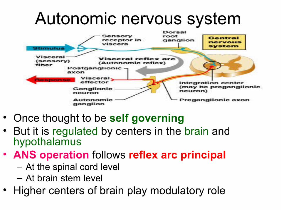
• Once thought to be self governing • But it is regulated by centers in the brain and
hypothalamus• ANS operation follows reflex arc principal
– At the spinal cord level– At brain stem level
• Higher centers of brain play modulatory role
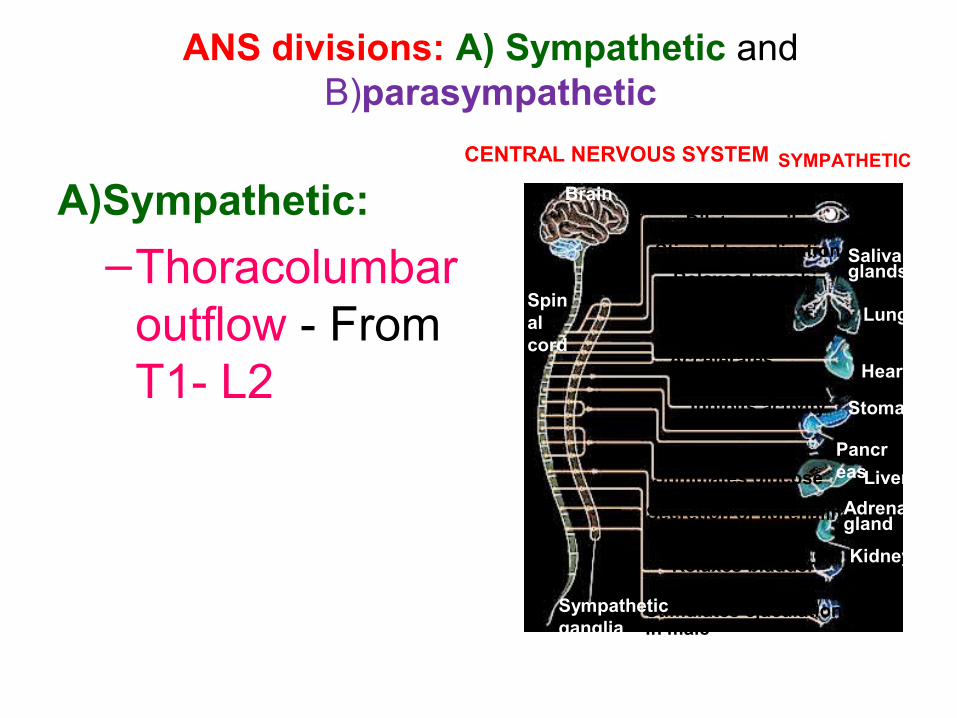
ANS divisions: A) Sympathetic and B)parasympathetic
A)Sympathetic:
–Thoracolumbar outflow - From T1- L2
CENTRAL NERVOUS SYSTEM
Brain
Spinalcord
SYMPATHETIC
Dilates pupil
Stimulates salivation
Relaxes bronchi
Accelerates heartbeat
Inhibits activity
Stimulates glucose
Secretion of adrenaline,nonadrenaline
Relaxes bladder
Stimulates ejaculationin male
Sympatheticganglia
Salivaryglands
Lungs
Heart
Stomach
PancreasLiver
Adrenalgland
Kidney
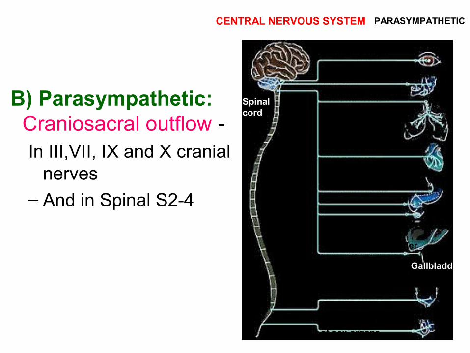
B) Parasympathetic: Craniosacral outflow - In III,VII, IX and X cranial
nerves – And in Spinal S2-4
CENTRAL NERVOUS SYSTEMBrain
PARASYMPATHETIC
Spinalcord
Stimulates salivation
Constricts bronchi
Slows heartbeat
Stimulates activity
Contracts bladder
Stimulates erectionof sex organs
Stimulates gallbladder
Gallbladder
Contracts pupil

• Most viscera receive both• Except sweat glands and arrector pili –
supplied by sympathetic • Normally their actions are opposite • ANS efferent's have two neurons • Preganglionic and postganglionic • Preganglionic are in the CNS – relay
on post ganglionic • Postganglionic are in the ganglia –
supply target organs
Autonomic nervous system
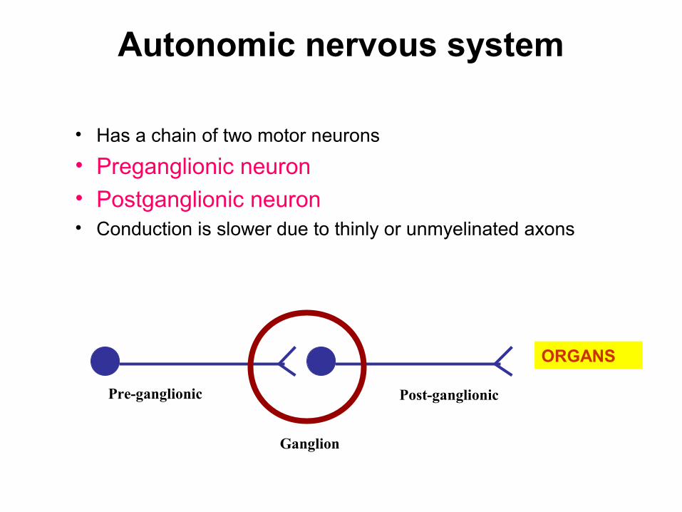
• Has a chain of two motor neurons
• Preganglionic neuron• Postganglionic neuron• Conduction is slower due to thinly or unmyelinated axons
Pre-ganglionic
Ganglion
Post-ganglionic
ORGANS
A.Sympathetic nervous system
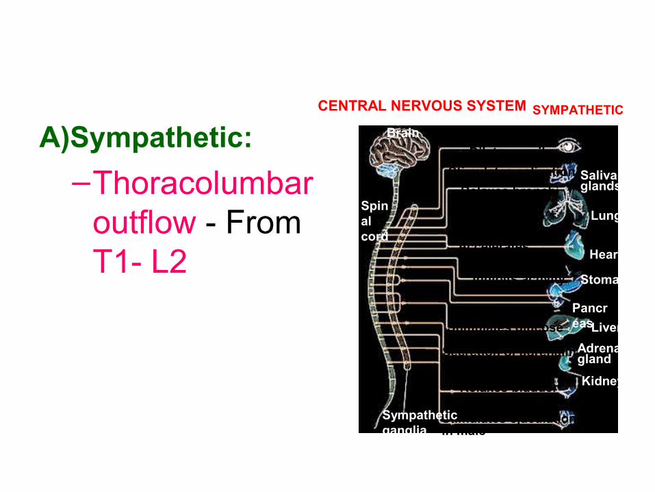
A)Sympathetic:
–Thoracolumbar outflow - From T1- L2
CENTRAL NERVOUS SYSTEM
Brain
Spinalcord
SYMPATHETIC
Dilates pupil
Stimulates salivation
Relaxes bronchi
Accelerates heartbeat
Inhibits activity
Stimulates glucose
Secretion of adrenaline,nonadrenaline
Relaxes bladder
Stimulates ejaculationin male
Sympatheticganglia
Salivaryglands
Lungs
Heart
Stomach
PancreasLiver
Adrenalgland
Kidney
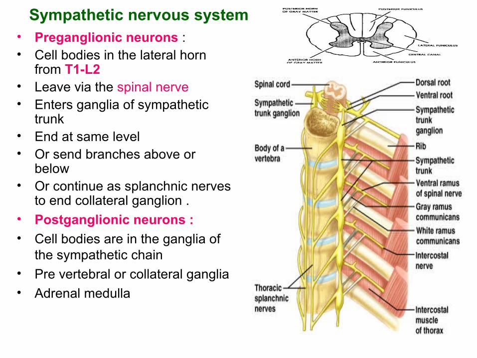
Sympathetic nervous system • Preganglionic neurons :• Cell bodies in the lateral horn
from T1-L2• Leave via the spinal nerve • Enters ganglia of sympathetic
trunk• End at same level • Or send branches above or
below • Or continue as splanchnic nerves
to end collateral ganglion .• Postganglionic neurons :• Cell bodies are in the ganglia of
the sympathetic chain • Pre vertebral or collateral ganglia• Adrenal medulla
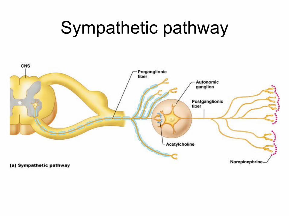
Sympathetic pathway

Sympathetic trunks
• They are two cords on either side of vertebral column
• From base of skull to coccyx
• Contains cell bodies of postganglionic neurons
• There are 31 pairs • Due to fusion number
reduced to 22-23
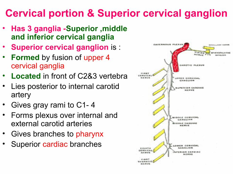
Cervical portion & Superior cervical ganglion• Has 3 ganglia -Superior ,middle
and inferior cervical ganglia • Superior cervical ganglion is :• Formed by fusion of upper 4
cervical ganglia • Located in front of C2&3 vertebra • Lies posterior to internal carotid
artery• Gives gray rami to C1- 4• Forms plexus over internal and
external carotid arteries • Gives branches to pharynx • Superior cardiac branches
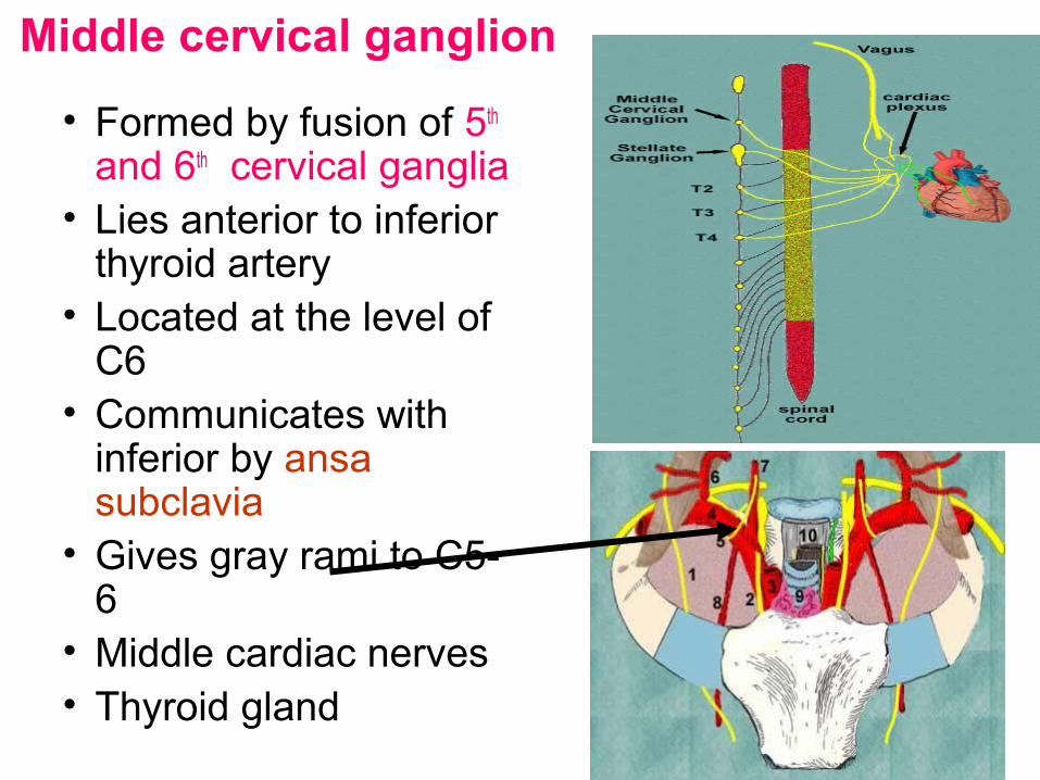
Middle cervical ganglion
• Formed by fusion of 5th and 6th cervical ganglia
• Lies anterior to inferior thyroid artery
• Located at the level of C6
• Communicates with inferior by ansa subclavia
• Gives gray rami to C5-6
• Middle cardiac nerves • Thyroid gland
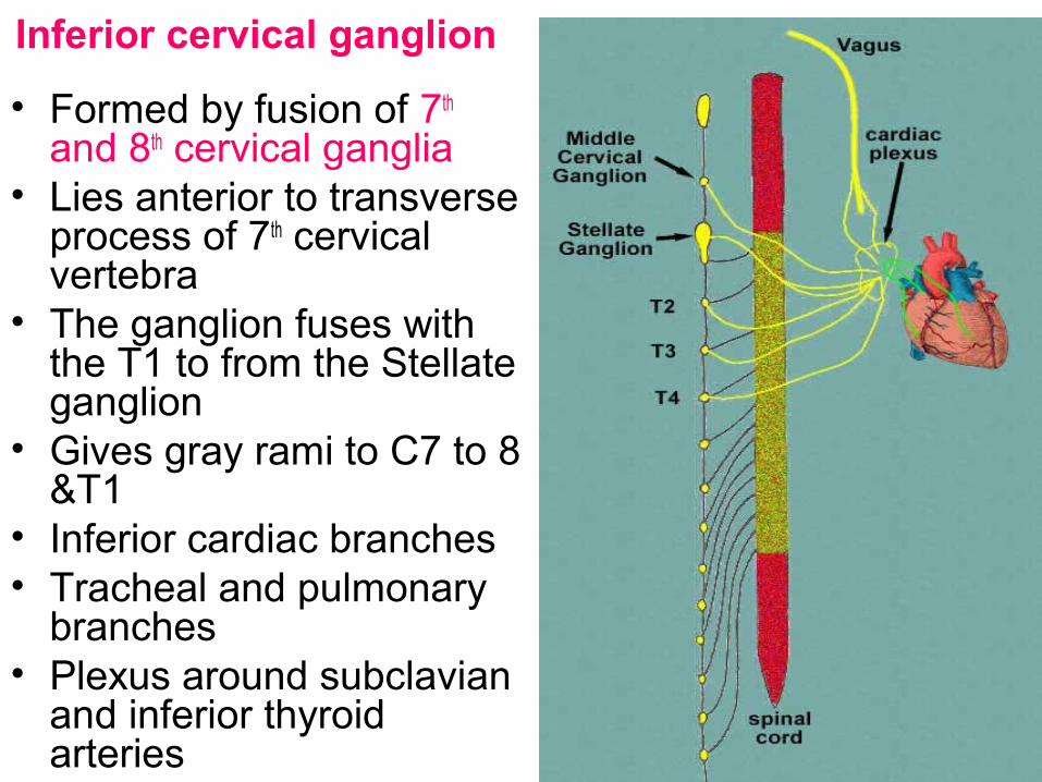
Inferior cervical ganglion
• Formed by fusion of 7th and 8th cervical ganglia
• Lies anterior to transverse process of 7th cervical vertebra
• The ganglion fuses with the T1 to from the Stellate ganglion
• Gives gray rami to C7 to 8 &T1
• Inferior cardiac branches • Tracheal and pulmonary
branches• Plexus around subclavian
and inferior thyroid arteries
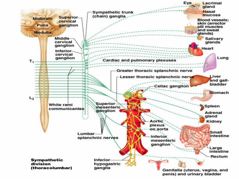
Sympathetic Division of the ANS
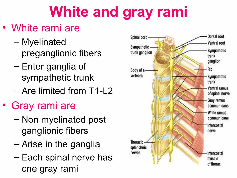
White and gray rami• White rami are
– Myelinated preganglionic fibers
– Enter ganglia of sympathetic trunk
– Are limited from T1-L2
• Gray rami are– Non myelinated post
ganglionic fibers– Arise in the ganglia – Each spinal nerve has
one gray rami

Parasympathetic nervous system

B) Parasympathetic: Craniosacral outflow - In III,VII, IX and X cranial
nerves – And in Spinal S2-4
CENTRAL NERVOUS SYSTEMBrain
PARASYMPATHETIC
Spinalcord
Stimulates salivation
Constricts bronchi
Slows heartbeat
Stimulates activity
Contracts bladder
Stimulates erectionof sex organs
Stimulates gallbladder
Gallbladder
Contracts pupil
Parasympathetic nervous system • preganglionic neurons :Cell bodies of preganglionic neurons
are found in the brain stem nuclei
• Of III, VII, IX and X cranial nerves • Lateral horns of S 2-4• Preganglionic are myelinated .• Postganglionic neurons :• Cell bodies are in the peripheral ganglia • They lie close to organ of innervation • They send short postganglionic fiber • They are not myelinated • In the head region there are 4 ganglia
– Ciliary –III CN– Pterygopalatine- VII CN– Submandibular _--VII CN– Otic - IX

The Parasympathetic Division
Figure 15.5

Parasympathetic pathway
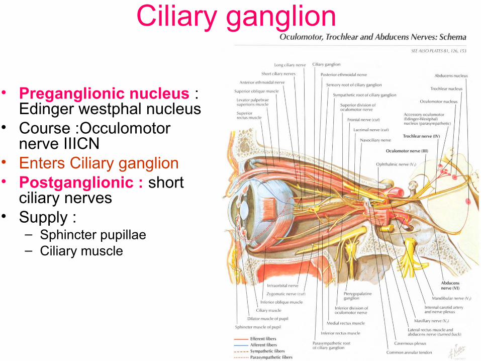
Ciliary ganglion
• Preganglionic nucleus : Edinger westphal nucleus
• Course :Occulomotor nerve IIICN
• Enters Ciliary ganglion • Postganglionic : short
ciliary nerves • Supply :
– Sphincter pupillae – Ciliary muscle
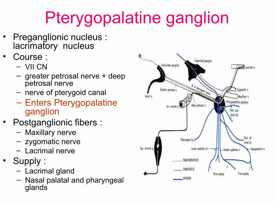
Pterygopalatine ganglion• Preganglionic nucleus :
lacrimatory nucleus • Course :
– VII CN– greater petrosal nerve + deep
petrosal nerve – nerve of pterygoid canal – Enters Pterygopalatine
ganglion• Postganglionic fibers :
– Maxillary nerve – zygomatic nerve – Lacrimal nerve
• Supply :– Lacrimal gland – Nasal palatal and pharyngeal
glands
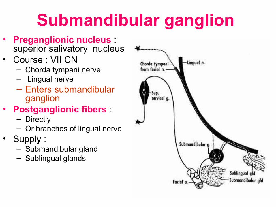
Submandibular ganglion • Preganglionic nucleus :
superior salivatory nucleus • Course : VII CN
– Chorda tympani nerve– Lingual nerve– Enters submandibular
ganglion • Postganglionic fibers :
– Directly – Or branches of lingual nerve
• Supply :– Submandibular gland – Sublingual glands

Otic ganglion
• Preganglionic nucleus : Inferior salivatory nucleus
• Course : IX CN– Tympanic branch of IX CN– Lesser petrosal nerve
– Enters otic ganglion • Postganglionic fibers :
– Auriculotemporal nerve
• Supply :– Parotid gland

Vagus nerve • Preganglionic nucleus : Dorsal nucleus of vagus • Course : vagal branches of
– Pulmonary– Cardiac – Esophageal – Gastric
• Postganglionic nuclei :– Ganglia of pulmonary plexus– Ganglia of cardiac plexus– Aurbach’s and Meissners’ plexus– Ganglia in the wall of viscera
• Postganglionic fibers : – Direct branches
• Supply :– Smooth muscle and glands of respiratory system – Conducting system of heart – Esophagus – Stomach, small intestine, ascending colon and
transverse colon
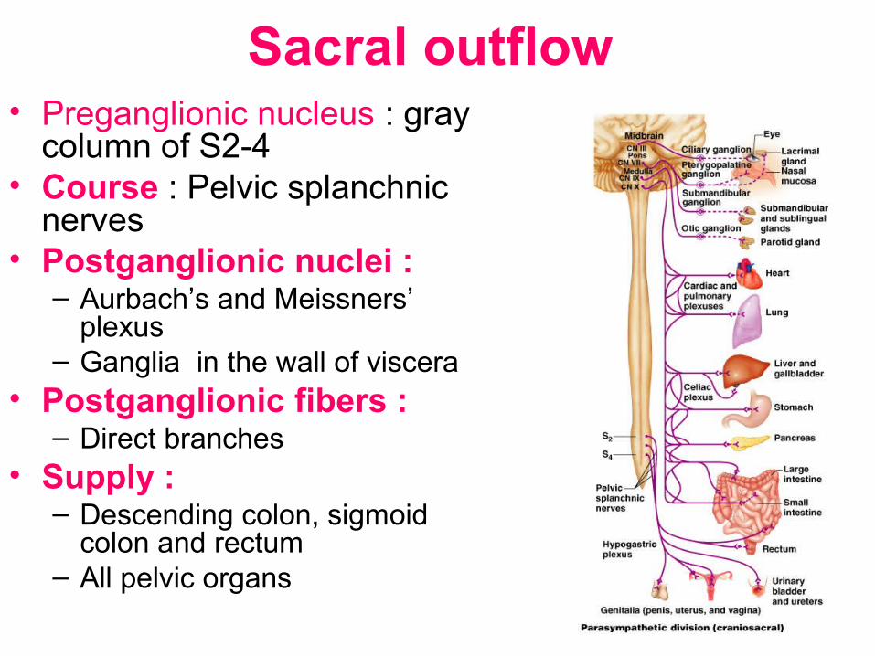
Sacral outflow • Preganglionic nucleus : gray
column of S2-4 • Course : Pelvic splanchnic
nerves • Postganglionic nuclei :
– Aurbach’s and Meissners’ plexus
– Ganglia in the wall of viscera • Postganglionic fibers :
– Direct branches • Supply :
– Descending colon, sigmoid colon and rectum
– All pelvic organs
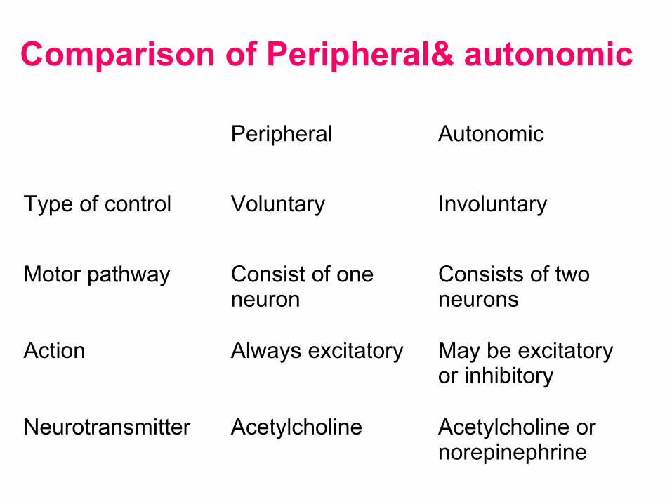
Comparison of Peripheral& autonomic
Peripheral Autonomic
Type of control Voluntary Involuntary
Motor pathway Consist of one neuron
Consists of two neurons
Action Always excitatory May be excitatory or inhibitory
Neurotransmitter Acetylcholine Acetylcholine or norepinephrine

Comparison of Sympathetic and parsympathetic
Sympathetic Parasympathetic
Outflow Thoracolumbar T1-L2 Craniosacral III, VII, IX , XCN & S2-4
Postganglionic neuron – location
Sympathetic trunk and pre vertebral ganglia – away from viscera
Outside CNS Near to viscera
Length of fibers Preganglionic fibers – short Postganglionic –long
Preganglionic fibers – long Postganglionic – short
Preganglionic fibers Synapse With many postganglionic neurons and supply many viscera
With few postganglionic neurons that supply single viscous

Comparison of Sympathetic and parsympathetic
Sympathetic Parasympathetic
Neurotransmitter at preganglionic
Acetylcholine Acetylcholine
Neurotransmitter at postganglionic
Noradrenalin But sweat glands & blood vessels – acetylcholine
Acetylcholine
Distribution Whole body Limited to:Head & neck, thorax , abdomen and pelvis
Type of system Fight or flight system Rest and repose system

Distribution
• Sympathetic :– Whole body
• Parasympathetic :– Limited to:– Head & neck– Thorax– Abdomen and
pelvis

Applied anatomy

• Horner's syndrome consists of :
• Miosis – constriction of pupil
• Ptosis – drooping of upper eye lid
• Anhydrosis –loss of sweating
• Enopthalmos- sunken eyeball due to paralysis of orbitalis muscle
• Due to lesion of sympathetic supply to head and neck – e.g. cervical rib
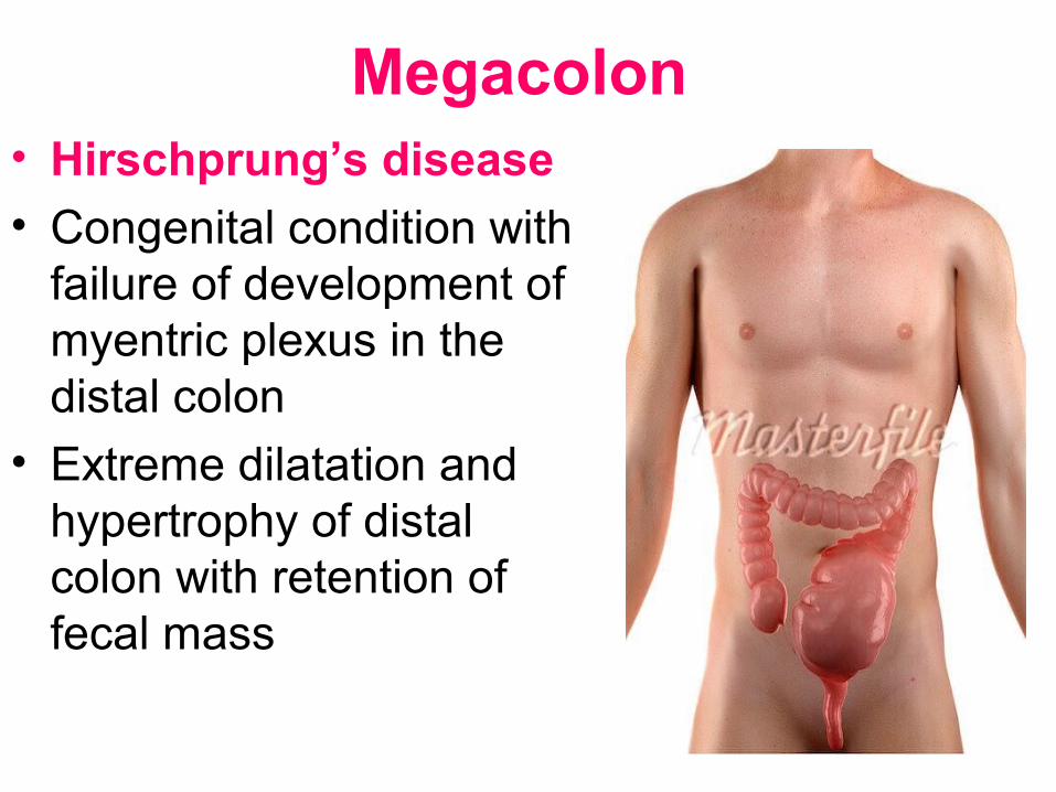
Megacolon • Hirschprung’s disease
• Congenital condition with failure of development of myentric plexus in the distal colon
• Extreme dilatation and hypertrophy of distal colon with retention of fecal mass
Thromboangitis obliterans
• It is an obliterative disease of arteries
• Particularly lower limbs
• Vascular spasm due to sympathetic over activity
• With reduced blood circulation
• Relieved by lumbar sympathectomy
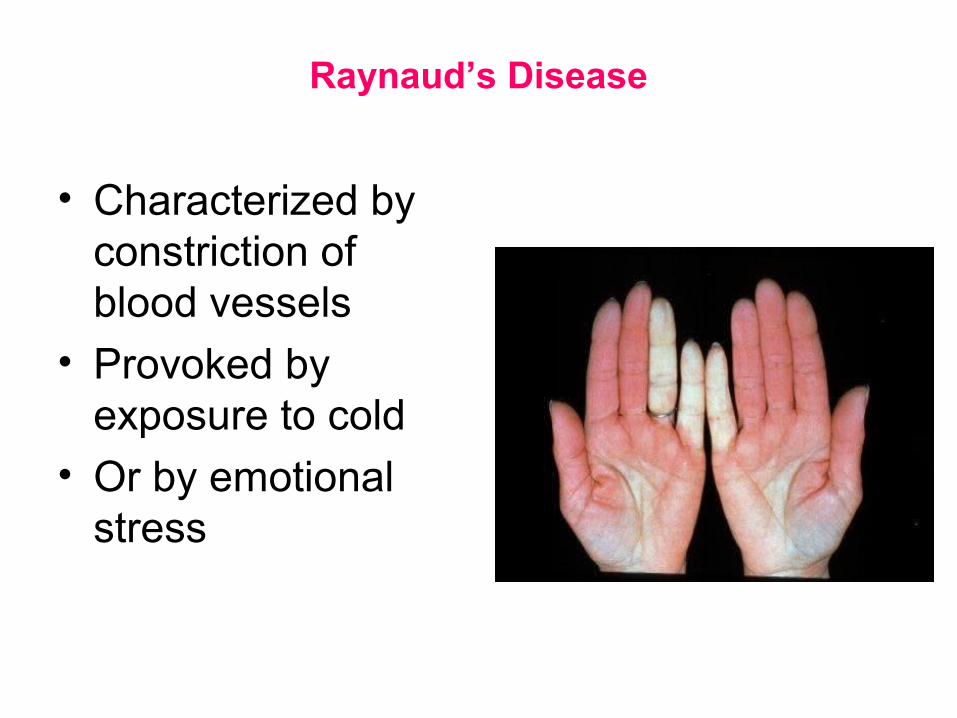
Raynaud’s Disease
• Characterized by constriction of blood vessels
• Provoked by exposure to cold
• Or by emotional stress
Hypertension
• Can result from overactive sympathetic vasoconstriction

Achalasia of the Cardia
• Defect in the autonomic innervation of the esophagus