Periodontal treatment: Manual Scaling - USM - Kampus … Erry/manual scaling... · Objectives After...
68
Periodontal Therapy: Manual Scaling and Root planing Erry Mochamad Arief Senior Lecturer in Periodontics
Transcript of Periodontal treatment: Manual Scaling - USM - Kampus … Erry/manual scaling... · Objectives After...

Periodontal Therapy:Manual Scaling and Root planing
Erry Mochamad AriefSenior Lecturer in Periodontics

Objectives After periodontal instrumentation session, student should be able to demonstrate
Proper position of the patient and the operator (Accessibility)Visibility, illumination and retraction Condition of instruments (sharpness)Maintaining a clean field Instrument stabilization: Instrument grasp, Fulcrum or Finger restInstrument activation: Adaptation, Angulation, Lateral pressure, and StrokeSelection of the proper instrumenTechnique for periodontal probing, determining attachment loss, using dental explorer, furcation detectionTechnique for tooth mobility quantificationTechnique for using Chisel, Sickle and Wing shaped, CuretteManual Scaling and root planing technique in different areas of the mouth
2 30/10/08

PHASES OF PERIODONTAL THERAPYPhase I therapy
Started after E&D and treatment planningRemoval bacterial plaque and all irritants (periodontal instrumentation)OH Instruction
Maintenance phaseAll patients that undergo periodontal therapyPatients return to clinic for evaluation, periodontal instrumentation, and reinforcement of OH techniques.
Phase II therapySurgical (periodontal instrumentation)
All phases need periodontal instrumentation
3 30/10/08
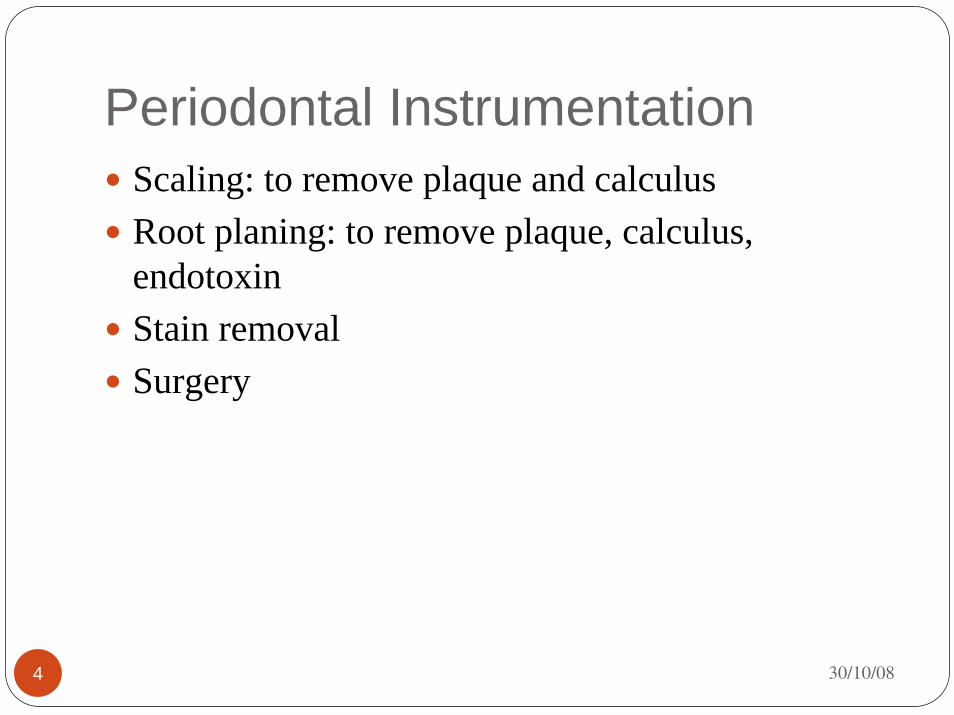
Periodontal InstrumentationScaling: to remove plaque and calculusRoot planing: to remove plaque, calculus, endotoxinStain removalSurgery
4 30/10/08

Principles of Instrumentation1. Accessibility (positioning of patient and operator)2. Visibility, illumination and retraction3. Condition of instruments (sharpness)4. Maintaining a clean field5. Instrument stabilization6. Instrument activation7. Selection of the proper instrument
5 30/10/08

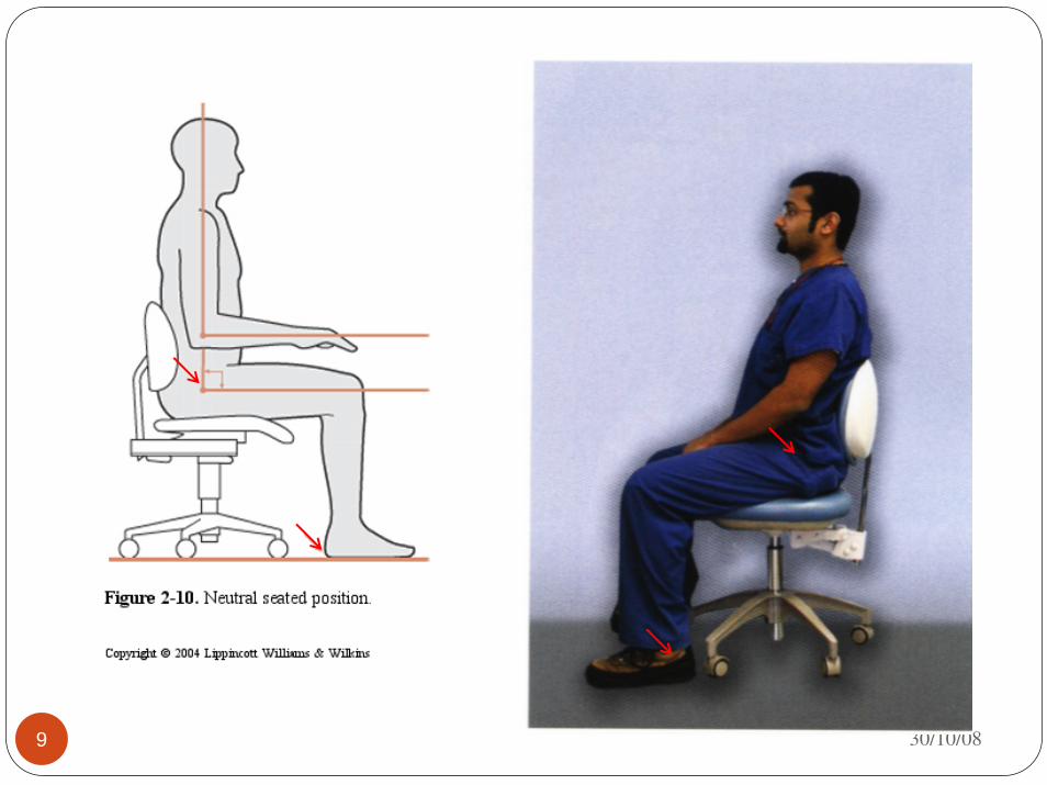
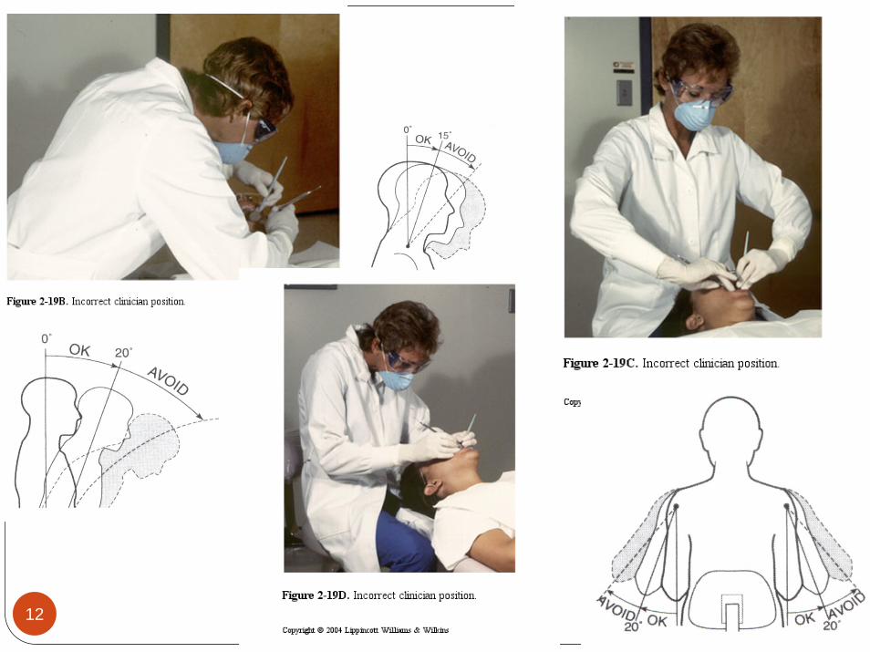
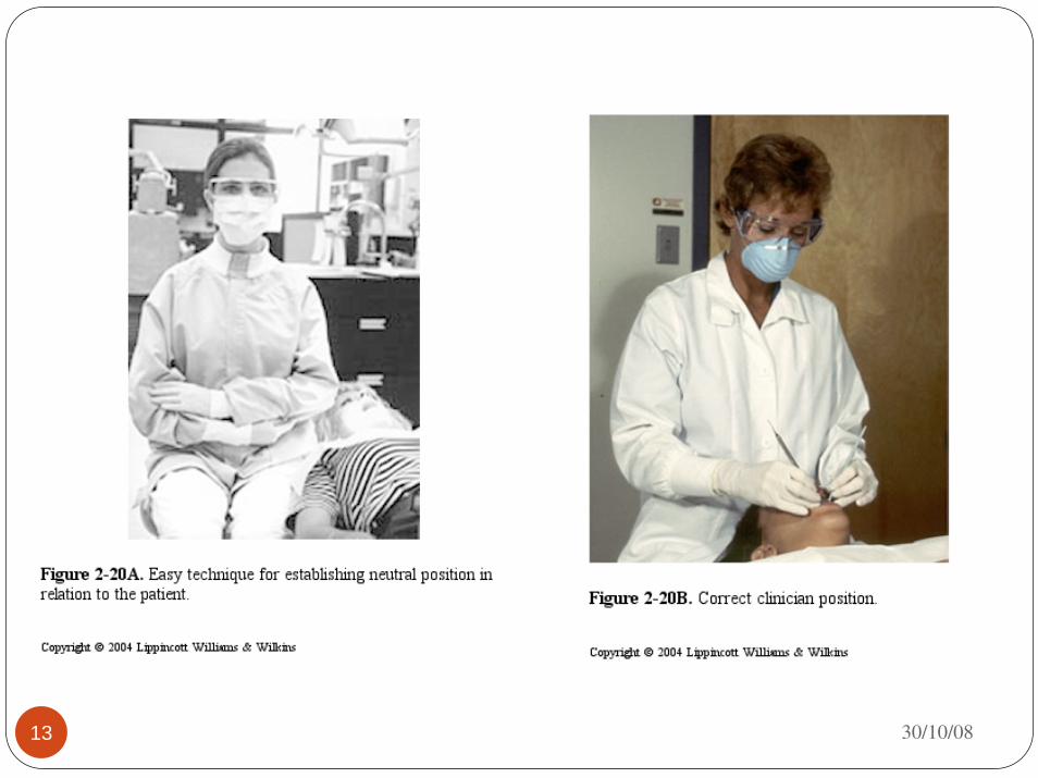
1. Positioning
prevent clinician and patient from discomfort and injury
permit a clear view, the best is direct vision
allow easy access to the teeth
efficient: time and energy
6 30/10/08

Clinical/Operator positionNeutral position
7 30/10/08

30/10/088
30/10/089

30/10/0810

30/10/0811
30/10/0812
13 30/10/08

30/10/0814
Patient Head Position
15 30/10/08

16
Supine patient position: The patient’s heels should be slightly higher than the tip of his or her nose
Adjust his/her head to provide best view: chin up/down, toward/straight/ away
30/10/08

Visibility, illumination and retractionDirect vision with direct illuminationIndirect vision by using mouth mirrorIndirect illumination by using mouth mirrorRetraction provides visibility, accessibility and illumination by using mirror or fingersSoftening the lips with petroleum jelly/vaseline to prevent cracking and bleeding
17 30/10/08

maxilla
mandibularDental light position
18 30/10/08

Sharpness
When the instrument is sharp, the cutting edge is a fine lineLight reflected from the rounded cutting edge of a dull instrument appears as a bright line.
19 30/10/08

Maintaining a clean fieldFrom saliva, blood, and debrisPooling saliva interferes: visibility, firm finger rest cannot be establishedBlood and debris can be removed by wiping with gauzeOperative field should be flushed occasionally with water
20 30/10/08

Instrument stabilization
30/10/0821
Instrument graspFinger rest
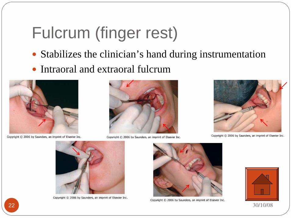
Fulcrum (finger rest) Stabilizes the clinician’s hand during instrumentationIntraoral and extraoral fulcrum
30/10/0822

Instrument activationAdaptationAngulationLateral pressureStrokes
23 30/10/08

Adaptation
Is placing the first one or two millimeters of the side of the working end in contact with the tooth surface
Adapted Not-adapted
24 30/10/08

30/10/08Adapted Not-adapted25

Angulation
Is the angle formed between the face of instrument and the tooth surfaces
30/10/0826

Incorrect
27 30/10/08

Subgingival scaling procedure.
A, Curette inserted with the face of the blade flush against the tooth.
B, Working angulation (45 to 90 degrees) is established at the base of the pocket.
C, Lateral pressure is applied, and the scaling stroke is activated in the coronal direction.
28 30/10/08

Lateral pressureIs the act of using the thumb and index finger to engage (press) the cutting edge against the tooth surface or calculus depositThe amount of pressure must be varied according to the nature of the calculus and according to whether the stroke is intended to remove calculus or to smooth the root surface.Lateral pressure may be firm, moderate, or lightThe careful application of lateral pressure during instrumentation is important
29 30/10/08

Wrist motion activationIs the act of rotating the hand and wrist as a unitSimilar with turning a doorknobRecommended for calculus removal with hand activated instr.More powerful and less fatigue than digital motion activation
30 30/10/08

Digital motion activationIs moving the instrument by flexing the fingersIt may be used with periodontal probes, explorers, and ultrasonic instruments or where movement is very restricted, such as in furcation areas
31 30/10/08

Instrumentation StrokesStrokes Direction
32 30/10/08

Instrumentation for calculus removal
A, Calculus is removed by engaging the apical or lateral edge of the deposit with the cutting edge of a scaler; vertical movement of the instrument will remove the fragment of calculus engaged by the instrumentB, The instrument is moved laterally and again engages the edge of the calculus, overlapping the previous stroke to some extentC, The final portion of the deposit is engaged and removed. Note how the procedure is performed in an interdental space by entering facially and lingually.
33 30/10/08

30/10/0834
SCALING IN DIFFERENT AREAS OF THE MOUTH
MAXILLARY TEETH
35 30/10/08

Maxillary right posterior sextant: facial aspect
Operator: Side position (9 o'clock). Illumination: Direct. Visibility: Direct (indirect for distal surfaces of molars). Retraction: Mirror or index finger of the non-operating hand. Finger rest: Extraoral, palm up. Backs of the middle and fourth fingers on the lateral aspect of the mandible on the right side of the face.
36 30/10/08

Maxillary right posterior sextant: lingual aspect.
Operator: Side position (9 o’clock). Illumination: Direct and indirect. Visibility: Direct or indirect. Retraction: None. Finger rest: Extraoral, palm up. Backs of the middle and fourth fingers on the lateral aspect of the mandible on the right side of the face. 37 30/10/08
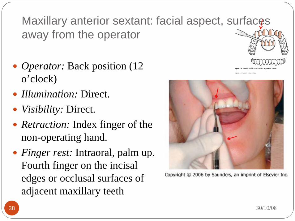
Maxillary anterior sextant: facial aspect, surfaces away from the operator
Operator: Back position (12 o’clock)Illumination: Direct. Visibility: Direct. Retraction: Index finger of the non-operating hand. Finger rest: Intraoral, palm up. Fourth finger on the incisal edges or occlusal surfaces of adjacent maxillary teeth
38 30/10/08

Maxillary anterior sextant: facial aspect, surfaces toward the operator
Operator: Front position (8 o’clock) Illumination: Direct. Visibility: Direct. Retraction: Index finger of the nonoperating hand. Finger rest: Intraoral, palm down. Fourth finger on the incisal edges or the occlusal or facial surfaces of adjacent maxillary teeth. 39 30/10/08

Maxillary anterior sextant: lingual aspect, surfaces away from the operator (surfaces toward the operator are scaled from a front position).
Operator: Back position (12 o’clock)Illumination: Indirect. Visibility: Indirect. Retraction: None. Finger rest: Intraoral, palm up. Fourth finger on the incisal edges or the occlusal surfaces of adjacent maxillary teeth
40 30/10/08

Maxillary left posterior sextant: facial aspect
Operator: Side position (9 o’clock). Illumination: Direct or indirect. Visibility: Direct or indirect. Retraction: Mirror. Finger rest: Intraoral, palm up. Fourth finger on the incisal edges or the occlusal surfaces of adjacent maxillary teeth.
41 30/10/08

Maxillary left posterior sextant: lingual aspect
Operator: Side position (9 o’clock). Illumination: Direct. Visibility: Direct. Retraction: None. Finger rest: Intraoral, palm up. Fourth finger on the occlusal surfaces of adjacent maxillary teeth.
42 30/10/08

Mandibular left posterior sextant: facial aspect
Operator: Side position (9 o’clock). Illumination: Direct. Visibility: Direct or indirect. Retraction: Index finger or mirror of the nonoperating hand. Finger rest: Intraoral, palm down. Fourth finger on the incisal edges or the occlusal or facial surfaces of adjacent mandibular teeth
43 30/10/08

Mandibular left posterior sextant: lingual aspect
Operator: Side position (9 o’clock). Illumination: Direct and indirect. Visibility: Direct. Retraction: Mirror retracts tongue. Finger rest: Intraoral, palm down. Fourth finger on the incisal edges or the occlusal surfaces of adjacent mandibular teeth.
44 30/10/08

Mandibular anterior sextant: facial aspect, surfaces toward the operator
Operator: Front position (8 o’clock) Illumination: Direct. Visibility: Direct. Retraction: Index finger of the nonoperating hand. Finger rest: Intraoral, palm down. Fourth finger on the incisal edges or the occlusal surfaces of adjacent mandibular teeth45 30/10/08
Mandibular anterior sextant: facial aspect, surfaces away from the operator
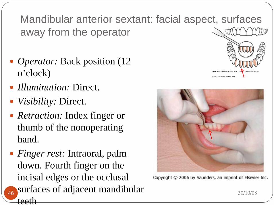
Operator: Back position (12 o’clock) Illumination: Direct. Visibility: Direct. Retraction: Index finger or thumb of the nonoperating hand. Finger rest: Intraoral, palm down. Fourth finger on the incisal edges or the occlusal surfaces of adjacent mandibular teeth
46 30/10/08

Mandibular anterior sextant: lingual aspect, surfaces away from the operator
Operator: Back position (12 o’clock) Illumination: Direct and indirect. Visibility: Direct and indirect. Retraction: Mirror retracts tongue. Finger rest: Intraoral, palm down. Fourth finger on the incisal edges or the occlusal surfaces of adjacent mandibular teeth
47 30/10/08

Mandibular anterior sextant: lingual aspect, surfaces toward the operator
Operator: Front position (8 o’clock) Illumination: Direct and indirect. Visibility: Direct and indirect. Retraction: Mirror retracts tongue. Finger rest: Intraoral, palm down. Fourth finger on the incisal edges or the occlusal surfaces of adjacent mandibular teeth
48 30/10/08

Mandibular right posterior sextant: facial aspect
Operator Side position (9 o’clock). Illumination: Direct. Visibility: Direct. Retraction: Mirror or index finger of the nonoperating hand. Finger rest: Intraoral, palm down. Fourth finger on the incisal edges or the occlusal surfaces of adjacent mandibular teeth49 30/10/08

Mandibular right posterior sextant: lingual aspect
Operator: Side position (9 o’clock). Illumination: Direct and indirect. Visibility: Direct and indirect. Retraction: Mirror retracts tongue. Finger rest: Intraoral, palm down. Fourth finger on the incisal edges or the occlusal surfaces of adjacent mandibular teeth
50 30/10/08

Instruments for scaling and root planingUniversal curettesGracey curettes
51 30/10/08

Universal Curettes
A, Columbia #4R-4L universal curette.B, Younger-Good #7-8, McCall's #17-18, and Indiana University #17-18 universal curettes.
52 30/10/08
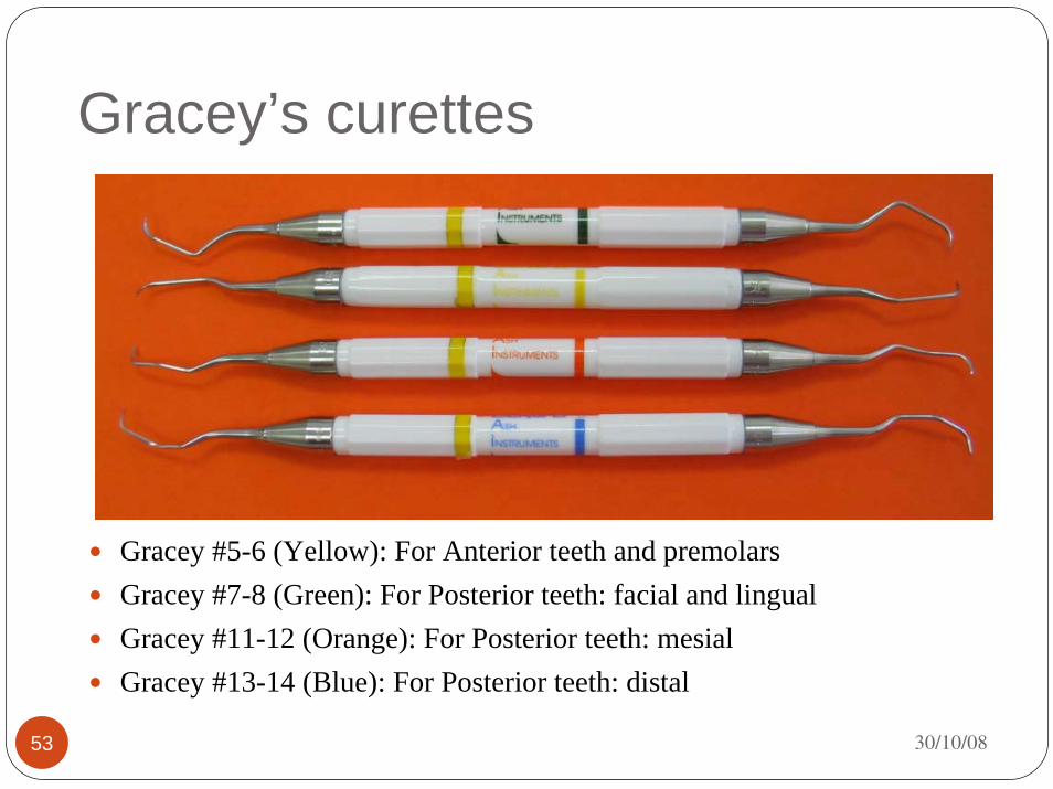
Gracey’s curettes
Gracey #5-6 (Yellow): For Anterior teeth and premolarsGracey #7-8 (Green): For Posterior teeth: facial and lingual Gracey #11-12 (Orange): For Posterior teeth: mesialGracey #13-14 (Blue): For Posterior teeth: distal
53 30/10/08

30/10/0854
Chisel Wing-shaped Hygienist

Push Scaler or Chisel.A pushing instrument used parallel to the occlusal plane. This instrument can be used supra-gingivally especially in the anterior. Great care need to be taken to avoid soft tissue damage.
Hygienist Sickle.Is a single-ended instrument can be used for mesial, distal, facial, and lingual surfaces of supra and sub gingival calculus.Adaptation of Sickle scaler is important to prevent damage to the tissue
Wing-shaped scalerThis instrument has a wing blade shape and a sharply pointed end. The mode of use is pull coronally for the supra and sub gingival area of the tooth
55 30/10/08

Conclusion: Principles of scaling and root planing
Determine the correct cutting edgeFinger restConcentrate on using the lower third of the cutting edge for calculus removal (adaptation)Allow the hand-forearm to carry the burden of the stroke, rather than flexing the fingers (digital motion activation)Modulate lateral pressure
56 30/10/08
Calculus detectionSupragingival calculus can be detected visually or by using compressed air and dental mirrorDry calculus has contrast visually with enamelSubgingival calculus are hidden beneath gingiva
57 30/10/08

technique to identify the presence of supra and subgingival calculus
58 30/10/08

Furcation involvement
59 30/10/08
Occurs when periodontal infection invades the area between and around the roots of bifurcated or trifurcated teethAccess to furcation area
Mandibular molars (bifurcated) →examined from the facial and lingual surfaces
Maxillary first premolar (bifurcated) →from the mesial and distal surfaces
Maxillary first molars (trifurcated) from the facial, mesial, distal surfaces

SCHEDULE FOR PERIODONTAL INSTRUMENTATION SKILLYear 3 2008/2009
Thursday8-10am Activities Venue(Supervisor)
8-9 am Lecture Periodontal Instrumentation DK 1 (ERRY)
30/10/089-10 am Putting Artificial Calculus on Frasaco model
MDL (HT, ERRY, WW, RARA, AKH, SLA)
8-9 am Demo Scaling and Root planning on real teeth (All supervisor)
6/11/08
9-10 am
Mounting 16 real teeth* on POP
PROSTHO LAB (HT, ERRY, WW, RARA, AKH, SLA)
13/11/08 KKKK
60 30/10/08

Activity: Supra and Subgingival scaling of artificial calculus on maxillary teeth, manuallySupra and Subgingival scaling of artificial calculus on mandibulary teeth, manually
Activity Grade* Signature
Preparation:a. Tray set up and cleanlinessb. Identification of instruments
Probing and exploring techniquea. Correct probing techniqueb. Correct exploring technique
Ergonomic position:Appropriate positioning of dummyCorrect working position of operator
Scaling technique:Use of appropriate instrumentUse appropriate technique
Calculus free: Teeth are free from calculus*Grading: 1=poor, 2=average, 3=good
61 30/10/08
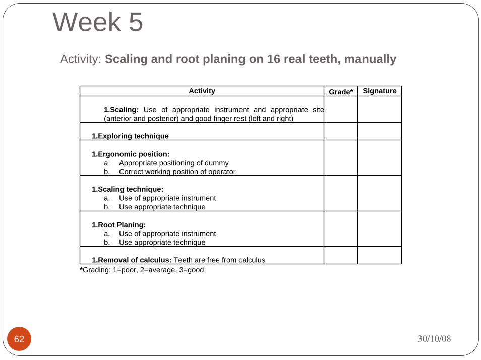
Week 5Activity: Scaling and root planing on 16 real teeth, manually
Activity Grade* Signature
1.Scaling: Use of appropriate instrument and appropriate site (anterior and posterior) and good finger rest (left and right)
1.Exploring technique
1.Ergonomic position:a. Appropriate positioning of dummyb. Correct working position of operator
1.Scaling technique:a. Use of appropriate instrumentb. Use appropriate technique
1.Root Planing:a. Use of appropriate instrumentb. Use appropriate technique
1.Removal of calculus: Teeth are free from calculus*Grading: 1=poor, 2=average, 3=good
62 30/10/08

63 30/10/08

Positioning for the anterior sextant
Anterior surface toward
Turn slightly toward the clinician chin-down position
Turn slightly toward the clinician chin-up position
64 30/10/08
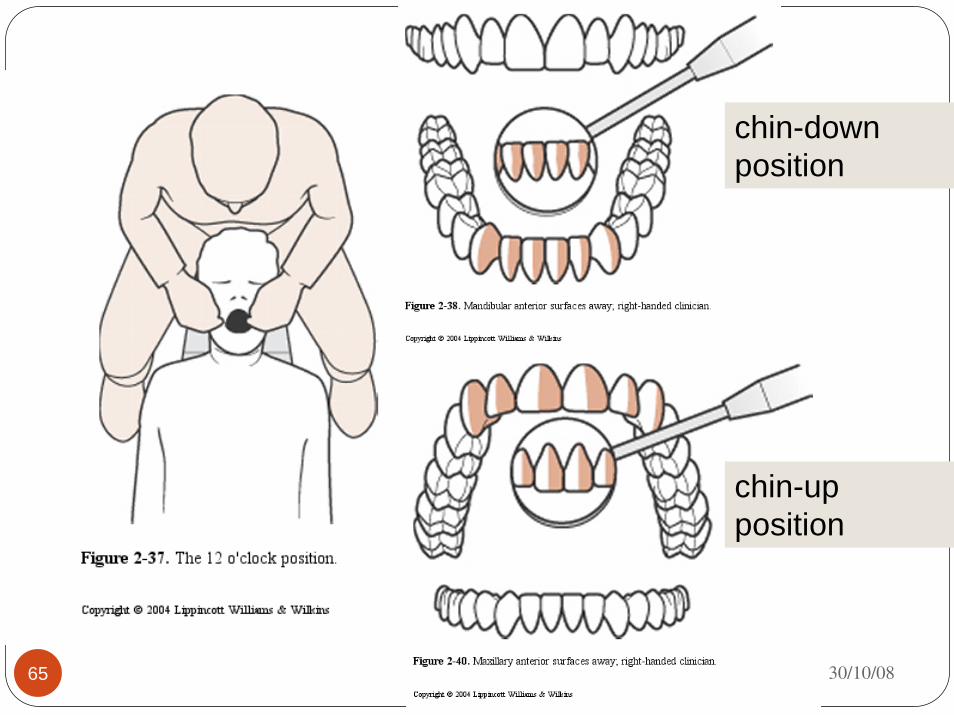
30/10/0865
chin-down position
chin-up position

30/10/0866
Positioning for the posterior sextant
Posterior aspects facing toward
Turned slightly away from the clinician chin-down position
Turned slightly away from the clinician chin-up position

30/10/0867
Positioning for the posterior sextant
Posterior aspects facing away
Turned slightly away from the clinician chin-down position
Turned slightly away from the clinician chin-up position

68 30/10/08


![Systemic doxycycline as an adjunct to scaling and root ... › content › pdf › 10.1186 › s12903-019-0873-… · periodontal treatment [2]. Scaling and root planing (SRP) is](https://static.fdocuments.us/doc/165x107/5f1b91b52924683d3a5d4ee7/systemic-doxycycline-as-an-adjunct-to-scaling-and-root-a-content-a-pdf-a.jpg)