Periodontal plastic surgery · Periodontal plastic surgery GIOVANNIZUCCHELLI &ILHAM MOUNSSIF...
36
Periodontal plastic surgery G IOVANNI Z UCCHELLI &I LHAM M OUNSSIF Mucogingival therapy is a general term used to describe periodontal treatment involving procedures for the correction of defects in the morphology, posi- tion and/or amount of soft tissue and underlying bone support around teeth and implants (12). At the beginning, ‘mucogingival surgery’, introduced by Friedman in 1957 (69), included surgical procedures designed to preserve gingival tissue, remove aberrant frenal or muscle attachments and increase the depth of the vestibule. Frequently, however, this term was used to describe certain pocket elimination approaches. Therefore, in 1993, Miller (132) intro- duced the term ‘periodontal plastic surgery’, accepted by the international scientific community in 1996, which was defined as ‘surgical procedures performed to prevent or correct anatomic, develop- mental, traumatic or disease-induced defects of the gingiva, alveolar mucosa or bone’ (203). This defini- tion includes various soft- and hard-tissue proce- dures aimed at gingival augmentation, root coverage, correction of mucosal defects at implants, crown lengthening, gingival preservation at ectopic tooth eruption, removal of aberrant frena, prevention of ridge collapse associated with tooth extraction and augmentation of the edentulous ridge. This paper focuses on gingival recession defects, their diagnosis and prognosis and the surgical procedures for root coverage. Etiology of gingival recessions The gingival margin is clinically represented by a scal- loped line that follows the outline of the cemento– enamel junction, 1–2 mm coronal to it. Gingival recession is an apical shift of the gingival margin with exposure of the root surface to the oral cavity (205) (Fig. 1). Gingival recession is often found in popula- tions with good oral hygiene (173, 177), when it is most commonly located at the buccal surfaces (117) and may be associated with wedge-shaped defects in the cervical area of one or more teeth (173). However, gingival recession is also found in populations with poor standards of oral hygiene in which it may affect other tooth surfaces (16, 117). Recession may exist in the presence of normal sulci and nondiseased inter- dental crestal bone levels, or it may occur as part of the pathogenesis of periodontal disease during which alveolar bone is lost. One etiological factor that may be associated with gingival recession is a pre-existing lack of alveolar buccal bone at the site (202) (Fig. 1). These deficiencies in alveolar bone may be develop- mental (anatomical) or acquired (physiological or pathological) (72). Anatomical factors Anatomical factors that have been related to gingi- val recession include fenestration and dehiscence of the alveolar bone, abnormal tooth position in the arch, an aberrant path of eruption of the tooth and the shape of the individual tooth (7). These anatom- ical factors are inter-related and may result in an alveolar osseous plate that is thinner than normal and that may be more susceptible to resorption. Anatomically, a dehiscence may be present because of the direction of tooth eruption or as a result of other developmental factors, such as buccal place- ment of the root relative to adjacent teeth, so that the cervical portion protrudes through the crestal bone (119). One surgical study found a correlation between gingival recession and bone dehiscence (21). A correlation between the pattern of eruption and gingival recession has also been suggested (134). Dehiscence may be present where the bucco- lingual thickness of a root is similar to or exceeds the crestal bone thickness (144). The same authors postulated that individuals with morphological bio- types characterized by narrow, long teeth are more prone to dehiscences than are individuals with broad, short teeth. Where gingival recession has developed, the underlying presence of dehiscences may be considered, and possibly discovered during flap procedures. 333 Periodontology 2000, Vol. 68, 2015, 333–368 © 2015 John Wiley & Sons A/S. Published by John Wiley & Sons Ltd Printed in Singapore. All rights reserved PERIODONTOLOGY 2000
Transcript of Periodontal plastic surgery · Periodontal plastic surgery GIOVANNIZUCCHELLI &ILHAM MOUNSSIF...
Periodontal plastic surgeryGIOVANNI ZUCCHELLI & ILHAM MOUNSSIF
Mucogingival therapy is a general term used todescribe periodontal treatment involving proceduresfor the correction of defects in the morphology, posi-tion and/or amount of soft tissue and underlyingbone support around teeth and implants (12). At thebeginning, ‘mucogingival surgery’, introduced byFriedman in 1957 (69), included surgical proceduresdesigned to preserve gingival tissue, remove aberrantfrenal or muscle attachments and increase the depthof the vestibule. Frequently, however, this termwas used to describe certain pocket eliminationapproaches. Therefore, in 1993, Miller (132) intro-duced the term ‘periodontal plastic surgery’,accepted by the international scientific communityin 1996, which was defined as ‘surgical proceduresperformed to prevent or correct anatomic, develop-mental, traumatic or disease-induced defects of thegingiva, alveolar mucosa or bone’ (203). This defini-tion includes various soft- and hard-tissue proce-dures aimed at gingival augmentation, root coverage,correction of mucosal defects at implants, crownlengthening, gingival preservation at ectopic tootheruption, removal of aberrant frena, prevention ofridge collapse associated with tooth extraction andaugmentation of the edentulous ridge. This paperfocuses on gingival recession defects, their diagnosisand prognosis and the surgical procedures for rootcoverage.
Etiology of gingival recessions
The gingival margin is clinically represented by a scal-loped line that follows the outline of the cemento–enamel junction, 1–2 mm coronal to it. Gingivalrecession is an apical shift of the gingival margin withexposure of the root surface to the oral cavity (205)(Fig. 1). Gingival recession is often found in popula-tions with good oral hygiene (173, 177), when it ismost commonly located at the buccal surfaces (117)and may be associated with wedge-shaped defects inthe cervical area of one or more teeth (173). However,
gingival recession is also found in populations withpoor standards of oral hygiene in which it may affectother tooth surfaces (16, 117). Recession may exist inthe presence of normal sulci and nondiseased inter-dental crestal bone levels, or it may occur as part ofthe pathogenesis of periodontal disease during whichalveolar bone is lost. One etiological factor that maybe associated with gingival recession is a pre-existinglack of alveolar buccal bone at the site (202) (Fig. 1).These deficiencies in alveolar bone may be develop-mental (anatomical) or acquired (physiological orpathological) (72).
Anatomical factors
Anatomical factors that have been related to gingi-val recession include fenestration and dehiscence ofthe alveolar bone, abnormal tooth position in thearch, an aberrant path of eruption of the tooth andthe shape of the individual tooth (7). These anatom-ical factors are inter-related and may result in analveolar osseous plate that is thinner than normaland that may be more susceptible to resorption.Anatomically, a dehiscence may be present becauseof the direction of tooth eruption or as a result ofother developmental factors, such as buccal place-ment of the root relative to adjacent teeth, so thatthe cervical portion protrudes through the crestalbone (119). One surgical study found a correlationbetween gingival recession and bone dehiscence(21). A correlation between the pattern of eruptionand gingival recession has also been suggested(134). Dehiscence may be present where the bucco-lingual thickness of a root is similar to or exceedsthe crestal bone thickness (144). The same authorspostulated that individuals with morphological bio-types characterized by narrow, long teeth are moreprone to dehiscences than are individuals withbroad, short teeth. Where gingival recession hasdeveloped, the underlying presence of dehiscencesmay be considered, and possibly discovered duringflap procedures.
333
Periodontology 2000, Vol. 68, 2015, 333–368 © 2015 John Wiley & Sons A/S. Published by John Wiley & Sons Ltd
Printed in Singapore. All rights reserved PERIODONTOLOGY 2000
Localized gingival recession may be associated withthe position of the teeth on the arch (106, 144). Theposition in which a tooth erupts through the alveolarprocess affects the amount of gingiva that will beestablished around the tooth. If a tooth erupts closeto the mucogingival line there may be very little, orno, keratinized tissue labially and localized recessionmay occur (214). In the developing dentition of pre-teenage children, buccal displacement of the lowerincisors is common and is often associated with gingi-val recession. Follow-up studies reveal spontaneousreversal of recession as the child matures (13).
Physiological factors
Physiological factors may include the orthodonticmovement of teeth to positions outside the labial orlingual alveolar plate, leading to dehiscence forma-tion (105, 206) that may act as ‘locus minoris resisten-tiae’ for gingival recession development (172, 206).The gingival recession may appear as a deep and nar-row lesion, similar to a ‘Stillman cleft’, in which domi-ciliary oral hygiene becomes very difficult to perform,and bacterial or viral infection may induce the forma-tion of a buccal probing pocket of sufficient depth toreach the periapical environment of the tooth. Some-times a delayed diagnosis is made only when an end-odontic abscess occurs.
The volume of the facial soft tissue may be a factorin predicting whether gingival recession will occurduring or after active orthodontic treatment. A thingingiva may be a greater risk factor for progression inthe presence of plaque-induced inflammation ortoothbrushing trauma (206). Therefore, the activeorthodontic movement of the teeth outside the alveo-lar bone may be considered as an etiological factor.
When, during the postorthodontic retention phase,wide and deep multiple gingival recessions occur, it istoothbrushing trauma that acts as an etiological fac-tor on gingival tissue that has been thinned as a resultof tooth malposition (buccal dislocation). In such aclinical situation, orthodontic therapy acts as a pre-disposing factor for gingival recession. Sometimes,isolated deep gingival recessions occur in the lowerincisors a few years after orthodontic therapy. Com-mon characteristics associated with these gingivaldefects are the presence of a round-wire lingual-bonded retainer from canine to canine, a differentaxial (facial–lingual) inclination of the affected toothwith respect to the adjacent incisors and the presenceof inflammatory tissue lateral to the root exposure(Fig. 2). In such a case, the etiological factor can befound in a patient’s chronic habits, such as fingernailbiting, digit sucking, or sucking on objects such aspens, pencils or toothpicks, that exert continuouspressure on the biting edge of the affected tooth(Fig. 2). As any lingual-crown movement is preventedby the round-wire lingual-bonded retainer, theapplied force leads to buccal displacement of theroot, bone dehiscence and gingival recession.
Pathological factors
Toothbrushing
Toothbrushing is commonly associated with gingivalrecession and partly explains the correlation betweenlow plaque levels found at sites of recession (2).Trauma can be caused by ‘improper toothbrushing’or by a number of potentially confounding variables,such as pressure, time, bristle type and the dentifriceused (108, 164). Clinical signs of gingival recessioncaused by toothbrushing are soft-tissue ulcers (with-
A B C
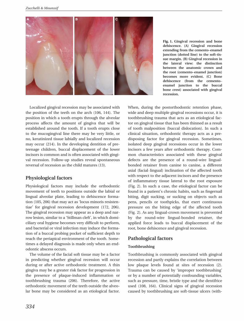
Fig. 1. Gingival recession and bonedehiscence. (A) Gingival recessionextending from the cemento–enameljunction (dotted line) to the soft-tis-sue margin. (B) Gingival recession inthe lateral view: the distinctionbetween the anatomic crown andthe root (cemento–enamel junction)becomes more evident. (C) Bonedehiscence (from the cemento–enamel junction to the buccalbone crest) associated with gingivalrecession.
Zucchelli & Mounssif
334
out pain) and hard-tissue cervical abrasions (noncari-ous cervical lesions). Sometimes, soft-tissue traumamay destroy all keratinized gingival tissue. The cervi-cal abrasions are caused by continued mechanicaltrauma after recession manifestation.
Improper flossing techniques
Flossing trauma can contribute to tooth abrasion andgingival injury (1, 66, 74). These lesions often occur inhighly motivated patients who have not been prop-erly instructed in the technique of flossing. Diagnosisof these injuries can often be confirmed by askingpatients to demonstrate their oral hygiene procedures(200). The initial injury may appear as an acutelyinflamed, ulcerated linear or V-shaped cleft that issymptomatic (74, 87) (Fig. 3). Chronic lesions areoften asymptomatic and may not appear to be ulcer-ated or clinically inflamed. The clefts may traversethe width of the interdental space and extend into theadjacent facial and lingual gingivae. At the histologi-cal level, gingival clefts are often lined by stratifiedsquamous epithelium. The base of the cleft may havea bifurcated appearance and exhibit varying degrees
of epithelialization (87). Often patients use a flossingtechnique with a ‘sawing’ motion (126) while advanc-ing the floss apically into the gingival crevice (200).When flossing trauma is involved, superficial gingivaltissue clefts are ‘red’ because the injury is confinedwithin connective tissue. In this case the lesion isreversible: flossing procedures have to be stopped forat least 2 weeks and chemical plaque control (i.e.chlorexidine rinses) only should be performed(Fig. 3). If the cleft appears ‘white’ the whole connec-tive tissue thickness is involved and the radicular sur-face becomes evident; in this case the gingival lesionis irreversible (87, 140) (Fig. 3).
Perioral and intraoral piercing
Piercing of the tongue and perioral regions is becom-ing an increasingly popular expression of so-calledbody art (79, 126). Tongue piercing has been directlyrelated to dental and gingival injuries on the lingualaspect of the anterior lower teeth (24, 37), and buccalgingival recession may occur in subjects in whom thelip stud is located such that it can traumatize the gin-giva (37, 63). Frequently, the lingual gingival lesion is
A B
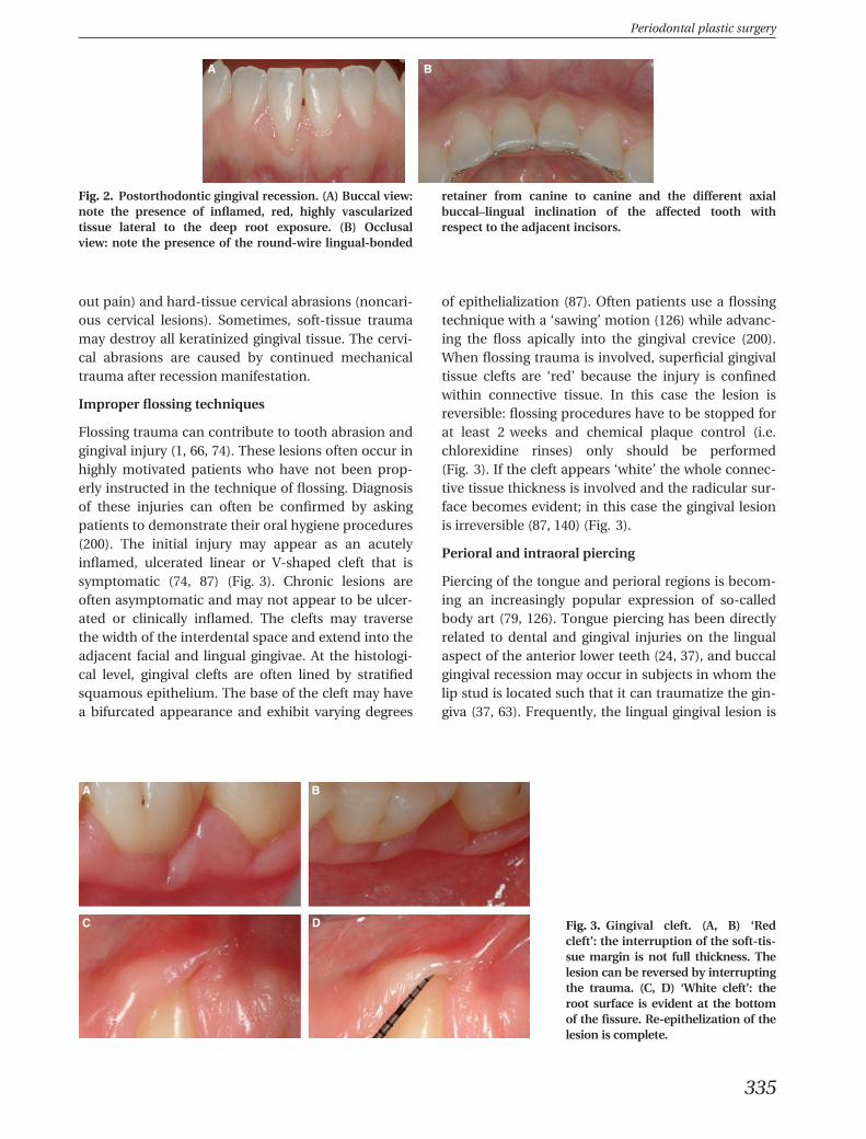
Fig. 2. Postorthodontic gingival recession. (A) Buccal view:note the presence of inflamed, red, highly vascularizedtissue lateral to the deep root exposure. (B) Occlusalview: note the presence of the round-wire lingual-bonded
retainer from canine to canine and the different axialbuccal–lingual inclination of the affected tooth withrespect to the adjacent incisors.
A B
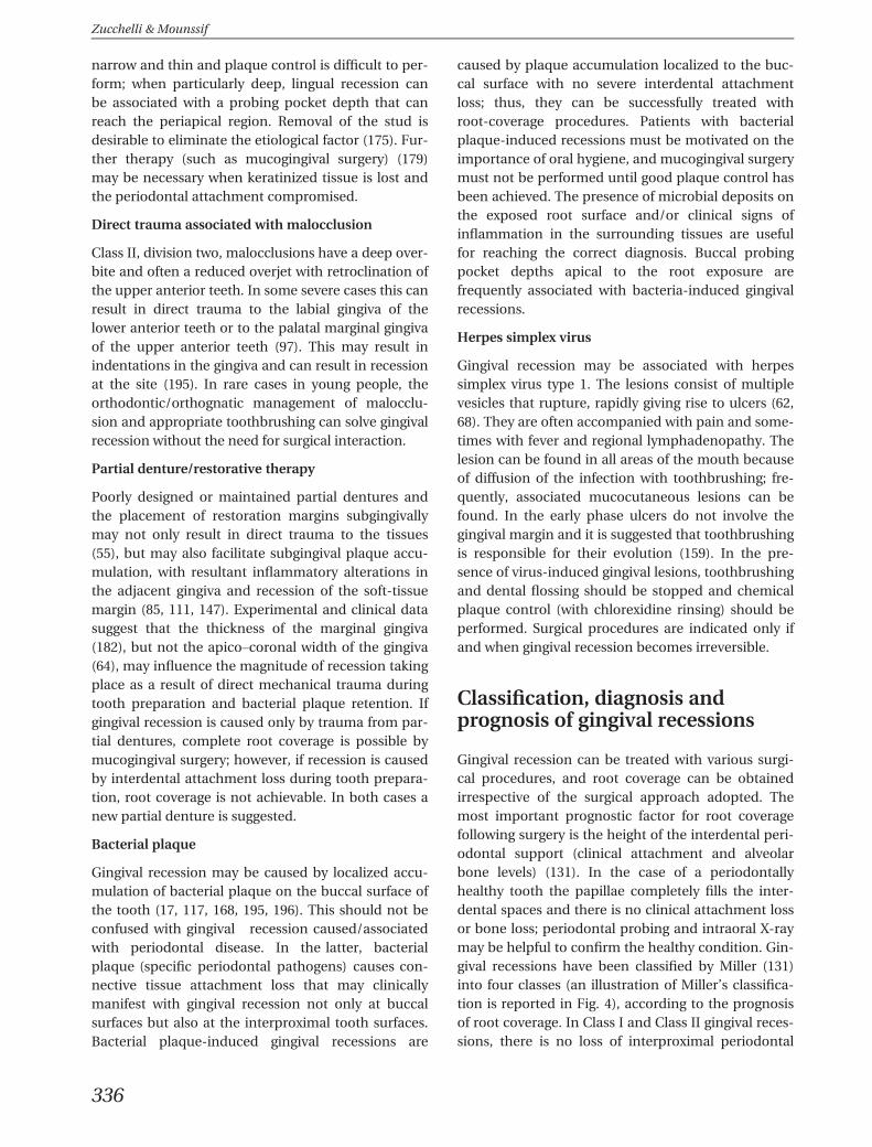
C D Fig. 3. Gingival cleft. (A, B) ‘Redcleft’: the interruption of the soft-tis-sue margin is not full thickness. Thelesion can be reversed by interruptingthe trauma. (C, D) ‘White cleft’: theroot surface is evident at the bottomof the fissure. Re-epithelization of thelesion is complete.
Periodontal plastic surgery
335
narrow and thin and plaque control is difficult to per-form; when particularly deep, lingual recession canbe associated with a probing pocket depth that canreach the periapical region. Removal of the stud isdesirable to eliminate the etiological factor (175). Fur-ther therapy (such as mucogingival surgery) (179)may be necessary when keratinized tissue is lost andthe periodontal attachment compromised.
Direct trauma associated with malocclusion
Class II, division two, malocclusions have a deep over-bite and often a reduced overjet with retroclination ofthe upper anterior teeth. In some severe cases this canresult in direct trauma to the labial gingiva of thelower anterior teeth or to the palatal marginal gingivaof the upper anterior teeth (97). This may result inindentations in the gingiva and can result in recessionat the site (195). In rare cases in young people, theorthodontic/orthognatic management of malocclu-sion and appropriate toothbrushing can solve gingivalrecession without the need for surgical interaction.
Partial denture/restorative therapy
Poorly designed or maintained partial dentures andthe placement of restoration margins subgingivallymay not only result in direct trauma to the tissues(55), but may also facilitate subgingival plaque accu-mulation, with resultant inflammatory alterations inthe adjacent gingiva and recession of the soft-tissuemargin (85, 111, 147). Experimental and clinical datasuggest that the thickness of the marginal gingiva(182), but not the apico–coronal width of the gingiva(64), may influence the magnitude of recession takingplace as a result of direct mechanical trauma duringtooth preparation and bacterial plaque retention. Ifgingival recession is caused only by trauma from par-tial dentures, complete root coverage is possible bymucogingival surgery; however, if recession is causedby interdental attachment loss during tooth prepara-tion, root coverage is not achievable. In both cases anew partial denture is suggested.
Bacterial plaque
Gingival recession may be caused by localized accu-mulation of bacterial plaque on the buccal surface ofthe tooth (17, 117, 168, 195, 196). This should not beconfused with gingival recession caused/associatedwith periodontal disease. In the latter, bacterialplaque (specific periodontal pathogens) causes con-nective tissue attachment loss that may clinicallymanifest with gingival recession not only at buccalsurfaces but also at the interproximal tooth surfaces.Bacterial plaque-induced gingival recessions are
caused by plaque accumulation localized to the buc-cal surface with no severe interdental attachmentloss; thus, they can be successfully treated withroot-coverage procedures. Patients with bacterialplaque-induced recessions must be motivated on theimportance of oral hygiene, and mucogingival surgerymust not be performed until good plaque control hasbeen achieved. The presence of microbial deposits onthe exposed root surface and/or clinical signs ofinflammation in the surrounding tissues are usefulfor reaching the correct diagnosis. Buccal probingpocket depths apical to the root exposure arefrequently associated with bacteria-induced gingivalrecessions.
Herpes simplex virus
Gingival recession may be associated with herpessimplex virus type 1. The lesions consist of multiplevesicles that rupture, rapidly giving rise to ulcers (62,68). They are often accompanied with pain and some-times with fever and regional lymphadenopathy. Thelesion can be found in all areas of the mouth becauseof diffusion of the infection with toothbrushing; fre-quently, associated mucocutaneous lesions can befound. In the early phase ulcers do not involve thegingival margin and it is suggested that toothbrushingis responsible for their evolution (159). In the pre-sence of virus-induced gingival lesions, toothbrushingand dental flossing should be stopped and chemicalplaque control (with chlorexidine rinsing) should beperformed. Surgical procedures are indicated only ifand when gingival recession becomes irreversible.
Classification, diagnosis andprognosis of gingival recessions
Gingival recession can be treated with various surgi-cal procedures, and root coverage can be obtainedirrespective of the surgical approach adopted. Themost important prognostic factor for root coveragefollowing surgery is the height of the interdental peri-odontal support (clinical attachment and alveolarbone levels) (131). In the case of a periodontallyhealthy tooth the papillae completely fills the inter-dental spaces and there is no clinical attachment lossor bone loss; periodontal probing and intraoral X-raymay be helpful to confirm the healthy condition. Gin-gival recessions have been classified by Miller (131)into four classes (an illustration of Miller’s classifica-tion is reported in Fig. 4), according to the prognosisof root coverage. In Class I and Class II gingival reces-sions, there is no loss of interproximal periodontal
Zucchelli & Mounssif
336
attachment and bone, and complete (up to the ce-mento–enamel junction) root coverage can beachieved. The difference between the two classes liesin the height of the root exposure reaching (Class II)or not reaching (Class I) the mucogingival junction.In Class III gingival recessions, the loss of interdentalperiodontal support is mild to moderate, and partialroot coverage can be accomplished; in addition,tooth/root malposition limits the possible amount ofroot coverage. In Class IV gingival recessions, the lossof interproximal periodontal attachment (or tooth/root malposition) is so severe that no root coverage isfeasible.
Some questions/doubts about the classification ofgingival recession, not clarified in Miller’s classifica-tion, have recently been highlighted (160). One ofthese doubts relates to the Miller’s class of gingivalrecession (Class I or Class II) extending beyond themucogingival line, but conserving a small, probableheight of keratinized tissue apical to the root expo-sure (Fig. 5). The distinction, even if not significantfrom a prognostic point of view, could be useful for
selecting the most successful root-coverage surgicalapproach. Other criticisms of Miller’s classificationrelate to the unclear procedures to ascertain theamount of soft-/hard-tissue loss in the interdentalarea to differentiate Class III and Class IV (Fig. 6) andthe unclear influence of tooth malpositioning (160)(Fig. 7). Cairo et al. (35) recently introduced a newclassification system of gingival recessions using thelevel of interproximal clinical attachment as an iden-tification criterion; they also explored the predictivevalue of the resulting classification system on finalroot coverage outcomes following surgery. Threerecession types (RT) were identified: class RT1included gingival recession with no loss of interproxi-mal attachment; class RT2 comprised recession withloss of interproximal attachment less than or equal tothe buccal site; and class RT3 showed interproximalattachment loss higher than the buccal site. Theresults of this study show that the recession type classis a strong predictor of the final recession reductionafter different surgical procedures. The authorshypothesized that the level of interproximal clinical
A B
C D
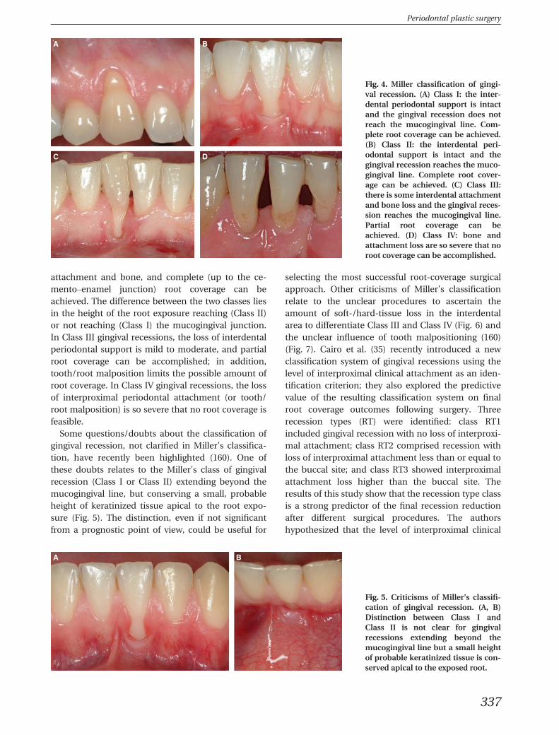
Fig. 4. Miller classification of gingi-val recession. (A) Class I: the inter-dental periodontal support is intactand the gingival recession does notreach the mucogingival line. Com-plete root coverage can be achieved.(B) Class II: the interdental peri-odontal support is intact and thegingival recession reaches the muco-gingival line. Complete root cover-age can be achieved. (C) Class III:there is some interdental attachmentand bone loss and the gingival reces-sion reaches the mucogingival line.Partial root coverage can beachieved. (D) Class IV: bone andattachment loss are so severe that noroot coverage can be accomplished.
A B
Fig. 5. Criticisms of Miller’s classifi-cation of gingival recession. (A, B)Distinction between Class I andClass II is not clear for gingivalrecessions extending beyond themucogingival line but a small heightof probable keratinized tissue is con-served apical to the exposed root.
Periodontal plastic surgery
337
attachment loss is the coronal limit of the achievableamount of root coverage at the buccal site after sur-gery. The RT1 class showed a higher mean reductionof recession compared with the RT2 class, highlight-ing the importance of baseline interproximal clinicalattachment loss for the prognosis of gingival reces-sion treatment. The same authors (34) recently pub-lished a randomized clinical trial evaluating theadjunctive benefit of connective tissue grafts com-pared with coronally advanced flaps for the treatmentof gingival recession associated with interdental clini-cal attachment loss the same as or smaller than buc-cal attachment loss (RT2). They concluded thatcomplete root coverage can be achieved in RT2affecting the upper anterior teeth with both coronallyadvanced flap alone and coronally advanced flap plusconnective tissue grafts; however, the additional useof a connective tissue graft resulted in a greater num-ber of sites with complete root coverage: >80% of thesites when the baseline amount of interdental clinicalattachment loss was ≤3 mm (34). Further longer-termstudies are advocated to evaluate root coverage inMiller Class III and Class IV gingival recessions.Another criticism of Miller’s classification regards thedifficulty of identifying the cemento–enamel junction
on teeth affected by gingival recession and noncari-ous cervical lesions (Fig. 8). Pini-Prato et al. (161)recently proposed a clinical classification of surfacedefects in teeth associated with gingival recession.Four classes of dental-surface defects in areas of gin-gival recession were identified on the basis of thepresence (Class A) or absence (Class B) of the cemen-to–enamel junction and of the presence (Class+) orabsence (Class�) of surface discrepancy (a step). Of1010 exposed root surfaces, 144 (14%) showed anidentifiable cemento–enamel junction associatedwith a root surface step (Class A+), 469 (46%) showedan identifiable cemento–enamel junction without anyassociated step (Class A�), 244 (24%) demonstratedan unidentifiable cemento–enamel junction with astep (Class B+) and 153 (15%) showed an unidentifi-able cemento–enamel junction without any associ-ated step (Class B�). According to the authors, theclassification of dental surface defects in conjunctionwith the classification of periodontal tissues is usefulfor reaching a more precise diagnosis in areas of gin-gival recession, and the condition of the exposed rootsurface may also be important for the prognosticevaluation of mucogingival surgery. In the literature(169, 203), predictability of root coverage was mea-sured in terms of the mean percentage of root cover-age (indicating the percentage of the root exposurecovered with soft tissues) and the percentage of com-plete root coverage (showing the percentage of teethwith the soft-tissue margin covering the cemento–enamel junction). For the correct evaluation of bothof these parameters, it is necessary to recognize thecemento–enamel junction, which anatomically sepa-rates the crown from the root, on the tooth with therecession defect. Therefore, the clinical healing pat-tern only of those gingival recessions in which the ce-mento–enamel junction is clinically detectable couldbe evaluated in terms of percentage and/or completeroot coverage. When the cemento–enamel junction isnot recognizable, it is no longer possible to measurethe depth (and width) of the recession and/or toassess the efficacy of a surgical technique in terms ofroot coverage, as a result of the lack of the referenceparameter (226). Furthermore, other tooth/gingival
A B
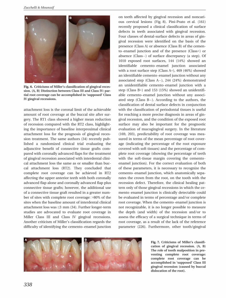
Fig. 6. Criticisms of Miller’s classification of gingival reces-sion. (A, B) Distinction between Class III and Class IV: par-tial root coverage can be accomplished in ‘supposed’ ClassIV gingival recessions.
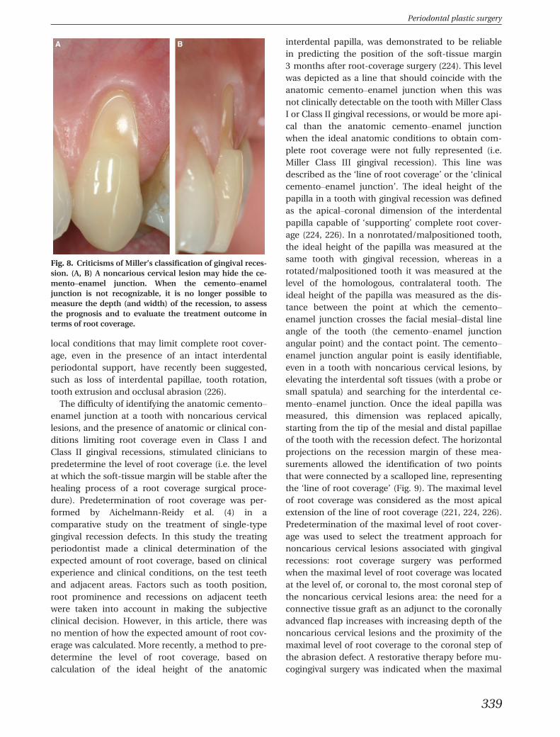
A BFig. 7. Criticisms of Miller’s classifi-cation of gingival recession. (A, B)The role of tooth malposition in pre-venting complete root coverage:complete root coverage can beaccomplished in ‘supposed’ Class IIIgingival recession (caused by buccaldislocation of the root).
Zucchelli & Mounssif
338
local conditions that may limit complete root cover-age, even in the presence of an intact interdentalperiodontal support, have recently been suggested,such as loss of interdental papillae, tooth rotation,tooth extrusion and occlusal abrasion (226).
The difficulty of identifying the anatomic cemento–enamel junction at a tooth with noncarious cervicallesions, and the presence of anatomic or clinical con-ditions limiting root coverage even in Class I andClass II gingival recessions, stimulated clinicians topredetermine the level of root coverage (i.e. the levelat which the soft-tissue margin will be stable after thehealing process of a root coverage surgical proce-dure). Predetermination of root coverage was per-formed by Aichelmann-Reidy et al. (4) in acomparative study on the treatment of single-typegingival recession defects. In this study the treatingperiodontist made a clinical determination of theexpected amount of root coverage, based on clinicalexperience and clinical conditions, on the test teethand adjacent areas. Factors such as tooth position,root prominence and recessions on adjacent teethwere taken into account in making the subjectiveclinical decision. However, in this article, there wasno mention of how the expected amount of root cov-erage was calculated. More recently, a method to pre-determine the level of root coverage, based oncalculation of the ideal height of the anatomic
interdental papilla, was demonstrated to be reliablein predicting the position of the soft-tissue margin3 months after root-coverage surgery (224). This levelwas depicted as a line that should coincide with theanatomic cemento–enamel junction when this wasnot clinically detectable on the tooth with Miller ClassI or Class II gingival recessions, or would be more api-cal than the anatomic cemento–enamel junctionwhen the ideal anatomic conditions to obtain com-plete root coverage were not fully represented (i.e.Miller Class III gingival recession). This line wasdescribed as the ‘line of root coverage’ or the ‘clinicalcemento–enamel junction’. The ideal height of thepapilla in a tooth with gingival recession was definedas the apical–coronal dimension of the interdentalpapilla capable of ‘supporting’ complete root cover-age (224, 226). In a nonrotated/malpositioned tooth,the ideal height of the papilla was measured at thesame tooth with gingival recession, whereas in arotated/malpositioned tooth it was measured at thelevel of the homologous, contralateral tooth. Theideal height of the papilla was measured as the dis-tance between the point at which the cemento–enamel junction crosses the facial mesial–distal lineangle of the tooth (the cemento–enamel junctionangular point) and the contact point. The cemento–enamel junction angular point is easily identifiable,even in a tooth with noncarious cervical lesions, byelevating the interdental soft tissues (with a probe orsmall spatula) and searching for the interdental ce-mento–enamel junction. Once the ideal papilla wasmeasured, this dimension was replaced apically,starting from the tip of the mesial and distal papillaeof the tooth with the recession defect. The horizontalprojections on the recession margin of these mea-surements allowed the identification of two pointsthat were connected by a scalloped line, representingthe ‘line of root coverage’ (Fig. 9). The maximal levelof root coverage was considered as the most apicalextension of the line of root coverage (221, 224, 226).Predetermination of the maximal level of root cover-age was used to select the treatment approach fornoncarious cervical lesions associated with gingivalrecessions: root coverage surgery was performedwhen the maximal level of root coverage was locatedat the level of, or coronal to, the most coronal step ofthe noncarious cervical lesions area: the need for aconnective tissue graft as an adjunct to the coronallyadvanced flap increases with increasing depth of thenoncarious cervical lesions and the proximity of themaximal level of root coverage to the coronal step ofthe abrasion defect. A restorative therapy before mu-cogingival surgery was indicated when the maximal
A B
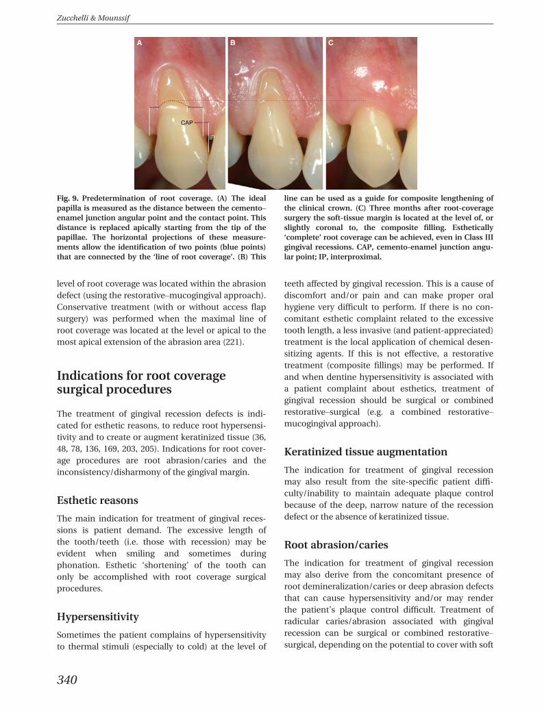
Fig. 8. Criticisms of Miller’s classification of gingival reces-sion. (A, B) A noncarious cervical lesion may hide the ce-mento–enamel junction. When the cemento–enameljunction is not recognizable, it is no longer possible tomeasure the depth (and width) of the recession, to assessthe prognosis and to evaluate the treatment outcome interms of root coverage.
Periodontal plastic surgery
339
level of root coverage was located within the abrasiondefect (using the restorative–mucogingival approach).Conservative treatment (with or without access flapsurgery) was performed when the maximal line ofroot coverage was located at the level or apical to themost apical extension of the abrasion area (221).
Indications for root coveragesurgical procedures
The treatment of gingival recession defects is indi-cated for esthetic reasons, to reduce root hypersensi-tivity and to create or augment keratinized tissue (36,48, 78, 136, 169, 203, 205). Indications for root cover-age procedures are root abrasion/caries and theinconsistency/disharmony of the gingival margin.
Esthetic reasons
The main indication for treatment of gingival reces-sions is patient demand. The excessive length ofthe tooth/teeth (i.e. those with recession) may beevident when smiling and sometimes duringphonation. Esthetic ‘shortening’ of the tooth canonly be accomplished with root coverage surgicalprocedures.
Hypersensitivity
Sometimes the patient complains of hypersensitivityto thermal stimuli (especially to cold) at the level of
teeth affected by gingival recession. This is a cause ofdiscomfort and/or pain and can make proper oralhygiene very difficult to perform. If there is no con-comitant esthetic complaint related to the excessivetooth length, a less invasive (and patient-appreciated)treatment is the local application of chemical desen-sitizing agents. If this is not effective, a restorativetreatment (composite fillings) may be performed. Ifand when dentine hypersensitivity is associated witha patient complaint about esthetics, treatment ofgingival recession should be surgical or combinedrestorative–surgical (e.g. a combined restorative–mucogingival approach).
Keratinized tissue augmentation
The indication for treatment of gingival recessionmay also result from the site-specific patient diffi-culty/inability to maintain adequate plaque controlbecause of the deep, narrow nature of the recessiondefect or the absence of keratinized tissue.
Root abrasion/caries
The indication for treatment of gingival recessionmay also derive from the concomitant presence ofroot demineralization/caries or deep abrasion defectsthat can cause hypersensitivity and/or may renderthe patient’s plaque control difficult. Treatment ofradicular caries/abrasion associated with gingivalrecession can be surgical or combined restorative–surgical, depending on the potential to cover with soft
A B C
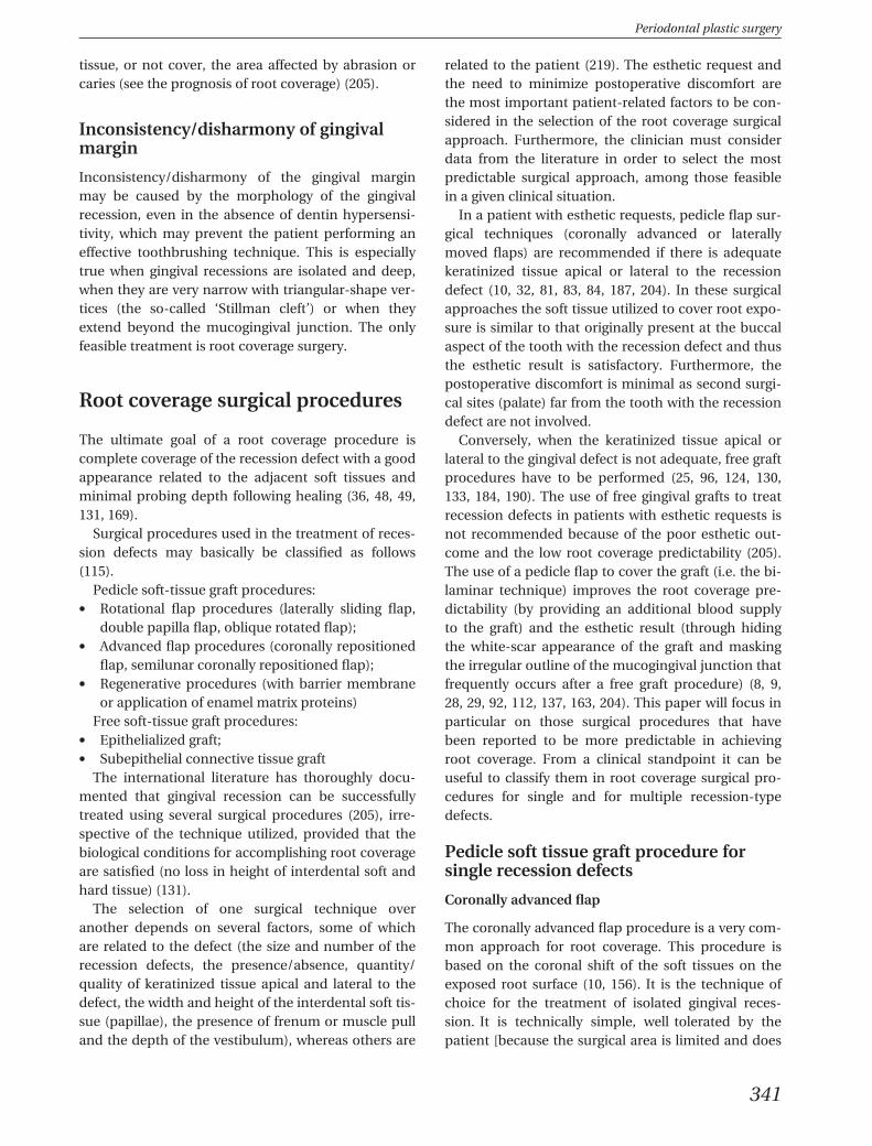
Fig. 9. Predetermination of root coverage. (A) The idealpapilla is measured as the distance between the cemento–enamel junction angular point and the contact point. Thisdistance is replaced apically starting from the tip of thepapillae. The horizontal projections of these measure-ments allow the identification of two points (blue points)that are connected by the ‘line of root coverage’. (B) This
line can be used as a guide for composite lengthening ofthe clinical crown. (C) Three months after root-coveragesurgery the soft-tissue margin is located at the level of, orslightly coronal to, the composite filling. Esthetically‘complete’ root coverage can be achieved, even in Class IIIgingival recessions. CAP, cemento-enamel junction angu-lar point; IP, interproximal.
Zucchelli & Mounssif
340
tissue, or not cover, the area affected by abrasion orcaries (see the prognosis of root coverage) (205).
Inconsistency/disharmony of gingivalmargin
Inconsistency/disharmony of the gingival marginmay be caused by the morphology of the gingivalrecession, even in the absence of dentin hypersensi-tivity, which may prevent the patient performing aneffective toothbrushing technique. This is especiallytrue when gingival recessions are isolated and deep,when they are very narrow with triangular-shape ver-tices (the so-called ‘Stillman cleft’) or when theyextend beyond the mucogingival junction. The onlyfeasible treatment is root coverage surgery.
Root coverage surgical procedures
The ultimate goal of a root coverage procedure iscomplete coverage of the recession defect with a goodappearance related to the adjacent soft tissues andminimal probing depth following healing (36, 48, 49,131, 169).
Surgical procedures used in the treatment of reces-sion defects may basically be classified as follows(115).
Pedicle soft-tissue graft procedures:� Rotational flap procedures (laterally sliding flap,
double papilla flap, oblique rotated flap);� Advanced flap procedures (coronally repositioned
flap, semilunar coronally repositioned flap);� Regenerative procedures (with barrier membrane
or application of enamel matrix proteins)Free soft-tissue graft procedures:
� Epithelialized graft;� Subepithelial connective tissue graft
The international literature has thoroughly docu-mented that gingival recession can be successfullytreated using several surgical procedures (205), irre-spective of the technique utilized, provided that thebiological conditions for accomplishing root coverageare satisfied (no loss in height of interdental soft andhard tissue) (131).
The selection of one surgical technique overanother depends on several factors, some of whichare related to the defect (the size and number of therecession defects, the presence/absence, quantity/quality of keratinized tissue apical and lateral to thedefect, the width and height of the interdental soft tis-sue (papillae), the presence of frenum or muscle pulland the depth of the vestibulum), whereas others are
related to the patient (219). The esthetic request andthe need to minimize postoperative discomfort arethe most important patient-related factors to be con-sidered in the selection of the root coverage surgicalapproach. Furthermore, the clinician must considerdata from the literature in order to select the mostpredictable surgical approach, among those feasiblein a given clinical situation.
In a patient with esthetic requests, pedicle flap sur-gical techniques (coronally advanced or laterallymoved flaps) are recommended if there is adequatekeratinized tissue apical or lateral to the recessiondefect (10, 32, 81, 83, 84, 187, 204). In these surgicalapproaches the soft tissue utilized to cover root expo-sure is similar to that originally present at the buccalaspect of the tooth with the recession defect and thusthe esthetic result is satisfactory. Furthermore, thepostoperative discomfort is minimal as second surgi-cal sites (palate) far from the tooth with the recessiondefect are not involved.
Conversely, when the keratinized tissue apical orlateral to the gingival defect is not adequate, free graftprocedures have to be performed (25, 96, 124, 130,133, 184, 190). The use of free gingival grafts to treatrecession defects in patients with esthetic requests isnot recommended because of the poor esthetic out-come and the low root coverage predictability (205).The use of a pedicle flap to cover the graft (i.e. the bi-laminar technique) improves the root coverage pre-dictability (by providing an additional blood supplyto the graft) and the esthetic result (through hidingthe white-scar appearance of the graft and maskingthe irregular outline of the mucogingival junction thatfrequently occurs after a free graft procedure) (8, 9,28, 29, 92, 112, 137, 163, 204). This paper will focus inparticular on those surgical procedures that havebeen reported to be more predictable in achievingroot coverage. From a clinical standpoint it can beuseful to classify them in root coverage surgical pro-cedures for single and for multiple recession-typedefects.
Pedicle soft tissue graft procedure forsingle recession defects
Coronally advanced flap
The coronally advanced flap procedure is a very com-mon approach for root coverage. This procedure isbased on the coronal shift of the soft tissues on theexposed root surface (10, 156). It is the technique ofchoice for the treatment of isolated gingival reces-sion. It is technically simple, well tolerated by thepatient [because the surgical area is limited and does
Periodontal plastic surgery
341
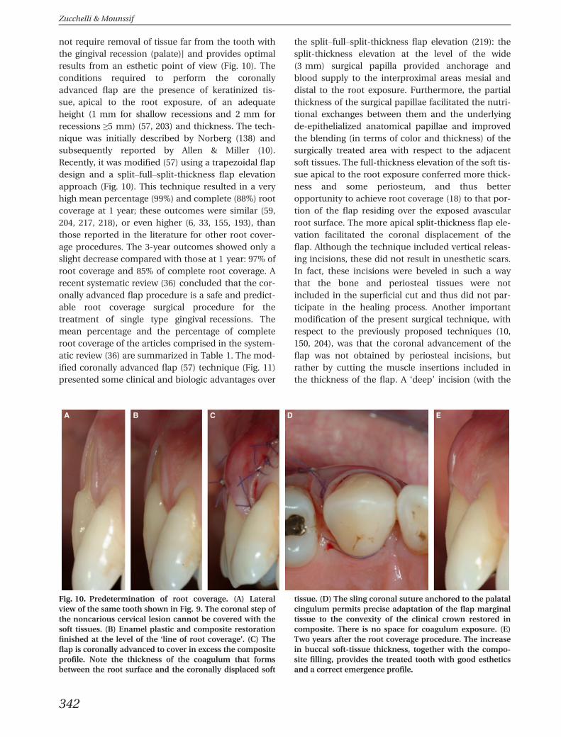
not require removal of tissue far from the tooth withthe gingival recession (palate)] and provides optimalresults from an esthetic point of view (Fig. 10). Theconditions required to perform the coronallyadvanced flap are the presence of keratinized tis-sue, apical to the root exposure, of an adequateheight (1 mm for shallow recessions and 2 mm forrecessions ≥5 mm) (57, 203) and thickness. The tech-nique was initially described by Norberg (138) andsubsequently reported by Allen & Miller (10).Recently, it was modified (57) using a trapezoidal flapdesign and a split–full–split-thickness flap elevationapproach (Fig. 10). This technique resulted in a veryhigh mean percentage (99%) and complete (88%) rootcoverage at 1 year; these outcomes were similar (59,204, 217, 218), or even higher (6, 33, 155, 193), thanthose reported in the literature for other root cover-age procedures. The 3-year outcomes showed only aslight decrease compared with those at 1 year: 97% ofroot coverage and 85% of complete root coverage. Arecent systematic review (36) concluded that the cor-onally advanced flap procedure is a safe and predict-able root coverage surgical procedure for thetreatment of single type gingival recessions. Themean percentage and the percentage of completeroot coverage of the articles comprised in the system-atic review (36) are summarized in Table 1. The mod-ified coronally advanced flap (57) technique (Fig. 11)presented some clinical and biologic advantages over
the split–full–split-thickness flap elevation (219): thesplit-thickness elevation at the level of the wide(3 mm) surgical papilla provided anchorage andblood supply to the interproximal areas mesial anddistal to the root exposure. Furthermore, the partialthickness of the surgical papillae facilitated the nutri-tional exchanges between them and the underlyingde-epithelialized anatomical papillae and improvedthe blending (in terms of color and thickness) of thesurgically treated area with respect to the adjacentsoft tissues. The full-thickness elevation of the soft tis-sue apical to the root exposure conferred more thick-ness and some periosteum, and thus betteropportunity to achieve root coverage (18) to that por-tion of the flap residing over the exposed avascularroot surface. The more apical split-thickness flap ele-vation facilitated the coronal displacement of theflap. Although the technique included vertical releas-ing incisions, these did not result in unesthetic scars.In fact, these incisions were beveled in such a waythat the bone and periosteal tissues were notincluded in the superficial cut and thus did not par-ticipate in the healing process. Another importantmodification of the present surgical technique, withrespect to the previously proposed techniques (10,150, 204), was that the coronal advancement of theflap was not obtained by periosteal incisions, butrather by cutting the muscle insertions included inthe thickness of the flap. A ‘deep’ incision (with the
A B C D E
Fig. 10. Predetermination of root coverage. (A) Lateralview of the same tooth shown in Fig. 9. The coronal step ofthe noncarious cervical lesion cannot be covered with thesoft tissues. (B) Enamel plastic and composite restorationfinished at the level of the ‘line of root coverage’. (C) Theflap is coronally advanced to cover in excess the compositeprofile. Note the thickness of the coagulum that formsbetween the root surface and the coronally displaced soft
tissue. (D) The sling coronal suture anchored to the palatalcingulum permits precise adaptation of the flap marginaltissue to the convexity of the clinical crown restored incomposite. There is no space for coagulum exposure. (E)Two years after the root coverage procedure. The increasein buccal soft-tissue thickness, together with the compo-site filling, provides the treated tooth with good estheticsand a correct emergence profile.
Zucchelli & Mounssif
342
blade parallel to the bone) detached the lip musclefrom the periosteum and permitted the performanceof a ‘superficial’ incision (with the blade parallel tothe lining mucosa) that allowed for coronal advance-ment of the flap. These incisions minimized lip ten-sion on the flap and permitted passive displacementof the flap soft-tissue margin in a coronal position. Afurther technical aspect that was considered criticalfor the success of the modified coronally advancedflap procedure related to the coronal sling suture.The anchorage to the palatal cingulum permits pre-cise adaptation of the keratinized tissue of the flap tothe convexity of the crown of the treated tooth. Thisminimizes exposure of the coagulum, which formsbetween the soft tissue and the root exposure, to thedetrimental microbiological and traumatic agents ofthe oral environment. The increased stability of thecoagulum may play a role in preventing early flapdehiscence and thus favor root coverage. The need
for a tight coronal adaptation of the keratinized tissueof the flaps at the time of suturing (Fig. 12C,D) repre-sented another indication (221), together with theesthetic indication, for a composite reconstruction,before surgery, of the convexity of the tooth crowninterrupted by the presence of noncarious cervicallesions (Figs 9 and 12).
A large increase in keratinized tissue height wasdemonstrated after coronally advanced flap surgeryin the study by De Sanctis & Zucchelli (57) (Fig. 13):in fact, 3 years after the surgery, the mean increase ofkeratinized tissue was 1.78 mm, and this increase wasgreater in sites with deeper recession and a loweramount of residual keratinized tissue at baseline. Verysimilar results were obtained in a previous study eval-uating the 5-year outcomes of the coronally posi-tioned flap for multiple gingival recessions (218).Some hypotheses were made in an attempt to explainthe increase of keratinized tissue after coronally
Table 1. Mean root coverage and complete root coverage (%) with coronally advanced flap technique
Study Flap procedure Mean rootcoverage (%)
Complete rootcoverage (%)
da Silva et al. (54) Coronally advanced flap 68.8 11.0
P. Cortellini (unpublished data) Coronally advanced flap 62.0 Not available
Lins et al. (116) Coronally advanced flap 60.0 Not available
Leknes et al.(114) Coronally advanced flap 34.0 Not available
Modica et al. (135) Coronally advanced flap 80.9 58.3
Del Pizzo et al. (59) Coronally advanced flap 67.0 60.0
Spahr et al. (180) Coronally advanced flap 86.7 23.0
Castellanos et al. (41) Coronally advanced flap 62.2 36.3
Pilloni et al. (152) Coronally advanced flap 65.5 31.2
Woodyard et al. (212) Coronally advanced flap 67.0 33.3
de Queiroz Cortes et al. (56) Coronally advanced flap 55.9 23.1
Huang et al. (98) Coronally advanced flap 83.5 58.3
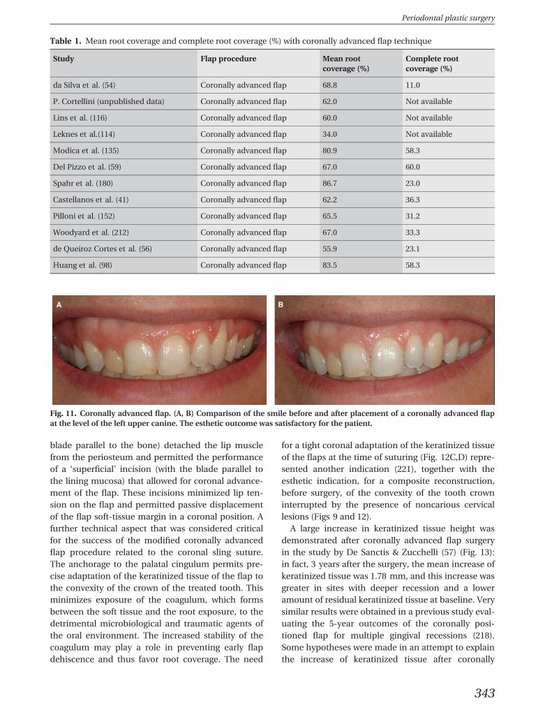
A B
Fig. 11. Coronally advanced flap. (A, B) Comparison of the smile before and after placement of a coronally advanced flapat the level of the left upper canine. The esthetic outcome was satisfactory for the patient.
Periodontal plastic surgery
343
advanced flap surgery: the tendency of the mucogin-gival line, coronally displaced during the surgery, toregain its original, ‘genetically determined’ position(5); or the capability of the connective tissue, derivingfrom the periodontal ligament, to participate in thehealing processes taking place at the dento–gingivalinterface (107, 121, 149). The observation that theincrease in keratinized tissue height was greaterwhen, before surgery, there was a greater recessiondepth and narrower residual band of attached gingiva
apical to the defects seems to support the hypothesisof the tendency of the mucogingival junction toregain its genetically determined position. In fact,these were the clinical situations in which a greatercoronal displacement of the mucogingival line wasperformed during the surgery. The repositioning ofthe mucogingival line could also explain the great var-iability among patients (and studies) in the increaseof keratinized tissue height after coronally advancedflap procedures. One can speculate that patient bio-
A B C D
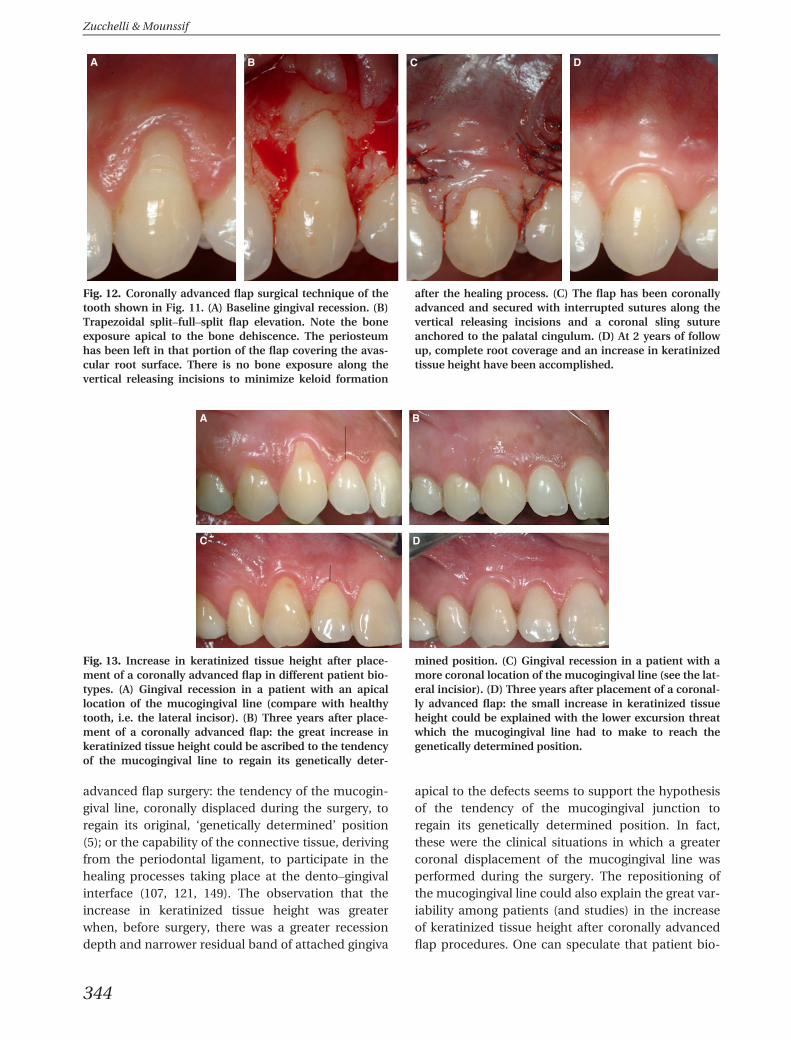
Fig. 12. Coronally advanced flap surgical technique of thetooth shown in Fig. 11. (A) Baseline gingival recession. (B)Trapezoidal split–full–split flap elevation. Note the boneexposure apical to the bone dehiscence. The periosteumhas been left in that portion of the flap covering the avas-cular root surface. There is no bone exposure along thevertical releasing incisions to minimize keloid formation
after the healing process. (C) The flap has been coronallyadvanced and secured with interrupted sutures along thevertical releasing incisions and a coronal sling sutureanchored to the palatal cingulum. (D) At 2 years of followup, complete root coverage and an increase in keratinizedtissue height have been accomplished.
A B
C D
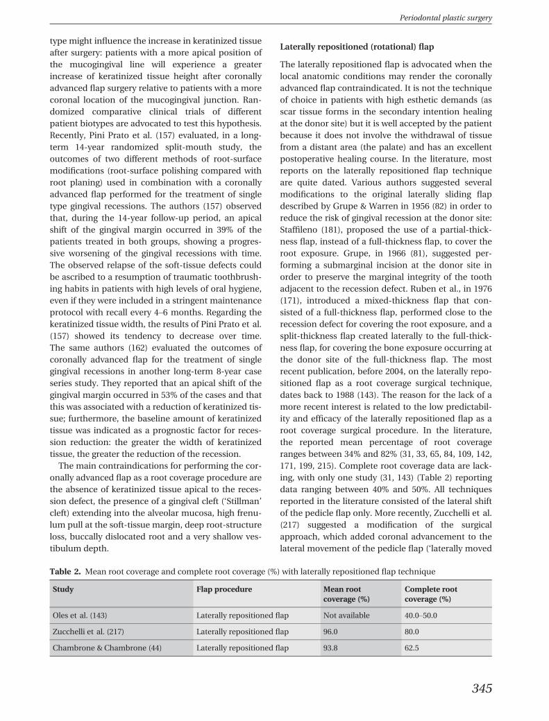
Fig. 13. Increase in keratinized tissue height after place-ment of a coronally advanced flap in different patient bio-types. (A) Gingival recession in a patient with an apicallocation of the mucogingival line (compare with healthytooth, i.e. the lateral incisor). (B) Three years after place-ment of a coronally advanced flap: the great increase inkeratinized tissue height could be ascribed to the tendencyof the mucogingival line to regain its genetically deter-
mined position. (C) Gingival recession in a patient with amore coronal location of the mucogingival line (see the lat-eral incisior). (D) Three years after placement of a coronal-ly advanced flap: the small increase in keratinized tissueheight could be explained with the lower excursion threatwhich the mucogingival line had to make to reach thegenetically determined position.
Zucchelli & Mounssif
344
type might influence the increase in keratinized tissueafter surgery: patients with a more apical position ofthe mucogingival line will experience a greaterincrease of keratinized tissue height after coronallyadvanced flap surgery relative to patients with a morecoronal location of the mucogingival junction. Ran-domized comparative clinical trials of differentpatient biotypes are advocated to test this hypothesis.Recently, Pini Prato et al. (157) evaluated, in a long-term 14-year randomized split-mouth study, theoutcomes of two different methods of root-surfacemodifications (root-surface polishing compared withroot planing) used in combination with a coronallyadvanced flap performed for the treatment of singletype gingival recessions. The authors (157) observedthat, during the 14-year follow-up period, an apicalshift of the gingival margin occurred in 39% of thepatients treated in both groups, showing a progres-sive worsening of the gingival recessions with time.The observed relapse of the soft-tissue defects couldbe ascribed to a resumption of traumatic toothbrush-ing habits in patients with high levels of oral hygiene,even if they were included in a stringent maintenanceprotocol with recall every 4–6 months. Regarding thekeratinized tissue width, the results of Pini Prato et al.(157) showed its tendency to decrease over time.The same authors (162) evaluated the outcomes ofcoronally advanced flap for the treatment of singlegingival recessions in another long-term 8-year caseseries study. They reported that an apical shift of thegingival margin occurred in 53% of the cases and thatthis was associated with a reduction of keratinized tis-sue; furthermore, the baseline amount of keratinizedtissue was indicated as a prognostic factor for reces-sion reduction: the greater the width of keratinizedtissue, the greater the reduction of the recession.
The main contraindications for performing the cor-onally advanced flap as a root coverage procedure arethe absence of keratinized tissue apical to the reces-sion defect, the presence of a gingival cleft (‘Stillman’cleft) extending into the alveolar mucosa, high frenu-lum pull at the soft-tissue margin, deep root-structureloss, buccally dislocated root and a very shallow ves-tibulum depth.
Laterally repositioned (rotational) flap
The laterally repositioned flap is advocated when thelocal anatomic conditions may render the coronallyadvanced flap contraindicated. It is not the techniqueof choice in patients with high esthetic demands (asscar tissue forms in the secondary intention healingat the donor site) but it is well accepted by the patientbecause it does not involve the withdrawal of tissuefrom a distant area (the palate) and has an excellentpostoperative healing course. In the literature, mostreports on the laterally repositioned flap techniqueare quite dated. Various authors suggested severalmodifications to the original laterally sliding flapdescribed by Grupe & Warren in 1956 (82) in order toreduce the risk of gingival recession at the donor site:Staffileno (181), proposed the use of a partial-thick-ness flap, instead of a full-thickness flap, to cover theroot exposure. Grupe, in 1966 (81), suggested per-forming a submarginal incision at the donor site inorder to preserve the marginal integrity of the toothadjacent to the recession defect. Ruben et al., in 1976(171), introduced a mixed-thickness flap that con-sisted of a full-thickness flap, performed close to therecession defect for covering the root exposure, and asplit-thickness flap created laterally to the full-thick-ness flap, for covering the bone exposure occurring atthe donor site of the full-thickness flap. The mostrecent publication, before 2004, on the laterally repo-sitioned flap as a root coverage surgical technique,dates back to 1988 (143). The reason for the lack of amore recent interest is related to the low predictabil-ity and efficacy of the laterally repositioned flap as aroot coverage surgical procedure. In the literature,the reported mean percentage of root coverageranges between 34% and 82% (31, 33, 65, 84, 109, 142,171, 199, 215). Complete root coverage data are lack-ing, with only one study (31, 143) (Table 2) reportingdata ranging between 40% and 50%. All techniquesreported in the literature consisted of the lateral shiftof the pedicle flap only. More recently, Zucchelli et al.(217) suggested a modification of the surgicalapproach, which added coronal advancement to thelateral movement of the pedicle flap (‘laterally moved
Table 2. Mean root coverage and complete root coverage (%) with laterally repositioned flap technique
Study Flap procedure Mean rootcoverage (%)
Complete rootcoverage (%)
Oles et al. (143) Laterally repositioned flap Not available 40.0–50.0
Zucchelli et al. (217) Laterally repositioned flap 96.0 80.0
Chambrone & Chambrone (44) Laterally repositioned flap 93.8 62.5
Periodontal plastic surgery
345
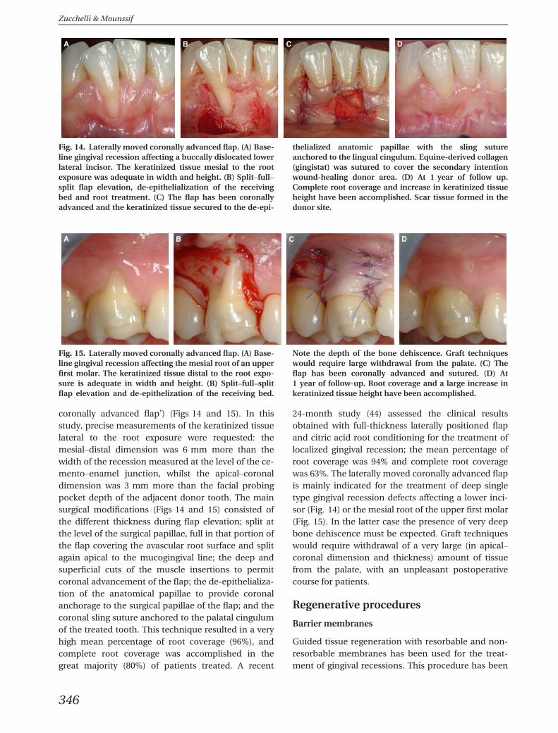
coronally advanced flap’) (Figs 14 and 15). In thisstudy, precise measurements of the keratinized tissuelateral to the root exposure were requested: themesial–distal dimension was 6 mm more than thewidth of the recession measured at the level of the ce-mento–enamel junction, whilst the apical–coronaldimension was 3 mm more than the facial probingpocket depth of the adjacent donor tooth. The mainsurgical modifications (Figs 14 and 15) consisted ofthe different thickness during flap elevation; split atthe level of the surgical papillae, full in that portion ofthe flap covering the avascular root surface and splitagain apical to the mucogingival line; the deep andsuperficial cuts of the muscle insertions to permitcoronal advancement of the flap; the de-epithelializa-tion of the anatomical papillae to provide coronalanchorage to the surgical papillae of the flap; and thecoronal sling suture anchored to the palatal cingulumof the treated tooth. This technique resulted in a veryhigh mean percentage of root coverage (96%), andcomplete root coverage was accomplished in thegreat majority (80%) of patients treated. A recent
24-month study (44) assessed the clinical resultsobtained with full-thickness laterally positioned flapand citric acid root conditioning for the treatment oflocalized gingival recession; the mean percentage ofroot coverage was 94% and complete root coveragewas 63%. The laterally moved coronally advanced flapis mainly indicated for the treatment of deep singletype gingival recession defects affecting a lower inci-sor (Fig. 14) or the mesial root of the upper first molar(Fig. 15). In the latter case the presence of very deepbone dehiscence must be expected. Graft techniqueswould require withdrawal of a very large (in apical–coronal dimension and thickness) amount of tissuefrom the palate, with an unpleasant postoperativecourse for patients.
Regenerative procedures
Barrier membranes
Guided tissue regeneration with resorbable and non-resorbable membranes has been used for the treat-ment of gingival recessions. This procedure has been
A B C D
Fig. 14. Laterally moved coronally advanced flap. (A) Base-line gingival recession affecting a buccally dislocated lowerlateral incisor. The keratinized tissue mesial to the rootexposure was adequate in width and height. (B) Split–full–split flap elevation, de-epithelialization of the receivingbed and root treatment. (C) The flap has been coronallyadvanced and the keratinized tissue secured to the de-epi-
thelialized anatomic papillae with the sling sutureanchored to the lingual cingulum. Equine-derived collagen(gingistat) was sutured to cover the secondary intentionwound-healing donor area. (D) At 1 year of follow up.Complete root coverage and increase in keratinized tissueheight have been accomplished. Scar tissue formed in thedonor site.
A B C D
Fig. 15. Laterally moved coronally advanced flap. (A) Base-line gingival recession affecting the mesial root of an upperfirst molar. The keratinized tissue distal to the root expo-sure is adequate in width and height. (B) Split–full–splitflap elevation and de-epithelization of the receiving bed.
Note the depth of the bone dehiscence. Graft techniqueswould require large withdrawal from the palate. (C) Theflap has been coronally advanced and sutured. (D) At1 year of follow-up. Root coverage and a large increase inkeratinized tissue height have been accomplished.
Zucchelli & Mounssif
346
shown to offer a predictable modality for root cover-age (158, 188, 189), especially in deep recessions,resulting in the regeneration of new connective tissueattachment and bone. The root coverage obtained bypolytetraethylene membranes or bioresorbable mem-branes ranges from 54% to 87% (with a mean of 74%).However, the use of the membrane technique alsoresulted in several problems such as membrane expo-sure and contamination, technical difficulties in plac-ing the barrier and possible damage of the newlyformed tissue as a result of membrane removal orabsorption. Furthermore, recent literature (36, 48,124) shows that the use of a barrier membrane, inconjunction with a coronally advanced flap, does notimprove the result of the coronally advanced flapalone in terms of complete root coverage and reces-sion reduction. At present, the use of a barrier mem-brane for root coverage procedures appears to beinadvisable, especially considering the high incidenceof complications (i.e. membrane exposure) (11, 102,116, 187, 194).
Enamel matrix derivate
Enamel matrix derivative, in combination with a cor-onally advanced flap, was introduced to treat gingivalrecession (135) with the double objective of enhanc-ing root coverage results and inducing periodontalregeneration (59). Recent literature reviews (36, 47,169) showed that enamel matrix derivative, in con-junction with a coronally advanced flap, improvedthe percentage of complete root coverage, increasedkeratinized tissue height and provided better reduc-tion of recession. Histological studies are contradic-tory, reporting either predominant attachmentconsisting of collagen fibers running parallel to theroot surface without new cementum or Sharpey’sfibers (39) and with new bone and new cementumforming only in the most apical portion of root sur-face, or periodontal regeneration with connective tis-sue attachment, new bone and new cementum (127,165). The true clinical rationale to choose thisapproach with respect to the coronally advanced flapalone or other techniques is unclear; thus, routineuse of enamel matrix derivative associated with a cor-onally advanced flap is not recommended. One mayspeculate that the application of enamel matrix deri-vative during mucogingival surgery may be recom-mended in situations in which a wider extension ofnew attachment formation between the soft tissueand the root surface could be of clinical relevance.This may be a result of the size of root exposure (avery wide and deep recession defect), or the toothposition (buccally dislocated root) or a concomitant
bucco–lingual attachment and bone loss (see histo-logical healing after root coverage surgery). Clinicaland histological studies are advocated to confirmsuch a hypothesis.
Free soft-graft procedures
Epithelialized graft
The free gingival graft is the most widely used surgicaltechnique for increasing the width of attached gin-giva. Nevertheless, several authors (20, 23, 151, 166)observed a low degree of predictability of favorableresults with this technique in the coverage of exposedroot surfaces. In fact, a portion of the graft placed onthe denuded root surface does not receive an ade-quate blood supply, with consequent partial necrosisof the grafted tissue. The literature on free gingivalgrafts is contradictory and reports percentages of rootcoverage ranging from 11% to 100% (22, 27, 96, 100,101, 123–125, 130, 133, 145, 176, 190). This variationmay be attributed to differences in the severity of thegingival lesion and in surgical techniques. Nowadays,free autogenous gingival grafts are the last resortwhen the main goal is root coverage or particularly tomeet the esthetic demands of patients. An unfavor-able esthetic outcome is related to incomplete rootcoverage, the white-scar appearance of the graftedtissue that contrasts with the adjacent soft tissues andthe malalignment of the mucogingival line. Free gin-gival grafts can still be used when the main goal ofthe surgical procedure is to augment keratinized tis-sue height (especially in mandibular incisors withoutattached gingiva and with aberrant frenuli), the thick-ness of gingival tissue and the vestibulum depth.When used for root coverage purposes (Fig. 16), thegraft should be sutured coronally to the cemento–enamel junction (to compensate for soft-tissueshrinkage); its thickness should be >1 mm (toincrease root coverage predictability) (3, 190); and itshould be adapted to the convexity of the crown (tominimize coagulum exposure and destabilization).The free gingival graft is contraindicated in patientswith esthetic demands, in deep and wide recessiondefects and in the presence of deep facial probingpockets associated with gingival recession. Freeautogenous gingival grafts can be used as the first sur-gical procedure in the two-stage technique describedby Bernimoulin et al. in 1975 (20). This consists of afirst stage of surgery, in which a free gingival graft isperformed to increase the keratinized tissue heightapical to the gingival recession, and a second stage inwhich the grafted tissue is coronally advanced tocover the exposed root surface (Fig. 17). A mean per-
Periodontal plastic surgery
347
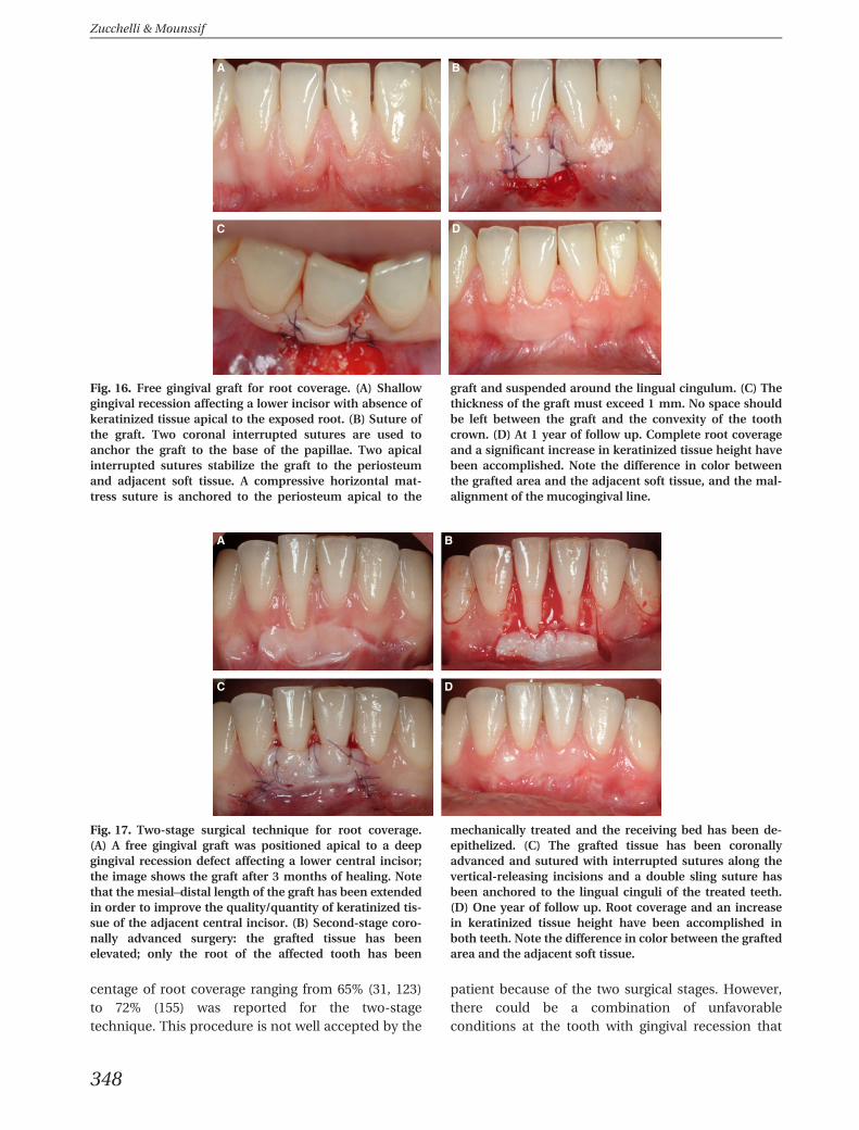
centage of root coverage ranging from 65% (31, 123)to 72% (155) was reported for the two-stagetechnique. This procedure is not well accepted by the
patient because of the two surgical stages. However,there could be a combination of unfavorableconditions at the tooth with gingival recession that
BA
C D
Fig. 16. Free gingival graft for root coverage. (A) Shallowgingival recession affecting a lower incisor with absence ofkeratinized tissue apical to the exposed root. (B) Suture ofthe graft. Two coronal interrupted sutures are used toanchor the graft to the base of the papillae. Two apicalinterrupted sutures stabilize the graft to the periosteumand adjacent soft tissue. A compressive horizontal mat-tress suture is anchored to the periosteum apical to the
graft and suspended around the lingual cingulum. (C) Thethickness of the graft must exceed 1 mm. No space shouldbe left between the graft and the convexity of the toothcrown. (D) At 1 year of follow up. Complete root coverageand a significant increase in keratinized tissue height havebeen accomplished. Note the difference in color betweenthe grafted area and the adjacent soft tissue, and the mal-alignment of the mucogingival line.
A B
C D
Fig. 17. Two-stage surgical technique for root coverage.(A) A free gingival graft was positioned apical to a deepgingival recession defect affecting a lower central incisor;the image shows the graft after 3 months of healing. Notethat the mesial–distal length of the graft has been extendedin order to improve the quality/quantity of keratinized tis-sue of the adjacent central incisor. (B) Second-stage coro-nally advanced surgery: the grafted tissue has beenelevated; only the root of the affected tooth has been
mechanically treated and the receiving bed has been de-epithelized. (C) The grafted tissue has been coronallyadvanced and sutured with interrupted sutures along thevertical-releasing incisions and a double sling suture hasbeen anchored to the lingual cinguli of the treated teeth.(D) One year of follow up. Root coverage and an increasein keratinized tissue height have been accomplished inboth teeth. Note the difference in color between the graftedarea and the adjacent soft tissue.
Zucchelli & Mounssif
348
render this technique as indispensable: the lack ofkeratinized tissue apical and/or lateral to the rootexposure; gingival cleft extending beyond the muco-gingival line; and the presence of a shallow vestibu-lum depth. A recent case report (220) introduced amodified two-stage surgical procedure aiming toimprove the esthetic outcome and reduce thepatient’s morbidity. The main modification of thefirst stage of surgery consisted of harvesting a freegingival graft of the same height as the keratinizedwidth of the adjacent teeth and suturing it on theperiosteum apical to the bone dehiscence. During thesecond stage of surgery the coronal advancement ofthe grafted tissue led to root coverage and realign-ment of the mucogingival line. Zucchelli and DeSanctis (220) showed that by minimizing the apicalcoronal dimension of the free graft and standardizingthe surgical techniques, successful results (in terms ofroot coverage, increase in keratinized tissue andachieving a color similar to that of the adjacent softtissues) could be obtained in the treatment of gingivalrecessions characterized by local conditions, whichotherwise preclude, or render unpredictable, the useof one-step root coverage surgical techniques. Ran-domized controlled studies are advocated to test theefficacy and predictability of the two-stage root cov-erage surgical technique.
Subepithelial connective tissue graft (bilaminartechnique)
The recent literature indicates the bilaminar tech-niques as the most predictable root coverage surgicalprocedures (36, 47–49, 51, 141, 169, 205). The biologi-cal rationale for these techniques is to provide thegraft with an increased blood supply from the cover-ing flap. This will increase the survival of the graftabove the avascular root surface (112) and improvethe esthetic outcome by hiding, partially or com-pletely, the white-scar appearance of the grated tis-sue. The mean percentage and the percentage ofcomplete root coverage in the articles of the system-atic review of Cairo et al. (36) are summarized inTable 3. During the last two decades clinicians haveintroduced several modifications to the original bila-minar technique described (163), resulting in morepredictable outcomes, in terms of root coverage, andgreater esthetic satisfaction for patients. These modi-fications were related to the type of graft (partially orcompletely de-epithelialized) harvested from the pal-ate and to the design (envelope type or with a verticalreleasing incision) of the covering flap. Some authorsused an envelope flap (8, 163) or a repositioned flap(112) to partially cover epithelial connective tissue
grafts. Others utilized coronally advanced flaps, with(137, 204) or without (29) vertical releasing incisions,or a laterally moved papillae flap (92) to coverconnective tissue grafts. In all surgical approachesreported, the size of the graft exceeded that of thebone dehiscence and it was positioned (and sutured)at the level of, or mainly coronal to, the cemento–enamel junction. Although root coverage becameincreasingly more predictable, the esthetic appear-ance of the surgically treated area was often differentfrom that of the adjacent soft tissues. This was causedby the chromatic difference between the uncoveredepithelialized portion of the graft and the adjacentsoft tissues (8, 112, 163), the dischromy associatedwith partial exposure of the connective tissue graft asa result of early dehiscence of the covering flap (29,137, 204), or the difference in thickness between thegrafted area and adjacent soft tissues. More recently,in a comparative study by Zucchelli et al. (216), a fur-ther modified approach was proposed to improve theesthetic outcome of the bilaminar root coverage pro-cedure (Fig. 18). The main surgical modificationsrelated to the size and positioning of the connectivetissue graft: the apico–coronal dimension of the graftwas equal to the depth of the bone dehiscence (mea-sured from the cemento–enamel junction to the mostapical extension of the buccal bone crest) minus thepreoperative height of keratinized tissue apical to therecession defect. The thickness of the graft was<1 mm. The connective tissue graft was positionedapical to the cemento–enamel junction at a distanceequal to the height of keratinized tissue originallypresent apical to the root exposure. This approachwas able to improve patient esthetic satisfaction andpostoperative course (as a result of the lower dimen-sion of the withdrawal), whereas no difference interms of root coverage outcomes (mean percentageand percentage of complete root coverage) werereported with respect to a more traditional approach.The successful root coverage outcome of thisapproach could be explained by the capacity of con-nective tissue grafts to reduce the apical relapse ofthe coronally positioned gingival margin during thehealing phase of the coronally advanced flap proce-dure (153). The main indications for the use of a bila-minar root coverage surgical technique are gingivalrecession in patients with a high esthetic demand inwhom the coronally advanced flap is contraindicatedas a result of the absence/inadequacy of keratinizedtissue apical to the root exposure; gingival recessionassociated with deep root abrasion, root prominenceand root pigmentation (a dark/orange root surface);and gingival recession associated with prosthetic
Periodontal plastic surgery
349
crowns or implants. Contradictions for the bilaminartechniques are those anatomic situations limiting thepossibility to perform pedicle covering flaps (mar-ginal frenuli, high muscle pull, gingival cleft extendingin alveolar mucosa and a very shallow vestibulumdepth), especially when these unfavorable conditions,in fact, occur more frequently in the lower incisionszone.
Connective tissue graft-harvesting procedures
Different connective tissue graft-harvesting proce-dures, with the purpose of achieving primary inten-tion palatal wound healing, have been described inthe literature: the most common are the trap-doorprocedures (60) and the envelope techniques withsingle (99, 118) or double (29) incisions. These pro-cedures have the following common characteristics:
a primary split-thickness access flap elevation; thewithdrawal of connective tissue graft; and completeclosure of the palatal wound with the access flap.The primary objective of these techniques is toreduce patient morbidity by obtaining primary clo-sure of the wound and primary intention healing;however, they need an adequate thickness of thepalatal fibromucosa to avoid desquamation of theundermined superficial flap as a result of compro-mised vascularization (60, 101, 112). The free gingi-val graft surgical wound heals by secondaryintention within 2–4 weeks (67) and has been con-sistently associated with greater discomfort for thepatient as a result of postoperative pain and/orbleeding (58, 67, 101). However, this technique iseasy to perform and can be utilized even in thepresence of a thin palatal fibromucosa.
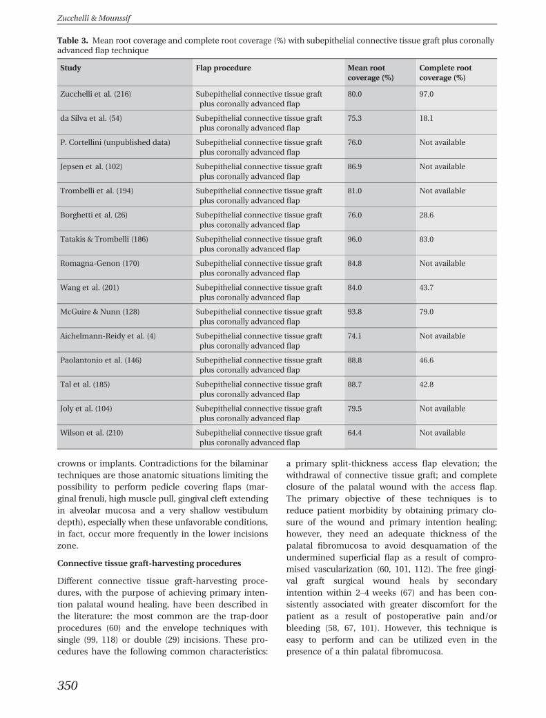
Table 3. Mean root coverage and complete root coverage (%) with subepithelial connective tissue graft plus coronallyadvanced flap technique
Study Flap procedure Mean rootcoverage (%)
Complete rootcoverage (%)
Zucchelli et al. (216) Subepithelial connective tissue graftplus coronally advanced flap
80.0 97.0
da Silva et al. (54) Subepithelial connective tissue graftplus coronally advanced flap
75.3 18.1
P. Cortellini (unpublished data) Subepithelial connective tissue graftplus coronally advanced flap
76.0 Not available
Jepsen et al. (102) Subepithelial connective tissue graftplus coronally advanced flap
86.9 Not available
Trombelli et al. (194) Subepithelial connective tissue graftplus coronally advanced flap
81.0 Not available
Borghetti et al. (26) Subepithelial connective tissue graftplus coronally advanced flap
76.0 28.6
Tatakis & Trombelli (186) Subepithelial connective tissue graftplus coronally advanced flap
96.0 83.0
Romagna-Genon (170) Subepithelial connective tissue graftplus coronally advanced flap
84.8 Not available
Wang et al. (201) Subepithelial connective tissue graftplus coronally advanced flap
84.0 43.7
McGuire & Nunn (128) Subepithelial connective tissue graftplus coronally advanced flap
93.8 79.0
Aichelmann-Reidy et al. (4) Subepithelial connective tissue graftplus coronally advanced flap
74.1 Not available
Paolantonio et al. (146) Subepithelial connective tissue graftplus coronally advanced flap
88.8 46.6
Tal et al. (185) Subepithelial connective tissue graftplus coronally advanced flap
88.7 42.8
Joly et al. (104) Subepithelial connective tissue graftplus coronally advanced flap
79.5 Not available
Wilson et al. (210) Subepithelial connective tissue graftplus coronally advanced flap
64.4 Not available
Zucchelli & Mounssif
350
The evidence in the literature evaluating differ-ences in patient outcomes and morbidity followinguse of the connective tissue graft and free gingivalgraft for root coverage procedures, is minimal. A fewprospective comparative studies (58, 80, 207) reportedpoorer patient outcomes, specifically a greater inci-dence of postoperative pain, for free gingival graftscompared with connective tissue graft procedures.Recently, a clinical randomized controlled study (223)was performed to compare the postoperative morbid-ity and root coverage outcomes in patients treatedwith trap-door connective tissue (control group) andepithelialized (test group) graft-harvesting techniquesfor the treatment of gingival recession using the bila-minar procedure. In the test group the connective tis-sue graft was obtained after de-epithelialization ofthe epithelialized graft with a scalpel blade. No statis-tically significant differences in painkiller consump-tion, postoperative discomfort and bleeding(recorded using the visual analog scale) were foundbetween the two groups. By contrast, necrosis of theprimary flap in the control patients resulted in a six-fold increase of the intake of anti-inflammatorydrugs. The reasons for the lack of differences betweenthe two patient groups are open to speculation; how-ever, a possible explanation may be found in the sur-gical techniques and, in particular, in the reduceddimensions of the graft or in the protection of thewound area with equine-derived collagen in the testgroup. At present, study data demonstrate that theheight (the apical–coronal dimension) and depth ofthe harvesting graft, but not the type (primarycompared with secondary) of palatal wound healing
influence postoperative analgesic consumption. Theresults of the study also indicate that both types ofconnective tissue graft can be successfully used undera coronally advanced flap to cover gingival recession,with no statistically significant difference in rootcoverage outcomes between the grafts. One yearpost-treatment, 92% of the control gingival defectsand 97% of the test gingival recessions were coveredwith the soft tissue. Furthermore, complete rootcoverage was achieved in 70% of the controls and in85% of the test subjects. The only statistically signifi-cant difference in the clinical outcomes between thetwo treatment groups was the greater increase ingingival thickness in the patients treated with thede-epithelialized graft. Any attempt to explain thisdifference is speculative in nature, but it may berelated to the better quality (greater stability and lessshrinkage) of the more superficial connective tissueresulting from the de-epithelialization of a free gingi-val graft with respect to the deeper connective tissueharvested using the trap-door approach (223).
Surgical procedures for multiplerecession defects
Gingival recession is rarely localized to a single tooth,and no reports are available on the prevalence of sin-gle recession defects compared with multiple reces-sion defects; nevertheless, clinical experienceindicates a greater incidence of multiple gingivalrecessions (219). In the presence of multiple defects,the attempt to reduce the number of surgeries andintraoral surgical sites, together with the need to sat-
A B C D E F
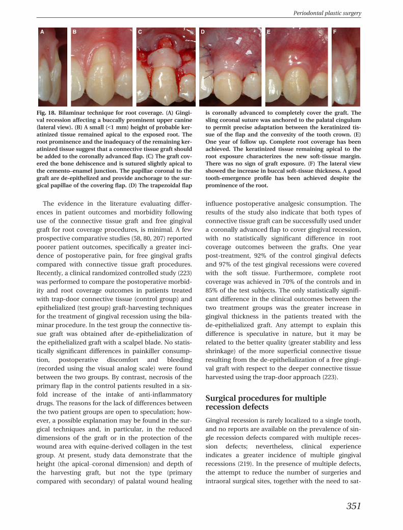
Fig. 18. Bilaminar technique for root coverage. (A) Gingi-val recession affecting a buccally prominent upper canine(lateral view). (B) A small (<1 mm) height of probable ker-atinized tissue remained apical to the exposed root. Theroot prominence and the inadequacy of the remaining ker-atinized tissue suggest that a connective tissue graft shouldbe added to the coronally advanced flap. (C) The graft cov-ered the bone dehiscence and is sutured slightly apical tothe cemento–enamel junction. The papillae coronal to thegraft are de-epithelized and provide anchorage to the sur-gical papillae of the covering flap. (D) The trapezoidal flap
is coronally advanced to completely cover the graft. Thesling coronal suture was anchored to the palatal cingulumto permit precise adaptation between the keratinized tis-sue of the flap and the convexity of the tooth crown. (E)One year of follow up. Complete root coverage has beenachieved. The keratinized tissue remaining apical to theroot exposure characterizes the new soft-tissue margin.There was no sign of graft exposure. (F) The lateral viewshowed the increase in buccal soft-tissue thickness. A goodtooth-emergence profile has been achieved despite theprominence of the root.
Periodontal plastic surgery
351
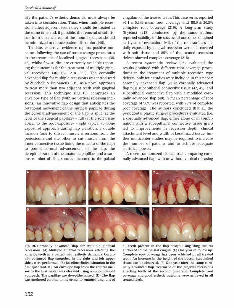
isfy the patient’s esthetic demands, must always betaken into consideration. Thus, when multiple reces-sions affect adjacent teeth they should be treated atthe same time and, if possible, the removal of soft tis-sue from distant areas of the mouth (palate) shouldbe minimized to reduce patient discomfort (46).
To date, extensive evidence reports positive out-comes following the use of root coverage proceduresin the treatment of localized gingival recessions (36,48), whilst few studies are currently available report-ing the outcomes for the treatment of multiple gingi-val recessions (46, 154, 218, 222). The coronallyadvanced flap for multiple recessions was introducedby Zucchelli & De Sanctis (219) as a novel approachto treat more than two adjacent teeth with gingivalrecession. This technique (Fig. 19) comprises anenvelope type of flap (with no vertical releasing inci-sions); an innovative flap design that anticipates therotational movement of the surgical papillae duringthe coronal advancement of the flap; a split (at thelevel of the surgical papillae) – full (at the soft tissueapical to the root exposure) – split (apical to boneexposure) approach during flap elevation; a doubleincision (one to dissect muscle insertions from theperiosteum and the other to cut muscle from theinner connective tissue lining the mucosa of the flap)to permit coronal advancement of the flap; thede-epithelization of the anatomic papillae; and a vari-ous number of sling sutures anchored to the palatal
cingulum of the treated teeth. This case series reported97.1 � 5.1% mean root coverage and 88.6 � 20.3%complete root coverage (219). A long-term study(5 years) (218) conducted by the same authorsreported stability of the successful outcomes obtainedat 1 year of evaluation: 94% of the root surfaces ini-tially exposed by gingival recession were still coveredwith soft tissue and 85% of the treated recessiondefects showed complete coverage (218).
A recent systematic review (46) evaluated theresults obtained with different root-coverage proce-dures in the treatment of multiple recession typedefects; only four studies were included in this paper:coronally advanced flap (218); coronally advancedflap plus subepithelial connective tissue (42, 45); andsubepithelial connective flap with a modified coro-nally advanced flap (40). A mean percentage of rootcoverage of 96% was reported, with 73% of completeroot coverage. The authors concluded that all theperiodontal plastic surgery procedures evaluated (i.e.a coronally advanced flap, either alone or in combi-nation with a subepithelial connective tissue graft)led to improvements in recession depth, clinicalattachment level and width of keratinized tissue; fur-ther multicenter studies may be required to increasethe number of patients and to achieve adequatestatistical power.
A recent randomized clinical trial comparing coro-nally advanced flap, with or without vertical releasing
A B C
D E F
Fig. 19. Coronally advanced flap for multiple gingivalrecessions. (A) Multiple gingival recessions affecting theanterior teeth in a patient with esthetic demands. Coron-ally advanced flap surgeries, in the right and left uppersides, were performed. (B) Baseline clinical situation in thefirst quadrant. (C) An envelope flap from the central inci-sor to the first molar was elevated using a split–full–splitapproach. The papillae are de-epithelialized. (D) The flapwas anchored coronal to the cemento–enamel junctions of
all teeth present in the flap design using sling suturesanchored to the palatal cinguli. (E) One year of follow up.Complete root coverage has been achieved in all treatedteeth. An increase in the height of the buccal keratinizedtissue can be observed. (F) One year after the same coro-nally advanced flap treatment of the gingival recessionsaffecting teeth of the second quadrant. Complete rootcoverage and good esthetic outcome were achieved in alltreated teeth.
Zucchelli & Mounssif
352
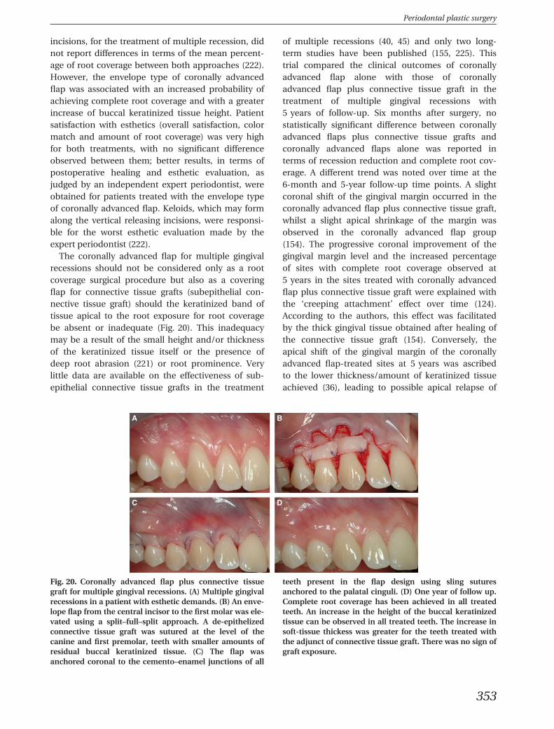
incisions, for the treatment of multiple recession, didnot report differences in terms of the mean percent-age of root coverage between both approaches (222).However, the envelope type of coronally advancedflap was associated with an increased probability ofachieving complete root coverage and with a greaterincrease of buccal keratinized tissue height. Patientsatisfaction with esthetics (overall satisfaction, colormatch and amount of root coverage) was very highfor both treatments, with no significant differenceobserved between them; better results, in terms ofpostoperative healing and esthetic evaluation, asjudged by an independent expert periodontist, wereobtained for patients treated with the envelope typeof coronally advanced flap. Keloids, which may formalong the vertical releasing incisions, were responsi-ble for the worst esthetic evaluation made by theexpert periodontist (222).
The coronally advanced flap for multiple gingivalrecessions should not be considered only as a rootcoverage surgical procedure but also as a coveringflap for connective tissue grafts (subepithelial con-nective tissue graft) should the keratinized band oftissue apical to the root exposure for root coveragebe absent or inadequate (Fig. 20). This inadequacymay be a result of the small height and/or thicknessof the keratinized tissue itself or the presence ofdeep root abrasion (221) or root prominence. Verylittle data are available on the effectiveness of sub-epithelial connective tissue grafts in the treatment
of multiple recessions (40, 45) and only two long-term studies have been published (155, 225). Thistrial compared the clinical outcomes of coronallyadvanced flap alone with those of coronallyadvanced flap plus connective tissue graft in thetreatment of multiple gingival recessions with5 years of follow-up. Six months after surgery, nostatistically significant difference between coronallyadvanced flaps plus connective tissue grafts andcoronally advanced flaps alone was reported interms of recession reduction and complete root cov-erage. A different trend was noted over time at the6-month and 5-year follow-up time points. A slightcoronal shift of the gingival margin occurred in thecoronally advanced flap plus connective tissue graft,whilst a slight apical shrinkage of the margin wasobserved in the coronally advanced flap group(154). The progressive coronal improvement of thegingival margin level and the increased percentageof sites with complete root coverage observed at5 years in the sites treated with coronally advancedflap plus connective tissue graft were explained withthe ‘creeping attachment’ effect over time (124).According to the authors, this effect was facilitatedby the thick gingival tissue obtained after healing ofthe connective tissue graft (154). Conversely, theapical shift of the gingival margin of the coronallyadvanced flap-treated sites at 5 years was ascribedto the lower thickness/amount of keratinized tissueachieved (36), leading to possible apical relapse of
A B
C D
Fig. 20. Coronally advanced flap plus connective tissuegraft for multiple gingival recessions. (A) Multiple gingivalrecessions in a patient with esthetic demands. (B) An enve-lope flap from the central incisor to the first molar was ele-vated using a split–full–split approach. A de-epithelizedconnective tissue graft was sutured at the level of thecanine and first premolar, teeth with smaller amounts ofresidual buccal keratinized tissue. (C) The flap wasanchored coronal to the cemento–enamel junctions of all
teeth present in the flap design using sling suturesanchored to the palatal cinguli. (D) One year of follow up.Complete root coverage has been achieved in all treatedteeth. An increase in the height of the buccal keratinizedtissue can be observed in all treated teeth. The increase insoft-tissue thickess was greater for the teeth treated withthe adjunct of connective tissue graft. There was no sign ofgraft exposure.
Periodontal plastic surgery
353
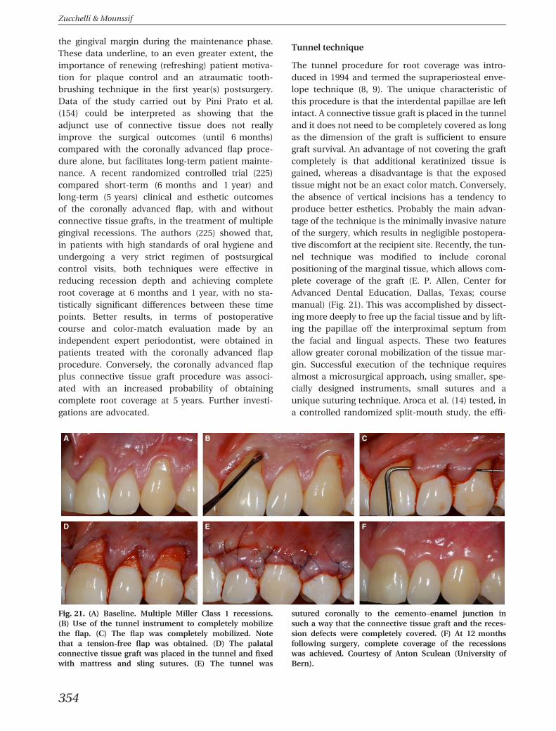
the gingival margin during the maintenance phase.These data underline, to an even greater extent, theimportance of renewing (refreshing) patient motiva-tion for plaque control and an atraumatic tooth-brushing technique in the first year(s) postsurgery.Data of the study carried out by Pini Prato et al.(154) could be interpreted as showing that theadjunct use of connective tissue does not reallyimprove the surgical outcomes (until 6 months)compared with the coronally advanced flap proce-dure alone, but facilitates long-term patient mainte-nance. A recent randomized controlled trial (225)compared short-term (6 months and 1 year) andlong-term (5 years) clinical and esthetic outcomesof the coronally advanced flap, with and withoutconnective tissue grafts, in the treatment of multiplegingival recessions. The authors (225) showed that,in patients with high standards of oral hygiene andundergoing a very strict regimen of postsurgicalcontrol visits, both techniques were effective inreducing recession depth and achieving completeroot coverage at 6 months and 1 year, with no sta-tistically significant differences between these timepoints. Better results, in terms of postoperativecourse and color-match evaluation made by anindependent expert periodontist, were obtained inpatients treated with the coronally advanced flapprocedure. Conversely, the coronally advanced flapplus connective tissue graft procedure was associ-ated with an increased probability of obtainingcomplete root coverage at 5 years. Further investi-gations are advocated.
Tunnel technique
The tunnel procedure for root coverage was intro-duced in 1994 and termed the supraperiosteal enve-lope technique (8, 9). The unique characteristic ofthis procedure is that the interdental papillae are leftintact. A connective tissue graft is placed in the tunneland it does not need to be completely covered as longas the dimension of the graft is sufficient to ensuregraft survival. An advantage of not covering the graftcompletely is that additional keratinized tissue isgained, whereas a disadvantage is that the exposedtissue might not be an exact color match. Conversely,the absence of vertical incisions has a tendency toproduce better esthetics. Probably the main advan-tage of the technique is the minimally invasive natureof the surgery, which results in negligible postopera-tive discomfort at the recipient site. Recently, the tun-nel technique was modified to include coronalpositioning of the marginal tissue, which allows com-plete coverage of the graft (E. P. Allen, Center forAdvanced Dental Education, Dallas, Texas; coursemanual) (Fig. 21). This was accomplished by dissect-ing more deeply to free up the facial tissue and by lift-ing the papillae off the interproximal septum fromthe facial and lingual aspects. These two featuresallow greater coronal mobilization of the tissue mar-gin. Successful execution of the technique requiresalmost a microsurgical approach, using smaller, spe-cially designed instruments, small sutures and aunique suturing technique. Aroca et al. (14) tested, ina controlled randomized split-mouth study, the effi-
A B C
D E F
Fig. 21. (A) Baseline. Multiple Miller Class 1 recessions.(B) Use of the tunnel instrument to completely mobilizethe flap. (C) The flap was completely mobilized. Notethat a tension-free flap was obtained. (D) The palatalconnective tissue graft was placed in the tunnel and fixedwith mattress and sling sutures. (E) The tunnel was
sutured coronally to the cemento–enamel junction insuch a way that the connective tissue graft and the reces-sion defects were completely covered. (F) At 12 monthsfollowing surgery, complete coverage of the recessionswas achieved. Courtesy of Anton Sculean (University ofBern).
Zucchelli & Mounssif
354
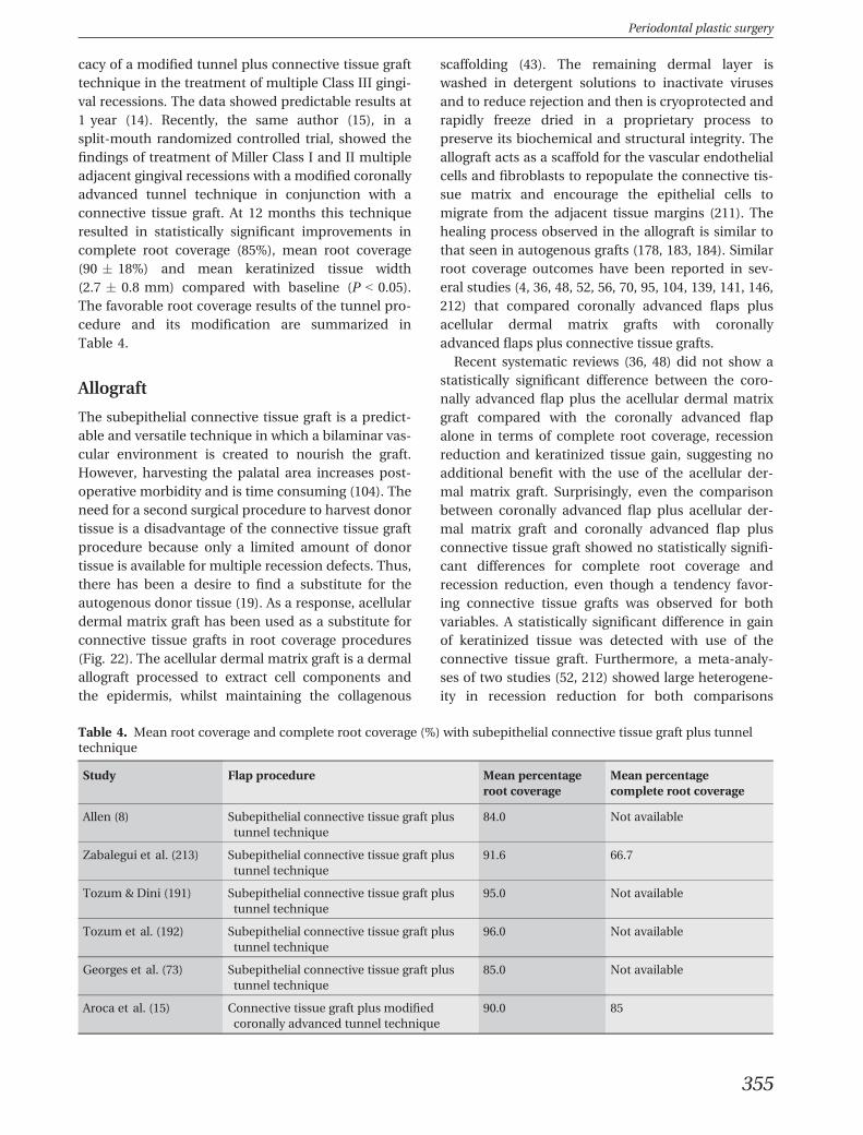
cacy of a modified tunnel plus connective tissue grafttechnique in the treatment of multiple Class III gingi-val recessions. The data showed predictable results at1 year (14). Recently, the same author (15), in asplit-mouth randomized controlled trial, showed thefindings of treatment of Miller Class I and II multipleadjacent gingival recessions with a modified coronallyadvanced tunnel technique in conjunction with aconnective tissue graft. At 12 months this techniqueresulted in statistically significant improvements incomplete root coverage (85%), mean root coverage(90 � 18%) and mean keratinized tissue width(2.7 � 0.8 mm) compared with baseline (P < 0.05).The favorable root coverage results of the tunnel pro-cedure and its modification are summarized inTable 4.
Allograft
The subepithelial connective tissue graft is a predict-able and versatile technique in which a bilaminar vas-cular environment is created to nourish the graft.However, harvesting the palatal area increases post-operative morbidity and is time consuming (104). Theneed for a second surgical procedure to harvest donortissue is a disadvantage of the connective tissue graftprocedure because only a limited amount of donortissue is available for multiple recession defects. Thus,there has been a desire to find a substitute for theautogenous donor tissue (19). As a response, acellulardermal matrix graft has been used as a substitute forconnective tissue grafts in root coverage procedures(Fig. 22). The acellular dermal matrix graft is a dermalallograft processed to extract cell components andthe epidermis, whilst maintaining the collagenous
scaffolding (43). The remaining dermal layer iswashed in detergent solutions to inactivate virusesand to reduce rejection and then is cryoprotected andrapidly freeze dried in a proprietary process topreserve its biochemical and structural integrity. Theallograft acts as a scaffold for the vascular endothelialcells and fibroblasts to repopulate the connective tis-sue matrix and encourage the epithelial cells tomigrate from the adjacent tissue margins (211). Thehealing process observed in the allograft is similar tothat seen in autogenous grafts (178, 183, 184). Similarroot coverage outcomes have been reported in sev-eral studies (4, 36, 48, 52, 56, 70, 95, 104, 139, 141, 146,212) that compared coronally advanced flaps plusacellular dermal matrix grafts with coronallyadvanced flaps plus connective tissue grafts.
Recent systematic reviews (36, 48) did not show astatistically significant difference between the coro-nally advanced flap plus the acellular dermal matrixgraft compared with the coronally advanced flapalone in terms of complete root coverage, recessionreduction and keratinized tissue gain, suggesting noadditional benefit with the use of the acellular der-mal matrix graft. Surprisingly, even the comparisonbetween coronally advanced flap plus acellular der-mal matrix graft and coronally advanced flap plusconnective tissue graft showed no statistically signifi-cant differences for complete root coverage andrecession reduction, even though a tendency favor-ing connective tissue grafts was observed for bothvariables. A statistically significant difference in gainof keratinized tissue was detected with use of theconnective tissue graft. Furthermore, a meta-analy-ses of two studies (52, 212) showed large heterogene-ity in recession reduction for both comparisons
Table 4. Mean root coverage and complete root coverage (%) with subepithelial connective tissue graft plus tunneltechnique
Study Flap procedure Mean percentageroot coverage
Mean percentagecomplete root coverage
Allen (8) Subepithelial connective tissue graft plustunnel technique
84.0 Not available
Zabalegui et al. (213) Subepithelial connective tissue graft plustunnel technique
91.6 66.7
Tozum & Dini (191) Subepithelial connective tissue graft plustunnel technique
95.0 Not available
Tozum et al. (192) Subepithelial connective tissue graft plustunnel technique
96.0 Not available
Georges et al. (73) Subepithelial connective tissue graft plustunnel technique
85.0 Not available
Aroca et al. (15) Connective tissue graft plus modifiedcoronally advanced tunnel technique
90.0 85
Periodontal plastic surgery
355
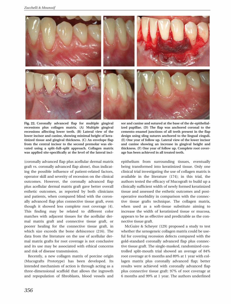
(coronally advanced flap plus acellular dermal matrixgraft vs. coronally advanced flap alone), thus indicat-ing the possible influence of patient-related factors,operator skill and severity of recession on the clinicaloutcomes. However, the coronally advanced flapplus acellular dermal matrix graft gave better overallesthetic outcomes, as reported by both cliniciansand patients, when compared blind with the coron-ally advanced flap plus connective tissue graft, eventhough it showed less complete root coverage (4).This finding may be related to different colormatches with adjacent tissues for the acellular der-mal matrix graft and connective tissue graft, orpoorer healing for the connective tissue graft, inwhich size exceeds the bone dehiscence (216). Thedata from the literature on the use of acellular der-mal matrix grafts for root coverage is not conclusiveand its use may be associated with ethical concernsand risk of disease transmission.
Recently, a new collagen matrix of porcine origin(Mucografts Prototype) has been developed. Itsintended mechanism of action is through acting as athree-dimensional scaffold that allows the ingrowthand repopulation of fibroblasts, blood vessels and
epithelium from surrounding tissues, eventuallybeing transformed into keratinized tissue. Only oneclinical trial investigating the use of collagen matrix isavailable in the literature (174); in this trial, theauthors tested the efficacy of Mucograft to build up aclinically sufficient width of newly formed keratinizedtissue and assessed the esthetic outcomes and post-operative morbidity in comparison with the connec-tive tissue grafts technique. The collagen matrix,when used as a soft-tissue substitute aiming toincrease the width of keratinized tissue or mucosa,appears to be as effective and predictable as the con-nective tissue graft.
McGuire & Scheyer (129) proposed a study to testwhether the xenogeneic collagen matrix could be use-ful for covering recession defects compared with thegold-standard coronally advanced flap plus connec-tive tissue graft. The single-masked, randomized-con-trolled split-mouth trial showed an average of 84%root coverage at 6 months and 89% at 1 year with col-lagen matrix plus coronally advanced flap; betterresults were achieved with coronally advanced flapplus connective tissue graft: 97% of root coverage at6 months and 99% at 1 year. The authors underlined
A B C
D E F
Fig. 22. Coronally advanced flap for multiple gingivalrecessions plus collagen matrix. (A) Multiple gingivalrecessions affecting lower teeth. (B) Lateral view of thelower incisor and canine, showing minimal height of kera-tinized tissue and gingival thickness. (C) An envelope flapfrom the central incisor to the second premolar was ele-vated using a split–full–split approach. Collagen matrixwas applied site-specifically at the level of the lateral inci-
sor and canine and sutured at the base of the de-epithelial-ized papillae. (D) The flap was anchored coronal to thecemento–enamel junctions of all teeth present in the flapdesign using sling sutures anchored to the lingual cinguli.(E) One year of follow up. Lateral view of the lower incisorand canine showing an increase in gingival height andthickness. (F) One year of follow up. Complete root cover-age has been achieved in all treated teeth.
Zucchelli & Mounssif
356
that the measures, evaluated statistically, were differ-ent but balanced with subject-reported outcomes(subjects’ assessments of pain/discomfort and esthet-ics), and that collagen matrix plus coronally advancedflap presented an intriguing comparison with the tra-ditional connective tissue graft gold standard. Arecent randomized controlled trial (38) evaluated theuse of a porcine collagen matrix plus coronallyadvanced flap as an alternative to coronally advancedflap plus connective tissue graft for the treatment ofgingival recessions. At 12 months, porcine collagenmatrix plus coronally advanced flap resulted in amean root coverage of 94% compared with a meanroot coverage of 97% for coronally advanced flap plusconnective tissue graft. From a statistical point ofview, these measures are different but, according tothe authors, the outcomes achieved by the porcinecollagen matrix plus coronally advanced flap proce-dure were clinically comparable with those of the cor-onally advanced flap plus connective tissue graftgroup and similar to those expected from the coro-nally advanced flap plus connective tissue graft, asstated in previous literature reviews. A recent single-blinded, randomized, controlled, split-mouth multi-center trial (103) evaluated the clinical outcomes ofthe use of a xenogeneic collagen matrix (test group)plus the coronally advanced flap or coronallyadvanced flap alone in the treatment of localizedrecession defects. At 6 months, root coverage (pri-mary outcome) was 76% for test defects and 73% forcontrol defects (P = 0.169), with 36% of test defectsand 31% of control defects exhibiting complete rootcoverage. The increase in the mean width of kerati-nized tissue was higher in test defects (from 1.97 to2.90 mm) than in control defects (from 2.00 to2.57 mm) (P = 0.036). Likewise, test sites had moregain in gingival thickness (0.59 mm) than did controlsites (0.34 mm) (P = 0.003). Larger (≥3 mm) reces-sions (n = 35 patients) treated with collagen matrixshowed higher root coverage (72% vs. 66%, P = 0.043),as well as more gain in keratinized tissue and gingivalthickness. The authors (103) concluded that coronallyadvanced flap plus collagen matrix was not superiorwith regard to root coverage, but enhanced gingivalthickness and width of keratinized tissue when com-pared with coronally advanced flap alone. For thecoverage of larger defects, coronally advanced flapplus collagen matrix was more effective.
Root conditioning
Chemical root-surface conditioning using a variety ofagents has been introduced in order to detoxify,
decontaminate and demineralize the root surface,thereby removing the smear layer and exposing thecollagenous matrix of dentin and cementum (88–91).Various acids have been used for chemical root-sur-face conditioning, including citric and phosphoricacids (167), ethylenediaminetetraacetic acid (113) andtetracycline hydrochloride (110). In an animal model,these procedures are believed to be able to induce ce-mentogenesis and enhance attachment by connectivetissue ingrowth and/or demineralization (71, 209).However, in human studies, no clinical advantageswere observed (61, 120). The clinical relevance of rootconditioning with an acid agent in routine periodon-tal surgery is still uncertain and there is no evidencethat these products will improve root coverage(36, 48).
Healing after root coverageprocedures
The major goal of periodontal plastic surgery is thecoverage of roots exposed by gingival recession (203).These days, the covering of denuded roots is a pre-dictable and effective procedure, usually with highlyesthetic results. However, the nature of the attach-ment between the grafted tissue and the root surfaceis not well understood. A potential weakness of thetechnique is that a pocket may be created where therecession has been covered (86) A true new connec-tive tissue attachment would be preferable to a longjunctional epithelium (86). The aim of the presentsection is to summarize current knowledge about theregenerative events following the surgical treatmentof recession defects. Specifically, the character of his-tological healing involved will be discussed. Histologi-cal evaluation of the nature of the interface betweenthe newly covered root surface and overlying gingivaltissues is based on animal studies and isolated casereports.
Animal studies
Animal studies in dogs and monkeys were under-taken as long ago as 1950, using different periodontalplastic surgery procedures: lateral (32, 208) and coro-nal (77) displaced flaps, coronal flap associated withmembranes (76) and connective tissue grafts wereperformed to achieve root coverage in experimentalgingival recession. Similar histological and histomor-phometrical findings were reported: connective tissueattachment (fibers functionally inserted or parallel tothe root) with new bone and cementum was found in
Periodontal plastic surgery
357
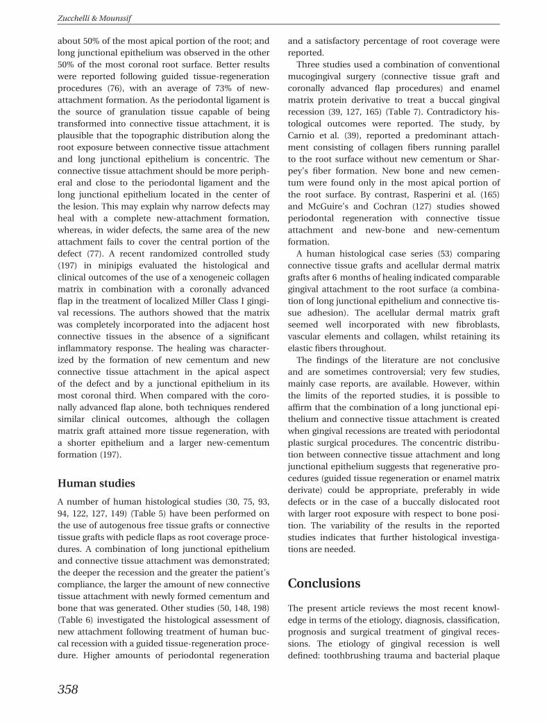
about 50% of the most apical portion of the root; andlong junctional epithelium was observed in the other50% of the most coronal root surface. Better resultswere reported following guided tissue-regenerationprocedures (76), with an average of 73% of new-attachment formation. As the periodontal ligament isthe source of granulation tissue capable of beingtransformed into connective tissue attachment, it isplausible that the topographic distribution along theroot exposure between connective tissue attachmentand long junctional epithelium is concentric. Theconnective tissue attachment should be more periph-eral and close to the periodontal ligament and thelong junctional epithelium located in the center ofthe lesion. This may explain why narrow defects mayheal with a complete new-attachment formation,whereas, in wider defects, the same area of the newattachment fails to cover the central portion of thedefect (77). A recent randomized controlled study(197) in minipigs evaluated the histological andclinical outcomes of the use of a xenogeneic collagenmatrix in combination with a coronally advancedflap in the treatment of localized Miller Class I gingi-val recessions. The authors showed that the matrixwas completely incorporated into the adjacent hostconnective tissues in the absence of a significantinflammatory response. The healing was character-ized by the formation of new cementum and newconnective tissue attachment in the apical aspectof the defect and by a junctional epithelium in itsmost coronal third. When compared with the coro-nally advanced flap alone, both techniques renderedsimilar clinical outcomes, although the collagenmatrix graft attained more tissue regeneration, witha shorter epithelium and a larger new-cementumformation (197).
Human studies
A number of human histological studies (30, 75, 93,94, 122, 127, 149) (Table 5) have been performed onthe use of autogenous free tissue grafts or connectivetissue grafts with pedicle flaps as root coverage proce-dures. A combination of long junctional epitheliumand connective tissue attachment was demonstrated;the deeper the recession and the greater the patient’scompliance, the larger the amount of new connectivetissue attachment with newly formed cementum andbone that was generated. Other studies (50, 148, 198)(Table 6) investigated the histological assessment ofnew attachment following treatment of human buc-cal recession with a guided tissue-regeneration proce-dure. Higher amounts of periodontal regeneration
and a satisfactory percentage of root coverage werereported.
Three studies used a combination of conventionalmucogingival surgery (connective tissue graft andcoronally advanced flap procedures) and enamelmatrix protein derivative to treat a buccal gingivalrecession (39, 127, 165) (Table 7). Contradictory his-tological outcomes were reported. The study, byCarnio et al. (39), reported a predominant attach-ment consisting of collagen fibers running parallelto the root surface without new cementum or Shar-pey’s fiber formation. New bone and new cemen-tum were found only in the most apical portion ofthe root surface. By contrast, Rasperini et al. (165)and McGuire’s and Cochran (127) studies showedperiodontal regeneration with connective tissueattachment and new-bone and new-cementumformation.
A human histological case series (53) comparingconnective tissue grafts and acellular dermal matrixgrafts after 6 months of healing indicated comparablegingival attachment to the root surface (a combina-tion of long junctional epithelium and connective tis-sue adhesion). The acellular dermal matrix graftseemed well incorporated with new fibroblasts,vascular elements and collagen, whilst retaining itselastic fibers throughout.
The findings of the literature are not conclusiveand are sometimes controversial; very few studies,mainly case reports, are available. However, withinthe limits of the reported studies, it is possible toaffirm that the combination of a long junctional epi-thelium and connective tissue attachment is createdwhen gingival recessions are treated with periodontalplastic surgical procedures. The concentric distribu-tion between connective tissue attachment and longjunctional epithelium suggests that regenerative pro-cedures (guided tissue regeneration or enamel matrixderivate) could be appropriate, preferably in widedefects or in the case of a buccally dislocated rootwith larger root exposure with respect to bone posi-tion. The variability of the results in the reportedstudies indicates that further histological investiga-tions are needed.
Conclusions
The present article reviews the most recent knowl-edge in terms of the etiology, diagnosis, classification,prognosis and surgical treatment of gingival reces-sions. The etiology of gingival recession is welldefined: toothbrushing trauma and bacterial plaque
Zucchelli & Mounssif
358
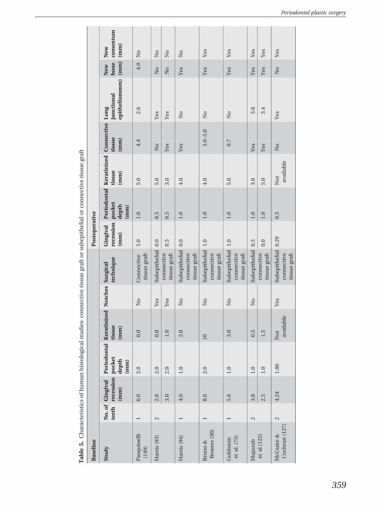
Tab
le5.
Charac
teristicsofh
uman
histologica
lstudies:co
nnective
tissuegraftorsu
bep
ithelialo
rco
nnective
tissuegraft
Baseline
Postoperative
Study
No.o
fteeth
Gingiva
lrecession
(mm)
Periodontal
pock
etdep
th(m
m)
Keratinized
tissue
(mm)
Notches
Surgical
tech
nique
Gingiva
lrecession
(mm)
Periodontal
pock
etdep
th(m
m)
Keratinized
tissue
(mm)
Connective
tissue
(mm)
Long
junctional
epitheliummm)
New
bone
(mm)
New
cemen
tum
(mm)
Pasquinelli
(149
)1
6.0
2.0
0.0
No
Connective
tissuegraft
5.0
1.0
5.0
4.4
2.6
4.0
No
Harris(93)
22.0
2.0
0.0
Yes
Subep
ithelial
connective
tissuegraft
0.0
0.5
5.0
No
Yes
No
No
3.0
2.0
1.0
Yes
0.5
0.5
3.0
Yes
Yes
No
No
Harris(94)
14.0
1.0
2.0
No
Subep
ithelial
connective
tissuegraft
0.0
1.0
4.0
Yes
No
Yes
No
Bruno&
Bowers(30)
18.0
2.0
10No
Subep
ithelial
connective
tissuegraft
1.0
1.0
4.0
3.0–
5.0
No
Yes
Yes
Goldstein
etal.(75
)1
5.0
1.0
3.0
No
Subep
ithelial
connective
tissuegraft
1.0
1.0
5.0
0.7
No
Yes
Yes
Majzo
ub
etal.(12
2)2
3.0
1.0
0.5
No
Subep
ithelial
connective
tissuegraft
0.5
1.0
3.0
Yes
3.8
Yes
Yes
2.5
1.0
1.5
0.0
1.0
5.0
Yes
3.4
Yes
Yes
McG
uire&
Coch
ran(127
)2
4.24
1.80
Not
available
Yes
Subep
ithelial
connective
tissuegraft
0.29
0.5
Not
available
No
Yes
No
Yes
Periodontal plastic surgery
359
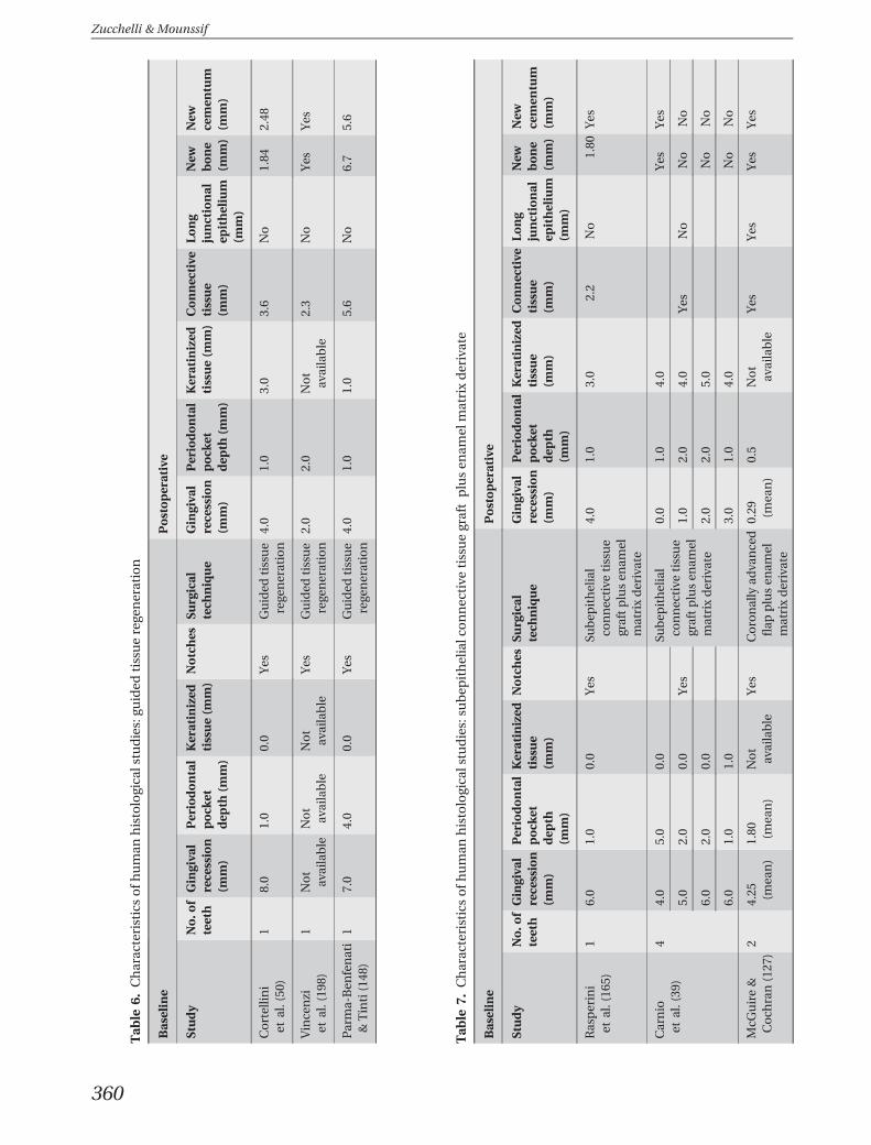
Tab
le6.
Charac
teristicsofh
uman
histologica
lstudies:gu
ided
tissuerege
neration
Baseline
Postoperative
Study
No.o
fteeth
Gingiva
lrecession
(mm)
Periodontal
pock
etdep
th(m
m)
Keratinized
tissue(m
m)
Notches
Surgical
tech
nique
Gingiva
lrecession
(mm)
Periodontal
pock
etdep
th(m
m)
Keratinized
tissue(m
m)
Connective
tissue
(mm)
Long
junctional
epithelium
(mm)
New
bone
(mm)
New
cemen
tum
(mm)
Cortellin
iet
al.(50
)1
8.0
1.0
0.0
Yes
Guided
tissue
rege
neration
4.0
1.0
3.0
3.6
No
1.84
2.48
Vincenzi
etal.(19
8)1
Not
available
Not
available
Not
available
Yes
Guided
tissue
rege
neration
2.0
2.0
Not
available
2.3
No
Yes
Yes
Parma-Ben
fenati
&Tinti(148
)1
7.0
4.0
0.0
Yes
Guided
tissue
rege
neration
4.0
1.0
1.0
5.6
No
6.7
5.6
Tab
le7.
Charac
teristicsofh
uman
histologica
lstudies:su
bep
ithelialc
onnective
tissuegraftplusen
amel
matrixderivate
Baseline
Postoperative
Study
No.o
fteeth
Gingiva
lrecession
(mm)
Periodontal
pock
etdep
th(m
m)
Keratinized
tissue
(mm)
Notches
Surgical
tech
nique
Gingiva
lrecession
(mm)
Periodontal
pock
etdep
th(m
m)
Keratinized
tissue
(mm)
Connective
tissue
(mm)
Long
junctional
epithelium
(mm)
New
bone
(mm)
New
cemen
tum
(mm)
Rasperini
etal.(16
5)1
6.0
1.0
0.0
Yes
Subep
ithelial
connective
tissue
graftplusen
amel
matrixderivate
4.0
1.0
3.0
2.2
No
1.80
Yes
Carnio
etal.(39
)4
4.0
5.0
0.0
Subep
ithelial
connective
tissue
graftplusen
amel
matrixderivate
0.0
1.0
4.0
Yes
Yes
5.0
2.0
0.0
Yes
1.0
2.0
4.0
Yes
No
No
No
6.0
2.0
0.0
2.0
2.0
5.0
No
No
6.0
1.0
1.0
3.0
1.0
4.0
No
No
McG
uire&
Coch
ran(127
)2
4.25 (mea
n)
1.80 (mea
n)
Not
available
Yes
Coronally
adva
nced
flap
plusen
amel
matrixderivate
0.29 (mea
n)
0.5
Not
available
Yes
Yes
Yes
Yes
Zucchelli & Mounssif
360
are the most frequent causative factors for gingivalrecessions acting on an existing lack of alveolarbuccal bone that may be anatomical or acquired.Conversely, diagnosis, prognosis and, especially, clas-sification of gingival recession will need to be revis-ited on the basis of the recent findings. Majordifficulties arise when the main reference parameterfor diagnosing, measuring and evaluating the treat-ment outcome of gingival recession is lack of thecemento–enamel junction. This occurs quite fre-quently when toothbrushing trauma also createsnoncarious cervical lesions, together with gingivalrecession. A criticism of Miller’s classification and theprognosis of gingival recessions relates to the unclearprocedures used to ascertain the amount of soft/hardtissue loss in the interdental area to differentiate ClassIII and IV and the unclear influence of tooth malposi-tioning. Until new knowledge better defines theseaspects, the use of clinical methods to predeterminethe level of root coverage is recommended, at least toimprove the patient’s perception of their esthetic out-come. Surgical coverage of gingival recession is verypredictable, at least for a single type of defect. Thegold standard is the bilaminar technique, whichmainly consists of a coronally advanced flap coveringa connective tissue graft because the adjunctive useof connective tissue grafts increases the likelihood ofachieving complete root coverage, with respect to useof the coronally advanced flap alone, especially inlong-term follow-up. Although because of the limiteddimension in single type recession defects theadjunctive use of connective tissue grafts is quiteoften indicated to improve complete root coveragepredictability, the same cannot be assumed for multi-ple gingival recessions. This is because of the limitedamount of tissue that can be harvested from the pal-ate, the increased patient morbidity and theenhanced possibility of dehiscence in the coveringflap as a result of increasing the dimension of theconnective tissue grafts. This last factor is particularlyunacceptable in patients with esthetic demands as aresult of the difference in color, texture and surfacecharacteristic of the exposed grafted area with respectto the adjacent soft tissue. Studies are needed to clar-ify, in greater detail, which, and how many, gingivalrecessions should be treated with the adjunctive useof connective tissue grafts when treating multiple gin-gival recessions. In this view, the utilization of substi-tutes for the connective tissue grafts would bestrongly encouraged. Unfortunately, despite recentimprovements, none of the available allograft materi-als can be considered as a true substitute for connec-tive tissue grafts.
One of the most important innovations in gingivalrecession treatment, which has already started butneeds future development and improvement, is thedesign of clinical trials with the patient’s outcome,esthetics and morbidity in particular, as primaryoutcome measures. This is likely to change currentsuccess evaluation criteria and perhaps also the deci-sional matrix in the surgical management of gingivalrecession.
References
1. Abrams H, Kopczyk RA. Gingival sequela from a retainedpiece of dental floss. J Am Dent Assoc 1983: 106: 57–58.
2. Addy M, Griffiths G, Dummer P, Kingdom A, Shaw WC.The distribution of plaque and gingivitis and the influenceof toothbrushing hand in a group of South Wales 11–12 year-old children. J Clin Periodontol 1987: 14: 564–572.
3. Agudio G, Nieri M, Rotundo R, Cortellini P, Pini Prato G.Free gingival grafts to increase keratinized tissue: a retro-spective long-term evaluation (10 to 25 years) of out-comes. J Periodontol 2008: 79: 587–594.
4. Aichelmann-Reidy M, Yukna R, Evans G, Nasr H, Mayer E.Clinical evaluation of acellular allograft dermis for thetreatment of human gingival recession. J Periodontol 2001:72: 998–1005.
5. Ainamo A, Bergenholtz A, Hugoson A, Ainamo J. Locationof the mucogingival junction 18 years after apically reposi-tioned flap surgery. J Clin Periodontol 1992: 19: 49–52.
6. Al-Hamdan K, Eber R, Sarment D, Kowalski C, Wang HL.Guided tissue regeneration-based root coverage: meta-analysis. J Periodontol 2003: 74: 1520–1533.
7. Alldritt W. Abnormal gingival form. Proc R Soc Med 1968:61: 137–142.
8. Allen A. Use of the supraperiosteal envelope in soft tissuegrafting for root coverage. II. Clinical results. Int JPeriodontics Restorative Dent 1994: 14: 302–315.
9. Allen A. Use of the supraperiosteal envelope in soft tissuegrafting for root coverage. I. Rationale and technique. Int JPeriodontics Restorative Dent 1994: 14: 216–227.
10. Allen EP, Miller PD Jr. Coronal positioning of existing gin-giva: short term results in the treatment of shallow mar-ginal tissue recession. J Periodontol 1989: 60: 316–319.
11. Amarante E, Leknes K, Skavland J, Lie T. Coronally posi-tioned flap procedures with or without a bioabsorbablemembrane in the treatment of human gingival recession.J Periodontol 2000: 71: 989–998.
12. American Academy of Periodontology. Glossary terms inperiodontology. Chicago: The American Academy ofPeriodontology, 2001.
13. Andlin-Sobocki A, Marcusson A, Persson M. 3-Year obser-vations on gingival recession in mandibular incisors inchildren. J Clin Periodontol 1991: 18: 155–159.
14. Aroca S, Keglevich T, Nikolidakis D, Gera I, Nagy K, Azzi R,Etienne D. Treatment of class III multiple gingival reces-sions: a randomized-clinical trial. J Clin Periodontol 2010:37: 88–97.
15. Aroca S, Molnar B, Windisch P, Gera I, Salvi GE, Nikolida-kis D, Sculean A. Treatment of multiple adjacent Miller
Periodontal plastic surgery
361
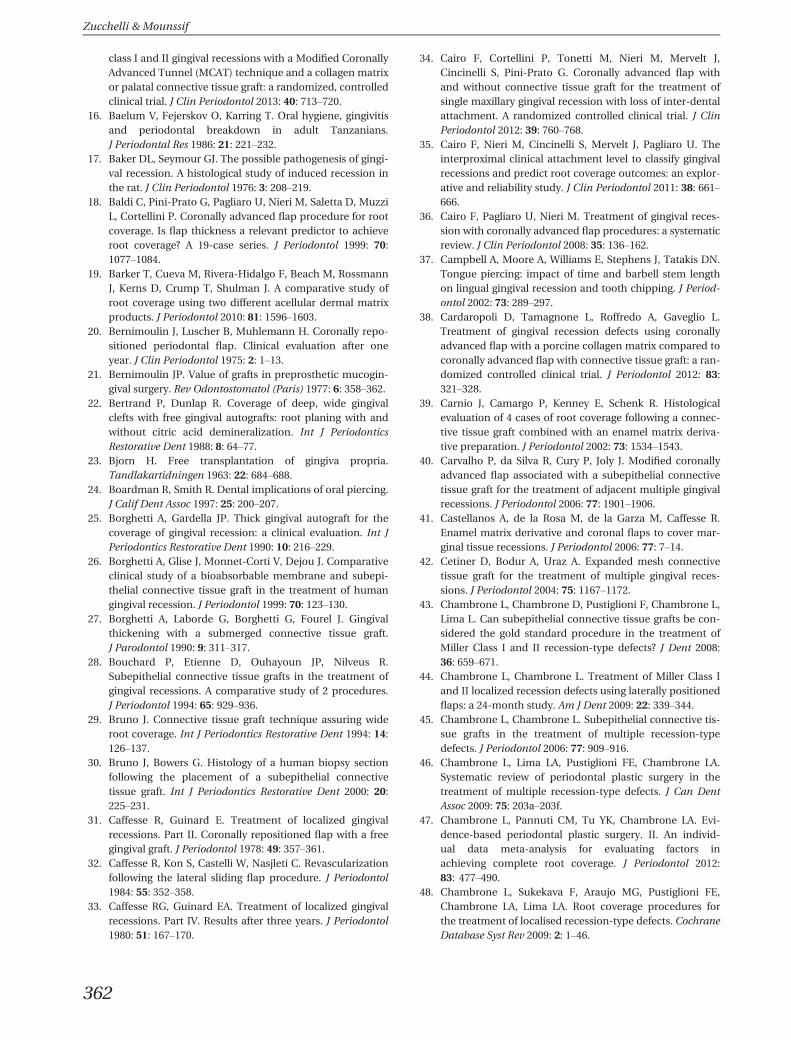
class I and II gingival recessions with a Modified CoronallyAdvanced Tunnel (MCAT) technique and a collagen matrixor palatal connective tissue graft: a randomized, controlledclinical trial. J Clin Periodontol 2013: 40: 713–720.
16. Baelum V, Fejerskov O, Karring T. Oral hygiene, gingivitisand periodontal breakdown in adult Tanzanians.J Periodontal Res 1986: 21: 221–232.
17. Baker DL, Seymour GJ. The possible pathogenesis of gingi-val recession. A histological study of induced recession inthe rat. J Clin Periodontol 1976: 3: 208–219.
18. Baldi C, Pini-Prato G, Pagliaro U, Nieri M, Saletta D, MuzziL, Cortellini P. Coronally advanced flap procedure for rootcoverage. Is flap thickness a relevant predictor to achieveroot coverage? A 19-case series. J Periodontol 1999: 70:1077–1084.
19. Barker T, Cueva M, Rivera-Hidalgo F, Beach M, RossmannJ, Kerns D, Crump T, Shulman J. A comparative study ofroot coverage using two different acellular dermal matrixproducts. J Periodontol 2010: 81: 1596–1603.
20. Bernimoulin J, Luscher B, Muhlemann H. Coronally repo-sitioned periodontal flap. Clinical evaluation after oneyear. J Clin Periodontol 1975: 2: 1–13.
21. Bernimoulin JP. Value of grafts in preprosthetic mucogin-gival surgery. Rev Odontostomatol (Paris) 1977: 6: 358–362.
22. Bertrand P, Dunlap R. Coverage of deep, wide gingivalclefts with free gingival autografts: root planing with andwithout citric acid demineralization. Int J PeriodonticsRestorative Dent 1988: 8: 64–77.
23. Bjorn H. Free transplantation of gingiva propria.Tandlakartidningen 1963: 22: 684–688.
24. Boardman R, Smith R. Dental implications of oral piercing.J Calif Dent Assoc 1997: 25: 200–207.
25. Borghetti A, Gardella JP. Thick gingival autograft for thecoverage of gingival recession: a clinical evaluation. Int JPeriodontics Restorative Dent 1990: 10: 216–229.
26. Borghetti A, Glise J, Monnet-Corti V, Dejou J. Comparativeclinical study of a bioabsorbable membrane and subepi-thelial connective tissue graft in the treatment of humangingival recession. J Periodontol 1999: 70: 123–130.
27. Borghetti A, Laborde G, Borghetti G, Fourel J. Gingivalthickening with a submerged connective tissue graft.J Parodontol 1990: 9: 311–317.
28. Bouchard P, Etienne D, Ouhayoun JP, Nilveus R.Subepithelial connective tissue grafts in the treatment ofgingival recessions. A comparative study of 2 procedures.J Periodontol 1994: 65: 929–936.
29. Bruno J. Connective tissue graft technique assuring wideroot coverage. Int J Periodontics Restorative Dent 1994: 14:126–137.
30. Bruno J, Bowers G. Histology of a human biopsy sectionfollowing the placement of a subepithelial connectivetissue graft. Int J Periodontics Restorative Dent 2000: 20:225–231.
31. Caffesse R, Guinard E. Treatment of localized gingivalrecessions. Part II. Coronally repositioned flap with a freegingival graft. J Periodontol 1978: 49: 357–361.
32. Caffesse R, Kon S, Castelli W, Nasjleti C. Revascularizationfollowing the lateral sliding flap procedure. J Periodontol1984: 55: 352–358.
33. Caffesse RG, Guinard EA. Treatment of localized gingivalrecessions. Part IV. Results after three years. J Periodontol1980: 51: 167–170.
34. Cairo F, Cortellini P, Tonetti M, Nieri M, Mervelt J,Cincinelli S, Pini-Prato G. Coronally advanced flap withand without connective tissue graft for the treatment ofsingle maxillary gingival recession with loss of inter-dentalattachment. A randomized controlled clinical trial. J ClinPeriodontol 2012: 39: 760–768.
35. Cairo F, Nieri M, Cincinelli S, Mervelt J, Pagliaro U. Theinterproximal clinical attachment level to classify gingivalrecessions and predict root coverage outcomes: an explor-ative and reliability study. J Clin Periodontol 2011: 38: 661–666.
36. Cairo F, Pagliaro U, Nieri M. Treatment of gingival reces-sion with coronally advanced flap procedures: a systematicreview. J Clin Periodontol 2008: 35: 136–162.
37. Campbell A, Moore A, Williams E, Stephens J, Tatakis DN.Tongue piercing: impact of time and barbell stem lengthon lingual gingival recession and tooth chipping. J Period-ontol 2002: 73: 289–297.
38. Cardaropoli D, Tamagnone L, Roffredo A, Gaveglio L.Treatment of gingival recession defects using coronallyadvanced flap with a porcine collagen matrix compared tocoronally advanced flap with connective tissue graft: a ran-domized controlled clinical trial. J Periodontol 2012: 83:321–328.
39. Carnio J, Camargo P, Kenney E, Schenk R. Histologicalevaluation of 4 cases of root coverage following a connec-tive tissue graft combined with an enamel matrix deriva-tive preparation. J Periodontol 2002: 73: 1534–1543.
40. Carvalho P, da Silva R, Cury P, Joly J. Modified coronallyadvanced flap associated with a subepithelial connectivetissue graft for the treatment of adjacent multiple gingivalrecessions. J Periodontol 2006: 77: 1901–1906.
41. Castellanos A, de la Rosa M, de la Garza M, Caffesse R.Enamel matrix derivative and coronal flaps to cover mar-ginal tissue recessions. J Periodontol 2006: 77: 7–14.
42. Cetiner D, Bodur A, Uraz A. Expanded mesh connectivetissue graft for the treatment of multiple gingival reces-sions. J Periodontol 2004: 75: 1167–1172.
43. Chambrone L, Chambrone D, Pustiglioni F, Chambrone L,Lima L. Can subepithelial connective tissue grafts be con-sidered the gold standard procedure in the treatment ofMiller Class I and II recession-type defects? J Dent 2008:36: 659–671.
44. Chambrone L, Chambrone L. Treatment of Miller Class Iand II localized recession defects using laterally positionedflaps: a 24-month study. Am J Dent 2009: 22: 339–344.
45. Chambrone L, Chambrone L. Subepithelial connective tis-sue grafts in the treatment of multiple recession-typedefects. J Periodontol 2006: 77: 909–916.
46. Chambrone L, Lima LA, Pustiglioni FE, Chambrone LA.Systematic review of periodontal plastic surgery in thetreatment of multiple recession-type defects. J Can DentAssoc 2009: 75: 203a–203f.
47. Chambrone L, Pannuti CM, Tu YK, Chambrone LA. Evi-dence-based periodontal plastic surgery. II. An individ-ual data meta-analysis for evaluating factors inachieving complete root coverage. J Periodontol 2012:83: 477–490.
48. Chambrone L, Sukekava F, Araujo MG, Pustiglioni FE,Chambrone LA, Lima LA. Root coverage procedures forthe treatment of localised recession-type defects. CochraneDatabase Syst Rev 2009: 2: 1–46.
Zucchelli & Mounssif
362
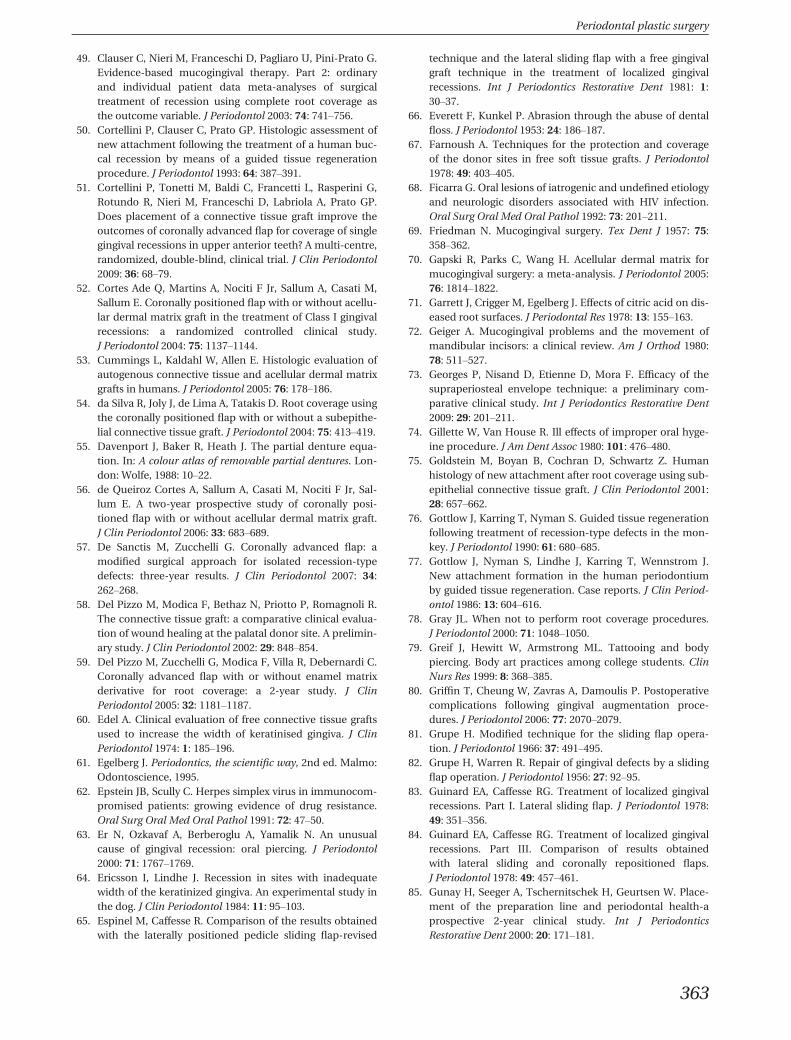
49. Clauser C, Nieri M, Franceschi D, Pagliaro U, Pini-Prato G.Evidence-based mucogingival therapy. Part 2: ordinaryand individual patient data meta-analyses of surgicaltreatment of recession using complete root coverage asthe outcome variable. J Periodontol 2003: 74: 741–756.
50. Cortellini P, Clauser C, Prato GP. Histologic assessment ofnew attachment following the treatment of a human buc-cal recession by means of a guided tissue regenerationprocedure. J Periodontol 1993: 64: 387–391.
51. Cortellini P, Tonetti M, Baldi C, Francetti L, Rasperini G,Rotundo R, Nieri M, Franceschi D, Labriola A, Prato GP.Does placement of a connective tissue graft improve theoutcomes of coronally advanced flap for coverage of singlegingival recessions in upper anterior teeth? A multi-centre,randomized, double-blind, clinical trial. J Clin Periodontol2009: 36: 68–79.
52. Cortes Ade Q, Martins A, Nociti F Jr, Sallum A, Casati M,Sallum E. Coronally positioned flap with or without acellu-lar dermal matrix graft in the treatment of Class I gingivalrecessions: a randomized controlled clinical study.J Periodontol 2004: 75: 1137–1144.
53. Cummings L, Kaldahl W, Allen E. Histologic evaluation ofautogenous connective tissue and acellular dermal matrixgrafts in humans. J Periodontol 2005: 76: 178–186.
54. da Silva R, Joly J, de Lima A, Tatakis D. Root coverage usingthe coronally positioned flap with or without a subepithe-lial connective tissue graft. J Periodontol 2004: 75: 413–419.
55. Davenport J, Baker R, Heath J. The partial denture equa-tion. In: A colour atlas of removable partial dentures. Lon-don: Wolfe, 1988: 10–22.
56. de Queiroz Cortes A, Sallum A, Casati M, Nociti F Jr, Sal-lum E. A two-year prospective study of coronally posi-tioned flap with or without acellular dermal matrix graft.J Clin Periodontol 2006: 33: 683–689.
57. De Sanctis M, Zucchelli G. Coronally advanced flap: amodified surgical approach for isolated recession-typedefects: three-year results. J Clin Periodontol 2007: 34:262–268.
58. Del Pizzo M, Modica F, Bethaz N, Priotto P, Romagnoli R.The connective tissue graft: a comparative clinical evalua-tion of wound healing at the palatal donor site. A prelimin-ary study. J Clin Periodontol 2002: 29: 848–854.
59. Del Pizzo M, Zucchelli G, Modica F, Villa R, Debernardi C.Coronally advanced flap with or without enamel matrixderivative for root coverage: a 2-year study. J ClinPeriodontol 2005: 32: 1181–1187.
60. Edel A. Clinical evaluation of free connective tissue graftsused to increase the width of keratinised gingiva. J ClinPeriodontol 1974: 1: 185–196.
61. Egelberg J. Periodontics, the scientific way, 2nd ed. Malmo:Odontoscience, 1995.
62. Epstein JB, Scully C. Herpes simplex virus in immunocom-promised patients: growing evidence of drug resistance.Oral Surg Oral Med Oral Pathol 1991: 72: 47–50.
63. Er N, Ozkavaf A, Berberoglu A, Yamalik N. An unusualcause of gingival recession: oral piercing. J Periodontol2000: 71: 1767–1769.
64. Ericsson I, Lindhe J. Recession in sites with inadequatewidth of the keratinized gingiva. An experimental study inthe dog. J Clin Periodontol 1984: 11: 95–103.
65. Espinel M, Caffesse R. Comparison of the results obtainedwith the laterally positioned pedicle sliding flap-revised
technique and the lateral sliding flap with a free gingivalgraft technique in the treatment of localized gingivalrecessions. Int J Periodontics Restorative Dent 1981: 1:30–37.
66. Everett F, Kunkel P. Abrasion through the abuse of dentalfloss. J Periodontol 1953: 24: 186–187.
67. Farnoush A. Techniques for the protection and coverageof the donor sites in free soft tissue grafts. J Periodontol1978: 49: 403–405.
68. Ficarra G. Oral lesions of iatrogenic and undefined etiologyand neurologic disorders associated with HIV infection.Oral Surg Oral Med Oral Pathol 1992: 73: 201–211.
69. Friedman N. Mucogingival surgery. Tex Dent J 1957: 75:358–362.
70. Gapski R, Parks C, Wang H. Acellular dermal matrix formucogingival surgery: a meta-analysis. J Periodontol 2005:76: 1814–1822.
71. Garrett J, Crigger M, Egelberg J. Effects of citric acid on dis-eased root surfaces. J Periodontal Res 1978: 13: 155–163.
72. Geiger A. Mucogingival problems and the movement ofmandibular incisors: a clinical review. Am J Orthod 1980:78: 511–527.
73. Georges P, Nisand D, Etienne D, Mora F. Efficacy of thesupraperiosteal envelope technique: a preliminary com-parative clinical study. Int J Periodontics Restorative Dent2009: 29: 201–211.
74. Gillette W, Van House R. Ill effects of improper oral hyge-ine procedure. J Am Dent Assoc 1980: 101: 476–480.
75. Goldstein M, Boyan B, Cochran D, Schwartz Z. Humanhistology of new attachment after root coverage using sub-epithelial connective tissue graft. J Clin Periodontol 2001:28: 657–662.
76. Gottlow J, Karring T, Nyman S. Guided tissue regenerationfollowing treatment of recession-type defects in the mon-key. J Periodontol 1990: 61: 680–685.
77. Gottlow J, Nyman S, Lindhe J, Karring T, Wennstrom J.New attachment formation in the human periodontiumby guided tissue regeneration. Case reports. J Clin Period-ontol 1986: 13: 604–616.
78. Gray JL. When not to perform root coverage procedures.J Periodontol 2000: 71: 1048–1050.
79. Greif J, Hewitt W, Armstrong ML. Tattooing and bodypiercing. Body art practices among college students. ClinNurs Res 1999: 8: 368–385.
80. Griffin T, Cheung W, Zavras A, Damoulis P. Postoperativecomplications following gingival augmentation proce-dures. J Periodontol 2006: 77: 2070–2079.
81. Grupe H. Modified technique for the sliding flap opera-tion. J Periodontol 1966: 37: 491–495.
82. Grupe H, Warren R. Repair of gingival defects by a slidingflap operation. J Periodontol 1956: 27: 92–95.
83. Guinard EA, Caffesse RG. Treatment of localized gingivalrecessions. Part I. Lateral sliding flap. J Periodontol 1978:49: 351–356.
84. Guinard EA, Caffesse RG. Treatment of localized gingivalrecessions. Part III. Comparison of results obtainedwith lateral sliding and coronally repositioned flaps.J Periodontol 1978: 49: 457–461.
85. Gunay H, Seeger A, Tschernitschek H, Geurtsen W. Place-ment of the preparation line and periodontal health-aprospective 2-year clinical study. Int J PeriodonticsRestorative Dent 2000: 20: 171–181.
Periodontal plastic surgery
363
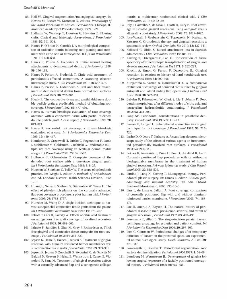
86. Hall W. Gingival augmentation/mucogingival surgery. In:Nevins M, Becker W, Kornman K, editors. Proceedings ofthe World Workshop in Clinical Periodontics. Chicago, IL:American Academy of Periodontology, 1989: 1–21.
87. Hallmon W, Waldrop T, Houston G, Hawkins B. Flossingclefts. Clinical and histologic observations. J Periodontol1986: 57: 501–504.
88. Hanes P, O’Brien N, Garnick J. A morphological compari-son of radicular dentin following root planing and treat-ment with citric acid or tetracycline HCl. J Clin Periodontol1991: 18: 660–668.
89. Hanes P, Polson A, Frederick G. Initial wound healingattachments to demineralized dentin. J Periodontol 1988:59: 176–183.
90. Hanes P, Polson A, Frederick T. Citric acid treatment ofperiodontitis-affected cementum. A scanning electronmicroscopic study. J Clin Periodontol 1991: 18: 567–575.
91. Hanes P, Polson A, Ladenheim S. Cell and fiber attach-ment to demineralized dentin from normal root surfaces.J Periodontol 1985: 56: 752–765.
92. Harris R. The connective tissue and partial thickness dou-ble pedicle graft: a predictable method of obtaining rootcoverage. J Periodontol 1992: 63: 477–486.
93. Harris R. Human histologic evaluation of root coverageobtained with a connective tissue with partial thicknessdouble pedicle graft. A case report. J Periodontol 1999: 70:813–821.
94. Harris R. Successful root coverage: a human histologicevaluation of a case. Int J Periodontics Restorative Dent1999: 19: 439–447.
95. Henderson R, Greenwell H, Drisko C, Regennitter F, LambJ, Mehlbauer M, Goldsmith L, Rebitski G. Predictable mul-tiple site root coverage using an acellular dermal matrixallograft. J Periodontol 2001: 72: 571–582.
96. Holbrook T, Ochsenbein C. Complete coverage of thedenuded root surface with a one-stage gingival graft.Int J Periodontics Restorative Dent 1983: 3: 8–27.
97. Houston W, Stephens C, Tulley W. The scope of orthodonticpractice. In: Wright J, editor. A textbook of orthodontics.2nd ed. London: Elsevier-Health Sciences Division, 1992:1–13.
98. Huang L, Neiva R, Soehren S, Giannobile W, Wang H. Theeffect of platelet-rich plasma on the coronally advancedflap root coverage procedure: a pilot human trial. J Period-ontol 2005: 76: 1768–1777.
99. Hurzeler M, Weng D. A single-incision technique to har-vest subepithelial connective tissue grafts from the palate.Int J Periodontics Restorative Dent 1999: 19: 279–287.
100. Ibbott C, Oles R, Laverty W. Effects of citric acid treatmenton autogenous free graft coverage of localized recession.J Periodontol 1985: 56: 662–665.
101. Jahnke P, Sandifer J, Gher M, Gray J, Richardson A. Thickfree gingival and connective tissue autografts for root cov-erage. J Periodontol 1993: 64: 315–322.
102. Jepsen K, Heinz B, Halben J, Jepsen S. Treatment of gingivalrecession with titanium reinforced barrier membranes ver-sus connective tissue grafts. J Periodontol 1998: 69: 383–391.
103. Jepsen K, Jepsen S, Zucchelli G, Stefanini M, de Sanctis M,Baldini N, Greven B, Heinz B, Wennstrom J, Cassel B, Vig-noletti F, Sanz M. Treatment of gingival recession defectswith a coronally advanced flap and a xenogeneic collagen
matrix: a multicenter randomized clinical trial. J ClinPeriodontol 2013: 40: 82–89.
104. Joly J, Carvalho A, da Silva R, Ciotti D, Cury P. Root cover-age in isolated gingival recessions using autograft versusallograft: a pilot study. J Periodontol 2007: 78: 1017–1022.
105. Joss-Vassalli I, Grebenstein C, Topouzelis N, Sculean A,Katsaros C. Orthodontic therapy and gingival recession: asystematic review. Orthod Craniofac Res 2010: 13: 127–141.
106. Kallestal C, Uhlin S. Buccal attachment loss in Swedishadolescents. J Clin Periodontol 1992: 19: 485–491.
107. Karring T, Ostergaard E, Loe H. Conservation of tissuespecificity after heterotopic transplantation of gingiva andalveolar mucosa. J Periodontal Res 1971: 6: 282–293.
108. Khocht A, Simon G, Person P, Denepitiya JL. Gingivalrecession in relation to history of hard toothbrush use.J Periodontol 1993: 64: 900–905.
109. Kunjamma S, Varma B, Nandakumar K. A comparativeevaluation of coverage of denuded root surface by gingivalautograft and lateral sliding flap operation. J Indian DentAssoc 1986: 58: 527–534.
110. Labahn R, Fahrenbach W, Clark S, Lie T, Adams D. Rootdentin morphology after different modes of citric acid andtetracycline hydrochloride conditioning. J Periodontol1992: 63: 303–309.
111. Lang NP. Periodontal considerations in prosthetic den-tistry. Periodontol 2000 1995: 9: 118–131.
112. Langer B, Langer L. Subepithelial connective tissue grafttechnique for root coverage. J Periodontol 1985: 56: 715–720.
113. Lasho D, O’Leary T, Kafrawy A. A scanning electron micro-scope study of the effects of various agents on instrumen-ted periodontally involved root surfaces. J Periodontol1983: 54: 210–220.
114. Leknes K, Amarante E, Price D, Boe O, Skavland R, Lie T.Coronally positioned flap procedures with or without abiodegradable membrane in the treatment of humangingival recession. A 6-year follow-up study. J Clin Period-ontol 2005: 32: 518–529.
115. Lindhe J, Lang N, Karring T. Mucogingival therapy. Peri-odontal plastic surgery. In: Ermes E, editor. Clinical peri-odontology and implant dentistry. 5th edn. Oxford:Blackwell Munksgaard, 2008: 995–1043.
116. Lins L, de Lima A, Sallum A. Root coverage: comparisonof coronally positioned flap with and without titanium-reinforced barrier membrane. J Periodontol 2003: 74: 168–174.
117. Loe H, Anerud A, Boysen H. The natural history of peri-odontal disease in man: prevalence, severity, and extent ofgingival recession. J Periodontol 1992: 63: 489–495.
118. Lorenzana E, Allen E. The single-incision palatal harvesttechnique: a strategy for esthetics and patient comfort. IntJ Periodontics Restorative Dent 2000: 20: 297–305.
119. Lost C, Geurtsen W. Periodontal changes after temporarydiffusion of Toxavit in the proximal space. An experimen-tal animal histological study. Dtsch Zahnarztl Z 1984: 39:379–387.
120. Lowenguth R, Blieden T. Periodontal regeneration: rootsurface demineralization. Periodontol 2000 1993: 1: 54–68.
121. Lundberg M, Wennstrom JL. Development of gingiva fol-lowing surgical exposure of a facially positioned unerupt-ed incisor. J Periodontol 1988: 59: 652–655.
Zucchelli & Mounssif
364

122. Majzoub Z, Landi L, Grusovin M, Cordioli G. Histology ofconnective tissue graft. A case report. J Periodontol 2001:72: 1607–1615.
123. Matter J. Free gingival graft and coronally repositionedflap. A 2-year follow-up report. J Clin Periodontol 1979: 6:437–442.
124. Matter J. Creeping attachment of free gingival grafts. Afive-year follow-up study. J Periodontol 1980: 51: 681–685.
125. Matter J, Cimasoni G. Creeping attachment after free gin-gival grafts. J Periodontol 1976: 47: 574–579.
126. Mayers LB, Judelson DA, Moriarty BW, Rundell KW.Prevalence of body art (body piercing and tattooing) inuniversity undergraduates and incidence of medicalcomplications. Mayo Clin Proc 2002: 77: 29–34.
127. McGuire M, Cochran D. Evaluation of human recessiondefects treated with coronally advanced flaps and eitherenamel matrix derivative or connective tissue. Part 2: his-tological evaluation. J Periodontol 2003: 74: 1126–1135.
128. McGuire M, Nunn M. Evaluation of human recessiondefects treated with coronally advanced flaps and eitherenamel matrix derivative or connective tissue. Part 1: com-parison of clinical parameters. J Periodontol 2003: 74:1110–1125.
129. McGuire M, Scheyer E. Xenogeneic collagen matrix withcoronally advanced flap compared to connective tissuewith coronally advanced flap for the treatment of dehis-cence-type recession defects. J Periodontol 2010: 81: 1108–1117.
130. Miller P Jr. Root coverage using the free soft tissue auto-graft following citric acid application. III. A successful andpredictable procedure in areas of deep-wide recession. IntJ Periodontics Restorative Dent 1985: 5: 14–37.
131. Miller PD Jr. A classification of marginal tissue recession.Int J Periodontics Restorative Dent 1985: 5: 8–13.
132. Miller PD Jr. Root coverage grafting for regeneration andaesthetics. Periodontol 2000 1993: 1: 118–127.
133. Mlinek A, Smukler H, Buchner A. The use of free gingivalgrafts for the coverage of denuded roots. J Periodontol1973: 44: 248–254.
134. Modeer T, Odenrick L. Post-treatment periodontal statusof labially erupted maxillary canines. Acta Odontol Scand1980: 38: 253–256.
135. Modica F, Del Pizzo M, Roccuzzo M, Romagnoli R. Coro-nally advanced flap for the treatment of buccal gingivalrecessions with and without enamel matrix derivative. Asplit-mouth study. J Periodontol 2000: 71: 1693–1698.
136. Needleman IG. A guide to systematic reviews. J ClinPeriodontol 2002: 29(Suppl. 3): 6–9; discussion 37-38.
137. Nelson S. The subpedicle connective tissue graft. A bilami-nar reconstructive procedure for the coverage of denudedroot surfaces. J Periodontol 1987: 58: 95–102.
138. Norberg O. Ar en utlakning utan vov-nadsfortust otankbarvid kirurgisk behandling av. S. K. Alveolarpyorrhoe?.Svensk Tandlaekare Tidskrift 1926: 19: 171–172.
139. Novaes A Jr, Grisi D, Molina G, Souza S, Taba M Jr, GrisiM. Comparative 6-month clinical study of a subepithelialconnective tissue graft and acellular dermal matrix graftfor the treatment of gingival recession. J Periodontol 2001:72: 1477–1484.
140. Novaes A, Ruben MP, Kon S, Goldman HM, Novaes A Jr.The development of the periodontal cleft. A clinical andhistopathologic study. J Periodontol 1975: 46: 701–709.
141. Oates T, Robinson M, Gunsolley J. Surgical therapies forthe treatment of gingival recession. A systematic review.Ann Periodontol 2003: 8: 303–320.
142. Oles R, Ibbott C, Laverty W. Effects of citric acid treatmenton pedicle flap coverage of localized recession. J Periodontol1985: 56: 259–261.
143. Oles R, Ibbott CG, Laverty WH. Effect of root curettageand sodium hypochlorite treatment on pedicle flap cover-age of localized recession. J Can Dent Assoc 1988: 54: 515–517.
144. Olsson M, Lindhe J. Periodontal characteristics in individ-uals with varying form of the upper central incisors. J ClinPeriodontol 1991: 18: 78–82.
145. Paolantonio M, di Murro C, Cattabriga A, Cattabriga M.Subpedicle connective tissue graft versus free gingival graftin the coverage of exposed root surfaces. A 5-year clinicalstudy. J Clin Periodontol 1997: 24: 51–56.
146. Paolantonio M, Dolci M, Esposito P, D’Archivio D, LisantiL, Di Luccio A, Perinetti G. Subpedicle acellular dermalmatrix graft and autogenous connective tissue graft in thetreatment of gingival recessions: a comparative 1-yearclinical study. J Periodontol 2002: 73: 1299–1307.
147. Parma-Benfenali S, Fugazzoto PA, Ruben MP. The effect ofrestorative margins on the postsurgical development andnature of the periodontium. Part I. Int J PeriodonticsRestorative Dent 1985: 5: 30–51.
148. Parma-Benfenati S, Tinti C. Histologic evaluation of newattachment utilizing a titanium-reinforced barrier mem-brane in a mucogingival recession defect. A case report.J Periodontol 1998: 69: 834–839.
149. Pasquinelli K. The histology of new attachment utilizing athick autogenous soft tissue graft in an area of deep reces-sion: a case report. Int J Periodontics Restorative Dent1995: 15: 248–257.
150. Patur B, Glickman I. Gingival pedicle flaps for coveringroot surfaces denuded by chronic destructive periodontaldisease. J Periodontol 1958: 29: 50–57.
151. Pennel B, Tabor J, King K, Towner J, Fritz B, Higgason J.Free masticatory mucosa graft. J Periodontol 1969: 40:162–166.
152. Pilloni A, Paolantonio M, Camargo PM. Root coverage witha coronally positioned flap used in combination withenamel matrix derivative: 18-month clinical evaluation.J Periodontol 2006: 77: 2031–2039.
153. Pini Prato G, Baldi C, Nieri M, Franseschi D, Cortellini P,Clauser C, Rotundo R, Muzzi L. Coronally advanced flap:the post-surgical position of the gingival margin is animportant factor for achieving complete root coverage.J Periodontol 2005: 76: 713–722.
154. Pini Prato G, Cairo F, Nieri M, Franceschi D, Rotundo R,Cortellini P. Coronally advanced flap versus connectivetissue graft in the treatment of multiple gingival reces-sions: a split-mouth study with a 5-year follow-up. J ClinPeriodontol 2010: 37: 644–650.
155. Pini Prato G, Clauser C, Cortellini P, Tinti C, Vincenzi G,Pagliaro U. Guided tissue regeneration versus mucogin-gival surgery in the treatment of human buccal recessions.A 4-year follow-up study. J Periodontol 1996: 67: 1216–1223.
156. Pini Prato G, Pagliaro U, Baldi C, Nieri M, Saletta D, CairoF, Cortellini P. Coronally advanced flap procedure for rootcoverage. Flap with tension versus flap without tension: a
Periodontal plastic surgery
365
randomized controlled clinical study. J Periodontol 2000:71: 188–201.
157. Pini Prato G, Rotundo R, Franceschi D, Cairo F, CortelliniP, Nieri M. Fourteen-year outcomes of coronally advancedflap for root coverage: follow-up from a randomized trial.J Clin Periodontol 2011: 38: 715–720.
158. Pini Prato G, Tinti C, Vincenzi G, Magnani C, Cortellini P,Clauser C. Guided tissue regeneration versus mucogingivalsurgery in the treatment of human buccal gingival reces-sion. J Periodontol 1992: 63: 919–928.
159. Pini Prato GP, Rotundo R, Magnani C, Ficarra G. Viral eti-ology of gingival recession. A case report. J Periodontol2002: 73: 110–114.
160. Pini-Prato G. The Miller classification of gingival recession:limits and drawbacks. J Clin Periodontol 2011: 38:243–245.
161. Pini-Prato G, Franceschi D, Cairo F, Nieri M, Rotundo R.Classification of dental surface defects in areas of gingivalrecession. J Periodontol 2010: 81: 885–890.
162. Pini-Prato G, Franceschi D, Rotundo R, Cairo F, CortelliniP, Nieri M. Long-term 8-year outcomes of coronallyadvanced flap for root coverage. J Periodontol 2012: 83:590–594.
163. Raetzke P. Covering localized areas of root exposureemploying the “envelope” technique. J Periodontol 1985:56: 397–402.
164. Rajapakse PS, McCracken GI, Gwynnett E, Steen ND,Guentsch A, Heasman PA. Does tooth brushing influencethe development and progression of non-inflammatorygingival recession? A systematic review. J Clin Periodontol2007: 34: 1046–1061.
165. Rasperini G, Silvestri M, Schenk R, Nevins M. Clinical andhistologic evaluation of human gingival recession treatedwith a subepithelial connective tissue graft and enamelmatrix derivative (Emdogain): a case report. Int JPeriodontics Restorative Dent 2000: 20: 269–275.
166. Rateitschak K, Egli U, Fringeli G. Recession: a 4-year longi-tudinal study after free gingival grafts. J Clin Periodontol1979: 6: 158–164.
167. Register A, Burdick F. Accelerated reattachment with ce-mentogenesis to dentin, demineralized in situ. I. Optimumrange. J Periodontol 1975: 46: 646–655.
168. Roberts-Harry EA, Clerehugh V. Subgingival calculus:where are we now? A comparative review. J Dent 2000: 28:93–102.
169. Roccuzzo M, Bunino M, Needleman I, Sanz M. Periodontalplastic surgery for treatment of localized gingival reces-sions: a systematic review. J Clin Periodontol 2002: 29(Sup-pl. 3): 178–194; discussion 176-195.
170. Romagna-Genon C. Comparative clinical study of guidedtissue regeneration with a bioabsorbable bilayer collagenmembrane and subepithelial connective tissue graft. J Pe-riodontol 2001: 72: 1258–1264.
171. Ruben M, Goldman H, Janson W. Biological consider-ations in laterally repositioned pedicle flaps and freeautogenous gingival grafts in periodontal therapy. In: StahlSS, editors. Periodontal surgery – biologic basis and tech-nique. Springfield: Charles C. Thomas, 1976: 235.
172. Ruf S, Hansen K, Pancherz H. Does orthodontic proclina-tion of lower incisors in children and adolescents causegingival recession? Am J Orthod Dentofacial Orthop 1998:114: 100–106.
173. Sangnes G, Gjermo P. Prevalence of oral soft and hard tis-sue lesions related to mechanical toothcleansing proce-dures. Community Dent Oral Epidemiol 1976: 4: 77–83.
174. Sanz M, Lorenzo R, Aranda J, Martin C, Orsini M. Clinicalevaluation of a new collagen matrix (Mucograft prototype)to enhance the width of keratinized tissue in patients withfixed prosthetic restorations: a randomized prospectiveclinical trial. J Clin Periodontol 2009: 36: 868–876.
175. Sardella A, Pedrinazzi M, Bez C, Lodi G, Carrassi A. Labialpiercing resulting in gingival recession. A case series. J ClinPeriodontol 2002: 29: 961–963.
176. Sbordone L, Ramaglia L, Spagnuolo G, De Luca M. A com-parative study of free gingival and subepithelial connectivetissue grafts. Periodontal Case Rep 1988: 10: 8–12.
177. Serino G, Wennstrom JL, Lindhe J, Eneroth L. The preva-lence and distribution of gingival recession in subjectswith a high standard of oral hygiene. J Clin Periodontol1994: 21: 57–63.
178. Soehren S, Allen A, Cutright D, Seibert J. Clinical and histo-logic studies of donor tissues utilized for free grafts of mas-ticatory mucosa. J Periodontol 1973: 44: 727–741.
179. Soileau KM. Treatment of a mucogingival defect associ-ated with intraoral piercing. J Am Dent Assoc 2005: 136:490–494.
180. Spahr A, Haegewald S, Tsoulfidou F, Rompola E, Heijl L,Bernimoulin J, Ring C, Sander S, Haller B. Coverage ofMiller class I and II recession defects using enamel matrixproteins versus coronally advanced flap technique: a 2-year report. J Periodontol 2005: 76: 1871–1880.
181. Staffileno H. Management of gingival recession and rootexposure problems with periodontal disease. Dent ClinNorth Am 1964: 3: 111–120.
182. Stetler KJ, Bissada NF. Significance of the width ofkeratinized gingiva on the periodontal status of teethwith submarginal restorations. J Periodontol 1987: 58: 696–700.
183. Sullivan H, Atkins J. Free autogenous gingival grafts. 3. Uti-lization of grafts in the treatment of gingival recession.Periodontics 1968: 6: 152–160.
184. Sullivan H, Atkins J. Free autogenous gingival grafts.I. Principles of successful grafting. Periodontics 1968: 6:121–129.
185. Tal H, Moses O, Zohar R, Meir H, Nemcovsky C. Root cov-erage of advanced gingival recession: a comparative studybetween acellular dermal matrix allograft and subepitheli-al connective tissue grafts. J Periodontol 2002: 73: 1405–1411.
186. Tatakis D, Trombelli L. Gingival recession treatment:guided tissue regeneration with bioabsorbable membraneversus connective tissue graft. J Periodontol 2000: 71: 299–307.
187. Tenenbaum H, Klewansky P, Roth JJ. Clinical evaluation ofgingival recession treated by coronally repositioned flaptechnique. J Periodontol 1980: 51: 686–690.
188. Tinti C, Vincenzi G, Cocchetto R. Guided tissue regenera-tion in mucogingival surgery. J Periodontol 1993: 64: 1184–1191.
189. Tinti C, Vincenzi G, Cortellini P, Pini Prato G, Clauser C.Guided tissue regeneration in the treatment of humanfacial recession. A 12-case report. J Periodontol 1992: 63:554–560.
Zucchelli & Mounssif
366
190. Tolmie P, Rubins R, Buck G, Vagianos V, Lanz J. The pre-dictability of root coverage by way of free gingival auto-grafts and citric acid application: an evaluation bymultiple clinicians. Int J Periodontics Restorative Dent1991: 11: 261–271.
191. Tozum T, Dini F. Treatment of adjacent gingival reces-sions with subepithelial connective tissue grafts and themodified tunnel technique. Quintessence Int 2003: 34:7–13.
192. Tozum T, Keceli H, Guncu G, Hatipoglu H, Sengun D.Treatment of gingival recession: comparison of two tech-niques of subepithelial connective tissue graft. J Periodon-tol 2005: 76: 1842–1848.
193. Trombelli L, Minenna L, Farina R, Scabbia A. Guided tis-sue regeneration in human gingival recessions. A 10-yearfollow-up study. J Clin Periodontol 2005: 32: 16–20.
194. Trombelli L, Scabbia A, Tatakis D, Calura G. Subpedicleconnective tissue graft versus guided tissue regenerationwith bioabsorbable membrane in the treatment of humangingival recession defects. J Periodontol 1998: 69: 1271–1277.
195. Tugnait A, Clerehugh V. Gingival recession-its significanceand management. J Dent 2001: 29: 381–394.
196. van Palenstein Helderman WH, Lembariti BS, van derWeijden GA, van’t Hof MA. Gingival recession and itsassociation with calculus in subjects deprived of pro-phylactic dental care. J Clin Periodontol 1998: 25: 106–111.
197. Vignoletti F, Nunez J, Discepoli N, De Sanctis F, CaffesseR, Munoz F, Lopez M, Sanz M. Clinical and histologicalhealing of a new collagen matrix in combination with thecoronally advanced flap for the treatment of Miller class-Irecession defects: an experimental study in the minipig.J Clin Periodontol 2011: 38: 847–855.
198. Vincenzi G, De Chiesa A, Trisi P. Guided tissue regenera-tion using a resorbable membrane in gingival recession-type defects: a histologic case report in humans. Int J Peri-odontics Restorative Dent 1998: 18: 24–33.
199. Waite I. An assessment of the postsurgical results follow-ing the combined laterally positioned flap and gingivalgraft procedure. Quintessence Int Dent Dig 1984: 15: 441–450.
200. Walters J, Chang E. Periodontal bone loss associated withan improper flossing technique: a case report. Int J DentHyg 2003: 1: 115–119.
201. Wang H, Bunyaratavej P, Labadie M, Shyr Y, MacNeil R.Comparison of 2 clinical techniques for treatment of gingi-val recession. J Periodontol 2001: 72: 1301–1311.
202. Watson PJ. Gingival recession. J Dent 1984: 12: 29–35.203. Wennstrom J. Mucogingival therapy. Ann Periodontol
1996: 1: 671–701.204. Wennstrom J, Zucchelli G. Increased gingival dimensions.
A significant factor for successful outcome of root cover-age procedures? A 2-year prospective clinical study. J ClinPeriodontol 1996: 23: 770–777.
205. Wennstrom JL. Mucogingival surgery. In: Lang NP, Kar-ring T, editors. Proceedings of the 1st European Work-shop on Periodontology. London: Quintessence, 1994:193–209.
206. Wennstrom JL, Lindhe J, Sinclair F, Thilander B. Someperiodontal tissue reactions to orthodontic tooth move-ment in monkeys. J Clin Periodontol 1987: 14: 121–129.
207. Wessel J, Tatakis D. Patient outcomes following subepithe-lial connective tissue graft and free gingival graft proce-dures. J Periodontol 2008: 79: 425–430.
208. Wilderman M, Wentz F. Repair of a dentogingival defectwith a pedicle flap. J Periodontol 1965: 36: 218–231.
209. Willey R, Steinberg A. Scanning electron microscopic stud-ies of root dentin surfaces treated with citric acid, elastase,hyaluronidase, pronase and collagenase. J Periodontol1984: 55: 592–596.
210. Wilson T Jr, McGuire M, Nunn M. Evaluation of the safetyand efficacy of periodontal applications of a living tissue-engineered human fibroblast-derived dermal substitute.II. Comparison to the subepithelial connective tissue graft:a randomized controlled feasibility study. J Periodontol2005: 76: 881–889.
211. Wong A, Schonmeyr B, Singh P, Carlson D, Li S, MehraraB. Histologic analysis of angiogenesis and lymphangiogen-esis in acellular human dermis. Plast Reconstr Surg 2008:121: 1144–1152.
212. Woodyard J, Greenwell H, Hill M, Drisko C, Iasella J, Sche-etz J. The clinical effect of acellular dermal matrix on gingi-val thickness and root coverage compared to coronallypositioned flap alone. J Periodontol 2004: 75: 44–56.
213. Zabalegui I, Sicilia A, Cambra J, Gil J, Sanz M. Treatmentof multiple adjacent gingival recessions with the tunnelsubepithelial connective tissue graft: a clinical report. Int JPeriodontics Restorative Dent 1999: 19: 199–206.
214. Zachrisson B. Orthodontics and periodontics. In: Clinicalperiodontology and implant dentistry, 3rd edn. Copenha-gen: Munksgaard, 1998: 741–793.
215. Zade R, Hirani S. A clinical study of localized gingivalrecession treated by lateral sliding flap. J Indian Dent Assoc1985: 57: 19–26, 40.
216. Zucchelli G, Amore C, Sforza N, Montebugnoli L, De Sanc-tis M. Bilaminar techniques for the treatment of recession-type defects. A comparative clinical study. J Clin Periodon-tol 2003: 30: 862–870.
217. Zucchelli G, Cesari C, Amore C, Montebugnoli L, De Sanc-tis M. Laterally moved, coronally advanced flap: a modi-fied surgical approach for isolated recession-type defects.J Periodontol 2004: 75: 1734–1741.
218. Zucchelli G, De Sanctis M. Long-term outcome followingtreatment of multiple Miller class I and II recession defectsin esthetic areas of the mouth. J Periodontol 2005: 76:2286–2292.
219. Zucchelli G, De Sanctis M. Treatment of multiple reces-sion-type defects in patients with esthetic demands.J Periodontol 2000: 71: 1506–1514.
220. Zucchelli G, De Sanctis M. Modified two-stage proceduresfor the treatment of gingival recession. Eur J Esthet Dent2013: 8: 24–42.
221. Zucchelli G, Gori G, Mele M, Stefanini M, Mazzotti C, Mar-zadori M, Montebugnoli L, De Sanctis M. Non-carious cer-vical lesions associated with gingival recessions: a decisionmaking process. J Periodontol 2011: 82: 1713–1724.
222. Zucchelli G, Mele M, Mazzotti C, Marzadori M, Monte-bugnoli L, De Sanctis M. Coronally advanced flap with andwithout vertical releasing incisions for the treatment ofmultiple gingival recessions: a comparative controlled ran-domized clinical trial. J Periodontol 2009: 80: 1083–1094.
223. Zucchelli G, Mele M, Stefanini M, Mazzotti C, MarzadoriM, Montebugnoli L, de Sanctis M. Patient morbidity and
Periodontal plastic surgery
367
root coverage outcome after subepithelial connective tis-sue and de-epithelialized grafts: a comparative random-ized-controlled clinical trial. J Clin Periodontol 2010: 37:728–738.
224. Zucchelli G, Mele M, Stefanini M, Mazzotti C, Mounssif I,Marzadori M, Montebugnoli L. Predetermination of rootcoverage. J Periodontol 2010: 81: 1019–1026.
225. Zucchelli G, Mounssif I, Mazzotti C, Stefanini M, Marzado-ri M, Montebugnoli L. Coronally advanced flap with and
without connective tissue graft for the treatment of multi-ple gingival recessions: a comparative short- andlong-term controlled randomized clinical trial. J Periodon-tol- 2013: Dec 30. doi: 10.1111/jcpe.12224 [Epub ahead ofprint].
226. Zucchelli G, Testori T, De Sanctis M. Clinical and anatomi-cal factors limiting treatment outcomes of gingival reces-sion: a new method to predetermine the line of rootcoverage. J Periodontol 2006: 77: 714–721.
Zucchelli & Mounssif
368
![Periodontal Flap Surgery Auto Saved]](https://static.fdocuments.us/doc/165x107/546e616db4af9f83598b466a/periodontal-flap-surgery-auto-saved.jpg)