Periodontal health at firstpermanent molars adjacent toprimary molar stainless steelcrowns
4
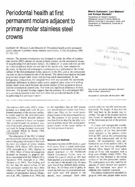
Periodontal health at first permanent molars adjacent to primary molar stainless steel crowns Marcio Guelmann', Lars Matston' and Enrique Bimstein' 'Department ot Pediatric Dentistry, Haddassati Faculty of Dental Medicine, Hebrew University In Jerusalem, Israel, and 'Department ol Pedodontics, University of Umea, Sweden Guelmann M, Matsson L and Bimstein E: Periodontal health at first permanent molars adjacent lo primary molar stainless steel crowns. J Ctin Periodontol 1988: 15: 531-533. Abstract. The present investigation was designed to study the effect of stainless steel crowns (SSC), placed on second primary molars, on the periodontal tissues of neighbouring first pennanent molars. 36 children (9-12 years old) with an SSC on a second primary molar on one side of the mouth only, were selected for the study. A clinical and radiographic examination was performed at the mesial surface of the first permanent molar, adjacent to the SSC, and at the correspond- ing area on the contraiateral side of the mouth. The ciinicai examination included gingival and plaque index scores and probing depth measurements. In the radiographic examination, the marginal bone level was assessed. No statistically significant differences in plaque index scores, gingival index scores and probing pocket depth were noted between the mesial surface of first permanent molars and the contraiateral control area. Nor were any significant differences in bone level seen. The present findings suggest that the presence of a well-adapted SSC on a second permanent molar does not affect the periodontal health of the neighbouring first permanent molar. UBRARY Key words: periodontal disease steel crowns; pedodontics. Accepted for publication 28 November 1987 The Stainless steel crown (SSC). manu- factured as a metal shell with the pre- formed anatomy of the occiusal surface, is mostly used for the semi-permanent restoration of badly decayed primary teeth. Its retention is based on margins that extend beyond the prepared por- tion of the tooth, adapted to the individ- ual tooth by being trimmed, contoured and crimped (Mink & Bennet 1968, USDOHEW 1977), The adaptation of the SSC is limited and its possible deleterious effect on the surrounding gingival tissues of the tooth has been the subject of several studies (Webber 1974, Myers 1975. Sarafanov 1979, Ashraphi et al 1981. Machen et al, 1981), Most of these investigations detected an increase in gingival irri- tation associated with SSC (Meyrs 1975, Sarafanov 1979, Ashraphi et al. 1981. Machen et al. 1981). This finding, and the fact that the mesial surface of the first permanent molars has been iden- tified as a "risk area" for the early devel- opment of periodontal disease (Hugo- son et al. 1981, Gjermo et al. 1984). led to the hypothesis that an SSC placed ona second primary molar may have a deleterious effect on the mesial perio- dontal tissues of the first pennanent mo- lar. The purpose of the present study was therefore to describe the effect of a second primary molar SSC on the perio- dontal tissues of the mesial surface of the first permanent molars. Material and Methods The dental records of 280 children, aged 9-12 years, from a mixed socio-econ- omic suburb of .Jerusalem, who received dental treatment at the Haddassah Health Community Center, were re- viewed for cases in which an SSC had been applied to a second primary molar on one side of the mouth only. 40 cases were found that fulfilled these criteria. Of these, 36 (15 boys and 21 girls), in which the SSC and its contraiateral con- trol tooth were still present in the mouth, were examined. Each child contrib uted with one SSC and one con- trol tooth. The length of time that the SSC had been in the mouth was re- corded. The age of the patients selected was chosen to represent the end stage of the functional period of the primary molars, A clinica] and radiographic examin- ation was performed of the area at the mesial surface of the first permanent molar adjacent to the primary molar with an SSC and at the corresponding area on the contraiateral side of the mouth. All examinations were per- formed by one of the authors (MG). In each child, two bite-wing radiographs (Kodak Ultra Speed D) were taken using an X-ray unit equipped with a long cone (Philips Oralix CHF). The central ray was directed at the mesial surface of the first permanent molars. The radiographs were also used for di- agnosis of caries disease and appoint- ments for treatment were given to children who required treatment. The clinical examination included: (i) gingival index and plaque index scores
-
Upload
erick-coral -
Category
Documents
-
view
214 -
download
1
description
articulo para AOP
Transcript of Periodontal health at firstpermanent molars adjacent toprimary molar stainless steelcrowns
Periodontal health at firstpermanent molars adjacent toprimary molar stainless steelcrowns
Marcio Guelmann', Lars Matston'and Enrique Bimstein''Department ot Pediatric Dentistry,Haddassati Faculty of Dental Medicine,Hebrew University In Jerusalem, Israel, and'Department ol Pedodontics, University ofUmea, Sweden
Guelmann M, Matsson L and Bimstein E: Periodontal health at first permanentmolars adjacent lo primary molar stainless steel crowns. J Ctin Periodontol 1988:15: 531-533.
Abstract. The present investigation was designed to study the effect of stainlesssteel crowns (SSC), placed on second primary molars, on the periodontal tissuesof neighbouring first pennanent molars. 36 children (9-12 years old) with an SSCon a second primary molar on one side of the mouth only, were selected forthe study. A clinical and radiographic examination was performed at the mesialsurface of the first permanent molar, adjacent to the SSC, and at the correspond-ing area on the contraiateral side of the mouth. The ciinicai examination includedgingival and plaque index scores and probing depth measurements. In theradiographic examination, the marginal bone level was assessed. No statisticallysignificant differences in plaque index scores, gingival index scores and probingpocket depth were noted between the mesial surface of first permanent molarsand the contraiateral control area. Nor were any significant differences in bonelevel seen. The present findings suggest that the presence of a well-adapted SSCon a second permanent molar does not affect the periodontal health of theneighbouring first permanent molar.
UBRARY
Key words: periodontal diseasesteel crowns; pedodontics.
Accepted for publication 28 November 1987
The Stainless steel crown (SSC). manu-factured as a metal shell with the pre-formed anatomy of the occiusal surface,is mostly used for the semi-permanentrestoration of badly decayed primaryteeth. Its retention is based on marginsthat extend beyond the prepared por-tion of the tooth, adapted to the individ-ual tooth by being trimmed, contouredand crimped (Mink & Bennet 1968,USDOHEW 1977),
The adaptation of the SSC is limitedand its possible deleterious effect on thesurrounding gingival tissues of the toothhas been the subject of several studies(Webber 1974, Myers 1975. Sarafanov1979, Ashraphi et a l 1981. Machen etal, 1981), Most of these investigationsdetected an increase in gingival irri-tation associated with SSC (Meyrs 1975,Sarafanov 1979, Ashraphi et al. 1981.Machen et al. 1981). This finding, andthe fact that the mesial surface of thefirst permanent molars has been iden-tified as a "risk area" for the early devel-opment of periodontal disease (Hugo-son et al. 1981, Gjermo et al. 1984). led
to the hypothesis that an SSC placedona second primary molar may have adeleterious effect on the mesial perio-dontal tissues of the first pennanent mo-lar. The purpose of the present studywas therefore to describe the effect of asecond primary molar SSC on the perio-dontal tissues of the mesial surface ofthe first permanent molars.
Material and Methods
The dental records of 280 children, aged9-12 years, from a mixed socio-econ-omic suburb of .Jerusalem, who receiveddental treatment at the HaddassahHealth Community Center, were re-viewed for cases in which an SSC hadbeen applied to a second primary molaron one side of the mouth only. 40 caseswere found that fulfilled these criteria.Of these, 36 (15 boys and 21 girls), inwhich the SSC and its contraiateral con-trol tooth were still present in themouth, were examined. Each child
contrib uted with one SSC and one con-trol tooth. The length of time that theSSC had been in the mouth was re-corded. The age of the patients selectedwas chosen to represent the end stageof the functional period of the primarymolars,
A clinica] and radiographic examin-ation was performed of the area at themesial surface of the first permanentmolar adjacent to the primary molarwith an SSC and at the correspondingarea on the contraiateral side of themouth. All examinations were per-formed by one of the authors (MG). Ineach child, two bite-wing radiographs(Kodak Ultra Speed D) were takenusing an X-ray unit equipped with along cone (Philips Oralix CHF). Thecentral ray was directed at the mesialsurface of the first permanent molars.The radiographs were also used for di-agnosis of caries disease and appoint-ments for treatment were given tochildren who required treatment.
The clinical examination included: (i)gingival index and plaque index scores

532 Guetmann et al.
Table I, Distribution of examined areas in plaque index classes
P.I.
Adjacent to cro\mesiobuecalmesiolingual
Controlmesiobuccalmesiolingual
Mesiobuccal: y = 0.82, N.S
score
vn
.: mesi
0
1210
1211
iolingual:
(%1
33.327.8
33.330.6
:z"-0 .25 .
1413
17II
N.S.
1
(%)
38.936.1
47.230.6
n
1013
714
2-3
(%)
27.836.1
19.438.9
(Loe 1967) mesiobuccaly and mesiolin-gualiy at the first permanent molar: (ii)subgingival depth of the SSC, measuredat the deepest point on the distal surfaceof the primary molar; (iii) probing depthmesiohuccaUy and mesiolingualJy at thefirst permanent molar. All measure-ments were performed using a 1 mmgraded periodontal probe (Hu-Friedy,GF-W, USA).
In the radiographic examination, themarginal bone level (the distance fromthe cemento-enamel junction (CEJ) ofthe first permanent molar to the inter-dental alveolar crest (AC)) was meas-ured with calipers on the image of thebite-wing radiographs projected onto ascreen of a slide viewer (Singer Clearina-te Caramate U SP. Rochester, N,Y.. en-largement X 7,2), The radiographs wereselected in accordance with the follow-ing criteria; (i) minimal evidence of dis-tortion; (ii) minimal overlapping be-tween the proximal surfaces of the teeth;(iii) clear image of the cemento-enameJjunction. After selection, 27 pairs ofradiographs from 16 girls and 11 boysremained for measurements.
Statistical comparisons of plaque in-dex and gingiva! index scores betweenthe 2 groups were performed using the/'-test. For the comparisons of probingdepth and bone level, the Student (-testfor paired observations was applied, Acorrelation analysis between the timethe SSC had been in the mouth, and thepocket depth and the radiographicailyassessed bone icvcK was performed. Dif-ferences at the 5% level of probabilitywere considered statistically significant.
Results
The SSC werejudged to be well-adaptedat the distal surface of the tooth. In 32of the 36 cases, the distal subgingivaldepth of the SSC was 2 mm or less. Inthe remaining cases, however, thesubgingival depth was 3 mm.
Comparison of the distribution of
plaque index scores and gingival indexscores, between the first permanent mo-lars adjacent to an SSC and their con-trols did not show any statistically sig-nificant differences (Tables 1. 2). Norwere any differences in probing pocketdepth seen between the 2 groups (Table3), The radiographic analysis revealed amean bone level of 0,9 mm at the firstpermanent molars adjacent to an SSCcompared with 0,7 mm at the controls.The differences was not statistically sig-nificant,
information about the length of timethe SSC had been in the mouth wasavailable for 27 crowns. The mean timewas 24.6 months (range 8-36), In 20 ofthese cases, acceptable radiographs forbone-ievei measurements were available. No significant correlation wasfound between the length of time theSSC had been in the mouth, and the
probing pocket depth and the bonelevel.
~ Discussion
It has been stated that pre-formed SSCcan be used successfully to restore pri-mary molar teeth without adverselyaffecting the health of the gingivae orthe status of the patient's oral hygiene(Webber 1974), However, other studiesindicate that clinical evidence of gingi-vitis may be associated with the pres-ence of an SSC, especially when defectssuch as poor crown crimp, contour orposition, and/or cement remaining inthe gingival sulcus are observed (Myers1975. Sarafanov 1979, Ashraphi et al.]98I, MachenetaJ. 1981).
The possible relationship of gingiva!disease around primary molar SSC lothe gingival health of their permanentsuccessor was studied by Fuks et al.(1983), who found no difference be-tween the permanent successors ofcrowned primary molars and theirhomologues or the rest of the mouth.Even when gingivitis was presentaround the crowned primary tooth, itwas resolved upon exfoliation of the pri-mary tooth and subsequent eruption ofthe permanent tooth. An additional re-lationship, between the presence of anSSC on a primary molar and the perio-dontal health of the permanent den-
Tcihle 2, Distribution of e ; in gingival index classesG.I. score
Adjacent to crownmesiobuccalmesiolingual
Controlmesiobuccalmesiolingual
n
1615
2318
0
(%)
44.441.7
63.950.0
»
1615
615
1
(Vo)
44.441.7
16.741.7
r,
46
73
2
(%)
11.116.7
19.48.3
Mesiobuccal: /==2,01, N,S.: mesiolingual: /- = 0,22. N,S,: index score 1 and 2 treated as onecell,
lahle 3, Probing pocket depth and marginal bone level; means (-v) and standard deviations(S,D.); /-test for paired observations applied
Probing pocket levelmesiobuccal
adjacent to SSCcontrol
mesioiingualadjacent to SSCcontrol
Marginal bone leveladjaeent to SSCcontrol
tl
3636
3636
2727
i-
3.03.1
3.13.2
0.90.7
S.D.
0.60.5
0.60.5
0.50.5
1
1.41
0.77
P
N.S.
N.S.
Feriodontal health related to preformed crowus 533
tition, is the possible deleterious effectof a second primary molar SSC on thegingiva! health of the first permanentmolars, induced either by the manipu-]ation during placement of Ehe crown orby a possible increase in retention ofdental piaque in the area. The mesialsurface of first permanent molars hasbeen identified as a risk area for thedevelopment of periodontal disease(Hugoson et al. 1981, Gjermo et al.1984).
The present findings of a lack of sig-nificant differences in the clinical andradiographic parameters between theside of the mouth with an SSC and thecontro] side suggest that for the agegroup examined, the presence of an SSCon a second primary mo]ar does notsignificantly affect the adjacent perio-donta] tissues of the first permanent mo-]ar. However, in most cases examined inthe present study, a weU-adapted SSCwas found, and keeping in mind thatprevious studies have demonstrated anassociation between gingiva! diseaseand poorly adapted SSC, the presentfindings do not overrule the possibilityof periodontal damage on the mesialsurface of first permanent molarscaused by poorly adapted SSC on sec-ond primary molars.
Based on the present results and thefindings of Fuks et al. (1983), it can beconcluded that, provided the crown iswe]]-adapted to the primary mo!ar, astainless steel crown does not seem tohave a detrimental effect upon the per-manent dentition.
Zusammenfassung
Die paradontale Gesundheit von ersten hlei-henden Molaren, direkt neben. mit Stahlkro-nen versorglen MihhmolarenDie vorliegende Untersuchung wurde konzipiert, um den Effekt von mil Stahlkronenversorgten Milchmolaren aufdie parodonta-len Gewebe benachbarter, erster bleibenderMolaren zu studieren. 36 Kinder (9-12 Jahri;alt) mit einer SSC (stainless steel crown) aufeinem zweiten Milchmolaren in nur eincrMundseite, wurden fur diese Studie ausge-wahlt. An der mesialen OberHache des erstenbleibenden Molaren, direkt neben der SSCund an der entsprechenden Stelle der kontra-latcralen Seite. wurdc cine klinische und ront-genographische Untersuchung vorgenom-men. Die klinische Untersuchung beinhalteteGingiva]- und P]aqueindex-Scores (Beurtei-
lungscinheiten) sowie Messungen der Sondie-rungsticfen. Bei der rontgenographischenUntersuchung wurde das marginale Kno-chennjveau bcstimmt. An den mesialen Ober-riachen der ersten bleibenden Molaren undder kontraiateralen Region wurden keine sta-tistisch signifikanten Unterschiede zwischenden Plaqueindex- und Gingival index-Scoresbeobachtet. Das gleiche gjU fiir die sondier-ten Taschentiefen. Auch zwischen den Kno-chenniveaus der gieichen Regionen wurdenkeinerlei abgesicherte Unterschiede gesehen.Die vorliegenden Resmtate besa^n, dasseine zweckentsprechend adaptierte SSC atifeinem zweiten Milchmolaren, die parodonta-le Gesundheit des benayhbarten ersten blei-ben Molaren nicht beeiiifiusst.
Resume
Etai parodontal des premieres molaires perma-nentes adjacentes a des inoUiires de iait recou-vertes de couronnes en acier inoxydahle
La presente etude a eu pour but d'evaluerl'effet de couronnes en acier inoxydable( C A I ) placees sur des secondes molaires tem-poraires sur les tissus parodontaux des pre-mieres molaires definitives adjacentes. Trenlesix enfants ages de 9 a 12 ans avec une CAIsur une seconde molaire temporaire d'un colede la bouche tiniquement ont ete selectionnespour cette etude. Un examen clinique et ra-diographique a ele effectue en mesial de lapremiere molaire definitive adjacente ainsiqu'au niveau contraiateral servant de contro-le, L'exameii clinique comprenaif ks indicesde plaque et gingival ainsi que la profondeurdes poches. L'examen radiographique a servia controler le nJveau osseux. Aucune diffe-rence stalistique n'a ele trouvee entre cesdonnees. Les resultats suggerent que le place-ment d'une CAI bien adaptee sur une secondemolaire temporaire n'affecte pas la same pa-rodontale de la molaire defmitive adjacente.
References
Ashraphi. M. H,, Durr. D. P. & Duncan, W, K, (!98I) Interrelationship between stainlesssteel crown, plaque Accumulation, and gingival health. American Academy of Pedodoniics- Annual Meeting Reports. Research Abstract R-26,
Fuks. A, B.. Zadok, S. & Chosack, A, (1983) Gingival health of premolar successors tocrowned primary molars. Pediatric Dentistry 5, 51-52.
Gjermo, P.. Bellini, T . Santos, V: P., M'artiins, / . G, d Berracyo/t, / . R. (1984) Prevalence ofbone loss in a group of Brazilian teenagers assessed on bite-wing radiographs. Journal ofClinical Periodontologv U . 104-113.
Hugoson, A,, Koch, C}, & Rylander, H, (1981) Prevalence and distribution of gingivitis-periodontitis in children and adolescents. Scandinavian Dental Journal 5. 91-103.
Loe. H, (1967) The gingival index, the piaque index and the retention index system. Journalof Feriodontology 38, 610-616.
Machen, E., Rapp, R., Baumhammers, A, & Zullo, T, (1981) The effecl of stainless steelcrowns on gingival tissue. American Academy of Pedodontics - Annual Meeting Reports.Research Abstract H-27.
Mink, J, R. & Bennet, I. C, (1968) The stainless steel crown. Journal of Dentistry for Children35, 188-196,
Myers, D, R, (1975) A tlinical study of the response of the gingival tissue surrounding stainlesssleel crowns. Journal of Dentistry for Children 42, 33-36,
US Department of Health. Education and Welfare (USDOHEW). (1977) Project Tupp: Stain-less steel crown, preparation and restoration (Quercus Corporanon).
Sarafanov. S. R. (1979) Coronas de acero cromo para molares primarios (evaiuadon de dostipos). Association Liental Mexicana 36, 134-147,
Stoner, J. E, (1972) An investigation into the accuracy of measurements made on radiographsof ihe alveolar crests of dried mandibles. Journal of Periodontology 43, 699-702,
Webber, D, L. (1974) Gingival health following placement of stainless steel crowns. Journal
of Dentistry for Children 41, I86-IB9.
Address:
Lars MatssonDepartment of PedodonticsUniversity of UmedS-9Q}H7VmeaSweden