Periodontal Diseases in Immunodeficient Patients
12
{96} International Dental Journal of Student’s Research, April - June 2015;3(2):96-107 CLINICAL DIAGNOSIS & GUIDELINES Periodontal Disease in Immunodeficient Patients: Clinical Guidelines for Diagnosis and Management Morvarid Oveisi 1 , Oriyah Barzilay 2 , Ahmed A. Hanafi 3 1,2,3 Group of Matrix Dynamics, Faculty of Dentistry, University of Toronto. 3 Periodontology Department, Faculty of Oral and Dental Medicine, Cairo University Corresponding Author: Morvarid Oveisi Email: [email protected] Access this Article Online Abstract Primary immunodeficiency diseases are rare hereditary conditions that usually occur at a young age; however, secondary immunodeficiency is acquired due to disease, drug treatment and is increasing in frequency among the population. Although periodontal diseases related to these conditions are secondary to other life threatening manifestations, they are very common and easily detectable by the patient, patient guardians and periodontists. Periodontists have a major role in both helping to detect undiagnosed diseases, as well as improving the oral care of diagnosed patients, thus a thorough knowledge of these conditions, causes, local and systemic involvement, diagnostic tools and proper management is very important. This article summarizes selected primary and secondary immunodeficiency conditions such as neutropenia, leukocyte adhesion deficiency (LAD) and Chediak- Hegashi syndrome, and places schematic, diagnostic, and management steps that may help periodontists manage unexplained severe periodontal diseases related to immunodeficiency. Introduction Immunodeficiency diseases are defects in the immune system in which the host defense mechanism cannot function properly. Primary immunodeficiency is inherited and therefore is caused by a gene defect. However, secondary immunodeficiency conditions are acquired and usually happen due a defect of lymphocyte function as a result of the usage of drugs, irradiation or invasion of pathogens such as HIV virus and measles.[1] There are many types of diseases caused by primary immunodeficiency , which is a result of a defect in T- cell or B-cell function, antibody deficiency, or loss of phagocyte function and/or number[2] ,which for the latter these can be included as any deficiency in the adhesion process of neutrophil [3], NADPH oxidase or chemotaxis.[4] The epidemiology of the diseases differ based on race, gender, ethnic factors and geographic region[4]. It has been estimated that one in 1200 people are affected by primary Immunodeficiency. Due to cancer therapies, usage of immunosuppressant’s and other biological therapies, the occurrence of secondary immunodeficiency is growing. Organ damage is preventable if there is minimal delay in the diagnosis of immunodeficiency[5]. Periodontal disease is an inflammatory state of the gingiva which affects the supporting structure of the teeth.[6, 7]. Periodontal disease is caused by plaque accumulation , and poor dental hygiene[8] leading to inflammation of surrounding tissue[4].It has been observed that individuals suffering from any types of immunodeficiency diseases may manifest some oral, dental and facial problems[4]which include periodontal diseases, oral lesions and developmental abnormalities. These can be a sign of immunity defect. Therefore, it is crucial for physicians and dentist to be able to recognize these systematic disorders by the oral manifestation, carry out an accurate diagnosis and perform the corresponding treatment[9]. Neutropenia Neutropenia is defined by a low absolute neutrophil count (ANC) in the blood lasting more than 6 months, which can cause recurrent infections to a patient [10] with varying severity from stomatitis Quick Response Code www.idjsr.com Use the QR Code scanner to access this article online in our database Article Code: IDJSR SE 0171
Transcript of Periodontal Diseases in Immunodeficient Patients
{96}
International Dental Journal of Student’s Research, April - June 2015;3(2):96-107
CLINICAL DIAGNOSIS & GUIDELINES
Periodontal Disease in Immunodeficient Patients: Clinical Guidelines for Diagnosis and Management
Morvarid Oveisi1, Oriyah Barzilay2, Ahmed A. Hanafi3
1,2,3Group of Matrix Dynamics, Faculty of Dentistry,
University of Toronto. 3Periodontology Department, Faculty of Oral and
Dental Medicine, Cairo University
Corresponding Author:
Morvarid Oveisi
Email: [email protected]
Access this Article Online
Abstract
Primary immunodeficiency diseases are rare
hereditary conditions that usually occur at a young
age; however, secondary immunodeficiency is
acquired due to disease, drug treatment and is
increasing in frequency among the population.
Although periodontal diseases related to these
conditions are secondary to other life threatening
manifestations, they are very common and easily
detectable by the patient, patient guardians and
periodontists. Periodontists have a major role in both
helping to detect undiagnosed diseases, as well as
improving the oral care of diagnosed patients, thus a
thorough knowledge of these conditions, causes,
local and systemic involvement, diagnostic tools and
proper management is very important. This article
summarizes selected primary and secondary
immunodeficiency conditions such as neutropenia,
leukocyte adhesion deficiency (LAD) and Chediak-
Hegashi syndrome, and places schematic, diagnostic,
and management steps that may help periodontists
manage unexplained severe periodontal diseases
related to immunodeficiency.
Introduction
Immunodeficiency diseases are defects in the
immune system in which the host defense mechanism
cannot function properly. Primary immunodeficiency
is inherited and therefore is caused by a gene defect.
However, secondary immunodeficiency conditions
are acquired and usually happen due a defect of
lymphocyte function as a result of the usage of drugs,
irradiation or invasion of pathogens such as HIV
virus and measles.[1]
There are many types of diseases caused by primary
immunodeficiency , which is a result of a defect in T-
cell or B-cell function, antibody deficiency, or loss of
phagocyte function and/or number[2] ,which for the
latter these can be included as any deficiency in the
adhesion process of neutrophil [3], NADPH oxidase
or chemotaxis.[4]
The epidemiology of the diseases differ based on
race, gender, ethnic factors and geographic region[4].
It has been estimated that one in 1200 people are
affected by primary Immunodeficiency. Due to
cancer therapies, usage of immunosuppressant’s and
other biological therapies, the occurrence of
secondary immunodeficiency is growing. Organ
damage is preventable if there is minimal delay in
the diagnosis of immunodeficiency[5].
Periodontal disease is an inflammatory state of the
gingiva which affects the supporting structure of the
teeth.[6, 7]. Periodontal disease is caused by plaque
accumulation , and poor dental hygiene[8] leading to
inflammation of surrounding tissue[4].It has been
observed that individuals suffering from any types of
immunodeficiency diseases may manifest some oral,
dental and facial problems[4]which include
periodontal diseases, oral lesions and developmental
abnormalities. These can be a sign of immunity
defect. Therefore, it is crucial for physicians and
dentist to be able to recognize these systematic
disorders by the oral manifestation, carry out an
accurate diagnosis and perform the corresponding
treatment[9].
Neutropenia
Neutropenia is defined by a low absolute neutrophil
count (ANC) in the blood lasting more than 6
months, which can cause recurrent infections to a
patient [10] with varying severity from stomatitis
Quick Response Code
www.idjsr.com
Use the QR Code scanner to
access this article online in our
database
Article Code: IDJSR SE 0171
{97}
International Dental Journal of Student’s Research, April - June 2015;3(2):96-107
and gingivitis, to more severe pneumonia and sepsis.
[11] Different forms of neutropenia such as cyclic
neutropenia, chronic benign neutropenia and severe
congenital neutropenia (Kostmann syndrome) can all
cause periodontal disease.[10, 12]
Chronic benign neutropenia
Chronic neutropenia is defined by a non-cyclic low
count of neutrophils in the blood without a known
underlying systemic disease lasting less than 6
months. It is the most common form of neutropenia
in infants and children and is usually not inherited.
80-98% of patients tested positive for the anti-
neutrophil antibody [13]. Its manifestation is less
severe than Kostmann syndrome, includes high
incidence of otitis media, upper respiratory
infections, lymphadenopathy and pneumonia but may
develop into life threatening infections and
sepsis.[10]
Oral Manifestations includes severe gingival
inflammation, edematous and hyperplastic
papilla,[14] may progress into periodontitis
[15]leading to severe horizontal bone loss and teeth
mobility [16]
Diagnosis is achieved by a persistent ANC 0.5x10⁹/l
with a normal total white blood cell count due to
elevated numbers of lymphocytes and monocytes
[17] and confirmed by anti-neutrophil antibody.
Kostmann Syndrome
Severe congenital neutropenia (Kostmann Syndrome)
is a rare hereditary syndrome characterized by a very
low ANC (less than 0.2x10⁹/l) [18] due to maturation
arrest during myelopoiesis process [4]and increased
apoptosis of myeloid progenitor cells in bone
marrow.[19]
Initial symptoms can be summarized as recurrent
bacterial infections of the skin, mucosa leading to
cellulitis, perirectal abscess, stomatitis, meningitis,
pneumonia, and sepsis [20] . Long term symptoms
are periodontitis, splenomegaly and hepatomegaly,
osteoporosis and myelodysplastic syndrome/acute
myeloid leukaemia (MDS/AML) [19].
Oral findings are usually more severe than other
forms of neutropenia, with recurrent painful
ulceration, [21] diffuse gingival inflammation,
alveolar bone loss, teeth mobility and loss of both
dentition [22]
Persistent ANC less than 0.5x10⁹/l is a significant
laboratory finding and diagnosis is confirmed with
bone marrow aspiration showing an arrest of
neutrophil hematopoiesis at the
promyelocyte/myelocyte stage.[10]
Cyclic neutropenia
Cyclic Neutropenia is characterized by the repetitive
occurrence of neutropenia at average of 21 day
period and last for approximately 3-6 days[23].The
mutation is passed along in an autosomal dominant
manner. It has been observed that this disease is
associated with the mutation in ELA2 gene mapped
to chromosome 19p13.3 which encodes neutrophil
elastase. Mutations in this gene lead to a shortened
neutrophil life[24] .
It is characterized by fever, mouth ulcers,
lymphadenopathy, multiple abscess formation,
exhaustion and susceptibility of infection which can
be lethal [25-29]. Reduction in the number of
polymorph nuclear leukocytes (PMNs) can be
associated with rapid and destructive periodontal
disease including aphthous-like lesions[10].
The initial oral characterization of patient includes
repetitive ulceration showing little evidence of an
inflammatory halo[30], severe gingival inflammation
and recession[14, 25],which extended from the
gingiva into the alveolar mucosa[25].Recurrent
gingival bleeding along with fever was noted as a
sign of this disease [25, 31],pocket depths exceeded
the 6- to 8-mm range[25] with various levels of tooth
mobility [31].
Diagnosis requires serial measurements of the ANC
(<1,500) daily or at least three times per week for
four to six weeks[32].
It has been demonstrated that Granulocyte Colony-
Stimulating Factor(GCSF) can be an efficient
treatment for neutropenia[33], as it can lead to a 10
fold increase in ANC and result in a higher life
expectancy[34].
Dental management is necessary for these patients to
control infections.
Regular dental appointments to check for the
accumulation of bacterial plaque.
Use of chlorhexidine gluconate mouth wash and
a light polishing and scaling in some part of the
teeth [25, 31].
Prophylactic antimicrobials.
Invasive dentistry should be avoided in
neutropenic episodes.
Oral surgeries to be performed only under
specific antibacterial (after microbiological
testing) and corticosteroid coverage [21]
LAD
LAD is a rare, autosomal recessive, primary
immunodeficiency syndrome; characterized by
impaired phagocytic functions[35]. LAD is classified
according to causative gene mutation into 3 types:
LAD I, LAD II and LAD III [36-38].
{98}
International Dental Journal of Student’s Research, April - June 2015;3(2):96-107
LAD I is caused by mutation in gene ITGB2 which
encodes for CD11/CD18 [39, 40] and ultimately
decreases the expression of three integrins on
leukocyte surfaces CD11a,CD11b and CD11c and
preventing the adhesion of neutrophils to endothelial
cells)[41]. Characterized delayed separation of
umbilical cord, major bacterial infections with no pus
formation [35] and impaired wound healing [40], the
severity of clinical features are directly related to
degree of CD18 deficiency and can be divided into
severe (less than 1% CD18 expression) and moderate
( 2.5% to 10% CD18 expression) [42, 43].
Morbidity rate of severe LAD I is high before the age
of 5 [44].
In LAD II, different gene mutations cause defects in
the specific Golgi GDP-fucose transporter [45, 46]
which reduce CD15s (Sialyl-Lewis X) on the
leukocyte surface, thus affecting the rolling phase of
neutrophil adhesion [35]. This is characterized by
mental retardation and less severe infections in
adolescence [41] . Less common LAD III, was only
reported in 4 patients suffering from bacterial
infections [38] and severe bleeding tendency[47], is
believed to be caused by general defect in integrin
activation [38].
Periodontal involvement starts as gingivitis at a
young age, just after primary teeth eruption. Deep
pocket formation and extensive bone loss [48, 49]
progress until partial or total premature loss of both
primary and permanent teeth [35]. Several case
reports showed oral ulceration and delayed wound
healing in more than 80% of patients.[35]
Primarily, blood test of patients with LAD shows
leukocytosis (20,000 to 80,000 cells/ml)[40,
50].Rebuck skin window or Boyden Chamber shows
decreased neutrophil migration and is confirmed
using flow cytometry which shows CD18 deficiency
in LAD I and sialy-Lewis x ligand deficiency in LAD
II [51]. Additional histological analysis of gingival
biopsy showed abundance of leukocytes in blood
vessels and no leukocyte in tissue [52].
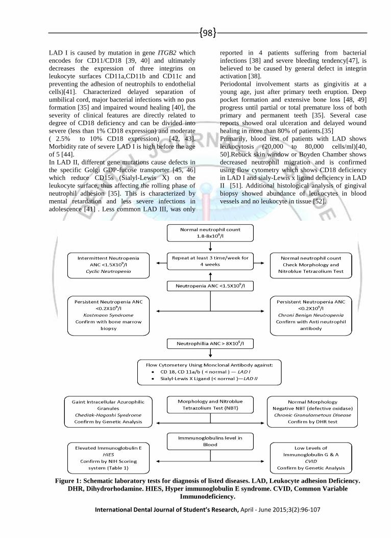
Figure 1: Schematic laboratory tests for diagnosis of listed diseases. LAD, Leukocyte adhesion Deficiency.
DHR, Dihydrorhodamine. HIES, Hyper immunoglobulin E syndrome. CVID, Common Variable
Immunodeficiency.
{99}
International Dental Journal of Student’s Research, April - June 2015;3(2):96-107
Bone marrow transplant is the treatment of choice for
young LAD patients [53, 54].However if not
possible, several maneuvers to adjust host response
can be achieved such as white blood cell transfusion,
antibiotics, interferon and allogenic stem cell
transplant [55]. Periodontal treatment usually ends
with tooth loss [10] however maintaining the teeth is
advocated with the goal of improving patients’
physiologic and psychological health by
Periodic oral prophylaxis [56]
Prophylactic Antimicrobial
Fluoride application and diet counseling
Extraction should be avoided (due to delayed
wound healing)
Chediak-Hegashi Syndrome
Chediakhigashi is a rare condition which is inherited
in an autosomal manner. This disease is usually fatal
and appears with the irregularly enormous lysosomal
granules in the leukocytes [57, 58]. This disorder is
characterized by numerous repetitive bacterial
infections, oculocutaneous albinism, susceptibility to
bruising, and mucosal bleeding as well as peripheral
neuropathy. In addition, patients may show
neurologic dysfunction and movement disorders[3].
Furthermore, the accelerated phase of CHS named
Hemophagocyticlymphohistiocytosis (HLH) can be
recognized by cytopenia, fever, bleeding,
lymphadenopathy and hepatosplenomegaly [59-61].
This disorder is connected to the fusion of
cytoplasmic granules which can take place in the
myelopoieses and can lead to the death of myeloid
precursors in the marrow and cause neutropenia. Also
neutrophils can have a problem in phagocytosis,
chemotaxis and killing bacteria[62].
Intraoral examination showed a full mouth plaque
score of 85% [63], gingival bleeding and teeth
mobility [3, 58] , high frequency of periodontal
pockets and bone loss at an early age [10, 64, 65],
probing showed more than 30% of the sites 5-8mm
deep with concomitant recession defects [66]
Blood testing and examination of giant granules
within neutrophils, lymphocytes and natural killer
cells using nitrobluetetrazolium dye [10] are essential
for diagnosis. Bone marrow aspiration and
examination of giant eosinophilic or azurophilic
cytoplasmic inclusion bodies within the myeloid
lineage cells show a positive reaction to peroxidase
staining
People with Chediak-Higashi disorder can be
recognized at a young age, and bone marrow
transplantation can be a positive treatment which can
lower the risk of periodontal disease.[3]
It has been noted that continuous periodontal
treatments with regular follow up can help patients
avoid further infection and reduce gingival
inflammation [67]. In addition, Kornmanet et al
described the importance of periodontal therapy in
preventing progressive periodontitis [68].
Long-term antibiotic treatment such as
amoxicillin[69] is administered to patients with
progressive periodontal disease along with
metronidazole [70]to help reduce the periodontal
probing depth and promote attachment in patients
with the milder form of the disease. [67].However,
full mouth extraction is inevitable in patients with
severe progressive periodontal disease refractory to
treatment .[71]
Chronic granulomatous Disease
Chronic granulomatous disease(CGD) is a very rare
immune deficiency syndrome perpetuated
genetically. CGD can be characterized by the
mutation of nicotinamide adenine dinucleotide
phosphate (NADPH) oxidase component, leading to
the failure of neutrophils and macrophages in killing
invading pathogens by impairing the respiratory
burst.
CYBB is a gene responsible for encoding the
gp91phox subunit and is reported as the most
common site of mutation. Liver abscesses, skin
infections, pneumonia, osteomyelitis, as well as
cervical or inguinal lymphadenitis can be seen in
young patients[72, 73]. Furthermore, infection and
sterile hyper inflammation is also present in CGD
patient[74].
Management of CDG is based on control of
infections. Broad spectrum antibacterial (as
cotrimoxazole)[75], antifungal prophylaxis
(asitraconazole)[76]. Interferon-ƴ has been shown to
improve oxidase activity in neutrophils and
monocytes of some patients[77], and to reduce
infection rates. Bone marrow transplantation is
effective and a more predictable treatment [78].
Some case reports show oral difficulties including
oral ulcers[79-84] such as multiple buccal mucosal
ulcers in direct contact with dental plaque [81],
severe gingivitis, periodontitis [79-81], enamel
hypoplasia [84], oral candidiasis [85] ,
granulomatous mucositis in the upper lip[86] and the
soft palate [87], geographic tongue[84] and
generalized prepubertal periodontitis,[88] loss of
attachment and recession of gingival was noted[88]
Variations of oral findings in CGD patients [72, 89,
90], and Neutrophil dysfunction[10, 91] are probably
due to immunosuppressive therapy specially steroids
[92].
Patients with CGD can be diagnosed through
flowcytometery, dihydrorhodamine 123 (DHR)
assay[93] and the nitrobluetetrazolium Test [94]
{100}
International Dental Journal of Student’s Research, April - June 2015;3(2):96-107
Treatment:
Regular dental care and frequent follow-up.
Antibacterial mouth washes.
Antibiotics such as clavulanic acid and
amoxicillin is needed for any dental work and
surgery related to bacteremia[95]
Antimycotic prophylaxis[95]
Hyper Immunoglobulin E Syndrome
HEIS or Jobs syndrome is a rare multisystemic
disease causing host immune defects as well as non-
immunological manifestations [96]. It presents itself
in two forms, the more common autosomal dominant
(AD-HEIS) and the less common autosomal
recessive (AR-HEIS) [97], both characterized by high
level of immunoglobulin E levels in blood, chronic
eczema, recurrent skin and lung infections [98] and
decreased bone density [99]. Although the etiology of
disease is not clear, several studies concluded that
immune defects may be due to defective neutrophil
chemotaxis, humoral and cellular immune response
impairment including T-cell cytokine signal
disruption.
Recent gene analysis showed that most AD-HIES
have a mutation in gene STAT3 which causes poor
maturation and activation of T17 helper cells,
however AR-HIES (different clinical picture with no
dental abnormalities) may be attributed to different
gene mutation, TYK2 gene (Tyrosine Kinase 2), or
the DOCK8 gene (Dedicator of Cytokinesis 8)[100].
Oral findings of AD-HIES is very characteristic to
retention of primary teeth or “Double-row” dentition
which is found in most reported cases (due to
persistence of Her twig epithelial root sheath on root
surface, thus preventing root resorption causing
delayed shedding) , severe candidiasis and oral
infections, poor oral hygiene and high plaque index,
gingivitis but rare progression to periodontitis, except
for 2 patients [79, 101] ( . This data contradicts most
immunodeficiency syndromes, which are more likely
to cause severe periodontal destruction.)
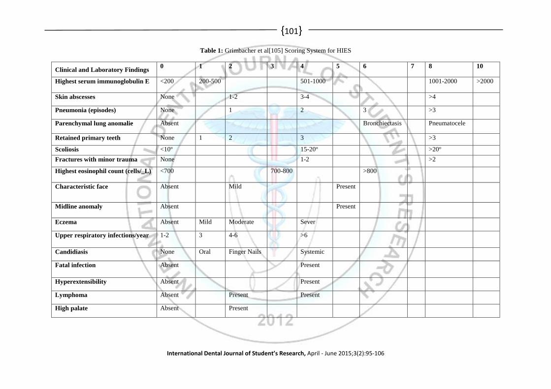
There is no one laboratory test that confirms HIES,
rather a scoring system (fig. 1)-based on all clinical
and laboratory tests is used to confirm HIES,
therefore diagnosis of HIES is usually difficult.
Recently some reported laboratory tests were added
to NIH-HIES score, that helps better detect HIES, as
very low Th17 CD4 cell count, and the genetic
analysis.[102] The presence of retained primary teeth
and unerupted permanent teeth resembles other
syndromes like Cleidocranial syndrome, Gardner’s
Syndrome, and Down syndrome. A differential
diagnosis with these syndromes is noted and
confirmed by clinical and laboratory tests. [103]
Treatment is based on prophylactic antimicrobials,
intravenous immunoglobulin, and bone marrow
transplant shows some success but not a full recovery
[104], dental management is focused on
Periodic follow up and oral hygiene
assessment.
Extraction of primary teeth at correct time of
shedding to prevent permanent teeth
impaction and enhance oral hygiene.
Antifungal and antibacterial for oral
infections
Orthodontic correction if needed.
Prognosis of gingival healing after extraction is fairly
good with no complications and in most patients
periodontal health of permanent teeth can be
maintained.
{101}
International Dental Journal of Student’s Research, April - June 2015;3(2):95-106
Table 1: Grimbacher et al[105] Scoring System for HIES
Clinical and Laboratory Findings 0 1 2 3 4 5 6 7 8 10
Highest serum immunoglobulin E <200 200-500 501-1000 1001-2000 >2000
Skin abscesses None 1-2 3-4 >4
Pneumonia (episodes) None 1 2 3 >3
Parenchymal lung anomalie Absent Bronchiectasis Pneumatocele
Retained primary teeth None 1 2 3 >3
Scoliosis <10o 15-20o >20o
Fractures with minor trauma None 1-2 >2
Highest eosinophil count (cells/_L) <700 700-800 >800
Characteristic face Absent Mild Present
Midline anomaly Absent Present
Eczema Absent Mild Moderate Sever
Upper respiratory infections/year 1-2 3 4-6 >6
Candidiasis None Oral Finger Nails Systemic
Fatal infection Absent Present
Hyperextensibility Absent Present
Lymphoma Absent Present Present
High palate Absent Present
{102}
International Dental Journal of Student’s Research, April - June 2015;3(2):96-107
Common Variable Immunodeficiency
Common variable immunodeficiency (CVID) is a
common heterogeneous primary immune deficiency.
Patients with CVID have a deficiency in humoral
immunity leading to a defective antibody response,
causing repetitive infections of the gastrointestinal
and upper respiratory tracts, and susceptibility to
some cancers such as lymphoma and autoimmune
diseases. Hypogammaglobulinemia has also seen in
these patients.
Although normal B-cell numbers have been
identified in lymphoid tissue and peripheral blood of
patients, it has been noted that B-cells of these
individuals have difficulty in differentiating into
immunoglobulin-secreting plasma cells. Furthermore,
it should be mentioned that the deficiency in
monocyte and macrophage function has also been
recognized. In addition, in some CVID patients, T-
cell malfunction has been identified with decreased
CD4 lymphocytes and T-cell receptors, loss of
antigen-specific and quick death of T-cells.[4]
Individual suffering from CVID have been diagnosed
by having a low level of IgG and IgA[106].
Mutations in a group of genes asTNFRSF13B
gene[107] involved in B-cells result in having a
defected immunity in CVID[108]
Clinical examinations show dental problems such as
gingivitis and lichenoid lesions with Wickham
striae,[109] necrotizing ulcerative periodontitis
(NUP) [109, 110] , severe periodontitis and gingival
pain along with bleeding and tooth mobility was
demonstrated in a case report.[110]
In order to treat CVID, the primary method used is to
replace the antibody by an intravenous or
subcutaneous means. This occurs in doses of 400-
600mg of antibody per kilogram of the patient’s
weight per month.[111]
Dental management as reported in some case reports
include
Regular oral prophylaxis with crown polishing
[109]
Chlorhexidinedigluconate rinse is recommended
twice a day.
Antibiotic therapy[109, 110] such as
Amoxycillin and clavulanic acid[109]
Acquired immunodeficiency syndrome
Acquired immunodeficiency syndrome(AIDS) is a
disease caused by a human immunodeficiency virus.
In this condition HIV targets and attacks T helper
cells(CD4) resulting in immune response suppression
, the disability of the body’s response to the invading
pathogen, predisposes the patient to neurological
problems, opportunistic infections ,malignancies and
oral manifestation [112].
HIV advancement can be detect by monitoring the
HIV viral load and T helper cells(CD4+) count,
however, it should be noted that there are some
common oral manifestations associated with HIV
positive patients. Therefore, it can be a useful
indicator for screening the immune condition of
potential HIV positive individuals and can be easily
recognized and detected by clinicians.[113-116]
It has been estimated that 70-90% of individual
suffering from HIV manifest oral lesions during their
phase of the disease [117-119]. Individuals with oral
manifestations have less CD4+ than ones without,
and it has been observed that there is a correlation
between oral candidiasis and a decrease in the CD4+
count(less than 200 cells/mm3).[120]
Some of the important oral manifestation and Lesions
present in HIV positive patients are
Oral candidiasis (most common oral lesion)
[113, 121-123] which are divided in to three
groups;pseudomembranous candidiasis,
erythematous candidiasis and angular
cheilitis[114]
Oral hairy leukoplakia [114].
ulcerative disease such as herpes simplex virus,
Aphthous ulcerations[114, 124], Neutropenic
ulceration [124]
linear gingival erythema[120, 124]
oral warts-human papilloma virus[124]
Necrotizing Ulcerative gingivitis and
Periodontitis(NUG/NUP)[124, 125]
The follow up appointments are needed for dental
care such as scaling and root planning. A 10%
povidone-iodine lavage or 0 .12% chlorhexidine
gluconate can be used for the elimination of dental
plaque and necrotic soft tissue. Utilizing antibiotics
such as clindamycin, metronidazole 500g and
amoxicillin can be helpful for the treatment. It is
crucial to establish proper nutrition in order to reduce
potential issues in the oral cavity that can be
produced by poor nutrition, as well as manage the
patient’s pain.[124]
Leukemia
Leukemia is a type of a cancer caused by an
uncontrolled differentiation and proliferation of blood
cell precursors resulting in the production of
immature cells. Clinically, leukemia is classified into
two types: chronic and acute, with the acute phase
possibly being fatal. In addition, according to
histogenicity, leukemia is divided in to lymphocytic
or myelocytic depending on the origin of the
cells[126-128].
Acute myeloid leukemia(AML) is more common in
adults and acute lymphoid leukemia (ALL) is mostly
seen in children[128-130]. Acute myeloblastic
leukemia (AML) is characterized by symptoms of
{103}
International Dental Journal of Student’s Research, April - June 2015;3(2):96-107
pancytopenia including fatigue, weaknesses,
infection, gingival bleeding, ecchymoses,
menorrhagia, and epistaxis [131, 132]. The direct
penetration of leukemic cells in lymph nodes, spleen,
central nervous system and gingival has been
reported [126, 129, 133-135].
Oral complication can be observed in all types of
leukemia[136]. Individuals having leukemia are
suffering from extreme enlargement of the gingiva
along with bleeding[127, 135-139], bulbous
enlargement in the interdental papillae [126, 127] a
pale blue gum with glazed texture ,and loss of
stippling is one the symptoms of leukemia[126, 127],
generalized horizontal bone loss was reported[127]
however in some cases bone loss is not
recognized[126] Ulceration and petechiae was noted
as a frequent sign[135]. In patient with acute
monocytic leukemia and acute myelomonocytic
leukemia, gingival infiltration of leukemic cells are
commonly seen[140].
Diagnosis by complete blood count peripheral blood
smear, shows the presence of blast cells and reveals
the type and quantity of white blood cells[126], and
flow cytometry of peripheral blood are used for
leukemia diagnosis[126, 127], biopsy such as bone
marrow aspiration also can be used to confirm
diagnosis and type of leukemia[126, 127, 135]
Regular oral prophylaxis is needed. Antibacterials
can be used in conjugation with scaling and sub
gingival debridement to lower the risk of dental
infection during the chemotherapy. Tooth extraction
of hopeless teeth can eliminate the infection.[141]
Conclusion
Managing periodontal disease of immunodeficient
patients is essential for improvement of their physical
and psychological health, thus knowledge of these
conditions, diagnostic methods and management
options is crucial for every dentist. Diagnosis of these
diseases is challenging, however some clues may
guide the clinician towards a definitive diagnosis, so
it is important that precise steps of history taking
(medical, familial and dental), clinical examination
(extra and intra oral) and laboratory investigations are
followed to achieve a successful diagnosis.
References
1. Janeway, C.A., et al., Immunobiology: the immune
system in health and disease. Vol. 2. 2001: Churchill
Livingstone.
2. Notarangelo, L., et al., Primary immunodeficiency
diseases: an update. Journal of Allergy and Clinical
Immunology, 2004. 114(3): p. 677-687.
3. Sollecito, T.P., et al., Systemic conditions associated
with periodontitis in childhood and adolescence. A
review of diagnostic possibilities. Medicina oral,
patologia oral y cirugia bucal, 2004. 10(2): p. 142-150.
4. Szczawinska-Poplonyk, A., et al., Oral manifestations
of primary immune deficiencies in children. Oral
Surgery, Oral Medicine, Oral Pathology, Oral
Radiology, and Endodontology, 2009. 108(3): p. e9-
e20.
5. Bright, P.D., et al., Laboratory clues to
immunodeficiency; missed chances for early
diagnosis? Journal of clinical pathology. 68(1): p. 1-5.
6. Ellison, S.A., Oral bacteria and periodontal disease.
Journal of dental research, 1970. 49(2): p. 197-202.
7. Genco, R.J., R.T. Evans, and S.A. Ellison, Dental
research in microbiology with emphasis on periodontal
disease. The Journal of the American Dental
Association, 1969. 78(5): p. 1016-1036.
8. Ram, V.S., et al., Bonebiomarkers in Periodontal
Disease: A Review Article.
9. Garcia, R.I., M.M. Henshaw, and E.A. Krall,
Relationship between periodontal disease and systemic
health. Periodontology 2000, 2001. 25(1): p. 21-36.
10. Deas, D.E., S.A. Mackey, and H.T. McDonnell,
Systemic disease and periodontitis: manifestations of
neutrophil dysfunction. Periodontology 2000, 2003.
32(1): p. 82-104.
11. Bernini, J.C., Diagnosis and management of chronic
neutropenia during childhood. Pediatric Clinics of
North America, 1996. 43(3): p. 773-792.
12. Watanabeb, K., Prepubertal periodontitis: a review of
diagnostic criteria, pathogenesis, and differential
diagnosis. Journal of periodontal research, 1990. 25(1):
p. 31-48.
13. Bux J1, M.-E.C., Autoimmune neutropenia. Semin
Hematal., 1992 Jan. 29(1): p. 45-53.
14. Deasy, M.J., et al., Familial benign chronic
neutropenia associated with periodontal disease: a case
report. Journal of periodontology, 1980. 51(4): p. 206-
210.
15. Zaromb, A., et al., Periodontitis as a manifestation of
chronic benign neutropenia. Journal of periodontology,
2006. 77(11): p. 1921-1926.
16. Reichart, P.A. and H. Dornow, Gingivo-periodontal
manifestations in chronic benign neutropenia. Journal
of clinical periodontology, 1978. 5(1): p. 74-80.
17. Cutting, H.O. and J.E. Lang, Familial benign chronic
neutropenia. Annals of internal medicine, 1964.
61(5_Part_1): p. 876-887.
18. Aprikyan, A.A.G., et al., Cellular and molecular
abnormalities in severe congenital neutropenia
predisposing to leukemia. Experimental hematology,
2003. 31(5): p. 372-381.
19. Carlsson, G.r., et al., Kostmann syndrome or infantile
genetic agranulocytosis, part one: celebrating 50 years
of clinical and basic research on severe congenital
neutropenia. Acta Paediatrica, 2006. 95(12): p. 1526-
1532.
20. Hakki, S.S., et al., Periodontal status in two siblings
with severe congenital neutropenia: diagnosis and
mutational analysis of the cases. Journal of
periodontology, 2005. 76(5): p. 837-844.
21. Tirali, R.E. and S.B.Ç. Zeynep Yalçınkaya-Erdemci,
Oral findings and clinical implications of patients with
congenital neutropenia: a literature review. The
Turkish journal of pediatrics. 55: p. 241-245.
22. Defraia, E. and A. Marinelli, Oral manifestations of
congenital neutropenia or Kostmann syndrome. Journal
of Clinical Pediatric Dentistry, 2002. 26(1): p. 99-102.
{104}
International Dental Journal of Student’s Research, April - June 2015;3(2):96-107
23. Dale, D.C. and W. Hammond, Cyclic neutropenia: a
clinical review. Blood reviews, 1988. 2(3): p. 178-185.
24. Dale, D.C., et al., Mutations in the gene encoding
neutrophil elastase in congenital and cyclic
neutropenia. Blood, 2000. 96(7): p. 2317-2322.
25. Prichard, J.F., et al., Prepubertal periodontitis affecting
the deciduous and permanent dentition in a patient with
cyclic neutropenia: a case report and discussion.
Journal of periodontology, 1984. 55(2): p. 114-122.
26. Spencer, P. and J.E. Fleming, Cyclic neutropenia: a
literature review and report of case. ASDC journal of
dentistry for children, 1985. 52(2): p. 108.
27. Andrews, R.G., et al., Chronic benign neutropenia of
childhood with associated oral manifestations. Oral
Surgery, Oral Medicine, Oral Pathology, 1965. 20(6):
p. 719-725.
28. Barrett, A.P., Neutropenic ulceration: a distinctive
clinical entity. Journal of periodontology, 1987. 58(1):
p. 51-55.
29. Gates, G.F., Chronic neutropenia presenting with oral
lesions. Oral Surgery, Oral Medicine, Oral Pathology,
1969. 27(4): p. 563-567.
30. Scully, C., E. MacFadyen, and A. Campbell, Oral
manifestation in cyclic neutropenia. British Journal of
Oral Surgery, 1982. 20(2): p. 96-101.
31. Yamahk, N., et al., Periodical gingival bleeding as a
presenting symptom of periodontitis due to underlying
cyclic neutropenia. Case report. Australian dental
journal, 1993. 38(4): p. 272-276.
32. Dale, D.C., ELANE-Related neutropenia.
GeneReviews [Internet]. Seattle (WA): University of
Washington, Seattle, 2002.
33. Hammond Iv, W.P., et al., Treatment of cyclic
neutropenia with granulocyte colony-stimulating
factor. New England Journal of Medicine, 1989.
320(20): p. 1306-1311.
34. Bux, J., et al., Influence of granulocyte antibodies on
granulocyte function. Vox sanguinis, 1993. 64(4): p.
220-225.
35. Nagendran, J., et al., Leukocyte adhesion deficiency: a
case report and review. Journal of Dentistry for
Children. 79(2): p. 105-110.
36. Etzioni, A., Adhesion molecules in host defense.
Clinical and diagnostic laboratory immunology, 1994.
1(1): p. 1.
37. Yakubenia, S. and M.K. Wild, Leukocyte adhesion
deficiency II. FEBS Journal, 2006. 273(19): p. 4390-
4398.
38. Etzioni, A. and R. Alon, Leukocyte adhesion
deficiency III: a group of integrin activation defects in
hematopoietic lineage cells. Current opinion in allergy
and clinical immunology, 2004. 4(6): p. 485-490.
39. Schmidt, S., M. Moser, and M. Sperandio, The
molecular basis of leukocyte recruitment and its
deficiencies. Molecular immunology. 55(1): p. 49-58.
40. Anderson, D.C. and T.A. Springer, Leukocyte
adhesion deficiency: an inherited defect in the Mac-1,
LFA-1, and p150, 95 glycoproteins. Annual review of
medicine, 1987. 38(1): p. 175-194.
41. Dababneh, R., et al., Periodontal manifestation of
leukocyte adhesion deficiency type I. Journal of
periodontology, 2008. 79(4): p. 764-768.
42. Fischer, A., et al., Leukocyte adhesion deficiency:
molecular basis and functional consequences.
Immunodefic Rev, 1988. 1(1): p. 39-54.
43. Lakshman, R. and A. Finn, Neutrophil disorders and
their management. Journal of clinical pathology, 2001.
54(1): p. 7-19.
44. Movahedi, M., et al., Clinical and laboratory findings
in Iranian patients with leukocyte adhesion deficiency
(study of 15 cases). Journal of clinical immunology,
2007. 27(3): p. 302-307.
45. Lühn, K., et al., The gene defective in leukocyte
adhesion deficiency II encodes a putative GDP-fucose
transporter. Nature genetics, 2001. 28(1): p. 69-72.
46. Lübke, T., et al., Complementation cloning identifies
CDG-IIc, a new type of congenital disorders of
glycosylation, as a GDP-fucose transporter deficiency.
Nature genetics, 2001. 28(1): p. 73-76.
47. McDowall, A., et al., A novel form of integrin
dysfunction involving β1, β2, and β3 integrins. Journal
of clinical investigation, 2003. 111(1): p. 51.
48. Dennison, D.K. and T.E. Dyke, The acute
inflammatory response and the role of phagocytic cells
in periodontal health and disease. Periodontology
2000, 1997. 14(1): p. 54-78.
49. Meyle, J., Leukocyte adhesion deficiency and
prepubertal periodontitis. Periodontology 2000, 1994.
6(1): p. 26-36.
50. Todd 3rd, R.F. and D.R. Freyer, The CD11/CD18
leukocyte glycoprotein deficiency.
Hematology/oncology clinics of North America, 1988.
2(1): p. 13-31.
51. Meyle, J. and J.R. Gonzales, Influences of systemic
diseases on periodontitis in children and adolescents.
Periodontology 2000, 2001. 26(1): p. 92-112.
52. Bowen, T.J., et al., Severe recurrent bacterial
infections associated with defective adherence and
chemotaxis in two patients with neutrophils deficient
in a cell-associated glycoprotein. The Journal of
pediatrics, 1982. 101(6): p. 932-940.
53. Haddadin, I., et al., Bone marrow transplantation for
leukocyte adhesion deficiency-I: Case report. Saudi
Journal of Kidney Diseases and Transplantation, 2006.
17(4): p. 564.
54. Hattori, H., et al., Successful human leukocyte antigen
one antigen-mismatched related bone marrow
transplantation in a 6-year-old boy with leukocyte
adhesion deficiency syndrome. Pediatrics international,
2001. 43(3): p. 306-309.
55. Engel, M.E., et al., Matched unrelated bone marrow
transplantation with reduced-intensity conditioning for
leukocyte adhesion deficiency. Bone marrow
transplantation, 2006. 37(7): p. 717-718.
56. Majorana, A., et al., Leukocyte adhesion deficiency in
a child with severe oral involvement. Oral Surgery,
Oral Medicine, Oral Pathology, Oral Radiology, and
Endodontology, 1999. 87(6): p. 691-694.
57. Wara-Aswapati, N., et al., Periodontitis in the child
and adolescent. ASDC journal of dentistry for children,
1999. 66: p. 167-174.
58. Delcourt-Debruyne, E.M.C., H.R.A. Boutigny, and
H.F. Hildebrand, Features of severe periodontal
disease in a teenager with Chediak-Higashi syndrome.
Journal of periodontology, 2000. 71(5): p. 816-824.
59. Nargund, A.R., et al., Accelerated Phase of Chediak
Higashi Syndrome Mimicking Lymphoma—A Case
Report. Journal of pediatric hematology/oncology.
32(6): p. e223-e226.
60. Gajendra, S., et al., Accelerated phase at initial
presentation in chediak-higashi syndrome: is it really
uncommon? Pediatric hematology and oncology.
31(4): p. 382-385.
61. Imran, T., et al., Chediak-Higashi syndrome presenting
in accelerated phase. J Coll Physicians Surg Pak.
22(8): p. 539-41.
{105}
International Dental Journal of Student’s Research, April - June 2015;3(2):96-107
62. Sánchez-Guiu, I., et al., Chediak–Higashi syndrome:
description of two novel homozygous missense
mutations causing divergent clinical phenotype.
European journal of haematology. 92(1): p. 49-58.
63. O'Leary, T.J., R.B. Drake, and J.E. Naylor, The plaque
control record. Journal of periodontology, 1972. 43(1):
p. 38-38.
64. Hart, T.C. and J.C. Atkinson, Mendelian forms of
periodontitis. Periodontology 2000, 2007. 45(1): p. 95-
112.
65. Hanna, S. and A. Etzioni, Leukocyte adhesion
deficiencies. Annals of the New York Academy of
Sciences. 1250(1): p. 50-55.
66. HR, M., Tooth mobility: a review of clinical aspects
and research findings. Journal of periodontology, 1967.
38(6): p. Suppl: 686.
67. Bailleul-Forestier, I., et al., Generalized periodontitis
associated with Chediak-Higashi syndrome. Journal of
periodontology, 2008. 79(7): p. 1263-1270.
68. Kornman, K.S., et al., The influence of supragingival
plaque control on clinical and microbial outcomes
following the use of antibiotics for the treatment of
periodontitis. Journal of periodontology, 1994. 65(9):
p. 848-854.
69. Winkel, E.G., et al., Clinical and microbiological
effects of initial periodontal therapy in conjunction
with amoxicillin and clavulanic acid in patients with
adult periodontitis. Journal of clinical periodontology,
1999. 26(7): p. 461-468.
70. Socransky, S.S., et al., Ecological considerations in the
treatment of Actinobacillus actinomycetemcomitans
and Porphyromonas gingivalis periodontal infections.
Periodontology 2000, 1999. 20(1): p. 341-362.
71. Shibutani, T., et al., Long-term follow-up of
periodontitis in a patient with Chediak-Higashi
syndrome. A case report. Journal of periodontology,
2000. 71(6): p. 1024-1028.
72. Winkelstein, J.A., et al., Chronic granulomatous
disease: report on a national registry of 368 patients.
Medicine, 2000. 79(3): p. 155-169.
73. Stasia, M.J. and X.J. Li. Genetics and
immunopathology of chronic granulomatous disease.
in Seminars in immunopathology. 2008: Springer.
74. Giannopoulou, C., K.-H. Krause, and M. F. The
NADPH oxidase NOX2 plays a role in periodontal
pathologies. in Seminars in immunopathology. 2008:
Springer.
75. Smith, J. and A. Finn, Antimicrobial prophylaxis.
Archives of disease in childhood, 1999. 80(4): p. 388-
392.
76. Mouy, R., et al., Long-term itraconazole prophylaxis
against Aspergillus infections in thirty-two patients
with chronic granulomatous disease. The Journal of
pediatrics, 1994. 125(6): p. 998-1003.
77. Ezekowitz, R.A.B., et al., Partial correction of the
phagocyte defect in patients with X-linked chronic
granulomatous disease by subcutaneous interferon
gamma. New England Journal of Medicine, 1988.
319(3): p. 146-151.
78. Calvino, M.C., et al., Bone marrow transplantation in
chronic granulomatous disease. European journal of
pediatrics, 1996. 155(10): p. 877-879.
79. Charon, J.A., S.E. Mergenhagen, and J.I. Gallin,
Gingivitis and oral ulceration in patients with
neutrophil dysfunction. Journal of Oral Pathology &
Medicine, 1985. 14(2): p. 150-155.
80. Cohen, M.S., P.A. Leong, and D.M. Simpson,
Phagocytic Cells in Periodontal Defense: Periodontal
Status of Patients with Chronic Granulomatous Disease
of Childhood*. Journal of periodontology, 1985.
56(10): p. 611-617.
81. Wolf, J.E. and L.K. Ebel, Chronic granulomatous
disease: report of case and review of the literature. The
Journal of the American Dental Association, 1978.
96(2): p. 292-295.
82. Landing, B.H. and H.S. Shirkey, A syndrome of
recurrent infection and infiltration of viscera by
pigmented lipid histiocytes. Pediatrics, 1957. 20(3): p.
431-438.
83. De Ravin, S.S., et al., Chronic granulomatous disease
as a risk factor for autoimmune disease. Journal of
Allergy and Clinical Immunology, 2008. 122(6): p.
1097-1103.
84. Scully, C., Orofacial manifestations of chronic
granulomatous disease of childhood. Oral Surgery,
Oral Medicine, Oral Pathology, 1981. 51(2): p. 148-
151.
85. Movahedi, M., et al., Gastrointestinal manifestations of
patients with chronic granulomatous disease. Iranian
Journal of Allergy, Asthma and Immunology, 2004.
3(2): p. 83-88.
86. Dusi, S., et al., Chronic granulomatous disease in an
adult female with granulomatous cheilitis. Acta
haematologica, 1990. 84(1): p. 49-56.
87. Miller, R., C.M. Myer, and S. Gray, Otolaryngologic
manifestations of chronic granulomatous disease.
American journal of otolaryngology, 1988. 9(2): p. 79-
82.
88. Buduneli, N., et al., Prepubertal periodontitis
associated with chronic granulomatous disease. Journal
of clinical periodontology, 2001. 28(6): p. 589-593.
89. Hasui, T.H.E., Chronic granulomatous disease in
Japan: incidence and natural history. Pediatrics
international, 1999. 41(5): p. 589-593.
90. Ahlin, A., et al., Prevalence, genetics and clinical
presentation of chronic granulomatous disease in
Sweden. Acta Paediatrica, 1995. 84(12): p. 1386-1394.
91. Beertsen, W., et al., Impaired phagosomal maturation
in neutrophils leads to periodontitis in lysosomal-
associated membrane protein-2 knockout mice. The
journal of immunology, 2008. 180(1): p. 475-482.
92. Kinane, D.F. and P. Mark Bartold, Clinical relevance
of the host responses of periodontitis. Periodontology
2000, 2007. 43(1): p. 278-293.
93. Jirapongsananuruk, O., et al., Diagnostic paradigm for
evaluation of male patients with chronic
granulomatous disease, based on the dihydrorhodamine
123 assay. Journal of Allergy and Clinical
Immunology, 2003. 111(2): p. 374-379.
94. Baehner, R.L. and D.G. Nathan, Quantitative nitroblue
tetrazolium test in chronic granulomatous disease. New
England Journal of Medicine, 1968. 278(18): p. 971-
976.
95. Heyworth, P.G., A.R. Cross, and J.T. Curnutte,
Chronic granulomatous disease. Current opinion in
immunology, 2003. 15(5): p. 578-584.
96. Grimbacher, B., et al., Hyper-IgE syndrome with
recurrent infections--an autosomal dominant
multisystem disorder. New England Journal of
Medicine, 1999. 340(9): p. 692-702.
97. Renner, E.D., et al., Autosomal recessive
hyperimmunoglobulin E syndrome: a distinct disease
entity. The Journal of pediatrics, 2004. 144(1): p. 93-
99.
{106}
International Dental Journal of Student’s Research, April - June 2015;3(2):96-107
98. Grimbacher, B., B.H. Belohradsky, and S.M. Holland,
Immunoglobulin E in primary immunodeficiency
diseases. Allergy, 2002. 57(11): p. 995-1007.
99. Borges, W.G., et al., The face of Job. The Journal of
pediatrics, 1998. 133(2): p. 303-305.
100. Engelhardt, K.R., et al., Large deletions and point
mutations involving the dedicator of cytokinesis 8
(DOCK8) in the autosomal-recessive form of hyper-
IgE syndrome. Journal of Allergy and Clinical
Immunology, 2009. 124(6): p. 1289-1302. e4.
101. Tsang, P., et al., Severe periodontitis in a 5-year-old
girl with hyperimmunoglobulin E syndrome. Pediatric
dentistry, 2005. 27(1): p. 68-73.
102. Woellner, C., et al., Mutations in STAT3 and
diagnostic guidelines for hyper-IgE syndrome. Journal
of Allergy and Clinical Immunology. 125(2): p. 424-
432. e8.
103. AC O'Connell, A.C., et al., Delayed eruption of
permanent teeth in hyperimmunoglobulinemia E
recurrent infection syndrome. Oral Surgery, Oral
Medicine, Oral Pathology, Oral Radiology, and
Endodontology, 2000. 89(2): p. 177-185.
104. Grimbacher, B., S.M. Holland, and J.M. Puck, Hyper-
IgE syndromes. Immunological reviews, 2005. 203(1):
p. 244-250.
105. Grimbacher, B., et al., Genetic linkage of hyper-IgE
syndrome to chromosome 4. The American Journal of
Human Genetics, 1999. 65(3): p. 735-744.
106. Cunningham-Rundles, C., How I treat common
variable immune deficiency. Blood.
107. Salzer, U., et al., Mutations in TNFRSF13B encoding
TACI are associated with common variable
immunodeficiency in humans. Nature genetics, 2005.
37(8): p. 820-828.
108. Salzer, U., S. Unger, and K. Warnatz, Common
variable immunodeficiency (CVID): exploring the
multiple dimensions of a heterogeneous disease.
Annals of the New York Academy of Sciences.
1250(1): p. 41-49.
109. Batista, E.L., et al., Necrotizing ulcerative periodontitis
associated with severe congenital immunodeficiency in
aprepubescent subject: clinical findings and response
to intravenous immunoglobulin treatment. Journal of
clinical periodontology, 1999. 26(8): p. 499-504.
110. Dalla Torre, D., et al., Necrotizing Periodontitis as a
possible manifestation of common variable
immunodeficiency. International journal of oral and
maxillofacial surgery. 41(12): p. 1546-1549.
111. Vukas, E., et al., Common variable immunodeficiency
case report. Journal of Health Sciences. 3(2): p. 170-
172.
112. Adurogbangba, M.I., et al., Oro-facial lesions and CD4
counts associated with HIV/AIDS in an adult
population in Oyo State, Nigeria. Oral diseases, 2004.
10(6): p. 319-326.
113. Patton, L.L., Sensitivity, specificity, and positive
predictive value of oral opportunistic infections in
adults with HIV/AIDS as markers of immune
suppression and viral burden. Oral Surgery, Oral
Medicine, Oral Pathology, Oral Radiology, and
Endodontology, 2000. 90(2): p. 182-188.
114. Pakfetrat, A., et al., Oral Manifestations of Human
Immunodeficiency Virus-Infected Patients. Iranian
journal of otorhinolaryngology. 27(78): p. 43.
115. Glick, M., et al., Oral manifestations associated with
HIV-related disease as markers for immune
suppression and AIDS. Oral Surgery, Oral Medicine,
Oral Pathology, 1994. 77(4): p. 344-349.
116. Margiotta, V., et al., HIV infection: oral lesions, CD4+
cell count and viral load in an ltalian study population.
Journal of Oral Pathology & Medicine, 1999. 28(4): p.
173-177.
117. Patton, L.L., et al., Oral manifestations of HIV in a
southeast USA population. Oral diseases, 1998. 4(3):
p. 164-169.
118. Patton, L.L., et al., Changing prevalence of oral
manifestations of human immuno-deficiency virus in
the era of protease inhibitor therapy. Oral Surgery,
Oral Medicine, Oral Pathology, Oral Radiology, and
Endodontology, 2000. 89(3): p. 299-304.
119. Arendorf, T.M., et al., Oral manifestations of HIV
infection in 600 South African patients. Journal of Oral
Pathology & Medicine, 1998. 27(4): p. 176-179.
120. Bodhade, A.S., S.M. Ganvir, and V.K. Hazarey, Oral
manifestations of HIV infection and their correlation
with CD4 count. Journal of oral science. 53(2): p. 203-
211.
121. Moniaci, D., et al., Epidemiology, clinical features and
prognostic value of HIV-1 related oral lesions. Journal
of Oral Pathology & Medicine, 1990. 19(10): p. 477-
481.
122. Nittayananta, W. and S. Chungpanich, Oral lesions in a
group of Thai people with AIDS. Oral diseases, 1997.
3(S1): p. S41-S45.
123. Laskaris, G., M. Hadjivassiliou, and J. Stratigos, Oral
signs and symptoms in 160 Greek HIV-infected
patients. Journal of Oral Pathology & Medicine, 1992.
21(3): p. 120-123.
124. Reznik, D.A., Oral manifestations of HIV disease.
Topics in HIV medicine: a publication of the
International AIDS Society, USA, 2004. 13(5): p. 143-
148.
125. Patton, L.L., Oral lesions associated with human
immunodeficiency virus disease. Dental clinics of
North America. 57(4): p. 673-698.
126. Demirer, S., et al., Gingival hyperplasia as an early
diagnostic oral manifestation in acute monocytic
leukemia: a case report. European journal of dentistry,
2007. 1(2): p. 111.
127. Lim, H.-C. and C.-S. Kim, Oral signs of acute
leukemia for early detection. Journal of periodontal &
implant science. 44(6): p. 293-299.
128. Bennett, J.M., et al., Proposed revised criteria for the
classification of acute myeloid leukemia: a report of
the French-American-British Cooperative Group.
Annals of internal medicine, 1985. 103(4): p. 620-625.
129. Williams, W.J., et al., Haematology. 1983, New York:
McGraw-Hill.
130. Little, J.W., Dental management of the medically
compromised patient. 1997: Mosby.
131. Orbak, R. and Z. Orbak, Oral condition of patients
with leukemia and lymphoma. The Journal of Nihon
University School of Dentistry, 1997. 39(2): p. 67-70.
132. Barrett, A.P., Gingival Lesions in Leukemia: A
Classification*. Journal of periodontology, 1984.
55(10): p. 585-588.
133. Fatahzadeh, M. and A.M. Krakow, Manifestation of
acute monocytic leukemia in the oral cavity: a case
report. Special Care in Dentistry, 2008. 28(5): p. 190-
194.
134. Reenesh, M., S. Munishwar, and S.K. Rath,
Generalised leukaemic gingival enlargement: a case
report. Journal of oral & maxillofacial research. 3(3).
135. Wu, J., J.E. Fantasia, and R. Kaplan, Oral
manifestations of acute myelomonocytic leukemia: a
case report and review of the classification of
{107}
International Dental Journal of Student’s Research, April - June 2015;3(2):96-107
leukemias. Journal of periodontology, 2002. 73(6): p.
664-668.
136. Bergmann, O.J., H.P. Philipsen, and J. Ellegaard,
Isolated gingival relapse in acute myeloid leukaemia.
European journal of haematology, 1988. 40(5): p. 473-
476.
137. Anil, S., et al., Gingival enlargement as a diagnostic
indicator in leukaemia. Case report. Australian dental
journal, 1996. 41(4): p. 235-237.
138. Hou, G.-L. and C.-C. Tsai, Primary gingival
enlargement as a diagnostic indicator in acute
myelomonocytic leukemia: a case report. Journal of
periodontology, 1988. 59(12): p. 852-855.
139. Long, R.G., L. Hlousek, and J.L. Doyle, Oral
manifestations of systemic diseases. The Mount Sinai
journal of medicine, New York, 1997. 65(5-6): p. 309-
315.
140. Felix, D.E. and J. Lukens, Oral symptoms as a chief
sign of acute monoblastic leukemia: report of case.
Journal of the American Dental Association (1939),
1986. 113(6): p. 899-900.
141. Stoopler, E.T., D.T. Vogl, and E.A. Stadtmauer,
Medical management update: multiple myeloma. Oral
Surgery, Oral Medicine, Oral Pathology, Oral
Radiology, and Endodontology, 2007. 103(5): p. 599-
609.