Periaqueductal Grey EP3 Receptors Facilitate Spinal Nociception in ...
15
Neurobiology of Disease Periaqueductal Grey EP3 Receptors Facilitate Spinal Nociception in Arthritic Secondary Hypersensitivity X R.A.R. Drake, 1 J.L. Leith, 1 F. Almahasneh, 2 J. Martindale, 3 A.W. Wilson, 3 X B. Lumb, 1 and X L.F. Donaldson 1,2 1 School of Physiology and Pharmacology, University of Bristol, Bristol BS8 1TD, United Kingdom, 2 Arthritis Research UK Pain Centre and School of Life Sciences, University of Nottingham, Nottingham NG7 2UH, United Kingdom, and 3 Neurosciences CEDD, GlaxoSmithKline, Harlow CM19 5AW, United Kingdom Descending controls on spinal nociceptive processing play a pivotal role in shaping the pain experience after tissue injury. Secondary hypersensitivity develops within undamaged tissue adjacent and distant to damaged sites. Spinal neuronal pools innervating regions of secondary hypersensitivity are dominated by descending facilitation that amplifies spinal inputs from unsensitized peripheral nocicep- tors. Cyclooxygenase–prostaglandin (PG) E 2 signaling within the ventrolateral periaqueductal gray (vlPAG) is pronociceptive in naive and acutely inflamed animals, but its contributions in more prolonged inflammation and, importantly, secondary hypersensitivity remain unknown. In naive rats, PG EP3 receptor (EP3R) antagonism in vlPAG modulated noxious withdrawal reflex (EMG) thresholds to preferential C-nociceptor, but not A-nociceptor, activation and raised thermal withdrawal thresholds in awake animals. In rats with inflammatory arthritis, secondary mechanical and thermal hypersensitivity of the hindpaw developed and was associated with spinal sensitization to A-nociceptor inputs alone. In arthritic rats, blockade of vlPAG EP3R raised EMG thresholds to C-nociceptor activation in the area of secondary hypersensitivity to a degree equivalent to that evoked by the same manipulation in naive rats. Importantly, vlPAG EP3R blockade also affected responses to A-nociceptor activation, but only in arthritic animals. We conclude that vlPAG EP3R activity exerts an equivalent facilitation on the spinal processing of C-nociceptor inputs in naive and arthritic animals, but gains in effects on spinal A-nociceptor processing from a region of secondary hypersensitivity. Therefore, the spinal sensitization to A-nociceptor inputs associated with secondary hypersensitivity is likely to be at least partly dependent on descending prostanergic facilitation from the vlPAG. Key words: arthritis; descending facilitation; inflammation; periaqueductal grey; prostaglandins; secondary hyperalgesia Introduction The midbrain periaqueductal gray (PAG) and downstream nu- clei within the medulla and brainstem, such as the rostroventral medulla, form a descending pain modulatory system that can augment or inhibit spinal processing of nociceptive information. This system affects pain experience by regulating the spinal pro- Received Dec. 4, 2015; revised June 17, 2016; accepted June 22, 2016. Author contributions: R.A.R.D., J.L.L., F.A., J.M., A.W.W., B.L., and L.F.D. designed research; R.A.R.D., F.A., J.M., and L.F.D. performed research; J.L.L. analyzed data; R.A.R.D., B.L., and L.F.D. wrote the paper. This work was supported by the Medical Research Council (MRC Doctoral Training Partnership Studentship to R.A.R.D. and MRC Project Grant G0801381 to B.M.L.); the Biotechnology & Biological Sciences Research Council (BBSRC CASE Studentship with GSK to J.L.L.); Yarmouk University, Yarmouk University, Jordan (scholarship to F.A.); The Wellcome Trust (Grant 071335/Z/03/A to B.M.L.); Arthritis Research UK (Project Grant 20400 to L.F.D.); and by a Biotechnology and Biological Sciences Research Council/MRC/British Pharmacological Society Strategic Skills Award (L.F.D. and B.M.L.). Data included in this manuscript have been presented in abstract form at the British Neurosci- ence Association Meeting 2015, at the 37th International Union of Physiological Societies 2013, and are also in- cluded in R.A.R.D.’s and J.L.L.’s doctoral theses (University of Bristol, UK, 2014 and 2008, respectively). We thank Drs. Stella Koutsikou and Nicholas Beazley-Long for technical advice on experiments in this study. Significance Statement After tissue damage, sensitivity to painful stimulation develops in undamaged areas (secondary hypersensitivity). This is found in many painful conditions, particularly arthritis. The periaqueductal gray (PAG) is an important center that controls spinal noci- ceptive processing, on which secondary hypersensitivity depends. Prostaglandins (PGs) are mediators of inflammation with pronociceptive actions within the PAG under normal conditions. We find that secondary hindpaw hypersensitivity in arthritic rats results from spinal sensitization to peripheral A-nociceptor inputs. In the PAG of arthritic, but not naive, rats, there is enhanced control of spinal A-nociceptor processing through PG EP3 receptors. The descending facilitatory actions of intra-PAG PGs play a direct and central role in the maintenance of inflammatory secondary hypersensitivity, particularly relating to the processing of A-fiber nociceptive information. 9026 • The Journal of Neuroscience, August 31, 2016 • 36(35):9026 –9040
Transcript of Periaqueductal Grey EP3 Receptors Facilitate Spinal Nociception in ...

Neurobiology of Disease
Periaqueductal Grey EP3 Receptors Facilitate SpinalNociception in Arthritic Secondary Hypersensitivity
X R.A.R. Drake,1 J.L. Leith,1 F. Almahasneh,2 J. Martindale,3 A.W. Wilson,3 X B. Lumb,1
and X L.F. Donaldson1,2
1School of Physiology and Pharmacology, University of Bristol, Bristol BS8 1TD, United Kingdom, 2Arthritis Research UK Pain Centre and School of LifeSciences, University of Nottingham, Nottingham NG7 2UH, United Kingdom, and 3Neurosciences CEDD, GlaxoSmithKline, Harlow CM19 5AW, UnitedKingdom
Descending controls on spinal nociceptive processing play a pivotal role in shaping the pain experience after tissue injury. Secondaryhypersensitivity develops within undamaged tissue adjacent and distant to damaged sites. Spinal neuronal pools innervating regions ofsecondary hypersensitivity are dominated by descending facilitation that amplifies spinal inputs from unsensitized peripheral nocicep-tors. Cyclooxygenase–prostaglandin (PG) E2 signaling within the ventrolateral periaqueductal gray (vlPAG) is pronociceptive in naiveand acutely inflamed animals, but its contributions in more prolonged inflammation and, importantly, secondary hypersensitivityremain unknown. In naive rats, PG EP3 receptor (EP3R) antagonism in vlPAG modulated noxious withdrawal reflex (EMG) thresholds topreferential C-nociceptor, but not A-nociceptor, activation and raised thermal withdrawal thresholds in awake animals. In rats withinflammatory arthritis, secondary mechanical and thermal hypersensitivity of the hindpaw developed and was associated with spinalsensitization to A-nociceptor inputs alone. In arthritic rats, blockade of vlPAG EP3R raised EMG thresholds to C-nociceptor activation inthe area of secondary hypersensitivity to a degree equivalent to that evoked by the same manipulation in naive rats. Importantly, vlPAGEP3R blockade also affected responses to A-nociceptor activation, but only in arthritic animals. We conclude that vlPAG EP3R activityexerts an equivalent facilitation on the spinal processing of C-nociceptor inputs in naive and arthritic animals, but gains in effects onspinal A-nociceptor processing from a region of secondary hypersensitivity. Therefore, the spinal sensitization to A-nociceptor inputsassociated with secondary hypersensitivity is likely to be at least partly dependent on descending prostanergic facilitation from thevlPAG.
Key words: arthritis; descending facilitation; inflammation; periaqueductal grey; prostaglandins; secondary hyperalgesia
IntroductionThe midbrain periaqueductal gray (PAG) and downstream nu-clei within the medulla and brainstem, such as the rostroventral
medulla, form a descending pain modulatory system that canaugment or inhibit spinal processing of nociceptive information.This system affects pain experience by regulating the spinal pro-
Received Dec. 4, 2015; revised June 17, 2016; accepted June 22, 2016.Author contributions: R.A.R.D., J.L.L., F.A., J.M., A.W.W., B.L., and L.F.D. designed research; R.A.R.D., F.A., J.M.,
and L.F.D. performed research; J.L.L. analyzed data; R.A.R.D., B.L., and L.F.D. wrote the paper.This work was supported by the Medical Research Council (MRC Doctoral Training Partnership Studentship to
R.A.R.D. and MRC Project Grant G0801381 to B.M.L.); the Biotechnology & Biological Sciences Research Council(BBSRC CASE Studentship with GSK to J.L.L.); Yarmouk University, Yarmouk University, Jordan (scholarship to F.A.);
The Wellcome Trust (Grant 071335/Z/03/A to B.M.L.); Arthritis Research UK (Project Grant 20400 to L.F.D.); and by aBiotechnology and Biological Sciences Research Council/MRC/British Pharmacological Society Strategic Skills Award(L.F.D. and B.M.L.). Data included in this manuscript have been presented in abstract form at the British Neurosci-ence Association Meeting 2015, at the 37th International Union of Physiological Societies 2013, and are also in-cluded in R.A.R.D.’s and J.L.L.’s doctoral theses (University of Bristol, UK, 2014 and 2008, respectively). We thank Drs.Stella Koutsikou and Nicholas Beazley-Long for technical advice on experiments in this study.
Significance Statement
After tissue damage, sensitivity to painful stimulation develops in undamaged areas (secondary hypersensitivity). This is found inmany painful conditions, particularly arthritis. The periaqueductal gray (PAG) is an important center that controls spinal noci-ceptive processing, on which secondary hypersensitivity depends. Prostaglandins (PGs) are mediators of inflammation withpronociceptive actions within the PAG under normal conditions. We find that secondary hindpaw hypersensitivity in arthritic ratsresults from spinal sensitization to peripheral A-nociceptor inputs. In the PAG of arthritic, but not naive, rats, there is enhancedcontrol of spinal A-nociceptor processing through PG EP3 receptors. The descending facilitatory actions of intra-PAG PGs play adirect and central role in the maintenance of inflammatory secondary hypersensitivity, particularly relating to the processing ofA-fiber nociceptive information.
9026 • The Journal of Neuroscience, August 31, 2016 • 36(35):9026 –9040

cessing of nociceptive inputs and the subsequent transmission ofnociceptive information to the brain (Millan, 2002).
The balance of descending inhibitory and facilitatory controlof spinal nociceptive processing contributes to the enhanced painthat develops after tissue injury. For example, sensitization tonoxious stimulation can develop within undamaged tissue adja-cent/distant to the damaged site, termed secondary hypersensi-tivity (also discussed as secondary hyperalgesia and/or allodynia;Treede and Magerl, 2000; Tracey and Mantyh, 2007). Spinaldorsal horn neurons that have receptive fields within the regionof secondary hypersensitivity (referred to as “secondary sites”herein) are subject to both descending facilitatory and inhibi-tory controls from the midbrain. However, facilitation often ex-erts more influence than inhibition on inputs from secondarysites (Urban et al., 1996; Vanegas and Schaible, 2004). Maladap-tation of these descending controls, resulting in net facilitation,is a major contributor to the development and maintenance ofchronic pain (De Felice et al., 2011; Staud, 2013).
Prostaglandin E2 (PGE2) is a lipid signaling molecule withwell described peripheral and spinal functions in nociception,particularly in inflammatory arthritis (Vanegas and Schaible,2001; Korotkova and Jakobsson, 2014). One identified, but notyet fully characterized, tonic descending facilitatory system fromthe PAG uses endogenous PGs (Leith et al., 2007). Cyclooxygen-ase (COX) enzymes, key enzymes in the production of PGs, andall four PGE2 receptors, known as EP receptors 1– 4 (Kennedy etal., 1982), are reportedly expressed in the PAG (Breder et al.,1992; Breder et al., 1995; Vaughan, 1998; Oliva et al., 2006;Palazzo et al., 2011; Myren et al., 2012). Unfortunately, completeinformation on cellular localization is not available due to a lackof available high-quality, specific antibodies (Myren et al., 2012).The endogenous tonic COX-1-PGE2 system within the ventrolat-eral PAG (vlPAG) primarily facilitates spinal processing ofC-nociceptor inputs in naive rats, in that PGE2 injection in thevlPAG produces hyperalgesia (Heinricher et al., 2004; Leith et al.,2007) and COX-1 inhibition in the PAG results in analgesia (Tor-torici and Vanegas, 1995; Leith et al., 2007). EP receptor signalingin the PAG contributes to both acute primary inflammatory hy-peralgesia and neuropathic pain in the rat (Oliva et al., 2006;Palazzo et al., 2008).
PGE2 signaling within the vlPAG modulates ON cell activitywithin the rostral ventral medulla (RVM) (Heinricher et al.,2004), an important area controlling both descending facilitationand inhibition of the spinal cord (Heinricher et al., 2009). TheRVM is critical in the development of secondary hypersensitivity(Urban et al., 1999; Vanegas, 2004; Ambriz-Tututi et al., 2011).However, the contribution of the PG/COX descending facili-tatory system to secondary hypersensitivity is not known. Sys-temic nonsteroidal anti-inflammatory drugs (NSAIDs), whichinhibit COX activity, can block sensitization in secondary sites inboth humans and animal models (Petersen et al., 1997; Koppertet al., 2004; Stubhaug et al., 2007).
A-nociceptors and C-nociceptors play distinct roles in the ini-tiation and maintenance of secondary hypersensitivity, respec-tively (Ziegler et al., 1999), and in the perceived quality of pain(Torebjork, 1985). Peripheral C-nociceptors innervating jointsin inflammatory arthritis are sensitized and display dramaticallyaltered properties (Schaible et al., 2009). The resulting increase inC-nociceptor drive is hypothesized to sensitize central neurons toA-nociceptor inputs from secondary sites of undamaged tissue,resulting in secondary, usually mechanical, hypersensitivity(Ziegler et al., 1999; Magerl et al., 2001; You et al., 2010). How-ever, our understanding of the additional contribution of descend-ing systems to enhanced spinal A-nociceptor processing and theimpact of this on secondary hypersensitivity remains limited.
In this study, we hypothesized that facilitatory vlPAG PG sig-naling through intra-PAG EP3 receptors (EP3Rs) contributes tosecondary hypersensitivity of the hindpaw by augmenting thespinal processing of A-nociceptor, but not C-nociceptor, inputsfrom undamaged tissue.
Materials and MethodsAnimal preparationA total of 125 male Wistar rats (250 –350 g) were used for all experiments.Any experimental animals subsequently excluded from analysis, and thereasons for doing so, are noted in the experimental procedures below. Allprocedures were performed in laboratories at the University of Bristoland Glaxo-Smith-Kline (GSK) in accordance with the UK Animals (Sci-entific Procedures) Act of 1986 plus associated guidelines and with theapproval of the University of Bristol and GSK Ethical Review Groups.Animals were housed at 21°C and 55% relative humidity with a 12 hlight/dark cycle. Food and water were provided ad libitum.
Implantation of chronic intracerebral guide cannulaeIntracerebral cannulae were implanted in 20 naive male rats. Anesthesiawas induced with 3–5% isofluorane in O2 and then maintained at 1–2%via a nose cone placed around the snout. Once anesthetized, all animalsreceived antibiotic treatment (1.0 ml/kg subcutaneous Synulox (clavu-lanic acid 35 mg/ml and amoxicillin 140 mg/ml; Pfizer) and preemptiveanalgesia with 0.4 ml of Rimadyl (carprofen50 mg/ml; Pfizer). Bodytemperature was maintained within physiological limits by means of afeedback-controlled heating blanket and rectal probe. Animals were thenpositioned in a stereotaxic frame and a guide cannula implanted into thePAG. A craniotomy was performed over the periaqueductal gray using asmall hand-held drill (RS Components) fitted with a fine dental burr.Three further holes were drilled through the skull and screws inserted toprovide stability for the guide cannula once in place. A stainless steelguide cannula (26 gauge, with the cannula cut 8 mm below the pedestal;tubing inner diameter 0.24 mm, outer diameter 0.46 mm; part numberC315G, Plastics One) was slowly lowered into the brain aimed at the leftvlPAG (coordinates relative to bregma were 7.4 –7.5 mm caudal, 0.9 mmleft of the midline, 6.5 mm below the skull surface). The guide cannulawas then secured to the skull and screws with cyanoacrylate gel (RSComponents). The area was thoroughly cleaned and the incision closedusing absorbable suture (Ethicon Vicryl Rapide 4-0; Johnson & John-son). A stylet cut to the same length as the implanted cannula ( partnumber C315DC; Plastics One) was inserted into the implanted cannulaand screwed tightly onto the cannula to maintain its patency until behav-ioral testing. After surgical procedures, animals received saline rehydra-tion (5 ml subcutaneously) and were placed in a warmed environmentuntil recovery of the righting reflex. Animals were then housed singly tominimize damage to the implanted cannula assembly and overlyingwound and received 5 d of postoperative care consisting of soft paperbedding, a soft wet diet, and monitoring of body weight. No furtherexperimental procedures were undertaken for at least 5 d and until pre-operative body weight had been achieved.
The authors declare no competing financial interests.This article is freely available online through the J Neurosci Author Open Choice option.Correspondence should be addressed to Lucy F. Donaldson, School of Life Sciences, Arthritis Research UK Pain
Centre and School of Life Sciences, University of Nottingham, Nottingham NG7 2UH, United Kingdom. E-mail:[email protected].
DOI:10.1523/JNEUROSCI.4393-15.2016Copyright © 2016 Drake et al.
This is an Open Access article distributed under the terms of the Creative Commons Attribution LicenseCreative Commons Attribution 4.0 International, which permits unrestricted use, distribution and reproduction in anymedium provided that the original work is properly attributed.
Drake et al. • Descending Facilitation in Arthritis J. Neurosci., August 31, 2016 • 36(35):9026 –9040 • 9027
Experimental protocol for microinjection of drugs into the PAGand nociceptive behavioral testing in naive animalsBaseline paw withdrawal thresholds to a linear thermal ramp stimulus(22–50°C ramp at 1°C/s delivered to the plantar paw surface using aPeltier device (contact area 3 � 4 cm) to the left hindpaw; equipmentbuilt in-house at GSK) were measured three times (Leith et al., 2014).The temperature at which the paw was withdrawn from the thermalstimulus was taken as the end point. A minimum of 30 min was leftbetween repeated thermal stimuli on the same animal.
Animals were assigned randomly into two experimental groups (eachn � 10) for drug administration. Compounds were administered via theimplanted guide cannula, vehicle (30% DMSO in physiological saline),or GW671021B (EP3R antagonist; 250 nM), as used previously (Leith etal., 2007; Leith et al., 2014), in a total volume of 300 nl. The experimenterwas blinded to the identity of the drug administered during the testingphase. Compounds were injected into the PAG using an internal “injec-tor” guide cannula cut to project 0.5 mm beyond the end of theimplanted guide cannula (Plastics One) connected to a 1 �l syringe (Sci-entific Glass Engineering). Animals were held securely and the styletremoved from the implanted guide cannula. Compounds were injectedover 1 min and the injector was left in place for an additional minute afterthe completion of the injection to prevent backflow of the compound upthe cannula. The stylet was then replaced into the implanted cannula.Paw withdrawal thresholds to the thermal ramp device were tested again30 min after drug administration. At the end of the behavioral experi-ments, animals were killed by placement in an enclosure containing nor-mal room air and were subject to a rising concentration of carbon dioxidegas, followed by confirmation of death by cessation of the circulation.Brains were removed and fixed in 4% paraformaldehyde in 0.1 M phos-phate buffer for at least 24 h, then cryoprotected in 30% sucrose solutionfor at least 24 h, before sectioning at 60 �m. PAG injection sites werelocalized with reference to a rat brain atlas (Paxinos and Watson, 2006).Animals in which the cannula was found to have been outwith the vlPAGwere used as a control for the regional effect of drug injection (n � 3).Animals receiving vehicle injection outside of the PAG were excluded(n � 4). Data from the vehicle-injected animals have been describedpreviously for comparison with intra-PAG ketoprofen injection (Leith etal., 2014).
Experimental protocol for induction of secondary inflammatoryhypersensitivity, nociceptive behavioral testing, and acuteelectrophysiological studyInflammation was induced in a total of 50 animals. To induce second-ary hyperalgesia of the hindpaw, animals received a single 100 �lintra-articular injection of complete Freund’s adjuvant (CFA; 1 mg/ml; catalog #F5881, Sigma-Aldrich) into the left knee intra-articularspace using a U100 needle (29G, U100, Terumo) under isofluraneanesthesia (2–3% in O2).
In a subset of the arthritic animals (n � 11) at 7 d after CFA, the kneewidth (n � 7) and the hindpaw thickness (n � 5) of the inflamed limbwas measured using micrometer calipers (Camlab) and compared withmeasurements taken from age-matched naive animals (n � 5) to assessthe extent of tissue edema. Before induction and 1, 3, and 7 d afterintra-articular injection, 7 CFA animals also underwent nociceptive test-ing to assess the development of hindpaw secondary hyperalgesia/allodynia. Animals were habituated to the holding apparatus and exper-imenter beginning 3 d before the start of testing. For thermal hyperalgesiatesting, the Hargreaves apparatus (Ugo Basile) was used to assess thedevelopment of thermal hyperalgesia after inflammation (Hargreaves etal., 1988). The latency to withdrawal from the radiant heat source di-rected onto the plantar surface of the left hindpaw was recorded andcompared before and after inflammation. On the baseline testing day,animals were allowed to habituate to the chamber for 5–10 min beforetesting began. The radiant heat source was placed directly below theplantar surface and hindfoot behind the footpad. The intensity of theradiant heat source was adjusted so that animals gave withdrawal laten-cies at 12 � 2 s. Three consecutive measurements were recorded and thenaveraged to give the withdrawal latency for that animal. An interstimulusinterval (ISI) of at least 8 min was allowed to prevent sensitization. On
experimental testing days, the same intensity of radiant heat was used andthree consecutive recordings were made and averaged. We saw no evi-dence of acute or chronic sensitization of the hindpaw to thermal stim-ulation at this ISI (see Fig. 3, which shows stable withdrawal latenciesbetween 1 and 7 d and very little variation in latency at each tested time).For mechanical hyperalgesia/allodynia testing, von Frey hairs (UgoBasile) were used to deliver a known gram force to the middle of theplantar surface of the left hindpaw (middle of foot pad). Each hair wasapplied five times in an ascending order of force and the occurrence of awithdrawal (or display of nocifensive behavior) recorded. The delivery ofa stimulus was stopped immediately after withdrawal/occurrence of no-cifensive behavior. The percentage of withdrawals evoked by each hairwas then calculated (between 0% and 100%). A stimulus–response curvewas generated by application of a range of forces (typically 0.6 – 60 g) togive a full stimulus response curve from 0% to 100%. Stimulus–responsecurves were plotted for each animal, at each time point, and the 50%withdrawal threshold calculated as grams. These values were then com-pared across the inflammatory period. The distribution of weight acrossthe hind limbs was measured using an incapacitance meter (incapaci-tance tester, Linton Instrumentation; Kobayashi et al., 2003) in 11 ani-mals. Animals were habituated to a Perspex box that positioned animalsso that the hindpaws rested on two force transducers. The forepaws weresupported on an inclined Perspex slope. The weight distributed on theleft leg was calculated as a percentage of the total weight distributedacross both hind limbs. Two consecutive readings were taken at eachexperimental time point and the mean calculated. The proportion oftotal body weight borne on the hindpaws did not vary significantly overthe course of the experiment.
Acute electrophysiological experiments in naive andarthritic animalsA total of 42 naive and 60 arthritic animals were used in these experi-ments. In 15 arthritic animals, EMG experiments could not be performeddue unattainable or unstable baseline EMG thresholds and these wereexcluded from further analyses. All surgical preparation was performedunder initial isoflurane anesthesia (2–3% in O2) and consisted of: (1)external jugular branch cannulation for anesthetic maintenance, (2) ex-ternal carotid artery branch cannulation for blood pressure measure-ment, and (3) tracheal cannulation to maintain clear airways. Once anintravenous anesthetic line had been established, anesthesia was switchedto a continuous infusion of alphaxalone diluted in physiological saline(1:2; �40 mg/kg/h; Alfaxan, Jurox) for the remainder of the experiments.Body temperature was maintained within physiological limits (37–38°C)by means of a feedback-controlled heating mat and rectal probe. Animalswere then placed in a stereotaxic frame and a craniotomy was performedover the caudal PAG (�7.56 to �7.92 mm from bregma) using a dentaldrill (RS Components) and burr (catalog #014; Emile Lange) to allow fordelivery of drugs to the PAG. Some animals (n � 21) also received alaminectomy of lumbar vertebrae 1 and 2 and thoracic vertebra 13 toallow for electrophysiological extracellular recordings of spinal dorsalhorn neurons.
Recording of EMG activity in terminally anesthetized animalsEvoked thresholds for EMG activity recorded in the hindleg bicep fem-oris was assessed and used to quantitate noxious thermal withdrawalthresholds in 39 naive and 24 arthritic anesthetized animals (subsets ofthe total number of animals given above). For EMG recordings, acustom-made bipolar electrode was made from Teflon-coated steel wire(0.075 mm diameter; Advent Research Materials) and inserted into thebicep femoris of the left hindleg. The signal across the electrodes wasamplified (x1K, A-B configuration; Neurolog NL104 amplifier, Digi-timer), filtered (50 Hz–5 kHz, NL125, Neurolog), and raw data digitizedvia a CED 1401 and stored for off-line analysis using Spike2 (CED).
Recording of dorsal horn neuronal activity in terminallyanesthetized animalsExtracellular recordings were made of spinal wide dynamic range (WDR)neurons in 21 arthritic animals (a subset of the total number of animalsgiven above) under continuous alphaxalone anesthesia. The vertebralcolumn was clamped on the vertebrae dorsal processes at each end of the
9028 • J. Neurosci., August 31, 2016 • 36(35):9026 –9040 Drake et al. • Descending Facilitation in Arthritis
laminectomy and at the lateral edges of the vertebral column around therecording site to stabilize the preparation. To locate the spinal site toperform electrophysiological recordings, spinal neuronal activity waselectrically evoked (�5 V, 0.1 ms, 0.1 Hz) from the contralateral hindpawdorsum using subcutaneous bipolar stimulating electrodes. A low im-pedance ball electrode was tracked along the rostrocaudal extent of theexposed spinal cord and used to determine the location at which maxi-mum dorsum potentials could be recorded. Once the site was deter-mined, the dura was removed, a pool made with skin flaps, and the wholearea filled with agar to further stabilize the preparation and to preventexcessive loss of tissue fluid. Once set, a recording window was cut out ofthe agar over the desired recording site and filled with warm paraffin oil.Using an electronic microstepper, a tungsten microelectrode (�5 M�;Microprobes) (Merrill and Ainsworth, 1972) was lowered into the cordat the rostrocaudal location at which maximum dorsum potentials hadbeen observed. Single-unit neuronal activity was amplified (x5K, A-Bconfiguration, Neurolog NL104, Digitimer), filtered (300 –5000 Hz,NL125, Digitimer), passed through a 50 Hz noise eliminator (Hum-Bug,Quest Scientific), and the raw data digitized via a CED 1401 and capturedby a computer running Spike5 software.
Preferential A- and C-nociceptor activationA- and C-heat nociceptors were activated preferentially using a custom-made heat lamp placed in contact with dorsal surface of the hindpaw(McMullan et al., 2004; Leith et al., 2007; Leith et al., 2014). This is anoninvasive method for preferential activation of either C-nociceptors(with slow rates of contact skin heating) or A-nociceptors (with fast ratesof heating). The rates of skin heating used here reliably reproduce thesame subcutaneous heating rates as those described previously that pref-erentially activate A- versus C-nociceptors at the single-fiber level (Yeo-mans and Proudfit, 1996; McMullan et al., 2004; Leith et al., 2007). Oneadvantage of this noninvasive preferential activation method is the re-moval of the potential confound of different stimulus modalities used toactivate different sensory neurons (thermal/mechanical) from the inter-pretation of the findings. This method also activates populations of neu-rons rather than individual fibers using a more physiological stimulusthan, for example, electrical stimulation. It should be borne in mind,however that this method is a preferential activation and not a selectiveactivation of different afferent groups. Briefly, a custom-built stainlesssteel casing positioned a sputter-coated 12 V bulb (halogen lamp, 15 v,150 w; Phillips) over a copper disk (contact area 45 mm 2) that was placedin contact with the surface of the paw. A constant voltage was applied tothe lamp to provide fast (7.5 � 1°C/s) or slow rates of heating (2.5 �1°C/s), which activate A-fiber (myelinated, capsaicin-insensitive) andC-fiber (unmyelinated, capsaicin-sensitive) heat nociceptors preferen-tially, respectively (McMullan et al., 2004; Leith et al., 2007). A T-typethermocouple attached to the copper disk was used to monitor temper-ature at the heat lamp–skin border during heating. The heat ramp appa-ratus was placed in contact with the paw for at least 60 s before the start ofheating to allow for adaptation of low-threshold mechanoreceptors.Stimuli were delivered with an ISI of at least 8 min to prevent tissuedamage and sensitization. An ISI of at least 8 min has been determined tobe sufficient to prevent paw sensitization during the use of this stimula-tion method (McMullan et al., 2004; McMullan and Lumb, 2006a,2006b; Leith et al., 2007; Geranton et al., 2009; Hughes et al., 2013; Leithet al., 2014). In addition, a feedback-controlled cutoff was set at 58°C forfast thermal ramps and 55°C for slow thermal ramps. In recordings inwhich thermal ramps reached cutoff temperature with no observableEMG activity, threshold was recorded as cutoff �2°C (Leith et al., 2007).
Experimental protocols and data analysisEMG experiments. Naive and arthritic (7 d) animals were anesthetized andunderwent preparatory surgery for acute electrophysiological experimentsas described above. After 1 h of postsurgery rest (under continued anesthe-sia), animals received alternating preferential A- and C-nociceptor stimula-tion of the hindpaw dorsum of the left leg. Alternating fast and slow thermalramps were delivered with an interstimulus of at least 8 min until 3 consec-utive thresholds within �2°C were achieved. The mean withdrawal thresh-olds for A- and C-nociceptor activation were calculated and then compared
between arthritic and naive animals. To assess effects of manipulating COXand EP3R function on A- and C-nociceptor evoked EMG thresholds, someanimals underwent additional surgery and a craniotomy was performed asdescribed above to deliver drugs to the midbrain PAG. In some animals(naive: 2 for vlPAG sulprostone, 5 for vlPAG GW671021B; arthritic: 4 fordorsolateral PAG (dlPAG) sulprostone, 6 for vlPAG ketoprofen, 7 for vlPAGvehicle, 6 for vlPAG GW671021B) drug effects on EMG (withdrawal)threshold to both A- and C-nociceptor activation were assessed in the sameanimal. After rest, mean baseline withdrawal thresholds to alternating A- andC-nociceptor activation were determined as above. Once a stable baselinehad been established, a glass micropipette containing drug or control solu-tion was lowered vertically into the vlPAG ipsilateral to the stimulated hind-paw. Solutions were injected into the PAG slowly (300 nl, 1 min) using acustom-made paraffin-filled pressure injection system connected to a 1 �lsyringe (Hamilton). Alternating A- and C-nociceptor stimulation of the ip-silateral hindpaw was resumed at 1 min after drug delivery and withdrawalthresholds measured up to 120 min after delivery. The order of A- andC-nociceptor stimulation was counterbalanced across experiments. In someexperiments, the effect of PAG-delivered drugs on EMG thresholds to A- orC-nociceptor stimulation was assessed separately in different rather than thesame animals. The numbers of animals in which only A- or C-nociceptorstimulation was used were as follows: naive: 4 for vlPAG vehicle �A-nociceptor stimulation, 5 for vlPAG vehicle � C-nociceptor stimulation,1 for vlPAG sulprostone � A-nociceptor stimulation, 3 for vlPAG sulpros-tone � C-nociceptor stimulation, 1 for vlPAG GW671021B � A-nociceptorstimulation, 2 for vlPAG GW671021B � C-nociceptor stimulation; and ar-thritic: 1 for vlPAG GW671021B � A-nociceptor stimulation. (Both A- andC-stimulation was used in other animals in each experimental group.) At theend of the experiments, animals were killed by overdose with sodium pen-tobarbital (30 mg, i.v. bolus; Sigma-Aldrich) and brains were dissected andprocessed as described above for injection site localization.
The threshold temperature (in degrees Celsius) at which EMG activityoccurred was taken as the withdrawal threshold and was plotted againsttime to display the time course of any drug or vehicle effects. In arthriticEMG experiments, the timing of postdrug A- or C-nociceptor stimula-tions varied between animals; that is, ISIs were often 8 min due tocounterbalancing. To more accurately display the data, mean postdrugstimulation and SEM values were calculated and plotted as x-axis error.The same analysis was not performed for EMG experiments in naiveanimals because postdrug A-/C-nociceptor stimulations were consis-tently delivered with an ISI of 8 min. Finally, because our experimentalhypothesis specifically related to drug versus vehicle effects on EMGthresholds rather than the time course of such action, individual net areaunder the curve (AUC) for drug and vehicle effects were calculated frombaseline values for individual animals and mean net AUC compared withdetermine specificity of drug actions.
Spinal neuronal electrophysiological experiments. Effects of intra-PAGdelivery of GW671021B/vehicle on action potential firing threshold ofspinal dorsal horn WDR neurons to peripheral A- or C-nociceptor stim-ulation was assessed in 21 arthritic animals. Drug or vehicle effects onWDR neuron firing thresholds to peripheral A- or C-nociceptor stimu-lation were assessed in separate animals with an ISI of at least 10 min.
Functional classification of spinal WDR (class 2) neuronsWDR neurons were located in arthritic animals using a low-threshold searchstimulus (brush, tap, prod) applied to the ipsilateral hindpaw to activatespinal neurons while an electrode was tracked vertically down through thespinal dorsal horn (250–1000 �m from the dorsal surface of the spinal cord)using an electronic microstepper. Once a cell was located and isolated, itsreceptive field was mapped using low-threshold stimuli to ensure that itsexcitatory receptive field could encompass the heat ramp contact area (45mm2). The spinal neuron was further characterized using limb movement,noxious, and non-noxious mechanical (26 g von Frey and brush) and nox-ious cold (ethyl chloride) to determine the neuronal type (proprioceptive,low-threshold, WDR, or nociceptive specific). Only cells that responded toboth low- and high-threshold stimuli (including heat) were characterizedfurther, corresponding to the class 2 neurons described previously (Mene-trey et al., 1977; Menetrey et al., 1979). Nociceptive-specific (class 3) were not
Drake et al. • Descending Facilitation in Arthritis J. Neurosci., August 31, 2016 • 36(35):9026 –9040 • 9029
identified because a low-threshold search stimulus was used and thereforeno class 3 neurons were isolated.
Neuronal activity of class 2 neurons was recorded continuously at 20 Ksamples/s using Spike2. A ‘wavemark’ template was created so that allmatching spikes from the same cell were written to a separate channel at20 K samples/s. This channel was duplicated and displayed both as thewaveform of each event and as firing frequency from which the rate ofspontaneous activity and the responses of the cell to peripheral stimulicould then be measured.
Spinal neuronal baseline responses to preferential A- or C-nociceptoractivation were determined by delivering three consecutive thermalramps by determination of the contact temperature at which single neu-ronal firing was stimulated. Either A- or C-nociceptor activation alonewas used in each animal. During the ramping thermal stimulus, theactivation threshold was taken as the temperature at which the evokedfiring rate exceeded and remained elevated for 10% of the total stimu-lus time (see Fig. 7). Spontaneous neuronal activity was calculatedover the period �10 to �5 s before the initiation of the thermal rampstimulus.
Once a stable baseline to heat stimulation was established (thresholdswithin 2°C of each other) a glass micropipette containing drug (EP3Rantagonist GW671021B) or vehicle was lowered into the ipsilateralcaudal vlPAG and injected as described above. Preferential A- orC-nociceptor activation was resumed 1 min after drug delivery and con-tinued for 60 min with an ISI of at least 8 min. Mean (two to threestimulations) baseline and postdrug/vehicle firing thresholds were plot-ted against time to display the time course of any effects on spinal WDRneurons. For consistency with analysis of EMG data, the overall drug orvehicle effects on WDR firing threshold over the 60 min of recording (netAUC) were calculated and compared statistically to determine the spec-ificity of drug actions.
DrugsStock solutions of all drugs (including vehicles) for microinjection weremade up in dimethyl sulfoxide (DMSO; Sigma-Aldrich) at 10 mM andstored at �20°C until required. On the day of the experiment, an aliquotwas thawed and diluted to working concentrations in vehicle solution(90% PBS, 10% DMSO and containing 3–5% w/v pontamine skyblue dye to mark injection sites in electrophysiological experiments; 70%physiological saline, 30% DMSO vehicle was used in behavioral experi-ments). Despite some differences in DMSO concentration and theaddition of pontamine sky blue, the vehicle control injections used inanesthetized and behavioral experiments produced no observable effectson EMG, spinal neuronal firing threshold (see Figs. 2, 4, 6, 7), or pawwithdrawal threshold (see Fig. 1), respectively. All drugs were delivered ina final volume of 300 nl with the final drug doses based on effective dosesas described by others and/or selectivity data for rat EP receptors. Drugswere as follows: ketoprofen (nonspecific COX inhibitor) 10 mM (10%DMSO); GW671021B 250 nM (1% DMSO); rat EP3R antagonist selec-tive over other EP, TP, FP, and DP receptors (Juteau et al., 2001; Belley etal., 2006; Su et al., 2008) made in-house at GSK; sulprostone, 1 nM (1%DMSO); potent full EP3 agonist displaying activity at 1 nM in the rat,with only partial agonist activity at the EP1 receptor at doses considerablyhigher than used here (Coleman et al., 1994; Boie et al., 1997).
Tissue isolation and PAG mRNA analysisIn five arthritic and five naive animals, tissue samples of the functionalPAG columns (ventrolateral and dorsolateral) were isolated from wholePAG using a custom-built tissue puncher. Briefly, the sharp tip of a 21 Gneedle (internal diameter 0.5 mm; Terumo) was removed using a Dremeland cutting disk, the newly cut edge sharpened to create a cutting edge,and the needle center bored out to create a tissue punch with an internaldiameter of 0.5 mm. Clean working habits were followed to preventcontamination of the apparatus. The work area and apparatus wascleaned with 70% alcohol before and after work was conducted on eachsample, a new apparatus (including microtome blade, cutting disk, andslides) were used for each sample, and sterile disposables and filter pi-pette tips were used whenever possible (e.g., gloves, needles, syringes,etc.). Animals were killed with an overdose of intraperitoneal barbiturate
(60 mg/kg). Brains were dissected and the PAG removed with a cleanrazor blade aided by a Perspex guide block (1 mm brain slicer; ZivicInstruments). Using a freezing microtome, and with reference to imagesfrom the rat brain atlas (Paxinos and Watson, 2006), 50 �m sections werecut from the caudal end of PAG to the level at which the ventrolateralcolumns ended. At �8.3 mm from bregma, a 1-mm-thick section con-taining vlPAG columns was carefully cut using the microtome, placedonto a glass slide, and the sample flash frozen on dry ice. A second1-mm-thick section (caudal end of the section �7.30 mm caudal tobregma) was then made immediately at the point where the dlPAG isclearly defined. Aided by a dissecting microscope, ventrolateral and dor-solateral holes were punched out from respective sections using the pre-made tissue puncher. Coordinates used were made with reference to therat brain atlas (Paxinos and Watson, 2006) to ensure tissue section con-tained relevant PAG columns throughout its thickness. Each finalpunched sample was 1 mm thick with a 0.5 mm diameter. Clean syringesand tissue punches were used for each sample. Samples were stored at�80°C until further processing.
Punched tissue samples from vlPAG and dlPAG were homogenized in1 ml of TRIzol/50 mg of tissue and RNA was extracted following themanufacturer’s instructions. Extracted RNA was treated with DNase I toeliminate any genomic DNA and then reverse-transcribed using MMLVreverse transcriptase. To detect COX-1, COX-2, and EP3R mRNA in thevlPAG, PCR was performed (PTC-200 Peltier Thermal Cycler; MJ Re-search) using previously published primers (Donaldson et al., 2001; Guoet al., 2006; Myren et al., 2010) and cycling conditions shown in Table 1.COX-2 primers were designed using the online primer design toolPRIMER-BLAST. GAPDH PCR was used as an internal control and “noRT” controls were included in all reactions to ensure that there was nogenomic DNA contamination of the samples. PCR products were runand visualized (Gel Doc Ez Imager; Bio-Rad) on 2% agarose gels (w/v) inTris-acetate-EDTA buffer containing ethidium bromide. PCR ampli-con intensities were quantitated using ImageJ. Data are shown as inten-sity relative to GAPDH intensity in the same sample.
StatisticsAll data are displayed as mean and SEM unless otherwise stated. Datawere tested for normality and results are reported in figure legends forsignificant findings only. Shapiro–Wilk tests were used with groups ofsufficient size, but in some instances where there were smaller groups, theKolmogorov–Smirnov test was used, as stated in the figure legends. Nor-mally distributed data with equal variance were compared using Graph-Pad Prism (version 4/5) and paired/unpaired Student’s t test or ordinaryor repeated-measures one-way ANOVA followed by adjusted post hoctests for multiple comparisons, respectively, as detailed in the text. Two-way ANOVA was used to compare the effect of drugs on EMG with-drawal thresholds over time (see Figs. 2, 4, 6, 7), followed by post hoc tests
Table 1. PCR primers, conditions, and amplicon sizes
Target Primer sequences
Amplification conditions(dehaturation, annealing,extension, cycle number)
Amplicon size(bp)
Cox-1 94°C 30 s 116Upstream GGCGTTGCACATCCATCTACTC 50°C 30 sDownstream AGCATCTGTGAGCAGTACCGG 72°C 60 s
35 cyclesCox-2 94°C 30 s 98Upstream GATGACGAGCGACTGTTCCA 45°C 30 sDownstream TGGTAACCGCTCAGGTGTTG 72°C 60 s
35 cyclesEP3 94°C 30 s 144Upstream GAGACGGCTATCCAGCTTAT 60°C 30 sDownstream ATTGCACTCCTTCTCCTTTC 72°C 60 s
35 cyclesGAPDH 94°C 30 s 180Upstream CAACTACATGGTTTACATGTTC 50°C 30 sDownstream GCCAGTGGACTCCACGAC 72°C 60 s
35 cycles
9030 • J. Neurosci., August 31, 2016 • 36(35):9026 –9040 Drake et al. • Descending Facilitation in Arthritis

for multiple comparisons as detailed in the text and figure legends. Fortwo-way ANOVA, normality of data was assumed. If the data did notmeet the assumptions of parametric tests (non-Gaussian, unequal vari-ances), then nonparametric test equivalents (Mann–Whitney U test,Wilcoxon signed-rank test, Kruskal–Wallis test, or Friedman’s test) wereused, as stated in the figure legends. For mRNA expression data, normal-ity was not tested or assumed and Mann–Whitney U tests were used todetermine the exact p-values. The probability at which significance wasaccepted (�) was adjusted to account for the multiple tests used (two)and therefore set at p � 0.025 ( p � 0.05/2).
ResultsBlockade of the PG EP3R within the vlPAG modulateswithdrawal reflex thresholds in conscious naive ratsWe have previously shown that inhibition of COX in the vl-PAG increases paw withdrawal thresholds in both awake rats(Leith et al., 2014) and anesthetized rats (Leith et al., 2007),suggesting a tonic PAG COX-dependent pronociceptive sys-tem. Delivery of the EP3R antagonist GW671021B into thevlPAG increased thermal paw withdrawal thresholds com-pared with both baseline and vehicle (EP3R antagonist base-line vs postdrug � 44.9 � 0.1 vs 45.9 � 0.2°C, vehicle baselinevs postvehicle � 44.4 � 0.3 vs 44.6 � 0.2°C; Fig. 1). Delivery ofGW671021B into the area outside of the PAG did not signifi-cantly alter paw withdrawal thresholds (baseline vs post-drug � 44.9 � 0.3 vs 45.0 � 0.1°C; Fig. 1).
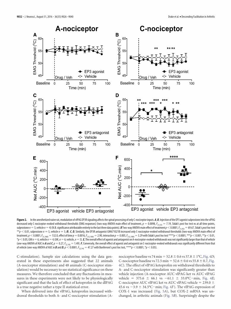
EP3Rs within the vlPAG modulate withdrawal reflexes evokedby peripheral C-nociceptor, but not A-nociceptor, activationin naive ratsThe EP3 agonist sulprostone injected into the vlPAG had noeffect on withdrawal thresholds evoked by A-nociceptor activa-tion (Fig. 2A,E), but decreased the withdrawal threshold toC-nociceptor activation compared with baseline (52.2 � 0.4°C toa minimum of 49.5 � 0.5°C at t � 33 min; Fig. 2B,F). Sulpros-tone injection into the dlPAG had no effect on either A- orC-nociceptor withdrawal thresholds [A-nociceptor: baseline(55 � 0.8°C) vs 15 min (54 � 3.2°C) vs 45 min (56 � 0.9 °C) vs 90min (54 � 1.4°C) post drug; C-nociceptor: baseline (55 � 1.1°C)vs 15 min (57 � 0.3°C) vs 45 min (54 � 1.5°C) vs 90 min (54 �1.9°C) postdrug, n � 3 for both]. The EP3R antagonistGW671021B had no effect on withdrawal thresholds evoked byA-nociceptor activation (Fig. 2C,E), but increased the withdrawalthreshold to C-nociceptor activation compared with baseline
(52 � 0.4°C to 56 � 0.4°C at t � 33 min; Fig. 2D). Comparison ofthe overall effect of the two compounds, the EP3R agonist sulpr-ostone and the EP3R antagonist GW671021B, confirmed thatthey had no significant effect on A-nociceptor evoked withdrawalthresholds compared with vehicle (AUC EP3 agonist vs AUCvehicle � �10.0 � 43.7 vs 41.4 � 24.0°C � min, AUC EP3 antag-onist vs AUC vehicle � 89.9 � 41.1 vs 41.4 � 24.0°C � min; Fig.2E). The effects of EP3 agonist and EP3 antagonist on C-nociceptorevoked withdrawal thresholds were significantly greater than vehicle(AUC EP3 agonist vs AUC vehicle � �164.6 � 38.50°C � min vs�6.2 � 18.50°C � min, AUC EP3 antagonist vs AUC vehicle �302.9 � 39.27 vs 6.2 � 18.50°C � min; Fig. 2F).
CFA-induced knee joint arthritis leads to a secondaryhyperalgesia and allodynia of the hindpaw associated with anenhanced spinal processing of A-nociceptor, but not C-nociceptor, inputsAs shown previously on many occasions (Rees et al., 1996; Mar-tindale et al., 2007), knee joint inflammatory arthritis resulted inan increase in joint swelling (mesio-lateral distance measuredacross the joint, baseline vs 7 d � 7.6 � 0.2 vs 9.1 � 0.2 mm; Fig.3A). The thickness of the hindpaw of the inflamed limb did notchange in size as a result of knee joint inflammation (baseline vs7 d � 4.1 � 0.1 vs 4.2 � 0.1 mm, p � 0.118; Fig. 3B). Inflamma-tory arthritis led to a significant fall in hindpaw thermal with-drawal latency (Fig. 3C), as described previously (Davis et al.,1994; Vermeirsch et al., 2007) apparent at 24 h and persisting forat least 7 d (baseline vs 24 h vs 7 d withdrawal latencies � 12.4 �0.4 vs 8.1 � 0.5 vs 8.3 � 0.4 s, respectively; Fig. 3C). Arthritis alsoresulted in a significant hindpaw mechanical allodynia over thesame time frame (baseline vs 24 h vs 7 d withdrawal thresholds �22 � 2 vs 14 � 4 vs 4 � 1 g, respectively; Fig. 3D) and a decreasein the weight borne on the inflamed limb (baseline vs 7 d � 49 �0.5 vs 23 � 1.5%; Fig. 3E).
As we have also shown in a different cohort of animals usingthe same model (Hsieh et al., 2015), anesthetized arthritic ani-mals had significantly lower withdrawal thresholds only toA-nociceptor stimulation in the hindpaw, the secondary hyper-algesic site (A-nociceptor-evoked withdrawal threshold naive vsarthritic, secondary hyperalgesia � 57 � 0.2 vs 54 � 0.3°C, re-spectively; Fig. 3F). C-nociceptor-evoked withdrawal thresholdswere unchanged in arthritic rats (naive vs arthritic � 54 � 0.2 vs54 � 0.1°C, p � 0.266; Fig. 3F).
vlPAG is the site of prostanergic descending facilitation in theanesthetized inflamed ratWe have found previously that the vlPAG, and not the dlPAG,forms the site of prostanergic descending facilitation in the naiveanesthetized rat (Leith et al., 2007). To determine whether a sim-ilar anatomical delineation extended to arthritic animals, thenonspecific COX inhibitor ketoprofen was injected into the dl-PAG and vlPAG to determine its effects on EMG withdrawalthresholds to A- or C-nociceptor activation in the area of second-ary hypersensitivity. Ketoprofen injected into the dlPAG had noeffect on withdrawal thresholds to either A- or C-nociceptor ac-tivation [A-nociceptor baseline (55 � 0.7°C) vs 9.5 min (55 �0.7°C) vs 42.5 min (54 � 0.7°C) vs 75 min (52.7 � 0.8°C); Fig. 4A;C-nociceptor baseline (52.7 � 0.5°C) vs 5.3 min (53.0 � 0.9°C) vs38.8 min (52.7 � 0.5°C) vs 86.3 min (53 � 0.6°C); Fig. 4B]. Thesedatasets had low variability, but also low numbers of animals,which would not allow for variability to be seen if present. Weperformed post hoc power calculations and determined that theactual power of these experiments was 0.99 (both A- and
Figure 1. Blockade of EP3 signaling within the vlPAG increases thermal withdrawal thresh-old in the awake naive rat. Delivery of the PG E2 EP3R antagonist (GW671021B) to the vlPAGincreased thermal withdrawal thresholds evoked from the hindpaw (Kruskal–Wallis test p �0.002, H � 18.64 with Dunn’s post hoc test, **p 0.01, *p 0.05, Shapiro–Wilk normalitytest for EP3 antagonist postdrug data, p � 0.006, W � 0.72; vehicle, n � 6; EP3R antagonist,n � 7; outside PAG, n � 3).
Drake et al. • Descending Facilitation in Arthritis J. Neurosci., August 31, 2016 • 36(35):9026 –9040 • 9031
C-stimulation). Sample size calculations using the data gen-erated in these experiments also suggested that 22 animals(A-nociceptor stimulation) and 48 animals (C-nociceptor stim-ulation) would be necessary to see statistical significance on thesemeasures. We therefore concluded that any fluctuations in mea-sures in these experiments were not likely to be physiologicallysignificant and that the lack of effect of ketoprofen in the dlPAGis a true negative rather a type II statistical error.
When delivered into the vlPAG, ketoprofen increased with-drawal thresholds to both A- and C-nociceptor stimulation (A-
nociceptor baseline vs 74 min � 52.8 � 0.6 vs 57.8 � 1°C, Fig. 4D;C-nociceptor baseline vs 72.5 min � 52.6 � 0.6 vs 55.8 � 0.7, Fig.4C). The effect of vlPAG ketoprofen on withdrawal thresholds toA- and C-nociceptor stimulation was significantly greater thanvehicle injection (A-nociceptor AUC vlPAG ket vs AUC vlPAGvehicle � 375.6 � 66.1 vs �61.1 � 33.0°C � min, Fig. 4E;C-nociceptor AUC vlPAG ket vs AUC vlPAG vehicle � 239.0 �45.6 vs �3.9 � 34.5°C � min; Fig. 4F). The vlPAG expression ofCOX-1 was increased (Fig. 5A), but COX-2 mRNA was un-changed, in arthritic animals (Fig. 5B). Surprisingly despite the
Figure 2. In the anesthetized naive rat, modulation of vlPAG EP3R signaling affects the spinal processing of only C-nociceptor inputs. A, B, Injection of the EP3 agonist sulprostone into the vlPAGdecreased only C-nociceptor-evoked withdrawal thresholds (EMG responses) (two-way ANOVA main effect of treatment, p � 0.0098, F(1,53) � 7.19, Sidak’s post hoc test ns at all time points,sulprostone n � 3, vehicle n � 4) (N.B. significance attributable entirely to the last three data points). B Two-way ANOVA main effect of treatment p 0.0001, F(1,92) � 69.67, Sidak’s post hoc test**p 0.01, sulprostone n � 5, vehicle n � 5; B). C, D, Similarly, the EP3R antagonist GW671021B increased only C-nociceptor-evoked withdrawal thresholds (two-way ANOVA main effect oftreatment, p 0.0001, F(1,100) � 132.0, effect of time p � 0.0016, F(12,100) � 2.90, interaction p � 0.005, F(12,100) � 3.29 with Sidak’s post hoc test ****p 0.0001, ***p 0.001, **p 0.01,*p 0.05, GW n � 6, vehicle n � 4; GW, n � 6; vehicle, n � 5). E, The overall effect of agonist and antagonist on A-nociceptor-evoked withdrawals was not significantly larger than that of vehicle(one-way ANOVA of AUC in A and C, p � 0.27, F(2,10) � 1.49). F, Conversely, the overall effect of agonist and antagonist on C-nociceptor-evoked withdrawals was significantly different from thatof vehicle (one-way ANOVA of AUC in B and D, p 0.0001, F(2,14) � 47.27 with Bonferroni’s post hoc test, ****p 0.0001, *p 0.05).
9032 • J. Neurosci., August 31, 2016 • 36(35):9026 –9040 Drake et al. • Descending Facilitation in Arthritis

lack of functional effects on nociceptive reflexes of COX inhibi-tion in the dlPAG, COX-1 mRNA was upregulated, and COX-2mRNA downregulated in the dlPAG of arthritic animals.
In the anesthetized arthritic rat, vlPAG EP3R antagonismaffects the spinal processing of both A- and C-nociceptorinputs from an area of secondary hypersensitivityEP3R antagonist reversed the lowered withdrawal thresholdsto A-nociceptor activation in the hindpaw area of secondary
hypersensitivity in arthritic rats (baseline vs postdrug �53.8 � 0.5°C vs 58.7 � 0.7°C at 4.5 min postdrug; Fig. 6A).Thresholds were raised to higher values than those seen innaive animals (cf. Figs. 2C, 6A) and the overall effect of EP3Rantagonist was significantly greater in inflammatory sec-ondary hypersensitivity compared with naive animals (A-nociceptor AUC naive vs AUC arthritic � 89.9 � 41.1 vs375.6 � 66.1°C � min; Fig. 6D). In contrast, the prostanergicfacilitatory tone on C-nociceptor reflex withdrawal thresholds
Figure 3. CFA-induced knee joint inflammation leads to behavioral secondary hyperalgesia and allodynia of the hindpaw. A, B, A single intra-articular knee joint injection of CFA (100 �l) leadsto an increase in the width of the knee joint (Wilcoxon’s signed-rank test, W � �28.0, p � 0.016, Shapiro–Wilk normality test for naive data, W � 0.58, p � 0.002; n � 7; A), but not in thehindpaw (unpaired t test, p � 0.11, t � 1.8, df � 8, n � 5; B). C, D, Knee joint inflammation produced a reduction in the withdrawal threshold to both thermal (C) and mechanical (D) stimulidelivered to the plantar surface of the hindpaw of the inflamed limb [repeated-measures ANOVA, p 0.0001 (for both), F(3,18) � 39.69 (for C) and F(3,18) � 14.80 (for D) with Bonferroni’s post hoctests, ****p 0.0001, ***p 0.001, *p 0.05, n � 7/group). E, Knee joint inflammation leads to a persistent reduction in the weight borne on the inflamed limb (Friedman test, Q � 21.99,p 0.0001 followed by Dunn’s multiple-comparison test, **p 0.01, ***p 0.001, Shapiro–Wilk normality test for day 0 data, W � 0.78, p � 0.005, n � 11). F, Withdrawal threshold topreferential A-nociceptor, but not C-nociceptor, activation was significantly lower in secondary sites in arthritic animals versus naive animals (one-way ANOVA, p 0.0001, F(3,60) � 39.26 withBonferroni’s post hoc test ****p 0.0001, n � 16).
Drake et al. • Descending Facilitation in Arthritis J. Neurosci., August 31, 2016 • 36(35):9026 –9040 • 9033

seen in naive animals was maintained at the same level insecondary hypersensitivity as seen in naive animals, as intra-vlPAG EP3R antagonist increased withdrawal thresholds toC-nociceptor activation (baseline vs postdrug � 53.0 � 0.8°Cvs 56 � 1.0°C at 60 min postdrug; Fig. 6B) to a degree equiv-alent to that seen in the naive animals (C-nociceptor AUCnaive vs AUC arthritic � 302.9 � 39.27 vs 239.0 �45.6°C � min; Fig. 6D). The overall effect of EP3R antagonismon A- and C-nociceptor-evoked thresholds in inflammatorysecondary hypersensitivity was equivalent in inflammatory ar-thritis and significantly greater than vehicle (EP3RA-nociceptor vs vehicle A-nociceptors � 375.6 � 66.1 vs�61.1 � 33.0°C � min, EP3R C-nociceptor vs vehicle C-noci-
ceptors � 239.0 � 45.6 vs �3 � 34.5°C � min; Fig. 6C). EP3RmRNA expression was unchanged in vlPAG in arthritis. LikeCOX-1, EP3R mRNA was upregulated in the dlPAG in ar-thritic rats (Fig. 5C).
In the anesthetized arthritic rat, vlPAG EP3R antagonismmodulates spinal dorsal horn neuronal responses to C- butnot A-nociceptor inputs from an area of secondaryhypersensitivityIntra-vlPAG EP3R antagonist had no effect on the firingthreshold of WDR neurons in knee arthritic rats to preferen-tial A-nociceptor activation on the hindpaw [baseline (46.0 �1.2°C) vs 10 (45.83 � 1.49°C) vs 30 (45.17 � 1.89°C) vs 60
Figure 4. In the arthritic rat, the ventrolateral and not the dorsolateral columns of the PAG mediate prostanergic effects on spinal nociception. A, B, Ketoprofen injected into the dlPAGhad no notable effect on EMG-derived withdrawal thresholds to preferential A- and C-nociceptor activation in the hindpaw secondary site (n � 4 for both groups). C, D, Ketoprofeninjected into the vlPAG increased withdrawal threshold to A- and C-nociceptor activation in the hindpaw secondary site (C: two-way ANOVA main effect of treatment p 0.0001, F(1,89)
� 26.23, effect of time p � 0.0051, F(8,89) � 2.99, interaction p � 0.0061, F(8,89) � 2.92 with Sidak’s post hoc test **p 0.01, *p 0.05, n � 7 for vehicle and n � 6 for ketoprofen;D: two-way ANOVA main effect of treatment p 0.0001, F(1,88) � 40.57, effect of time p � 0.048, F(7,88) � 2.13 with Sidak’s post hoc test ***p 0.001, *p 0.05, n � 5 forketoprofen, n � 7 for vehicle). E, Ketoprofen injected into the vlPAG had a significantly greater effect on EMG thresholds evoked by A-nociceptors than that of a vehicle injection. Inaddition, when delivered into the vlPAG, ketoprofen had a significantly greater effect on EMG thresholds evoked by A-nociceptors compared with delivery into the dlPAG (one-way ANOVAp � 0.0001, F(2,14) � 18.98 with Bonferroni’s post hoc test ***p 0.001, **p 0.01; n � 4 for dlPAG, n � 6 for vlPAG ket, n � 7 for vlPAG vehicle). F, Ketoprofen injected into thevlPAG had a greater effect on EMG thresholds evoked by C-nociceptor activation than that of vehicle. In addition, when delivered into the vlPAG, ketoprofen had a greater effect on EMGthresholds evoked by C-nociceptor activation compared with delivery into the dlPAG (one-way ANOVA p � 0.0012, F(2,13) � 11.83 with Bonferroni’s post hoc test **p 0.01; n � 4 fordlPAG, n � 5 for vlPAG ket, n � 7 for vlPAG vehicle).
9034 • J. Neurosci., August 31, 2016 • 36(35):9026 –9040 Drake et al. • Descending Facilitation in Arthritis

(43.67 � 0.67°C) mins postdrug, evoking responses at thresh-olds similar to those previously reported (McMullan andLumb, 2006a; Leith et al., 2014); Fig. 7 A, C,E]. In contrast,EP3R antagonist increased neuronal firing threshold in re-sponse to preferential activation of hindpaw C-nociceptors(baseline vs 20 min postdrug � 45.7 � 1.4°C and 51.0 � 2°C;Fig. 7 B, D,E). The effect of EP3R antagonism was significa-
ntly greater on C-nociceptor compared with A-nociceptorresponses and significantly greater than vehicle (EP3C-nociceptor vs EP3 A-nociceptor � 175.7 � 39.75 vs�39.17 � 48.76°C � min; EP3 C-nociceptor vs vehicle C-no-ciceptor � 175.7 � 39.7 vs �38.73 � 55.7°C � min; Fig. 7E).
The thresholds of evoked EMG withdrawals are higher than,but encompass the range of, thresholds of WDR neurons in thisstudy, (e.g., A-nociceptor evoked WDR firing threshold range �42–55°C; A-nociceptor evoked EMG thresholds � 52–56°C).Values for EMG and WDR thresholds are consistent withour previous studies (Leith et al., 2007; Leith et al., 2014).Hindlimb withdrawal requires coordinated, integrated activity ina multineuronal pathway spanning dorsal and ventral horns,which includes contributions from low-threshold, WDR, andnociceptive-specific neurons, not WDR neurons alone. There-fore, the threshold for limb withdrawal (EMG activity) is likely tobe higher than any single neuron within that circuitry and shouldbe closest to those spinal neurons with the highest thresholds(e.g., nociceptive specific neurons). It should also be noted thatthe anesthetic alphaxalone augments GABAergic inhibition,which will affect network function and exert a cumulative effecton the polysynaptic withdrawal reflex pathway. Both of theseactions might be expected to raise EMG thresholds relative toWDR action potential firing thresholds.
DiscussionCOX–PG signaling has well described pro-nociceptive and anti-nociceptive effects at peripheral and spinal sites, but its actions atsupraspinal sites are less well defined. The PAG expresses bothCOX isoforms (Fig. 5; Breder et al., 1992; Breder et al., 1995) andthe PG EP receptors EP1– 4, although precise cellular localiza-tions are still unclear (Palazzo et al., 2011; Myren et al., 2012). ThevlPAG, rather than other PAG subregions, exerts a specific facili-tatory effect over spinal nociceptive processing in naive animals(Heinricher et al., 2004; Leith et al., 2007; Palazzo et al., 2011) andin acute inflammatory and neuropathic models (Oliva et al.,2006; Palazzo et al., 2011).
Central inhibition of COX in lateral PAG (Tortorici andVanegas, 1995) and rostroventral medulla contributes to de-scending inhibition through potentiation of opioid analgesia(Vaughan et al., 1997; Vaughan, 1998). Cannabinoid-stress-induced analgesia derives from multiple PAG columns (Licht-man et al., 1996; Olango et al., 2012) and can be engaged byintra-PAG COX inhibition (Vanegas et al., 2010). Our dataare, however, consistent with a discrete endogenous vlPAGprostanergic-EP3R receptor mechanism directly facilitating spi-nal C-nociceptive processing in conscious and anesthetized naiveanimals and A-nociceptive processing in hindpaw secondary hy-persensitivity in arthritic animals. Hindpaw secondary hypersen-sitivity occurs in knee-joint arthritis (Herrero and Cervero, 1996;Urban et al., 1999; Zhang et al., 2002; Martindale et al., 2007),with sensitization of the spinal processing of A-nociceptor inputs(Fig. 3F; Hsieh et al., 2015). De novo central A-nociceptor sensi-tization is dependent on intra-vlPAG PGs, most likely PGE2, be-cause it is reversed with the EP3R antagonist.
The absence of effect of vlPAG EP3R antagonism on A-nocicep-tor-evoked WDR firing thresholds in arthritic animals was surpris-ing considering the clear sensitization of A-nociceptor-evokednoxious withdrawals. Deep dorsal horn WDR neurons are integralto spinal reflex circuitry and activity in WDR neurons is well corre-lated with motor unit activity during withdrawal (You et al., 2003),hence the targeting of WDR neurons in this study. One interpreta-tion of our findings is that the effects on A-nociceptor processing are
Figure 5. COX enzyme and EP3R mRNA expression are regulated within the PAG after kneejoint arthritis. A, COX-1 mRNA expression was significantly increased in both vlPAG and dlPAG7 d after induction of knee joint arthritis [Mann–Whitney U � 0 (sum of ranks, 15, 30; vlPAG)and U � 1 (16,39; dlPAG), p � 0.016 for both (� � 0.025, corrected for multiple compari-sons)]. B, COX-2 mRNA expression was significantly decreased in dlPAG [U � 0 (40, 15), p �0.008], but unchanged in vlPAG [U � 3 (18, 27), p � 0.11]. C, EP3 mRNAs had a tendency toincrease in vlPAG [U � 2 (17, 28), p � 0.06] and was significantly increased in dlPAG [U � 1(16, 39), p � 0.02].
Drake et al. • Descending Facilitation in Arthritis J. Neurosci., August 31, 2016 • 36(35):9026 –9040 • 9035

mediated in the ventral rather than dorsal horn. The PAG has directprojections to the lumbar spinal ventral horn despite the majority ofPAG projections targeting the cervical cord (Mouton and Holstege,1994). We consider an action in the ventral horn to be unlikely as ifthe additional facilitation of A-nociceptor processing observed inarthritic animals were mediated at the level of the ventral horn thensimilar effects on C-nociceptor processing would be expected (Fig.6D). Because A- and C-nociceptor information is integrated at thisspinal level, it is therefore more likely that effects on A-nociceptiveprocessing in arthritic animals are mediated through alternative dor-sal horn neuronal populations, such as nociceptive-specific and/orWDR neurons located in superficial laminae. Descending inhibitorypathways originating from the vlPAG modulate both A- and C-no-ciceptor evoked c-fos expression in superficial laminae (indicative ofspinal neuronal activation), but affect only C-nociceptor-evoked c-fos expression in deeper laminae (Koutsikou et al., 2007).
Peripheral nociceptors innervating secondary sites do not displaysignificant sensitization (Baumann et al., 1991; LaMotte et al., 1992;Torebjork et al., 1992; also see Serra et al., 2004). The development ofsecondary hypersensitivity is critically dependent on central sensiti-zation, including potentiation of synaptic efficacy, and engagementof descending controls (Urban et al., 1999; Sandkuhler, 2009; Nakaet al., 2013). Our findings are consistent with the hypothesis that
activity in capsaicin-sensitive C-nociceptors (McMullan et al., 2004;Leith et al., 2007) innervating the area of tissue damage drives centralsensitization (Woolf, 1983; Leith et al., 2007; You et al., 2010) andfacilitates the spinal processing of A-nociceptor inputs from second-ary sites, in part through descending facilitation (LaMotte et al.,1992; Treede et al., 1992; Magerl et al., 2001; Hsieh et al., 2015).Interpretation of these findings is, however, dependent on the selec-tivity of the heat-ramp stimulation. If a significant number ofA-nociceptors were also activated by slow ramps, then we would alsoexpect to see facilitation of slow-ramp-evoked reflexes in secondaryhypersensitivity, which we do not. In addition, capsaicin, a sensitizerof TRPV1-expressing C-nociceptors, affects only slow-ramp re-sponses; DMSO, a sensitizer of A-nociceptors, affects only fast-rampresponses. In WDR neurons, the magnitude of C-fiber response toelectrical stimulation correlates with the response to slow-ramp ac-tivation (Leith et al., 2007). This evidence suggests that this methodactivates cutaneous C- and A-nociceptor populations preferentially(Yeomans and Proudfit, 1996).
Changes in the balance of descending inhibitory and facili-tatory controls can reduce the threshold for long-term potentia-tion (LTP) induction, allowing noxious stimuli that are usuallyunable to drive central sensitization to do so (Sandkuhler, 2009).For instance, electrical stimulation of peripheral nerves at A-fiber
Figure 6. In the arthritic rat, EP3R antagonism in the vlPAG increases EMG withdrawal thresholds to A- and C-nociceptor activation in the hindpaw secondary hypersensitive site. Injection of theEP3 antagonist GW671021B into the vlPAG increased EMG thresholds evoked by preferential A-nociceptor and C-nociceptor activation in the hindpaw secondary site (A: two-way ANOVA main effectof treatment p 0.0001, F(1,86) � 38.53 with Sidak’s post hoc test **p 0.01, *p 0.05, n � 6 for GW671021B and 7 for vehicle; B: two-way ANOVA main effect of treatment p 0.0001,F(1,78) � 56.51, interaction p � 0.41 F(7,78) � 2.22 with Sidak’s post hoc test ****p 0.0001, ***p 0.001, **p 0.01, *p 0.05, n � 5 for GW671021B and n � 7 for vehicle). C, EP3Rantagonist had a greater effect on EMG thresholds evoked by A- and C-nociceptor activation than vehicle (one-way ANOVA p 0.0001, F(3,21) � 20.84 with Bonferroni’s post test ****p 0.0001,**p 0.01, n � 6 and 5 for EP3 A- and C-nociceptor groups, respectively, and n � 7 for both vehicle groups). D, Effect of the EP3R antagonist on EMG thresholds evoked by C-nociceptor stimulationwas equivalent in arthritic and naive animals. However, the effect of the EP3R antagonist on EMG thresholds evoked by A-nociceptor stimulation was greater in arthritic versus naive animals(one-way ANOVA p � 0.0037, F(3,20) � 6.2 with Bonferroni’s post test, **p 0.01; n � 6 for A-nociceptor naive and arthritic, n � 7 for C-nociceptor naïve, and n � 5 for C-nociceptor arthritic).
9036 • J. Neurosci., August 31, 2016 • 36(35):9026 –9040 Drake et al. • Descending Facilitation in Arthritis

Figure 7. In the arthritic rat, EP3R antagonism in the vlPAG increases the firing threshold of spinal dorsal horn WDR neurons to C-nociceptor, but not A-nociceptor, activation in the hindpawsecondary hypersensitive site. A, B, Example traces of spinal WDR neuronal recordings and their responses to peripheral A-nociceptor (A) and C-nociceptor (C) activation before (baseline) and afterinjection of the EP3R antagonist into the vlPAG. Digitized data traces show: firing frequency of sorted spikes of individual WDR neurons, examples of action potential spike trains of spinal WDRneurons from which the firing frequency traces are derived, and changes in hindpaw dorsum contact skin temperature in response to the ramping thermal stimuli used to activate A- andC-nociceptors preferentially. Horizontal cursors in on firing frequency traces mark the mean firing rate in the 10 s preceding the start of thermal stimulus (e.g., 0.1 and 0.9 Hz in A). The thermal firingthreshold is taken at the point in which thermally evoked activity exceeded the mean baseline firing rate and remained elevated for at least 10% of the total duration of thermal stimuli (shown bythe vertical cursors (1) in A and B). WDR neuron thermal firing threshold temperatures (in degrees Celsius) are thus indicated using the horizontal cursors in skin contact temperature trace (e.g.,44.4°C and 43.1°C in A) as the skin temperature at which these conditions are met. Digitized data examples for both baseline responses and after EP3R antagonist (Figure legend continues.)
Drake et al. • Descending Facilitation in Arthritis J. Neurosci., August 31, 2016 • 36(35):9026 –9040 • 9037
strength produces LTP at C-fiber synapses only when descendingcontrols are disrupted (Liu et al., 1998). C-nociceptive drivefrom the primary injury site may affect the spinal processing ofA-nociceptor inputs from the secondary sites through bothspinal and supraspinal mechanisms because C-nociceptors ter-minate exclusively in the superficial dorsal horn and drive spinal–supraspinal–spinal loops via spinal projection neurons (Lightand Perl, 1979; Suzuki et al., 2002; Mantyh and Hunt, 2004).Importantly, heterosynaptic LTP of synaptic efficacy may un-mask and/or increase A-fiber drive to spinal nociceptive circuits.This could lead to the central sensitization to A-fiber inputs.Descending facilitation from the rostral ventral medulla (RVM)is critical for the maintenance of secondary hyperalgesia in ro-dents (Urban and Gebhart, 1999) and, notably, PG signalingwithin the vlPAG modulates RVM ON and OFF cell activity innaive and neuropathic rats (Heinricher et al., 2004; Palazzo et al.,2011). This suggests that a spinal/PAG/RVM/spinal loop under-pins the vlPAG-prostanergic facilitatory pathway.
Secondary hypersensitivity is often suggested to be limited tomechanical stimulation (Treede et al., 1992) because inflammatorysecondary thermal hyperalgesia is not present in the capsaicin model(Raja et al., 1984; Ali et al., 1996). However, secondary thermal hy-persensitivity is sometimes, but not always (Pertovaara, 1998), re-ported in clinical studies (Bajaj et al., 2001; Schaible et al., 2009) andexperimental studies (Fig. 3C; Herrero and Cervero, 1996; Urban etal., 1999; Zhang et al., 2002; Martindale et al., 2007; Hsieh et al.,2015), so it is more likely that secondary hypersensitivity is a centralA-nociceptor-sensitization, not a modality-specific (thermal/mechanical) process (Hsieh et al., 2015).
Although peripheral C-nociceptors are not sensitized in sec-ondary sites (Baumann et al., 1991; LaMotte et al., 1992; Toreb-jork et al., 1992), C-nociceptor-evoked reflexes and neuronalresponses are reported to be facilitated at secondary sites (Her-rero and Cervero, 1996; Martindale et al., 2007). However, thismay represent a potential confound of electrical C-fiber stimula-tion parameters in inflamed animals. The simplest interpretationof our finding of unchanged C-nociceptor-evoked reflexes in ar-thritic animals is that there is no additional facilitation ofC-nociceptive inputs from secondary sites, in contrast to the ef-fect on A-nociceptive processing.
In vlPAG, COX-1 mRNA levels are altered, suggesting that ex-tension of the prostanergic facilitation to A-nociceptor inputs mayresult from increased intra-vlPAG COX-1-dependent PG produc-tion acting on EP3R. Altered CNS COX expression has been attrib-uted to increased central interleukin-1beta (IL-1beta) levels found ininflammatory arthritis in humans and animals (Samad et al., 2001;Kosek et al., 2015). IL-1beta exerts different effects in different re-gions of the brain (An et al., 2011), for example, increased expressionof COX-1 in perivascular, endothelial, and glial cells (García-Buenoet al., 2009; Matousek et al., 2010); COX-2 expression in multiple celltypes including neurons (Crofford et al., 1994; Serou et al., 1999;
Samad et al., 2001); and EP3Rs on CNS glial cells (Waschbisch et al.,2006). Intra-vlPAG EP1 receptors are implicated in the facilitation ofneuropathic pain; surprisingly, these receptors are reportedly down-regulated but remain functional in this condition (Palazzo et al.,2011).
Although COX-1, COX-2, and EP3R expression is regulatedin the dlPAG in inflamed rats, inhibition of this system in dlPAGhad no effect on nociceptive processing. The dlPAG is implicatedin active rather than passive behavioral responses to stress/fear,including autonomic control of breathing and cardiovascularfunction (Dampney et al., 2013). There is no published evidencelinking COXs and/or PGs with any dlPAG functional responses,although PGE2 modulates dlPAG neuronal function by EP3R-dependent inhibition of glutamatergic inputs (Lu et al., 2007).
Here, we have demonstrated that descending facilitation fromthe PAG affects nociceptive processing in awake animals throughintra-vlPAG EP3R signaling. In the naive state, this descendingfacilitation is restricted to the spinal processing of C-nociceptiveinputs, but extends to affect A-nociceptive inputs from secondarysites in arthritic animals. Prostanergic descending facilitationfrom the PAG may provide a major contribution to mechanismsof central sensitization that is critical to the inflammatory painphenotype. These data suggest that, for centrally penetratingNSAIDs at least, their principle analgesic effect may be to reducedescending facilitation, rather than alter spinal processing, whereEP receptors may exert anti-nociceptive effects (Bar et al., 2004;Natura et al., 2013). This may account for the ineffectiveness ofspinally delivered NSAIDs in humans (Eisenach et al., 2010a;Eisenach et al., 2010b).
ReferencesAli Z, Meyer RA, Campbell JN (1996) Secondary hyperalgesia to mechanical
but not heat stimuli following a capsaicin injection in hairy skin. Pain68:401– 411. CrossRef Medline
Ambriz-Tututi M, Cruz SL, Urquiza-Marín H, Granados-Soto V (2011)Formalin-induced long-term secondary allodynia and hyperalgesia aremaintained by descending facilitation. Pharmacol Biochem Behav 98:417– 424. CrossRef Medline
An Y, Chen Q, Quan N (2011) Interleukin-1 exerts distinct actions on dif-ferent cell types of the brain in vitro. J Inflamm Res 2011:11–20. Medline
Bajaj P, Bajaj P, Graven-Nielsen T, Arendt-Nielsen L (2001) Osteoarthritisand its association with muscle hyperalgesia: an experimental controlledstudy. Pain 93:107–114. CrossRef Medline
Bar KJ, Natura G, Telleria-Diaz A, Teschner P, Vogel R, Vasquez E, SchaibleHG, Ebersberger A (2004) Changes in the effect of spinal prostaglandinE2 during inflammation: prostaglandin E (EP1-EP4) receptors in spinalnociceptive processing of input from the normal or inflamed knee joint.J Neurosci 24:642– 651. CrossRef Medline
Baumann TK, Simone DA, Shain CN, LaMotte RH (1991) Neurogenic hy-peralgesia: the search for the primary cutaneous afferent fibers that con-tribute to capsaicin-induced pain and hyperalgesia. J Neurophysiol 66:212–227. Medline
Belley M, Chan CC, Gareau Y, Gallant M, Juteau H, Houde K, Lachance N,Labelle M, Sawyer N, Tremblay N, Lamontagne S, Carriere MC, Denis D,Greig GM, Slipetz D, Gordon R, Chauret N, Li C, Zamboni RJ, MettersKM (2006) Comparison between two classes of selective EP(3) antago-nists and their biological activities. Bioorg Med Chem Lett 16:5639 –5642.CrossRef Medline
Boie Y, Stocco R, Sawyer N, Slipetz DM, Ungrin MD, Neuschafer-Rube F,Puschel GP, Metters KM, Abramovitz M (1997) Molecular cloning andcharacterization of the four rat prostaglandin E2 prostanoid receptor sub-types. Eur J Pharmacol 340:227–241. CrossRef Medline
BrederCD,SmithWL,RazA,Masferrer J,SeibertK,NeedlemanP,SaperCB (1992)Distribution and characterization of cyclooxygenase immunoreactivity in theovine brain. J Comp Neurol 322:409–438. CrossRef Medline
Breder CD, Dewitt D, Kraig RP (1995) Characterization of inducible cyclo-oxygenase in rat brain. J Comp Neurol 355:296 –315. CrossRef Medline
Coleman RA, Smith WL, Narumiya S (1994) International Union of Phar-
4
(Figure legend continued.) injection into vlPAG are shown for A-nociceptor activation (A) andC-nociceptor activation (C). C, D, Injection of the EP3R antagonist GW6789101B had no significanteffect on the firing threshold of spinal WDR neuronal firing thresholds to A-nociceptor stimulation(n � 6 for drug and 4 for vehicle; C) but increased WDR neuronal firing threshold to C-nociceptorstimulation (two-way ANOVA, main effect of treatment p0.0001, F(1,63)�20.72 with Sidak’s posthoc test **p0.01, n�7 for drug and 4 for vehicle; D). E, Overall effect of EP3R antagonism on WDRneuron firing thresholds to C-nociceptor activation was significantly greater than that of a vehicleinjection and greater when evoked by C- versus A-nociceptor activation (Kruskal–Wallis test, H �11.31, p � 0.01 with Dunn’s post test, *p 0.05 Shapiro–Wilk normality test for C-nociceptor �EP3 antagonist data, W � 0.79, p � 0.03).
9038 • J. Neurosci., August 31, 2016 • 36(35):9026 –9040 Drake et al. • Descending Facilitation in Arthritis

macology classification of prostanoid receptors: properties, distribution,and structure of the receptors and their subtypes. Pharmacol Rev 46:205–229. Medline
Crofford LJ, Wilder RL, Ristimaki AP, Sano H, Remmers EF, Epps HR, Hla T(1994) Cyclooxygenase-1 and -2 expression in rheumatoid synovial tis-sues: effects of interleukin-1 beta, phorbol ester, and corticosteroids.J Clin Invest 93:1095–1101. CrossRef Medline
Dampney RA, Furlong TM, Horiuchi J, Iigaya K (2013) Role of dorsolateralperiaqueductal grey in the coordinated regulation of cardiovascular andrespiratory function. Auton Neurosci 175:17–25. CrossRef Medline
Davis AJ, Kelly D, Perkins MN (1994) The induction of des-Arg9-brad-ykinin-mediated hyperalgesia in the rat by inflammatory stimuli. BrazJ Med Biol Res 27:1793–1802. Medline
De Felice M, Sanoja R, Wang R, Vera-Portocarrero L, Oyarzo J, King T,Ossipov MH, Vanderah TW, Lai J, Dussor GO, Fields HL, Price TJ, Por-reca F (2011) Engagement of descending inhibition from the rostralventromedial medulla protects against chronic neuropathic pain. Pain152:2701–2709. CrossRef Medline
Donaldson LF, Humphrey PS, Oldfield S, Giblett S, Grubb BD (2001) Ex-pression and regulation of prostaglandin E receptor subtype mRNAs inrat sensory ganglia and spinal cord in response to peripheral inflamma-tion. Prostaglandins Other Lipid Mediat 63:109 –122. CrossRef Medline
Eisenach JC, Curry R, Rauck R, Pan P, Yaksh TL (2010a) Role of spinalcyclooxygenase in human postoperative and chronic pain. Anesthesiol-ogy 112:1225–1233. CrossRef Medline
Eisenach JC, Curry R, Tong C, Houle TT, Yaksh TL (2010b) Effects of in-trathecal ketorolac on human experimental pain. Anesthesiology 112:1216 –1224. CrossRef Medline
García-Bueno B, Serrats J, Sawchenko PE (2009) Cerebrovascular cyc-looxygenase-1 expression, regulation, and role in hypothalamic-pituitary-adrenal axis activation by inflammatory stimuli. J Neurosci 29:12970 –12981. CrossRef Medline
Geranton SM, Jimenez-Díaz L, Torsney C, Tochiki KK, Stuart SA, Leith JL,Lumb BM, Hunt SP (2009) A rapamycin-sensitive signaling pathway isessential for the full expression of persistent pain states. J Neurosci 29:15017–15027. CrossRef Medline
Guo JY, Huo HR, Zhao BS, Liu HB, Li LF, Ma YY, Guo SY, Jiang TL (2006)Cinnamaldehyde reduces IL-1beta-induced cyclooxygenase-2 activity inrat cerebral microvascular endothelial cells. Eur J Pharmacol 537:174 –180. CrossRef Medline
Hargreaves K, Dubner R, Brown F, Flores C, Joris J (1988) A new and sen-sitive method for measuring thermal nociception in cutaneous hyperal-gesia. Pain 32:77– 88. CrossRef Medline
Heinricher MM, Martenson ME, Neubert MJ (2004) Prostaglandin E2 inthe midbrain periaqueductal gray produces hyperalgesia and activatespain-modulating circuitry in the rostral ventromedial medulla. Pain 110:419 – 426. CrossRef Medline
Heinricher MM, Tavares I, Leith JL, Lumb BM (2009) Descending controlof nociception: specificity, recruitment and plasticity. Brain Res Rev 60:214 –225. CrossRef Medline
Herrero JF, Cervero F (1996) Changes in nociceptive reflex facilitation duringcarrageenan-induced arthritis. Brain Res 717:62–68. CrossRef Medline
Hsieh MT, Donaldson LF, Lumb BM (2015) Differential contributions ofA- and C-nociceptors to primary and secondary inflammatory hypersen-sitivity in the rat. Pain 156:1074 –1083. CrossRef Medline
Hughes SW, Hickey L, Hulse RP, Lumb BM, Pickering AE (2013) Endoge-nous analgesic action of the pontospinal noradrenergic system spatiallyrestricts and temporally delays the progression of neuropathic pain fol-lowing tibial nerve injury. Pain 154:1680 –1690. CrossRef Medline
Juteau H, Gareau Y, Labelle M, Sturino CF, Sawyer N, Tremblay N, Lamon-tagne S, Carriere MC, Denis D, Metters KM (2001) Structure-activityrelationship of cinnamic acylsulfonamide analogues on the human EP3prostanoid receptor. Bioorg Med Chem 9:1977–1984. CrossRef Medline
Kennedy I, Coleman RA, Humphrey PP, Levy GP, Lumley P (1982) Studieson the characterisation of prostanoid receptors: a proposed classification.Prostaglandins 24:667– 689. Medline
Kobayashi K, Imaizumi R, Sumichika H, Tanaka H, Goda M, Fukunari A,Komatsu H (2003) Sodium iodoacetate-induced experimental osteoar-thritis and associated pain model in rats. J Vet Med Sci 65:1195–1199.CrossRef Medline
KoppertW,WehrfritzA,KorberN,SittlR,AlbrechtS,SchuttlerJ,SchmelzM (2004)
The cyclooxygenase isozyme inhibitors parecoxib and paracetamol reduce cen-tral hyperalgesia in humans. Pain 108:148–153. CrossRef Medline
Korotkova M, Jakobsson PJ (2014) Persisting eicosanoid pathways in rheu-matic diseases. Nat Rev Rheumatol 10:229 –241. CrossRef Medline
Kosek E, Altawil R, Kadetoff D, Finn A, Westman M, Le Maître E, AnderssonM, Jensen-Urstad M, Lampa J (2015) Evidence of different mediators ofcentral inflammation in dysfunctional and inflammatory pain–inte-rleukin-8 in fibromyalgia and interleukin-1 beta in rheumatoid arthritis.J Neuroimmunol 280:49 –55. CrossRef Medline
Koutsikou S, Parry DM, MacMillan FM, Lumb BM (2007) Laminar organi-zation of spinal dorsal horn neurones activated by C- vs. A-heat nocicep-tors and their descending control from the periaqueductal grey in the rat.Eur J Neurosci 26:943–952. CrossRef Medline
LaMotte RH, Lundberg LE, Torebjork HE (1992) Pain, hyperalgesia andactivity in nociceptive C units in humans after intradermal injection ofcapsaicin. J Physiol 448:749 –764. CrossRef Medline
Leith JL, Wilson AW, Donaldson LF, Lumb BM (2007) Cyclooxygenase-1-derived prostaglandins in the periaqueductal gray differentially controlC- versus A-fiber-evoked spinal nociception. J Neurosci 27:11296 –11305.CrossRef Medline
Leith JL, Wilson AW, You HJ, Lumb BM, Donaldson LF (2014) Periaque-ductal grey cyclooxygenase-dependent facilitation of C-nociceptive driveand encoding in dorsal horn neurons in the rat. J Physiol 592:5093–5107.CrossRef Medline
Lichtman AH, Cook SA, Martin BR (1996) Investigation of brain sites me-diating cannabinoid-induced antinociception in rats: evidence support-ing periaqueductal gray involvement. J Pharmacol Exp Ther 276:585–593.Medline
Light AR, Perl ER (1979) Spinal termination of functionally identified primary af-ferent neurons with slowly conducting myelinated fibers. J Comp Neurol 186:133–150. CrossRef Medline
Liu XG, Morton CR, Azkue JJ, Zimmermann M, Sandkuhler J (1998) Long-term depression of C-fibre-evoked spinal field potentials by stimulationof primary afferent A delta-fibres in the adult rat. Eur J Neurosci 10:3069 –3075. CrossRef Medline
Lu J, Xing J, Li J (2007) Prostaglandin E2 (PGE2) inhibits glutamatergicsynaptic transmission in dorsolateral periaqueductal gray (dl-PAG).Brain Res 1162:38 – 47. CrossRef Medline
Magerl W, Fuchs PN, Meyer RA, Treede RD (2001) Roles of capsaicin-insensitive nociceptors in cutaneous pain and secondary hyperalgesia.Brain 124:1754 –1764. CrossRef Medline
Mantyh PW, Hunt SP (2004) Setting the tone: superficial dorsal horn pro-jection neurons regulate pain sensitivity. Trends Neurosci 27:582–584.CrossRef Medline
Martindale JC, Wilson AW, Reeve AJ, Chessell IP, Headley PM (2007)Chronic secondary hypersensitivity of dorsal horn neurones followinginflammation of the knee joint. Pain 133:79 – 86. CrossRef Medline
Matousek SB, Hein AM, Shaftel SS, Olschowka JA, Kyrkanides S, O’Banion MK(2010) Cyclooxygenase-1 mediates prostaglandin E(2) elevation and con-textual memory impairment in a model of sustained hippocampalinterleukin-1beta expression. J Neurochem 114:247–258. CrossRef Medline
McMullan S, Lumb BM (2006a) Spinal dorsal horn neuronal responses tomyelinated versus unmyelinated heat nociceptors and their modulationby activation of the periaqueductal grey in the rat. J Physiol 576:547–556.CrossRef Medline
McMullan S, Lumb BM (2006b) Midbrain control of spinal nociceptiondiscriminates between responses evoked by myelinated and unmyelinatedheat nociceptors in the rat. Pain 124:59 – 68. CrossRef Medline
McMullan S, Simpson DA, Lumb BM (2004) A reliable method for the pref-erential activation of C- or A-fibre heat nociceptors. J Neurosci Methods138:133–139. CrossRef Medline
Menetrey D, Giesler GJ Jr, Besson JM (1977) An analysis of response prop-erties of spinal cord dorsal horn neurones to nonnoxious and noxiousstimuli in the spinal rat. Exp Brain Res 27:15–33. Medline
Menetrey D, Chaouch A, Besson JM (1979) Responses of spinal cord dorsalhorn neurones to non-noxious and noxious cutaneous temperaturechanges in the spinal rat. Pain 6:265–282. CrossRef Medline
Merrill EG, Ainsworth A (1972) Glass-coated platinum-plated tungsten mi-croelectrodes. Med Biol Engineer 10:662– 672. CrossRef
Millan MJ (2002) Descending control of pain. Prog Neurobiol 66:355– 474.CrossRef Medline
Mouton LJ, Holstege G (1994) The periaqueductal gray in the cat projects to
Drake et al. • Descending Facilitation in Arthritis J. Neurosci., August 31, 2016 • 36(35):9026 –9040 • 9039

lamina VIII and the medial part of lamina VII throughout the length ofthe spinal cord. Exp Brain Res 101:253–264. Medline
Myren M, Baun M, Ploug KB, Jansen-Olesen I, Olesen J, Gupta S (2010)Functional and molecular characterization of prostaglandin E2 dilatoryreceptors in the rat craniovascular system in relevance to migraine. Ceph-alalgia 30:1110 –1122. CrossRef Medline
Myren M, Olesen J, Gupta S (2012) Prostaglandin E2 receptor expression inthe rat trigeminal-vascular system and other brain structures involved inpain. Neurosci Lett 506:64 – 69. CrossRef Medline
Naka A, Gruber-Schoffnegger D, Sandkuhler J (2013) Non-Hebbian plas-ticity at C-fiber synapses in rat spinal cord lamina I neurons. Pain 154:1333–1342. CrossRef Medline
Natura G, Bar KJ, Eitner A, Boettger MK, Richter F, Hensellek S, EbersbergerA, Leuchtweis J, Maruyama T, Hofmann GO, Halbhuber KJ, Schaible HG(2013) Neuronal prostaglandin E2 receptor subtype EP3 mediates anti-nociception during inflammation. Proc Natl Acad Sci U S A 110:13648 –13653. CrossRef Medline
Olango WM, Roche M, Ford GK, Harhen B, Finn DP (2012) The endocan-nabinoid system in the rat dorsolateral periaqueductal grey mediates fear-conditioned analgesia and controls fear expression in the presence ofnociceptive tone. Br J Pharmacol 165:2549 –2560. CrossRef Medline
Oliva P, Berrino L, de Novellis V, Palazzo E, Marabese I, Siniscalco D, ScafuroM, Mariani L, Rossi F, Maione S (2006) Role of periaqueductal greyprostaglandin receptors in formalin-induced hyperalgesia. Eur J Pharma-col 530:40 – 47. CrossRef Medline
Palazzo E, Rossi F, Maione S (2008) Role of TRPV1 receptors in descendingmodulation of pain. Mol Cell Endocrinol 286:S79 –S83. CrossRef Medline
Palazzo E, Guida F, Gatta L, Luongo L, Boccella S, Bellini G, Marabese I, deNovellis V, Rossi F, Maione S (2011) EP1 receptor within the ventrolat-eral periaqueductal grey controls thermonociception and rostral ventro-medial medulla cell activity in healthy and neuropathic rat. Mol Pain 7:82.CrossRef Medline
Paxinos G, Watson C (2006) The rat brain in stereotaxic coordinates. SanDiego: Academic.
Pertovaara A (1998) A neuronal correlate of secondary hyperalgesia in therat spinal dorsal horn is submodality selective and facilitated by supraspi-nal influence. Exp Neurol 149:193–202. CrossRef Medline
Petersen KL, Brennum J, Dahl JB (1997) Experimental evaluation of theanalgesic effect of ibuprofen on primary and secondary hyperalgesia. Pain70:167–174. CrossRef Medline
Raja SN, Campbell JN, Meyer RA (1984) Evidence for different mechanismsof primary and secondary hyperalgesia following heat injury to the gla-brous skin. Brain 107:1179 –1188. CrossRef Medline
Rees H, Sluka KA, Lu Y, Westlund KN, Willis WD (1996) Dorsal root re-flexes in articular afferents occur bilaterally in a chronic model of arthritisin rats. J Neurophysiol 76:4190 – 4193. Medline
Samad TA, Moore KA, Sapirstein A, Billet S, Allchorne A, Poole S, BonventreJV, Woolf CJ (2001) Interleukin-1beta-mediated induction of Cox-2 inthe CNS contributes to inflammatory pain hypersensitivity. Nature 410:471– 475. CrossRef Medline
Sandkuhler J (2009) Models and mechanisms of hyperalgesia and allodynia.Physiol Rev 89:707–758. CrossRef Medline
Schaible HG, Richter F, Ebersberger A, Boettger MK, Vanegas H, Natura G,Vazquez E, Segond von Banchet G (2009) Joint pain. Exp Brain Res196:153–162. CrossRef Medline
Serou MJ, DeCoster MA, Bazan NG (1999) Interleukin-1 beta activates ex-pression of cyclooxygenase-2 and inducible nitric oxide synthase in pri-mary hippocampal neuronal culture: platelet-activating factor as apreferential mediator of cyclooxygenase-2 expression. J Neurosci Res 58:593–598. Medline
Serra J, Campero M, Bostock H, Ochoa J (2004) Two types of C nociceptorsin human skin and their behavior in areas of capsaicin-induced secondaryhyperalgesia. J Neurophysiol 91:2770 –2781. CrossRef Medline
Staud R (2013) The important role of CNS facilitation and inhibition forchronic pain. Int J Clin Rheumtol 8:639 – 646. CrossRef Medline
Stubhaug A, Romundstad L, Kaasa T, Breivik H (2007) Methylpred-nisolone and ketorolac rapidly reduce hyperalgesia around a skin burninjury and increase pressure pain thresholds. Acta Anaesthesiol Scand51:1138 –1146. Medline
Su X, Leon LA, Wu CW, Morrow DM, Jaworski JP, Hieble JP, Lashinger ES,Jin J, Edwards RM, Laping NJ (2008) Modulation of bladder function by
prostaglandin EP3 receptors in the central nervous system. Am J PhysiolRenal Physiol 295:F984 –F994. CrossRef Medline
Suzuki R, Morcuende S, Webber M, Hunt SP, Dickenson AH (2002) SuperficialNK1-expressing neurons control spinal excitability through activation of de-scending pathways. Nat Neurosci 5:1319–1326. CrossRef Medline
Torebjork E (1985) Nociceptor activation and pain. Philos Trans R SocLond B Biol Sci 308:227–234. CrossRef Medline
Torebjork HE, Lundberg LE, LaMotte RH (1992) Central changes in pro-cessing of mechanoreceptive input in capsaicin-induced secondary hy-peralgesia in humans. J Physiol 448:765–780. CrossRef Medline
Tortorici V, Vanegas H (1995) Anti-nociception induced by systemic orPAG-microinjected lysine-acetylsalicylate in rats. Effects on tail-flick re-lated activity of medullary off- and on-cells. Eur J Neurosci 7:1857–1865.CrossRef Medline
Tracey I, Mantyh PW (2007) The cerebral signature for pain perception andits modulation. Neuron 55:377–391. CrossRef Medline
Treede RD, Magerl W (2000) Multiple mechanisms of secondary hyperal-gesia. Prog Brain Res 129:331–341. CrossRef Medline
Treede RD, Meyer RA, Raja SN, Campbell JN (1992) Peripheral and centralmechanisms of cutaneous hyperalgesia. Prog Neurobiol 38:397– 421.CrossRef Medline
Urban MO, Gebhart GF (1999) Supraspinal contributions to hyperalgesia.Proc Natl Acad Sci U S A 96:7687–7692. CrossRef Medline
Urban MO, Smith DJ, Gebhart GF (1996) Involvement of spinal cholecysto-kininB receptors in mediating neurotensin hyperalgesia from the medul-lary nucleus raphe magnus in the rat. J Pharmacol Exp Ther 278:90 –96.Medline
Urban MO, Zahn PK, Gebhart GF (1999) Descending facilitatory influencesfrom the rostral medial medulla mediate secondary, but not primaryhyperalgesia in the rat. Neuroscience 90:349 –352. CrossRef Medline
Vanegas H (2004) To the descending pain-control system in rats,inflammation-induced primary and secondary hyperalgesia are two dif-ferent things. Neurosci Lett 361:225–228. CrossRef Medline
Vanegas H, Schaible HG (2001) Prostaglandins and cyclooxygenases in thespinal cord. Prog Neurobiol 64:327–363. CrossRef Medline
VanegasH,SchaibleHG (2004) Descendingcontrolofpersistentpain:inhibitoryorfacilitatory? Brain Res Brain Res Rev 46:295–309. CrossRef Medline
Vanegas H, Vazquez E, Tortorici V (2010) NSAIDs, opioids, cannabinoidsand the control of pain by the central nervous system. Pharmaceuticals3:1335–1347. CrossRef
Vaughan CW (1998) Enhancement of opioid inhibition of GABAergic syn-aptic transmission by cyclo-oxygenase inhibitors in rat periaqueductalgrey neurones. Br J Pharmacol 123:1479 –1481. CrossRef Medline
Vaughan CW, Ingram SL, Connor MA, Christie MJ (1997) How opioids inhibitGABA-mediated neurotransmission. Nature 390:611–614. CrossRef Medline
Vermeirsch H, Biermans R, Salmon PL, Meert TF (2007) Evaluation of painbehavior and bone destruction in two arthritic models in guinea pig andrat. Pharmacol Biochem Behav 87:349 –359. CrossRef Medline
Waschbisch A, Fiebich BL, Akundi RS, Schmitz ML, Hoozemans JJ, Candelario-JalilE, Virtainen N, Veerhuis R, Slawik H, Yrjanheikki J, Hull M (2006)Interleukin-1 beta-induced expression of the prostaglandin E-receptor subtypeEP3 in U373 astrocytoma cells depends on protein kinase C and nuclear factor-kappaB. J Neurochem 96:680–693. CrossRef Medline
Woolf CJ (1983) Evidence for a central component of post-injury pain hy-persensitivity. Nature 306:686 – 688. CrossRef Medline
Yeomans DC, Proudfit HK (1996) Nociceptive responses to high and low rates ofnoxious cutaneous heating are mediated by different nociceptors in the rat: elec-trophysiological evidence. Pain 68:141–150. CrossRef Medline
You HJ, Dahl Morch C, Chen J, Arendt-Nielsen L (2003) Simultaneous recordingsof wind-up of paired spinal dorsal horn nociceptive neuron and nociceptive flex-ion reflex in rats. Brain Res 960:235–245. CrossRef Medline
You HJ, Lei J, Sui MY, Huang L, Tan YX, Tjølsen A, Arendt-Nielsen L (2010)Endogenous descending modulation: spatiotemporal effect of dynamicimbalance between descending facilitation and inhibition of nociception.J Physiol 588:4177– 4188. CrossRef Medline
Zhang L, Lu Y, Chen Y, Westlund KN (2002) Group I metabotropic gluta-mate receptor antagonists block secondary thermal hyperalgesia in ratswith knee joint inflammation. J Pharmacol Exp Ther 300:149 –156.CrossRef Medline
Ziegler EA, Magerl W, Meyer RA, Treede RD (1999) Secondary hyperalgesiato punctate mechanical stimuli. Central sensitization to A-fibre nocicep-tor input. Brain 122:2245–2257. CrossRef Medline
9040 • J. Neurosci., August 31, 2016 • 36(35):9026 –9040 Drake et al. • Descending Facilitation in Arthritis