PERCUTANEOUS TRANSLUMINAL BALLOON CATHETER …Valvulotomy and debridement 38 ... Predictors of early...
204
PERCUTANEOUS TRANSLUMINAL BALLOON CATHETER AORTIC VALVULOPLASTY IN ELDERLY PATIENTS
Transcript of PERCUTANEOUS TRANSLUMINAL BALLOON CATHETER …Valvulotomy and debridement 38 ... Predictors of early...

PERCUTANEOUS TRANSLUMINAL BALLOON CATHETER AORTIC
VALVULOPLASTY IN ELDERLY PATIENTS

Cover: Leonardo da Vinci. Notes et dessins sur le coeur et sa constitution
anatomique. Paris, Edouard Rouveyre ed, MDCCCCI.
Courtesy Koninklijke Bibliotheek, 's-Gravenhage
Print: Haveka B.V., Alblasserdam, The Netherlands

PERCUTANEOUS TRANSLUMINAL BALLOON CATHETER AORTIC
VALVULOPLASTY IN ELDERLY PATIENTS
VALVULOPLASTIEKVAN VERNAUWDEAORTAKLEPPEN BIJ
OUDERE PATIENTEN DOOR MIDDEL VAN EEN PERCUTAAN
INGEBRACHTE BALLON CATHETER
PROEFSCHRIFT
ter verkrijging van de graad van doctor aan de Erasmus Universiteit te Rotterdam op
gezag van de rector magnificus, Prof.Dr. C.J. Rijnvos en volgens het besluit van het
college van dekanen. De openbare verdediging zal plaatsvinden op woensdag 21
november om 13.45 uur
door
Marcel Joannes Baptist Maria van den Brand
geboren te
Nispen

Commissieleden
Promotores Prof. Dr. E. Bas
Prof. P.G. Hugenholtz
Overige !eden Prof.Dr. J. Hess
Prof.Dr. J.R.T.C. Roelandt
Financial support by the Netherlands Heart Foundation for the publication of this
thesis is gratefully acknowledged

CONTENTS
Chapter 1: Introduction 11
References 14
Chapter 2: Isolated aortic valve stenosis: history, pathology, physiology 17
Summary 17
History 18
Etiology 19
Pathology 20
Congenital malformation 21
Rheumatic valvular disease 22
Primary degenerative calcification 23
Incidence 24
Unicommissural, unicuspid aortic valve stenosis 24
Bicuspid aortic valve stenosis 24
Rheumatic aortic stenosis 25
Primary degenerative calcific aortic stenosis 25
Pathophysiology 25
Natural course 29
Coexisting conditions 30
References 32
Chapter 3: Surgical interventions for the treatment of aortic valvular stenosis 37
Summary 37
A historical review of now defunct techniques and designs 38
Valvulotomy and debridement 38
Aortic valve replacement for aortic valve incompetence 41
Homo and heterologous aortic valve transplants 43
Currently used surgical techniques for aortic valve replacement or
reconstruction
Aortic valve reconstruction
Homo and heterografts to replace the aortic valve
Homografts
Heterografts
Prosthetic heart valves
Indications for operation in patients with aortic stenosis
45
45
45
45
46
49
50

Operative mortality of prosthetic aortic valve implantation and morbidity
after implantation 51
Operation decade 51
Age at operation 51
Preoperative functional status and left ventricular ejection fraction 52
Gender 53
Concomitant coronary artery disease 53
Urgent operation 54
~m~~w~~~~ ~
Nature of underlying disease 55
Probability of operative mortality after aortic valve replacement
based on logistic regression analysis 56
Perioperative morbidity 58
Survival after aortic valve replacement 58
Valve related morbidity after aortic valve replacement 60
Conclusion 60
References 62
Chapter 4: Percutaneous balloon aortic valvuloplasty for the relief of
isolated aortic valve stenosis in the elderly
Summary
Introduction
Patients and methods
Cardiac status of valvuloplasty patients
Valvuloplasty procedure and hemodynamic evaluation
Echocardiographic and Doppler measurements
Follow-up
Estimation of surgical operative risk
Statistical analysis
Actuarial survival
Analysis of risk factors for poor early or late outcome
Results
Immediate hemodynamic results
Complications
Mortality
Neurological
69
69
70
71
74
75
78
78
78
78
78
78
79
79
82
82
82

Aortic valvular insufficiency 83
Other cardiac complications 83
Vascular 83
Estimated risk of surgical operative mortality 84
In hospital stay 84
Follow-up catheterization 84
Clinical follow-up 86
Mortality 86
Aortic valve replacement 86
Patients without further interventions 86
Actuarial survival analysis 88
Predictors of early and late mortality and events after valvuloplasty 89
Discussion 90
Procedural risk 90
Mortality 90
Other complications 90
Direct hemodynamic results 94
Restenosis and clinical results 95
Left ventricular function after valvuloplasty 97
Very old patients and indications 98
References 100
Chapter 5: Evaluation of percutaneous balloon aortic valvuloplasty in
elderly patients with Doppler echocardiography 1 05
Summary 105
Introduction 106
Patients and methods 107
Results 111
Gradients and flow velocity 111
Echo Doppler estimated aortic valve area 112
Transoesophageal echocardiography 114
Discussion 114
Transvalvular pressure gradients 114
Aortic valve area calculations 116
Transoesophageal echocardiography 117
References 119

Chapter 6: Aortic valvuloplasty of calcific aortic stenosis with monofoil and
trefoil balloon catheters: practical considerations 123
Summary 123
Introduction 124
Experimental valvuloplasty 126
Material and methods 126
Results and comments 129
Clinicopathological observations 131
Methods 132
Results and comments 134
Discussion 136
Conclusion 139
References 141
Chapter 7: Assessment of the long sheath technique for percutaneous
balloon aortic valvuloplasty 145
Summary 145
Introduction 145
Methods 146
Introducer design 146
Patients 146
Technique 147
Statistical analysis 148
Results 148
Discussion 153
Conclusion 156
References 157
Chapter 8: Aortic valve replacement after failed or complicated aortic
valvuloplasty, or after restenosis. Immediate results,
follow-up course and histologic findings 159
Summary 159
Introduction 160 Patients and methods 161
Valvuloplasty procedure 162
Mortality and procedural failure 163 Surgical procedure 165

Histological technique
Results
Operation and postoperative course
Microscopy
Discussion
References
Chapter 9: Mechanisms, newer techniques and indications for
percutaneous balloon aortic valve dilatation
Mechanisms
Newer balloon techniques
Newer introduction techniques
Newer monitoring techniques
Newer circulatory support
Indications for percutaneous balloon aortic valvuloplasty
References
Samenvatting
Een woord van dank
Curriculum vitae
165
166
166
168
172
176
179
179
179
182
182
183
184
186
189


CHAPTER 1
INTRODUCTION
Narrowing of the aortic valve by calcified deposits has been recognized for about
500 years, as seen in drawings by Leonardo da Vinci (1). The physiologic implications
of such aortic valvular calcified deposits were only appreciated in the last 100 to 200
years. It was James Hope who in his " A Treatise on the Diseases of the Heart and
Great Vessels", recognized that the contraction of the aortic valve must be very great
to influence the vigor of the peripheral pulse (2).
At the beginning of this century, Tuftier attempted to treat aortic valvular stenosis by
digitally invaginating a portion of the aortic wall through the opening of the stenotic
aortic valve, thus creating a truly closed commissurotomy (3). Eleven years after this
attempt the patient was reported living and his condition improved.
Beginning with Smithy and Parker (4) in 1947, a number of experimental operations
have been reported, using a hooked blade initially, which was inserted into the
ascending aorta, and, after passage through the stenotic valve, this thin-bladed knife
was retracted, in order to lacerate one or more aortic valve cusps. Bailey and
coworkers (5) used punchcutters, Goodell uterine dilators and a Brock-like
valvulotome. This last instrument had an umbrella like configuration with three blades,
which was advanced retro or antegradely through the stenotic aortic valve, opened
and retracted. These investigators later developed a pencil-like instrument with a blunt
point which could be opened and forced through the stenotic aortic valve. They felt
that this instrument had insufficient dilating force, and that the forcible passage of the
dilator head was unduly traumatic. Therefore they replaced the umbrella-like spring
blades, by three parallel dilating bars with wedge like cross sections, to dilate the valve,
without retracting the instrument. The results left Bailey reflecting that " .... we do not
accomplish much opening of the stenotic valve by any closed method. It is not
surprising that many of our surviving patients are beginning to show evidence of
recurrence of the stenosis" (6).
For this reason Bailey proposed an exclusive open method to remove the extremely
calcified layer of the aortic cusps (6). This technique was initially hampered due to the
time needed to perform a satisfactory "debridement". With the introduction of general
hypothermia, which considerably extended the time available for a cardiac operation,
more extensive and accurate sculpturing of the stenotic aortic leaflets became

12 INTRODUCTION
possible, and was performed by Lewis and Shumway (7). Another milestone was
attained with the introduction of extracorporeal circulation and the pumpoxygenator (8),
which allowed surgeons ample time to perfect the debridement technique, and to attain
more satisfactory results (9). At the same time, however, more definite alternatives
came into vogue to completely replace the diseased aortic valve, and halted the era
of operative valvuloplasty (10). Although surgical pioneers developed in the course of
time many different prosthetic heart valves to improve durability and reduce
thrombogenecity, no prosthesis proved ideal once subjected to the test of time.
As a result of these imperfections, techniques were developed to replace the aortic
valve with homo and heterografts initially procured from post mortems and sewn into
the descending aorta without the use of extracorporeal circulation (11). Later D. Ross
(12) and Barratt Boyes (13) used freeze-dried aortic valve homografts, inserted below
the coronary ostia. Heterografts, mainly consisting of pig or calf aortic valves were also
employed in the same time period (14). Later Carpentier perfected these valves by
employing a support ring (15), instead of suturing these valves directly to the aortic
ring. Other biomaterials have been used to reconstruct or replace the diseased aortic
valve, such as autologous fascia lata (16, 17), heterologous pericardia! tissue (18) and
homologous dura mater tissue (19), with or without a supporting frame.
Nowadays aortic valve replacement or reconstruction is a widely accepted operation,
which can be performed with an overall in hospital mortality of about 5% and an
excellent life expectancy after the operation for the survivors (20). However several
preoperative variables have been recognized that increase the operative risk of aortic
valve replacement (21). Among these factors are age over 70 years, female gender,
poor left ventricular function, concomitant coronary artery disease and emergency
operation. Accumulation of several of these risk factors might lead to such a high
expected operative risk, that patients, however severely symptomatic, are denied
operation. Moreover the high age of some patients with severe aortic stenosis, can be
a prohibitive factor in performing cardiac surgery.
In 1985, Cribier and coworkers from Rauen in France, extended the area of the
interventional cardiologist, by applying percutaneously introduced balloon catheters to
dilate acquired aortic valve stenosis in three elderly patients (22). Immediate results of
these procedures appeared very encouraging, so that the authors subtitled their first
publication with: "an alternative to valve replacement?". This balloon procedure had
CHAPTER 1 13
been considered until then inconceivable for application in patients with longstanding,
calcified stenotic aortic valves, although it had been preceded by the introduction of
the balloon dilatation technique in young patients with congenital aortic valve stenosis
for several years. This was the direct reason for the investigation laid down in this
thesis.
On March 19, 1986, Cribier assisted Patrick Serruys and myself with our first
percutaneous balloon valvuloplasty procedure in a male patient of 75 years old, with
severe aortic stenosis. Till January 1, 1990, thirty seven other patients have been
treated. The results obtained in these patients constitute the main topic of this thesis.
In chapter 2 we describe the pathology and etiology of the various forms of aortic
valvular stenosis.
In chapter 3 we review the initial surgical attempts to relieve aortic valve stenosis
with the help of remarkable instruments and later endeavors to replace the aortic valve,
ultimately with prosthetic materials.
In chapter 4 we report our immediate and medium term results in elderly patients
who underwent percutaneous balloon aortic valvuloplasty. Complications of the
procedure and symptomatology of the patients after valvuloplasty were assessed
systematically, by asking specifically about anginal symptoms, syncope or dizziness,
dyspnea and daily activities.
In chapter 5 we describe the measurements obtained with echo Doppler techniques
before and after valvuloplasty in these patients and we compare these data with
catheter derived values.
In chapter 6 we describe experimental post mortem balloon aortic valve dilatations,
evaluated with Computer Tomographic scans. We extrapolated these experimental
data to actual relations between balloon design and valve anatomy.
In chapter 7 we report about the merits of a long introducer sheath inserted during
the valvuloplasty procudure.
In chapter 8 we report the results of aortic valve replacement in patients with
previous percutaneous balloon dilatation of the stenotic valve, and we describe the
histologic changes in these valves excised during operation.
We conclude in chapter 9 with an inventarization of present and future indications
for the procedure and some reflections on technical improvements to make the
procedure safer and eventually more efficient.

14 INTRODUCTION
REFERENCES
1. O'Malley CD, Saunders JB de CM. Leonardo da Vinci on the Human Body. New York, Henry
Schuman, 1952, p 263.
2. Hope J. A Treatise on the diseases of the heart and great vessels. 2d American ed. from 3d
London ed., Philadelphia, Lea & Blanchard, 1846.
3. Tuftier T, Carrel A. Presse med 1914; 22: 173.
4. Smithy HG, Parker EF. Experimental aortic valvulotomy, preliminary report. Surg Gynec Obstet
1947; 34: 625.
5. Bailey CP, Glover RP, O'Neill TJE, Redondo-Ramirez HP. Experiences with the experimental
surgical relief of aortic stenosis: A preliminary report. J Thorac Surg 1950; 20: 516.
6. Bailey CP. Surgery of the heart. Philadephia, Lea and Febiger, 1955, p 740.
7. Lewis JF, Shumway NE, Niazi SA, Benjamin RB. Aortic valvulotomy under direct vision during
hypothermia. J Thorac Surg 1956; 32: 481.
8. Lillehei CW, DeWall RA, Read RC, Warden HE, Varco RL. Direct vision intracardiac surgery in man
using a simple, disposable artificial oxygenator. Dis Chest 1956; 29: 1.
9. Kirklin JW, Mankin HT. Open operation in the treatment of calcific aortic stenosis. Circulation 1960;
21: 578.
10. Hufnagel CA, Conrad PW. The direct approach for the correction of aortic insufficiency. JAMA
1961; 178: 275.
11. Murray G. Homologous aortic valve segment transplant as surgical treatment for aortic and mitral
insufficiency. Angiology 1956; 7: 466.
12. Ross ON. Homograft replacement of the aortic valve. Lancet 1962; 2: 487.
13. Barratt-Bayes BG. Homograft aortic valve replacement in aortic incompetence and stenosis.
Thorax 1964; 19: 131.
14. Binet JP, Duran CG, Carpentier A, Langlois J. Heterologous aortic valve transplantation. Lancet
1965; 2: 1275.
15. Carpentier A, Lemaigre G, Robert L, Carpentier S, Dubost C. Biological factors affecting long-
term results of valvular heterografts. J Thorac Cardiovasc Surg 1969; 58: 467.
16. Senning A. Fascia lata replacement of aortic valves. J Thorac Cardiovasc Surg 1967; 54: 465.
17. lonescu Ml, Ross ON. Heart-valve replacement with autologous fascia lata. Lancet 1969; 2: 1.
18. lonescu Ml, Pakrashi BC, Holden MP, Mary DH, Wooler GH. Results of aortic valve replacement
with frame-supported fascia lata and pericardia! grafts. J Thorac Cardiovasc Surg 1972; 64: 340.
19. Puig LB, Verginelli G, Belotti G, Kawabe L, Frack CRR, Pileggi F, Decourt LV, Zerbini EJ.
Homologous dura mater cardiac valve. Preliminary study of 30 cases. J Thorac Cardiovasc Surg
1972; 64: 154.

CHAPTER 1 15
20. Craver JM, Weintraub WS, Jones EL, Guyton RA, Hatcher CR. Predictors of mortality,
complications, and length of stay in aortic valve replacement for aortic stenosis. Circulation 1988;
78 (suppl 1): 1-85.
21. Fremes SE, Goldman BS, Ivanov J et al. Valvular surgery in the elderly. Circulation 1989;80 (suppl
1): 1-77.
22. Cribier A, Saoudi N, Berland J, Savin T, Rocha P, Letac B. Percutaneous transluminal valvuloplasty
of acquired aortic stenosis in elderly patients: an alternative to valve replacement? Lancet 1986;
i:63.


CHAPTER 2
ISOLATED AORTIC VAlVE STENOSIS:
HISTORY, ETIOlOGY, PATHOlOGY, PHYSIOlOGY
SUMMARY
Isolated aortic valvular stenosis has been recognized for about half a millennium.
Many of the disputes around the etiology of this disease which have raged since, can
be ascribed to changing prevalence of the different disease entities and gradual ageing
of the population in general. While, only 40 years ago rheumatic fever was the principal
cause of aortic valvular stenosis, nowadays this is only found in a small minority of
patients in western countries. Thus the proportion of congenital aortic stenosis, or
congenitally bicuspid aortic valves, which become gradually stenotic, has increased,
and most of all the degenerative form prevalent in elderly patients is now the main
cause. Calcification of aortic valves can occur in a congenitally abnormal aortic valve,
after rheumatic valvulitis or as a degenerative process in an otherwise normal tricuspid
aortic valve. This process of fibrosing and calcification of the valve leaflets regardless
of cause, creates a resistance to ejection of blood from the left ventricle, which leads
to cellular hypertrophy, hyperplasia and wall thickening in an effort to normalize wall
stress. However sustained pressure overload eventually leads to a depression of the
contractile state of the myocardium, usually late in the course of the disease, and then
is attended by left ventricular dilatation. Thus symptoms usually appear late in the
course of aortic valvular stenosis, and consist of angina pectoris, syncopal attacks,
sudden death or dyspnea when patients enter the stage of cardiac failure. The onset
of symptoms is an indicator of moderately severe aortic valvular stenosis, with an
aortic valve orifice restricted to i .0-0.7 cm2. Sudden death is uncommon as a first
symptom of aortic valve stenosis, and most patients remain asymptomatic until this
critical value of an aortic valve area of less than 0.7 cm2 is attained. Angina pectoris
may occur earlier and is explained on the basis of subendocardial hypoperfusion.
Syncope and sudden death are interpreted as a consequence of a malfunctioning
baroreceptor mechanism rather than critical stenosis percentage. Dyspnea is caused
by left atrial pressure elevation and pulmonary venous congestion in turn preceded by
cardiac hypertrophy, dilatation and dysfunction in diastole. Evaluation of the natural
course of aortic valve stenosis has become difficult because efforts at the objective

18 ISOlATED AORTIC VALVE STENOSIS
measurements of its severity over time by means of sequential left heart catheterization
have been thwarted by operative treatment. Most studies on the natural history of this
disease are therefore either based on early symptoms or on post mortem
examinations, assessing retrospectively the symptoms of the deceased. Yet it is
generally accepted that life expectancy in patients with critical aortic stenosis of :::o.7 cm2 is low, with more than 80% of patients dying within five years. Upon the
occurrence of angina pectoris life expectancy is usually limited to three or four years,
after syncope to three years or less and after signs of cardiac failure to one year.
The most important coexisting condition in aortic valve stenosis is coronary artery
disease, which has a high prevalence among elderly people, the population
predominantly affected by isolated aortic valve stenosis. Incidences of 24 to 52% of
obstructive coronary artery disease have been reported in patients with severe aortic
valve stenosis, the highest percentage being demonstrated in those over 80 years. As
the absence of angina pectoris does not always indicate absence of coronary artery
disease, selective coronary angiography should routinely be performed as part of a
preoperative invasive evaluation in all patients.
HISTORY
The earliest description of aortic valvular stenosis is probably that of Riverius in 164 7
who correlated the anatomic changes of aortic valvular stenosis, with the clinical
features of a weak pulse and diminished arterial pulsations (1). Carolus Rayger (1672),
annotated by Bonetus in his Sepulchretum, also contributed to the recognition of this
disease (2). He noted that the aortic valves in a middle aged Parisian taylor who
dropped dead on the street, had the consistency of bone. Many physicians, beginning
with Corvisart, Napoleon's physician, recorded their impressions of the clinical
manifestations ofthe disease (3). These early recordings led to various misconceptions
about the physical signs of aortic valve stenosis. It was James Hope in his " A Treatise
on the Diseases of the Heart and Great Vessels" who corrected these to a large extent.
He noted that the "contraction of the aortic valves must be very great to render the
pulse small, weak, intermittent and irregular". He also wrote that "irregularity of the
pulse is not necessarily or usually produced by contraction of the aortic valves, unless
extreme; nor are the size and strength of the pulse materially diminished by moderate

CHAPTER 2
contractions"(4).
ETIOLOGY
Valvular aortic stenosis may be a congenital or acquired malformation (Table 1).
Age
years
0-30
30-70
> 70
Table 1
Disease prevalence
listed in decreasing frequency
Unicuspid valve
Bicuspid valve
Bicuspid valve
Rheumatic valvulitis
Unicuspid valve
Degenerative calcification
Bicuspid valve
Rheumatic valvulitis
Etiology of isolated aortic valve stenosis (after Hurst (22)).
19
The pathogenesis and etiology of isolated aortic stenosis has long been a subject
of great controversy. This controversy followed Monckeberg's attempt to determine the
pathogenesis of aortic stenosis in 1904 (5). He studied 32 cases between 26 and 84
years and concluded that either inflammation or degenerative calcification was the
cause of aortic stenosis. This concept was the basis for numerous studies summarized
by Karsner and Koletsky in 1947 (6). However, they concluded that out of 200 patients
with aortic stenosis, 196 exhibited signs of rheumatic valvulitis (6), which view was
supported by other cardiologists (7-9). Rheumatic fever is generally considered to be
caused by an unusual response to a B hemolytic streptococcal infection. Cross
reactivity between membrane antigen determinants of B hemolytic streptococci
(following a throat infection) and certain proteins present in the heart, may produce an
immune- conveyed injury to cardiac tissues, evoking inflammation. This inflammatory
reaction, in its acute stage, is dominated by edema. Fibrinoid necrosis of connective
tissues and cellular reaction occur next. When the inflammatory process is located in

20 ISOLATED AORTIC VALVE STENOSIS
the heart valves, this valvulitis leads to verrucous fibrin deposits along the line of
closure of the leaflets. Organization of the acute inflammatory reaction will go together
with fibrosis and retraction or thickening. Recurrent attacks of rheumatic fever may
thus produce repeated episodes of injury and repair leading to fibrosis of the valves
and commissural fusion (10,11, 12). This process may affect all three commissures, but
is occasionally confined to only one cusp, leading to acquired bicuspidy of the aortic
valve. Calcifications in the valve affected by rheumatic disease are as a rule a
concomitant finding in patients over the age of 40. Campbell and Kauntze pointed out,
that among congenital bicuspid aortic valves, aortic stenosis was more prevalent at a
later age, observations which were confirmed by others (3, 13-17). This association,
however, had already been recognized in 1858 by Peacock who indicated that
bicuspid valves could develop 11Chronic inflammation II and become thick, unyielding and
often ossified (18). Patients developing aortic stenosis over the age of 70, usually
manifest degenerative calcifications of an in origin tricuspid aortic valve (19,20).
Many of the disputes around the etiology of valvular aortic stenosis can be ascribed
to two causes. First of all the prevalence of several disease entities has changed over
the years with for example a dramatic decrease in rheumatic fever especially in
developed countries. While Karsner and Koletsky in 1947 described in 200 cases of
autopsied aortic valve stenosis a 74% incidence of cases with commissural fusion (6),
Petersen in 1985, found only a 10% incidence of the same phenomenon in 109
surgical patients with aortic valve stenosis (21). Secondly the longer survival of the
population in general has had a great impact on the distribution of the different disease
entities. As can be concluded from Table I, proposed by Willis Hurst (22), ageing of
a population in general might favor the incidence of degenerative calcification as a
cause of aortic valve stenosis.
PATHOLOGY
Calcific aortic stenosis is a term applicable in a broad sense to all varieties of adult
aortic stenosis, but is now generally reserved for aortic stenosis with pure calcification
of the valve leaflets. It is now clear that aortic stenosis does not have a single etiology,
and in almost all cases can be attributed to one of three processes: calcification in a
congenitally abnormal aortic valve, calcification following, usually rheumatic, valvulitis

CHAPTER 2 21
and degenerative (or senile) calcifications in an otherwise initially normal tricuspid aortic
valve (i9, 23-25).
Congenital malformation of the aortic valve, includes unicuspidy, bicuspidy and
tricuspidy. Unicuspid valves are usually the most common anatomical base for severe
valvular aortic stenosis under the age of one year (26-28). Moreover, calcific aortic
stenosis seen in children, teenagers and young adults will likely be due to either a
unicuspid aortic valve or one with no obvious commissure (acommissural valve).
Bicuspid valves may be stenotic at birth, but more commonly are initially not a serious
impediment for aortic valve flow (29-3 i). Their abnormal architecture induces turbulent
flow, which leads to trauma of the leaflets and later to rigidity of all cusps due to
fibrosis, combined with calcification of the valves and narrowing of the valve orifice
(32). The calcification usually develops at the free edges first, and then progresses to
the base. If infective endocarditis develops on a calcified bicuspid aortic valve, the
destructive inflammatory process renders the valve incompetent, leading to aortic
regurgitation. However regurgitation is a rare finding in congenital bicuspid aortic
valves without preceding infection. Also severe deposits of calcium do not occur in
purely incompetent congenitally bicuspid aortic valves. Based on this observation
Roberts states that valvular stenosis seems to be a prerequisite for the development
of heavy deposits of calcium at this site. However, the inverse seems to be more
plausible, i.e., only heavy calcifications cause stenotic aortic valves (33). Edwards (34),
offers an attractive alternate explanation for a congenital bicuspid valve to become
stenotic. In his view it is not mechanically possible for a congenitally bicuspid valve to
open and close properly. The free margins of normal tricuspid valvular cusps are
curved lines. Here the extra length allows the cusps to move freely during opening and
closing of the valves. In contrast the distances between the lateral attachments of
congenitally bicuspid valves are almost straight lines. Indeed if they were completely
straight the valve could not even open during ventricular systole. Consequently at least
one cusp is larger than the other, producing abnormal contact between the cusps.
This in turn causes abnormal fibrous thickening, in the course of time progressing
into calcification. Such an explanation does not elucidate why one congenital bicuspid
aortic valve becomes stenotic, another incompetent, and yet another remains free of
complications. Furthermore the congenitally bicuspid aortic valve induces calcific aortic

22 ISOLATED AORTIC VALVE STENOSIS
stenosis in patients 40 to 70 years old, yet also tricuspid valves may be congenitally
misformed, with cusps of unequal size, which may lead, as in the case of bicuspid
valves, to turbulent flows and fibrosis of the valve leaflets. In 20 percent of cases of
aortic stenosis between the age of 15 and 65 years, tricuspid valves were found at
autopsy, without signs of rheumatic fever. These cases must be considered to have
been either congenital tricuspid malformed valves, or degenerative tricuspid valves, or
a combination of both (35).
Rheumatic valvular disease is seen in the 30 to 50 age group, especially in
women. The aortic lesion is almost always combined with mitral rheumatic valvular
disease. Isolated aortic stenosis is a rare manifestation of rheumatic heart disease,
accounting for only 5% of cases. Combined mitral and aortic disease occurs in about
one third of patients with rheumatic heart disease often leading to the misrecognition
of the other valve involvement (36). The stenosis of the aortic valve in this condition is
initiated by recurrent episodes of valvulitis in due time leading to fibrosis of the valve
leaflets and to fusion of commissures (10, 11).
The fusion occurs most often between the left and right aortic leaflet converting the
aortic valve into a bicuspid configuration, subjected to the same stresses as congenital
bicuspid aortic valves (37). Thus, both for the pathologist and the surgeon it may be
problematic to differentiate congenital bicuspid aortic valves from acquired bicuspidy.
Osler (38), deduced characteristics to make this differentiation by macroscopic
inspection. The raphe (false commissure or ridge) is located where the true
commissure normally would be. The length of the free margin of the conjoint cusp, is
slightly larger than the margin of the cusp without a raphe. The border of attachment
of the conjoint cusp viewed from the ventricular aspect, presents either the contour of
a single semilunar cusp or a shallow groove, indicative of the junction of two cusps.
The raphe may be located on the aortic wall alone or extend onto the cusp for variable
distances. According to Lewis and Grant (39) the subdivided cusp is larger than one
normal cusp, but smaller than two normal cusps. As pointed out by Edwards (40) the
raphe might also display macroscopic features suggesting fusion of two distinguishable
separate cusps in infants with congenital bicuspid stenotic aortic valves. Lewis and
Grant (39) considered gross examination to make a distinction between congenital and
acquired bicuspid aortic valves unreliable, and supported their macroscopic

CHAPTER 2 23
observations with histologic examination. They noticed that microscopically a "true"
raphe is similar to the media of the aortic wall containing elastic fibres, while fused
commissures lacked elastic fibres and consisted mainly of fibrous valvular tissue.
Koletsky (24) pointed out that congenital raphes were devoid of vascular channels and
inflammatory cells, whereas both were seen in acquired, fused commissures. However
helpful these histologic criteria might seem, the experience is that by the time the
patient suffers from the complications of his valve anomaly and the valves can be
examined either after surgical excision or at autopsy, they will be so heavily scarred
and calcified that histologic examination is often fruitless (33).
Primary degenerative calcification of otherwise initially normal tricuspid aortic
valves. It appears that tricuspid aortic valves are rarely stenotic at birth. Possibly minor
abnormalities in the size of the aortic valve cusps induce abnormal contact of the
leaflets with one another, resulting in fibrosis and finally stenosis (41). According to
Vollebergh and Becker three aortic valve cusps of equal size are the exception rather
than the rule (42). Approximately 5 percent of aortic tricuspid valves show some
degree of calcification by the age of 55, with an increase in incidence of 1 percent per
year, up to 30% at the age of 85 (25). These degenerative changes were already
recognized by M6nckeberg (5), but are not due to atherosclerosis as he suggested.
In atherosclerosis the early lesion includes fatty macrophages, which are absent in the
early stages of degenerative valvular calcification, the primary change in the latter
condition being an alteration of connective tissue with extracellular droplets of neutral
fat occurring within the collagen. This material serves as a precursor for calcium
deposition. The bases of the cusps are the first sites to be affected with this alteration
of the collagen, which later extends to the free margins of the cusps with progressive
calcification. The calcification is entirely on the aortic side of the cusps, without fusion
of commissures. Calcifications may also extend to the aortic wall while these three
phenomena are rarely encountered in rheumatic aortic valve disease (15, 19,37,43). The
described changes are the same as those which occur at an earlier age in many
bicuspid valves and are in the case of bicuspid valves ascribed to simple wear and tear
as described.earlier in this chapter. Support for the traumatic nature of the valve
stenosis is found in the presence of the histologic counterpart of fatty deposits within
the collagenous tissue and secondary calcifications in the supraspinatus tendon in

24 ISOLATED AORTIC VALVE STENOSIS
cases of subdeltoid bursitis (43). Although there is an overlap in the different age
groups, the basic architecture of the aortic valve in general correlates with the age of
the patient. Roberts noticed that there is a definite relationship between the number
of cusps present at birth and the age at which aortic stenosis becomes manifest (33).
Below the age of i 5 years 60% of isolated aortic stenosis is due to unicuspidy and
only i 5% of these patients have tricuspid aortic valves. On the other hand 90% of
patients above the age of 65 years with aortic stenosis manifest tricuspid valves, while
none of these elderly patients have unicuspid valves. Aortic stenosis with underlying
bicuspid anatomy was found in 20% of patients below the age of i 5 years, 60% in
patients between i 6 and 65 years old, and in i 0% of patients over the age of 65. (see
Table 1). We conclude therefore that congenital aortic stenosis is most frequently
encountered with congenital unicuspidy.
INCIDENCE OF VALVULAR AORTIC STENOSIS
Unicommissural, unicuspid aortic valve stenosis
This type of valve is generally considered to be stenotic from birth on, and one of
the common forms in very young children. Exceptionally this form of valve stenosis can
be free of symptoms for a long period. The oldest reported patient with this type of
lesion was 69 years old (i2). The incidence of this congenital malformation is not
known.
Bicuspid aortic valve stenosis
Congenital bicuspid aortic valves are not intrinsically stenotic (34). Campbell (25)
stated that a bicuspid aortic valve occurred in 0.4% of live births, males dominating
females in a ratio of 4 : i. One quarter of these valves became calcified by the age of
40, and one half over the age of 50. Roberts (44) in i800 autopsied hearts, without
preexistent cardiac disease, found i 7 congenitally bicuspid valves with normal
appearance (i %). The same author concluded that the frequency of this malformation
is as high as 2% of all human births (i6). This incidence suggested by Roberts is far
higher than that encountered by Becker (i 2) although he did not mention a specific
percentage. Fenoglio reported that among children born with a bicuspid aortic valve
one third will have normal function throughout life, one third will have calcific aortic

CHAPTER 2 25
stenosis, and one third will have aortic regurgitation (29).
Rheumatic aortic stenosis
The incidence of isolated rheumatic aortic stenosis in a general population is not
known. Of all patients affected with rheumatic heart disease, only 5% manifest isolated
aortic valve stenosis (36). While the proportion of stenotic aortic valves with all
commissures fused has been reported to be as high as 74% in autopsied hearts in
1947, in two recently reported series, from 1984 and 1985, it was only 27 and 10%
(6,21,45).
Primary degenerative calcific aortic stenosis
This condition is now reported as accounting for 30% of surgical cases (21 ,45). As
this disease affects the oldest people, the true incidence of the disease might be
underestimated, because many patients are not candidates for surgery.
PATHOPHYSIOLOGY
Stenosis of the aortic valves creates resistance to the ejection of blood from the left
ventricle. During systole a pressure difference develops between the left ventricle and
the ascending aorta. The left ventricle adapts by hyperplasia and hypertrophy.
Hypertrophy by definition is characterized by an increase in volume of the myocytes
(12), but initially there is also hyperplasia, an increment in the number of cells. The
cellular hypertrophy leads to a regular increase in thickness of the wall and left
ventricular mass, known as concentric hypertrophy. This concentric hypertrophy
without chamber dilatation normalizes the wall stress, and preserves normal left
ventricular ejection function (46). Dilatation of the left ventricle does not occur until, in
the more advanced stage of the disease, the contractile state of the myocardium is
severely depressed. Eventually the increase in left ventricular volume and mass result
in an elevation of the left ventricular end-diastolic pressure. Additionally atrial systole
contributes more to the volume ejected by the left ventricle in severe aortic stenosis,
as compared to the normal ventricle and so further raises end-diastolic left ventricular
pressure (47). Such increases in end-diastolic pressure stretch the myocardial muscle
fibres and so evoke a larger stroke volume by the Frank-Starling effect. Initially this will

26 ISOLATED AORTIC VALVE STENOSIS
aid left ventricular emptying (48). However, sustained pressure overload eventually
leads to a depression of the contractile state (49). In turn the declining mechanical
performance is attended by further dilatation of the left ventricle. This extra left
ventricular dilatation will increase the left ventricular radius, and elevate wall stress to
unsustainable degrees. Such consistently uncompensated and elevated wall stress
jeopardizes coronary perfusion, leading to myocardial ischemia, especially in the
subendocardial region. This event can be reinforced by the lengthening of the coronary
capillaries in the dilated heart. Moreover, myocardial hypertrophy impairs oxygen
delivery to the myocytes even more by increasing the diffusion distance from the
capillaries to the center of the enlarged myocardial fibres. For all reasons stated above,
myocardial fibres will suffer from ischemia, which not only will lower their performance,
but may lead to cell death and fibrosis. This sequence of cellular events also occurs
in patients with aortic stenosis without coronary sclerosis. However, it will be clear that
in the case of even minor atherosclerotic narrowing of the coronary arteries the
effective perfusion pressure of the coronary artery distally from the stenosis will decline
even further, increasing the risk of developing ischemic damage to the myocytes.
Cellular malfunctioning can lead to a decline in ejection fraction, which implies a
reduction in cardiac output. Reduced cardiac output will increase the left ventricular
end-diastolic pressure, causing a rise of the effective left ventricular filling pressure, in
its turn leading to left atrial pressure elevation. These events give cause for pulmonary
venous dilatation and congestion, going together with dilatation of lymph vessels and
edema of the intralobular septa. To protect the pulmonary capillary bed, temporarily
a vasoconstriction of the arterial side of the pulmonary circulation will occur, leading
to an increase in pulmonary resistance. Eventually this process will lead to structural
changes in the pulmonary veins as well as in the pulmonary arteries and overload of
the right ventricle.
Histologically myocardial hypertrophy is characterized by myocytes that have an
enlarged cross sectional diameter, far exceeding the mean normal myocyte diameter
of 12 microns. The nucleus of the hypertrophied cell is also increased in size and often
shows a bizarre contour. Dilatation of the left ventricle leads to attenuation of the
myocardial fibers and the cytoplasm of the cells is stretched along the enlarged
nucleus leading to a discrepancy between nuclear size and fiber diameter. Myocardial

CHAPTER 2 27
ischemic damage eventually may cause loss of myocytes. This goes together with a
scarring reaction in which the lost myocytes are replaced by connective tissue (50).
A variety of ultrastructural changes in the myocardium have been documented in
patients with aortic stenosis. However, it must be kept in mind that these changes are
by no means specific for aortic stenosis, and can also be found in other cardiac
diseases. Loss of cellular elements, such as myofibrils and mitochondria, and
proliferation of fibroblasts and collagen fibers in the interstitium, as part of the scarring
reaction, have been described (51) in coronary artery disease and other inflammatory
disorders.
Symptoms appear late in the course of aortic valve stenosis, and consequently
patients may have severe stenosis, unrecognized for years. During this asymptomatic
period a small percentage of the patients die suddenly (52,53). However the onset of
angina pectoris, syncope or symptoms of left ventricular failure are the usual indicators
of critical aortic stenosis. Critical in this sense, is commonly used by most authors as
an aortic valve area of less than 0.7 cm2, i.e. less than one quarter of the normal aortic
orifice (54).
Angina pectoris is the most common initial symptom of severe aortic stenosis. The
incidence of this symptom has been reported to be as high as 70 percent. The
average life expectancy when this symptom appears is 5 years, with longer survival
(1 0-20 years) noted only in 5 percent of the patients (7). This symptom can also occur
in the absence of obstructive coronary lesions. It is explained on the basis of
subendocardial hypoperfusion due to diminished coronary reserve, as described earlier
(55,56).
Syncope and an increased risk of sudden death have long been considered to be
unique features of aortic stenosis. Three hypotheses are being offered to explain the
mechanism of syncope: arrhythmia, abrupt failure of the overloaded left ventricle and
a faulty baroreceptor mechanism, leading to peripheral vasodilatation. In the series of
patients with syncope observed by Schwartz, the first sign was always an abrupt fall
in arterial pressure, followed by ventricular rhythm disturbances (57). It seems therefore
that arrhythmias are secondary to a drop in pressure. It was Johnson who offered the
hypothesis of baroreceptor mechanism malfunctioning in 1971, as the cause of
syncope (58). He explained that an abrupt rise in pressure during exercise without a
concomitant rise in aortic pressure, may stimulate left ventricular baroreceptors, to

28 ISOLATED AORTIC VALVE STENOSIS
produce a severe depressor effect. Very little support for the theory of abrupt left
ventricular failure has been found in literature. One observation was published in 1967
by Flamm (59). Moreover Lombard (60) stated that syncope usually occurs at the time
when left ventricular function is still normal. So it seems that up to now Johnson's
hypothesis of baroreceptor mechanism malfunction is the most plausible explanation
for syncope in patients with aortic stenosis. Sudden death has been related more to
syncopal attacks which become irreversible, rather than to primary ventricular
fibrillation (61).
Left ventricular function is well preserved in most patients, not only in the
asymptomatic stage, but also in the patients with angina or syncope. Depression of left
ventricular function may be caused by inadequate left ventricular hypertrophy and
progression of the severity of aortic stenosis. Correction of the stenosis usually
reverses hypertrophy and restores left ventricular function, mediated by regression of
myocardial cellular hypertrophy. But even 6-7 years after valve replacement the
regression of structural abnormalities of left ventricular hypertrophy is still incomplete,
as recently demonstrated by left ventricular biopsies by KrayenbOhl and co-workers.
(62).
When left ventricular dysfunction is such that cardiac output is depressed, outflow
obstruction becomes relatively more severe. These events might explain the rapid
deterioration of patients with aortic stenosis, who enter the stage of cardiac failure (63).
Progression of the severity of aortic stenosis can be accomplished either by a further
reduction of the orifice size, or by restricting the mobility of the cusps. In congenital
aortic valve stenosis the size of the orifice is determined at birth. The degree of
stenosis may remain unchanged, progress, due to secondary degenerative changes,
or possibly regress with the growth of the heart (64-66).
Rheumatic aortic stenosis develops in its early stages from cusp fibrosis and
commissural fusion. Bland and Jones indicate that the stenotic process in the aortic
valve is slower than in the mitral valve (67). A further decrease in the mobility of the
movable portion of the cusps is achieved by calcium deposition, furthering progression
of stenosis. This hypothesis is supported by findings at autopsy and surgery, where
patients with rheumatic aortic valve stenosis show postinflammatory fibrosis and
usually extensive calcifications (68).
In calcific aortic stenosis, a term usually applied to calcified bicuspid and tricuspid

CHAPTER 2 29
aortic valves, the commissures are usually unaffected. The valve becomes more
stenotic as the mobility decreases by fibrosis and deposition of calcium, as
demonstrated by Wagner and Seizer in a serial study of 50 patients. Progression of
stenosis was more rapid in older patients with calcific aortic stenosis (63). As stated
by these authors, calculation of aortic valve area by means of cardiac output and mean
transvalvular pressure gradient, constitutes a more reliable measure of progression of
disease than the pressure gradient alone, because a decrease in valve opening force
might reduce both cardiac output and transvalvular pressure gradient.
NATURAL COURSE
Evaluation of the natural course of aortic stenosis has been difficult because the
objective assessment of its severity by means of left heart catheterization, and the initial
attempts at correction by operative treatment, occurred almost simultaneously (52).
Therefore most information on the natural history of aortic valve stenosis must be
assembled from clinical and postmortem studies of the pre-surgery era. As mentioned
earlier, the course of valvular aortic stenosis is characterized by a long asymptomatic
stage. The normal aortic valve area in adults varies between 3 and 4 cm2. Resistance
to flow, manifesting itself by a transvalvular pressure gradient, appears when the aortic
orifice is reduced to 1 to 1.5 cm2. Symptoms seldom develop before the stenosis is
at least moderately severe (0.7 to 1.0 cm2), but even patients with critical aortic
stenosis(< 0.7 cm2) may remain asymptomatic (60).
Angina and syncope are early warning symptoms, present in about two thirds of
patients with aortic valve stenosis. In the remaining one third congestive heart failure
is the initial presentation (60). Also the age at which clinical symptoms present has
changed. Ross and Braunwald in an overview of aortic stenosis collected several
studies between 1934 and 1967, and found a mean age of clinical presentation of
symptoms of 48 years (52). In the more recent study (1987) of Lombard and Seizer
the age at presentation in symptomatic patients was 61.1 years (60). Many patients are
not even considered for presurgical evaluation, due to other concomitant life
expectancy limiting disease, so that the mean age at presentation might even be
higher nowadays.
Only a few prospective clinical studies about the natural course and mortality in

30 ISOLATED AORTIC VALVE STENOSIS
aortic valve stenosis are available. Most of these studies, however, are based on
symptoms, without evaluating the actual severity of the disease. Grant reported a 65%
mortality in symptomatic patients at the end of.10 years follow up {69). Wood followed
32 patients, who were considered clinically to have severe aortic disease, for periods
of one to seven years. Eighteen of these patients died during the observation period
(7). Of Takeda's series of 60 patients admitted to the hospital with a diagnosis of
isolated aortic valve stenosis, 82% were dead after 4 to 15 years (53). In the only
reported series of 12 patients with critical aortic valve stenosis (valve orifice :::o. 7 cm2),
assessed by cardiac catheterization, without subsequent operation, 10 were dead, at
the end of five years (70).
In various studies based on data obtained at post mortem examination the average
duration of various symptoms was as follows: angina pectoris three years, syncope
three years, dyspnea two years and congestive heart failure 1.5 to 2 years (52). Olesen
and Wartburg showed a mean survival in the presence of angina of 4. 7 years, in the
presence of syncope of 3.2 years, and with concomitant cardiac failure of less than
one year (71). The prognosis extracted from the literature might nowadays be more
favorable, because the fatal outcome might have been modified by modern therapeutic
measures. This is especially valid in the case of bacterial endocarditis, which counted
for 15 to 20% of all deaths with isolated aortic valve stenosis in post mortem studies
(52). Seizer recently stated that infective endocarditis is a rare complication of aortic
valve stenosis, its incidence being known only for congenital aortic stenosis, and
estimated to be 1.8 per 1000 patients-years {61, 72).
COEXISTING CONDITIONS
The most important coexisting condition is coronary artery disease, which has a high
prevalence among older people, which also happens to be the population
predominantly affected by aortic stenosis. Roberts found a 48% incidence of a severe
obstruction in at least one major coronary artery in 21 autopsied patients with severe
aortic stenosis over the age of 65 years (20). Exadactylos et al. reported in 1984 a 34%
incidence of coronary artery disease in a consecutive group of 88 patients with a mean
age of 58 years requiring aortic valve replacement for isolated aortic valve stenosis.
Interestingly, in view of current findings, they did not find significant coronary artery

CHAPTER 2 31
disease when patients were free of angina (73). Bessone recently in a group of 219
patients, with a mean age of 7 4.8 years, identified 112 patients with concomitant
coronary artery disease (52%) (74). In the same year, an incidence of only 24% was
reported in a comparable population but with a lower age of 59 years (75). In another
report from Emory, comprising 152 patients over the age of 70 who underwent aortic
valve replacement, 41% of patients over the age of 70 underwent concomitant
coronary bypass grafting, while under the age of 70 this figure was 31% (76). Although
there is already a relatively high incidence of coronary artery disease in aortic stenosis,
there is a tendency to an even higher incidence in older people. Angina pectoris is not
always present in these patients. A study of Hancock revealed that, in a series of 45
patients with aortic stenosis without anginal complaints, 33% had significant coronary
artery disease ! (77). Since coronary artery disease occurs so frequently in patients
with aortic stenosis and may even exist without angina it is advisable to perform
coronary arteriography in all patients with severe aortic valve stenosis, in need of
aortic valve replacement.
An association between aortic stenosis and idiopathic gastrointestinal bleeding has
been postulated for the first time in 1958 (78, 79). Later reports more specifically related
aortic stenosis to intestinal angiodysplasia (80,81). Although several case control
studies on this subject have been reported (82-86), Imperiale and Ransohoff (87)
recently concluded, after a critical review of the literature, that, due to methodological
inadequacies, an association between aortic stenosis and angiodysplasia has not
been demonstrated.

32 ISOLATED AORTIC VALVE STENOSIS
REFERENCES
1. Riverius L. 421 st Observation in Opera Medica Universa. Lugduni, Joannis-Antonii Huguetan and
Soc., 1679, p 539.
2. Bonetus T. Sepulchretum sive anatomia practica ex cadaveribus morbo denatis, Genera,
Chouet, liber secundus, 1679, p 702.
3. Corvisart JN. Essai sur les maladies et les lesions organiques du coeur et des gros vaisseaux.
3 me Ed, 1818.
4. Hope _J. A Treatise on the diseases of the heart and great vessels. 2d American ed. from 3d
London ed., Philadelphia, Lea & Blanchard, 1846.
5. Monckeberg JG. Der normale histologische Bau und die Ski erose der Aorten- Klappen. Virchows
Arch. [Pathol. Anat.]1904; 176: 472.
6. Karsner HT, Koletsky S. Calcific Disease of Aortic Valves. Philadelphia, J.B. Lippincott, 1947.
7. Wood P. Aortic Stenosis. Am J Cardiol1958; 1: 553.
8. White PW. Heart Disease. New York, The Macmillan Company, 1951.
9. Contratto AW, Levine SA. Aortic stenosis with special reference to angina pectoris and syncope.
Ann lnter'n Med 1936; 10: 1636.
10. Stein PD, Sabbah HN, Pitha JV. Continuing disease process of calcific aortic stenosis: role of
microthrombi and turbulent flow. Am J Cardiel 1977; 39: 159.
11. Tweedy PS. The pathogenesis of valvular thickening in rheumatic heart disease. Br Heart J 1956;
18: 173.
12. Becker AE, Anderson RH. Cardiac pathology, Edinburgh, Churchill Livingstone, 1983.
13. Campbell M, Kauntze R. Congenital aortic valvular stenosis. Br Heart J 1953; 15: 179.
14. Bacon AO, Matthews MB. Congenital bicuspid aortic valves and the aetiology of isolated aortic
valvular stenosis. QJ Med 1959; 28: 545.
15. Hudson RE. Cardiovascular Pathology: The Aortic Valve: Valvular, Subvalvular, and Supravalvular
Lesions. London, Edward Arnold Ltd., vol. 3, 1965, p 581.
16. Roberts WC. The congenitally bicuspid aortic valve: A study of 85 autopsy cases. Am J Cardiel
1970; 26: 72.
17. Storstein 0. Etiology of aortic valvular disease. Acta Med Scand 1969; 185: 17.
18. Peacock T. On malformations of the human heart. London, J Churchill & Sons, 2nd ed, 1866.
19. Pomerance A. Pathogenesis of aortic stenosis and its relation to age. Br Heart J 1972; 34: 569.
20. Roberts WC, Perloff JK, Costantino T. Severe valvular aortic stenosis in patients over 65 years
of age: A clinicopathologic study. Am J Cardiel 1971; 27: 497.
21. Petersen MD, Roach RM, Edwards JE. Types of aortic stenosis in surgically removed valves. Ach
Pathol Lab Med 1985; 109: 829.
22. Hurst JW, Logue RB, Schlant RC, Wenger NK.The Heart, Arteries, and Veins. New York,
McGraw-Hill Book Co, 1978, p 1031.
23. Hultgren HN. Calcific disease of the aortic valve. Arch Pathol 1948; 45: 694.

CHAPTER 2 33
24. Koletsky S. Congenital bicuspid aortic valves. Arch Intern Med 1941; 67: 129.
25. Campbell M. Calcific aortic stenosis and congenital bicuspid aortic valves. Br Heart J 1968; 30:
606.
26. Roberts WC, Morrow AG. Congenital aortic stenosis produced by a unicommissural valve. Br
Heart J 1965; 27: 505.
27. Moller JH, Nakib A, Elliott RS, Edwards JE. Symptomatic congenital aortic stenosis in the first
year of life. J Pediat 1966; 67: 728.
28. Edwards JE. Pathologic aspects of cardiac valvular insufficiences. Arch Surg 1958; 77: 634.
29. Fenoglio JJ, Jr, McAllister HA, Jr, DeCastro CM, Davies JE, Cheitlin MD. Congenital bicuspid
aortic valve after age 20. Am J Cardiol 1977; 29: 164.
30. Mills P, Leech G, Davies M, Leatham A. The natural history of a non-stenotic bicuspid aortic
valve. Br Heart J 1978; 40:951.
31. Emanuel R, Withers R, O'Brien K, Ross P, Feizi 0. Congenitally bicuspid aortic valves.
Clinicogenetic study of 41 families. Br Heart J 1978; 40: 1402.
32. Braunwald E, Goldblatt A, Aygen MM, Rockoff SO, Morrow AG. Congenital aortic stenosis:
Clinical and hemodynamic findings in 100 patients. Circulation 1963; 27: 426.
33. Roberts WC. Congenital heart disease in adults. Philadelphia, F.A. Davis Company, 1979, p 416.
34. Edwards JE. The congenital bicuspid aortic valve. Circulation 1961; 23: 485.
35. Roberts WC. Valvular, subvalvular and supravalvular aortic stenosis. Morphology features.
Cardiovasc Clin 1973; 5: 97.
36. Cooke WT, White PD. Tricuspid stenosis: with particular reference to diagnosis and prognosis.
Br Heart J 1941; 3: 147.
37. Edwards JE. Calcific aortic stenosis: Pathologic features, Mayo Clin Proc 1961; 36: 444.
38. Osler W. The bicuspid condition of the aortic valves. Trans Assoc Am Physicians 1886; 2: 185.
39. Lewis T, Grant RT. Observations relating to subacute infective endocarditis. Part 1. Notes on the
normal structure of the aortic valve. Part 2. Bicuspid aortic valves of congenital origin. Part 3.
Bicuspid aortic valves in subacute infective endocarditis. Heart 1923; 10: 21.
40. Edwards JE. Congenital malformations of the heart and great vessels: C. Malformations of the
valves. In Gould SE (ed.): Pathology of the heart and blood vessels, Springfield, Charles C.
Thomas, 1986, p 341.
41. Roberts WC. The structure of the aortic valve in clinically-isolated aortic stenosis. An autopsy
study of 162 patients over 15 years of age. Circulation 1970; 42: 91.
42. Vollebergh FE, Becker AE. Minor congenital variations of cusp size in tricuspid aortic valves.
Possible link with isolated aortic stenosis. Br Heart J 1977; 39: 1006.
43. Edwards JE. On the etiology of calcific aortic stenosis. Circulation 1962; 26: 817.
44. Roberts WC. Congenital heart disease in adults. Philadelphia, F.A. Davis Company, 1979, p 407.
45. Subramanian R, Olson LJ, Edwards WD. Surgical pathology of pure aortic stenosis: a study of
374 cases. Mayo Clin Proc 1984; 59: 683.

34 ISOLATED AORTIC VALVE STENOSIS
46. Gunther S, Grossman W. Determinants of ventricular function in pressure-overload hypertrophy
in man. Circulation 1979; 59: 679.
47. Stott DK, Marpole DG, Bristow JD, Kloster FE, Griswold HE. The role of left atrial transport in
aortic and mitral stenosis. Circulation 1970; 41 : 1 031.
48. Braunwald E, Frahm CJ. Studies on Starling's law of the heart: IV. Observations on the
hemodynamic functions of the left atrium in man. Circulation 1961; 24: 633.
49. Liedtke AJ, Gentzler RD, Babb JD, Hunter AS, Gault JH. Determinants of cardiac performance
in severe aortic stenosis. Chest 1976; 69: 192.
50. Rushmer RF. Cardiac diagnosis. A physiologic approach. Philadelphia and London, W.B.
Saunders Company, 1955, p 127.
51. Schwarz F,Flameng W, Schaper J, Langebartels F, Sesto M, Hehrlein F, Schlepper M.
Myocardial structure and function in patients with aortic valve disease and their relation to
postoperative results. Am J Cardiol 1978; 41: 661.
52. Ross J, Jr, Braunwald E. Aortic Stenosis. Circulation 1968; 38: 61.
53. Takeda J, Warren R, Holzman D. Prognosis of aortic stenosis: Special reference to indications
for operative treatment. Arch Surg 1963; 87: 931.
54. Morrow AG, Roberts WC, Ross J,Jr, Fisher DR, Behrendt D M, Mason DT, Braunwald E. Clinical
staff conference. Obstruction to left ventricular outflow. Current concepts of management and
operative treatment. Ann Intern Med 1968; 69: 1255.
55. Marcus ML, Doty DB, Hiratzka LF, Wright CB, Eastham CL. Decreased coronary reserve: a
mechanism for angina pectoris in patients with aortic stenosis and normal coronary arteries. N
Eng! J Med 1982; 307: 1362.
56. Bell house B, Bell house F. Fluid mechanics of model normal and stenotic aortic valves. Circ Res
1969; 25: 693.
57. Schwartz LS, Goldfischer F, Sprague GJ, Schwartz SP. Syncope and sudden death in aortic
stenosis. Am J Cardiol 1969; 23: 647.
58. Johnson AM. Aortic stenosis, sudden death, and the left ventricular baroreceptors. Br Heart J
1971; 33: 1.
59. Flamm MD, Braniff BA, Kimball R, Hancock EW. Mechanism of effort syncope in aortic stenosis.
Circulation 1967; 36 (suppl 2): 109, abstract.
60. Lombard JT, Selzer A. Valvular aortic stenosis: clinical and hemodynamic profile of patients. Ann
Intern Med 1987; 106: 292.
61. Selzer A.. Changing aspects of the natural history of valvular aortic stenosis. New Eng J Med
1987; 317: 91.
62. KrayenbOhl HP, Hess OM, Monrad ES, Schneider J, Mall G, Turina M. Left ventricular myocardial
structure in aortic valve disease before, intermediate, and late after aortic valve replacement.
Circulation 1989; 79: 744.
63. Wagner S, Selzer A. Patterns of progression of aortic stenosis: a longitudinal hemodynamic
study. Circulation 1982; 65: 709.

CHAPTER 2 35
64. Friedman WF, Modlinger J, Morgan JR. Serial hemodynamic observations in asymptomatic
children with valvar aortic stenosis. Circulation 1971; 43: 91.
65. Cohen LS, Friedman WF, Braunwald E. Natural history of mild congenital aortic stenosis
elucidated by serial hemodynamic studies. Am J Cardiel 1972; 30: 1.
66. Hohn AR, Van Praagh S, Douglas Moore AA, Vlad P, Lambert EC. Aortic stenosis. Circulation
1965; 32 (suppl 3): 4.
67. Bland EF, Jones TO. Rheumatic fever and rheumatic heart disease: a twenty year report of 1000
patients followed since childhood. Circulation 1951; 4: 836.
68. Edwards JE. An atlas of acquired diseases of the heart and great vessels. Philadelphia, W.B.
Saunders, Vol 1, 1961, p 99.
69. Grant RT. After histories for 10 years of a thousand men suffering from heart disease: A study
in prognosis. Heart 1933; 15: 275.
70. Frank S, Ross J, Jr. Natural history of severe, acquired valvular aortic stenosis. Amer J Cardiel
1967; 19: 128 (abstract).
71. Olesen KH, Warburg E. Isolated aortic stenosis-the late prognosis. Acta Med Scand 1957; 160:
437.
72. Gersony WM, Hayes CJ. Bacterial endocarditis in patients with pulmonary stenosis, aortic
stenosis, or ventricular septal defect. Circulation 1977; 56 (suppll): 84.
73. Exadactylos N, Sugrue DO, Oakley CM. Prevalence of coronary artery disease in patients with
isolated aortic valve stenosis. Br Heart J 1984; 51: 121.
74. Bessone LN, Pupello OF, Hiro SP, Lopez-Cuenca E, Glatterer MS, Ebra G. Surgical management
of aortic valve disease in the elderly: A longitudinal analysis. Ann Thorac Surg 1988; 46: 264.
75. Vandeplas A, Willems JL, Piessens J, De Geest H. Frequency of angina pectoris and coronary
artery disease in severe isolated aortic valve stenosis. Am J Cardiol 1988; 62: 117.
76. Craver JM, Weintraub WS, Jones EL, Guyton RA, Hatcher CR. Predictors of mortality,
complications and length of stay in aortic valve replacement for aortic stenosis. Circulation 1988;
78 (suppl 1): 85.
77. Hancock EW. Clinical assessment of coronary artery disease in patients with aortic stenosis. Am
J Cardiol 1975; 35: 142 (abstract).
78. Heyde EC. Gastrointestinal bleeding in aortic stenosis. N Engl J Med 1958; 259: 196c.
79. Schwartz BM. Additional note on bleeding in aortic stenosis. N Engl J Med 1958; 259: 456c.
80. Scheffer SM, Leatherman LL. Resolution of Heyde's sydrome of aortic stenosis and
gastrointestinal bleeding after aortic valve replacement. Ann Thorac Surg 1986; 42: 477.
81. Love JW. Heyde's syndrome. Ann Thorac Surg 1986; 42: 359.
82. Williams RC. Aortic stenosis and unexplained gastrointestinal bleeding. Arch Intern Med 1961;
108: 859.
83. McNamara JJ, Austen WG. Gastrointestinal bleeding occurring in patients with acquired valvular
heart disease. Arch Surg 1968;97: 538.

36 ISOLATED AORTIC VALVE STENOSIS
84. Cody MC, Donovan PB, Hughes RW, Jr. Idiopathic gastrointestinal bleeding and aortic stenosis.
Dig Dis 1974; 19: 393.
85. Schoenfeld Y, Eldar M, Bedazovsky B, Levy MJ, Pinkhas J. Aortic stenosis associated with
gastrointestinal bleeding. A survey of 612 patients. Am Heart J 1980; 100: 179.
86. Greenstein RJ, McElhinney AJ, Reuben D, Greenstein AJ. Colonic vascular ectasias and aortic
stenosis: coincidence or causal relationship? Am J Surg 1986; 151: 347.
87. Imperiale TF, Ransohoff DF. Aortic stenosis, idiopathic gastrointestinal bleeding, and
angiodysplasia: is there an association? A methodologic critic of the literature. Gastroenterology
1988; 95: 1670.

CHAPTER3
SURGICAL INTERVENTIONS FOR THE TREATMENT OF AORTIC
VALVULAR STENOSIS
SUMMARY
In 1913 for the first time an attempt was made to dilate an aortic valve by digitally
invaginating the aortic wall. Since then, the attention of, surgeons was initially more
directed to dilating stenotic mitral valves, but around 1950 several instruments were
developed to dilate, or separate stenotic aortic valves, without much success. In 1951
Hufnagel implanted an artificial valve in the descending aorta. With the introduction of
cardiopulmonary bypass and the pumpoxygenator in 1954, techniques for surgical
debridement of calcified aortic valves were developed, some of which still persist.
Between 1950 and 1960 aortic cusp substitution, cusp extension and valve replacement,
both by Starr - Edwards caged ball prostheses, as well as by homologous and later
heterologous aortic valve grafts were the techniques used. These techniques were
complemented by the use of auto and heterologous allografts, such as pericardium and
dura mater. Homografts after cryopreseNation appear to provide better results than
nutrient medium preseNed homografts. Some hospitals predominantly use aortic
homografts for all age groups, with fairly good results, also in the long-term. Despite
several large studies comparing biological valves with prosthetic valves, mainly Starr -
Edwards and Bjork - Shiley prostheses, the evidence is not yet decisive concerning
failure rates into the second decade after implantation. For the first five years biological
valves are superior, but in the second five year period results are equal for both valve
types. Biological valves seem especially indicated in older patients, because they do not
require anticoagulation, and results are better than in younger patients.
The evolution of prosthetic heart valves revolved around improvements of
hemodynamic characteristics and durability, together with reduction of thrombogenicity.
Caged-ball, disc-valve and bileaflet valve prostheses, are the valves most often
employed. All patients need anticoagulation after implantation of these valves. There is
also still a place nowadays for surgical valvuloplasty, which evidently does not need
anticoagulation afterward.

38 SURGICAL INTERVENTIONS
Operative risk of aortic valve replacement has declined over the years, mainly due to
better myocardial preservation techniques, but also by operating on patients earlier.
Nevertheless there is an increasing demand for aortic valve replacement in the elderly,
as the general population ages and because patients are better informed, and more
often referred to cardiosurgical centers. Increasing age, advanced functional class, poor
left ventricular function, female gender, urgent operation and the presence of coronary
artery disease all are important independent predictors of operative mortality. It is
advisable to calculate the operative risk, before discussing the options with the patient
involved. After aortic valve replacement survival is improved in all age groups, especially
if compared with a group of patients who were deferred or refused the operation.
Linearized mortality rates were the same after aortic valve replacement for both
mechanical and biological valves.
Hemorrhage and thromboembolism are the most frequent valve related causes of
morbidity after aortic valve replacement. It is concluded that age alone should not be a
contraindication for surgical treatment of aortic valve disease, but that all factors
pertinent to the operative risk and postoperative course should be included in the
weighing process, before discussing the risks and potential benefits with the patient
and his/her relatives.
A HISTORICAL REVIEW OF NOW DEFUNCT TECHNIQUES AND DESIGNS
Valvulotomy and debridement
The first referral to surgical repair of the aortic valve is by Allen and Graham who in
1922 divided the aortic valves in dogs by means of a cardioscope (1). Tuftier in 1913
made the first attempt to dilate a stenotic aortic valve in a patient, digitally invaginating
the aortic wall (2). In 1924 this patient was reported to be still improved. In 1947 Smithy
and Parker reported an animal study of aortic valvulotomy with open chest dogs using
a hooked blade introduced through a sheath, which was inserted into the ascending
aorta. They intended to perforate the normal aortic cusps in order to achieve a complete
division of the free margin of the valve leaflet by retracting the barbed blade. More than
one third of the animals died of bleeding from the ascending aorta, while division of the
cusp could only be achieved in 30% of all surviving dogs (3). Bailey and coworkers
stated that "only an entirely new valve could offer a reasonable solution" (4) and

CHAPTER3 39
continued the experiments of Smithy. To test this hypothesis they resected part of the
aortic valve in mongrel dogs and replaced the valve action by inserting either an inverted
vein graft, or direct valve grafts, or free aorta grafts between the left ventricle and the
aorta. Later they redirected their attention towards punch resection of the aortic valve
by a punchcutter either, from the left ventricle, retrogradely, or from the ascending aorta.
As these methods produced an impressive amount of regurgitation they were
abandoned. The same investigators later developed a Brock-like type of valvulotome,
which had been used for division of the pulmonic valve in congenital pulmonary valve
stenosis (5). The valvulotome has an umbrella like configuration, with three blades. They
intended to cut through the posterior cusp and through the commissure between both
anterior cusps. For this purpose the instrument was inserted in the left ventricle via the
left ventricular apex and advanced through the aortic valve with retracted blades. Once
the aortic valve was passed the instrument was opened and withdrawn into the left
ventricle. On March 9, 1950, they performed this procedure for the first time in a 26 year
old woman with severe aortic stenosis. Although they encountered problems crossing
the valve, they ultimately succeeded in pulling back the partially opened instrument into
the left ventricle. Directly after this manoeuvre, the patient died however. Autopsy
revealed that the knife had been properly inserted, cutting the posterior valve leaflet for
about one centimeter. Appreciable aortic regurgitation must have been produced by
division of this cusp, leading to the fatal outcome. After this disappointing experience
they devised an instrument shaped like a large pencil with a blunt point. This instrument
was guided into the left ventricle via an incision in the left common carotid artery. After
arriving in the left ventricle, the dilating mechanism was opened, to gradually dilate the
narrowed orifice. Bailey and coworkers performed this procedure in 2 patients, and both
withstood the procedure well. A third patient, however, died as a result of the procedure.
This last event led to a change in approach to the aortic valve (4). In a later
communication (6) they reported the use of this instrument in 11 patients, entering the
aortic valve via an apical ventriculotomy. Two of these patients died in the hospital, one
of them during operation. At autopsy this case manifested the degenerative type of valve
stenosis, without any commissural fusion. The other patient died at day 13 of multiple
peripheral arterial emboli. Two other patients died respectively at 12 days and one month
after discharge, one due to cardiac failure the other due to septicemia. One further
patient had, after several months, recurrence of anginal pain and myocardial

40 SURGICAL INTERVENTIONS
insufficiency. They felt that this instrument had insufficient dilating force and that forced
passage of the opened, umbrella like dilatorhead back and forth through the valve
orifice, was unduly traumatic (6).
By April 1952, a much superior instrument was developed to dilate stenotic aortic
valves. The original umbrella spring type blades, had been replaced by three parallel
dilating bars with wedge like cross sections. It contained a beaded guidewire at the distal
tip, and an operating control mechanism at the proximal end (7). The beaded wire was
inserted into the left ventricular apex under purse string control, and advanced into the
stenosed aortic valve. After setting of the control mechanism, so that the open diameter
of the instrument would be slightly less than that of the aortic ring, and after aligning the
instrument, so that the triradiate arms fitted into the commissures, the instrument was
opened. In their first publication, 23 patients underwent aortic commissurotomy. In 13
patients this was combined with mitral commissurotomy. Only one patient died as a
result of the operation (7). Aortic valve surgery had come of age.
In 1956, applying general hypothermia, Lewis and Shumway for the first time
performed aortic valvulotomy under direct vision, after occluding the inflow of blood to
the right heart. In their description of the first three patients, of whom one died due to a
technical error, they state that an accurate separation of all three cusps was achieved
under direct vision. They thought this would be a better operation than blind transaortic
or transventricular procedures, especially in non calcified stenotic valves, with fused
commissures (8). In the discussion of this paper, the superiority of "seeing being better
than feeling" was questioned by Harken, who stated that lightly fused tricuspid aortic
valves may best be managed by finger fracture. In the same discussion Larzelere
showed an improved Donaldson instrument. This dilating instrument had about the same
features as the one introduced by himself (7), but was advanced retrogradely, using a
pouch or tunnel sewn onto the ascending aorta to control bleeding (9). The instrument
was fingertip guided through the aortic valve and then opened (8).
With the introduction of cardiopulmonary bypass and the pumpoxygenator in 1954,
a still more effective approach to the surgical treatment of aortic valve disease became
possible.
Lillehei was one of the first to use total bypass of the heart and lungs together with
retroperfusion of the coronary sinus, to retain a healthy pink color of the myocardium
(1 0). The next logical step was to combine the advantages of extracorporeal circulation

CHAPTER3 41
with aortic valvulotomy and removal of calcareous deposits form the leaflets
(debridement), a technique advocated by Bailey (11). Kirklin and Mankin described 14
patients operated upon in 1959, using extracorporeal circulation and valvulotomy
combined with debridement for both isolated aortic stenosis and aortic stenosis
combined with aortic incompetence (12). Thirteen out of 14 patients survived the
operation; one died later, and another one was unimproved at short term follow-up. The
debridement and other sculpturing techniques for restoring aortic valve anatomy were
largely superseded by techniques involving valve replacement. However, already in 1967
Hurley et al reported their very disappointing results with debridement operations in a
group of 76 patients. Thirteen out of the 61 patients with isolated aortic valve stenosis
died during, or directly after the operation (21%), while another 6 died at follow-up.
Although the majority of the surviving patients felt improved (60%), objective evaluation
of this group at a mean of 46 months after operation, revealed a good or satisfactory
result in only 5 patients (13%). They felt that debridement valvulotomy should no longer
be considered as a suitable treatment (13). McGoon et al., however, had concluded two
years earlier, that the method might have limited application, in patients where
restoration of valve mobility could be anticipated (14). Indeed this surgical method has
up to the present not been abandoned. Recently Mindich et al reported an increase in
valve area from 0.55 cm2 to 1.56 cm2, using improved surgical valvuloplasty techniques
in 23 patients with a mean age of 75 years. Follow-up ranged from 6 months to six years,
in which period two patients died and two required reoperation. These authors conclude
that aortic valvuloplasty, carefully performed, may be an excellent alternative in a select
group of patients with aortic stenosis (15). Patients with a trileaflet valve, a small aortic
valve annulus, a contra indication for oral anticoagulation and without significant aortic
regurgitation are in their view candidates for this procedure.
Aortic valve replacement for aortic valve incompetence
In 1951, Hufnagel in Washington, developed a ball valve prosthesis for rapid insertion
into the descending aorta just distal to the left subclavian artery to palliate severe aortic
incompetence (16, 17). Although palliation was observed in some patients, signs of aortic
incompetence in the upper body became very severe (17,18). Some years later in 1961
a cusp-type valve prosthesis was developed by Hufnagel and Conrad, and was used
widely after being commercially available (19). These authors introduced the technique

42 SURGICAL INTERVENTIONS
of topical cooling of the myocardium, combined with cardiopulmonary bypass. They
stated that if the myocardial temperature could be reduced to between 5 and 8°C,
periods of cardiac arrest of at least 1 00 minutes would be safe. A complete prosthetic
valve consisted of 3 separate leaflets, made of terylene cloth impregnated with silicone
rubber, while the free edge was sealed to prevent ingrowth of fibrous tissue. In over 100
patients with aortic incompetence, operated upon with replacement of the incompetent
valve by this prosthesis, all except one resumed satisfactory cardiac output following
topical cooling.
McGoon, also in 1961, employed a single unit prosthesis, consisting of a Teflon
sleeve, fashioned from a piece of ordinary woven Teflon aortic prosthesis. Three of the
first seven patients died in hospital (20). Further application of this valve in the Mayo
Clinic proved to be rather unsuccessful because initial competence was sometimes not
achieved, leading to an appreciable hospital mortality (21).
After several rather unsuccessful attempts to reduce aortic incompetence by
circumclusion (narrowing of the aortic base by a woven silk band from the outside) and
direct plastic procedures to provide a better apposition of incompetent valve cusps,
Harken and coworkers directed their attention to cusp substitution, cusp extension and
valve replacement (22). Teflon cloth used initially was subsequently abandoned for fear
of ingrowth of fibrous tissue. A stiff woven Teflon patch for the cusp extension proved
unsatisfactory.
They subsequently constructed a caged-ball valve, of stainless steel, with a Lucite ball,
used since 1955 in human beings (23). This valve was quiet, atraumatic, competent,
offered little resistance to flow (25 mm Hg pressure difference at a continuous flow of 20
ljmin) and produced normal pulse contours, under laboratory conditions. However from
the first five patients with massive aorta incompetence, only one survived implantation
of this valve. The fatalities were ascribed to the preoperative grossly dilated left ventricles
(22).
About one year later Starr and Edwards used a slightly modified caged-ball prosthesis
with a silicone rubber ball and a vitallium like alloy cage. Out of their five first patients 4
died in hospital, one from valve suture dehiscence, one from hemolysis, one from
cerebral thrombosis and one from preexistent left main coronary artery disease. The fifth
patient died at 8 months due to a thrombus formed on the Teflon coated prosthesis ring
and extending into the left coronary ostium. Afterwards they changed the sewing ring

CHAPTER 3 43
from the rigid short Teflon cloth sleeve to a semirigid more compressible sewing margin.
In 1962 this new type of valve was implanted in 11 patients. One patient died during
operation, and there was one late death (24).
These efforts at replacing incompetent valves have strongly influenced the designs of
valves utilized in replacing the stenotic valve.
Homo and heterologous aortic valve transplants
In 1956 Murray from Toronto reported the use of homologous aortic valve segments
in three patients with severe aortic incompetence. Aortic valve segments were obtained
from human postmortems and kept in saline gauze. Without extracorporeal circulation
the segment was sewn into the descending aorta, just distal to the left subclavian artery.
Circulation to the descending aorta was interrupted in the first case for 37 minutes. All
three patients improved considerably following the operation (25). Six years after
operation the first patient was reported to do well, while his cardia-thoracic ratio had
decreased. Another 8 patients had been operated since 1956, of whom 3 died in the
immediate post-operative period (26).
In Europe, D. Ross was the first to insert a freeze-dried aortic homograft below the
coronary ostia. The patient did well after operation with a medication of anticoagulants
and hydrocortisone (27). Barratt - Boyes from New-Zealand reported a series of 57
patients operated upon in 1962 and 1963, with the same technique as used by Ross.
Four patients died in the post-operative phase, while three others died during follow-up.
He used sterilized, freeze-dried valves, removed within 15 hours from the donor hearts
(28).
The first use of a heterograft in a human being was reported in 1965 by Binet, Duran,
Carpentier and Langlois. In their preliminary communication 5 patients were discussed
who all had their stenotic or incompetent valves replaced by pig or calf aortic valves, all
with good immediate results (29). A year earlier Duran and Gunning had transplanted a
heterologous aortic valve in a very ill patient, who died 24 hours later from causes
unrelated to the heterograft (30).
Carpentier used a support ring, instead of direct suturing of the heterologous valve
graft; the support was subsequently modified into a frame on which the graft was
mounted. He also modified the mode of preservation, from sterilization to electrodialysis,
oxidation and cross linkage. These two combined changes improved the immediate as

44 SURGICAL INTERVENTIONS
well as long-term functioning of the graft. Grafts were stored in a gluteraldehyde solution
{31).
Senning in Zurich used a strip of autologous fascia lata tissue to form three cusps.
From his first series of 90 patients, 13 died within two months, most of them due to
myocardial failure, coronary air embolism or myocardial infarction. Another 10 patients
died 3 to 45 months postoperatively with a high incidence of endocarditis, while 4
patients were reoperated on due to valvular incompetence (32). Although no signs of
fascia lata calcification or thromboembolism were found in Senning's patients, the high
incidence of post-operative bacterial endocarditis led to the abandonment of this
method.
From 1969 on lonescu and Ross implanted autologous fascia lata valves, attached
to a supporting frame {33). lonescu later introduced heterologous pericardia! tissue,
also mounted on a frame as aortic valve replacement. In his first 80 patients with aortic
stenosis, a 10% operative mortality is mentioned. Causes of death were heart failure,
endocarditis, cerebral infarction and necrosis of the ileum. Another 8 patients died 2 to
15 months postoperatively, 3 of infective endocarditis, two of cardiac failure and one of
mesenteric artery thrombosis {34). In two patients no cause of death was found.
Because there is only limited time available in the operation theatre to construct these
valves meticulously, lonescu and coworkers later preferred stent mounted homologous
fascia lata, or heterologous stent mounted pericardia! tissue valves. These were
prepared beforehand in the laboratory, sterilized and preserved, according to
Carpentier's method {31).
Puig and Zerbini from Sao Paulo employed homologous dura mater tissue, prepared
according to lonescu's method. Dura mater was obtained at most 12 hours after the
donor's death, and preserved in 98 per cent glycerin at room temperature for at least 12
days. After construction of the valve under aseptic conditions, it was preserved in a
solution of physiologic saline with kanamycin and cephalothin, and stored at 4°C. Their
initial results were satisfactory, but with a follow-up period of only 7 to 14 months no
definite conclusions could be drawn as to the durability of these valves (35).

CHAPTER3
CURRENTLY USED SURGICAL TECHNIQUES FOR AORTIC VALVE
REPLACEMENT OR RECONSTRUCTION
Aortic valve reconstruction
45
Although debridement was already described in 1967 by Hurley {13), with very
disappointing results, McGoon stated that there still might be a very limited use of this
method (14). In a well preserved three leaflet aortic valve, without commissural fusion,
debridement of the individual leaflets might be performed, if for example the patients
condition or age precluded valve replacement (15). In our own institute debridement
has been conducted in 3 patients during the last year, with good direct results, a
reduction of the maximal flow velocity measured with Doppler over the aortic valve, and
good functional improvement (E. Bas, personal communication).
Homo and heterografts to replace the aortic valve
Homografts
After freshly procured aortic valves by Ross and Barratt - Boyes in the early sixties
(27,28), preserved and sterilized valves came into use. However, irradiated and
chemically sterilized valves showed a high incidence of cusp rupture, necessitating
reoperations in a third of all patients within 6 years {36). In a later period valve failure
was reduced when homografts were sterilized with antibiotics, and preserved in a
nutrient medium (37). O'Brien recently compared the results of 124 allograft (nonviable)
aortic valves, preserved by antibiotic sterilization and nutrient medium preservation at
4°C with 192 viable allografts cryopreserved at -196°C in liquid nitrogen. A total of 316
aortic valve replacement operations were performed from 1969 through 1986. Operative
mortality was 6.0% while survival at 4 and 15 years was respectively 83 and 60%, without
differences between the two groups (mean age at operation 51 years). Thirty eight
patients underwent reoperation, 84% of the fresh valve group being at ten years without
reoperation, while this was 92% for viable cryopreserved valves. Freedom from a
thromboembolic event was the same between the two groups, being 97% at ten years
and 96% at 15 years (38).
Aortic valve replacement with an aortic homograft sewn freehand in the aortic ring,
is in some institutions, like the Green Lane Hospital in New Zealand, routinely used in
all ages when no contraindications are present. The only real contraindications are

46 SURGICAL INTERVENTIONS
circumstances that predispose the patient to progressive dilatation of the aortic sinus,
or aortic root, like aneurysms and media-necrosis, leading to aortic incompetence (21).
Heterografts
Since the introduction of the glutaraldehyde preservation technique by Carpentier,
bioprostheses have provided an appealing alternative for mechanical valves, primarily
because no long-term anticoagulant treatment is needed. A recently published
randomized collaborative trial of 13 Veterans Administration hospitals compared 198
mechanical valves (Bjork- Shiley), with 196 biological valves (Hancock) implanted in
the aortic position. After a relatively short follow-up period of, on average, 5 years with
a maximum of 8 years, a significantly higher proportion of patients with a biological valve
proved to be free from all valve related complications, compared with the patients with
mechanical valves, 63 vs 53%. This difference was entirely due to a higher hemorrhage
rate with the mechanical valves (85 vs 67%). Thromboembolism rates were comparable
with 9 and 1 0% respectively for mechanical and biological valves. No failure of one or
another valve type had yet occurred at a relative short follow-up period of 5 years.
Operative mortality was respectively 9.2 and 6.1% for implantation of a biological valve
or mechanical valve in this group of patients with a mean age of 60.2 and 59.1 years
respectively (39).
Another study compared three valve types implanted in the aortic position in 207 4
patients in 3 different institutions. Results with the Starr - Edwards Silastic ball valve
(Oregon), the Hancock (Stanford) and Carpentier - Edwards (Vancouver) porcine valves
are compared with respect to treatment failure, defined as a valve-related death or
permanent patient disability. Using this definition, biological valves are superior at 5
years; at 10 years the results are comparable. Structural failures of implanted valves
had occurred at 7 years in 0% of Starr - Edwards prostheses, 5% of Hancock valves
and 3% of Carpentier - Edwards valves. At 10 years no structural failure had occurred
in the Starr - Edwards valves, while in 21% of Hancock valves a structural failure did
occur. In the widely used Stanford definition of structural failure, which includes any
valve related complication causing death or requiring valve removal, at 5 years
respectively 95, 93 and 98% of all implanted aortic Starr - Edwards, Hancock and
Carpentier - Edwards valves, were free of failure. At 1 0 years these numbers were 88
and 72% for Starr- Edwards and Hancock valves respectively, while follow-up time for

CHAPTER3 47
Carpentier valves was still less than 10 years (Table 1) {40).
Aortic
Variable Starr Hancock Carpentier
Patient-years 3,310 4,747 2,532
Treatment failure-free
At 5 years 93 (1) 96 (1) 97 (1)
At 7 years 91 (2) 94 (1) 93 (2)
At 10 years 90 (2) 87 (3)
Structural failure-free
At 5 years 100 (0) 98 (1) 99 (1)
At ?years 100 (0) 95 (1) 97 (3)
At 10 years 100 (0) 79 (4)
Valve failure- free
At 5 years 95 (1) 93 (1) 98 (1)
At 7 years 94 (1) 88 (1) 95 (4)
At 10 years 88 (2) 72 (4)
Table 1 Actuarial comparison of % of patients free from treatment, structural or valve failure at 5, 7
and 10 years after implantation of Starr- Edwards, Hancock or Carpentier- Edwards valves
in the aortic position (Standard error percentage)
Another analysis of biological versus mechanical valves, comparing total morbidity and
valve related mortality significantly favored the biological valves in the first 5 years after
implantation, and the mechanical valves in the ensuing five years, but the net 10 years
results were not significantly different (41 ). This study included four different biological
and five different mechanical types of valves, implanted in three different positions. These
authors also noted an increased failure rate of biological valves in younger patients, as
stated earlier by several authors {42-44).
A Spanish report compared long-term survival in 85 patients in whom 38 Bjork - Shirley
and 47 Hancock valves were implanted in 1975. Hospital mortality was 13 and 6%
respectively. Unfortunately these authors did not state the age at which these patients
were operated, as they only gave global figures of mean ages of the total group, which

48 SURGICAL INTERVENTIONS
also included mitral and double valve replacement. Mean survival at 10 years was 94 ±
4.2% and 89 ± 4.9% in patients with mechanical and biological valves respectively, a
difference which is not significant. However in the 10 year follow-up freedom of
reoperation was statistically significant higher in the Bjork - Shirley valves than in the
Hancock valves implanted in the aortic position, being 90 ± 5.2 and 63 ± 8.3%
respectively (45).
Two studies from Scotland compared a mechanical valve prosthesis (Bjork - Shiley)
with a porcine aortic bioprosthesis (Hancock or Carpentier - Edwards) (46,47).
Bloomfield et al randomized patients to a mechanical disc valve, or a porcine heterograft
prosthesis. In hospital mortality was 7.6% for all patients undergoing aortic valve
replacement, without significant differences between the three types of valves implanted.
At a follow-up of 7 years no significant differences in actuarial survival were noted,
survival being 69.6 ± 9.golo after 7 years for the whole group with aortic valve replacement.
In the same follow-up period no significant differences in thromboembolic events were
noted, all patients with a mechanical valve prosthesis and some of the patients with a
porcine heterograft receiving anticoagulation therapy. Likewise reoperation was evenly
distributed between the three types of valves being associated with a high operative
mortality of 30%. The risks of anticoagulation in this trial were low with an overall
incidence of complications of one per 100 patient years. These authors concluded that
after a median follow-up period of 5.6 years no significant advantage of the use of any
of the three prostheses studied had yet appeared, but that important differences might
emerge later.
The other Scottish study, reported by Nashef et al, compared 169 Bjork- Shiley aortic
valves with 205 Carpentier - Edwards valves implanted in the same position. Hospital
mortality in this study in patients with a mean age of 51 years was not statistically
different being 5.9 and 4.9% respectively. At a mean follow-up of 3.2 and 3.5 years
respectively no significant differences were found in overall incidence of
thromboembolism, endocarditis, periprosthetic leak, anticoagulant related complications,
incidence of reoperation, or late mortality.
Less information is available regarding the bovine pericardia! valve implanted in the
aortic position. At 10 years follow-up lonescu et al found an actuarial survival of 85.8 ±
8.7%. In this group 83.0 ± 9.1% of all patients were valve failure free, and 72.5 ± 13.2%
were free from all major complications. The linearized rate of embolism without

CHAPTER3 49
anticoagulant treatment is 0.55% per annum, with no deaths related to embolism (48).
It is possible that newer generations of porcine and pericardia! valves might improve the
durability of these valves (49-51).
The evidence presented above establishes the fact that for the first five years tissue
valves are superior to mechanical valves. They thus present an alternative choice when
life expectancy is limited and anticoagulation problematic. In the second five year period
results are equal for biological and mechanical valves, while beyond 10 years antibiotic
treated homografts are definitely inferior compared to mechanical prostheses. Thus in
younger patients and in areas where anticoagulant therapy is well controlled, this type
of valve is preferred. Cryopreserved homografts show the same effectiveness as
mechanical grafts, with 83% being free from intrinsic tissue disruption at 13 years (52).
In children this appears to become the favoured approach. Another approach to treat
valvular aortic stenosis in younger patients, introduced by D. Ross in 1967, is aortic valve
replacement by pulmonary autograft (53). This technique, has also shown good results
with an actuarial event free rate of 74.2% at 19 years. For the porcine valves no decisive
figures concerning failure rates into the second decade are available.
Prosthetic heart valves
As pointed out earlier, Hufnagel and associates were the first to implant a caged ball
prosthesis in the descending aorta in September 1951 (10). The subsequent history of
prosthetic valves revolved around modification of the basic ball valve design, alteration
of construction material and introduction of new valve designs.
The first caged ball valves to be widely used were the Starr - Edwards prostheses,
which have undergone several refinements (54). The caged ball principle is also applied
in the Smeloff- Cutter and Braunwald -Cutter valves, designed to diminish the stenotic
nature of ball valves and problems associated with cloth covering (55,56).
Disc prostheses (Kay- Shiley, Kay- Suzuki and Cross- Jones) were developed to
overcome problems of poppet inertia, by employing a light weight disc as an occluder
and reducing the length of the cage (57,58). However, the incidence of
thromboembolism remained high in these prostheses and led to the development of
the Beall valve, a cloth covered disc prosthesis (59). However pressure gradients across
these valves remained high as the valve orifice remained small (60).

50 SURGICAL INTERVENTIONS
To enlarge effective valve area, eccentric, monocusp, central flow prosthetic devices
were introduced. First of these new types was the Wada - Cutter prosthesis which
suffered intermittent fixation of the valve disc in an open position (61). One year later
the Bjork- Shiley design was introduced with a free floating and rotating disc (62).
Although this model has been utilized in numerous patients, recently fracture of the
struts restraining the disc has been reported in the convexo-concave valve types CC60
and CC70, especially with a 70 degree opening angle, leading to embolization of the disc
and immediate massive aortic incompetence (63). The last two valves mentioned have
an opening angle of 60°- 70°, while a third pivoting disc valve, the Lillehei- Kaster has
an opening angle of 80°, which hopefully would lessen the incidence of
thromboembolism (64).
The St. Jude bileaflet valve contains two valve discs which open vertically, and
approach most closely normal physiological conditions. Haemodynamically superior,
this valve also has a lower incidence of thromboembolic complications ranging in various
reports from 0.3 to 2.5% per year and valve thrombosis between 0 to 1.2% per year.
Rates are higher when anticoagulation is not given (65-70).
INDICATIONS FOR OPERATION IN PATIENTS WITH AORTIC STENOSIS
Patients with calcific aortic stenosis usually present because of symptoms, and when
in such patients the presence of an important stenosis is confirmed by catheterization
or echo Doppler measurements, i.e. a valve area of ,:::0.7 cm2, operation is clearly
indicated. Signs and symptoms of left ventricular failure make the operation urgent.
When symptoms are absent or trivial, and the stenosis is clearly important with a residual
aortic valve area of ,:::0.7 cm2, operation is advisable. A positive exercise test lends
weight to the decision to operate. In patients with predominantly anginal symptoms,
who are found on cardiac catheterization to have a moderate to severe aortic stenosis
(valve area 0. 7 - 1. 0 cm2) and severe coronary artery disease, coronary artery bypass
grafting should be performed, and the aortic valve replaced at the same time. Moderate
to severe stenosis alone can also be a reason to replace the aortic valve, depending on
the patients physical activities, and the diminished quality of life experienced.

CHAPTER3 51
OPERATIVE MORTALITY OF PROSTHETIC AORTIC VAlVE IMPlANTATION AND
MORBIDITY AFTER IMPlANTATION
The risk of aortic valve replacement depends on several, sometimes interrelated
factors. Recognized risk factors are:
Operation decade
Acar reported a i 9% early mortality in patients operated for aortic stenosis with or
without aortic incompetence over the age of 65 in the period from i968 to i979 (7i).
Jamieson collecting his data in the same period found a 4.7% thirty day hospital mortality
in aortic valve replacements for all causes in the age group over 65 (72). In his oveNiew
of the literature an overall operative mortality in the sixties was found of i 3.3% for aortic
valve replacement over the age of 60. In the seventies this figure dropped to 7% for the
same condition in the same age group. Cormier reported a 8. i% operative mortality in
a group of patients, with a majority having aortic valve replacement for aortic stenosis,
before i983, while this was 3.7% after i983 (73). The year of operation was not a
predictor for hospital mortality in Craver's review on operated aortic stenosis from i 975
to i987, the overall death rate being 4.6% (74). So it seems reasonable to state that
operative mortality for aortic valve replacement for mainly aortic stenosis is around 5%
in i989 taking all patients together, an experience matched by that of The Thoraxcenter.
Age at operation
Older age at the time of operation seems to contribute substantially to operative
mortality. Deloche reported a i2% operative mortality in a group of iOi patients over
the age of 70 with aortic valve replacement for aortic stenosis, between i970 and i980
(75). DOren showed a iO% hospital mortality in a comparable group of 20 patients,
operated between i977 and i985 (76). Sethi found in male patients with comparable
disease and operation, a 10.2% mortality in the age group between 60 and 70 years,
and an i8.2% mortality in patients over 70 years (77). More recently Magovern reported
a 5.9% operative mortality for elective valve replacement in the age group over 70 (78),
while in the age group over 80 a 28% early mortality ( < 90 days) was seen (79). Craver
found an in hospital mortality of 11.0% for aortic valve replacement for aortic stenosis
above the age of 70, while this risk was 3.9% below the age of 70 years (74). Kay

52 SURGICAL INTERVENTIONS
reported earlier in i98i an in hospital mortality of i4 and 6% in patients with aortic valve
replacement over and under the age of 70 (80), while Rioux found a ; 0. i % operative
mortality in the same age group over 70, treated with aortic valve replacement mainly for
aortic stenosis (8i). In patients over 75 years, who underwent aortic valve replacement
from i982 to i985 the same authors found an 8.8% mortality, while Teply gave a 23%
operative mortality from i964 to i980 in the same age group (82). Most recently
Levinson found a 9.4% in hospital mortality for the age group over 80, treated with aortic
valve replacement for aortic stenosis (83).
It is concluded that the operative risk for patients over the age of 70 for aortic valve
replacement for aortic stenosis ranges from 5.9 to i8.2%, over the age of 75 it is
estimated as 8.8 to 23%, while in octogenarians the operative mortality risk varies from
9.4to 28%.
Preoperative functional status and left ventricular ejection fraction
Although Rediker reported no mortality in 42 patients with ejection fractions less or
equal to 45% undergoing aortic valve replacement of isolated aortic valve stenosis (84),
impaired left ventricular function has been an independent predictor of operative
mortality in patients over 70 years of age. In a recent report on 469 patients of whom
67% had aortic valve replacement New York Heart Association class IV was associated
with increased risk of operative mortality compared with classes I, II and Ill (85). The
group from Stanford also reported advanced functional class and the presence of
congestive heart failure as univariate determinants of a higher operative mortality in
patients with aortic valve replacement (86). In their multivariate analysis advanced
functional class remained statistically the most significant independent determinant of
operative mortality, although congestive heart failure was not a significant predictor. In
a smaller group of 64 patients over the age of 80 who underwent aortic valve
replacement for aortic stenosis, left ventricular ejection fraction was not a significant
univariate predictor of a complicated postoperative course, nor of unfavorable outcome
such as hospital death or permanent severe neurological deficit (83).
Functional status of the patient preoperatively also was an important determinant of
mortality after aortic valve replacement in the experience from La Pitie Hospital in Paris
between i98i and i984 (87). Another French study also showed a linear relationship
between functional class and operative mortality, varying from 3.3% for patients in NYHA

CHAPTER3 53
class I or II to 16.4% for class IV patients (73). In the earlier mentioned series from
Stanford, a collection of 1479 patients, with aortic valve replacement, a tendency to
operate on patients in a lower functional class has been noted. While NYHA class Ill or
IV patients constituted more than 60% of all patients operated on before 1974, this was
about 10% less after 1974 (86). This difference in preoperative functional class, might
account partly for the higher operative mortality noted before 1980,(see decade of
operation), while improvement in surgical techniques in recent years, might also have
lowered operative mortality.
Gender
Although female gender increased the risk for operative mortality in the series reported
by the University of Toronto, which included 469 patients over 70 years, it is not clear
from this report whether this was the case for all preoperative diagnoses, as only about
60% of all patients had isolated aortic valve procedures (85). The Stanford experience
by univariate analysis did not reveal female gender to be associated with a higher
operative mortality (86). In fact in the group with aortic valve replacement over the age
of 80, women had a better chance to have an uncomplicated in hospital course than
men, 56 vs 32% (83). On the other hand in the data reported by Lytle, female gender was
the most important variable predictive of operative mortality for combined aortic valve
replacement and coronary artery bypass grafting (88). The same factor has been
identified as having a negative influence on operative mortality for isolated coronary
artery surgery (89). So if there is an excess in mortality in female patients undergoing
aortic valve replacement this could be due to a less favorable concomitant bypass
operation result or to smaller aortic roots requiring smaller prostheses. The suggestion
from the Coronary Artery Surgery Study (CASS) which compared medical and surgical
treatment for relief of angina pectoris is, that small stature may be more important than
female gender (89). Yet Sethi's large study analyzing predictors for operative mortality
after valve replacement did this in males only (77). It confirmed however that a smaller
body surface is independently related to a higher operative mortality.
Concomitant coronary artery disease
In Magovern's study, univariate analysis selected patients having combined aortic
valve replacement and coronary artery bypass grafting as a high risk group, whereas

54 SURGICAL INTERVENTIONS
multivariate analysis did not (78). Here, unlike emergency operation and age greater
than 70 years, the need for coronary bypass grafting in addition to aortic valve
replacement had no independent effect on the mortality rate of the operation. Also in
Scott's study concomitant coronary bypass grafting had no independent predictive value
for operative mortality (86). Yet, in Fremes' experience, aortic valve replacement, with
concomitant surgery for coronary artery disease, showed an elevated risk of operation.
However this risk was lower when coronary arteries with significant stenoses were
grafted than when left alone (respectively 8.0 and 10% operative mortality)(84). The
same finding has been reported by Cormier for all age groups for aortic valve
replacement. He recorded the lowest operative mortality in patients without significant
coronary artery disease, a higher risk in patients with significant coronary artery disease
and revascularization and the highest risk in patients with significant concomitant
coronary artery disease without additional bypass grafting (73).
Urgent operation
A definition of urgent surgery is not given in the reported study populations but most
authors include in this category patients with unstable angina, rapidly progressive heart
failure, bacterial endocarditis and severe rhythm disturbances. Urgent surgery in these
studies was associated with a higher operative mortality in most of the reported larger
patient groups both old and young (74,78,85).
Urgent surgery was not, however, identified as a predictor of operative mortality in
the Veterans Administration Cooperative Study (77), in the group over 80 years old
reported by the Massachusetts General Hospital (83), in the Stanford University (86),
or in the Cleveland Clinic study (88).
All these studies are not quite comparable as some contained all age groups
(74,77,78,86,88), others only older patients (83,85); some comprised all valve
replacements and combinations of these (77,85), others only aortic valve replacement
(74,78,83,86,88); all incorporated both sexes, except the Veterans Administration study
which included only male patients (77).
One study exclusively contained only older patients with aortic valve replacement
comparing these two given data with urgent surgery as a variable (82). But the numbers
in this last study were rather small: 54 patients undergoing elective and 10 patients
undergoing urgent aortic valve replacement, so the absence of a predictive value of

CHAPTER 3 55
urgent surgery for an unfavorable outcome in the last mentioned study is not very
convincing.
Most probably urgent aortic valve replacement in elderly patients increases the
operative mortality only moderately and to a lesser extent than urgent mitral valve
replacement (85).
Combined valve surgery
In the patient series from the University of Toronto (85), aortic valve replacement
combined with mitral valve replacement influenced operative mortality adversely only
in the old patient group(> 70 years). Jamieson and associates (72) reported the same
correlation in their patient population over 65 years of age, while a combined valve
replacement had already been recognized as a risk factor for operative mortality in 197 4
by Barnhorst and associates (90). The risk of double valve replacement is estimated to
be 3 to 9 times higher than for aortic valve replacement alone.
Nature of underlying disease
The type of underlying valve disorder i.e. predominantly aortic stenosis, aortic
insufficiency or combined defect, has been postulated to have an influence on operative
mortality. Earlier studies had reported that operative survival was influenced adversely
by aortic regurgitation (91). This relation was still manifest in Scott's series, from 1967
to 1981 (86). However in his latest publication, with patients undergoing surgery between
1982 and 1986, this relation was no longer manifest (85). The use of cardioplegia instead
of anoxic arrest, and referral of patients earlier for surgical treatment must have
outweighed the excess mortality associated with aortic valve replacement when aortic
valve insufficiency was predominant (88).
Summarizing the previous data it is evident that, in the two largest series, comprising
1479 and 1148 patients respectively, with aortic valve replacement, analysis of the data
such as advanced New York Heart Association functional class, renal dysfunction, type
of underlying physiopathology, atrial fibrillation, older age and urgent operation showed,
that all had independent predictive value for operative mortality. Only age emerged as
a common predictor in both studies (86,74). In Magovern's smaller series also age in
excess of 70 years and emergency operation were identified as independent predictors

56 SURGICAL INTERVENTIONS
of operative mortality (78). In a male population of 661 subjects having isolated aortic
valve replacement and three vessel coronary artery disease, a higher left ventricular
systolic pressure, presence of prior cardiac operation, smaller body surface area and
lower cardiac index were all factors contributing independently to higher operative
mortality (77). These studies included analysis of the most pertinent preoperative, and
in two studies also intraoperative variables, which all had, except the ones mentioned
previously, no independent predictive value for operative mortality.
Probability of operative mortality after aortic valve replacement based on logistic
regression analysis.
The probability of an event is represented by the formula:
(p) In = ------------- = X
(1-p)
(p) X -------------- = e
1-p
ex p = --------------
1 +ex
where p = the probability of an event and
X = 80 + 81X1 + 82X2 + ..... 8nXn
Where 80 represents a constant, 81, 82, ...... 8n represent coefficients for each of the
risk variables x1, x2, ...... Xn.
Fremes calculated factor adjusted odd ratios for the independent predictors of
operative mortality (85). These factors are given in Table 2 for age groups below 70 and
over 70. The probability of operative mortality ranges from 4.5 to 76% for patients below
70 years and from 0.9 to 40% in patients over 70 years (85).

CHAPTER3 57
young patients old patients < 70 years > 70 years
coefficient factor- adjusted coefficient factor -adjusted risk ratio risk ratio
Intercept -3.2274 -4.6823
Timing elective 1.00 1.00 urgent 1.29312 3.664 0.7196 2.05
NYHAclass I 1.00 II -0.51659 0.60 Ill -0.93906 0.39 IV 0.35488 1.43
Tricuspid valve disease no 1.00 yes 1.13876 3.12
Left venticular EF % > 60 1.00 1.00 40-60 -0.02159 0.98 1.0598 2.89 20-40 0.80239 2.23 0.7465 2.22 < 20 0.77638 2.17 1.2553 3.51
Redo valvular surgery none 1.00 previous repair -0.59266 0.55 previous replacement 0.75434 2.13
Age < 40 years 1.00 40-60 years 0.17364 1.25 60-70 years 0.75965 2.15
Position aortic 1.00 mitral 0.8126 2.25 double 1.5508 4.71
Coronary artery disease no 1.00 yes with CABG 0.7729 2.17 without CABG 1.2829 3.61
Gender male 1.00 female 1.0042 2.73
Table 2 Predictors of operative mortality in young and old patients, after aortic, mitral or double
valve replacement.

58 SURGICAL INTERVENTIONS
Perioperative morbidity
Morbidity is significantly increased in the older population. Perioperative morbidity
including myocardial infarction, postoperative low output syndrome, intra aortic balloon
counterpulsation and cerebrovascular accident has been reported in 36% of patients
> 70 years and in 23% of younger patients. The incidence of operative mortality was
28% after any major complication (85).
In a group of 64 patients aged 80 to 89 years, the following perioperative
complications were recorded: six patients with debilitating neurological events, 15 with
temporary encephalopathy, 9 with major technical difficulties in the operating room of
whom two died, while 11 patients required additional surgical procedures after aortic
valve replacement, of whom 4 died. Another 24 patients had postoperative
complications, leading to a mean hospital stay of 18 days, with 3.5 days in intensive
care, but with ultimately good outcome, although 9 were discharged to rehabilitation
facilities. Six patients had devastating central nervous system events, and were
permanently unable to take care of themselves. These last six patients spent on average
15 days in intensive care, with a mean total hospital stay of 48 days. Altogether 44% of
the overall study group had no major complications and were discharged directly home
after a mean hospital stay of 15 days, including 2.3 days in intensive care. So the mean
stay in intensive care of the 58 operation survivors was 4.1 days, with a mean hospital
stay of 19.7 days (83). Craver reported a 4.0% perioperative myocardial infarction rate
in patients over 70 years after aortic valve replacement, while this complication occurred
in 3.1% of younger patients. Neurological events in both age groups were recorded in
7.5 and 3.0% respectively (74). Age was the only independent predictor of neurological
events in this last study, while length of stay in the hospital after aortic valve replacement
was significantly longer with increasing age at time of surgery, emergency of the
operation and the year in which surgery was performed. It is clear that advanced age
and aortic valve surgery form a complex combination with major morbidity and mortality.
Survival after aortic valve replacement
The data from the literature about survival after aortic valve replacement, including
operative mortality are summarized in Table 3. Most authors used the actuarial survival
method to calculate survival percentages (92).

CHAPTER 3
Author
(reference)
Jamieson (72)
Cormier (73)
Rioux (81)
Craver (74)
Cabral (87)
Levinson (83)
Table 3
age group
(years)
> 65
> 65
> 70
> 70
> 70
> 80
year of
operation
1966-1979
1968-1986
1971 -1985
1975-1987
1981 -1984
1974- 1987
number
320
272
355
322
244
64
survival percentage
1y 3y 5y 6y 8y 10y 12y
68 56
71 51 29
83 76 71 60 57
83 52
73
83 67 49
Actuarial survival in elderly patients after aortic valve replacement
59
The results of aortic valve replacement on survival compare favorably with that
recorded in 50 patients aged 60-89 years who while candidates for aortic valve
replacement, did not undergo this procedure. The actuarial survival in these refusers
was 57,37 and 25% after one, two and three years respectively (93).
Turina recorded a 2 year survival of only one patient, out of a group of 18 patients
who were denied operation (94). Irrespective of age Ross and Braunwald found a
90% three year mortality in patients with unoperated aortic valve disease and angina or
syncope, while those with heart failure died within two years (95).
Survival percentages in these patients after aortic valve replacement approximate the
survival of the total apparently healthy population in the same age group (81). Some
series have reported a better long-term survival after aortic valve replacement, than in
the unselected general population of the same age (83). These differences must be
ascribed to the preoperative selection process and the small numbers of patients at risk.
Linearized rates of mortality were the same in patients over 70 years for mechanical
valves and bioprostheses, being 6.12 and 5.71 per 100 patient years respectively (81).
Causes of death most frequently encountered are thromboembolism, infective
endocarditis, valve dysfunction and hemorrhage (74).

60 SURGICAL INTERVENTIONS
Valve related morbidity after aortic valve replacement
Thromboembolism and hemorrhage are the most frequent valve related causes of
morbidity after aortic valve replacement. The linearized rate of non fatal
thromboembolism for all age groups is reported as 0.96 and 1.14 per 100 patient years
for bio and mechanical prostheses respectively, the latter with the use of oral
anticoagulants (73). The incidence of non fatal bleeding of some importance was scored
by the same authors as 0.3 and 1.06 per 100 patient years for bioprostheses and
mechanical valves respectively. Hemorrhages in the mechanical valve group were twice
as frequent with poor anticoagulation. Valve related morbidity in another French series
in patients over 70 years was 1.5 and 1.3 per 100 patient years for mechanical and
biologic aortic valve prostheses respectively (81). In an earlier series on results of aortic
valve replacement in patients over 65 years, Jamieson reported a 2.63 and 2. 76 non fatal
morbidity rate per 100 patient years in mechanical and porcine aortic valves (72).
CONCLUSION
Most authors conclude that aortic valve replacement can be performed in elderly
patients with an acceptable operative mortality, and that advanced age alone should
not be a contraindication for surgical management. However identification of appropriate
patients for aortic valve replacement remains quite difficult. These decisions involve
ethical and social issues as well as economic realities. Consideration of the desires of
the patient and the family is a prime consideration while advice from the surgeon, the
cardiologist and other involved medical personnel is essential. Preoperative risk factor
analysis as proposed by Fremes and summarized in Table 3 may simplify some of these
decisions (85), and can provide a background risk profile in these patients, which can
serve as a comparative standard for other interventions. Open discussions both within
and outside the medical community can furnish preliminary answers to questions such
as those raised by Edmunds (79). Among these are: Can we afford to offer expensive
operations to elderly patients at high risk (96)? Is there a level of risk that precludes
operation, and if so, who will decide what it is? Is it ethical to refuse to perform high risk
operations when the alternatives have higher risks? Are surgeons and cardiologists
justified in exercising preoperative selection criteria without including the patient in the
decision process (97)? Can patients demand operations against medical advice (98)?

CHAPTER3 61
Only ongoing analysis, preferably with systematic registration of aortic valve replacement
will be able to provide a useful reply.

62 SURGICAL INTERVENTIONS
REFERENCES
1. Allen OS, Graham EA. lntracardiac Surgery. JAm MAss 1922; 79: 1028.
2. TuffierT. Etat actuel de Ia chirurgie intrathoracique. Trans lnt Cong Med 1913; 7: 247.
3. Smithy HG, Parker EF. Experimental aortic valvulotomy, preliminary report. Surg Gynec Obstet
1947; 34: 625.
4. Bailey CP, Glover RP, O'Neill TJE, Redondo-Ramirez HP. Experiences with the experimental surgical
relief of aortic stenosis: A preliminary report. J Thorac Surg 1950; 20:516.
5. Brock RC. Pulmonary valvulotomy for relief of congenital pulmonary stenosis. Brit M J 1948; 1:
1121.
6. Bailey CP, Redondo-Ramirez HP, Larzelere HB. Surgical treatment of aortic stenosis. JAMA 1952;
150: 1647.
7. Larzelere HB, Bailey CP. New instrument for cardiac valvular commissurotomy. J Thorac Surg 1953;
25:78.
8. Lewis JF, Shumway NE, Niazi SA, Benjamin RB. Aortic valvulotomy under direct vision during
hypothermia. J Thorac Surg 1956; 32: 481.
9. Swann WK, Bradsher JT Jr, Rodriquez-Arroyo J. lntracardiac surgery with the aid of artificial
operative tunnels. J Thorac Surg 1954; 28: 266.
10. Lillehei CW, DeWall RA, Read RC, Warden HE, and Varco RL. Direct vision intracardiac surgery in
man using a simple, disposable artificial oxygenator. Dis Chest 1956; 29: 1.
11. Bailey CP. Discussion. J Thorac Surg 1958; 36: 776.
12. Kirklin JW, Mankin HT. Open operation in the treatment of calcific aortic stenosis. Circulation 1960;
21:578.
13. Hurley PJ, Lowe JB, Barratt-Boyes BG. Debridement valvotomy for aortic stenosis in adults: A
follow-up of 76 patients. Thorax 1967; 22:314.
14. McGoon DC, Ellis FH Jr, Kirklin JW. Late results of operation for acquired aortic valvular disease.
Circulation 1965; 31 (suppll):l-108.
15. Mindich BP, Guarino T, Goldman ME. Aortic Valvuloplasty for acquired aortic stenosis. Circulation
1986:74 (suppll):l-130.
16. Hufnagel CA. Aortic plastic valvular prosthesis. Bull Georgetown Univ M Center 1951; 4: 128.
17. Hufnagel CA, Harvey WP. The surgical correction of aortic regurgiation. Bull Georgetown Univ M
Center 1953; 6: 60.
18. Ellis FH Jr, Kirklin JW. Aortic stenosis. Surg Clin N Amer 1955; 35: 1029.
19. Hufnagel CA, Conrad PW. The direct approach for the correction of aortic insufficiency. JAMA 1961 ;
178:275.
20. McGoon DC. Prosthetic reconstruction of the aortic valve. Proc Staff Mtgs Mayo Clin 1961; 36: 88.
21. Kirklin JW, Barratt-Boyes BG. Cardiac Surgery. Ed J Wiley and Sons, New York, 1986.

CHAPTER 3 63
22. Harken DE, Soroff HS, Taylor WJ, Lefemine AA, Gupta SK, Lunzer S. Partial and complete prostheses
in aortic insufficiency. J Thorac Cardiovasc Surg 1960; 40: 744.
23. Hufnagel CA, Vilkgas PD, Nahas H. Experience with new types of aortic valvular prostheses. Ann
Surg 1958; 147: 636.
24. Starr A, Edwards ML, McCord CW, Griswold HE. Aortic replacement. Clinical experience with a
semirigid ball-valve prosthesis. Circulation 1963; 27: 779.
25. Murray G. Homologous aortic valve segment transplant as surgical treatment for aortic and mitral
insufficiency. Angiology 1956; 7: 466.
26. Kerwin AJ, Lenkei SC, Wilson DR. Aortic valve homograft in the treatment of aortic insufficiency:
Report of nine cases with one followed for six years. N Engl J Med 1962; 266: 852.
27. Ross DN. Homograft replacement of the aortic valve. Lancet 1962; 2: 487.
28. Barratt-Boyes BG. Homograft aortic valve replacement in aortic incompetence and stenosis. Thorax
1964; 19: 131.
29. Binet JP, Duran CG, Carpentier A, Langlois J. Heterologous aortic valve transplantation. Lancet
1965; 2: 1275.
30. Duran CG, Gunning AJ. Unpublished data 1964.
31. Carpentier A, Lemaigre G, Robert L, Carpentier S, Dubost C. Biological factors affecting long-term
results of valvular heterografts. J Thorac Cardiovasc Surg 1969; 58: 467.
32. Senning A. Fascia lata replacement of aortic valves. J Thorac Cardiovasc Surg 1967; 54: 465.
33. lonescu Ml, Ross DN. Heart-valve replacement with autologous fascia lata. Lancet 1969; 2: 1.
34. lonescu Ml, Pakrashi BC, Holden MP, Mary DH, Wooler GH. Results of aortic valve replacement
with frame-supported fascia lata and pericardia! grafts. J Thorac Cardiovasc Surg 1972; 64: 340.
35. Puig LB, Verginelli G, Belotti G, Kawabe L, Frack CRR, Pileggi F, Decourt LV, Zerbini EJ. Homologous
dura mater cardiac valve. Preliminary study of 30 cases. J Thorac Cardiovasc Surg 1972; 64: 154.
36. Wallace RB. Tissue Valves. Am J Cardiol1975; 35: 866.
37. Barratt-Boyes BG, Roche AHG, Whitlock RML Six year review of the results of freehand aortic valve
replacement using an antibiotic sterilized homograft valve. Circulation 1977; 55: 353.
38. O'Brien MF, Stafford EG, Gardner MA, Pohlner PG, McGiffin DC, Kirklin JW. A comparison of aortic
valve replacement with viable cryopreserved and fresh allograft valves, with a note on chromosomal
studies. J Thorac Cardiovasc Surg 1987; 94: 812.
39. Hammermeister KE, Henderson WG, Burchfield CM et al. Comparison of outcome after valve
replacement with a bioprosthetic versus a mechanical prosthesis. Initial five year results of a
randomized trial. JAm Coli Cardiol1987; 10: 719.
40. Cobanoglu A, Jamieson WRE, Miller DC et al. A tri-institiutional comparison of tissue and mechanical
valves using a patient-oriented definition of "treatment failure". Ann Thorac Surg 1987; 43: 245.
41. Hammond GL, Geha AS, Kopf FS, Hashim SW. Biological versus mechanical valves: analysis of
1116 valves inserted in 1012 adult patients with a 4818 patient-year and 5327 valve-year follow-up.
J Thorac Cardiovasc Surg 1987; 93: 182.

64 SURGICAL INTERVENTIONS
42. Geha AS, Stansel HC, Cornhill JF, Kilman JW, Buckley MJ, Roberts WC. Late failure of porcine valve
heterografts in children. J Thorac Cardiovasc Surg 1979; 78:351.
43. Kopf GS, Geha AS, Hellenbrand WE, Kleinman CS. Fate of left-sided cardiac bioprosthetic valves
in children. Arch Surg 1986; 121: 488.
44. Magilligan DJ, Lewis JW, Tilley B, Peterson E. The porcine bioprosthetic valve: twelve years later.
J Thorac Cardiovasc Surg 1985; 89: 499.
45. Martinell J, Fraile J, Artiz V, Moreno J, Rabago G. Long-term comparative analysis of the Bjork
Shiley and Hancock valves implanted in 1975. J Thorac Gardiovasc Surg 1985; 90: 741.
46. Bloomfield P, Kitchin AH, Wheatly DJ, Walbaum PR, Lutz W, Miller HC. A prospective evaluation of
the Bjork-Shiley, Hancock, and Carpentier-Edwards heart valve prostheses. Circulation 1986; 73:
1213.
47. Nashef SAM, Sethia B, Turner MA, Davidson KG, Lewis S, Bain WH. Bjork-Shiley and Carpentier
Edwards valves. A comparative analysis. J Thorac Cardiovasc Surg 1987; 93:394.
48. lonescu Ml, Tandon AP, Silverton NP, Chidambaram M, Smith DR. Long-term durability of the
pericardia! valve. In: Bodnar E, Yacoub M, eds. Biologic and bioprosthetic valves: proceedings of
the 3rd international symposium. New York: Yorke Medical Books, 1986; 165.
49. Wright JTM, Eberhardt CE, Gibbs ML, Saul T, Gilpin CB. Hancock II - an improved bioprosthesis.
In: Cohn LH, Gallucci V, eds. Cardiac bioprosthesis: proceedings of the 2nd international
symposium. New York: Yorke Medical Books, 1982; 425.
50. Copeland JG, Emery RW, Levinson MM, Copeland J. The lonescu-Shiley low profile valve: early
clinical evaluation. In: Bodnar E, Yacoub M, eds. Biologic and bioprosthetic valves: proceedings
of the 3rd international symposium. New York: Yorke Medical Books, 1986; 731.
51. Swanson J, Perier P, Reiland J et al. The Carpentier-Edwards pericardia! valve: concept and
preliminary results. In: Bodnar E, Yacoub M, eds. Biological and bioprosthetic valves: proceedings
of the 3rd international symposium. New York: Yorke Medical Books, 1986; 739.
52. Angell WW, Angell JD, Oury JH, Lamberti JJ, Grehl TM. Long-term follow-up of viable frozen aortic
homografts. J Thorac Cardiovasc Surg 1987; 93: 815.
53. Osama Matsuki, Yutaka Okita, Almeida RS et al. Two decades' experience with aortic valve
replacement with pulmonary autograft. J Thorac Cardiovasc Surg 1988; 95: 705.
54. Bonchek Ll, and Starr A. Ball valve prostheses: Current appraisal of late results. Am J Cardiol1975;
35:843.
55. Braunwald NS, Tatooles C, Turina M, Detmer D. New developments in the design of fabric-covered
prosthetic heart valves. J Thorac Cardiovasc Surg 1971; 62: 673.
56. Brawley RK, Donahoo JS, Gott VL Current status of the Beall, Bjork-Shiley, Braunwald-Cutter,
Lillehei-Kaster, and Smeloff-Cutter cardiac valve prostheses. Am J Cardiol1975; 35: 855.
57. Kay EB, Suzuki A, Demaney M, Zimmerman HA. Comparison of ball and disc valves for mitral valve
replacement Am J Cardiol1966; 18:504.
58. Cross FS, Jones RD. A Caged-Lens prosthesis for replacement of the aortic and mitral valves. Ann
Thorac Surg 1966; 2: 499.

CHAPTER3 65
59. Beall AC Jr, Bloodwell RD, Liotta D, Cooley DA, DeBakey ME. Clinical experience with a dacron
velour-covered teflon-disc mitral-valve prosthesis. Ann Thorac Surg 1968; 5: 402.
60. Hurst JW, Logue RB, Schlant RC, Wenger NK: The heart, arteries, and veins. New York, 1978,
McGraw-Hill Book Co, p 1063.
61. Bjork VO. Experience with the Wada-Cutter valve prosthesis in the aortic area: One-year follow-up.
J Thorac Cardiovasc Surg 1970; 60: 26.
62. Bjork VO, Olin CA. A hydrodynamic comparison between the new tilting disc aortic valve prosthesis
(Bjork-Shiley) and the corresponding prosthesis of Starr-Edwards, Kay-Shiley, Smeloff-Cutter and
Wada-Cutter in the pulse duplicator. Scand J Thorac Cardiovasc Surg 1970; 4: 31.
63. Lindblom D, Bjork VO, Semb BK. Mechanical failure of the Bjork-Shiley Valve. J Thorac Cardiovasc
Surg 1986; 92: 894.
64. Lillehei CW, Kaster RL, Coleman M, Block JH. Heart-Valve replacement with Lillehei-Kaster pivoting
disc prosthesis. NY State J Med 1974; 74: 1426.
65. Kinsley RH, Antunes MJ, Col sen PR. St Jude Medical valve replacement. J Thorac Cardiovasc Surg
1986; 92: 349.
66. Duncan JM, Cooley DA, Reul GJ et al. Durability and low thrombogenicity of the St Jude Medical
valve at 5-year follow-up. Ann Thorac Surg 1986; 42: 500.
67. Friedrich G, Hoge R, Fiedler R, Hehrlein FW. Five-years actuarial analysis after heart valve
replacement with the St Jude Medical prosthesis. Z Kardiol1986; 75: 282.
68. Arom KV, Nicoloff DM, Kersten TE, Linsday WG, Northrup WF Ill. St Jude Medical prosthesis: valve
related deaths and complications. Ann Thorac Surg 1987; 43: 591.
69. Czer LSC, Matloff J, Chaux A, DeRobertis M, Yoganathan A, Gray RJ. A 6-year experience with the
St Jude Medical valve: haemodynamic performance, surgical results, biocompatibility and follow
up. J Am Coli Cardiel 1985; 6: 904.
70. Baudet EM, Oca CC, Roques XF et al. A 5 1/2 year experience with the St Jude Medical cardiac
valve prosthesis. J Thorac Cardiovasc Surg 1985; 90: 137.
71. Acar J, Luxereau P, Ducimetiere P et al. Prognosis of surgically treated chronic aortic valve disease.
J Thorac Cardiovasc Surg 1981; 82: 114.
72. Jamieson WRE, Dooner J, Munro AL et al. Cardiac valve replacement in the elderly: a review of 320
consecutive cases. Circulation 1981; 64 (suppl II): 11-177.
73. Cormier B, Luxereau P, Bloch C, Ducimetiere P, Boustani F, Badaoui G, Dybantsa P, Cheung P,
Preud'homme G, Acar J. Prognosis and long term results of surgically treated aortic stenosis. Eur
Heart J 1988; 9 (supplE): 113.
74. Craver JM, Weintraub WS, Jones EL, Guyton RA, Hatcher CR. Predictors of mortality, complications,
and length of stay in aortic valve replacement for aortic stenosis. Circulation 1988; 78 (suppll): 1-
85.
75. Deloche A, Dreyfus G, Fabiani JN et al. La chirurgie valvulaire (a coeur ouvert) au-dela de 70 ans.
Arch Mal Coeur 1981; 74:781.

66 SURGICAL INTERVENTIONS
76. Duren DR, Romijn KH, Henselmans MJ, Meyne NG, Dunni~g AJ. Hartklepvervanging bij patienten
van 70 jaar en ouder. Ned Tijdschr Geneeskd 1986; 130:261.
77. Sethi GK. Miller CD, Souchek J, Oprian C, Henderson WG, Hassal Z, Folland E, Khuri S, Stewart
SM, Burchfield C, Hammermeister KE. Clinical, hemodynamic and angiographic predictors of
operative mortality in patients undergoing single valve replacement. J Thorac Cardiovasc Surg
1987; 93: 884.
78. Magovern JA, Pennock JL. Campbell DB, et al. Aortic valve replacement and combined aortic valve
replacement and coronary artery bypass grafting: predicting high risk groups. J Am Coli Cardiel
1987; 9:38.
79. Edmunds LH Jr. Stephenson LW, Edie RN, Ratcliffe MB. Open-heart surgery in octogenarians. N
Engl J Med 1988; 319: 131.
80. Kay PH, Paneth M. Aortic valve replacement in the over seventy age group. J Cardiovasc Surg
1981; 22:312.
81. Rioux C, Logeais Y, Leguerrier A et al. Valvular replacement for aortic stenosis in patients over 70
years: immediate risk and long-term results (from a consecutive series of 355 patients). Eur Heart
J 1988;9 (supplE): 121.
82. Teply JF, Grunkemeier GL, Starr A. Cardiac valve replacement in patients over 75 years of age.
Thorac Cardiovasc Surgeon 1981; 29:47.
83. Levinson JR. Akins CW, Buckley MJ et al. Octogenarians with aortic stenosis.
Outcome after aortic valve replacement. Circulation 1989;80 (Suppll): 1-49.
84. Rediker DE, Boucher CA, Block PB et al. Degree of reversibility of left ventricular systolic dysfunction
after aortic valve replacement for isolated aortic valve stenosis. Am J Cardiel 1987;60: 112.
85. Fremes SE, Goldman BS, Ivanov J et al. Valvular surgery in the elderly. Circulation 1989; 80 (suppl
1): 1-77.
86. Scott WC, Miller DC, Haverich A et al. Determinants of operative mortality for patients undergoing
aortic valve replacement. J Thorac Cardiovasc Surg 1985; 89: 400.
87. Cabral C, Pavie A, Socis E et al. Calcified aortic stenosis: operative risk. Eur Heart J 1988;9 (Suppl
E): 105.
88. Lytle BW, Cosgrove DM, Loop FD, Taylor PC, Gill CC, Golding LAR, Goormastic M, Groves LK.
Replacement of aortic valve combined with myocardial revascularization. Determinants of early and
late risk for 500 patients, 1967-1981. Circulation 1983; 68: 1149.
89. Fisher LD, Kennedy JW, Davis KB, Fritz JK, Kaiser G, Myers WO, and the Participating CASS Oinics.
Association of sex, physical size and operative mortality after coronary artery bypass in the Coronary
Artery Surgery Study (CASS). J Thorac Cardiovasc Surg 1982; 84:334.
90. Barnhorst DA, Giuliani ER, Pluth JR, Danielson GK, Wallace RB, McGoon DC. Open heart surgery
in patients more than 65 years old. Ann Thorac Surg 197 4; 18: 81.
91. Copeland JG, Girepp RB, Stinson EB, Shumway NE. Isolated aortic valve replacement in patients
older than 65 years. JAMA 1977; 237: 1578.

CHAPTER3 67
92. Kaplan EL, Meier P. Nonparametric estimation from incomplete observations. J Am Statis Assoc
1958; 53: 457.
93. O'Keefe JH, Vlietstra RE, Bailey KR, Holmes DR. Natural history of candidates for balloon aortic
valvuloplasty. Mayo Clin Proc 1987; 62: 986.
94. Turina J, Hess OM, Turina M, KrayenbOhl HP. Schwere symptomatische Klappenfehler bei alteren
Patienten. Spontanprognose und chirurgische Resultate. Schweiz Med Wochenschr 1985; 115: 698.
95. Ross J Jr, Braunwald E. Aortic stenosis. Circulation 1968; 38:61.
96. Avorn J. Benefit and cost analysis in geriatric care: turning age discrimination into health policy. N
Engl J Med 1984; 310: 1294.
97. Buckley MJ, Cheitlin MD, Goldman L, Kaplan JA, Kouchoukos NT. Cardiac surgery and noncardiac
surgery in elderly patients with heart disease. JAm Coli Cardiol1987; 10: (suppl A) 35A.
98. Daniels N. Why saying no to patients in the United States is so hard: cost containment, justice, and
provider autonomy. N Engl J Med 1986; 314: 1380.


CHAPTER4
PERCUTANEOUS BALLOON AORTIC VALVULOPLASTY FOR THE
RELIEF OF ISOLATED AORTIC STENOSIS IN THE ELDERLY
SUMMARY
From March 1986 through December 1989 at the Thoraxcenter, 39 balloon aortic
valvuloplasty procedures were performed in 38 patients. Mean age of these patients
was 75.1 ± 7.1 years, male -female ratio was 15-23. Twenty five cases were considered
as inoperable or high risk surgical candidates. lnoperability was judged to exist when
poor left ventricular function, recently operated or unoperated carcinoma and severe
pulmonary, neurological or renal disease were present. Very advanced age (_:::80 years)
classified patients, in combination with other operative risk increasing factors, as high
risk surgical candidates. Four patients died in hospital, two during the procedure and two
within 8 days, one after dissection of the aortic base. In the remaining 35 cases, a
satisfactory result (aortic valve area increase _:::25%) was obtained in 31. Mean aortic
valve area increased from 0.47 ± 0.14 to 0.75 ± 0.19 cm2 in the hospital survivors. Three
patients were operated early ( < 3 months), one for severe aortic incompetence, one
after a failed procedure, and one for recurrence of symptoms. The other patients all
experienced symptomatic improvement early after valvuloplasty, but thirteen had
recurrence of symptoms and were then accepted for surgery, at a mean time of 15
months after valvuloplasty. No patient died after aortic valve replacement. Eight patients
died during follow-up, which now extends to 45 months. Death occurred at a mean of
15 ± 5 months after valvuloplasty. The last eleven patients were alive and still improved
at a mean follow-up time of 23 ± 12 months. Event free survival after one, two and three
years was calculated as 62, 28 and 25% respectively. Total survival was 87, 68 and 68%
after the same time intervals.
We feel that balloon aortic valvuloplasty is a palliative procedure which supplements
surgical aortic valve replacement. In our view this new therapy is indicated in patients
where surgical risk is prohibitive, or carries an unduly high risk.

70 PERCUTANEOUS BALLOON AORTIC VALVULOPLASTY
INTRODUCTION
The use of a catheter for intracardiac therapeutic measures had appeared already
in 1953, when Rubio-Aivarez and coworkers, used rigid catheters to dilate isolated cases
of pulmonary valve stenosis (1). Charles Dotter in 1969 used rigid catheters of increasing
size, to dilate acquired peripheral arterial stenoses (2). Apart from the isolated cases
performed by Rubio-Aivarez, Rashkind was the first to introduce an intracardiac
therapeutic procedure with a catheter to be used as a routine form of therapy. He
published his atrial balloon septostomy, a crude, but often lifesaving procedure in the
new born with transposition of the great arteries, in 1966 (3). After the introduction by
Gruntzig in 1978 of small fixed diameter balloon catheters, which were inflated at high
pressures to dilate coronary arteries (4) the same concept, but using larger balloons,
was utilized in 1982 for the dilatation of pulmonary valve stenosis (5).
Dilatation of valvular aortic stenosis in infants and children was first performed in
October 1982 by Lababidi, Wu and Walls and reported in 1984 (6). They percutaneously
introduced balloons, with maximal inflatable diameters of 10-20 mm, and with a length
of 40 mm, and positioned these balloons over a 0.035 inch guide wire in the aortic orifice.
Care was taken to select an inflated balloon diameter at least one mm smaller than the
aortic annulus. The balloon catheter was sequentially inflated to pressures of 80, 100 and
120 psi for 5 to 1 0 seconds. The results obtained in their first 23 patients, ranging in age
from 2 to 17 years, were very promising. Peak systolic pressure differences between left
ventricle and aorta ranged from 50 to 223 mm Hg before dilatation and from 11 to 80
mm Hg after dilatation. Cardiac index did not change after dilatation. No severe
complications occurred, although 8 balloons ruptured and 10 patients showed a mild
increase of aortic incompetence after dilatation. Two patients underwent open aortic
commissurotomy, because peak systolic pressure difference was still 80 respectively 65
mm Hg after dilatation. At operation both aortic valves were bicuspid with small tears
measuring 1 to 4 mm on the free ends of the commissures. Repeat catheterization in six
patients 3 to 9 months after the procedure, showed no increase in pressure gradient
over the aortic valve. The same favorable results were obtained by others, with increases
in indexed aortic valve areas in children aged 10 days to 15 years from 0.48 to 0.65
cm2 jm2 (7) and 0.44 to 0.73 cm2 jm2 (8), the first authors using balloons always smaller

CHAPTER4 71
than the aortic annulus, while the second group employed sometimes larger balloons
with a balloon/aortic annulus ratio, ranging from 0.67 to 1.1.
In September 1985, Cribier et al first applied percutaneous transluminal balloon
valvuloplasty in three inoperable elderly patients with calcific aortic stenosis (9). The
amazingly good results in these patients led to an extension of the technique to other
patients with high surgical risk. Since then Cribier and other groups have published their
indications, immediate results and complications of this technique (I 0-19). Later reports,
also from our department, indicated a high restenosis rate, limited hemodynamic results,
with a still high mortality ·rate and need for subsequent interventions, a fairly high
immediate risk and a high number of complications, leading to a reevaluation of the
indications of this technique (20-23). In this chapter we report our elderly patient
population, that has undergone balloon aortic valvuloplasty, between March 1986 and
January 1990.
PATIENTS AND METHODS
Since March 1986, a total number of 38 patients have undergone balloon aortic
valvuloplasty, with a gradual decrease in the number of patients over the years, and a
decrease in the proportion of operable patients (Table I).
year nr of male/female mean age range inoperable high risk operable patients ratio
1986 10 6/4 72.4 (63- 88) 3 6
1987 17 6/11 76.3 (60- 88) 9 7
1988 8 3/5 74.8 (58- 85) 6
1989 4 0/4 77.3 (72- 81) 2 2 0
Total 39 15/24 75.1 ± 7.1 (58 - 88) 20 5 14
Table 1 Characteristics of balloon valvuloplasty patients
One patient underwent a second valvuloplasty, so 39 procedures were performed. All
patients were discussed with the cardia-surgical team.
Patients were considered acceptable for a balloon valvuloplasty procedure if they

72 PERCUTANEOUS BALLOON AORTIC VALVULOPLASTY
fulfilled one of the following criteria:
a) deemed inoperable after a combined cardia-surgical discussion
b) classified as a high risk case for surgical intervention
c) classified as a normal risk patient, but with preference of the patient for balloon
valvuloplasty instead of surgical valve replacement or valvuloplasty.
All patients and a relative had to give their informed consent, after explanation of the
experimental character of the procedure, and the potential risks and benefits. No patient
was acceptable if during diagnostic angiography aortic insufficiency more severe than
grade II was demonstrated.
The decision of inoperability was based on the following factors:
severe cardiac failure (Ejection Fraction _::::0.30) in 6 patients, moderate cardiac failure
combined with neurological or renal disease or advanced age (:::so years) in 5 patients,
recently operated or unoperated carcinoma in 5 patients, severe primary lung disease
in 2 patients and gangrene of the toes in 2 cases (same patient). Patients were classified
as high risk surgical candidates if the age was over 80, but no strict contraindication for
surgery existed. This was the case in 5 patients. The other patients were considered as
operable although a variety of concomitant diseases existed in these patients, as is not
unusual in this age group (Figure 1).
N u M B E R
0 8 F
p 6 A
T 4 E N 2 T s
60 65 70 75 80 85 AGE GROUP IN YEARS
90
~OPERABLE ~OPERABLE. HIGH RISK CJ INOPERABLE
Figure 1: age distribution and estimated surgical risk of all aortic valvuloplasty patients
Before valvuloplasty 14 patients were in New York Heart Association validity class IV,
while 25 were in class Ill. Angina on effort or at rest was noted in 33 patients while i i
patients reported syncopal attacks (fable 2).

CHAPTER4 73
Case Follow-up NYHAclass Angina Syncope Dyspnea Aortic valve No. time replacement
(months) Before At FU Before AtFU Before AtFU Before AtFU AfterPBAV PBAV PBAV PBAV PBAV (months)
1 45 Ill I +++ + 6 2 45 Ill I ++ + 13 3 44 Ill II ++ + 36 4 18 Ill # +++ + 5 39 Ill II ++ + + 1 6 39 Ill I ++ + 10 7 37 IV I + ++ 7 8 23 IV # ++ ++ 9 36 IV I ++ ++ 9 10 36 Ill II ++ + + 11 35 Ill II ++ + + 24 12 34 IV I +++ 27 13 34 Ill II ++ + + + + 8 14 34 Ill I ++ 6 15 34 Ill I ++ 20 16 33 Ill II ++ + + + + 17 33 Ill I ++ 13 18 33 Ill II ++ ++ + 19 19 Ill # + 20 31 Ill Ill ++ ++ ++ ++ 21 31 IV II +++ + + ++ 22 31 Ill II ++ + ++ + 23 15 IV # + +++ 24 8days IV # +++ 25 0 days IV # ++ +++ 26 14 Ill # ++ + 27 13 Ill # ++ + 28 23 Ill I +++ 2,5 29 22 IV II ++ + + +++ 30 5 Ill # ++ ++ 31 20 IV I ++ +++ 6 32 16 Ill II ++ + + + 33 16 Ill Ill ++ ++ ++ 13 34 16 IV II ++ +++ + 35 1 day IV # +++ 36=26 Odays IV # ++ +++ 37 10 Ill # +++ 38 6 Ill I +++ 39 10 days IV II +++ + + ++ +
Mean 26.3 (hospital suNivors n = 35) .±. SD 11.9
NYHA - New York Heart Association; PBAV - percutaneous balloon aortic valvuloplasty; At FU =at follow-up.#= Patient died Angina: - = absent; + = angina on strenuous effort; + + = angina on moderate effort; + + + = angina on minimal effort or at rest. Syncope: - = absent; + = present Dyspnea: - = absent; + = slight; + + = moderate; + + + = severe also at rest.
Table 2 Validity class and severity of symptoms before and 0-45 months after balloon aortic valvuloplasty

74 PERCUTANEOUS BALLOON AORTIC VALVULOPLASTY
Cardiac status of valvuloplasty patients
The ejection fraction was calculated from a 30° right anterior oblique left ventricular
angiogram in all but two patients. Two patients had undergone previous open heart
surgery, one with mitral valve replacement, while the other patient underwent previous
bypass operation for three vessel disease with invalidating angina pectoris. Three
patients had undergone previous coronary angioplasty.
The ejection fraction of the patients varied from 0.12 to 0. 72. Individual ejection
fractions, the grade of aortic insufficiency as assessed from a pre valvuloplasty
supravalvular, 60° left anterior oblique, aortic root contrast injection, and the severity or
absence of coronary artery disease are summarized in Table 3.
EF>0.55
EF >0.40 - 0.54
>0.20 -0.39
<0.19
not assessed
Aortic incompetence
grade 0
grade I
grade II
Coronary artery disease
Absent
1 vessel disease
2 vessel disease
3 vessel disease
left main disease
Number of patients
22
6
5
4
2
10
20
9
24
7
2
5
Table 3 Ejection fraction, severity of aortic incompetence and coronary artery disease in 39 valvuloplasty
patients

CHAPTER4 75
Coronary artery disease was considered to be present if one of the three major
coronary arteries i.e. right coronary, left anterior descending, left circumflex or the left
main coronary artery showed a more than 50% diameter stenosis in one or more of
multiple views.
In seven patients a grade II mitral insufficiency was present while in two other patients
a mitral stenosis of moderate severity (ostium 1.5- 2.0 cm2) was demonstrated. Three
cases had suffered a previous myocardial infarction. Other prevalvuloplasty
hemodynamic data will be presented later, together with post valvuloplasty
measurements.
Valvuloplasty procedure and hemodynamic evaluation
All procedures were performed using the femoral route and under local anesthesia
except in one case, where general anesthesia was applied. Left ventricular and aortic
pressures, and mean systolic transvalvular gradients were averaged and calculated on
line after acquisition periods of 20 seconds. Cardiac output was computed from
duplicate thermodilution measurements. A computerized system also provided instant
calculation of the mean systolic aortic valve flow to compute aortic valve area (24)(fig
2) using the Gorlin formula (25):
Aortic valve area (cm2) = systolic valve flow (ml sec-1)
44.5 .;mean pressure gradient (mm Hg)
09: 40: 09 THERMO
::1 z=;;J Illtl!l<lk = HR·llO PAR•J•O t:O•J.l TPR•l204 Cl•l.B ':W• 26
~~»JJWA~ ~ ~ ~BES.tlll "'AX•24B HR•IIO SYS•l2B CRAD• 91 TVR•2::1!35 HlN• 0 Olf•• 74 FLOW•ll3 EOP• 43 MEAN• liB AREA•O.J MSY5•215 MSYS•!Dil HillA• BB
Figure 2: Example of computer output for
calculation of cardiac output and aortic valve
area. The 4 representative beats from which
aortic valve area is calculated are marked under
the respective beats, with small vertical bars,
indicating beginsystole, endsystole and
enddiastole.

76 PERCUTANEOUS BALLOON AORTIC VALVULOPLASTY
Left ventricular end diastolic and endsystolic volume indices were calculated from the
diagnostic left ventricular angiogram, either performed just before the valvuloplasty
procedure, or during a separate session. Volume indices were calculated using the area
length method from a 30° right anterior oblique projection angiogram (26). A
supravalvular aortic root injection of contrast was performed to grade the amount of
aortic valve insufficiency ranging from 0 to IV. Aortic incompetence was scored as 0
when no contrast was visible in the left ventricle after supravalvular aortic contrast
injection, as I when only minimal opacification of the left ventricle occurred, as II when
the whole left ventricle was outlined with a density of contrast less than in the aortic root,
as Ill when filling of the left ventricle was of the same density as the opacification of the
aortic root and finally as grade IV when the contrast density of the left ventricle was
higher than that of the aortic root.
After femoral venous and arterial puncture, 5000 I.U. of heparin and 1.0 mg of atropine
was administered intravenously. A Swan- Ganz thermodilution catheter was introduced
through the venous sheath into the pulmonary artery for measuring right heart pressures
and cardiac output. In the first 17 patients a straight 0.038 inch, 260 em long guidewire
(Cordis Corporation, Miami), with its soft tip manually fashioned to a large J shape, was
advanced into the left ventricle. In the remaining patients the distal part of a 0.035 inch,
300 em long, "back-up" guidewire (Schneider- Shiley, Zurich) was introduced into the
left ventricle, while over the proximal stiffer part of this guidewire a 16.5 French diameter,
100 em long valvuloplasty introducer (Schneider - Shiley, Zurich) was positioned
immediately above the aortic valve level, in an attempt to facilitate introduction and
removal of multiple balloon catheters (see chapter 7). A balloon catheter was then
pushed up along this wire and positioned across the aortic valve. Before insertion,
particular care was taken to remove air from the balloon, by inflating and deflating the
balloon before introduction with a mixture of contrast medium and saline (30/70).
Inflations were performed by manual force, using a special 50 ml syringe with revolving
piston, to increase the balloon pressure, under manometer control. Maximal inflation
pressure ranged from 3 to 7 atmospheres. Single, double and trefoil balloons of
increasing diameters and areas were used in an attempt to reduce the mean aortic
transvalvular gradient to < 40 mm Hg, and to increase the aortic valve area to ,::::1.0 cm2.
If this result was not accomplished oversized balloons (balloonjaortic annulus diameter

CHAPTER4 77
ratio> i.O) were employed. Each inflation duration ranged from i5 to 260 s (mean 40
s) depending upon the individual tolerance. In most patients only a moderate decrease
of systemic arterial pressure, continuously monitored throughout the procedure, was
observed during balloon inflation (see also chapter 6). However, the maximal systolic
pressure transiently fell to _::: 60 mm Hg in 20 patients, resulting in dizziness andjor
promptly reversible syncopal attacks in 7 cases. We instructed our last i 4 patients to
cough during balloon inflation to maintain minimal cerebral arterial flow. Since then only
one patient fainted.
In some of the first patients, 80 mm long balloons were used, in order to achieve a
stable position during inflation, but with the inherent disadvantage of longer inflation
durations to achieve adequate intra balloon pressures. In the more recent patients
shorter balloons could be used because to and fro motion across the aortic valve during
inflation was prevented by the long sheath positioned immediately above the inflated
balloon (Fig 3). At the end of the procedure hemodynamic results and aortic valve
regurgitation were systematically reassessed.
Figure 3: A 3 x 12, 40 mm long trefoil balloon (Schneider Medintag, ZOrich) is inflated across the calcific
stenotic aortic valve. The presence of the sheath (arrows) immediately above the balloon, prevents possible
to-and-fro motion during inflation, allowing the use of shorter balloons.
Percutaneous balloon aortic valvuloplasty immediately followed the diagnostic
catheterization in 8 patients, while valvuloplasty was performed in a separate session in
the remaining 3i patients. The decision not to perform valvuloplasty directly after the
diagnostic procedure was mainly taken in view of the old age of the patients, for whom
a long procedure time is very uncomfortable.

78 PERCUTANEOUS BALLOON AORTIC VALVULOPLASTY
Echocardiographic and Doppler measurements
These findings are detailed in chapter 5: Evaluation of percutaneous balloon aortic
valvuloplasty in elderly patients with Doppler echocardiography.
Follow-up
Survival data and functional class of all patients discharged home were obtained
mostly by telephone interview via general practitioners, referring cardiologists and the
patients themselves. Follow-up coronary and left ventricular angiography was performed
in 6 patients in five as a presurgery evaluation, and in one for assessment, before repeat
valvuloplasty, after recurrence of symptoms.
Estimation of surgical operative risk
By means of the factor adjusted odds ratios for each risk variable, calculated by
Fremes (27, see also chapter 3), the mortality probability of each patient as a surgical
candidate was estimated. These risk variables included: gender, urgentjnon urgent
operation, ejection fraction of the left ventricle, the presence or absence of coronary
artery disease and age below or over 70 years.
Statistical analysis
Student's t-test for paired data was used whenever appropriate.
Actuarial survival
Life -table analysis was performed using the Kaplan - Meier method (28). Both
eventfree survival after valvuloplasty i.e. survival without further interventions, such as
aortic valve replacement or revalvuloplasty, as total survival were calculated.
Analysis of risk factors for poor early or late outcome
The following three untoward events were defined.
1. Early mortality 2. Late mortality 3. Late events: revalvuloplasty or aortic valve
replacement.
Eighteen variables, including all13 hemodynamic parameters from Table 4, and age,
gender, left ventricular ejection fraction, presence of coronary artery disease and

CHAPTER4 79
urgency of the procedure, were analyzed, to determine the relation between these
preprocedural variables and untoward events. This relation between predictors and
outcome was expressed as a relative risk or risk ratio, that is· as the ratio of the rate of
the respective outcome event observed in patients belonging to one category relative
to that observed in the other category. For instance the risk associated with an urgent
procedure is the rate of the outcome event after an urgent procedure divided by that
rate after a "routine" procedure. Continuous variables were dichotomized. The 95%
confidence limits of the relative risk estimates are also given (29).
RESULTS
Immediate hemodynamic results
Hemodynamic data before and after aortic balloon valvuloplasty are summarized in
Table 4. In five cases no aortic valve area was calculated after the last balloon inflation.
In cases 14 and 34 the aortic valve area could not be computed after aortic valve
dilatation because of the development of grade Ill aortic insufficiency. In one case this
complication occurred after a single 19 mm balloon inflation, in the other after inflation
of a bifoil2 x 19 mm balloon. The first patient, who was operated on 6 days later, showed
a bicuspid aortic valve, with heavy calcifications and scarring in one of these cusps, while
the other cusp was less affected by the degenerative process and manifested a tearing
off of the aortic wall of 7 mm. After inflation of a bifoil 2 x 19 mm balloon the second
patient showed a dissection of the aortic wall just distal to the opening of the ·left
coronary ostium, together with severe aortic incompetence (Fig 4).
Figure 4: dissection of the aortic base (arrow)
and aortic valve incompetence, (filling of the left
ventricular cavity, after supravalvular contrast
injection) after aortic valve dilatation with a bifoil
2 x 19 mm balloon.

80 PERCUTANEOUS BALLOON AORTIC VALVULOPLASTY
Case LVPSP LVEDP P-PSG MSG AF AVA %Increase No. B A B A B A B A B A B A AVA
1 247 229 24 11 56 28 57 39 179 245 0.53 0.88 66 2 275 222 31 16 98 68 78 56 234 270 0.59 0.81 37 3 264 177 15 2 66 35 81 31 134 197 0.33 0.80 142 4 230 188 16 19 73 22 67 28 136 166 0.37 0.71 92 5 204 210 23 23 54 36 48 41 152 163 0.49 0.57 16 6 238 212 12 2 105 70 80 58 136 120 0.34 0.36 6 7 203 188 28 15 47 33 53 37 231 259 0.71 0.96 35 8 275 240 17 12 119 66 118 64 198 230 0.47 0.65 38 9 191 177 30 27 90 40 68 41 125 181 0.37 0.64 73 10 197 223 27 17 58 23 50 31 193 258 0.61 1.04 70 11 257 179 13 8 135 51 106 49 260 286 0.57 0.95 67 12 232 154 29 3 121 52 93 52 166 229 0.39 0.71 82 13 234 193 11 14 75 40 74 47 176 214 0.46 0.70 52 14 190 140 13 22 65 54 60 nc 226 nc 0.66 nc 15 211 188 5 4 48 32 60 39 215 238 0.62 0.85 37 16 230 104 9 11 96 52 79 38 86 160 0.26 0.60 131 17 204 nr 28 nr 89 nr 82 nr 236 nr 0.59 nr 18 218 192 9 8 83 47 74 53 144 205 0.35 0.67 91 19 219 164 7 6 103 83 67 60 238 253 0.65 0.72 11 20 232 156 13 3 89 46 72 31 180 198 0.48 0.80 67 21 207 199 28 12 96 24 75 54 119 140 0.31 0.44 42 22 303 193 12 17 124 59 115 62 145 231 0.30 0.66 120 23 180 181 11 11 72 37 89 49 139 151 0.33 0.49 48 24 157 123 11 14 60 28 39 30 169 200 0.61 0.82 34 25 105 nr* 27 nr* 35 nr* 36 nr* 90 nr* 0.34 nr* 26 231 202 13 13 75 32 79 53 198 245 0.50 0.76 52 27 240 173 13 8 107 23 91 28 161 250 0.38 1.06 179 28 200 169 11 20 69 33 83 41 233 210 0.57 0.74 30 29 233 164 4 0 70 33 70 36 127 152 0.34 0.57 68 30 196 175 8 2 46 29 61 40 307 329 0.88 1.17 33 31 188 133 8 4 106 37 80 46 163 213 0.41 0.71 73 32 231 185 8 28 63 27 63 35 192 295 0.54 1.12 107 33 232 205 12 17 69 28 74 39 204 216 0.53 0.78 47 34 170 176 39 39 85 50 52 52 159 249 0.50 0.78 56 35 153 77 20 27 67 13 51 nc 88 nc 0.28 nc 36=26 197 nr* 25 nr* 55 nr* 69 nr* 159 nr* 0.43 nr* 37 242 184 9 0 86 33 84 45 136 145 0.33 0.49 48 38 231 148 11 3 108 44 95 46 228 193 0.53 0.64 21 39 230 150 14 10 117 58 94 27 192 175 0.45 0.76 69 Mean 217 177 16 12 82 41 74 43 176 214 0.47 0.75 63 ±so 37 34 9 9 24 15 19 10 50 49 0.14 0.19 38 pvalue <0,05 <0.05 <0,05 <0,05 <0,05 <0,05
AVA - aortic valve area (cm2); AF - systolic aortic valve flow (ml s-1); LVEDP - left ventricular end-diastolic pressure (mmHg); LVPSP = left ventricular peak-systolic pressure (mmHg); MSG = mean systolic aortic gradient (mmHg); P-PSG = peak to peak systolic gradient (mmHg); nc = not computed because of significant aortic valve insufficiency after valvuloplasty; nr = not recorded at the end of the last balloon inflation; nr* = not recorded because of patient's demise immediately after the first balloon inflation. B = before; A = after.
Table 4 Hemodynamic data before and after 39 balloon aortic valvuloplasty procedures.

CHAPTER 4 81
This patient, deemed inoperable, died 24 hours later of cardiac failure. At autopsy the
dissection of the aortic wall seen on the angiogram was confirmed, compromising the
left main coronary artery. No tears in the tricuspid aortic valve were observed, the aortic
incompetence probably being caused by loss of support of the three heavily calcified
cusps. Two patients died immediately after the first balloon inflation (nrs 25 and 36). The
first patient was in severe cardiac failure, managed with dopamine infusion, with a left
ventricular systolic pressure of 105 mm Hg, a left ventricular end diastolic pressure of 27
mm Hg, an ejection fraction of 0.14 and left main coronary artery disease. The second
patient who died during the procedure (nr 36) had already undergone previous aortic
valvuloplasty 14 months before, and previous coronary bypass operation 60 months
earlier. During the first 25 mm balloon inflation the balloon "popped" out of the aortic
valve possibly occluding the only remaining blood flow to the heart, i.e. the previously
inserted jump graft with distal connections on 3 main coronary arteries. No autopsy was
performed in these two last cases. In the last of the five patients without valve area
calculation after the procedure, a severe epileptic fit occurred, with dislocation of the
long wire from the left ventricle. In this patient no attempt was made to reposition the
wire and measure the transvalvular aortic pressure gradient.
Comparison between pre and post valvuloplasty values in the remaining patients
revealed highly significant changes in left ventricular peak systolic and end diastolic
pressures, mean and peak systolic aortic valve gradients, mean systolic aortic valve
flow, and aortic valve area (Table 4, figs 5 and 6).
120
110 0 J: 100
f 90 0
t 80
~ 70
~ 60
:ii 50
}l 40
30
0
Mean .. 74 s.d.:!:19 n-34
Before
Mean • 43 s.d.:!:10
I After
Figure 5: mean systolic aortic valve gradient
before and after PBAV
"e ~ ~
~ ~ ~ .2 ~ <
Mean • 0.47 Mean- 0.75 1.2 s.d~0.14 n-34 8.<1:!:0.19
1.1
~~ 1.0
0.9
0.8
I 0.7
0.6
0.5
0.4
Before After
Figure 6: Aortic valve area
before and after PBAV

82 PERCUTANEOUS BALLOON AORTIC VALVULOPLASTY
However, when the individual absolute values after valvuloplasty are considered, the
presence of a mean systolic gradient of> 40 mm Hg in 19 patients (49%), and an aortic
valve area < 0.70 cm2 in 12 patients (31%) should be emphasized. A less than 25%
increase in valve area was obtained in 4 patients {10%). Increasing balloon sizes did not
linearly affect the ultimate result of balloon aortic valvuloplasty, the increase in aortic
valve area being 76 ± 37%, 52 ± 28% and 86 ± 53%, when a maximal single balloon size
of 23 mm, trefoil 3 x 12 mm balloons or bifoil 2 x 19 mm balloons respectively were
employed.
Complications
Mortality
In patient nr 24, a 77 year old man, admitted to the intensive care unit in cardiogenic
shock, mentally confused and an uric with a left ventricular ejection fraction of 15%, the
valve area was increased from 0.61 to 0.82 cm2 as a result of valvuloplasty. Although
temporary clinical benefit was observed for the first days after the procedure, all attempts
at reducing the intravenous inotropic support were unsuccessful and the patient died
in cardiogenic shock 8 days after the procedure. At autopsy no cardiac or vascular
damage related to the procedure could be observed. Thus, in hospital mortality occurred
in 4 patients or 10%.
Neurological
In patient nr. 10 a complete left hemiplegia developed at the end of the aortic
valvuloplasty procedure, with persistent impairment of the mobility of the left arm at
discharge from the hospital, but almost complete functional recovery at late follow-up.
One patient developed complete left hemianopia during the procedure. At follow-up
minimal residual defects were still present. Two patients (nrs 17 and 29) experienced
syncopal attacks after balloon inflation. One of these patients had been known to have
preexisting epilepsy, in the other patient syncope occurred after the balloon burst with
possible air embolism in the cerebral circulation. Both patients recovered quickly and
without sequelae.

CHAPTER4 83
Aortic valvular insufficiency
Apart from the two patients described under immediate hemodynamic results and
complications, no patient experienced an increase in the severity of aortic insufficiency
of more than one grade. In the patients where aortic incompetence after the procedure
could be reassessed (n = 32), a grade I increase in aortic valve insufficiency was noted
in 11 patients, in 7 of these from grade 0 to grade I and in 4 from grade l to grade II. In
three other patients minimal aortic insufficiency was no longer detectable after the
procedure.
Other cardiac complications
One patient developed complete atrioventricular block immediately after insertion of
a "back-up" guide wire in the left ventricle, requiring implantation of a permanent
pacemaker. One other patient developed a complete left bundle branch block during
the procedure, disappearing during follow-up. Two patients had temporary ventricular
fibrillation in the catheterization department; this was managed by DC countershock.
One patient had to be resuscitated for a short period after a prolonged fall in aortic
pressure after balloon inflation, which led to an immediate fall in blood pressure to 20 mm
Hg. One patient exhibited signs of severe pulmonary edema at the end of the procedure
which was managed medically.
Vascular
Three patients required surgical interventions at the puncture site, two because of
development of a femoral artery pseudo aneurysm, and one for the removal of a balloon
remnant from the femoral artery after balloon rupture. Four patients developed large
groin hematomata, requiring multiple blood transfusions in two cases. No hemorrhagic
or other complications were observed at the puncture site in the last 21 procedures, after
a long arterial valvuloplasty introducer was used (see chapter 7).
For all patients the procedure was free of the above complications in 23 cases (59%),
this number being 61% after the use of a long sheath, and 57% before that period.

84 PERCUTANEOUS BALLOON AORTIC VALVULOPLASTY
Estimated risk of surgical operative mortality
The patients that were deemed inoperable mainly on the basis of their cardiac
condition (n = 16) had a calculated operative risk of 4 to 33% (mean 13.2 ± 7.4%). Three
of these patients died in hospital for a calculated valvuloplasty risk of 18.8%. The other
patients had a calculated mean risk of operative mortality of 4.4%. Eventually one died,
i.e. a risk of valvuloplasty related mortality of 4.3%. Three other patients have died while
waiting for their valvuloplasty procedure, which had already been planned in one case,
while the other two were considered candidates and were evaluating the pro's and
con's.
In hospital stay
Thirty five of the patients were discharged home, or referred to a rehabilitation center
after a mean hospital stay of 6.1 ± 7.9 days. A prolonged hospital stay of more than six
days was observed in 7 patients (range 7 to 37 days), mainly due to complications of the
procedure or inability of these patients to take care of themselves and waiting for
admittance to a recovery home. During this prolonged hospital stay also one pacemaker
implant, one surgical aortic valve replacement and one surgical repair of a femoral artery
pseudoaneurysm took place.
Follow-up catheterization
A total of six patients have been reinvestigated with left and right heart catheterization
and left ventricular and aortic root angiography. Data obtained pre and post valvuloplasty
and at follow-up catheterization are summarized in Table 5.
Four patients (nrs 3, 12,26,33) had a return to pre valvuloplasty values, one had a
moderate restenosis (nr 9) while one did not show signs of restenosis (nr 28).
Nevertheless this last patient (85 years old) who had undergone angioplasty of the left
anterior descending coronary artery one month before aortic valvuloplasty , and
angioplasty of the left circumflex artery one month after valvuloplasty, was accepted for
aortic valve replacement and a coronary bypass operation, due to persisting symptoms
of angina pectoris. Four of the other patients underwent aortic valve replacement as well,
while the last (inoperable) patient underwent revalvuloplasty.

case LVPSP LVEDP MSG AF AVA 1-D EF AI Interval Coronary
number 8 A FU 8 A FU 8 A FU 8 A FU 8 A FU I D 8 FU 8 A FU V-FU artery
(months) disease
3 264 177 273 15 2 16 81 31 93 134 197 151 0.33 0.80 0.35 142 128 0.63 0.60 I I I 33 1 VD
9 191 177 215 30 27 30 68 41 72 125 181 199 0.37 0.64 0.53 73 30 0.19 0.38 I II I 9 none
12 232 154 221 29 3 9 93 52 n.a. 166 229 n.a. 0.39 0.71 n.a. 38 n.a. n.a. 0.37 II II Ill 27 none
26 231 202 197 13 13 25 79 53 69 198 245 159 0.50 0.76 0.43 52 77 0.51 0.37 II II II 14 3VD open jump graft
28 200 169 181 11 20 21 83 41 56 233 210 257 0.57 0.74 0.77 30 -4 0.60 0.60 0 I I 1 2VD
33 232 205 234 12 17 18 74 39 70 204 216 203 0.53 0.78 0.55 47 42 0.58 0.61 I I II 13 none
LVPSP = left venticular peak systolic pressure (mm Hg); LVEDP = left venticular enddiastolic pressure (mm Hg); MSG = mean systolic aortic valve gradient (mm Hg);
AF = systolic aortic valve flow (ml s-1); AVA= aortic valve area (cm2); 1-D = increase, respectively decrease; in AVA(%); EF =ejection fraction of the lett venticle;
AI = grade of aortic incompetence; 8 = before valvuloplasty; A = after valvuloplasty; FU = at follow-up; 1-2-3 VD = single, double, triple vessel disease;
V = valvuloplasty; n.a. = not assessed
Table 5 Hemodynamic and angiographic data before and after balloon valvuloplasty and at follow-up catheterization
(') :I: > ~ ::c .j:>.
~

86
Clinical follow-up
Mortality
PERCUTANEOUS BALLOON AORTIC VALVULOPLASTY
Eight patients died 5 to 23 months after hospital discharge (mean 15 ± 5 months).
With the exception of a 58 year old man, who was refused surgery on the basis of a
pleural mesothelioma, and one other 70 year old male patient who rejected surgery, all
other 6 patients were deemed inoperable on the basis of their age and cardiac and
general condition (mean age 83 ± 5 years). They all experienced some relief of their
anginal symptoms and dyspnea, while two patients with syncopal attacks did not report
this complaint in the follow-up period before they died of natural causes.
Aortic valve replacement
A total of sixteen patients underwent aortic valve replacement, one after a complicated
aortic valvuloplasty, two for failed valvuloplasty and thirteen for clinical, hemodynamic
and echocardiographic signs of restenosis. All but one of these patients had been
classified as inoperable or high risk cases. Six cases, previously deemed inoperable,
were acceptable for surgery, after improvement of left ventricular function, or elimination
of other diseases which had caused the decision of inoperability. Nine other patients,
initially classified as high risk cases, were after a second discussion, when valvuloplasty
had proved to be ineffective, accepted for surgical valve replacement. These 16 patients
will be discussed in a separate chapter (chapter 8). At a mean follow-up of 34 (± 8)
months after valvuloplasty and 22 ( ± 11) months after aortic valve replacement, none of
these patients have died. Eleven patients are in New York Heart Association validity
class I, four are in class II,_ while one is in class Ill. Thirteen patients are taking oral
anticoagulants, three digoxin and six are on diuretics.
Patients without further interventions
Eleven patients were alive after a one-off balloon aortic valvuloplasty, at a follow-up
time of 10 days to 35 months (mean 23 ± 12 months). One patient is in validity class I,
9 are in class II and one is in class Ill. The last patient is 31 months after valvuloplasty
in the same validity class as before valvuloplasty, and is now waiting for reevaluation
and eventually revalvuloplasty. The other patients have experienced an increase in
validity class of on average 1.5. Syncopal attacks which were present in 4 of these

CHAPTER 4 87
patients before valvuloplasty, were not reported after valvuloplasty. Anginal symptoms
and dyspnea improved as well after the procedure. The mean age of this group of
patients was at the time of valvuloplasty 76.6 years.
Symptoms and validity class before inteNention and at follow-up are summarized in
Table 2. One patient is taking oral anticoagulants, nine are on diuretics, 5 on digoxin and
three are taking long-acting nitrates. Table 6 summarizes some clinical and
hemodynamic data of patients dying early (in hospital mortality), late deaths and long
term suNivors with and without aortic valve replacement.
Survivors operated
(n = 16)
Age (years)
72.1 ± 5.6
Male/Female Follow-up ratio (months)
6/10 34 ± 8
Aortic valve area (cm2)
Before After
Aortic valve area Validity class Increase (%) Before
Ill IV
0.51 ± 0.11 0.75 ± 0.15 52± 32 12 4
Survivors not operated 76.7 ± 6.6 4/7 23 ± 12 0.42 ± 0.11 0.73 ± 0.19 77 ± 31 7 4
(n = 11)
Late deaths
(n = 8)
Early deaths
(n = 4)
Table 6
78.7 ± 9.5 3/5 15 ± 5 0.49 ± 0.18 0.76 ± 0.23 67 ± 48 6 2
76.2 ± 1.5 2/2 2 ± 3 days 0.42 ± 0.12 0 4
Clinical and hemodynamic data at the time of percutaneous balloon aortic valvuloplasty
in patients with early and late mortality and in survivors, with and without aortic valve
replacement. Data are given as mean values ± standard deviation
Figure 7, depicting the relation between aortic valve area increase and late outcome,
indicates a trend for better long-term results if the aortic valve area is increased by at
least 50%.

88 PERCUTANEOUS BALLOON AORTIC VALVULOPLASTY
% AORTIC VALVE AREA INCREASE 200
+
150 *
0 0
0 100 + 0
* 0 t o+* 0 0
50 *+ *+** *
0
* 0 0~-,--~*r--,---+,_-,---,---,--,
0 5 10 15 20 25 30 35 40
MONTHS OF FOLLOW-UP
Actuarial suNival analysis
Fig 7: Relation between
percentage aortic valve
area increase during
valvuloplasty and late
outcome
0
+
*
UNEVENTFUL
MORTALITY
AORTIC VALVE
REPLACEMENT
Actuarial survival for 38 patients was 87% after one year, and 68% after two as well
as three years. Excluding patients who had a reintervention an eventfree survival of 62,
28 and 25% after 1 ,2 and 3 years respectively was found (fig 8).
probability of survival %
1 ]_ __ _
I ·-------: __ ,_
0.8
' with evenu ------------------.
0.6
0.4
0.2 at risk
- - 38 30
21
19
8
control group (O"K .. fe)
9
2
OL---------~--------~--------~ 0 2
time of survival (years)
3
Fig 8: Kaplan - Meier survival curve for 38
patients after percutaneous balloon aortic
valvuloplasty. Survival curves with and without
reinterventions are shown. For comparison,
survival in a group of 50 patients with severe
aortic stenosis who did not undergo surgical
treatment or aortic valvuloplasty is also shown
(ref 62).

CHAPTER 4 89
Predictors of early and late mortality and events after valvuloplasty
Table 7 summarizes predictors for periprocedural and late mortality, as well as for
late events. Procedural mortality was higher in patients with a left ventricular pressure
< 200 mm Hg, a peak and mean systolic aortic gradient of < 75 mm Hg, an ejection
fraction of less than 0.55 and in cases of an urgent procedure. The risk of aortic valve
replacement during follow-up is higher in patients with a less than 50% aortic valve area
increase after aortic valvuloplasty.
Unfavorable outcome
Early mortality
Late mortality
Late events
Variable*
Left venticular peak systolic pressure
< 200 mm Hg
Peak systolic gradient < 75 mm Hg
Mean systolic gradient < 75 mm Hg
Ejection Fraction < 0.55
Urgent valvuloplasty
none
Aortic valve area increase < 50 %
* only the significant variables are tabulated
Risk ratio 95 % confidence
limits
1.5 1.0-2.2
1.3 1.0- 1.6
1.2 1.0-1.5
1.3 1.0 -1.8
1.4 1.0-2.0
2.0 1.0-4.0
Table 7 Univariate analysis to predict immediate and late unfavorable outcome after aortic
valvuloplasty

90
DISCUSSION
Procedural risk
Mortality
PERCUTANEOUS BALLOON AORTIC VALVULOPLASTY
Comparison of in hospital mortality rates following aortic valve replacement versus
percutaneous balloon valvuloplasty, may be somewhat misleading, as all calculated
risks for valve replacement are by definition obtained from operated patients only
(27,30,31). In fact we do not know what the operative risk ofthese patients would have
been, had they been accepted for surgery as a first choice. Also we do not know how
the factors that make patients unsuitable candidates for aortic valve replacement, would
influence operative mortality. Another part of the problem of estimating surgical risk is
that results obtained in one hospital may not be applicable in another. Finally the
calculated risk probabilities for operative mortality, were derived by Fremes (27) from a
study group of 31 5 patients with aortic valve replacement who had a mean age of 7 4 ±
3.4 years. Although the higher risk for operative mortality in patients over 80 is well
recognized, compared to the age group over 70 (30,31), we could not adjust our
calculated probabilities for patients over 80 years. In fact we have calculated the risk
probabilities in these patients as those belonging to Fremes' group over 70 years old.
With these limitations in mind operative mortality was found to be 8.0% for the whole
group, while the actual valvuloplasty mortality was 10.3%. Other authors have reported
valvuloplasty related mortalities in hospital to range from 3.5% (23) to 9.6% (32) in a
series comprising 170 and 104 patients respectively. It might well be, that after the initial
"Samson feeling, the Delilah of restenosis" (33) tempered the enthusiasm among
interventional cardiologists, and the procedure is now reserved as palliation for patients
with severe symptoms who are denied surgery, resulting in an inherent higher
procedural risk. But the opposite could also be true, as pointed out by Cribier (34). He
found in the French Registry a mortality of 13% in the first 215 patients, while this figure
dropped to 4% in the following 237 patients.
Other complications
a) Vascular complications at the introduction site
The most frequent complication of the procedure, is local trauma to the femoral artery

CHAPTER4 91
at the puncture site. In particular prior to the availability of introducer sheaths, and with
balloons which had a high profile even when deflated, local vascular injury was
encountered in about 1 0% of patients, while 23.5% of all patients required blood
transfusion (23). Our vascular complication rate was 7.7%, while only 5% of patients
were in need of blood transfusions. Vascular complications did not occur in our series
in the last 21 procedures when a long introducer sheath was used (see chapter 7).
Skillman et al found in 7333 patients undergoing percutaneous left heart catheterization
75 operative repairs of catheterization related vascular complications (1%). For
diagnostic heart catheterization, percutaneous transluminal coronary angioplasty and
balloon valvuloplasty the percentages were 0.6, 0.9 and 5.2% respectively (35).
b) Embolic complications
Embolic events produced by calcific emboli, dislodged from the calcified leaflets
during valvuloplasty are rare, but serious. In the French registry a 2";b incidence of stroke
was noted (34). Embolization to the coronary arteries, with ensuing myocardial infarction
is even more rare ( < 1%) (23). Neurological complications with residual impairment at
follow-up, occurred in our series in two patients, in one a complete stroke occurred,
while the other patient exhibited signs of hemianopia. Davidson et al systematically
investigated the occurrence of emboli associated with percutaneous mitral and aortic
valvuloplasty by computed tomography of the head, funduscopy and
electrocardiography in 32 patients. They demonstrated signs of old cerebral infarction
in nine patients, with three patients showing a new cerebral abnormality after
valvuloplasty. Two of these three episodes were symptomatic, and occurred in patients
with bicuspid aortic valves. Serial electrocardiograms were unchanged in all patients
(36). Detachment of a large calcific fragment from the aortic valve was recently described
in a case report. The calcific embolus was recovered from the abdominal aorta during
operative aortic valve replacement (37).
c) Aortic valve and aortic root complications
Complications at the level of the aortic valve were observed in two patients, in both
instances leading to severe aortic incompetence, one due to a tear in one of the leaflets
of a bicuspid valve, the second caused by aortic root dissection (fig 4). Severe aortic
incompetence has been reported previously but with a remarkably low incidence, mostly
as case reports (38-40). In larger series the incidence of severe aortic valve
incompetence ranged from 1 to 2% (23,34). Two papers addressing specifically the

92 PERCUTANEOUS BALLOON AORTIC VALVULOPLASTY
problem of aortic valve insufficiency after aortic valvuloplasty came to the same
conclusion in that the procedure neither produces significant increases nor decreases
in aortic valve regurgitation (41 ,42).
Although the use of balloons larger than the aortic annulus has been shown to lead
to a greater increase in aortic valve area (43), a balloon -aortic annular area ratio of
more than 1.21ed to aortic annular tears in 2 out of 100 patients, while this complication
did not occur with ratios below 1.2 (44). If for example a trefoil balloon with 3 parallel
balloons of 12 mm diameter mounted on a single catheter, is inflated, the radius is 14.2
mm, the area occupied is 633 mm2, and the effective circumference 89 mm. If the aortic
annulus diameter is 24 mm, the aortic annulus area is 452 mm2, and the circumference
75 mm. So this balloon causes in a relatively large annulus diameter of 24 mm, already
an overstretching of 19% of the aortic wall, where as a single balloon with a 25 mm
diameter overstretches it by only 3%. Assuming a tricuspid valve, with equal cusp margin
lengths in the previous example of a 24 mm diameter annular ring, the cusps are
overstretched by 25%. In our patient whose aortic annulus was 20 mm with severe aortic
incompetence after a single 19 mm balloon inflation, only one of the bicuspid leaflets was
heavily calcified. The radial force of the inflated balloon must have been exerted
eccentrically, overstretching the half of the annulus with the more supple valve by 56 %,
which led to a tearing of the aortic attachment of this valve leaflet.
Calculated circumferences, leaflet free margin lengths and annular areas are
summarized in Table 8, for annular diameters varying from 20 to 26 mm in trileaflet
valves. These values are compared with the same data derived from inflated balloons
with increasing diameters. Leaflet length at the time of balloon inflation is derived from
dividing the balloon circumference by 3. Maximal advisable balloon sizes are for annulus
diameters of 20, 22, 24 and 26 mm: trefoil3 x 10 mm, single 25 mm, trefoil 3 x 12 mm
and trefoil3 x 13 mm respectively.
The application of double balloons for mitral valve dilatations, has also encouraged
the use of this type of balloon in aortic valve stenosis (45-47). Calculations such as
shown in Table 8 can also be performed for bifoil balloons. The circumferences of bifoil
2 x 15, 2 x 17 and 2 x 19 mm balloons are respectively 77, 87 and 98 mm. By the nature
of its design this balloon exerts more force on one leaflet than on the other two in
tricuspid valves, so the result is more unpredictable. In bicuspid valves a 2 x 15 mm

CHAPTER4 93
balloon overstretches the leaflet edges in a 20 mm aortic annular ring by (30/20-1) x
100 = 50%. The use of a bifoil2 x 19 mm balloon in patient nr 35 with an aortic annulus
diameter of about 22 mm must have overstretched the wall circumference by 42%,
causing fatal laceration and dissection of the aortic wall.
Aortic annulus diameter
20mm 22mm 24mm 26mm cf II area cf II area cf II area cf II area mmmm mm2 mmmmmm2 mmmm mm2 mm mm mm2 63 20 314 69 22 380 75 24 452 82 26 534
Balloons
Single20 mm 63 21 314 Overstretch(%) 0 5 0
Single23mm 72 24 415 72 24 415 Overstretch (%) 14 20 32 4 9 9
Trefoil 3 x 1 0 mm 74 25 439 74 25 439 Overstretch (%) 17 25 40 7 14 16
Single25 mm 79 26 491 79 26 491 79 26 491 Overstretch (%) 25 30 56 14 18 29 5 8 9
Trefoil3x 12 mm 89 30 633 89 30 633 89 30 633 89 30 633 Overstretch(%) 41 50 101 29 36 66 19 25 40 8 15 19
Trefoil3 x 13 mm 97 32 745 97 32 745 97 32 745 97 32 745 Overstretch (%) 54 60 137 41 45 96 29 33 65 18 23 40
Trefoil3 x 14 mm 104 35 860 104 35 860 104 35 860 104 35 860 Overstretch (%) 65 75 174 51 59 126 39 46 90 27 35 62
Trefoil3 x 15 mm 111 37 989 111 37 989 111 37 989 111 37 989 Overstretch(%) 76 85 215 61 68 160 48 54 119 35 42 86
cf = circumference; II = length of free margin of leaflet
Table 8 Comparison of aortic annulus circumference, leaflet length and area for annulus diameters
of 20 to 26 mm in tricuspid aortic valves, with the same data for inflated balloons of increasing
sizes. Percentage overstretch is the ratio of the three calculated parameters for the balloons
and the aortic annulus -1 (x 100). The balloons above the drawn line in the table can be used
with an overstretch of < 20% of the aortic annular circumference.
The fact that we obtained a larger increase in valve area with bifoil 2 x 19 mm balloons
is of some interest, but one has to keep in mind that these balloons were only employed
in patients where trefoil 3 x 12 balloons gave unsatisfactory results. One has to weigh the

94 PERCUTANEOUS BALLOON AORTIC VALVULOPLASTY
higher risk of an oversized balloon in each individual against an unsatisfactory
valvuloplasty result.
d) Incidental complications
Other complications mentioned in the literature were mitral valve rupture by balloon
inflation while the balloon was entrapped in the chordae tendineae (48) and in another
case report, the rupture of an aberrant chorda, crossing the left ventricular outflow tract
(49).
Direct hemodynamic results
In our experience balloon aortic valvuloplasty resulted in an increase in aortic valve
area from 0.47 ± 0.14 to 0.75 ± 0.19 cm2 with an average increase of 63 ± 38% (fig 6).
This increase in valve area resulted in a significant higher systolic aortic valve flow and
a significant lower mean systolic pressure difference between the left ventricle and the
aorta (fig 5). Larger series from Rauen (50) and Boston (23) reported increases in aortic
valve area of 0.53 ± 0.18 to 0.97 ± 0.34 cm2 and from 0.6 ± 0.2 to 0.9 ± 0.3 cm2
respectively, representing an increase of 83 and 50% respectively. These two series
comprising 300 and 170 patients respectively of the same mean age as our series, also
contained some younger patients, the youngest being 30 and 35 years old respectively.
The technique of valvuloplasty used, was also comparable, the largest balloon used was
no bigger than in our institution. Letac (50) like others, noticed a significant correlation
between the initial and post dilatation valve area. He also found that patients with an
increase in valve area of less than 75% were younger (mean age 71 years) than those
with an increase :::::75 % (mean age 75 years). On the other hand Cribier, the pioneer
from the Rauen group, states that "best results were usually obtained in younger
patients" (34), a finding confirmed by Safian (23). Sex, etiology of valve disease, clinical
symptoms, ejection fraction of the left ventricle or bi or tricuspidy were not predictive for
a good result (50). Although the difference in the relative increase in valve area between
Letac's and our own patient series is obvious (83 and 63%), we have no explanation for
their better results, albeit that the Rauen group applied longer and more inflations than
we did. Although this group calculates valve area from mean gradients between aorta
and left ventricle, they never mentioned in their last or previous publications the actual

CHAPTER4 95
values of the mean gradients. If we had used peak systolic gradients across the aortic
valve for our calculations as they did, the increase in valve area would have been about
10% higher.
Restenosis and clinical results
After the initial enthusiasm for this new technique, Dancy was one of the first to point
out the high frequency of early recurrence after balloon aortic valvuloplasty (20). Later
reports confirmed this finding, Grollier (51) e.g. routinely recatheterized 24 out of 30
patients 8 days after aortic valvuloplasty. Eight of these patients must be considered as
failed valvuloplasties, with a less than 25% increase in aortic valve area, 9 had a
persistent more than 25% increase while 7 had not. Of note is that 4 patients with failed
procedures had a more than 25% increase in valve area at recatheterization. The same
finding has been reported by Davidson, using echo Doppler measurements routinely
in 20 consecutive patients 2-4 days after valvuloplasty. The 50% increase in aortic valve
area directly after the procedure, had been reduced to 25% in a few days (52). Spielberg
found signs of restenosis at recatheterization in all but one of 23 patients who had
undergone valvuloplasty 6-9 months earlier (53). Letac, routinely recatheterized 45
patients, at 5 months, but excluded 8 patients from his analysis, because they had
incomplete dilatations, and thus could not be taken into account for restenosis (54). Of
the 37 remaining patients, 9 (24%) showed restenosis. At 5 months follow-up, Bashore
found signs of restenosis in 30 out of 44 patients, measured invasively (68%) (55).
Desnoyers recorded a return to prevalvuloplasty measured aortic valve flow velocities
in 52% of patients at 9 months follow-up (56). So the rate of restenosis whether
measured invasively or not is very high, ranging from 24 to 96%, depending on the
definition of restenosis and selection of patients. We did not routinely measure invasively
the result of valvuloplasty at follow-up, mostly in view of the age and "brittleness" of these
patients. Echo and Doppler measurements where available during follow-up will be
discussed in a separate chapter (chapter 5).
In contradistinction with these disappointing data, the functional improvement
encountered in most of these patients is good. An improvement in functional class was
noted in the series from Rauen in 84% of all patients, at a mean follow-up of 8 months
(54). This improvement was reflected in a reduction of the severity of all symptoms,
present before valvuloplasty: syncopal attacks from 26% to 0%; dyspnea class III-IV from

96 PERCUTANEOUS BALLOON AORTIC VALVULOPLASTY
69 to 12%; severe angina pectoris from 24 to 6%. In the series from Boston (23), 26%
of patients had a recurrence of symptoms, treated with revalvuloplasty in 9%, aortic valve
replacement in 10%, while 7% were managed on medical therapy, after a mean follow
up time of 9 months. In our patient group improvement in clinical symptoms was present
initially in all patients after a successful procedure (31 out of 35 hospital survivors). Event
free improvement on the contrary persisted only in 26% of patients, at a mean follow
up time of 22.5 months. Of the remaining 74% of patients, 10% died in hospital, 41%
underwent aortic valve replacement after a mean of 13 months, 20.5% died out of
hospital on average 15 months after valvuloplasty, while one inoperable patient is now
waiting for revalvuloplasty.
The literature (23,53,54,57.58,59,60,61 ,62) is summarized in fig 9 with data on event
free and total survival.
PERCENTAGE SURVIVAL 100
TC
23 z
~Zft z•1 75 5QZ 58 53
2554 z 60 5Q z 57
0 @TC
23 * 50 0
58
0 0
25 53 0
TC $ 111
* TC
~
0 Event free
Z Eventful
~ * Control group
TC
* or----,,----,----~-----,-----,-----,----,-----,
0 5 10 15 20 25 30 35 40
FOLLOW-UP TIME iN MONTHS Figure 9: Survival data from literature reports (with reference numbers) and present data, indicated by
TC. Control data again derived from O'Keefe (ref 62). Total survival, including survival of patients who
underwent aortic valve replacement after aortic valvuloplasty, and event free survival data are given.
For comparison survival data are given from a group of 50 patients with severe aortic
stenosis in whom operation was either declined by the patient or deferred by the
physician. Survival at one, two and three years in this last group was 57,37 and 25%

CHAPTER4 97
respectively, with 5 patients having aortic valve replacement (62). All reported data show
better total survival at any time of follow-up after balloon aortic valvuloplasty, but data
beyond twelve months are scanty.
Left ventricular function after valvuloplasty
Three patients from our study population, in whom surgery was deferred on the basis
of poor left ventricular function, had improved ejection fractions after valvuloplasty, and
were operated on when symptoms recurred. Impaired left ventricular function is a
common phenomenon in patients with long standing, severe aortic valve stenosis. This
depression of the ejection fraction may be reversible after aortic valve replacement (63).
Safian et al (64) noted a significant increase in ejection fraction after aortic valvuloplasty
in almost half of 28 patients with low ejection fractions before valvuloplasty. The increase
was from 34 to 58%, three months after the valve dilatation in these responders. These
authors found no differences between responders and non responders with respect to
aortic valve area after valvuloplasty. Berland et al published immediate results and one
year follow-up in a group of 55 patients with ejection fractions < 40%, who subsequently
underwent valvuloplasty. In hospital mortality was 5.5%, while at a mean follow-up of 11
months, another 40% of patients died. Recatheterization was carried out in 20 patients
from this study group. Ejection fraction rose only significantly in 11 patients without
restenosis (65).
The value of valvuloplasty in patients who have low ejection fractions and urgently
need valve replacement is hard to establish. According to Fremes, urgent surgery and
a low ejection fraction together, increase predicted probability of operative mortality in
elderly patients by a factor 4 to 6 and in younger patients by a factor 6 (27). So it might
be worthwhile performing valvuloplasty as an initial procedure, and make a subsequent
operation less risky. A similar line of reasoning goes for coronary artery dilatation and
the ultimate need for bypass grafting. But one has to add to the total risk, the risk of the
valvuloplasty procedure itself, which is certainly not negligible (around 5-10%) in these
patients. One has to individualize these decisions, also explaining to patients what the
dilemmas and options are, because some of the preoperative variables that increase the
surgical risk also increase the risk of valvuloplasty (Table 7).

98 PERCUTANEOUS BALLOON AORTIC VALVULOPLASTY
Very old patients and indications
In a consecutive series of 26 patients aged 80 years or older undergoing aortic
valvuloplasty, Brady et al (66) determined the in hospital mortality as 7.5%, mortality at
6 months at 32%, and functional improvement occurred in 63% of all patients. Letac
reported in the same age group in 92 patients, a perioperative mortality of 6.5%, a late
mortality at 13 months of 29%, and functional improvement in about 85% of patients (67).
The improvement in symptoms is impressive in both studies, and it is the most
important result obtained in these patients. While death is inevitable, and life expectancy
is probably not changed in this very old age group, no comparative group with the same
amount of disease and without valvuloplasty is available. We fully agree with Cheitlin (33)
who wrote in a recent Editorial in Circulation: "In this elderly age group with severe aortic
stenosis, the quality of the last months of life can be miserable, with unremitting
congestive heart failure, angina and syncope and multiple hospitalizations. If balloon
valvuloplasty can alter these last years and improve the quality of life for these patients,
then the procedure, even though its benefits are temporary, should be used in at least
this one group of patients as the primary procedure " From our own series of patients,
11 were older than 80 years at the time of valvuloplasty. Five of these patients died a
mean of 16 ± 4.5 months after the dilatation procedure. One patient underwent aortic
valve replacement, while the other five all still experience an improvement in symptoms
at a mean follow-up time of 24 ± ; 2 months. The same improvement in validity class has
also occurred before the demise of those patients who died. So although we can
probably not offer them a longer life, at least a better quality of their remaining life span
is in perspective.
Aortic valvuloplasty is also indicated in that group of patients who are initially denied
cardiac surgery. It provides a bridge for non cardiac surgical procedures (68,69), and
is also indicated as a palliative treatment during pregnancy (70). Finally we feel that the
procedure could be used in all patients with urgent need for relief of aortic valvular
stenosis, as well as those who present with poor left ventricular function, as a bridge to
surgical aortic valve replacement.
In conclusion, just as previous attempts at surgical aortic valvuloplasty with dilating
instruments were only partly or temporarily successful (71) and recent intraoperative
studies of balloon valvuloplasty showed only a minimal or moderate increase in the

CHAPTER 4 99
functional aortic orifice in a minority of patients (72), percutaneous balloon aortic
valvuloplasty remains a technique which is lifesaving in selected patients with an
acceptable procedural risk and marked functional improvement at medium term follow
up in the majority of patients.
We would recommend percutaneous valvuloplasty in patients over the age of 80, in
those considered too high at risk for open heart surgery, in those with unoperated or
recently operated carcinoma and finally in all patients who are denied cardiac surgery
for other reasons. In chapter 9 these recommendations will be detailed.

100 PERCUTANEOUS BALLOON AORTIC VALVULOPLASTY
REFERENCES
1. Rubio-Aivarez V, Umon RL, Soni J. Valvulotomias intracardiacas par media de un catheter. Arch
lnst Cardiol Mex 1953; 23: 183.
2. Dotter CT, Judkins MP. Transluminal treatment of arteriosclerotic obstruction. Circulation 1964; 30:
654.
3. Rashkind WJ, Miller WW. Creation of an atrial septal defect without thoracotomy: A palliative
approach to complete transposition of the great arteries. JAMA 1966; 196: 991.
4. GrOntzig AR. Transluminal dilation of coronary artery stenosis. Lancet 1978; 1: 263.
5. Kan JS, White Rl, Jr, Mitchell SE, GardnerTJ. Percutaneous balloon valvuloplasty: A new method
for treating congenital pulmonary valve stenosis. N Engl J Med 1982; 307: 540.
6. Labadidi Z, Wu RJ, Walls T J. Percutaneous balloon aortic valvuloplasty: Results in 23 patients. Am
J Cardiol1984; 53: 194.
7. Choy M, Beekman RH, Rocchini AP, Crowley DC, Snider AR, Dick M, Rosenthal A. Percutaneous
balloon valvuloplasty forvalvar aortic stenosis in infants and children. Am J Cardiol1987; 59: 1010.
8. Helgason H, Keane JF, Fellows KE, Kulik T J, Lock JE. Balloon dilation of the aortic valve: Studies
in normal lambs and in children with aortic stenoses. J Am Coli Cardiol 1987; 9: 816.
9. Cribier A, Saoudi N, Berland J, Savin T, Rocha P, Letac B. Percutaneous transluminal valvuloplasty
of acquired aortic stenosis in elderly patients: an alternative to valve replacement? Lancet 1986; i:
63.
10. McKay RG, Safian RD, Lock JE, Mandell VS, Thurer RL, Schnitt SJ, Grossman W. Balloon dilatation
of calcific aortic stenosis in elderly patients: post-mortem, intraoperative, and percutaneous
valvuloplasty studies. Circulation 1986; 74: 119.
11. Jackson G, Thomas S, Monaghan M, Forsyth A, Jewitt D. Inoperable aortic stenosis in the elderly:
benefit from percutaneous transluminal valvuloplasty. Br Med J 1987; 294: 83.
12. Isner JM, Salem DN, Denoyers MR et al. Treatment of calcific aortic stenosis by balloon valvuloplasty.
Am J Cardiol1987; 59:313.
13. McKay RG, Safian RD, Lock JE et al. Assessment of left ventricular and aortic valve function after
aortic balloon valvuloplasty in adult patients with critical aortic stenosis. Circulation 1987; 75:192.
14. DiMario C, Beatt KJ, Feyter de PJ, Brand van den M, Essed CE, Serruys PW. Percutaneous aortic
balloon dilatation for calcific aortic stenosis in elderly patients: immediate haemodynamic results and
short-term follow-up. Br Heart J 1987; 58: 644.
15. Cribier A, Savin T, Berland J et al. Percutaneous transluminal balloon valvuloplasty of adult aortic
stenosis. Report of 92 cases. JAm Coli Cardiol1987; 9: 381.
16. Drobinski G, Lechat P, Metzger JPh, Lepailleur C, Vacheron A, Grosgogeat J. Results of
percutaneous catheter valvuloplasty for calcified aortic stenosis in the elderly. Eur Heart J 1987; 8:
322.

CHAPTER4 101
17. BOehler JR, Braga SLN, Assis SF, Pimentel WA, Sousa JEMR, Gimenes VML. Balloon valvuloplasty
in calcific aortic stenosis: a therapeutic alternative. J lnt Cardiol1987; 16: 263.
18. Berland J, Cribier A, Letac B, Guermonprez JL. Percutaneous aortic valvuloplasty in adults:
immediate results of the French Registry. Eur Heart J 1987; 8 (Supplll): 241 (Abstr).
19. Slama M, Vahanian A, Michel PL, Cormier B, Van Vliet A, Dermine P, Acar J. Brachial approach for
aortic balloon valvuloplasty of aortic stenosis in the elderly. Eur Heart J 1987; 8 (Suppl II): 240
(Abstr).
20. Dancy M, Redwood D, Pumphrey CW, Ward DE. Balloon aortic valvuloplasty: high frequency of
early recurrence of aortic stenosis. Br Heart J 1987; 57: 561 (Abstr).
21. Serruys PW, Luyten HE, Beatt KJ, Di Mario C, de Feyter PJ, Essed CE, Roelandt JRTC, van den
Brand M. Percutaneous balloon valvuloplasty for calcific aortic stenosis. A treatment "sine cure"?
Eur Heart J 1988; 9: 782.
22. Commeau P, Grollier G, Lamy E, Foucault JP, Durand C, Maffei G, Maiza D, Khayat A, Potier JC.
Percutaneous balloon dilatation of calcific aortic valve stenosis: anatomical and haemodynamic
evaluation. Br Heart J 1988; 59: 227.
23. Safian RD, Berman AD, Diver DJ, McKay LL, Come PC, Riley MF, Warren SE, Cunningham MJ,
Wyman RM, Weinstein JS, Grossman W, McKay RG. Balloon aortic valvuloplasty in 170 consecutive
patients. N Engl J Med 1988; 319: 125.
24. Meester GT, Bernard N, Zeelenberg C, Brower RW, Hugenholtz PG. A computer system for real
time analysis of cardiac catheterization data. Cathet Cardiovasc Diagn 1975; 1: 113.
25. Gorlin R, Gorlin SG. Hydraulic formula for calculation of the area of the stenotic mitral valve, other
cardiac valves, and central circulatory shunts. I. Am Heart J 1951; 41: 1.
26. Sandier H, Dodge HT. The use of single plane angiocardiograms for the calculation of left ventricular
volume in man. Am Heart J 1968; 75: 325.
27. Fremes SE, Goldman BS, Ivanov Jet al. Valvular surgery in the elderly. Circulation 1989; 80 (Suppl
1):1-77.
28. Kaplan EL, Meier P. Non parametric estimation from incomplete observations. JAm Statis Assoc
1958; 53: 457.
29. Miettinen 0, Nurminen M. Comparative analysis of two rates. Stat Med 1985; 4: 213.
30. Levinson JR, Akins CW, Buckley MJ et al. Octogenarians with aortic stenosis. Outcome after aortic
valve replacement. Circulation 1989; 80 (Suppll):l-49.
31. Edmunds LH Jr, Stephenson LW, Edie RN, Ratcliffe MB. Open-heart surgery in octogenarians. N
Engl J Med 1988; 319: 131.
32. McKay RG. Clinical outcome following balloon valvuloplasty for severe aortic stenosis. J Am Coli
Card 1989; 13: 1218 (Abstr).
33. Cheitlin MD. Severe aortic stenosis in the sick octogenarian. A clear indication for balloon
valvuloplasty as the initial procedure. Circulation 1989; 80: 1906.

102 PERCUTANEOUS BALLOON AORTIC VALVULOPLASTY
34. Cribier A, Berland DJ, Koning R, Bellefleur JP, Mechmeche R, Letac B. Percutaneous transluminal
aortic valvuloplasty: indications and results in adult aortic stenosis. Eur Heart J 1988; 9 (SupplE):
149.
35. Skillman JJ, Kim D, Bairn OS. Vascular complications of percutaneous femoral cardiac interventions.
Incidence and operative repair. Arch Surg 1988; 123: 1207.
36. Davidson CJ, Skelton TN, Kisslo KB, Kong Y, Peter RH, Simonton CA, Phillips HR, Behar VS, Bashore
TM. The risk for systemic emoblization associated with percutaneous balloon valvuloplasty in adults.
A prospective comprehensive evaluation. Ann Intern Med 1988; 108:557.
37. Hamm CW, Langes K, Vogel M, Schroder S, Bleifeld W. Avulsion of a calcfied leaflet as a
complication of aortic valvuloplasty. Z Kardiol 1988; 77: 67 4.
38. Alexopoulos D, Sherman W. Unusual hemodynamic presentation of acute aortic regurgitation
following percutaneous balloon valvuloplasty. Am Heart J 1988; 116: 1622.
39. Dean LS, Chandler JW, Saenz CB, Baxley WA, Bulle TM. Severe aortic regurgitation complicating
percutaneous aortic valve valvuloplasty. Cathet Cardiovasc Diagn 1989; 16: 130.
40. Treasure CB, Schoen FJ, Treseler PA, Bittl JA. Leaflet entrapment causing acute severe aortic
insufficiency during balloon aortic valvuloplasty. Clin Cardiol1989; 12: 405.
41. Voudris V, Drobinski G, Sotirov I, Moussallem N, Fraysse JB, Grosgogeat Y. Course of aortic valve
insufficiency in percutaneous aortic valvuloplasty. Arch Mal Coeur 1989; 82: 27.
42. Davidson CJ, Kisslo K, Burgess R, Bashore TM. Quantification of aortic regurgitation after balloon
aortic valvuloplasty using videodensitometric analysis of digital subtraction aortography. Am J
Cardiol1989; 63:585.
43. Rocha P, Baron B, Lacombe P, Bernier A, Kahn JC, Liot F, Bourdarias JP. Aortic percutaneous
transluminal valvuloplasty in elderly patients by balloon larger than aortic annulus. Cathet Cardiovasc
Diagn 1988; 15: 81.
44. Lewin RF, Dorros G, King JF, Seifert PE, Schmahl TM, Auer JE. Aortic annular tear after valvuloplasty:
the role of aortic annulus echocardiographic measurement. Cathet Cardiovasc Diagn 1989; 16: 123.
45. Meier B, Friedli B, von Segesser L. Valvuloplasty with trefoil and bifoil balloons and the long sheath
technique. Herz 1988; 13: 1.
46. Midei MG, Brennan M, Walford GO, Aversano T, Gottlieb SO, Brinker JA. Double vs single balloon
technique for aortic balloon valvuloplasty. Chest 1988; 94: 245.
47. Isner JM, Salem ON, Desnoyers MR, Fields CD, Halaburka KR, Slovenkai GA, Hougen T J, Eichhorn
EJ, Rosenfield K. Dual balloon technique for valvuloplasty of aortic stenosis in adults. Am J Cardiel
1988; 61 : 583.
48. de Ubago JL, Vazquez de Prada JA, Moujir F, Olalla JJ, Figueroa A, Colman T. Mitral valve rupture
during percutaneous dilation of aortic valve stenosis. Cathet Cardiovasc Diagn 1989; 16: 115.
49. Baudouy PY, Masquet C, Eiferman C, Julliard JM, Guiomard A, Slama M. An unusual complication
of percutaneous aortic valvuloplasty: rupture of an aberrant mitral chorda. Arch Mal Coeur 1988; 81:
227.
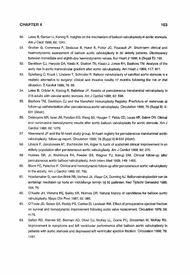
CHAPTER 4 103
50. Letac B, Gerber Ll, Koning R. Insights on the mechanism of balloon valvuloplasty in aortic stenosis.
Am J Card 1988; 62: 1241.
51. Grollier G, Commeau P, Sesboue B, Huret B, Potier JC, Foucault JP. Short-term clinical and
haemodynamic assessment of balloon aortic valvuloplasty in 30 elderly patients. Discrepancy
between immediate and eighth-day haemodynamic values. Eur Heart J 1988; 9 (SupplE): 155.
52. Davidson CJ, Harpole DA, Kisslo K, Skelton TN, Kisslo J, Jones RH, Bashore TM. Analysis of the
early rise in aortic transvalvular gradient after aortic valvuloplasty. Am Heart J 1989; 117: 411.
53. Spielberg C, Kruck I, Linderer T, Schroder R. Balloon valvuloplasty of calcified aortic stenosis is a
realistic alternative to surgery: clinical and invasive results 17 months following the 1st or 2nd
dilatation. Z Kardiol1989; 78: 86.
54. Letac B, Cribier A, Koning R, Bellefleur JP. Results of percutaneous transluminal valvuloplasty in
218 adults with valvular aortic stenosis. Am J Cardiol 1988; 62: 598.
55. Bashore TM, Davidson CJ and the Mansfield Valvuloplasty Registry. Predictors of restenosis at
follow-up catheterization after percutaneous aortic valvuloplasty. Circulation 1988; 78 (Supplll): 11-
531 (Abstr).
56. Desnoyers MR, Isner JM, Pandian NG, Wang SS, Hougen T, Fields CD, Lucas AR, Salem DN. Oinical
and noninvasive hemodynamic results after aortic balloon valvuloplasty for aortic stenosis. Am J
Cardiol1988; 62: 1078.
57. Kleaveland JP and the M-heart study group. M-heart registry for percutaneous transluminal aortic
valvuloplasty: follow-up report. Circulation 1988; 78 (Supplll):ll-533 (Abstr).
58. Litvack F, Jakubowski AT, Buchbinder NA, Eigler N. Lack of sustained clinical improvement in an
elderly population after percutaneous aortic valvuloplasty. Am J Cardiol1988; 62: 270.
59. Holmes DR, Jr, Nishimura RA, Reeder GS, Wagner PJ, llstrup OM. Clinical follow-up after
percutaneous aortic balloon valvuloplasty. Arch Intern Med 1989; 149: 1405.
60. Block PC, Palacios IF. Clinical and hemodynamic follow-up after percutaneous aortic valvuloplasty
in the elderly. Am J Cardiol1988; 62: 760.
61. Hoedemaker G, van den Brink RB, Verheul JA, Visser CA, Dunning AJ. Ballonvalvuloplastiek van de
aortaklep: resultaten op korte en middellange termijn bij 50 patienten. Ned Tijdschr Geneeskd 1990;
134:76.
62. O'Keefe JH, Vlietstra RE, Bailey KR, Holmes DR. Natural history of candidates for balloon aortic
valvuloplasty. Mayo Clin Proc 1987; 62:986.
63. O'Toole JD, Geiser EA, Reddy PS, Curtiss El, Landeair RM. Effect of preoperative ejection fraction
on suNival and hemodynamic improvement following aortic valve replacement. Circulation 1978; 58:
1175.
64. Safian RD, Warren SE, Berman AD, Diver OJ, McKay LL, Come PC, Grossman W, McKay RG.
Improvement in symptoms and left ventricular performance after balloon aortic valvuloplasty in
patients with aortic stenosis and depressed left ventricular ejection fraction. Circulation 1988; 78:
1181.

104 PERCUTANEOUS BALLOON AORTIC VALVULOPLASTY
65. Berland J, Cribier A, Savin T, Lefebvre E, Koning R, Letac B. Percutaneous balloon valvuloplasty in
patients with severe aortic stenosis and low ejection fraction. Immediate results and 1 year follow
up. Circulation 1989; 79: 1189.
66. Brady ST, Davis CA, Kussmaul WG, Laskey WK, Hirshfeld JW, Jr, Herrmann HC. Percutaneous aortic
balloon valvuloplasty in octogenarians: morbidity and mortality. Ann Intern Med 1989; 110: 761.
67. Letac B, Cribier A, Koning R, Lefebvre E. Aortic stenosis in elderly patients, aged 80 or older.
Treatment by percutaneous balloon valvuloplasty in a series of 92 cases. Circulation 1989: 80: 1514.
68. Levine MJ, Berman AD, Safian RD, Diver DJ, McKay RG. Balliation of valvular aortic stenosis by
balloon valvuloplasty as preoperative preparation for non' cardiac surgery. Am J Cardiol1988; 62:
1309.
69. Roth RB, Palacios I, Block PC. Percutaneous aortic balloon valvuloplasty: its role in the management \
of patients with aortic stenosis requiring major non cardiac surgery. JAm Coli Cardiol 1989; 13:
1039.
70. Angel JL. Chapman C, Knuppel RA. Morales WJ, Sims CJ. Percutaneous balloon aortic valvuloplasty
in pregnancy. Obstet Gynecol 1988; 72: 438.
71. Larzelere HB, Bailey CP. New intrument for cardiac valvular commissurotomy. J Thorac Surg 1953;
25:78.
72. Robiscek F, Harbold NB. Limited value of balloon dilatation in calcified aortic stenosis in adults:
direct observations during open heart surgery. Am J Cardiol1987; 60: 857.

CHAPTERS
EVALUATION OF PERCUTANEOUS BALLOON AORTIC
VALVULOPLASTY IN ELDERLY PATIENTS WITH DOPPLER
ECHOCARDIOGRAPHY
(This chapter has been accepted in a slightly different format for publication in the
Netherlands Journal of Cardiology, 1990. Authors are: van den Brand M, Bellerse L,
Vletter WB, Plante S, Serruys PW, de Feyter PJ, Suryapranata H, Roelandt JR)
SUMMARY
From March 1986through December 1989,38 patients underwent39 percutaneous
balloon aortic valvuloplasty (PBA V) procedures, for isolated severe aortic valve stenosis.
Mean age of these patients was 75 years, range 63-88. Nineteen of these patients were
considered to be inoperable either due to cardiac or extracardiac conditions. Five
patients were classified as high risk cases (age over 80 years), while the remaining 14,
although presenting with several operative risk increasing factors, were acceptable
candidates for aortic valve replacement. Aortic valve area calculated by the Gorlin
formula increased, from 0.47 (± 0.14) to 0.75 (± 0.19) cm2 after PBAV. Four patients died
in hospital, eight patients died out of hospital 5-23 months after valvuloplasty (mean 15
± 5 months). Sixteen patients underwent aortic valve replacement 6 days to 36 months
after valvuloplasty (mean 13 ± 9 months), while 11 patients are still without events at a
mean follow-up period of 23 ± 12 months.
Thirty-one patients had maximal aortic valve flow velocity measured with echo Doppler
techniques both immediate before and 24-48 hours after valvuloplasty.
Conversion of this maximal velocity to millimeters mercury and comparing these
values with the pressure difference between peak left ventricular and peak aortic
pressure, when estimated hemodynamically, showed poor correlations both before and
after valvuloplasty (r = 0.49 and 0.55 respectively). The Doppler technique tended to
overestimate aortic valve pressure drops over 74 and 25 mm Hg, when
hemodynamically derived, and to underestimate pressure drops below that value
respectively before and after PBAV. After valvuloplasty the maximal velocity over the
aortic valve was significantly reduced from 4.6 ± 0.7 mjsec to 4.1 ± 0.8 mjsec.

106 EVALUATION OF PERCUTANEOUS BALLOON AORTIC VALVULOPLASTY
Twenty two patients had immediate Doppler evaluation after valvuloplasty as well as
at a mean follow-up time of 10 ± 7 months. Maximal velocity over the aortic valve
increased in that period from 4.1 to 4.4 mjsec (not significant), with the highest increase
in velocity in the group of patients dying late, 4.0 to 4.5 mjsec (not significant).
Comparing catheter derived aortic valve areas calculated by the Gorlin formula directly
post PBAV, with echo Doppler derived areas calculated with the continuity equation at
follow-up, showed a good correlation in 6 patients with persisting clinical improvement
(r = 0.94).
It is concluded that the echo Doppler derived aortic valve areas, show a better
correlation with hemodynamically derived aortic valve areas, than maximal aortic valve
flow velocities with peak to peak pressure aortic valve gradients. Although both
techniques have their inherent limitations, the outcome of both estimations for aortic
valve area is very similar.
INTRODUCTION
Percutaneous balloon aortic valvuloplasty is recently being performed with increasing
frequency for the relief of severe calcified aortic stenosis in selected elderly patients.
Most investigators now believe that aortic valvuloplasty is a palliative procedure, that
should be limited to patients who are not surgical candidates for whatever reason (1-
5). In patients with severe congestive heart failure, balloon aortic valvuloplasty may
improve the patient's condition, as a bridge to later aortic valve replacement.
The procedure can produce moderate increases in aortic valve area, generally from
0.4-0.6 before to 0.7 to 0.9 cm2 after valvuloplasty and can be performed with an
acceptable periprocedural risk (1 ,2).
Unfortunately, the mortality of these patients remains high after the procedure and
mortality rates of 15 to 28% after 6 to 9 months have been reported (1 ,2). This high
mortality is probably caused by restenosis, whose rate has been shown to be high both
after invasive and non invasive assessment (6,7). Doppler echocardiography is a
noninvasive method, especially convenient for elderly people with a low validity class, the
population mostly referred for valvuloplasty. Symptomatic improvement and a non
invasively measured reduction in the severity of aortic valve stenosis might indicate a
persisting good result (8).

CHAPTERS 107
Previous studies using Doppler echocardiography have shown a fair or good
correlation between pre and post valvuloplasty measured aortic valve areas, with
catheterization data using the Gorlin formula (7,9-15).
We studied the correlation between the drop in pressure across the stenotic aortic
valve as well as valve area, measured during catheterization and estimated by Doppler
echocardiography technique before and after aortic valvuloplasty. Long-term follow-up
Doppler measurements were performed to correlate changes in maximal valve flow
velocity with clinical events, mortality, aortic valve replacement, and functional status.
PATIENTS AND METHODS
From March 1986 through December 1989, 38 patients underwent 39 aortic
valvuloplasty procedures. All patients were diagnosed as having severe aortic valve
stenosis by physical examination, Doppler echocardiography and heart catheterization.
All patients were discussed with the cardia-surgical team. Patients were acceptable
candidates for a balloon valvuloplasty procedure if they fulfilled one of the following
criteria:
a) deemed inoperable in a combined cardia-surgical discussion
b) classified as a high risk case
c) classified as a normal risk patient, but with preference of the patient for balloon
valvuloplasty instead of surgical valve replacement or surgical valvuloplasty.
All patients and a relative had to give their informed consent, after explanation of the
experimental character of the procedure, and the potential risks and benefits. No patient
was acceptable if during diagnostic angiography aortic insufficiency more severe than
grade II was demonstrated.

108 EVALUATION OF PERCUTANEOUS BALLOON AORTIC VALVULOPLASTY
Clinical characteristics and the cardiac condition of these patients before valvuloplasty
are summarized in tables 1 and 2.
Nr of Patients
Female sex
Mean age (years)
Inoperable
High risk case
Acceptable risk case
Validity class IV
Validity class Ill
39
24
75.1 ± 7.1
20
5
14
14
25
Table 1 Clinical characteristics of balloon aortic valvuloplasty patients
EF>0.55
EF >0.40- 0.54
>0.20 -0.39
<0.19
not assessed
Aortic incompetence
grade 0
grade I
grade II
Coronary artery disease
Absent
1 vessel disease
2 vessel disease
3 vessel disease
left main disease
Number of patients
22
6
5
4
2
10
20
9
24
7
2
5
Table2 Ejection fraction, severity of aortic incompetence and coronary artery disease in 39
valvuloplasty patients

CHAPTERS 109
Table 3 summarizes the hemodynamic data of these patients before and after balloon
aortic valvuloplasty.
LVPSP LVEDP P-PSG MSG AF AVA %Increase
B A B A B A B A B A B A AVA
Mean 217 177 16 12 82 41 74 43 176 214 0.47 0.75 63
.±.SO 37 34 9 9 24 15 19 10 50 49 0.14 0.19 38
p value <0,05 <0.05 <0,05 <0,05 <0,05 <0,05
AVA= aortic valve area (cm2); AF =systolic aortic valve flow (ml s-1); LVEDP =left ventricular end-diastolic pressure (mmHg);
LVPSP = left ventricular peak-systolic pressure (mmHg); MSG = mean systolic aortic gradient (mmHg); P-PSG = peak to peak
systolic gradient (mmHg); B = before; A = after.
Table3 Hemodynamic data before and after 39 balloon aortic valvuloplasty procedures.
The direct result of balloon aortic valvuloplasty, and the clinical follow-up of these
patients are combined in table 4.
Survivors operated
(n = 16)
Survivors not operated
(n = 11)
late deaths
(n = 8)
Early deaths
(n = 4)
Age M/F
(years) ratio
72.1:!: 5.6 6/10
76.7:!: 6.6 4/7
78.7:!: 9.5 3/5
76.2:!: 1.5 2/2
Follow-up time Aortic valve area (cm2) Aortic valve area Validity class
(months) Before After Increase (%) Before Ill IV
34:!: 8 0.51:!: 0.11 0.75:!: 0.15 52:!: 32 12 4
23:!: 12 0.42:!: 0.11 0.73:!: 0.19 77:!: 31 7 4
15:!: 5 o.4g:!: 0.18 0.76:!: 0.23 67:!: 48 6 2
2:!: 3 days 0.42:!: 0.12 0 4
Table 4 Clinical and hemodynamic data at the time of percutaneous balloon aortic valvuloplasty in
patients with early and late mortality and in survivors, with and without aortic valve
replacement. Data are given as mean values ± standard deviation
Doppler echocardiography was performed in 34 patients within two weeks before and
in 35 patients 24-48 hours after balloon aortic valvuloplasty. 1-27 months after
valvuloplasty 24 patients were re-investigated (mean 10 ± 7 months).
Patients were examined in the supine left lateral position. The continuous wave Doppler

110 EVALUATION OF PERCUTANEOUS BALLOON AORTIC VALVULOPLASTY
examination was performed using a Toshiba SSH160 or a Diasonics Vingmed ultrasound
unit. Peak aortic valve blood flow velocity was obtained by a non imaging continuous
wave Doppler probe, placed in various positions to obtain apical right, left parasternal
and suprasternal views. Spectral velocity traces were recorded on strip charts at a speed
of 50 mmjs.
The maximal instantaneous transvalvular pressure difference was estimated from the
maximal aortic Doppler velocity by the modified Bernoulli equation:
Pressure difference = 4 x max. velocity2
Aortic valve area (AVA) was calculated from two dimensional echo and Doppler data
with the continuity equation as follows:
AVA= ALVOTx (TVI) LVOT, (11) ------------------------------
(TV I) AV
where AV = aortic valve, ALVOT = area of left ventricular outflow tract in cm2, AVA =
aortic valve area in cm2, LVOT = left ventricular outflow tract and TVI =time velocity
integral. Time velocity integrals were obtained by integrating the respective Doppler
velocity spectra on a digitizing tablet, connected to an Olivetti c·omputer. Aortic valve
area was calculated before and after valvuloplasty in one patient and in six other patients
after valvuloplasty using echo and Doppler data.
In two patients transoesophageal echocardiography was performed, in one during
valvuloplasty under general anaesthesia, and in one other patient before and after
valvuloplasty. The transoesophageal probe (5 MHz, single plane transverse transducer)
was inserted through the oropharynx into the oesophagus. Two dimensional images
were obtained in the standard transoesophageal echocardiographic planes.
During catheterization the aortic valve area was calculated using the Gorlin formula
(9).

CHAPTERS 111
RESULTS
Gradients and flow velocity
Comparison of the peak to peak systolic aortic valve pressure drop measured before
and after valvuloplasty by catheterization, and maximal velocity over the aortic valve
measured with Doppler, revealed poor correlations (fig 1 ).
CATHETERIZATION DERIVED PEAK TO PEAK 160 GRADIENT BEFORE (mmHG)
140
120
100
80
60
40
20
0
0
0
0
0
0 0
Cb 0 @
0 0
0 0
00 8
0 0 0
00 ° 0 0
0 0
y•40.1 • 0.46x r•0.49
CATHETERIZATION DERIVED PEAK TO PEAK 120 GRADIENT AFTER (mmHG)
100
80
60
40
20
0
y•16.0 • 0.35x r•0.55
0 0
~ 0 0 © 0
oL_~--~--~--~--~---L--_L--~ oL---_L----~--~----~----~--~
0 ~ ~ M M ~ ~ ~ ~ ECHO DERIVED GRADIENT BEFORE
(4x max velocity m/sec2)
Figure 1a
0 20 40 60 80 100
Figure 1b
ECHO DERIVED GRADIENT AFTER (4x max velocity m/sec2)
120
Figure 1: Regression analysis of catheter and echo Doppler derived pressure drop over the aortic valve,
(a) before and (b) after percutaneous balloon aortic valvuloplasty
This proved also to be true for a regression analysis of the change in pressure drop.
The maximal flow velocity over the aortic valve fell significantly after valvuloplasty from
4.6 ± 0.7 to 4.1 ± 0.8 mjsec (p < 0.05) (fig 2).
7.0
6.5 0
" 6.0 "' .... E
" 5.5 > iii > 5.0 0
<0: 4.5
~ "(3
4.0 0 Qj > X 3.5
"' ::; 3.0
2.5
0
mean • 4.6 s.d:!:0.7
I Before
mean • 4.1 mean • 4.1 s.d:!:0.8 s.d:!:0.7
I I After After
n-31 n•22
mean • 4.4 s.d:!:0.6
I
Late
Figure 2: Maximal flow velocity over the aortic valve, before and after balloon valvuloplasty and at late
follow-up. The decrease in flow velocity after valvuloplasty is significant (p < 0.05), while the
increase in flow velocity at late follow-up is not significant

112 EVALUATION OF PERCUTANEOUS BAllOON AORTIC VALVULOPLASTY
The highest aortic valve flow velocity after valvuloplasty was recorded in the group
with subsequent aortic valve replacement (fable 5). At a mean follow-up of 10 ± 7 months
an increase in aortic valve flow velocity to 4.4 ± 0.6 mjsec was recorded (fig 2, table 5).
This increase was not significant. The increase was on average highest in patients dying
late, and lowest in patients who underwent late aortic valve replacement, although in
none of the subgroups was this increase significant.
All patients AVR Late mortality Uneventful FU
(n = 39) (n = 16) (n = 8) (n = 11)
Examined (n = 31) (n = 16) (n = 7) (n = 6)
Max. Ao Velocity (mjsec)
before mean 4.6 * 4.7 * 4.6 * 4.9* PBAV ± SD 0.7 0.5 0.5 0.9 after mean 4.1 4.2 4.1 4.2 PBAV ± SD 0.8 0.8 0.5 0.7
(n = 22) (n = 11) (n = 5) (n = 6)
Interval mean 10 8 8 16 (months) ± SD 7 6 4 8
after mean 4.1 4.3 4.0 3.9 PBAV ± SD 0.7 0.8 0.4 0.3
late mean 4.4 4.3 4.5 4.2 FU ± SD 0.6 0.7 0.7 0.5
* p < 0.05
Table 5 Comparison between maximal Doppler measured aortic valve flow velocity before and after
PBAVand at follow-up in all patients, and in three different outcomes: valve replacement (AVR),
late mortality and uneventful follow-up (FU)
Prevalvuloplasty aortic valve flow velocities were also compared between groups
(Table 5). The values measured directly before and at late follow-up after valvuloplasty
were not significantly different in the total group, nor in any subgroup.
Echo Doppler estimated aortic valve area
The only patient with prevalvuloplasty estimated echo Doppler aortic valve area
showed an increase from 0.41 cm2 to 0.44 cm2 after valvuloplasty. If aortic valve area
before and after valvuloplasty was measured with pressure difference and cardiac output

CHAPTER 5 113
this increase was from 0.53 to 0.64 cm2. Six other patients had echo Doppler estimated
aortic valve areas at a mean follow-up of 13 months after valvuloplasty. Aortic valve area
varied from 0.47 to 1.22 (Table 6).
Case number 2 3 4 5 6
Catheter measured AVA 0.67 0.80 0.57 0.78 1.12 0.78
post PBAV (cm2)
Echo Doppler estimated AVA 0.75 0.96 0.47 0.85 1.22 0.96
at follow-up (cm2)
Follow-up period 22.5 21.0 12.5 9 6.5 5.5
(months)
Table 6 Echo Doppler estimated aortic valve area (AVA) at follow-up, and catheter measured
aortic valve area directly after valvuloplasty
Only patient nr 4 underwent aortic valve replacement, 4.5 months after this
measurement, because of recurrent symptoms of angina pectoris. This patient had
undergone coronary angioplasty for two vessel disease as well as aortic valvuloplasty.
Regression analysis of echo Doppler estimated aortic valve areas at follow-up with aortic
valve areas calculated from hemodynamic data at the end of the valvuloplasty procedure
shows that AVA (cath) = 0.18 + 0.70 AVA (echo). There was a good correlation
between both values, with a r value of 0.94. Below a hemodynamic derived AVA of 0.60
cm2, echo Doppler measurements underestimated aortic valve area, while above a
hemodynamic derived AVA of 0.60 cm2, echo Doppler tended to overestimate the AVA.

114 EVALUATION OF PERCUTANEOUS BALLOON AORTIC VALVULOPLASTY
Transoesophageal echocardiography
In two patients transoesophageal echocardiograms were performed. In the first
patient a tricuspid heavily calcified aortic valve with very limited cusp mobility was
documented before valvuloplasty. After valvuloplasty an improved mobility of
predominantly the non coronary cusp was seen. Five months later, after recurrence of
symptoms, the mobility of the non coronary cusp was similar to prevalvuloplasty. This
patient subsequently underwent valve replacement. In the other patient
transoesophageal echocardiographic monitoring was performed under general
anaesthesia during valvuloplasty. A heavily calcified tricuspid aortic valve, with a big
calcified lump in the left coronary cusp was present and valve opening was solely
confined to the non coronary cusp. After valvuloplasty no apparent difference in cusp
mobility was noted. This patient showed an increase in echo Doppler estimated aortic
valve area from 0.4; cm2 to 0.44 cm2 and in hemodynamic derived aortic valve area
from 0.53 to 0.64 cm2. These values are in agreement with the minimal changes
observed during transoesphageal echocardiography.
DISCUSSION
Transvalvular pressure gradients
Several studies have shown the usefulness of Doppler echocardiography in assessing
the systolic transvalvular pressure drop in aortic stenosis (iO, 16-28). Although the
systolic pressure gradient is an important determinant of aortic valve area, transvalvular
flow is even more important. In our study a poor correlation was found between maximal
Doppler estimated flow velocity and peak to peak systolic gradients measured at
catheterization, both before and after aortic valvuloplasty. Doppler measured maximal
instantaneous systolic transvalvular pressure differences before valvuloplasty were on
average 7 mm Hg higher than catheter derived peak to peak systolic gradients. After
aortic valve dilatation the mean difference between the two methods increased to 29 mm
Hg, with again the higher values for Doppler derived pressure differences. Danielsen (29)
comparing maximal Doppler and maximal catheter derived pressure differences, found
these gradients both to be significantly higher than peak to peak systolic gradients,
recorded during catheterization. Defined this way the Doppler technique systematically
underestimated the maximum catheter derived pressure difference (29).

CHAPTERS 115
Both Doppler and catheterization measurements have several sources of inaccuracy
for deriving the pressure drop over a stenotic valve. For catheterization derived values,
the acquisition mode is very important. Calculations from left ventricle to aorta pull back
tracings, can be misleading in failing to achieve a good superimposition of left ventricular
and aortic pressure recordings (30). This is more the case if any irregularity of the heart
rhythm exists. Also measuring pressure differences with tipmanometered catheters
instead of fluid filled catheters might result in different values, because fluid filled
catheters overestimate especially the left ventricular pressure, and lead to an
overestimation of the catheter derived instantaneous maximal pressure difference.
Danielsen (29) for example calculated in 80% of his patients pressure drops across
the aortic valve from pull back tracings, while we always used simultaneous recorded
pressures, derived mostly from fluid filled catheters. Introducing a catheter in the left
ventricle through the aortic valve further reduces the aortic orifice. An 8F catheter for
example occupies an area of 3.14 x (4F)2 = 5.6 mm2, reducing the actual aortic valve
area in severe aortic stenosis by about 1 0%. An additional variability in the catheter data
is introduced by the pressure recovery. In a region distal to an obstruction the pressure
gradually increases. Clark (31) using aortic stenosis models, demonstrated that pressure
recovery extended to 9 em downstream of the stenotic area. Pressure recovery is
determined by the flow velocity at the site of the obstruction, and the ratio between the
cross sectional flow area at the obstruction site, and that of the flow channel distal to the
obstruction. In order to obtain a fully recovered pressure, the catheter must be
positioned outside the turbulent area, which is at a variable distance from the stenotic
valve.
The application of the Bernoulli equation in its most simple form (pressure drop = 4
x (fluid velocity)2) has also several assumptions as put forward by Rijsterborgh and
Roelandt (32).
It is assumed that the conversion of potential energy into kinetic energy takes place
without energy losses. Also a uniform velocity distribution at the cross sectional flow
area in the obstruction is assumed, which might not be the case. A standard Doppler
instrument measures the maximum velocity at the site of the obstruction. It is
conceivable that especially after valvuloplasty, non uniform flow velocities across the
stenotic aortic valve exist, resulting in overestimation of actual velocities, by measuring
only maximal velocities. Also the energy losses may not be negligible and vary with the

116 EVALUATION OF PERCUTANEOUS BALLOON AORTIC VALVULOPLASTY
shape and size of the obstruction, underestimating the pressure drop by Doppler
echocardiography. These two potential errors partly compensate for each other by
operating in opposite directions. The simplified Bernoulli equation will overestimate the
actual pressure drop in mild aortic stenosis and underestimate it in severe aortic stenosis
(32). This theory is confirmed by the data of several reports. Below a peak
catheterization pressure drop of 50 mm Hg, the simplified Bernoulli equation tends to
overestimate the pressure drop, while above a pressure difference of 50 mm Hg the
same equation underestimates the pressure difference obtained by catheter (16-28).
One technical aspect of measuring maximal velocities across the aortic valve by Doppler
echocardiography deserves attention. The Doppler beam must be derived as parallel as
possible to the blood flow jet across the stenotic valve (24,29). This requires the use of
a small non imaging transducer with multiple angulations from various positions.
Nonetheless experienced echocardiographers can assess accurately aortic valve
gradients (24). Finally the measured difference might well be real, because
measurements were not made simultaneously. In the post valvuloplasty situation
alterations in aortic valve flow might partly be responsible for the demonstrated
differences.
Aortic valve area calculations
Data from previous studies indicate that for a given, catheter calculated valve area,
a wide scatter of peak Doppler derived pressure gradients exists especially for mild to
moderate aortic stenosis (29). This scatter is the result of variation in transvalvular flow,
which is unknown. A peak aortic flow velocity of >4.5 mjsec for example was highly
specific for severe aortic stenosis, but had a low sensitivity. Half of the 70 patients
described by Oh with a peak aortic flow velocity of < 4.5 mjsec had severe aortic
stenosis (15), by hemodynamic measurements. These authors found good correlations
between catheter and Doppler derived mean gradients (r = 0.86; y = 0.89x + 2.3).
With use of the continuity equation, relating the flow velocity with the cross sectional
area of flow at a given flow volume, it was demonstrated in several studies that aortic
valve area can be reliably estimated (11-13). The same equation has been applied to
calculate aortic valve area both before and after aortic valvuloplasty (7, 14, 15). According
to these authors the Doppler continuity equation can accurately predict the severity of
aortic stenosis pre and post valvuloplasty. Pre valvuloplasty correlation coefficients

CHAPTERS 117
between catheter and Doppler derived aortic valve areas were 0.72, 0.71 and 0.80. Two
of these studies also correlated postvalvuloplasty valve areas and found correlation
coefficients of 0.61 and 0.85 respectively (7, 14).
It is rather remarkable that two techniques, each with their inherent sources of errors,
show such good correlations. Catheter measured aortic valve area, measured with aortic
valve flow and mean transvalvular pressure gradient, has also been questioned,
especially when assessing severe stenotic valves with low transvalvular flow rates
(33,34). During dobutamine and isoproterenol infusion, aortic valve areas calculated by
the Gorlin formula (9), increased from 0.67 to 0.81 cm2 and from 0.86 to 1.09 cm2
respectively. In one of these studies, aortic valve area was calculated during dobutamine
infusion in five patients simultaneously using the continuity equation. In these patients
calculated aortic valve area echo Doppler derived changed from 0.69 ± 0.23 cm2 to 0.71
± 0.21 cm2.
These findings suggest flow dependence of the Gorlin formula. Come et al recording
two sets of catheter derived aortic valve areas before valvuloplasty, found only a
moderate correlation between the two sets of values (r = 0.42) (35). Catheter derived
aortic valve area can therefore not be considered as the "gold standard" (32).
Although there was a time delay of several months in our patients in whom aortic valve
area was measured both with echo Doppler and with catheter derived methods, good
agreement existed between the values calculated in this way. As all patients mentioned
in table 6 were free of clinical symptoms of restenosis, we might assume that aortic
valve areas measured at the end of the valvuloplasty procedure did not change much
during the follow-up period. Moreover none of these patients showed an increase in
maximal aortic valve flow velocity, measured repeatedly during this follow-up period.
Transoesophageal echocardiography
One literature report adresses the usefulness of transoesophageal echocardiography
during aortic valvuloplasty (36). The potential advantages are: readily visible anatomy
of the stenotic aortic valve, monitoring of left ventricular function during and after
balloon inflation and assessment of aortic incompetence after dilatation.
Transoesophageal echocardiography can be of help in deciding whether further valve
dilatation with a larger balloon is warranted, evaluate directly the effect of balloon inflation
on valve cusp mobility and space balloon inflations till left ventricular function has

118 EVALUATION OF PERCUTANEOUS BALLOON AORTIC VALVULOPLASTY
returned to preinflation values.
We feel that in patients with symptomatic aortic valve stenosis, echo Doppler
evaluation with aortic valve area calculation is indicated. In surgical candidates,
presurgical evaluation should include coronary angiography. In non surgical candidates
coronary angiography and valvuloplasty could be performed in the same session. The
procedure is preferably performed under general anaesthesia while transoesophageal
echocardiography may become a major monitoring technique. After valvuloplasty,
follow-up investigations should include echo Doppler aortic valve area measurements
at regular intervals e.g. every three months. If recurrence of symptoms occurs and aortic
valve area measured this way returns to prevalvuloplasty values, the mentioned
investigation scheme should be repeated. In patients without symptoms, or with
moderate symptoms and with signs of restenosis documented objectively by calculating
aortic valve area with echo Doppler, a conservative attitude is justified, with more
frequent follow-up visits, because asymptomatic patients with moderate to severe aortic
stenosis are at relatively low risk for sudden death as recently documented by Kelly et
al (37). If complaints persist or recur in patients without objective signs of restenosis,
alternative explanations for the clinical deterioration should be sought before the patient
is subjected to recatheterization.

CHAPTERS 119
REFERENCES
1. Cribier A, Berland DJ, Koning R, Bellefleur JP, Mechmeche R, Letac B. Percutaneous transluminal
aortic valvuloplasty: indications and results in adult aortic stenosis. Eur Heart J 1988; 9 (Supple E):
149.
2. Safian RD, Berman AD, Diver DJ, McKay LL, Come PC, Riley MF, Warren SE, Cunningham MJ,
Wyman RM, Weinstein JS, Grossman W, McKay RG. Balloon aortic valvuloplasty in 170 consecutive
patients. N Engl J Med 1988; 319: 125.
3. Block PC, Palacios IF. Clinical and hemodynamic follow-up after percutaneous aortic valvuloplasty
in the elderly. Am J Cardiel 1988; 62: 760.
4. Letac B, Cribier A, Koning R, Lefebvre E. Aortic stenosis in elderly patients, aged 80 or older.
Treatment by percutaneous balloon valvuloplasty in a series of 92 cases. Circulation 1989; 80: 1514.
5. Cheitlin MD. Severe aortic stenosis in the sick octogenarian. A clear indication for balloon
valvuloplasty as the initial procedure. Circulation 1989; 80: 1906.
6. Grollier A, Commeau P, Sesboue B, Huret B, Potier JC, Foucault JP. Short-term clinical and
hemodynamic assessment of balloon aortic valvuloplasty in 30 elderly patients: Discrepancy between
immediate and eighth-day hemodynamic values. Eur Heart J 1988; 9 (supplE): 155.
7. Nishimura RA, Holmes DR, Jr, Reeder GS, Orszulak TA, Bresnahan JF, llstrup DM, Tajik AK Doppler
evaluation of results of percutaneous aortic balloon valvuloplasty in calcific aortic stenosis.
Circulation 1988; 78: 791.
8. Bernard Y, Bassand JB, Anguenot T, Schiele F, Habert C, Jacoulet P, Roche C, Maurat JP. Early
and late evaluation of percutaneous aortic valvuloplasty. A combined hemodynamic and Doppler
echocardiography study. JAm Coli Cardiol1988; 11: 14A (abstr).
9. Gorlin R, Gorlin SG. Hydraulic formula for calculation of the area of the stenotic mitral valve, other
cardiac valves, and central circulatory shunts. Am Heart J 1951; 41: 1.
10. Hatle L, Angelsen BA, Tromsdal A. Non-invasive assessment of aortic stenosis by Doppler
ultrasound. Br Heart J 1980; 43: 284.
11. Skjaerpe T, Hegrenaes L, Hatle L. Noninvasive estimation of valve area in patients with aortic stenosis
by Doppler ultrasound and two-dimensional echocardiography. Circulation 1985; 72: 810.
12. Zoghbi WA, Farmer KL, Soto JG, Nelson JG, Quinones MA. Accurate noninvasive quantification of
stenotic aortic valve area by Doppler echocardiography. Circulation 1986; 73: 452.
13. Teirstein P, Yeager M, Yock PG, Popp RL. Doppler echocardiographic measurement of aortic valve
area in aortic stenosis: a noninvasive application of the Gorlin formula. J Am Coli Cardiel 1986; 8:
1059.
14. Come PC, Riley MF, McKay RG, Safian R. Echocardiographic assessment of aortic valve area in
elderly patients with aortic stenosis and of changes in valve area after percutaneous balloon
valvuloplasty. J Am Coli Cardiol1987; 10: 115.

120 EVALUATION OF PERCUTANEOUS BALLOON AORTIC VALVULOPLASTY
15. Oh JK, Taliercio CP, Holmes DR, Jr, Reeder GS, Bailey KR, Seward JB, Tajik JA. Prediction of
severity of aortic stenosis by Doppler aortic valve area determination: prospective Doppler
catheterization correlation in 100 patients. J Am Coli Cardiel 1988; 11: 1227.
16. Stamm R, Martin RP. Quantification of pressure gradients across stenotic valves by Doppler
ultrasound. JAm Coli Cardiol1983; 2: 707.
17. Berger M, Berdorf RL, Galterstein PE, Goldberg E. Evaluation of aortic stenosis by continuous wave
Doppler ultrasound. JAm Coli Cardiol1984; 3: 150.
18. Stevenson JG, Kawabori I. Noninvasive determination of pressure gradients in children: two methods
employing pulsed Doppler echocardiography. JAm Coli Cardiol1984; 4: 179.
19. Kosturakis S, Allen HD, Goldberg SJ, Sahn DJ, Valdes - Cruz LM. Noninvasive quantification of
stenotic semilunar valve areas by Doppler echocardiography. J Am Coli Cardiel 1984; 3: 1256.
20. Krafchek J, Robertson JH, Radford M, Adams D, Kisslo J. A reconsideration of Doppler assessed
gradients in suspected aortic stenosis. Am Heart J 1985; 110: 765.
21. Agatson AS, Chengot M, Rao A, Hildner F, Samet P. Doppler diagnosis of valvular aortic stenosis
in patients over 60 years of age. Am J Cardiel 1985; 56: 106.
22. Hegrenaes L, Hatle L. Aortic stenosis in adults. Noninvasive estimation of pressure differences by
continuous wave Doppler echocardiography. Br Heart J 1985; 54: 396.
23. Simpson lA, Houston AB, Sheldon CD, Hutton I, Lawrie TDV. Clinical value of Doppler
echocardiography in the assessment of adults with aortic stenosis. Br Heart J 1985; 53: 636.
24. Currie PJ, Seward JB, Reeder GS, et al. Continuous-wave Doppler echocardiographic
assessment of severity of calcific aortic stenosis: a simultaneous Doppler-catheter correlative study
in 100 adult patients. Circulation 1985; 71: 1162.
25. Yeager M, Yock PG, Popp RL. Comparison of Doppler-<lerived pressure gradient to that determined
at cardiac catheterization in adutls with aortic valve stenosis: implications for management Am J
Cardiol1986; 57: 644.
26. Panidis IP, Mintz GS, Ross J. Value and limitations of Doppler ultrasound in the evaluation of aortic
stenosis: a statistical analysis of 70 consecutive patients. Am Heart J 1986; 112: 150.
27. Smith MD, Dawson PL, Elion JL, Wisenbaugh T, Kwan OL, Handshoe S, DeMaria AN. Systematic
correlation of continuous wave Doppler and hemodynamic measurements in patients with aortic
stenosis. Am Heart J 1986; 111: 245.
28. Zhang Y, Ihlen H, Nitter-Hauge S. Estimation of peak-to-peak pressure gradient in aortic stenosis
by Doppler echocardiography. lnt J Cardiel 1986; 10: 197.
29. Danielsen R, Nordrehaug JE, Stangeland L, Vik-Mo H. Limitations in assessing the severity of aortic
stenosis by Doppler gradients. Br Heart J 1988; 59: 551.
30. Grosman W. Cardiac catheterization and angiography. Philadelphia, Lea and Febiger, 197 4, p 94.
31. Clark C. The fluid mechanics of aortic stenosis: I. Theory and steady flow experiments. J
Biomechanics 1976; 9: 521.
32. Rijsterborgh H, Roeland! J. Doppler assessment of aortic stenosis: Bernoulli revisited. Ultrasound
Med Biol1987; 13: 241.

CHAPTERS 121
33. Casale PN, Palacios IF, Abascal VM, Choong CY, Block PC, Boucher CA, Weyman AE, Fifer MA.
Gorlin valve area varies with cardiac output in aortic stenosis. J Am Coli Cardiol 1988; 11: 63 A
(Abstr).
34. McCriskin JW, Herman RL, Spaccavento LJ, Tomlinson GC.Isoproterenol infusion increases Gorlin
fromula aortic valve area in isolated aortic stenosis. JAm Coli Cardiol1988; 11: 63 A (Abstr).
35. Come PC, Riley MF, Safian RD, Ferguson JF, Diver DJ, McKay RG. Usefulness of noninvasive
assessment of aortic stenosis before and after percutaneous aortic valvuloplasty. Am J Cardiol
1988; 61: 1300.
36. Cyran SE, Kimball TR, Schwartz DC, Meyer RA, Steed RD, Kaplan S. Evaluation of balloon aortic
valvuloplasty with transesophageal echocardiography. Am Heart J 1988; 115: 460.
37. KellyTA, Rothbart RM, Cooper CM, Kaiser DL, Smucker ML, Gibson RS. Comparison of outcome
of asymptomatic to symptomatic patients older than 20 years of age with valvular aortic stenosis.
Am J Cardiol 1988; 61 : 123.


CHAPTER 6
AORTIC VALVULOPLASTY OF CALCIFIC AORTIC STENOSIS WITH
MONOFOIL AND TREFOIL BALLOON CATHETERS: PRACTICAL
CONSIDERATIONS
(This chapter has been published in the lnt J Cardiac Imaging i990; 5:249. Authors
are: Plante S, van den Brand M, van Veen L, di Mario C, Essed CE, Beatt KJ, Serruys
PW)
SUMMARY
In order to evaluate the relation between balloon design (monofoil, trefoil) and
valvular configuration, experimental aortic valvuloplasty was performed in four post
mortem hearts with calcific aortic stenosis of various morphology. The degree of
obstruction of the aortic orifice was assessed by computed axial tomography during
inflation of monofoil i5 and i9 mm and trefoil 3 x i2 mm balloon catheters. We also
evaluated the hemodynamic repercussion of balloon inflation (fall in systolic aortic
pressure) in four elderly patients with acquired aortic stenosis who underwent a
percutaneous transluminal aortic balloon valvuloplasty, with stepwise increasing balloon
sizes of i5 mm, ;g mm and 3 x i2 mm, as during our in vitro experiments, and who
underwent aortic valve replacement later on. In these patients, we correlated the
anatomy of the excised aortic valves with the retrospective analysis of aortic pressure
curves recorded during previous valvuloplasty procedures.
Our experimental and clinicopathological observations showed that the degree of
obstruction of the aortic orifice in post -mortem specimens and the tolerance to balloon
inflation in live patients are dependent of the valvular configuration. Although trefoil
balloons have the theoretical advantage to avoid complete obstruction of the aortic
orifice during inflation, we observed that in presence of a tricuspid configuration, they
could be potentially more occlusive than monofoil balloons since each of the 3
individual components of the trefoil balloon occupied the intercommissural spaces
while inflated. However, they offered more residual free space when inflated in aortic
valves with a bicuspid configuration (i.e. congenitally bicuspid valves or tricuspid valves
with one fused commissure).

124 PRACTICAL CONSIDERATIONS
In our opinion, these observations are relevant, since degenerative disease of the
aortic valve (i.e. tricuspid valve without commissural fusion) is now recognized as the
most common etiology of aortic stenosis in the elderly.
INTRODUCTION
Percutaneous transluminal balloon aortic balloon valvuloplasty (PBAV) for acquired
aortic stenosis (AS) has been introduced a few years ago as an alternative for valve
replacement. in patients with high surgical risk or for those who refused surgical
intervention (1 ,2). Procedure-related complications, limited hemodynamic improvement
and the high recurrence rate reported until now (3-16) have decreased the initial
enthusiasm for this relatively new technique.
Despite these current limitations, PBAV is currently regarded by many cardiologists
as a palliative treatment in a subset of patients with prohibitive surgical risk. Some
authors recently advocated PBAV as a "bridge" procedure to improve left ventricular
function before aortic valve replacement or non-cardiac surgery (17, 18).
However, other shortcomings of the procedure are 1) the largely unpredictable
individual tolerance to balloon inflation (hypotension) and 2) the unpredictable
immediate hemodynamic results in an individual patient. The incidence of non
responding or poorly responding patients (aortic valve area still < 0.7 cm2 at the end
of the procedure) is ranging from 18.7% to 38.2% (12). These facts suggest that some
morphological aspects (bicuspid or tricuspid valve, fused or non-fused commissures)
or other factors like the volume occupied by nodular calcifications and residual
elasticity of some leaflets (19) can adversely influence the hemodynamic results of the
procedure. Furthermore, it seems conceivable that balloon catheters of different
shapes (monofoil, bifoil, trefoil) operate with distinct mechanisms of dilatation in the
presence of a specific valvular morphology.
To circumvent hypotensive responses during balloon inflation, trefoil balloon
catheters have been introduced recently. Attractive advantages of this device have
been demonstrated in animal experiments (20) and it has been effective in allowing
continuous blood flow during inflation in the setting of pulmonary valvuloplasty in adults
(21), aortic valvuloplasty in children and young adults (22), and in mitral and aortic
valvuloplasty in adults (23-24). However, the degenerative, remodeling and calcifying

CHAPTER 6 125
process peculiar to acquired AS in elderly patients differs largely from the pathological
changes observed in congenital and rheumatic valvular diseases; this may explain why
some of these patients still present abrupt and severe falls in aortic pressure, even
when PBAV is performed with trefoil balloon catheters.
In order to achieve better hemodynamic results and to obtain technical refinements
in the valvuloplasty material, we have to improve our knowledge of the operational
mechanisms of dilatation. Intra-operative, post-mortem and experimental studies on the
mechanisms of aortic valvuloplasty have been reported until now, based mainly on
visual inspection and low-energy x-ray techniques (19,25-27). In order to obtain
detailed quantitative information on the intravalvular calcium distribution before and
after valvuloplasty and to further elucidate the operational mechanisms of dilatation, we
have performed thin-slice computed tomographic scanning in post-mortem hearts with
calcific AS before, during and after balloon valvuloplasty (28). This technique avoided
superimposition of structures and provided vastly superior density resolution compared
to conventional x-ray techniques. In our study, fracture of nodular calcifications,
commissural separation and central raphe splitting were the observed mechanisms of
balloon valvuloplasty. However, calcium redistribution (27) could not be recognized as
an operational mechanism, since no dilatation-related modification in density and
volume of the calcific deposits was observed with computed tomographic scanning.
Furthermore, computed axial tomography provided useful informations concerning the
relations between balloon size and shape and valvular configuration. To our
knowledge, few reports have focused on this particular aspect of aortic balloon
valvuloplasty.
In this chapter, we describe our experimental and clinicopathological observations
related to aortic valvuloplasty performed with single circular (monofoil) and trefoil
balloon catheters in human hearts with acquired AS. We will present a review of our
previous experimental findings and we will correlate these results with our recent
clinicopathological observations, based on retrospective analysis of valvuloplasty
procedures performed in some patients who underwent subsequently a valve
replacement and from whom excised aortic valves were available for
anatomopathological evaluation. For more detailed information concerning our in vitro
experiments, the reader is referred to our previous publication (28).

126 PRACTICAL CONSIDERATIONS
EXPERIMENTAL VALVULOPLASTY
Material and methods
Four hearts with calcific, stenotic aortic valves and intact aortic rings were selected
from our collection of autopsy specimens (Pathology Department 1, Erasmus
University). The mean heart weight was 583 .±. 78 g (range: 538-700 g). The average
thickness of the left ventricular wall was 19.5 .±. 1.9 mm.
Case 1: This specimen was obtained from a 67 year-old female who died suddenly.
The aortic valve was tricuspid, with extensive nodular calcifications of the valve cusps
projecting into the valve sinuses, but without commissural fusion. These findings were
compatible with a degenerative etiology.
Case 2: The second specimen came from a 71 year-old male patient in whom a pre
mortem cardiac catheterization had shown a mean systolic aortic gradient and a valve
area of 70 mm Hg and 0.6 cm2, respectively. Mild aortic regurgitation, moderate
combined mitral valve disease and subocclusive stenoses of the left anterior
descending and circumflex arteries were also present. This patient died after having
sustained a large anterior myocardial infarction. At pathological examination, the
coexisting mitral valve disease and the tight, heavily calcified commissural fusion
between non-coronary and right coronary aortic cusps were in accordance with a
rheumatic etiology. Although originally tricuspid, the aortic valve had a bicuspid
configuration, owing to the tightly fused commissure. Nodular calcifications on the
aortic aspects of the cusps were also observed.
Case 3: Irreversible cardiogenic shock occurred in this 73 year-old male patient, as
a consequence of a posterior myocardial infarction. Post-mortem examination revealed
a congenitally bicuspid aortic valve. The larger anterior cusp was divided by a central
calcified raphe, extending across the floor of the aortic sinus from aorta to the free
edge. The valve had moderate nodular calcifications, but no commissural fusion.
Case 4: The fourth heart was obtained from a 50 year-old female in whom a
previous catheterization had demonstrated a mean systolic aortic gradient of 77 mm
Hg and a calculated valve area of 0.7 cm2. This patient, while awaiting surgical
correction, developed fatal acute pulmonary edema. At autopsy, the valve was also
congenitally bicuspid, but with an almost symmetric configuration, a moderate bilateral
commissural fusion, and diffuse calcifications confined to the leaflets. In the anterior

CHAPTER 6 127
cusp, a calcified central raphe was also observed.
Specimens were originally preserved in a 10% formalin solution but were kept in
water for 30 days before the experiment. Cylindrical calipers of increasing diameter
(range: 3-30 mm) were used to assess the aortic orifice area, while the aortic annulus
diameter was measured directly. All specimens were examined on a Tomoscan 350
clinical unit (Philips, Phillips Medical Systems, Best, Netherlands). Three radiopaque
markers were sutured to the aortic wall at the leaflet level and used as reference points
in the scanning procedures. Axial scan series, covering the entire aortic valve region,
were performed with contiguous scans of 1.5 mm thickness. Off-line evaluation was
performed on a Philips Stand Alone Viewing System, enabling sagittal reconstruction,
contour detection of the region of interest, and calculation of volumes and mean
computed tomographic attenuation values (Hounsfield Units) of calcium deposits. In
all specimens the region of interest contour mode was used to determine the residual
orifice area during each inflation. Evaluation of all the axial scans was used to assess
the minimal free space between leaflets and balloon.
Post-mortem aortic valvuloplasty was performed with balloons of increasing
diameter, passed retrogradely from the aorta into the left ventricle. First, a monofoil 15
mm balloon (inflated cross-sectional area: 1.8 cm2) was manually inflated across the
aortic valve with a pressure of 3 atmospheres. Thereafter, a second and third dilatation
were performed with a monofoil 19 mm (area: 2.8 cm2) and a trefoil 3 x 12 mm (3.4
cm2) balloon catheter, both inflated to a pressure of 3 or 4 atmospheres. Balloons
were inflated until complete disappearance of indentation of the valves on the balloons.
Leaflet avulsion, valve ring disruption or liberation of valvular debris did not occur in
any specimen. Experimental valvuloplasty with bifoil-designed balloon catheters was
not assessed in this study, since they were not commercially available at that time in
our institution.
Measurement of the aortic valve orifice by cylindrical calipers was performed at the
beginning and after each dilatation. Visual inspection, photographs and axial computed
tomographic scans were obtained before, during and after each balloon inflation.
Figure 1 provides an example of computed axial tomography scans obtained at the
level of the aortic valve orifice during experimental valvuloplasty in a post-mortem
specimen.

128
Figure 1A
Figure 18
PRACTICAL CONSIDERATIONS
Figure 1: Axial scans of a post-mortem specimen (case
#1) performed at the level of the aortic valve orifice,
during inflation of 15 mm, 19 mm and trefoil 3 x 12 mm
balloons (enlargement factor: 4 X). This valve was the
most heavily calcified. The density is decreasing from
white areas (calcium deposits, catheters, radiopaque
markers), to the grey (heart and valve soft tissue, water
in inflated balloons), and the black areas (air). During
inflation, the 15 mm balloon was displaced towards the
less calcified left aortic cusp (Fig.1 A). Minimal increment
in valve area was achieved. The inflated 19 mm balloon
occupied also an asymmetrical position in the aortic
orifice, but the displacement of the free edges of the
cusps was greater than with the previous dilatation,
involving also the most heavily calcified leaflet (Fig.1 B).
Fracture of nodular calcifications was noted at visual
inspection. During inflation of the trefoil balloon (Fig.1 C),
a rotating movement was observed. At the end of
inflation, the three intercommissural spaces were
occupied by the 3 individual components of the balloon,
almost totally occluding the aortic orifice. At visual
inspection, the increase in cusps mobility was
associated with full-thickness fracture of the heavy
nodular calcifications. L: left; R: right.
Figure 1C

CHAPTER 6 129
Results and comments
Table 1 summarizes clinical data and macroscopic aspects of the four valves. Volumes and mean
computed tomographic attenuation values of the calcific deposits are also tabulated.
Calcium Deposits
Valve Ring
No Commissural Diameter Volume CT Value
Case Sex Age Cusps Fusion (mm) (cm3) (HU)
F 67 3 No 24 1.21 948
2 M 71 3 Yes 24 0.29 132
3 M 73 2 No 22 0.19 281
4 F 50 2 Yes 20 0.17 121
CT value = computed tomographic attenuation value; HU = Hounsfield Units; F = female; M = male
Table 1 Clinical data, macroscopic aspects and computed tomographic evaluation of valve
calcium deposits of the four post-mortem specimens.
Table 2 shows the aortic valve area (AVA) obtained before and after dilatation with
15 mm, 19 mm and 3 x 12 mm balloons in each case, as calculated by cylindrical
calipers.
Balloon Size
BASAL
15 mm
19 mm
3 x 12 mm
Table 2
Case 1 Case 2 Case 3 Case 4
0.20 0.78 0.95 0.95
0.28 1.76 0.95 0.95
0.50 2.26 1.76 1.76
0.95 3.14 2.83 2.54
Aortic valve area (cm2) before experimental valvuloplasty and after dilatation with 15
mm, 19 mm and 3 x 12 mm balloons, as calculated by cylindrical calipers.

130 PRACTICAL CONSIDERATIONS
Figure 2 represents the minimal free space (residual aortic orifice) observed during
inflation, as determined by the region of interest contour mode.
Residual Aortic Orifice ( cm2 ) 1.00~----------,-----------,----------,-----------,
0.90
0.80
0.70
0.60
0.50
0.40
0.30
0.20
0.10
TRICUSPID
CONFIGURATION
0.00 --L..L.~~'fLL" Case 1 Case 2
BICUSPID
CONFIGURATION
Case 3
congenitally bicuspid
with partially fused
commissures
Case 4
0 16 mm ~ 19 mm - 3 x 12 mm
BALLOON TYPE
Figure 2: Computed tomographic evaluation of the residual aortic orifice (cm2) during inflation of 15
mm, 19 mm and 3 x 12 mm balloons in the four post-mortem specimens, as determined by the region
of interest contour mode. In cases where a bicuspid configuration was present (cases 2, 3 and 4), the
trefoil 3 x 12 mm balloon allowed more free space between balloon and leaflets, while the monofoil 15
mm and 19 mm balloons were both more occlusive, despite their smaller cross-sectional surfaces.
Conversely, when the valve had a tricuspid configuration (case 1: tricuspid valve without fused
commissure), the trefoil balloon was much more occlusive (residual aortic orifice: 0.09 cm2) than
monofoil15 mm and 19 mm balloons (residual aortic orifice: 0.53 and 0.30 cm2, respectively).
During inflation of the 15 mm balloon, a residual free space between the balloon and
the aortic cusps of at least 0.30 cm2 was observed in cases 1 and 2. However, in
presence of a congenitally bicuspid valve (cases 3 and 4), balloon inflation resulted in
an almost complete obstruction of the aortic orifice (0.07 and 0.02 cm2, respectively).
No gross fracture of nodular calcifications nor commissural or raphe splitting were
observed in any specimen. A moderate increase in AVA occurred only in case 2, while
minimal or no changes were achieved in the remaining cases (Table 2). It should be
noted that in none of the specimens did the inflated 15 mm balloon occupy a central

CHAPTER 6 131
position in the aortic orifice. The stretching induced by the balloon was more important
on the less calcified, more flexible valve leaflets.
Concerning the 19 mm balloon, a significant increment in AVA was achieved in all
the specimens. Although the balloon still occupied an asymmetrical position in the
aortic orifice, larger displacement of the free edges of the cusps were observed,
involving the most heavily calcified leaflets. Except in case 1, which was a tricuspid
valve without commissural fusion, this balloon was almost occlusive in the remaining
cases. The better residual free space observed in case 1 (0.30 cm2) can be explained
by a wider opening of the leaflets, which in turn created an increment in
intercommissural spaces. Fracture of nodular calcifications were noted in cases 1 and
2 at visual inspection. Partial commissural separation or central raphe splitting occurred
in cases 2, 3 and 4.
Conversely, despite the larger transverse diameter and cross-sectional area of the
inflated 3 x 12 mm trefoil-shaped balloon, the residual orifice during inflation was
paradoxically greater than with previous dilatations in all cases except in case 1, due
presumably to the peripheral and central spaces generated by the trefoil configuration.
This advantage of the trefoil shape was already known, but we remarked that it was
mostly evident when a "bicuspid" configuration could be observed (case 2: tricuspid
valve with one fused commissure; cases 3 and 4: congenitally bicuspid valves).
However, in presence of a tricuspid morphology (case 1), the opposite effect was
observed. This may be explained by the rigidity of the leaflets related to the
degenerative process, which have oriented passively the individual parts of the inflating
trefoil balloon to engage each of the three intercommissural spaces.
CLINICOPATHOLOGICAL OBSERVATIONS
In order to compare the results of experimental valvuloplasty to the clinical situation,
we analysed retrospectively aortic pressure curves recorded during dilatation at the
time of PBAV in patients who underwent subsequently a valve replacement and we
have correlated these hemodynamic data with the anatomopathological evaluation of
the surgically excised aortic valves.

132 PRACTICAL CONSIDERATIONS
Methods
From March 1986 to October 1988, thirty-five elderly patients underwent PBAV in our
institution. The technique of the valvuloplasty procedure itself in our institution has
been described earlier by our group (10). Ten of these patients underwent
subsequently an aortic valve replacement (7 for restenosis, 2 for a failed procedure
and one patient for severe aortic regurgitation). In four of these surgical patients, a
stepwise dilatation with 15 mm, 19 mm and trefoil 3 x 12 mm balloons had been
performed, in the same sequence as during our in vitro experiments described above.
Two of these patients underwent surgery for failed PBAV, since increments in AVA
achieved by valvuloplasty were only minimal, without significant symptomatic
improvements. The two other patients underwent valve replacement for recurrence
of stenosis, defined as a clinical recurrence of symptoms with either a return of
maximal aortic valve flow velocity to pre-valvuloplasty values on echo Doppler flow
measurements, or an invasively measured AVA which was less than 25% larger than
before PBAV.
The hemodynamic repercussion of aortic orifice obstruction generated by balloon
inflation was assessed by the systolic aortic pressure, continuously recorded
throughout the procedure by a 7 F pigtail catheter positioned in the descending aorta.
Systolic aortic pressure values were obtained by reviewing the aortic pressure tracings
recorded on paper during the PBAV procedures. The pressure curves recorded during
dilatation were analyzed and the lowest systolic aortic pressure values (nadir) were
selected. Since the pressure scale on the recording paper grid was 4 mm Hg j mm,
the lowest systolic aortic pressure value was arbitrarily rounded to 5 mm Hg (for
example: 12 mm = 48 mm Hg; rounded to 50 mm Hg). In cases where more than one
inflation was performed with the same balloon, the pressure curve showing the lowest
systolic aortic pressure value was selected. Figure 3 gives an example of the aortic
pressure curves recorded during a valvuloplasty procedure.
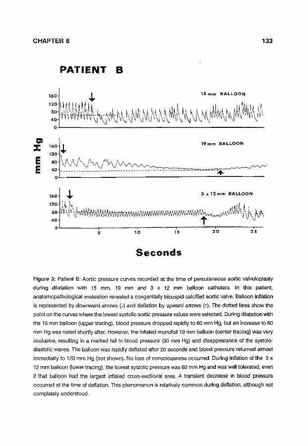
CHAPTER 6 133
PATIENT B
160 + 15 mm BALLOON
'!: V\~~IAM~~~~~~ij\ 0~-----------------------------------------------------
en ::r:: 160 + 120
19 mm BALLOON
E 80
E 40 ---. 0
5 10 15 20 25
Seconds
Figure 3: Patient B: Aortic pressure cuNes recorded at the time of percutaneous aortic valvuloplasty
during dilatation with 15 mm, 19 mm and 3 x 12 mm balloon catheters. In this patient,
anatomopathological evaluation revealed a congenitally bicuspid calcified aortic valve. Balloon inflation
is represented by downward arrows (•) and deflation by upward arrows (r). The dotted lines show the
point on the cuNes where the lowest systolic aortic pressure values were selected. During dilatation with
the 15 mm balloon (upper tracing), blood pressure dropped rapidly to 60 mm Hg, but an increase to 80
mm Hg was noted shortly after. However, the inflated monofoil19 mm balloon (center tracing) was very
occlusive, resulting in a marked fall in blood pressure {30 mm Hg) and disappearance of the systolo
diastolic waves. The balloon was rapidly deflated after 20 seconds and blood pressure returned almost
immediatlyto 120 mm Hg (not shown). No loss of consciousness occurred. During inflation of the 3 x
12 mm balloon {lower tracing), the lowest systolic pressure was 80 mm Hg and was well tolerated, even
if that balloon had the largest inflated cross-sectional area. A transient decrease in blood pressure
occurred at the time of deflation. This phenomenon is relatively common during deflation, although not
completely understood.

134 PRACTICAL CONSIDERATIONS
Results and comments
Table 3 summarizes clinical data, pathological findings related to the excised valves
and hemodynamic data of the PBAV procedures.
C Age (years) L I N Interval* I (months) c A Reason for L Surgery
p Valve A Anatomy T H Commissural 0 Fusion L 0 Calcifications G y Tearing of
Leaflets
v A L Gradient v (mean) u L 0 p A.V~. L (em) A s T y
Patient A
65
F
T
3+
PRE 48
POST 41
PRE 0.5
POST 0.6
Patient B Patient C Patient D
73 64 74
9 9 9
F R R
B B T
+ **
3+ 2+ 3+
80 68 74
58 41 47
0.3 0.4 0.5
0.4 0.6 0.7
B - bicuspid;F - failed valvuloplasty; R = restenosis; T = tricuspid; A.V.A. = aortic valve area, calculated with the Gorlin's formula; PRE = before valvuloplasty;POST = after valvuloplasty
* Time elapsed between aortic valvuloplasty and valve replacement ** PBAV did not result in commissural separation in patient D
Table 3 clinical data, pathological findings related to the excised valves and hemodynamic data
obtained before and after PBAV, in patients who underwent valve replacement after
PBAV.

CHAPTER 6 135
Figure 4 illustrates the lowest systolic aortic pressure recorded in the four patients,
when the different balloons were fully inflated.
Systolic Aortic Pressure (mm Hg) 180~---------,,----------r~--------.----------.
TRICUSPID 160 CONFIGURATION
140
120
100
80
60
40
20
125
Patient A
congenitally bicuspid
Patient B
BICUSPID CO NFI GURATI ON
--------·-+---------------congenitally
bicuspid
Patient C
tricuspid with one fused
commissure
Patient D
D 15 mm ~ 19 mm - 3 x 12 mm
BALLOON TYPE
Figure 4: Lowest systolic aortic pressure values recorded during inflation of monofoil 15 mm, 19 mm
and trefoil 3 x 12 mm balloons in four patients who underwent elective percutaneous aortic valvuloplasty
1 to 10 months before valve replacement. In cases where more than one inflation was performed with
the same balloon catheter, the curve showing the lowest systolic pressure value was selected. In patients
in whom a bicuspid configuration was present (B and C: congenitally bicuspid valves; D: tricuspid valve
with one commissural fusion not separated by balloon valvuloplasty), the 19 mm balloon generated a
more dramatic fall in blood pressure and was less tolerated than the larger trefoil 3 x 12 mm balloon (30
vs 80 mm Hg, 25 vs 50 mm Hg, and 30 vs 60 mm Hg, respectively). Conversely, in patient A (tricuspid
valve without commissural fusion), the trefoil balloon offered no advantage compared to the 19 mm
balloon, as reflected by the slightly lower blood pressure observed during the 3 x 12 mm balloon
inflation.
The mean age was 69.0 years (range: 64-74 years) and the mean time interval
between PBAV and valve replacement was 7.0 months (range: i-9 months). Despite
the limited number of patients available, we noted that in patients in whom a bicuspid
valve configuration was present (patients Band C: congenitally bicuspid valves; patient
D: tricuspid valve with one fused commissure in which PBAV did not result in
commissural separation), dilatation with the single circular ;g mm balloon was less well

136 PRACTICAL CONSIDERATIONS
tolerated than with the larger 3 x 12 mm trefoil balloon, as revealed by the systolic
aortic pressure during inflation (Figure 4). These observations correlated well with our
experimental findings, which have shown that inflated trefoil-shaped 3 x 12 mm
balloons allowed more residual free space in presence of a bicuspid configuration.
Conversely, pathological examination of the aortic valve in patient A revealed a
calcified tricuspid valve without commissural fusion (i.e. tricuspid configuration), in
accordance with a degenerative etiology. In this case, dilatation with the 3 x 12 mm
balloon did not appear to offer any advantage, being as occlusive as the 19 mm
circular balloon (Figure 4). These findings do not confirm but suggest strongly that
intercommissural spaces may have been obstructed by each of the three individual
parts of the trefoil balloon, as observed in our in vitro experiments.
DISCUSSION
In contrast with other valvular heart diseases in which balloon valvuloplasty has been
applied, acquired AS in the elderly is characterized by a broad spectrum of valvular
morphology. We have assessed qualitatively and quantitatively the effects of dilatation
with different balloons in aortic valves of various morphology (bicuspid, tricuspid, fused
and non-fused commissures) and etiology (degenerative, congenital and rheumatic),
but the extrapolation of our experimental results to other cases with different anatomy
should be made with caution.
The increments in AVA achieved after experimental valvuloplasty were much greater
than those reported in clinical trials. It seems unlikely that this discrepancy was related
to the effects of formalin fixation. In fact, formalin preservation alters valvular fibrous
proteins and leads to stiffening of the valve leaflets and reduction of the elastic
properties of the aortic cusps, resulting in increased rigidity of the aortic valve leaflets.
Thus, it seems conceivable that smaller increments in AVA would have been obtained
after formalin fixation. However, it should be noted that previous reports (26) compared
the effects of balloon dilatation on 27 calcific, stenotic aortic valves dilated after variable
periods of fixation in formalin and on 12 non-preserved valves dilated either at post
mortem examination or in the operating room and observed no apparent difference in
results. In our experimental study, the greater increments in AVA, compared to those

CHAPTER 6 137
achieved in the clinical situation, are rather explained by: 1) easy and optimal
positioning of the balloon in a non-beating heart, 2) the possibility to perform
prolonged inflation time without creating adverse effects such as hypotension and 3)
direct measurement of AVA by cylindrical calipers, which differs greatly from the
Gorlin's formula used in live patients.
Our experimental observations have demonstrated that use of balloons with
diameter smaller than 19 mm resulted generally in minimal or no increment in aortic
valve area. The radial forces of the inflating 15 mm balloon were initially applied on the
less calcified and more flexible leaflets and the space occupied by the fully inflated
balloon was not sufficient to displace substantially the free edges of the more rigid
leaflets. This was depicted by the asymmetrical position of the inflated 15 mm balloon
observed in all the experimental cases. Clinical experience has also shown that better
hemodynamic improvements can usually be achieved with balloons of larger diameter
(29). Some authors advocate the use of oversized balloons (i.e. larger than the aortic
ring) to obtain better hemodynamic results (30) but some controversies still exist,
based mainly on the fear of producing catastrophic complications such as tearing of
aortic leaflets or aortic wall dissection, resulting in major aortic regurgitation, cardiac
tamponnade or fatal aortic rupture (31 ,32). Furthermore, acquired aortic stenosis is
frequently associated with extensive calcific vegetations, protruding into the valve
sinuses. During balloon inflation, this calcium bulk may prevent the complete
apposition of the aortic leaflets against the aortic wall. If oversized balloons are used
in this situation, the trapped calcific vegetations can further increase the mechanical
stress generated by the inflating balloon on the aortic wall, which may lead to
disastrous consequences. Extensive calcifications may also provide additional
explanations on the limited hemodynamic improvements achieved by PBAV in a non
negligeable proportion of patients (19). For instance, the lowest obtained AVA in our
experimental study occurred in the most heavily calcified valve (case 1), reflected by
the largest volume and the highest density on computed tomographic scanning (Table
1). In our clinical observations, the failed PBAV procedures occurred in patients A and
B, in whom aortic valves were also heavily calcified.

138 PRACTICAL CONSIDERATIONS
Trefoil balloons have been developed in attempt to avoid complete interruption of
blood flow during inflation and to reduce the frequency of marked hypotension during
valvuloplasty procedures. This advantage has been observed during inflation across
the aortic valve in dogs, in pulmonary valvuloplasty in adults, aortic valvuloplasty in
children and young adults, and in mitral and aortic valvuloplasty in adults (20-24). Our
experimental and clinical observations suggest that trefoil balloons allow a larger
residual orifice and can provide a better blood flow during inflation in presence of a
bicuspid configuration, despite its superior inflated cross-sectional area (3.4 cm2)
compared to the circular 19 mm balloon (area: 2.8 cm2); on the contrary, the trefoil
shaped balloon does not appear to offer the same advantages in presence of a
tricuspid aortic configuration. In order to compare the trefoil 3 x 12 mm balloon with
a single circular balloon of equivalent diameter, experimental valvuloplasty using a
monofoil 25 mm balloon would have been interesting, but this size was not
commercially available at the time of our in vitro experiments. Nevertheless, it seems
conceivable that the obtained results would have been similar in cases where a
bicuspid configuration was present, since the 19 mm balloon was already almost
completely occlusive.
Balloon catheters with a bifoil design have also been introduced recently. As
mentioned previously, bifoil balloons were not assessed in our study, since they were
not commercially available in our institution at the time of our in vitro experiments and
they have not been used during valvuloplasty procedures in patients from whom our
clinicopathological observations were derived. In a recent report of Voudris et al. (33),
it has been stated that bifoil balloons offered better hemodynamic results and a better
tolerance during inflation than monofoil and trefoil balloon catheters. The balloon
catheters assessed in that study were monofoil19 mm (inflated. area: 2.8 cm2), bifoil
2 x 15 mm (area: 3.5 cm2) and trefoil 3 x 10 mm (area: 2.4 cm2). The better
hemodynamic results achieved may be related simply to the greater inflated area of the
bifoil balloon used in that study. They also reported a higher rate of marked
hypotension in the group of patients in which a 3 x 10 mm trefoil balloon was used.
However, this subgroup had the more depressed left ventricular function before PBAV;
thus, the relatively poorer tolerance of inflation observed might not be entirely related
to the trefoil design. Furthermore, since the valvular configuration was not known, it

CHAPTER 6 139
may be speculated that some of these patients had indeed aortic valves with tricuspid
configuration and according to our observations, use of trefoil balloons in this situation
can lead to an almost complete interruption of aortic flow and severe hypotension.
Further experimental studies with monofoil, bifoil and trefoil balloon catheters of
equivalent inflated cross-sectional areas, assessed in different valvular configurations,
would certainly be very useful for improving our knowledge of the balloon - leaflets
relationship in PBAV.
CONCLUSION
Our experimental and clinicopathological observations have shown that the degree
of aortic orifice obstruction during inflation of a monofoil or a trefoil balloon is
dependent on the underlying valvular morphology. In our opinion, these observations
are relevant, since degenerative disease of the aortic valve is now recognized as the
most common etiology of aortic stenosis in the elderly, based on the knowledge of the
changing natural history of aortic stenosis observed since a few decades (34). Some
authors have also reported that heavily calcified congenitally bicuspid aortic valves
were at higher risk for complications from PBAV, such as irreversible hypotension
(35). These findings demand that, in clinical practice, a careful assessment of the
valvular anatomy should be attempted in patients in whom PBAV is planned, in order
to individualize the choice of the valvuloplasty material and to optimize the tolerance
of the procedure. Unfortunately, accurate characterization of the valvular morphology
of stenotic aortic valves in elderly patients can be often misleading, due to the
remodeling of the valve architecture by the degenerative process and calcium
deposition. Until now, conventional techniques such as contrast angiography and
precordial echocardiography have not been proved to be reliable for assessing
accurately the valvular morphology of acquired AS. Transoesophageal (36) or
intravascular echocardiography (37) can be valuable techniques to provide useful
images of valvular anatomy and represent promising solutions to this diagnostic
problem, especially in obese or emphysematous patients. Fiberoptic angioscopy can
also provide diagnostic informations on aortic valvular morphology (35), but probably
with a lesser degree of accuracy and applicability, compared to transoesophageal and
intravascular ultrasound imaging.

140 PRACTICAL CONSIDERATIONS
However, all these techniques have the major disadvantage to be relatively invasive.
But recently, ultrafast computed tomography has been used in the diagnosis of aortic
valve stenosis and assessment of its severity in a small number of patients (38,39).
Good correlations with catheterization-derived AVA have been obtained and in some
patients, heavily calcified bicuspid and tricuspid aortic valves were clearly identified.
Even if the overall accuracy is not yet defined, this noninvasive technique seems very
promising as a diagnostic adjunct to improve the safety of PBAV.

CHAPTER 6 141
REFERENCES
1. Cribier A, Saoudi N, Berland J, Savin T, Rocha P, Letac B. Percutaneous transluminal valvuloplasty
of acquired aortic stenosis in elderly patients: an alternative to valve replacement? Lancet 1986;
1: 63.
2. Serruys PW, de Feyter PJ, van den Brand M. Percutaneous transluminal valvuloplasty of stenotic
aortic valve in an elderly patient (dutch publication). Hart Bull 1986; 17: 171.
3. Cribier A, Savin T, Berland J, Rocha P, Mechmeche R, Saoudi N, Behar P, Letac B. Percutaneous
transluminal balloon valvuloplasty of adult aortic stenosis: report of 92 cases. J Am Coli Cardiol
1987; 9: 381.
4. Di Mario C, Beatt KJ, van den Brand M, de Feyter PJ, Essed CE, Serruys PW. Percutaneous
balloon dilatation in elderly patients with calcific aortic stenosis. Immediate hemodynamic
assessment and short-term clinical follow-up. Br Heart J 1987; 58: 644.
5. Isner JM, Salem BN, Desnoyers MR, Hougen T J, Mackey WC, Pandian NG, Eichhorn EJ, Konstam
MA, Levine HJ. Treatment of calcific aortic stenosis by balloon valvuloplasty. Am J Cardiol 1987;
59: 313.
6. McKay RG, Safian RD, Lock JE, Diver DJ, Berman AD, Warren SE, Come PC, Bairns DS, Mandell
VE, Royal HD, Grossman W. Assessment of left ventricular and aortic valve function after aortic
balloon valvuloplasty in adult patients with critical aortic stenosis. Circulation 1987; 75: 192.
7. Drobinski G, Lechat P, Metzger JP, Lepailleur C, Vacheron A, Grosgogeat Y. Results of
percutaneous catheter valvuloplasty for calcified aortic stenosis in the elderly. Eur Heart J 1987;
8:322.
8. Commeau P, Grollier G, Lamy E, Foucault JP, Durand C, Maffei G, Maiza D, Khayat A, Potier JC.
Percutaneous balloon dilatation of calcific aortic valve stenosis: anatomical and hemodynamic
evaluation. Br Heart J 1988; 59:227.
9. Serruys PW, Di Mario C, Essed CE. Restenosis 3 months after successful percutaneous aortic
valvuloplasty. A clinicopathological report. lnt J Cardiol1987; 17: 210.
10. Serruys PW, Luijten HE, Beatt KJ, DiMario C, de Feyter PJ, Essed CE, Roelandt JRTC, van den
Brand M. Percutaneous balloon valvuloplasty for calcific aortic stenosis. A treatment "sine cure"?
Eur Heart J 1988; 9: 782.
11. Meany BT, Sprigings D, Chambers J, Monaghan M, Thomas S, Jackson G, Jewitt DE. Aortic
valvuloplasty in the elderly: is restenosis inevitable? (abstract). Circulation 1988; 78(Suppl.ll): 531.
12. Berland J, Cribier A, Letac B, Guermonprez JL. Percutaneous aortic valvuloplasty in adults:
immediate results of the French Registry (abstract). Eur Heart J 1987; 8(Suppl.2): 241.
13. Litvack F, Jakubowski AT, Buchbinder NA, Eigler N. Lack of sustained clinical improvement in an
elderly population after percutaneous aortic valvuloplasty. Am J Cardiol 1988; 62: 270.
14. Desnoyers MR. Isner JM, Pandian NG, Wang SS, Hougen T, Fields CD, Lucas AR, Salem DN.
Clinical and noninvasive hemodynamic results after aortic balloon valvuloplasty for aortic stenosis.
Am J Cardiol 1988; 62: 1078.

142 PRACTICAL CONSIDERATIONS
15. Nishimura RA, Holmes Jr DR, Reeder GS, Orszulak TA, Bresnahan JF, llstrup DM, Tajik AJ.
Doppler evaluation of results of percutaneous aortic balloon valvuloplasty in calcific aortic stenosis.
Circulation 1988; 78: 791.
16. Safian RD, Berman AD, Diver oj, McKay LL, Come PC, Riley MF, Warren SE, Cunningham MJ,
Wyman RM, Weinstein JS, Grossman W, McKay RG. Balloon aortic valvuloplasty in 170
consecutive patients. N Engl J Med 1988; 319: 125.
17. Levine MJ, Berman AD, Safian RD, Diver DJ, McKay RG. Palliation of valvular aortic stenosis by
balloon valvuloplasty as preoperative preparation for noncardiac surgery. Am J Cardiol1988; 62:
1309.
18. Block PC. Aortic valvuloplasty- A valid alternative? N Engl J Med 1988; 319: 169.
19. Letac B, Gerber Ll, Koning R. Insights on the mechanism of balloon valvuloplasty in aortic
stenosis. Am J Cardiol1988; 62: 1241.
20. Meier B, Friedli B, Oberhaensli I, Belenger J, Finci L. Trefoil balloon for percutaneous valvuloplasty.
Cathet Cardiovasc Diagn 1986; 12: 277.
21. van der Berg EJM, Niemeyer MG, Plokker TWM, Ernst SMPG, de Korte J. New triple-lumen balloon
catheter for percutaneous pulmonary valvuloplasty. Cathet Cardiovasc Diagn 1986; 11:352.
22. Meier B, Friedli B, Oberhaensli I. Trefoil balloon for aortic valvuloplasty. Br Heart J 1986; 56: 292.
23. Vahanian A, Michel PL, Slama M, Cormier B, Savier C, Acar J. Percutaneous mitral
commissurotomy by a trefoil balloon in adults (abstract). Circulation 1986; 74(Suppl.2): 204.
24. Meier B, Friedli B, Segesser L. Valvuloplasty with trefoil and bifoil balloons and the long-sheath
technique. Herz 1988; 13: 1.
25. McKay RG, Safian RD, Lock JE, Mandell VS, Thurer RE, Schnitt SJ, Grossman W. Balloon
dilatation of calcific aortic stenosis in elderly patients: post-mortem, intra-operative, and
percutaneous valvuloplasty studies. Circulation 1986; 74: 119.
26. Safian RD, Mandell VS, Thurer RE, Hutchins GM, Schnitt SJ, Grossman W, McKay RG. Post
mortem and intraoperative balloon valvuloplasty of calcific aortic stenosis in elderly patients. J Am
Coli Cardiel 1987; 9: 655.
27. Vahanian A, Guerinon J, Michel PL, Slama M, Griveaux M, Acar J. Experimental balloon
valvuloplasty of calcified aortic stenosis in the elderly (abstract). Circulation 1986; 74(Suppl.ll): 365.
28. Di Mario C, van Veen L, de Baat L, Essed CE, Beatt KJ, Leborgne 0, Serruys PW. Computed
tomography during experimental balloon dilatation for calcific aortic stenosis. A look into the
mechanism of valvuloplasty. J lnterven Cardiel 1988; 1: 95.
29. Cribier A, Grigera F, Eltchaninoff H, Lefebvre E, Berland J, Letac B. New developments in aortic
balloon valvuloplasty (abstract). JAm Coli Cardiel 1989; 13: 17A.
30. Rocha P, Baron B, Lacombe P, Bernier A, Kahn JC, Liot F, Bourdarias JP. Aortic percutaneous
transluminal valvuloplasty in elderly patients by balloon larger than aortic annulus. Cathet
Cardiovasc Diagn 1988; 15: 81.

CHAPTER 6 143
31. Vrolix M, Piessens J, Moerman P, Vanhaecke J, De Geest H. Fatal aortic rupture: an unusual
complication of percutaneous balloon valvuloplasty for acquired valvular aortic stenosis. Cathet
Cardiovasc Diagn 1989; 16: 119.
32. Lewin RF, Dorros G, King JF, Seifert PE, Schmahl TM, Auer JE. Aortic annular tear after
valvuloplasty: the role of aortic annulus echocardiographic measurement. Cathet Cardiovasc Diagn
1989; 16: 123.
33. Voudris V, Drobinski G, L'Epine Y, Sotirov I, Moussallem N, Canny M. Results of percutaneous
valvuloplasty for calcific aortic stenosis with different balloon catheters. Cathet Cardiovasc Diagn
1989; 17: 80.
34. Seizer A. Changing aspects of the natural history of valvular aortic stenosis. N Engl J Med 1987;
317: 91.
35. Fields CD, Isner JM. Balloon valvuloplasty in adults. Cardiology Clinics 1988; 6: 383.
36. Cyran SE, Kimball TR, Schwartz DC, Meyer RA, Steed RD, Kaplan S. Evaluation of balloon aortic
valvuloplasty with transesophageal echocardiography. Am Heart J 1988; 115: 460.
37. Pandian NG, Kreis A, Brockway B, Isner JM, Sacharoff A, Boleza E, Caro R, Muller D. Ultrasound
angioscopy: real-time, two-dimensional, intraluminal ultrasound imaging of blood vessels. Am J
Cardiel 1988; 62: 493.
38. MacMillan RM, Rees MR, Lumia MJ, Maranhao V. Preliminary experience in the use of ultrafast
computed tomography to diagnose aortic valve stenosis. Am Heart J 1988; 115: 665.
39. Fields CD, Isner JM. Ultrafast CT in aortic stenosis (letter). Am Heart J 1988; 116: 1647.


CHAPTER 7
ASSESSMENT OF THE "LONG SHEATH" TECHNIQUE FOR
PERCUTANEOUS AORTIC BALLOON VALVULOPLASTY
(This chapter has been published in Cathet and Cardiovasc Diagn i990; i9: i29.
Authors are: Plante S, Beatt KJ, van den Brand M, di Mario C, Meier B, Serruys PW)
SUMMARY
A i 00 em long i 6.5 F valvuloplasty catheter introducer was assessed as an adjunct
for percutaneous transluminal aortic valvuloplasty (PBAV) via the femoral artery in 3i
patients with severe aortic stenosis. Observed improvements in peak systolic gradient
(8i .6 ± 29.9 mm Hg vs 35.5 ± i6.0 mm Hg, p < O.OOOOOi) and aortic valve area (0.6
± 0.4 cm2 vs i .0 ± 0.6 cm2, p < O.OOOOi) were similar to those achieved in a control
group (C) of i 7 patients in which no femoral sheath was used. However, a shorter
procedure duration (2i i ± 8i min. vs i i7 ± 30 min., p < O.OOi) and a reduced rate
of vascular complications at the femoral puncture site (4i% vs 6.5%) were observed
in patients in whom the long sheath (LS) technique was used. The frequency of other
PBAV-related complications was comparable (C = 35%, LS = 29%, p = n.s.).
Other technical advantages of this device are: i) prevention of looping and bending
of the balloon catheter in tortuous vessels and easy positioning of the balloon across
the aortic orifice provided by the LS trackability, 2) stabilisation of the balloon during
inflation, 3) monitoring of supravalvular aortic pressure provided by the side-arm of the
LS and reliable measurement of systolic gradient and 4) the ability to perform
aortograms without the need of another catheter in the ascending aorta. Thus, in our
experience, the long sheath technique is a valuable adjunct for PBAV.
INTRODUCTION
Percutaneous transluminal aortic balloon valvuloplasty (PBAV) is ct •rrently regarded
as a palliative treatment for elderly patients with critical aortic stenosis (AS) not suitable
for open-heart surgery (i ,2). Although the limited immediate hemodynamic
improvement, inherent procedure-related complications and possible development of
late valve restenosis have decreased the initial enthusiasm for this alternative technique

146 LONG SHEATH TECHNIQUE
(3-6), PBAV remains the only therapeutical tool availp.ble for severely symptomatic
patients with unacceptably high or prohibitive surgical risk. These current limitations
of PBA V demand simplification of the technique, in order to decrease the duration of
the procedure and the rate of vascular complications, and to facilitate passage of
balloon catheters through tortuous vessels and across severely narrowed aortic
orifices. The device described here has been developed in an attempt to achieve these
objectives.
Long sheaths are currently used in catheterization laboratories for trans-septal
catheterization and myocardial biopsy by the femoral vein. Recently, they have been
used to help the performance of PBAV (7). In a previous report (8), we have presented
results of PBAV performed in 12 patients with severe AS, using this 100 em long
introducer especially designed for retrograde PBAV via the femoral artery. In this
article, we report the results of PBAV procedures performed with this device in 31
patients and we compare the observed results and complications with our previous
experience with the conventional percutaneous femoral artery approach.
METHODS
Introducer design
The introducer consists of a polyurethane inner dilator and a 100 em long Teflon
sheath (Schneider AG) with a 15 F internal lumen and a 16.5 F external diameter,
connected to a proximal valve system to prevent retrograde bleeding. This proximal
adjustable adaptor is formed by a self-sealing diabolo-shaped silicon valve and a
proximal screw-seal. A side-arm, connected to the transparent chamber in which the
silicon valve is located, allows intermittent flushing of both the silicon valve and the
internal lumen of the sheath and enables the recording of the hydraulically transmitted
pressure from the distal tip of the sheath.
Patients
From March 1986 to April 87, 17 patients (47% male; mean age ± SO: 73.2 ± 6.8
years) underwent PBAV in our institution, using a conventional technique of direct
percutaneous introduction of the valvuloplasty balloon catheters through the femoral

CHAPTER 7 147
artery, as previously described (2). These patients, in whom no femoral sheaths were
used, served as our control group.
Thereafter, PBAV was performed with the use of the long sheath technique in 31
patients (42% male; mean age ± SO: 66.5 ± 22.5 years) from two catheterization
centers. The AS etiology was presumed degenerative in twenty-four patients (77%) and
congenital (bicuspid) in the remaining cases. Informed consent was obtained before
each procedure.
Technique
In the 17 patients who served as our control group, a conventional approach was
used, consisting of direct introduction and removal of various balloon catheters
through the femoral artery. In the long sheath group, the valvuloplasty introducer was
inserted through the femoral artery, following adequate positioning of a 0.035 inch, 300
em exchange guidewire across the aortic valve orifice. The guidewire is designed with
a stiff proximal part to facilitate the insertion of the introducer and to provide a better
trackability when attempting to cross the aortic orifice with the balloon catheter. The
distal tip of the guidewire (7-12 em) is soft and flexible to prevent ventricular wall
trauma. The gradual tapering of the tip of the inner dilator combined with the support
provided by the stiff guidewire allowed easy introduction into the femoral artery.
Progression up the ascending aorta and positioning of the distal end of the sheath just
above the aortic valve is also facilitated, despite the frequently tortuous iliac and
femoral vessels commonly seen in elderly patients. After removal of the inner dilator,
multiple balloon catheters were easily positioned across the aortic valve orifice, over
the exchange guidewire with its tip in the left ventricular cavity. The side-arm was used
for pressure monitoring, performance of aortograms and for irrigation and intermittent
flushing with heparinized solution; this was mandatory since blood clots can form
rapidly in the large dead space of the sheath (Figure 1 ). At the end of the procedure,
after reversal of the residual effect of heparin, the sheath was removed and hemostasis
achieved by careful inguinal compression.
In the control group, the largest balloon catheter used was 15 to 20 mm in 8
patients, trefoil 3 x 9 mm in one patient and 3 x 12 mm in the remaining cases. In the
long sheath group, the largest balloon catheter used was 23 to 25 mm in two cases,
trefoil 3 x 8 in one, 3 x 9 mm in four and 3 x 12 mm in 19 patients. In two cases, the

148 LONG SHEATH TECHNIQUE
bifoil 2 x 19 mm balloon was the largest balloon used.
Figure 1: Blood clots in the lumen of the long sheath. After two balloon dilatations, the contrast injector
was connected to the side-arm of the long sheath, for the performance of an aortogram. During pre
filling by contrast medium, large clots were observed in the lumen of the long sheath (arrows). The
screw-seal was removed, allowing spontaneous expulsion of the thrombi. After, the sheath was irrigated
with heparinized solution and a supplementary dose of I.V. heparin was given. The procedure was
continued without complications.
Statistical analysis
Means and standard deviations were calculated for each group of data. Unpaired
and paired Student's t test, Chi square test and Fisher's exact test were used
whenever appropriate.
RESULTS
Table i shows the hemodynamic variables measured immediatly before the
procedure and after the last balloon dilatation in the two groups. The procedure
duration is also tabulated. The two groups were comparable for age, sex, peak systolic
gradient and aortic valve area before and after the procedure. Significant improvements
in peak systolic gradient and aortic valve area were achieved in both groups. However,
the mean procedure duration was much shorter in the long sheath group (117 ± 30
min. vs 21; ± 81 min., p < 0.001), as reliably assessed in 14 patients (82%) of the
control group and in 23 patients (74%) of the long sheath group. This was calculated

CHAPTER 7 149
as the time elapsed from femoral puncture to the last recorded intra-aortic pressure,
just before removal of the sheath and catheters. This included the time required for
complete diagnostic catheterization (control group: 5 cases; long sheath group: 4
cases) and transluminal coronary angioplasty (long sheath group: 2 cases) in the
same session. To achieve hemostasis after removal of the long sheath, the mean
inguinal compression time was 43 minutes (range 30-70 min., as assessed in the first
14 patients), which was comparable to the time usually required to achieve hemostasis
after a conventional PBAV procedure.
Control Group Long Sheath Group
No. Patients 17 31
Age (years) 73.2 ± 6.8 66.5 ± 22.5 (p = n.s.)
Sex{% Male} 47% 42% (p = n.s.)
Peak Systolic Gradient {mm Hg)
Before 83.8 ± 28.8 81.6 ± 29.9 (p = n.s.)
After 39.6 ± 17.0 35.5 ± 16.0 (p = n.s.)
Aortic Valve Area {cm2) * Before 0.5 ± 0.1 0.6 ± 0.4 {p = n.s.)
After 0.7 ± 0.2 1.0 ± 0.6 {p = n.s.)
Duration of Procedure {min.) ** 211 ± 81 117 ± 30 p <.001
* Assessed with the Gorlin's formula
** The procedure duration (minutes) was calculated as the time elapsed from femoral puncture to
the last recorded intra-aortic pressure, just before removal of the sheath and catheters.
Table 1
For some patients {see text), this included the time required for complete diagnostic
catheterisation or transluminal coronary angioplasty in the same session.
Age, sex, hemodynamic data and procedure duration
In the control group, unsuccessful introduction of a 3 x 12 mm trefoil-shaped balloon
catheter into the femoral artery or across the aortic valve occurred in 3 patients
(17.6%), mainly due to tortuous vessels and lack of pushability. Such unsuccessful
attempts were not encountered in any patient of the long sheath group; in only one

150 LONG SHEATH TECHNIQUE
case, we experienced a relatively difficult retrieval of a 2 x 19 mm bifoil-shaped balloon
after rupture. The use of the long sheath technique in patients with tortuous vessels
has facilitated the manipulation of the catheters and the exchange procedures, by
preventing looping and bending of the balloon catheters. A good example of facilitated
positioning of the balloon catheter across the aortic valve provided by the long sheath
is shown in Figure 2.
Figure 2a Figure 2b
Figure 2: Fig.A: This 80 year-old female patient with severe aortic stenosis (aortic valve area: 0.4 cm2;
peak systolic gradient: 66 mm Hg) was scheduled for an elective PBAV. After introduction of a 23 em
16.5 F sheath into the right femoral artery, a 0.035 inch exchange guidewire was positioned in the left
ventricle. However, multiple attempts at crossing the stenotic valve with a 3 x 8 mm trefoil balloon
catheter were unsuccessful, due to bending of the catheter in the descending aorta. The procedure was
aborted, but the short sheath was left in place and irrigated with a heparinized solution. We urgently
ordered a long sheath, which was provided by the manufacturer within 24 hours. (Thick arrows: bendings
of the shaft of the 3 x 8 mm trefoil balloon catheter in the descending aorta; thin arrow: Swan-Ganz
catheter; black dot: exchange guidewire in the left ventricle.)
Fig.B: The following day, a second PBAV was attempted. After positioning of the guidewire into the left
ventricle, the short sheath was removed and replaced by a 100 em long sheath. The aortic orifice was
easily crossed and consecutive dilatations with trefoil-shaped 3 x 8 mm and 3 x 12 mm balloon catheters
were performed, without bending of the balloon catheters. The procedure was uncomplicated and
satisfactory (aortic valve area: 0.7 cm2; peak systolic gradient: 47 mm Hg). (Thick arrows: shaft of the
3 x 8 mm trefoil balloon catheter inside the long sheath; open arrows: distal tip of the long sheath above
the inflated balloon; thin arrow: Swan-Ganz catheter; black dot: exchange guidewire in left ventricular
cavity.)

CHAPTER 7 151
The procedure-related complications observed in the two groups are summarized
in Table 2.
CONTROL GROUP
Femoral pseudoaneurysm
Surgical removal of balloon remnant
Groin hematoma
Total
CONTROL GROUP
Left hemiplegia
Left hemianopia
Grade 3 Aortic insufficiency
Ventricular fibrillation
Total
VASCULAR COMPLICATIONS AT PUNCTURE SITE
( 2)
( 1 )
( 4) **
LONG SHEATH GROUP
Surgical repair of damaged femoral artery
Femoral intimal dissection with mural thrombosis
7 (41 %) Total
( 1 )
( 1 ) *
2 (6.5 %) p < 0.05
OTHER PBAV-RELATED COMPLICATIONS
LONG SHEATH GROUP
( 1 )
( 2)
( 1 )
( 2)
6 (35 %)
Procedure-related death
Pericardia! tamponnade
Third degree AV block
Grade 3 Aortic insufficiency
Stroke
Acute pulmonary edema
Embolic myocardial infarction
Ventricular tachycardia
Total
( 2) *
( 1 )
( 1 )
( 1 ) *
( 1 )
( 1 )
( 1 )
( 1 )
9 (29 %) p = n.s.
*
**
These complications occurred in the same patient (see text for details).
Large groin hematomas requiring multiple blood transfusions in two patients.
Table 2 Procedure - related complications
Seven patients ( 41%) of the control group developed vascular complications at the
femoral puncture site: three required surgical intervention (2 for pseudoaneurysm

152 LONG SHEATH TECHNIQUE
formation and one for removal of a balloon remnant after balloon rupture) and large
groin hematomas developed in 4 patients, requiring multiple blood transfusions in two
cases.
Vascular complications occurred only in 2 patients (6.5 %) of the long sheath group.
A 22 year-old male patient needed surgical repair of the femoral artery for severe
bleeding after unsuccessful attempts at introducing the long sheath over a conventional
guidewire. The other vascular complication occurred in a 73 year-old female patient in
functional class IV (N.Y.H.A.). The procedure was uncomplicated with a single circular
19 mm balloon (2 inflations) and a 23 mm balloon (3 inflations). In an attempt to
achieve better hemodynamic results, the balloon catheter was exchanged for a 2 x 19
mm bifoil-shaped balloon. After the second inflation with the latter, an aortogram
showed severe aortic regurgitation with evidence of contrast extravasation around the
left main coronary artery. In the following hours, her clinical condition deteriorated and
she died one day after the procedure in a state of irreversible cardiogenic shock.
Before death, signs of right leg ischemia were noted, suggesting a procedure-related
complication. Post-mortem examination showed congested lungs and liver, along with
signs of hypoperfusion in both kidneys. Examination of the heart revealed a
hypertrophied and dilated left ventricle, moderate atherosclerosis of the coronary
arteries and a calcified tricuspid stenotic aortic valve showing severe iatrogenic aortic
insufficiency. An aortic intimal dissection was found, originating at the junction of the
right and left coronary cusps, with extension to the proximal part of the left coronary
artery. Inspection of the right leg vasculature revealed a mural dissection with occlusive
thrombosis originating at the level of the right femoral artery puncture site.
The rate of other PBAV-related complications (cardiac, neurological, etc.) was similar
in the two groups (35% vs 29% for the long sheath group, p = n.s.). One intra
operative death (long sheath group) occurred in a 77 year-old patient, with coexistent
severe stenosis of the left main coronary artery, an occluded right coronary artery and
a left ventricular ejection fraction of 14%, who developed irreversible cardiogenic shock
at the end of the first inflation. Post-mortem examination did not reveal any cardiac or
vascular damage that could have been related to the procedure itself.

CHAPTER 7 153
DISCUSSION
Cribier et al (1) reported that tortuosity of the iliac arteries or aorta impeded balloon
exchanging procedures in 16 of 82 patients (19.5%) in whom the femoral route was
used, preventing further dilatations with larger sized balloons, and possibly leading to
suboptimal results. Drobinski et al. (4) atso reported that a right axillary percutaneous
arterial approach has to be used in 4 of 37 patients (11%) for the same reasons. We
observed a similar rate of unsuccessful attempts in our control group (17.6%), but
such difficulties were not encountered in the long sheath group.
It should be noted that, although many patients had tortuous iliac and femoral
vessels, none of them had severe narrowings or subtotal occlusions, a condition which
appears less amenable to the proposed technique. Nevertheless, the combined use
of a stiff guidewire and a long sheath was able to straighten the vessels, allowing more
easy passage of balloon catheters up to the ascending aorta. Stiffer guidewires were
introduced almost simultaneously with the use of the long sheath and may have had
an independent effect in facilitating negotiations of tortuous vessels and passages of
balloon catheters across the aortic valve orifice.
The improvement in procedure duration observed in the long sheath group was
mainly related to the facilitated balloon catheters manipulations and exchange
procedures, especially in presence of tortuous vessels. However, other factors may
have influenced these results. For instance, the long sheath technique has been
introduced after the performance of 17 conventional PBAV procedures in our
institution. Therefore, the operators's previous experience (the "learning curve" effect)
may have influenced favorably and independently the duration of the PBAV procedures
in the long sheath group. Another bias may also arise from the small number of
patients in the study and the incomplete data collection, since it was not possible to
reliably assess the procedure duration in all cases, for various reasons. Other groups
have reported procedure duration of less than 2 hours with a standard approach (4,9),
but the time interval used for the assessment of the procedure duration was not clearly
defined. Nevertheless, we observed a dramatic reduction in the time required for
performing PBAV with the long sheath technique in our institution. It should be
emphasized that patients undergoing PBAV are often severely ill or in poor general
condition and improvement in procedure duration with easier and quicker access to

154 LONG SHEATH TECHNIQUE
the ascending aorta may be crucial in reducing the inherent complication rate.
Since the introduction of PBAV for calcific AS, the most frequently reported
procedure-related complication has been related to local damage at the femoral
puncture site, due to multiple insertions and removals of large balloon catheters and
to the use of larger balloons required to achieve better hemodynamic results. The
rate of vascular complications reported in the literature varies from 11.7% up to 32%,
depending upon the severity of complications described (10-13). According to data
obtained from the French Registry (more than 600 patients) (14), this rate was
approximately 12%. In the present series, we observed a rate of 41% in patients
treated with the standard approach, compared to only 6.5% when the long sheath was
used. A shorter sheath, of course, would suffice for reducing the rate of vascular
complications at the femoral puncture site, since others have reported a reduction from
14.8% to 4.9%, using a shorter 14 F femoral sheath (9).
The observation of such a low frequency of vascular complications with a 16.5 F
arterial sheath may be unexpected, since the reported incidence of peripheral vascular
damage related to the use of insertion sheaths of similar size for intraaortic
counterpulsation balloons varies from 10% to 20% (15). However, this may be more
related to the duration of use rather than the size of the catheter itself. On the other
hand, the potential damage to the artery wall caused by multiple insertions and
removals of balloon catheters frequently presenting irregular shapes andjor sharp
edges after several inflations or after balloon rupture may be greater than that induced
by the single insertion of a long sheath over its well-tapered inner dilator. Long sheaths
with 14, 11.5 and 10 F diameter were also available at the time of our study. A 16.5 F
sheath was selected in order to accommodate easily the shafts of the largest balloon
catheters commercially available, to avoid difficult retrieval of deflated or ruptured
balloons and to avoid damping of the aortic pressure tracing when deflated balloon
catheters were pulled back in the lumen of the sheath. In some cases, a 14 F sheath
might have been of sufficient size, but after having experienced a relatively difficult
retrieval of a ruptured 2 x 19 mm bifoil balloon in one of our first patients, it was
decided not to use sheaths smaller than 16.5 F.
Other advantages of the long sheath technique should be emphasized. First, the
long sheath provides stability during inflation (Figure 3), allowing the use of shorter
balloons and preventing the to-and-fro motion of the balloon across the aortic valve,

CHAPTER 7 155
which is frequently observed during inflation and may be implicated as a factor of
ventricular perforation.
Figure 3: Trefoil-shaped 3 x 12 mm, 40 mm long balloon (Schneider AG) inflated across the stenotic
aortic valve. The presence of the distal end of the sheath (arrows), just above the balloon, prevents
possible to-and-fro motion during inflations.
Second, the side-arm located in the proximal part of the sheath enables recording
of the hydraulically transmitted supravalvular aortic pressure, allowing reliable
measurement of the aortic gradient. Even during balloon inflation, high quality pressure
tracings were obtained. In a few cases, damped pressure curves were observed and
obstruction of the distal tip of the sheath by the the inflated balloon was suspected. In
those cases, immediate injection of contrast via the side-arm revealed that the distal
end of the sheath was free and that the recorded aortic pressure, which correlated with
patients's symptoms, was rather related to aortic orifice obstruction by the inflated
balloon. Third, iatrogenic aortic valve regurgitation can be assessed easily, since good
quality aortograms can be obtained by contrast injection either by the side-arm or via
a pigtail catheter inserted through the long sheath. Finally, as with the use of shorter
sheaths, the exchange procedures are performed without bleeding or the need of a
second person to compress the punctured artery throughout the procedure.
A major disadvantage of the long sheath is the fact that its position in the ascending
aorta provides a potential source for cerebral/peripheral air or clot embolism, although
we observed more cerebrovascular complications in the control group (3 patients) than
in the long sheath group (I patient) (Table 2). In the latter, air embolism from the
balloon itself was more likely, since the patient had a stroke almost immediatly after

156 LONG SHEATH TECHNIQUE
balloon rupture. This patient was discharged a few days later, after partial recovery. In
another patient however, a clot embolization in the left circumflex coronary artery,
resulting in a non-fatal myocardial infarction, was probably related to the use of the
long sheath. Meticulous purging and adequate heparinization are thus crucial.
CONCLUSION
In our experience, the long sheath technique provides easier and quicker access to
the aortic orifice, especially in patients with tortuous vessels. It gives additional support
for the passage of the balloon catheters and offers a better stability during balloon
inflation. In our hands, the long sheath technique has been associated with a decrease
in the number of unsuccessful attempts, a decrease in procedure duration and a
reduction in the frequency of vascular complications at the femoral puncture site.
However, since the two groups have been compared in a sequential way, a direct
comparison with our previous experience may be misleading, as the improvements
observed with the long sheath technique may be related in part to the "learning curve"
effect.
With the commercially available balloons catheters, the long sheath technique is a
valuable adjunct for the performance of PBAV, despite its large diameter. Technological
improvements are needed for manufacturing balloon catheters with smaller shafts and
with effective balloon cross-sectional area, in order to use femoral sheaths with a
smaller diameter and eventually further decrease the rate of vascular complications
related to the performance of PBAV.

CHAPTER 7 157
REFERENCES
1. Cribier A, Savin T, Berland J, Rocha P, Mechmeche R, Saoudi N, Behar P, Letac B: Percutaneous
transluminal balloon valvuloplasty of adult aortic stenosis: report of 92 cases. J Am Coli Cardiel
1987; 9: 381.
2. Di Mario C, Beatt KJ, van den Brand M, de Feyter PJ, Essed CE, Serruys PW: Percutaneous
balloon dilatation in elderly patients with calcific aortic stenosis. Immediate hemodynamic
assessment and short-term clinical follow-up. Br Heart J 1987; 58: 644.
3. McKay RG, Safian RD, Lock JE, Diver DJ, Berman AD, Warren SE, Come PC, Bairns DS, Mandell
VE, Royal HD, Grossman W: Assessment of left ventricular and aortic valve function after aortic
balloon valvuloplasty in adult patients with critical aortic stenosis. Circulation 1987; 75: 192.
4. Drobinski G, Lechat P, Metzger JP, Lepailleur C, Vacheron A, Grosgogeat Y: Results of
percutaneous catheter valvuloplasty for calcified aortic stenosis in the elderly. Eur Heart J 1987;
8:322.
5. Commeau P, Grollier G, Lamy E, Foucault JP, Durand C, Maffei G, Maiza D, Khayat A, Potier JC:
Percutaneous balloon dilatation of calcific aortic valve stenosis: anatomical and hemodynamic
evaluation. Br Heart J 1988; 59: 227.
6. Serruys PW, Luijten HE, Beat! KJ, Di Mario C, de Feyter PJ, Essed CE, Roeland! JRTC, van den
Brand M: Percutaneous balloon valvuloplasty for calcific aortic stenosis. A treatment "sine cure"?
Eur Heart J 1988; 9: 782.
7. Meier B, Friedli B, Segesser L: Valvuloplasty with trefoil and bifoil balloons and the long-sheath
technique. Herz 1988; 13: 1.
8. Serruys PW, Di Mario C, Koning R, de Feyter PJ, Beat! KJ, van den Brand M: The "long-sheath"
technique in percutaneous aortic balloon valvuloplasty. J lnterven Cardiel 1988; 1: 85.
9. Cribier A, Grigera F, Eltchaninoff H, Lefebvre E, Berland J, Letac B. New developments in aortic
balloon valvuloplasty (abstract). JAm Coli Cardiol1989; 13: 17A
10. Berland J, Cribier A, Letac B, Guermonprez JL: Percutaneous aortic valvuloplasty in adults:
immediate results of the French Registry (abstract). Eur Heart J 1987; 8(Suppl.2): 241.
11. Litvack F, Jakubowski AT, Buchbinder NA, Eigler N: Lack of sustained clinical improvement in an
elderly population after percutaneous aortic valvuloplasty. Am J Cardiel 1988; 62: 270.
12. Block PC, Palacios IF: Comparison of hemodynamic results of anterograde versus retrograde
percutaneous balloon aortic valvuloplasty. Am J Cardiel 1987; 60: 659.
13. McKay RG. Balloon valvuloplasty for treating pulmonic, mitral and aortic valve stenosis: Am J
Cardiel 1988; 61: 1 02G.
14. Cribier A, Berland J, Koning R, Bellefleur JP, Mechmeche R, Letac B: Percutaneous transluminal
aortic valvuloplasty: indications and results in adult aortic stenosis. Eur Heart J 1988; 9 (SuppLE):
149.
15. Aroesty JM: Percutaneous intraaortic balloon insertion. In Grossman W (ed): "Cardiac
Catheterization and Angiography." Philadelphia: Lea & Febiger, 1986, p 493.


CHAPTER 8
AORTIC VAlVE REPlACEMENT AFTER FAilED OR COMPliCATED
BALLOON AORTIC VALVULOPLASTY, OR AFTER RESTENOSIS.
IMMEDIATE RESUlTS, FOllOW-UP COURSE AND
HISTOlOGIC FINDINGS
(Part of this chapter has been submitted for publication to the American Journal of
Cardiology under the title: Histologic changes in the aortic valve after balloon aortic
valvuloplasty: evidence for a delayed healing process. Authors are: van den Brand M,
Essed CE, Plante S, Mochtar B, de Feyter P, Suryapranata S, Serruys PW).
SUMMARY
Out of a consecutive series of 39 elderly patients who underwent percutaneous
balloon dilatation of a severely stenotic aortic valve, 4 died within 28 days. In four other
patients a less than 25% increase in valve area was achieved. Two of them underwent
surgery at i and 9 months after valvuloplasty, the third patient died i9 months after
valvuloplasty, while the fourth patient is in validity class II without operation. One other
patient had severe aortic incompetence due to tearing off of one aortic valve leaflet,
and was operated on within one week. Of the remaining 30 patients, ; 3, showing signs
of restenosis, after an initial increase in valve area of at least 25%, have also been
operated on 3 to 36 months after valvuloplasty (mean i7 months). None of the i6
operated patients died, the in hospital postoperative course being mostly
uncomplicated, with a mean hospital stay of i6.9 days (range i0-62 days). At a mean
follow-up of 22.0 months after operation (range 3-39 months) none of the patients died.
Macroscopic inspection of previously dilated aortic valves showed a tear off of the
aortic wall of 7 mm in one case. In all other valves no macroscopic changes
attributable to previous valve dilatation could be detected. Histology of the resected
valves was available in i 2 patients and was compared with histology of resected non
dilated aortic valves from iO patients in a matching age group. Microscopically the
dilated aortic valves showed small tears or lacerations of the collagenous valve stroma,
fractures in the calcifications and splitting of commissures that were all filled up by
young scar tissue. This scarification may play a role in the development of restenosis.

160 AORTIC VALVE REPLACEMENT
However, it seems that organization and collagenisation of the scar tissue takes
longer to develop in these aortic valves as compared to other tissues. Young scar
tissue without collagenisation was still seen 24 months after dilatation. It is suggested
that due to this belated collagenisation of scar tissue, restenosis might develop more
slowly than is expected.
INTRODUCTION
Balloon valvuloplasty of the aortic valve has been advocated by several authors as
an alternative for aortic valve replacement in patients with high surgical risk, or as an
intermediate procedure to improve left ventricular function, before aortic valve
replacement (1-8). A recent Editorial by Block stated that one has to weigh the
benefits of a single intervention with a higher risk (operation) against a palliative
procedure with a high recurrence rate and cumulative risks (valvuloplasty) (9).
Recurrence rates, both clinically defined and measured invasively or noninvasively are
indeed high, varying from 24 to 67% (6-1 0). With this high restenosis rate, subsequent
procedures are numerous. Two recently published series reported reinterventions in
about a quarter of the patients, after a mean follow up of six months, while another
quarter did not survive this follow-up period (6,8). As the vast majority of patients
undergoing balloon valvuloplasty for aortic valve stenosis are poor surgical candidates,
operative mortality and postoperative course should be documented in these patients,
both after unsuccessful or complicated procedures, as well as after restenosis of the
dilated valve.
The mechanisms and effects of aortic valve dilatation by inflated balloons depend on
the underlying anatomy. "Calcific aortic stenosis" is applicable to all varieties of aortic
stenosis in adults, but is usually reserved for the late calcification of bicuspid or
tricuspid aortic valves (11). In these forms the principal mechanism of dilatation might
be fracture of nodular calcifications, with an increased leaflet mobility. In the types of
aortic stenosis where there is concomitant fusion of commissures, like in rheumatic
valvular disease, separation of these commissures at dilatation might have a beneficial
effect (12, 13). Little is known about the effects of balloon dilatation at a microscopical
level and possible change in microscopic anatomy with time (1 0, 14, 15). To obtain an
insight into the frequency and magnitude of these potential changes and the alterations

CHAPTER 8 161
these induced damages undergo in time after dilatation, 12 valves excised during valve
replacement were histologically examined and compared with histology of resected
non-dilated aortic valves of patients in a matching age-group.
PATIENTS AND METHODS
From March 1986 through December 1989, 39 elderly patients underwent
percutaneous balloon dilatation of the aortic valve (PBAV). Sixteen of these later
underwent aortic valve replacement. Characteristics of all patients undergoing aortic
valve dilatation and of the group with subsequent aortic valve replacement are
summarized in Table 1.
Number
Male/female
Mean age (years)
Range
Ejection fraction :::0,55
0.40-0.54
0.20-0.39
<0.19
not assessed
Aortic valve area before dilatation
(cm2)
Aortic valve area after dilatation
Aortic valve area increase (%)
BAV
39
15/24
75.1
58-88
22
6
5
4
2
0.47 ± 0.14
0.75 ± 0.19
63.0 ± 38.0
AVR
16
6/10
72.1
63-85
10
2
2
0.51 ± 0.11
0.75 ± 0.15
52.0 ± 32.0
non AVR
19
7/12
77.3
58-88
12
4
2
1
0
0.45 ± 0.15
0.74 ± 0.21
64.0 ± 33.0
Table 1 Characteristics of all patients undergoing balloon aortic valvuloplasty (BAV) and of patients
with and without subsequent aortic valve replacement (AVR). Data are given as mean values
± standard deviation.

162 AORTIC VALVE REPLACEMENT
Valvuloplasty procedure
The technique of the valvuloplasty procedure itself in our institution has been
described earlier (i6). All procedures were performed using a percutaneous femoral
approach. After recording of baseline values to calculate on line aortic valve area, by
measuring mean transvalvular pressure gradient and aortic valve flow, balloons of
increasing size were introduced to increase the aortic valve area by at least 50%. The
maximal balloon diameter employed in the first 29 patients was a trefoil 3 x i 2 mm
balloon, while in the last i 0 patients a bifoil 2 x i 9 mm balloon was used in six
(manufactured by Schneider-Shiley, Zurich). The bifoil balloons were employed after
examination of the results obtained with these larger balloons in other institutes (i7-
i9). In the first seventeen patients a balloon was positioned in the aortic valve area
over a 260 em long 0.038" diameter guide wire with a soft tip (Cordis Corporation,
Miami). In the last 22 patients a 0.035" diameter, 300 em long back up guide wire was
used (Schneider-Shiley, Zurich). Over this wire a iOO em long, i6.5 French diameter
valvuloplasty balloon introducer (Schneider-Shiley, ZOrich) was positioned immediately
above the aortic valve. This introducer sheath facilitates the exchange of balloon
catheters, restricts the balloon movement during inflation, enables a check for aortic
incompetence by injecting contrast through it after dilatation, and reduces the trauma
of multiple balloon insertions (20) (see also chapter 7). A successful procedure was
arbitrarily defined as an increase in aortic valve area of at least 25%, without death or
major complications necessitating urgent surgery. Restenosis was defined as a clinical
recurrence of symptoms with either, on echo Doppler flow measurements, a return of
maximal aortic valve flow velocity to pre valvuloplasty values, or an invasive measured
aortic valve area which was less than 25% larger than before valvuloplasty.
Hemodynamic data before and after valvuloplasty are summarized in Table 2.
LVSP LVEDP MSG SAVF AVA AVA
(mmHg) (mmHg) (mmHg) (mljsec) (cm2) increase(%)
Before (N = 39) 218 ± 37 16 ± 9 74 ± 19 176 ±50 0.47 ± 0.14
After (N = 34) 177 ± 34 12 ± 9 43 ± 10 214 ± 49 0.75 ± 0.19 63 ± 38
LVSP = left ventricular systolic pressure; LVEDP - left ventricular enddiastolic pressure; MSG- mean
systolic aortic valve gradient; SAVF = systolic aortic valve flow; AVA = aortic valve area
Table 2 Hemodynamic data of 39 patients before and after balloon aortic valvuloplasty

CHAPTER 8 163
In five patients no valve area could be calculated after the procedure: two patients
died after the first balloon inflation, two patients had severe aortic regurgitation after
valvuloplasty, which precluded aortic valve area calculation, while in a fifth patient data
were not recorded due to neurological complications.
Mortality and procedural failure
Four patients died within 28 days after balloon valvuloplasty. Two patients in severe
failure died during the procedure after the first balloon inflation. One other patient, also
with severe failure, had a successful procedure but died one week later due to
progressive failure. The fourth patient had signs of aortic base dissection after inflation
of a bifoil2 x 19 mm balloon, and died 24 hours after the procedure. One patient was
operated on within one week after the procedure, because of severe aortic
incompetence (Table 3, nr 7). Of the remaining 34 patients, 4 must be considered as
failures, with an increase in valve area after dilatation of less than 25% (see chapter 4).
Two of these patients have been operated upon (Table 3, nr 3,4). One was operated
on within one month, the other one experienced some relief of symptoms and was
operated on 10 months later. Of the other two with failed procedures, one died 19
months after the procedure, while the last patient is in validity class II, 6 months after
valvuloplasty. Of the remaining 30 patients, 13 cases with clinical signs of restenosis,
i.e. recurrence of symptoms such as angina andjor syncope andjor dyspnoea which
were present before dilatation and had improved afterwards were acceptable for
operation. Six of these 13 cases had been classified as inoperable before valvuloplasty,
three for poor left ventricular function (Table 3, nr 5, 6, 13), one for possible dementia,
one for unoperated carcinoma uteri and one for advanced age combined with intestinal
angiodysplasia. Another six had been classified as increased risk candidates and one
patient preferred valvuloplasty. They all exhibited an increase in Doppler measured
aortic valve velocities at the time of recurrence of symptoms compared with post
valvuloplasty measurements. Hemodynamic and clinical details of these 16 operated
patients are given in table 3.

Nr Age Sex EF LVEDP Maximal (mmHG) Balloon size
B B A (mm)
1 63 M 0.42 31 16 20 2 76 M 0.65 24 11 20
3 74 F 0.63 12 2 3 X 12 4 5 F 0.56 23 23 3 X 12 5 75 M 0.27 25 13 20 6 64 M 0.19 40 39 3 X 12 7 72 F 0.72 13 22 19 8 74 F 0.60 11 14 3 X 12 9 85 F 0.55 11 20 2 X 19 10 73 M 0.64 28 n.a. 3 X 12 11 64 F 0.63 15 2 20 12 75 M 0.60 13 8 20 13 75 F n.a. 27 3 3 X 12 14 72 F n.a. 5 4 15 15 71 F 0.43 8 4 '23
16 77 F 0.60 12 17 3 X 12
AVA %AVA (cm2) Increase
B A
0.59 0.81 37 0.53 0.88 66
0.34 0.36 6 0.49 0.57 16 0.71 0.96 35 0.37 0.64 73 0.66 AI grade Ill 0.46 0.70 52 0.57 0.71 30 0.59 n.a. 0.33 0.80 142 0.57 0.95 67 0.39 0.71 82 0.62 0.85 37 0.41 0.71 73
0.53 0.78 47
Interval Valvuloplasty
Operation (months)
13 6
10 1 7 9 6 days 8 3
13 36 24 27 20 6
13
Concomitant Disease
Coronary artery disease diabetes, nephrolithiasis Rheumatoid arthritis, anemia Mitral prosthesis, COPD COPD Usual interstitial pneumonitis Rheumatoid arthritis, anemia
..... Ol ~
Hypertension, ulcus duodeni, polyposis recti Coronary artery disease, intestinal anglodysplasla Epllepsia tarda Cholelithiasis, dysbasia, hypertension Intestinal angiodysplasia and bleeding Congestive heart failure COPD, hypertension, coronary artery disease SjOgren's disease, dementia?, coronary artery disease Adenocarcinoma uteri
EF = ejection fraction of the left ventricle; LVEDP =left ventricular enddiastolic pressure; AVA= aortic valve area; B = before valvuloplasty; A= after valvuloplasty; COPD = chronic
obstructive pulmonary disease; n.a. = not assessed;
Table 3 Clinical characteristics and outcome of balloon aortic valvuloplasty in 16 patients subsequently operated on

CHAPTER 8 165
Surgical procedure
All patients operated on in our hospital (n = 14) were treated according to the same
standard surgical protocol. Extracorporeal circulation was started combined with
moderate (28°C) hypothermia and hemodilution. After aortic cross clamping cold
crystalloid potassium cardioplegia was infused either in the aortic root or directly into
the coronary ostia. Topical cooling was applied. Left ventricular venting was routinely
instituted via the right upper pulmonary vein. After incision of the aortic root special
attention was given to macroscopic inspection of the previously dilated valve,
specifically looking for tearing of valve leaflets, fracture of calcific deposits and rupture
of previously fused commissures. The underlying valve anatomy was established as
mono-, bi- or tricuspid. The amount of calcification was scored as + to + + +, indicating, minimal, moderate or extensive involvement. After inspection, the valve
leaflets were excised and stored for microscopic examination in twelve cases. Optimal
decalcification of the valve ring was performed. Eleven valves were replaced by St
Jude Medical bileaflet valves ranging in size from 21 to 25 mm. (St Jude Medical Inc.,
Minnesota, USA). Three valves were replaced by Carpentier-Edwards bioprostheses
size no 23. (Edwards Laboratories AG, Switzerland) in one case a Mitroflow
bioprosthesis no 23 was implanted (Symbiom Inc, Arizona, USA), while in the last
patient surgical debridement of the tricuspid aortic valve was performed. All devices
were implanted with interrupted Ticron 2-0 sutures. In four cases concomitant coronary
artery bypass grafting was carried out. Mean cardiopulmonary bypass time was 122
minutes (± 32, range 72-192) while mean aortic cross-clamping time was 90 minutes
(± 24, range 48-135).
Histological technique
The fragmented resected valves from patients 2 through 12 and 15 (Table 3) were
fixed in I 0% formalin and decalcified in rapid bone decalcifier (ROO: Dupage Kinetic
Laboratories Inc, Plainfield, USA). Subsequently a routine paraffin processing cycle was
applied and 51Lm sections were stained with hematoxylin and eosin and elastic-van
Gieson stain.

166 AORTIC VALVE REPLACEMENT
RESULTS
Operation and postoperative course
As can be concluded from Table 3, one of these patients (nr 7) had aortic valve
replacement after a complicated valvuloplasty procedure, resulting in severe aortic
incompetence. Two other patients must be considered as failed procedures, with less
than 25% aortic valve area increase, while the last 13 patients had restenosis.
Nr. Valve Commissural Calcifi- Tearing Implanted In hospital In hospital anatomy fusion cations of leaflets valve stay rehabilitation
(days) (weeks)
1 B No 3 + No StJ 25 12
2 B No 3 + No StJ 23 11 4
3 B No 3 + No StJ 21 14 2
4 T No 3 + No StJ 21 12
5 B No 3 + No StJ 23 16 3
6 B No 2 + No StJ 23 11
7 BfT YesjNo 2+ Yes StJ 19 21
8 T Yes 3 + No StJ 21 14 2
9 T No 3 + No CE 23 11 3
10 B No 3 + No CE 23 11
11 T No 3 + No StJ 21 13
12 T No 3 + No CE 23 10
13 n.a n.a 3 + n.a StJ 21 15
14 T No 3 + No surgical 11 valvuloplasty
15 T No 3+ No StJ 21 62 10
16 n.a n.a 3+ n.a M 23 26
B = Bicuspid aortic valve; T = Tricuspid aortic valve; StJ = St Jude Medical valve prosthesis; CE = Carpentier Edwards biological valve prosthesis; M = Mitroflow biological valve prosthesis;
Table 4 Peroperative findings and early postoperative course after inadequate valvuloplasty and subsequent aortic valve replacement
Gross anatomical findings and in hospital course in these 16 operated patients are
summarized in Table 4. Six patients had an, in origin bicuspid valve, seven a tricuspid

CHAPTER 8 167
valve and in 1 patient bicuspidy or tricuspidy could not be established with certainty.
Two patients were operated on in another hospital. In these cases macroscopy (and
microscopy) was not available(nr 13 and 16). Macroscopic findings attributable to the
previous valve dilatation could be demonstrated in one patient only. In this case (nr
7, Table 4), the original valvular anatomy could not be established with certainty. There
were two cusps, the one posteriorly situated was thickened and heavily calcified. The
other cusp, probably a fusion of the left and the right cusps, was remarkably
unaffected by the degenerative process, but exhibited a tearing off of the aortic wall
over 7 mm on the right side. It must be assumed that, through the rigidity of the
posterior cusp, all radial balloon force had been exerted on the more pliable cusp, with
subsequent tearing.
Fracturing of calcific deposits was not macroscopically noticed in any case.
The in hospital course of all patients was favorable. No patient died after the
procedure, while the post-operative course was uncomplicated in eleven patients. In
four patients the postoperative recovery was prolonged, once due to a sternal
hematoma, once due to pneumonitis, once due to urinary tract infection, mental
confusion and a Calles fracture, and in one patient due to intrathoracic bleeding
necessitating rethoracotomy 5 days after the operation. One other patient had a
rethoracotomy on the day of operation for intrathoracic bleeding, which did not prolong
the recovery period. After discharge 6 patients were rehabilitated in a convalescent
home for 2 to 10 weeks, mainly due to the social situation of these patients (older,
unattached people).
At a mean follow-up of 22.0 months after operation (range 3-39) no patient has died.
Eleven patients are in validity class I (NYHA), four are in class II, and one is in class
Ill. Thirteen patients are taking anticoagulants, three digoxin and six diuretics. The
other medication of these patients is mainly determined by coexistent disease, such
as hypertension, rheumatoid arthritis and epilepsia.

168 AORTIC VALVE REPLACEMENT
Microscopy
The control group consisted of 4 female and 6 male patients. They underwent aortic
valve replacement for critical aortic stenosis. Microscopically the valves basically
presented the picture that is seen in calcific aortic stenosis (21 ). They showed marked
fibrosis, with nodular calcifications in a hypocellular dense collagenous connective
tissue stroma (Fig 1 ).
Figure 1: Histologic segment of non dilated aortic valve leaflet after excision, shows nodular
calcifications (C), in a hypocellular collagenous tissue stroma (*).
Lymphocytes, plasmacells and capillaries could be present in the more loose
connective tissue stroma surrounding the collagenous stroma. Where calcifications
eroded the valve surface fibrin deposits were present between the protrusions. Small
hemorrhages and iron laden macrophages were present subendothelially. The
hemorrhages are considered to be an effect of manipulation during valve excision.
All resected dilated valves showed the basic histologic changes that were also seen
in the control group (Table 5). In two patients degenerative bone formation was
present in the calcifications. However, a few striking differences with the control group
are noticed. In 11 of the 12 patients, young scar tissue, consisting of a rather cell-rich

Patient Nr.
2 3 4 5 6 7 8 9 10 11 12
15
Patient Nr.
2 3 4 5 6 7 8 9 10 11 12 15
Interval Months
6 9 3 weeks 6 9 6 days 9 2
13 36 24
6
Interval Months
6 9 3 weeks 6 9 6 days 9 2
13 36 24 6
R = Restenosis
Nature stenosis
R F F R R c R R F/R? R R
R
Nature stenosis
R F F R R c R R F/R? R R R
'Pre-existent' pathology collagenary calcifi- bone superficial superficial
stroma cation formation fibrin deposits hemorrhages
++ ++ + ++ ++ ++ -- -- --++ ++ -- -- --++ ++ + -- + ++ ++ + -- + ++ ++ -- -- --++ ++ + ++ ++ + + ++ ++ -- --++ ++ + ++ ++ + +
++ ++
---pathology as result of valvuloplasty
young scar in young scar in calcium fragments Iron laden hemorrhages Interstitial collagenous stroma calcified nodule In scar macrophages deep In stroma fibrin
+ ++
++ ++
+++ + + with sealed commissure + + superficial
++ ++
++ ++ ++ + + superficial
++ +
+ + + histiocytic infiltrate ++ ++ +
++ ++
+
+ +
-- + + ---- + + + -- +
+ -- + +
C = Complicated valvuloplasty F = Valvuloplasty Failure
Table 5 Histologic findings in 12 excised aortic valves operated on after PBAV ...... m

170 AORTIC VALVE REPLACEMENT
pattern of fibroblasts, capillaries and an inflammatory infiltrate composed of
lymphocytes, plasmacells and histiocytes was present. This scar tissue seemed to fill
up small tears or lacerations in the dense collagenous tissue stroma (11 patients) (Fig
2a) and it also filled up fractures in the calcified nodules (7 patients) (Fig 2b).
Figure 2a: Histologic segment of aortic valve leaflet after balloon dilatation.
Small laceration in the dense collagenous tissue stroma (*), filled up with scar tissue (S).
Figure 2b: Histologic segment of aortic valve leaflet after balloon dilatation.
Scar tissue (S) filling up fractured calcified nodule (C).

CHAPTER 8 171
Fragments of calcium embedded in the young scar tissue were seen in 10 patients
(Fig 2c).
Figure 2c: Histologic segment of aortic valve leaflet after balloon dilatation.
Calcium fragments embedded in young scar tissue.
In one patient a commissure was identified to be sealed together again with recent
scar tissue. Hemorrhages and fibrin, more deeply in the valve stroma were seen in 6
patients. Superficial fibrin deposits and hemorrhages might very well be, but were not
considered as an effect of dilatation because they were also seen in the control group.
One patient only showed the basic pathology of calcified aortic stenosis and no recent
scar tissue. However, this patient was operated on 36 months after dilatation and it
may very well be that by that time the scar tissue is completely organized and not
recognizable as "new". There was no clear difference in microscopical appearance
between the successfully and the 2 non-successfully dilated patients, although 1 of the
non-successfully dilated patients showed, in addition to the presence of young scar
tissue in the collagenous valve stroma, an extensive histiocytic reaction around
severely fragmented calcifications (nr 3). The second patient only showed scar tissue
between fragmented calcifications (nr 4).

172 AORTIC VALVE REPLACEMENT
DISCUSSION
The reasons not to operate on these sixteen patients as a first choice were manifold.
One patient, 85 years old, who had recently undergone hemicolectomy for
angiodysplasia, had angioplasty of the left anterior descending artery in a previous
session, without much improvement of her predominantly anginal symptoms. After
valvuloplasty as a second procedure she experienced no improvement of her
symptoms and she underwent a second angioplasty of the left circumflex artery in a
third session. When symptoms did not improve afterwards, she was ultimately
operated on, despite her age.
Three other patients were declined surgery for poor left ventricular functions with
ejection fractions of respectively 0.19 and 0.27 (one not assessed). After valvuloplasty
their ejection fractions were respectively 0.38 ,0.42 and 0.37 and after recurrence of
symptoms they were subsequently operated on. Two other patients were declined
surgery, one for unoperated carcinoma and one for possible dementia. After operation
in the former, and reevaluation in the latter case they both were accepted for surgery.
Another nine patients all had concomitant disease such as diabetes, nephrolithiasis,
coronary artery disease, previous mitral valve replacement, lung disease, epilepsia,
arthritis and hypertension, or a combination of these, which together with advanced
age made surgery less desirable. However, after recurrence of symptoms or after
unsuccessful valvuloplasty they were nevertheless accepted for surgery. The last
patient preferred valvuloplasty as a first choice, but underwent valve replacement after
restenosis.
Without intervention life expectancy is limited in patients with symptomatic aortic
valve stenosis. Natural history data are only available from the pre-surgery era and
show a life expectancy of 3 to 4.7 years in the presence of angina andjor syncope
(21-23). In patients with cardiac failure the expectancy is from less than one year to
two years (21-23).
None of the patients died after surgical replacement of the severely stenotic aortic
valves. The risk of valve replacement is hard to establish in this heterogeneous group
of patients. Acar reported a 19% early mortality in patients operated on for aortic
stenosis with or without aortic incompetence over the age of 65 in the period from
1968 to 1979 (24). Jamieson collecting his data in the same period found a 4.7% thirty

CHAPTER 8 173
day hospital mortality in aortic valve replacements for all causes in the age group over
65 (25). In his overview of the literature an overall operative mortality was found of
13.3% for aortic valve replacement over the age of 60 in the sixties. In the seventies
this figure dropped to 7% for the same condition in the same age group. Deloche
reported a 12% operative mortality in a group of 101 patients over the age of 70 with
aortic valve replacement for aortic stenosis, between 1970 and 1980 (26). Duren
reported a 10% hospital mortality in a comparable group of 20 patients, operated on
between 1977 and 1985 (27). More recently Magovern reported a 4.3% operative
mortality for aortic valve replacement in the age group over 70, while it was 26,7%
when combined with bypass surgery for the same age group (28). In the age group
over 80 a 30% early mortality ( < 90 days) has been reported (29). Preoperative
selection must be the most important determinant of peri-operative mortality as stated
correctly by the last author. So it is at least remarkable that in this group with more
than average risk factors no in hospital mortality occurred, and the post-operative
course was so favorable. This is the more the case as these operations were
performed in three different hospitals, with five surgeons operating. Two authors
reported in hospital mortality after aortic valve replacement in patients with previous
balloon aortic valvuloplasty, the mortality being 13 and 14% respectively (30,31).
After aortic valve replacement the prognosis for patients with severe aortic valve
stenosis is considerably improved. An annual mortality after operation of 2.0% to 11.3%
has been reported (25). In our series at a mean follow-up of 22 months after operation
no patient who had undergone previous balloon aortic valvuloplasty died. It is very
unlikely that this group of patients constituted a selected group with a more favorable
prognosis with or without surgery, because they were all but one considered high risk
candidates for aortic valve replacement.
Several reports mention the percentage reinterventions after balloon aortic
valvuloplasty after different follow-up periods. After 7 to 10 months percentages are
noted from 12 to 29% (6,31,32). At a longer follow-up period of 17 and 24 months
these percentages were 36 and 48% respectively (33,34). Revalvuloplasty is less
influenced by the time of follow-up, and varies between 10 and 18% (7,33), probably
more reflecting local policy, or different patient populations, than differences in
restenosis percentages. Our proportion of patients operated upon after valvuloplasty
was 41% at a mean of 17 months after valvuloplasty, while one patient underwent

174 AORTIC VALVE REPLACEMENT
revalvuloplasty after 14 months. Combining the operation and revalvuloplasty numbers
yields a 44% reintervention rate. It has been our policy to refer patients for surgery in
case of restenosis, if they are operable. Of five inoperable patients presenting with
signs of restenosis all but one refused redilatation.
It is difficult to appreciate these outcomes in different hospitals. Selection of patients,
definition of inoperability, and acceptability for (re)valvuloplasty must at least in part be
responsible for these divergencies. Also local and national logistics and availability of
cardiac surgery might influence operative mortality. In our country with excellent
cardiac surgical facilities, balloon aortic valvuloplasty seems only justified in very old
patients, patients with untreatable infections, patients with unoperated or recently
operated carcinoma, while it is open for debate whether patients with very poor left
ventricular function should undergo balloon aortic valvuloplasty first as a bridge to
surgery.
The morphologic substrate of calcific aortic stenosis in the elderly is, irrespective of
the mechanism initiating it, represented by severe fibrosis and heavy calcifications of
the valve leaflets sometimes in combination with commissural fusion. These pathologic
changes render the valve rigid and immobile (35,36).
As was seen in our study, and supported by other workers (12,14,37) the effect of
balloon valvuloplasty lies in decreasing this immobility in three ways:
1. Cleavage or tearing of the dense collagenous valve stroma.
2. Fracture andjor fragmentation of calcifications.
3. Cleavage of fused commissures.
It seems that the final outcome of PBA V mainly depends on the severity of the basic
pathologic changes in the valve and the extent of the injury the balloon is able to
create. In the 2 patients that had insufficient dilatation procedures with an increase of
the aortic valve area of less than 25%, we indeed saw some injury in the valve leaflets,
but apparently it was not extensive enough. Commeau et al (1 0) made a comparable
observation in a peroperative study. They noticed, that when the valve was extensively
calcified, dilatation macroscopically hardly showed any effect. On the other hand, a too
extensive injury, holds the risk of creating too severe a damage to the valve with a
chance of aortic insufficiency, as happened in one of our patients, where a valve leaflet
was actually torn off the aortic wall.
The injury inflicted to the aortic valve, appears to be followed by a scarring reaction.

CHAPTER 8 175
We saw that small tears or lacerations in the collagenous valve stroma and fractures
in the calcifications as well as cleavages of commissures were filled up with young scar
tissue. Our observations find support in literature (14,37). Scarification is recognized
as a possible mechanism for restenosis (14,15). Indeed, development, organization
and collagenisation of the young scar tissue, in the tears and fractures in the valve, will
in the course of time restore the valve's immobility, and so have the potential to lead
to restenosis.
However, from our study it appears that organization and collagenisation of the scar
tissue in these valves takes longer to develop as compared to other tissues. We saw
non-collagenous scar still present in the valves 24 months after dilatation. Normally a
scar should be completely organized and collagenised within 6 weeks. This
observation shows the potential for restenosis to develop slowly, so that the patient
may expect to enjoy an acceptable complaint-free episode.
Another conclusion that could be drawn from our observations is that the per- and
post-operative risk of surgery in valvuloplasty patients might be overestimated. After
all, one of the more important spin offs of the introduction of balloon valvuloplasty, so
far has been a more liberal attitude of surgeons to operate on patients with an
increased but acceptable risk.
We feel that PBAV still has a place in relieving aortic stenosis in the inoperable
elderly with severe aortic stenosis, albeit on strict indications.

i76 AORTIC VALVE REPLACEMENT
REFERENCES
1. Cribier A, Savin T, Berland J, Saoudi N, Rocha P, Letac B. Percutaneous transluminal
valvuloplasty of acquired aortic stenosis in elderly patients: an alternative to valve
replacement? Lancet 1986; 1 : 63.
2. McKay RG, Safian RD, Lock JE, Diver DJ, Berman AD, Warren SE, Come PC, Bairn OS,
Mandell VE, Royal HD, Grossman W. Assessment of left ventricular and aortic valve function
after aortic balloon valvuloplasty in adult patients with critical aortic stenosis. Circulation
1987; 75: 192.
3. Isner JM, Salem ON, Desnoyers MR, Hougen T J, MacKey WC, Pandian NG, Eichhorn EJ,
Konstam MA, Levine HJ. Treatment of calcific aortic stenosis by balloon valvuloplasty. Am
J Cardiol 1987; 59: 313.
4. Block PC, Palacios IP. Comparison of hemodynamic results of anterograde versus
retrograde percutaneous balloon aortic valvuloplasty. Am J Cardiol 1987; 60: 659.
5. DiMario C, Beatt KJ, van den Brand M, de Feyter PJ, Essed CE, Serruys PW. Percutaneous
balloon dilatation in elderly patients with calcific aortic stenosis. Immediate hemodynamic
assessment and short-term clinical follow-up. Br Heart J 1987; 58: 644.
6. Safian RD, Berman AD, Diver DJ, McKay LL, Come PC, Riley MF, Warren SE, Cunningham
MJ, Wyman RM, Weinstein JS, Grossman W, McKay RG. Balloon aortic valvuloplasty in 170
consecutive patients. N Engl J Med 1988; 319: 125.
7. Letac B, Cribier A, Koning R, Bellefleur J. Results of percutaneous transluminal valvuloplasty
in 218 adults with valvular aortic stenosis. Am J Cardiol 1988; 62: 598.
8. Nishimura RA, Holmes DR, Reeder GS, Orszulak TA, Breshnahan JF, llstrup OM, Tajik AJ.
Doppler evaluation of results of percutaneous aortic balloon valvuloplasty in calcific aortic
stenosis. Circulation 1988; 78: 791.
9. Block PC. Aortic valvuloplasty-a valid alternative? Editorial. N Engl J Med 1988; 319: 169.
10. Commeau P, Grollier G, Lamy E, Foucault JP, Durand C, Maffei G, Maiza D, Khayat A,
Potier JC. Percutaneous balloon dilatation of calcific aortic valve stenosis: anatomical and
hemodynamic evaluation. Br Heart J 1988; 59: 227.
11. Selzer A. Changing aspects of the natural history of valvular aortic stenosis. N Engl J Med
1987; 317: 91.
12. Safian RD, Mandell VS, Thurer RE, Hutchins GM, Schnitt SJ, Grossman W, McKay RG.
Postmortem and intraoperative balloon valvuloplasty in calcific aortic stenosis in elderly
patients: mechanisms of successful dilatation. J Am Coli Cardiol 1987; 9: 655.
13. DiMario C, Van Veen LCP, De Baat L, Essed CE, Beatt KJ, LeBorgne 0, Serruys PW.
Computed tomography during experimental balloon dilatation for calcific aortic stenosis.
A look into the mechanism of valvuloplasty. J lnterven Cardiol 1988; 1: 95.
14. Berdoff RL, Strain J, Crandall C, Ghali V, Goldman B. Pathology of aortic valvuloplasty:
findings after post mortem successful and failed dilatations. Am Heart J 1989; 117: 688.

CHAPTER 8
15. Serruys PW, Di Mario C, Essed CE. Late valve restenosis after successful aortic
valvuloplasty. A case report. lnternat J Cardiel 1987; 17: 210.
16. Serruys PW, Luijten HE, Beatt KJ, di Mario C,de Feyter P, Essed CE, Roeland! J, van den
Brand M. Percutaneous balloon valvuloplasty for calcific aortic stenosis. A treatment "sine
cure"? Eur Heart J 1988; 9: 782.
17. Dorros G, Lewin RF, King JF, Janke LM. Percutaneous transluminal valvuloplasty in calcific
aortic stenosis; the double balloon technique. Cathet Cardiovasc Diagn 1987; 13: 151.
18. Isner JM, Salem DN, Desnoyers MR, Fields CD, Halaburka KR, Slovenkai GA, Hougen TJ,
Eichhorn EJ, Rosenfeld K. Dual balloon technique for valvuloplasty of aortic stenosis in
adults. Am J Cardiol1988; 61: 583.
19. Midei MG, Brennan M, Walford GD, Aversano T, Gottlieb SO, Brinker JA. Double vs single
balloon technique for aortic balloon valvuloplasty. Chest 1988; 94: 245.
20. Plante S, Beatt KJ, van den Brand M, Di Mario C, Meier B, Serruys PW. Assessment of the
"long sheath" technique for percutaneous aortic balloon valvuloplasty. Cathet Cardiovasc
Diagn 1990; 19: 129.
21. Olesen KH, Warburg E. Isolated aortic stenosis -the late prognosis. Acta Med Scand 1957;
160: 437.
22. Anderson MW. The clinical course of patients with calcific aortic stenosis. Proc Staff Meet
Mayo Clin 1961 ; 36: 439.
23. Ross J Jr, Braunwald E. Aortic stenosis. Circulation 1968; 38 (Suppl V): V-61.
24. Acar J, Luxereau P, Ducimetiere P, Cardilhac M, Jallut H, Vahanian A. Prognosis of
surgically treated chronic aortic valve disease. J Thorac Cardiovasc Surg 1981; 82: 114.
25. Jamieson WRE, Dooner J, Munro AI, Janusz MT, Burgess JJ, Miyagishima RR, Gerein AN,
Allen P. Cardiac Valve replacement in the elderly: a review of 320 consecutive cases.
Circulation 1981; 64 (supplll): 11-177.
26. Deloche A, Dreyfus G, Fabiani JN, D'AIIaines C, Blondeau P, Carpentier A, Vigreux J,
Suarez J, Massoud H, Dubost C. La chirurgie valvulaire (a coeur ouvert) au-dela de 70 ans
. Arch Mal Coeur 1981; 74: 781.
27. Duren DR, Romijn KH, Henselmans MJ, Meyne NG, Dunning AJ. Hartklepvervanging bij
patienten van 70 jaar en ouder. Ned Tijdschr Geneeskd 1986; 130: 261.
28. Magovern JA, Pennock JL, Campbell DB, Pae WE, Bartholomen M, Pierce WS, Waldhausen
JA. Aortic valve replacement and combined aortic valve replacement and coronary artery
bypass grafting: predicting high risk groups. JAm Coli Cardiel 1987; 9: 38.
29. Edmunds LH Jr, Stephenson LW, Edie RN, Ratcliffe MB. Open-heart surgery in
octogenarians. N Engl J Med 1988; 319: 131.
30. Kleaveland JP and the M-heart study group. M-heart registry for percutaneous transluminal
aortic valvuloplasty: follow-up report. Circulation 1988; 78 (Suppl II): 11-533 (Abstr).
31. Litvack F, Jakubowski AT, Buchbinder NA, Eigler N. Lack of sustained clinical improvement
in an elderly population after percutaneous aortic valvuloplasty. Am J Cardiol1988; 62: 270.
177

178 AORTIC VALVE REPLACEMENT
32. Desnoyers MR, Isner JM, Pandian NG, Wang SS, Hougen T, Fields CD, Lucas AR, Salem DN.
Clinical and noninvasive hemodynamic results after aortic balloon valvuloplasty for aortic stenosis.
Am J Cardiel 1988; 62: 1 078.
33. Spielberg C, Kruck I, Linderer T, Schroder R. Balloon valvuloplasty of calcified aortic stenosis is
a realistic alternative to surgery: clinical and invasive results 17 months following the 1st or 2nd
dilatation. Z Kardiol 1989; 78: 86.
34. Hoedemaker G, van den Brink RB, Verheul JA, Visser CA, Dunning AJ. Ballonvalvuloplastiek van
de aortaklep: resultaten op korte en middellange termijn bij 50 patienten. Ned Tijdschr Geneeskd
1990; 134: 76.
35. Roberts WC, Perloff JK, Constantino T. Severe valvular aortic stenosis in patients over 65 years
of age. Am J Cardiel 1971; 27: 497.
36. Silver MD. Obstruction to blood flow related to the aortic valve. In: Cardiovascular pathology ed.
Silver MD, London, Churchill Livingstone, 1983 p 599.
37. Farb A, Moses JW, Wallerson DC, Gold JP, Subramanian VA. Valve injury and repair in balloon
aortic valvuloplasty. Cathet Cardiovasc Diagn 1989; 18: 90.

CHAPTER 9
MECHANISMS, NEWER TECHNIQUES AND INDICATIONS FOR
PERCUTANEOUS BALLOON AORTIC VALVE DILATATION
MECHANISMS
Several mechanisms for successful dilatation have been proposed. These
mechanisms include fracture of nodular calcifications, separation of fused
commissures, or stretching of rigid valve leaflets, all with the aim of restoring some of
the valve's normal mobility (1-3). Most data have been derived from inspection after
balloon dilatations in the operating theatre (1 ,4), after postmortem dilatations (1 ,3,5),
or from inspection of aortic valves which have previously undergone balloon
valvuloplasty (2,6, 7). Our own histologic observations, described in chapter 8, suggest
fragmentation of calcific deposits, also on a microscopic level, with filling of the
cleavage line with fresh scar tissue. This scarring reaction may play a role in the
development of restenosis, although organization and collagen deposition of the scar
tissue occurs over a prolonged period. Becker and Hoedemaker state that the nature
of the damage inflicted on these valves, almost by necessity dictates that restenosis
must occur (8).
Kennedy et al. noted in five postmortem examinations· of aortic valves previously
subjected to balloon dilatation that in the two tricuspid valves increase in cusp mobility
was greater than in the two bicuspid valves (9). Here the difference in outcome could
be explained by the underlying anatomy. Unfortunately the exact anatomy of a stenotic
aortic valve can only be ascertained before operation in a minority of patients. The
creation of hinge points by fracturing calcific deposits by the inflated balloon, might be
one of the more important working mechanisms of successful percutaneous
valvuloplasty. If correct, more mobility of the valve leaflets might be achieved, by
applying more force on the calcified, scarred valve leaflets.
NEWER BALLOON TECHNIQUES
Balloons with larger diameters and inflated to higher pressures, exert a greater stress
on the wall than smaller balloons. On the other hand, oversized balloons have the
intrinsic disadvantage of possibly dissecting the aortic wall, resulting in less support of

180 MECHANISMS, NEWER TECHNIQUES AND INDICATIONS
the valve leaflets on the aortic wall, with ensuing aortic valve leaflet incompetence. A
recent publication from Dorros et al. showed a greater valve area increase after
employment of double balloons than after the use of single balloons (1 0). The number
of complications appeared not to increase with this alternative method although
medium term results were not much better than with single normal sized balloons. So
while a slightly better direct result is obtained with these larger balloons, the procedure
is potentially more hazardous, and the medium term outcome is still at its best, only
fair, with a high mortality and high recurrence rate after the procedure. It might be
inferred that overstretching accelerates the restenosis process.
Recently a new device has been described for valvuloplasty in calcified aortic
stenosis (11). The system consists of 3 expandable prongs mounted on a freely
movable catheter tip. The prongs can be manipulated into an expanded position from
outside of the body. No obstruction of bloodflow occurs as with conventional balloon
dilatation. Use of this instrument in the operating room in 10 patients, just prior to
aortic valve replacement, yielded a substantial increase in planimetered aortic valve
area, primarily due to improved splitting of fused commissures. Unfortunately
commissural fusion is encountered only in a minority of cases of calcified aortic valve
stenosis in elderly patients. Evenso, the instrument has certain potential by virtue of
the fact that it does not obstruct aortic valve flow during expansion. Its disadvantage
is that it fractures calcific deposits to a lesser extent. The design of the instrument has
a high resemblance to the instruments developed around 1950 by the surgeons Brock,
Bailey and Larzelere, as discussed in chapter 3.
Other techniques have been advocated to create a greater cusp mobility by
fracturing calcific deposits, retained within leaflets. Worley et al (12) described a
technique to decalcify aortic valves by electrohydraulic shock waves. They reported
their experiences with a lithotripsy electrode in four postmortem specimens and in five
living patients undergoing aortic valve debridement valvuloplasty for severe aortic
stenosis. Under both circumstances the hand held lithotriptor fragmented the
calcifications contained within the valve leaflets, leading to less rigidity of the valve as
estimated by palpation. In the surgical cases subsequent removal of calcific deposits
was facilitated after the lithotriptor had been used, by creating a cleavage plane
between fragmented calcifications and the surrounding valve tissue. No perforations
of valve cusps were seen. One patient was operated on at 6 months follow-up for

CHAPTER 9 181
severe aortic incompetence, the other four still experienced a measurable
improvement, with mild or moderate aortic incompetence in all patients. Potential
problems in the application of this technique in the operating theatre include
perforation in the area of previously existing ulcerations, or burning and distortion of
aortic valve cusps by direct contact between spark and tissue.
The use of electrohydraulic shock waves is not necessarily the optimal means of
decalcification. The use of ultrasonic destruction and aspiration of calcium fragments
from the valve cusps, might improve the results of surgical debridement techniques,
and decrease aortic cross-clamp time. Other energy sources will undoubtedly be
introduced and might make the procedure safer (12).
Erny and Waller (13) constructed a balloon catheter which incorporated an
electrohydraulic lithotripsy probe at its tip. In three of five postmortem human
specimens with severe aortic valve stenosis, a slight increase in valve area was
apparent after utilization of this device. _No apparent increase in calcium nodule
fracturing over a conventional balloon technique was noted, nor was there a
perceptible ~elease of calcific fragments from the valves. In a calcified porcine aortic
valve this lithotripsy balloon catheter resulted in massive release of calcific fragments,
when compared to conventional balloon dilatation.
Laser balloon valvuloplasty has also been tried recently in normal canine aortic
valves (14). Simultaneous application of pressure and heat might be more effective
than pressure alone to prevent elastic recoil. For this purpose two optical fibers were
inserted in the central portion of a balloon catheter, terminating in a cylindrical diffuser.
During balloon inflation and laser activation in vitro, a 70°C temperature was measured
at the aortic adventitial surface. Laser balloon valvuloplasty increased (normal) aortic
valve area more than balloon valvuloplasty. No loss of integrity of valve leaflets and
aortic wall or charring was evident. We must point out, however, that normal aortic
valves are hard to compare to severely stenotic valves, especially in in vitro
experiments. The temperature recorded in this experiment will certainly be lower during
in vivo balloon valvuloplasty, by the cooling effect of the bloodstream. And finally if
recoil is prevented by heat application, severe valve insufficiency might ensue.
Nevertheless experiments which aim at preventing recoil after aortic valve dilatation,
should continue. After all, during a 20 mm balloon inflation, without a visible waist,
aortic valve area is temporarily 3.14 cm2. Research should be directed to develop

182 MECHANISMS, NEWER TECHNIQUES AND INDICATIONS
instruments to stent the aortic valve area, which might ultimately lead to percutaneous
implantable artificial aortic valves.
NEWER INTRODUCTION TECHNIQUES
As described in chapter 4, arterial complications at the balloon insertion site are
numerous. With the introduction of long and short arterial sheaths, as described in
chapter 7, arterial complication rate dropped from 41 to 6.5 %, due to less trauma from
smooth surface sheaths, rather than sliding multiple, uneven balloons over a wire
through the femoral artery wall. The availability of polyethylene balloons, which deflate
to a lower profile, and pass through 12 French (4.0 mm) sheaths, has also diminished
the arterial introduction complication rate (15). Another approach to reduction of
vascular complications of percutaneous valvuloplasty has been reported by Cole and
Krone (16). They advise placement of the large-bore arterial sheath in the common
femoral artery, and to confirm this by introducing first a 5 French catheter, and inject
8-10 cc of non ionic contrast through this catheter. Should the catheter be in the
superficial femoral artery, than a more cephalad puncture site is selected, and the
angiogram repeated. Puncturing the common femoral artery however, bears a
potentially higher risk of retroperitoneal bleeding, especially if the posterior wall of the
common femoral artery above the inguinal ligament is perforated during the puncture.
NEWER MONITORING TECHNIQUES
The usefulness of transoesophageal echocardiography, has been mentioned in
chapter 5. The advantages are: readily visible anatomy of the stenotic aortic valve,
monitoring of left ventricular function during and after balloon inflation, assessment of
aortic incompetence after balloon inflation, and measurement of the aortic annulus and
inflated balloon diameter (17). Especially when the procedure is performed under
general anaesthesia, this method of monitoring the patient is without problems in our
experience of three cases. Afterload reduction during general anaesthesia by injecting
nitroglycerin intravenously just prior to balloon inflation, has also proven in our
experience to make balloon inflations better tolerated, with the option of longer
duration of balloon inflation, and quicker return to pre inflation left ventricular function.

CHAPTER 9 183
Also recently the use of a percutaneously introduced intravascular ultrasound catheter
during percutaneous balloon valvuloplasty has been reported (18). These investigators
were able to record not only the nature of the underlying anatomy of the stenotic aortic
valve, but also the improved mobility of the aortic valve leaflets post valvuloplasty, with
visualisation of a crack in a calcific aortic valve nodule post valvuloplasty. Also the
extent of the atrial septal defect after mitral balloon valvuloplasty, was clearly visible
with this new instrument. Thus transoesophageal echocardiography and intravascular
ultrasound catheters, may become important adjunctives to monitor valve anatomy
before and after balloon valvuloplasty.
NEWER CIRCULATORY SUPPORT
Balloon valvuloplasty for the management of aortic valvular stenosis is generally
performed in patients who are high risk operative candidates, or in whom surgery is
actually contraindicated. Unfortunately these patients are also at high risk during
valvuloplasty, because balloon inflation across the aortic valve is linked with a
substantial reduction in cardiac output, leading to hypotension.
Recently percutaneous cardiopulmonary support systems have been introduced, and
the use of this technique during aortic valvuloplasty has been reported (19). The long
valvuloplasty sheath and a venous sheath are introduced on one side, as described
in chapter 4. The femoral vein and artery in the other groin were used for introducing
20 French sheaths. Via the femoral venous sheath on this side, an 18 or 20 French
multihole catheter was passed over a guidewire into the right atrium. Via the arterial
sheath an 18 or 20 French catheter was positioned in the distal abdominal aorta. Both
cannulae were then connected to the cardiopulmonary support system, which
consisted of a pump and oxygenator. Cardiopulmonary support was achieved by
pumping blood from the right atrium to the distal aorta at a maximal flow rate of 5.0
liters/min. After attaining satisfactory cardiopulmonary support, valvuloplasty was
performed in the usual manner. Vogel (19) reported 6 patients undergoing aortic
valvuloplasty with such an approach. Aortic valve areas increased as a result of
valvuloplasty at least twofold in all patients. In two patients self terminating ventricular
tachycardia lasting 10 seconds was noted. No other major problems were encountered
during these support procedures. Declining filling pressures during cardiopulmonary

184 MECHANISMS, NEWER TECHNIQUES AND INDICATIONS
support required fluid administration. At the termination of the procedure approximately
1.5 liters of blood remained in the bypass system, which could not be pumped back
into the patient. Consequently blood transfusions were required in all patients to
compensate for this blood loss. The femoral vein and artery were closed by suture
after removal of the cardiopulmonary support catheters at the end of the procedure.
One patient died 12 hours after valvuloplasty following sudden ventricular fibrillation.
Such support during aortic valvuloplasty could extend the application of aortic
valvuloplasty to patients with severely diminished left ventricular function and
hemodynamic instability. It also permits the use of extended balloon inflation times,
which might improve the results of valvuloplasty. We agree with Vogel's statement
(19): "As interventional cardiology advances into the treatment of more complex and
advanced coronary and valvular disease, circulatory support procedures are likely to
bridge the gap between standard transcatheter interventions and open heart surgery".
Moreover, cardiopulmonary support, installed in the interventional catheterization room,
also allows for less urgency during transfer of the patient to the operating room, should
the need arise. Nevertheless these decisions depend on local policies and the
willingness of the surgical team to accept patients for valve replacement, even if they
are in a poor cardiac condition. It has not been demonstrated that supported
valvuloplasty, eventually followed by aortic valve replacement offers these patients a
more favorable outlook for the future, than direct aortic valve replacement.
INDICATIONS FOR PERCUTANEOUS BALLOON AORTIC VALVULOPLASTY
It should be stressed that at least in the group of elderly patients described in this
thesis, the valve area increases only minimally, and can not compete with the effects
achieved by aortic valve replacement. Looking at these heavily calcified, fibrous,
scarred valves in the operating theatre, makes this conclusion hardly unexpected. But
as pointed out in chapter 4, even minimal or temporary improvements in valve area
can lead to a better quality of life and to a better hemodynamic situation, albeit
temporary, by diminishing the afterload which an already severely hypertropied
ventricle has to overcome. Minor improvements have yielded major effects. So the
paradoxical fact presents itself that elderly patients, who are at high risk during aortic

CHAPTER 9 185
valve replacement, are also the very patients who by the nature of their disease might
expect to benefit the least from a percutaneous balloon dilatation. Balloon dilatation
creates a greater valve area, especially if the commissures are fused. Unfortunately this
is only the case in a minority of the elderly population described here. This is in
contradistinction with congenital aortic stenosis, where commissural fusion is rather the
rule than the exception. In such patients better results have been obtained. A particular
matter of discussion could be the group of adult patients with congenital aortic
stenosis, who are not yet symptomatic. It could be expected that in this group of
patients, the best results could be obtained by reducing effectively and early on, the
pressure overload on these ventricles. As hypertrophy is a process which is only partly
reversible as shown by Krayenbuhl and coworkers (20), it might be worthwhile to
prevent the more severe forms of hypertrophy, by dilating these valves in younger
patients at the right moment. If the procedural risk were to become negligible, then
even presymptomatic patients might be candidates for this procedure.
For the elderly age group the procedure should be restricted to severely
symptomatic patients who for whatever reason, are inacceptable for aortic valve
replacement. These reasons could include recently operated, or inoperable
malignancies, provided such patients are not in a terminal phase of their malignant
disease, or very advanced age with diminished cerebral function. Infectious diseases
precluding the implantation of an artificial valve, the necessity of performing major non
cardiac surgery, and very poor left ventricular function are other reasons. In the last
instance one has to weigh the risk of the palliative valvuloplasty procedure itself,
against the diminished risk of valve replacement, once left ventricular function has
improved.
As most risk factors and the relative importance of these individual risk factors for
aortic valve replacement are well known, a more accurate risk profile should be
calculated in each patient before making an individual decision for aortic valve
replacement or percutaneous balloon aortic valvuloplasty. These considerations should
be discussed with the patients and their relatives in a frank and open manner.

186 MECHANISMS, NEWER TECHNIQUES AND INDICATIONS
REFERENCES
1. McKay R, Safian R, Lock J, et al. Balloon dilatation of calcific aortic stenosis in elderly patients:
postmortem, intraoperative, and percutaneous valvuloplasty studies. Circulation 1986; 1:119.
2. Isner J, Samuels D, Slovenkai G, et al. Mechanism of aortic balloon valvuloplasty: Fracture of
valvular calcific deposits. Ann Intern Med 1988; 108:377.
3. Letac B, Gerber Ll, Koning R. Insights on the mechanism of balloon valvuloplasty in aortic
stenosis. Am J Cardiol 1988; 62:1241.
4. Robicsek F, Harbold NB, Daugherty HK, Cook JW, Selle JG, Hess PJ, Callagher JJ. Balloon
valvuloplasty in calcified aortic stenosis: a cause for caution and alarm. Ann Thorac Surg 1988;
45:515.
5. DiMario C, Beatt KJ, Feyter de PJ, Brand van den M, Essed CE, Serruys PW. Percutaneous
aortic balloon dilatation for calcific aortic stenosis in elderly patients: immediate haemodynamic
results and short-term follow-up. Br Heart J 1987; 58:0..4.
6. Kalan JM, Mann JM, Leon MB, Pichard A, Kent KM, Roberts WC. Morphologic findings in
stenotic aortic valves that have had "successful" percutaneous balloon valvuloplasty. Am J
Cardiol 1988; 62:152.
7. Berdorff RL, Strain J, Crandall C, Ghali V, Goldman B. Pathology of aortic valvuloplasty: findings
after postmortem successful and failed dilatations. Am Heart J 1989; 117:688.
8. Becker AE, Hoedemaker G. Balloon valvuloplasty in congenital and acquired heart disease:
morphologic considerations. Z Kardiol 1987; 76 suppl 6:73.
9. Kennedy KD, Hauck AJ, Edwards WD, Holmes DR Jr, Reeder GS, Nishimura RA. Mechanism
of reduction of aortic valvular stenosis by percutaneous transluminal balloon valvuloplasty:
report of five cases and review of literature. Mayo Clin Proc 1988; 63:769.
10. Dorros G, Lewin RF, Stertzer SH et al. Percutaneous transluminal aortic valvuloplasty: the acute
outcome and follow-up of 149 patients who underwent the double balloon technique. Eur Heart
J 1990; 11 :429.
11. Voelker W, Seboldt H, Michel J, Karsch KR. A novel dilatation device for valvuloplasty in calcific
aortic stenosis. An intraoperative study. Circulation 1989; 80:11-7 4 (Abstr).
12. Worley SJ, King RM, Edwards WD, Holmes DR. Electrohydraulic shock wave decalcification of
stenotic aortic valves: postmortem and intraoperative studies. JAm Coli Cardiol 1988; 12:458.
13. Erny RE, Waller BF. Potential use of lithotripsy in balloon aortic valvuloplasty. Circulation 1988;
78:11-529 (Abstr).
14. McMath L, Rezkalla S, Spears JR. Laser balloon valvuloplasty: in vitro feasibility in canine aortic
valves. Circulation 1989; 80:11-359 (Abstr).
15. Cribier A, Grigera F, Eltchaninoff H. Lefebvre E, Berland J, Letac B. New developments in aortic
balloon valvuloplasty. JAm Coli Cardiol 1989; 13:17A (Abstr).

CHAPTER 9 187
16. Cole PL, Krone. RJ. Approach to reduction of vascular complications of percutaneous
valvuloplasty. Cath and Cardiov Diagnosis 1987; 13:331.
17. Derumeaux G, Cribier A, Koning R, Letac B. Transoesophageal echographic assessment of valve
morphological changes during the procedure of balloon aortic valvuloplasty. Circulation 1989;
80:11-72 (Abstr).
18. Rosenfield K, Losordo BW, Ramaswamy K, Kelly S, Pastore JO, Kosowsky BD, Isner JM.
Percutaneous intravascular ultrasound: initial findings in patients undergoing percutaneous
balloon valvuloplasty. Circulation 1989; 80:11-375 (Abstr).
19. Vogel RA. The Maryland experience: angioplasty and valvuloplasty using percutaneous
cardiopulmonary support. Am J Cardiol 1988; 62:11 K.
20. Krayenbuhl HP, Hess OM, Monrad ES, SchneiderJ, Mall G, TurinaM. Left ventricular myocardial
structure in aortic valve disease before, intermediate, and late after aortic valve replacement.
Circulation 1989; 79: 744.


SAMENVATTING
Aortastenose is een term die algemeen gebruikt wordt voor de aandoening, waarbij
de aortaklep vernauwd is. De aortaklep bevindt zich tussen de linker kamer en het
opstijgende deel van de lichaamsslagader, en heeft als functie het terugstromen van
bloed uit de lichaamsslagader naar de linker kamer te bel etten.
Ongeveer 500 jaar geleden maakte Leonardo da Vinci schetsen van deze klep en
trachtte hij zich met glasmodellen een inzicht te verwerven in de stroompatronen die
optreden als het bloed vanuit de linker hartkamer deze klep passeert.
Een eerste beschrijving van vernauwde aortakleppen vinden we terug in 1647 in het
werk van Riverius.
Ongeveer 200 jaar later vinden we de eerste min of meer complete beschrijving van
aortastenose en de invloed die deze aandoening uitoefent op het overige deel van de
bloedsomloop. Toen reeds werd vastgesteld dat een normale aortaklep, die ongeveer
tot 3 a 4 cm2 oppervlakte kan openen, ernstig vernauwd moet zijn aleer deze
vernauwing leidt tot een vermindering van de bloedstroom door deze klep.
In het algemeen kan het ziektebeeld samenhangend met vernauwing van de
aortakleppen ingedeeld worden in twee hoofdgroepen: de aangeboren en de verworven
aortastenose. Bij de aangeboren vorm is de meest voorkomende onderliggende
anatomie een aortaklep die bestaat uit een of twee slippen, in plaats van de normale drie
slippige klep. Be halve dat deze aangeboren afwijking al kan lei den tot vernauwing van
de klepopening bij de geboorte, kan een aangeboren afwijking in het aantal klepslippen
oak met een normale wijdte van de opening gepaard gaan, en pas op latere leeftijd
aanleiding geven tot een vernauwing. Naast deze aangeboren vorm door een te gering
aantal klepslippen, kan het dus oak voorkomen dat deze aangeboren afwijking in de
aanleg van de normaal voorhanden zijnde drie klepslippen, pas op latere leeftijd leidt tot
aortastenose. In het algemeen geldt dat hoe geringer het aantal klepslippen, hoe eerder
de verschijnselen van aortastenose zich zullen manifesteren. De verkregen vorm van
aortastenose kan zowel bij een abnormaal aantal klepslippen als oak bij een in aanleg
drieslippige klep optreden. De meest voorkomende oorzaak van vernauwing van een in
aanleg normale klep was acuut rheuma, maar door een verminderd voorkomen van dit
ziektebeeld in westerse Ianden, is nu een degeneratieve afwijking van de aortaklep, die
zich manifesteert op oudere leeftijd, de meest voorkomende oorzaak in onze
samenleving, doordat de bevolking steeds ouder wordt. In aile drie de ziektebeelden die

190 SAMENVATTING
zich op latere leeftijd voordoen, dus vernauwing van congenitaal misvormde
aortakleppen, rheumatische ontsteking van de aortakleppen en degeneratieve
vernauwing van in aanleg "normale" aortakleppen kunnen verkalkingen optreden, die de
reeds verdikte en vergroeide klepslippen nog minder beweeglijk maken. Dit proces van
verdikking en verkalking leidt to een verhoogde weerstand voor de uitstoot van bloed uit
de linker hartkamer, hetgeen weer leidt tot een verdikking van de spierwand van de
hartkamer. In een later stadium kan deze voortdurende overbelasting leiden tot een
verminderde samentrekking van de hartkamer. Verschijnselen samenhangend met een
vernauwde aortaklep treden meestal pas laat op in het beloop van de ziekte en bestaan
uit pijn op de borst, aanvankelijk vooral bij inspanning, duizeligheid en flauwvallen, terwijl
kortademigheid meestal pas optreedt bij falen van de linker hartkamer. Het optreden van
plotse dood is zeldzaam als eerste symptoom van aortastenose. Symptomen doen zich
voor als de aortaklepopening tot minder dan 1.0 cm2 is teruggebracht, terwijl de
meerderheid van aile patienten zelfs zonder verschijnselen blijft zolang de klepopening
meer dan 0.7 cm2 bedraagt. Het natuurlijke beloop van deze ziekte is moeilijk vast te
stellen, omdat de mogelijkheid tot het vaststellen van de ernst van de aortaklepafwijking
door middel van hartcatheterisatie samen vie! met de eerste pogingen een dergelijke
afwijking door middel van een hartoperatie te behandelen. Toch wordt algemeen
aangenomen, onder andere door retrospectief onderzoek van overleden patienten, dat
de levensverwachting van patienten met symptomen wijzend op aortastenose beperkt
is tot enkele jaren. Omdat vooral oudere mensen in onze westerse samenleving
getroffen worden door dit ziektebeeld, is het begrijpelijk dat een groat aantal patienten
in de oudere leeftijdscategorie, ook afwijkingen aan de kransslagaderen vertonen. Het
zichtbaar maken van de kransslagaderen door middel van hartcatheterisatie en
selectieve coronaire angiografie behoort dan oak routine matig te geschieden bij deze
groep patienten.
In 1913 werd door Tuffier voor het eerst een paging gedaan een vernauwde aortaklep
op te rekken door met de vinger de aortaklepopening te verwijden door instulpen van
een dee! van de aortawand, juist boven het klepniveau. In de vijftiger jaren werden door
chirurgen vaak ingenieuze instrumenten ontwikkeld om een vernauwde aortaklep in te
snijden of op te rekken. Tegelijkertijd werden pogingen ondernomen tot het verwijderen
van kalkneerslagen in de aortaslippen en het losmaken van aan elkaar vergroeide

SAMENVATTING 191
klepslippen, welke techniek verder werd verfijnd na de invoering van de hart-long
machine, waardoor de operatie tijd aanzienlijk kon worden verlengd. Deze techniek
aangeduid met de Franse term " debridement", bestaat nag tot op de dag van vandaag
en wordt toegepast bij patienten met drieslippige kleppen, redelijk behouden normale
anatomische verhoudingen, zonder aortaklep lekkage en bij wie behandeling met
antistollingsmiddelen, zeals gebruikelijk na implanteren van een kunststofklep, minder
gewenst is.
In i95i werd door Hufnagel voor het eerst een kunststofklep in de afdalende aorta
ge"implanteerd bij een patient met aortaklep insufficientie. Deze kunstklep bestond uit
een metalen ring met een kooi, waarin een metalen bal op en neer kon bewegen. Dit
antwerp werd later vervolmaakt door Starr en Edwards door gebruik te maken van een
siliconen bal en beklecing van de stalen paten van de kooi. Oak werden in dezelfde
periode kunststof klepprothesen ontwikkeld, gelijkend op de natuurlijke aortaklep, welke
terplaatse van de insufficiente aortaklep werden ingehecht.
In dezelfde periode werd door Murray in Canada en D. Ross in Europa voor het eerst
gebruik gemaakt van klepsegmenten van humane oorsprong. Later werden deze donor
kleppen eerst gesteriliseerd, ingevroren en gedroogd, welke techniek vooral door Barratt
Boyes in Nieuw Zeeland werd ontwikkeld. Oak niet-humane kleppen werden voor
transplantatie gebruikt, alsook andere humane of dierlijke weefsels, al of niet met
gebruikmaking van een frame waarop deze klep vervangende weefsels · werden
gemonteerd. Oak prothesen uit kunststof vervaardigd ondergingen de afgelopen 40 jaar
vele veranderingen, aile bedoeld om de kunstklep minder thrombogeen te maken,
duurzamer en minder belemmerend voor de bloedstroom.
Momenteel worden zowel de "debridement" techniek, verschillende homo- en
heterografts, als kunststof kleppen gebruikt om zieke vernauwde aortakleppen te
remodelleren of te vervangen. De controverse of biologische kleppen van humane of
dierlijke oorsprong beter zijn dan kunstkleppen is niet in het voordeel van een van beide
beslecht. Biokleppen hebben ongetwijfeld het voordeel dat na implantatie geen
antistollingstherapie vereist is, maar het nadeel dat de duurzaamheid in de tweede
decade na implantatie mogelijk minder is dan van artificiele kleppen.
Hoe dit oak zij, vast staat dat chirurgische vervanging van de aortaklep, met welke
prothese dan oak, nag steeds een niet anaanzienlijke kans op sterfte met zich
meebrengt, vaaral bij audere patienten, met een verminderde linker kamerfunctie. Het

192 SAMENVATTING
risico van overlijden ten gevolge van het chirurgisch vervangen van de aortaklep kan
varieren van 5 tot 25% afhankelijk van de aanwezigheid van een aantal risicofactoren
die door retrospectief onderzoek van geopereerde patienten konden worden
vastgesteld. Hoewel de vooruitzichten zowel wat betreft de levensduur als de kwaliteit
van dit resterende Ieven na een succesvolle operatie zeer gunstig zijn, heeft met name
het risico van overlijden ten gevolge van de operatie er toe geleid dat alternatieve
behandelingsmethoden werden aangewend, vooralsnog in de groep patienten die zich
presenteren met ernstige symptomen van aortaklepstenose, en bij wie het operatie risico
of door de chirurg of door de patient zelf als onaanvaardbaar hoog werd ingeschat.
In 1966 werd door Rashkind voor het eerst een catheter met een ballon aan het distale
einde gebruikt om een levensreddende behandeling toe te passen in pasgeborenen met
een volledige transpositie van de long en de lichaamsslagader om een betere menging
van beide bloedsomlopen te creeren. De ballon werd in lege toestand door het nag
open zijnde foramen ovale van de rechter in de linker voorkamer gebracht, en na gevuld
te zijn met contraststof teruggetrokken naar het rechter atrium om zo door een grotere
opening tussen beide voorkamers de menging tussen beide bloedstromen te
verbeteren. Na de introductie door Gruntzig in 1978 van kleinere opblaasbare ballonnen
om kransslagadervernauwingen op te rekken, werd hetzelfde concept oak toegepast om
aanvankelijk longslagader en later aortakleppen te dilatateren, vooral bij aangeboren
vernauwingen van deze kleppen, met een bevredigdend resultaat. En hoewel pogi'lgen
van 30 jaar geleden vooral door chirurgen ondernomen om vernauwde en verkalkte
aortakleppen op te rekken meestal tot een onbevredigend resultaat leidden, en werden
achterhaald door betere chirurgische technieken, is het concept van het oprekken van
vernauwde aortakleppen nu door percutaan ingebrachte ballon catheters bij
volwassenen in 1985 door Cribier uit Rouaan opnieuw geTntroduceerd. Zijn inspanningen
richtten zich vooral op de groep patienten met een onaanvaardbaar hoog operatie risico,
zoals zeer oude mensen.
In maart 1986 hebben wij met Cribier samen de eerste patient door middel van deze
techniek, die in de internationale literatuur de naam: percutane ballon
aortavalvuloplastiek heeft meegekregen (PBAV), behandeld. Tot januari 1990 hebben
wij 39 van dergelijke procedures uitgevoerd in 38 patienten, 23 vrouwen en 15 mannen.
De gemiddelde leeftijd van deze patienten bedroeg 75 jaar. Van deze groep patienten

SAMENVATTING 193
werden 25 beschouwd als inoperabel of zij vertegenwoordigden een zeer hoog operatie
risico. Deze inschatting was gebaseerd op de leeftijd van de patienten, hun linker
kamerfunktie en het bestaan van andere bijkomende, vaak de levensverwachting
ernstige beperkende ziekten, zoals de aanwezigheid van een geopereerd of met
geopereerd carcinoom, ernstige longfunctie stoornissen of infecties. De overige 13
patienten werden niet als inoperabel beschouwd, maar hadden een aantal bijkomende
afwijkingen die operatie minder gewenst maakten, of de patienten gaven zelf de
voorkeur aan ballondilatatie. Van deze groep patienten zijn er vier in het ziekenhuis
overleden, twee tijdens de procedure na een eerste ballon inflatie, een derde patient
overleed een dag na de procedure ten gevolge van ernstige aortainsufficientie
veroorzaakt door een dissectie van de aortabasis na ballondilatatie, terwijl de laatste
patient 8 dagen na de procedure is overleden aan onomkeerbaar hartfalen. Een
toeneming van de aortaklep opening met tenminste 25% kon worden bereikt in 31 van
de overige 35 patienten. Bij de patienten die het ziekenhuis konden verlaten nam het
klepoppervlak toe van 0.47 cm2 voor tot 0.75 cm2 na de dilatatie, terwijl het gemiddelde
drukverval over de aortaklep afnam van 7 4 naar 43 mm Hg.
Bij 3 patienten vond binnen 3 maanden een chirurgische vervanging van de aortaklep
plaats, bij een van hen wegens ernstige aortainsufficientie na de procedure, bij een
wegens een onvoldoende resultaat van de ballondilatatie, terwijl de derde patient al snel
tekenen van restenose vertoonde. De overige patienten ondervonden allen een
symptomatische verbetering na aortaklepdilatatie, syncopale aanvallen bijvoorbeeld,
die tevoren bij acht patienten voorkwamen, deden zich na dilatatie niet meer voor. Oak
klachten van angina pectoris en kortademigheid waren bij de meeste patienten
verdwenen of verminderd na de procedure, resulterend in een betre validiteitsklasse.
Vooral in de beginperiode ging de procedure vaak gepaard met soms ernstige
complicaties. Complicaties ten gevolge van het invoeren en wisselen van ballon
catheters in de arteria femoralis kwamen frequent voor, voordat een lange invoerschede
werd gebruikt. Deze lange schede werd opgevoerd tot juist boven het aortaklepniveau
en verhinderde dat de opgeblazen ballon uit de aortaklep opening kon schieten. Een
ander voordeel van deze lange schede was een aanzienlijke vermindering van
plaatselijke complicaties in de lies. Het opvoeren van ballon catheters door deze schede
geeft aanzienlijk minder beschadiging van het gepuncteerde vat, dan het wisselen van
balloncatheters over een ingebrachte voerdraad. Het aantal plaatselijke arteriele

194 SAMENVATTING
complicaties verminderde dan oak van 41 naar 6,5%, zeals in een onderzoek in
samenwerking met de universiteitskliniek van Geneve werd aangetoond. Oak de tijd
benodigd voor een procedure verminderde aanzienlijk van 211 maar 117 minuten, nadat
gebruik werd gemaakt van deze invoermethode. Zeals vermeld konden 35 patienten het
ziekenhuis verlaten, na een gemiddelde opname duur van 6 dagen. Acht van dezen zijn
overleden gemiddeld 15 maanden na de dilatatie, voor een groat dee! ten gevolge van
hun hartlijden, voor een deel ten gevolge van andere ziekten, welke andere ziekten een
aortaklepvervanging minder gewenst maakten. De gemiddelde leeftijd van deze
overledenen bedroeg 79 jaar. Behalve de drie eerdergenoemde patienten, ondergingen
nag 13 anderen een aortaklep vervanging, gemiddeld 17 maanden na de klepdilatatie,
ten gevolge van het opnieuw optreden van ernstige klachten en tekenen van restensose.
De gemiddelde leeftijd van aile geopereerde patienten bedroeg 72 jaar. Van de dertien
patienten geopereerd drie maanden of meer na valvuloplastiek, waren 6 in eerste
instantie als inoperabel beschouwd, 3 vanwege een sterk verminderde linker
kamerfunctie, een vanwege een niet geopereerd uterus carcinoom, een wegens
dementie, en een wegens hoge leeftijd gecombineerd met angiodysplasieen. Niettemin
werden zij in tweede termijn geaccepteerd voor operatie. Een inoperabele patient met
tekenen van restenose onderging een revalvuloplastiek. De zeven overige geopereerde
patienten werden in eerste instantie geaccepteerd voor ballondilatatie vanwege
bijkomende ziekten, of een voorkeur van de patient voor deze ingreep in plaats van
operatie. Van aile zestien geopereerde patienten is er geen enkele overleden tijdens het
ziekenhuisverblijf, noch gedurende een vervolg periode van gemiddeld 22 maanden.
Het feit dat het hier een oudere groep patienten betreft met een grater of kleiner aantal
risicofactoren voor sterfte tijdens of na de operatie, en de omstandigheid dat deze
patienten door minstens vijf verschillende chirurgen in drie ziekenhuizen werden
geopereerd, maakt dit uitstekende resultaat nag opmerkelijker.
Een groep van 11 patienten tenslotte, met een gemiddelde leeftijd van 77 jaar is
gemiddeld 23 maanden na een eenmalige klepdilatatie nag steeds in Ieven, en op een
na, met aanzienlijk minder klachten.
Van de gehele groep patienten was 87, 68 en 68% in Ieven na respectievelijk een,
twee en drie jaar, terwijl deze percentages 62, 28 en 25% bedroegen indien aileen
patienten die geen vervolg ingrepen ondergingen, worden beschouwd. Alles bijeen

SAMENVATTING 195
verdient chirurgische therapie de voorkeur boven klepdilatatie bij patienten bij wie het
operatierisico aanvaardbaar laag wordt ingeschat (bv. < 10%). Voor ernstig
symptomatische patienten die niet voor operatie geaccepteerd worden, vanwege hun
algemene, cardiale of extracardiale conditie, verdient valvuloplastiek overwogen te
worden, omdat deze ingreep tot symptomatische verbetering leidt, ondanks het frequent
optreden van restenose, welke dan opnieuw met valvuloplastiek behandeld kan worden,
indien patienten nag steeds inoperabel worden geacht.
De beslissing om tot operatie of valvuloplastiek over te gaan bij een groep patienten
met een hoog operatierisico, zoals te verwachten is bij zeer oude patienten of patienten
met een sterk verminderde linker kamerfunctie, dient genomen te worden in een overleg
tussen patient, cardiochirurg en cardioloog. Oak kan valvuloplastiek bij sommige
patienten leiden tot een verbetering van de verminderde linker kamerfunctie, hetgeen
een latere klepvervanging minder riskant maakt. Deze beslissingen zullen mede bepaald
worden door plaatselijke omstandigheden, zoals de beschikbaarheid en de kwaliteit van
chirurgisch handelen.
Doppler echocardiografie is een niet invasieve methode om de ernst van
aortaklepstenose vast te stellen. Vooral bij hogere bloedstoomsnelheden over de
aortaklep is deze methode specifiek voor het vaststellen van ernstige aortaklepstenose,
bij lagere snelheden is een ernstige aortastenose niet uit te sluiten indien tevens een
verminderde linker kamerfuctie bestaat. De stroomsnelheid kan via de vereenvoudigde
Bernoulli formule omgerekend worden tot millimeters kwik en zo vergeleken worden met
het tijdens hartcatheterisatie gevonden drukverval over de vernauwde aortaklep.
Metingen bij 31 patienten voor en na ballonvalvuloplastiek, toonden een matige correlatie
tussen de met Doppler techniek gemeten maximale stroomsnelheid en het piek
systolische drukverschil tussen linker kamer en aorta, hemodynamisch geevalueerd.
lndien met behulp van echo Doppler en catheterisatie technieken het resterende
aortaklep oppervlak wordt berekend ton en deze waarden een zeer goede correlatie, al
heeft elk van beide technieken zijn specifieke tekortkomingen. Niettemin daalde de
maximale stroomsnelheid over de aortaklep met Doppler techniek gemeten van 4,6 tot
4,1 meter per seconde onmiddellijk na ballondilatatie. Bij Doppler meting en gemiddeld
10 maanden na ontslag uit het ziekenhuis nam deze snelheid weer toe tot 4,4 meter per
seconde, wijzend op een partiele restenose. Evaluatie van patienten na aortaklepdilatatie
door middel van Doppler stroomsnelheid metingen, gecombineerd met het registreren

196 SAMENVATTING
van veranderingen in het klachtenpatroon, lijkt de aangewezen methode om deze
patienten te vervolgen.
De eerste catheters die gebruikt werden om aortakleppen te dilateren, bevatten aan
het einde een enkele ballon, van verschillende lengte en met verschillende doorsneden.
Modificaties in dit antwerp leidden tot de ontwikkeling van twee of drie parallel geplaatste
ballonnen aan het einde van de catheter. In hoofstuk zes hebben wij getracht nadere
informatie te verkrijgen omtrent de relatie tussen het model van de toegepaste ballon en
de onderliggende anatomie van de te dilateren aortaklep. De mate van de theoretische
belemmering van de bloedstroom door opgeblazen enkelvoudige en drieweg ballonnen
werd door middel van computer tomografie gemeten in vier vernauwde aortakleppen van
overleden patienten. Het bleek dat drieweg ballonnen, die theoretisch het voordeel
hebben enige bloedstroom langs de opgeblazen ballonnen toe te Iaten, in deze
experimenten tot een meer complete afsluiting van de aortaklepopening konden leiden
dan enkelvoudige ballonnen, indien de te dilateren aortaklep drieslippig was. Deze
theoretische beschouwingen werden geextrapoleerd naar de werkelijkheid door
metingen van de daling van de aortadruk bij opblazen van verschillende ballonontwerpen
in vier aortakleppen die twee- of drieslippig waren. De anatomie van deze kleppen was
bekend doordat deze vier patienten na aortaklepdilatatie later een aortaklep vervanging
ondergingen. Deze observaties toonden een goede correlatie met onze in vitro
experimenten, in die zin dat dilatatie van tweeslippige kleppen met een drieweg ballon
beter werd verdragen en tot minder aortadruk daling leidde dan oprekking door
enkelvoudige ballonnen, en omgekeerd het gebruik van drieweg ballonnen geen
voordeel opleverde ten opzichte van enkelvoudige ballonnen bij drieslippige
aortakleppen.
In hoofstuk acht worden naast de resultaten behaald met operatie van patienten die
eerder een ballondilatatie hadden ondergaan, en die eerder zijn besproken in deze
samenvatting, macroscopische en microscopische bevindingen bij geopereerde
patienten gerapporteerd. Voor deze evaluatie waren 12 kleppen, die bij operatie waren
verwijderd, beschikbaar. Slechts bij een patient werden macroscopisch afwijkingen
gevonden die toegeschreven kunnen worden aan de voorafgaande klepdilatatie. Bij
deze patient was een klepslip over een lengte van 7 mm losgescheurd van de
aortawand, resulterend in ernstige aortaklep insufficientie, welke spoedoperatie

SAMENVATTING 197
noodzakelijk maakte. Histologische afwijkingen in de gereseceerde kleppen, werden
vergeleken met afwijkingen bij een controle groep van 10 patienten die tevoren geen
klepdilatatie hadden ondergaan en oak werden geopereerd vanwege een ernstige
aortaklepstenose. De niet tevoren met ballondilatatie behandelde kleppen toonden een
aanzienlijke fibrose, met knobbelige verkalkingen ingebed in een celarm dicht collageen
bindweefsel. Bij aile kleppen die verwijderd werden na voorafgaande percutane
valvuloplastiek werden de afwijkingen die oak in de controle groep werden gezien,
waargenomen, en bovendien in 11 van de 12 verwijderde kleppen een celrijk Iitteken
weefsel, welk weefsel de breuklijnen in kalkophopingen en scheurtjes in het collagene
bindweefsel opvulde. Dit Iitteken weefsel werd tot 24 maanden na voorafgaande dilatatie
aangetroffen, en was bij een patient, 36 maanden na valvuloplastiek niet meer
aantoonbaar. Dit zou de suggestie kunnen wekken dat organisatie van dit Iitteken
weefsel laat optreedt en dat deze vertraagde omzetting van Iitteken in collageen weefsel
het opnieuw optreden van vernauwing van de aortaklep vertraagt. Fragmentatie van
kalkneerslagen in de klepslippen, het losmaken van aanelkaar vergroeide klepslippen
en oprekking van starre klepbladen zijn waarschijnlijk gezamenlijk verantwoordelijk voor
een grotere klepopening na dilatatie door middel van een ballon catheter.
Diverse technieken zijn de laatste jaren aangeprezen om de resultaten van percutane
dilatatie te verbeteren. Naast ballonnen met een grotere diameter in opgeblazen
toestand, welke het gevaar van overrekking en verscheuring van klepslippen of de
aortawand met zich meebrengen, werd recent een metalen dilatator geintroduceerd. Dit
instrument, dat veel gelijkenis vertoont met de in vroeger tijden door chirurgen a vue
gebruikte dilatator, bestaat uit drie ijzeren armen die van buitenaf geopend kunnen
worden. Het voordeel van dit instrument is dat het minder belemmerend werkt op de
bloedstroom en dat een grotere kracht kan worden uitgeoefend op de verkalkte en soms
vergroeide klepslippen. Een nadeel is dat het minder invloed heeft op het kraken van
kalkophopingen in de klepbladen. Andere methoden om de kleppen op te rekken maken
gebruik van een vergruizer tip op een catheter gemonteerd, of catheters met een laser
tip welke plaatselijk de opgerekte aortaklep kunnen verhitten, om zo een betere
fragmentatie van kalkneerslagen te verkrijgen of elastische recoil na dilatatie te
voorkomen. Voorlopig lijken deze technieken geen betere resultaten op te leveren dan
ballon catheters.

198 SAMENVATTING
Het gebruik van een lange invoerschede is reeds besproken, en biedt naar ons idee
aanzienlijke voordelen, zowel om het aantal complicaties van ballonvalvuloplastiek te
verminderen, als oak om de duur van de ingreep te bekorten. Behalve het voordurend
meten van de aortadruk om het effect van ballondilatatie op de bloedstroom langs de
opgeblazen ball on te kunnen vaststellen, verdient transoesophageale echocardiografie
overweging tijdens de procedure. Naast een beter inzicht in de anatomie van de
aortaklep en de invloed die ballondilatatie uitoefent op de beweeglijkheid van de
klepslippen, kan deze methode ons een goed inzicht verschaffen in de samentrekking
van de linker hartkamer. Met een volgende balloninflatie zou dan gewacht kunnen
worden, totdat de linker kamertunctie zich weer hersteld heeft. Oak de toepassing van
een hart-long machine in het catheterisatie laboratorium tijdens percutane valvuloplastiek
is recent gerapporteerd. Het voordeel hiervan is dat de bloedcirculatie op peil blijft tijdens
afsluiting van de aortaklepopening met een opgeblazen ballon, en de ballon dus Ianger
opgeblazen kan blijven, zonder dat de patient bewusteloos raakt. Deze techniek kan
overwogen worden als tijdelijke ondersteuning van de circulatie, vooral bij patienten met
een sterk verminderde linker kamertunctie. Dit houdt wei in dat de hulp van een
perfusionist in de catheterisatie kamer onontbeerlijk wordt, hetgeen tot wrijvingen met
de afdeling cardiochirurgie, het normale werkterrein van perfusionisten, kan leiden.
Concluderend zijn wij van mening dat percutane ballon valvuloplastiek een nuttige
aanvulling kan zijn op de reeds zijn waarde bewezen hebbende chirurgische
klepvervanging, indien patienten om wat voor reden dan oak niet aanvaardbaar zijn
voor operatie. De techniek biedt deze groep patienten een redelijke kans op een betere
kwaliteit van het resterende Ieven, al wordt de levensduur door deze ingreep
waarschijnlijk vooralsnog niet verlengd. Ballondilatatie kan oak overwogen worden bij
patienten met een sterk verminderde linker kamerfunctie, omdat zelfs minimale
vergroting van het aortaklepoppervlak kan leiden tot een verbeterde linker kamerfunctie,
en zo een latere aortaklep vervanging minder riskant kan maken.
In ieder geval dient het risico van operatie en ballondilatatie vrijelijk met de patienten
besproken te worden, om zo tot een beslissing te geraken welke ingreep bij deze
individuele patient de voorkeur verdient.

EEN WOORD VAN DANK
Het enthousiasme van Prof. P.G. Hugenholtz en Prof.Dr. J. Roelandt voor hun
vakgebied, hebben mij in 1971 doen besluiten mijn opleiding tot internist om te zetten
in een opleiding tot cardioloog. Prof.Dr. G.T. Meester leerde mij al tijdens mijn opleiding
de magie van het catheteriseren ondergaan. Samen met Prof. Dr. A. Querida die mijn
interne vooropleiding verzorgde, wil ik hen danken, voor mijn opleiding tot cardioloog.
Patrick Serruys dank ik voor zijn grate kennis van zaken en de aanstekelijke manier
waarop hij de introductie van nieuwe technieken nastreefde.
Pim de Feyter, Harry Suryapranata, Gert-Jan Laarman en Ivan De Scheerder dank
ik voor hun collegialiteit.
In de personen van Susan Veldhof en Nico Vogelaar wil ik mijn dank overbrengen aan
de verpleegkundige en de technische stat van de catheterisatie afdeling, welke van
onschatbare waarde was bij de behandeling van de patienten.
Eveneens dank ik de verpleegkundigen van de afdelingen intensive care, 3 zuid en
1200 voor hun voortreffelijke zorg aan de patienten besteed.
Deze dank wil ik oak overbrengen aan de cardiologen en cardiologen in opleiding
werkzaam in het Thoraxcentrum, voor hun bereidheid deze kwetsbare groep patienten
mede te begeleiden. Speciaal dank ik hiervoor Rene Geuskens.
De afdeling cardiochirurgie onder Ieiding van Prof. Dr. J. Nauta en Prof. Dr. E. Bas en
de afdeling cardioanaesthesie onder Ieiding van Dr. 0. Prakash ben ik veel dank
verschuldigd voor de steun die zij mij gaven bij het betreden van het onontgonnen terrein
van de interventie cardiologie, en het aandeel dat zij in mijn opleiding hebben gehad.
Oak de overige medewerkers van de operatiekamers dank ik voor hun bereidheid om
bij voorkomende problem en klaar te staan en ingewikkelde situaties op te lassen.
Zander vooral de hulp van Marjolein Wapenaar, maar oak van Claudia Sprenger de
Rover en Laetitia Bautz, was dit boek nooit in druk verschenen. Hartelijk dank voor de
vanzelfsprekendheid van jullie inzet.
Nienke Essed was het meest actief betrokken bij het tot stand komen van dit
proefschrift. Kritiek van jou leek altijd meer op een lofzang.
Oak Aida Azar, Louise Bellersen, Ron van Domburg, Albert McNeill, Marie-Angele
Morel, John de Kuyper, Sylvain Plante, Jan Tuin en Wim Vletter ben ik zeer erkentelijk
voor hun daadwerkelijke bijdrage aan de tekst, de illustraties en het onderzoek van de
patienten.
Marianne Eichholtz was van onschatbare waarde bij het vinden van de weg in de

bureacratische doolhof random een promotie.
Tot slot dank ik allen die mij het schrijven van dit proefschrift hebben mogelijk
gemaakt. Niet in het laatst geldt dat voor mijn kinderen Eva, Igor, Jurriaan, Mischa,
Crispijn en Charlotte. Door jullie liefderijke houding en niet aflatende belangstelling
hebben jullie me aangespoord dit werk te beginnen en af te maken. lk ben er trots op
jullie vader te mogen zijn.

CURRICULUM VITAE
Born 16th of July 1937 in Nispen
1943- 1948:
1948- 1954:
1954- 1964:
1960-1962:
1964- 1965:
1965-1967:
1967- 1969:
Primary school in Nispen
Gymnasium in Roosendaal
Medical School, Katholieke Universiteit Nijmegen
Student assistant in social medicine
General practitioner in Gennep
Military Service in Bussum and Amersfoort
Rotating internship Majella Ziekenhuis in Bussum, Hemofiliekliniek in
Bussum, Kinderbuitenkliniek in Bussum
(internal medicine, cardiology, surgery, obstetrics)
1969- 1971: Training in internal medicine,
Dept. of Endocrinology, Dijkzigt Ziekenhuis Rotterdam
1971 - 1973: Training in cardiology
Thoraxcenter, Dept. of Cardiology, Dijkzigt Ziekenhuis
Rotterdam
1973- 1974: Training in catheterization laboratories,
Thoraxcenter, Dept. of cardiology, Dijkzigt Ziekenhuis Rotterdam
1975- 1988: Director of catheterization laboratories,
Thoraxcenter, Dijkzigt Ziekenhuis Rotterdam
1988- Staff member of Dept. of cardiology,
Thoraxcenter, Dijkzigt Ziekenhuis Rotterdam