Percutaneous Removal of Knotted Swan-Ganz Catheter · Percutaneous Removal of Knotted Swan-Ganz...
1
IMAGES IN CARDIOLOGY Percutaneous Removal of Knotted Swan-Ganz Catheter Resolución percutánea de un catéter de Swan-Ganz anudado RICARDO LEVIN, IGNACIO VACA VALVERDE, RAFAEL PORCILE Insertion of a catheter in the pulmonary artery can be associated with a variety of frequent complications such as arrhythmias (20%-50%), and extremely rare ones, such as catheter knotting, which can hinder its withdrawal with the eventual need for surgical removal. These images correspond to a 63-year-old male patient, admitted to the critical care unit after a coronary artery bypass grafting with intraoperative insertion of a Swan-Ganz cath- eter, a procedure that was referred to as difficult because it required several advance and withdrawal maneuvers for its placement. Once the patient was connected to the monitor, inadequate tracing and difficult fluid infusion were observed; also, the curvature of the catheter was quickly lost, which resulted in impossible infusion. Chest x-ray clearly showed a knot of the catheter in the right chambers, but several careful attempts to remove it failed. Before considering surgical removal, the interventional cardiology unit first attempted its withdrawal by intro- ducing a guidewire through the inner lumen of the catheter, with no positive result. A change of strategy was decided, and a loop-snare catheter (EN snare Merit Medical, Utah, USA) was intro- duced via right femoral puncture, attaching the distal end of the catheter. Bilateral traction from the guidewire introduced in the Swan-Ganz catheter and distally through the loop was applied, and unknotting of the catheter was achieved in a few maneuvers. (Figure 1) The Swan-Ganz catheter presented a complex knot with closed loops at 15 cm from its distal end. Control echocardiography ruled out lesions in the tricuspid valve system. Among intravascular catheters, pulmonary artery catheters are prone to knotting due to their excessive length and thin walls; in addition, their flexibility increases the likelihood of knotting. (1) While the number -around one hundred- of cases reported is limited, certain factors have been associated to catheter knotting, such as excessive manipulation during insertion with various advances and withdrawals, an insertion that goes beyond 50 cm, access by the same route where another catheter has been simultaneously implanted (which would promote its curvature and rotation) or the advance in dilated right chambers, factors manifested in our patient. (2) In general, the suspected diagnosis results from the difficulty to obtain a tracing, the inability to infuse, and its difficult or impossible mobilization, which is confirmed by radiology, radioscopy or echocardiography. The catheter knot may be single, double or involve several cardiac structures, such as papil- lary muscle, chordae tendinae, or the lead from a cardiac pacemaker, complicating the situation. Among the ap- proaches described to solve the problem, different types of procedures have been proposed, both surgical and with percutaneous techniques -such as the one described in our case-, the latter being the ones preferred nowadays. (3) Conflicts of interest None declared (See authors’ conflicts of interest forms on the website/ Supplementary Material). REFERENCES 1. Ahmed H, Kaufman D, Zenilman ME. A knot in the heart. Am Surg 2008;74:235-6. 2. Ismail KM, Deckmyn TJ, Vandermeersch E, Stockx L. Nonsurgical extraction of intracardiac double-knotted pulmonary artery catheter. J Clin Anesth 1997;10:160-2. http://doi.org/crfdn7 3. Karanikas ID, Polychronidis A, Vrachatis A, Arvanitis DP, Simopoulos CE, Lazarides MK. Removal of knotted intravascular devices. Case report and review of the literature. Eur J Vasc Endovasc Surg 2002;23:189-94. http://doi.org/fqd3f8 University Hospital - Universidad Abierta Interamericana. Buenos Aires, Argentina. REV ARGENT CARDIOL 2019;87:309. http://dx.doi.org/10.7775.rac.v87.i4.13409 Cateter loop A B C D ←
Transcript of Percutaneous Removal of Knotted Swan-Ganz Catheter · Percutaneous Removal of Knotted Swan-Ganz...
IMAGES IN CARDIOLOGY
Percutaneous Removal of Knotted Swan-Ganz Catheter
Resolución percutánea de un catéter de Swan-Ganz anudado
riCarDo leVin, ignaCio VaCa ValVerDe, raFael porCile
Insertion of a catheter in the pulmonary artery can be associated with a variety of frequent complications such as arrhythmias (20%-50%), and extremely rare ones, such as catheter knotting, which can hinder its withdrawal with the eventual need for surgical removal. These images correspond to a 63-year-old male patient, admitted to the critical care unit after a coronary artery bypass grafting with intraoperative insertion of a Swan-Ganz cath-eter, a procedure that was referred to as difficult because it required several advance and withdrawal maneuvers for its placement. Once the patient was connected to the monitor, inadequate tracing and difficult fluid infusion were observed; also, the curvature of the catheter was quickly lost, which resulted in impossible infusion. Chest x-ray clearly showed a knot of the catheter in the right chambers, but several careful attempts to remove it failed. Before considering surgical removal, the interventional cardiology unit first attempted its withdrawal by intro-ducing a guidewire through the inner lumen of the catheter, with no positive result.
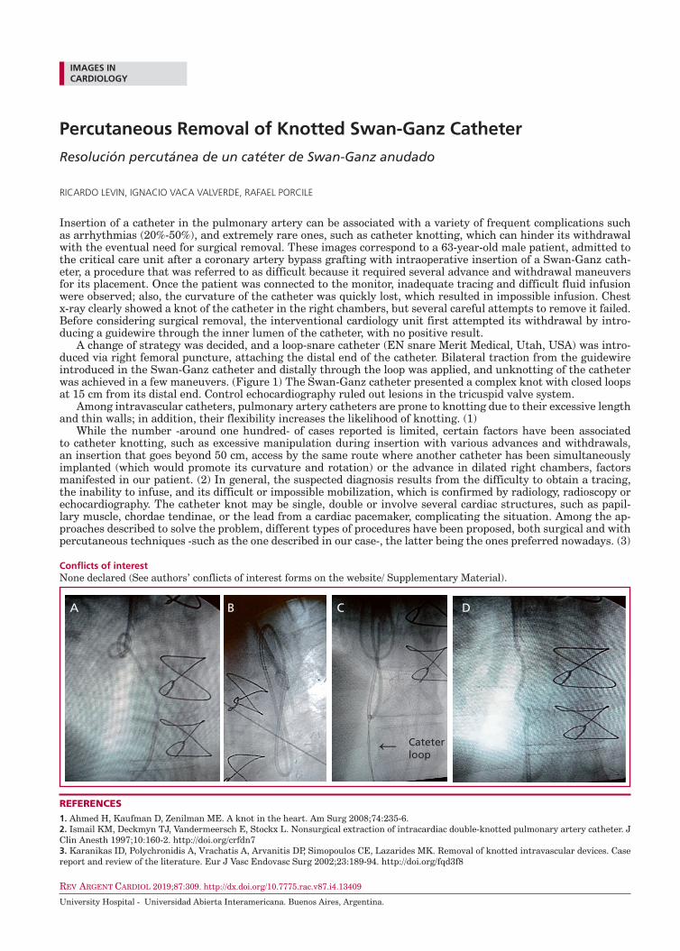
A change of strategy was decided, and a loop-snare catheter (EN snare Merit Medical, Utah, USA) was intro-duced via right femoral puncture, attaching the distal end of the catheter. Bilateral traction from the guidewire introduced in the Swan-Ganz catheter and distally through the loop was applied, and unknotting of the catheter was achieved in a few maneuvers. (Figure 1) The Swan-Ganz catheter presented a complex knot with closed loops at 15 cm from its distal end. Control echocardiography ruled out lesions in the tricuspid valve system.
Among intravascular catheters, pulmonary artery catheters are prone to knotting due to their excessive length and thin walls; in addition, their flexibility increases the likelihood of knotting. (1)
While the number -around one hundred- of cases reported is limited, certain factors have been associated to catheter knotting, such as excessive manipulation during insertion with various advances and withdrawals, an insertion that goes beyond 50 cm, access by the same route where another catheter has been simultaneously implanted (which would promote its curvature and rotation) or the advance in dilated right chambers, factors manifested in our patient. (2) In general, the suspected diagnosis results from the difficulty to obtain a tracing, the inability to infuse, and its difficult or impossible mobilization, which is confirmed by radiology, radioscopy or echocardiography. The catheter knot may be single, double or involve several cardiac structures, such as papil-lary muscle, chordae tendinae, or the lead from a cardiac pacemaker, complicating the situation. Among the ap-proaches described to solve the problem, different types of procedures have been proposed, both surgical and with percutaneous techniques -such as the one described in our case-, the latter being the ones preferred nowadays. (3)
conflicts of interestNone declared (See authors’ conflicts of interest forms on the website/ Supplementary Material).
REFERENCES
1. Ahmed H, Kaufman D, Zenilman ME. A knot in the heart. Am Surg 2008;74:235-6.2. Ismail KM, Deckmyn TJ, Vandermeersch E, Stockx L. Nonsurgical extraction of intracardiac double-knotted pulmonary artery catheter. J Clin Anesth 1997;10:160-2. http://doi.org/crfdn73. Karanikas ID, Polychronidis A, Vrachatis A, Arvanitis DP, Simopoulos CE, Lazarides MK. Removal of knotted intravascular devices. Case report and review of the literature. Eur J Vasc Endovasc Surg 2002;23:189-94. http://doi.org/fqd3f8
University Hospital - Universidad Abierta Interamericana. Buenos Aires, Argentina.
REV ARGENT CARDIOL 2019;87:309. http://dx.doi.org/10.7775.rac.v87.i4.13409
Cateter loop
A B C D
←