Controlling supramolecular chirality in peptide-π-peptide ...
Upload
hoangkhuongCategory
view
215download
0
Peptide Design for Mesenchymal Stem Cell Specific Attachment on Apatite Surfaces
for Bone Tissue Regeneration
by
Sriharsha Ramaraju
A dissertation submitted in partial fulfillment
of the requirements for the degree of
Doctor of Philosophy
(Biomedical Engineering)
in The University of Michigan
2015
Doctoral Committee:
Professor David H. Kohn, Chair
Professor Renny T. Franceschi
Associate Professor Andrew J. Putnam
Professor Jan P. Stegemann

Sriharsha Ramaraju
All Rights Reserved
2015

ii
To my family

iii
ACKNOWLEDGMENTS
This work would not have been possible without the mentorship and support provided by
my graduate advisor David Kohn. Thanks for providing me the freedom to develop my
own scientific curiosity while facilitating access to necessary resources. Thank you as
well for allowing me to seek out and pursue my professional interests. I would like to
thank my dissertation committee, Dr. Franceschi, Dr. Stegemann, and Dr. Putnam, for
their insightful feedback that improved the focus of my research questions.
I would like to thank my labmates whom I’ve had the pleasure of working with over the
years. Thank you Janani Ramaswamy, Erin McNerny, and Joseph Gardinier for the long
chats that helped me shape my scientific questions and the many ice-cream and coffee
breaks. Thank you Genny Romanowicz for your various academic and confectionary
contributions. I would like to thank Nianli Zhang, Nicholas Landgraf and Michelle Lynch
for their assistance with various experimental procedures. Thank you Nadder Sahar, Linh
Luong, and Michael Friedman for your constructive feedback during presentations. A
special thanks to Sharon Miller for applying phage-display technology to bone tissue
engineering, identifying the peptides that are the focus of this work, and for being a co-
author on my publications.
I would like to thank Dr. Paul Krebsbach’s lab. Specifically, I would like to thank Dr.
Hongli Sun for assistance with animal surgeries, and Dr. Luis Villa-Diaz for generating
iPS-MSCs and assistance with iPS-MSC characterization which allowed me to complete

iv
my subsequent aims. I would also like to thank Dr. Sergei Kuznetsov for the generous
contribution of primary MSCs derived from human bone marrow stroma. I would also
like to thank Chris Strayhorn from the Dental Histology Core, Taocong Jin from the
Dental Molecular biology core, Dr. Henriette Remmer at the Proteomics Core, and the
staff at the microscopy and image analysis core for speeding up materials and data
acquisition. I would also like to thank our past and present administrative staff Liz
Rodriguiz, Kumud Danak, Deb Keedy, Joel Clandenin, and Maria Steele for making
things run smoothly.
I would like to thank and acknowledge my undergraduate research advisor Dr. Alfred H.
Merrill, Jr for introducing me to academic research and providing me the opportunity to
continue my undergraduate research beyond graduation. I would like to thank Dr. Dennis
Liotta, Dr. John Petros, Dr. Carrie Sun and Dr. Rebecca Arnold for providing me the
guidance, environment, and confidence to broaden my research skills and experiences.
I would like to thank all of my friends and my sister for their various visits to Ann Arbor
when I was unable to travel and for enduring Michigan winters. I would like to thank my
wife Neelima for being a constant source of strength throughout my graduate school.
Your patience, positivity, and support were integral to the completion of this work.
Lastly, I would like to thank my parents for the struggles they’ve endured and the
sacrifices they have made in order to provide me the opportunities to pursue my academic
interests.

v
TABLE OF CONTENTS
DEDICATION ii
ACKNOWLEDGMENTS iii
LIST OF FIGURES viii
LIST OF TABLES x
LIST OF APPENDICIES xi
ABSTRACT xii
CHAPTER ONE: INTRODUCTION 1
Clinical Significance 1
Biomaterial Design 2
Recapitulating Cellular Microenvironments 3
Peptide Therapies for Tissue Regeneration 5
Peptide Specificity 5
Peptide Presentation 6
Display Technologies for Peptide-based Tissue Regeneration Therapies 7
Phage Display Peptides that Form the Basis for this Dissertation 8
Summary of Significance 9
Aims and Hypothesis 10
References 15
CHAPTER TWO: DUAL-FUNCTIONING PEPTIDES DISCOVERED BY PHAGE DISPLAY IMPROVE BMSC SPECIFIC ATTACHMENT TO MINERALIZED BIOMATERIALS 21
Introduction 21
Materials and Methods 23
Biomaterial preparation 23
Cell sources and culture 24
Peptide synthesis 24
Langmuir isotherms 25
vi
Cell attachment assays 26
Cell morphology and immunohistochemistry 27
Statistical methods 27
Results 28
Peptide binding isotherms on HA 28
Cell adhesion strength on dual-peptide coated mineral 28
HBMSC specificity to dual-peptide coated mineral 29
MSC adhesion dependence on cell seeding density 29
Discussion 30
Acknowledgements 34
References 45
CHAPTER THREE: PHAGE DISPLAY DERIVED BI-FUNCTIONAL PEPTIDE IMPROVES MSC ADHESION, PROLIFERATION AND DIFFERENTIATION ON APATITE 49
Introduction 49
Materials and Methods 51
Mineralized film fabrication 51
Peptide synthesis and adsorption to bone-like mineral 52
Cell Culture 52
Competitive adhesion of MSC 53
Cell spreading and histomorphometry 53
Msc Differentiation On Peptide Coated Apatite 54
Peptide Cell Internalization 55
MSC Recruitment on Peptide Coated Apatite 55
Statistical Methods 56
Results 56
MSC Spreading And Contact Distribution On Peptide Coated Apatite 56
Competitive MSC Binding Assays 57
MSC Proliferation On Peptide Coated Apatite 57
MSC Differentiation On Peptide Coated Apatite 58
Peptide Internalization 59
Multipotent-MSC Recruitment From Murine Bone Marrow 59
Discussion 59
Acknowledgements 63
References 76
CHAPTER FOUR: IMPROVING MSC BASED BONE TISSUE REGENERATION IN VIVO USING CELL-SPECIFIC PEPTIDE COATED MINERALIZED BIOMATERIALS 80
Introduction 80
Materials and Methods 84
Scaffold Fabrication 84
Scaffold Mineralization 84

vii
Scaffold Characterization 85
Peptide Synthesis and Characterization 86
Peptide loading and Characterization 86
Cell Culture 87
Subcutaneous Transplantation of Cell-Seeded Constructs 88
Ossicle Microcomputed Tomography 89
Ossicle Histology and Histomorphometry 89
Statistical Methods 90
Results 90
Characterization of Mineralized Scaffolds 90
Peptide Loading on Mineralized Scaffolds 91
Cell Attachment and Distribution on Peptide-Laden Scaffolds 91
Ossicle Bone Volume Fraction and Distribution 92
Histology of Reconstructed Ossicles 93
Regression Analysis 93
Discussion 94
Acknowledgements 99
References 117
CHAPTER FIVE: SUMMARY AND FUTURE WORK 122
Phage display derived peptide design 124
Dual-peptide tissue engineering applications 125
Future directions 126
Advancements in display technology 129
References 130
APPENDICIES 135
viii
LIST OF FIGURES
CHAPTER ONE FIGURES
Figure 1.1 Overview of Aims 14
CHAPTER TWO FIGURES
Figure 2.1 (A,B). Langmuir isotherms 37
Figure 2.2(A-E). HBMSC attachment, adhesion strength and spreading on single and dual-
functioning peptide coated apatite films 38
Figure 2.3(A-D). MSC specific adhesion strength and spreading on DPI-VTK and RGD-VTK
coated apatite films. 41
Figure 2.4(A-C). Cell seeding density effects on cell spreading on VTK and DPI-VTK coated
apatite films. 43
CHAPTER THREE FIGURES
Figure 3.1(A-E) Cell spreading and contact distribution on peptide coated apatite surfaces 65
Figure 3.2(A-C). iPS-MSC competition with soluble integrin binding competitors. 68
Figure 3.3. MSC proliferation on peptide coated apatite substrates. 70
Figure 3.4(A-G). IPS-MSC differentiation on TCPS in osteogenic media. 71
Figure 5(A-F). Differentiation of iPS-MSCs on biomimetic apatite and peptide coated apatite 73

ix
Figure 3.6. Cell association of fluorescently tagged VTK peptide after 1 hr of incubation. 74
Figure 3.7. Multipotent murine-MSC recruitment onto peptide coated apatite 75
CHAPTER FOUR FIGURES
Figure 4.1(A-J) Morphology and distribution of bone-like mineral precipitated on PLGA
scaffolds 101
Figure 4.2(A-H). Peptide adsorption and distribution on mineralized PLGA Scaffolds. 103
Figure 4.3(A-G) - Cell Seeding efficiency and cell distribution on peptide coated scaffolds. 105
Figure 4.4(A-H). Bone volume fractions of ectopically regenerated ossicles 8 weeks post
transplantation. 108
Figure 4.5(A-H). Bone distribution in ectopically regenerated ossicles 8 weeks post
transplantation. 111
Figure 4.6(A-K). Histological staining and bone quality scoring of ectopically regenerated
ossicles 8 weeks post transplantation. 114
x
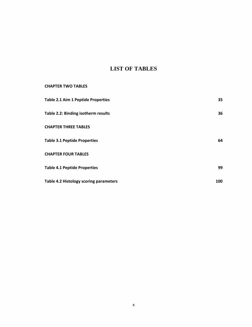
LIST OF TABLES
CHAPTER TWO TABLES
Table 2.1 Aim 1 Peptide Properties 35
Table 2.2: Binding isotherm results 36
CHAPTER THREE TABLES
Table 3.1 Peptide Properties 64
CHAPTER FOUR TABLES
Table 4.1 Peptide Properties 99
Table 4.2 Histology scoring parameters 100
xi
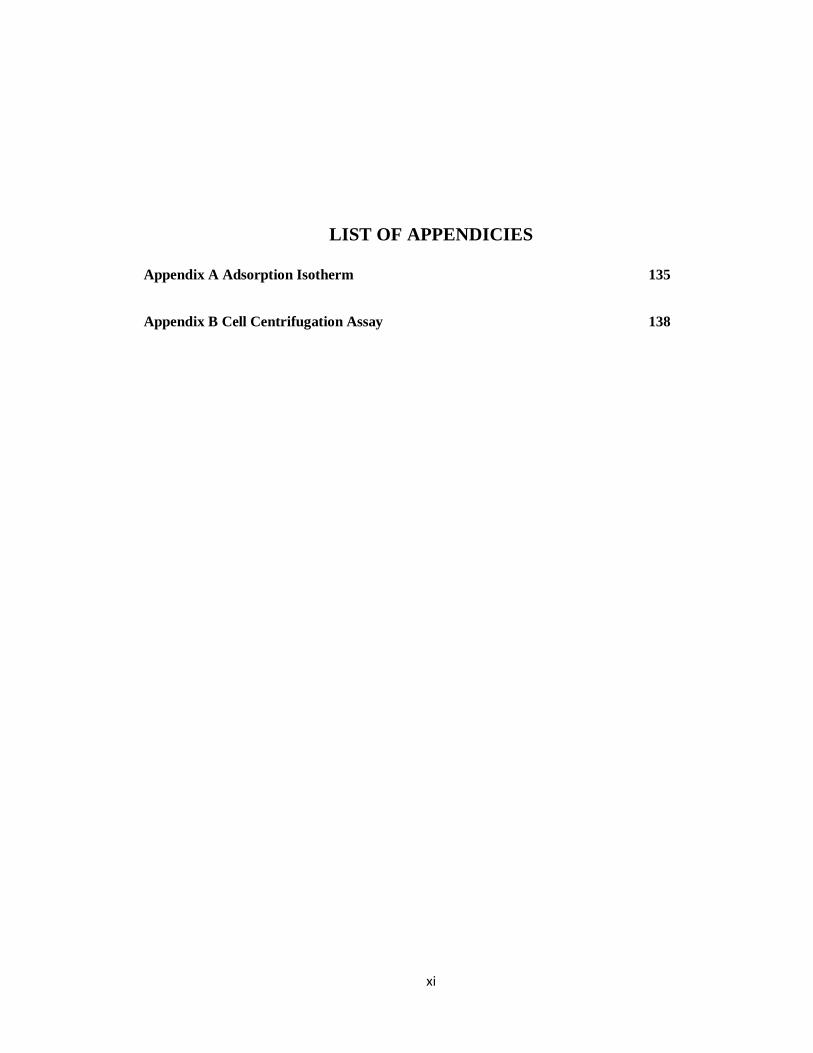
LIST OF APPENDICIES
Appendix A Adsorption Isotherm 135
Appendix B Cell Centrifugation Assay 138

xii
ABSTRACT
Over 2 million bone grafting procedures are performed annually worldwide for the
treatment of bone defects. Cell transplantation therapies are promising alternatives to
conventional auto-, allo-, and xenograft therapies. Successfully delivering stem and
progenitor cells to the defect site requires biomaterials that support and guide
reconstruction. Biomaterial functionalization with extracellular matrix derivatives to
improve adhesion and guide tissue regeneration lacks specificity towards particular
regenerative cell populations. In order to direct cell specific adhesion to specific
biomaterial surface chemistries, we used a combinatorial phage display strategy to
identify 2 sequences, 1 with high affinity towards apatite (VTK) and a second with high
affinity to clonally derived mesenchymal stem cells (MSC) from human bone marrow
stroma (DPI) and combined the two sequences into a dual-functioning peptide (DPI-
VTK).
Dual-functioning peptide DPI-VTK exhibited greater apatite binding compared to single
peptide controls (p < 0.01). Mesenchymal stem cells on DPI-VTK coated apatite
substrates exhibited greater adhesion strength compared to pre-osteoblasts and fibroblasts
(p <0.01). DPI-VTK also increased MSC spreading (p < 0.001) and proliferation (p <
0.001) compared to apatite controls while supporting differentiation on apatite substrates.
Competitive inhibition revealed RGD-binding integrin involvement in MSC attachment

xiii
to DPI-VTK. MSC driven bone formation, cellularity and vascularization in a
subcutaneous mouse model were greater on DPI-VTK coated PLGA-mineral composite
scaffolds compared to VTK (p < 0.017) and uncoated controls (p <0.001) and acellular
peptide-coated controls (p <0.002). Taken together, DPI-VTK improves MSC specific
attachment and subsequent adhesion on mineralized substrates driving greater
proliferation and bone formation compared to acellular and non-peptide coated controls.
A vast array of biomaterials and multitude of regenerative cell sources are available for
tissue regeneration applications. As tissue engineering shifts from developing
technologies to meet general clinical challenges to addressing more focused clinical
applications, there will be an increased need for delivering cell specific cues to material
surfaces with defined surface chemistries. Combinatorial phage display is a powerful
platform to enable focused cell based tissue regeneration through the discovery of cell
specific and material specific peptide sequences.

1
CHAPTER ONE
INTRODUCTION
CLINICAL SIGNIFICANCE
Current clinical solutions for tissue replacement and regeneration utilize auto-, allo- or
xenografts which require multiple interventional procedures and could result in donor site
morbidity and immunogenicity[1], [2]. De novo tissue regeneration utilizing cells
extracted from the host offers a promising alternative to the current clinical standards.
Most organ systems in the body contain a repository of stem and progenitor cells that
replace dying and injured cells to preserve the integrity of tissues. For instance, cells of
the bone marrow stroma contain a mesenchymal population that can give rise to
osteogenic, chondrogenic, myogenic, and adipogenic lineages[1], [3], [4]. These cells
secrete tropic factors to improve tissue repair, suppress immune responses and are a
readily available and relatively non-invasive cell source to extract from the host.
Moreover, stem and progenitors can be expanded in monolayer culture or 3D culture
under perfusion to be delivered to a defect site on an appropriate transplant material[3],
[5]–[8]. Stem and progenitor cell mediated bone regeneration in vivo is dependent on
biomaterial properties that control cell seeding efficiency, and cell distribution, and can
provide instructive cues to guide cell proliferation and differentiation[9]–[12].
2
BIOMATERIAL DESIGN
The ideal 3D biomaterial provides an environment that allows cell adhesion and
encourages cell growth and differentiation while allowing transport of nutrients and
waste to facilitate new tissue formation[8], [13]. A conductive biomaterial supports cell
adhesion, which is an important starting point for functional tissue regeneration. In
addition to the number of cells seeded on a 3D biomaterial construct, the distribution of
cells plays a critical role in bone formation as well. Constructs that improve cell seeding
density and promote uniform distribution of cells in a 3D biomaterial while directing cell-
cell communication and cell-matrix interactions are hypothesized to improve bone
formation in vivo[9], [14], [15].
Both bulk and surface properties of a material contribute to cell adhesion. Metals,
ceramics, polymers, and composites are used in conjunction with an array of surface and
bulk modification techniques to yield material properties conducive to supporting cell
based tissue regeneration [2], [8], [16]–[18]. In the context of bone, properties such as
compliance, surface free energy, wettability, surface topography, and crystallinity
influence cell mediated bone formation in vivo[19]–[21]. Many of these properties exhibit
collinear relationships due to the underlying biological processes driving cell adhesion,
proliferation, and differentiation.
Osteoinductive biomaterials stimulate and drive differentiation of primitive
undifferentiated pluripotent or multipotent cells towards a terminal bone forming cell
lineage[22]. Inductive biomaterials utilize growth factors, recombinant DNA, and

3
peptides to regulate cell growth and differentiation[22]–[24]. These biomolecules can be
adsorbed, encapsulated within or immobilized to biomaterials in order to temporally and
spatially control transplanted cell behavior. The conductive and inductive properties of a
material that impact cell adhesion, proliferation, and differentiation result in an expansive
number of design considerations for developing the ideal carrier for functional tissue
regeneration. Biomimetic biomaterial design strategies specifically addresses
osetoconduction and osteoinduction by recapitulating physical and chemical attributes of
the native ECM to drive functional tissue regeneration[25]–[28].
RECAPITULATING CELLULAR MICROENVIRONMENTS
Biomimetic design of biomaterials involves studying the physical and chemical
properties of functional tissues and incorporating these properties into biomaterial design.
The extracellular matrix (ECM) of bone is comprised of 50-70% inorganic mineral
matrix, 20-40% organic matrix, 5-10% water and less than 3% lipids. The inorganic
matrix is comprised of a non-stoichiometric, semi-crystalline, calcium and hydroxide
deficient carbonate substituted hydroxyapatite mineral [Ca10(PO4)6(OH)2]. The organic
matrix is predominantly comprised of Type I collagen. Non-collagenous proteins
comprise the remaining 10-15% of the organic component and include proteoglycans,
glycosylated proteins, and γ-carboxylated proteins. These non-collagenous proteins are
involved in directing organic matrix assembly, maintaining structural integrity of the
tissue, sequestering and interacting with growth factors, and regulating bone metabolism
and mineralization. Both the organic and inorganic components of bone contribute to
conductive and inductive properties. Therefore reproducing aspects of both organic and

4
inorganic components of bone tissue in biomaterial design is hypothesized to improve
regeneration.
Surface coating via a biomimetic procedure can be used to form a biomimetic apatite
coating on biocompatible and biodegradable polymer substrates. For instance, 3D porous
poly-Lactide-co-Glycolide (PLGA) scaffold surfaces can be coated with a continuous
bone like mineral (BLM) layer through immersion in a supersaturated simulated body
fluid (SBF). This SBF contains similar ionic concentrations to that of blood plasma and
results in the precipitation of a bone-like-mineral that is semicrystalline, nano-structured,
and is carbonate substituted [29]–[31]. Biomimetically mineralized polymers exhibit a 5-
fold increase in compressive modulus, support higher bone marrow stromal cell adhesion
through well distributed fibrillar contacts, and higher bone volume fractions when used to
transplant BMSCs in vivo compared to non-mineralized polymer scaffolds[18], [29],
[32], [33].
The organic components of extracellular matrices are comprised of collagenous and non-
collagenous proteins that provide mechanical support and cell instructive cues that
facilitate tissue growth and preserve tissue integrity. These components impart cues that
regulate an elegant sequence of spatially and temporally controlled events resulting in
tissue repair and regeneration. Collagen, fibronectin, vitronectin, osteocalcin,
osteopontin, bone morphogenetic protein-2 (BMP-2) and fibroblast growth factor-2
(FGF-2) have all been used to drive osteoblastic differentiation of and improve quantity
of regenerated bone [34]–[36]. However, full length recombinant ECM proteins are
challenged by degradation rate, conformational control, and high processing costs.
5
PEPTIDE THERAPIES FOR TISSUE REGENERATION
Peptide epitopes offer a promising alternative to protein based therapies by allowing
control of conformation, cell and material specificity, degradation rate, and reduced
processing costs. Peptides derived from the active sites of ECM proteins have been used
to impart instructive cues to stem and progenitor cell lines that form bone [34], [36]–[38].
The method of delivering these signals onto a biomaterial and the modification of these
peptides play an important role in mediating cell responses. Peptide delivery methods
involve adsorption, covalent immobilization or encapsulation into a biomaterial.
Adsorption of peptides on a biomaterial surface involves weak molecular and
electrostatic forces at the substrate-peptide interface. While adsorption allows
conformational freedom of peptides, it is challenged by control of release rates and short
half-lives of some unbound peptide. Peptide mediated control of cell viability,
differentiation, and tissue formation, are variable across cell sources, materials, and mode
of delivery. This variability can be linked to both the specificity of the peptide sequences
to cell lineages and the proper presentation of these sequences suggesting the important
interplay of these factors.
PEPTIDE SPECIFICITY
The cell binding sites of ECM protein domains exhibit different binding affinities to
different integrin receptors. For instance, DGEA and GFOGER peptides from collagen
predominantly bind to α2β1 integrin receptors, whereas fibronectin and vitronectin
fragments bind α5β1 receptors. Most ECM proteins also have multiple binding domains
that interact and contribute to this specificity. As an example, the RGD sequence

6
prevalent in many proteins binds αVβ3 integrins whereas co-delivery with the fibronectin
sequence PHSRN synergistically improves binding affinity to α5β1[34], [38]–[40].
Targeting different integrins results in variable cell responses across cell lineages. For
instance, the DGEA collagen fragment and RGD preferentially bind different integrin
subunits and increase FAK signaling pathways, but only the DGEA peptide improves
MAPK mediated signal transduction [41]. Differences in specificity for these integrin
receptors could also be responsible for variability of tissue regeneration in vivo. For
example, collagen derived peptides improve MSC cell differentiation in vitro and bone
formation in vivo, whereas RGD exhibits inverse effects [41], [42].
In addition to using components of known ECM peptides, novel cryptic peptides from
collagen have also been derived to improve conductive properties [43], [44]. Strategies
using components of known ECM proteins targeting specific integrins lack specificity to
a particular cell type. The ability to adhere and recruit specific stem or progenitor cell
populations to a biomaterial surface and impart instructive cues can improve tissue repair
and regeneration[38]. In addition to delivering cell specific sequences, proper
presentation of these signals can also improve specificity, cell adhesion, proliferation and
differentiation.
PEPTIDE PRESENTATION
In addition to different delivery methods, peptide structure and conformation can be
altered to improve loading efficiency on biomaterials and binding affinity to cell surface
receptors. For example, recapitulating the circular conformation of RGD that is
commonly found in ECM proteins by altering the structure of RGD not only improves

7
solution stability and adsorption to biominerals, but also increases bone formation when
immobilized on alginate hydrogels[45], [46].
Many ECM proteins have multifunctional domains that work in conjunction with one
another to present the cell instructive domains to the cell surface receptors. Combining
the cell adhesive sequence with a material binding sequence can improve presentation of
the cell instructive peptide to the cell and enhance cellular response[47], [48]. For
example, peptide fragment 17-25 of the bone ECM protein osteocalcin contains 3
glutamic acid residues at positions 17, 21, and 24. These residues strongly bind
hydroxyapatite and have been used to improve RGD, BMP, and VEGF derived peptide
adsorption to biomineral surfaces[37], [47]–[49]. Improving the binding affinity of the
cell adhesive peptides by attaching them to material binding sequences improves cell
proliferation, differentiation and mineralization in vitro.
The combination of sequence specificity, peptide modification methods, and peptide
loading methods with respect to the carrier material contribute to the presentation of the
cell instructive cues to the cell surface. Therefore, the ability to systematically design
specific peptides and appropriately present them to the cell surface receptor can improve
tissue regeneration.
DISPLAY TECHNOLOGIES FOR PEPTIDE-BASED TISSUE REGENERATION
THERAPIES
Bacteriophage display, bacterial display and cell surface display technologies are
commonly used in oncology and immunology to humanize targeted monoclonal
antibody(MAb) based therapies[50], [51]. The application of phage display for tissue

8
regeneration is gaining traction through the development of biomaterial specific or cell
specific peptides. Material specific peptides are used to increase physiochemical
deposition of biomineral on biomaterials or deliver growth factors to the surface of
mineralized biomaterials[52]–[55]. Cell binding phage-derived peptides have been
tethered to polymer biomaterials or self-assembled into 3D structures to improve
conductivity and inductivity [56]–[58] . Phage-derived mimetic peptides of the enamel
protein amelogenin improve cell conductivity and tissue formation in vivo[59].
PHAGE DISPLAY PEPTIDES THAT FORM THE BASIS FOR THIS
DISSERTATION
The primary aim of this dissertation is to evaluate the bone forming potential of MSCs
specifically tethered to an apatite surface using a phage display derived apatite specific
peptide combined with a cell specific peptide. A commercially available M13
bacteriophage display kit containing 109 sequences of 12-mers was used to identify
biomineral and cell specific sequences to improve the specificity of human bone marrow
stromal cell binding to apatite surfaces[60], [61].
After 3 rounds of phage panning on HA disks and BLM coated PLGA films, 243
sequences were identified. Amongst these 243 sequences, 68 sequences were identified
as high information clones through RELIC analysis. From the 68 sequences, 19 were
ordered in terms of their binding affinity to different crystal faces of HA [001, 200], and
10 sequences were selected for ELISA on PLGA, BLM-PLGA films, HA disks, and
tissue cultured polystyrene(TCPS). The top 5 sequences exhibiting positive ELISA
signals, compared to high information clones from RELIC, and high binding affinity
sequences form computational modeling resulted in 3 consensus sequences. Adsorption

9
assays measuring efficiency of peptide binding to apatite, resulted in the identification of
1 peptide sequence, VTKHLNQISQSY (VTK), with superior binding affinity to
BLM[50], [60], [61].
After 3 rounds of phage panning on BMSCs, 50 recurring sequences were identified,
amongst which 27 were high information clones and 10 high information and 10 low
information peptides were tested using immunohistochemistry (IHC). From the 50
sequences identified, the MOTIF1 program identified continuous motif sequences 3 and 4
amino acids long as NHT, and (S/T)(I/V)LS. Although no consensus sequences were
found from MOTIF1 and RELIC analysis, two sequences were identified as having high
scores from both RELIC and IHC. A cell adhesion assay with BMSCs attached to peptide
laden BLM coated PLGA films and peptide coated TCPS resulted in the identification of
1 sequence, DPIYALSWSGMA (DPI), that improves cell seeding efficiency on BLM in
vitro[60]. The cell specific and mineral specific peptides were combined using glycine
residues to yield GGDPIYALSWSGMAGGGSVTKHLNQISQSY (DPI-VTK).
In addition to combining these peptides, post-translational modification of the mineral
binding sequence improves apatite binding affinity in vitro. Many naturally occurring
ECM proteins contain phosphorylated serine residues which improve apatite binding
affinity. Phosphorylation of the C-terminal serine residues of VTK improves binding
affinity to apatite but reduces cell adhesion to apatite surfaces [60].
SUMMARY OF SIGNIFICANCE
The ability to provide specific cues to stem and progenitor cells is an increasing area of
focus for biomaterial design. Combining these cues with material specific cues can have
10
broad implications for tissue regeneration. Dual functioning peptides could impart spatial
control over cell adhesion and differentiation, which can translate to increased quantity
and quality of tissue regenerated in vivo. Moreover, dual functioning peptides containing
material and cell specific domains could also be translated to enhance mineralized
implant integration in vivo.
AIMS AND HYPOTHESIS
The primary aims of this dissertation were to: 1) design and verify peptide functionality towards
specifically recruiting MSCs to mineral substrates. 2) assess osteoconduction and osteoinduction
of MSCs on dual peptide coated biomimetic apatite surfaces in vitro, and 3) examine the
relationship between initial cell attachment and peptide distribution on dual peptide coated 3D
mineralized PLGA scaffolds in-vitro and the quantity, quality and distribution of regenerated
bone in vivo.
Global hypothesis: Combining material and cell specific peptides into a dual-functioning
peptide can improve initial cell attachment, cell-specific adhesion, proliferation and
differentiation in vitro and improve quality, quantity and distribution of bone formation
in vivo compared to non-specific peptides or specific peptides with only a single
functionality.
The overall aim of this thesis is to demonstrate the utility of phage display
technologies towards improving cell-specific attachment to biomaterials contributing to
increased proliferation, differentiation and tissue regeneration. With the growing
abundance of regenerative cell sources, it will become increasingly important to control
cell specific delivery and recruitment to biomaterial surfaces (Figure 1.1).

11
Aim 1: Quantify material binding affinity and MSC binding affinity of dual functioning
peptide
Hypothesis: Combining a mineral specific peptide sequence with a clonally-derived
HBMSC specific peptide sequence will improve the binding affinity of cell specific
peptides to mineralized substrates and subsequently improve cell specific adhesion to
mineralized substrates
Aim 1.1: Quantify the influence of mineral binding sequence on dual functioning
peptide binding affinity to mineralized biomaterials with Langmuir isotherms
Hypothesis: Attaching phage display derived mineral specific sequences
to human bone marrow stromal cell specific sequences will improve the
binding affinity of the dual sequence to mineral surfaces in vitro
Aim 1.2: Quantify dual functioning peptide specificity to human bone marrow
stromal cells with a cell detachment assay
Hypothesis: Addition of a mineral binding sequence to a cell specific
sequence to yield a dual functioning peptide can improve initial HBMSC
attachment to 2D mineralized surfaces in vitro
Chapter 2, which relates to Aim 1, demonstrates the ability of our combinatorial phage
display process to select material-specific and cell-specific sequences with dual
functionality. The dual peptide DPI-VTK exhibits strong affinity towards hydroxyapatite
and biomimetic apatite while concomitantly promoting MSC specific adhesion on apatite
surfaces.

12
Aim 2: Determine effects of dual functioning peptides on HBMSC specific adhesion,
proliferation, and differentiation on 2D mineral films in vitro
Hypothesis: Dual functioning peptides improve cell spreading and influence
proliferation and differentiation on 2D mineral films in vitro
Chapter 3, which relates to Aim 2, demonstrates the ability of the dual peptide DPI-VTK
to improve cell adhesion and promotes cell proliferation by binding an RGD-integrin
specific receptor. There was only a marginal improvement of MSC differentiation on
DPI-VTK compared to uncoated apatite films; however, MSC specificity towards DPI-
VTK, improved adhesion compared to untreated mineral and cooperative interactions
with serum proteins translate to improved cell delivery for tissue regeneration in vivo.
Aim 3: Demonstrate that dual functioning peptides improve HBMSC seeding efficiency
and distribution on biomimetically mineralized PLGA scaffolds and consequently
improve quantity, distribution and quality of bone formation in vivo
Hypothesis: Dual functioning peptides improve cell seeding efficiency and distribution
of HBMSCs on biomimetically mineralized PLGA scaffolds, which correlates with
improved quantity, distribution, and quality of bone formation in vivo.
Chapter 4, which relates to Aim 3, demonstrates the ability of dual peptide DPI-VTK to
improve the quantity, cellularity and vascularization of bone formation in vivo compared
to uncoated controls. Although bone quantity, cellularity and vascularization of
reconstructed ossicles were greater on DPI-VTK coated constructs, ossicles still exhibited
a shell of bone formation with less bone volume in the interior. Regardless, the quantity

13
of regenerated bone was significantly improved by coating DPI-VTK on mineralized
scaffolds for cell-based tissue regeneration.
Overall, this work demonstrates the specificity and bioactivity of DPI-VTK in vitro and
its osteogenic potential in vivo while demonstrating the significance of display
technology towards tissue regeneration applications.
14
CHAPTER ONE FIGURES
FIGURE 1.1 OVERVIEW OF AIMS
Aim 1.1: Dual Peptide
specificity to mineral
Aim 1.2: Dual Peptide
specificity to cells
DESIGN/CHARACTERIZE
Aim 2: Dual Peptide
bioactivity in vitro
Aim 3: Dual Peptide
bioactivity in vivo
BIOACTIVITY/APPLICATION

15
REFERENCES
[1] P. H. Krebsbach, S. a. Kuznetsov, P. Bianco, and P. Gehron Robey, “Bone Marrow Stromal
Cells: Characterization and Clinical Application,” Crit. Rev. Oral Biol. Med., vol. 10, no. 2,
pp. 165–181, Jan. 1999.
[2] E. Alsberg, E. E. Hill, and D. J. Mooney, “Craniofacial tissue engineering.,” Crit. Rev. Oral
Biol. Med., vol. 12, no. 1, pp. 64–75, Jan. 2001.
[3] P. G. Robey, S. A. Kuznetsov, M. Riminucci, and P. Bianco, “Skeletal (‘mesenchymal’) stem
cells for tissue engineering.,” Methods Mol. Med., vol. 140, pp. 83–99, Jan. 2007.
[4] P. G. Robey and P. Bianco, “The use of adult stem cells in rebuilding the human face.,” J.
Am. Dent. Assoc., vol. 137, no. 7, pp. 961–72, Jul. 2006.
[5] D. Benayahu, U. D. Akavia, and I. Shur, “Differentiation of bone marrow stroma-derived
mesenchymal cells.,” Curr. Med. Chem., vol. 14, no. 2, pp. 173–9, Jan. 2007.
[6] A. I. Caplan, “Review: mesenchymal stem cells: cell-based reconstructive therapy in
orthopedics ,” Tissue Eng., vol. 11, no. 7–8, pp. 1198–1211, 2005.
[7] J. R. Mauney, C. Jaquiéry, V. Volloch, M. Heberer, I. Martin, and D. L. Kaplan, “In vitro and
in vivo evaluation of differentially demineralized cancellous bone scaffolds combined
with human bone marrow stromal cells for tissue engineering,” Biomaterials, vol. 26, no.
16, pp. 3173–3185, 2005.
[8] A. G. Mikos, S. W. Herring, P. Ochareon, J. Elisseeff, H. H. Lu, R. Kandel, F. J. Schoen, M.
Toner, D. Mooney, A. Atala, M. E. Van Dyke, D. Kaplan, and G. Vunjak-Novakovic,
“Engineering complex tissues.,” Tissue Eng., vol. 12, no. 12, pp. 3307–39, Dec. 2006.
[9] K. Kim, D. Dean, A. G. Mikos, and J. P. Fisher, “Effect of initial cell seeding density on early
osteogenic signal expression of rat bone marrow stromal cells cultured on cross-linked
poly(propylene fumarate) disks.,” Biomacromolecules, vol. 10, no. 7, pp. 1810–7, Jul.
2009.
[10] C. E. Wilson, W. J. A. Dhert, C. A. Van Blitterswijk, A. J. Verbout, and J. D. De Bruijn,
“Evaluating 3D bone tissue engineered constructs with different seeding densities using
the alamarBlue assay and the effect on in vivo bone formation.,” J. Mater. Sci. Mater.
Med., vol. 13, no. 12, pp. 1265–9, Dec. 2002.
[11] M. H. Mankani, S. A. Kuznetsov, B. Fowler, A. Kingman, and P. G. Robey, “In vivo bone
formation by human bone marrow stromal cells: effect of carrier particle size and
shape.,” Biotechnol. Bioeng., vol. 72, no. 1, pp. 96–107, Jan. 2001.
[12] P. H. Krebsbach, S. A. Kuznetsov, K. Satomura, R. V Emmons, D. W. Rowe, and P. G.
Robey, “Bone formation in vivo: comparison of osteogenesis by transplanted mouse and
16
human marrow stromal fibroblasts.,” Transplantation, vol. 63, no. 8, pp. 1059–69, Apr.
1997.
[13] V. Mouriño and A. R. Boccaccini, “Bone tissue engineering therapeutics: controlled drug
delivery in three-dimensional scaffolds.,” J. R. Soc. Interface, vol. 7, no. 43, pp. 209–27,
Feb. 2010.
[14] R. A. Rossello and D. H. Kohn, “Gap junction intercellular communication: a review of a
potential platform to modulate craniofacial tissue engineering ,” J. Biomed. Mater. Res.
B, Appl. Biomater., vol. 88, no. 2, pp. 509–518, Feb. 2009.
[15] J. Eyckmans, T. Boudou, X. Yu, and C. S. Chen, “A hitchhiker’s guide to mechanobiology.,”
Dev. Cell, vol. 21, no. 1, pp. 35–47, Jul. 2011.
[16] J. D. Kretlow and A. G. Mikos, “Review: mineralization of synthetic polymer scaffolds for
bone tissue engineering.,” Tissue Eng., vol. 13, no. 5, pp. 927–38, May 2007.
[17] G. J. Meijer, J. D. de Bruijn, R. Koole, and C. A. van Blitterswijk, “Cell-Based Bone Tissue
Engineering ,” PLoS Med., vol. 4, no. 2, p. e9, Feb. 2007.
[18] W. L. Murphy, S. Hsiong, T. P. Richardson, C. a Simmons, and D. J. Mooney, “Effects of a
bone-like mineral film on phenotype of adult human mesenchymal stem cells in vitro.,”
Biomaterials, vol. 26, no. 3, pp. 303–10, Jan. 2005.
[19] Ramaswamy, J., Ramaraju, S., and Kohn D. H., “Bone-Like Mineral and Organically
Modified Bone-Like Mineral Coatings,” in Biological and Biomedical Coatings Handbook,
Processing and Characterization, S. Zhang, Ed. Boca Raton, FL: CRC Press, 2011, pp. 1–36.
[20] P. Habibovic, H. Yuan, C. M. van der Valk, G. Meijer, C. A. van Blitterswijk, and K. de
Groot, “3D microenvironment as essential element for osteoinduction by biomaterials ,”
Biomaterials, vol. 26, no. 17, pp. 3565–3575, Jun. 2005.
[21] J. S. Temenoff and A. G. Mikos, Biomaterials: The Intersection of Biology and Materials
Science. Prentice Hall, 2008.
[22] T. Albrektsson and C. Johansson, “Osteoinduction, osteoconduction and
osseointegration.,” Eur. Spine J., vol. 10 Suppl 2, pp. S96–101, Oct. 2001.
[23] K. Y. Lee, M. C. Peters, K. W. Anderson, and D. J. Mooney, “Controlled growth factor
release from synthetic extracellular matrices.,” Nature, vol. 408, no. 6815, pp. 998–1000,
2000.
[24] J. A. Hubbell, “Bioactive biomaterials,” Curr. Opin. Biotechnol., vol. 10, no. 2, pp. 123–
129, Jan. 1999.
17
[25] H. Shin, S. Jo, and A. G. Mikos, “Biomimetic materials for tissue engineering ,”
Biomaterials, vol. 24, no. 24, pp. 4353–4364, Nov. 2003.
[26] J. Yuan, Y. Cao, and W. Liu, “Biomimetic scaffolds: implications for craniofacial
regeneration.,” J. Craniofac. Surg., vol. 23, no. 1, pp. 294–7, Jan. 2012.
[27] J. a Burdick and G. Vunjak-Novakovic, “Engineered microenvironments for controlled
stem cell differentiation.,” Tissue Eng. Part A, vol. 15, no. 2, pp. 205–19, Feb. 2009.
[28] L. E. Freed, G. Vunjak-Novakovic, R. J. Biron, D. B. Eagles, D. C. Lesnoy, S. K. Barlow, and R.
Langer, “Biodegradable Polymer Scaffolds for Tissue Engineering”, Bio/Technology, vol.
12, no. 7, p. 689 <last_page> 693, 1994.
[29] W. L. Murphy, D. H. Kohn, and D. J. Mooney, “Growth of continuous bonelike mineral
within porous poly(lactide-co-glycolide) scaffolds in vitro.,” J. Biomed. Mater. Res., vol.
50, no. 1, pp. 50–8, Apr. 2000.
[30] H. M. Kim Miyaji, F., Kokubo,T., Nakamura T., “Bonding strength of bonelike apatite layer
to Ti metal substrate,” J. Biomed. Mater. Res. Part B Appl. Biomater., vol. 38, no. 2, pp.
121–127, 1998.
[31] T. Kokubo, “Bioactive glass ceramics: properties and applications ,” Biomaterials, vol. 12,
no. 2, pp. 155–163, Mar. 1991.
[32] E. V Leonova, K. E. Pennington, P. H. Krebsbach, and D. H. Kohn, “Substrate
mineralization stimulates focal adhesion contact redistribution and cell motility of bone
marrow stromal cells.,” J. Biomed. Mater. Res. A, vol. 79, no. 2, pp. 263–70, Nov. 2006.
[33] G. A. Hudalla and W. L. Murphy, “Using ‘click’ chemistry to prepare SAM substrates to
study stem cell adhesion ,” Langmuir, vol. 25, no. 10, pp. 5737–5746, May 2009.
[34] A. Shekaran and A. J. García, “Extracellular matrix-mimetic adhesive biomaterials for
bone repair.,” J. Biomed. Mater. Res. A, vol. 96, no. 1, pp. 261–72, Jan. 2011.
[35] A. Bhat, S. a Boyadjiev, C. W. Senders, and J. K. Leach, “Differential growth factor
adsorption to calvarial osteoblast-secreted extracellular matrices instructs osteoblastic
behavior.,” PLoS One, vol. 6, no. 10, p. e25990, Jan. 2011.
[36] M. P. Lutolf, F. E. Weber, H. G. Schmoekel, J. C. Schense, T. Kohler, R. Muller, and J. A.
Hubbell, “Repair of bone defects using synthetic mimetics of collagenous extracellular
matrices ,” Nat. Biotechnol., vol. 21, no. 5, pp. 513–518, May 2003.
[37] D. Itoh, S. Yoneda, S. Kuroda, H. Kondo, A. Umezawa, K. Ohya, T. Ohyama, and S. Kasugai,
“Enhancement of osteogenesis on hydroxyapatite surface coated with synthetic peptide
(EEEEEEEPRGDT) in vitro.,” J. Biomed. Mater. Res., vol. 62, no. 2, pp. 292–8, Nov. 2002.

18
[38] R. G. LeBaron and K. A. Athanasiou, “Extracellular matrix cell adhesion peptides:
functional applications in orthopedic materials ,” Tissue Eng., vol. 6, no. 2, pp. 85–103,
Apr. 2000.
[39] D. L. Hern and J. a Hubbell, “Incorporation of adhesion peptides into nonadhesive
hydrogels useful for tissue resurfacing.,” J. Biomed. Mater. Res., vol. 39, no. 2, pp. 266–
76, Feb. 1998.
[40] J. Benesch, J. F. Mano, and R. L. Reis, “Proteins and their peptide motifs in acellular
apatite mineralization of scaffolds for tissue engineering.,” Tissue Eng. Part B. Rev., vol.
14, no. 4, pp. 433–45, Dec. 2008.
[41] M. Gilbert, W. J. Shaw, J. R. Long, K. Nelson, G. P. Drobny, C. M. Giachelli, and P. S.
Stayton, “Chimeric peptides of statherin and osteopontin that bind hydroxyapatite and
mediate cell adhesion.,” J. Biol. Chem., vol. 275, no. 21, pp. 16213–8, May 2000.
[42] K. M. Hennessy, B. E. Pollot, W. C. Clem, M. C. Phipps, A. A. Sawyer, B. K. Culpepper, and
S. L. Bellis, “The effect of collagen I mimetic peptides on mesenchymal stem cell adhesion
and differentiation, and on bone formation at hydroxyapatite surfaces.,” Biomaterials,
vol. 30, no. 10, pp. 1898–909, Apr. 2009.
[43] V. Agrawal, J. Kelly, S. Tottey, K. A. Daly, S. A. Johnson, B. F. Siu, J. Reing, and S. F.
Badylak, “An isolated cryptic peptide influences osteogenesis and bone remodeling in an
adult mammalian model of digit amputation.,” Tissue Eng. Part A, vol. 17, no. 23–24, pp.
3033–44, Dec. 2011.
[44] V. Agrawal, S. Tottey, S. A. Johnson, J. M. Freund, B. F. Siu, and S. F. Badylak,
“Recruitment of progenitor cells by an extracellular matrix cryptic peptide in a mouse
model of digit amputation.,” Tissue Eng. Part A, vol. 17, no. 19–20, pp. 2435–43, Oct.
2011.
[45] S. X. Hsiong, T. Boontheekul, N. Huebsch, and D. J. Mooney, “Cyclic arginine-glycine-
aspartate peptides enhance three-dimensional stem cell osteogenic differentiation ,”
Tissue Eng. A, vol. 15, no. 2, pp. 263–272, Feb. 2009.
[46] M. Kantlehner, P. Schaffner, D. Finsinger, J. Meyer, A. Jonczyk, B. Diefenbach, B. Nies, G.
Holzemann, S. L. Goodman, and H. Kessler, “Surface coating with cyclic RGD peptides
stimulates osteoblast adhesion and proliferation as well as bone formation ,”
Chembiochem, vol. 1, no. 2, pp. 107–114, Aug. 2000.
[47] J. S. Lee, J. S. Lee, A. Wagoner-Johnson, and W. L. Murphy, “Modular peptide growth
factors for substrate-mediated stem cell differentiation.,” Angew. Chem. Int. Ed. Engl.,
vol. 48, no. 34, pp. 6266–9, Jan. 2009.

19
[48] J. S. Lee, A. J. Wagoner Johnson, and W. L. Murphy, “A modular, hydroxyapatite-binding
version of vascular endothelial growth factor.,” Adv. Mater., vol. 22, no. 48, pp. 5494–8,
Dec. 2010.
[49] R. Fujisawa, M. Mizuno, Y. Nodasaka, and Y. Kuboki, “Attachment of osteoblastic cells to
hydroxyapatite crystals by a synthetic peptide (Glu7-Pro-Arg-Gly-Asp-Thr) containing two
functional sequences of bone sialoprotein.,” Matrix Biol., vol. 16, no. 1, pp. 21–8, Apr.
1997.
[50] S. Segvich and D. H. Kohn, “Phage Display as a Strategy for Designing Organic/Inorganic
Biomaterials,” in Biological Interactions on Material Surfaces, D. A. Puleo and R. Bizios,
Eds. Springer US, 2009, pp. 115–132.
[51] A. Sergeeva, M. G. Kolonin, J. J. Molldrem, R. Pasqualini, and W. Arap, “Display
technologies: application for the discovery of drug and gene delivery agents.,” Adv. Drug
Deliv. Rev., vol. 58, no. 15, pp. 1622–54, Dec. 2006.
[52] H.-E. Jin, W.-J. Chung, and S.-W. Lee, “Phage display for the discovery of hydroxyapatite-
associated peptides.,” Methods Enzymol., vol. 532, pp. 305–23, Jan. 2013.
[53] D. Khatayevich, M. Gungormus, H. Yazici, C. So, S. Cetinel, H. Ma, A. Jen, C. Tamerler, and
M. Sarikaya, “Biofunctionalization of materials for implants using engineered peptides.,”
Acta Biomater., vol. 6, no. 12, pp. 4634–41, Dec. 2010.
[54] U. O. S. Seker, B. Wilson, J. L. Kulp, J. S. Evans, C. Tamerler, and M. Sarikaya,
“Thermodynamics of engineered gold binding peptides: establishing the structure-
activity relationships.,” Biomacromolecules, vol. 15, no. 7, pp. 2369–77, Jul. 2014.
[55] L. M. Alvarez, J. J. Rivera, L. Stockdale, S. Saini, R. T. Lee, and L. G. Griffith, “Tethering of
Epidermal Growth Factor (EGF) to Beta Tricalcium Phosphate (βTCP) via Fusion to a High
Affinity, Multimeric βTCP-Binding Peptide: Effects on Human Multipotent Stromal
Cells/Connective Tissue Progenitors.,” PLoS One, vol. 10, no. 6, p. e0129600, Jan. 2015.
[56] R. N. Shah, N. a Shah, M. M. Del Rosario Lim, C. Hsieh, G. Nuber, and S. I. Stupp,
“Supramolecular design of self-assembling nanofibers for cartilage regeneration.,” Proc.
Natl. Acad. Sci. U. S. A., vol. 107, no. 8, pp. 3293–8, Feb. 2010.
[57] J. Wang, M. Yang, Y. Zhu, L. Wang, A. P. Tomsia, and C. Mao, “Phage nanofibers induce
vascularized osteogenesis in 3D printed bone scaffolds.,” Adv. Mater., vol. 26, no. 29, pp.
4961–6, Aug. 2014.
[58] J.-Y. Lee, J.-E. Choo, Y.-S. Choi, J.-S. Suh, S.-J. Lee, C.-P. Chung, and Y.-J. Park,
“Osteoblastic differentiation of human bone marrow stromal cells in self-assembled
BMP-2 receptor-binding peptide-amphiphiles.,” Biomaterials, vol. 30, no. 21, pp. 3532–
41, Jul. 2009.
20
[59] M. Gungormus, E. E. Oren, J. A. Horst, H. Fong, M. Hnilova, M. J. Somerman, M. L. Snead,
R. Samudrala, C. Tamerler, and M. Sarikaya, “Cementomimetics-constructing a
cementum-like biomineralized microlayer via amelogenin-derived peptides.,” Int. J. Oral
Sci., vol. 4, no. 2, pp. 69–77, Jun. 2012.
[60] S. J. Segvich, “Design of Peptides with Targeted Apatite and Human Bone Marrow
Stromal Cell Adhesion for Bone Tissue Engineering.,” University of Michigan, 2008.
[61] S. Segvich, S. Biswas, U. Becker, and D. H. Kohn, “Identification of peptides with targeted
adhesion to bone-like mineral via phage display and computational modeling.,” Cells.
Tissues. Organs, vol. 189, no. 1–4, pp. 245–51, Jan. 2009.
[62] Y. Liu, K. de Groot, and E. B. Hunziker, “BMP-2 liberated from biomimetic implant
coatings induces and sustains direct ossification in an ectopic rat model ,” Bone, vol. 36,
no. 5, pp. 745–757, May 2005.
21
CHAPTER TWO
DUAL-FUNCTIONING PEPTIDES DISCOVERED BY PHAGE
DISPLAY IMPROVE BMSC SPECIFIC ATTACHMENT TO
MINERALIZED BIOMATERIALS
INTRODUCTION
Cell based tissue regeneration is a promising alternative to auto-, allo- or xenografts [1], [2].
Regeneration of large defects in vivo using transplanted stem and progenitor cells is dependent on
having a biomaterial carrier with surface properties that maximize cell attachment and promote
cell growth, differentiation and formation of functional extracellular matrix (ECM) [3]–[6].
Additionally, designing a biomaterial that can promote adhesion of specific cell populations can
improve the efficiency of cell based therapies [7].
In the context of bone tissue engineering, inorganic biomaterials and mineralized synthetic or
natural polymers, as well as polymer-mineral composites are often used to deliver physical and
chemical cues to drive osteogenesis [8]–[10]. Biomaterials with a mineral component provide a
favorable environment for osteogenic differentiation of stem and osteogenic progenitor cells.
Providing cell-specific and cell-instructive cues on the mineral component can further improve
osteogenic differentiation and bone formation. For example, functionalizing mineralized
22
biomaterials with ECM proteins increases cell attachment, proliferation and differentiation,
leading to increased bone healing [11]–[13].
Peptides derived from the functional domains of ECM proteins have been used to direct stem and
progenitor cells toward a bone lineage [7], [11], [14], [15]. Peptide delivery methods involve
adsorption, covalent immobilization or encapsulation into a biomaterial. The method of delivery
along with post-synthesis modification involving cyclization, post-translational modification, and
combining with other peptides play an important role in mediating cell responses (17).
Adsorption is the primary mode of peptide delivery to mineral surfaces since covalent
immobilization is not possible. Therefore the accessibility of cell binding domains once the
peptide is delivered to a mineral substrate is an important design consideration.
Variability of peptide mediated cell attachment, proliferation, differentiation and tissue
regeneration can be linked to both the lack of proper presentation of these sequences to cells and
a lack of peptide specificity to certain sequences [16], [17]. For example, recapitulating the
cyclized conformation of the RGD motif prevalent in native ECM proteins increases adsorption
to biominerals, and also increases cell adhesion and subsequent bone formation when
immobilized on alginate hydrogels [12], [18]. Furthermore, a competitive interaction between
peptide coatings and serum proteins can have an inhibitory effect on tissue regeneration. For
instance, RGD peptide coatings on HA reduce serum protein adsorption through charge-charge
repulsions[19]. In addition to changing the structure of a peptide, a dual functioning peptide
having a material adsorption component can independently control the presentation of the cell
binding sequence to cell surface receptors [13], [20].
Many ECM proteins have multifunctional domains that work in conjunction with one another to
present cell instructive domains to cell surface receptors. In addition to a cell binding sequence,
incorporating a second sequence that tethers the peptide to a biomaterial can recapitulate these
23
ECM multifunctional domains. For example, peptide fragment 17-25 of the bone ECM protein
osteocalcin contains 3 glutamic acid residues at positions 17, 21, and 24. These residues strongly
bind hydroxyapatite and improve RGD, BMP, and VEGF derived peptide adsorption to
biomineral surfaces [13], [15], [20], [21]. Increased control of cell binding peptide presentation to
cell surface by combining them to appropriate material binding peptides can improve cell
proliferation, differentiation and mineralization in vitro [21], [22].
Peptide sequence, post-translational modification, peptide conformation, and peptide loading
method influence presentation of the cell instructive cues to the cell surface. By combining cell
adhesive peptides with specific material binding domains, we can systematically control factors
that influence cell recognition. This ability to systematically design specific peptides and
appropriately present them to the cell surface can improve cell-material interactions, leading to
greater quantity and quality of regenerated tissue in vivo.
We have identified peptides specific for human bone marrow stromal cells (hBMSC)
(DPIYALSWSGMA, DPI) and apatite surfaces (VTKHLNQISQSY,VTK) using phage display.
The primary aims of this study were to combine cell specific DPI sequence with VTK and
measure apatite binding affinity, hBMSC adhesion strength, and specificity to hBMSCs when the
apatite and cell-specific peptides are combined into a dual functioning peptide.
MATERIALS AND METHODS
BIOMATERIAL PREPARATION
Hydroxyapatite disks (HA) for the phage display experiments (10 mm diameter x 4 mm thick)
were pressed from powder (Plasma Biotal Ltd. P220) at 1 metric ton for 1 minute and sintered at
1350°C for 5 hours (heating rate of 10°C/minute). Biomimetic apatite films were used to model a
heterogeneous apatite surface similar to the inorganic bone microenvironment to characterize cell
attachment. Apatite films were prepared by immersing PLGA thin films in simulated body fluid
24
to precipitate carbonate substitute apatite with plate like nanofeatures. A 5 w/v% 85:15
polylactic-co-glycolic acid (PLGA, Alkermes)-chloroform solution was cast on 15mm diameter
glass slides. The PLGA films were etched in 0.5M NaOH and immersed in modified simulated
body fluid (mSBF) for 5 days at 37°C with fluid changes every 24 hrs. The mSBF was made by
dissolving the following reagents in Millipore water at 25°C and titrating to pH 6.8 using NaOH:
141 mM NaCl, 4.0 mM KCl, 0.5 mM MgSO4, 1.0 mM MgCl2, 4.2 mM NaHCO3, 5.0 mM
CaCl2•2H2O, and 2.0 mM KH2PO4.
CELL SOURCES AND CULTURE
Clonally derived human bone marrow stromal cells (hBMSC), were a generous gift from the
NIH[23], [24]. Murine bone marrow stromal cells(mBMSCs) were harvested from femora and
tibiae of 5-6 week old C57/BL 6 mice (Jackson Laboratories). All BMSCs were maintained in
alpha minimum essential media (α-MEM) (Gibco, #12561) with glutamine containing 20% fetal
bovine serum (FBS) and antibiotics (100 U/mL penicillin, 0.1 mg/mL streptomycin (P/S))
(Gibco, #15140) at 37°C in a 5% CO2 incubator. Induced Pluripotent Stem Cell derived MSCs
(IPS-MSC) were a generous gift from Dr. Paul Krebsbach. IPS-MSCs were maintained in (α-
MEM), 20% FBS, antibiotics, 200mM L-glutamine, and 10mM non-essential amino acids.
MC3T3-E1 and mouse dermal fibroblasts (MDFs) were a gift from Dr. Renny Franceschi.
MC3T3-E1 cells and mouse dermal fibroblasts were cultured in alpha minimum essential media
(α-MEM), 10% FBS, and antibiotics. All cells were passaged when they reached 80-90%
confluence. Media was replaced every 2-3 days.
PEPTIDE SYNTHESIS
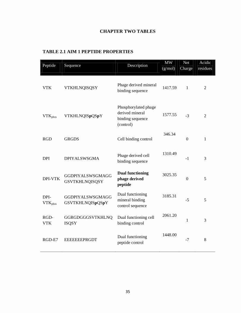
Single and dual function experimental and control peptides were synthesized using solid phase
synthesis and protective chemistry (Table 2.1). High performance liquid chromatography was
used to verify > 95% purity. Peptides were stored at -20oC until use in each experiment.
25
LANGMUIR ISOTHERMS
Peptides were solubilized in water and diluted in Trizma buffer (pH 7.5). Photometric readings
for peptide standard curves and samples were taken at 25oC using a multi-well plate reader
measuring UV absorbance from 205-240nm at 5nm increments. The absorbance wavelength that
produced the best linear curve fit for standards was used to calculate sample concentrations
before and after the adsorption assay. This wavelength varied between 205-215nm for single
peptides and 230-235nm for dual peptides. Isotherm studies were conducted using HA powder
with an average particle size of 18-30 μm and a surface area of 50m2/g suspended in Trizma
buffer pH 7.5 for 2-3 hrs at 37oC prior to experiments. A powder presents a relatively high
surface to volume ratio allowing less peptide usage in a smaller volume. Adsorption assays were
conducted by incubating an HA suspension and peptide solution in a 96-well Millipore filter plate
for 3hr at 37oC under agitation. Solution was filtered into a fresh 96-well plate and the amount of
unbound peptide was determined using UV absorbance and standards. Bulk peptide
concentrations tested ranged from 0-2000µg/mL. Material specific peptides demonstrated no
affinity towards tissue culture polystyrene surfaces, negating the possibility of non-specific
binding. To assess peptide affinity to apatite, Langmuir isotherms of bulk versus bound peptide
were constructed to determine binding affinity (1/KD) and maximal adsorption concentration
(Vmax) using the following equation: [25], [26]
Where Cb is the bulk concentration of peptide and Cs is the concentration of peptide bound to
substrate, and KD is the dissociation constant. Two experiments were done, each with 3
replicates(n=6). The Gibbs free energy of adsorption was calculated as previously
described[27] using the following equation:

26
Where the gas constant (R) = 8.314 J mol-1
K-1
, ambient temperature (T) = 310 K, and the molar
concentration of solvent water is Csolv = 55.5 mol L-1
.
CELL ATTACHMENT ASSAYS
Biomimetic apatite films were incubated in ddH2O overnight to remove excess salts, and then
incubated in Trizma buffer prior to use. Mineralized films were attached to the bottom of 24 well
plates with sticky tabs. Films were subsequently incubated in 100 μg/mL of peptide solution for 3
hrs, washed and blocked with 1% denatured BSA to reduce non-specific cell attachment. The
amount of peptide on apatite coated films was quantified using UV absorbance and a BCA assay.
Loading efficiency on biomimetic apatite was not significantly different across peptide groups.
Cell centrifugation assays using hBMSCs, IPS-MSCs, mBMSCs, MC3T3s and MDFs were
conducted using a seeding density of 35,000 cells/cm2. Peptide coated films, no peptide controls,
and films used for standard curves were incubated with cells for 3hrs at 37oC and 5% CO2 in
serum-free media. Peptide coated films and no-peptide controls were subsequently washed, wells
were filled with PBS, inverted, sealed and centrifuged with forces of 10-8
10-7
dynes using an
Eppendorf 5810r centrifuge[28], [29]. At forces above 10-6 dynes, mineral and FITC-BSA were
washed away. Detached cells were removed, and cell numbers on peptide coated films, no
peptide controls and standard curves were determined using the WST-1 assay (Clontech
Laboratories, Inc.). Adherent cell fractions on peptide coated apatite groups were normalized to
no-peptide biomimetic apatite film controls at each centrifugation speed. Half-cell detachment
forces (τ50) were calculated by fitting the remaining adherent cell fraction(%) to sigmoidal curves
using the Boltzmann equation.
27
( (
))
Where L1 = lower asymptote, L2 = upper asymptote, is the inflection point, and is the
slope at the inflection point. A least squares regression with the current data allows for
determination of these parameters. The τ50 values, detachment force where 50% cells are
detached, are determined using these parameters. Using excel solver, a least squares regression
was done with bounded constraints on x0.
CELL MORPHOLOGY AND IMMUNOHISTOCHEMISTRY
A range of hBMSC seeding densities spanning 1,000-50,000 cells/cm2 was used to demonstrate
the interplay between initial cell seeding density and seeding efficiency on dual-peptide coated
apatite surfaces. Mineralized peptide coated and uncoated controls containing adherent cells from
detachment forces assays and seeding density experiments were washed twice in PBS, fixed in
10% formalin buffer, permeabilized in Triton X, and stained with Rhodhamine-
Pholloidin(ThermoFisher Scientific) for F-Actin and mounted in Vectashield containing DAPI
(Vectorlabs) on glass coverslips. Images were acquired with a NIKON Ti-Eclipse Confocal
Microscope using a 20x objective. Images from 4 samples per group and 10 fields per
sample(n=40) were analyzed using Image J software (NIH). Each field was analyzed for cell
number using the dapi stain, total cell spread area marked by F-actin stain, and total cell spread
area per cell was calculated from the initial two measurements.
STATISTICAL METHODS
Single factor ANOVA was used to determine differences in binding affinity, half-cell detachment
forces and quantitative histomorphometry amongst the different peptides using Sigmastat. Two-
way ANOVA on ranks with Tukey test pairwise comparisons and interactions was used to
determine differences and interaction between peptide groups across seeding densities.

28
RESULTS
PEPTIDE BINDING ISOTHERMS ON HA
Mineral binding and dual peptides reached an adsorption equilibrium between 0-1000µg/mL
indicating apatite saturation (Fig 2.1a,b). The phosphorylated mineral specific sequence VTKphos
demonstrated a greater binding affinity to apatite than VTK (Table 2.2). The concentration
required to reach the equilibrium condition is lower for VTKphos than for VTK. The dual-
functioning phage derived peptides DPI-VTK (p<0.001), DPI-VTKphos, (p <0.01) and dual
functioning peptide with cell binding control RGD-VTK (p < 0.01) demonstrated higher binding
affinities than the single peptides. DPI-VTK had a lower KD than DPI-VTKphos (p < 0.01).
Although the acidic residues in E7 bind strongly with the cationic components of HA, the binding
affinity of RGD-E7 was lower than predominantly charge neutral RGD-VTK and DPI-VTK
(p<0.01).
Values for Vmax were within the range of reported monolayer adsorption concentrations
for proteins and peptides [25], [26]. Moreover, calculated Vmax values based on cross-sectional
area of each peptide in a linear and cyclic conformation fall within the range of experimentally
observed values(Table 2.2). Vmax for single peptides was higher than dual peptides (p < 0.01) with
the exception of RGD-E7.
CELL ADHESION STRENGTH ON DUAL-PEPTIDE COATED MINERAL
Dual peptides RGD-VTK and RGD-E7 yielded the highest cell attachment when no force
was applied compared to remaining peptide coated and control groups (Fig. 2.2a, p < 0.01).
However, as forces were applied, larger cell fractions were adherent to DPI-VTK compared to
other peptide coated and control groups. Consequently, τ50 for the DPI-VTK sequence was
higher than for the rest of the peptides (Fig. 2.2b, p<0.01).

29
HBMSCs exhibited a more spread morphology on RGD-VTK, DPI-VTK and RGD-E7
compared to other peptides (Fig. 2.2c). The total surface area covered by cells was greater on
RGD-VTK and RGD-E7 coated surfaces, however, spread area/cell was greater on DPI-VTK
compared to RGD-VTK and RGD-E7 (Fig. 2.2d,e p<0.01).
HBMSC SPECIFICITY TO DUAL-PEPTIDE COATED MINERAL
HBMSCs, mBMSCs and IPS-MSCs bound more strongly to DPI-VTK than murine pre-
osteoblasts and fibroblasts (Fig. 2.3a, p<0.01). RGD-VTK bound weakly to hBMSCs, however it
promoted stronger adhesion to MC3T3s and MDFs than DPI-VTK (p < 0.01). Although IPS-
MSC adhesion to DPI-VTK was greater than to RGD-VTK (p < 0.01), IPS-MSCs demonstrated
similar adhesion strength on RGD-VTK compared to pre-osteoblasts and fibroblasts.
HBMSC morphology on DPI-VTK and RGD-VTK (Fig. 2.2c) was rounder and less
spread compared to mBMSC and IPS-MSC morphology on these peptides (Fig. 2.3b). MC3T3s
and MDFs spread more on RGD-VTK compared to DPI-VTK. Conversely, MSCs spread more
on DPI-VTK compared to RGD-VTK. Total cell spread area was greater on RGD-VTK
compared to DPI-VTK across all cell types (p < 0.01), with the exception of mBMSCs. However,
spread area normalized to number of MSCs was greater on DPI-VTK compared to RGD-VTK
and the converse relationship was observed with MC3T3 and MDFs (Fig. 2.3c).
HBMSCs on RGD-VTK had higher total cell spread area than MC3T3 and MDF. This
indicates that more hBMSCs bound RGD-VTK, but MC3T3s and MDFs bound and spread more
favorably on these surfaces. This could arise from interplay between cell spreading and cell
density after initial cell attachment.
MSC ADHESION DEPENDENCE ON CELL SEEDING DENSITY
Adherent cell numbers are dependent on peptide groups and initial cell seeding density
(p<0.001). The total cell coverage area is correlated with initial cell seeding density (p < 0.05)

30
which is stronger than the correlation between spread area per cell and initial seeding density. The
interaction between initial seeding density, initial cell attachment and spread area per cell can be
observed at the higher seeding densities 50, 40, and 30 k cells/cm2(Fig. 2.4a). DPI-VTK improves
initial cell attachment on apatite compared to VTK. However at 50k cells/cm2, although the total
spread area is higher on DPI-VTK, there is equivalent spread area/cell to that of VTK (Fig. 2.4
b,c). This indicates a surface saturation of hBMSCs on VTK. HBMSCs exhibit more spreading
on DPI-VTK at 40k cells/cm2 compared to 50k cells/cm
2 but spread area/cell on DPI-VTK and
VTK at 50k cells/cm2 is not significantly different. Moreover, there is no significant difference
between spread area/cell between HBMSCs on DPI-VTK at 30k cells/cm2 and HBMSCs on VTK
at 50k cells/cm2. Trends in spread area per cell between both peptides across various densities
are aligned with data attained at a seeding density of 37.5 k cells/cm2 for cell centrifugation
assays.
DISCUSSION
HBMSC specific peptide sequences were identified using a combinatorial phage display.
Apatite-specific peptide sequences were similarly identified using a combinatorial phage display
[30] and in this study were combined with cell-specific sequences to create dual functioning
peptides with one domain having preferential affinity for a specific material chemistry and a
second domain having preferential affinity for a specific cell population.
Both cell and mineral binding domains contributed to the binding of dual functioning
peptides to apatite (Table 2.2). Phosphorylation is a common post translational modification in
natural ECM peptides and proteins that interact with mineral [16], [31]. Molecular dynamics
simulations of dentin phosphoproteins(DPP) reveal un-phosphorylated DPP peptide derivatives
are more mobile, flexible, and can fold over HA surfaces however phosphorylated peptides drive
mineral association [32]. Although phosphorylation drives apatite binding affinity of DPP and
osteopontin derived ASARM peptides, it is the glutamic and aspartic acid residues that are

31
involved in binding the apatite surface [32], [33]. These data indicate the interplay between
sequence, conformation, charge distribution and apatite affinity of phosphorylation which could
all be contributing to the differences between DPI-VTK and DPI-VTKphos compared to VTKphos
and VTK. Moreover, RGD-E7, which has an acidic mineral binding motif, had the lowest binding
affinity amongst the dual functioning peptides. The KD values for RGD-E7 (Table 2.2) are
consistent with studies using similar materials and assay conditions [21]. Conformational and
charge distributions of bound peptide could also be contributing to the differences in Vmax. Single
peptides can pack more efficiently because of smaller size, however the increased saturation
concentration of RGD-E7 could be a result of non-Langmuir kinetics and potential aggregation in
the solution state. Taken together, peptide sequence and conformation also contribute to packing
efficiency.
The half-cell detachment force is the force at which 50% of the initially bound cell
population becomes detached. Detachment forces are a surrogate for how strongly cells attach to
the substrate. The strength of attachment is related to the formation of specific peptide and cell
surface receptor interactions. Since the adhesion timeframe in serum-free media allows for initial
attachment only, the detachment force correlates with the average number of peptide-cell ligand
interactions over a population of cells [29]. The dual-functioning phage derived peptide DPI-
VTK had the highest hBMSC adhesion strength (Fig. 2.3b). Although VTK and VTKphos
exhibited similar initial attachment to DPI-VTK, they did not exhibit the same level of adhesion
strength as DPI-VTK. This indicates that adhesion strength of cells to DPI-VTK is largely driven
by the interaction with the DPI domain of the peptide.
Moreover, the weaker attachment of hBMSCs to DPI-VTKphos compared to DPI-VTK
(Fig. 2.3b) indicates that phosphorylating the mineral sequence compromises the presentation of
the DPI sequence to the cell. Favorable binding to apatite and improved adhesion strength to
hBMSCs of DPI-VTK, compared to DPI-VTKphos, indicates an involvement indirect effect of the

32
cell specific sequence on mineral association and an indirect effect of mineral sequence in
directing cell association. For instance, adding DPI to the VTK sequence could lead to
conformational changes to either the mineral or cell binding sequences that enhance mineral
binding affinity. Once bound, association with the biomimetic apatite could cause another
structural change in either DPI or VTK, resulting in a change in affinity to cell binding targets.
Structural changes occur in both solution and apatite-bound states and can drive adsorption of
peptides [34]–[36].
More hBMSCs were initially bound to RGD-VTK, RGD-E7, DPI-VTK, and DPI-
VTKphos compared to VTK and VTKphos (Fig. 2.3a). The increase in cell attachment to these
peptides also indicates that hBMSC cell surface receptors bind to both DPI and RGD peptides, as
expected. Although there was less initial cell attachment on DPI-VTK compared to RGD-VTK,
DPI-VTK was more favorable for cell spreading (Fig. 2.3d), a finding that also supports the
adhesion data as force is applied (Fig. 2.3a). The prevalence of more spread cells indicates more
cell-matrix interactions that result in stronger attachment forces.
The adhesion strength of MSCs to DPI-VTK was greater than to MC3T3-E1 cells and
MDFs (Fig. 2.4a). This data indicates that the phage derived peptide has interactions that are
specific to cell-surface receptors on MSCs, and verifies that phage display is capable of yielding
cell-specific peptide sequences. Although hBMSCs demonstrated higher initial attachment to
RGD-VTK and RGD-E7 (Fig. 2.3a), the weaker adhesion strength is indicative of weak
association between hBMSC binding targets and RGD when presented with either E7 or VTK
material binding sequences. Another driver of DPI-VTK specificity to MSCs could arise from
differential integrin expression profiles of MC3T3c and MDFs compared to MSCs.
Initial cell seeding density plays a critical role in the degree of cell spreading. This
supports the contributions of both cell-cell and cell-substrate interactions to cell adhesion. The

33
differences in hBMSC spreading on DPI-VTK and VTK at 50k, 40k, and 30k cells/cm2 (Fig. 2.4)
indicate an interaction between cell density and peptide function. HBMSCs are able to attach
specifically to DPI-VTK and have sufficient room to spread below the seeding density of 30k -
40k cells/cm2. However, as seeding density increased to 50k cells/cm
2, initial cell attachment was
so prevalent that the cells were unable to spread past a critical threshold. Initial cell attachment,
subsequent adhesion and spreading, and density of seeded cells all contribute to tissue
regeneration.
Phage display identifies sequences that bind strongly to markers expressed on the cell
surface. The absence of highly acidic residues in either set of sequences is an interesting outcome
of the phage selection process. Phage display has been used to identify peptides that target
biomaterials [37], [38], as well as stem, progenitor, carcinogenic and bacterial cells (22,23,34),
and there is a bias for hydrophobic peptide selection [39]. The prevalence of hydrophobic
sequences could be an artifact of the selection process and needs to be further explored. Although
this hydrophobic bias seems counterintuitive for apatite binding, it could be a promising tool for
biomolecule delivery. Peptides with acidic residues that react with the defined periodicity of Ca2+
ions in the HA crystal lattice also interfere with the adsorption of serum proteins [19], [40]. For
instance, RGD improves cell attachment, proliferation and differentiation in vitro, but has varied
results in vivo. This could be both a function of how cells attach and spread on RGD covered
surfaces and how the presence of RGD interferes with adsorption of serum proteins. Moreover,
RGD is not specific to any particular cell type and can react with various integrin subunits.
Therefore, using a hydrophobic mineral binding sequence like VTK to deliver a human bone
marrow stromal cell specific sequence like DPI will not only anchor cell-specific sequences to
HA surfaces, but could also allow adsorption of serum proteins and facilitate specific cell
mediated tissue regeneration. The results collectively indicate that phage display can be used to

34
identify cell specific hBMSCs and material specific peptides that improve cell attachment and
spreading on biomaterial surfaces, potentially leading to improved regeneration.
DPIYALSWSGMA was identified as a high MSC binding sequence. This peptide was
combined with previously identified mineral binding sequence VTKHLNQISQSY to specifically
recruit MSCs to biomimetic apatite substrates. The dual peptide DPI-VTK increased binding
affinity to apatite compared to cell, mineral, and dual peptide controls. Moreover, DPI-VTK
improved MSC attachment and specificity, and promoted cell promote spreading on apatite.
These data demonstrate the utility of phage display to recruit specific cell populations to specific
biomaterial substrates.
ACKNOWLEDGEMENTS
We would like to thank Dr. Sergei Kuznetsov for the generous contribution of primary human
bone marrow stromal cells and Dr. Paul Krebsbach for the induced pluripotent mesenchymal
cells. Funded by NIH DE015411 and NIH DE13380.
35
CHAPTER TWO TABLES
TABLE 2.1 AIM 1 PEPTIDE PROPERTIES
Peptide Sequence Description MW
(g/mol)
Net
Charge
Acidic
residues
VTK VTKHLNQISQSY Phage derived mineral
binding sequence
1417.59
1 2
VTKphos VTKHLNQISpQSpY
Phosphorylated phage
derived mineral
binding sequence
(control)
1577.55
-3 2
RGD GRGDS Cell binding control 346.34
0 1
DPI DPIYALSWSGMA Phage derived cell
binding sequence
1310.49
-1 3
DPI-VTK GGDPIYALSWSGMAGG
GSVTKHLNQISQSY
Dual functioning
phage derived
peptide
3025.35
0 5
DPI-
VTKphos
GGDPIYALSWSGMAGG
GSVTKHLNQISpQSpY
Dual functioning
mineral binding
control sequence
3185.31
-5 5
RGD-
VTK
GGRGDGGGSVTKHLNQ
ISQSY
Dual functioning cell
binding control
2061.20
1 3
RGD-E7 EEEEEEEPRGDT Dual functioning
peptide control
1448.00
-7 8

36
TABLE 2.2: BINDING ISOTHERM RESULTS
Kd.
(µM)
Vmax
(μmol/cm2)
ΔGads
(kJ)/mol
r2
VTK
74.57±3.59
30.67±5.44
-34.8
0.94
VTKphos *
32.67±1.69
21.49±4.35
-37.0
0.93
RGD-E7
74.13±3.47
328.86±54.39
-34.9
0.95
RGD-VTK**
7.36±0.35
4.92±2.56
-40.8
0.90
DPI-VTKphos**
5.04±0.55
30.05±6.41
-41.8
0.97
DPI-VTK***
2.68±0.57
17.94±3.22
-43.4
0.93
* significantly different from RGD-E7 and VTK(p <0.01)
**significantly different from dual peptides * and ***(p<0.01)
ΔG= -RT Ln(Csolv/Kd) R = 8.314 J mol-1
K-1
T = 310 K Csolv = 55.5 mol/L

37
CHAPTER TWO FIGURES
FIGURE 2.1 (A,B). LANGMUIR ISOTHERMS
Figure 2.1. Langmuir isotherms of A)mineral binding and B)dual functioning peptides on HA
powder. Cb is the bulk concentration of peptide in solution and Cs is the bound concentration of
peptide on hydroxyapatite particles. Data points represent triplicates from 2 separate adsorption
studies (n=6).
0
10
20
30
40
50
0 200 400 600
VTK
VTKphos
A
Cs(
μg/
cm2)
Cb (μg/mL)
0
50
100
150
200
250
0 200 400 600
RGD-E7
RGD-VTK
DPI-VTKphos
DPI-VTK
B
Cb (μg/mL)
Cs(
μg/
cm2)

38
FIGURE 2.2(A-E). HBMSC ATTACHMENT, ADHESION STRENGTH AND
SPREADING ON SINGLE AND DUAL-FUNCTIONING PEPTIDE COATED
APATITE FILMS
Thousands
-20
0
20
40
60
80VTK VTKphos DPI -VTKphos
DPI-VTK RGD-VTK RGD-E7*
*
*
μdynes
A A
dh
ere
nt
Ce
ll
0.0
0.2
0.4
0.6
τ5
0 (
μd
yn
es)
Mineral binding Dual-functioning
*
B
VTK VTKphos
DPI-VTKphos
DPI-VTK RGD-VTK RGD-E7
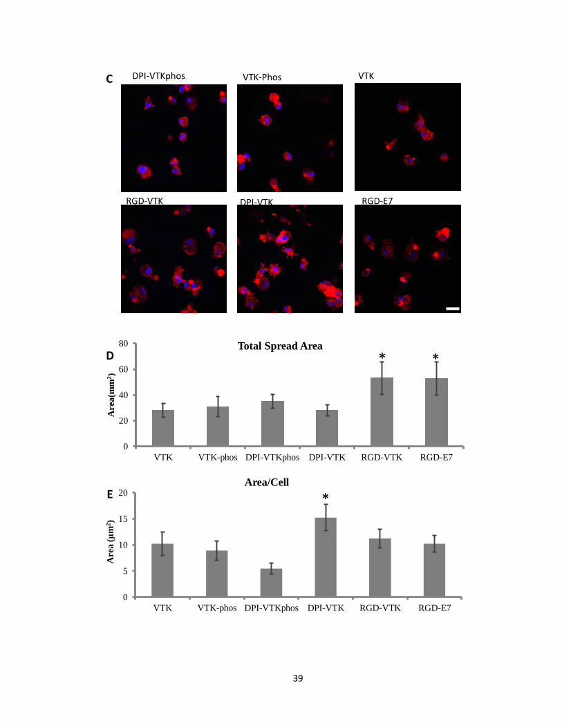
39
DPI-VTKphos VTK-Phos VTK
RGD-VTK DPI-VTK RGD-E7
C
0
5
10
15
20
VTK VTK-phos DPI-VTKphos DPI-VTK RGD-VTK RGD-E7
Area (
μm
2)
Area/Cell
0
20
40
60
80
VTK VTK-phos DPI-VTKphos DPI-VTK RGD-VTK RGD-E7
Area
(mm
2)
Total Spread Area
* *
*
D
E

40
Figure 2.2. HBMSC attachment, adhesion strength and spreading on single and dual-functioning
peptide coated apatite films. A) Cell attachment across different peptide groups exposed to a
range of detachment forces(n=6). *denotes significance from unmarked peptide groups within
detachment forces (p <0.01). B) Half-cell detachment forces calculated from sigmoidal curve-
fitting of cell attachment data indicating force at which 50% of the initially adherent cell
populations become detached (n=6). * denotes difference form remaining peptide groups
(p<0.01). C) Representative confocal microscopy images at 40x depicting cell spreading on
peptide coated apatite films. F-actin labeled with Rhodhamine-Pholloidin and nuclei labeled
with DAPI. Scale bars 10µm. D) Total cell spread area calculated from Image J analysis of
Pholloidin-stained area (n=4 per group x 10 FOV per sample). * denotes different from
unmarked groups(p<0.01) E) Area /cell calculated from Image J(NIH) analysis of Pholloidin-
stained area (n=4 per group x 10 FOV per sample). * denotes different from unmarked
groups(p<0.01)
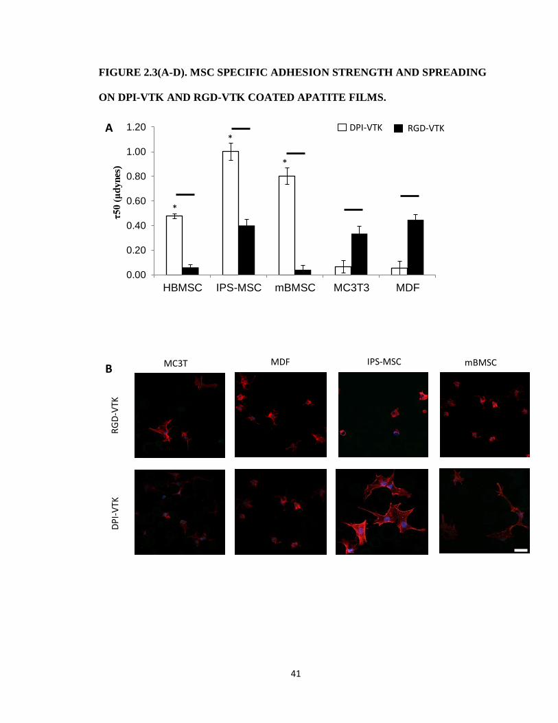
41
FIGURE 2.3(A-D). MSC SPECIFIC ADHESION STRENGTH AND SPREADING
ON DPI-VTK AND RGD-VTK COATED APATITE FILMS.
0.00
0.20
0.40
0.60
0.80
1.00
1.20
HBMSC IPS-MSC mBMSC MC3T3 MDF
τ5
0 (
μd
yn
es)
*
*
*
DPI-VTK RGD-VTK A
RG
D-V
TK
DP
I-V
TK
MC3T
3
MDF IPS-MSC mBMSC B
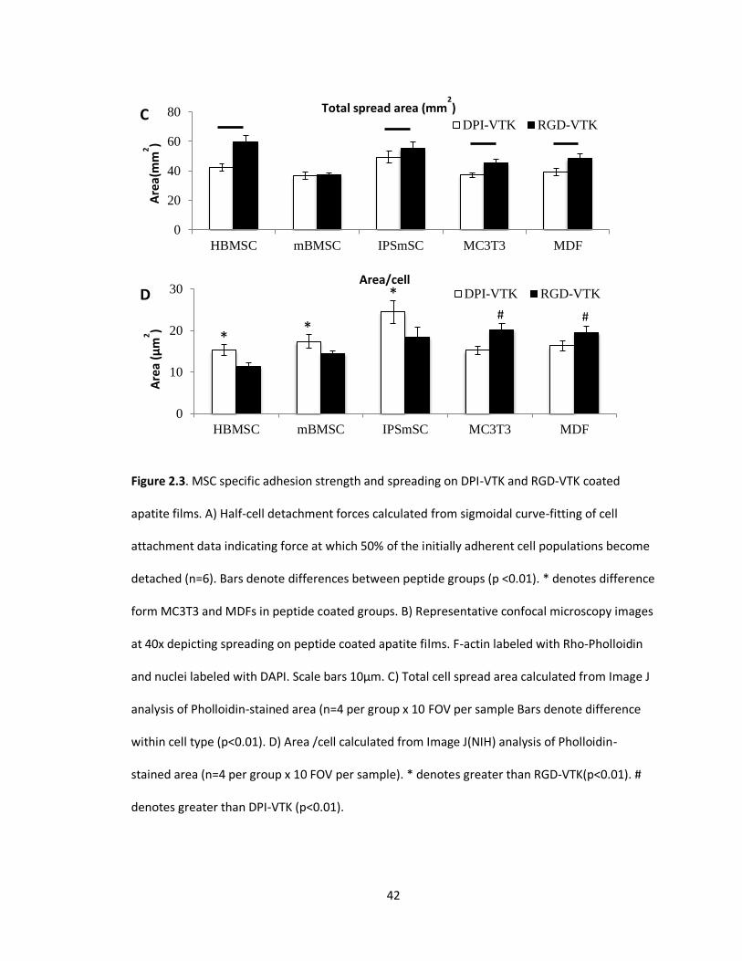
42
Figure 2.3. MSC specific adhesion strength and spreading on DPI-VTK and RGD-VTK coated
apatite films. A) Half-cell detachment forces calculated from sigmoidal curve-fitting of cell
attachment data indicating force at which 50% of the initially adherent cell populations become
detached (n=6). Bars denote differences between peptide groups (p <0.01). * denotes difference
form MC3T3 and MDFs in peptide coated groups. B) Representative confocal microscopy images
at 40x depicting spreading on peptide coated apatite films. F-actin labeled with Rho-Pholloidin
and nuclei labeled with DAPI. Scale bars 10µm. C) Total cell spread area calculated from Image J
analysis of Pholloidin-stained area (n=4 per group x 10 FOV per sample Bars denote difference
within cell type (p<0.01). D) Area /cell calculated from Image J(NIH) analysis of Pholloidin-
stained area (n=4 per group x 10 FOV per sample). * denotes greater than RGD-VTK(p<0.01). #
denotes greater than DPI-VTK (p<0.01).
0
20
40
60
80
HBMSC mBMSC IPSmSC MC3T3 MDF
DPI-VTK RGD-VTK
0
10
20
30
HBMSC mBMSC IPSmSC MC3T3 MDF
DPI-VTK RGD-VTKD
C
Are
a(m
m2)
Are
a (μ
m2)
Area/cell
Total spread area (mm2)
* *
*
# #
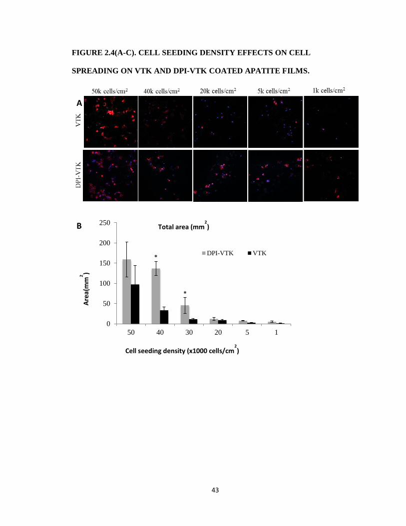
43
FIGURE 2.4(A-C). CELL SEEDING DENSITY EFFECTS ON CELL
SPREADING ON VTK AND DPI-VTK COATED APATITE FILMS.
0
50
100
150
200
250
50 40 30 20 5 1
DPI-VTK VTK
Cell seeding density (x1000 cells/cm2)
Total area (mm2)
Are
a(m
m2)
B
*
*
A

44
Figure 2.4. Cell seeding density effects on cell spreading on VTK and DPI-VTK coated apatite
films. A) Representative confocal microscopy images at 20x depicting cell spreading on peptide
coated apatite films. F-actin labeled with Rho-Pholloidin and nuclei labeled with DAPI. Scale bars
20µm. B) Total cell spread area calculated from Image J analysis of Pholloidin-stained area (n=4
per group x 10 FOV per sample Bars denote difference within cell type (p<0.01).* denotes
difference from VTK (p < 0.01) C) Area/cell calculated from Image J(NIH) analysis of Pholloidin-
stained area (n=4 per group x 10 FOV per sample). * denotes difference from VTK (p < 0.01)
Cell seeding density (x1000 cells/cm2)
0
10
20
30
40
50
50 40 30 20 5 1
DPI-VTK VTK
Area/cell
Are
a (μ
m2)
C
*
*
* *
*

45
REFERENCES
[1] P. H. Krebsbach, S. a. Kuznetsov, P. Bianco, and P. Gehron Robey, “Bone Marrow Stromal
Cells: Characterization and Clinical Application,” Crit. Rev. Oral Biol. Med., vol. 10, no. 2,
pp. 165–181, Jan. 1999.
[2] E. Alsberg, E. E. Hill, and D. J. Mooney, “Craniofacial tissue engineering.,” Crit. Rev. Oral
Biol. Med., vol. 12, no. 1, pp. 64–75, Jan. 2001.
[3] K. Kim, D. Dean, A. G. Mikos, and J. P. Fisher, “Effect of initial cell seeding density on early
osteogenic signal expression of rat bone marrow stromal cells cultured on cross-linked
poly(propylene fumarate) disks.,” Biomacromolecules, vol. 10, no. 7, pp. 1810–7, Jul.
2009.
[4] C. E. Wilson, W. J. A. Dhert, C. A. Van Blitterswijk, A. J. Verbout, and J. D. De Bruijn,
“Evaluating 3D bone tissue engineered constructs with different seeding densities using
the alamarBlue assay and the effect on in vivo bone formation.,” J. Mater. Sci. Mater.
Med., vol. 13, no. 12, pp. 1265–9, Dec. 2002.
[5] M. H. Mankani, S. A. Kuznetsov, B. Fowler, A. Kingman, and P. G. Robey, “In vivo bone
formation by human bone marrow stromal cells: effect of carrier particle size and
shape.,” Biotechnol. Bioeng., vol. 72, no. 1, pp. 96–107, Jan. 2001.
[6] P. H. Krebsbach, S. A. Kuznetsov, K. Satomura, R. V Emmons, D. W. Rowe, and P. G.
Robey, “Bone formation in vivo: comparison of osteogenesis by transplanted mouse and
human marrow stromal fibroblasts.,” Transplantation, vol. 63, no. 8, pp. 1059–69, Apr.
1997.
[7] R. G. LeBaron and K. A. Athanasiou, “Extracellular matrix cell adhesion peptides:
functional applications in orthopedic materials ,” Tissue Eng., vol. 6, no. 2, pp. 85–103,
Apr. 2000.
[8] S. J. Segvich and D. H. Kohn, Biological Interactions on Materials Surfaces. New York, NY:
Springer US, 2009.
[9] Ramaswamy, J., Ramaraju, S., and Kohn D. H., “Bone-Like Mineral and Organically
Modified Bone-Like Mineral Coatings,” in Biological and Biomedical Coatings Handbook,
Processing and Characterization, S. Zhang, Ed. Boca Raton, FL: CRC Press, 2011, pp. 1–36.
[10] W. L. Murphy, D. H. Kohn, and D. J. Mooney, “Growth of continuous bonelike mineral
within porous poly(lactide-co-glycolide) scaffolds in vitro.,” J. Biomed. Mater. Res., vol.
50, no. 1, pp. 50–8, Apr. 2000.
[11] A. Shekaran and A. J. García, “Extracellular matrix-mimetic adhesive biomaterials for
bone repair.,” J. Biomed. Mater. Res. A, vol. 96, no. 1, pp. 261–72, Jan. 2011.

46
[12] S. X. Hsiong, T. Boontheekul, N. Huebsch, and D. J. Mooney, “Cyclic arginine-glycine-
aspartate peptides enhance three-dimensional stem cell osteogenic differentiation ,”
Tissue Eng. A, vol. 15, no. 2, pp. 263–272, Feb. 2009.
[13] J. S. Lee, J. S. Lee, A. Wagoner-Johnson, and W. L. Murphy, “Modular peptide growth
factors for substrate-mediated stem cell differentiation.,” Angew. Chem. Int. Ed. Engl.,
vol. 48, no. 34, pp. 6266–9, Jan. 2009.
[14] M. P. Lutolf, F. E. Weber, H. G. Schmoekel, J. C. Schense, T. Kohler, R. Muller, and J. A.
Hubbell, “Repair of bone defects using synthetic mimetics of collagenous extracellular
matrices ,” Nat. Biotechnol., vol. 21, no. 5, pp. 513–518, May 2003.
[15] D. Itoh, S. Yoneda, S. Kuroda, H. Kondo, A. Umezawa, K. Ohya, T. Ohyama, and S. Kasugai,
“Enhancement of osteogenesis on hydroxyapatite surface coated with synthetic peptide
(EEEEEEEPRGDT) in vitro.,” J. Biomed. Mater. Res., vol. 62, no. 2, pp. 292–8, Nov. 2002.
[16] M. Gilbert, W. J. Shaw, J. R. Long, K. Nelson, G. P. Drobny, C. M. Giachelli, and P. S.
Stayton, “Chimeric peptides of statherin and osteopontin that bind hydroxyapatite and
mediate cell adhesion.,” J. Biol. Chem., vol. 275, no. 21, pp. 16213–8, May 2000.
[17] P. S. Stayton, G. P. Drobny, W. J. Shaw, J. R. Long, and M. Gilbert, “Molecular Recognition
At the Protein-Hydroxyapatite Interface,” Crit. Rev. Oral Biol. Med., vol. 14, no. 5, pp.
370–376, Sep. 2003.
[18] M. Kantlehner, P. Schaffner, D. Finsinger, J. Meyer, A. Jonczyk, B. Diefenbach, B. Nies, G.
Holzemann, S. L. Goodman, and H. Kessler, “Surface coating with cyclic RGD peptides
stimulates osteoblast adhesion and proliferation as well as bone formation ,”
Chembiochem, vol. 1, no. 2, pp. 107–114, Aug. 2000.
[19] K. M. Hennessy, W. C. Clem, M. C. Phipps, A. A. Sawyer, F. M. Shaikh, and S. L. Bellis, “The
effect of RGD peptides on osseointegration of hydroxyapatite biomaterials.,”
Biomaterials, vol. 29, no. 21, pp. 3075–83, Jul. 2008.
[20] J. S. Lee, A. J. Wagoner Johnson, and W. L. Murphy, “A modular, hydroxyapatite-binding
version of vascular endothelial growth factor.,” Adv. Mater., vol. 22, no. 48, pp. 5494–8,
Dec. 2010.
[21] R. Fujisawa, M. Mizuno, Y. Nodasaka, and Y. Kuboki, “Attachment of osteoblastic cells to
hydroxyapatite crystals by a synthetic peptide (Glu7-Pro-Arg-Gly-Asp-Thr) containing two
functional sequences of bone sialoprotein.,” Matrix Biol., vol. 16, no. 1, pp. 21–8, Apr.
1997.
[22] R. Fujisawa, Y. Wada, Y. Nodasaka, and Y. Kuboki, “Acidic amino acid-rich sequences as
binding sites of osteonectin to hydroxyapatite crystals.,” Biochim. Biophys. Acta, vol.
1292, no. 1, pp. 53–60, Jan. 1996.

47
[23] S. A. Kuznetsov, M. H. Mankani, and P. G. Robey, “In vivo formation of bone and
haematopoietic territories by transplanted human bone marrow stromal cells generated
in medium with and without osteogenic supplements.,” J. Tissue Eng. Regen. Med., vol. 7,
no. 3, pp. 226–35, Mar. 2013.
[24] S. a Kuznetsov, P. H. Krebsbach, K. Satomura, J. Kerr, M. Riminucci, D. Benayahu, and P.
G. Robey, “Single-colony derived strains of human marrow stromal fibroblasts form bone
after transplantation in vivo.,” J. Bone Miner. Res., vol. 12, no. 9, pp. 1335–47, Sep. 1997.
[25] B. . Young, W. . Pitt, and S. . Cooper, “Protein adsorption on polymeric biomaterials I.
Adsorption isotherms,” J. Colloid Interface Sci., vol. 124, no. 1, pp. 28–43, Jul. 1988.
[26] T. Tsai, R. C. Mehta, and P. P. Deluca, “Adsorption of peptides to poly(D,L-lactide-co-
glycolide): 1. Effect of physical factors on the adsorption,” Int. J. Pharm., vol. 127, no. 1,
pp. 31–42, Jan. 1996.
[27] F. Meder, H. Hintz, Y. Koehler, M. M. Schmidt, L. Treccani, R. Dringen, and K. Rezwan,
“Adsorption and Orientation of the Physiological Extracellular Peptide Glutathione Disul
fi de on Surface Functionalized Colloidal Alumina Particles,” 2013.
[28] N. D. Gallant, “Stick and Grip,” Cell Biochem. Biophys., vol. 39, no. 9, 2003.
[29] D. R. McClay and P. L. Hertzler, “Quantitative measurement of cell adhesion using
centrifugal force.,” Curr. Protoc. Cell Biol., vol. Chapter 9, p. Unit 9.2, May 2001.
[30] S. J. Segvich, H. C. Smith, and D. H. Kohn, “The adsorption of preferential binding
peptides to apatite-based materials.,” Biomaterials, vol. 30, pp. 1287–1298, 2009.
[31] W. N. Addison, S. J. Miller, J. Ramaswamy, A. Mansouri, D. H. Kohn, and M. D. McKee,
“Phosphorylation-dependent mineral-type specificity for apatite-binding peptide
sequences.,” Biomaterials, vol. 31, no. 36, pp. 9422–30, Dec. 2010.
[32] E. Villarreal-Ramirez, R. Garduño-Juarez, A. Gericke, and A. Boskey, “The role of
phosphorylation in dentin phosphoprotein peptide absorption to hydroxyapatite
surfaces: a molecular dynamics study.,” Connect. Tissue Res., vol. 55 Suppl 1, pp. 134–7,
Aug. 2014.
[33] W. N. Addison, D. L. Masica, J. J. Gray, and M. D. McKee, “Phosphorylation-dependent
inhibition of mineralization by osteopontin ASARM peptides is regulated by PHEX
cleavage.,” J. Bone Miner. Res., vol. 25, no. 4, pp. 695–705, Apr. 2010.
[34] D. L. Masica, J. J. Gray, and W. J. Shaw, “Partial High-resolution structure of
phosphorylated and non-phosphorylated leucine-rich amelogenin protein adsorbed to
hydroxyapatite,” J. Phys. Chem. C, vol. 115, pp. 13775–13785, 2011.
48
[35] W. N. Addison, D. L. Masica, J. J. Gray, and M. D. McKee, “Phosphorylation-dependent
inhibition of mineralization by osteopontin ASARM peptides is regulated by PHEX
cleavage.,” J. Bone Miner. Res., vol. 25, pp. 695–705, 2010.
[36] D. L. Masica and J. J. Gray, “Solution- and adsorbed-state structural ensembles predicted
for the statherin-hydroxyapatite system.,” Biophys. J., vol. 96, no. 8, pp. 3082–91, Apr.
2009.
[37] M. C. Weiger, J. J. Park, M. D. Roy, C. M. Stafford, A. Karim, and M. L. Becker,
“Quantification of the binding affinity of a specific hydroxyapatite binding peptide.,”
Biomaterials, vol. 31, no. 11, pp. 2955–63, Apr. 2010.
[38] M. Gungormus, E. E. Oren, J. A. Horst, H. Fong, M. Hnilova, M. J. Somerman, M. L. Snead,
R. Samudrala, C. Tamerler, and M. Sarikaya, “Cementomimetics-constructing a
cementum-like biomineralized microlayer via amelogenin-derived peptides.,” Int. J. Oral
Sci., vol. 4, no. 2, pp. 69–77, Jun. 2012.
[39] K. Luck and G. Travé, “Phage display can select over-hydrophobic sequences that may
impair prediction of natural domain-peptide interactions.,” Bioinformatics, vol. 27, no. 7,
pp. 899–902, Apr. 2011.
[40] K. M. Hennessy, B. E. Pollot, W. C. Clem, M. C. Phipps, A. A. Sawyer, B. K. Culpepper, and
S. L. Bellis, “The effect of collagen I mimetic peptides on mesenchymal stem cell adhesion
and differentiation, and on bone formation at hydroxyapatite surfaces.,” Biomaterials,
vol. 30, no. 10, pp. 1898–909, Apr. 2009.
49
CHAPTER THREE
PHAGE DISPLAY DERIVED BI-FUNCTIONAL PEPTIDE
IMPROVES MSC ADHESION, PROLIFERATION AND
DIFFERENTIATION ON APATITE
INTRODUCTION
Cell based tissue regeneration strategies often require a biomaterial carrier with defined
surface chemistries. With the proliferation of biomaterials and regenerative cell sources,
it is important to recruit specific regenerative cell populations to specific biomaterial
chemistries to meet clinical challenges[1]. In addition to attachment of specific cell
populations, filling critical sized defects with adequate transplanted cell numbers
becomes non-trivial. Improving the seeding efficiency of transplanted cell populations on
biomaterial carriers can have a significant impact on reducing material requirements,
healing time and healthcare costs[2].
Mesenchymal stem cells (MSCs) are a promising source for cell-based tissue
regeneration [3], [4]. MSCs can form various tissues, including bone, cartilage,
hematopoiesis-supporting stroma, and fat. In addition to multi-potent tissue formation,
MSCs are immunomodulatory and can induce angiogenesis . Since their initial discovery
in the bone marrow stromal niche, MSCs have been identified in various adult and
neonatal tissues including but not limited to adipose tissue, peripheral blood, amniotic
fluid, and umbilical cord[5]. MSCs can also be derived from embryonic and induced

50
pluripotent stem cell (iPS) lineages[6]–[8]. Furthermore, induced pluripotent stem cell
derived MSCs (iPS-MSCs) provide a more continuous supply compared to other sources
of MSCs since they can be programmed from more readily available somatic sources.
MSCs are characterized by their ability to adhere to plastic, proliferate readily in vitro
and form multi-lineage tissue in vivo. A profile of cell surface markers, STRO-1, CD146,
CD105, CD49a, positive for human bone marrow stroma and, CD34−, CD45
−, CD14
−,
negative for hematopoietic and endothelial lineages are also used to verify MSC sub-
populations[9]. However, there are no markers or set of markers that can separate
multipotent MSCs from non-multipotent MSCs[10], [11].
Improving specific attachment of multipotent MSCs to biomaterials towards cell-based
therapies can improve in vivo tissue regeneration outcomes. In the context of bone tissue
regeneration, materials containing a mineral component are used to mechanically and
chemically support transplanted cell populations. Precipitation of a heterogeneous layer
of biomimetic apatite on biomaterial surfaces by immersion in simulated body fluid
(SBF) under physiological conditions improves osteoconductivity and mechanical
properties of the underlying substrate[12], [13]. Adsorption of extracellular matrix cell
binding proteins, functional peptide domains, and peptide motifs onto biomaterial
surfaces can further enhance cell attachment[14], [15]. Mineral binding peptide domains
and peptide motifs are also adsorbed to mineral surfaces to improve delivery of cell
signals[16], [17]. Furthermore, peptide-based strategies to target α2β1 integrin domains
that are highly expressed in MSCs have been used to improve cell-specific attachment to
mineral[18], [19]. In addition to being osteoconductive, these peptides exhibit

51
osteoinductivity by mechanotransductively regulating osteogenic signal transduction
cascades within adherent MSCs[19]–[21].
However ECM peptide domains targeting integrins do not exhibit cell specificity; rather,
they target cell surface receptors prevalent across many cell types. Using a combinatorial
phage display we have identified peptides specific towards multipotent-MSCs clonally
derived from human bone marrow stromal cells (hBMSC) (DPIYALSWSGMA, DPI)
and apatite surfaces (VTKHLNQISQSY,VTK) and demonstrated conserved mineral and
cell specificity when the peptides are combined into a single dual-functioning peptide
(DPI-VTK)[22], [23] . In addition to mediating cell-specific attachment to apatite
surfaces, in vitro spreading, proliferation, and differentiation are important indicators of
biological performance. This study characterizes multi-potent MSC spreading and
cytoskeletal contact distribution in the presence and absence of serum proteins, identifies
biological targets for DPI mediated MSC attachment, assesses MSC proliferation and
differentiation on DPI-VTK coated biomimetic apatite surfaces, and examines selectivity
towards multi-potent MSC subpopulations.
MATERIALS AND METHODS
MINERALIZED FILM FABRICATION
Biomimetic apatite films were used to characterize cell adhesion, proliferation and
differentiation in vitro. Apatite films were prepared by immersing PLGA thin films in
simulated body fluid to precipitate carbonated apatite with plate like nanofeatures. A 5
w/v% 85:15 polylactic-co-glycolic acid (PLGA, Lakeshore Biomaterials)-chloroform
solution was cast on 15mm diameter glass slides. The PLGA films were etched in 0.5M

52
NaOH and immersed in modified simulated body fluid (mSBF) for 5 days at 37°C with
fluid changes every 24 hrs. The mSBF was made by dissolving the following reagents in
Millipore water at 25°C and titrating to pH 6.8 using NaOH: 141 mM NaCl, 4.0 mM
KCl, 0.5 mM MgSO4, 1.0 mM MgCl2, 4.2 mM NaHCO3, 5.0 mM CaCl2•2H2O, and 2.0
mM KH2PO4.
PEPTIDE SYNTHESIS AND ADSORPTION TO BONE-LIKE MINERAL
Single, dual-function, and fluorescently labeled peptides were synthesized using solid
phase synthesis and protective chemistry (Table 3.1). HPLC was used to verify > 95%
purity. Peptides were stored at -20oC until use. HBMSC specific peptide sequences were
combined with previously identified mineral binding sequences VTK and
VTKphos(Chapter 2). Films were subsequently incubated in 100 μg/mL of peptide
solution for 3 hrs, washed and blocked with 1% denatured BSA to reduce non-specific
cell attachment. The amount of peptide on apatite coated films was quantified using UV
absorbance and a Pierce™ BCA assays (ThermoFisher Scientific ). Loading efficiency on
biomimetic apatite was not significantly different across peptide groups 12.35±3nmol.
CELL CULTURE
Clonally derived human bone marrow stromal cells (hBMSC), were a generous gift from
the NIH[24], [25]. Cells were maintained in alpha minimum essential media (α-MEM)
(Gibco, #12561) with glutamine containing 20% fetal bovine serum (FBS) and antibiotics
(100 U/mL penicillin, 0.1 mg/mL streptomycin (P/S)) (Gibco, #15140) at 37°C in a 5%
CO2 incubator. Induced Pluripotent Stem Cell derived MSCs (iPS-MSC) verified for
multi-potency were a generous gift from Dr. Paul Krebsbach[7]. iPS-MSCs were
maintained in (α-MEM), 20% FBS, antibiotics, 200mM L-glutamine, and 10mM NEAA.

53
Cells were cultured in alpha minimum essential media (α-MEM), 10% FBS, and
antibiotics. Cells were passaged when they reached 80-90% confluence was replaced
every 2-3 days. Human bone marrow stromal cells were plated at 3000 cells/cm2 for
spreading (n=6/group) and proliferation assays (n=6/group). Mineralized films were held
in place with Teflon O-rings for proliferation assays to prevent films from floating.
COMPETITIVE ADHESION OF MSC
iPS-MSCs were incubated in the presence of competing antibodies (Cell Signaling
Technology, 10ug/mL) or soluble RGD (1, 0.1, 0.01mM) for 10 min at 37oC under slight
agitation (n=6/group) [26]–[28]. Mineralized films were attached to the bottom of 24 well
plates with sticky tabs. Cells were subsequently added to peptide-coated apatite films in
serum-free media for 3 hours. Adherent cells were measured using a WST-1 assay and a
standard curve from 100-10,000 cells. Cells were subsequently fixed, F-actin was stained
with Rhodhamine-Pholloidin, and nuclei were stained with DAPI. Cell number was
evaluated using Image J and compared to the WST-1 assay standard curve. Cell
spreading was qualitatively evaluated.
CELL SPREADING AND HISTOMORPHOMETRY
Mineralized films were attached to the bottom of 24-well plates using sticky tabs. Films
were subsequently incubated in 100 μg/mL of peptide solution for 3 hrs. Human bone
marrow stromal cells were plated at 3000 cells/cm2 (n=6/group) with and without serum
for 12 hrs. at 37oC and 5% CO2. Cells were fixed in 10% phosphate buffered formalin,
permeabilized in Triton X and stained with Rhodhamine-pholloidin(Life technologies)
for F-actin and anti-vinculin-Alexa 488 for focal adhesions. Nuclei were counterstained
and cells were mounted in DAPI containing Vectashield(Vector Labs). Images were

54
acquired using a Nikon Ti Eclipse confocal microscope was used to image fluorescent
sections. Cell counts and spreading were evaluated using Image J (NIH). A total of 4
samples per group with 10 fields of view per sample at 20x were imaged to attain
histomorphometric data. Cell diameter was measured using pholloidin and vinculin as
cell boundary measures. Vinculin labeling was used to measure focal contact distribution.
A modified concentric circle algorithm combined with particle measurements was used to
evaluate total focal contacts from the outermost region from the cell center to the
innermost region[29].
MSC DIFFERENTIATION ON PEPTIDE COATED APATITE
Mineralized films were held in place with Teflon O-rings for proliferation and
differentiation assays to prevent films from floating. Films were subsequently incubated
in 100 μg/mL of peptide solution for 3 hrs. iPS-MSCs were plated at 15,000 cells/cm2
and grown to confluence until differentiated in complete media containing osteogenic
factors (10-8
M Dexamethasone, 2-5mM β-glycerophosphate, 10-4
M ascorbic acid). Cells
were differentiated for 0, 3, 7, 10, 14, 21, 28 days and collected (n=3/group, 6 biological
replicates pooled pairwise) in TRIZOL® (ThermoFisher Scientific). The manufacturer’s
protocol for mRNA extraction was followed for isolation of mRNA. Briefly, cells were
homogenized in TRIZOL®, phase-separated in chloroform, and RNA was precipitated in
500uL isopropanol. RNA was subsequently washed in 80% ethanol, dried and dissolved
in RNA grade double distilled water (Millipore) at 70oC. The amount of RNA was
measured using a spectrophotometer and 1g was used for reverse transcription using
SuperScript II reagents (ThermoFisher Scientific). TaqMan® Universal PCR Master Mix
and TaqMan® primer probes were used for all qRT-PCR reactions. Cycle threshold (CT)

55
values were normalized to GAPDH expression and day zero values were used to attain
ΔΔCT values. Gene expression was expressed as a fold change 2 -ΔΔC
T. Cells cultured on
TCPS were stained using the Von Kossa method and images were acquired using a Leica
dissection microscope.
PEPTIDE CELL INTERNALIZATION
Cells were plated at 15,000 cells/cm2 on tissue culture treated glass coverslips in 24 well
plates overnight in alpha-mem +10% FBS + 0.1% P/S at 37oC and 5% CO2. Peptides
were dissolved in ddH2O and diluted to 300M in OPTIMEM (Invitrogen Carlsbad,
CA). Media was removed from adherent cells, which were incubated in 300M of FITC-
VTK and OPTIMEM control media for 1hr. Cells were subsequently washed in PBS,
fixed in 10% formalin, and mounted on glass slides in Vectashield containing the nuclear
stain DAPI (Vector Labs Burlingame, CA). Nikon Ti-Eclipse confocal microscope was
used to gather (n=10) images per group across 4 samples. Each fluorescent channel,
DAPI and FITC, was imaged individually and images were merged using Image J.
MSC RECRUITMENT ON PEPTIDE COATED APATITE
To address the ability of DPI-VTK to specifically recruit multi-potent MSCs from
primary bone marrow, murine bone marrow stromal cells were harvested from femora
and tibiae of 5-6 week old C57/BL 6 mice (Jackson Laboratories). Cells were added to
peptide coated apatite films and untreated controls in complete and serum-free media.
Non-adherent cells were removed and complete media was replaced after 3 days of
attachment. Adherent cells were collected after 5 days, counted using a hemocytometer,
expanded, and proliferation (n=6/group) and differentiation (n=3/group, 6 biological
replicates pooled pairwise) of adherent cell populations were examined on TCPS. Cell

56
proliferation was measured using CellTracker assay at 0, 3, 7, 10, and 14 days. Cell
differentiation was assessed visually by observing mineral formation and staining using
the Von Kossa method on day 14, 17, 21, and 28.
STATISTICAL METHODS
All statistical analysis was done using SigmaPlot 13.0 (Systat Software Inc). One-way
ANOVA with Tukey tests for pairwise comparisons was used to assess initial cell
recruitment across peptide groups. Two-way ANOVA with Tukey tests for pairwise
comparison was used to assess cell spreading(peptide, serum condition), competitive
inhibition (peptide, inhibitor), cell proliferation(peptide, time) and differentiation(peptide,
time). Three-way ANOVA with Tukey test for pairwise comparisons was used to
evaluate focal adhesion contact distribution (peptide, serum condition, section from cell
center).
RESULTS
MSC SPREADING AND CONTACT DISTRIBUTION ON PEPTIDE COATED APATITE
In serum-free media, cells interacting with DPI-VTK had a significantly greater cell
diameter (Fig 3.1a,c) than cells on BLM (p < 0.001). In serum, cells attaching to DPI-
VTK had a significantly greater cell diameter than cells attached to RGD-VTK (p <
0.02). However, cells interacting with RGD-VTK formed significantly more contacts
than cells on either BLM or DPI-VTK in the serum free condition (Fig 3.1b,c; p < 0.001).
Cells cultured with both RGD-VTK (p < 0.004) and DPI-VTK (p < 0.014) were able to
form significantly more contacts than cells on BLM in serum. Cells on DPI-VTK formed
significantly more contacts in serum compared to serum-free media (p < 0.002).

57
There were a significantly greater number of contacts at the periphery of the cells
(sections 1-2) than at the center (sections 5,6) (p < 0.01), except on BLM in the serum-
free condition (Fig 3.1d). Cells cultured on DPI-VTK and RGD-VTK demonstrated a
significantly greater number of cell contacts towards the periphery (sections 1-3)
compared to BLM (p < 0.001) in both culture conditions.
COMPETITIVE MSC BINDING ASSAYS
Cell attachment was significantly greater in the absence of soluble RGD compared to all
concentrations and across both peptide groups (p <0.001). Cell attachment was
significantly greater in the presence of 0.01mM soluble RGD compared to 0.1 or 1mM
RGD (Fig. 3.2a; p < 0.02). There was significantly greater cell spreading in the absence
of soluble RGD on both DPI-VTK and RGD-VTK (Fig 3.2b). There were significantly
less adherent cell fractions in the presence of RGD binding integrin subunits versus the
collagen binding integrin (p < 0.001) on both DPI-VTK and RGD-VTK coated apatite
films (Fig 3.2c). The adherent cell fraction on DPI-VTK in the presence of αV, α5, and β3
was significantly greater compared to RGD-VTK, indicating a weaker interaction
between these domains and DPI peptide compared to RGD.
MSC PROLIFERATION ON PEPTIDE COATED APATITE
Cells cultured on BLM with RGD-VTK had significantly higher cell numbers at 18 hr
compared to cells cultured on BLM (Fig. 3.3; p < 0.016). However, by day 7, there was a
significant increase in cell number between DPI-VTK and BLM (p < 0.001). Cell
numbers on DPI-VTK were also significantly higher than on VTK and RGD-VTK on day
10 (p < 0.05). By day 10 cells reached a saturation density beyond which proliferation
slowed down and cell number exceeded detectable limits of the assay.
58
MSC DIFFERENTIATION ON PEPTIDE COATED APATITE
iPS-MSCs were cultured on TCPS to verify osteogenic differentiation potential and
relative gene expression levels were expressed as fold changes 2- ΔΔCT
(Fig 3.4). The
osteogenic transcription factor Runx2 is elevated by day 10 followed by an increase in
Osterix, indicative of early stage induction of osteogenic differentiation (Fig 3.4a,b).
Markers of osteogenic differentiation alkaline phosphatase (ALP), osteocalcin (OCN),
osteopontin (OPN), and bone sialoprotein (BSP) were all elevated by day 20 (Fig 3.4c-f),
which coincided with mineralization (Fig 3.4g). The osteogenic genes Runx2 at day 10,
OSX at day 15, ALP starting at day 10, OCN starting at Day 10, OPN starting at day 28
and BSP starting at day 20 were greater on TCPS compared to biomimetic apatite(Fig
3.4a-c, Fig 5 a-c; p < 0.001). Therefore, mineralized films were used as a baseline for
comparing iPS-MSC differentiation on peptide coated apatite films (Fig 3.5). Although
fold changes are depicted, ΔΔCT values were used for all statistical analyses. iPS-MSCs
cultured on VTK (p < 0.001) and DPI-VTK (p < 0.023) had significantly greater Runx2
expression(Fig 3.5a) compared to apatite films, whereas RGD exhibited no difference.
All peptide groups had significantly increased Runx2 expression on day 7 compared to
day 0, whereas Runx2 increased by day 10 on apatite controls (p < 0.05). RGD-VTK (p <
0.001) and VTK (p < 0.01) had greater OSX expression(Fig 3.5b) compared to apatite.
Across all groups, OSX peaked at day 10 (p < 0.001). Similarly across all groups, ALP
expression(Fig 3.5c) was greater on days 14, 21, and 28 compared to days 3 and 7 (p <
0.001). Both DPI-VTK at day 14 and VTK starting at day 20 exhibited higher OCN(Fig
3.5d) expression compared to BLM (p < 0.001). OPN(Fig 3.5e) and BSP(Fig 3.5f)
expression on VTK coated apatite substrates was significantly higher than the other
peptide and control groups (p < 0.001).

59
PEPTIDE INTERNALIZATION
Fluorescently tagged peptides associate with cells and internalize within 1hr of
incubation (Fig. 3.6). The fibrillar pattern of FITC fluorescence is indicative of
cytoskeletal association. Moreover, punctate vesicular association indicates intracellular
processing of the peptides.
MULTIPOTENT-MSC RECRUITMENT FROM MURINE BONE MARROW
Improving the recruitment of multi-potent MSCs from bone marrow can have a
significant impact on cell-based therapies. After 5 days of attachment, a greater number
of cells adhered to VTK coated BLM (Fig 3.7a) compared to TCPS (p < 0.017), BLM (p
< 0.05), and DPI-VTK without serum (p < 0.001). More cells adhered to DPI-VTK than
TCPS (p < 0 .05) and DPI-VTK (p < 0.008) without serum. There were more cells from
the VTK-adherent population by day 10 (Fig 3.7b) compared to DPI-VTK adherent
population in the no serum condition (p < 0.004).
DISCUSSION
DPI-VTK improves cell spreading and proliferation on apatite substrates compared to
control and non-peptide coated surfaces, however differentiation and cell recruitment
data suggest a biological effect is induced by VTK alone. Cells interact with material
surfaces through adsorbed serum proteins such as fibronectin (Fn) and vitronectin (Vn).
Interaction with serum components is also responsible for proliferation and
differentiation of mesenchymal stem cells. Cell spreading and focal contacts on DPI-
VTK are greater in the presence of serum compared to no serum, indicating a cooperative
interaction between serum components and DPI-VTK(Fig 3.1). Conversely, RGD-VTK
did not increase cell spreading, indicating a non-cooperative interaction with serum

60
proteins. This is consistent with the attributed negative interaction between RGD peptides
and serum proteins [19], [30].
The greater percentage of cell contacts on DPI-VTK and RGD-VTK at the periphery of
cells is indicative of greater adhesion strength(Fig 3.1 d,e). Focal adhesions result from
an aggregation of integrin clustering and maturation of focal complexes[31], [32]. These
transmembrane complexes are further reinforced by tethering the extracellular matrix to
stabilizing actin-myosin cytoskeletal structures. As cells spread, focal adhesion
reorganization and tension within the cytoskeleton can impart biological effects through
phosphorylation of tyrosine kinases in signal transduction cascades regulating osteogenic
differentiation[33], [34]. Decoupling the contributions of spatial distribution of adhesive
complexes and cell spreading indicate the importance of focal adhesion distribution in
regulating adhesion strength independent of the size and spread area of focal
adhesions[35]. In addition to cell-peptide interactions, focal adhesion formation and
distribution can also be regulated by underlying substrate stiffness [36]. Matrix elasticity,
independent of soluble factors, is capable of driving differentiation. Surface roughness
and surface chemistry of apatite films also play an important role on focal contact
distribution in the presence of serum[12], [29]. The predominant effect of the underlying
substrate properties is reflected in the similarities of spreading and contact distribution of
cells in the presence of serum.
The increase in adhesion strength (Chapter 2) of MSCs on DPI-VTK coated apatite
compared to RGD-VTK and VTK spurned further investigation of the mechanisms
mediating cell-peptide interaction. Competition with soluble RGD indicated the role of
RGD binding integrins in mediating cell-DPI interactions(Fig 3.2a). Furthermore,

61
inhibition of cell spreading on DPI-VTK in the presence of soluble RGD demonstrated
the requirement of RGD binding integrins in mediating cell spreading on DPI-VTK.
Competition with RGD-binding integrin antibodies α5, β1, αV, β3, β5 further confirmed the
role of DPI-VTK mediated cell attachment towards RGD-binding integrins(Fig 3.2c).
However, the differences in adherent cell fractions between DPI-VTK and RGD-VTK in
the presence of integrin antibodies can be attributed to either different affinities or
conformational flexibility in binding different integrin domains. The reduced cell
attachment on DPI-VTK in the presence of the collagen binding integrin α2β1 also reveals
some binding to this receptor with weaker affinity than to the RGD binding integrin
subunits. This weak association may arise from the conformational flexibility in binding
the β1 subunit.
The promiscuity of DPI-VTK adhesion to various RGD binding integrins can be
attributed to phage display selection of cell surface targets that are highly expressed.
MSCs express a distinct profile of integrins at distinct differentiation times [18], [37],
[38]. The phage display derived cell binding sequence, DPI, could be selective towards a
profile of integrins. Although an explicit DPI-VTK cell binding domain was not
identified, the competitive inhibition findings suggest a role of RGD binding integrin
mediated cell attachment towards DPI-VTK, while maintaining specificity towards
MSCs.
Initially adherent cell fractions were greater on RGD-VTK compared to DPI-VTK
consistent with previous studies[22]. Although initial adherent cells are greater on RGD-
VTK, there were more adherent cells on DPI-VTK compared to apatite and VTK by day
7(Fig 3.3). Greater proliferation on DPI-VTK compared to VTK can result from co-

62
operative serum interactions coupled with integrin mediated attachment. MSCs reached a
surface saturation density by day 10. Early expression of Runx2 and OSX followed by
increased ALP expression indicate osteogenic differentiation of MSCs on DPI-VTK(Fig
3.5). Interestingly MSCs cultured on VTK exhibit elevated gene expression for
differentiation markers OCN, SPPI, and BSP compared to apatite films. The
osteoinductive properties of VTK can arise from an indirect physiochemical interaction
with the cell secreted mineral or through a direct cellular interaction. Addition of soluble
VTK indicates cell association, internalization, and intracellular compartmentalization
suggests a role in direct osteogenic induction(Fig 3.6).
Interestingly, murine bone marrow stromal cell derived MSCs that were bound to VTK
coated apatite substrates exhibited a much greater proliferation rate (Fig 3.7b). Moreover
there were a greater number of adherent cells to VTK compared to DPI-VTK and DPI-
VTK without serum (Fig 3.7a). Increased proliferation of VTK extracted cells indicates a
more rapidly self-renewing population, often a characteristic of multipotent MSCs, or the
ability of VTK to promote proliferation of initially adherent fractions. Inability of VTK to
promote multipotent hBMSC proliferation (Fig 3.3) indicates a greater likelihood of a
rapidly self-renewing population initially adherent to VTK coated apatite. However,
none of the initially adherent cells across peptide and control groups differentiated in
vitro to form mineralized cultures (data not shown). Future studies sorting fluorescent
VTK and DPI labeled cells using conventional markers and testing multipotency can
more thoroughly identify the specificity of DPI towards multi-potent MSC sub
populations. Efficiently identifying multipotent-MSCs can reduce ex vivo expansion
times, improve consistent expansion profiles, and reduce time and costs for cell-based

63
therapies. Furthermore, selectively recruiting multipotent-MSCs towards a biomaterial
can improve tissue regeneration outcomes in vivo.
Taken together, phage display derived DPI-VTK peptide is an integrin associated MSC
specific sequence that improves cell adhesion strength and promotes cell proliferation.
Although there was a marginal improvement of MSC differentiation on DPI-VTK
compared to apatite films, MSC specificity towards DPI-VTK, cooperative interactions
with serum proteins, improved proliferation and improved adhesion strength (Chapter 2)
compared to untreated mineral and can translate to improved cell delivery and tissue
regeneration outcomes in vivo.
ACKNOWLEDGEMENTS
I would like to thank Dr. Paul Krebsbach and Luis Villa for iPS-MSCs used in these
studies. This work is supported by NIH R01 DE 013380, DE 015411 (DHK).

64
CHAPTER THREE TABLES
TABLE 3.1 PEPTIDE PROPERTIES
Peptide Sequence Description MW
(g/mol)
Net
Charge
Acidic
residues
VTK VTKHLNQISQSY Phage derived mineral
binding sequence
1417.5
9
1 2
DPI-
VTK
GGDPIYALSWSGMAGG
GSVTKHLNQISQSY
Dual functioning
peptide containing
phage derived cell and
mineral binding
sequences
3025.3
5
0 5
RGD-
VTK
GGRGDGGGSVTKHLNQ
ISQSY
Dual functioning peptide
containing cell binding
control sequence
2061.2
0
1 3
65
CHAPTER THREE FIGURES
FIGURE 3.1(A-E) CELL SPREADING AND CONTACT DISTRIBUTION ON
PEPTIDE COATED APATITE SURFACES
0
15
30
45
60
75
Biomimetic Apatite RGD-VTK DPI-VTK
Cell
dia
me
ter
(μ
m)
Serum No Serum
*
A
**
0
50
100
150
200
250
Biomimetic Apatitie RGD-VTK DPI-VTK
Serum No Serum
To
tal fo
ca
l c
on
tac
ts
*
**
***
B
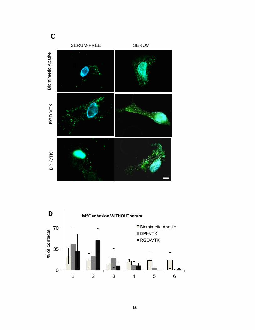
66
SERUM-FREE SERUM B
iom
ime
tic A
pa
tite
R
GD
-VT
K
DP
I-V
TK
B
C
0
35
70
1 2 3 4 5 6
% o
f co
nta
cts
Biomimetic Apatite
DPI-VTK
RGD-VTK
MSC adhesion WITHOUT serum D
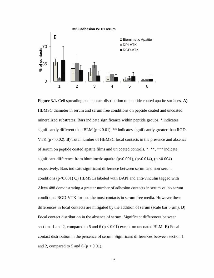
67
Figure 3.1. Cell spreading and contact distribution on peptide coated apatite surfaces. A)
HBMSC diameter in serum and serum free conditions on peptide coated and uncoated
mineralized substrates. Bars indicate significance within peptide groups. * indicates
significantly different than BLM (p < 0.01). ** indicates significantly greater than RGD-
VTK (p < 0.02). B) Total number of HBMSC focal contacts in the presence and absence
of serum on peptide coated apatite films and un coated controls. *, **, *** indicate
significant difference from biomimetic apatite (p<0.001), (p<0.014), (p <0.004)
respectively. Bars indicate significant difference between serum and non-serum
conditions (p<0.001) C) HBMSCs labeled with DAPI and anti-vinculin tagged with
Alexa 488 demonstrating a greater number of adhesion contacts in serum vs. no serum
conditions. RGD-VTK formed the most contacts in serum free media. However these
differences in focal contacts are mitigated by the addition of serum (scale bar 5 µm). D)
Focal contact distribution in the absence of serum. Significant differences between
sections 1 and 2, compared to 5 and 6 (p < 0.01) except on uncoated BLM. E) Focal
contact distribution in the presence of serum. Significant differences between section 1
and 2, compared to 5 and 6 (p < 0.01).
0
35
70
1 2 3 4 5 6
% o
f co
nta
cts
Biomimetic Apatite
DPI-VTK
RGD-VTK
MSC adhesion WITH serum
E
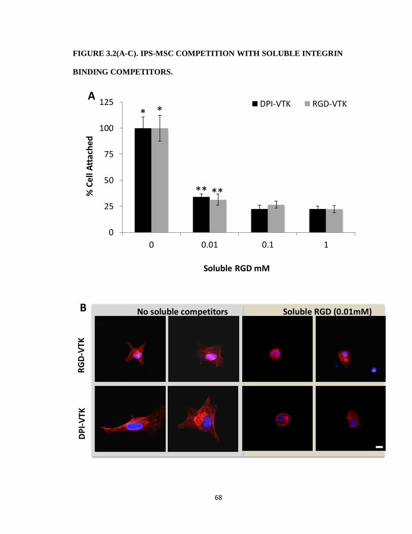
68
FIGURE 3.2(A-C). IPS-MSC COMPETITION WITH SOLUBLE INTEGRIN
BINDING COMPETITORS.
0
25
50
75
100
125
0 0.01 0.1 1
% C
ell
Att
ach
ed
Soluble RGD mM
DPI-VTK RGD-VTK* *
** **
A
DP
I-V
TK
RG
D-V
TK
No soluble competitors Soluble RGD (0.01mM) B

69
Figure 3.2. iPS-MSC competition with soluble integrin binding competitors. A)
Fraction of adherent cells on peptide coated apatite in the presence of soluble RGD
normalized to no soluble competitors. * denotes significant difference from remaining
concentrations within peptide group (p<0.01). ** denotes significant difference from the
0.1 and 1mM concentrations (p<0.02) within each peptide group. B) Representative
images at 40x of MSCs on DPI-VTK and RGD-VTK coated substrates in the presence
and absence of soluble RGD. Nuclei are stained with DAPI and F-actin is stained with
Rhodhamine-Pholloidin (scale bars 5 µm). C) Fraction of adherent cells on peptide
coated apatite in the presence of RGD integrin binding antibodies and Col1binding
antibodies normalized to cell bound in the absence of soluble competitors. * denotes
difference from RGD binding integrins (p <0.001). Bars denote difference from RGD-
VTK within same soluble treatment group (p <0.05).
Col RGD receptors 0
30
60
90
120
Control α5 αV β1 β3 β5 α2β1
RGD-VTK
DPI-VTK
Ad
he
rent
ce
ll
frac
tio
n
(% o
f co
ntro
l)
*
* * *
C

70
FIGURE 3.3. MSC PROLIFERATION ON PEPTIDE COATED APATITE
SUBSTRATES.
Figure 3.3. MSC proliferation on peptide coated apatite substrates. Histomorphometric
analysis of 4 samples per group covering 10 fields of view per sample was analyzed to
quantify the number of cells. * different from TCPS and apatite controls (p < 0.001). *
Different from apatite control (p < 0.001). ** Different from VTK (p < 0.05).
0
1
2
3
4
0.75 3 7 10
To
tal C
ells
x 1
0000
Days
TCPS
Biomimetic Apatite
VTK
DPI-VTK
RGD-VTK
*
*
**

71
FIGURE 3.4(A-G). IPS-MSC DIFFERENTIATION ON TCPS IN OSTEOGENIC
MEDIA.
0
2
4
6
8
0 5 10 15
DAYS
Runx2
0
10
20
30
0 5 10 15
DAYS
OSX
0
125
250
10 20 30
DAYS
ALP
0
20
40
60
10 20 30
DAYS
OCN
-3
0
3
6
10 20 30
DAYS
OPN
0
10
20
30
10 20 30
DAYS
BSP
A B
D C
E F

72
Figure 3.4. iPS-MSC differentiation on TCPS in osteogenic media. A,B). Relative gene
expression of osteogenic transcription factors Runx2 and Osterix normalized to day zero
and GAPDH values (n=3). C-F) relative gene expression of genes regulating osteogenic
markers ALP, OCN, OPN, and BSP normalized to day zero and GAPDH values (n=3).
G) von Kossa staining of mineral.
Da
y 2
0
Da
y 2
4
Da
y 2
8
Sample 1 Sample 2 Sample 3
G

73
FIGURE 3.5(A-F). DIFFERENTIATION OF IPS-MSCS ON BIOMIMETIC
APATITE AND PEPTIDE COATED APATITE
Figure 3.5. Differentiation of iPS-MSCs on biomimetic apatite and peptide coated apatite.
A,B). Relative gene expression of osteogenic transcription factors Runx2 and OSX
normalized to day zero and GAPDH values (n=3/group, 6 biological replicates pooled
pairwise). C-F) relative gene expression of genes regulating osteogenic markers ALP,
OCN, OPN, and BSP normalized to day zero and GAPDH values (n=3/group, 6 biological
replicates pooled pairwise)
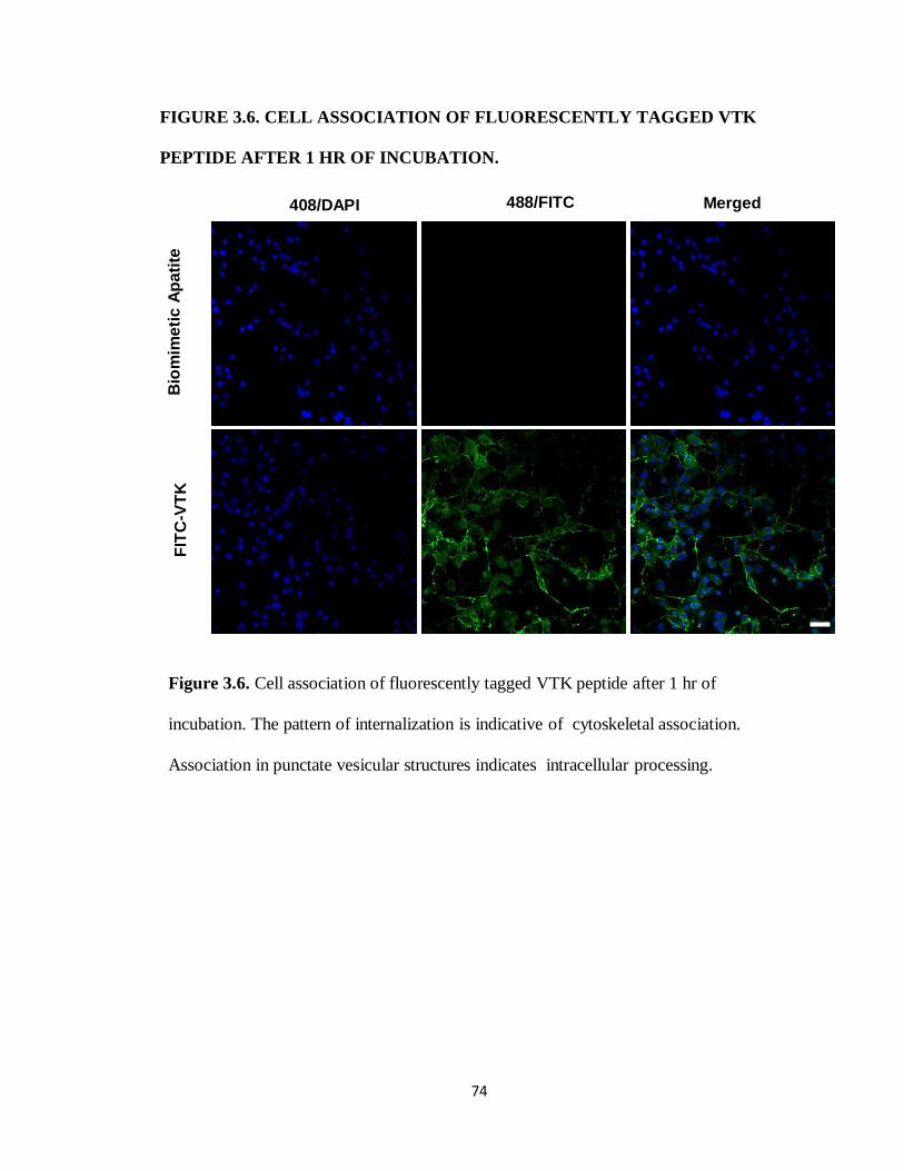
74
FIGURE 3.6. CELL ASSOCIATION OF FLUORESCENTLY TAGGED VTK
PEPTIDE AFTER 1 HR OF INCUBATION.
Figure 3.6. Cell association of fluorescently tagged VTK peptide after 1 hr of
incubation. The pattern of internalization is indicative of cytoskeletal association.
Association in punctate vesicular structures indicates intracellular processing.
Bio
mim
eti
c A
pa
tite
F
ITC
-VT
K
408/DAPI 488/FITC Merged

75
FIGURE 3.7. MULTIPOTENT MURINE-MSC RECRUITMENT ONTO
PEPTIDE COATED APATITE
Multipotent MSC recruitment on peptide coated apatite. A) Initial adherent cell numbers
on peptide coated apatite and controls. * significant from DPI-VTK no serum (p<0.001).
B) Proliferation of adherent cell fractions plates at 3000cells/cm2 on TCPS for 10 days. *
significant from DPI-VTK no serum (p<0.001).
0
0.1
0.2
0.3
0.4
TCPS BLM VTK DVTK DVTK -NS
Mill
ion
s
TCPS Biomimetic
Apatite
VTK DPI-VTK DPI-VTK
No Serum
Ad
he
ren
t C
ells
A
*
0
15
30
45
60
75
0 3 7 10
Th
ou
sa
nd
s
TCPS
Biomimetic Apatite
VTK
DPI-VTK
DPI-VTK NS
*
B
days

76
REFERENCES
[1] A. G. Mikos, S. W. Herring, P. Ochareon, J. Elisseeff, H. H. Lu, R. Kandel, F. J. Schoen, M.
Toner, D. Mooney, A. Atala, M. E. Van Dyke, D. Kaplan, and G. Vunjak-Novakovic,
“Engineering complex tissues.,” Tissue Eng., vol. 12, no. 12, pp. 3307–39, Dec. 2006.
[2] E. Alsberg, E. E. Hill, and D. J. Mooney, “Craniofacial tissue engineering.,” Crit. Rev. Oral
Biol. Med., vol. 12, no. 1, pp. 64–75, Jan. 2001.
[3] A. I. Caplan, “Review: mesenchymal stem cells: cell-based reconstructive therapy in
orthopedics ,” Tissue Eng., vol. 11, no. 7–8, pp. 1198–1211, 2005.
[4] P. Bianco and P. G. Robey, “Skeletal stem cells,” Development, vol. 142, no. 6, pp. 1023–
1027, 2015.
[5] R. Hass, C. Kasper, S. Böhm, and R. Jacobs, “Different populations and sources of human
mesenchymal stem cells (MSC): A comparison of adult and neonatal tissue-derived
MSC.,” Cell Commun. Signal., vol. 9, p. 12, Jan. 2011.
[6] S. E. Brown, W. Tong, and P. H. Krebsbach, “The derivation of mesenchymal stem cells
from human embryonic stem cells.,” Cells. Tissues. Organs, vol. 189, no. 1–4, pp. 256–60,
Jan. 2009.
[7] L. G. Villa-Diaz, S. E. Brown, Y. Liu, A. M. Ross, J. Lahann, J. M. Parent, and P. H.
Krebsbach, “Derivation of mesenchymal stem cells from human induced pluripotent stem
cells cultured on synthetic substrates.,” Stem Cells, vol. 30, no. 6, pp. 1174–81, Jun. 2012.
[8] V. Russo, C. Yu, P. Belliveau, A. Hamilton, and L. E. Flynn, “Directed Differentiation of
Human Induced Pluripotet Stem Cells Toard Bone and Cartilage In vitro Versus In Vivo
Assays,” Stem Cells Transl. Med., vol. 3, pp. 206–217, 2014.
[9] P. Bianco, X. Cao, P. S. Frenette, J. J. Mao, P. G. Robey, P. J. Simmons, and C.-Y. Wang,
“The meaning, the sense and the significance: translating the science of mesenchymal
stem cells into medicine.,” Nat. Med., vol. 19, no. 1, pp. 35–42, Jan. 2013.
[10] P. Bianco, P. G. Robey, and P. J. Simmons, “Mesenchymal stem cells: revisiting history,
concepts, and assays.,” Cell Stem Cell, vol. 2, no. 4, pp. 313–9, Apr. 2008.
[11] B. J. Sworder, S. Yoshizawa, P. J. Mishra, N. Cherman, S. A. Kuznetsov, G. Merlino, A.
Balakumaran, and P. G. Robey, “Molecular profile of clonal strains of human skeletal
stem/progenitor cells with different potencies.,” Stem Cell Res., vol. 14, no. 3, pp. 297–
306, May 2015.
[12] Ramaswamy, J., Ramaraju, H., and Kohn D. H., “Bone-Like Mineral and Organically
Modified Bone-Like Mineral Coatings,” in Biological and Biomedical Coatings Handbook,
Processing and Characterization, S. Zhang, Ed. Boca Raton, FL: CRC Press, 2011, pp. 1–36.

77
[13] W. L. Murphy, D. H. Kohn, and D. J. Mooney, “Growth of continuous bonelike mineral
within porous poly(lactide-co- glycolide) scaffolds in vitro,” J. Biomed. Mater. Res., vol.
50, pp. 50–58, 2000.
[14] A. Shekaran and A. J. García, “Extracellular matrix-mimetic adhesive biomaterials for
bone repair.,” J. Biomed. Mater. Res. A, vol. 96, no. 1, pp. 261–72, Jan. 2011.
[15] R. G. LeBaron and K. A. Athanasiou, “Extracellular matrix cell adhesion peptides:
functional applications in orthopedic materials ,” Tissue Eng., vol. 6, no. 2, pp. 85–103,
Apr. 2000.
[16] a. a. Sawyer, D. M. Weeks, S. S. Kelpke, M. S. McCracken, and S. L. Bellis, “The effect of
the addition of a polyglutamate motif to RGD on peptide tethering to hydroxyapatite and
the promotion of mesenchymal stem cell adhesion,” Biomaterials, vol. 26, no. 34, pp.
7046–7056, 2005.
[17] R. Fujisawa, M. Mizuno, Y. Nodasaka, and Y. Kuboki, “Attachment of osteoblastic cells to
hydroxyapatite crystals by a synthetic peptide (Glu7-Pro-Arg-Gly-Asp-Thr) containing two
functional sequences of bone sialoprotein.,” Matrix Biol., vol. 16, no. 1, pp. 21–8, Apr.
1997.
[18] C. Popov, T. Radic, F. Haasters, W. C. Prall, a Aszodi, D. Gullberg, M. Schieker, and D.
Docheva, “Integrins α2β1 and α11β1 regulate the survival of mesenchymal stem cells on
collagen I.,” Cell Death Dis., vol. 2, no. 7, p. e186, 2011.
[19] K. M. Hennessy, B. E. Pollot, W. C. Clem, M. C. Phipps, A. A. Sawyer, B. K. Culpepper, and
S. L. Bellis, “The effect of collagen I mimetic peptides on mesenchymal stem cell adhesion
and differentiation, and on bone formation at hydroxyapatite surfaces.,” Biomaterials,
vol. 30, no. 10, pp. 1898–909, Apr. 2009.
[20] C. D. Reyes, T. A. Petrie, K. L. Burns, Z. Schwartz, and A. J. Garcia, “Biomolecular surface
coating to enhance orthopaedic tissue healing and integration ,” Biomaterials, vol. 28,
no. 21, pp. 3228–3235, Jul. 2007.
[21] F. Gomar, R. Orozco, J. L. Villar, and F. Arrizabalaga, “P-15 small peptide bone graft
substitute in the treatment of non-unions and delayed union. A pilot clinical trial.,” Int.
Orthop., vol. 31, no. 1, pp. 93–9, Feb. 2007.
[22] H. Ramaraju, S. J. Miller, and D. H. Kohn, “Dual-functioning phage-derived peptides
encourage human bone marrow cell-specific attachment to mineralized biomaterials.,”
Connect. Tissue Res., vol. 55 Suppl 1, pp. 160–3, Aug. 2014.
[23] S. J. Segvich, “Design of Peptides with Targeted Apatite and Human Bone Marrow
Stromal Cell Adhesion for Bone Tissue Engineering.,” University of Michigan, 2008.
78
[24] S. A. Kuznetsov, M. H. Mankani, and P. G. Robey, “In vivo formation of bone and
haematopoietic territories by transplanted human bone marrow stromal cells generated
in medium with and without osteogenic supplements.,” J. Tissue Eng. Regen. Med., vol. 7,
no. 3, pp. 226–35, Mar. 2013.
[25] S. a Kuznetsov, P. H. Krebsbach, K. Satomura, J. Kerr, M. Riminucci, D. Benayahu, and P.
G. Robey, “Single-colony derived strains of human marrow stromal fibroblasts form bone
after transplantation in vivo.,” J. Bone Miner. Res., vol. 12, no. 9, pp. 1335–47, Sep. 1997.
[26] J. a. Rowley, G. Madlambayan, and D. J. Mooney, “Alginate hydrogels as synthetic
extracellular matrix materials,” Biomaterials, vol. 20, no. 1, pp. 45–53, 1999.
[27] T. a. Petrie, J. R. Capadona, C. D. Reyes, and A. J. García, “Integrin specificity and
enhanced cellular activities associated with surfaces presenting a recombinant
fibronectin fragment compared to RGD supports,” Biomaterials, vol. 27, no. 31, pp.
5459–5470, 2006.
[28] Y. Feng and M. Mrksich, “The synergy peptide PHSRN and the adhesion peptide RGD
mediate cell adhesion through a common mechanism,” Biochemistry, vol. 43, no. 50, pp.
15811–15821, 2004.
[29] E. V Leonova, K. E. Pennington, P. H. Krebsbach, and D. H. Kohn, “Substrate
mineralization stimulates focal adhesion contact redistribution and cell motility of bone
marrow stromal cells.,” J. Biomed. Mater. Res. A, vol. 79, no. 2, pp. 263–70, Nov. 2006.
[30] K. M. Hennessy, W. C. Clem, M. C. Phipps, A. A. Sawyer, F. M. Shaikh, and S. L. Bellis, “The
effect of RGD peptides on osseointegration of hydroxyapatite biomaterials.,”
Biomaterials, vol. 29, no. 21, pp. 3075–83, Jul. 2008.
[31] J. Eyckmans, T. Boudou, X. Yu, and C. S. Chen, “A hitchhiker’s guide to mechanobiology.,”
Dev. Cell, vol. 21, no. 1, pp. 35–47, Jul. 2011.
[32] A. L. Berrier and K. M. Yamada, “Cell – Matrix Adhesion,” J. Cell. Physiol., no. June, pp.
565–573, 2007.
[33] N. D. Gallant, “Stick and Grip,” Cell Biochem. Biophys., vol. 39, no. 9, 2003.
[34] D. E. Ingber, “Cellular mechanotransduction: putting all the pieces together again ,”
FASEB J., vol. 20, no. 7, pp. 811–827, May 2006.
[35] K. K. Elineni and N. D. Gallant, “Regulation of cell adhesion strength by peripheral focal
adhesion distribution.,” Biophys. J., vol. 101, no. 12, pp. 2903–11, Dec. 2011.
[36] A. J. Engler, S. Sen, H. L. Sweeney, and D. E. Discher, “Matrix Elasticity Directs Stem Cell
Lineage Specification,” Cell, vol. 126, no. 4, pp. 677–689, 2006.
79
[37] A. K. Kundu, C. B. Khatiwala, and A. J. Putnam, “Extracellular matrix remodeling, integrin
expression, and downstream signaling pathways influence the osteogenic differentiation
of mesenchymal stem cells on poly(lactide-co-glycolide) substrates.,” Tissue Eng. Part A,
vol. 15, no. 2, pp. 273–283, 2009.
[38] D. Docheva, C. Popov, W. Mutschler, and M. Schieker, “Human mesenchymal stem cells
in contact with their environment: surface characteristics and the integrin system.,” J.
Cell. Mol. Med., vol. 11, no. 1, pp. 21–38, Jan. .

80
CHAPTER FOUR
IMPROVING MSC BASED BONE TISSUE REGENERATION IN
VIVO USING CELL-SPECIFIC PEPTIDE COATED MINERALIZED
BIOMATERIALS
INTRODUCTION
There are over 2 million bone grafting procedures worldwide with 500, 000 bone grafting
procedures/year in the US alone required for the treatment of critical sized defects e[1],
[2]. Autograft therapies utilizing resected host bone are the gold standard for bone
augmentation and bone grafting, but are limited in availability and often result in donor
site morbidity[3]. Allogenic bone or demineralized bone matrix from cadaveric sources
are often used as an alternative, but exhibit a reduction in cellularity and vascularization
with increased reabsorption rates [4]. Synthetic bone grafts using calcium phosphates,
calcium sulfates, collagen, and collagen mineral composites promote osteoconduction at
the defect site, but exhibit limited osteoinductivity[5]. As a result these materials are co-
delivered with adsorbed growth factors which can result in further complications. The
limitations of current therapies, coupled with an increasing aging population and
increasing global access to healthcare, drive the need for new regenerative strategies that
can alleviate the economic burden of non-union defects[6].

81
Cell based therapies are a promising alternative for conventional auto and allograft
therapies[7]–[9]. Bioresorbable natural and synthetic polymers, calcium phosphates and
sulfates and polymer mineral composites materials are often considered for delivering
cell based therapies[10], [11]. Amongst these materials, biodegradable synthetic
polymers of the α-hydroxyester family, specifically the co-polymer poly(D,L-lactide-co-
glycolide), are also considered for their improved control over mechanical properties,
chemical functionalization, degradation rates, and ease of manufacture compared to
naturally derived polymers[12]–[14]. The mineral component provides mechanical
support and recapitulates the chemical microenvironment conducive for
osteogenecity[15], [16]. Amongst the various methods of incorporating a mineral
component onto a soft material, growth of a heterogeneous mineral layer by immersion in
simulated body fluid (SBF) containing the ionic concentrations of blood plasma under
physiological pH is a promising strategy. [16]–[18].
A continuous mineral coating throughout a porous polymer scaffold can protect the
polymer from hydrophilic attack consequently attenuating polymer degradation rates[15],
[19]. The concentration of SBF can also control the physical and chemical properties of
the mineral coating and which subsequently affects mineral dissolution rates. For
instance, mineral precipitated from 2x SBF concentrations are more amorphous than 1x
SBF and results in faster dissolution rates. Therefore characteristics of precipitated
mineral coating can also be used to further control polymer degradation timeframes[17].
Coating polymer substrates with bone-like-mineral (BLM) improves osteoconductivity in
vitro and in vivo[14], [15], [20]. Although dissolution byproducts of the mineral coating
have been implicated in biological activity, the chemical and physical properties of the
82
mineral alone are insufficient to promote osteoinductivity. Nanoscale morphology,
crystallinity, and compositional characteristics of the mineral surface drive the adsorption
of serum proteins which subsequently affect osteoconduction[21]–[23]. However, the
adhesion of cells to mineral-adsorbed serum proteins is non-specific and indiscriminative
of bone forming cell populations. To improve bone forming cell specific attachment and
osteoinductivity of mineral substrates, tissue engineers utilize extracellular matrix
proteins, peptides, and motifs specific to integrin binding domains. For instance, integrins
α2β1 and α5β1 promote osteoinductivy by mechanotransductively activating osteogenic
differentiation pathways. Derivatives of extracellular protein cell binding motifs that bind
α2β1 and α5β1, such as RGD and Col(a1) derived peptides like GFOGER and N-
GTPGPQGIAGQRGVV-C (P15), are used to further enhance osteoconductivity and
osteoinductivity [24]–[26].
Due to the limitations of immobilizing peptide to mineral substrates, ECM proteins
associated with binding mineral are used for appropriate delivery and presentation of cell
adhesive peptides[27], [28]. For instance the glutamic acid repeat motif(E7) in bone
sialoprotien was combined with RGD to improve adsorption and promote
osteoconductivity and osteoinductivity of mineral substrates[29]. Although RGD-E7
peptide improves osteoconductivity and osteoinductivity on mineral substrates in vitro, it
surprisingly inhibits osseointegration in vivo[26]. The varied successes with RGD and
various integrin binding peptides prompted the identification of cell specific peptides that
can selectively bind bone forming cell populations form a heterogeneous stem or
progenitor source. A mineral binding peptide is also required to improve adsorption to the

83
apatite surface and control presentation of the cell specific peptide to the cell surface
receptor.
To meet these objectives, we used a commercially available phage display coupled with
bioinformatic approaches and in vitro screening techniques to identify MSC specific
[DPIYALSWSGMA, DPI] and apatite specific [VTKHLNQISQSY, VTK] sequences
which were combined into a dual peptide
[GGDPIYALSWSGMAGGGSVTKHLNQISQSY, DPI-VTK] for cell specific
recruitment to mineral substrates. DPI-VTK efficiently targets apatite substrates and
improves adhesion strength and specificity to murine bone marrow, human bone marrow,
and induced pluripotent stem cell(iPS) derived mesenchymal stem cells(MSCs)[30].
Moreover, DPI-VTK improves spreading and proliferation while supporting
differentiation on dual peptide coated constructs compared to uncoated controls (Chapter
3). These findings suggest a role for in vivo biological potential
In addition to improving the quantity of tissue through improved conductivity and
inductivity, improving the uniformity of newly reconstructed tissue throughout the defect
site is an important regeneration outcomes. In this study we hypothesize that uniformity
of mineral coverage, uniformity of DPI-VTK adsorption and consequent uniformity of
ips-MSC distribution in vitro contribute to increase quantity and uniform distribution of
bone regenerated in vivo compared to non-peptide coated controls. The ubiquitous cell
binding domain RGD was combined with the mineral binding domain VTK to be used as
a dual-peptide positive control (RGD-VTK) for in vitro characterization (Table 4.1). P15
peptide, a component of Pep-Gen P15-BGS (Dentsply Implants, CA) currently available

84
as a bone graft substitute for void filling and augmentation in the oral cavity[31], was
used as an in vivo control.
MATERIALS AND METHODS
SCAFFOLD FABRICATION
Polylactic-co-glycolic acid(PLGA 85:15 , Lakeshore Biomaterials) was solubilized in
chloroform at 5%w/v. NaCl was sieved to 250-425µm and packed into 5mm diameter
well in a Delrin® mold. PLGA-chloroform solution was added to each well of the Delrin
mold which was subsequently covered and dried for 36hrs. The mold was transferred to a
vacuum chamber for 5 days and subsequently leached in double-distilled water (ddH2O)
for 36hrs with intermittent fluid changes. Leached scaffolds were etched in 0.5M sodium
hydroxide under slight agitation and rinsed twice in ddH2O.
SCAFFOLD MINERALIZATION
Scaffolds were mineralized by filtration of simulated body fluid(SBF) through the
interconnected pores as previously described[19]. SBF (1x) contains 141 mM NaCl, 4.0
mM KCl, 0.5 mM MgSO4, 1.0 mM MgCl2, 4.2 mM NaHCO3, 5.0 mM CaCl2•2H2O,and
2.0 mM KH2PO4[32]. SBF was sterile filtered using a 0.22µm filter and 0.005% sodium
azide was added to prevent bacterial contamination. 2x SBF and 4x SBF solutions titrated
to pH 6.4 with 1M NaOH were used with the filtration method. A Delrin® mold
containing leached and etched scaffolds was attached to the actuator of an Instron 8521
servo-hydraulic system. The mold was lowered into a base containing sufficient pre-
warmed 4x SBF (37oC) to fully submerge all scaffolds during the course of
mineralization. The fully submerged mold was cycled in the base at an amplitude of
25.4mm at 0.011Hz. The 4x SBF was changed every 6 hours the first day and was

85
replaced with 2x SBF which was changed every 12 hours on days 2-5. Solution
temperature in the base was maintained at 37oC using a heating sleeve connected to a
thermocouple and temperature controller (Extech Instruments, Model 48VTR).
Mineralized scaffolds (5mm diameter x 2 mm thickness) were carefully detached from
the mold, rinsed in ddH2O for 12 hrs, air-dried in a biosafety cabinet for 12 hrs and stored
in a desiccator. Scaffolds were distributed evenly across control and treatment groups to
improve mean MV% and variance (Fig. 4.1i).
SCAFFOLD CHARACTERIZATION
Morphology of mineralized PLGA scaffolds was assessed using scanning electron
microscopy (Philips XL30 FEG SEM). Scaffolds were gold coated and assessed at 20kV
(n=9 total). Scanco μCT 100 was used to examine mineralization of scaffolds at 10 μm
voxel size at 10 μm slice increments. MicroCT scans were attained at 70kV, 114 A using
a 0.5 Al filter. Reconstructed images of all mineralized scaffolds were analyzed using the
Scanco μCT_evaluation tool to calculate mineral volume fractions (MV%).
Reconstructed images were rendered in (MicroView, Parallax Innovations Inc) using the
isosurface tool at a threshold of 1000 with the smoothing filter and a surface quality
factor of 0.51 (n=10/group). A custom volumetric shrinkage application was used to
generate five concentric volumetric shells each 20% smaller than the previous volume.
Mineral volume fractions were calculated for each shell and compared across the five
volumetric shells to examine mineral distribution from the exterior toward the interior of
the scaffold.

86
PEPTIDE SYNTHESIS AND CHARACTERIZATION
Peptides (Table 4.1) were synthesized at the University of Michigan peptide core. All
peptides were chemically synthesized using solid phase synthesis and protective
chemistry. HPLC was used to verify > 95% purity. Peptides were stored at -20oC until
use.
PEPTIDE LOADING AND CHARACTERIZATION
Scaffolds pre-wet overnight in ddH2O were sterilized, transferred to a new micro-plate,
and incubated in Trizma buffer for 4hrs at 37oC. Peptides were weighed, dissolved in
sterile ddH2O, and diluted in Trizma buffer pH 7.4. Trizma pre-incubated scaffolds were
immersed in peptide solution (100ug/mL) and set on a shaker under gentle agitation for
3hrs at 37oC. Pierce BCA assay (Thermo-Fisher Scientific Inc.) was used to determine
total peptide amount on scaffolds (n=6/group) and compared to a standard curve (1-
200µg/mL). Peptide distribution on scaffolds was assessed using fluorescein
isothiocyanote (FITC) tagged peptides (n=6/group). FITC-VTK has equivalent binding
affinity to apatite as unlabeled peptide. Fluorescently tagged peptide coated scaffolds
were paper-embedded in cryosection molds in freezing media (OCT, Tissue Tek) on a
bed of dry ice and acetone[33]. A Leica cryostat was used to make five 200µm thick
sections along the thickness of each scaffold. Sections were mounted in Vectashield
(Vector Laboratories) and imaged using a Nikon Ti-Eclipse confocal microscope.
Sections were imaged at constant intensity and gain settings across all samples using a 4x
objective that incorporated the entire section in the field of view[34]. Edges were clipped
to limit artifact and 40 images were acquired along the thickness of each section at 5µm
intervals. Image J software (NIH) was used to stack 40 images projected in the Z

87
direction based on average intensity. A modified concentric circle algorithm was used to
measure fluorescence intensity or cell counts within each concentric circle (outmost
circle(a) for section 1 to innermost circle(e) for section 5) (Fig. 4.1b,c-g). Intensity
measurements were correlated with corresponding sections to assess distribution from the
outside to the interior of the scaffold.
CELL CULTURE
Mesenchymal stem cells derived from induced pluripotent stem cells (iPS-MSCs) were a
generous gift from Dr. Paul Krebsbach. These cells were verified for osteogenic
differentiation and bone forming potential in vivo[35]. iPS-MSCs were cultured in (α-
MEM), 10% FBS, antibiotics, 200mM L-glutamine, and 10mM non-essential amino
acids. iPS-MSCs (passage 5) were expanded twice (passage 7) when they reached 80-
90% confluence before transplantation. Growth media was replaced every 2-3 days and
switched to osteogenic media supplemented with osteogenic factors (10-8
M
Dexamethasone, 2-5mM β-glycerophosphate, 10-4
M ascorbic acid) for 7 days prior to
transplantation.
CELL SEEDING ON PEPTIDE COATED SCAFFOLDS
Peptide-coated scaffolds were placed into 100uL PCR tubes. Confluent IPS-MSCs
(passage 7) were trypsinized and suspended in either serum-free or complete media in a
20µL volume containing 50,000 cells. Cell suspension was added to each scaffold within
each PCR tube for 3hrs at 5% CO2 and 37oC. Cells seeded on peptide-laden scaffolds
(n=6/group) were fixed in 10% phosphate buffered formalin. Fixed cell-seeded scaffolds
were paper embedded in cryosection molds in freezing media (OCT, Tissue Tek) on a
bed of dry ice and acetone[33]. A Leica cryostat was used to make five 200µm thick

88
sections along the thickness of each scaffold. Cell-seeded scaffold sections were dried,
permeablized in Triton X, and stained with DAPI. All sections were mounted in
Vectashield (Vector Laboratories) and imaged using a Nikon Ti-Eclipse confocal
microscope. Sections were imaged at constant intensity and gain settings across all
samples using a 4x objective that incorporated the entire section in the field of view[34].
Edges were clipped to limit artifact and 40 images were acquired along the thickness of
each section at 5µm intervals. Image J software (NIH) was used to stack 40 images
projected in the Z direction based on average intensity. A modified concentric circle
algorithm used for peptide distribution analysis was used to measure cell counts from the
outside to the interior of the scaffold.
SUBCUTANEOUS TRANSPLANTATION OF CELL-SEEDED CONSTRUCTS
Peptide coated scaffolds and uncoated mineralized control scaffolds were placed in PCR
tubes and statically seeded in a 20µL volume of complete media at 3 x 10⁶ cells/construct
for 3 hours. This micromass seeding method was used to improve seeding efficiency on
peptide coated constructs. Moreover, a significantly higher cell number than in vitro
studies was used to ensure bone formation in an ectopic model[36]. The cell seeded
mineralized scaffolds and mineralized-peptide-laden scaffolds (n=10/group) were then
stored on ice prior to transplantation and cell counts in the remaining volume of media in
tube were used to calculate seeding efficiency. Acellular mineralized scaffolds and
acellular mineralized scaffolds coated with DPI-VTK were included as controls
(n=4/group). NIH guidelines for the care and use of laboratory animals (NIH Publication
#85-23 Rev. 1985) were adhered to. All surgical procedures were performed in
accordance with University of Michigan’s Committee on Use and Care of Animals. Male

89
nude mice (NIH-Lystbg-J
Foxn1nu
Btkxid
, Charles Rivers) between 25-30g were
anesthetized with isoflurane in O2 (5% induction and 2% maintenance at 1mL/min). A
midline longitudinal incision was made on the back of each mouse and 4 pockets (2 on
each side) were made in the subcutaneous tissue beneath the dorsal skin. Cell seeded
constructs were placed into each pocket and the incision was closed with surgical staples.
Animals were sacrificed 8 weeks postoperatively and transplants were harvested.
OSSICLE MICROCOMPUTED TOMOGRAPHY
Ossicles of regenerated bone were fixed in 4% phosphate buffered formalin, embedded in
agarose and imaged in a Scanco μCT 100 at 12 μm voxel size at 12 μm slice increments
at 70kV, 114 A using a 0.5 Al filter. Reconstructed images were rendered in MicroView (
Parallax Innovations Inc) using the iso-surface tool with the smoothing filter and a
surface quality factor of 0.51 (n=10/group). Advanced ROI tool, combined with
histogram values were used to calculate bone volume fraction (BVF). A custom
volumetric shrinkage application in MATLAB was used to generate five concentric
volumetric shells each 20% smaller than the previous volume. Bone volume fractions
were calculated for each shell and compared across the five volumetric shells to examine
mineral distribution from the exterior toward the interior of the scaffold.
OSSICLE HISTOLOGY AND HISTOMORPHOMETRY
Osscicles were retrieved from agarose and decalcified in 10% EDTA. Transplants
(n=5/group) were sectioned through the midline, paraffin embedded, and 5µm thick
triplicate sections were made of 3 ossicle regions progressing from the interior to the
periphery. Sections were subsequently deparaffinized, hydrated, and stained with
hematoxylin and eosin. Sections were scored using parameters outlined in Table 4.2[37],

90
[38]. A 20x field of view was used to scan sections and assess bone coverage. Totals for
each ossicle region were averaged.
STATISTICAL METHODS
Statistical analysis was conducted using Sigmaplot 13.0. One-way ANOVA on ranks
using Tukey test for pairwise comparison was used to compare peptide adsorption, cell
seeding efficiency, and bone quality across treatment groups. One-Way ANOVA on
ranks with Dunn test for pairwise comparisons was used to assess bone volume fractions.
Two-Way ANOVA on ranks using Tukey test for pairwise comparisons was used to
assess peptide, cell, mineral and bone volume distribution. Multiple and single linear
regression were conducted across peptide groups, cell distribution, peptide distribution,
mineral distribution, bone volume distribution and histological bone scores.
RESULTS
CHARACTERIZATION OF MINERALIZED SCAFFOLDS
Continuous mineral coverage on porous PLGA scaffolds was visually verified across all
batches using SEM (Fig 4.1 a,b). Average mineral volume fractions (MV%) for all
treatment groups were 16.5±3.8% (Fig 4.1i). Filtration of SBF resulted in mineralization
throughout each scaffold (Fig 4.1, d-i). There was a greater fraction of mineral in the
interior region compared to shells 1 and 2 (p < 0.001, n=10/group). Greater
mineralization in the interior region may arise from increased turbulent flow conditions,
and early nucleation within the interior of the scaffold pores. Regardless, there was no
observable pore occlusion which limits transport and vascularization.

91
PEPTIDE LOADING ON MINERALIZED SCAFFOLDS
Peptide adsorption on mineralized scaffolds was consistent no significantly different
between VTK, DPI-VTK and RGD-VTK groups (Fig 4.2a, n=6/group). The amount of
P15 adsorbed to the scaffold surface (0.75 ±0.053 nmols, n=6/group) was less than DPI-
VTK (p <0.001) , RGD-VTK (p <0.01) and VTK (p <0.001). Adsorbed VTK, DPI-VTK
and RGD-VTK were distributed uniformly across scaffold sections spanning from the
periphery to the innermost shell (Fig 4.2 c-h). Fraction of adsorbed peptide was not
significantly different within peptide groups (Fig 4.2h). There was a higher adsorbed
fraction of VTK than RGD-VTK in the outermost section (p <0.02).
CELL ATTACHMENT AND DISTRIBUTION ON PEPTIDE-LADEN SCAFFOLDS
Cells adhered readily to DPI-VTK coated mineral scaffolds (Fig. 4.3 a). Seeding 50,000
cells on a DPI-VTK coated scaffold (Fig. 4.3a) did not result in the surface saturation
with cells evident on scaffolds seeded with 3 x 10⁶ cells for in-vivo implantation (Fig.
4.3b-d). There was more efficient IPS-MSC seeding on VTK (p<0.04), DPI-VTK
(p<0.002), RGD-VTK (p<0.001), and serum coated BLM (p<0.001) compared to serum-
free BLM control groups (Fig 4.3e). RGD-VTK exhibited higher seeding efficiency than
BLM (p <0.001), VTK (p<0.001), and DPI-VTK (p<0.01). Seeding efficiency was
greater on DPI-VTK and serum-coated BLM compared to VTK (p < 0.05). The fraction
of cells adherent to VTK in the outermost section of the mineralized scaffold was greater
than BLM with serum (Fig. 4.3f; p <0.02). Moreover, the fraction of cells adherent to the
outermost section on VTK coated scaffolds was greater than the middle and inner-most
sections (Fig. 4.3g; p<0.001). Seeding efficiency on scaffolds prior to in vivo
implantation was 98.3% ±5.35% across all peptide and control groups (n=6/group).

92
OSSICLE BONE VOLUME FRACTION AND DISTRIBUTION
MicroCT images reveal, qualitatively greater bone formation on scaffolds with DPI-VTK
(Fig 4e) and P15 (Fig 4.4f) compared to scaffolds with cells, but no peptide (Fig, 4.4c),
scaffolds with just the mineral-binding peptide (Fig. 4.4d) and acellular controls (Fig
4.4a,b) controls. However, scaffolds coated with both DPI-VTK and P15 exhibited shell
formation characteristic of ectopically reconstructed ossicles. MicroCT analysis (Fig
4.4g) quantify the greater BVF in constructs coated with DPI-VTK compared to VTK (p
< 0.002), BLM (p <0.037), acellular DPI-VTK (p <0.003) and acellular BLM controls (p
<0.00). P15 coated constructs had greater BVF than BLM(p <0.05) and acellular BLM (p
<0.005). There were noticeable differences between VTK coated constructs and uncoated
and acellular controls, however the large and unequal variance resulted in failed
significance. Total volumes were also greater for DPI-VTK and P15 groups compared to
BLM, VTK and acellular controls (Fig 4.4h, p <0.001).
Rendered MicroCT images of concentric shells of regenerated bone (Fig 4.5b-f) revealed
greater bone formation towards the peripheral sections compared to interior sections.
There were no significant differences in bone volume within sections across peptide
groups (Fig 4.5g). VTK coated constructs had a significantly greater fraction of bone in
section 2 compared to sections 4 (p<0.004) and 5 (p <0.026). DPI-VTK coated constructs
had a greater fraction of bone in section 1 compared to section 5 (p<0.002) and section 4
(p<0.003), as well as a greater fraction of bone in section 2 compared to section 5
(p<0.005) and section 4 (p<0.008). Similarly, P15 coated constructs had a greater
fraction of bone in section 1 compared to section 5 (p<0.004) and section 4 (p<0.005), as

93
well as a greater fraction of bone in section 2 compared to section 5 (p<0.015) and
section 4 (p<0.019).
HISTOLOGY OF RECONSTRUCTED OSSICLES
There was greater bone formation on DPI-VTK and P15 coated constructs compared to
VTK and BLM constructs (Fig 4.6 a-h). Ossicles from BLM and VTK demonstrated
some bone formation surrounding scaffold pores (p), however the DPI-VTK and P15
constructs were relatively devoid of pores(p) and struts(t). Ossicles from BLM and VTK
coated scaffolds look compressed and elongated compared to pre-transplanted scaffolds,
whereas DPI-VTK and P15 constructs appear larger than the originally transplanted
constructs which is consistent with total volumes. Regenerated bone from DPI-VTK and
P15 also demonstrated greater vascularization (h) and cellularity (c) compared to VTK
coated and BLM constructs. Bone score indicate greater bone formation on DPI-VTK
and P-15 compared to VTK and BLM (Fig. 4.6i; p <0.001). There were no significant
differences in bone score within different sections of scaffolds for each peptide group
(Fig 6j). Across sections (Fig 6k), DPI-VTK and P15 exhibited greater bone scores
compared to VTK and BLM (p <0.001).
REGRESSION ANALYSIS
Multivariate linear regression revealed no correlation between in vitro cell distribution,
peptide distribution, and mineral distribution on bone distribution in vivo. Similarly
regressing bone distribution against in vitro cell distribution, peptide distribution, and
mineral distribution by individual treatment group revealed no correlation with a desired
p<0.05. Regressing cell distribution against mineral distribution and peptide distribution

94
revealed no correlation with P <0.05. Similarly regressing cell distribution by individual
treatment group revealed no correlation with a desired p<0.05.
DISCUSSION
DPI-VTK coated on mineralized PLGA constructs improved overall bone formation in a
ectopic model compared to non-peptide coated, VTK and acellular controls. Moreover,
bone formed by DPI-VTK coated constructs exhibited increased cellularity and
vascularization compared to uncoated, VTK and acellular controls. However, uniform
peptide distribution, cell distribution, and mineral distribution did not correlate to
improved bone distribution in vivo.
Peptides containing the VTK, DPI-VTK, and RGD-VTK mineral specific sequence
improved overall adsorption to mineral and delivery of cell binding sequences to apatite
compared to collagen derived P15 sequence (Fig 4.2a). The increase in mineral binding
between VTK vs. RGD-VTK in the outermost region of mineralized scaffolds can result
from different packing efficiencies arising from conformational differences in solution
and bound states. Furthermore VTK and RGD-VTK both have a net charge of 1 however;
the distribution of charged residues within the peptide can result in varying steric
repulsions when saturating the mineral surface.
Although less RGD-VTK was bound in the outermost region of the construct(Fig 4.2h),
there were no observable differences in the fraction of bound cells between VTK and
RGD-VTK in the outermost region(Fig 4.3f). Cell attachment to DPI-VTK is improved
compared to BLM without serum and VTK without serum. Although cell seeding
efficiency on DPI-VTK is equivalent to RGD-VTK and BLM in serum, the DPI peptide

95
promotes cell specific attachment to MSC whereas RGD and serum proteins are non-
specific. Moreover, RGD coated substrates exhibit poorer osteogenic activity in vivo
hypothesized to result from unfavorable serum protein interactions[26], [39]. However,
DPI-VTK cooperatively interacts with serum proteins towards improving MSC
attachment (Chapter 2). The cell-specific interaction combined with cooperative serum
interaction could also play a role in observed improvements in the quantity and quality of
bone formation in vivo. Serum-free or xeno-free media are increasingly used to meet
current good manufacturing practices for clinical grade MSCs in order to limit risk of
disease transmission and immunogenicity while maintaining consistency of appropriately
tested and qualified starting materials[40]–[42]. The ability of DPI-VTK to attach
equivalent numbers of cells in serum free media as serum coated BLM also implies a
potential ex vivo application for serum-free expansion on mineral substrates[43], [44].
Cell-seeded mineralized scaffolds containing DPI-VTK significantly improved the total
volume, the volume fraction and bone scores compared to VTK coated and uncoated
BLM, and acellular BLM and DPI-VTK scaffolds (Fig 4.4). Acellular mineral and
acellular DPI-VTK was unable to form bone in the ectopic mouse model. Although the
adipose tissue surrounding the ectopic implantation pocket contain a resident population
of mesenchymal stem cells, the low percentage of mesenchymal cells within the tissue
and their location with respect to the DPI-VTK transplant may preclude infiltration and
recruitment. Furthermore, the rate of degradation of the mineral and construct can impact
conduction. Regardless, both acellular BLM and acellular DPI-VTK coated constructs
provide a baseline for comparing transplanted IPS-MSC driven tissue reconstruction on
control and peptide coated substrates.

96
Initial mineral volumes are commonly subtracted from the final bone volumes in order to
calculate total regenerated bone volume. However, these calculations assume inert
mineral substrates that do not dissolve over the course of the transplantation. The
mineralized PLGA substrate coated with 2x SBF results in a rapidly dissolving mineral
substrate that is nearly resorbed as evidenced in the acellular controls. Even within
cellularized constructs, the 2x precipitated SBF results in poor bone formation compared
to previous studies utilizing 1x SBF precipitated mineral[45]. Moreover, a thrombin-
fibrin clot is typically used to provide further retention of cells within the construct. The
inability to use the fibrin clot in order to directly test peptide-cell effects resulted in poor
bone formation on BLM constructs compared to previously observed studies. Although
there were no significant differences in total volume, bone volume fraction or bone scores
of regenerated bone between P15 and DPI-VTK coated cellular constructs, further
engineering the dual-peptide sequence to improve cell specificity can drive greater bone
formation.
Cell-seeded constructs, especially those containing MSCs, can drive vascularization
through secretion of pro-angiogenic factors contributing to hematopoiesis which
concomitantly drives greater bone formation[46], [47]. Hematopoietic regions were
evident across all cell-transplanted treatment groups. Greater cell numbers were observed
within constructs coated with DPI-VTK and P-15(Fig 4.6). VTK cell-seeded constructs
exhibited observably greater bone formation compared to BLM controls and acellular
controls however the variance was too large for significance.
Bone regenerated from murine bone marrow stromal cells on a mineralized PLGA
scaffold construct in an ectopic mouse model exhibited 90% of cells with donor origin

97
up to 12 weeks post transplantation[45], [48], [49]. MSC driven bone formation on β-
TCP constructs also reveals retention of donor cells for up to 45 weeks post
transplantation[50]. It can be inferred that the majority of cells present within DPI-VTK
and P15 are from a donor origin. Moreover, the limited cellularity within acellular DPI-
VTK and acellular BLM constructs further indicates the lack of host cell contribution to
regenerating ectopic ossicles. Taken together, the ability of DPI-VTK to sequester donor
cells and/or recruit host cells within transplanted constructs is one of the leading drivers
of improved quantity and quality of bone formation.
However, regression analysis indicates no correlation between in vitro mineralization,
peptide distribution, cell distribution and in vivo bone formation. Furthermore, a
characteristic shell of bone indicating non-uniformity was evident in both P15 and DPI-
VTK coated constructs (Fig 4.5). A shell is commonly formed in an ectopic model[48],
[51], [52]. The subcutaneous environment is physically and temporally limited by the
ability to form deeply penetrating vascular networks before cell mediated mineral
formation causes pore occlusion and isolation of the interior regions[53].Furthermore,
attenuated clearance of biomaterial substrate degradation byproducts can increase toxicity
of the interior microenvironment adding to the shell [54]. Another artifact arising from
the shell formation could cause attenuated degradation of the scaffold interior resulting in
higher measured bone volume fractions. There was limited to no evidence of this artifact
from histological assessment of bone (Fig 4.6). Bony spicules within the center regions of
both DPI-VTK coated and P15 coated constructs exhibited thick cellularized sections
which are inconsistent with latent mineral from pre-implanted sources. Despite the shell
98
formation, DPI-VTK promoted moderate bone formation penetrating 60% of the scaffold
interior.
Osteoinductive potential of DPI-VTK could also be a contributing factor towards
improved bone formation. DPI-VTK competes with many of the same integrin domains
involved in osteogenic differentiation namely α5β1 and β5 domain(Chapter 2). These
domains are implicated in osteoblastic differentiation and could drive the differentiation
of initially adherent cell fractions. P15 is also suggested to promote osteogenic
differentiation through the α2β1 cell binding domain which is implicated in activation of
osteoblastic differentiation pathways [25].
The marked improvement in in vivo osteogenic potential of DPI-VTK compared to non-
peptide coated constructs warrants further investigation towards the mechanism of action.
Future studies to elucidate specific cell binding targets in addition to conformational
regulation DPI sequence when present with the VTK sequence can further improve the
osteogenic potential of these phage derived peptides. As tissue engineering shifts from
developing technologies to meet general clinical challenges to addressing more focused
clinical applications, there will be an increased need for delivering cell specific cues to
material surfaces with defined surface chemistries [7], [55]. Combinatorial phage display
is a powerful technology platform to enable focused cell based tissue regeneration
through the discovery of cell specific and material specific peptide sequences.

99
ACKNOWLEDGEMENTS
I would like to thank Dr. Paul Krebsbach and Luis Villa for iPS-MSCs used in these
studies, Michelle Lynch for her assistance with MicroCT and Chris Strayhorn for the
histology. This work is supported by NIH R01 DE 013380, DE 015411 (DHK).
CHAPTER FOUR TABLES
TABLE 4.1 PEPTIDE PROPERTIES
Peptide Sequence Description MW
(g/mol)
Net
Charge
Acidic
residues
VTK VTKHLNQISQSY
Phage derived mineral
binding sequence
1417.59
1 2
DPI-
VTK
GGDPIYALSWSGMAGG
GSVTKHLNQISQSY
Dual functioning
peptide containing
phage derived cell and
mineral binding
sequences
3025.35
0 5
RGD-
VTK
GGRGDGGGSVTKHLN
QISQSY
Dual functioning
peptide containing cell
binding control
sequence and phage
derived mineral binding
sequence
2061.20
1 3
P15 GTPGPQGIAGQRGVV
[23]
Col1a1 derived cell and
mineral binding peptide
for in vivo control 1393.57
1 0
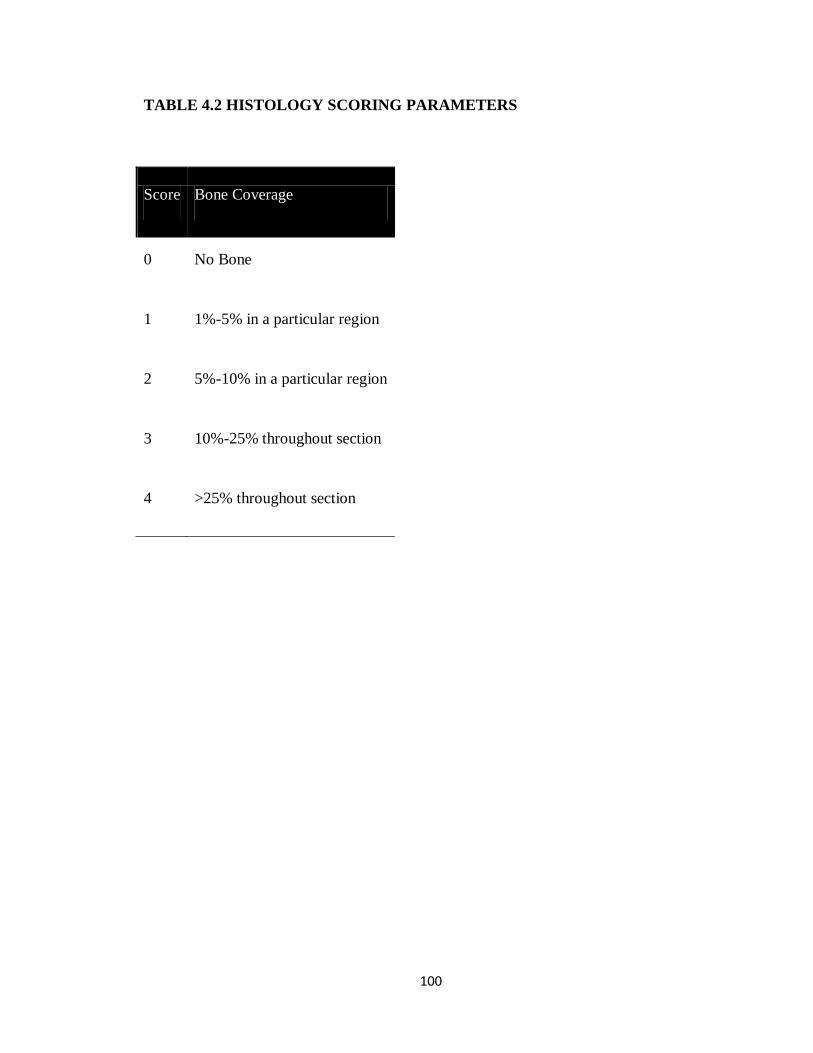
100
TABLE 4.2 HISTOLOGY SCORING PARAMETERS
Score Bone Coverage
0 No Bone
1 1%-5% in a particular region
2 5%-10% in a particular region
3 10%-25% throughout section
4 >25% throughout section

101
CHAPTER FOUR FIGURES
FIGURE 4.1(A-J) MORPHOLOGY AND DISTRIBUTION OF BONE-LIKE
MINERAL PRECIPITATED ON PLGA SCAFFOLDS
A B
C D E
F G H

102
Figure 4.1: Morphology and distribution of bone-like mineral precipitated on PLGA
scaffolds. Representative SEM images of mineralized scaffolds at A)low and B)high
magnification. scale bars 200µm and 50µm respectively. C)Representative MicroCT
image of mineralized scaffold rendered in MicroView® D-H)Representative images of
mineral distribution volumetric shells progressing from the outermost to innermost
regions of the scaffold. I) Mineral volume fraction across peptide groups. Fraction of
mineral volume progressing from the outermost to the innermost volumetric shell of the
scaffold (J). * indicates significant difference from section 1 and 2 (p<0.001).
0
5
10
15
20
25
BLM VTK DPI-VTK RGD-VTK P-15
Min
eral
Vo
lum
e Fr
acti
on
0
15
30
45
1 2 3 4 5
Frac
tio
n o
f M
ine
ral
outer
shell inner
shell
*
J
I

103
FIGURE 4.2(A-H). PEPTIDE ADSORPTION AND DISTRIBUTION ON
MINERALIZED PLGA SCAFFOLDS.
0
5
10
15
VTK DPI-VTK RGD-VTK P15
Ad
sorb
ed p
epti
de
(nm
ol)
A
*
Section 1
Section 2
Section 3
Section 4
Section 5
B
Section 1 Section 2 Section 3 Section 4 Section 5
C D E F G
a b c d e

104
Figure 4.2. Peptide adsorption and distribution on mineralized PLGA Scaffolds. (A)
Peptide adsorption evaluated by BCA assay demonstrates no significant differences
across peptide groups. (B) Schematic depicting relative position of each cryosection
within the scaffold. (C-F) Representative images of DPI-VTK distribution on PLGA
mineralized scaffolds with (a-e) regions indicating sampled area within corresponding
sections. Each image is a composite stack of 40 images projected in the Z direction based
on average intensity (scale bar 0.5mm). (H) Uniform distribution of adsorbed peptide
(n=6) across all groups was analyzed using Image J(NIH) software with a customized
concentric circle algorithm. * indicates significantly different from Section 1 of RGD-
VTK (p <0.02).
0
10
20
30
40
VTK DPI-VTK RGD-VTK
Frac
tio
n A
dso
rbed
Pep
tid
e (%
) 1 2 3 4 5H
*

105
FIGURE 4.3(A-G) - CELL SEEDING EFFICIENCY AND CELL DISTRIBUTION
ON PEPTIDE COATED SCAFFOLDS.
A C
D B
0
25
50
75
BLM VTK DPI-VTK RGD-VTK BLMw/Serum
**
*
**
**
E
*
Se
edin
g ef
fici
ency
(%
)

106
0
30
60
90
1 2 3 4 5
BLM VTK DPI-VTK RGD-VTKw/serum
0
30
60
90
BLM VTK DPI-VTK RGD-VTK
1 2 3 4 5
w/serum
outer
shell inner
shell
Fra
ction o
f A
dhere
nt
Cells
(%
) F
raction o
f A
dhere
nt
Cells
(%
)
*
*
F
G
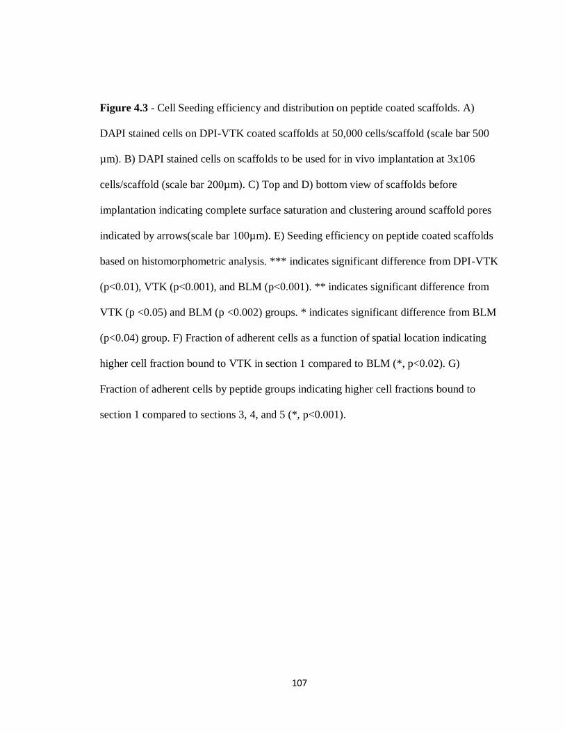
107
Figure 4.3 - Cell Seeding efficiency and distribution on peptide coated scaffolds. A)
DAPI stained cells on DPI-VTK coated scaffolds at 50,000 cells/scaffold (scale bar 500
µm). B) DAPI stained cells on scaffolds to be used for in vivo implantation at 3x10⁶
cells/scaffold (scale bar 200µm). C) Top and D) bottom view of scaffolds before
implantation indicating complete surface saturation and clustering around scaffold pores
indicated by arrows(scale bar 100µm). E) Seeding efficiency on peptide coated scaffolds
based on histomorphometric analysis. *** indicates significant difference from DPI-VTK
(p<0.01), VTK (p<0.001), and BLM (p<0.001). ** indicates significant difference from
VTK (p <0.05) and BLM (p <0.002) groups. * indicates significant difference from BLM
(p<0.04) group. F) Fraction of adherent cells as a function of spatial location indicating
higher cell fraction bound to VTK in section 1 compared to BLM (*, p<0.02). G)
Fraction of adherent cells by peptide groups indicating higher cell fractions bound to
section 1 compared to sections 3, 4, and 5 (*, p<0.001).

108
FIGURE 4.4(A-H). BONE VOLUME FRACTIONS OF ECTOPICALLY
REGENERATED OSSICLES 8 WEEKS POST TRANSPLANTATION.
A B
C D
E F

109
G
**
*
Bo
ne
Vo
lum
e Fr
acti
on
DP
I-V
TK-A
BLM
VTK
DP
I-V
TK
P1
5
BLM
-A
0
25
50
75
100
BLM-A DPI-VTK-a BLM VTK DPI-VTK P15
Tota
l Vo
lum
e
H
*
*

110
Figure 4.4. Bone volume fractions of ectopically regenerated ossicles 8 weeks post
transplantation. Representative rendered MicroView® images of A) acellular BLM, B)
acellular BLM containing DPI-VTK, C) BLM with iPS-MSCs, D) BLM containing VTK
with iPS-MSCs, E) BLM containing DPI- VTK with iPS-MSCs, F) P15 coated BLM
constructs (scale bar=1mm). G) Bone volume fractions from MicroCT analysis indicate
greater bone formation in scaffolds with DPI-VTK and P15 compared to VTK, BLM,
DPI-VTK acellular and BLM acellular controls. * indicates a significant difference from
BLM and acellular BLM (p <0.003). ** indicates a significant difference from VTK (p <
0.017),BLM (p <0.001), acellular DPI-VTK (p <0.002) and acellular BLM controls (p
<0.01). H) Total volume is greater on DPI-VTK and P15 compared to
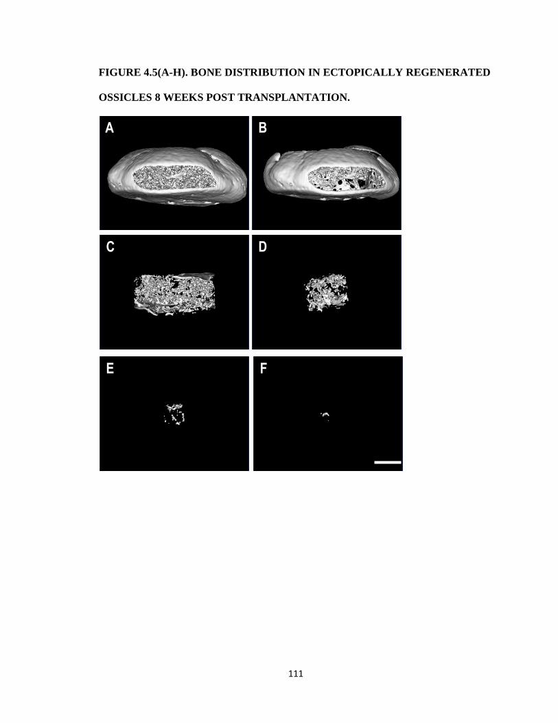
111
FIGURE 4.5(A-H). BONE DISTRIBUTION IN ECTOPICALLY REGENERATED
OSSICLES 8 WEEKS POST TRANSPLANTATION.
A B
C D
E F

112
0
20
40
60
80
1 2 3 4 5
Fra
ctio
n o
f To
tal B
on
e
BLM VTK DPI-VTK P15G
Outer shell Inner shell
0
20
40
60
80
BLM VTK DPI-VTK P15
Fra
ctio
n o
f To
tal B
on
e
1 2 3 4 5H
*
*
*
* *
6.54±
11.1
9.33±
9.57
38.5±
7.0
37.9±
8.2
Bone
volume
mm3

113
Figure 4.5. Bone distribution in ectopically regenerated ossicles 8 weeks post
transplantation. A) Representative rendered MicroCT image of ossicle regenerated from
cell seeded DPI-VTK coated mineralized scaffold (1mm scale bar). B-F) Concentric
volumetric shells progressing from the outermost to innermost region of regenerated
ossicle. G) Fraction of total bone by section indicating no significant differences in % of
total bone in each section amongst peptides. H) Fraction of total bone by peptide
revealing greater fraction of bone towards the periphery compared to the interior regions
in peptide groups. * indicates significant difference from sections 4 and 5.
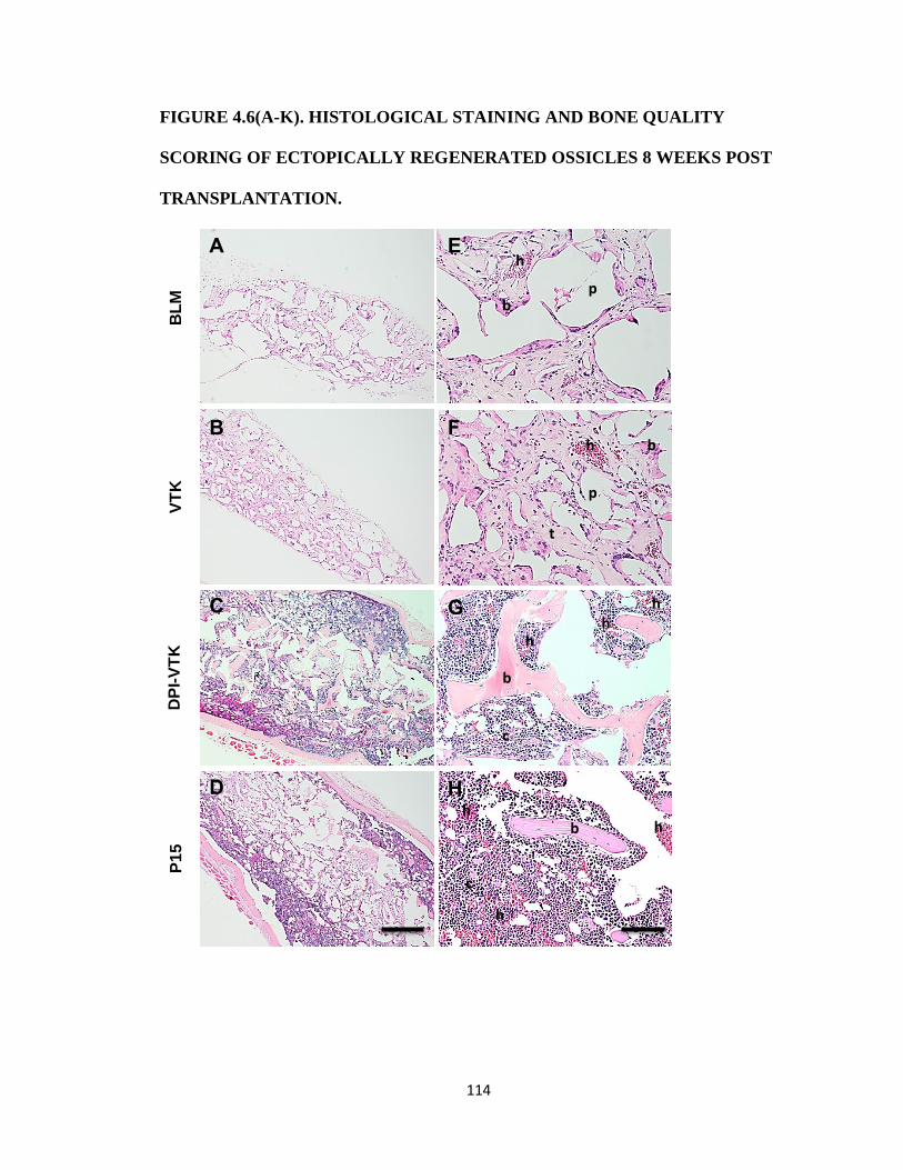
114
FIGURE 4.6(A-K). HISTOLOGICAL STAINING AND BONE QUALITY
SCORING OF ECTOPICALLY REGENERATED OSSICLES 8 WEEKS POST
TRANSPLANTATION.
BL
M
VT
K
DP
I-V
TK
P
15
A E
B F
C
D
G
H
p
p
t
c
c
h
h
h
h
h
h
b
b
b
b
h
h

115
Bo
ne
Sco
re
BLM VTK DPI-VTK P15
* *
I
0
2
4
6
1 2 3
BLM VTK DPI-VTK P15
0
2
4
6
BLM VTK DPI-VTK P15
1 2 3
Bone
Score
B
one
Score
J
K
* * * * * *

116
Figure 4.6. Histological staining and bone quality scoring of ectopically regenerated
ossicles 8 weeks post transplantation. A-D) Representative low magnification images of
cross sections through the center of ossicles indicating overall degree of bone formation
in the interior of the construct (scale bar 500µm). E-H) High Magnification images of
cross sections through the center of ossicles indicating increased bone formation,
vascularization, and cellularity on DPI-VTK and P15 compared to VTK and BLM. (b)
indicates bone, (h) indicates hematopoesis, (c) indicates greater density of cells, (p)
indicates pores, (t) indicate scaffold struts replaced by tissue (scale bars 100µm). I) Bone
score based on bone coverage. * indicates significant differences from BLM and VTK (p
<0.001). J) Bone Score by peptide group across innermost section 1 to outermost section
3. K) Bone score by section across peptide groups. * indicates significant difference from
BLM and VTK (p<0.001).

117
REFERENCES
[1] V. Campana, G. Milano, E. Pagano, M. Barba, C. Cicione, G. Salonna, W. Lattanzi, and G.
Logroscino, “Bone substitutes in orthopaedic surgery: from basic science to clinical
practice.,” J. Mater. Sci. Mater. Med., vol. 25, no. 10, pp. 2445–61, Oct. 2014.
[2] O. Faour, R. Dimitriou, C. A. Cousins, and P. V Giannoudis, “The use of bone graft
substitutes in large cancellous voids: any specific needs?,” Injury, vol. 42 Suppl 2, pp.
S87–90, Sep. 2011.
[3] W. L. Grayson, M. Fröhlich, K. Yeager, S. Bhumiratana, M. E. Chan, C. Cannizzaro, L. Q.
Wan, X. S. Liu, X. E. Guo, and G. Vunjak-Novakovic, “Engineering anatomically shaped
human bone grafts.,” Proc. Natl. Acad. Sci. U. S. A., vol. 107, no. 8, pp. 3299–304, Feb.
2010.
[4] G. J. Meijer, J. D. de Bruijn, R. Koole, and C. A. van Blitterswijk, “Cell-Based Bone Tissue
Engineering ,” PLoS Med., vol. 4, no. 2, p. e9, Feb. 2007.
[5] T. T. Roberts and A. J. Rosenbaum, “Bone grafts, bone substitutes and orthobiologics: the
bridge between basic science and clinical advancements in fracture healing.,”
Organogenesis, vol. 8, no. 4, pp. 114–24, Jan. .
[6] P. G. W, U. S. Bone, and J. Decade, “Burden of Musculoskeletal Disease Overview,” Popul.
(English Ed., no. March 2002, pp. 1–19, 2011.
[7] A. Atala, F. K. Kasper, and A. G. Mikos, “Engineering complex tissues.,” Sci. Transl. Med.,
vol. 4, no. 160, p. 160rv12, 2012.
[8] H. Shin, S. Jo, and A. G. Mikos, “Biomimetic materials for tissue engineering ,”
Biomaterials, vol. 24, no. 24, pp. 4353–4364, Nov. 2003.
[9] A. G. Mikos, S. W. Herring, P. Ochareon, J. Elisseeff, H. H. Lu, R. Kandel, F. J. Schoen, M.
Toner, D. Mooney, A. Atala, M. E. Van Dyke, D. Kaplan, and G. Vunjak-Novakovic,
“Engineering complex tissues.,” Tissue Eng., vol. 12, no. 12, pp. 3307–39, Dec. 2006.
[10] J. D. Kretlow and A. G. Mikos, “Founder’s award to Antonios G. Mikos, Ph.D., 2011
Society for Biomaterials annual meeting and exposition, Orlando, Florida, April 13-16,
2011: Bones to biomaterials and back again--20 years of taking cues from nature to
engineer synthetic polymer scaffold,” J. Biomed. Mater. Res. A, vol. 98, no. 3, pp. 323–31,
Sep. 2011.
[11] E. J. Lee, F. K. Kasper, and A. G. Mikos, “Biomaterials for tissue engineering.,” Ann.
Biomed. Eng., vol. 42, no. 2, pp. 323–37, Feb. 2014.

118
[12] L. Lu, S. J. Peter, M. D. Lyman, H. L. Lai, S. M. Leite, J. A. Tamada, J. P. Vacanti, R. Langer,
and A. G. Mikos, “In vitro degradation of porous poly(L-lactic acid) foams,” Biomaterials,
vol. 21, no. 15, pp. 1595–1605, 2000.
[13] L. Wu and J. Ding, “In vitro degradation of three-dimensional porous poly(D,L-lactide-co-
glycolide) scaffolds for tissue engineering.,” Biomaterials, vol. 25, no. 27, pp. 5821–5830,
2004.
[14] W. L. Murphy, S. Hsiong, T. P. Richardson, C. a Simmons, and D. J. Mooney, “Effects of a
bone-like mineral film on phenotype of adult human mesenchymal stem cells in vitro.,”
Biomaterials, vol. 26, no. 3, pp. 303–10, Jan. 2005.
[15] J. D. Kretlow and A. G. Mikos, “Review: mineralization of synthetic polymer scaffolds for
bone tissue engineering.,” Tissue Eng., vol. 13, no. 5, pp. 927–38, May 2007.
[16] W. L. Murphy, D. H. Kohn, and D. J. Mooney, “Growth of continuous bonelike mineral
within porous poly(lactide-co- glycolide) scaffolds in vitro,” J. Biomed. Mater. Res., vol.
50, pp. 50–58, 2000.
[17] L. N. Luong, S. I. Hong, R. J. Patel, M. E. Outslay, and D. H. Kohn, “Spatial control of
protein within biomimetically nucleated mineral.,” Biomaterials, vol. 27, no. 7, pp. 1175–
86, Mar. 2006.
[18] S. J. Segvich and D. H. Kohn, Biological Interactions on Materials Surfaces. New York, NY:
Springer US, 2009.
[19] S. Segvich, H. C. Smith, L. N. Luong, and D. H. Kohn, “Uniform deposition of protein
incorporated mineral layer on three-dimensional porous polymer scaffolds ,” J. Biomed.
Mater. Res. B, Appl. Biomater., vol. 84, no. 2, pp. 340–349, Feb. 2008.
[20] W. L. Murphy, C. a. Simmons, D. Kaigler, and D. J. Mooney, “Bone Regeneration via a
Mineral Substrate and Induced Angiogenesis,” J. Dent. Res., vol. 83, no. 3, pp. 204–210,
Mar. 2004.
[21] E. V Leonova, K. E. Pennington, P. H. Krebsbach, and D. H. Kohn, “Substrate
mineralization stimulates focal adhesion contact redistribution and cell motility of bone
marrow stromal cells.,” J. Biomed. Mater. Res. A, vol. 79, no. 2, pp. 263–70, Nov. 2006.
[22] K. Shin, A. C. Jayasuriya, and D. H. Kohn, “Effect of ionic activity products on the structure
and composition of mineral self assembled on three-dimensional poly ( lactide-co-
glycolide ) scaffolds,” J. Biomed. Mater. Res. Part A, 2007.
[23] Y. Ramaswamy, C. Wu, and H. Zreiqat, “Orthopedic coating materials: considerations and
applications ,” Expert Rev. Med. Devices, vol. 6, no. 4, pp. 423–430, Jul. 2009.
119
[24] A. Shekaran and A. J. García, “Extracellular matrix-mimetic adhesive biomaterials for
bone repair.,” J. Biomed. Mater. Res. A, vol. 96, no. 1, pp. 261–72, Jan. 2011.
[25] K. M. Hennessy, B. E. Pollot, W. C. Clem, M. C. Phipps, A. A. Sawyer, B. K. Culpepper, and
S. L. Bellis, “The effect of collagen I mimetic peptides on mesenchymal stem cell adhesion
and differentiation, and on bone formation at hydroxyapatite surfaces.,” Biomaterials,
vol. 30, no. 10, pp. 1898–909, Apr. 2009.
[26] K. M. Hennessy, W. C. Clem, M. C. Phipps, A. A. Sawyer, F. M. Shaikh, and S. L. Bellis, “The
effect of RGD peptides on osseointegration of hydroxyapatite biomaterials.,”
Biomaterials, vol. 29, no. 21, pp. 3075–83, Jul. 2008.
[27] M. Gilbert, W. J. Shaw, J. R. Long, K. Nelson, G. P. Drobny, C. M. Giachelli, and P. S.
Stayton, “Chimeric peptides of statherin and osteopontin that bind hydroxyapatite and
mediate cell adhesion.,” J. Biol. Chem., vol. 275, no. 21, pp. 16213–8, May 2000.
[28] D. L. Masica and J. J. Gray, “Solution- and adsorbed-state structural ensembles predicted
for the statherin-hydroxyapatite system.,” Biophys. J., vol. 96, no. 8, pp. 3082–91, Apr.
2009.
[29] R. Fujisawa, M. Mizuno, Y. Nodasaka, and Y. Kuboki, “Attachment of osteoblastic cells to
hydroxyapatite crystals by a synthetic peptide (Glu7-Pro-Arg-Gly-Asp-Thr) containing two
functional sequences of bone sialoprotein.,” Matrix Biol., vol. 16, no. 1, pp. 21–8, Apr.
1997.
[30] H. Ramaraju, S. J. Miller, and D. H. Kohn, “Dual-functioning phage-derived peptides
encourage human bone marrow cell-specific attachment to mineralized biomaterials.,”
Connect. Tissue Res., vol. 55 Suppl 1, pp. 160–3, Aug. 2014.
[31] F. Gomar, R. Orozco, J. L. Villar, and F. Arrizabalaga, “P-15 small peptide bone graft
substitute in the treatment of non-unions and delayed union. A pilot clinical trial.,” Int.
Orthop., vol. 31, no. 1, pp. 93–9, Feb. 2007.
[32] T. Kokubo, H. Kushitani, C. Ohtsuki, S. Sakka, and T. Yamamuro, “Chemical reaction of
bioactive glass and glass-ceramics with a simulated body fluid,” J. Mater. Sci. Mater.
Med., vol. 3, no. 2, pp. 79–83, Mar. 1992.
[33] S. R. Peters, A Practical Guide to Frozen Section Technique. New York: Springer Science &
Business Media, 2010.
[34] P. Thevenot, M. Sc, A. Nair, M. Sc, J. Dey, M. Sc, J. Yang, D. Ph, L. Tang, and D. Ph,
“Method to Analyze Three-Dimensional Cell Distribution and Infiltration in Degradable
Scaffolds,” vol. 14, no. 4, 2008.

120
[35] L. G. Villa-Diaz, S. E. Brown, Y. Liu, A. M. Ross, J. Lahann, J. M. Parent, and P. H.
Krebsbach, “Derivation of mesenchymal stem cells from human induced pluripotent stem
cells cultured on synthetic substrates.,” Stem Cells, vol. 30, no. 6, pp. 1174–81, Jun. 2012.
[36] K. Kim, D. Dean, A. G. Mikos, and J. P. Fisher, “Effect of initial cell seeding density on early
osteogenic signal expression of rat bone marrow stromal cells cultured on cross-linked
poly(propylene fumarate) disks.,” Biomacromolecules, vol. 10, no. 7, pp. 1810–7, Jul.
2009.
[37] B. D. Boyan, C. H. Lohmann, a. Somers, G. G. Niederauer, J. M. Wozney, D. D. Dean, D. L.
Carnes, and Z. Schwartz, “Potential of porous poly-D,L-lactide-co-glycolide particles as a
carrier for recombinant human bone morphogenetic protein-2 during osteoinduction in
vivo,” J. Biomed. Mater. Res., vol. 46, no. 1, pp. 51–59, 1999.
[38] M. H. Mankani, S. A. Kuznetsov, R. M. Wolfe, G. W. Marshall, and P. G. Robey, “In vivo
bone formation by human bone marrow stromal cells: reconstruction of the mouse
calvarium and mandible.,” Stem Cells, vol. 24, no. 9, pp. 2140–9, Sep. 2006.
[39] C. D. Reyes, T. A. Petrie, K. L. Burns, Z. Schwartz, and A. J. Garcia, “Biomolecular surface
coating to enhance orthopaedic tissue healing and integration ,” Biomaterials, vol. 28,
no. 21, pp. 3228–3235, Jul. 2007.
[40] P. Swamynathan, P. Venugopal, S. Kannan, C. Thej, U. Kolkundar, S. Bhagwat, M. Ta, A.
Sen Majumdar, and S. Balasubramanian, “Are serum-free and xeno-free culture
conditions ideal for large scale clinical grade expansion of Wharton’s jelly derived
mesenchymal stem cells? A comparative study.,” Stem Cell Res. Ther., vol. 5, no. 4, p. 88,
Jan. 2014.
[41] F. dos Santos, P. Z. Andrade, M. M. Abecasis, J. M. Gimble, L. G. Chase, A. M. Campbell, S.
Boucher, M. C. Vemuri, C. L. da Silva, and J. M. S. Cabral, “Toward a clinical-grade
expansion of mesenchymal stem cells from human sources: a microcarrier-based culture
system under xeno-free conditions.,” Tissue Eng. Part C. Methods, vol. 17, no. 12, pp.
1201–10, Dec. 2011.
[42] R. Giancola, T. Bonfini, and A. Iacone, “Cell therapy: cGMP facilities and manufacturing.,”
Muscles. Ligaments Tendons J., vol. 2, no. 3, pp. 243–7, Jul. 2012.
[43] S. Alimperti, P. Lei, Y. Wen, J. Tian, A. M. Campbell, and S. T. Andreadis, “Serum-free
spheroid suspension culture maintains mesenchymal stem cell proliferation and
differentiation potential.,” Biotechnol. Prog., vol. 30, no. 4, pp. 974–83, Jan. .
[44] K. Y. Tan, K. L. Teo, J. F. Y. Lim, A. K. L. Chen, S. Reuveny, and S. K. Oh, “Serum-free media
formulations are cell line-specific and require optimization for microcarrier culture.,”
Cytotherapy, vol. 17, no. 8, pp. 1152–65, Aug. 2015.

121
[45] R. A. Rossello and D. H, “Cell communication and tissue engineering.,” Commun. Integr.
Biol., vol. 3, no. 1, pp. 53–6, Jan. 2010.
[46] A. I. Caplan, “Review: mesenchymal stem cells: cell-based reconstructive therapy in
orthopedics ,” Tissue Eng., vol. 11, no. 7–8, pp. 1198–1211, 2005.
[47] P. G. Robey, S. A. Kuznetsov, M. Riminucci, and P. Bianco, “Skeletal (‘mesenchymal’) stem
cells for tissue engineering.,” Methods Mol. Med., vol. 140, pp. 83–99, Jan. 2007.
[48] R. A. Rossello and D. H. Kohn, “Gap junction intercellular communication: a review of a
potential platform to modulate craniofacial tissue engineering ,” J. Biomed. Mater. Res.
B, Appl. Biomater., vol. 88, no. 2, pp. 509–518, Feb. 2009.
[49] R. A. Rosselló, Z. Wang, E. Kizana, P. H. Krebsbach, and D. H. Kohn, “Connexin 43 as a
signaling platform for increasing the volume and spatial distribution of regenerated
tissue.,” Proc. Natl. Acad. Sci. U. S. A., vol. 106, no. 32, pp. 13219–24, Aug. 2009.
[50] S. A. Kuznetsov, P. H. Krebsbach, K. Satomura, J. Kerr, M. Riminucci, D. Benayahu, and P.
G. Robey, “Single-colony derived strains of human marrow stromal fibroblasts form bone
after transplantation in vivo.,” J. Bone Miner. Res., vol. 12, no. 9, pp. 1335–47, Sep. 1997.
[51] B. A. Byers, R. E. Guldberg, and A. J. García, “Synergy between genetic and tissue
engineering: Runx2 overexpression and in vitro construct development enhance in vivo
mineralization.,” Tissue Eng., vol. 10, no. 11–12, pp. 1757–66, Jan. .
[52] D. Wendt, A. Marsano, M. Jakob, M. Heberer, and I. Martin, “Oscillating perfusion of cell
suspensions through three-dimensional scaffolds enhances cell seeding efficiency and
uniformity.,” Biotechnol. Bioeng., vol. 84, no. 2, pp. 205–14, Oct. 2003.
[53] S. N. Stephansson, B. A. Byers, and A. J. García, “Enhanced expression of the osteoblastic
phenotype on substrates that modulate fibronectin conformation and integrin receptor
binding,” Biomaterials, vol. 23, no. 12, pp. 2527–2534, 2002.
[54] K. Fu, D. W. Pack, A. M. Klibanov, and R. Langer, “Visual evidence of acidic environment
within degrading poly(lactic-co-glycolic acid) (PLGA) microspheres.,” Pharm. Res., vol. 17,
no. 1, pp. 100–6, Jan. 2000.
[55] S. J. Segvich, H. C. Smith, and D. H. Kohn, “The adsorption of preferential binding
peptides to apatite-based materials.,” Biomaterials, vol. 30, pp. 1287–1298, 2009.
122
CHAPTER FIVE
SUMMARY AND FUTURE WORK
The work in this dissertation addressed the discovery and design of novel peptides for
tissue engineering. Bony non-unions arising from pathological conditions or trauma are
often surrounded by poorly cellularized tissue with interrupted vasculature, further
attenuating regenerative bridging processes. Cell-based therapies to heal critical sized
defects through de novo tissue regeneration are a promising alternative to conventional
auto-, allo- and xenograft therapies. The primary objectives for tissue engineering
require the mechanical, structural and compositional properties of regenerated tissue to
seamlessly match those of native tissues surrounding non-unions and critical sized
defects[1].
Biomaterial carriers used to deliver cells to a defect site provide physical support, as well
as instructive cues. A multitude of stem and progenitor cell sources from various lineages
and anatomic locations show promise in regenerating defects for a multitude of clinical
applications[2]–[5]. For instance, mesenchymal stem cells from both adult and neonatal
sources, as well as pluripotent stem cells from both embryonic and induced systems, are
commonly considered for tissue regeneration applications[6]–[8]. Similarly, a variety of
biomaterial carriers are used to deliver cells to defects. For instance, natural and synthetic
123
polymers, polymer-mineral composites, and biomineral composites are used to heal
critical sized bone defects. Biomaterial design strategies that control the attachment and
delivery of specific regenerative cell populations to a critical size defect can promote
functional tissue regeneration in vivo[9], [10]. Moreover, strategies to specifically recruit
host cells towards biomaterial implants can further improve the quantity and quality of
regenerated tissue. In addition to tissue regeneration strategies, grafting to bridge non-
unions can also be improved though specific host cell recruitment to implanted metal,
ceramic and polymer biomaterials.
Use of display technologies to identify cell specific targets is prevalent in the areas of
oncology and immunology. Many of the monoclonal antibody based therapeutics are
generated from humanizing antibodies from animal sources using phage display. Through
a combinatorial phage display, we identified bone marrow MSC binding
(DPIYALSWSGMA-DPI) and mineral binding sequences (VTKHLNQISQSY-VTK),
and combined them with glycine linkers to form a dual-functioning cell and mineral
binding peptide (GGDPIYALSWSGMAGGGSVTKHLNQISQSY, DPI-VTK)[11], [12].
The work presented in this thesis demonstrated that DPI-VTK improves adhesion
strength towards MSCs compared to fibroblasts and pre-osteoblasts (Chapter 2),
promotes spreading and proliferation on mineral substrates compared to apatite controls
through an RGD integrin binding receptor, and supports differentiation of adherent MSCs
(Chapter 3). Delivering MSCs on a mineralized porous polymer scaffold coated with
DPI-VTK improves the quantity and quality of bone compared to uncoated and acellular
controls (Chapter 4).

124
PHAGE DISPLAY DERIVED PEPTIDE DESIGN
The DPI sequence, when added to VTK, directly or indirectly regulates attachment to
apatite (Chapter 2, Table 2). DPI-VTKphos, containing phosphorylated serine residues in
the VTK domain, resulted in less adsorption to apatite, a result which is contrary to the
greater binding affinity of VTKphos compared to VTK. Apatite binding affinity
differences between DPI-VTK and DPI-VTKphos can be driven by differences in solution
state conformation, as well as degree of freedom in bound state peptide conformation.
For instance, the binding DPI to the apatite surface using the VTK peptide can improve
the conformational flexibility of DPI compared to delivery with DPI-VTKphos. The bound
state conformation of DPI-VTKphos also limited hBMSC adhesion strength towards
apatite surfaces. Cell attachment to VTK indicates potential cell association with the
mineral binding sequence however the lack of spreading and adhesion strength on these
surfaces indicates that the DPI sequence exhibits greater cell binding affinity.
The greater specificity of MSCs toward DPI-VTK indicates affinity towards a cell
specific target. Phage display on clonally derived human bone marrow stromal cells
identified peptide sequences that bound specifically to cell surface receptors. MSC
surfaces contain a variety of receptors involved in signal transduction, ion transport, and
cell adhesion. There is an equal likelihood of phage display identifying peptide sequences
that bind specifically to the most highly expressed cell surface receptor or the most highly
expressed profile of cell surface receptors. The relative promiscuity of DPI-VTK towards
the RGD binding integrin receptor subunits suggests either a non-integrin mediated cell
attachment, specificity to a profile of integrins expressed on MSCs, or a combination of
these mechanisms.
125
Integrins can direct osteoinduction by transducing mechanical stimuli from the
extracellular matrix into biological responses. For instance, α2β1 and α5β1 are involved in
osteogenic differentiation of MSCs and osteoblasts through the MAPK/PI3K/ERK
pathways [13]–[16]. DPI-VTK demonstrates competition with both α5 and β1 antibody in
solution and partial competition with α2β1 in solution. Furthermore, the ability to
specifically adhere MSCs may arise from the conformational flexibility of DPI when
binding various integrin receptors. Designing peptide sequences that target a profile of
integrin receptors can be a promising design strategy to deliver greater numbers of
specific regenerative cell populations on a biomaterial to a critical sized defect.
DUAL-PEPTIDE TISSUE ENGINEERING APPLICATIONS
DPI-VTK improved cell proliferation on apatite coated substrates compared to
biomimetic apatite without peptide and with just the mineral binding peptide VTK
(Chapter 3 Figure 3). The role of integrin mediated attachment to DPI-VTK can explain
the greater proliferation of cells on DPI-VTK. DPI-VTK also supports osteogenic
differentiation in vitro. ALP and OCN expression was increased in MSCs cultured on
DPI-VTK compared to apatite surfaces with serum or the dual peptide RGD-VTK.
Surprisingly, there was an increase in osteogenic markers on VTK compared to DPI-
VTK, apatite controls and RGD-VTK. The inductive role of VTK may be affiliated with
the ability to associate with the cell surface and internalize. Further investigations
regarding the translocation of VTK can elucidate osteoinductive mechanisms of action.
Furthermore, understanding the stability, degradation rate, and potential translocation of
cleaved VTK fragments from DPI-VTK coated apatite in the presence of cells, can
provide further insight towards inductive mechanisms of DPI-VTK.

126
The in vivo biological activity of MSCs transplanted on DPI-VTK coated mineral-PLGA
composite scaffolds was examined in an ectopic model (Chapter 4). Constructs
containing DPI-VTK formed more bone, contained more cells, and exhibited greater
vascularization compared to acellular controls, non-peptide coated apatite and VTK
coated apatite. Increased bone formation with DPI-VTK can be attributed to improved
cell attachment and potentially improved osteoinduction.
However, uniformity of bone-like mineral coverage, peptide distribution and cell
distribution did not result in uniform bone formation. The dynamics of bone formation
within the subcutaneous environment may ultimately drive this characteristic shell
formation. The subcutaneous environment is limited by the ability to form deeply
penetrating vascular networks before cell mediated mineral formation causes peripheral
pore occlusion causing isolation of the interior [17]. Evaluating bone regeneration in a
critical-sized calvarial defect model may reveal the relative contributions of uniform
mineral, peptide and cell distribution to in vivo bone formation.
FUTURE DIRECTIONS
Computational modeling identified basic Lys-3 and the polar residues Gln-10 and Tyr-12
in the VTK sequence as key residues responsible for binding to apatite [18].
Computational modeling of DPI, DPI-VTK, and scrambled sequences coupled with solid-
state NMR or Raman spectroscopy could reveal sequence and conformational aspects
that regulate cell and apatite specificity. This information regarding specific peptide
sequence characteristics associated with cell binding and conformation will further
improve peptide design and functionality[19], [20]. In addition to the cell and mineral
binding domains, the linker sequence can be used to improve mineral binding and

127
mineral association. Identifying sequences and lengths of linkers that control solution and
bound state conformation towards improving apatite affinity and cell attachment.
In addition to promoting MSC specific attachment to apatite, DPI-VTK improves
spreading in the presence of serum proteins, indicating a cooperative interaction (Chapter
3). Peripheral focal contact distribution relates to adhesion strength and spreading area
until a particular threshold of spread area[21]. The peripheral focal adhesion contact
distribution on DPI-VTK clustered towards the peripheral sections compared to BLM in a
no serum condition (Chapter 3. Figure 1c. p <0.001), which implies greater adhesion
strength. A more rigorous detachment force assay using DPI-micropatterned surfaces and
higher fluid shear forces could quantify the aggregate strength of focal contacts formed
on DPI in relation to spread area during later stages of adhesion.
Rigorous quantification of adhesion strength using the DPI sequence immobilized to a
polymer substrate could be used to more specifically identify cell binding targets.
Competitive inhibition assays can be coupled with adhesion strength assays to quantify
binding affinity of DPI to a specific integrin or integrin subunit [22]–[24]. Fragmentation
of the DPI sequence and sequence scrambling can also be used to identify amino acid
residues in DPI that interact with cell surface receptors. Finally, examining the role of
DPI-MSC receptor mediated osteoinductivity through cytoskeletal, signal transduction,
and mineralization inhibitors could provide further information regarding mechanism of
action with relation to peptide design.
Longitudinal in vivo studies using fluorescent peptide labeling, MSC-labeling, and in vivo
imaging techniques can reveal the interplay between transplanted versus host cell

128
contributions towards increasing bone volume, cellularity, and vascularization. Although
an ectopic model demonstrates proof-of-concept for in vivo biological activity, an
orthotopic model provides a more clinically relevant environment to assess tissue
regeneration. A critical sized calvarial defect model can be used to assess the ability of
DPI-VTK not only improve the ostoinductivity of transplanted cells but also recruit host
cell populations, promote anastomosis and form penetrating vascular networks, and
generate functional bone matching the structural and tissue level properties native bone.
In addition to improving cell based therapies, DPI-VTK can also be applied towards
functionalizing grafting materials. A variety of natural and synthetic materials are used
for grafting, including synthetic hydroxyapatite, mineral substituted calcium phosphates
and sulfates, collagen, mineral collagen composites, and synthetic biodegradable
polymers. Although these materials are commonly used for bridging non critical-sized
defects, they exhibit limited osteoinductivity, thereby requiring cells, growth factors or
osteoinductive peptides to improve reconstruction of non-union defects. For instance, the
osteoinductive peptide P15 adsorbed to bone graft substitutes is approved for clinical use
in the oral cavity. Furthermore, the application of BMP2 antibodies, BMP2 mimicking
peptides, and BMP2 sequestering receptors on bone grafting materials have shown
promise in laboratory and clinical settings [25]–[27]. Verifying the osteoinductive
potential of DPI-VTK in promoting host cell infiltration and osteoinduction in a critical-
sized calvarial defect model can provide further insight towards a clinical opportunity for
bone grafting applications.
The cell specific DPI sequence could be applied to separate or extract specific cell
populations from primary tissues prior to ex vivo expansion. For instance, p-selectin

129
coated tubing was used to extract CD34+ at 94% purity and CD45+ at 93% purity
compared to 74% purity with flow cytometer and ficoll extraction systems [28], [29].
Furthermore, sorting cells based on fluorescent-antibodies can interfere with cell
proliferation and differentiation, while the adhesive nature of mononuclear cells results in
loss during density gradient purification[30], [31]. Similarly, DPI-coated or immobilized
substrates coupled with detachment flow technologies could be used to improve
extraction purity of MSCs from primary tissue populations. Moreover, fluorescently
labeled DPI peptide could be used to sort specific MSC subpopulations. Specifically
extracting MSCs could significantly reduce ex vivo expansion time and subsequent time
to therapy. Therefore, extracting cells using adhesion strength or fluorescent cell sorting
can improve purity in a non-invasive manner. For instance, adhering populations of
fibroblasts undergoing induced pluripotent reprogramming within a microfluidic
substrate and subjecting them to fluidic flow to detach non-adherent cells can improve
the extraction efficiency of specific iPSCs with up to 95% efficiency significantly
reducing culture and characterization times.
ADVANCEMENTS IN DISPLAY TECHNOLOGY
At the time of discovery, there were relatively few studies utilizing phage display in
tissue regeneration. However, over recent years phage display has been used to identify
sequences specific towards various biomaterials and cells[32]–[38]. Furthermore, display
technologies are becoming increasingly sophisticated by customizing the presentation
and structure of the displayed proteins or peptides through organism selection,
engineering coat proteins, or engineering fusion protein expression. For instance,
retroviral, bacterial, yeast, mammalian and even ribosomal display systems are being

130
used to customize display technologies towards specific target identification[39].
Furthermore, the conformation of presented sequences are being engineered to provide
more physiologically relevant secondary structures. For instance cyclized and bicyclized
peptide libraries have been created to improve the specificity and binding affinity of
identified sequences towards receptor targets[40], [41]. Advancements in computational
processing power coupled with development of computational modeling algorithms for
identifying structure function relationships have further expanded the potential for new
molecule discovery[20], [42]. Coupling these advancements in display technologies with
new methodologies for identifying specific binding targets can further improve display
technology based biomolecular discovery towards tissue engineering applications.
REFERENCES
[1] F. Guilak, D. L. Butler, S. A. Goldstein, and F. P. T. Baaijens, “Biomechanics and
mechanobiology in functional tissue engineering.,” J. Biomech., vol. 47, no. 9, pp. 1933–
40, Jun. 2014.
[2] R. Hass, C. Kasper, S. Böhm, and R. Jacobs, “Different populations and sources of human
mesenchymal stem cells (MSC): A comparison of adult and neonatal tissue-derived
MSC.,” Cell Commun. Signal., vol. 9, p. 12, Jan. 2011.
[3] L. G. Villa-Diaz, S. E. Brown, Y. Liu, A. M. Ross, J. Lahann, J. M. Parent, and P. H.
Krebsbach, “Derivation of mesenchymal stem cells from human induced pluripotent stem
cells cultured on synthetic substrates.,” Stem Cells, vol. 30, no. 6, pp. 1174–81, Jun. 2012.
[4] S. E. Brown, W. Tong, and P. H. Krebsbach, “The derivation of mesenchymal stem cells
from human embryonic stem cells.,” Cells. Tissues. Organs, vol. 189, no. 1–4, pp. 256–60,
Jan. 2009.
[5] P. G. Robey, S. A. Kuznetsov, M. Riminucci, and P. Bianco, “Skeletal (‘mesenchymal’) stem
cells for tissue engineering.,” Methods Mol. Med., vol. 140, pp. 83–99, Jan. 2007.
[6] A. I. Caplan, “Review: mesenchymal stem cells: cell-based reconstructive therapy in
orthopedics ,” Tissue Eng., vol. 11, no. 7–8, pp. 1198–1211, 2005.

131
[7] A. G. Mikos, S. W. Herring, P. Ochareon, J. Elisseeff, H. H. Lu, R. Kandel, F. J. Schoen, M.
Toner, D. Mooney, A. Atala, M. E. Van Dyke, D. Kaplan, and G. Vunjak-Novakovic,
“Engineering complex tissues.,” Tissue Eng., vol. 12, no. 12, pp. 3307–39, Dec. 2006.
[8] P. G. Robey, S. A. Kuznetsov, M. Riminucci, and P. Bianco, “Skeletal Development and
Repair,” vol. 1130, pp. 279–293, 2014.
[9] R. G. LeBaron and K. A. Athanasiou, “Extracellular matrix cell adhesion peptides:
functional applications in orthopedic materials ,” Tissue Eng., vol. 6, no. 2, pp. 85–103,
Apr. 2000.
[10] A. Shekaran and A. J. García, “Extracellular matrix-mimetic adhesive biomaterials for
bone repair.,” J. Biomed. Mater. Res. A, vol. 96, no. 1, pp. 261–72, Jan. 2011.
[11] H. Ramaraju, S. J. Miller, and D. H. Kohn, “Dual-functioning phage-derived peptides
encourage human bone marrow cell-specific attachment to mineralized biomaterials.,”
Connect. Tissue Res., vol. 55 Suppl 1, pp. 160–3, Aug. 2014.
[12] S. Segvich, S. Biswas, U. Becker, and D. H. Kohn, “Identification of peptides with targeted
adhesion to bone-like mineral via phage display and computational modeling.,” Cells.
Tissues. Organs, vol. 189, no. 1–4, pp. 245–51, Jan. 2009.
[13] J. Eyckmans, T. Boudou, X. Yu, and C. S. Chen, “A hitchhiker’s guide to mechanobiology.,”
Dev. Cell, vol. 21, no. 1, pp. 35–47, Jul. 2011.
[14] A. K. Kundu, C. B. Khatiwala, and A. J. Putnam, “Extracellular matrix remodeling, integrin
expression, and downstream signaling pathways influence the osteogenic differentiation
of mesenchymal stem cells on poly(lactide-co-glycolide) substrates.,” Tissue Eng. Part A,
vol. 15, no. 2, pp. 273–283, 2009.
[15] G. Xiao, D. Wang, M. D. Benson, G. Karsenty, and R. T. Franceschi, “Role of the alpha2-
integrin in osteoblast-specific gene expression and activation of the Osf2 transcription
factor.,” J. Biol. Chem., vol. 273, no. 49, pp. 32988–32994, 1998.
[16] A. J. García, J. E. Schwarzbauer, and D. Boettiger, “Distinct activation states of α5β1
integrin show differential binding to RGD and synergy domains of fibronectin,”
Biochemistry, vol. 41, no. 29, pp. 9063–9069, 2002.
[17] S. N. Stephansson, B. A. Byers, and A. J. García, “Enhanced expression of the osteoblastic
phenotype on substrates that modulate fibronectin conformation and integrin receptor
binding,” Biomaterials, vol. 23, no. 12, pp. 2527–2534, 2002.
[18] W. N. Addison, D. L. Masica, J. J. Gray, and M. D. McKee, “Phosphorylation-dependent
inhibition of mineralization by osteopontin ASARM peptides is regulated by PHEX
cleavage.,” J. Bone Miner. Res., vol. 25, no. 4, pp. 695–705, Apr. 2010.

132
[19] D. L. Masica, J. J. Gray, and W. J. Shaw, “Partial High-resolution structure of
phosphorylated and non-phosphorylated leucine-rich amelogenin protein adsorbed to
hydroxyapatite,” J. Phys. Chem. C, vol. 115, pp. 13775–13785, 2011.
[20] D. L. Masica and J. J. Gray, “Solution- and adsorbed-state structural ensembles predicted
for the statherin-hydroxyapatite system.,” Biophys. J., vol. 96, no. 8, pp. 3082–91, Apr.
2009.
[21] K. K. Elineni and N. D. Gallant, “Regulation of cell adhesion strength by peripheral focal
adhesion distribution.,” Biophys. J., vol. 101, no. 12, pp. 2903–11, Dec. 2011.
[22] T. a. Petrie, J. R. Capadona, C. D. Reyes, and A. J. García, “Integrin specificity and
enhanced cellular activities associated with surfaces presenting a recombinant
fibronectin fragment compared to RGD supports,” Biomaterials, vol. 27, no. 31, pp.
5459–5470, 2006.
[23] T. A. Petrie, J. E. Raynor, C. D. Reyes, K. L. Burns, D. M. Collard, and A. J. García, “The
effect of integrin-specific bioactive coatings on tissue healing and implant
osseointegration,” Biomaterials, vol. 29, no. 19, pp. 2849–2857, 2008.
[24] B. A. Byers, R. E. Guldberg, and A. J. García, “Synergy between genetic and tissue
engineering: Runx2 overexpression and in vitro construct development enhance in vivo
mineralization.,” Tissue Eng., vol. 10, no. 11–12, pp. 1757–66, Jan. .
[25] M. Freire, J.-H. Choi, A. Nguyen, Y. D. Chee, J.-K. Kook, H.-K. You, and H. H. Zadeh,
“Application of AMOR in craniofacial rabbit bone bioengineering.,” Biomed Res. Int., vol.
2015, p. 628769, Jan. 2015.
[26] P. T. Hamilton, M. S. Jansen, S. Ganesan, R. E. Benson, R. Hyde-Deruyscher, W. F. Beyer, J.
C. Gile, S. A. Nair, J. A. Hodges, and H. Grøn, “Improved bone morphogenetic protein-2
retention in an injectable collagen matrix using bifunctional peptides.,” PLoS One, vol. 8,
no. 8, p. e70715, Jan. 2013.
[27] Z. Sardar, D. Alexander, W. Oxner, S. du Plessis, A. Yee, E. K. Wai, D. G. Anderson, and P.
Jarzem, “Twelve-month results of a multicenter, blinded, pilot study of a novel peptide
(B2A) in promoting lumbar spine fusion.,” J. Neurosurg. Spine, vol. 22, no. 4, pp. 358–66,
Apr. 2015.
[28] S. D. Narasipura, J. C. Wojciechowski, N. Charles, J. L. Liesveld, and M. R. King, “P-Selectin
coated microtube for enrichment of CD34+ hematopoietic stem and progenitor cells
from human bone marrow.,” Clin. Chem., vol. 54, no. 1, pp. 77–85, Jan. 2008.
[29] S. D. Narasipura, J. C. Wojciechowski, B. M. Duffy, J. L Liesveld, and M. R. King,
“Purification of CD45+ hematopoietic cells directly from human bone marrow using a

133
flow-based P-selectin-coated microtube.,” Am. J. Hematol., vol. 83, no. 8, pp. 627–9, Aug.
2008.
[30] P. G. Robey, S. a Kuznetsov, J. Ren, H. G. Klein, M. Sabatino, and D. F. Stroncek,
“Generation of clinical grade human bone marrow stromal cells for use in bone
regeneration,” Bone, vol. 70, pp. 87–92, 2014.
[31] J. Tong, R. Hoffman, S. Siena, E. F. Srour, M. Bregni, and A. M. Gianni, “Characterization
and quantitation of primitive hematopoietic progenitor cells present in peripheral blood
autografts.,” Exp. Hematol., vol. 22, no. 10, pp. 1016–24, Oct. 1994.
[32] M. Gungormus, E. E. Oren, J. A. Horst, H. Fong, M. Hnilova, M. J. Somerman, M. L. Snead,
R. Samudrala, C. Tamerler, and M. Sarikaya, “Cementomimetics-constructing a
cementum-like biomineralized microlayer via amelogenin-derived peptides.,” Int. J. Oral
Sci., vol. 4, no. 2, pp. 69–77, Jun. 2012.
[33] U. O. S. Seker, B. Wilson, J. L. Kulp, J. S. Evans, C. Tamerler, and M. Sarikaya,
“Thermodynamics of engineered gold binding peptides: establishing the structure-
activity relationships.,” Biomacromolecules, vol. 15, no. 7, pp. 2369–77, Jul. 2014.
[34] D. Khatayevich, M. Gungormus, H. Yazici, C. So, S. Cetinel, H. Ma, A. Jen, C. Tamerler, and
M. Sarikaya, “Biofunctionalization of materials for implants using engineered peptides.,”
Acta Biomater., vol. 6, no. 12, pp. 4634–41, Dec. 2010.
[35] S. Segvich and D. H. Kohn, “Phage Display as a Strategy for Designing Organic/Inorganic
Biomaterials,” in Biological Interactions on Material Surfaces, D. A. Puleo and R. Bizios,
Eds. Springer US, 2009, pp. 115–132.
[36] L. M. Alvarez, J. J. Rivera, L. Stockdale, S. Saini, R. T. Lee, and L. G. Griffith, “Tethering of
Epidermal Growth Factor (EGF) to Beta Tricalcium Phosphate (βTCP) via Fusion to a High
Affinity, Multimeric βTCP-Binding Peptide: Effects on Human Multipotent Stromal
Cells/Connective Tissue Progenitors.”
[37] J. Wang, M. Yang, Y. Zhu, L. Wang, A. P. Tomsia, and C. Mao, “Phage nanofibers induce
vascularized osteogenesis in 3D printed bone scaffolds.,” Adv. Mater., vol. 26, no. 29, pp.
4961–6, Aug. 2014.
[38] R. N. Shah, N. a Shah, M. M. Del Rosario Lim, C. Hsieh, G. Nuber, and S. I. Stupp,
“Supramolecular design of self-assembling nanofibers for cartilage regeneration.,” Proc.
Natl. Acad. Sci. U. S. A., vol. 107, no. 8, pp. 3293–8, Feb. 2010.
[39] J. McCafferty and D. Schofield, “Identification of optimal protein binders through the use
of large genetically encoded display libraries.,” Curr. Opin. Chem. Biol., vol. 26C, pp. 16–
24, Jan. 2015.

134
[40] C. Heinis, T. Rutherford, S. Freund, and G. Winter, “Phage-encoded combinatorial
chemical libraries based on bicyclic peptides.,” Nat. Chem. Biol., vol. 5, no. 7, pp. 502–7,
Jul. 2009.
[41] S. W. Millward, S. Fiacco, R. J. Austin, and R. W. Roberts, “Design of Cyclic Peptides That
Bind Protein Surfaces with Antibody-Like Affinity,” Sep. 2007.
[42] D. Masica, J. Gray, and W. Shaw, “partial high-resolution structure of phosphorylated and
non-Phosphorylated leucine-rich amelogenin protein adsorbed to hydroxyapatite,” J.
Phys. Chem. …, 2011.
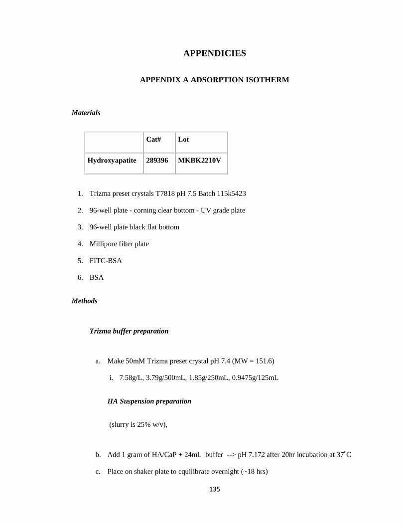
135
APPENDICIES
APPENDIX A ADSORPTION ISOTHERM
Materials
Cat# Lot
Hydroxyapatite 289396 MKBK2210V
1. Trizma preset crystals T7818 pH 7.5 Batch 115k5423
2. 96-well plate - corning clear bottom - UV grade plate
3. 96-well plate black flat bottom
4. Millipore filter plate
5. FITC-BSA
6. BSA
Methods
Trizma buffer preparation
a. Make 50mM Trizma preset crystal pH 7.4 (MW = 151.6)
i. 7.58g/L, 3.79g/500mL, 1.85g/250mL, 0.9475g/125mL
HA Suspension preparation
(slurry is 25% w/v),
b. Add 1 gram of HA/CaP + 24mL buffer --> pH 7.172 after 20hr incubation at 37oC
c. Place on shaker plate to equilibrate overnight (~18 hrs)
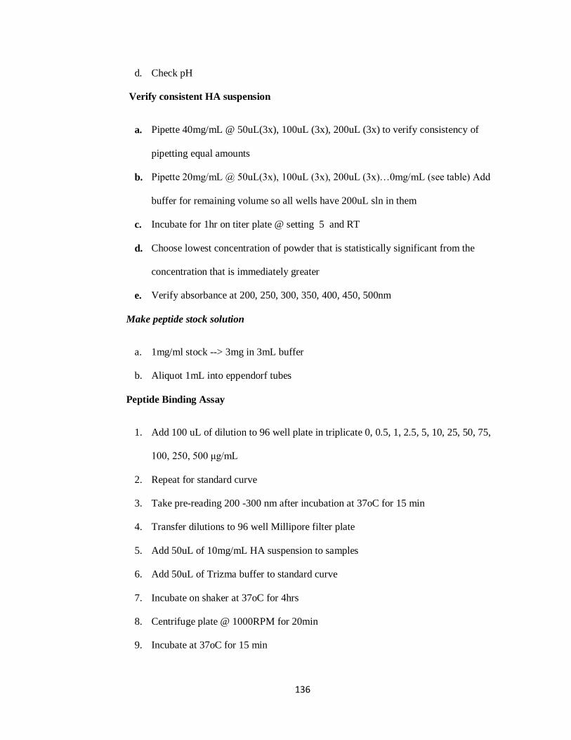
136
d. Check pH
Verify consistent HA suspension
a. Pipette 40mg/mL @ 50uL(3x), 100uL (3x), 200uL (3x) to verify consistency of
pipetting equal amounts
b. Pipette 20mg/mL @ 50uL(3x), 100uL (3x), 200uL (3x)…0mg/mL (see table) Add
buffer for remaining volume so all wells have 200uL sln in them
c. Incubate for 1hr on titer plate @ setting 5 and RT
d. Choose lowest concentration of powder that is statistically significant from the
concentration that is immediately greater
e. Verify absorbance at 200, 250, 300, 350, 400, 450, 500nm
Make peptide stock solution
a. 1mg/ml stock --> 3mg in 3mL buffer
b. Aliquot 1mL into eppendorf tubes
Peptide Binding Assay
1. Add 100 uL of dilution to 96 well plate in triplicate 0, 0.5, 1, 2.5, 5, 10, 25, 50, 75,
100, 250, 500 μg/mL
2. Repeat for standard curve
3. Take pre-reading 200 -300 nm after incubation at 37oC for 15 min
4. Transfer dilutions to 96 well Millipore filter plate
5. Add 50uL of 10mg/mL HA suspension to samples
6. Add 50uL of Trizma buffer to standard curve
7. Incubate on shaker at 37oC for 4hrs
8. Centrifuge plate @ 1000RPM for 20min
9. Incubate at 37oC for 15 min

137
10. Take post reading
Micro BCA Assay
1. Preparation of working reagents
2. Pipette 150uL into each well containing filtered solution
3. Place on plate shaker for 30seconds
4. Cover plate using sealing tape and incubate at 37oC for 2 hrs
5. Cool plate to RT
6. Measure absorbance at 562nm
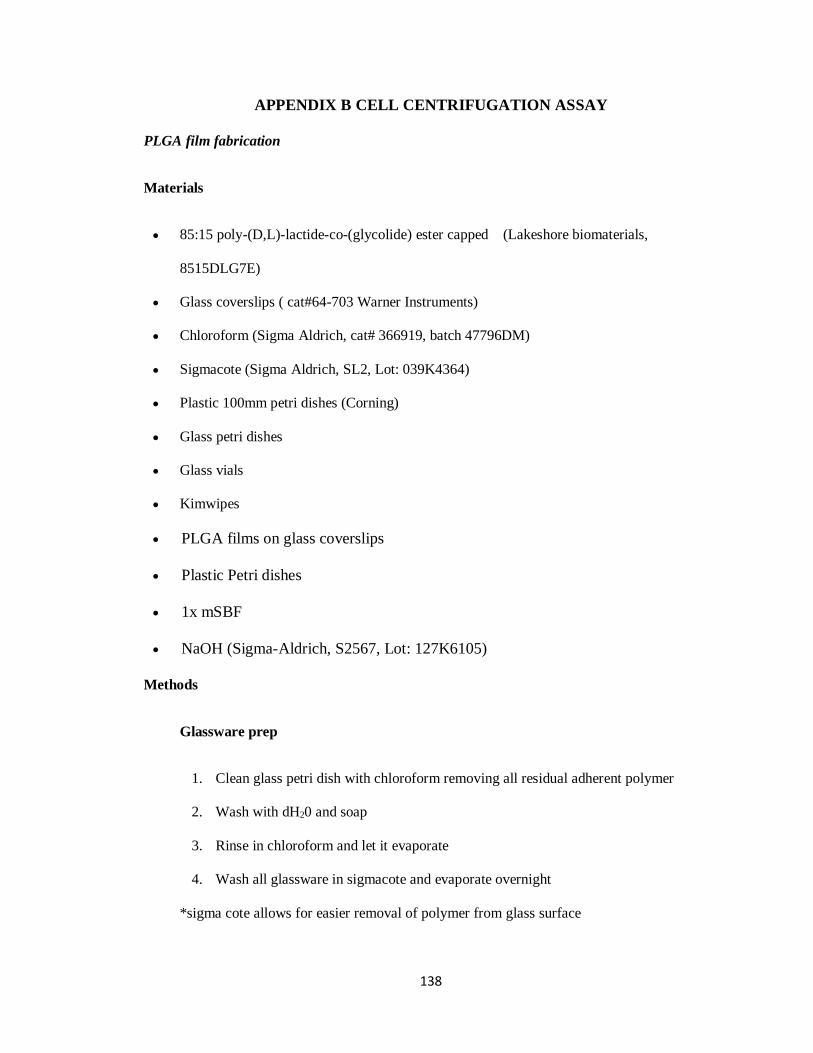
138
APPENDIX B CELL CENTRIFUGATION ASSAY
PLGA film fabrication
Materials
85:15 poly-(D,L)-lactide-co-(glycolide) ester capped (Lakeshore biomaterials,
8515DLG7E)
Glass coverslips ( cat#64-703 Warner Instruments)
Chloroform (Sigma Aldrich, cat# 366919, batch 47796DM)
Sigmacote (Sigma Aldrich, SL2, Lot: 039K4364)
Plastic 100mm petri dishes (Corning)
Glass petri dishes
Glass vials
Kimwipes
PLGA films on glass coverslips
Plastic Petri dishes
1x mSBF
NaOH (Sigma-Aldrich, S2567, Lot: 127K6105)
Methods
Glassware prep
1. Clean glass petri dish with chloroform removing all residual adherent polymer
2. Wash with dH20 and soap
3. Rinse in chloroform and let it evaporate
4. Wash all glassware in sigmacote and evaporate overnight
*sigma cote allows for easier removal of polymer from glass surface
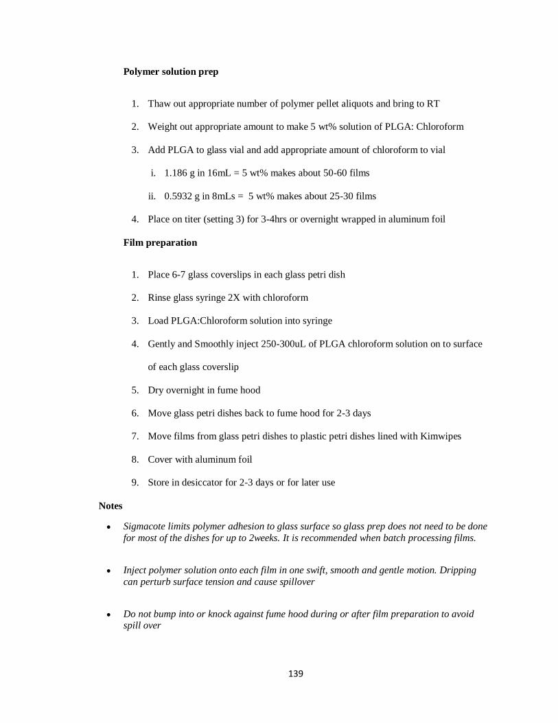
139
Polymer solution prep
1. Thaw out appropriate number of polymer pellet aliquots and bring to RT
2. Weight out appropriate amount to make 5 wt% solution of PLGA: Chloroform
3. Add PLGA to glass vial and add appropriate amount of chloroform to vial
i. 1.186 g in 16mL = 5 wt% makes about 50-60 films
ii. 0.5932 g in 8mLs = 5 wt% makes about 25-30 films
4. Place on titer (setting 3) for 3-4hrs or overnight wrapped in aluminum foil
Film preparation
1. Place 6-7 glass coverslips in each glass petri dish
2. Rinse glass syringe 2X with chloroform
3. Load PLGA:Chloroform solution into syringe
4. Gently and Smoothly inject 250-300uL of PLGA chloroform solution on to surface
of each glass coverslip
5. Dry overnight in fume hood
6. Move glass petri dishes back to fume hood for 2-3 days
7. Move films from glass petri dishes to plastic petri dishes lined with Kimwipes
8. Cover with aluminum foil
9. Store in desiccator for 2-3 days or for later use
Notes
Sigmacote limits polymer adhesion to glass surface so glass prep does not need to be done
for most of the dishes for up to 2weeks. It is recommended when batch processing films.
Inject polymer solution onto each film in one swift, smooth and gentle motion. Dripping
can perturb surface tension and cause spillover
Do not bump into or knock against fume hood during or after film preparation to avoid spill over

140
No other work should be done in fume hood until they have been moved to back of the fume
hood the following day
Mineralization of films
Methods
1. Add 50mL 0.5M NaOH to each 100mm2 plastic petri dish
2. Remove films from desiccator
3. Submerge films per dish in NaOH for 7 min
4. Wash twice in 50mL ddH2O for 10 min each
5. Add 50mL 1x mSBF in cell culture hood to new petri dishes
6. Warm up the SBF to 37oC
7. Add films to SBF cover dish and seal with parafilm
8. Place at 37oC and replace SBF every day for 5-7 days
9. Remove and rinse in ddH20 1x for 10 min
10. Remove 1-3 samples to verify degree of mineralization and Ca/P ratio using SEM-EDX
Preparation of biomaterial surfaces
Make dBSA solution > block the background to improve signal: noise
**specific time and temperature conditions of denaturation are very critical for proper
blocking
Solubilize BSA 1.5 g in 150 mL PBS(1%) in a glass Nalgene bottle on stir plate using
magnetic stir bar for 1hr
Pre-heat hybridization oven to 90oC
Microwave 300mL tap water in 500mL beaker until it reaches 90oC
Place 1% BSA solution bottle in hot water beaker and transfer to hybridization chamber.
Measure solution temperature of sample using thermocouple.
Start timer once solution temperature reaches 80oC.
Remove after 11-12 min.
Cool in RT water bath and sterile filter in cell culture hood.
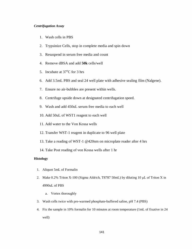
141
Centrifugation Assay
1. Wash cells in PBS
2. Trypsinize Cells, stop in complete media and spin down
3. Resuspend in serum free media and count
4. Remove dBSA and add 50k cells/well
5. Incubate at 37oC for 3 hrs
6. Add 3.5mL PBS and seal 24 well plate with adhesive sealing film (Nalgene).
7. Ensure no air-bubbles are present within wells.
8. Centrifuge upside down at designated centrifugation speed.
9. Wash and add 450uL serum free media to each well
10. Add 50uL of WST1 reagent to each well
11. Add water to the Von Kossa wells
12. Transfer WST-1 reagent in duplicate to 96 well plate
13. Take a reading of WST-1 @420nm on microplate reader after 4 hrs
14. Take Post reading of von Kossa wells after 1 hr
Histology
1. Aliquot 5mL of Formalin
2. Make 0.2% Triton X-100 (Sigma Aldrich, T8787 50mL) by diluting 10 μL of Triton X in
4990uL of PBS
a. Vortex thoroughly
3. Wash cells twice with pre-warmed phosphate-buffered saline, pH 7.4 (PBS)
4. Fix the sample in 10% formalin for 10 minutes at room temperature (1mL of fixative in 24
well)

142
a. Tissue to fixative volume ratio should be 15-20% if specimen receives more, than
recommended then it is ok, but undersaturation should be avoided.
b. Note: Methanol can disrupt actin during the fixation process. Therefore, it is best to
avoid any methanol containing fixatives. The preferred fixative is methanol-free
formaldehyde.
5. Wash two or more times with PBS.
6. Place each coverslip in a glass petri dish and extract it with a solution of acetone at ≤–20°C
or 0.1% Triton X-100 in PBS for 3 to 5 minutes.
7. Wash two or more times with PBS.
8. When staining with any of the fluorescent phallotoxins, dilute 5 μL methanolic stock
solutioninto 200 μL PBS for each coverslipto be stained.
a. To reduce nonspecific background staining with these conjugates, add 1% bovine
serum albumin (BSA) to the staining solution. It may also be useful to pre-incubate
fixed cells with PBS containing 1% BSA or with the Image-iT™FX signal enhancer
(I36933) for 20–30 minutes prior to adding the phallotoxin staining solution.
9. When staining more than one coverslip, adjust volumes accordingly. For a stronger signal,
use 2 or 3 units per coverslip.
10. Place the staining solution on the coverslip for 20 minutes at room temperature (generally,
any temperature between 4°C and 37°C is suitable). To avoid evaporation, keep the
coverslips inside a covered container during the incubation.
11. Wash two or more times with PBS.
12. For long-term storage, the cells should be air dried and then mounted in a permanent
mountant such as ProLong® Gold reagent or Cytoseal. Specimens prepared in this manner
retain actin staining for at least six months when stored in the dark at 2–6°C.