Penile US Presentation - Russell Group...
5
PG Course 102HO AUA 2011 1 Penile Ultrasound Bruce R. Gilbert, MD, PhD Chairman, National Urologic Ultrasound Faculty Associate Clinical Professor of Urology and Reproductive Medicine Weill Cornell Medical College Director of Reproductive and Sexual Medicine Smith Institute for Urology North Shore LIJ Health System 2 Penile Ultrasound Anatomy • Phallus consists of the two corpora cavernosa (cc) and the corpora spongiosum (cs) which surrounds the urethra. All three covered by the tunica albuginea • The two penile arteries arise from branches of the internal pudendal arteries giving rise to: – Penile bulbar artery – Urethral artery – Superficial dorsal artery – Deep penile artery which within the cc branch into helicine arteries which open into the sinusoids. • The cc are drained by subtunical veins that empty into the deep dorsal vein www.bartleby.com 3 Scanning Terminology Penile Orientation www.bartleby.com Dorsal Ventral Right Left Dorsal Ventral Right Left 4 Orientation Urethra Urethra Cavernosal A. Cavernosal A. 5 Physical Principles Ultrasonography • Pulsed Wave Doppler (PW) – Single crystal, phase shift measured, speed:direction:depth • B-mode (gray scale) • Color Doppler (Duplex) – Speed and direction encoded in color as indicated by the color bar (BART) • Spectral Doppler (Triplex) – spectrum of flow velocities represented graphically on the Y-axis and time on the X-axis Spectral Doppler Color Doppler 6 Scanning Protocol penile ultrasound - overview • High resolution grey scale imaging with transducers from 7 to 18 mHz • Color and spectral Doppler capabilities are essential • Transverse and longitudinal views obtained from ventral and/or dorsal surfaces – Survey Scan (Video Clips) – Specific Images (Proximal, Mid, Distal, Lateral) • The specific measurements obtained should be documented on the images. • The specific images obtained should document the findings discussed in the report.
Transcript of Penile US Presentation - Russell Group...

PG Course 102HO AUA 2011
1
Penile Ultrasound Bruce R. Gilbert, MD, PhD Chairman, National Urologic Ultrasound Faculty Associate Clinical Professor of Urology and Reproductive Medicine Weill Cornell Medical College Director of Reproductive and Sexual Medicine Smith Institute for Urology North Shore LIJ Health System
2
Penile Ultrasound Anatomy • Phallus consists of the two corpora
cavernosa (cc) and the corpora spongiosum (cs) which surrounds the urethra. All three covered by the tunica albuginea
• The two penile arteries arise from branches of the internal pudendal arteries giving rise to: – Penile bulbar artery – Urethral artery – Superficial dorsal artery – Deep penile artery which within
the cc branch into helicine arteries which open into the sinusoids.
• The cc are drained by subtunical veins that empty into the deep dorsal vein
www.bartleby.com
3
Scanning Terminology Penile Orientation
www.bartleby.com
Dorsal
Ventral
Right Left
Dorsal
Ventral
Right Left
4
Orientation Urethra
Urethra
Cavernosal A.
Cavernosal A.
5
Physical Principles Ultrasonography
• Pulsed Wave Doppler (PW) – Single crystal, phase shift measured,
speed:direction:depth • B-mode (gray scale) • Color Doppler (Duplex)
– Speed and direction encoded in color as indicated by the color bar (BART)
• Spectral Doppler (Triplex) – spectrum of flow velocities represented graphically on
the Y-axis and time on the X-axis
Spectral Doppler
Color Doppler
6
Scanning Protocol penile ultrasound - overview
• High resolution grey scale imaging with transducers from 7 to 18 mHz
• Color and spectral Doppler capabilities are essential
• Transverse and longitudinal views obtained from ventral and/or dorsal surfaces – Survey Scan (Video Clips) – Specific Images (Proximal, Mid, Distal, Lateral)
• The specific measurements obtained should be documented on the images.
• The specific images obtained should document the findings discussed in the report.

PG Course 102HO AUA 2011
2
7
Normal Imaging Documentation
• The report should include: – patient identification – date of examination – measurement
parameters and anatomical findings of examination.
• The report is signed by the physician who performed the ultrasound examination
• Indication for performing the examination is clear and provided on the report.
• Images should include: – patient identification – date and time of
each image – Clear image with
orientation and measurements
– Labeling of anatomy and any abnormalities
• Images should be attached to the report
8
Indications • Structural Pathology
– Penile plaque • Peyronie’s • Iatrogenic fibrosis
– Penile mass • Penile fracture • Penile tumor • Hematoma • Cavernosal
herniation
• Vascular Pathology – Erectile dysfunction – Priapism
• High flow • Low flow
– Thrombosis • Urethral Pathology
– Diverticula – Abscess – Stricture – Calculus
• Post surgical follow up
9
Indications structural – Peyronie’s plaque
• Plaques may or may not be calcified • May be better visualized with tumescence • Arterial venous disease more common with Peyronie’s disease
• Images/Measurements • thickness and length of the plaque • blood flow of the corpora cavernosa and corpora spongiosa
10
Indications structural - penile fracture • Usually presents with
pain, swelling and sudden loss of erections with intercourse
• Ultrasound is useful for initial diagnosis (hematoma, tunica albuginea defect) and long term follow up (corporal fibrosis, plaque formation)
• Images/Measurements • width of defect • Transverse and longitudinal image of defect • Color flow confirmation of viable tissue
CJ Wi kin, PS Sidhu, in Ultrasound of the Urogenital System,GM Baxter,PS Sidhu, Thieme,2006
11
Indications structural - penile fracture
CJ Wi kin, PS Sidhu, in Ultrasound of the Urogenital System,GM Baxter,PS Sidhu, Thieme,2006
12
Indications structural - penile tumor
• Squamous cell carcinoma of penis confined to subepithelial tissue
• Tunica albuginea of the corpora cavernosa is intact
• Bladder cancer metastatic to penis with diffuse and nodular involvement (N) of the corpora cavernosa
CJ Wi kin, PS Sidhu, in Ultrasound of the Urogenital System,GM Baxter,PS Sidhu, Thieme,2006
PG Course 102HO AUA 2011
3
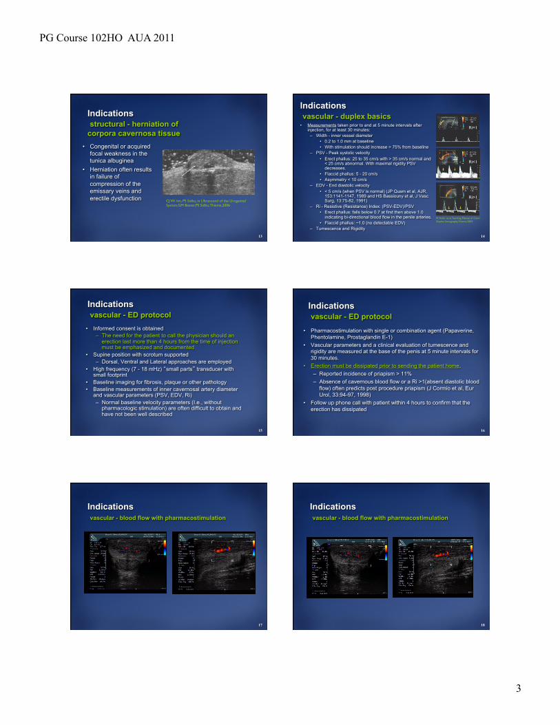
13
Indications structural - herniation of corpora cavernosa tissue
• Congenital or acquired focal weakness in the tunica albuginea
• Herniation often results in failure of compression of the emissary veins and erectile dysfunction CJ Wi kin, PS Sidhu, in Ultrasound of the Urogenital
System,GM Baxter,PS Sidhu, Thieme,2006
14
Indications vascular - duplex basics • Measurements taken prior to and at 5 minute intervals after
injection, for at least 30 minutes: – Width - inner vessel diameter
• 0.2 to 1.0 mm at baseline • With stimulation should increase > 75% from baseline
– PSV - Peak systolic velocity • Erect phallus: 25 to 35 cm/s with > 35 cm/s normal and
< 25 cm/s abnormal. With maximal rigidity PSV decreases.
• Flaccid phallus: 5 - 20 cm/s • Asymmetry < 10 cm/s
– EDV - End diastolic velocity • < 5 cm/s (when PSV is normal) (JP Quam et al, AJR,
153:1141-1147, 1989 and HS Bassiouny et al, J Vasc Surg, 13:75-82, 1991)
– Ri - Resistive (Resistance) Index: (PSV-EDV)/PSV • Erect phallus: falls below 0.7 at first then above 1.0
indicating bi-directional blood flow in the penile arteries. • Flaccid phallus: ~1.0 (no detectable EDV)
– Tumescence and Rigidity
M Hofer et al, Teaching Manual of Color Duplex Sonography,Thieme,2004
Ri=1
Ri<1
Ri>1
15
Indications vascular - ED protocol
• Informed consent is obtained – The need for the patient to call the physician should an
erection last more than 4 hours from the time of injection must be emphasized and documented
• Supine position with scrotum supported – Dorsal, Ventral and Lateral approaches are employed
• High frequency (7 - 18 mHz) “small parts” transducer with small footprint
• Baseline imaging for fibrosis, plaque or other pathology • Baseline measurements of inner cavernosal artery diameter
and vascular parameters (PSV, EDV, Ri) – Normal baseline velocity parameters (I.e., without
pharmacologic stimulation) are often difficult to obtain and have not been well described
16
Indications vascular - ED protocol
• Pharmacostimulation with single or combination agent (Papaverine, Phentolamine, Prostaglandin E-1)
• Vascular parameters and a clinical evaluation of tumescence and rigidity are measured at the base of the penis at 5 minute intervals for 30 minutes.
• Erection must be dissipated prior to sending the patient home. – Reported incidence of priapism > 11% – Absence of cavernous blood flow or a Ri >1(absent diastolic blood
flow) often predicts post procedure priapism (J Cormio et al, Eur Urol, 33:94-97, 1998)
• Follow up phone call with patient within 4 hours to confirm that the erection has dissipated
17
Indications vascular - blood flow with pharmacostimulation
18
Indications vascular - blood flow with pharmacostimulation
PG Course 102HO AUA 2011
4
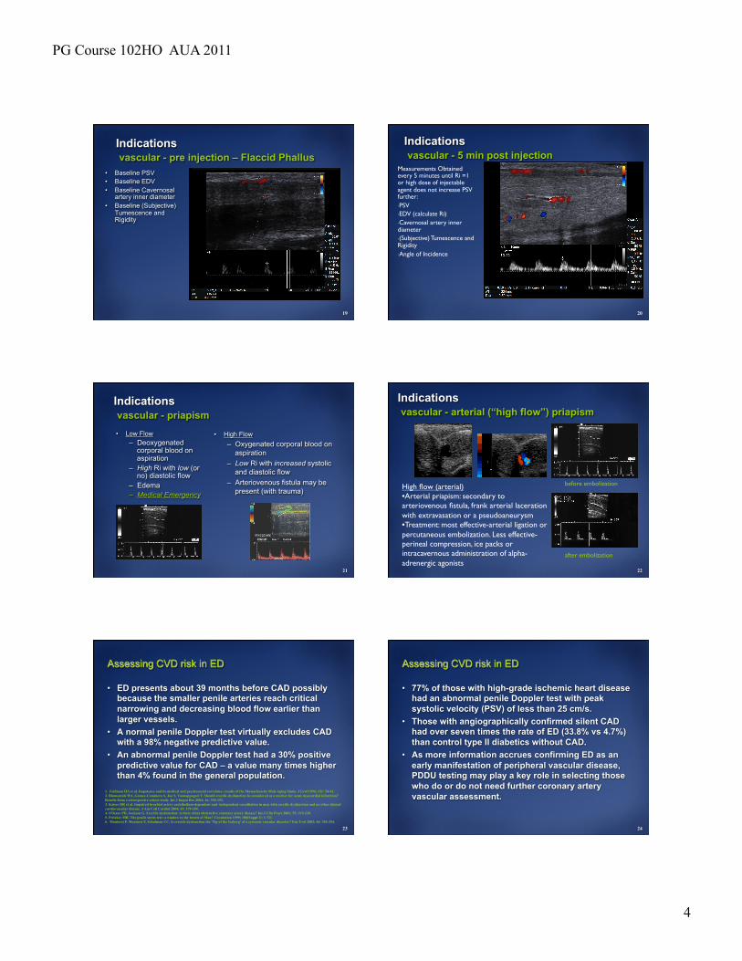
19
Indications vascular - pre injection – Flaccid Phallus
• Baseline PSV • Baseline EDV • Baseline Cavernosal
artery inner diameter • Baseline (Subjective)
Tumescence and Rigidity
20
Indications vascular - 5 min post injection
Measurements Obtained every 5 minutes until Ri =1 or high dose of injectable agent does not increase PSV further: • PSV • EDV (calculate Ri) • Cavernosal artery inner diameter • (Subjective) Tumescence and Rigidity • Angle of Incidence
21
Indications vascular - priapism • Low Flow
– Deoxygenated corporal blood on aspiration
– High Ri with low (or no) diastolic flow
– Edema – Medical Emergency
• High Flow – Oxygenated corporal blood on
aspiration – Low Ri with increased systolic
and diastolic flow – Arteriovenous fistula may be
present (with trauma)
22
Indications vascular - arterial (“high flow”) priapism
before embolization
after embolization
High flow (arterial) • Arterial priapism: secondary to arteriovenous fistula, frank arterial laceration with extravasation or a pseudoaneurysm • Treatment: most effective-arterial ligation or percutaneous embolization. Less effective-perineal compression, ice packs or intracavernous administration of alpha-adrenergic agonists
23
Assessing CVD risk in ED
• ED presents about 39 months before CAD possibly because the smaller penile arteries reach critical narrowing and decreasing blood flow earlier than larger vessels.
• A normal penile Doppler test virtually excludes CAD with a 98% negative predictive value.
• An abnormal penile Doppler test had a 30% positive predictive value for CAD – a value many times higher than 4% found in the general population.
1. Feldman HA et al. Impotence and its medical and psychosocial correlates: results of the Massachusetts Male Aging Study. J Urol 1994; 151: 54-61. 2. Blumentals WA, Gomez-Caminero A, Joo S, Vannappagari V. Should erectile dysfunction be considered as a marker for acute myocardial infarction? Results from a retrospective cohort study. Int J Impot Res 2004; 16: 350-353. 3. Kaiser DR et al. Impaired brachial artery endothelium-dependent and -independent vasodilation in men with erectile dysfunction and no other clinical cardiovascular disease. J Am Coll Cardiol 2004; 43: 179-184. 4. O'Kane PD, Jackson G. Erectile dysfunction: is there silent obstructive coronary artery disease? Int J Clin Pract 2001; 55: 219-220. 5. Pritzker MR. The penile stress test: a window to the hearts of Man? Circulation 1999; 100(Suppl 1): 1-711. 6. Montorsi P, Montorsi F, Schulman CC. Is erectile dysfunction the 'Tip of the Iceberg' of a systemic vascular disorder? Eur Urol 2003; 44: 352-354. 24
Assessing CVD risk in ED
• 77% of those with high-grade ischemic heart disease had an abnormal penile Doppler test with peak systolic velocity (PSV) of less than 25 cm/s.
• Those with angiographically confirmed silent CAD had over seven times the rate of ED (33.8% vs 4.7%) than control type II diabetics without CAD.
• As more information accrues confirming ED as an early manifestation of peripheral vascular disease, PDDU testing may play a key role in selecting those who do or do not need further coronary artery vascular assessment.

PG Course 102HO AUA 2011
5
25
Assessing CVD risk in ED
• The physician evaluating ED has a unique opportunity to diagnosis vascular impairment at a time when lifestyle changes and possible medical intervention have the potential to change morbidity and mortality of cardiovascular disease.
• As suggested by Miner there might be a “window of curability” in which the significant risk of future cardiovascular events might be averted through early diagnosis and treatment
Miner MM. Erectile Dysfunction: A Harbinger or Consequence: Does Its Detection Lead to a "Window of Curability?". J Androl. Sep 23 2011;32(2):125-134
26
Indications vascular - dorsal vein thrombosis
“Mondor’s phlebitis” • Acute: inflammation, pain fever • Subacute: induration and minimal pain • Spontaneous recanalization in 6 to 8 weeks
27
Indications structural - urethral stricture
Normal A. Radio-urethrography B. Sono-urethrography
Urethral Stricture A. Sono-urethrography B. Color Doppler
M Mitterberger et al, J Urol, 177, 992-997, 2007


![Penile Low-Intensity Shock Wave Therapy: A Promising Novel ...€¦ · blood flow and endothelial function by stimulating angio-genesis in the penis [7]. This review summarizes the](https://static.fdocuments.us/doc/165x107/5ecd2cef404b97379a36781c/penile-low-intensity-shock-wave-therapy-a-promising-novel-blood-flow-and-endothelial.jpg)