Pelvic biomechanics and muscle activation patterns during ...
117
Pelvic biomechanics and muscle activation patterns during non- weighted squats in U/19 university- level rugby union players M. Greyling 20273266 B.Sc. Honnours in Biokinetics Dissertation submitted in fulfillment of the requirements for the degree Magister Scientiae in Biokinetics at the Potchefstroom Campus of the North-West University Supervisor E.J. Bruwer November 2013
Transcript of Pelvic biomechanics and muscle activation patterns during ...

Pelvic biomechanics and muscle
activation patterns during non-
weighted squats in U19 university-
level rugby union players
M Greyling
20273266
BSc Honnours in Biokinetics
Dissertation submitted in fulfillment of the requirements for
the degree Magister Scientiae in Biokinetics at the Potchefstroom Campus of the North-West University
Supervisor EJ Bruwer
November 2013
Acknowledgements
i
Dedicated to my family
Acknowledgements
ii
A C K N O W L E D G E M E N T S
I wish to express my extreme gratitude to the following people who made the
completion of this dissertation possible
Firstly my heavenly Father who gave me the abilities and strength to
complete this study
To my husband Christie for the understanding and loving support which
carried me through
To my parents Manie and Miemie who supported me financially and
emotionally throughout my studies
To my supervisor Erna Bruwer for all the long hours of listening reading
editing thinking testing and correcting to make this dissertation possible
Prof Suria Ellis from the Statistical Consultation Services of the North-
West University for the statistical analysis of the data and the
interpretation of the results
Theo Pistorius for assisting with the testing of all participants
The U19 rugby union training squad for participating in the study
The training staff of the U19 rugby union training squad for allowing the
time for the testing to be completed
All my friends and family for their understanding and loving support
Miemie Greyling
November 2013
Authorrsquos contribution
iii
A U T H O R rsquo S C O N T R I B U T I O N
The principal author of this dissertation is Miss M Greyling The contribution of the co-author
is summarised below
Co-author Contribution
Ms EJ Bruwer Supervisor
Co-author - assistance in writing of manuscripts study design
executing of testing data extraction technical editing interpretation of
results
The following is a statement by the co-author confirming her individual role in this study and
giving her permission that the manuscript may form part of this dissertation
I hereby declare that my role in the preparation of the above mentioned manuscripts is as
indicated above and that I give my consent that it may be published as part of the MSc
dissertation of Miemie Greyling
_________________________
EJ Bruwer
Abstract
iv
A B S T R A C T
Pelvic biomechanics and muscle activation patterns during non-weighted squats in U19
university-level rugby union players
Hyperlordosis or anterior pelvic tilt is a common non-neutral spinal posture associated with
weak core stability low back pain and altered lumbopelvic muscle activation patterns Yet the
effects of altered lumbopelvic posture and core stability on muscle activation patterns have not
been evaluated during a functional movement The main purpose of this study was to determine
the relationship between pelvic tilt core stability and muscle activation patterns during non-
weighted squats in U19 university-level rugby union players A total of 49 rugby union
players participated in this study Pelvic tilt (dominant side) was measured from a digital photo
with clear reflector markers on the anterior superior iliac spine (ASIS) and posterior superior
iliac spine (PSIS) using the Kinovea video analysis software programme (version 0815)
Flexibility of the hamstrings hip flexors and knee extensors was assessed with goniometry
Core stability was assessed using the pressure biofeedback unit and muscle onset times during
the ascent phase of non-weighted squats The onset times of the transverse abdominis (TrA)
erector spinae (ES) gluteus maximus (GM) and biceps femoris (BF) muscles were measured
using electromyography (EMG) Players were then grouped according to pelvic tilt (anterior
and neutral) and by playing position (forwards and backs) The between group differences
were evaluated for the abovementioned variables using p-value (statistical significance) and d-
value (practical significance) measures Muscle activation patterns and firing order were
determined using descriptive statistics
The mean pelvic tilt of all participants (N=49) was an anterior tilt of 1535deg When grouped
by pelvic tilt the anterior tilt group showed a mean pelvic tilt of 1783deg (n=27) and the neutral
pelvic tilt group showed a mean pelvic tilt of 1175deg (n=22) Despite the differences in pelvic
tilt there was no significant difference in flexibility between the groups Another controversial
result is that the anterior tilt group showed practical significantly better core stability (d=054)
than the neutral tilt group (4693deg vs 563deg)
During the double leg squat the muscle activation patterns were consistent between the groups
TrA activated first followed by ES Thereafter the BF muscle activated followed by the GM
The first place activation of TrA is consistent with the literature stating that the deep abdominal
stabilisers of individuals with good core stability activate before the movement is initiated The
Abstract
v
early onset of muscle activity of ES points to a focus on back extension during the ascent of
the squat Because the pelvic tilt was measured during static standing only it is unclear whether
the players in the neutral tilt group were able to hold the neutral pelvic tilt posture throughout
the movement Research has shown that there is an increased focus on trunk extension during
the ascent phase of the squat which is not present during the descent Future research should
focus on assessing the pelvic tilt at the beginning of the ascent phase of the squat to ensure
accurate results
The delay in GM activation during the ascent of the squat is concerning GM acts as a
lumbopelvic stabilizer and its slow activation points to a decrease in lumbopelvic stability
This is very important in weight training because weight training increases the strain on the
lumbar spinal structures which decreases performance and increases the risk of injury
Keywords Rugby union players anterior pelvic tilt electromyography transverse
abdominis activation flexiblity
Opsomming
vi
O P S O M M I N G
Pelvis biomeganika en spieraktiveringspatrone gedurende die squat beweging in O19
rugby-unie spelers
Hiperlordose of anterior pelvis tilt is lsquon algemene nie-neutrale laerug postuur wat geassosieer
word met swak abdominale stabilisering laerug pyn en veranderde lumbo-pelviese
spieraktiveringspatrone Tog is die verband tussen hierdie nie-neutrale laerug postuur en
abdominale stabilisering nog nie geeumlvalueer tydens lsquon funksionele beweging nie Daarom is
die doel van hierdie studie om die verwantskap tussen pelvis tilt abdominale stabiliseringskrag
en spieraktiveringspatrone tydens die squat in O19 rugby-unie spelers te evalueer Nege en
veertig rugby-unie spelers het deelgeneem aan die studie Pelvis tilt (dominante kant) is gemeet
vanaf lsquon digitale foto met duidelike merkers op die anterior superior iliale spina (ASIS) en
posterior superior iliale spina (PSIS) met behulp van die Kinovea analitiese sagteware program
(weergawe 0815) Soepelheid van die hampese groep heup fleksore en knie ekstensore is
gemeet met behulp van goniometrie Abdominale stabiliseringkrag is bepaal met behulp van
druksensitiewe bioterugvoer-eenheid en spieraktiveringspatrone van die transverse abdominus
(TrA) erector spinae (ES) gluteus maximus (GM) en biceps femoris (BF) spiere is met behulp
van elektromiografie (EMG) gemeet tydens die opstaan-fase van die squat beweging Tydens
statistiese analise is die spelers gegroepeer volgens pelvis tilt (anterior of neutraal) en volgens
speel-posisie (voorspelers of agterspelers) Die tussen-groep verskille is bereken vir die
bogenoemde veranderlikes deur gebruik te maak van die p-waarde (statistiese
betekenesvolheid) en die effekgrootte is ook bepaal om praktiese betekenisvolheid aan te dui
(d-waarde) Spieraktiveringspatrone en die aktiveringsorde is bepaal deur gebruik te maak van
beskrywende statistiek
Die gemiddelde pelvis tilt van die proefpersone in totaal (N=49) is lsquon anterior pelvis tilt van
1535deg Die gemiddelde tilt van die anterior tilt geklassifiseerde groep is 1783deg (n=27) en
1175deg vir die neutrale tilt geklassifiseerde groep (n=22) Ten spyte van die verskil in pelvis
tilt tussen die groepe is daar geen betekenisvolle verskil in soepelheid tussen die groepe nie
Die anterior pelvis tilt groep vertoon ook met prakties betekenisvol sterker abdominale
stabiliseringskrag (d=054) as die neutrale tilt groep wat in teenstelling is met onlangse
literatuur (4693deg teenoor 563deg)
Opsomming
vii
Die spieraktiveringspatrone tydens die dubbel-been squat was dieselfde vir beide pelvis tilt
groepe Aktivering van TrA was die vinnigste gevolg deur ES Daarna het BF geaktiveer met
GM wat konstant laaste geaktiveer het Die vroeeuml aktivering van TrA is in lyn met die literatuur
wat voorstel dat in individue met goeie abdominale stabilisering die TrA spier sal aktiveer voor
die aanvang van lsquon beweging Daar is wel ook lsquon vroeeuml aktivering van ES waargeneem in
hierdie studie wat dui op lsquon fokus op rug-ekstensie tydens die opstaan-fase van die squat
Omdat die pelvis tilt slegs tydens lsquon stilstaande posisie gemeet was is dit onduidelik of die
spelers in die neutrale pelvis tilt groep die neutral postuur kon behou regdeur die squat
beweging Literatuur beweer dat daar lsquon verhoogde fokus op rug-ekstensie tydens die opstaan
fase van die squat is wat nie teenwoordig is tydens die afsak fase nie - dit lei tot die vroeeuml
aktivering van ES GM is verantwoordelik vir lumbo-pelviese stabiliteit en die vertraging in
aktivering mag lei tot onstabiliteit rondom die pelvis Dit is veral belangrik tydens krag-
oefeninge omdat hierdie tipe oefening spanning op die lumbale werwels verhoog
Toekomstige navorsing moet fokus op die evaluasie van die pelvis tilt tydens die beweging
om seker te maak dat die pelvis tilt nie oormatig verander tydens die beweging nie
Sleutelwoorde Rugby unie spelers anterior pelvis tilt elektromiografie transverse
abdominus aktivering soepelheid
Table of content
viii
T A B L E O F C O N T E N T
ACKNOWLEDGEMENTShelliphelliphelliphelliphelliphelliphelliphelliphelliphelliphelliphelliphelliphelliphelliphelliphelliphelliphelliphelliphelliphelliphellip
AUTHORSrsquoS CONTRIBUTIONhelliphelliphelliphelliphelliphelliphelliphelliphelliphelliphelliphelliphelliphelliphelliphelliphelliphelliphelliphellip
ABSTRACThelliphelliphelliphelliphelliphelliphelliphelliphelliphelliphelliphelliphelliphelliphelliphelliphelliphelliphelliphelliphelliphelliphelliphelliphelliphelliphelliphelliphellip
OPSOMMINGhelliphelliphelliphelliphelliphelliphelliphelliphelliphelliphelliphelliphelliphelliphelliphelliphelliphelliphelliphelliphelliphelliphelliphelliphelliphelliphelliphellip
TABLE OF CONTENThelliphelliphelliphelliphelliphelliphelliphelliphelliphelliphelliphelliphelliphelliphelliphelliphelliphelliphelliphelliphelliphelliphelliphellip
LIST OF TABLEShelliphelliphelliphelliphelliphelliphelliphelliphelliphelliphelliphelliphelliphelliphelliphelliphelliphelliphelliphelliphelliphelliphelliphelliphelliphellip
LIST OF FIGUREShelliphelliphelliphelliphelliphelliphelliphelliphelliphelliphelliphelliphelliphelliphelliphelliphelliphelliphelliphelliphelliphelliphelliphelliphelliphellip
LIST OF ABBREVIATIONShelliphelliphelliphelliphelliphelliphelliphelliphelliphelliphelliphelliphelliphelliphelliphelliphelliphelliphelliphelliphelliphellip
CHAPTER 1 helliphelliphelliphelliphelliphelliphelliphelliphelliphelliphelliphelliphelliphelliphelliphelliphelliphelliphelliphelliphelliphelliphelliphelliphelliphelliphelliphelliphellip
Introduction
11 Problem statement helliphelliphelliphelliphelliphelliphelliphelliphelliphelliphelliphelliphelliphelliphelliphelliphelliphelliphelliphelliphelliphelliphellip
12 Objectives helliphelliphelliphelliphelliphelliphelliphelliphelliphelliphelliphelliphelliphelliphelliphelliphelliphelliphelliphelliphelliphelliphelliphelliphelliphelliphellip
13 Hypotheses helliphelliphelliphelliphelliphelliphelliphelliphelliphelliphelliphelliphelliphelliphelliphelliphelliphelliphelliphelliphelliphelliphelliphelliphelliphellip
14 Structure of dissertation helliphelliphelliphelliphelliphelliphelliphelliphelliphelliphelliphelliphelliphelliphelliphelliphelliphelliphelliphelliphellip
References helliphelliphelliphelliphelliphelliphelliphelliphelliphelliphelliphelliphelliphelliphelliphelliphelliphelliphelliphelliphelliphelliphelliphelliphelliphellip
CHAPTER 2 helliphelliphelliphelliphelliphelliphelliphelliphelliphelliphelliphelliphelliphelliphelliphelliphelliphelliphelliphelliphelliphelliphelliphelliphelliphelliphelliphelliphellip
Pelvic biomechanics and injury a literature review
21 Introduction helliphelliphelliphelliphelliphelliphelliphelliphelliphelliphelliphelliphelliphelliphelliphelliphelliphelliphelliphelliphelliphelliphelliphelliphelliphellip
22 Neutral spine and core stability helliphelliphelliphelliphelliphelliphelliphelliphelliphelliphelliphelliphelliphelliphelliphelliphelliphellip
23 Non-neutral spine helliphelliphelliphelliphelliphelliphelliphelliphelliphelliphelliphelliphelliphelliphelliphelliphelliphelliphelliphelliphelliphelliphelliphellip
24 Pelvic biomechanics and muscle activation patterns helliphelliphelliphelliphelliphelliphelliphelliphellip
25 Muscle activation patterns during the squat helliphelliphelliphelliphelliphelliphelliphelliphelliphelliphelliphelliphellip
251 Normal squat
252 Single leg squat
253 Knee-dominant vs hip-dominant squat
26 Lumbopelvic injuries in rugby union helliphelliphelliphelliphelliphelliphelliphelliphelliphelliphelliphelliphelliphelliphelliphellip
27 Summary helliphelliphelliphelliphelliphelliphelliphelliphelliphelliphelliphelliphelliphelliphelliphelliphelliphelliphelliphelliphelliphelliphelliphelliphelliphellip
References helliphelliphelliphelliphelliphelliphelliphelliphelliphelliphelliphelliphelliphelliphelliphelliphelliphelliphelliphelliphelliphelliphelliphelliphelliphelliphellip
i
iii
iv
vi
viii
xi
xii
xiii
1
1
5
5
6
7
12
12
13
15
17
18
19
21
22
25
26
28
Table of content
ix
CHAPTER 3 helliphelliphelliphelliphelliphelliphelliphelliphelliphelliphelliphelliphelliphelliphelliphelliphelliphelliphelliphelliphelliphelliphelliphelliphelliphelliphelliphellip
Core stability muscle flexibility and pelvic tilt in rugby union players
Abstract helliphelliphelliphelliphelliphelliphelliphelliphelliphelliphelliphelliphelliphelliphelliphelliphelliphelliphelliphelliphelliphelliphelliphelliphelliphellip
31 Introduction helliphelliphelliphelliphelliphelliphelliphelliphelliphelliphelliphelliphelliphelliphelliphelliphelliphelliphelliphelliphelliphelliphelliphellip
32 Methods helliphelliphelliphelliphelliphelliphelliphelliphelliphelliphelliphelliphelliphelliphelliphelliphelliphelliphelliphelliphelliphelliphelliphelliphelliphellip
321 Locality helliphelliphelliphelliphelliphelliphelliphelliphelliphelliphelliphelliphelliphelliphelliphelliphelliphelliphelliphelliphelliphelliphellip
322 Population helliphelliphelliphelliphelliphelliphelliphelliphelliphelliphelliphelliphelliphelliphelliphelliphelliphelliphelliphelliphelliphellip
323 Measurements helliphelliphelliphelliphelliphelliphelliphelliphelliphelliphelliphelliphelliphelliphelliphelliphelliphelliphelliphelliphellip
324 Statistical analysis helliphelliphelliphelliphelliphelliphelliphelliphelliphelliphelliphelliphelliphelliphelliphelliphelliphelliphellip
33 Results helliphelliphelliphelliphelliphelliphelliphelliphelliphelliphelliphelliphelliphelliphelliphelliphelliphelliphelliphelliphelliphelliphelliphelliphelliphelliphellip
34 Discussion helliphelliphelliphelliphelliphelliphelliphelliphelliphelliphelliphelliphelliphelliphelliphelliphelliphelliphelliphelliphelliphelliphelliphelliphelliphellip
35 Conclusion helliphelliphelliphelliphelliphelliphelliphelliphelliphelliphelliphelliphelliphelliphelliphelliphelliphelliphelliphelliphelliphelliphelliphelliphellip
References helliphelliphelliphelliphelliphelliphelliphelliphelliphelliphelliphelliphelliphelliphelliphelliphelliphelliphelliphelliphelliphelliphelliphelliphelliphellip
CHAPTER 4 helliphelliphelliphelliphelliphelliphelliphelliphelliphelliphelliphelliphelliphelliphelliphelliphelliphelliphelliphelliphelliphelliphelliphelliphelliphelliphelliphellip
Pelvic tilt and muscle activation patterns during non-weighted squats
Abstract helliphelliphelliphelliphelliphelliphelliphelliphelliphelliphelliphelliphelliphelliphelliphelliphelliphelliphelliphelliphelliphelliphelliphelliphelliphelliphellip
41 Introduction helliphelliphelliphelliphelliphelliphelliphelliphelliphelliphelliphelliphelliphelliphelliphelliphelliphelliphelliphelliphelliphelliphelliphelliphellip
42 Methods helliphelliphelliphelliphelliphelliphelliphelliphelliphelliphelliphelliphelliphelliphelliphelliphelliphelliphelliphelliphelliphelliphelliphelliphelliphelliphellip
421 Participants helliphelliphelliphelliphelliphelliphelliphelliphelliphelliphelliphelliphelliphelliphelliphelliphelliphelliphelliphelliphelliphellip
422 Anthropometric measurements
423 Pelvic tilt measurement helliphelliphelliphelliphelliphelliphelliphelliphelliphelliphelliphelliphelliphelliphelliphelliphellip
424 Core stability measurement helliphelliphelliphelliphelliphelliphelliphelliphelliphelliphelliphelliphelliphelliphellip
425 EMG muscle activity helliphelliphelliphelliphelliphelliphelliphelliphelliphelliphelliphelliphelliphelliphelliphelliphelliphellip
426 Statistical analysis helliphelliphelliphelliphelliphelliphelliphelliphelliphelliphelliphelliphelliphelliphelliphelliphelliphelliphellip
43 Results helliphelliphelliphelliphelliphelliphelliphelliphelliphelliphelliphelliphelliphelliphelliphelliphelliphelliphelliphelliphelliphelliphelliphelliphelliphelliphellip
44 Discussion helliphelliphelliphelliphelliphelliphelliphelliphelliphelliphelliphelliphelliphelliphelliphelliphelliphelliphelliphelliphelliphelliphelliphelliphelliphellip
45 Conclusion helliphelliphelliphelliphelliphelliphelliphelliphelliphelliphelliphelliphelliphelliphelliphelliphelliphelliphelliphelliphelliphelliphelliphelliphellip
References helliphelliphelliphelliphelliphelliphelliphelliphelliphelliphelliphelliphelliphelliphelliphelliphelliphelliphelliphelliphelliphelliphelliphelliphelliphellip
38
39
40
40
40
41
41
42
42
44
46
47
49
50
51
52
52
52
52
52
53
54
54
57
58
60
Table of content
x
CHAPTER 5 helliphelliphelliphelliphelliphelliphelliphelliphelliphelliphelliphelliphelliphelliphelliphelliphelliphelliphelliphelliphelliphelliphelliphelliphelliphelliphellip
Summary conclusion limitations and recommendations
51 Summary helliphelliphelliphelliphelliphelliphelliphelliphelliphelliphelliphelliphelliphelliphelliphelliphelliphelliphelliphelliphelliphelliphelliphelliphelliphellip
52 Conclusions helliphelliphelliphelliphelliphelliphelliphelliphelliphelliphelliphelliphelliphelliphelliphelliphelliphelliphelliphelliphelliphelliphelliphelliphellip
521 Hypothesis 1 helliphelliphelliphelliphelliphelliphelliphelliphelliphelliphelliphelliphelliphelliphelliphelliphelliphelliphelliphelliphelliphellip
522 Hypothesis 2 helliphelliphelliphelliphelliphelliphelliphelliphelliphelliphelliphelliphelliphelliphelliphelliphelliphelliphelliphelliphelliphellip
523 Hypothesis 3 helliphelliphelliphelliphelliphelliphelliphelliphelliphelliphelliphelliphelliphelliphelliphelliphelliphelliphelliphelliphelliphellip
53 Limitations and recommendations helliphelliphelliphelliphelliphelliphelliphelliphelliphelliphelliphelliphelliphelliphellip
APPENDIX A South African Journal of Sports Medicine
(Guidelines for Authors) helliphelliphelliphelliphelliphelliphelliphelliphelliphelliphelliphelliphelliphelliphellip
APPENDIX B Preventive Medicine (Guidelines for Authors) helliphelliphelliphelliphelliphelliphellip
APPENDIX C Demographic information and informed consent helliphelliphelliphelliphelliphellip
APPENDIX D Testing protocol helliphelliphelliphelliphelliphelliphelliphelliphelliphelliphelliphelliphelliphelliphelliphelliphelliphelliphellip
APPENDIX E Letter for ethical approval helliphelliphelliphelliphelliphelliphelliphelliphelliphelliphelliphelliphelliphelliphellip
APPENDIX F Letter from language editing helliphelliphelliphelliphelliphelliphelliphelliphelliphelliphelliphelliphelliphellip
63
63
66
66
67
67
68
70
77
92
97
99
101
List of tables
xi
L I S T O F T A B L E S
CHAPTER 2
Table 1 Functional classification of lumbopelvic muscles helliphelliphelliphelliphelliphelliphellip
Table 2 Muscle contraction during different phases of the squat helliphelliphelliphellip
Table 3 Correct squatting alignment and posture helliphelliphelliphelliphelliphelliphelliphelliphelliphellip
CHAPTER 3
Table 1 Descriptive statistics of rugby union players helliphelliphelliphelliphelliphelliphelliphelliphellip
Table 2 Pelvic biomechanics characteristics of rugby union players helliphellip
Table 3 Differences in core stability and degree of pelvic tilt helliphelliphelliphelliphellip
Table 4 Dominant side flexibility measures and pelvic tilt helliphelliphelliphelliphelliphelliphellip
CHAPTER 4
Table 1 Basic characteristics of rugby union players helliphelliphelliphelliphelliphelliphelliphelliphellip
Table 2 Differences in core stability and degree of pelvic tilt helliphelliphelliphelliphellip
Table 3 Differences in activation time according to pelvic position in the
Table 2 non-weighted double leg squat helliphelliphelliphelliphelliphelliphelliphelliphelliphelliphelliphelliphelliphellip
14
19
25
42
43
43
44
54
55
55
List of figures
xii
L I S T O F F I G U R E S
CHAPTER 2
Figure 1 Anterior pelvic tilt helliphelliphelliphelliphelliphelliphelliphelliphelliphelliphelliphelliphelliphelliphelliphelliphelliphelliphellip
Figure 2 Posterior pelvic tilt helliphelliphelliphelliphelliphelliphelliphelliphelliphelliphelliphelliphelliphelliphelliphelliphelliphelliphellip
Figure 3 Optimal squatting technique helliphelliphelliphelliphelliphelliphelliphelliphelliphelliphelliphelliphelliphelliphellip
CHAPTER 4
Figure 1 Firing order according to pelvic tilt in the non-weighted double
Figure 1 leg squat helliphelliphelliphelliphelliphelliphelliphelliphelliphelliphelliphelliphelliphelliphelliphelliphelliphelliphelliphelliphelliphelliphellip
16
17
24
56
List of abbreviations
xiii
L I S T O F A B B R E V I A T O N S
ACL Anterior cruciate ligament
ASIS Anterior superior iliac spine
ASLR active straight leg raise
BF Biceps femoris
cm centimetre
DLLT double leg lowering test
EMG Electromyographic
EO External Obliques
ES Erector spinae
GM Gluteus maximus
Hz Hertz
HF Hip flexor
HS Hamstring
KE Knee extensor
kg kilogram
L Left
L4 Fourth lumbar vertebrae
L5 Fifth lumbar vertebrae
LBP Low back pain
mmHG MillimeterMillimetre of Mercury
NWU North-West University
PHE prone hip extension
PSIS Posterior superior iliac spine
R Right
RA Rectus abdominis
RF Rectus femoris
SD Standard deviation
SIJ Sacro-iliac joint
SPSS Statistical Package for the Social Sciences
TrA Transverse abdominis
List of abbreviations
xiv
U19 Under nineteen
yrs years
Chapter 1
1
C H A P T E R 1
INTRODUCTION
1 1 P r o b l e m s t a t e m e n t
1 2 O b j e c t i v e s
1 3 H y p o t h e s i s
1 4 S t r u c t u r e o f d i s s e r t a t i o n
R e f e r e n c e s
1
5
5
6
7
11 PROBLEM STATEMENT
Rugby is a high intensity and physically demanding contact sport that requires strength
endurance speed and agility combined with sport-specific skills (Gamble 200410 Quarrie
et al 2013358) The all-round physically intense nature of the sport contributes to the rising
number of players reporting lower back pain (LBP) (Iwamoto et al 2005163) The game
subjects the lumbar spine to compressive shear and lateral bending forces due to scrum
formation tackling mauling and rucking (Iwamoto et al 2005166) These forces increase
stress on the inter-vertebral discs facet joints and pars inter-articularis in the lumbar spine and
can be exacerbated by an excessive lordotic lumbar curvature (Takasaki et al 2009484) The
squat is one of the foundational exercises used in functional strength training for the back and
lower extremities but is rarely performed correctly and can result in injury of multiple joints
(Liebenson 2003230) The pelvis is the link between the torso and the lower extremities and
contributes towards the stability of the whole body (Kibler et al 2006189)
Lubahn et al (2011101) suggests that sufficient activation of the muscles surrounding the
pelvis may improve safety during functional and athletic movements Pelvic or core stability
is therefore considered to be crucial for performance enhancement and injury prevention in
rugby union players (Butcher et al 2007229 Leetun et al 2004933)
Chapter 1
2
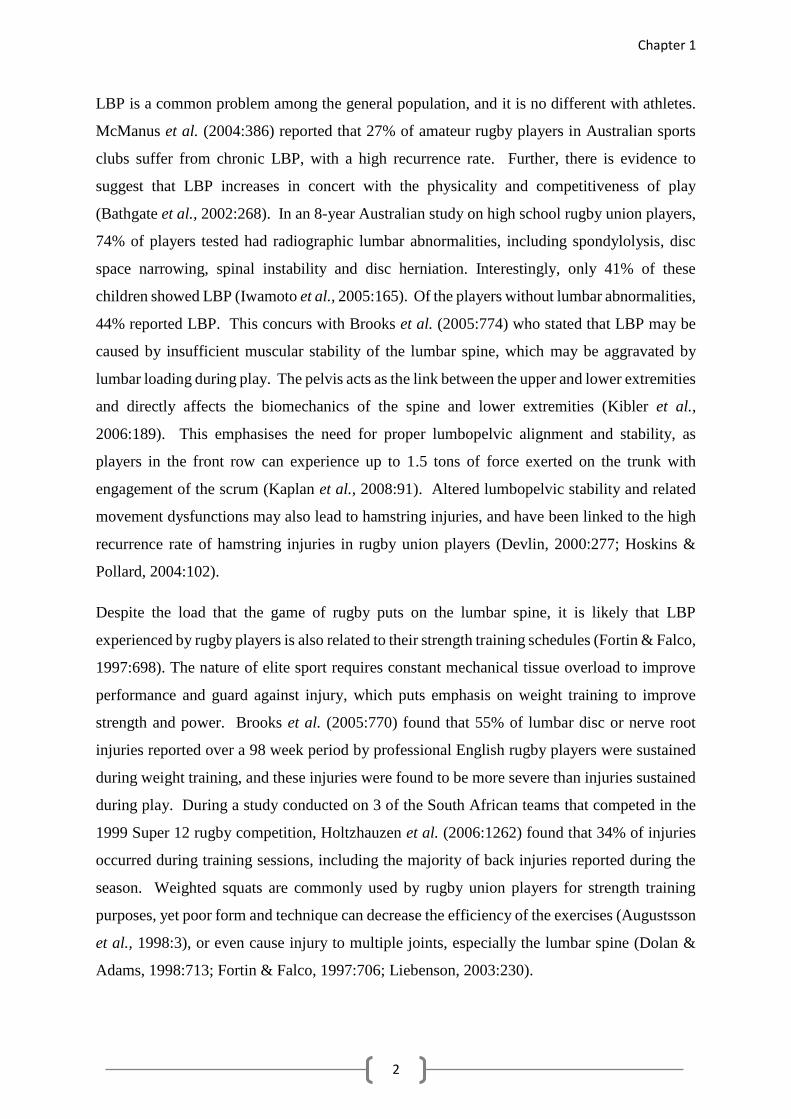
LBP is a common problem among the general population and it is no different with athletes
McManus et al (2004386) reported that 27 of amateur rugby players in Australian sports
clubs suffer from chronic LBP with a high recurrence rate Further there is evidence to
suggest that LBP increases in concert with the physicality and competitiveness of play
(Bathgate et al 2002268) In an 8-year Australian study on high school rugby union players
74 of players tested had radiographic lumbar abnormalities including spondylolysis disc
space narrowing spinal instability and disc herniation Interestingly only 41 of these
children showed LBP (Iwamoto et al 2005165) Of the players without lumbar abnormalities
44 reported LBP This concurs with Brooks et al (2005774) who stated that LBP may be
caused by insufficient muscular stability of the lumbar spine which may be aggravated by
lumbar loading during play The pelvis acts as the link between the upper and lower extremities
and directly affects the biomechanics of the spine and lower extremities (Kibler et al
2006189) This emphasises the need for proper lumbopelvic alignment and stability as
players in the front row can experience up to 15 tons of force exerted on the trunk with
engagement of the scrum (Kaplan et al 200891) Altered lumbopelvic stability and related
movement dysfunctions may also lead to hamstring injuries and have been linked to the high
recurrence rate of hamstring injuries in rugby union players (Devlin 2000277 Hoskins amp
Pollard 2004102)
Despite the load that the game of rugby puts on the lumbar spine it is likely that LBP
experienced by rugby players is also related to their strength training schedules (Fortin amp Falco
1997698) The nature of elite sport requires constant mechanical tissue overload to improve
performance and guard against injury which puts emphasis on weight training to improve
strength and power Brooks et al (2005770) found that 55 of lumbar disc or nerve root
injuries reported over a 98 week period by professional English rugby players were sustained
during weight training and these injuries were found to be more severe than injuries sustained
during play During a study conducted on 3 of the South African teams that competed in the
1999 Super 12 rugby competition Holtzhauzen et al (20061262) found that 34 of injuries
occurred during training sessions including the majority of back injuries reported during the
season Weighted squats are commonly used by rugby union players for strength training
purposes yet poor form and technique can decrease the efficiency of the exercises (Augustsson
et al 19983) or even cause injury to multiple joints especially the lumbar spine (Dolan amp
Adams 1998713 Fortin amp Falco 1997706 Liebenson 2003230)
Chapter 1
3
The squat is a closed-chain kinetic exercise with biomechanical and neurological similarities
to several functional multi-joint sporting movements (Augustsson et al 19987 Wilson et al
200598) For this reason squats are advocated for the training of sportsmen including rugby
union players Squats are also used in clinical settings to treat several hip knee and ankle
injuries (Bunton et al 199319 Dionisio et al 2008134) The correct squatting technique
requires practice especially when progressing to weighted squats If the squat is performed
with an excessive lumbar lordosis or anterior pelvic tilt there is an increased reliance on
ligamentous support (Norris 1995129) resulting in strain of the lumbar facet joints (Norris
199412) Even with the correct posture squatting can generate compressive shear tensile
and torsional forces on the lumbar spine (Durall amp Manske 200564) Stability of the lumbar
spine and pelvis is therefore strongly indicated during strength training regimes for safety and
efficiency (Brooks et al 2005774)
Stability of the lumbopelvic hip complex is maintained by a combination of bony structures
ligaments and muscle actions (Akuthota amp Nadler 200486 Muscolino amp Cipriani 200417)
The transverse abdominus (TrA) rectus abdominus (RA) internal and external obliques (EO)
quadratus lumborum (QL) multifidi and pelvic floor muscles form part of the core musculature
that enables stability and support for all trunk and spinal movements (Akuthota amp Nadler
200487 Norris 1995129 Norris 1999151 Queiroz et al 201087) The gluteal muscle
group is responsible for hip stability providing a stable base for movements of the lower
extremities (Oliver amp Keeley 20103015) Malalignment of this pelvic region can be caused
by muscle tightness or weakness leading to LBP (Bendova et al 2007980 Lehman et al
20044 Norris 199412 Takasaki et al 2009484 Wilson et al 200596) An anterior pelvic
tilt is caused by tight hip flexor muscles (iliopsoas) putting the femur in flexion and shortening
the hip flexor muscles even more (Deckert 2007110) The anterior tilt posture results in
repetitive impingement of the lumbar vertebral facets during dynamic movements (Takasaki et
al 2009484 Trainor amp Trainor 200443) and more so during functional exercises such as
the weighted squat (Fry et al 2003631) The forward inclination of the pelvis results in a
lordotic curvature in the lumbar spine shortening the erector spinae muscles (ES) and
lengthening the abdominal and gluteal muscle groups (Norris 1999154) Tightness of the ES
and iliopsoas muscles causes these muscles functions in a restricted inner range of movement
and increases muscle tone This muscular restriction results in inhibition of their antagonist
muscles the RA and the gluteal muscle group due to their lengthened state (Norris 199410
Queiroz et al 201090) If these muscles are lengthened over prolonged period stretch
Chapter 1
4
induced weakness will occur due to a reduced capacity of these muscles to activate in their
outer range of movement (Muscolino amp Cipriani 200421 Norris 199410) If the pelvis is
tilted posteriorly to a neutral spinal position the intra-umbilical portion of the RA TrA and
pelvic floor muscles can be activated more easily contributing to pelvic stability (Norris
199411 Queiroz et al 201090) Additionally the reduction in anterior pelvic tilt may also
increase gluteus maximus (GM) activation (Oh et al 2007323) resulting in sacro-iliac joint
(SIJ) compression and increased pelvic stability (Oliver amp Keeley 20103015) This
combination of muscle actions results in optimal load transfer through the pelvis during
functional movements (Hungerford et al 20031598) and are necessary for lumbar spine
health during training and sporting activities
Many studies have described muscle activation patterns during the squat movement focusing
on squat depth (Caterisano et al 2002428 Robertson et al 2008333) stance width
(Anderson et al 1998236) supported wall squat technique (Blanpied 1999123) unstable
base (Anderson amp Behm 200533 McBride et al 2006915) warm-up (Sotiropoulos et al
2010326) and the single-leg squat (McCurdy et al 201057) Studies evaluating muscle
activation patterns during the prone hip extension (PHE) have also been widely published
(Lehman et al 20045 Lewis amp Sahrmann 2009239 Oh et al 2007321 Sakamoto et al
2009106) and is considered as a screening test for altered muscle activation patters when
assessing for lumbopelvic dysfunction Even though these authors discuss activation patterns
of GM and ES among others the PHE is completed on the prone lying position and is therefore
not functional It cannot be assumed that muscle activation patterns observed during prone
lying will be the same during functional athletic movements This prone position also gives
rise to a common procedure error in which the subject initiates lifting of the thigh by going
into an anterior pelvic tilt which compromises the normal activation patterns (Liebenson
2004112) Yet no recent studies evaluate the effects of pelvic stability and biomechanics on
muscle activation patterns during the squat movement
Therefore the research questions to be answered by this study are firstly what are the
lumbopelvic biomechanical characteristics of U19 rugby union players at the North-West
University (NWU) Potchefstroom Campus Secondly does core stability and selected
lumbopelvic flexibility measures differ according to pelvic tilt in U19 rugby union players at
the NWU (Potchefstroom Campus) Thirdly do selected lumbopelvic muscle activation
patterns differ according to pelvic tilt during the non-weighted squat in U19 rugby union
players at the NWU (Potchefstroom Campus)
Chapter 1
5
Considering the application of the squat in strength training for rugby union the results of this
study will provide information on muscle activation patterns in relation to core stability and
pelvic function during execution of this functional exercise
12 OBJECTIVES
The objectives of this study are to
Evaluate selected pelvic biomechanical characteristics in U19 university-level rugby
union players (NWU Potchefstroom Campus)
Determine whether core stability and selected lumbopelvic flexibility measures differ
according to pelvic tilt in U19 university-level rugby union players at the NWU
(Potchefstroom Campus)
Determine whether selected lumbopelvic muscle activation patterns of U19 university-
level rugby union players differed according to pelvic tilt during the non-weighted
squat
13 HYPOTHESES
The study is based on the following hypotheses
The majority of U19 university-level rugby union players will not present with a
neutral pelvic position and will also have insufficient lumbopelvic stability
U19 university-level rugby union players with a neutral pelvic tilt will have
significantly better core stability and lumbopelvic flexibility than players with an
anterior pelvic tilt
U19 university-level rugby union players with a neutral pelvic tilt will show
significantly more correct muscle activation patterns than players with an anterior
pelvic tilt
Chapter 1
6
14 STRUCTURE OF DISSERTATION
Chapter 1 Introduction
Chapter 2 Pelvic biomechanics and injury a literature review
Chapter 3 Pelvic biomechanics in university level rugby players (This article will be
presented to South African journal of sports medicine)
Chapter 4 Pelvic biomechanics and muscle activation patterns during non-weighted squats
(This article will be presented to Preventive medicine)
Chapter 5 Summary conclusion and recommendations
Each chapter in the dissertation will be followed by references with Chapter 1 and Chapter 2
written according to Harvard style Chapter 3 and Chapter 4 was written in accordance with
the reference style required by the peer-reviewed journal it will be submitted to These
requirements are listed in Appendix A and Appendix B
Chapter 1
7
REFERENCES
Akuthota V amp Nadler SF 2004 Core stabilityening Archives of physical medicine
and rehabilitation 85(1)86-92
Anderson K amp Behm DG 2005 Trunk muscle activity increases with unstable squat
movements Canadian journal of applied physiology 30(1)33-45
Anderson R Courtney C amp Carmeli E 1998 EMG analysis of the vastus
medialisvastus lateralis muscles utilizing the unloaded narrow- and wide-stance squats
Journal of sport rehabilitation 7(4)236-247
Augustsson J Esko A Thomee R amp Svantesson U 1998 Weight training of the thigh
muscles using closed vs open kinetic chain exercises a comparison of performance
enhancement Journal of orthopeadic and sports physical therapy 27(1)3-8
Bathgate A Best JP Craig G amp Jamieson M 2002 A prospective study of injuries
to elite Australian rugby union players British journal of sports medicine 36(4)265-269
Bendova P Ruzicka P Peterova V Fricova M amp Springrova I 2007 MRI-based
registration of pelvic alignment affected by altered pelvic floor muscle characteristics
Clinical biomechanics 22(9)980-987
Blanpied PR 1999 Changes in muscle activation during wall slides and squat-machine
exercise Journal of sport rehabilitation 8(2)123-134
Brooks JHM Fuller CW Kemp SPT amp Reddin DB 2005 Epidemiology of
injuries in English professional rugby union part 2 training injuries British journal of
sports medicine 39(10)767-775
Bunton EE Pitney WA Kane AW amp Cappaert TA 1993 The role of limb torque
muscle action and proprioception during closed kinetic chain rehabilitation of lower extremity
Journal of athletic training 28(1)10-20
Butcher SJ Craven BR Chilibeck PD Spink KS Grona SL amp Sprigings EJ 2007
The effect of trunk stability training on vertical takeoff velocity Journal of orthopaedic and
sport physical therapy 37(5)223-231
Chapter 1
8
Caterisano A Moss RF Pellinger TK Woodruff K Lewis VC Booth W amp Khadra
T 2002 The effect of back squat depth on the EMG activity of 4 superficial hip and thigh
muscles Journal of strength and conditioning research 16(3)428-432
Deckert JL Barry SM amp Welsh TM 2007 Analysis of pelvic alignment in university
ballet majors Journal of dance medicine and science 11(4)110-117
Devlin L 2000 Recurrent posterior thigh symptoms detrimental to performance in rugby
union predisposing factors Sports medicine 29(4)273-287
Dionisio VC Almeida GL Duarte M amp Hirata RP 2008 Kinematic kinetic and EMG
patterns during downward squatting Journal of electromyography and kinesiology 18(1)134-
143
Dolan P amp Adams MA 1998 Repetitive lifting tasks fatigue the back muscles and increase
the bending moment acting on the lumbar spine Journal of biomechanics 31(8)713-721
Durall CJ amp Manske RC 2005 Avoiding lumbar spine injury during resistance training
National strength and conditioning association 27(4)64-72
Fortin JD amp Falco FJE 1997 The biomechanical principles of preventing weightlifting
injuries Physical medicine and rehabilitation 11(3)697-716
Fry AC Smith JC amp Schilling BK 2003 Effect of knee position on hip and knee torques
during the barbell squat Journal of strength and conditioning research 17(4)629-633
Gamble P 2004 Physical preparation for elite-level rugby union football National strength
and conditioning association 26(4)10-23
Holtzhausen LJ Schwellnus MP Jakoet I amp Pretorius AL 2006 The incidence and
nature of injuries in South African rugby players in the rugby super 12 competition South
African medical journal 96(12)1260-1265
Hoskins W amp Pollard H 2004 The management of hamstring injury ndash Part 1 issues in
diagnosis Manual therapy 10(1)96-107
Hungerford B Gilleard W amp Hodges P 2003 Evidence of altered lumbopelvic muscle
recruitment in the presence of sacroiliac joint pain Spine 28(14)1593-1600
Chapter 1
9
Iwamoto J Abe H Tsukimura Y amp Wakano K 2005 Relationship between radiographic
abnormalities of lumbar spine and incidence of low back pain in high school rugby players a
prospective study Scandinavian journal of medicine and science in sport 15(3)163-168
Kaplan KM Goodwillie A Strauss EJ amp Rosen JE 2008 Rugby injuries Bulletin of
the NYU hospital for joint disease 66(2)86-93
Kibler WB Press J amp Sciascia A 2006 The role of core stability in athletic function
Sports medicine 36(3)189-198
Leetun DT Ireland ML Willson JD Ballantyne BT amp Davis IM 2004 Core stability
measures as risk factors for lower extremity injury in athletes Medicine and science in sport
and exercise 36(6)926-934
Lehman GJ Lennon D Tresidder B Rayfield B amp Poschar M 2004 Muscle
recruitment patterns during the prone leg extension Biomed central musculoskeletal disorders
5(1)3-9
Lewis CL amp Sahrmann SA 2009 Muscle activation and movement patterns during prone
hip extension exercise in women Journal of athletic training 44(3)238-248
Liebenson C 2003 Activity modification advice part II ndash squats Journal of bodywork and
movement therapies 7(4)230-232
Liebenson C 2004 Hip dysfunction and back pain Journal of bodywork and movement
therapies 11(1)111-115
Lubahn AJ Kernozek TW Tyson TL Merkitch KW Reutemann P amp Chestnut JM
2011 Hip muscle activation and knee frontal plane motion during weight bearing therapeutic
exercises The international journal of sports physical therapy 6(2)92-103
McBride JM Cormie P amp Deane R 2006 Isometric squat force output and muscle activity
in stable and unstable conditions Journal of strength and conditioning research 20(4)915-
918
McCurdy K OrsquoKelley E Kutz M Langford G Ernest J amp Torres M 2010 Comparison
of lower extremity EMG between the 2-leg squat and modified single-leg squat in female
athletes Journal of sport rehabilitation 19(1)57-70
Chapter 1
10
McManus A Stevenson M Finch CF Eliott B Hamer P Lower A amp Bulsara M
2004 Incidence and risk factors for injury in non-elite Australian football Journal of science
and medicine in sport 7(3)384-391
Muscolino JE amp Cipriani S 2004 Pilates and the ldquopowerhouserdquo ndash I Journal of bodywork
and movement therapies 8(1)15-24
Norris CM 1994 Abdominal training dangers and exercise modifications Physiotherapy
in sport 19(5)10-14
Norris CM 1995 Spinal stabilization muscle imbalance and the low back Physiotherapy
81(3)127-138
Norris CM 1999 Functional load abdominal training part 1 Journal of bodywork and
movement therapies 3(3)150-158
Oh J Cynn H Won J Kwon O amp Yi C 2007 Effects of performing an abdominal
drawing-in maneuver during prone hip extension exercises on hip and back extensor muscle
activity and amount of anterior pelvic tilt Journal of orthopaedic and sports physical therapy
37(6)320-324
Oliver GD amp Keeley DW 2010 Gluteal muscle group activation and its relationship with
pelvis and torso kinematics in high-school baseball pitchers Journal of strength and
conditioning research 24(11)3015-3022
Quarrie KL Hopkins WG Anthony MJ amp Gil ND 2013 Positional demands of
international rugby union evaluation of player actions and movements Journal of science and
medicine in sport 16(4)353-359
Queiroz BZ Cagliari MF Amorim CF amp Sacco IC 2010 Muscle activation during
four pilates core stability exercises in quadruped position Archives of physical medicine and
rehabilitation 91(1)86-92
Robertson DGE Wilson JMJ amp Pierre TAS 2008 Lower extremity muscle functions
during full squats Journal of applied biomechanics 24(4)333-339
Sakamoto AC Teixeira-Salmela LF De Paula-Goulart FR De Morias Faria CD amp
Guimaraes CQ 2009 Muscular activation patterns during active prone hip extension
exercises Journal of electromyography and kinesiology 19(1)105-112
Chapter 1
11
Sotiropoulos K Smilios I Christou M Barzouka K Spaias A Douda H amp Tokmakidis
SP 2010 Effects of warm-up on vertical jump performance and muscle electrical activity
using half-squats at low and moderate intensity Journal of sports science and medicine
9(2)326-331
Takasaki H Iizawa T Hall T Nakamura T amp Kaneko S 2009 The influence of
increasing sacroiliac joint force closure on the hip and lumbar spine extensor muscle firing
pattern Manual therapy 14(5)484-489
Trainor TJ amp Trainor MA 2004 Etiology of low back pain in athletes Current sport
medicine reports 3(1)41-46
Wilson J Ferris E Heckler A Maitland L amp Taylor C 2005 A structured review of the
role of gluteus maximus in rehabilitation New Zealand journal of physiotherapy 33(3)95-
100
Chapter 2
12
C H A P T E R 2
PELVIC BIOMECHANICS AND INJURY
A LITERATURE REVIEW
2 1 I n t r o d u c t i o n
2 2 N e u t r a l s p i n e a n d c o r e s t a b i l i t y
2 3 N o n - n e u t r a l s p i n e
2 4 P e l v i c b i o m e c h a n i c s a n d m u s c l e a c t i v a t i o n
i i i i i i p a t t e r n s
2 5 M u s c l e a c t i v a t i o n p a t t e r n s d u r i n g t h e s q u a t
2 6 L u m b o p e l v i c i n j u r i e s i n r u g b y u n i o n
2 7 C o n c l u s i o n
R e f e r e n c e s
1 2
1 3
1 6
1 7
1 8
2 5
2 7
2 8
21 INTRODUCTION
Injuries around the pelvic region are very common among the sporting elite with the majority
of these injuries being related to increased strain micro-trauma and excessive loading of the
surrounding joints (Fredericson amp Moore 2005669 Geraci amp Brown 2005711)
Dysfunction of the lumbopelvic girdle causes inefficient and compensatory movement patterns
(Fredericson amp Moore 2005669) that are implicated in hip buttock and groin pain (Geraci amp
Brown 2005713) The pelvis acts as a link between the spine and the lower extremities and
a detailed biomechanical approach is necessary to determine the cause of dysfunction
whichmay be related to functional deficits in the lumbar spine pelvis hip or thigh (Geraci amp
Brown 2005711) Lumbopelvic stability is an important component optimal athletic function
Chapter 2
13
as it mediates movements through the kinetic chain in all planes of movement (Kibler et al
2006190)
This chapter will cover the biomechanics and anatomy of the pelvis the effects of dysfunction
on the surrounding musculoskeletal structures and its relation to rugby union injuries
22 NEUTRAL SPINE AND CORE STABIILTY
Neutral spine is defined as the ability to hold a lumbopelvic position in space during which
load transfer is optimised through the weight-bearing structures and where the length-tension
relationships of the motion segments are balanced (Akuthota amp Nadler 200488 Geraci amp
Brown 2005713 Scannell amp McGill 2003908 Wallden 2009351) Neutral spine differs for
every person and depends on the individualrsquos natural anatomical spinal structure (Deckert
2007117) it does not mean a posterior pelvic tilt as is commonly believed A degree of
lordosis is necessary to protect the spine against the compressive forces of gravity and assists
in absorbing impact forces during high-impact activities (Fredericson amp Moore 2005670) a
lordosis further provides biomechanical stability and strength (Morningstar 2003137)
Neutral spine refers to the lumbopelvic posture in which the least amount of strain is put on
any of the adjacent structures and force can be generated without excessive movement (Nesser
et al 20081750) and is associated with an increased automatic activation of the deep spinal
stabilisers (Pinto et al 2011582 Wallden 2009356) This posture is generally achieved via
an anterior pelvic tilt within the range of 7 - 15deg (Magee 2002623)
This neutral spine position requires the synergistic muscle activity of all the lumbopelvic
stabilising muscles or ldquocore musclesrdquo namely transversus abdominis (TrA) the pelvic floor
muscles multifidus quadratus lumborum (QL) the diaphragm internal and external obliques
(EO) paraspinals and the gluteus group (Faries amp Greenwood 200712 Norris 1999151
Willardson 2007979) These ldquocore musclesrdquo are stabilisers ie they do not only generate
movement but act to stabilise and support the lumbar spine (Faries amp Greenwood 200712
Norris 1999155) by working synergistically through antagonistic muscle activity to maintain
neutral spinal posture and stability (Akuthota amp Nadler 200486 Stokes et al 2011797) The
core muscles prevent movement instead of initiating it (McGill 201034) The other muscles
surrounding the pelvis are mobilisers (such as rectus abdominis and rectus femoris) and are
better adapted to generate movements (Comerford amp Mottram 200116 Norris 1999151)
Chapter 2
14
Some of these mobilising muscles can however have a secondary stabilising role such as rectus
abdominis (RA) (Faries amp Greenwood 200712 Norris 1999151) If working correctly the
stabilising muscles control the inter-segmental motion and stiffness of the spine while the
mobilising muscles transfer loads through the pelvis (Comerford amp Mottram 200116) This
stability provides support during axial rotation (Wallden 2009351) and explosive movements
(Fredericson amp Moore 2005669) With muscle imbalances the mobilisers tend to shorten
while the stabilizers are lengthened (Wallden 2009351) These changes alter the muscle
activity around the joint and can cause malalignment and pain (Norris 1999153)
Table 1 Functional classification of lumbopelvic muscles
Primary local
stabilisers
Secondary local
stabilisers
Global mobilisers Both a stabilizer
and mobiliser
Maintains
mechanical spinal
stiffness
Controls
intersegmental
movement
Maintains neutral
spinal posture
Maintains
mechanical
spinal stiffness
Controls
intersegmental
movement
Maintains
neutral
spinal
posture
Load transfer
through
thoracic spine
or pelvis
Controls larger
movement
(power and
speed limb
movements)
Generates larger
movements
Assists in
lumbopelvic
stability
Transverse abdominis
Multifidi
Internal oblique
Medial fibers of
external oblique
Quadratus
Lumborum
Diaphragm
Pelvic floor muscles
Iliocostalis (lumbar)
Longissimus
Erector spinae
Iliocostalis
(thoracic)
Latissimus dorsi
Rectus abdominis
Lateral fibers of
external obliques
Psoas major
Gluteus medius
Gluteus maximus
Rectus abdominis
Lateral hamstring
Compiled from Comerford amp Mottram 200116 Faries amp Greenwood 200712 Norris
1999151
Recent studies have provided evidence to support the theory that impaired function of the
stabilising muscles of the pelvis contributes to low back pain (Stokes et al 2011798 Takasaki
et al 2009484) and discomfort in the hips gluteal group and groin (Dawson-Cook 201127)
The pelvic girdle plays a significant role in the kinetic chain acting as a link between the lower
extremities and the spine (Akuthota amp Nadler 200488) Weak lumbopelvic stability has also
been shown to increase the risk of injury due to the altered transfer of energy through the
muscles (Nesser et al 20081750) and compensatory movement patterns (Fredericson amp
Chapter 2
15
Moore 2005669) This suggests that core stability is necessary for pain-free function and
performance
Core stability is a ldquomoving targetrdquo which will change through different planes of movement
and with varying loads (McGill et al 2003358 Reed et al 2012698) Core exercises need
to be task specific to train the muscles for the function required such as during functional
sporting movements (McGill 201033) The goal of such a training program should be to
optimise the efficiency and fluency of movement (Lynn amp Noffal 20122417) to improve
performance and decrease the strain on the musculoskeletal components (Robertson et al
2008333)
Activities such as pushing pulling lifting carrying and torsional exertion can be completed
without activation of the core muscles but energy output is compromised if the spine bends or
buckles (McGill 201034) The control of these ldquoenergy leaksrdquo may account for increased
lifting strength in research subjects undertaking core training programmes (Myers et al
2008619 Szymanski et al 20071124) even though the effects of improved core stability on
power is indirect (Hibbs et al 20081006 Willardson 2007983) The core musculature
stabilises the lumbopelvic girlde allowing the proximal and distal segments to generate or
resist forces to optimise athletic function (Kibler et al 2006193 Willardson et al 2007984)
This function implies that core stability will enhance athletic performance (Akuthota amp Nadler
200486)
Core training should focus on the role it will play in upper and lower extremity function and
sport specific requirements (Kibler et al 2006195) It should not focus on isolating a few
muscles but should train a simultaneous co-activation of all core and movement producing
muscles governing the action required (Vera-Garcia et al 2007557) This suggests that after
achieving activation of the deep stabilising muscles such as TrA the program should change
to include functional resistance exercises of the global mobilising muscles (McGill 201041
Willardson 2007980)
23 NON-NEUTRAL SPINE
If the spine is in a non-neutral position one or more skeletal components will bear greater loads
than they are able to resulting in cumulative micro-stress (Bendova et al 2007980)
intervertebral joint strains (Han et al 2011477) and subsequent potentially degenerative
Chapter 2
16
changes (Wallden 2009352) The result of a non-neutral posture on muscle tissue is that the
muscles on one side of the joint will be in a relatively shortened or compressed position while
those on the other side of the joint will lengthen and become distractively loaded (Fredericson
amp Moore 2005676 Wallden 2009351) This is communicated to the inner muscle unit
musculature which goes into a tonic state to try to restore neutral spinal position (Wallden
2009351) Over time this impairs the ability of surrounding the joint to passively restrict
excessive joint movement (Wallden 2009351) This change in muscle activation patterns
decreases pelvic stability and mechanical efficiency of the body during movement (Takasaki
et al 2009484)
The most common non-neutral spinal position described in recent literature is the anterior
pelvic tilt (Lim et al 201366) This causes shortening of the muscles anterior to the hip (psoas
major) and lumbar paraspinals (erector spinae (ES)) and stretch weakness of the abdominal
muscles (TrA) hamstrings and gluteals due to their anatomical insertion on the pelvis (Yerys
et al 2002222) The tension these muscles exert on the
pelvis becomes asymmetrical which results in pelvic
malalignment (Bendova et al 2007986) The resulting
lordosis causes the centre of gravity to align with the
spineous processes and not the body of the vertebrae
(Jensen 1980767) potentially causing facet joint strain
nerve impingement and increased pressure on the
intervertebral discs (Han et al 2011477) It has been
proven that a structured core strengthening program (3
sessions per week of 50 minute duration for 7 weeks) can
reduce the degree of lumbar hyperlordosis significantly
(Carpes et al 200827)
Figure 1 Anterior pelvic tilt
Figure 1 Anterior pelvic tilt
Chapter 2
17
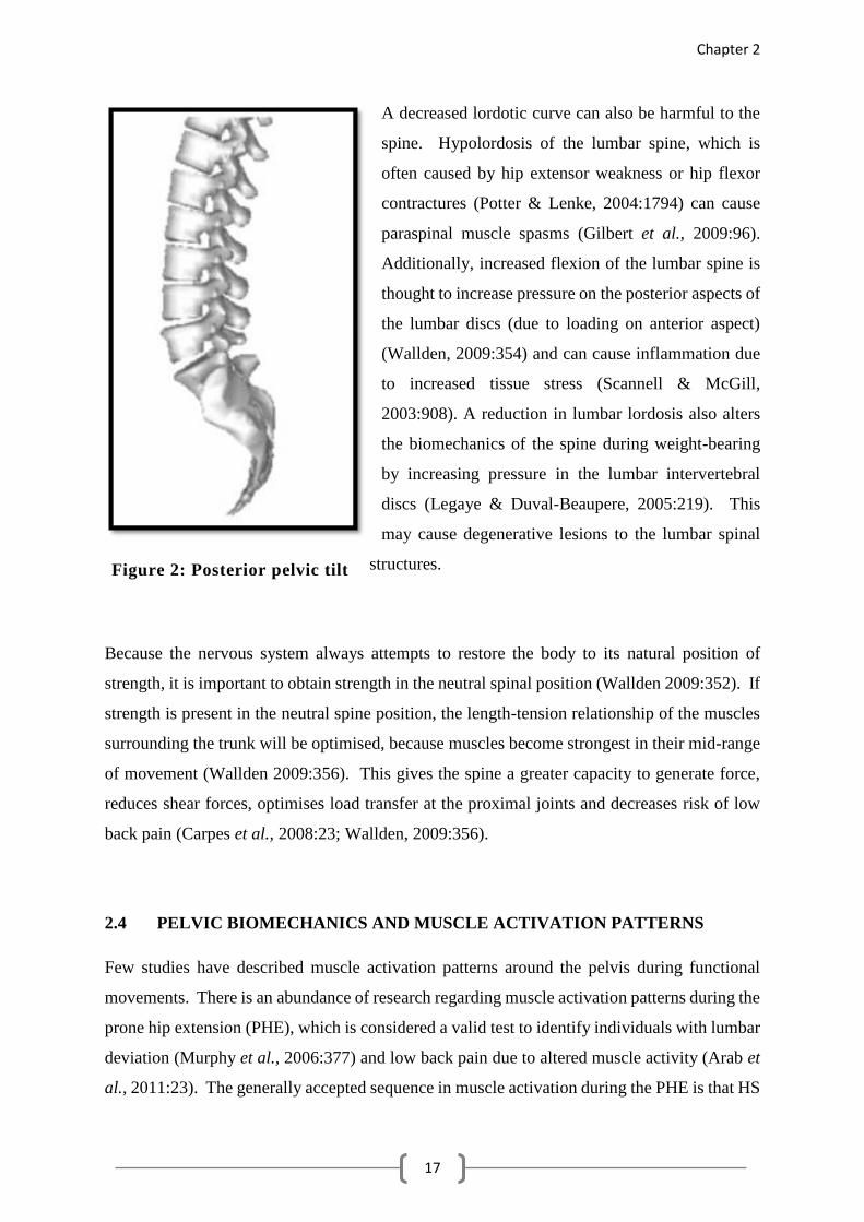
A decreased lordotic curve can also be harmful to the
spine Hypolordosis of the lumbar spine which is
often caused by hip extensor weakness or hip flexor
contractures (Potter amp Lenke 20041794) can cause
paraspinal muscle spasms (Gilbert et al 200996)
Additionally increased flexion of the lumbar spine is
thought to increase pressure on the posterior aspects of
the lumbar discs (due to loading on anterior aspect)
(Wallden 2009354) and can cause inflammation due
to increased tissue stress (Scannell amp McGill
2003908) A reduction in lumbar lordosis also alters
the biomechanics of the spine during weight-bearing
by increasing pressure in the lumbar intervertebral
discs (Legaye amp Duval-Beaupere 2005219) This
may cause degenerative lesions to the lumbar spinal
structures
Because the nervous system always attempts to restore the body to its natural position of
strength it is important to obtain strength in the neutral spinal position (Wallden 2009352) If
strength is present in the neutral spine position the length-tension relationship of the muscles
surrounding the trunk will be optimised because muscles become strongest in their mid-range
of movement (Wallden 2009356) This gives the spine a greater capacity to generate force
reduces shear forces optimises load transfer at the proximal joints and decreases risk of low
back pain (Carpes et al 200823 Wallden 2009356)
24 PELVIC BIOMECHANICS AND MUSCLE ACTIVATION PATTERNS
Few studies have described muscle activation patterns around the pelvis during functional
movements There is an abundance of research regarding muscle activation patterns during the
prone hip extension (PHE) which is considered a valid test to identify individuals with lumbar
deviation (Murphy et al 2006377) and low back pain due to altered muscle activity (Arab et
al 201123) The generally accepted sequence in muscle activation during the PHE is that HS
Figure 2 Posterior pelvic tilt
Chapter 2
18
activates first followed by ES and GM (Bruno amp Bagust 200775 Guimaraes et al
2010355) However Oh et al (2007323) observed a decrease in ES muscle activity with a
reduction in the degree of anterior pelvic tilt Also the muscle activation of GM was
significantly greater when the movement was initiated while the subject performed the
abdominal drawing-in maneuver (Oh et al 2007320) or when the pelvis was stabilised (Lewis
amp Sahrmann 2009247) The PHE is performed in the prone lying position and more research
into the effect of anterior pelvic tilt and core muscle activation on more functional movements
in required
The importance of muscle activation patterns lies in the fact that if one muscle fatigues or is
unable to activate correctly the task is transferred partially or totally to another muscle
resulting in reduced performance and stability (Bradl et al 2005275) This resulting
compensatory mechanism has been widely researched proving that decreased activation of the
GM muscle results in increased workload on the biceps femoris (BF) muscle with resulting
recurring muscle strains (Hoskins amp Pollard 2005100 Vogt et al 200324) Fatigue of the
GM has been found to increase the anterior tilt of the pelvis (Alvim et al 2010211) and
unilateral weakness of the GM may create an ipsilateral disruption of the pelvic position or
angle (Alvim et al 2010211) Studies have also suggested that early activation of the BF may
cause delayed activation of the deep abdominal stabilisers such as TrA (Hungerford et al
20031596) It can thus be proposed that efficient core stability will support normal muscle
activation patterns during functional movements (Devlin 2000281)
25 MUSCLE ACTIVATION PATTERNS DURING THE SQUAT
Athletes employ the weighted squat as a strength training exercise for the hip thigh and back
(Dionisio et al 2008134 Escamilla Fleisig Lowry et al 2001984 Lynn amp Noffal
20122418) Researchers have been trying to establish the best squatting posture for decades
due to the apparent correlation between the squat and low back pain (Delitto et al 19871329)
The single leg squat is often used in the rehabilitation of several back hip knee and ankle
injuries (Richards et al 2008482) Efficient execution of the squat requires mobility of the
ankle hip and thoracic spine and sufficient stability of the foot knee and lumbar spine (Kritz
et al 200983)
Chapter 2
19
251 NORMAL SQUAT
The ascent phase of the squat has been widely accepted as the most important and difficult part
of the movement and shows greater muscle activity than the descent phase (Escamilla Fleisig
Zheng et al 20011557) During the descent phase the body falls freely due to gravitational
forces resulting in small activation of the quadriceps and hamstring muscle groups (Dionisio
et al 2008141) Muscle activity during the ascent phase increases by 25-50 for the
quadriceps group and 100-180 for the hamstrings group (Escamilla Fleisig Zheng et al
20011560)
Table 2 Muscle contraction during different phases of the squat
Phases Concentric
lumbopelvic
contraction
Eccentric
lumbopelvic
contraction
Isometric
lumbopelvic
contaction
Gravitational influences
Descent Hamstrings Quadriceps
Erector spinae
Gluteus
maximus
Hamstrings
Transverse
abdominis
Multifidus
Causes free fall of body with
small levels of muscle
activity to control descent
Ascent Erector spinae
Quadriceps
Hamstrings
Gluteus
maximus
Hamstrings Transverse
abdominis
Multifidus
Causes increase in the level
of muscular activity during
first part of movement to
overcome gravity and
initiate ascent
Compiled from Anderson amp Behm 200543 Dionisio et al 2008141 Escamilla
Fleisig Zheng et al 20011560 Schoenfeld 20103500
The ascent phase is initiated by strong activation of the quadriceps to extend the knees
(Escamilla Fleisig Zheng et al 20011560) and has been shown to be the muscle group that
activates most strongly during the ascent phase of the squat (Caterisano et al 2002431)
Many studies evaluate the effect of stance width foot position bar load and squat depth has on
muscle activity (Caterisano et al 2002429 Dionisio et al 2008135 Distefano et al
2009533 Gullett et al 2009286 Wallace et al 2002142)
The squat irrespective of the technique or posture with which it is performed is a favoured
quadriceps exercise The rectus femoris (RF) muscle has its origin on the anterior superior iliac
spinae (ASIS) and will produce an increased anterior pelvic tilt if it dominates the squat
movement (Lynn amp Noffal 20122423) Thus it is important to spread the load to include the
Chapter 2
20
other muscles surrounding the pelvis to control excessive RF activation during the squat which
can lead to knee injuries (John amp Liebenson 2013137 Kulas et al 201219)
During the ascent phase of the squat hamstring muscle activity increases to stabilise the pelvis
and extend the hips (Dionisio et al 2008141) The activity levels of the hamstring group are
lower than those of the quadriceps most likely because of their shared muscular function with
the GM (Caterisano et al 2002431 Escamilla Fleisig Zheng et al 20011556) The
maximum activity levels of the hamstrings occur during the first third of the ascent phase
(Escamilla Fleisig Zheng et al 20011560) This may imply that during this phase the GM
muscle has not activated yet due to the postural anatomy changes in the muscle (Escamilla
Fleisig Zheng et al 20011560) This is in agreement with Schoenfeld (20103500) who
stated that the biomechanical position of the GM at 90deg of hip flexion has the lowest capacity
to produce torque Also because the hamstring group acts as both a hip extensor and a knee
flexor its length stays fairly consistent and may contribute to the consistent production of
force throughout the squat (Schoenfeld 20103501)
The abdominal stabilisers play a role in stabilising the spine and pelvis and should activate
strongly during the first half of the ascent phase (Anderson amp Behm 200543) Continued
activity levels at a lower intensity are expected throughout the movement to maintain intra-
abdominal pressure and lumbar stabilisation (Willardson 2007984) Recent research has
concluded that the lumbopelvic stabilizer TrA and multifidus activate before any movement
starts which increases intra-abdominal pressure and tightens the thoracolumbar fascia
assisting in stability of the spine (Kibler et al 2006190) This serves to alleviate vertebral
loading (Schoenfeld 20103501) The muscle activation of the abdominal muscle group has
also been shown to increase with increased resistance and unstable surfaces (Clark et al
201211767)
The ES muscle has also been shown to activate significantly more during the ascent phase than
the descent phase of the squat (Anderson amp Behm 200542) However at the start of the ascent
phase a significant drop in lumbo-sacral ES muscle activity occurs due to the lumbar spine
going into flexion (Anderson amp Behm 200543) After the start of the ascent the ES muscle
activity seems to vary with individual back postures during squatting Squatting with the
lumbar spine in flexion puts the muscle in a lengthened position and decreases the amount of
muscle activity (Schoenfeld 20103501) This places more strain on the intervertebral discs
and vertebral bodies increasing the risk of injury (Legaye amp Duval-Beaupere 2005219)
Chapter 2
21
Conversely lumbar extension increases ES activity and consequently spinal compressive
forces (Schoenfeld 20103501) ES activity has also been shown to decrease when ES co-
contracts with the abdominal stabilisers diminishing the spinal tension that would have been
created by the ES muscle action alone (Schoenfeld 20103501)
The GM muscle produces the most varied recorded activation levels in the squat movement
(Caterisano et al 2002430) The muscle is believed to act eccentrically to control the descent
phase and will contract powerfully to initiate ascent (Schoenfeld 20103500) However as
mentioned earlier GM produces less force at 90deg of hip flexion as the muscle is in a lengthened
position (Schoenfeld 20103500) It has the function of assisting hip extension assists in
control of knee abduction and adduction and stabilises the pelvis (Lieberman et al 20062144)
The GM muscle also serves to avoid any lateral pelvic rotation and compresses the sacro-iliac
joint to maintain pelvic stability (Alvim et al 2010211) Therefore the GM muscle has great
importance during the squat as it serves to both stabilise the pelvis and extend the hips during
the ascent phase of the squat
Errors in squatting technique include back hyperlordosis and excessive anterior knee
displacement (John amp Liebenson 2013137) non-neutral postures that lead to an increase in
ES muscle activity during the ascent phase of the squat (Sorensen et al 2011150) To perform
the squat safely requires rigidity of the spine with minimal planar motion (Schoenfeld
20103501) It has also been shown that an excessive anterior pelvic tilt can decrease GM
muscle activity (Alvim et al 2010211) reducing both strength and stability These factors
combine to result in impingement of the lumbar facet joints (Han et al 2011477) and low
back pain
252 SINGLE LEG SQUAT
The single limb squat has been widely used in rehabilitation as a screening tool (Livengood et
al 200424) a post-rehabilitation clearance test (DiMattia et al 2005109) and a
strengthening exercise (Boudreau et al 200992) This particular exercise has been favoured
for strengthening due to the marked increase in muscle activity when compared to other single
limb exercises (Boudreau et al 200998) This exercise incorporates a dynamic version of the
Trendelenburg test to identify gluteus medius weakness (Livengood et al 200424) and
challenges the neuromuscular control of the trunk hip knee and ankle (DiMattia et al
2005109) Correct technique for the single leg squat requires hip flexion ˂ 65deg hip abduction
Chapter 2
22
or adduction of ˂10deg and knee valgus or varus of ˂10deg at the maximum descent phase
(Livengood et al 200425)
The single limb squat has been proven to strongly activate the gluteus muscles mostly the
gluteus medius of the weight-bearing leg (Boudreau et al 200999 Distefano et al 2009537)
The gluteus medius muscle of the non-weight bearing leg is also activated (Boudreau et al
200999) most possible due to the gluteal group resisting the gravitational force towards hip
adduction of the raised leg whilst standing unilaterally (Distefano et al 2009538) The single
limb squat also strongly activates the GM possibly due to its role in lumbo-pelvic stability
eccentric control of hip flexion and concentric hip extension (Distefano et al 2009538) But
more than any other muscle the RF shows the highest level of muscle activity during the single
leg squat (Boudreau et al 200998)
The single leg squat has been widely incorporated into sport specific training programs due to
its neuromuscular similarities to unilateral weight-bearing activities (McCurdy et al
201057) Research suggests that it is a better strengthening exercise than the double leg squat
due to the increased demand of the neuromuscular system to support the body in the frontal
plane of movement (McCurdy et al 201058) Additionally the smaller support base of a
single leg may mimic more accurately the strength and proprioception requirements of
functional athletic movements (McCurdy et al 201058) However the unstable posture of
the single leg squat makes it risky to incorporate weighted resistance as the exercise requires
synergistic activation of the knee hip and trunk stabilisers to be completed safely (DiMattia et
al 2005119) Risks during this exercise include excessive knee valgusvarus movement
(McCurdy et al 201065) lateral pelvic drop (McCurdy et al 201066) and increased lumbar
extension loading (DiNaso et al 201255) Therefore a modified version of this exercise has
been promoted with the trail leg providing support and balance (placed on a stable structure)
without being fully weight-bearing (McCurdy et al 201058) This still provides increased
muscle activity when compared to the normal squat (McCurdy et al 201064) but adds the
necessary stability to enable progression to moderately loaded strength training
253 KNEE-DOMINANT SQUAT VS HIP-DOMINANT SQUAT
The knee-dominant squat as the name implies has an increased knee flexion-based movement
pattern while the hip-dominant squat is characterised by an increase in hip flexion causing a
Chapter 2
23
forward trunk lean and a decreased knee flexion angle (McCurdy et al 201064) It has been
proposed that a hip-dominant squat produces a more efficient movement pattern than a knee-
dominant squat (Lynn amp Noffal 20122418) because of the decreased load it places on the
lumbar spine and knee joints (John amp Liebenson 2013137)
The squat is a closed-chain kinetic movement with biomechanical and neurological similarities
to several functional multi-joint movements performed daily (Augustsson et al 19987
Wilson et al 200598) Decreasing the load this movement places on the joints is therefore of
great importance The knee-dominant squat increases loading of the knee joint (John amp
Liebenson 2013137) and the anterior cruciate ligament (ACL) (Kulas et al 201219) An
earlier study has shown that preventing the knees moving over the toes during the squat
exercise decreased knee torque from 1501 plusmn 508 Nm to 1173 plusmn 342 Nm (Comfort amp Kasim
200711) It has also been demonstrated that a moderate forward trunk lean during the squat
decreased the amount of strain and force placed on the ACL (Kulas et al 201220) An
increased knee flexion angle has been shown to have an effect on the lumbar angle during squat
lifting (Hwang et al 200919) and should be corrected for safe squatting
The biomechanical change from knee to hip dominance during the functional squat movement
has been proven to change muscle activation patterns significantly (Anderson amp Behm
200541) Forward trunk lean has been associated with increased activation of the hip extensor
(GM and the hamstring group) and abdominal stabiliser muscles (DiNaso et al 201255
McCurdy et al 201064) and decreased activation of the hip flexors (RF) (Kulas et al
201219) The forward trunk lean associated with the hip dominant squat will cause a
mechanical shortening of the RF muscle and decreased knee joint torque (Kulas et al
201219) The RF muscle has its origin on the anterior inferior iliac spinae (ASIS) and will
produce an increased anterior pelvic tilt if it dominates the squat movement increasing the load
on the lumbar facet joints (Lynn amp Noffal 20122423) Therefore the hip-dominant squatting
pattern does not only decrease muscle activity that increases pelvic inclination (RF) but also
provides a more favourable posture for muscle activity that provides lumbopelvic stability
(Anderson amp Behm 200543)
The hip-dominant squat will also increase the torque going through the hip joint rather than the
knees (Comfort amp Kasim 200711) possibly because of the increased GM activity it produces
during the ascent phase of the squat (Lynn amp Noffal 20102422) Strengthening of the extensor
portion of the GM is also important as this muscle exerts force on the pelvis in the saggital
Chapter 2
24
plane and can assist in pelvic stability and effective load transfer through the trunk to the lower
extremities (Barker et al 20136) Finally the GM has a larger cross sectional area than the
entire quadriceps group combined (4842 mm2 versus 43175 mm2) (Ito et al 200349) and is
supported by the hamstrings group (Ward et al 201099) thus indicating the benefits of hip
extension during the ascent This technique of squatting has also been proven to increase
hamstring muscle activity especially when the knee is flexed more than 65deg placing focus on
its importance during the ascent phase of the squat (Kulas et al 201219)
Correcting muscle activation deficiencies during the squat can delay or even prevent the
occurrence of musculoskeletal injuries (Lynn amp Noffal 20122417) Instructions on how to
activate the core muscles during the execution of the squat movement can lead to an immediate
increase in EMG activity of RA EO and TrA with improved recruitment patterns in a short
period of time (Bressel et al 2009503) Researchers agree that the best way to avoid injury
during weight training is to maintain a neutral spinal position at all times (Colado amp Garcia-
Masso 2009106 Durall amp Manske 200570 Han et al 2011477 McGill et al 2003356)
yet no studies have evaluated the effects of core stability on lumbopelvic posture and muscle
activation patterns during strength training
The correct posture for the squat exercise has been widely debated For the squat to be
performed safely and efficiently the mitigation of impact across all joints needs to be optimised
(Fry et al 2003632) After a review of the literature regarding squatting technique Kritz et
al (200983) concluded that for execution of a safe squat a person requires mobility of the
ankle and hip joints and stability of the feet knees and trunk
Figure 3 Optimal squatting technique
Chapter 2
25
Table 3 Correct squatting alignment and posture
Body area Proposed alignment Reason for proposed
alignment
Head Head held straight or looking slightly
up head in line with shoulders
Prevents excessive lumbar and
thoracic flexion
Thoracic spine Slight extension of spine scapulae in
neutral position and held stable
Avoids excessive loading on
lumbar spine
Lumbar spine No extension or flexion at any time
during movement held stable in
neutral
Avoids excessive loading on
lumbar spine
Hip joints Hips aligned with knees stable no
medial or lateral rotation
Optimises load transfer through
the pelvis
Knee joints Aligned with hips and feet no
excessive medial or lateral rotation no
excessive movement forward (over
toes) or backwards
Avoids excessive knee torque
and excessive trunk flexion
during ascent phase of squat
Feetankle
joints
Feet stable heels on the ground
throughout movement
Maintains balance and control of
squat movement
Compiled from Fry et al 2003632 Kritz et al 200978
26 LUMBOPELVIC INJURIES IN RUGBY UNION
Rugby union is a high intensity contact sport with varying phases of play exposing participants
to axial loading (line out) rotational forces (scrum ruck and maul) impact collisions (tackle)
and runningsprinting (Castinel et al 2007337) The strain the game places on the body has
been widely documented and several aspects of rugby union can lead to injury (Brown et al
201253 Gianotti et al 2009372 Hermanus et al 2010231 Murray et al 20121) Most
injuries reported result from contact with another player such as tackles rucks mauls and
scrums (Brooks et al 2005a292 Brooks et al 2005b760 Hermanus et al 2010232 King
et al 2011127 Sinibaldi amp Smith 200718) The number of tackles rucks and mauls during
rugby union matches has steadily increased in recent years (Quarrie amp Hopkins 2007900)
increasing the risk of injury to players
Researchers have evaluated the forces generated during scrumming (Du Toit et al 200433
Sinibaldi amp Smith 200718) indicating high levels of force Even at junior level forwards can
generate 1400N on average during sustained scrumming (Du Toit et al 200446) The scrum
is a contact manoeuvre involving 16 players that can injure the cervical spine (Du Toit et al
200434 Hermanus et al 2010232 Kuster et al 2012550 MacLean amp Hutchison
Chapter 2
26
2012592 Sinibaldi amp Smith 200718) thoracic spine (Boran et al 2011862) lumbar spine
(Davis et al 2009796 Sakai et al 2010284) and shoulder (Crichton et al 2011539
Sundaram et al 2011112) The impact of scrumming on the spine can cause long-term
damage compromising the integrity of the intervertebral discs joints and ligaments (Sinibaldi
amp Smith 200719) Players in the front row of the scrum are at increased risk of injury due to
the biomechanical loading of the spine especially if they scrum in a hyperlordotic posture
(Castinel et al 2006337)
Of further is the high number of injuries induced by weight training (Brooks et al 2005b770)
Weight training can result in chronic low back pain that manifests over time due to repetitive
damage to the vertebral bodies growth plates and intervertebral discs (Colado amp Garcia-
Masso 2009104 Durall amp Manske 200567 Walsh et al 2007928) This risk of injury is
increased if the proper technique is not applied If the squat is performed with the back in
hyperlordosis the lumbar spinersquos ability to distribute the compressive force evenly is reduced
and reaction forces increase (Adams et al 20001632 Durall amp Manske 200566 Han et al
2011477 Walsh et al 2007928) Adams et al (20001629) found that the stress peak in the
posterior annulus of the lumbar spine increased by 114 if the spine is extended only 2deg away
from neutral Awareness of lumbar extension is of particular concern in players who have not
reached skeletal maturity yet (Walsh et al 2007931)
27 SUMMARY
It is well established that both training for and playing rugby carry a risk of injury The lumbar
spine in particular is vulnerable to injury if the player presents with an anterior pelvic tilt
Increased core muscle strength is commonly believed to reduce the degree of anterior pelvic
tilt (McGregor amp Hukins 2009220) and used in clinical settings as treatment for low back
pain Even though core training has been included in the training regimes of sporting
professionals in recent years little is known about the effects of core stability on posture and
muscle activity during sport
No previous research has evaluated the effects of core stability and lumbopelvic biomechanics
on muscle activation during functional movement Lumbopelvic stability has been proven to
play an integral part in optimal muscle functioning (Fredericson amp Moore 2005676) and we
need to better understand the role it plays during functional closed-chain sporting movements
Chapter 2
27
This knowledge may help to prevent injuries during training and game play by individualising
each playerrsquos conditioning program to promote optimal functioning
Chapter 2
28
REFERENCES
Adams MA Freeman BJC Morrison HP Nelson IW amp Dolan P 2000 Mechanical
initiation of intervertebral disc degeneration Spine 25(13)1625-1636
Akuthota V amp Nadler SF 2004 Core stabilityening Archives of physical medicine and
rehabilitation 85(1)86-92
Alvim FC Peixoto JG Vicente EJD Chagas PSC amp Fonseca DS 2010 Influences
of the extensor portion of the gluteus maximus muscle on pelvic tilt before and after the
performance of a fatigue protocol The Brazilian journal of physical therapy 14(3)206-213
Anderson K amp Behm DG 2005 Trunk muscle activity increases with unstable squat
movements Canadian journal of applied physiology 30(1)33-45
Arab AM Ghamkhar L Emami M amp Nourbakhsh MR 2011 Altered muscle activation
patterns during prone hip extension in women with and without low back pain Chiropractic
and manual therapies 19(1)18-23
Augustsson J Esko A Thomee R amp Svantesson U 1998 Weight training of the thigh
muscles using closed vs open kinetic chain exercises a comparison of performance
enhancement Journal of orthopeadic and sports physical therapy 27(1)3-8
Barker PJ Hapuarachchi KS Ross JA Sambaiew E Ranger TA amp Briggs CA 2013
Anatomy and biomechanics of gluteus maximus and the thoracolumbar fascia at the sacroiliac
joint
httponlinelibrarywileycomnwulibnwuaczadoi101002ca22233abstract Date of
access 18 Nov 2013
Bendova P Ruzicka P Peterova V Fricova M amp Springrova I 2007 MRI-based
registration of pelvic alignment affected by altered pelvic floor muscle characteristics Clinical
biomechanics 22(9)980-987
Boran S Lenehan B Street J amp Mccormack D 2011 A 10-year review of sports-related
spinal injuries International journal of medicine and science 108(4)859-863
Chapter 2
29
Boudreau SN Dwyer MK Mattacola CG Latterman C Uhl TL amp Mckeon JM
2009 Hip-muscle activation during the lunge single-leg squat and step-up-and-over
exercises Journal of sport rehabilitation 18(1)91-103
Bradl I Morl F Scholle HC Grassme R Muller R amp Grieshaber R 2005 Back muscle
activation patterns and spectrum in defined load situations Pathophysiology 12(4)275-280
Bressel E Willardson JM Thompson B amp Fontana FE 2009 Effect of instruction
surface stability and load intensity on trunk muscle activity Journal of electromyography and
kinesiology 19(6)500-504
Brooks JHM Fuller CW Kemp SPT amp Reddin DB 2005a A prospective study of
injuries and training amongst the England 2003 Rugby World Cup squad British journal of
sport medicine 39(5)288-293
Brooks JHM Fuller CW Kemp SPT amp Reddin DB 2005b Epidemiology of injuries
in English professional rugby union part 2 training injuries British journal of sport medicine
39(10)767-775
Brown JC Verhagen E Viljoen W Readhead C Van Mechelen W Hendricks S amp
Lambert MI 2012 The incidence and severity of injuries at the 2001 South African Rugby
Union (SARU) Youth Week tournaments South African journal of sport medicine 24(2)49-
54
Bruno PA amp Bagust J 2007 An investigation into motor pattern differences used during
prone hip extension between subjects with and without low back pain Clinical chiropractic
10(2)68-80
Carpes FPC Reinehr FB amp Mora CB 2008 Effects of a program for trunk strength and
stability on pain low back and pelvic kinematics and body balance a pilot study Journal of
bodywork and movement therapies 12(1)22-30
Castinel BH Adam P amp Prat C 2007 A stress fracture of the lumbar spine in a
professional rugby player British journal of sport medicine 41(5)337-338
Caterisano A Moss RF Pellinger TK Woodruff K Lewis VC Booth W amp Khadra
T 2002 The effect of back squat depth on the EMG activity of 4 superficial hip and thigh
muscles Journal of strength and conditioning research 16(3)428-432
Chapter 2
30
Clark DR Lambert MI amp Hunter AM 2012 Muscle activation in the loaded free barbell
squat a brief review Journal of strength and conditioning research 26(4)1169-1178
Colado JC amp Garcia-Masso X 2009 Technique and safety aspects of resistance exercises
a systematic review of the literature The physician and sports medicine 37(2)104-111
Comerford MJ amp Mottram SL 2001 Movement and stability dysfunction ndash contemporary
developments Manual therapy 6(1)15-26
Comfort P amp Kasim P 2007 Optimizing squat technique National strength and
conditioning association 29(6)10-13
Crichton J Jones DR amp Funk L 2011 Mechanisms of traumatic shoulder injury in elite
rugby players British journal of sport medicine 46(7)538-542
Davis G Johnson E McCormick PC Roger EP Ugokwe K Benzel EC Sears WR
amp McCrory P 2009 Clinics in neurology and neurosurgery of sport lumbar spine
Sequestrated disc prolapse and disc bulge British journal of sports medicine 43(10)796-801
Dawson-Cook S 2011 Howrsquos your posture American fitness 29(3)24-30
Deckert JL Barry SM amp Welsh TM 2007 Analysis of pelvic alignment in university
ballet majors Journal of dance medicine and science 11(4)110-117
Delitto RS Rose SJ amp Apts DW 1987 Electromyographic analysis of two techniques
for squat lifting Physical therapy 67(9)1329-1334
Devlin L 2000 Recurrent posterior thigh symptoms detriment to performance in the rugby
union predisposing factors Sports medicine 29(4)273-287
DiMattia MA Livengood AL Uhl TL Mattacola CG amp Malone TR 2005 What are
the validity of the single-leg-squat test and its relationship to hip-abduction strength Journal
of sports rehabilitation 14(2)108-123
DiNaso JJ Pritschet BL Emmett JD Owen JD Willarson JM Beck TW Defreitas
JM amp Fontana FE 2012 Comparing thigh muscle cross-sectional area and squat strength
among national class Olympic weightlifters power lifters and body builders International
sports medicine journal 13(2)48-57
Chapter 2
31
Dionisio VC Almeida GL Duarte M amp Hirata RP 2008 Kinematic kinetic and EMG
patterns during downward squatting Journal of electromyography and kinesiology 18(1)134-
143
Distefano LJ Blackburn JT Marshall SW amp Padua DA 2009 Gluteul muscle
activation during common therapeutic exercises Journal of orthopaedic amp sports physical
therapy 39(7)532-540
Durall CJ amp Manske RC 2005 Avoiding lumbar spine injury during resistance training
National strength and conditioning association 27(4)64-72
Du Toit DE Venter DJL Buys FJ amp Olivier PE 2004 Kinetics of rugby union
scrumming in under 19 schoolboy rugby forwards South African journal for research in sport
physical education and recreation 26(2)33-50
Escamilla RF Fleisig GS Zheng N Lander JE Barrentine SW Andrews JR
Bergemann BW amp Moorman CT 2001 Effects of technique variations on knee
biomechanics during the squat and leg press Medicine and science in sport and exercise
33(9)1552-1566
Escamilla RF Fleisig GS Lowry TM Barrentine ST amp Andrews JR 2001 A three-
dimensional biomechanical analysis of the squat during varying stance widths Medicine and
science in sport and exercise 33(6)984-998
Faries MD amp Greenwood M 2007 Core training stabilizing the confusion National
strength and conditioning journal 29(2)10-25
Fredericson M amp Moore T 2005 Muscular balance core stability and injury prevention
for middle- and long-distance runners Physical medicine and rehabilitation clinics of North
America 16(3)669-689
Fry AC Smith JC amp Schilling BK 2003 Effect of knee position on hip and knee torques
during the barbell squat Journal of strength and conditioning research 17(4)629-633
Geraci MC amp Brown W 2005 Evidence-based treatment of hip and pelvic injuries in
runners Physical medicine and rehabilitation clinics of North America 16(3)711-747
Chapter 2
32
Gianotti SM Quarrie KL amp Hume PA 2009 Evaluation of RugbySmart a rugby union
community injury prevention programme Journal of science and medicine in sport
12(3)371-375
Gilbert JW Wheeler GR Storey BB Mick GE Herder SL Richardson G
Wyttenbach WH amp Martin JC 2009 Lumbar magnetic resonance imaging hypolordosis
in symptomatic patients association with paraspinal muscle spasms Journal of chiropractic
medicine 8(3)95-100
Guimaraes CQ Sakamoto ACL Laurentino GEC amp Teixeira-Salmela LF 2010
Electromyographic activity during prone hip extension did not discriminate individuals with
and without low back pain Revista Brasileira de fisioterapia 14(4)351-357
Gullett JC Tillman MD Gutierrez GM amp Chow JW 2009 A biomechanical
comparison of back and front squats in healthy trained individuals Journal of strength and
conditioning research 23(1)284-292
Han K Rohlmann A Yang S Kim BS amp Lim T 2011 Spinal muscles can create
compressive follower loads in the lumbar spine in a neutral standing posture Medical
engineering amp physics 33(4)472-478
Hermanus FJ Draper CE amp Noakes TD 2010 Spinal cord injuries in South African
Rugby Union (1980 ndash 2007) South African medical journal 100(4)230-234
Hibbs AE Thompson KG French D Wrigley A amp Spears I 2008 Optimizing
performance by improving core stability and core stability Sports medicine 38(12)995-1008
Hoskins WT amp Pollard H 2005 The management of hamstring injury ndash part 1 issues in
diagnosis Manual therapy 10(2)96-107
Hungerford B Gilleard W amp Hodges P 2003 Evidence of altered lumbopelvic recruitment
in the presence of sacroiliac joint pain Spine 28(14)1593ndash1600
Hwang S Kim Y amp Kim Y 2009 Lower extremity joint kinetics and lumbar curvature
during squat and stoop lifting Musculoskeletal disorders 10(1)15-24
Ito J Moriyama H Inokuchi S amp Goto N 2003 Human lower limb muscles An
evaluation of weight and fiber size Okajimas folia anatomica Japonica 80(2-3)47-56
Chapter 2
33
Jensen GM 1980 Biomechanics of the lumbar intervertebral disk a review Physical
therapy 60(6)765-773
John D amp Liebenson C 2013 How should I squat Journal of bodywork and movement
therapies 17(1)137-139
Kibler WB Press J amp Sciascia A 2006 The role of core stability in athletic function
Sports medicine 36(3)189-198
King DA Hume PA Gianotti S amp Clark T 2011 Neck back and spine injuries in
amateur rugby league a review of nine years of Accident Compensation Corporation injury
entitlement claims and costs Journal of science and medicine in sport 14(2)126-129
Kritz M Cronin J amp Hume P 2009 The bodyweight squat A movement screen for the
squat pattern Journal of strength and conditioning research 31(1)76-85
Kulas AS Hortobagyi T amp Devita P 2012 Trunk position modulates anterior cruciate
ligament forces and strain during a single-leg squat Clinical biomechanics 27(1)16-21
Kuster D Gibson A Abboud R amp Drew T 2012 Mechanisms of cervical spine injury in
rugby union a systematic review of the literature British journal of sport medicine 46(8)550-
554
Legaye J amp Duval-Beaupere G 2005 Sagittal plane alignment of the spine and gravity a
radiological and clinical evaluation Acta orthopaedica Belgica 71(2)213-220
Lewis KL amp Sahrmann SA 2009 Muscle activation and movement patterns during the
prone hip extension exercise in women Journal of athletic training 44(3)238-248
Lieberman DE Raichlen DA Pontzer H Bramble DM amp Cutright-Smith E 2006 The
human gluteus maximus and its role in running The journal of experimental biology
209(11)2143-2155
Lim HS Roh SY amp Lee SM 2013 The relationship between pelvic tilt angle and
disability associated with low back pain Journal of physical therapy and science 25(1)65-
68
Livengood AL Dimattia MA amp Uhl TL 2004 lsquoDynamic Trendelenburgrsquo single-leg-
squat test for gluteus medius strength Athletic therapy today 9(1)24-25
Chapter 2
34
Lynn SK amp Noffal GJ 2012 Lower extremity biomechanics during a regular and
counterbalanced squat Journal of strength and conditioning research 26(9)2417-2425
MacLean JDB amp Hutchison JD 2012 Serious neck injuries in U19 rugby union players
an audit of admissions to spinal injury units in Great Britain and Ireland British journal of
sports medicine 46(8)591-594
Magee DJ 2002 Orthopedic physical assessment 5th ed St Louis MOSaunders Elsevier
McCurdy K OrsquoKelley E Kutz M Langford G Ernest J amp Torres M 2010 Comparison
of lower extremity EMG between the 2-leg squat and modified single-leg squat in female
athletes Journal of sport rehabilitation 19(1)57-70
McGill S 2010 Core training evidence translating to better performance and injury
prevention Strength and conditioning journal 32(3)33-46
McGill SM Grenier S Kavcic N amp Cholewicki J 2003 Coordination of muscle activity
to assure stability of the lumbar spine Journal of electromyography and kinesiology
13(4)353-359
McGregor AH amp Hukins DWL 2009 Lower limb involvement in spinal function and low
back pain Journal of back and musculoskeletal rehabilitation 22(4)219-222
Morningstar MW 2003 Strength gains through lumbar lordosis restoration Journal of
chiropractic medicine 4(2)137-141
Murphy DR Byfield D Mccarthy P Humphreys K Gregory AA amp Rochon R 2006
Interexaminer reliability of the hip extension test for suspected impaired motor control of the
lumbar spine Journal of manipulative and physiological therapeutics 29(5)374-377
Murray AD Murray IR amp Robson J 2012 Rugby Union faster higher stronger keeping
an evolving sport safe httpbjsmbmjcomcontentearly20121221bjsports-2012-
091844fullpdf+html Date of access 22 Mar 2013
Myers GD Brent JL Ford KR amp Hewett TE 2008 A pilot study to determine the effect
of trunk and hip focused neuromuscular training on hip and knee isokinetic strength British
journal of sport medicine 42(7)614-619
Chapter 2
35
Nesser TW Huxel KCl Tincher JL amp Okada T 2008 The relationship between core
stability and performance in division I football players Journal of strength and conditioning
research 22(6)1750-1754
Norris CM 1999 Functional load abdominal training part 1 Journal of bodywork and
movement therapies 3(3)150-158
Oh J Cynn H Won J Kwon O amp Yi C 2007 Effects of performing an abdominal
drawing-in maneuver during prone hip extension exercises on hip and back extensor muscle
activity and amount of anterior pelvic tilt Journal of orthopaedic and sports physical therapy
37(6)320-324
Pinto RZ Ferreira PH Franco MR Ferreira MC Ferreira ML Teixeira-Salmela
LF Oliveira VC amp Maher C 2011 The effect of lumbar posture on abdominal muscle
thickness during an isometric leg task in people with and without non-specific low back pain
Manual therapy 16(6)578-584
Potter BK amp Lenke LG 2004 Prevention and management of iatrogenic flatback
syndrome Journal of bone and joint surgery 86A(8)1793-1808
Quarrie KL amp Hopkins WG 2007 Changes in players characteristics and match activities
in Bledisloe Cup rugby union from 1972 to 2004 Journal of sport science 25(8)895-903
Reed CA Ford KR Myer GD amp Hewett TE 2012 The effects of isolated and
integrated lsquocore stabilityrsquo training on athletic performance measures a systematic review
Sports medicine 42(8)697-706
Richards J Thewlis D Selfe J Cunningham A amp Hayes C 2008 A biomechanical
investigation of a single-limb squat implications for lower extremity rehabilitation exercise
Journal of athletic training 43(5)477-482
Robertson DGE Wilson JJ amp Saint Pierre TA 2008 Lower extremity muscle functions
during full squats Journal of applied biomechanics 24(4)333-339
Sakai T Sairyo K Suzue N Kosaka H amp Yasui N 2010 Incidence and etiology of
lumbar spondylolysis a review of the literature Journal of orthopaedic science 15(3)281-
288
Chapter 2
36
Scannell JP amp McGill SM 2003 Lumbar posture ndash should it and can it be modified A
study of passive tissue stiffness and lumbar position during activities of daily living Physical
therapy 83(10)907-917
Schoenfeld BJ 2010 Squatting kinematics and kinetics and their application to exercise
performance Journal of strength and conditioning research 24(12)3497-3506
Sinibaldi KS amp Smith DR 2007 Prevention of Spinal Injuries in Rugby National strength
and conditioning association 29(4)18-24
Sorensen CJ Haddad O Campbell S amp Mirka GA 2011 The effect of stance width on
trunk kinematics and trunk kinetics during sagitally symmetric lifting International journal
of industrial ergonomics 41(2)147-152
Stokes IAF Gardner-Morse MG amp Henry SM 2011 Abdominal muscle activation
increases lumbar spinal stability Analysis of contributions of different muscle groups Clinical
biomechanics 26(8)797-803
Sundaram A Bokor DJ Davidson AS 2011 Rugby Union on-field position and its
relationship to shoulder injury leading to anterior reconstruction for instability Journal of
science and medicine in sport 14(2)111-114
Szymanski DJ McIntyre JS Szymanski JM Bradford TJ Schade RL Madsen NH
amp Pascoe DD 2007 Effect of torso rotational strength on angular hip angular shoulder and
linear bat velocities of high school baseball players Journal of strength and conditioning
research 21(4)1117-1125
Takasaki H Iizawa T Hall T Nakamura T amp Kaneko S 2009 The influence of
increasing sacroiliac joint force closure on the hip and lumbar spine extensor muscle firing
pattern Manual therapy 14(5)484-489
Vera-Garcia FJ Elvira JLL Brown SHM amp McGill SM 2007 Effects of abdominal
stabilization maneuvers on the control of spine motion and stability against sudden trunk
perturbations Journal of electromyography and kinesiology 17(5)556-567
Vogt L Pfeifer K amp Banzer W 2003 Neuromuscular control of walking with chronic low-
back pain Manual therapy 8(1)21-28
Chapter 2
37
Wallace DA Salem GJ Salinas R amp Powers CM 2002 Patellofemoral joint kinetics
while squatting with and without an external load Journal of orthhopaedics and sport physical
therapy 32(4)141-148
Wallden M 2009 The neutral spine principle Prevention and rehabilitation 13(4)350-361
Walsh JC Quinlan JF Stapleton R Fitzpatrick DP amp McCormack D 2007 Three-
dimensional motion analysis of the lumbar spine during ldquofree squatrdquo weight lift training The
American journal of sports medicine 38(6)927-932
Ward SR Winters TM amp Blemker SS 2010 The architectural design of the gluteal
muscle group implications for movement and rehabilitation Journal of orthopaedics and
sports physical therapy 40(2)95-102
Willardson JM 2007 Core stability training applications to sports conditioning programs
Journal of strength and conditioning research 21(3)979-985
Wilson J Ferris E Heckler A Maitland L amp Talor C 2005 A structured review of the
role of gluteus maximus in rehabilitation New Zealand journal of physiotherapy 33(3)95-
100
Yerys S Makofsky H Byrd C Pennachio J amp Cinkay J 2002 Effect of mobilization of
the anterior hip capsule on gluteus maximus strength The journal of manual and manipulative
therapy 10(4)218-224
Chapter 3
38
C H A P T E R 3
CORE STABILITY MUSCLE FLEXIBILITY
AND PELVIC TILT IN RUGBY UNION PLAYERS
Authors Greyling M1 amp Bruwer EJ2
1 Student in PhASRec North-West University (Potchefstroom campus) Potchefstroom
South Africa 2 Physical Activity Sport and Recreation Research Focus Area North-West University
(Potchefstroom campus) Potchefstroom South Africa
Corresponding author
EJ Bruwer
School for Biokinetics Recreation and Sport Science
North-West University (Potchefstroom campus)
Private bag x6001
Potchefstroom
South Africa
2520
E-mail ernabruwernwuacza
Tel (018) 299 2034
Fax (018) 299 1825
South African Journal of Sports Medicine
Word count 3 014
Chapter 3
39
ABSTRACT
Background The anterior pelvic tilt posture causes altered muscle activation in the trunk and
hip area resulting in decreased core muscle activation and lumbopelvic instability
Objectives To determine whether core stability and selected lumbopelvic flexibility measures
in U19 university-level rugby union players with an anterior pelvic tilt differs significantly
from those with a neutral pelvic tilt grouped by playing position
Methods This once-off cross sectional study was performed on the male U19 rugby union
training group of the North-West University for the 2013 season (N=49) Pelvic tilt was
measured from a digital photo with clear reflector markers on the ASIS and PSIS using a video
analysis software programme Flexibility of the hamstrings hip flexors and knee extensors
was assessed with goniometry measurements Core stability was assessed with the double leg
lowering test
Results There was a significant difference (p=0000) in the mean degree of pelvic inclination
between the group classified as having a neutral pelvic tilt (1175⁰ n=20) and the anterior
pelvic tilt group (1783⁰ n=29) There was however no statistical significant (ple005) or
practical significant (dge08) difference in core stability or flexibility measures between the
anterior and neutral pelvic tilt groups The mean core stability of the entire group was classified
as being good (5076⁰) with the anterior pelvic tilt group showing slightly better core stability
than the neutral tilt group
Conclusion Pelvic tilt does not influence core stability and flexibility in selected pelvic
muscles in rugby union players during the midseason Future research should be conducted at
the beginning of the year to eliminate the possible effect of core training regimes on the results
observed
Word count 267
Chapter 3
40
31 INTRODUCTION
As attested to in recent literature the anterior pelvic tilt posture seems to be the most common
non-neutral spinal position[1] This position causes altered muscle activation patterns around
the trunk and hips[2] resulting in decreased lumbopelvic stability[3] It has been theorised that
the anterior pelvic tilt posture places the abdominal and gluteal muscles in a lengthened
position causing stretch weakness and hindering normal muscle activity[4] A reduction in
anterior pelvic tilt will increase gluteus maximus (GM) activation[5] resulting in sacro-iliac
joint (SIJ) compression and increased pelvic stability[6] It is also believed that a neutral
lumbopelvic posture can increase the degree of transverse abdominis (TrA) muscle
activation[7] Overall it seems that a reduction in the degree of anterior pelvic tilt can support
lumbopelvic stability
Core stability defined as the ability to hold a neutral lumbopelvic position while load transfer
is optimised through the weight-bearing structures[7] has been suggested to contribute to a
reduction in low back pain[4] An increase in core stability has also been proven to optimize
athletic function[8] By stabilising the central body segments a stable core allows for optimal
forces to be transferred through the pelvis as well as through the upper and lower
extremities[89] This is of particular importance in participants of a high intensity contact sport
as the core muscles also assist in absorbing shocks during high-impact activities[2] As a result
core muscle training has been included in the conditioning programs of most individuals who
participate in sport[9]
It is commonly believed that anterior pelvic tilt is one of the leading causes of low back pain[10]
Not only does this posture cause repetitive impingement of the lumbar facet joints[11] but has
also been identified as a possible contributing factor in the development of
spondylolysthesis[10] If untreated this posture may cause pain and degenerative change to the
lumbar spinal structures[7] This has led to the that sportsmenwomen can improve
performance and reduce the likelihood of injury by improving their core stability and back
posture[12] This is relevant to a high intensity contact sport such as rugby union in which the
spine is subjected to compressive shear and lateral bending forces due to scrumming tackling
mauling and rucking[13] In addition to the high risk of injury in rugby union recent studies
have documented a high incidence of weight training-related injuries to the lumbar spine[1214]
Given the emphasis on weight training in rugby the avoidance of such injuries is arguably of
high priority indicating the need for core strengthening and the use of proper posture and
technique when performing exercises with weights
Therefore the aim of this study is to determine whether core stability as well as selected
lumbopelvic flexibility measures of U19 rugby union players differ according to pelvic tilt and
playing position
32 METHODS
321 Locality
The study was conducted by a Biokineticist at the High Performance Institute of the North-
West Universityrsquos Potchefstroom campus during a week-long testing period in April 2013
Chapter 3
41
322 Population
The subjects for this once-off cross sectional study was the male U19 rugby union training
group (N=49 average age 1851 years) for the 2013 season at the North-West University
(NWU) Potchefstroom campus This particular age group was chosen because of the increased
training intensity rugby union players experience in the first year of participation in semi-
professional rugby union Ethical approval was obtained from the Ethics Committee of the
NWU (NWU-00048-11-A1) Prior to the study an educational session on pelvic stability was
presented to the players and coaches of the U19 squad Players who agreed to participate
signed informed consent prior to testing Players who were injured or recovering from back
or knee surgery or any other unresolved orthopaedic conditions and players who underwent
physical rehabilitation in the previous season were excluded from the study
323 Measurements
The test protocol comprised a demographic and injury history questionnaire (used for the
exclusion criteria see Appendix C) anthropometric assessment selected pelvic flexibility
measures and assessment of core stability
Anthropometric measurement
Stature was measured with a stadiometer to the nearest 01 cm with shoes removed and head
held in the Frankfort plane Weight was determined using an electronic scale (Beurer 3V
Lithium electronic scale CR2032) to the nearest 01 kg with minimal clothing[15]
Pelvic tilt measurement
Reflective markers were placed on the anterior superior iliac spine (ASIS) and posterior
superior iliac spine (PSIS) The participant was then asked to turn sideways with the dominant
side facing towards a camera placed on a stationary tripod (Manfrotto) two meters away A
digital photo was taken with the arms crossed over the chest ensuring the reflecting markers
were clearly visible The pelvic tilt (the angle between the horizontal line of the PSIS and the
line connecting the reflecting markers of the ASIS and PSIS) was measured to the nearest
degree using the Kinovea video analysis software programme (version 0815)
Flexibility measurements
Bilateral flexibility was assessed using a goniometer recorded as a mean of two measurements
to the nearest degree A third measurement was taken if the initial two measurements differed
by more than 5deg Landmarks were placed on the greater trochanter of the femur and the head
and lateral malleolus of the fibula
Hamstring flexibility was assessed with the active straight leg raise (ASLR) performed from
the supine position A goniometer was used the measure the angle from the horizontal to the
highest level of straight leg hip flexion before any knee flexion occurred (the line connecting
the greater trochanter of the femur and the head of the fibula)[16]
Hip flexor and knee extensor flexibility was assessed using the modified Thomas and Kendall
method[17] The hip flexor angle was measured with the stable arm of the goniometer horizontal
from the greater trochanter and the moving arm aligned with the landmark on the fibula
head[17]
Chapter 3
42
Knee extensor flexibility was measured as the angle between the horizontal line created by
connecting the greater trochanter of the femur and the fibula head and the line created by
connecting the fibula head and the lateral malleolus[17]
Core stability measurement
Each participantrsquos core stability was determined using the double leg lowering test (DLLT)[18]
In the supine position with a pressure biofeedback unit under the lumbar spine the participant
was asked to lift both legs to 90deg of hip flexion and 90deg of knee flexion The biofeedback unit
was then inflated by one researcher to 40 mmHg with the participant activating their core
muscles The participant was then instructed to lower and extend both legs down towards the
plinth trying to keep the lumbar spine in a neutral position The first researcher stopped the
participant as soon as the biofeedback unit moved more than 10 mmHg up or down with a
second researcher measuring the degree of descent Measurement started with the goniometer
at a 90deg angle placed on the greater trochanter of the femur The stable arm is placed on the
horizontal line of the plinth and the angle of descent is measured on the line connecting the
greater trochanter and the head of the fibula The test was completed twice the best
measurement being recorded to the nearest degree (better core stability was indicated by a
smaller value)
324 Statistical analysis
The statistical analyses were performed with the Statistica 10 software package (StatSoft Inc
2011) by the Statistical Consultation Services of the North-West University Potchefstroom
campus Independent t-tests (ple005) were used to determine the differences in measured
parameters between forward and backline players and to determine differences according to
pelvic tilt (neutral- and anterior pelvic tilt) Effect sizes were calculated to indicate practical
significant differences for measured parameters according to pelvic tilt A small effect size is
indicated by dge02 a medium effect size by dge05 and a large effect size by dge0819
33 RESULTS
The basic characteristics of the participants (N=49) are presented in Table 1 The forwards
(n=27) were significantly taller and heavier than the backline players (n=22)
Table 1 Descriptive statistics of rugby union players
Variables Total group
(N=49)
Forwards
(n=27)
Backs
(n=22) p-value
Basic characteristics (Mean plusmn SD)
Age (yrs) 1851 plusmn 051 1848 plusmn 051 1855 plusmn 051 066
Stature (cm) 18258 plusmn 580 18496 plusmn 554 17967 plusmn 477 000
Weight (kg) 9290 plusmn 1226 10111 plusmn 858 8282 plusmn 769 000
SD = standard deviation = statistically significant p-value ( p le 005)
Chapter 3
43
Table 2 presents the pelvic biomechanical characteristics of the group Flexibility measures
did not differ significantly between the two groups Both groups presented with tightness in
their hamstrings (norm is 90deg) and quadriceps muscle groups (norm is 125deg) with the hip flexor
muscle group presenting within the normal recommended range (norm is 5deg) The dominant
leg showed decreased flexibility in all muscle groups tested when compared to the non-
dominant side
The mean anterior pelvic tilt for the group is 1535deg which is higher than the normal
recommended levels There was no significant difference in pelvic inclination between the
forwards (1496deg) and backs (1582deg)
The mean core stability for the group was 5076plusmn1776deg which is classified as good core
stability There was no significant difference in core stability between the forwards and backs
Table 3 Differences in core stability and degree of pelvic tilt
Table 2 Pelvic biomechanics characteristics of rugby union players
Variables Total group
(n=49)
Forwards
(n=27)
Backs
(n=22) p-value d-value
Flexibility
(mean deg plusmn SD)
HS dominant 7131 plusmn 857 7037 plusmn 937 7246 plusmn 753 040 022
HS non-dominant 7310 plusmn 755 7270 plusmn 840 7359 plusmn 650 069 011
HF dominant 390 plusmn 764 319 plusmn795 477 plusmn 734 048 02
HF non-dominant 469 plusmn 579 437 plusmn 602 509 plusmn 561 067 012
KE dominant 13429 plusmn 778 13256 plusmn 790 13641 plusmn 726 009 049
KE non-dominant 13327 plusmn 750 13222 plusmn 814 13455 plusmn 659 029 029
Core stability
(mean deg plusmn SD)
Double leg lowering test 5076 plusmn 1776 5215 plusmn 1716 4905 plusmn 1873 055 017
Pelvic tilt
(mean deg plusmn SD)
Anterior pelvic tilt 1535 plusmn 392 1496 plusmn 479 1582 plusmn 252 0454 018
HS = hamstrings HF = hip flexors KE = knee extensors deg = degrees plusmnSD = standard deviation p-value = p le 005
d ge 02 = small practical significance d ge 05 = medium practical significance d ge 08 = large practical significance
Pelvic position Degree of tilt (meandeg plusmn SD) p-value d-value
Total group Neutral pelvic tilt (n=20) 563 plusmn 1735 007 054ψ
Anterior pelvic tilt (n=29) 4693 plusmn 1731
Forwards Neutral pelvic tilt (n=11) 5791 plusmn 1861 015 052 ψ
Anterior pelvic tilt (n=16) 4819 plusmn 1543
Backs Neutral pelvic tilt (n=9) 5433 plusmn 1654 028 045 ψ
Anterior pelvic tilt (n=13) 4539 plusmn 1991
SD = standard deviation ψ
= practical significant d-value d ge 02 = small practical significance d ge 05 = medium practical
significance d ge 08 = large practical significance
Chapter 3
44
Table 3 presents the differences in core stability between players according to the pelvic tilt
The average pelvic tilt of players classified as having an anterior pelvic tilt was 1783deg while
those with a neutral tilt had a statistically significantly (p=0000) decreased anterior pelvic tilt
of 1175deg Although not statistically significant players with an anterior pelvic tilt seem to
have a slightly better core stability than those with a neutral tilt with a smaller value indicating
better core stability (medium practical significance observed d=054)
Table 4 Dominant side flexibility measures and pelvic tilt
Table 4 presents the flexibility measures classified by pelvic tilt No statistical or large practical
significant differences were indicated in flexibility measures between players with an anterior
pelvic tilt and those with a neutral tilt
34 DISCUSSION
This study aimed to determine whether flexibility and core stability parameters differed
according to pelvic tilt in U19 rugby union players The average tilt of the group was in an
anterior inclination Although backline players had a slightly higher degree of anterior pelvic
tilt when compared to the forwards they presented with slightly better core stability (although
not statistically significant) This is consistent with the results of Nourbakhsh[4] who found no
relationship between lumbar lordosis and decreased lumbopelvic stability
Injuries and pain surrounding the lumbar spine have commonly been reported in rugby
union[20] but are not specific to specific playing positions Even though lumbar disc and nerve
Total group
Neutral (n=20)
Anterior (n=29) p-value d-value
Flexibility (mean deg plusmn SD)
Hamstrings 7255plusmn819 7045plusmn887 0405 024
Hip flexors 305plusmn868 449plusmn694 0525 017
Knee extensors 13425plusmn577 13431plusmn902 0979 001
Forwards
Neutral (n=11)
Anterior
(n=16) p-value d-value
Flexibility (mean deg plusmn SD)
Hamstrings 7273plusmn910 6875plusmn949 0287 042 ψ
Hip flexors 209plusmn731 394plusmn850 0563 022
Knee extensors 13255plusmn622 13256plusmn622 0996 000
Backs
Neutral
(n=9) Anterior (n=13) p-value d-value
Flexibility (mean deg plusmn SD)
Hamstrings 7233plusmn745 7254plusmn789 0952 003
Hip flexors 422plusmn1045 515plusmn458 0777 009
Knee extensors 13633plusmn466 13646plusmn882 0969 001
SD = standard deviation ψ
= practical significant d-value d ge 02 = small practical significance
d ge 05 = medium practical significance d ge 08 = large practical significance
Chapter 3
45
root injuries have been correlated with scrumming and forward play backline players also
incur frequent injuries to the lumbar spine as well as lumbar muscle strains[21] This highlights
the need for lumbopelvic stability in all rugby union players and the need to achieve a neutral
spinal posture in order to reduce lumbar spinal strain This study found no significant
difference between forwards and backs and the entire group presented with good core stability
Tables 3 and 4 presented core stability measures in relation to pelvic tilt No statistically
significant differences in these parameters were observed between players with anterior and
neutral pelvic tilts Interestingly those with an anterior pelvic tilt showed slightly better core
stability although only of medium practical significance This is in contrast with the literature
which states that an anterior pelvic tilt is associated with a decrease in muscle activity of the
lumbopelvic stabilising muscles[2] The literature also reports that an increase in core stability
will decrease the degree of anterior pelvic tilt[22] supporting injury prevention to the lumbar
spine[7] and reducing muscular imbalances[2] It is common for core training regimes to be
completed in the supine position which is how core stability was assessed in this study There
is however a need for more functional movement-based core stability exercises These are
needed to specifically strengthen the core and improve lumbopelvic posture for the functional
movements performed in the high intensityimpact situations encountered in rugby union
No significant differences in flexibility measures were indicated between the pelvic positions
However both groups of players in an anterior and neutral pelvic tilt were tested to have limited
knee extensor flexibility As mentioned earlier the average pelvic tilt for the group in total
was 1535deg in the anterior position One would therefore expect the hip flexor muscles and the
knee extensor muscles to test as being tight[4] and the hamstrings to test as being tight as they
are in a lengthened position[23] An anterior pelvic tilt posture has also been linked to the
occurrence of recurrent hamstring strain[23] because of the musclersquos insertion on the ischial
tuberosity on the posterior aspect of the pelvis[24] This is especially important for rugby union
players because of the abrupt direction changes and sprints that occur during play[20] If left
untreated this posture can result in chronic pain or injury Another common injury in which
anterior pelvic tilt may be implicated is the Gilmorersquos groin (herniation of the oblique
aponeurosis)[25] This is especially common in sport with quick accelerations and sudden
directional changes and this condition can be aggravated by muscular imbalance around the
pelvis[25]
Despite the difference in pelvic tilt between the anterior and neutral pelvic tilt groups no
difference in the flexibility between these groups was measured This is consistent with the
results of Nourbakhsh[4] who found no correlation between hip flexor tightness and anterior
pelvic tilt An increased anterior pelvic tilt could contribute to the development of
spondylolisthesis[10] and result in intervertebral joint strain[11] and degenerative changes to the
lumbar spine[7] The RF muscle is both a hip flexor and a knee extensor and originates from
the anterior inferior iliac spine of the os coxa[26] It is possible that tightness or dominance of
RF could affect the lumbar spinal position especially during functional movements in a
standing position However in the current study the knee extensors were also not flexible in
the players with a neutral pelvic position
The findings of the current study did not show core stability and flexibility to be influenced by
pelvic position When grouped according to pelvic tilt and playing position the number of
participants in each group decreased significantly and might have influenced the statistical
significance of the tests Testing was completed in April 4 months after the players started
their preseason training The coaches of the U19-squad told the researchers that core stability
training had been an integral part of the training programme since January 2013 For a more
Chapter 3
46
adequate data set testing should be done during the pre-season and followed up regularly
during the season
35 CONCLUSION
In conclusion the study showed that core stability and flexibility of rugby union players with
an anterior pelvic tilt did not differ significantly from players with a neutral pelvic tilt
Therefore core stability and flexibility is not dependant on pelvic tilt in selected pelvic muscles
in rugby union players during the midseason
Study Limitations
There are several limitations to this study Firstly the measurements took place during April
which is the start of the midseason for this rugby union squad This implied that the players
have been exposed to core training regimes for almost 4 months before testing commenced -
this might have influenced our results Ideally testing should be completed before the start of
the season and follow-up testing should take place at regular intervals during the season and
at its end This would provide a baseline against which changes in core stability could be
recorder The most important limitation of this study is the number of participants After
grouping the players according to playing position and pelvic biomechanics the number of
participants per group was too small for meaningful comparison
Chapter 3
47
REFERENCES
1 Lim HS Roh SY Lee SM The relationship between pelvic tilt angle and disability
associated with low back pain J Phys Ther Sci 201325(1)65-68
2 Fredericson M Moore T Muscular balance core stability and injury prevention for
middle- and long-distance runners Phys Med Rehabil Clin N Am 200516(3)669-689
[httpdxdoiorg101016jpmr200503001] [PMID 16005399]
3 Chance-Larsen K Littlewood C Garth A Prone hip extension with lower abdominal
hollowing improves the relative timing of gluteus maximus activation in relation to
biceps femoris Man Ther 200915(1)61-65
[httpdxdoiorg101016jmath200907001] [PMID 19679506]
4 Nourbakhsh MR Arabloo AM Salavati M The relationship between pelvic cross
syndrome and chronic low back pain J Back Musculoskelet Rehabil 200619(4)119-
128 [httpdxdoiorg103233BMR-2009-0239]
5 Oh JS Cynn HS Won JH Kwon OY Yi CH Effects of performing an abdominal
drawing-in maneuver during prone hip extension exercises on hip and back extensor
muscle activity and amount of anterior pelvic tilt J Orthop Sports Phys Ther
200737(6)320-324 [httpdxdoiorg1025192Fjospt20072435] [PMID
17612358]
6 Oliver GD Keeley DW Gluteal muscle group activation and its relationship with
pelvis and torso kinematics in high-school baseball pitchers J Strength Cond Res
201024(11)3015-3022 [httpdxdoiorg101519JSC0b013e3181c865ce] [PMID
20664365]
7 Wallden M The neutral spine principle J Bodyw Mov Ther 200913(4)350-361
[httpdxdoiorg101016jjbmt200907006] [PMID 19761959]
8 Willardson JM Core stability training applications to sports conditioning programs
J Strength Cond Res 200721(3)979-985 [httpdxdoiorg1015192FR-202551]
[PMID 17685697]
9 Kibler WB Press J Sciascia A The role of core stability in athletic function Sports
Med 200636(3)189-198 [httpdxdoiorg1021652F00007256-200636030-
00001] [PMID 16526831]
10 Oh SK Chung SS Lee CS Correlation of pelvic parameters with isthmic
spondylolisthesis Asian Spine J 20093(1)21-26
[httpdxdoiorg104184asj20093121] [PMCID 2852036]
11 Han K Rohlmann A Yang S Kim BS Lim T Spinal muscles can create compressive
follower loads in the lumbar spine in a neutral standing posture Med Eng Phys
201133(4)472-478 [httpdxdoiorg101016jmedengphy201011014] [PMID
21163681]
12 Brooks JHM Fuller CW Kemp SPT Reddin DB Epidemiology of injuries in
English professional rugby union part 2 training injuries Br J Sports Med
200539(10)767-775 [PMID 16183775]
13 Iwamoto J Abe H Tsukimura Y Wakano K Relationship between radiographic
abnormalities of lumbar spine and incidence of low back pain in high school rugby
players a prospective study Scand J Med Sci Sports 200515(3)163-168 [PMID
15885037]
14 Holtzhausen LJ Schwellnus MP Jakoet I Pretorius AL The incidence and nature of
injuries in South African rugby players in the rugby super 12 competition S Afr Med
J 200696(12)1260-1265 [PMID 17252156]
15 Marfell-Jones M Olds T Stewart A Carter JEL International Standards for
Anthropometric Assessment Australia ISAK 2006137
Chapter 3
48
16 Shultz SJ Houglam PA Perrin DH Examination of musculoskeletal injuries 2nd ed
Champaign Illinois Human Kinetics 2005698
17 Magee DJ 2002 Orthopedic physical assessment 5th ed St Louis MOSaunders
Elsevier 20021138
18 Ashmen KJ Swanik CB Lephart SM Strength and flexibility characteristics of
athletes with chronic low-back pain J Sport Rehabil 19965(4)275-285
19 Ellis SM Steyn HS Practical significance (effect sizes) versus or in combination with
statistical significance (p-values) Management Dynamics 200312(4) 51-53
20 Hoskins W Pollard H Daff C et al Low back pain status in elite and semi-elite
Australian football codes a cross-sectional survey of football (soccer) Australian rules
rugby league rugby union and non-athletic controls BMC Musculoskelet Disord
200910(1)38-46 [httpdxdoiorg1011861471-2474-10-38] [PMID 19371446]
21 Brooks JHM Kemp SPT Injury-prevention priorities according to playing position in
professional rugby union players Br J Sports Med 201145(10)765-775
[httpdxdoiorg101136bjsm2009066985] [PMID 20484316]
22 McGregor AH Hukins DWL Lower limb involvement in spinal function and low
back pain J Back Musculoskelet Rehabil 200922(4)219-222
[httpdxdoiorg103233BMR-2009-0239] [PMID 20023353]
23 Panayi S The need for lumbar-pelvic assessment in the resolution of chronic hamstring
strain J Bodyw Mov Ther 201014(3)294-298
[httpdxdoiorg101016jjbmt200908004] [PMID 20538228]
24 Lempainen L Sarimo J Heikkila J Mattila K Orava S Surgical treatment of partial
tears of the proximal origin of the hamstring muscles Br J Sports Med 200640(8)688-
691 [httpdxdoiorg101136bjsm2006028191] [PMCID PMC2579455]
25 Morelli V Weaver V Groin injuries and groin pain in athletes Part 1 Prim Care
200532(1)163-183 [PMID 15831317]
26 Kolber MJ Fiebert IM Addressing flexibility of the rectus femoris in the athlete with
low back pain Strength Cond J 200527(5)66-73
[httpdxdoiorg10151900126548-200510000-00012]
Chapter 4
49
C H A P T E R 4
PELVIC TILT AND MUSCLE ACTIVATION
PATTERNS DURING NON-WEIGHTED SQUATS
Authors Greyling M1 amp Bruwer EJ2
1 Student in PhASRec North-West University (Potchefstroom campus) Potchefstroom
South Africa 2 Physical Activity Sport and Recreation Research Focus Area North-West University
(Potchefstroom campus) Potchefstroom South Africa
Corresponding author
EJ Bruwer
School for Biokinetics Recreation and Sport Science
North-West University (Potchefstroom campus)
Private bag x6001
Potchefstroom
South africa
2520
E-mail ernabruwernwuacza
Tel (018) 299 2034
Fax (018) 299 1825
Preventive Medicine
Word count 2 724
Chapter 4
50
ABSTRACT
Objectives To determine whether selected lumbopelvic muscle activation patterns of
university-level rugby union players differ according to pelvic tilt during the non-weighted
squat
Method U19 rugby union players (N=49) of the North-West University in Potchefstroom
South Africa were tested during a week in April 2013 Pelvic tilt angle was measured from a
digital photo with clear reflector markers on the ASIS and PSIS using the Kinovea computer
programme and core stability was assessed with a pressure biofeedback unit EMG activity
was assessed bilaterally during the non-weighted double leg squat of Transverse abdominis
(TrA) Erector spinae (ES) Biceps femoris (BF) and Gluteus Maximus (GM)
Result The mean core stability of the group can be classified as good (5076⁰) with the
anterior tilt group showing better core stability (4693deg) than the neutral tilt group (563deg) No
statistical- (ple005) or practical significant (dge08) differences in muscle activation patterns
were observed between players in an anterior and neutral pelvic tilt The activation sequence
during ascent of the squat was consistent in both the anterior and neutral tilt groups with TrA
first ES second BF third and GM fourth
Conclusion The lumbopelvic muscle activation patterns of the university-level rugby union
players tested in this study did not show significant differences according to pelvic tilt during
the ascending phase of the non-weighted squat This finding may be attributed to the good
core stability observed in both the neutral and anterior pelvic tilt groups GM activation was
however delayed in both groups which could reduce lumbopelvic stability during a functional
movement
Word count 252
Chapter 4
51
41 INTRODUCTION
A neutral lumbar spine has been shown to be important in the activation of the abdominal
stabilising muscles (Pinto et al 2011) Muscle activation of the deep abdominal stabilisers has
been documented before the onset of lower limb movements most probably to stabilise the
lumbopelvic complex before force is generated for the task at hand (Pinto et al 2011) A stable
lumbopelvic girdle during functional movements is extremely important for optimal
performance and injury prevention in sport (Akuthota amp Nadler 200486) especially in a high
intensity high impact sport such as rugby union
Anterior pelvic tilt or lumbar lordotic posture is commonly believed to cause chronic low back
pain (Lim et al 2013 Evcik and Yucel 2003) Numerous studies have also identified an
altered muscle activation pattern around the lumbopelvic girdle in subjects with low back pain
when compared to pain free subjects (Dankaerts et al 2006 Rasouli et al 2011 Van der Hulst
et al 2010) This altered muscle activation pattern results in insufficient hip stability delayed
or decreased core muscle activation (Pinto et al 2011) decreased stability around the trunk
(Tateuchi et al 2012) and excessive loading of the surrounding muscle groups (such as the
hamstring muscle group) (Chance-Larsen et al 2009)
A high number of lumbar spinal injuries in rugby union players occur during training (Brooks
et al 2005) Weight training has been shown to cause strain on the vertebral bodies and
intervertebral discs (Walsh et al 2007) The squat which is frequently used during strength
training for the lower limbs is a closed chain kinetic exercise with biomechanical and
neuromuscular similarities to several functional multi-joint sporting movements (Wilson et
al 2005) and is therefore a popular exercise for rugby union players However if the squat
is performed in the anterior tilt posture the strain on the lumbar spine is increased (Han et al
2011) lumbopelvic stability is decreased increasing risk of serious back injury
Therefore the aim of the study is to determine whether selected lumbopelvic muscle activation
patterns differ according to pelvic tilt in U19 university-level rugby union players during the
non-weighted squat
Chapter 4
52
42 METHODS
421 PARTICIPANTS
The target population of this once-off cross sectional study was the male U19 rugby union
training group (N=49 average age 1851 years) for the 2013 season at the North-West
University (NWU) Potchefstroom campus This particular group was chosen due to the
increased training intensity rugby union players experience in the first year of participation in
semi-professional rugby union Ethical approval was obtained from the Ethics Committee of
the NWU (NWU-00048-11-A1) Prior to the study an educational session on pelvic stability
was presented to the players and coaches of the u19 squad Players whom agreed voluntarily
to participate signed informed consent prior to testing Players suffering from any injury
recovering from back or knee surgery or any other unresolved orthopaedic condition as well
as players who underwent physical rehabilitation in the previous season were excluded from
the study
422 ANTHROPOMETRIC MEAUSREMENTS
Stature was measured with a stadiometer to the nearest 01 cm with shoes removed and head
held in the Frankfort plane Weight was determined using an electronic scale (Beurer 3V
Lithium electronic scale CR2032) to the nearest 01 kg with minimal clothing (Marfell-Jones
et al 2006)
423 PELVIC TILT MEASUREMENT
Reflective markers were placed on the anterior superior iliac spine (ASIS) and posterior
superior iliac spine (PSIS) The participant was then asked to turn sideways with the dominant
side facing towards a camera placed on a stationary tripod (Manfrotto) two meters away A
digital photo was taken with the arms crossed over the chest ensuring the reflecting markers
were clearly visible The pelvic tilt (the angle between the horizontal line of the PSIS and the
line connecting the reflecting markers of the ASIS and PSIS) was measured to the nearest
degree using the Kinovea video analysis software programme (version 0815)
424 CORE STABILITY
Each participantrsquos core stability was determined using the double leg lowering test (DLLT)
(Ashmen et al 1996) In the supine position with a pressure biofeedback unit under the lumbar
Chapter 4
53
spine the participant was asked to lift both legs to 90deg of hip flexion and 90deg of knee flexion
The biofeedback unit was then inflated by one researcher to 40 mmHg with the participant
activating their core muscles The participant was then instructed to lower and extend both
legs down towards the plinth trying to keep the lumbar spine in a neutral position The first
researcher stopped the participant as soon as the biofeedback unit moved more than 10 mmHg
up or down with a second researcher measured the degree of descent Measurement started
with the goniometer at a 90deg angle placed on the greater trochanter of the femur The stable
arm is placed on the horizontal line of the plinth and the angle of descent is measured on the
line connecting the greater trochanter and the head of the fibula The test was completed twice
the best measurement being recorded to the nearest degree (better core stability was indicated
by a smaller value)
425 EMG MUSCLE ACTIVITY
Muscle activation times were recorded using surface electromyography (EMG) (MyoTrace
400 Noraxon USA Inc Scottsdale Arizona) during the execution of non-weighted squats
The onset of muscular activity was considered to occur when the activity of each of the muscles
exceeded two standard deviations from the mean value The EMG signals were full wave
rectified and low and high pass filtered with cut-off frequency set between 500 and 10 Hertz
(Hz) Latency was measured in milliseconds with the EMG-onset time set at 0001 sec After
cleaning the skin thoroughly with isopropyl alcohol EMG electrodes were placed bilaterally
parallel to the muscle fibers on the following muscles gluteus maximus (GM) biceps femoris
(BF) erector spinae (ES) and transverse abdominis (TrA) The GM electrodes were placed
midway between the last sacral vertebrae and greater trochanter (Sakamoto et al 2009) BF
electrodes were placed midway between the greater trochanter and lateral condyle (Sakamoto
et al 2009) ES electrode placement was parallel to the lumbar spine at the level of the third
lumbar vertebra 2cm lateral to the spinal process (Sakamoto et al 2009) TrA measuring
electrodes were placed on the mid-axillary line 2cm inferior to the twelfth rib (Crommert et
al 2011)
Squatting procedure
Subjects completed three repetitions of a normal non-weighted squat descending to 90deg of
knee flexion The last trial repetition for each squat test was analyzed to check that the subject
was comfortable with the movement During the descent phase the body falls freely due to
Chapter 4
54
gravitational forces resulting in small levels of muscle activity mainly to decelerate the
descent (Dionisio et al 2008) Therefore only the ascent phase of the squat was analyzed for
the purpose of this study
426 STATISTICAL ANALYSIS
The statistical analyses were performed with the Statistica 10 software package (StatSoft Inc
2011) by the Statistical Consultation Services of the North-West University Potchefstroom
campus Independent t-tests (ple005) were used to determine the differences in measured
parameters for players with anterior and neutral pelvic tilts Effect sizes were calculated to
indicate practical significant difference for measured parameters according to pelvic tilt A
small effect size is indicated by dge02 a medium effect size by dge05 and a large effect size by
dge08 (Ellis and Steyn 2003) The differences in placement frequency (how many times a
muscle is activated in a certain place in the firing order) were determined using the chi-square
test (ple005)
43 RESULTS
Table 1 Basic characteristics of rugby union players
Variables Total group
(n=49)
Forwards
(n=27)
Backs
(n=22)
p-value
Basic characteristics (Mean plusmn SD)
Age (yrs) 1851 plusmn 051 1848 plusmn 051 1855 plusmn 051 066
Stature (cm) 18258 plusmn 580 18496 plusmn 554 17967 plusmn 477 000
Weight (kg) 9290 plusmn 1226 10111 plusmn 858 8282 plusmn 769 000
Core stability (deg) 5076 plusmn 1776 5215 plusmn 1716 4905 plusmn 1873 055
Pelvic tilt (deg) 1535 plusmn 392 1496 plusmn 479 1582 plusmn 252 045
deg = degrees SD = standard deviation = statistically significant p-value ( p le 005)
The basic characteristics of the participants (N=49) are presented in Table 1 The forwards
(n=27) were significantly taller and heavier than the backs (n=22) The mean degree of anterior
pelvic tilt for the group in total was 1535deg which is higher than the normal recommended
levels Twenty players (forwards=11 and backs=9) were categorized in the neutral pelvic tilt
group with the mean of this group being 1175⁰ The mean degrees of tilt for the players
categorized in the anterior pelvic tilt group were 1783⁰ (n=29 forwards=16 and backs =13)
There was no statistical significant difference (ple005) in pelvic inclination between the
Chapter 4
55
forwards and backs The mean degree representing core stability for the entire group was
5076deg which is classified as good and there was no significant difference in core stability was
observed between the forwards (5215deg) and backs (4905deg)
Table 2 Differences in core stability and degree of pelvic tilt
Table 3 Differences in activation time according to pelvic position in the non-
weighted double leg squat
Muscles Neutral
(n=20)
Anterior
(n=29)
p-value d-value
Mean onset time
(secplusmnSD)
TrA L 0136plusmn028 0123plusmn019 086 005
TrA R 0119plusmn016 0183plusmn027 031 023
ES L 0124plusmn013 0142plusmn019 069 010
ES R 0143plusmn012 0212plusmn027 024 025
GM L 1183plusmn030 1081plusmn042 033 024
GM R 0984plusmn056 1146plusmn037 026 029
BF L 0300plusmn027 0361plusmn022 040 023
BF R 0485plusmn038 0381plusmn032 032 027
TrA = Transverse abdominis ES= erector spinae GM= gluteus maximus BF= bicep femoris SD= Standard deviation d ge 02 = small practical significance d ge 05 = medium practical significance
d ge 08 = large practical significance
The mean muscle onset times of the ascent phase of the double leg squat are presented in Table
3 There was no statistically significant difference in onset times between the anterior tilt and
neutral tilt groups In both groups the TrA and ES muscles showed the fastest muscle activation
times with the GM indicating the slowest onset times
Pelvic position Core stability (meandeg plusmn SD) p-value d-value
Total group Neutral pelvic tilt (n=20) 563 plusmn 1735 007 054ψ
Anterior pelvic tilt (n=29) 4693 plusmn 1731
Forwards Neutral pelvic tilt (n=11) 5791 plusmn 1861 015 052 ψ
Anterior pelvic tilt (n=16) 4819 plusmn 1543
Backs Neutral pelvic tilt (n=9) 5433 plusmn 1654 028 045 ψ
Anterior pelvic tilt (n=13) 4539 plusmn 1991
SD = standard deviation ψ
= practical significant d-value d ge 02 = small practical significance d ge 05 = medium practical
significance d ge 08 = large practical significance
Chapter 4
56
Figure 1 Firing order according to pelvic tilt in the non-weighted double leg squat
There were no statistical significant differences (ple005) in the placement frequency for the
tested muscles between the pelvic tilt groups Fig 1(a) indicates that in the neutral pelvic tilt
group TrA on both the left and right sides showed the highest percentages of first activations
with the ES group showing the highest percentages of activating second The GM muscles had
the highest percentages for activating last in both neutral and anterior pelvic tilt groups (fig
0 10 20 30 40 50
8th
7th
6th
5th
4th
3rd
2nd
1st
Percentage occurance
(a) Neutral tilt
Fir
ing o
rder
TrA L
TrA R
ES L
ES R
GM L
GM R
BF L
BF R
0 10 20 30 40 50 60 70
8th
7th
6th
5th
4th
3rd
2nd
1st
Percentage occurance
(b) Anterior tilt
Fir
ing o
rder
TrA L
TrA R
ES L
ES R
GM L
GM R
BF L
BF R
Chapter 4
57
1(b)) The percentages of first activations for the TrA muscles were not as high in the anterior
pelvic tilt group (fig 1(b)) as in the neutral pelvic tilt group
44 DISCUSSION
This study aimed to determine whether selected muscle activation patterns differed according
to pelvic tilt during the non-weighted squat in U19 rugby union players The average tilt of
the group was anterior and no statistically significant differences emerged for muscle
activation patterns between the anterior and neutral pelvic tilt groups during the double leg
squat A possible reason for this is that the core stability of subjects with both anterior and
neutral tilts was good a finding unsupported by the available literature which suggests a
correlation between anterior tilt and poor core stability (Takasaki et al 2009)
Although not statistical significant TrA consistently fired first in both the neutral and anterior
tilt groups This is supported by literature which states that the deep abdominal stabilisers fire
before the start of a movement in individuals with good core stability (Kibler et al 2006) In
the current study the core stability of both pelvic tilt groups was classified as good However
these findings are in contrast to recent biomechanical literature which states that there is a
decrease in deep abdominal stabiliserrsquos muscle activity in subjects with a non-neutral or
anterior pelvic tilt (Fredericson and Moore 2005) Despite the ability of the subjects in this
studyrsquos ability to activate their abdominal stabilisers efficiently there is still an inherent risk
for injury to the lumbar spine with an anterior pelvic tilt or hyperlordotic posture such as
lumbar nerve root impingement increased pressure on the intervertebral discs and lumbar facet
joint strain (Han et al 2011) This is especially important for rugby union players due to the
high number of injuries to the lumbar spine which occurs during weight training (Brooks et al
2005)
After TrA the sequence of activation was ES BF and finally GM Because of ES being in a
mechanically shortened position in the anterior tilt posture (Yerys et al 2002) it would be
expected that ES would fire sooner than was observed in this study On the contrary ES
activated second more frequently in the neutral tilt group A possible explanation for this result
is that the ES might be in a better length-tension relationship in neutral pelvic tilt and can act
more effectively in its role as trunk extensor Similar results were found by Tateuchi et al
Chapter 4
58
(2012) who observed delayed firing of the contra-lateral ES muscle in subjects with an increase
in anterior pelvic tilt (Tateuchi et al 2012)
GM was consistently last to activate in both pelvic tilt groups GM produces less force at 90deg
of hip flexion as the muscle is in a lengthened position (Schoenfeld 2010) which could explain
a slight delay in muscle activity Also during the ascent phase of the squat there is an increase
in the curvature of the lumbar spine which is not present during the descent phase (List et al
2013) possibly to counter act the trunk flexion seen during the descent of the squat with
increased lumbar extension (Hartmann et al 2013) This could result in an increase in anterior
tilt angle not seen during the static posture analysis resulting in delay in GM activation due to
the biomechanical disadvantage The GM plays a stabilising role around the pelvis (Lieberman
et al 2006) and this delay in activation could result in decreased stability and increased injury
risk during the squat movement
A high number of lumbar spinal injuries observed in rugby union players occur during weight
training Even though the TrA (deep abdominal stabiliser) activated first in both anterior and
neutral pelvic tilt groups increased extension loading on the lumbar spine was observed in this
study with the consistent early activation of ES (even in the neutral tilt group) This supports
the proposed increase in extension loading of the lumbar spine and musculature during the
ascent of the squat
45 CONCLUSION
No statistical or practical significant differences in muscle activation patterns were indicated
for university-level rugby union players between anterior and neutral tilts a finding that may
be attributed to the good core stability observed in both pelvic tilt groups To clarify the
relationship between core stability pelvic tilt and susceptibility to lumbar spinal injury it is
advocated that future research initiate testing prior to the onset of the rugby season in order to
establish a baseline predating the assumption of core stability training exercises Mid and end-
of-season measurements could then be evaluated relative to pre-season fitness to describe the
relation between core conditioning pelvic orientation muscle activation and injury
Future research should also include analysis of the pelvic inclination throughout the squat
movement There is a strong possibility that the subjects in the neutral tilt group were not able
Chapter 4
59
to maintain neutral tilt throughout the ascent of the squat movement resulting in similar results
to the subjects in anterior pelvic tilt To ensure an accurate depiction of the effect anterior
pelvic tilt has on muscle activation patters digital photos taken during the ascent of the squat
should be used to describe pelvic tilt during this functional movement For future studies the
use of intramuscular EMG testing should be considered to eliminate interference on the EMG
from other muscles
Chapter 4
60
REFERENCES
Akuthota V Nadler SF 2004 Core stabilityening Arch Phys Med Rehabil 8586-92
Ashmen KJ Swanik CB Lephart SM 1996 Strength and flexibility characteristics of
athletes with chronic low-back pain J Sport Rehab 5275-285
Brooks JHM Fuller CW Kemp SPT Reddin DB 2005 Epidemiology of injuries in English
professional rugby union part 1 match injuries Brit J Sport Med 39(10)757-766
Chance-Larsen K Littlewood C Garth A 2009 Prone hip extension with lower abdominal
hollowing improves the relative timing of gluteus maximus activation in relation to biceps
femoris Man Ther 1561-65
Crommert ME Ekblom MM Thorstensson A 2011 Activation of transversus abdominis
varies with postural demands in standing Gait Posture 33473-477
Dankaerts W OrsquoSullivan P Burnett A Straker L 2006 Altered patterns of superficial trunk
muscle activation during sitting in nonspecific chronic low back pain patients importance of
subclassification Spine 312017-2023
Dionisio VC Almeid GL Duarte M Hirata RP 2008 Kinematic kinetic and EMG patterns
during downward squatting J Electromyogr Kinesiol 18134-143
Ellis SM Steyn HS 2003 Practical significance (effect sizes) versus or in combination with
statistical significance (p-values) Management Dynamics 12 51-53
Evcik D Yucel A 2003 Lumbar lordosis in acute and chronic low back pain patients
Rheumatol Int 23163-165
Fredericson M Moore T 2005 Muscular balance core stability and injury prevention for
middle- and long-distance runners Phys Med Rehabil Clin N Am 16(3)669-689
Chapter 4
61
Han K Rohlmann A Yang S Kim BS Lim T 2011 Spinal muscles can create compressive
follower loads in the lumbar spine in a neutral standing posture Med Eng Phys 33(4)472-478
Hartmann H With K Klusemann M 2013 Analysis of the load on the knee joint and vertebral
column with changes in squatting depth and weight load Sports Med 43993-1008
Kibler WB Press J Sciascia A 2006 The role of core stability in athletic function Sports
Med 36189-198
Lieberman DE Riachlen DA Pontzer H Bramble DM Cutright-Smith E 2006 The human
gluteus maximus and its role in running J Exp Biol 209(11)2143-2155
Lim HS Roh SY Lee SM 2013 The relationship between pelvic tilt angle and disability
associated with low back pain J Phys Ther Sci 2565-68
List R Gulay T Stoop M Lorenzetti S 2013 Kinematics of the trunk and the lower
extremities during restricted and unrestricted squats J Strength Cond Res 271529-1538
Magee DJ 2002 Orthopedic physical assessment 5th ed Saunders Elsevier Missouri
Marfell-Jones M Olds T Stewart A Carter JEL 2006 International Standards for
Anthropometric Assessment ISAK Australia
Pinto RZ Ferreira PH Franco MR Ferreira MC Ferreira ML Teixeira-Salema LF Oliveira
VC Maher C 2011 The effect of lumbar posture on abdominal muscle thickness during an
isometric leg task in people with and without non-specific low back pain Man Ther 16578-
584
Rasouli O Arab AM Amiri M Jaberzadeh S 2011 Ultrasound measurement of deep
abdominal muscle activity in sitting positions with different stability levels in subjects with and
without chronic low back pain Man Ther 16388-393
Chapter 4
62
Sakamoto AC Teixeira-Salmela LF De Paula-Goulart FR De Morias Faria CD Guimaraes
CQ 2009 Muscular activation patterns during active prone hip extension exercises J
Electromyogr Kinesiol 19105-112
Schoenfeld BJ 2010 Squatting kinematics and kinetics and their application to exercise
performance J Strength Cond Res 243497-3506
Takasaki H Izawa T Hall T Nakamura T Kaneko S 2009 The influence of increasing
sacroiliac joint force closure on the hip and lumbar spine extensor muscle firing pattern Man
Ther 14484-489
Tateuchi H Taniguchi M Mori N Ichihashi N 2012 Balance of hip and trunk muscle activity
is associated with increased anterior pelvic tilt during prone hip extension J Electromyogr
Kinesiol 22392-397
Van der Hulst M Vollenbroek-Hutten MM Rietman JS Hermens HJ 2010 Lumbar and
abdominal muscle activity during walking in subjects with chronic low back pain support of
the ldquoguardingrdquo hypothesis J Electromyogr Kinesiol 2031-38
Walsh JC Quinlan JF Stapleton R Fitzpatrick DP McCormack D 2007 Three-
dimensional motion analysis of the lumbar spine during ldquofree squatrdquo weight lift training Am
J Sports Med 38927-932
Wilson J Ferris E Heckler A Maitland L Taylor C 2005 A structured review of the role of
gluteus maximus in rehabilitation N Z J Physiother 3341-46
Yerys S Makofsky H Byrd C Pennachio J Cinkay J 2002 Effects of mobilization of the
anterior hip capsule on gluteus maximus strength J Man Manip Ther 10(4)218-224
Chapter 5
63
C H A P T E R 5
SUMMARY CONCLUSION LIMITATIONS AND
RECOMMENDATIONS
5 1 S u m m a r y
5 2 C o n c l u s i o n
5 3 L i m i t a t i o n s a n d r e c o m m e n d a t i o n s
6 3
6 7
6 9
51 SUMMARY
The main aim of this study was to determine whether core stability lumbopelvic flexibility
measures and muscle activation patterns differ according to the pelvic tilt in U19 university-
level rugby union players during the non-weighted squat
In Chapter 1 the problem statement aim of the study and hypotheses were explained and the
structure of the dissertation laid out Chapter 2 provided a literature review which discussed
the lumbopelvic core musculature and its role in functional stability The lumbopelvic
stabilising muscles were identified as transeverse abdominis (TrA) multifidus external
oblique (EO) internal oblique (IO) rectus abdominis (RA) gluteus maximus (GM) gluteus
medius (Gmed) and the lumbar portion of erector spinae (ES) These muscles work
synergistically to provide muscular support for lumbopelvic stability
Chapter 5
64
Core stability is a ldquomoving targetrdquo which is task specific and continually changing according
to the demands placed on the body by the specific situation Core exercises for
sportsmenwomen also need to be sport-specific and aimed at training the muscles in a
functional manner The goal of such a training program should be to optimise the efficiency
and fluency of required movement to improve performance and decrease the strain on the
musculoskeletal components around the lumbopelvic girdle
When the lumbar spinal posture is altered core muscles cannot function optimally The
required posture for optimal muscular function around the pelvis is the neutral spinal position
which minimises the strain being placed on muscular or spinal structures around the lumbar
spine This posture also promotes increased automated activation of the deep abdominal
stabilisers before initiation of movement providing an optimal lumbopelvic condition for
performance
Hyperlordosis or anterior pelvic tilt is a common non-neutral spinal posture commonly
believed to contribute to low back pain (LBP) This posture intensifies the gravitational forces
on the spineous processes of the spine creating strain on the lumbar facet joint and the lumbar
intervertebral discs it is also associated with weak abdominal and GM activation and tightness
in the ES and hip flexor muscles
A decreased lordotic curve can also be harmful to the spine Hypolordosis of the lumbar spine
is thought to increase pressure in the posterior aspects of the lumbar discs (due to loading on
anterior aspect) causing inflammation due to increased tissue stress It also alters the
biomechanics of the spine during weight-bearing increasing pressure in the lumbar
intervertebral discs and may cause degenerative lesions to the lumbar spinal structures
Because the body always attempts to re-establish its position of strength it is important to
build strength in the neutral spinal position If strength is built in the neutral spine position
the length-tension relationship of the muscles surrounding the trunk will be optimised This
confers on the spine a greater capacity to generate force lower levels of shear forces
optimisation of load transfer at the proximal joints and decreased risk of low back pain
While there is an abundance of research on muscle activation patterns during the prone hip
extension (PHE) studies testing the effect of lumbar posture on muscle activation during a
functional movement are limited According to research during the PHE the muscle activation
of GM was significantly greater during the abdominal drawing-in manoeuvre or when the
Chapter 5
65
pelvis was stabilised It is unclear whether this response will translate into the same results
during a functional movement This information is important in clinical settings because
inefficient performance in a muscle group causes the task to be transferred partially or totally
to another muscle group resulting in sub-optimal performance and stability
The squat is a multi-joint closed chain exercise used by athletes to target the hip thigh and
back The squat requires mobility of the ankle hip and thoracic spine and sufficient stability
of the foot knee and lumbar spine Correcting muscle activation deficiencies during the squat
can delay or even prevent the occurrence of musculoskeletal injuries There is much research
on muscle activity during the squat little on the effects of core stability and lumbar posture on
muscle activity During the prone hip extension research has showed a delay in ES activation
and improved GM activation associated with a neutral spinal posture and pelvic stability
However it is not known whether these results will translate to the squat Thus more research
is needed to determine the effects of anterior pelvic tilt and core stability on muscle activation
patterns for functional movements
From the gathered literature the following research question was formulated Do core stability
and lumbopelvic posture affect muscle activation patterns around the pelvis during the squat
movement
Chapter 3 presented the results following an assessment of core stability and pelvic flexibility
as attributes of pelvic tilt Research subjects had a mean pelvic tilt with an anterior inclination
(1535deg) and subjects with an anterior pelvic tilt showed slightly better core stability than those
with a neutral pelvic tilt This contrasts with the literature which states that an anterior pelvic
tilt is associated with a decrease in muscle activity of the lumbopelvic stabilising muscles As
mentioned earlier the average pelvic tilt for the group was in the anterior position One would
therefore expect the hip flexor and knee extensor muscles to test as being tight and the
hamstrings to test as being tight as they are in a lengthened position However this study found
no correlation between pelvic position and core stability or flexibility possibly because the
average group pelvic tilt was anterior and the core stability of the group in total was classified
as good
In chapter 4 the difference in core stability and muscle activation patterns according to pelvic
tilt was assessed during a double leg squat No significant differences in muscle activation
patterns were found between the anterior and neutral tilt groups The activation sequence was
as follows TrA first ES second BF third and GM fourth The activation of TrA first is
Chapter 5
66
consistent with recent research which holds that the deep abdominal stabilisers will activate
first in individuals with good core stability as was the case in this study The activation of ES
second shows that there is a focus on trunk extension early in the squat movement BFrsquos third
position indicates possible delayed muscle activation due to the lengthened position of the
muscle in the anterior tilt posture Even subjects with neutral pelvic tilt showed this delay
possibly due to a squatting technique that emphasises trunk extension before hip extension
GM was consistently last to activate Even though the muscle is at a biomechanical
disadvantage at the beginning of the ascent phase GM has a stabilising role around the pelvis
and assists BF in hip extension This muscle activation sequence indicates a focus on trunk
extension early in the squat movement increasing the lumbar extension angle This posture
can increase the risk of lumbar spinal injury which has been shown to be common among
rugby union players during weight training
52 CONCLUSION
The conclusions of the study are derived from the stated hypothesis
521 Hypothesis 1
The majority of U19 university-level rugby union players will not indicate a neutral
pelvic position and will also have insufficient lumbopelvic stability
An anterior pelvic tilt was observed in 59 of the players with a mean pelvic tilt across the
entire group of 1535deg in an anterior orientation The entire group showed good core stability
with 73 being categorised as either good or excellent Interestingly the anterior pelvic tilt
group showed better core stability than the neutral pelvic tilt group but this difference was not
statistically significant While 59 of the research subjects displayed an anterior pelvic
orientation this condition did not result in poor core stability as it is commonly believed to
The first part of Hypothesis 1 is partially accepted and the second part being rejected
Chapter 5
67
522 Hypothesis 2
U19 university-level rugby union players with a neutral pelvic tilt will have significantly
better core stability and lumbopelvic flexibility than players with an anterior pelvic tilt
The results of this study showed a non-significant difference in core stability between the
anterior and neutral pelvic tilt groups (p=007) with the anterior pelvic tilt group showing
better core stability than the neutral tilt group This is in contrast with recent literature which
states that an anterior pelvic tilt is related to a decrease in core stability Also the findings of
the current study did not show pelvic position to influence core flexibility
Hypothesis 2 is therefore rejected
523 Hypothesis 3
U19 university-level rugby union players with a neutral pelvic tilt will show significantly
more optimal muscle activation patterns than players with an anterior pelvic tilt
In the majority of cases TrA activated first which according to the literature is the expected
activation sequence in individuals with good core stability ES activated second which points
to a focus on lumbar extension early in the squat movement Third was BF and finally GM
pointing to a delay in hip extension during the ascent of the squat movement There was
however no significant difference in muscle activation patterns between the anterior and neutral
pelvic tilt groups
Hypothesis 3 is therefore rejected
The conclusion of this study - that an anterior pelvic tilt posture does not correlate to weak core
stability - is in contrast with the expected results While the anterior tilt group showed better
core stability than the neutral tilt group the entire group could be classified as having good
core stability Therefore the results were very similar for the anterior and neutral tilt groups
Flexibility measures also bore no relationship to pelvic tilt measurements and pelvic tilt did
not appear to affect muscle activation patterns during the ascent phase of the non-weighted
squat Even though pelvic tilt was assessed during static standing there was a consistent focus
on lumbar extension seen in the entire group This could have affected the results because
Chapter 5
68
pelvic inclination was not assessed on different angles of the squat movement and the posture
of players could have deviated from the static pelvic tilt measured
The muscle activation pattern recorded showed that TrA activated first as is expected in
individuals with good core stability ES followed indicating a focus on trunk extension during
the ascent phase of the squat in most subjects BF and GM the muscles responsible for hip
extension were last to activate with GM showing delayed activation Even though the deep
abdominal stabilisers were quick to activate the delay in GM activation could result in
lumbopelvic instability during the functional squat movement This could further result in an
increased risk of injury and decreased efficiency of the squat movement Further research
needs to be done to clarify the effect of squatting posture on muscle activity
53 LIMITATIONS AND RECOMMENDATIONS
No conclusive results were obtained from this study with no significant differences seen
between the anterior and neutral pelvic tilt groups This study had several limitations which
may have affected the results The following limitations were identified with recommendations
on how to improve future research
The study was completed in April 2013 and not at the beginning of the season in January
At the time of testing the players had already been exposed to core strengthening regimes
which could have affected the results that were obtained Future research needs to be
initiated at the beginning of the pre-season preparation period and repeated throughout the
season This could result in cleared differences being seen between the groups and could
also provide useful information to the coaches with regard to the effect of core
strengthening regimes used
The sample size was too small for the purpose of this study especially after players were
grouped according to pelvic tilt the amount of participants in each group resulted in the
lowering of statistical power during data analysis
Chapter 5
69
After conclusion of this study the following statements were identified as recommendations
for future research
The quality of the movement should be assessed Rather than using a digital photo of the
upright static posture to measure the pelvic inclination a video recording of the entire
squat movement should be taken This will ensure the possibility to measure the pelvic
inclination at different angles during the movement to assess whether there is a change in
the pelvic tilt angle from the previously measured static posture
The maximum voluntary contraction of the muscles was not tested during
electromyographic testing These data would have indicated the contribution of tested
muscles to the ascent phase of the squat perhaps clarifying further the effects of anterior
pelvic tilt on strength and efficiency in performing this exercise
Appendix A
70
A P P E N D I X A
AUTHORrsquoS GUIDELINES
SOUTH AFRICAN JOURNAL OF SPORTS
MEDICINE
Appendix A
71
Authorrsquos Guidelines
Accepted manuscripts that are not in the correct format specified in these guidelines will
be returned to the author(s) for correction and will delay publication
AUTHORSHIP
Named authors must consent to publication Authorship should be based on substantial
contribution to (i) conception design analysis and interpretation of data (ii) drafting or
critical revision for important intellectual content and (iii) approval of the version to be
published These conditions must all be met (uniform requirements for manuscripts submitted
to biomedical journals refer to wwwicmjeorg)
CONFLICT OF INTEREST
Authors must declare all sources of support for the research and any association with a product
or subject that may constitute conflict of interest
RESEARCH ETHICS COMMITTEE APPROVAL
Provide evidence of Research Ethics Committee approval of the research where relevant
PROTECTION OF PATIENTS RIGHTS TO PRIVACY
Identifying information should not be published in written descriptions photographs and
pedigrees unless the information is essential for scientific purposes and the patient (or parent
or guardian) gives informed written consent for publication The patient should be shown the
manuscript to be published Refer towwwicmjeorg
ETHNIC CLASSIFICATION
References to ethnic classification must indicate the rationale for this
MANUSCRIPTS
Shorter items are more likely to be accepted for publication owing to space constraints and
reader preferences
Original articles not exceeding 3 000 words with up to 6 tables or illustrations are usually
observations or research of relevance to sports medicine and exercise science References
should be limited to 15 Please provide a structured abstract not exceeding 250 words with the
following recommended headings
Appendix A
72
Background Objectives Methods Results and Conclusion
Short reports Commentaries or Case Studies should be 1500 words or less with 1 table or
illustration and no more than 6 references Please provide an accompanying abstract not
exceeding 150 words
Editorials Opinions etc should be about 1000 words and are welcome but unless invited
will be subjected to the SAJSM peer review process
Review articles are rarely accepted unless invited
Letters to the editor for publication should be about 400 words with only one illustration or
table and must include a correspondence address
Obituaries should be about 400 words and may be accompanied by a photograph
MANUSCRIPT PREPARATION
Refer to articles in recent issues for the presentation of headings and subheadings If in doubt
refer to uniform requirements - wwwicmjeorg
Manuscripts must be provided in UK English
Qualification affiliation and contact details of ALL authors must be provided in the
manuscript and in the online submission process
Abbreviations should be spelt out when first used and thereafter used consistently eg
intravenous (IV) or Department of Health (DoH)
Scientific measurements must be expressed in SI units except blood pressure (mmHg) and
haemoglobin (gdl) Litres is denoted with a lowercase l eg ml for millilitres) Units should
be preceded by a space (except for ) eg 40 kg and 20 cm but 50 Greatersmaller than
signs (gt and lt) should be placed immediately preceding the relevant number ie women gt40
years of age The same applies to plusmn and ordm ie 35plusmn6 and 19ordmC
Statistical methods must be described with sufficient detail to enable a knowledgeable reader
with access to the original data to verify the reported results All data should be presented with
appropriate indicators of measurement error or uncertainty (standard deviations or confidence
intervals) Avoid sole reliance on statistical hypothesis testing such as the use of p-values
which fails to convey important quantitative information Precise p-values must be shown as
indirect indications (such as pgt005 or p=NS) are unacceptable and difficult for other
researchers undertaking meta-analyses
Numbers should be written as grouped per thousand-units ie 4 000 22 160
Quotes should be placed in single quotation marks ie The respondent stated
Appendix A
73
Round brackets (parentheses) should be used as opposed to square brackets which are
reserved for denoting concentrations or insertions in direct quotes
General formatting
The manuscript must be in Microsoft Word or RTF document format Text must be single-
spaced in 12-point Times New Roman font and contain no unnecessary formatting (such as
text in boxes with the exception of Tables)
ILLUSTRATIONS AND TABLES
If tables or illustrations submitted have been published elsewhere the author(s) should provide
consent to republication obtained from the copyright holder
Tables may be embedded in the manuscript or provided as supplementary files They must
be numbered in Arabic numerals (123) and referred to consecutively in the text (eg Table
1) Tables should be constructed carefully and simply for intelligible data representation
Unnecessarily complicated tables are strongly discouraged Tables must be cell-based (ie not
constructed with text boxes or tabs) and accompanied by a concise title and column headings
Footnotes must be indicated with consecutive use of the following symbols dagger Dagger sect para || then
daggerdagger DaggerDagger etc
Figures must be numbered in Arabic numerals and referred to in the text eg (Fig 1) Figure
legends Fig 1 Title
All illustrationsfiguresgraphs must be of high resolutionquality 300 dpi or more is
preferable but images must not be resized to increase resolution Unformatted and
uncompressed images must be attached as supplementary files upon submission (not
embedded in the accompanying manuscript) TIFF and PNG formats are preferable JPEG and
PDF formats are accepted but authors must be wary of image compression Illustrations and
graphs prepared in Microsoft Powerpoint or Excel must be accompanied by the original
workbook
REFERENCES
Authors must verify references from the original sources Only complete correctly formatted
reference lists will be accepted Reference lists must be generated manually and not with the
use of reference manager software
Citations should be inserted in the text as superscript numbers between square brackets eg
These regulations are endorsed by the World Health Organization[2] and others[34-6]
All references should be listed at the end of the article in numerical order of appearance in
the Vancouver style (not alphabetical order) Approved abbreviations of journal titles must be
used see the List of Journals in Index Medicus
Appendix A
74
Names and initials of all authors should be given if there are more than six authors the first
three names should be given followed by et al First and last page volume and issue numbers
should be given
Wherever possible references must be accompanied by a digital object identifier (DOI)
link and PubMed ID (PMID)PubMed Central ID (PMCID)Authors are encouraged to use
the DOI lookup service offered by CrossRef
Journal references
Price NC Jacobs NN Roberts DA et al Importance of asking about glaucoma Stat Med
1998289(1)350-355 [httpdxdoiorg101000hgjr182] [PMID 2764753]
Book references
Jeffcoate N Principles of Gynaecology 4th ed London Butterworth 197596-101
Chaptersection in a book
Weinstein L Swartz MN Pathogenic Properties of Invading Microorganisms In Sodeman
WA jun Sodeman WA eds Pathologic Physiology Mechanisms of Disease Philadelphia
WB Saunders 1974457-472
Internet references
World Health Organization The World Health Report 2002 - Reducing Risks Promoting
Healthy Life Geneva World Health Organization 2002 httpwwwwhointwhr2002
(accessed 16 January 2010)
Other references (eg reports) should follow the same format
Author(s) Title Publisher place publisher name year pages
Cited manuscripts that have been accepted but not yet published can be included as references
followed by (in press)
Unpublished observations and personal communications in the text must not appear in the
reference list The full name of the source person must be provided for personal
communications eg (Prof Michael Jones personal communication)
PROOFS
A PDF proof of an article may be sent to the corresponding author before publication to resolve
remaining queries At that stage only typographical changes are permitted the corresponding
author is required having conferred with hisher co-authors to reply within 2 working days in
order for the article to be published in the issue for which it has been scheduled
Appendix A
75
CHANGES OF ADDRESS
Please notify the Editorial Department of any contact detail changes including email to
facilitate communication
CPD POINTS
Authors can earn up to 15 CPD CEUs for published articles Certificates may be requested after
publication of the article
Submission Preparation Checklist
As part of the submission process authors are required to check off their submissions
compliance with all of the following items and submissions may be returned to authors that
do not adhere to these guidelines
1 Named authors consent to publication and meet the requirements of authorship as set
out by the journal
2 The submission has not been previously published nor is it before another journal for
consideration
3 The text complies with the stylistic and bibliographic requirements in Author
Guidelines
4 The manuscript is in Microsoft Word or RTF document format The text is single-
spaced in 12-point Times New Roman font and contains no unnecessary formatting
5 Illustrationsfigures are high resolutionquality (not compressed) and in an acceptable
format (preferably TIFF or PNG) These must be submitted as supplementary files (not
in the manuscript)
6 For illustrationsfigures or tables that have been published elsewhere the author has
obtained written consent to republication from the copyright holder
7 Where possible references are accompanied by a digital object identifier (DOI) and
PubMed ID (PMID)PubMed Central ID (PMCID)
8 An abstract has been included where applicable
9 The research was approved by a Research Ethics Committee (if applicable)
10 Any conflict of interest (or competing interests) is indicated by the author(s)
Appendix A
76
Copyright Notice
The South African Journal of Sports Medicine (SAJSM) reserves copyright of the material
published The work is licensed under a Creative Commons Attribution ndash Noncommercial
Works License
Material submitted for publication in the SAJSM is accepted provided it has not been
published elsewhere
The SAJSM does not hold itself responsible for statements made by the authors
Privacy Statement
The SAJSM is committed to protecting the privacy of the users of this journal website The
names personal particulars and email addresses entered in this website will be used only for
the stated purposes of this journal and will not be made available to third parties without the
userrsquos permission or due process Users consent to receive communication from the SAJSM for
the stated purposes of the journal Queries with regard to privacy may be directed
to publishinghmpgcoza
Appendix B
77
A P P E N D I X B
AUTHORrsquoS GUIDELINES PREVENTIVE
MEDICINE
Appendix B
78
AUTHORrsquoS GUIDELINES
Types of paper
Preventive Medicine is an international journal publishing original scholarly manuscripts on
all topics pertaining to preventive medicine primary care epidemiology and public health as
well as methodology on and historical aspects of all these topics Article types (maximum
number of words in main text) include Original Research Paper (3500 words for comparative
intervention studies (eg randomized controlled trials) or 2500 words for studies not involving
comparative interventions) or Brief Original Report (maximums 1200 words 2 tables or
figures 20 references) Commentary (1000-1500 words) Review Article (4000-4500 words)
Book Review (1000-1500 words) and Letter to the Editor (maximums 600 words 1 table or
figure and 10 references) Word count ranges do not include author citations within the text
Contact details for submission
Preventive Medicine manuscripts should be submitted using the journals online submission
and review website httpwwweeselseviercompm To use this submission route please go
to the web site and upload your article and its associated artwork A PDF is generated and the
reviewing process is carried out using that PDF All correspondence between the Editors and
the corresponding author is performed on this system Paper copies are no longer required
However please note that source files will be required if your paper is accepted If the
manuscript cannot be submitted online or for any questions regarding submission please
contact
Preventive Medicine Editorial Office
525 B Street Suite 1900
San Diego CA 92101-4495 USA
Telephone (619) 699 6767
Fax (619) 699-6859
E-mail pmelseviercom
BEFORE YOU BEGIN
Ethics in publishing
For information on Ethics in publishing and Ethical guidelines for journal publication see
httpwwwelseviercompublishingethics and httpwwwelseviercomjournal-authorsethics
Conflict of interest
All authors are requested to disclose any actual or potential conflict of interest including any
financial personal or other relationships with other people or organizations within three years
of beginning the submitted work that could inappropriately influence or be perceived to
influence their work See also httpwwwelseviercomconflictsofinterest Preventive
Medicine requires full disclosure of all potential conflicts of interest All authors of all types
Appendix B
79
of journal articles are required to indicate any financial interest in or arrangements with any
company whose product was used in a study or is referred to in the article or any other situations
that may have potentially biased the work reported its conclusions implications or opinion
statements These include for example direct or indirect sources of funding for the individual
author(s) or for the associated department(s) or organization(s) personal relationships or direct
academic competition
If the manuscript is published acknowledgment of all sources of funding and financial
involvements related to work must be made in the paper and whether the project was initiated
and analyzed by the investigator or by the funding source must be divulged At the end of the
manuscript text (and in the cover letter of the manuscript) under a subheading Conflict of
Interest Statement all authors must disclose any financial and personal relationships with
other people or organisations that could inappropriately influence (bias) their work If there are
no conflicts of interest the authors should state The authors declare that there are no conflicts
of interest Signed copies of the Preventive
Medicine Conflict of Interest policy forms are required upon submission The Conflict of
Interest policy form can be downloaded here In order to minimize delays we strongly advise
that the signed copies of these statements are prepared before you submit your manuscript The
corresponding author is responsible for sharing this document with all co-authors Each and
every co-author must sign an individual disclosure form The corresponding author is
responsible for uploading their form and those of their co-authors
Submission declaration
Submission of an article implies that the work described has not been published previously
(except in the form of an abstract or as part of a published lecture or academic thesis or as an
electronic
preprint see httpwwwelseviercompostingpolicy) that it is not under consideration for
publication elsewhere that its publication is approved by all authors and tacitly or explicitly
by the responsible authorities where the work was carried out and that if accepted it will not
be published elsewhere including electronically in the same form in English or in any other
language without the written consent of the copyright-holder
Clinical trial results
In line with the position of the International Committee of Medical Journal Editors the journal
will not consider results posted in the same clinical trials registry in which primary registration
resides to be prior publication if the results posted are presented in the form of a brief structured
(less than 500 words) abstract or table However divulging results in other circumstances (eg
investors meetings) is discouraged and may jeopardise consideration of the manuscript
Authors should fully disclose all posting in registries of results of the same or closely related
work
Appendix B
80
Reporting clinical trials
All randomised controlled trials submitted for publication should include a completed
Consolidated Standards of Reporting Trials (CONSORT) flow chart and checklist Please refer
to the CONSORT statement website at httpwwwconsort-statementorg for more
information This journal has adopted the proposal from the International Committee of
Medical Journal Editors (ICMJE) which require as a condition of consideration for publication
of clinical trials registration in a public trials registry Trials must register at or before the onset
of patient enrolment The clinical trial registration number should be included at the end of the
abstract of the article For this purpose a clinical trial is defined as any research study that
prospectively assigns human participants or groups of humans to one or more health-related
interventions to evaluate the effects of health outcomes Health related interventions include
any intervention used to modify a biomedical or health-related outcome (for example drugs
surgical procedures devices behavioural treatments dietary interventions and process-of-care
changes) Health outcomes include any biomedical or health-related measures obtained in
patients or participants including pharmacokinetic measures and adverse events Purely
observational studies (those in which the assignment of the medical intervention is not at the
discretion of the investigator) will not require registration Further information can be found at
httpwwwicmjeorg
Language (usage and editing services)
Please write your text in good English (American or British usage is accepted but not a mixture
of these) Authors who feel their English language manuscript may require editing to eliminate
possible grammatical or spelling errors and to conform to correct scientific English may wish
to use the English Language Editing service available from Elseviers WebShop
httpwebshopelseviercomlanguageediting or visit our customer support site
httpsupportelseviercom for more information
Submission
Submission to this journal proceeds totally online and you will be guided stepwise through the
creation and uploading of your files The system automatically converts source files to a single
PDF file of the article which is used in the peer-review process Please note that even though
manuscript source files are converted to PDF files at submission for the review process these
source files are needed for further processing after acceptance All correspondence including
notification of the Editors decision and requests for revision takes place by e-mail removing
the need for a paper trail
Appendix B
81
PREPARATION
NEW SUBMISSIONS
Submission to this journal proceeds totally online and you will be guided stepwise through the
creation and uploading of your files The system automatically converts your files to a single
PDF file which is used in the peer-review process
As part of the Your Paper Your Way service you may choose to submit your manuscript as a
single file to be used in the refereeing process This can be a PDF file or a Word document in
any format or layout that can be used by referees to evaluate your manuscript It should contain
high enough quality figures for refereeing If you prefer to do so you may still provide all or
some of the source files at the initial submission Please note that individual figure files larger
than 10 MB must be uploaded separately
References
There are no strict requirements on reference formatting at submission References can be in
any style or format as long as the style is consistent Where applicable author(s) name(s)
journal titlebook title chapter titlearticle title year of publication volume numberbook
chapter and the pagination must be present Use of DOI is highly encouraged The reference
style used by the journal will be applied to the accepted article by Elsevier at the proof stage
Note that missing data will be highlighted at proof stage for the author to correct
Formatting requirements
There are no strict formatting requirements but all manuscripts must contain the essential
elements needed to convey your manuscript for example Abstract Keywords Introduction
Materials and Methods Results Conclusions Artwork and Tables with Captions
If your article includes any Videos andor other Supplementary material this should be
included in your initial submission for peer review purposes Divide the article into clearly
defined sections
Figures and tables embedded in text
If you choose to use our Your Paper Your Way service please ensure the figures and the tables
included in the single file are placed next to the relevant text in the manuscript rather than at
the bottom or the top of the file
REVISED SUBMISSIONS
First Submission
When you submit your article for the first time please make sure that it has a structured
abstract
Methods Results and Discussion sections and that the references are formatted and cited
according to the Harvard (alphabetical non-numbered) style Please ensure that your paper
Appendix B
82
conforms to the following word counts 600 words for a Letter to the Editor 1200 words for a
Brief Original Report 2500 words for a full Original Research Paper 3500 words for a full
Original Research Paper reporting the results of a randomized controlled trial or 4500 words
for a Review Article Your paper WILL THEN HAVE TO also conform to the more detailed
author guidelines listed below
Use of wordprocessing software
Regardless of the file format of the original submission at revision you must provide us with
an editable file of the entire article Keep the layout of the text as simple as possible Most
formatting codes will be removed and replaced on processing the article The electronic text
should be prepared in a way very similar to that of conventional manuscripts (see also the Guide
to Publishing with Elsevier httpwwwelseviercomguidepublication ) See also the section
on Electronic artwork
To avoid unnecessary errors you are strongly advised to use the spell-check and grammar-
check functions of your wordprocessor
Article structure
Please include appropriate Cover Letter (with CONSORT checklist is appropriate) The Title
Page should include the usual - title authors names and affiliations the corresponding authors
name and email address - as well as the word counts of the main text and abstract (word count
excludes in-text citations and references) Text should be 15 line-spaced Do not use footnotes
in the text
Subdivision - numbered sections
Main headings are Introduction Methods Results Discussion and Conclusion
Introduction
State the objectives of the work and provide an adequate background avoiding a detailed
literature survey or a summary of the results
Materials and methods
Provide sufficient detail to allow the work to be reproduced Methods already published should
be indicated by a reference only relevant modifications should be described The Methods
section should include a separate second-level subsection Statistical analyses (if applicable)
which concisely describes the statistical methodology
Experimental
Provide sufficient detail to allow the work to be reproduced Methods already published should
be indicated by a reference only relevant modifications should be described
Appendix B
83
Theorycalculation
A Theory section should extend not repeat the background to the article already dealt with in
the
Introduction and lay the foundation for further work In contrast a Calculation section
represents a practical development from a theoretical basis
Results
Results should be clear and concise
Discussion
This should explore the significance of the results of the work not repeat them A combined
Results and Discussion section is often appropriate Avoid extensive citations and discussion
of published literature In this section a second-level subsection entitled Study limitations and
strengths is strongly encouraged
Conclusions
The main conclusions of the study may be presented in a short Conclusions section which may
stand alone or form a subsection of a Discussion or Results and Discussion section
Appendices
If there is more than one appendix they should be identified as A B etc Formulae and
equations in appendices should be given separate numbering Eq (A1) Eq (A2) etc in a
subsequent appendix Eq (B1) and so on Similarly for tables and figures Table A1 Fig A1
etc
Essential title page information
bull Title Concise and informative Titles are often used in information-retrieval systems Avoid
abbreviations and formulae where possible
bull Author names and affiliations Where the family name may be ambiguous (eg a double
name) please indicate this clearly Present the authors affiliation addresses (where the actual
work was done) below the names Indicate all affiliations with a lower-case superscript letter
immediately after the authors name and in front of the appropriate address Provide the full
postal address of each affiliation including the country name and if available the e-mail
address of each author
bull Corresponding author Clearly indicate who will handle correspondence at all stages of
refereeing and publication also post-publication Ensure that phone numbers (with country
and area code) are provided in addition to the e-mail address and the complete postal
address
Contact details must be kept up to date by the corresponding author
Appendix B
84
bull Presentpermanent address If an author has moved since the work described in the article
was done or was visiting at the time a Present address (or Permanent address) may be
indicated as a footnote to that authors name The address at which the author actually did the
work must be retained as the main affiliation address Superscript Arabic numerals are used
for such footnotes
Abstract
A structured abstract 200 words or less comprising of Objective Method Results
Conclusion Abstracts should include sample sizes and the location and date of the study
Graphical abstract
A Graphical abstract is optional and should summarize the contents of the article in a concise
pictorial form designed to capture the attention of a wide readership online Authors must
provide images that clearly represent the work described in the article Graphical abstracts
should be submitted as a separate file in the online submission system Image size Please
provide an image with a minimum of 531 times 1328 pixels (h times w) or proportionally more The
image should be readable at a size of 5 times 13 cm using a regular screen resolution of 96 dpi
Preferred file types TIFF EPS PDF or MS Office files See
httpwwwelseviercomgraphicalabstracts for examples
Authors can make use of Elseviers Illustration and Enhancement service to ensure the best
presentation of their images also in accordance with all technical requirements Illustration
Service
Highlights
Highlights are mandatory for this journal They consist of a short collection of bullet points
that convey the core findings of the article and should be submitted in a separate file in the
online submission system Please use Highlights in the file name and include 3 to 5 bullet
points (maximum 85 characters including spaces per bullet point) See
httpwwwelseviercomhighlights for examples
Abbreviations
Define abbreviations that are not standard in this field in a footnote to be placed on the first
page of the article Such abbreviations that are unavoidable in the abstract must be defined at
their first mention there as well as in the footnote Ensure consistency of abbreviations
throughout the article
Acknowledgements
Collate acknowledgements in a separate section at the end of the article before the references
and do not therefore include them on the title page as a footnote to the title or otherwise List
here those individuals who provided help during the research (eg providing language help
writing assistance or proof reading the article etc)
Appendix B
85
Database linking
Elsevier encourages authors to connect articles with external databases giving their readers
one click access to relevant databases that help to build a better understanding of the described
research Please refer to relevant database identifiers using the following format in your article
Database xxxx
(eg TAIR AT1G01020 CCDC 734053 PDB 1XFN)
See httpwwwelseviercomdatabaselinking for more information and a full list of supported
databases
Math formulae
Present simple formulae in the line of normal text where possible and use the solidus () instead
of a horizontal line for small fractional terms eg XY In principle variables are to be
presented in italics Powers of e are often more conveniently denoted by exp Number
consecutively any equations that have to be displayed separately from the text (if referred to
explicitly in the text)
Footnotes
Footnotes should be used sparingly Number them consecutively throughout the article Many
Word processors build footnotes into the text and this feature may be used Should this not be
the case indicate the position of footnotes in the text and present the footnotes themselves
separately at the end of the article Do not include footnotes in the Reference list
Table footnotes
Indicate each footnote in a table with a superscript lowercase letter
Electronic artwork
General points
bull Make sure you use uniform lettering and sizing of your original artwork
bull Preferred fonts Arial (or Helvetica) Times New Roman (or Times) Symbol Courier
bull Number the illustrations according to their sequence in the text
bull Use a logical naming convention for your artwork files
bull Indicate per figure if it is a single 15 or 2-column fitting image
bull For Word submissions only you may still provide figures and their captions and tables within
a single file at the revision stage
bull Please note that individual figure files larger than 10 MB must be provided in separate source
files
Appendix B
86
A detailed guide on electronic artwork is available on our website
httpwwwelseviercomartworkinstructions
You are urged to visit this site some excerpts from the detailed information are given
here
Formats
Regardless of the application used when your electronic artwork is finalized please save as
or convert the images to one of the following formats (note the resolution requirements for line
drawings halftones and linehalftone combinations given below)
EPS (or PDF) Vector drawings Embed the font or save the text as graphics
TIFF (or JPG) Color or grayscale photographs (halftones) always use a minimum of 300 dpi
TIFF (or JPG) Bitmapped line drawings use a minimum of 1000 dpi
TIFF (or JPG) Combinations bitmapped linehalf-tone (color or grayscale) a minimum of 500
dpi is required
Please do not
bull Supply files that are optimized for screen use (eg GIF BMP PICT WPG) the resolution
is too low
bull Supply files that are too low in resolution
bull Submit graphics that are disproportionately large for the content
Color artwork
Please make sure that artwork files are in an acceptable format (TIFF (or JPEG) EPS (or PDF)
or MS Office files) and with the correct resolution If together with your accepted article you
submit usable color figures then Elsevier will ensure at no additional charge that these figures
will appear in color on the Web (eg ScienceDirect and other sites) regardless of whether or
not these illustrations are reproduced in color in the printed version For color reproduction
in print you will receive information regarding the costs from Elsevier after receipt of
your accepted article Please indicate your preference for color in print or on the Web only
For further information on the preparation of electronic artwork please see
httpwwwelseviercomartworkinstructions
Please note Because of technical complications which can arise by converting color figures to
gray scale (for the printed version should you not opt for color in print) please submit in
addition usable black and white versions of all the color illustrations
Appendix B
87
Figure captions
Ensure that each illustration has a caption A caption should comprise a brief title (not on the
figure itself) and a description of the illustration Keep text in the illustrations themselves to a
minimum but explain all symbols and abbreviations used
Text graphics
Text graphics may be embedded in the text at the appropriate position If you are working with
LaTeX and have such features embedded in the text these can be left
Tables
Number tables consecutively in accordance with their appearance in the text Place footnotes
to tables below the table body and indicate them with superscript lowercase letters Avoid
vertical rules Be sparing in the use of tables and ensure that the data presented in tables do not
duplicate results described elsewhere in the article
Tables should be short single-spaced and begin on a separate page Table legends should
provide details on the location and date of the study and the study population (if applicable)
The aim of
presenting tables results is not only to show adjusted effects but also to enable readers to
understand the methods used evaluate the results and potentially integrate them into meta-
analyses Thus presentation of sufficient detail in tables to permit readers to compute crude
(unadjusted) effects is strongly encouraged (For example adjusted odds ratios should also be
accompanied by subgroup sample sizes or percentages for each variable included in the model)
References
Reference List Style
References should be arranged first alphabetically and then further sorted chronologically if
necessary
More than one reference from the same author(s) in the same year must be identified by the
letters ldquoardquo ldquobrdquo ldquocrdquo etc placed after the year of publication
Author names last name amp initial(s) - without period(s) or comma between in the reference
list should appear as follows
1 Single author the authors name and the year of publication
2 Two authors both authors names and the year of publication
3 Three or more authors first nine (maximum) authors names followed by ldquoet alrdquo and the
year of publication
Appendix B
88
Examples
References to journal publications (list volume but not issue number)
Milikan R 2002 The changing face of epidemiology in the genomics era Epidemiology
13472-480
Altman DG Royston P 2000 What do we mean by validating a prognostic model Stat Med
19453-73
Blair A Saracci R Vineis P Cocco P Forastiere F Grandjean P Kogevinas M Kriebel D
McMichael A
et al 2009 Epidemiology public health and the rhetoric of false positives Environ Health
Perspect
1171809-1813
Reference to a book
Strunk Jr W White EB 1979 The Elements of Style 3rd ed Macmillan New York
Reference to a chapter in an edited book
Mettam GR Adams LB 1999 How to prepare an electronic version of your article in Jones
BS
Smith RZ (Eds) Introduction to the Electronic Age E-Publishing Inc New York 281-304
Citations in the text
Please ensure that every reference cited in the text is also present in the reference list (and vice
versa) Any references cited in the abstract must be given in full Unpublished results and
personal communications are not recommended in the reference list but may be mentioned in
the text If these references are included in the reference list they should follow the standard
reference style of the journal and should include a substitution of the publication date with
either Unpublished results or Personal communication Citation of a reference as in press
implies that the item has been accepted for publication
All citations in the text should refer to
1 Single author the authors name (without initials unless there is ambiguity) and the year of
publication
2 Two authors both authors names and the year of publication
3 Three or more authors first authors name followed by ldquoet alrdquo and the year of publication
Citations may be made directly (or parenthetically) Groups of references should be listed first
alphabetically then chronologically
Appendix B
89
Examples ldquoas demonstrated (Allan 1996a 1996b 1999 Allan and Jones 1995) Kramer et
al
(2000) have recently shown rdquo
Reference links
Increased discoverability of research and high quality peer review are ensured by online links
to the sources cited In order to allow us to create links to abstracting and indexing services
such as
Scopus CrossRef and PubMed please ensure that data provided in the references are correct
Please note that incorrect surnames journalbook titles publication year and pagination may
prevent link creation When copying references please be careful as they may already contain
errors Use of the DOI is encouraged
Web references
As a minimum the full URL should be given and the date when the reference was last accessed
Any further information if known (DOI author names dates reference to a source publication
etc) should also be given Web references can be listed separately (eg after the reference
list) under a different heading if desired or can be included in the reference list
Reference management software
This journal has standard templates available in key reference management packages EndNote
(httpwwwendnotecomsupportenstylesasp) and Reference Manager
(httprefmancomsupportrmstylesasp) Using plug-ins to word processing packages authors
only need to select the appropriate journal template when preparing their article and the list of
references and citations to these will be formatted according to the journal style which is
described above
Free Reference Style
There are no strict requirements on reference formatting at submission References can be in
any style or format as long as the style is consistent Where applicable author(s) name(s)
journal titlebook title chapter titlearticle title year of publication volume and issuebook
chapter and the pagination must be present Use of DOI is highly encouraged The reference
style used by the journal will be applied to the accepted article by Elsevier at the proof stage
Note that missing data will be highlighted at proof stage for the author to correct If you do
wish to format the references yourself they should be arranged according to the following
examples
Journal abbreviations source
Journal names should be abbreviated according to the
List of title word abbreviations httpwwwissnorg2-22661-LTWA-onlinephp
Appendix B
90
Video data
Elsevier accepts video material and animation sequences to support and enhance your scientific
research Authors who have video or animation files that they wish to submit with their article
are strongly encouraged to include links to these within the body of the article This can be
done in the same way as a figure or table by referring to the video or animation content and
noting in the body text where it should be placed All submitted files should be properly labeled
so that they directly relate to the video files content In order to ensure that your video or
animation material is directly usable please provide the files in one of our recommended file
formats with a preferred maximum size of 50 MB Video and animation files supplied will be
published online in the electronic version of your article in Elsevier Web products including
ScienceDirect httpwwwsciencedirectcom
Please supply stills with your files you can choose any frame from the video or animation or
make a separate image These will be used instead of standard icons and will personalize the
link to your video data For more detailed instructions please visit our video instruction pages
at
httpwwwelseviercomartworkinstructions Note since video and animation cannot be
embedded in the print version of the journal please provide text for both the electronic and the
print version for the portions of the article that refer to this content
Supplementary data
Elsevier accepts electronic supplementary material to support and enhance your scientific
research
Supplementary files offer the author additional possibilities to publish supporting applications
high resolution images background datasets sound clips and more Supplementary files
supplied will be published online alongside the electronic version of your article in Elsevier
Web products including
ScienceDirect httpwwwsciencedirectcom In order to ensure that your submitted material
is directly usable please provide the data in one of our recommended file formats Authors
should submit the material in electronic format together with the article and supply a concise
and descriptive caption for each file For more detailed instructions please visit our artwork
instruction pages at
httpwwwelseviercomartworkinstructions
Submission checklist
The following list will be useful during the final checking of an article prior to sending it to the
journal for review Please consult this Guide for Authors for further details of any item
Appendix B
91
Ensure that the following items are present
One author has been designated as the corresponding author with contact details
bull E-mail address
bull Full postal address
bull Telephone
All necessary files have been uploaded and contain
bull Keywords
bull All figure captions
bull All tables (including title description footnotes)
Further considerations
bull Manuscript has been spell-checked and grammar-checked
bull All references mentioned in the Reference list are cited in the text and vice versa
bull Permission has been obtained for use of copyrighted material from other sources (including
the Web)
bull Color figures are clearly marked as being intended for color reproduction on the Web (free
of charge) and in print or to be reproduced in color on the Web (free of charge) and in black-
and-white in print
bull If only color on the Web is required black-and-white versions of the figures are also supplied
for printing purposes
For any further information please visit our customer support site at
httpsupportelseviercom
Appendix C
92
A P P E N D I X C
DEMOGRAPHIC INFORMATION
amp
INFORMED CONSENT
Appendix C
93
Demographic information
Name
Date of birth
Gender
Ethnicity
Playing position
Cell number
Email address
Physical address
Emergency contact person
Injury history
Site of Injury
AcuteChronic
During gametraining
Diagnosed by
Severity Minor (returned to gamesession)
Mild (missed one week)
Moderate (missed two weeks)
Severe (missed more than two weeks)
Appendix C
94
CONFIDENTIAL
Informed consent form
PART 1
1 School Institute
School for Biokinetics Recreation and Sport Science
2 Title of the projecttrail
Pelvic biomechanics and muscle activation patterns during non-weighted squats in
university level rugby union players
3 Full names surname and contact details of project leader
Erna Jana Bruwer (018) 299-2034 ErnaBruwernwuacza
4 Rankposition of project leader
Lecturer of anatomy and orthopedic rehabilitation in the School for Biokinetics Recreation
and Sport Science
5 Full names surname and qualifications of supervisor of the project
Not applicable
6 Name and address of supervising medical officer
Not applicable
7 Aims of this project
To evaluate selected pelvic biomechanical parameters in u19 university level rugby
union players
To determine muscle activation patterns during the non-weighted squats (double and
single legged) in u19 university level rugby union players
To determine the relationship between selected pelvic biomechanical parameters and
the muscle activation patterns during non-weighted squats
8 Explanation of the nature of all procedures including identification of new
procedures
Demographic and general information
The subjectrsquos demographic and personal information (age gender race etc) will be collected
by means of a general information questionnaire The subjectrsquos exercising habits injury history
and competing level will also be determined by means of this questionnaire Data will be
collected via a test battery which will include the following measurements
Appendix C
95
Anthropometic measurements
Anthropometric measurements such as body mass (electronic scale to the nearest 1kg) and
stature (stadiometer to the neares 1cm) will be measured for each player
Pelvic biomechanics
The level of pelvic tilt will be measured by placing bright markers on the ASIS and PSIS and
taking a digital photograph The tilt will be analyzed by using the computer program Dartfish
version 4060 (Dartfish Switzerland)
Core stability
The double leg lowering test will be used to analyze the subjectrsquos core stability using the
Biofeedback unit (Chattanooga Group limited Bicester UK) to assess the ability to keep
neutral spine In the supine position the subject will aim to keep the Biofeedback unit at
40mmHg while lowering and extending the legs down
Flexibility
Flexibility measures will be taken by a registered Biokineticist using a goniometer
Measurements will be taken twice and recorded to the nearest degree The following muscle
groups will be evaluated
Hamstrings
Hip flexors
Quadriceps
EMG during squats
For the EMG testing the skin will be cleaned with an alcohol swab and two pre-gelled self-
adhesive active AgAgCl surface electrodes will be attached to each muscle Muscle activity
will be measured with an electromyograph (MyoTrace 400 Noraxon USA Inc Scottsdale
Arizona) The subject will start by completing a double leg squat with corrective feedback
where neccasary The test will be completed for 3 repetitions after which the single leg squat
will be completed for 3 repetitions on each leg After completing all repetitions the subject
will be qued into a neutral spinal posture and the tests will be repeated EMG activity of the
following muscles will be recorded bilaterally
Erector Spinae
Transverse Abdominus
Gluteus Maximus
Biceps Femoris
9 Description of the nature of discomfort or hazards of probable permanent
consequences for the subjects which may be associated with the project
The participant may experience slight discomfort during the flexibility assessment
10 Precautions taken to protect the subjects
Only subjects that do not suffer from injuries at the time of testing will participate in the
project A familiarization period will precede each of the testing sessions Qualified
Biokineticists will perform all test procedures and the intervention sessions will also be
guided by these Biokineticists
Appendix C
96
11 Description of the benefits which may be expected from this project
Athletes will gain a better understanding of their pelvic biomechanics and core stability
and will gain information with regards to better posture and exercise techniques to prevent
injury
12 Alternative procedures which may be beneficial to the subjects
Baseline testing will equip athletes with knowledge of weak links in their postural range
of motion and strength characteristics
PART 2
To the subject signing the consent as in part 3 of this document
You are invited to participate in a research project as described in part 1 of this document It
is important that you read the following general principles which apply to all participants in
our research project
1 Participation in this project is voluntary
2 You will be free to withdraw from the project at any stage without having to explain the
reasons for your withdrawal However we would like to request that you would rather not
withdraw without a thorough consideration of your decision since it may have an effect on
the statistical reliability of the results of the project
3 We encourage you to ask questions at any stage about the project and procedures to the
project leader or personnel who will readily give more information
4 If you are a minor we need written approval of your parent or guardian before you may
participate
5 We require that you indemnify the University from any liability due to detrimental effects
of treatment to yourself or another person due to participation in this project as explained
in Part 1
6 If you are married it is required that your spouse abandon any claims that heshe could
have against the University regarding treatment or death of yourself due to the project
explained in Part 1
Appendix D
97
A P P E N D I X D
TESTING PROTOCOL
Appendix D
98
PELVIC BIOMECHANICS AND MUSCLE ACTIVATION PATTERNS
TESTING PROTOCOL 2013 Name ___________________________________ ID
______________________________________
Contact nr __________________________ Age _____ Dominant side _________________
Playing position _____________________________
HISTORYPREVIOUS INJURY ______________________________________________________________________________
______________________________________________________________________________
1 Anthropometric assessment
Stature _____________cm
Body mass _____________kg
2 Pelvic biomechanics
Pelvic tilt _______deg (Normal 7deg - 15deg)
Core stability _______deg (Poor 90deg-75deg Fair 75deg-60deg Good 60deg-30deg Excellent 30deg-0deg)
3 Flexibility
Test 1 Test 2 Test 3
Left Right Left Right Left Right
Active straight leg raise
ThomasKendall hip flexion
ThomasKendall knee
extension
4 Muscle activation patterns
Gluteus maximus
Midway between last sacral vertebrae and greater trochanter
Erecter spinae
Parallel to lumbar spine at level of L3 1cm lateral to spinal process
Transverse abdominal
Mid-axillary line 2cm posterior to the 12th rib
Biceps femoris
Midway between greater trochanter and lateral condyle
Appendix E
99
A P P E N D I X E
LETTER FOR ETHICAL APPROVAL
Appendix E
100
Appendix F
101
A P P E N D I X F
LETTER FROM LANGUAGE EDITOR
Appendix F
102

Acknowledgements
i
Dedicated to my family
Acknowledgements
ii
A C K N O W L E D G E M E N T S
I wish to express my extreme gratitude to the following people who made the
completion of this dissertation possible
Firstly my heavenly Father who gave me the abilities and strength to
complete this study
To my husband Christie for the understanding and loving support which
carried me through
To my parents Manie and Miemie who supported me financially and
emotionally throughout my studies
To my supervisor Erna Bruwer for all the long hours of listening reading
editing thinking testing and correcting to make this dissertation possible
Prof Suria Ellis from the Statistical Consultation Services of the North-
West University for the statistical analysis of the data and the
interpretation of the results
Theo Pistorius for assisting with the testing of all participants
The U19 rugby union training squad for participating in the study
The training staff of the U19 rugby union training squad for allowing the
time for the testing to be completed
All my friends and family for their understanding and loving support
Miemie Greyling
November 2013
Authorrsquos contribution
iii
A U T H O R rsquo S C O N T R I B U T I O N
The principal author of this dissertation is Miss M Greyling The contribution of the co-author
is summarised below
Co-author Contribution
Ms EJ Bruwer Supervisor
Co-author - assistance in writing of manuscripts study design
executing of testing data extraction technical editing interpretation of
results
The following is a statement by the co-author confirming her individual role in this study and
giving her permission that the manuscript may form part of this dissertation
I hereby declare that my role in the preparation of the above mentioned manuscripts is as
indicated above and that I give my consent that it may be published as part of the MSc
dissertation of Miemie Greyling
_________________________
EJ Bruwer
Abstract
iv
A B S T R A C T
Pelvic biomechanics and muscle activation patterns during non-weighted squats in U19
university-level rugby union players
Hyperlordosis or anterior pelvic tilt is a common non-neutral spinal posture associated with
weak core stability low back pain and altered lumbopelvic muscle activation patterns Yet the
effects of altered lumbopelvic posture and core stability on muscle activation patterns have not
been evaluated during a functional movement The main purpose of this study was to determine
the relationship between pelvic tilt core stability and muscle activation patterns during non-
weighted squats in U19 university-level rugby union players A total of 49 rugby union
players participated in this study Pelvic tilt (dominant side) was measured from a digital photo
with clear reflector markers on the anterior superior iliac spine (ASIS) and posterior superior
iliac spine (PSIS) using the Kinovea video analysis software programme (version 0815)
Flexibility of the hamstrings hip flexors and knee extensors was assessed with goniometry
Core stability was assessed using the pressure biofeedback unit and muscle onset times during
the ascent phase of non-weighted squats The onset times of the transverse abdominis (TrA)
erector spinae (ES) gluteus maximus (GM) and biceps femoris (BF) muscles were measured
using electromyography (EMG) Players were then grouped according to pelvic tilt (anterior
and neutral) and by playing position (forwards and backs) The between group differences
were evaluated for the abovementioned variables using p-value (statistical significance) and d-
value (practical significance) measures Muscle activation patterns and firing order were
determined using descriptive statistics
The mean pelvic tilt of all participants (N=49) was an anterior tilt of 1535deg When grouped
by pelvic tilt the anterior tilt group showed a mean pelvic tilt of 1783deg (n=27) and the neutral
pelvic tilt group showed a mean pelvic tilt of 1175deg (n=22) Despite the differences in pelvic
tilt there was no significant difference in flexibility between the groups Another controversial
result is that the anterior tilt group showed practical significantly better core stability (d=054)
than the neutral tilt group (4693deg vs 563deg)
During the double leg squat the muscle activation patterns were consistent between the groups
TrA activated first followed by ES Thereafter the BF muscle activated followed by the GM
The first place activation of TrA is consistent with the literature stating that the deep abdominal
stabilisers of individuals with good core stability activate before the movement is initiated The
Abstract
v
early onset of muscle activity of ES points to a focus on back extension during the ascent of
the squat Because the pelvic tilt was measured during static standing only it is unclear whether
the players in the neutral tilt group were able to hold the neutral pelvic tilt posture throughout
the movement Research has shown that there is an increased focus on trunk extension during
the ascent phase of the squat which is not present during the descent Future research should
focus on assessing the pelvic tilt at the beginning of the ascent phase of the squat to ensure
accurate results
The delay in GM activation during the ascent of the squat is concerning GM acts as a
lumbopelvic stabilizer and its slow activation points to a decrease in lumbopelvic stability
This is very important in weight training because weight training increases the strain on the
lumbar spinal structures which decreases performance and increases the risk of injury
Keywords Rugby union players anterior pelvic tilt electromyography transverse
abdominis activation flexiblity
Opsomming
vi
O P S O M M I N G
Pelvis biomeganika en spieraktiveringspatrone gedurende die squat beweging in O19
rugby-unie spelers
Hiperlordose of anterior pelvis tilt is lsquon algemene nie-neutrale laerug postuur wat geassosieer
word met swak abdominale stabilisering laerug pyn en veranderde lumbo-pelviese
spieraktiveringspatrone Tog is die verband tussen hierdie nie-neutrale laerug postuur en
abdominale stabilisering nog nie geeumlvalueer tydens lsquon funksionele beweging nie Daarom is
die doel van hierdie studie om die verwantskap tussen pelvis tilt abdominale stabiliseringskrag
en spieraktiveringspatrone tydens die squat in O19 rugby-unie spelers te evalueer Nege en
veertig rugby-unie spelers het deelgeneem aan die studie Pelvis tilt (dominante kant) is gemeet
vanaf lsquon digitale foto met duidelike merkers op die anterior superior iliale spina (ASIS) en
posterior superior iliale spina (PSIS) met behulp van die Kinovea analitiese sagteware program
(weergawe 0815) Soepelheid van die hampese groep heup fleksore en knie ekstensore is
gemeet met behulp van goniometrie Abdominale stabiliseringkrag is bepaal met behulp van
druksensitiewe bioterugvoer-eenheid en spieraktiveringspatrone van die transverse abdominus
(TrA) erector spinae (ES) gluteus maximus (GM) en biceps femoris (BF) spiere is met behulp
van elektromiografie (EMG) gemeet tydens die opstaan-fase van die squat beweging Tydens
statistiese analise is die spelers gegroepeer volgens pelvis tilt (anterior of neutraal) en volgens
speel-posisie (voorspelers of agterspelers) Die tussen-groep verskille is bereken vir die
bogenoemde veranderlikes deur gebruik te maak van die p-waarde (statistiese
betekenesvolheid) en die effekgrootte is ook bepaal om praktiese betekenisvolheid aan te dui
(d-waarde) Spieraktiveringspatrone en die aktiveringsorde is bepaal deur gebruik te maak van
beskrywende statistiek
Die gemiddelde pelvis tilt van die proefpersone in totaal (N=49) is lsquon anterior pelvis tilt van
1535deg Die gemiddelde tilt van die anterior tilt geklassifiseerde groep is 1783deg (n=27) en
1175deg vir die neutrale tilt geklassifiseerde groep (n=22) Ten spyte van die verskil in pelvis
tilt tussen die groepe is daar geen betekenisvolle verskil in soepelheid tussen die groepe nie
Die anterior pelvis tilt groep vertoon ook met prakties betekenisvol sterker abdominale
stabiliseringskrag (d=054) as die neutrale tilt groep wat in teenstelling is met onlangse
literatuur (4693deg teenoor 563deg)
Opsomming
vii
Die spieraktiveringspatrone tydens die dubbel-been squat was dieselfde vir beide pelvis tilt
groepe Aktivering van TrA was die vinnigste gevolg deur ES Daarna het BF geaktiveer met
GM wat konstant laaste geaktiveer het Die vroeeuml aktivering van TrA is in lyn met die literatuur
wat voorstel dat in individue met goeie abdominale stabilisering die TrA spier sal aktiveer voor
die aanvang van lsquon beweging Daar is wel ook lsquon vroeeuml aktivering van ES waargeneem in
hierdie studie wat dui op lsquon fokus op rug-ekstensie tydens die opstaan-fase van die squat
Omdat die pelvis tilt slegs tydens lsquon stilstaande posisie gemeet was is dit onduidelik of die
spelers in die neutrale pelvis tilt groep die neutral postuur kon behou regdeur die squat
beweging Literatuur beweer dat daar lsquon verhoogde fokus op rug-ekstensie tydens die opstaan
fase van die squat is wat nie teenwoordig is tydens die afsak fase nie - dit lei tot die vroeeuml
aktivering van ES GM is verantwoordelik vir lumbo-pelviese stabiliteit en die vertraging in
aktivering mag lei tot onstabiliteit rondom die pelvis Dit is veral belangrik tydens krag-
oefeninge omdat hierdie tipe oefening spanning op die lumbale werwels verhoog
Toekomstige navorsing moet fokus op die evaluasie van die pelvis tilt tydens die beweging
om seker te maak dat die pelvis tilt nie oormatig verander tydens die beweging nie
Sleutelwoorde Rugby unie spelers anterior pelvis tilt elektromiografie transverse
abdominus aktivering soepelheid
Table of content
viii
T A B L E O F C O N T E N T
ACKNOWLEDGEMENTShelliphelliphelliphelliphelliphelliphelliphelliphelliphelliphelliphelliphelliphelliphelliphelliphelliphelliphelliphelliphelliphelliphellip
AUTHORSrsquoS CONTRIBUTIONhelliphelliphelliphelliphelliphelliphelliphelliphelliphelliphelliphelliphelliphelliphelliphelliphelliphelliphelliphellip
ABSTRACThelliphelliphelliphelliphelliphelliphelliphelliphelliphelliphelliphelliphelliphelliphelliphelliphelliphelliphelliphelliphelliphelliphelliphelliphelliphelliphelliphelliphellip
OPSOMMINGhelliphelliphelliphelliphelliphelliphelliphelliphelliphelliphelliphelliphelliphelliphelliphelliphelliphelliphelliphelliphelliphelliphelliphelliphelliphelliphelliphellip
TABLE OF CONTENThelliphelliphelliphelliphelliphelliphelliphelliphelliphelliphelliphelliphelliphelliphelliphelliphelliphelliphelliphelliphelliphelliphelliphellip
LIST OF TABLEShelliphelliphelliphelliphelliphelliphelliphelliphelliphelliphelliphelliphelliphelliphelliphelliphelliphelliphelliphelliphelliphelliphelliphelliphelliphellip
LIST OF FIGUREShelliphelliphelliphelliphelliphelliphelliphelliphelliphelliphelliphelliphelliphelliphelliphelliphelliphelliphelliphelliphelliphelliphelliphelliphelliphellip
LIST OF ABBREVIATIONShelliphelliphelliphelliphelliphelliphelliphelliphelliphelliphelliphelliphelliphelliphelliphelliphelliphelliphelliphelliphelliphellip
CHAPTER 1 helliphelliphelliphelliphelliphelliphelliphelliphelliphelliphelliphelliphelliphelliphelliphelliphelliphelliphelliphelliphelliphelliphelliphelliphelliphelliphelliphelliphellip
Introduction
11 Problem statement helliphelliphelliphelliphelliphelliphelliphelliphelliphelliphelliphelliphelliphelliphelliphelliphelliphelliphelliphelliphelliphelliphellip
12 Objectives helliphelliphelliphelliphelliphelliphelliphelliphelliphelliphelliphelliphelliphelliphelliphelliphelliphelliphelliphelliphelliphelliphelliphelliphelliphelliphellip
13 Hypotheses helliphelliphelliphelliphelliphelliphelliphelliphelliphelliphelliphelliphelliphelliphelliphelliphelliphelliphelliphelliphelliphelliphelliphelliphelliphellip
14 Structure of dissertation helliphelliphelliphelliphelliphelliphelliphelliphelliphelliphelliphelliphelliphelliphelliphelliphelliphelliphelliphelliphellip
References helliphelliphelliphelliphelliphelliphelliphelliphelliphelliphelliphelliphelliphelliphelliphelliphelliphelliphelliphelliphelliphelliphelliphelliphelliphellip
CHAPTER 2 helliphelliphelliphelliphelliphelliphelliphelliphelliphelliphelliphelliphelliphelliphelliphelliphelliphelliphelliphelliphelliphelliphelliphelliphelliphelliphelliphelliphellip
Pelvic biomechanics and injury a literature review
21 Introduction helliphelliphelliphelliphelliphelliphelliphelliphelliphelliphelliphelliphelliphelliphelliphelliphelliphelliphelliphelliphelliphelliphelliphelliphelliphellip
22 Neutral spine and core stability helliphelliphelliphelliphelliphelliphelliphelliphelliphelliphelliphelliphelliphelliphelliphelliphelliphellip
23 Non-neutral spine helliphelliphelliphelliphelliphelliphelliphelliphelliphelliphelliphelliphelliphelliphelliphelliphelliphelliphelliphelliphelliphelliphelliphellip
24 Pelvic biomechanics and muscle activation patterns helliphelliphelliphelliphelliphelliphelliphelliphellip
25 Muscle activation patterns during the squat helliphelliphelliphelliphelliphelliphelliphelliphelliphelliphelliphelliphellip
251 Normal squat
252 Single leg squat
253 Knee-dominant vs hip-dominant squat
26 Lumbopelvic injuries in rugby union helliphelliphelliphelliphelliphelliphelliphelliphelliphelliphelliphelliphelliphelliphelliphellip
27 Summary helliphelliphelliphelliphelliphelliphelliphelliphelliphelliphelliphelliphelliphelliphelliphelliphelliphelliphelliphelliphelliphelliphelliphelliphelliphellip
References helliphelliphelliphelliphelliphelliphelliphelliphelliphelliphelliphelliphelliphelliphelliphelliphelliphelliphelliphelliphelliphelliphelliphelliphelliphelliphellip
i
iii
iv
vi
viii
xi
xii
xiii
1
1
5
5
6
7
12
12
13
15
17
18
19
21
22
25
26
28
Table of content
ix
CHAPTER 3 helliphelliphelliphelliphelliphelliphelliphelliphelliphelliphelliphelliphelliphelliphelliphelliphelliphelliphelliphelliphelliphelliphelliphelliphelliphelliphelliphellip
Core stability muscle flexibility and pelvic tilt in rugby union players
Abstract helliphelliphelliphelliphelliphelliphelliphelliphelliphelliphelliphelliphelliphelliphelliphelliphelliphelliphelliphelliphelliphelliphelliphelliphelliphellip
31 Introduction helliphelliphelliphelliphelliphelliphelliphelliphelliphelliphelliphelliphelliphelliphelliphelliphelliphelliphelliphelliphelliphelliphelliphellip
32 Methods helliphelliphelliphelliphelliphelliphelliphelliphelliphelliphelliphelliphelliphelliphelliphelliphelliphelliphelliphelliphelliphelliphelliphelliphelliphellip
321 Locality helliphelliphelliphelliphelliphelliphelliphelliphelliphelliphelliphelliphelliphelliphelliphelliphelliphelliphelliphelliphelliphelliphellip
322 Population helliphelliphelliphelliphelliphelliphelliphelliphelliphelliphelliphelliphelliphelliphelliphelliphelliphelliphelliphelliphelliphellip
323 Measurements helliphelliphelliphelliphelliphelliphelliphelliphelliphelliphelliphelliphelliphelliphelliphelliphelliphelliphelliphelliphellip
324 Statistical analysis helliphelliphelliphelliphelliphelliphelliphelliphelliphelliphelliphelliphelliphelliphelliphelliphelliphelliphellip
33 Results helliphelliphelliphelliphelliphelliphelliphelliphelliphelliphelliphelliphelliphelliphelliphelliphelliphelliphelliphelliphelliphelliphelliphelliphelliphelliphellip
34 Discussion helliphelliphelliphelliphelliphelliphelliphelliphelliphelliphelliphelliphelliphelliphelliphelliphelliphelliphelliphelliphelliphelliphelliphelliphelliphellip
35 Conclusion helliphelliphelliphelliphelliphelliphelliphelliphelliphelliphelliphelliphelliphelliphelliphelliphelliphelliphelliphelliphelliphelliphelliphelliphellip
References helliphelliphelliphelliphelliphelliphelliphelliphelliphelliphelliphelliphelliphelliphelliphelliphelliphelliphelliphelliphelliphelliphelliphelliphelliphellip
CHAPTER 4 helliphelliphelliphelliphelliphelliphelliphelliphelliphelliphelliphelliphelliphelliphelliphelliphelliphelliphelliphelliphelliphelliphelliphelliphelliphelliphelliphellip
Pelvic tilt and muscle activation patterns during non-weighted squats
Abstract helliphelliphelliphelliphelliphelliphelliphelliphelliphelliphelliphelliphelliphelliphelliphelliphelliphelliphelliphelliphelliphelliphelliphelliphelliphelliphellip
41 Introduction helliphelliphelliphelliphelliphelliphelliphelliphelliphelliphelliphelliphelliphelliphelliphelliphelliphelliphelliphelliphelliphelliphelliphelliphellip
42 Methods helliphelliphelliphelliphelliphelliphelliphelliphelliphelliphelliphelliphelliphelliphelliphelliphelliphelliphelliphelliphelliphelliphelliphelliphelliphelliphellip
421 Participants helliphelliphelliphelliphelliphelliphelliphelliphelliphelliphelliphelliphelliphelliphelliphelliphelliphelliphelliphelliphelliphellip
422 Anthropometric measurements
423 Pelvic tilt measurement helliphelliphelliphelliphelliphelliphelliphelliphelliphelliphelliphelliphelliphelliphelliphelliphellip
424 Core stability measurement helliphelliphelliphelliphelliphelliphelliphelliphelliphelliphelliphelliphelliphelliphellip
425 EMG muscle activity helliphelliphelliphelliphelliphelliphelliphelliphelliphelliphelliphelliphelliphelliphelliphelliphelliphellip
426 Statistical analysis helliphelliphelliphelliphelliphelliphelliphelliphelliphelliphelliphelliphelliphelliphelliphelliphelliphelliphellip
43 Results helliphelliphelliphelliphelliphelliphelliphelliphelliphelliphelliphelliphelliphelliphelliphelliphelliphelliphelliphelliphelliphelliphelliphelliphelliphelliphellip
44 Discussion helliphelliphelliphelliphelliphelliphelliphelliphelliphelliphelliphelliphelliphelliphelliphelliphelliphelliphelliphelliphelliphelliphelliphelliphelliphellip
45 Conclusion helliphelliphelliphelliphelliphelliphelliphelliphelliphelliphelliphelliphelliphelliphelliphelliphelliphelliphelliphelliphelliphelliphelliphelliphellip
References helliphelliphelliphelliphelliphelliphelliphelliphelliphelliphelliphelliphelliphelliphelliphelliphelliphelliphelliphelliphelliphelliphelliphelliphelliphellip
38
39
40
40
40
41
41
42
42
44
46
47
49
50
51
52
52
52
52
52
53
54
54
57
58
60
Table of content
x
CHAPTER 5 helliphelliphelliphelliphelliphelliphelliphelliphelliphelliphelliphelliphelliphelliphelliphelliphelliphelliphelliphelliphelliphelliphelliphelliphelliphelliphellip
Summary conclusion limitations and recommendations
51 Summary helliphelliphelliphelliphelliphelliphelliphelliphelliphelliphelliphelliphelliphelliphelliphelliphelliphelliphelliphelliphelliphelliphelliphelliphelliphellip
52 Conclusions helliphelliphelliphelliphelliphelliphelliphelliphelliphelliphelliphelliphelliphelliphelliphelliphelliphelliphelliphelliphelliphelliphelliphelliphellip
521 Hypothesis 1 helliphelliphelliphelliphelliphelliphelliphelliphelliphelliphelliphelliphelliphelliphelliphelliphelliphelliphelliphelliphelliphellip
522 Hypothesis 2 helliphelliphelliphelliphelliphelliphelliphelliphelliphelliphelliphelliphelliphelliphelliphelliphelliphelliphelliphelliphelliphellip
523 Hypothesis 3 helliphelliphelliphelliphelliphelliphelliphelliphelliphelliphelliphelliphelliphelliphelliphelliphelliphelliphelliphelliphelliphellip
53 Limitations and recommendations helliphelliphelliphelliphelliphelliphelliphelliphelliphelliphelliphelliphelliphelliphellip
APPENDIX A South African Journal of Sports Medicine
(Guidelines for Authors) helliphelliphelliphelliphelliphelliphelliphelliphelliphelliphelliphelliphelliphelliphellip
APPENDIX B Preventive Medicine (Guidelines for Authors) helliphelliphelliphelliphelliphelliphellip
APPENDIX C Demographic information and informed consent helliphelliphelliphelliphelliphellip
APPENDIX D Testing protocol helliphelliphelliphelliphelliphelliphelliphelliphelliphelliphelliphelliphelliphelliphelliphelliphelliphelliphellip
APPENDIX E Letter for ethical approval helliphelliphelliphelliphelliphelliphelliphelliphelliphelliphelliphelliphelliphelliphellip
APPENDIX F Letter from language editing helliphelliphelliphelliphelliphelliphelliphelliphelliphelliphelliphelliphelliphellip
63
63
66
66
67
67
68
70
77
92
97
99
101
List of tables
xi
L I S T O F T A B L E S
CHAPTER 2
Table 1 Functional classification of lumbopelvic muscles helliphelliphelliphelliphelliphelliphellip
Table 2 Muscle contraction during different phases of the squat helliphelliphelliphellip
Table 3 Correct squatting alignment and posture helliphelliphelliphelliphelliphelliphelliphelliphelliphellip
CHAPTER 3
Table 1 Descriptive statistics of rugby union players helliphelliphelliphelliphelliphelliphelliphelliphellip
Table 2 Pelvic biomechanics characteristics of rugby union players helliphellip
Table 3 Differences in core stability and degree of pelvic tilt helliphelliphelliphelliphellip
Table 4 Dominant side flexibility measures and pelvic tilt helliphelliphelliphelliphelliphelliphellip
CHAPTER 4
Table 1 Basic characteristics of rugby union players helliphelliphelliphelliphelliphelliphelliphelliphellip
Table 2 Differences in core stability and degree of pelvic tilt helliphelliphelliphelliphellip
Table 3 Differences in activation time according to pelvic position in the
Table 2 non-weighted double leg squat helliphelliphelliphelliphelliphelliphelliphelliphelliphelliphelliphelliphelliphellip
14
19
25
42
43
43
44
54
55
55
List of figures
xii
L I S T O F F I G U R E S
CHAPTER 2
Figure 1 Anterior pelvic tilt helliphelliphelliphelliphelliphelliphelliphelliphelliphelliphelliphelliphelliphelliphelliphelliphelliphelliphellip
Figure 2 Posterior pelvic tilt helliphelliphelliphelliphelliphelliphelliphelliphelliphelliphelliphelliphelliphelliphelliphelliphelliphelliphellip
Figure 3 Optimal squatting technique helliphelliphelliphelliphelliphelliphelliphelliphelliphelliphelliphelliphelliphelliphellip
CHAPTER 4
Figure 1 Firing order according to pelvic tilt in the non-weighted double
Figure 1 leg squat helliphelliphelliphelliphelliphelliphelliphelliphelliphelliphelliphelliphelliphelliphelliphelliphelliphelliphelliphelliphelliphelliphellip
16
17
24
56
List of abbreviations
xiii
L I S T O F A B B R E V I A T O N S
ACL Anterior cruciate ligament
ASIS Anterior superior iliac spine
ASLR active straight leg raise
BF Biceps femoris
cm centimetre
DLLT double leg lowering test
EMG Electromyographic
EO External Obliques
ES Erector spinae
GM Gluteus maximus
Hz Hertz
HF Hip flexor
HS Hamstring
KE Knee extensor
kg kilogram
L Left
L4 Fourth lumbar vertebrae
L5 Fifth lumbar vertebrae
LBP Low back pain
mmHG MillimeterMillimetre of Mercury
NWU North-West University
PHE prone hip extension
PSIS Posterior superior iliac spine
R Right
RA Rectus abdominis
RF Rectus femoris
SD Standard deviation
SIJ Sacro-iliac joint
SPSS Statistical Package for the Social Sciences
TrA Transverse abdominis
List of abbreviations
xiv
U19 Under nineteen
yrs years
Chapter 1
1
C H A P T E R 1
INTRODUCTION
1 1 P r o b l e m s t a t e m e n t
1 2 O b j e c t i v e s
1 3 H y p o t h e s i s
1 4 S t r u c t u r e o f d i s s e r t a t i o n
R e f e r e n c e s
1
5
5
6
7
11 PROBLEM STATEMENT
Rugby is a high intensity and physically demanding contact sport that requires strength
endurance speed and agility combined with sport-specific skills (Gamble 200410 Quarrie
et al 2013358) The all-round physically intense nature of the sport contributes to the rising
number of players reporting lower back pain (LBP) (Iwamoto et al 2005163) The game
subjects the lumbar spine to compressive shear and lateral bending forces due to scrum
formation tackling mauling and rucking (Iwamoto et al 2005166) These forces increase
stress on the inter-vertebral discs facet joints and pars inter-articularis in the lumbar spine and
can be exacerbated by an excessive lordotic lumbar curvature (Takasaki et al 2009484) The
squat is one of the foundational exercises used in functional strength training for the back and
lower extremities but is rarely performed correctly and can result in injury of multiple joints
(Liebenson 2003230) The pelvis is the link between the torso and the lower extremities and
contributes towards the stability of the whole body (Kibler et al 2006189)
Lubahn et al (2011101) suggests that sufficient activation of the muscles surrounding the
pelvis may improve safety during functional and athletic movements Pelvic or core stability
is therefore considered to be crucial for performance enhancement and injury prevention in
rugby union players (Butcher et al 2007229 Leetun et al 2004933)
Chapter 1
2
LBP is a common problem among the general population and it is no different with athletes
McManus et al (2004386) reported that 27 of amateur rugby players in Australian sports
clubs suffer from chronic LBP with a high recurrence rate Further there is evidence to
suggest that LBP increases in concert with the physicality and competitiveness of play
(Bathgate et al 2002268) In an 8-year Australian study on high school rugby union players
74 of players tested had radiographic lumbar abnormalities including spondylolysis disc
space narrowing spinal instability and disc herniation Interestingly only 41 of these
children showed LBP (Iwamoto et al 2005165) Of the players without lumbar abnormalities
44 reported LBP This concurs with Brooks et al (2005774) who stated that LBP may be
caused by insufficient muscular stability of the lumbar spine which may be aggravated by
lumbar loading during play The pelvis acts as the link between the upper and lower extremities
and directly affects the biomechanics of the spine and lower extremities (Kibler et al
2006189) This emphasises the need for proper lumbopelvic alignment and stability as
players in the front row can experience up to 15 tons of force exerted on the trunk with
engagement of the scrum (Kaplan et al 200891) Altered lumbopelvic stability and related
movement dysfunctions may also lead to hamstring injuries and have been linked to the high
recurrence rate of hamstring injuries in rugby union players (Devlin 2000277 Hoskins amp
Pollard 2004102)
Despite the load that the game of rugby puts on the lumbar spine it is likely that LBP
experienced by rugby players is also related to their strength training schedules (Fortin amp Falco
1997698) The nature of elite sport requires constant mechanical tissue overload to improve
performance and guard against injury which puts emphasis on weight training to improve
strength and power Brooks et al (2005770) found that 55 of lumbar disc or nerve root
injuries reported over a 98 week period by professional English rugby players were sustained
during weight training and these injuries were found to be more severe than injuries sustained
during play During a study conducted on 3 of the South African teams that competed in the
1999 Super 12 rugby competition Holtzhauzen et al (20061262) found that 34 of injuries
occurred during training sessions including the majority of back injuries reported during the
season Weighted squats are commonly used by rugby union players for strength training
purposes yet poor form and technique can decrease the efficiency of the exercises (Augustsson
et al 19983) or even cause injury to multiple joints especially the lumbar spine (Dolan amp
Adams 1998713 Fortin amp Falco 1997706 Liebenson 2003230)
Chapter 1
3
The squat is a closed-chain kinetic exercise with biomechanical and neurological similarities
to several functional multi-joint sporting movements (Augustsson et al 19987 Wilson et al
200598) For this reason squats are advocated for the training of sportsmen including rugby
union players Squats are also used in clinical settings to treat several hip knee and ankle
injuries (Bunton et al 199319 Dionisio et al 2008134) The correct squatting technique
requires practice especially when progressing to weighted squats If the squat is performed
with an excessive lumbar lordosis or anterior pelvic tilt there is an increased reliance on
ligamentous support (Norris 1995129) resulting in strain of the lumbar facet joints (Norris
199412) Even with the correct posture squatting can generate compressive shear tensile
and torsional forces on the lumbar spine (Durall amp Manske 200564) Stability of the lumbar
spine and pelvis is therefore strongly indicated during strength training regimes for safety and
efficiency (Brooks et al 2005774)
Stability of the lumbopelvic hip complex is maintained by a combination of bony structures
ligaments and muscle actions (Akuthota amp Nadler 200486 Muscolino amp Cipriani 200417)
The transverse abdominus (TrA) rectus abdominus (RA) internal and external obliques (EO)
quadratus lumborum (QL) multifidi and pelvic floor muscles form part of the core musculature
that enables stability and support for all trunk and spinal movements (Akuthota amp Nadler
200487 Norris 1995129 Norris 1999151 Queiroz et al 201087) The gluteal muscle
group is responsible for hip stability providing a stable base for movements of the lower
extremities (Oliver amp Keeley 20103015) Malalignment of this pelvic region can be caused
by muscle tightness or weakness leading to LBP (Bendova et al 2007980 Lehman et al
20044 Norris 199412 Takasaki et al 2009484 Wilson et al 200596) An anterior pelvic
tilt is caused by tight hip flexor muscles (iliopsoas) putting the femur in flexion and shortening
the hip flexor muscles even more (Deckert 2007110) The anterior tilt posture results in
repetitive impingement of the lumbar vertebral facets during dynamic movements (Takasaki et
al 2009484 Trainor amp Trainor 200443) and more so during functional exercises such as
the weighted squat (Fry et al 2003631) The forward inclination of the pelvis results in a
lordotic curvature in the lumbar spine shortening the erector spinae muscles (ES) and
lengthening the abdominal and gluteal muscle groups (Norris 1999154) Tightness of the ES
and iliopsoas muscles causes these muscles functions in a restricted inner range of movement
and increases muscle tone This muscular restriction results in inhibition of their antagonist
muscles the RA and the gluteal muscle group due to their lengthened state (Norris 199410
Queiroz et al 201090) If these muscles are lengthened over prolonged period stretch
Chapter 1
4
induced weakness will occur due to a reduced capacity of these muscles to activate in their
outer range of movement (Muscolino amp Cipriani 200421 Norris 199410) If the pelvis is
tilted posteriorly to a neutral spinal position the intra-umbilical portion of the RA TrA and
pelvic floor muscles can be activated more easily contributing to pelvic stability (Norris
199411 Queiroz et al 201090) Additionally the reduction in anterior pelvic tilt may also
increase gluteus maximus (GM) activation (Oh et al 2007323) resulting in sacro-iliac joint
(SIJ) compression and increased pelvic stability (Oliver amp Keeley 20103015) This
combination of muscle actions results in optimal load transfer through the pelvis during
functional movements (Hungerford et al 20031598) and are necessary for lumbar spine
health during training and sporting activities
Many studies have described muscle activation patterns during the squat movement focusing
on squat depth (Caterisano et al 2002428 Robertson et al 2008333) stance width
(Anderson et al 1998236) supported wall squat technique (Blanpied 1999123) unstable
base (Anderson amp Behm 200533 McBride et al 2006915) warm-up (Sotiropoulos et al
2010326) and the single-leg squat (McCurdy et al 201057) Studies evaluating muscle
activation patterns during the prone hip extension (PHE) have also been widely published
(Lehman et al 20045 Lewis amp Sahrmann 2009239 Oh et al 2007321 Sakamoto et al
2009106) and is considered as a screening test for altered muscle activation patters when
assessing for lumbopelvic dysfunction Even though these authors discuss activation patterns
of GM and ES among others the PHE is completed on the prone lying position and is therefore
not functional It cannot be assumed that muscle activation patterns observed during prone
lying will be the same during functional athletic movements This prone position also gives
rise to a common procedure error in which the subject initiates lifting of the thigh by going
into an anterior pelvic tilt which compromises the normal activation patterns (Liebenson
2004112) Yet no recent studies evaluate the effects of pelvic stability and biomechanics on
muscle activation patterns during the squat movement
Therefore the research questions to be answered by this study are firstly what are the
lumbopelvic biomechanical characteristics of U19 rugby union players at the North-West
University (NWU) Potchefstroom Campus Secondly does core stability and selected
lumbopelvic flexibility measures differ according to pelvic tilt in U19 rugby union players at
the NWU (Potchefstroom Campus) Thirdly do selected lumbopelvic muscle activation
patterns differ according to pelvic tilt during the non-weighted squat in U19 rugby union
players at the NWU (Potchefstroom Campus)
Chapter 1
5
Considering the application of the squat in strength training for rugby union the results of this
study will provide information on muscle activation patterns in relation to core stability and
pelvic function during execution of this functional exercise
12 OBJECTIVES
The objectives of this study are to
Evaluate selected pelvic biomechanical characteristics in U19 university-level rugby
union players (NWU Potchefstroom Campus)
Determine whether core stability and selected lumbopelvic flexibility measures differ
according to pelvic tilt in U19 university-level rugby union players at the NWU
(Potchefstroom Campus)
Determine whether selected lumbopelvic muscle activation patterns of U19 university-
level rugby union players differed according to pelvic tilt during the non-weighted
squat
13 HYPOTHESES
The study is based on the following hypotheses
The majority of U19 university-level rugby union players will not present with a
neutral pelvic position and will also have insufficient lumbopelvic stability
U19 university-level rugby union players with a neutral pelvic tilt will have
significantly better core stability and lumbopelvic flexibility than players with an
anterior pelvic tilt
U19 university-level rugby union players with a neutral pelvic tilt will show
significantly more correct muscle activation patterns than players with an anterior
pelvic tilt
Chapter 1
6
14 STRUCTURE OF DISSERTATION
Chapter 1 Introduction
Chapter 2 Pelvic biomechanics and injury a literature review
Chapter 3 Pelvic biomechanics in university level rugby players (This article will be
presented to South African journal of sports medicine)
Chapter 4 Pelvic biomechanics and muscle activation patterns during non-weighted squats
(This article will be presented to Preventive medicine)
Chapter 5 Summary conclusion and recommendations
Each chapter in the dissertation will be followed by references with Chapter 1 and Chapter 2
written according to Harvard style Chapter 3 and Chapter 4 was written in accordance with
the reference style required by the peer-reviewed journal it will be submitted to These
requirements are listed in Appendix A and Appendix B
Chapter 1
7
REFERENCES
Akuthota V amp Nadler SF 2004 Core stabilityening Archives of physical medicine
and rehabilitation 85(1)86-92
Anderson K amp Behm DG 2005 Trunk muscle activity increases with unstable squat
movements Canadian journal of applied physiology 30(1)33-45
Anderson R Courtney C amp Carmeli E 1998 EMG analysis of the vastus
medialisvastus lateralis muscles utilizing the unloaded narrow- and wide-stance squats
Journal of sport rehabilitation 7(4)236-247
Augustsson J Esko A Thomee R amp Svantesson U 1998 Weight training of the thigh
muscles using closed vs open kinetic chain exercises a comparison of performance
enhancement Journal of orthopeadic and sports physical therapy 27(1)3-8
Bathgate A Best JP Craig G amp Jamieson M 2002 A prospective study of injuries
to elite Australian rugby union players British journal of sports medicine 36(4)265-269
Bendova P Ruzicka P Peterova V Fricova M amp Springrova I 2007 MRI-based
registration of pelvic alignment affected by altered pelvic floor muscle characteristics
Clinical biomechanics 22(9)980-987
Blanpied PR 1999 Changes in muscle activation during wall slides and squat-machine
exercise Journal of sport rehabilitation 8(2)123-134
Brooks JHM Fuller CW Kemp SPT amp Reddin DB 2005 Epidemiology of
injuries in English professional rugby union part 2 training injuries British journal of
sports medicine 39(10)767-775
Bunton EE Pitney WA Kane AW amp Cappaert TA 1993 The role of limb torque
muscle action and proprioception during closed kinetic chain rehabilitation of lower extremity
Journal of athletic training 28(1)10-20
Butcher SJ Craven BR Chilibeck PD Spink KS Grona SL amp Sprigings EJ 2007
The effect of trunk stability training on vertical takeoff velocity Journal of orthopaedic and
sport physical therapy 37(5)223-231
Chapter 1
8
Caterisano A Moss RF Pellinger TK Woodruff K Lewis VC Booth W amp Khadra
T 2002 The effect of back squat depth on the EMG activity of 4 superficial hip and thigh
muscles Journal of strength and conditioning research 16(3)428-432
Deckert JL Barry SM amp Welsh TM 2007 Analysis of pelvic alignment in university
ballet majors Journal of dance medicine and science 11(4)110-117
Devlin L 2000 Recurrent posterior thigh symptoms detrimental to performance in rugby
union predisposing factors Sports medicine 29(4)273-287
Dionisio VC Almeida GL Duarte M amp Hirata RP 2008 Kinematic kinetic and EMG
patterns during downward squatting Journal of electromyography and kinesiology 18(1)134-
143
Dolan P amp Adams MA 1998 Repetitive lifting tasks fatigue the back muscles and increase
the bending moment acting on the lumbar spine Journal of biomechanics 31(8)713-721
Durall CJ amp Manske RC 2005 Avoiding lumbar spine injury during resistance training
National strength and conditioning association 27(4)64-72
Fortin JD amp Falco FJE 1997 The biomechanical principles of preventing weightlifting
injuries Physical medicine and rehabilitation 11(3)697-716
Fry AC Smith JC amp Schilling BK 2003 Effect of knee position on hip and knee torques
during the barbell squat Journal of strength and conditioning research 17(4)629-633
Gamble P 2004 Physical preparation for elite-level rugby union football National strength
and conditioning association 26(4)10-23
Holtzhausen LJ Schwellnus MP Jakoet I amp Pretorius AL 2006 The incidence and
nature of injuries in South African rugby players in the rugby super 12 competition South
African medical journal 96(12)1260-1265
Hoskins W amp Pollard H 2004 The management of hamstring injury ndash Part 1 issues in
diagnosis Manual therapy 10(1)96-107
Hungerford B Gilleard W amp Hodges P 2003 Evidence of altered lumbopelvic muscle
recruitment in the presence of sacroiliac joint pain Spine 28(14)1593-1600
Chapter 1
9
Iwamoto J Abe H Tsukimura Y amp Wakano K 2005 Relationship between radiographic
abnormalities of lumbar spine and incidence of low back pain in high school rugby players a
prospective study Scandinavian journal of medicine and science in sport 15(3)163-168
Kaplan KM Goodwillie A Strauss EJ amp Rosen JE 2008 Rugby injuries Bulletin of
the NYU hospital for joint disease 66(2)86-93
Kibler WB Press J amp Sciascia A 2006 The role of core stability in athletic function
Sports medicine 36(3)189-198
Leetun DT Ireland ML Willson JD Ballantyne BT amp Davis IM 2004 Core stability
measures as risk factors for lower extremity injury in athletes Medicine and science in sport
and exercise 36(6)926-934
Lehman GJ Lennon D Tresidder B Rayfield B amp Poschar M 2004 Muscle
recruitment patterns during the prone leg extension Biomed central musculoskeletal disorders
5(1)3-9
Lewis CL amp Sahrmann SA 2009 Muscle activation and movement patterns during prone
hip extension exercise in women Journal of athletic training 44(3)238-248
Liebenson C 2003 Activity modification advice part II ndash squats Journal of bodywork and
movement therapies 7(4)230-232
Liebenson C 2004 Hip dysfunction and back pain Journal of bodywork and movement
therapies 11(1)111-115
Lubahn AJ Kernozek TW Tyson TL Merkitch KW Reutemann P amp Chestnut JM
2011 Hip muscle activation and knee frontal plane motion during weight bearing therapeutic
exercises The international journal of sports physical therapy 6(2)92-103
McBride JM Cormie P amp Deane R 2006 Isometric squat force output and muscle activity
in stable and unstable conditions Journal of strength and conditioning research 20(4)915-
918
McCurdy K OrsquoKelley E Kutz M Langford G Ernest J amp Torres M 2010 Comparison
of lower extremity EMG between the 2-leg squat and modified single-leg squat in female
athletes Journal of sport rehabilitation 19(1)57-70
Chapter 1
10
McManus A Stevenson M Finch CF Eliott B Hamer P Lower A amp Bulsara M
2004 Incidence and risk factors for injury in non-elite Australian football Journal of science
and medicine in sport 7(3)384-391
Muscolino JE amp Cipriani S 2004 Pilates and the ldquopowerhouserdquo ndash I Journal of bodywork
and movement therapies 8(1)15-24
Norris CM 1994 Abdominal training dangers and exercise modifications Physiotherapy
in sport 19(5)10-14
Norris CM 1995 Spinal stabilization muscle imbalance and the low back Physiotherapy
81(3)127-138
Norris CM 1999 Functional load abdominal training part 1 Journal of bodywork and
movement therapies 3(3)150-158
Oh J Cynn H Won J Kwon O amp Yi C 2007 Effects of performing an abdominal
drawing-in maneuver during prone hip extension exercises on hip and back extensor muscle
activity and amount of anterior pelvic tilt Journal of orthopaedic and sports physical therapy
37(6)320-324
Oliver GD amp Keeley DW 2010 Gluteal muscle group activation and its relationship with
pelvis and torso kinematics in high-school baseball pitchers Journal of strength and
conditioning research 24(11)3015-3022
Quarrie KL Hopkins WG Anthony MJ amp Gil ND 2013 Positional demands of
international rugby union evaluation of player actions and movements Journal of science and
medicine in sport 16(4)353-359
Queiroz BZ Cagliari MF Amorim CF amp Sacco IC 2010 Muscle activation during
four pilates core stability exercises in quadruped position Archives of physical medicine and
rehabilitation 91(1)86-92
Robertson DGE Wilson JMJ amp Pierre TAS 2008 Lower extremity muscle functions
during full squats Journal of applied biomechanics 24(4)333-339
Sakamoto AC Teixeira-Salmela LF De Paula-Goulart FR De Morias Faria CD amp
Guimaraes CQ 2009 Muscular activation patterns during active prone hip extension
exercises Journal of electromyography and kinesiology 19(1)105-112
Chapter 1
11
Sotiropoulos K Smilios I Christou M Barzouka K Spaias A Douda H amp Tokmakidis
SP 2010 Effects of warm-up on vertical jump performance and muscle electrical activity
using half-squats at low and moderate intensity Journal of sports science and medicine
9(2)326-331
Takasaki H Iizawa T Hall T Nakamura T amp Kaneko S 2009 The influence of
increasing sacroiliac joint force closure on the hip and lumbar spine extensor muscle firing
pattern Manual therapy 14(5)484-489
Trainor TJ amp Trainor MA 2004 Etiology of low back pain in athletes Current sport
medicine reports 3(1)41-46
Wilson J Ferris E Heckler A Maitland L amp Taylor C 2005 A structured review of the
role of gluteus maximus in rehabilitation New Zealand journal of physiotherapy 33(3)95-
100
Chapter 2
12
C H A P T E R 2
PELVIC BIOMECHANICS AND INJURY
A LITERATURE REVIEW
2 1 I n t r o d u c t i o n
2 2 N e u t r a l s p i n e a n d c o r e s t a b i l i t y
2 3 N o n - n e u t r a l s p i n e
2 4 P e l v i c b i o m e c h a n i c s a n d m u s c l e a c t i v a t i o n
i i i i i i p a t t e r n s
2 5 M u s c l e a c t i v a t i o n p a t t e r n s d u r i n g t h e s q u a t
2 6 L u m b o p e l v i c i n j u r i e s i n r u g b y u n i o n
2 7 C o n c l u s i o n
R e f e r e n c e s
1 2
1 3
1 6
1 7
1 8
2 5
2 7
2 8
21 INTRODUCTION
Injuries around the pelvic region are very common among the sporting elite with the majority
of these injuries being related to increased strain micro-trauma and excessive loading of the
surrounding joints (Fredericson amp Moore 2005669 Geraci amp Brown 2005711)
Dysfunction of the lumbopelvic girdle causes inefficient and compensatory movement patterns
(Fredericson amp Moore 2005669) that are implicated in hip buttock and groin pain (Geraci amp
Brown 2005713) The pelvis acts as a link between the spine and the lower extremities and
a detailed biomechanical approach is necessary to determine the cause of dysfunction
whichmay be related to functional deficits in the lumbar spine pelvis hip or thigh (Geraci amp
Brown 2005711) Lumbopelvic stability is an important component optimal athletic function
Chapter 2
13
as it mediates movements through the kinetic chain in all planes of movement (Kibler et al
2006190)
This chapter will cover the biomechanics and anatomy of the pelvis the effects of dysfunction
on the surrounding musculoskeletal structures and its relation to rugby union injuries
22 NEUTRAL SPINE AND CORE STABIILTY
Neutral spine is defined as the ability to hold a lumbopelvic position in space during which
load transfer is optimised through the weight-bearing structures and where the length-tension
relationships of the motion segments are balanced (Akuthota amp Nadler 200488 Geraci amp
Brown 2005713 Scannell amp McGill 2003908 Wallden 2009351) Neutral spine differs for
every person and depends on the individualrsquos natural anatomical spinal structure (Deckert
2007117) it does not mean a posterior pelvic tilt as is commonly believed A degree of
lordosis is necessary to protect the spine against the compressive forces of gravity and assists
in absorbing impact forces during high-impact activities (Fredericson amp Moore 2005670) a
lordosis further provides biomechanical stability and strength (Morningstar 2003137)
Neutral spine refers to the lumbopelvic posture in which the least amount of strain is put on
any of the adjacent structures and force can be generated without excessive movement (Nesser
et al 20081750) and is associated with an increased automatic activation of the deep spinal
stabilisers (Pinto et al 2011582 Wallden 2009356) This posture is generally achieved via
an anterior pelvic tilt within the range of 7 - 15deg (Magee 2002623)
This neutral spine position requires the synergistic muscle activity of all the lumbopelvic
stabilising muscles or ldquocore musclesrdquo namely transversus abdominis (TrA) the pelvic floor
muscles multifidus quadratus lumborum (QL) the diaphragm internal and external obliques
(EO) paraspinals and the gluteus group (Faries amp Greenwood 200712 Norris 1999151
Willardson 2007979) These ldquocore musclesrdquo are stabilisers ie they do not only generate
movement but act to stabilise and support the lumbar spine (Faries amp Greenwood 200712
Norris 1999155) by working synergistically through antagonistic muscle activity to maintain
neutral spinal posture and stability (Akuthota amp Nadler 200486 Stokes et al 2011797) The
core muscles prevent movement instead of initiating it (McGill 201034) The other muscles
surrounding the pelvis are mobilisers (such as rectus abdominis and rectus femoris) and are
better adapted to generate movements (Comerford amp Mottram 200116 Norris 1999151)
Chapter 2
14
Some of these mobilising muscles can however have a secondary stabilising role such as rectus
abdominis (RA) (Faries amp Greenwood 200712 Norris 1999151) If working correctly the
stabilising muscles control the inter-segmental motion and stiffness of the spine while the
mobilising muscles transfer loads through the pelvis (Comerford amp Mottram 200116) This
stability provides support during axial rotation (Wallden 2009351) and explosive movements
(Fredericson amp Moore 2005669) With muscle imbalances the mobilisers tend to shorten
while the stabilizers are lengthened (Wallden 2009351) These changes alter the muscle
activity around the joint and can cause malalignment and pain (Norris 1999153)
Table 1 Functional classification of lumbopelvic muscles
Primary local
stabilisers
Secondary local
stabilisers
Global mobilisers Both a stabilizer
and mobiliser
Maintains
mechanical spinal
stiffness
Controls
intersegmental
movement
Maintains neutral
spinal posture
Maintains
mechanical
spinal stiffness
Controls
intersegmental
movement
Maintains
neutral
spinal
posture
Load transfer
through
thoracic spine
or pelvis
Controls larger
movement
(power and
speed limb
movements)
Generates larger
movements
Assists in
lumbopelvic
stability
Transverse abdominis
Multifidi
Internal oblique
Medial fibers of
external oblique
Quadratus
Lumborum
Diaphragm
Pelvic floor muscles
Iliocostalis (lumbar)
Longissimus
Erector spinae
Iliocostalis
(thoracic)
Latissimus dorsi
Rectus abdominis
Lateral fibers of
external obliques
Psoas major
Gluteus medius
Gluteus maximus
Rectus abdominis
Lateral hamstring
Compiled from Comerford amp Mottram 200116 Faries amp Greenwood 200712 Norris
1999151
Recent studies have provided evidence to support the theory that impaired function of the
stabilising muscles of the pelvis contributes to low back pain (Stokes et al 2011798 Takasaki
et al 2009484) and discomfort in the hips gluteal group and groin (Dawson-Cook 201127)
The pelvic girdle plays a significant role in the kinetic chain acting as a link between the lower
extremities and the spine (Akuthota amp Nadler 200488) Weak lumbopelvic stability has also
been shown to increase the risk of injury due to the altered transfer of energy through the
muscles (Nesser et al 20081750) and compensatory movement patterns (Fredericson amp
Chapter 2
15
Moore 2005669) This suggests that core stability is necessary for pain-free function and
performance
Core stability is a ldquomoving targetrdquo which will change through different planes of movement
and with varying loads (McGill et al 2003358 Reed et al 2012698) Core exercises need
to be task specific to train the muscles for the function required such as during functional
sporting movements (McGill 201033) The goal of such a training program should be to
optimise the efficiency and fluency of movement (Lynn amp Noffal 20122417) to improve
performance and decrease the strain on the musculoskeletal components (Robertson et al
2008333)
Activities such as pushing pulling lifting carrying and torsional exertion can be completed
without activation of the core muscles but energy output is compromised if the spine bends or
buckles (McGill 201034) The control of these ldquoenergy leaksrdquo may account for increased
lifting strength in research subjects undertaking core training programmes (Myers et al
2008619 Szymanski et al 20071124) even though the effects of improved core stability on
power is indirect (Hibbs et al 20081006 Willardson 2007983) The core musculature
stabilises the lumbopelvic girlde allowing the proximal and distal segments to generate or
resist forces to optimise athletic function (Kibler et al 2006193 Willardson et al 2007984)
This function implies that core stability will enhance athletic performance (Akuthota amp Nadler
200486)
Core training should focus on the role it will play in upper and lower extremity function and
sport specific requirements (Kibler et al 2006195) It should not focus on isolating a few
muscles but should train a simultaneous co-activation of all core and movement producing
muscles governing the action required (Vera-Garcia et al 2007557) This suggests that after
achieving activation of the deep stabilising muscles such as TrA the program should change
to include functional resistance exercises of the global mobilising muscles (McGill 201041
Willardson 2007980)
23 NON-NEUTRAL SPINE
If the spine is in a non-neutral position one or more skeletal components will bear greater loads
than they are able to resulting in cumulative micro-stress (Bendova et al 2007980)
intervertebral joint strains (Han et al 2011477) and subsequent potentially degenerative
Chapter 2
16
changes (Wallden 2009352) The result of a non-neutral posture on muscle tissue is that the
muscles on one side of the joint will be in a relatively shortened or compressed position while
those on the other side of the joint will lengthen and become distractively loaded (Fredericson
amp Moore 2005676 Wallden 2009351) This is communicated to the inner muscle unit
musculature which goes into a tonic state to try to restore neutral spinal position (Wallden
2009351) Over time this impairs the ability of surrounding the joint to passively restrict
excessive joint movement (Wallden 2009351) This change in muscle activation patterns
decreases pelvic stability and mechanical efficiency of the body during movement (Takasaki
et al 2009484)
The most common non-neutral spinal position described in recent literature is the anterior
pelvic tilt (Lim et al 201366) This causes shortening of the muscles anterior to the hip (psoas
major) and lumbar paraspinals (erector spinae (ES)) and stretch weakness of the abdominal
muscles (TrA) hamstrings and gluteals due to their anatomical insertion on the pelvis (Yerys
et al 2002222) The tension these muscles exert on the
pelvis becomes asymmetrical which results in pelvic
malalignment (Bendova et al 2007986) The resulting
lordosis causes the centre of gravity to align with the
spineous processes and not the body of the vertebrae
(Jensen 1980767) potentially causing facet joint strain
nerve impingement and increased pressure on the
intervertebral discs (Han et al 2011477) It has been
proven that a structured core strengthening program (3
sessions per week of 50 minute duration for 7 weeks) can
reduce the degree of lumbar hyperlordosis significantly
(Carpes et al 200827)
Figure 1 Anterior pelvic tilt
Figure 1 Anterior pelvic tilt
Chapter 2
17
A decreased lordotic curve can also be harmful to the
spine Hypolordosis of the lumbar spine which is
often caused by hip extensor weakness or hip flexor
contractures (Potter amp Lenke 20041794) can cause
paraspinal muscle spasms (Gilbert et al 200996)
Additionally increased flexion of the lumbar spine is
thought to increase pressure on the posterior aspects of
the lumbar discs (due to loading on anterior aspect)
(Wallden 2009354) and can cause inflammation due
to increased tissue stress (Scannell amp McGill
2003908) A reduction in lumbar lordosis also alters
the biomechanics of the spine during weight-bearing
by increasing pressure in the lumbar intervertebral
discs (Legaye amp Duval-Beaupere 2005219) This
may cause degenerative lesions to the lumbar spinal
structures
Because the nervous system always attempts to restore the body to its natural position of
strength it is important to obtain strength in the neutral spinal position (Wallden 2009352) If
strength is present in the neutral spine position the length-tension relationship of the muscles
surrounding the trunk will be optimised because muscles become strongest in their mid-range
of movement (Wallden 2009356) This gives the spine a greater capacity to generate force
reduces shear forces optimises load transfer at the proximal joints and decreases risk of low
back pain (Carpes et al 200823 Wallden 2009356)
24 PELVIC BIOMECHANICS AND MUSCLE ACTIVATION PATTERNS
Few studies have described muscle activation patterns around the pelvis during functional
movements There is an abundance of research regarding muscle activation patterns during the
prone hip extension (PHE) which is considered a valid test to identify individuals with lumbar
deviation (Murphy et al 2006377) and low back pain due to altered muscle activity (Arab et
al 201123) The generally accepted sequence in muscle activation during the PHE is that HS
Figure 2 Posterior pelvic tilt
Chapter 2
18
activates first followed by ES and GM (Bruno amp Bagust 200775 Guimaraes et al
2010355) However Oh et al (2007323) observed a decrease in ES muscle activity with a
reduction in the degree of anterior pelvic tilt Also the muscle activation of GM was
significantly greater when the movement was initiated while the subject performed the
abdominal drawing-in maneuver (Oh et al 2007320) or when the pelvis was stabilised (Lewis
amp Sahrmann 2009247) The PHE is performed in the prone lying position and more research
into the effect of anterior pelvic tilt and core muscle activation on more functional movements
in required
The importance of muscle activation patterns lies in the fact that if one muscle fatigues or is
unable to activate correctly the task is transferred partially or totally to another muscle
resulting in reduced performance and stability (Bradl et al 2005275) This resulting
compensatory mechanism has been widely researched proving that decreased activation of the
GM muscle results in increased workload on the biceps femoris (BF) muscle with resulting
recurring muscle strains (Hoskins amp Pollard 2005100 Vogt et al 200324) Fatigue of the
GM has been found to increase the anterior tilt of the pelvis (Alvim et al 2010211) and
unilateral weakness of the GM may create an ipsilateral disruption of the pelvic position or
angle (Alvim et al 2010211) Studies have also suggested that early activation of the BF may
cause delayed activation of the deep abdominal stabilisers such as TrA (Hungerford et al
20031596) It can thus be proposed that efficient core stability will support normal muscle
activation patterns during functional movements (Devlin 2000281)
25 MUSCLE ACTIVATION PATTERNS DURING THE SQUAT
Athletes employ the weighted squat as a strength training exercise for the hip thigh and back
(Dionisio et al 2008134 Escamilla Fleisig Lowry et al 2001984 Lynn amp Noffal
20122418) Researchers have been trying to establish the best squatting posture for decades
due to the apparent correlation between the squat and low back pain (Delitto et al 19871329)
The single leg squat is often used in the rehabilitation of several back hip knee and ankle
injuries (Richards et al 2008482) Efficient execution of the squat requires mobility of the
ankle hip and thoracic spine and sufficient stability of the foot knee and lumbar spine (Kritz
et al 200983)
Chapter 2
19
251 NORMAL SQUAT
The ascent phase of the squat has been widely accepted as the most important and difficult part
of the movement and shows greater muscle activity than the descent phase (Escamilla Fleisig
Zheng et al 20011557) During the descent phase the body falls freely due to gravitational
forces resulting in small activation of the quadriceps and hamstring muscle groups (Dionisio
et al 2008141) Muscle activity during the ascent phase increases by 25-50 for the
quadriceps group and 100-180 for the hamstrings group (Escamilla Fleisig Zheng et al
20011560)
Table 2 Muscle contraction during different phases of the squat
Phases Concentric
lumbopelvic
contraction
Eccentric
lumbopelvic
contraction
Isometric
lumbopelvic
contaction
Gravitational influences
Descent Hamstrings Quadriceps
Erector spinae
Gluteus
maximus
Hamstrings
Transverse
abdominis
Multifidus
Causes free fall of body with
small levels of muscle
activity to control descent
Ascent Erector spinae
Quadriceps
Hamstrings
Gluteus
maximus
Hamstrings Transverse
abdominis
Multifidus
Causes increase in the level
of muscular activity during
first part of movement to
overcome gravity and
initiate ascent
Compiled from Anderson amp Behm 200543 Dionisio et al 2008141 Escamilla
Fleisig Zheng et al 20011560 Schoenfeld 20103500
The ascent phase is initiated by strong activation of the quadriceps to extend the knees
(Escamilla Fleisig Zheng et al 20011560) and has been shown to be the muscle group that
activates most strongly during the ascent phase of the squat (Caterisano et al 2002431)
Many studies evaluate the effect of stance width foot position bar load and squat depth has on
muscle activity (Caterisano et al 2002429 Dionisio et al 2008135 Distefano et al
2009533 Gullett et al 2009286 Wallace et al 2002142)
The squat irrespective of the technique or posture with which it is performed is a favoured
quadriceps exercise The rectus femoris (RF) muscle has its origin on the anterior superior iliac
spinae (ASIS) and will produce an increased anterior pelvic tilt if it dominates the squat
movement (Lynn amp Noffal 20122423) Thus it is important to spread the load to include the
Chapter 2
20
other muscles surrounding the pelvis to control excessive RF activation during the squat which
can lead to knee injuries (John amp Liebenson 2013137 Kulas et al 201219)
During the ascent phase of the squat hamstring muscle activity increases to stabilise the pelvis
and extend the hips (Dionisio et al 2008141) The activity levels of the hamstring group are
lower than those of the quadriceps most likely because of their shared muscular function with
the GM (Caterisano et al 2002431 Escamilla Fleisig Zheng et al 20011556) The
maximum activity levels of the hamstrings occur during the first third of the ascent phase
(Escamilla Fleisig Zheng et al 20011560) This may imply that during this phase the GM
muscle has not activated yet due to the postural anatomy changes in the muscle (Escamilla
Fleisig Zheng et al 20011560) This is in agreement with Schoenfeld (20103500) who
stated that the biomechanical position of the GM at 90deg of hip flexion has the lowest capacity
to produce torque Also because the hamstring group acts as both a hip extensor and a knee
flexor its length stays fairly consistent and may contribute to the consistent production of
force throughout the squat (Schoenfeld 20103501)
The abdominal stabilisers play a role in stabilising the spine and pelvis and should activate
strongly during the first half of the ascent phase (Anderson amp Behm 200543) Continued
activity levels at a lower intensity are expected throughout the movement to maintain intra-
abdominal pressure and lumbar stabilisation (Willardson 2007984) Recent research has
concluded that the lumbopelvic stabilizer TrA and multifidus activate before any movement
starts which increases intra-abdominal pressure and tightens the thoracolumbar fascia
assisting in stability of the spine (Kibler et al 2006190) This serves to alleviate vertebral
loading (Schoenfeld 20103501) The muscle activation of the abdominal muscle group has
also been shown to increase with increased resistance and unstable surfaces (Clark et al
201211767)
The ES muscle has also been shown to activate significantly more during the ascent phase than
the descent phase of the squat (Anderson amp Behm 200542) However at the start of the ascent
phase a significant drop in lumbo-sacral ES muscle activity occurs due to the lumbar spine
going into flexion (Anderson amp Behm 200543) After the start of the ascent the ES muscle
activity seems to vary with individual back postures during squatting Squatting with the
lumbar spine in flexion puts the muscle in a lengthened position and decreases the amount of
muscle activity (Schoenfeld 20103501) This places more strain on the intervertebral discs
and vertebral bodies increasing the risk of injury (Legaye amp Duval-Beaupere 2005219)
Chapter 2
21
Conversely lumbar extension increases ES activity and consequently spinal compressive
forces (Schoenfeld 20103501) ES activity has also been shown to decrease when ES co-
contracts with the abdominal stabilisers diminishing the spinal tension that would have been
created by the ES muscle action alone (Schoenfeld 20103501)
The GM muscle produces the most varied recorded activation levels in the squat movement
(Caterisano et al 2002430) The muscle is believed to act eccentrically to control the descent
phase and will contract powerfully to initiate ascent (Schoenfeld 20103500) However as
mentioned earlier GM produces less force at 90deg of hip flexion as the muscle is in a lengthened
position (Schoenfeld 20103500) It has the function of assisting hip extension assists in
control of knee abduction and adduction and stabilises the pelvis (Lieberman et al 20062144)
The GM muscle also serves to avoid any lateral pelvic rotation and compresses the sacro-iliac
joint to maintain pelvic stability (Alvim et al 2010211) Therefore the GM muscle has great
importance during the squat as it serves to both stabilise the pelvis and extend the hips during
the ascent phase of the squat
Errors in squatting technique include back hyperlordosis and excessive anterior knee
displacement (John amp Liebenson 2013137) non-neutral postures that lead to an increase in
ES muscle activity during the ascent phase of the squat (Sorensen et al 2011150) To perform
the squat safely requires rigidity of the spine with minimal planar motion (Schoenfeld
20103501) It has also been shown that an excessive anterior pelvic tilt can decrease GM
muscle activity (Alvim et al 2010211) reducing both strength and stability These factors
combine to result in impingement of the lumbar facet joints (Han et al 2011477) and low
back pain
252 SINGLE LEG SQUAT
The single limb squat has been widely used in rehabilitation as a screening tool (Livengood et
al 200424) a post-rehabilitation clearance test (DiMattia et al 2005109) and a
strengthening exercise (Boudreau et al 200992) This particular exercise has been favoured
for strengthening due to the marked increase in muscle activity when compared to other single
limb exercises (Boudreau et al 200998) This exercise incorporates a dynamic version of the
Trendelenburg test to identify gluteus medius weakness (Livengood et al 200424) and
challenges the neuromuscular control of the trunk hip knee and ankle (DiMattia et al
2005109) Correct technique for the single leg squat requires hip flexion ˂ 65deg hip abduction
Chapter 2
22
or adduction of ˂10deg and knee valgus or varus of ˂10deg at the maximum descent phase
(Livengood et al 200425)
The single limb squat has been proven to strongly activate the gluteus muscles mostly the
gluteus medius of the weight-bearing leg (Boudreau et al 200999 Distefano et al 2009537)
The gluteus medius muscle of the non-weight bearing leg is also activated (Boudreau et al
200999) most possible due to the gluteal group resisting the gravitational force towards hip
adduction of the raised leg whilst standing unilaterally (Distefano et al 2009538) The single
limb squat also strongly activates the GM possibly due to its role in lumbo-pelvic stability
eccentric control of hip flexion and concentric hip extension (Distefano et al 2009538) But
more than any other muscle the RF shows the highest level of muscle activity during the single
leg squat (Boudreau et al 200998)
The single leg squat has been widely incorporated into sport specific training programs due to
its neuromuscular similarities to unilateral weight-bearing activities (McCurdy et al
201057) Research suggests that it is a better strengthening exercise than the double leg squat
due to the increased demand of the neuromuscular system to support the body in the frontal
plane of movement (McCurdy et al 201058) Additionally the smaller support base of a
single leg may mimic more accurately the strength and proprioception requirements of
functional athletic movements (McCurdy et al 201058) However the unstable posture of
the single leg squat makes it risky to incorporate weighted resistance as the exercise requires
synergistic activation of the knee hip and trunk stabilisers to be completed safely (DiMattia et
al 2005119) Risks during this exercise include excessive knee valgusvarus movement
(McCurdy et al 201065) lateral pelvic drop (McCurdy et al 201066) and increased lumbar
extension loading (DiNaso et al 201255) Therefore a modified version of this exercise has
been promoted with the trail leg providing support and balance (placed on a stable structure)
without being fully weight-bearing (McCurdy et al 201058) This still provides increased
muscle activity when compared to the normal squat (McCurdy et al 201064) but adds the
necessary stability to enable progression to moderately loaded strength training
253 KNEE-DOMINANT SQUAT VS HIP-DOMINANT SQUAT
The knee-dominant squat as the name implies has an increased knee flexion-based movement
pattern while the hip-dominant squat is characterised by an increase in hip flexion causing a
Chapter 2
23
forward trunk lean and a decreased knee flexion angle (McCurdy et al 201064) It has been
proposed that a hip-dominant squat produces a more efficient movement pattern than a knee-
dominant squat (Lynn amp Noffal 20122418) because of the decreased load it places on the
lumbar spine and knee joints (John amp Liebenson 2013137)
The squat is a closed-chain kinetic movement with biomechanical and neurological similarities
to several functional multi-joint movements performed daily (Augustsson et al 19987
Wilson et al 200598) Decreasing the load this movement places on the joints is therefore of
great importance The knee-dominant squat increases loading of the knee joint (John amp
Liebenson 2013137) and the anterior cruciate ligament (ACL) (Kulas et al 201219) An
earlier study has shown that preventing the knees moving over the toes during the squat
exercise decreased knee torque from 1501 plusmn 508 Nm to 1173 plusmn 342 Nm (Comfort amp Kasim
200711) It has also been demonstrated that a moderate forward trunk lean during the squat
decreased the amount of strain and force placed on the ACL (Kulas et al 201220) An
increased knee flexion angle has been shown to have an effect on the lumbar angle during squat
lifting (Hwang et al 200919) and should be corrected for safe squatting
The biomechanical change from knee to hip dominance during the functional squat movement
has been proven to change muscle activation patterns significantly (Anderson amp Behm
200541) Forward trunk lean has been associated with increased activation of the hip extensor
(GM and the hamstring group) and abdominal stabiliser muscles (DiNaso et al 201255
McCurdy et al 201064) and decreased activation of the hip flexors (RF) (Kulas et al
201219) The forward trunk lean associated with the hip dominant squat will cause a
mechanical shortening of the RF muscle and decreased knee joint torque (Kulas et al
201219) The RF muscle has its origin on the anterior inferior iliac spinae (ASIS) and will
produce an increased anterior pelvic tilt if it dominates the squat movement increasing the load
on the lumbar facet joints (Lynn amp Noffal 20122423) Therefore the hip-dominant squatting
pattern does not only decrease muscle activity that increases pelvic inclination (RF) but also
provides a more favourable posture for muscle activity that provides lumbopelvic stability
(Anderson amp Behm 200543)
The hip-dominant squat will also increase the torque going through the hip joint rather than the
knees (Comfort amp Kasim 200711) possibly because of the increased GM activity it produces
during the ascent phase of the squat (Lynn amp Noffal 20102422) Strengthening of the extensor
portion of the GM is also important as this muscle exerts force on the pelvis in the saggital
Chapter 2
24
plane and can assist in pelvic stability and effective load transfer through the trunk to the lower
extremities (Barker et al 20136) Finally the GM has a larger cross sectional area than the
entire quadriceps group combined (4842 mm2 versus 43175 mm2) (Ito et al 200349) and is
supported by the hamstrings group (Ward et al 201099) thus indicating the benefits of hip
extension during the ascent This technique of squatting has also been proven to increase
hamstring muscle activity especially when the knee is flexed more than 65deg placing focus on
its importance during the ascent phase of the squat (Kulas et al 201219)
Correcting muscle activation deficiencies during the squat can delay or even prevent the
occurrence of musculoskeletal injuries (Lynn amp Noffal 20122417) Instructions on how to
activate the core muscles during the execution of the squat movement can lead to an immediate
increase in EMG activity of RA EO and TrA with improved recruitment patterns in a short
period of time (Bressel et al 2009503) Researchers agree that the best way to avoid injury
during weight training is to maintain a neutral spinal position at all times (Colado amp Garcia-
Masso 2009106 Durall amp Manske 200570 Han et al 2011477 McGill et al 2003356)
yet no studies have evaluated the effects of core stability on lumbopelvic posture and muscle
activation patterns during strength training
The correct posture for the squat exercise has been widely debated For the squat to be
performed safely and efficiently the mitigation of impact across all joints needs to be optimised
(Fry et al 2003632) After a review of the literature regarding squatting technique Kritz et
al (200983) concluded that for execution of a safe squat a person requires mobility of the
ankle and hip joints and stability of the feet knees and trunk
Figure 3 Optimal squatting technique
Chapter 2
25
Table 3 Correct squatting alignment and posture
Body area Proposed alignment Reason for proposed
alignment
Head Head held straight or looking slightly
up head in line with shoulders
Prevents excessive lumbar and
thoracic flexion
Thoracic spine Slight extension of spine scapulae in
neutral position and held stable
Avoids excessive loading on
lumbar spine
Lumbar spine No extension or flexion at any time
during movement held stable in
neutral
Avoids excessive loading on
lumbar spine
Hip joints Hips aligned with knees stable no
medial or lateral rotation
Optimises load transfer through
the pelvis
Knee joints Aligned with hips and feet no
excessive medial or lateral rotation no
excessive movement forward (over
toes) or backwards
Avoids excessive knee torque
and excessive trunk flexion
during ascent phase of squat
Feetankle
joints
Feet stable heels on the ground
throughout movement
Maintains balance and control of
squat movement
Compiled from Fry et al 2003632 Kritz et al 200978
26 LUMBOPELVIC INJURIES IN RUGBY UNION
Rugby union is a high intensity contact sport with varying phases of play exposing participants
to axial loading (line out) rotational forces (scrum ruck and maul) impact collisions (tackle)
and runningsprinting (Castinel et al 2007337) The strain the game places on the body has
been widely documented and several aspects of rugby union can lead to injury (Brown et al
201253 Gianotti et al 2009372 Hermanus et al 2010231 Murray et al 20121) Most
injuries reported result from contact with another player such as tackles rucks mauls and
scrums (Brooks et al 2005a292 Brooks et al 2005b760 Hermanus et al 2010232 King
et al 2011127 Sinibaldi amp Smith 200718) The number of tackles rucks and mauls during
rugby union matches has steadily increased in recent years (Quarrie amp Hopkins 2007900)
increasing the risk of injury to players
Researchers have evaluated the forces generated during scrumming (Du Toit et al 200433
Sinibaldi amp Smith 200718) indicating high levels of force Even at junior level forwards can
generate 1400N on average during sustained scrumming (Du Toit et al 200446) The scrum
is a contact manoeuvre involving 16 players that can injure the cervical spine (Du Toit et al
200434 Hermanus et al 2010232 Kuster et al 2012550 MacLean amp Hutchison
Chapter 2
26
2012592 Sinibaldi amp Smith 200718) thoracic spine (Boran et al 2011862) lumbar spine
(Davis et al 2009796 Sakai et al 2010284) and shoulder (Crichton et al 2011539
Sundaram et al 2011112) The impact of scrumming on the spine can cause long-term
damage compromising the integrity of the intervertebral discs joints and ligaments (Sinibaldi
amp Smith 200719) Players in the front row of the scrum are at increased risk of injury due to
the biomechanical loading of the spine especially if they scrum in a hyperlordotic posture
(Castinel et al 2006337)
Of further is the high number of injuries induced by weight training (Brooks et al 2005b770)
Weight training can result in chronic low back pain that manifests over time due to repetitive
damage to the vertebral bodies growth plates and intervertebral discs (Colado amp Garcia-
Masso 2009104 Durall amp Manske 200567 Walsh et al 2007928) This risk of injury is
increased if the proper technique is not applied If the squat is performed with the back in
hyperlordosis the lumbar spinersquos ability to distribute the compressive force evenly is reduced
and reaction forces increase (Adams et al 20001632 Durall amp Manske 200566 Han et al
2011477 Walsh et al 2007928) Adams et al (20001629) found that the stress peak in the
posterior annulus of the lumbar spine increased by 114 if the spine is extended only 2deg away
from neutral Awareness of lumbar extension is of particular concern in players who have not
reached skeletal maturity yet (Walsh et al 2007931)
27 SUMMARY
It is well established that both training for and playing rugby carry a risk of injury The lumbar
spine in particular is vulnerable to injury if the player presents with an anterior pelvic tilt
Increased core muscle strength is commonly believed to reduce the degree of anterior pelvic
tilt (McGregor amp Hukins 2009220) and used in clinical settings as treatment for low back
pain Even though core training has been included in the training regimes of sporting
professionals in recent years little is known about the effects of core stability on posture and
muscle activity during sport
No previous research has evaluated the effects of core stability and lumbopelvic biomechanics
on muscle activation during functional movement Lumbopelvic stability has been proven to
play an integral part in optimal muscle functioning (Fredericson amp Moore 2005676) and we
need to better understand the role it plays during functional closed-chain sporting movements
Chapter 2
27
This knowledge may help to prevent injuries during training and game play by individualising
each playerrsquos conditioning program to promote optimal functioning
Chapter 2
28
REFERENCES
Adams MA Freeman BJC Morrison HP Nelson IW amp Dolan P 2000 Mechanical
initiation of intervertebral disc degeneration Spine 25(13)1625-1636
Akuthota V amp Nadler SF 2004 Core stabilityening Archives of physical medicine and
rehabilitation 85(1)86-92
Alvim FC Peixoto JG Vicente EJD Chagas PSC amp Fonseca DS 2010 Influences
of the extensor portion of the gluteus maximus muscle on pelvic tilt before and after the
performance of a fatigue protocol The Brazilian journal of physical therapy 14(3)206-213
Anderson K amp Behm DG 2005 Trunk muscle activity increases with unstable squat
movements Canadian journal of applied physiology 30(1)33-45
Arab AM Ghamkhar L Emami M amp Nourbakhsh MR 2011 Altered muscle activation
patterns during prone hip extension in women with and without low back pain Chiropractic
and manual therapies 19(1)18-23
Augustsson J Esko A Thomee R amp Svantesson U 1998 Weight training of the thigh
muscles using closed vs open kinetic chain exercises a comparison of performance
enhancement Journal of orthopeadic and sports physical therapy 27(1)3-8
Barker PJ Hapuarachchi KS Ross JA Sambaiew E Ranger TA amp Briggs CA 2013
Anatomy and biomechanics of gluteus maximus and the thoracolumbar fascia at the sacroiliac
joint
httponlinelibrarywileycomnwulibnwuaczadoi101002ca22233abstract Date of
access 18 Nov 2013
Bendova P Ruzicka P Peterova V Fricova M amp Springrova I 2007 MRI-based
registration of pelvic alignment affected by altered pelvic floor muscle characteristics Clinical
biomechanics 22(9)980-987
Boran S Lenehan B Street J amp Mccormack D 2011 A 10-year review of sports-related
spinal injuries International journal of medicine and science 108(4)859-863
Chapter 2
29
Boudreau SN Dwyer MK Mattacola CG Latterman C Uhl TL amp Mckeon JM
2009 Hip-muscle activation during the lunge single-leg squat and step-up-and-over
exercises Journal of sport rehabilitation 18(1)91-103
Bradl I Morl F Scholle HC Grassme R Muller R amp Grieshaber R 2005 Back muscle
activation patterns and spectrum in defined load situations Pathophysiology 12(4)275-280
Bressel E Willardson JM Thompson B amp Fontana FE 2009 Effect of instruction
surface stability and load intensity on trunk muscle activity Journal of electromyography and
kinesiology 19(6)500-504
Brooks JHM Fuller CW Kemp SPT amp Reddin DB 2005a A prospective study of
injuries and training amongst the England 2003 Rugby World Cup squad British journal of
sport medicine 39(5)288-293
Brooks JHM Fuller CW Kemp SPT amp Reddin DB 2005b Epidemiology of injuries
in English professional rugby union part 2 training injuries British journal of sport medicine
39(10)767-775
Brown JC Verhagen E Viljoen W Readhead C Van Mechelen W Hendricks S amp
Lambert MI 2012 The incidence and severity of injuries at the 2001 South African Rugby
Union (SARU) Youth Week tournaments South African journal of sport medicine 24(2)49-
54
Bruno PA amp Bagust J 2007 An investigation into motor pattern differences used during
prone hip extension between subjects with and without low back pain Clinical chiropractic
10(2)68-80
Carpes FPC Reinehr FB amp Mora CB 2008 Effects of a program for trunk strength and
stability on pain low back and pelvic kinematics and body balance a pilot study Journal of
bodywork and movement therapies 12(1)22-30
Castinel BH Adam P amp Prat C 2007 A stress fracture of the lumbar spine in a
professional rugby player British journal of sport medicine 41(5)337-338
Caterisano A Moss RF Pellinger TK Woodruff K Lewis VC Booth W amp Khadra
T 2002 The effect of back squat depth on the EMG activity of 4 superficial hip and thigh
muscles Journal of strength and conditioning research 16(3)428-432
Chapter 2
30
Clark DR Lambert MI amp Hunter AM 2012 Muscle activation in the loaded free barbell
squat a brief review Journal of strength and conditioning research 26(4)1169-1178
Colado JC amp Garcia-Masso X 2009 Technique and safety aspects of resistance exercises
a systematic review of the literature The physician and sports medicine 37(2)104-111
Comerford MJ amp Mottram SL 2001 Movement and stability dysfunction ndash contemporary
developments Manual therapy 6(1)15-26
Comfort P amp Kasim P 2007 Optimizing squat technique National strength and
conditioning association 29(6)10-13
Crichton J Jones DR amp Funk L 2011 Mechanisms of traumatic shoulder injury in elite
rugby players British journal of sport medicine 46(7)538-542
Davis G Johnson E McCormick PC Roger EP Ugokwe K Benzel EC Sears WR
amp McCrory P 2009 Clinics in neurology and neurosurgery of sport lumbar spine
Sequestrated disc prolapse and disc bulge British journal of sports medicine 43(10)796-801
Dawson-Cook S 2011 Howrsquos your posture American fitness 29(3)24-30
Deckert JL Barry SM amp Welsh TM 2007 Analysis of pelvic alignment in university
ballet majors Journal of dance medicine and science 11(4)110-117
Delitto RS Rose SJ amp Apts DW 1987 Electromyographic analysis of two techniques
for squat lifting Physical therapy 67(9)1329-1334
Devlin L 2000 Recurrent posterior thigh symptoms detriment to performance in the rugby
union predisposing factors Sports medicine 29(4)273-287
DiMattia MA Livengood AL Uhl TL Mattacola CG amp Malone TR 2005 What are
the validity of the single-leg-squat test and its relationship to hip-abduction strength Journal
of sports rehabilitation 14(2)108-123
DiNaso JJ Pritschet BL Emmett JD Owen JD Willarson JM Beck TW Defreitas
JM amp Fontana FE 2012 Comparing thigh muscle cross-sectional area and squat strength
among national class Olympic weightlifters power lifters and body builders International
sports medicine journal 13(2)48-57
Chapter 2
31
Dionisio VC Almeida GL Duarte M amp Hirata RP 2008 Kinematic kinetic and EMG
patterns during downward squatting Journal of electromyography and kinesiology 18(1)134-
143
Distefano LJ Blackburn JT Marshall SW amp Padua DA 2009 Gluteul muscle
activation during common therapeutic exercises Journal of orthopaedic amp sports physical
therapy 39(7)532-540
Durall CJ amp Manske RC 2005 Avoiding lumbar spine injury during resistance training
National strength and conditioning association 27(4)64-72
Du Toit DE Venter DJL Buys FJ amp Olivier PE 2004 Kinetics of rugby union
scrumming in under 19 schoolboy rugby forwards South African journal for research in sport
physical education and recreation 26(2)33-50
Escamilla RF Fleisig GS Zheng N Lander JE Barrentine SW Andrews JR
Bergemann BW amp Moorman CT 2001 Effects of technique variations on knee
biomechanics during the squat and leg press Medicine and science in sport and exercise
33(9)1552-1566
Escamilla RF Fleisig GS Lowry TM Barrentine ST amp Andrews JR 2001 A three-
dimensional biomechanical analysis of the squat during varying stance widths Medicine and
science in sport and exercise 33(6)984-998
Faries MD amp Greenwood M 2007 Core training stabilizing the confusion National
strength and conditioning journal 29(2)10-25
Fredericson M amp Moore T 2005 Muscular balance core stability and injury prevention
for middle- and long-distance runners Physical medicine and rehabilitation clinics of North
America 16(3)669-689
Fry AC Smith JC amp Schilling BK 2003 Effect of knee position on hip and knee torques
during the barbell squat Journal of strength and conditioning research 17(4)629-633
Geraci MC amp Brown W 2005 Evidence-based treatment of hip and pelvic injuries in
runners Physical medicine and rehabilitation clinics of North America 16(3)711-747
Chapter 2
32
Gianotti SM Quarrie KL amp Hume PA 2009 Evaluation of RugbySmart a rugby union
community injury prevention programme Journal of science and medicine in sport
12(3)371-375
Gilbert JW Wheeler GR Storey BB Mick GE Herder SL Richardson G
Wyttenbach WH amp Martin JC 2009 Lumbar magnetic resonance imaging hypolordosis
in symptomatic patients association with paraspinal muscle spasms Journal of chiropractic
medicine 8(3)95-100
Guimaraes CQ Sakamoto ACL Laurentino GEC amp Teixeira-Salmela LF 2010
Electromyographic activity during prone hip extension did not discriminate individuals with
and without low back pain Revista Brasileira de fisioterapia 14(4)351-357
Gullett JC Tillman MD Gutierrez GM amp Chow JW 2009 A biomechanical
comparison of back and front squats in healthy trained individuals Journal of strength and
conditioning research 23(1)284-292
Han K Rohlmann A Yang S Kim BS amp Lim T 2011 Spinal muscles can create
compressive follower loads in the lumbar spine in a neutral standing posture Medical
engineering amp physics 33(4)472-478
Hermanus FJ Draper CE amp Noakes TD 2010 Spinal cord injuries in South African
Rugby Union (1980 ndash 2007) South African medical journal 100(4)230-234
Hibbs AE Thompson KG French D Wrigley A amp Spears I 2008 Optimizing
performance by improving core stability and core stability Sports medicine 38(12)995-1008
Hoskins WT amp Pollard H 2005 The management of hamstring injury ndash part 1 issues in
diagnosis Manual therapy 10(2)96-107
Hungerford B Gilleard W amp Hodges P 2003 Evidence of altered lumbopelvic recruitment
in the presence of sacroiliac joint pain Spine 28(14)1593ndash1600
Hwang S Kim Y amp Kim Y 2009 Lower extremity joint kinetics and lumbar curvature
during squat and stoop lifting Musculoskeletal disorders 10(1)15-24
Ito J Moriyama H Inokuchi S amp Goto N 2003 Human lower limb muscles An
evaluation of weight and fiber size Okajimas folia anatomica Japonica 80(2-3)47-56
Chapter 2
33
Jensen GM 1980 Biomechanics of the lumbar intervertebral disk a review Physical
therapy 60(6)765-773
John D amp Liebenson C 2013 How should I squat Journal of bodywork and movement
therapies 17(1)137-139
Kibler WB Press J amp Sciascia A 2006 The role of core stability in athletic function
Sports medicine 36(3)189-198
King DA Hume PA Gianotti S amp Clark T 2011 Neck back and spine injuries in
amateur rugby league a review of nine years of Accident Compensation Corporation injury
entitlement claims and costs Journal of science and medicine in sport 14(2)126-129
Kritz M Cronin J amp Hume P 2009 The bodyweight squat A movement screen for the
squat pattern Journal of strength and conditioning research 31(1)76-85
Kulas AS Hortobagyi T amp Devita P 2012 Trunk position modulates anterior cruciate
ligament forces and strain during a single-leg squat Clinical biomechanics 27(1)16-21
Kuster D Gibson A Abboud R amp Drew T 2012 Mechanisms of cervical spine injury in
rugby union a systematic review of the literature British journal of sport medicine 46(8)550-
554
Legaye J amp Duval-Beaupere G 2005 Sagittal plane alignment of the spine and gravity a
radiological and clinical evaluation Acta orthopaedica Belgica 71(2)213-220
Lewis KL amp Sahrmann SA 2009 Muscle activation and movement patterns during the
prone hip extension exercise in women Journal of athletic training 44(3)238-248
Lieberman DE Raichlen DA Pontzer H Bramble DM amp Cutright-Smith E 2006 The
human gluteus maximus and its role in running The journal of experimental biology
209(11)2143-2155
Lim HS Roh SY amp Lee SM 2013 The relationship between pelvic tilt angle and
disability associated with low back pain Journal of physical therapy and science 25(1)65-
68
Livengood AL Dimattia MA amp Uhl TL 2004 lsquoDynamic Trendelenburgrsquo single-leg-
squat test for gluteus medius strength Athletic therapy today 9(1)24-25
Chapter 2
34
Lynn SK amp Noffal GJ 2012 Lower extremity biomechanics during a regular and
counterbalanced squat Journal of strength and conditioning research 26(9)2417-2425
MacLean JDB amp Hutchison JD 2012 Serious neck injuries in U19 rugby union players
an audit of admissions to spinal injury units in Great Britain and Ireland British journal of
sports medicine 46(8)591-594
Magee DJ 2002 Orthopedic physical assessment 5th ed St Louis MOSaunders Elsevier
McCurdy K OrsquoKelley E Kutz M Langford G Ernest J amp Torres M 2010 Comparison
of lower extremity EMG between the 2-leg squat and modified single-leg squat in female
athletes Journal of sport rehabilitation 19(1)57-70
McGill S 2010 Core training evidence translating to better performance and injury
prevention Strength and conditioning journal 32(3)33-46
McGill SM Grenier S Kavcic N amp Cholewicki J 2003 Coordination of muscle activity
to assure stability of the lumbar spine Journal of electromyography and kinesiology
13(4)353-359
McGregor AH amp Hukins DWL 2009 Lower limb involvement in spinal function and low
back pain Journal of back and musculoskeletal rehabilitation 22(4)219-222
Morningstar MW 2003 Strength gains through lumbar lordosis restoration Journal of
chiropractic medicine 4(2)137-141
Murphy DR Byfield D Mccarthy P Humphreys K Gregory AA amp Rochon R 2006
Interexaminer reliability of the hip extension test for suspected impaired motor control of the
lumbar spine Journal of manipulative and physiological therapeutics 29(5)374-377
Murray AD Murray IR amp Robson J 2012 Rugby Union faster higher stronger keeping
an evolving sport safe httpbjsmbmjcomcontentearly20121221bjsports-2012-
091844fullpdf+html Date of access 22 Mar 2013
Myers GD Brent JL Ford KR amp Hewett TE 2008 A pilot study to determine the effect
of trunk and hip focused neuromuscular training on hip and knee isokinetic strength British
journal of sport medicine 42(7)614-619
Chapter 2
35
Nesser TW Huxel KCl Tincher JL amp Okada T 2008 The relationship between core
stability and performance in division I football players Journal of strength and conditioning
research 22(6)1750-1754
Norris CM 1999 Functional load abdominal training part 1 Journal of bodywork and
movement therapies 3(3)150-158
Oh J Cynn H Won J Kwon O amp Yi C 2007 Effects of performing an abdominal
drawing-in maneuver during prone hip extension exercises on hip and back extensor muscle
activity and amount of anterior pelvic tilt Journal of orthopaedic and sports physical therapy
37(6)320-324
Pinto RZ Ferreira PH Franco MR Ferreira MC Ferreira ML Teixeira-Salmela
LF Oliveira VC amp Maher C 2011 The effect of lumbar posture on abdominal muscle
thickness during an isometric leg task in people with and without non-specific low back pain
Manual therapy 16(6)578-584
Potter BK amp Lenke LG 2004 Prevention and management of iatrogenic flatback
syndrome Journal of bone and joint surgery 86A(8)1793-1808
Quarrie KL amp Hopkins WG 2007 Changes in players characteristics and match activities
in Bledisloe Cup rugby union from 1972 to 2004 Journal of sport science 25(8)895-903
Reed CA Ford KR Myer GD amp Hewett TE 2012 The effects of isolated and
integrated lsquocore stabilityrsquo training on athletic performance measures a systematic review
Sports medicine 42(8)697-706
Richards J Thewlis D Selfe J Cunningham A amp Hayes C 2008 A biomechanical
investigation of a single-limb squat implications for lower extremity rehabilitation exercise
Journal of athletic training 43(5)477-482
Robertson DGE Wilson JJ amp Saint Pierre TA 2008 Lower extremity muscle functions
during full squats Journal of applied biomechanics 24(4)333-339
Sakai T Sairyo K Suzue N Kosaka H amp Yasui N 2010 Incidence and etiology of
lumbar spondylolysis a review of the literature Journal of orthopaedic science 15(3)281-
288
Chapter 2
36
Scannell JP amp McGill SM 2003 Lumbar posture ndash should it and can it be modified A
study of passive tissue stiffness and lumbar position during activities of daily living Physical
therapy 83(10)907-917
Schoenfeld BJ 2010 Squatting kinematics and kinetics and their application to exercise
performance Journal of strength and conditioning research 24(12)3497-3506
Sinibaldi KS amp Smith DR 2007 Prevention of Spinal Injuries in Rugby National strength
and conditioning association 29(4)18-24
Sorensen CJ Haddad O Campbell S amp Mirka GA 2011 The effect of stance width on
trunk kinematics and trunk kinetics during sagitally symmetric lifting International journal
of industrial ergonomics 41(2)147-152
Stokes IAF Gardner-Morse MG amp Henry SM 2011 Abdominal muscle activation
increases lumbar spinal stability Analysis of contributions of different muscle groups Clinical
biomechanics 26(8)797-803
Sundaram A Bokor DJ Davidson AS 2011 Rugby Union on-field position and its
relationship to shoulder injury leading to anterior reconstruction for instability Journal of
science and medicine in sport 14(2)111-114
Szymanski DJ McIntyre JS Szymanski JM Bradford TJ Schade RL Madsen NH
amp Pascoe DD 2007 Effect of torso rotational strength on angular hip angular shoulder and
linear bat velocities of high school baseball players Journal of strength and conditioning
research 21(4)1117-1125
Takasaki H Iizawa T Hall T Nakamura T amp Kaneko S 2009 The influence of
increasing sacroiliac joint force closure on the hip and lumbar spine extensor muscle firing
pattern Manual therapy 14(5)484-489
Vera-Garcia FJ Elvira JLL Brown SHM amp McGill SM 2007 Effects of abdominal
stabilization maneuvers on the control of spine motion and stability against sudden trunk
perturbations Journal of electromyography and kinesiology 17(5)556-567
Vogt L Pfeifer K amp Banzer W 2003 Neuromuscular control of walking with chronic low-
back pain Manual therapy 8(1)21-28
Chapter 2
37
Wallace DA Salem GJ Salinas R amp Powers CM 2002 Patellofemoral joint kinetics
while squatting with and without an external load Journal of orthhopaedics and sport physical
therapy 32(4)141-148
Wallden M 2009 The neutral spine principle Prevention and rehabilitation 13(4)350-361
Walsh JC Quinlan JF Stapleton R Fitzpatrick DP amp McCormack D 2007 Three-
dimensional motion analysis of the lumbar spine during ldquofree squatrdquo weight lift training The
American journal of sports medicine 38(6)927-932
Ward SR Winters TM amp Blemker SS 2010 The architectural design of the gluteal
muscle group implications for movement and rehabilitation Journal of orthopaedics and
sports physical therapy 40(2)95-102
Willardson JM 2007 Core stability training applications to sports conditioning programs
Journal of strength and conditioning research 21(3)979-985
Wilson J Ferris E Heckler A Maitland L amp Talor C 2005 A structured review of the
role of gluteus maximus in rehabilitation New Zealand journal of physiotherapy 33(3)95-
100
Yerys S Makofsky H Byrd C Pennachio J amp Cinkay J 2002 Effect of mobilization of
the anterior hip capsule on gluteus maximus strength The journal of manual and manipulative
therapy 10(4)218-224
Chapter 3
38
C H A P T E R 3
CORE STABILITY MUSCLE FLEXIBILITY
AND PELVIC TILT IN RUGBY UNION PLAYERS
Authors Greyling M1 amp Bruwer EJ2
1 Student in PhASRec North-West University (Potchefstroom campus) Potchefstroom
South Africa 2 Physical Activity Sport and Recreation Research Focus Area North-West University
(Potchefstroom campus) Potchefstroom South Africa
Corresponding author
EJ Bruwer
School for Biokinetics Recreation and Sport Science
North-West University (Potchefstroom campus)
Private bag x6001
Potchefstroom
South Africa
2520
E-mail ernabruwernwuacza
Tel (018) 299 2034
Fax (018) 299 1825
South African Journal of Sports Medicine
Word count 3 014
Chapter 3
39
ABSTRACT
Background The anterior pelvic tilt posture causes altered muscle activation in the trunk and
hip area resulting in decreased core muscle activation and lumbopelvic instability
Objectives To determine whether core stability and selected lumbopelvic flexibility measures
in U19 university-level rugby union players with an anterior pelvic tilt differs significantly
from those with a neutral pelvic tilt grouped by playing position
Methods This once-off cross sectional study was performed on the male U19 rugby union
training group of the North-West University for the 2013 season (N=49) Pelvic tilt was
measured from a digital photo with clear reflector markers on the ASIS and PSIS using a video
analysis software programme Flexibility of the hamstrings hip flexors and knee extensors
was assessed with goniometry measurements Core stability was assessed with the double leg
lowering test
Results There was a significant difference (p=0000) in the mean degree of pelvic inclination
between the group classified as having a neutral pelvic tilt (1175⁰ n=20) and the anterior
pelvic tilt group (1783⁰ n=29) There was however no statistical significant (ple005) or
practical significant (dge08) difference in core stability or flexibility measures between the
anterior and neutral pelvic tilt groups The mean core stability of the entire group was classified
as being good (5076⁰) with the anterior pelvic tilt group showing slightly better core stability
than the neutral tilt group
Conclusion Pelvic tilt does not influence core stability and flexibility in selected pelvic
muscles in rugby union players during the midseason Future research should be conducted at
the beginning of the year to eliminate the possible effect of core training regimes on the results
observed
Word count 267
Chapter 3
40
31 INTRODUCTION
As attested to in recent literature the anterior pelvic tilt posture seems to be the most common
non-neutral spinal position[1] This position causes altered muscle activation patterns around
the trunk and hips[2] resulting in decreased lumbopelvic stability[3] It has been theorised that
the anterior pelvic tilt posture places the abdominal and gluteal muscles in a lengthened
position causing stretch weakness and hindering normal muscle activity[4] A reduction in
anterior pelvic tilt will increase gluteus maximus (GM) activation[5] resulting in sacro-iliac
joint (SIJ) compression and increased pelvic stability[6] It is also believed that a neutral
lumbopelvic posture can increase the degree of transverse abdominis (TrA) muscle
activation[7] Overall it seems that a reduction in the degree of anterior pelvic tilt can support
lumbopelvic stability
Core stability defined as the ability to hold a neutral lumbopelvic position while load transfer
is optimised through the weight-bearing structures[7] has been suggested to contribute to a
reduction in low back pain[4] An increase in core stability has also been proven to optimize
athletic function[8] By stabilising the central body segments a stable core allows for optimal
forces to be transferred through the pelvis as well as through the upper and lower
extremities[89] This is of particular importance in participants of a high intensity contact sport
as the core muscles also assist in absorbing shocks during high-impact activities[2] As a result
core muscle training has been included in the conditioning programs of most individuals who
participate in sport[9]
It is commonly believed that anterior pelvic tilt is one of the leading causes of low back pain[10]
Not only does this posture cause repetitive impingement of the lumbar facet joints[11] but has
also been identified as a possible contributing factor in the development of
spondylolysthesis[10] If untreated this posture may cause pain and degenerative change to the
lumbar spinal structures[7] This has led to the that sportsmenwomen can improve
performance and reduce the likelihood of injury by improving their core stability and back
posture[12] This is relevant to a high intensity contact sport such as rugby union in which the
spine is subjected to compressive shear and lateral bending forces due to scrumming tackling
mauling and rucking[13] In addition to the high risk of injury in rugby union recent studies
have documented a high incidence of weight training-related injuries to the lumbar spine[1214]
Given the emphasis on weight training in rugby the avoidance of such injuries is arguably of
high priority indicating the need for core strengthening and the use of proper posture and
technique when performing exercises with weights
Therefore the aim of this study is to determine whether core stability as well as selected
lumbopelvic flexibility measures of U19 rugby union players differ according to pelvic tilt and
playing position
32 METHODS
321 Locality
The study was conducted by a Biokineticist at the High Performance Institute of the North-
West Universityrsquos Potchefstroom campus during a week-long testing period in April 2013
Chapter 3
41
322 Population
The subjects for this once-off cross sectional study was the male U19 rugby union training
group (N=49 average age 1851 years) for the 2013 season at the North-West University
(NWU) Potchefstroom campus This particular age group was chosen because of the increased
training intensity rugby union players experience in the first year of participation in semi-
professional rugby union Ethical approval was obtained from the Ethics Committee of the
NWU (NWU-00048-11-A1) Prior to the study an educational session on pelvic stability was
presented to the players and coaches of the U19 squad Players who agreed to participate
signed informed consent prior to testing Players who were injured or recovering from back
or knee surgery or any other unresolved orthopaedic conditions and players who underwent
physical rehabilitation in the previous season were excluded from the study
323 Measurements
The test protocol comprised a demographic and injury history questionnaire (used for the
exclusion criteria see Appendix C) anthropometric assessment selected pelvic flexibility
measures and assessment of core stability
Anthropometric measurement
Stature was measured with a stadiometer to the nearest 01 cm with shoes removed and head
held in the Frankfort plane Weight was determined using an electronic scale (Beurer 3V
Lithium electronic scale CR2032) to the nearest 01 kg with minimal clothing[15]
Pelvic tilt measurement
Reflective markers were placed on the anterior superior iliac spine (ASIS) and posterior
superior iliac spine (PSIS) The participant was then asked to turn sideways with the dominant
side facing towards a camera placed on a stationary tripod (Manfrotto) two meters away A
digital photo was taken with the arms crossed over the chest ensuring the reflecting markers
were clearly visible The pelvic tilt (the angle between the horizontal line of the PSIS and the
line connecting the reflecting markers of the ASIS and PSIS) was measured to the nearest
degree using the Kinovea video analysis software programme (version 0815)
Flexibility measurements
Bilateral flexibility was assessed using a goniometer recorded as a mean of two measurements
to the nearest degree A third measurement was taken if the initial two measurements differed
by more than 5deg Landmarks were placed on the greater trochanter of the femur and the head
and lateral malleolus of the fibula
Hamstring flexibility was assessed with the active straight leg raise (ASLR) performed from
the supine position A goniometer was used the measure the angle from the horizontal to the
highest level of straight leg hip flexion before any knee flexion occurred (the line connecting
the greater trochanter of the femur and the head of the fibula)[16]
Hip flexor and knee extensor flexibility was assessed using the modified Thomas and Kendall
method[17] The hip flexor angle was measured with the stable arm of the goniometer horizontal
from the greater trochanter and the moving arm aligned with the landmark on the fibula
head[17]
Chapter 3
42
Knee extensor flexibility was measured as the angle between the horizontal line created by
connecting the greater trochanter of the femur and the fibula head and the line created by
connecting the fibula head and the lateral malleolus[17]
Core stability measurement
Each participantrsquos core stability was determined using the double leg lowering test (DLLT)[18]
In the supine position with a pressure biofeedback unit under the lumbar spine the participant
was asked to lift both legs to 90deg of hip flexion and 90deg of knee flexion The biofeedback unit
was then inflated by one researcher to 40 mmHg with the participant activating their core
muscles The participant was then instructed to lower and extend both legs down towards the
plinth trying to keep the lumbar spine in a neutral position The first researcher stopped the
participant as soon as the biofeedback unit moved more than 10 mmHg up or down with a
second researcher measuring the degree of descent Measurement started with the goniometer
at a 90deg angle placed on the greater trochanter of the femur The stable arm is placed on the
horizontal line of the plinth and the angle of descent is measured on the line connecting the
greater trochanter and the head of the fibula The test was completed twice the best
measurement being recorded to the nearest degree (better core stability was indicated by a
smaller value)
324 Statistical analysis
The statistical analyses were performed with the Statistica 10 software package (StatSoft Inc
2011) by the Statistical Consultation Services of the North-West University Potchefstroom
campus Independent t-tests (ple005) were used to determine the differences in measured
parameters between forward and backline players and to determine differences according to
pelvic tilt (neutral- and anterior pelvic tilt) Effect sizes were calculated to indicate practical
significant differences for measured parameters according to pelvic tilt A small effect size is
indicated by dge02 a medium effect size by dge05 and a large effect size by dge0819
33 RESULTS
The basic characteristics of the participants (N=49) are presented in Table 1 The forwards
(n=27) were significantly taller and heavier than the backline players (n=22)
Table 1 Descriptive statistics of rugby union players
Variables Total group
(N=49)
Forwards
(n=27)
Backs
(n=22) p-value
Basic characteristics (Mean plusmn SD)
Age (yrs) 1851 plusmn 051 1848 plusmn 051 1855 plusmn 051 066
Stature (cm) 18258 plusmn 580 18496 plusmn 554 17967 plusmn 477 000
Weight (kg) 9290 plusmn 1226 10111 plusmn 858 8282 plusmn 769 000
SD = standard deviation = statistically significant p-value ( p le 005)
Chapter 3
43
Table 2 presents the pelvic biomechanical characteristics of the group Flexibility measures
did not differ significantly between the two groups Both groups presented with tightness in
their hamstrings (norm is 90deg) and quadriceps muscle groups (norm is 125deg) with the hip flexor
muscle group presenting within the normal recommended range (norm is 5deg) The dominant
leg showed decreased flexibility in all muscle groups tested when compared to the non-
dominant side
The mean anterior pelvic tilt for the group is 1535deg which is higher than the normal
recommended levels There was no significant difference in pelvic inclination between the
forwards (1496deg) and backs (1582deg)
The mean core stability for the group was 5076plusmn1776deg which is classified as good core
stability There was no significant difference in core stability between the forwards and backs
Table 3 Differences in core stability and degree of pelvic tilt
Table 2 Pelvic biomechanics characteristics of rugby union players
Variables Total group
(n=49)
Forwards
(n=27)
Backs
(n=22) p-value d-value
Flexibility
(mean deg plusmn SD)
HS dominant 7131 plusmn 857 7037 plusmn 937 7246 plusmn 753 040 022
HS non-dominant 7310 plusmn 755 7270 plusmn 840 7359 plusmn 650 069 011
HF dominant 390 plusmn 764 319 plusmn795 477 plusmn 734 048 02
HF non-dominant 469 plusmn 579 437 plusmn 602 509 plusmn 561 067 012
KE dominant 13429 plusmn 778 13256 plusmn 790 13641 plusmn 726 009 049
KE non-dominant 13327 plusmn 750 13222 plusmn 814 13455 plusmn 659 029 029
Core stability
(mean deg plusmn SD)
Double leg lowering test 5076 plusmn 1776 5215 plusmn 1716 4905 plusmn 1873 055 017
Pelvic tilt
(mean deg plusmn SD)
Anterior pelvic tilt 1535 plusmn 392 1496 plusmn 479 1582 plusmn 252 0454 018
HS = hamstrings HF = hip flexors KE = knee extensors deg = degrees plusmnSD = standard deviation p-value = p le 005
d ge 02 = small practical significance d ge 05 = medium practical significance d ge 08 = large practical significance
Pelvic position Degree of tilt (meandeg plusmn SD) p-value d-value
Total group Neutral pelvic tilt (n=20) 563 plusmn 1735 007 054ψ
Anterior pelvic tilt (n=29) 4693 plusmn 1731
Forwards Neutral pelvic tilt (n=11) 5791 plusmn 1861 015 052 ψ
Anterior pelvic tilt (n=16) 4819 plusmn 1543
Backs Neutral pelvic tilt (n=9) 5433 plusmn 1654 028 045 ψ
Anterior pelvic tilt (n=13) 4539 plusmn 1991
SD = standard deviation ψ
= practical significant d-value d ge 02 = small practical significance d ge 05 = medium practical
significance d ge 08 = large practical significance
Chapter 3
44
Table 3 presents the differences in core stability between players according to the pelvic tilt
The average pelvic tilt of players classified as having an anterior pelvic tilt was 1783deg while
those with a neutral tilt had a statistically significantly (p=0000) decreased anterior pelvic tilt
of 1175deg Although not statistically significant players with an anterior pelvic tilt seem to
have a slightly better core stability than those with a neutral tilt with a smaller value indicating
better core stability (medium practical significance observed d=054)
Table 4 Dominant side flexibility measures and pelvic tilt
Table 4 presents the flexibility measures classified by pelvic tilt No statistical or large practical
significant differences were indicated in flexibility measures between players with an anterior
pelvic tilt and those with a neutral tilt
34 DISCUSSION
This study aimed to determine whether flexibility and core stability parameters differed
according to pelvic tilt in U19 rugby union players The average tilt of the group was in an
anterior inclination Although backline players had a slightly higher degree of anterior pelvic
tilt when compared to the forwards they presented with slightly better core stability (although
not statistically significant) This is consistent with the results of Nourbakhsh[4] who found no
relationship between lumbar lordosis and decreased lumbopelvic stability
Injuries and pain surrounding the lumbar spine have commonly been reported in rugby
union[20] but are not specific to specific playing positions Even though lumbar disc and nerve
Total group
Neutral (n=20)
Anterior (n=29) p-value d-value
Flexibility (mean deg plusmn SD)
Hamstrings 7255plusmn819 7045plusmn887 0405 024
Hip flexors 305plusmn868 449plusmn694 0525 017
Knee extensors 13425plusmn577 13431plusmn902 0979 001
Forwards
Neutral (n=11)
Anterior
(n=16) p-value d-value
Flexibility (mean deg plusmn SD)
Hamstrings 7273plusmn910 6875plusmn949 0287 042 ψ
Hip flexors 209plusmn731 394plusmn850 0563 022
Knee extensors 13255plusmn622 13256plusmn622 0996 000
Backs
Neutral
(n=9) Anterior (n=13) p-value d-value
Flexibility (mean deg plusmn SD)
Hamstrings 7233plusmn745 7254plusmn789 0952 003
Hip flexors 422plusmn1045 515plusmn458 0777 009
Knee extensors 13633plusmn466 13646plusmn882 0969 001
SD = standard deviation ψ
= practical significant d-value d ge 02 = small practical significance
d ge 05 = medium practical significance d ge 08 = large practical significance
Chapter 3
45
root injuries have been correlated with scrumming and forward play backline players also
incur frequent injuries to the lumbar spine as well as lumbar muscle strains[21] This highlights
the need for lumbopelvic stability in all rugby union players and the need to achieve a neutral
spinal posture in order to reduce lumbar spinal strain This study found no significant
difference between forwards and backs and the entire group presented with good core stability
Tables 3 and 4 presented core stability measures in relation to pelvic tilt No statistically
significant differences in these parameters were observed between players with anterior and
neutral pelvic tilts Interestingly those with an anterior pelvic tilt showed slightly better core
stability although only of medium practical significance This is in contrast with the literature
which states that an anterior pelvic tilt is associated with a decrease in muscle activity of the
lumbopelvic stabilising muscles[2] The literature also reports that an increase in core stability
will decrease the degree of anterior pelvic tilt[22] supporting injury prevention to the lumbar
spine[7] and reducing muscular imbalances[2] It is common for core training regimes to be
completed in the supine position which is how core stability was assessed in this study There
is however a need for more functional movement-based core stability exercises These are
needed to specifically strengthen the core and improve lumbopelvic posture for the functional
movements performed in the high intensityimpact situations encountered in rugby union
No significant differences in flexibility measures were indicated between the pelvic positions
However both groups of players in an anterior and neutral pelvic tilt were tested to have limited
knee extensor flexibility As mentioned earlier the average pelvic tilt for the group in total
was 1535deg in the anterior position One would therefore expect the hip flexor muscles and the
knee extensor muscles to test as being tight[4] and the hamstrings to test as being tight as they
are in a lengthened position[23] An anterior pelvic tilt posture has also been linked to the
occurrence of recurrent hamstring strain[23] because of the musclersquos insertion on the ischial
tuberosity on the posterior aspect of the pelvis[24] This is especially important for rugby union
players because of the abrupt direction changes and sprints that occur during play[20] If left
untreated this posture can result in chronic pain or injury Another common injury in which
anterior pelvic tilt may be implicated is the Gilmorersquos groin (herniation of the oblique
aponeurosis)[25] This is especially common in sport with quick accelerations and sudden
directional changes and this condition can be aggravated by muscular imbalance around the
pelvis[25]
Despite the difference in pelvic tilt between the anterior and neutral pelvic tilt groups no
difference in the flexibility between these groups was measured This is consistent with the
results of Nourbakhsh[4] who found no correlation between hip flexor tightness and anterior
pelvic tilt An increased anterior pelvic tilt could contribute to the development of
spondylolisthesis[10] and result in intervertebral joint strain[11] and degenerative changes to the
lumbar spine[7] The RF muscle is both a hip flexor and a knee extensor and originates from
the anterior inferior iliac spine of the os coxa[26] It is possible that tightness or dominance of
RF could affect the lumbar spinal position especially during functional movements in a
standing position However in the current study the knee extensors were also not flexible in
the players with a neutral pelvic position
The findings of the current study did not show core stability and flexibility to be influenced by
pelvic position When grouped according to pelvic tilt and playing position the number of
participants in each group decreased significantly and might have influenced the statistical
significance of the tests Testing was completed in April 4 months after the players started
their preseason training The coaches of the U19-squad told the researchers that core stability
training had been an integral part of the training programme since January 2013 For a more
Chapter 3
46
adequate data set testing should be done during the pre-season and followed up regularly
during the season
35 CONCLUSION
In conclusion the study showed that core stability and flexibility of rugby union players with
an anterior pelvic tilt did not differ significantly from players with a neutral pelvic tilt
Therefore core stability and flexibility is not dependant on pelvic tilt in selected pelvic muscles
in rugby union players during the midseason
Study Limitations
There are several limitations to this study Firstly the measurements took place during April
which is the start of the midseason for this rugby union squad This implied that the players
have been exposed to core training regimes for almost 4 months before testing commenced -
this might have influenced our results Ideally testing should be completed before the start of
the season and follow-up testing should take place at regular intervals during the season and
at its end This would provide a baseline against which changes in core stability could be
recorder The most important limitation of this study is the number of participants After
grouping the players according to playing position and pelvic biomechanics the number of
participants per group was too small for meaningful comparison
Chapter 3
47
REFERENCES
1 Lim HS Roh SY Lee SM The relationship between pelvic tilt angle and disability
associated with low back pain J Phys Ther Sci 201325(1)65-68
2 Fredericson M Moore T Muscular balance core stability and injury prevention for
middle- and long-distance runners Phys Med Rehabil Clin N Am 200516(3)669-689
[httpdxdoiorg101016jpmr200503001] [PMID 16005399]
3 Chance-Larsen K Littlewood C Garth A Prone hip extension with lower abdominal
hollowing improves the relative timing of gluteus maximus activation in relation to
biceps femoris Man Ther 200915(1)61-65
[httpdxdoiorg101016jmath200907001] [PMID 19679506]
4 Nourbakhsh MR Arabloo AM Salavati M The relationship between pelvic cross
syndrome and chronic low back pain J Back Musculoskelet Rehabil 200619(4)119-
128 [httpdxdoiorg103233BMR-2009-0239]
5 Oh JS Cynn HS Won JH Kwon OY Yi CH Effects of performing an abdominal
drawing-in maneuver during prone hip extension exercises on hip and back extensor
muscle activity and amount of anterior pelvic tilt J Orthop Sports Phys Ther
200737(6)320-324 [httpdxdoiorg1025192Fjospt20072435] [PMID
17612358]
6 Oliver GD Keeley DW Gluteal muscle group activation and its relationship with
pelvis and torso kinematics in high-school baseball pitchers J Strength Cond Res
201024(11)3015-3022 [httpdxdoiorg101519JSC0b013e3181c865ce] [PMID
20664365]
7 Wallden M The neutral spine principle J Bodyw Mov Ther 200913(4)350-361
[httpdxdoiorg101016jjbmt200907006] [PMID 19761959]
8 Willardson JM Core stability training applications to sports conditioning programs
J Strength Cond Res 200721(3)979-985 [httpdxdoiorg1015192FR-202551]
[PMID 17685697]
9 Kibler WB Press J Sciascia A The role of core stability in athletic function Sports
Med 200636(3)189-198 [httpdxdoiorg1021652F00007256-200636030-
00001] [PMID 16526831]
10 Oh SK Chung SS Lee CS Correlation of pelvic parameters with isthmic
spondylolisthesis Asian Spine J 20093(1)21-26
[httpdxdoiorg104184asj20093121] [PMCID 2852036]
11 Han K Rohlmann A Yang S Kim BS Lim T Spinal muscles can create compressive
follower loads in the lumbar spine in a neutral standing posture Med Eng Phys
201133(4)472-478 [httpdxdoiorg101016jmedengphy201011014] [PMID
21163681]
12 Brooks JHM Fuller CW Kemp SPT Reddin DB Epidemiology of injuries in
English professional rugby union part 2 training injuries Br J Sports Med
200539(10)767-775 [PMID 16183775]
13 Iwamoto J Abe H Tsukimura Y Wakano K Relationship between radiographic
abnormalities of lumbar spine and incidence of low back pain in high school rugby
players a prospective study Scand J Med Sci Sports 200515(3)163-168 [PMID
15885037]
14 Holtzhausen LJ Schwellnus MP Jakoet I Pretorius AL The incidence and nature of
injuries in South African rugby players in the rugby super 12 competition S Afr Med
J 200696(12)1260-1265 [PMID 17252156]
15 Marfell-Jones M Olds T Stewart A Carter JEL International Standards for
Anthropometric Assessment Australia ISAK 2006137
Chapter 3
48
16 Shultz SJ Houglam PA Perrin DH Examination of musculoskeletal injuries 2nd ed
Champaign Illinois Human Kinetics 2005698
17 Magee DJ 2002 Orthopedic physical assessment 5th ed St Louis MOSaunders
Elsevier 20021138
18 Ashmen KJ Swanik CB Lephart SM Strength and flexibility characteristics of
athletes with chronic low-back pain J Sport Rehabil 19965(4)275-285
19 Ellis SM Steyn HS Practical significance (effect sizes) versus or in combination with
statistical significance (p-values) Management Dynamics 200312(4) 51-53
20 Hoskins W Pollard H Daff C et al Low back pain status in elite and semi-elite
Australian football codes a cross-sectional survey of football (soccer) Australian rules
rugby league rugby union and non-athletic controls BMC Musculoskelet Disord
200910(1)38-46 [httpdxdoiorg1011861471-2474-10-38] [PMID 19371446]
21 Brooks JHM Kemp SPT Injury-prevention priorities according to playing position in
professional rugby union players Br J Sports Med 201145(10)765-775
[httpdxdoiorg101136bjsm2009066985] [PMID 20484316]
22 McGregor AH Hukins DWL Lower limb involvement in spinal function and low
back pain J Back Musculoskelet Rehabil 200922(4)219-222
[httpdxdoiorg103233BMR-2009-0239] [PMID 20023353]
23 Panayi S The need for lumbar-pelvic assessment in the resolution of chronic hamstring
strain J Bodyw Mov Ther 201014(3)294-298
[httpdxdoiorg101016jjbmt200908004] [PMID 20538228]
24 Lempainen L Sarimo J Heikkila J Mattila K Orava S Surgical treatment of partial
tears of the proximal origin of the hamstring muscles Br J Sports Med 200640(8)688-
691 [httpdxdoiorg101136bjsm2006028191] [PMCID PMC2579455]
25 Morelli V Weaver V Groin injuries and groin pain in athletes Part 1 Prim Care
200532(1)163-183 [PMID 15831317]
26 Kolber MJ Fiebert IM Addressing flexibility of the rectus femoris in the athlete with
low back pain Strength Cond J 200527(5)66-73
[httpdxdoiorg10151900126548-200510000-00012]
Chapter 4
49
C H A P T E R 4
PELVIC TILT AND MUSCLE ACTIVATION
PATTERNS DURING NON-WEIGHTED SQUATS
Authors Greyling M1 amp Bruwer EJ2
1 Student in PhASRec North-West University (Potchefstroom campus) Potchefstroom
South Africa 2 Physical Activity Sport and Recreation Research Focus Area North-West University
(Potchefstroom campus) Potchefstroom South Africa
Corresponding author
EJ Bruwer
School for Biokinetics Recreation and Sport Science
North-West University (Potchefstroom campus)
Private bag x6001
Potchefstroom
South africa
2520
E-mail ernabruwernwuacza
Tel (018) 299 2034
Fax (018) 299 1825
Preventive Medicine
Word count 2 724
Chapter 4
50
ABSTRACT
Objectives To determine whether selected lumbopelvic muscle activation patterns of
university-level rugby union players differ according to pelvic tilt during the non-weighted
squat
Method U19 rugby union players (N=49) of the North-West University in Potchefstroom
South Africa were tested during a week in April 2013 Pelvic tilt angle was measured from a
digital photo with clear reflector markers on the ASIS and PSIS using the Kinovea computer
programme and core stability was assessed with a pressure biofeedback unit EMG activity
was assessed bilaterally during the non-weighted double leg squat of Transverse abdominis
(TrA) Erector spinae (ES) Biceps femoris (BF) and Gluteus Maximus (GM)
Result The mean core stability of the group can be classified as good (5076⁰) with the
anterior tilt group showing better core stability (4693deg) than the neutral tilt group (563deg) No
statistical- (ple005) or practical significant (dge08) differences in muscle activation patterns
were observed between players in an anterior and neutral pelvic tilt The activation sequence
during ascent of the squat was consistent in both the anterior and neutral tilt groups with TrA
first ES second BF third and GM fourth
Conclusion The lumbopelvic muscle activation patterns of the university-level rugby union
players tested in this study did not show significant differences according to pelvic tilt during
the ascending phase of the non-weighted squat This finding may be attributed to the good
core stability observed in both the neutral and anterior pelvic tilt groups GM activation was
however delayed in both groups which could reduce lumbopelvic stability during a functional
movement
Word count 252
Chapter 4
51
41 INTRODUCTION
A neutral lumbar spine has been shown to be important in the activation of the abdominal
stabilising muscles (Pinto et al 2011) Muscle activation of the deep abdominal stabilisers has
been documented before the onset of lower limb movements most probably to stabilise the
lumbopelvic complex before force is generated for the task at hand (Pinto et al 2011) A stable
lumbopelvic girdle during functional movements is extremely important for optimal
performance and injury prevention in sport (Akuthota amp Nadler 200486) especially in a high
intensity high impact sport such as rugby union
Anterior pelvic tilt or lumbar lordotic posture is commonly believed to cause chronic low back
pain (Lim et al 2013 Evcik and Yucel 2003) Numerous studies have also identified an
altered muscle activation pattern around the lumbopelvic girdle in subjects with low back pain
when compared to pain free subjects (Dankaerts et al 2006 Rasouli et al 2011 Van der Hulst
et al 2010) This altered muscle activation pattern results in insufficient hip stability delayed
or decreased core muscle activation (Pinto et al 2011) decreased stability around the trunk
(Tateuchi et al 2012) and excessive loading of the surrounding muscle groups (such as the
hamstring muscle group) (Chance-Larsen et al 2009)
A high number of lumbar spinal injuries in rugby union players occur during training (Brooks
et al 2005) Weight training has been shown to cause strain on the vertebral bodies and
intervertebral discs (Walsh et al 2007) The squat which is frequently used during strength
training for the lower limbs is a closed chain kinetic exercise with biomechanical and
neuromuscular similarities to several functional multi-joint sporting movements (Wilson et
al 2005) and is therefore a popular exercise for rugby union players However if the squat
is performed in the anterior tilt posture the strain on the lumbar spine is increased (Han et al
2011) lumbopelvic stability is decreased increasing risk of serious back injury
Therefore the aim of the study is to determine whether selected lumbopelvic muscle activation
patterns differ according to pelvic tilt in U19 university-level rugby union players during the
non-weighted squat
Chapter 4
52
42 METHODS
421 PARTICIPANTS
The target population of this once-off cross sectional study was the male U19 rugby union
training group (N=49 average age 1851 years) for the 2013 season at the North-West
University (NWU) Potchefstroom campus This particular group was chosen due to the
increased training intensity rugby union players experience in the first year of participation in
semi-professional rugby union Ethical approval was obtained from the Ethics Committee of
the NWU (NWU-00048-11-A1) Prior to the study an educational session on pelvic stability
was presented to the players and coaches of the u19 squad Players whom agreed voluntarily
to participate signed informed consent prior to testing Players suffering from any injury
recovering from back or knee surgery or any other unresolved orthopaedic condition as well
as players who underwent physical rehabilitation in the previous season were excluded from
the study
422 ANTHROPOMETRIC MEAUSREMENTS
Stature was measured with a stadiometer to the nearest 01 cm with shoes removed and head
held in the Frankfort plane Weight was determined using an electronic scale (Beurer 3V
Lithium electronic scale CR2032) to the nearest 01 kg with minimal clothing (Marfell-Jones
et al 2006)
423 PELVIC TILT MEASUREMENT
Reflective markers were placed on the anterior superior iliac spine (ASIS) and posterior
superior iliac spine (PSIS) The participant was then asked to turn sideways with the dominant
side facing towards a camera placed on a stationary tripod (Manfrotto) two meters away A
digital photo was taken with the arms crossed over the chest ensuring the reflecting markers
were clearly visible The pelvic tilt (the angle between the horizontal line of the PSIS and the
line connecting the reflecting markers of the ASIS and PSIS) was measured to the nearest
degree using the Kinovea video analysis software programme (version 0815)
424 CORE STABILITY
Each participantrsquos core stability was determined using the double leg lowering test (DLLT)
(Ashmen et al 1996) In the supine position with a pressure biofeedback unit under the lumbar
Chapter 4
53
spine the participant was asked to lift both legs to 90deg of hip flexion and 90deg of knee flexion
The biofeedback unit was then inflated by one researcher to 40 mmHg with the participant
activating their core muscles The participant was then instructed to lower and extend both
legs down towards the plinth trying to keep the lumbar spine in a neutral position The first
researcher stopped the participant as soon as the biofeedback unit moved more than 10 mmHg
up or down with a second researcher measured the degree of descent Measurement started
with the goniometer at a 90deg angle placed on the greater trochanter of the femur The stable
arm is placed on the horizontal line of the plinth and the angle of descent is measured on the
line connecting the greater trochanter and the head of the fibula The test was completed twice
the best measurement being recorded to the nearest degree (better core stability was indicated
by a smaller value)
425 EMG MUSCLE ACTIVITY
Muscle activation times were recorded using surface electromyography (EMG) (MyoTrace
400 Noraxon USA Inc Scottsdale Arizona) during the execution of non-weighted squats
The onset of muscular activity was considered to occur when the activity of each of the muscles
exceeded two standard deviations from the mean value The EMG signals were full wave
rectified and low and high pass filtered with cut-off frequency set between 500 and 10 Hertz
(Hz) Latency was measured in milliseconds with the EMG-onset time set at 0001 sec After
cleaning the skin thoroughly with isopropyl alcohol EMG electrodes were placed bilaterally
parallel to the muscle fibers on the following muscles gluteus maximus (GM) biceps femoris
(BF) erector spinae (ES) and transverse abdominis (TrA) The GM electrodes were placed
midway between the last sacral vertebrae and greater trochanter (Sakamoto et al 2009) BF
electrodes were placed midway between the greater trochanter and lateral condyle (Sakamoto
et al 2009) ES electrode placement was parallel to the lumbar spine at the level of the third
lumbar vertebra 2cm lateral to the spinal process (Sakamoto et al 2009) TrA measuring
electrodes were placed on the mid-axillary line 2cm inferior to the twelfth rib (Crommert et
al 2011)
Squatting procedure
Subjects completed three repetitions of a normal non-weighted squat descending to 90deg of
knee flexion The last trial repetition for each squat test was analyzed to check that the subject
was comfortable with the movement During the descent phase the body falls freely due to
Chapter 4
54
gravitational forces resulting in small levels of muscle activity mainly to decelerate the
descent (Dionisio et al 2008) Therefore only the ascent phase of the squat was analyzed for
the purpose of this study
426 STATISTICAL ANALYSIS
The statistical analyses were performed with the Statistica 10 software package (StatSoft Inc
2011) by the Statistical Consultation Services of the North-West University Potchefstroom
campus Independent t-tests (ple005) were used to determine the differences in measured
parameters for players with anterior and neutral pelvic tilts Effect sizes were calculated to
indicate practical significant difference for measured parameters according to pelvic tilt A
small effect size is indicated by dge02 a medium effect size by dge05 and a large effect size by
dge08 (Ellis and Steyn 2003) The differences in placement frequency (how many times a
muscle is activated in a certain place in the firing order) were determined using the chi-square
test (ple005)
43 RESULTS
Table 1 Basic characteristics of rugby union players
Variables Total group
(n=49)
Forwards
(n=27)
Backs
(n=22)
p-value
Basic characteristics (Mean plusmn SD)
Age (yrs) 1851 plusmn 051 1848 plusmn 051 1855 plusmn 051 066
Stature (cm) 18258 plusmn 580 18496 plusmn 554 17967 plusmn 477 000
Weight (kg) 9290 plusmn 1226 10111 plusmn 858 8282 plusmn 769 000
Core stability (deg) 5076 plusmn 1776 5215 plusmn 1716 4905 plusmn 1873 055
Pelvic tilt (deg) 1535 plusmn 392 1496 plusmn 479 1582 plusmn 252 045
deg = degrees SD = standard deviation = statistically significant p-value ( p le 005)
The basic characteristics of the participants (N=49) are presented in Table 1 The forwards
(n=27) were significantly taller and heavier than the backs (n=22) The mean degree of anterior
pelvic tilt for the group in total was 1535deg which is higher than the normal recommended
levels Twenty players (forwards=11 and backs=9) were categorized in the neutral pelvic tilt
group with the mean of this group being 1175⁰ The mean degrees of tilt for the players
categorized in the anterior pelvic tilt group were 1783⁰ (n=29 forwards=16 and backs =13)
There was no statistical significant difference (ple005) in pelvic inclination between the
Chapter 4
55
forwards and backs The mean degree representing core stability for the entire group was
5076deg which is classified as good and there was no significant difference in core stability was
observed between the forwards (5215deg) and backs (4905deg)
Table 2 Differences in core stability and degree of pelvic tilt
Table 3 Differences in activation time according to pelvic position in the non-
weighted double leg squat
Muscles Neutral
(n=20)
Anterior
(n=29)
p-value d-value
Mean onset time
(secplusmnSD)
TrA L 0136plusmn028 0123plusmn019 086 005
TrA R 0119plusmn016 0183plusmn027 031 023
ES L 0124plusmn013 0142plusmn019 069 010
ES R 0143plusmn012 0212plusmn027 024 025
GM L 1183plusmn030 1081plusmn042 033 024
GM R 0984plusmn056 1146plusmn037 026 029
BF L 0300plusmn027 0361plusmn022 040 023
BF R 0485plusmn038 0381plusmn032 032 027
TrA = Transverse abdominis ES= erector spinae GM= gluteus maximus BF= bicep femoris SD= Standard deviation d ge 02 = small practical significance d ge 05 = medium practical significance
d ge 08 = large practical significance
The mean muscle onset times of the ascent phase of the double leg squat are presented in Table
3 There was no statistically significant difference in onset times between the anterior tilt and
neutral tilt groups In both groups the TrA and ES muscles showed the fastest muscle activation
times with the GM indicating the slowest onset times
Pelvic position Core stability (meandeg plusmn SD) p-value d-value
Total group Neutral pelvic tilt (n=20) 563 plusmn 1735 007 054ψ
Anterior pelvic tilt (n=29) 4693 plusmn 1731
Forwards Neutral pelvic tilt (n=11) 5791 plusmn 1861 015 052 ψ
Anterior pelvic tilt (n=16) 4819 plusmn 1543
Backs Neutral pelvic tilt (n=9) 5433 plusmn 1654 028 045 ψ
Anterior pelvic tilt (n=13) 4539 plusmn 1991
SD = standard deviation ψ
= practical significant d-value d ge 02 = small practical significance d ge 05 = medium practical
significance d ge 08 = large practical significance
Chapter 4
56
Figure 1 Firing order according to pelvic tilt in the non-weighted double leg squat
There were no statistical significant differences (ple005) in the placement frequency for the
tested muscles between the pelvic tilt groups Fig 1(a) indicates that in the neutral pelvic tilt
group TrA on both the left and right sides showed the highest percentages of first activations
with the ES group showing the highest percentages of activating second The GM muscles had
the highest percentages for activating last in both neutral and anterior pelvic tilt groups (fig
0 10 20 30 40 50
8th
7th
6th
5th
4th
3rd
2nd
1st
Percentage occurance
(a) Neutral tilt
Fir
ing o
rder
TrA L
TrA R
ES L
ES R
GM L
GM R
BF L
BF R
0 10 20 30 40 50 60 70
8th
7th
6th
5th
4th
3rd
2nd
1st
Percentage occurance
(b) Anterior tilt
Fir
ing o
rder
TrA L
TrA R
ES L
ES R
GM L
GM R
BF L
BF R
Chapter 4
57
1(b)) The percentages of first activations for the TrA muscles were not as high in the anterior
pelvic tilt group (fig 1(b)) as in the neutral pelvic tilt group
44 DISCUSSION
This study aimed to determine whether selected muscle activation patterns differed according
to pelvic tilt during the non-weighted squat in U19 rugby union players The average tilt of
the group was anterior and no statistically significant differences emerged for muscle
activation patterns between the anterior and neutral pelvic tilt groups during the double leg
squat A possible reason for this is that the core stability of subjects with both anterior and
neutral tilts was good a finding unsupported by the available literature which suggests a
correlation between anterior tilt and poor core stability (Takasaki et al 2009)
Although not statistical significant TrA consistently fired first in both the neutral and anterior
tilt groups This is supported by literature which states that the deep abdominal stabilisers fire
before the start of a movement in individuals with good core stability (Kibler et al 2006) In
the current study the core stability of both pelvic tilt groups was classified as good However
these findings are in contrast to recent biomechanical literature which states that there is a
decrease in deep abdominal stabiliserrsquos muscle activity in subjects with a non-neutral or
anterior pelvic tilt (Fredericson and Moore 2005) Despite the ability of the subjects in this
studyrsquos ability to activate their abdominal stabilisers efficiently there is still an inherent risk
for injury to the lumbar spine with an anterior pelvic tilt or hyperlordotic posture such as
lumbar nerve root impingement increased pressure on the intervertebral discs and lumbar facet
joint strain (Han et al 2011) This is especially important for rugby union players due to the
high number of injuries to the lumbar spine which occurs during weight training (Brooks et al
2005)
After TrA the sequence of activation was ES BF and finally GM Because of ES being in a
mechanically shortened position in the anterior tilt posture (Yerys et al 2002) it would be
expected that ES would fire sooner than was observed in this study On the contrary ES
activated second more frequently in the neutral tilt group A possible explanation for this result
is that the ES might be in a better length-tension relationship in neutral pelvic tilt and can act
more effectively in its role as trunk extensor Similar results were found by Tateuchi et al
Chapter 4
58
(2012) who observed delayed firing of the contra-lateral ES muscle in subjects with an increase
in anterior pelvic tilt (Tateuchi et al 2012)
GM was consistently last to activate in both pelvic tilt groups GM produces less force at 90deg
of hip flexion as the muscle is in a lengthened position (Schoenfeld 2010) which could explain
a slight delay in muscle activity Also during the ascent phase of the squat there is an increase
in the curvature of the lumbar spine which is not present during the descent phase (List et al
2013) possibly to counter act the trunk flexion seen during the descent of the squat with
increased lumbar extension (Hartmann et al 2013) This could result in an increase in anterior
tilt angle not seen during the static posture analysis resulting in delay in GM activation due to
the biomechanical disadvantage The GM plays a stabilising role around the pelvis (Lieberman
et al 2006) and this delay in activation could result in decreased stability and increased injury
risk during the squat movement
A high number of lumbar spinal injuries observed in rugby union players occur during weight
training Even though the TrA (deep abdominal stabiliser) activated first in both anterior and
neutral pelvic tilt groups increased extension loading on the lumbar spine was observed in this
study with the consistent early activation of ES (even in the neutral tilt group) This supports
the proposed increase in extension loading of the lumbar spine and musculature during the
ascent of the squat
45 CONCLUSION
No statistical or practical significant differences in muscle activation patterns were indicated
for university-level rugby union players between anterior and neutral tilts a finding that may
be attributed to the good core stability observed in both pelvic tilt groups To clarify the
relationship between core stability pelvic tilt and susceptibility to lumbar spinal injury it is
advocated that future research initiate testing prior to the onset of the rugby season in order to
establish a baseline predating the assumption of core stability training exercises Mid and end-
of-season measurements could then be evaluated relative to pre-season fitness to describe the
relation between core conditioning pelvic orientation muscle activation and injury
Future research should also include analysis of the pelvic inclination throughout the squat
movement There is a strong possibility that the subjects in the neutral tilt group were not able
Chapter 4
59
to maintain neutral tilt throughout the ascent of the squat movement resulting in similar results
to the subjects in anterior pelvic tilt To ensure an accurate depiction of the effect anterior
pelvic tilt has on muscle activation patters digital photos taken during the ascent of the squat
should be used to describe pelvic tilt during this functional movement For future studies the
use of intramuscular EMG testing should be considered to eliminate interference on the EMG
from other muscles
Chapter 4
60
REFERENCES
Akuthota V Nadler SF 2004 Core stabilityening Arch Phys Med Rehabil 8586-92
Ashmen KJ Swanik CB Lephart SM 1996 Strength and flexibility characteristics of
athletes with chronic low-back pain J Sport Rehab 5275-285
Brooks JHM Fuller CW Kemp SPT Reddin DB 2005 Epidemiology of injuries in English
professional rugby union part 1 match injuries Brit J Sport Med 39(10)757-766
Chance-Larsen K Littlewood C Garth A 2009 Prone hip extension with lower abdominal
hollowing improves the relative timing of gluteus maximus activation in relation to biceps
femoris Man Ther 1561-65
Crommert ME Ekblom MM Thorstensson A 2011 Activation of transversus abdominis
varies with postural demands in standing Gait Posture 33473-477
Dankaerts W OrsquoSullivan P Burnett A Straker L 2006 Altered patterns of superficial trunk
muscle activation during sitting in nonspecific chronic low back pain patients importance of
subclassification Spine 312017-2023
Dionisio VC Almeid GL Duarte M Hirata RP 2008 Kinematic kinetic and EMG patterns
during downward squatting J Electromyogr Kinesiol 18134-143
Ellis SM Steyn HS 2003 Practical significance (effect sizes) versus or in combination with
statistical significance (p-values) Management Dynamics 12 51-53
Evcik D Yucel A 2003 Lumbar lordosis in acute and chronic low back pain patients
Rheumatol Int 23163-165
Fredericson M Moore T 2005 Muscular balance core stability and injury prevention for
middle- and long-distance runners Phys Med Rehabil Clin N Am 16(3)669-689
Chapter 4
61
Han K Rohlmann A Yang S Kim BS Lim T 2011 Spinal muscles can create compressive
follower loads in the lumbar spine in a neutral standing posture Med Eng Phys 33(4)472-478
Hartmann H With K Klusemann M 2013 Analysis of the load on the knee joint and vertebral
column with changes in squatting depth and weight load Sports Med 43993-1008
Kibler WB Press J Sciascia A 2006 The role of core stability in athletic function Sports
Med 36189-198
Lieberman DE Riachlen DA Pontzer H Bramble DM Cutright-Smith E 2006 The human
gluteus maximus and its role in running J Exp Biol 209(11)2143-2155
Lim HS Roh SY Lee SM 2013 The relationship between pelvic tilt angle and disability
associated with low back pain J Phys Ther Sci 2565-68
List R Gulay T Stoop M Lorenzetti S 2013 Kinematics of the trunk and the lower
extremities during restricted and unrestricted squats J Strength Cond Res 271529-1538
Magee DJ 2002 Orthopedic physical assessment 5th ed Saunders Elsevier Missouri
Marfell-Jones M Olds T Stewart A Carter JEL 2006 International Standards for
Anthropometric Assessment ISAK Australia
Pinto RZ Ferreira PH Franco MR Ferreira MC Ferreira ML Teixeira-Salema LF Oliveira
VC Maher C 2011 The effect of lumbar posture on abdominal muscle thickness during an
isometric leg task in people with and without non-specific low back pain Man Ther 16578-
584
Rasouli O Arab AM Amiri M Jaberzadeh S 2011 Ultrasound measurement of deep
abdominal muscle activity in sitting positions with different stability levels in subjects with and
without chronic low back pain Man Ther 16388-393
Chapter 4
62
Sakamoto AC Teixeira-Salmela LF De Paula-Goulart FR De Morias Faria CD Guimaraes
CQ 2009 Muscular activation patterns during active prone hip extension exercises J
Electromyogr Kinesiol 19105-112
Schoenfeld BJ 2010 Squatting kinematics and kinetics and their application to exercise
performance J Strength Cond Res 243497-3506
Takasaki H Izawa T Hall T Nakamura T Kaneko S 2009 The influence of increasing
sacroiliac joint force closure on the hip and lumbar spine extensor muscle firing pattern Man
Ther 14484-489
Tateuchi H Taniguchi M Mori N Ichihashi N 2012 Balance of hip and trunk muscle activity
is associated with increased anterior pelvic tilt during prone hip extension J Electromyogr
Kinesiol 22392-397
Van der Hulst M Vollenbroek-Hutten MM Rietman JS Hermens HJ 2010 Lumbar and
abdominal muscle activity during walking in subjects with chronic low back pain support of
the ldquoguardingrdquo hypothesis J Electromyogr Kinesiol 2031-38
Walsh JC Quinlan JF Stapleton R Fitzpatrick DP McCormack D 2007 Three-
dimensional motion analysis of the lumbar spine during ldquofree squatrdquo weight lift training Am
J Sports Med 38927-932
Wilson J Ferris E Heckler A Maitland L Taylor C 2005 A structured review of the role of
gluteus maximus in rehabilitation N Z J Physiother 3341-46
Yerys S Makofsky H Byrd C Pennachio J Cinkay J 2002 Effects of mobilization of the
anterior hip capsule on gluteus maximus strength J Man Manip Ther 10(4)218-224
Chapter 5
63
C H A P T E R 5
SUMMARY CONCLUSION LIMITATIONS AND
RECOMMENDATIONS
5 1 S u m m a r y
5 2 C o n c l u s i o n
5 3 L i m i t a t i o n s a n d r e c o m m e n d a t i o n s
6 3
6 7
6 9
51 SUMMARY
The main aim of this study was to determine whether core stability lumbopelvic flexibility
measures and muscle activation patterns differ according to the pelvic tilt in U19 university-
level rugby union players during the non-weighted squat
In Chapter 1 the problem statement aim of the study and hypotheses were explained and the
structure of the dissertation laid out Chapter 2 provided a literature review which discussed
the lumbopelvic core musculature and its role in functional stability The lumbopelvic
stabilising muscles were identified as transeverse abdominis (TrA) multifidus external
oblique (EO) internal oblique (IO) rectus abdominis (RA) gluteus maximus (GM) gluteus
medius (Gmed) and the lumbar portion of erector spinae (ES) These muscles work
synergistically to provide muscular support for lumbopelvic stability
Chapter 5
64
Core stability is a ldquomoving targetrdquo which is task specific and continually changing according
to the demands placed on the body by the specific situation Core exercises for
sportsmenwomen also need to be sport-specific and aimed at training the muscles in a
functional manner The goal of such a training program should be to optimise the efficiency
and fluency of required movement to improve performance and decrease the strain on the
musculoskeletal components around the lumbopelvic girdle
When the lumbar spinal posture is altered core muscles cannot function optimally The
required posture for optimal muscular function around the pelvis is the neutral spinal position
which minimises the strain being placed on muscular or spinal structures around the lumbar
spine This posture also promotes increased automated activation of the deep abdominal
stabilisers before initiation of movement providing an optimal lumbopelvic condition for
performance
Hyperlordosis or anterior pelvic tilt is a common non-neutral spinal posture commonly
believed to contribute to low back pain (LBP) This posture intensifies the gravitational forces
on the spineous processes of the spine creating strain on the lumbar facet joint and the lumbar
intervertebral discs it is also associated with weak abdominal and GM activation and tightness
in the ES and hip flexor muscles
A decreased lordotic curve can also be harmful to the spine Hypolordosis of the lumbar spine
is thought to increase pressure in the posterior aspects of the lumbar discs (due to loading on
anterior aspect) causing inflammation due to increased tissue stress It also alters the
biomechanics of the spine during weight-bearing increasing pressure in the lumbar
intervertebral discs and may cause degenerative lesions to the lumbar spinal structures
Because the body always attempts to re-establish its position of strength it is important to
build strength in the neutral spinal position If strength is built in the neutral spine position
the length-tension relationship of the muscles surrounding the trunk will be optimised This
confers on the spine a greater capacity to generate force lower levels of shear forces
optimisation of load transfer at the proximal joints and decreased risk of low back pain
While there is an abundance of research on muscle activation patterns during the prone hip
extension (PHE) studies testing the effect of lumbar posture on muscle activation during a
functional movement are limited According to research during the PHE the muscle activation
of GM was significantly greater during the abdominal drawing-in manoeuvre or when the
Chapter 5
65
pelvis was stabilised It is unclear whether this response will translate into the same results
during a functional movement This information is important in clinical settings because
inefficient performance in a muscle group causes the task to be transferred partially or totally
to another muscle group resulting in sub-optimal performance and stability
The squat is a multi-joint closed chain exercise used by athletes to target the hip thigh and
back The squat requires mobility of the ankle hip and thoracic spine and sufficient stability
of the foot knee and lumbar spine Correcting muscle activation deficiencies during the squat
can delay or even prevent the occurrence of musculoskeletal injuries There is much research
on muscle activity during the squat little on the effects of core stability and lumbar posture on
muscle activity During the prone hip extension research has showed a delay in ES activation
and improved GM activation associated with a neutral spinal posture and pelvic stability
However it is not known whether these results will translate to the squat Thus more research
is needed to determine the effects of anterior pelvic tilt and core stability on muscle activation
patterns for functional movements
From the gathered literature the following research question was formulated Do core stability
and lumbopelvic posture affect muscle activation patterns around the pelvis during the squat
movement
Chapter 3 presented the results following an assessment of core stability and pelvic flexibility
as attributes of pelvic tilt Research subjects had a mean pelvic tilt with an anterior inclination
(1535deg) and subjects with an anterior pelvic tilt showed slightly better core stability than those
with a neutral pelvic tilt This contrasts with the literature which states that an anterior pelvic
tilt is associated with a decrease in muscle activity of the lumbopelvic stabilising muscles As
mentioned earlier the average pelvic tilt for the group was in the anterior position One would
therefore expect the hip flexor and knee extensor muscles to test as being tight and the
hamstrings to test as being tight as they are in a lengthened position However this study found
no correlation between pelvic position and core stability or flexibility possibly because the
average group pelvic tilt was anterior and the core stability of the group in total was classified
as good
In chapter 4 the difference in core stability and muscle activation patterns according to pelvic
tilt was assessed during a double leg squat No significant differences in muscle activation
patterns were found between the anterior and neutral tilt groups The activation sequence was
as follows TrA first ES second BF third and GM fourth The activation of TrA first is
Chapter 5
66
consistent with recent research which holds that the deep abdominal stabilisers will activate
first in individuals with good core stability as was the case in this study The activation of ES
second shows that there is a focus on trunk extension early in the squat movement BFrsquos third
position indicates possible delayed muscle activation due to the lengthened position of the
muscle in the anterior tilt posture Even subjects with neutral pelvic tilt showed this delay
possibly due to a squatting technique that emphasises trunk extension before hip extension
GM was consistently last to activate Even though the muscle is at a biomechanical
disadvantage at the beginning of the ascent phase GM has a stabilising role around the pelvis
and assists BF in hip extension This muscle activation sequence indicates a focus on trunk
extension early in the squat movement increasing the lumbar extension angle This posture
can increase the risk of lumbar spinal injury which has been shown to be common among
rugby union players during weight training
52 CONCLUSION
The conclusions of the study are derived from the stated hypothesis
521 Hypothesis 1
The majority of U19 university-level rugby union players will not indicate a neutral
pelvic position and will also have insufficient lumbopelvic stability
An anterior pelvic tilt was observed in 59 of the players with a mean pelvic tilt across the
entire group of 1535deg in an anterior orientation The entire group showed good core stability
with 73 being categorised as either good or excellent Interestingly the anterior pelvic tilt
group showed better core stability than the neutral pelvic tilt group but this difference was not
statistically significant While 59 of the research subjects displayed an anterior pelvic
orientation this condition did not result in poor core stability as it is commonly believed to
The first part of Hypothesis 1 is partially accepted and the second part being rejected
Chapter 5
67
522 Hypothesis 2
U19 university-level rugby union players with a neutral pelvic tilt will have significantly
better core stability and lumbopelvic flexibility than players with an anterior pelvic tilt
The results of this study showed a non-significant difference in core stability between the
anterior and neutral pelvic tilt groups (p=007) with the anterior pelvic tilt group showing
better core stability than the neutral tilt group This is in contrast with recent literature which
states that an anterior pelvic tilt is related to a decrease in core stability Also the findings of
the current study did not show pelvic position to influence core flexibility
Hypothesis 2 is therefore rejected
523 Hypothesis 3
U19 university-level rugby union players with a neutral pelvic tilt will show significantly
more optimal muscle activation patterns than players with an anterior pelvic tilt
In the majority of cases TrA activated first which according to the literature is the expected
activation sequence in individuals with good core stability ES activated second which points
to a focus on lumbar extension early in the squat movement Third was BF and finally GM
pointing to a delay in hip extension during the ascent of the squat movement There was
however no significant difference in muscle activation patterns between the anterior and neutral
pelvic tilt groups
Hypothesis 3 is therefore rejected
The conclusion of this study - that an anterior pelvic tilt posture does not correlate to weak core
stability - is in contrast with the expected results While the anterior tilt group showed better
core stability than the neutral tilt group the entire group could be classified as having good
core stability Therefore the results were very similar for the anterior and neutral tilt groups
Flexibility measures also bore no relationship to pelvic tilt measurements and pelvic tilt did
not appear to affect muscle activation patterns during the ascent phase of the non-weighted
squat Even though pelvic tilt was assessed during static standing there was a consistent focus
on lumbar extension seen in the entire group This could have affected the results because
Chapter 5
68
pelvic inclination was not assessed on different angles of the squat movement and the posture
of players could have deviated from the static pelvic tilt measured
The muscle activation pattern recorded showed that TrA activated first as is expected in
individuals with good core stability ES followed indicating a focus on trunk extension during
the ascent phase of the squat in most subjects BF and GM the muscles responsible for hip
extension were last to activate with GM showing delayed activation Even though the deep
abdominal stabilisers were quick to activate the delay in GM activation could result in
lumbopelvic instability during the functional squat movement This could further result in an
increased risk of injury and decreased efficiency of the squat movement Further research
needs to be done to clarify the effect of squatting posture on muscle activity
53 LIMITATIONS AND RECOMMENDATIONS
No conclusive results were obtained from this study with no significant differences seen
between the anterior and neutral pelvic tilt groups This study had several limitations which
may have affected the results The following limitations were identified with recommendations
on how to improve future research
The study was completed in April 2013 and not at the beginning of the season in January
At the time of testing the players had already been exposed to core strengthening regimes
which could have affected the results that were obtained Future research needs to be
initiated at the beginning of the pre-season preparation period and repeated throughout the
season This could result in cleared differences being seen between the groups and could
also provide useful information to the coaches with regard to the effect of core
strengthening regimes used
The sample size was too small for the purpose of this study especially after players were
grouped according to pelvic tilt the amount of participants in each group resulted in the
lowering of statistical power during data analysis
Chapter 5
69
After conclusion of this study the following statements were identified as recommendations
for future research
The quality of the movement should be assessed Rather than using a digital photo of the
upright static posture to measure the pelvic inclination a video recording of the entire
squat movement should be taken This will ensure the possibility to measure the pelvic
inclination at different angles during the movement to assess whether there is a change in
the pelvic tilt angle from the previously measured static posture
The maximum voluntary contraction of the muscles was not tested during
electromyographic testing These data would have indicated the contribution of tested
muscles to the ascent phase of the squat perhaps clarifying further the effects of anterior
pelvic tilt on strength and efficiency in performing this exercise
Appendix A
70
A P P E N D I X A
AUTHORrsquoS GUIDELINES
SOUTH AFRICAN JOURNAL OF SPORTS
MEDICINE
Appendix A
71
Authorrsquos Guidelines
Accepted manuscripts that are not in the correct format specified in these guidelines will
be returned to the author(s) for correction and will delay publication
AUTHORSHIP
Named authors must consent to publication Authorship should be based on substantial
contribution to (i) conception design analysis and interpretation of data (ii) drafting or
critical revision for important intellectual content and (iii) approval of the version to be
published These conditions must all be met (uniform requirements for manuscripts submitted
to biomedical journals refer to wwwicmjeorg)
CONFLICT OF INTEREST
Authors must declare all sources of support for the research and any association with a product
or subject that may constitute conflict of interest
RESEARCH ETHICS COMMITTEE APPROVAL
Provide evidence of Research Ethics Committee approval of the research where relevant
PROTECTION OF PATIENTS RIGHTS TO PRIVACY
Identifying information should not be published in written descriptions photographs and
pedigrees unless the information is essential for scientific purposes and the patient (or parent
or guardian) gives informed written consent for publication The patient should be shown the
manuscript to be published Refer towwwicmjeorg
ETHNIC CLASSIFICATION
References to ethnic classification must indicate the rationale for this
MANUSCRIPTS
Shorter items are more likely to be accepted for publication owing to space constraints and
reader preferences
Original articles not exceeding 3 000 words with up to 6 tables or illustrations are usually
observations or research of relevance to sports medicine and exercise science References
should be limited to 15 Please provide a structured abstract not exceeding 250 words with the
following recommended headings
Appendix A
72
Background Objectives Methods Results and Conclusion
Short reports Commentaries or Case Studies should be 1500 words or less with 1 table or
illustration and no more than 6 references Please provide an accompanying abstract not
exceeding 150 words
Editorials Opinions etc should be about 1000 words and are welcome but unless invited
will be subjected to the SAJSM peer review process
Review articles are rarely accepted unless invited
Letters to the editor for publication should be about 400 words with only one illustration or
table and must include a correspondence address
Obituaries should be about 400 words and may be accompanied by a photograph
MANUSCRIPT PREPARATION
Refer to articles in recent issues for the presentation of headings and subheadings If in doubt
refer to uniform requirements - wwwicmjeorg
Manuscripts must be provided in UK English
Qualification affiliation and contact details of ALL authors must be provided in the
manuscript and in the online submission process
Abbreviations should be spelt out when first used and thereafter used consistently eg
intravenous (IV) or Department of Health (DoH)
Scientific measurements must be expressed in SI units except blood pressure (mmHg) and
haemoglobin (gdl) Litres is denoted with a lowercase l eg ml for millilitres) Units should
be preceded by a space (except for ) eg 40 kg and 20 cm but 50 Greatersmaller than
signs (gt and lt) should be placed immediately preceding the relevant number ie women gt40
years of age The same applies to plusmn and ordm ie 35plusmn6 and 19ordmC
Statistical methods must be described with sufficient detail to enable a knowledgeable reader
with access to the original data to verify the reported results All data should be presented with
appropriate indicators of measurement error or uncertainty (standard deviations or confidence
intervals) Avoid sole reliance on statistical hypothesis testing such as the use of p-values
which fails to convey important quantitative information Precise p-values must be shown as
indirect indications (such as pgt005 or p=NS) are unacceptable and difficult for other
researchers undertaking meta-analyses
Numbers should be written as grouped per thousand-units ie 4 000 22 160
Quotes should be placed in single quotation marks ie The respondent stated
Appendix A
73
Round brackets (parentheses) should be used as opposed to square brackets which are
reserved for denoting concentrations or insertions in direct quotes
General formatting
The manuscript must be in Microsoft Word or RTF document format Text must be single-
spaced in 12-point Times New Roman font and contain no unnecessary formatting (such as
text in boxes with the exception of Tables)
ILLUSTRATIONS AND TABLES
If tables or illustrations submitted have been published elsewhere the author(s) should provide
consent to republication obtained from the copyright holder
Tables may be embedded in the manuscript or provided as supplementary files They must
be numbered in Arabic numerals (123) and referred to consecutively in the text (eg Table
1) Tables should be constructed carefully and simply for intelligible data representation
Unnecessarily complicated tables are strongly discouraged Tables must be cell-based (ie not
constructed with text boxes or tabs) and accompanied by a concise title and column headings
Footnotes must be indicated with consecutive use of the following symbols dagger Dagger sect para || then
daggerdagger DaggerDagger etc
Figures must be numbered in Arabic numerals and referred to in the text eg (Fig 1) Figure
legends Fig 1 Title
All illustrationsfiguresgraphs must be of high resolutionquality 300 dpi or more is
preferable but images must not be resized to increase resolution Unformatted and
uncompressed images must be attached as supplementary files upon submission (not
embedded in the accompanying manuscript) TIFF and PNG formats are preferable JPEG and
PDF formats are accepted but authors must be wary of image compression Illustrations and
graphs prepared in Microsoft Powerpoint or Excel must be accompanied by the original
workbook
REFERENCES
Authors must verify references from the original sources Only complete correctly formatted
reference lists will be accepted Reference lists must be generated manually and not with the
use of reference manager software
Citations should be inserted in the text as superscript numbers between square brackets eg
These regulations are endorsed by the World Health Organization[2] and others[34-6]
All references should be listed at the end of the article in numerical order of appearance in
the Vancouver style (not alphabetical order) Approved abbreviations of journal titles must be
used see the List of Journals in Index Medicus
Appendix A
74
Names and initials of all authors should be given if there are more than six authors the first
three names should be given followed by et al First and last page volume and issue numbers
should be given
Wherever possible references must be accompanied by a digital object identifier (DOI)
link and PubMed ID (PMID)PubMed Central ID (PMCID)Authors are encouraged to use
the DOI lookup service offered by CrossRef
Journal references
Price NC Jacobs NN Roberts DA et al Importance of asking about glaucoma Stat Med
1998289(1)350-355 [httpdxdoiorg101000hgjr182] [PMID 2764753]
Book references
Jeffcoate N Principles of Gynaecology 4th ed London Butterworth 197596-101
Chaptersection in a book
Weinstein L Swartz MN Pathogenic Properties of Invading Microorganisms In Sodeman
WA jun Sodeman WA eds Pathologic Physiology Mechanisms of Disease Philadelphia
WB Saunders 1974457-472
Internet references
World Health Organization The World Health Report 2002 - Reducing Risks Promoting
Healthy Life Geneva World Health Organization 2002 httpwwwwhointwhr2002
(accessed 16 January 2010)
Other references (eg reports) should follow the same format
Author(s) Title Publisher place publisher name year pages
Cited manuscripts that have been accepted but not yet published can be included as references
followed by (in press)
Unpublished observations and personal communications in the text must not appear in the
reference list The full name of the source person must be provided for personal
communications eg (Prof Michael Jones personal communication)
PROOFS
A PDF proof of an article may be sent to the corresponding author before publication to resolve
remaining queries At that stage only typographical changes are permitted the corresponding
author is required having conferred with hisher co-authors to reply within 2 working days in
order for the article to be published in the issue for which it has been scheduled
Appendix A
75
CHANGES OF ADDRESS
Please notify the Editorial Department of any contact detail changes including email to
facilitate communication
CPD POINTS
Authors can earn up to 15 CPD CEUs for published articles Certificates may be requested after
publication of the article
Submission Preparation Checklist
As part of the submission process authors are required to check off their submissions
compliance with all of the following items and submissions may be returned to authors that
do not adhere to these guidelines
1 Named authors consent to publication and meet the requirements of authorship as set
out by the journal
2 The submission has not been previously published nor is it before another journal for
consideration
3 The text complies with the stylistic and bibliographic requirements in Author
Guidelines
4 The manuscript is in Microsoft Word or RTF document format The text is single-
spaced in 12-point Times New Roman font and contains no unnecessary formatting
5 Illustrationsfigures are high resolutionquality (not compressed) and in an acceptable
format (preferably TIFF or PNG) These must be submitted as supplementary files (not
in the manuscript)
6 For illustrationsfigures or tables that have been published elsewhere the author has
obtained written consent to republication from the copyright holder
7 Where possible references are accompanied by a digital object identifier (DOI) and
PubMed ID (PMID)PubMed Central ID (PMCID)
8 An abstract has been included where applicable
9 The research was approved by a Research Ethics Committee (if applicable)
10 Any conflict of interest (or competing interests) is indicated by the author(s)
Appendix A
76
Copyright Notice
The South African Journal of Sports Medicine (SAJSM) reserves copyright of the material
published The work is licensed under a Creative Commons Attribution ndash Noncommercial
Works License
Material submitted for publication in the SAJSM is accepted provided it has not been
published elsewhere
The SAJSM does not hold itself responsible for statements made by the authors
Privacy Statement
The SAJSM is committed to protecting the privacy of the users of this journal website The
names personal particulars and email addresses entered in this website will be used only for
the stated purposes of this journal and will not be made available to third parties without the
userrsquos permission or due process Users consent to receive communication from the SAJSM for
the stated purposes of the journal Queries with regard to privacy may be directed
to publishinghmpgcoza
Appendix B
77
A P P E N D I X B
AUTHORrsquoS GUIDELINES PREVENTIVE
MEDICINE
Appendix B
78
AUTHORrsquoS GUIDELINES
Types of paper
Preventive Medicine is an international journal publishing original scholarly manuscripts on
all topics pertaining to preventive medicine primary care epidemiology and public health as
well as methodology on and historical aspects of all these topics Article types (maximum
number of words in main text) include Original Research Paper (3500 words for comparative
intervention studies (eg randomized controlled trials) or 2500 words for studies not involving
comparative interventions) or Brief Original Report (maximums 1200 words 2 tables or
figures 20 references) Commentary (1000-1500 words) Review Article (4000-4500 words)
Book Review (1000-1500 words) and Letter to the Editor (maximums 600 words 1 table or
figure and 10 references) Word count ranges do not include author citations within the text
Contact details for submission
Preventive Medicine manuscripts should be submitted using the journals online submission
and review website httpwwweeselseviercompm To use this submission route please go
to the web site and upload your article and its associated artwork A PDF is generated and the
reviewing process is carried out using that PDF All correspondence between the Editors and
the corresponding author is performed on this system Paper copies are no longer required
However please note that source files will be required if your paper is accepted If the
manuscript cannot be submitted online or for any questions regarding submission please
contact
Preventive Medicine Editorial Office
525 B Street Suite 1900
San Diego CA 92101-4495 USA
Telephone (619) 699 6767
Fax (619) 699-6859
E-mail pmelseviercom
BEFORE YOU BEGIN
Ethics in publishing
For information on Ethics in publishing and Ethical guidelines for journal publication see
httpwwwelseviercompublishingethics and httpwwwelseviercomjournal-authorsethics
Conflict of interest
All authors are requested to disclose any actual or potential conflict of interest including any
financial personal or other relationships with other people or organizations within three years
of beginning the submitted work that could inappropriately influence or be perceived to
influence their work See also httpwwwelseviercomconflictsofinterest Preventive
Medicine requires full disclosure of all potential conflicts of interest All authors of all types
Appendix B
79
of journal articles are required to indicate any financial interest in or arrangements with any
company whose product was used in a study or is referred to in the article or any other situations
that may have potentially biased the work reported its conclusions implications or opinion
statements These include for example direct or indirect sources of funding for the individual
author(s) or for the associated department(s) or organization(s) personal relationships or direct
academic competition
If the manuscript is published acknowledgment of all sources of funding and financial
involvements related to work must be made in the paper and whether the project was initiated
and analyzed by the investigator or by the funding source must be divulged At the end of the
manuscript text (and in the cover letter of the manuscript) under a subheading Conflict of
Interest Statement all authors must disclose any financial and personal relationships with
other people or organisations that could inappropriately influence (bias) their work If there are
no conflicts of interest the authors should state The authors declare that there are no conflicts
of interest Signed copies of the Preventive
Medicine Conflict of Interest policy forms are required upon submission The Conflict of
Interest policy form can be downloaded here In order to minimize delays we strongly advise
that the signed copies of these statements are prepared before you submit your manuscript The
corresponding author is responsible for sharing this document with all co-authors Each and
every co-author must sign an individual disclosure form The corresponding author is
responsible for uploading their form and those of their co-authors
Submission declaration
Submission of an article implies that the work described has not been published previously
(except in the form of an abstract or as part of a published lecture or academic thesis or as an
electronic
preprint see httpwwwelseviercompostingpolicy) that it is not under consideration for
publication elsewhere that its publication is approved by all authors and tacitly or explicitly
by the responsible authorities where the work was carried out and that if accepted it will not
be published elsewhere including electronically in the same form in English or in any other
language without the written consent of the copyright-holder
Clinical trial results
In line with the position of the International Committee of Medical Journal Editors the journal
will not consider results posted in the same clinical trials registry in which primary registration
resides to be prior publication if the results posted are presented in the form of a brief structured
(less than 500 words) abstract or table However divulging results in other circumstances (eg
investors meetings) is discouraged and may jeopardise consideration of the manuscript
Authors should fully disclose all posting in registries of results of the same or closely related
work
Appendix B
80
Reporting clinical trials
All randomised controlled trials submitted for publication should include a completed
Consolidated Standards of Reporting Trials (CONSORT) flow chart and checklist Please refer
to the CONSORT statement website at httpwwwconsort-statementorg for more
information This journal has adopted the proposal from the International Committee of
Medical Journal Editors (ICMJE) which require as a condition of consideration for publication
of clinical trials registration in a public trials registry Trials must register at or before the onset
of patient enrolment The clinical trial registration number should be included at the end of the
abstract of the article For this purpose a clinical trial is defined as any research study that
prospectively assigns human participants or groups of humans to one or more health-related
interventions to evaluate the effects of health outcomes Health related interventions include
any intervention used to modify a biomedical or health-related outcome (for example drugs
surgical procedures devices behavioural treatments dietary interventions and process-of-care
changes) Health outcomes include any biomedical or health-related measures obtained in
patients or participants including pharmacokinetic measures and adverse events Purely
observational studies (those in which the assignment of the medical intervention is not at the
discretion of the investigator) will not require registration Further information can be found at
httpwwwicmjeorg
Language (usage and editing services)
Please write your text in good English (American or British usage is accepted but not a mixture
of these) Authors who feel their English language manuscript may require editing to eliminate
possible grammatical or spelling errors and to conform to correct scientific English may wish
to use the English Language Editing service available from Elseviers WebShop
httpwebshopelseviercomlanguageediting or visit our customer support site
httpsupportelseviercom for more information
Submission
Submission to this journal proceeds totally online and you will be guided stepwise through the
creation and uploading of your files The system automatically converts source files to a single
PDF file of the article which is used in the peer-review process Please note that even though
manuscript source files are converted to PDF files at submission for the review process these
source files are needed for further processing after acceptance All correspondence including
notification of the Editors decision and requests for revision takes place by e-mail removing
the need for a paper trail
Appendix B
81
PREPARATION
NEW SUBMISSIONS
Submission to this journal proceeds totally online and you will be guided stepwise through the
creation and uploading of your files The system automatically converts your files to a single
PDF file which is used in the peer-review process
As part of the Your Paper Your Way service you may choose to submit your manuscript as a
single file to be used in the refereeing process This can be a PDF file or a Word document in
any format or layout that can be used by referees to evaluate your manuscript It should contain
high enough quality figures for refereeing If you prefer to do so you may still provide all or
some of the source files at the initial submission Please note that individual figure files larger
than 10 MB must be uploaded separately
References
There are no strict requirements on reference formatting at submission References can be in
any style or format as long as the style is consistent Where applicable author(s) name(s)
journal titlebook title chapter titlearticle title year of publication volume numberbook
chapter and the pagination must be present Use of DOI is highly encouraged The reference
style used by the journal will be applied to the accepted article by Elsevier at the proof stage
Note that missing data will be highlighted at proof stage for the author to correct
Formatting requirements
There are no strict formatting requirements but all manuscripts must contain the essential
elements needed to convey your manuscript for example Abstract Keywords Introduction
Materials and Methods Results Conclusions Artwork and Tables with Captions
If your article includes any Videos andor other Supplementary material this should be
included in your initial submission for peer review purposes Divide the article into clearly
defined sections
Figures and tables embedded in text
If you choose to use our Your Paper Your Way service please ensure the figures and the tables
included in the single file are placed next to the relevant text in the manuscript rather than at
the bottom or the top of the file
REVISED SUBMISSIONS
First Submission
When you submit your article for the first time please make sure that it has a structured
abstract
Methods Results and Discussion sections and that the references are formatted and cited
according to the Harvard (alphabetical non-numbered) style Please ensure that your paper
Appendix B
82
conforms to the following word counts 600 words for a Letter to the Editor 1200 words for a
Brief Original Report 2500 words for a full Original Research Paper 3500 words for a full
Original Research Paper reporting the results of a randomized controlled trial or 4500 words
for a Review Article Your paper WILL THEN HAVE TO also conform to the more detailed
author guidelines listed below
Use of wordprocessing software
Regardless of the file format of the original submission at revision you must provide us with
an editable file of the entire article Keep the layout of the text as simple as possible Most
formatting codes will be removed and replaced on processing the article The electronic text
should be prepared in a way very similar to that of conventional manuscripts (see also the Guide
to Publishing with Elsevier httpwwwelseviercomguidepublication ) See also the section
on Electronic artwork
To avoid unnecessary errors you are strongly advised to use the spell-check and grammar-
check functions of your wordprocessor
Article structure
Please include appropriate Cover Letter (with CONSORT checklist is appropriate) The Title
Page should include the usual - title authors names and affiliations the corresponding authors
name and email address - as well as the word counts of the main text and abstract (word count
excludes in-text citations and references) Text should be 15 line-spaced Do not use footnotes
in the text
Subdivision - numbered sections
Main headings are Introduction Methods Results Discussion and Conclusion
Introduction
State the objectives of the work and provide an adequate background avoiding a detailed
literature survey or a summary of the results
Materials and methods
Provide sufficient detail to allow the work to be reproduced Methods already published should
be indicated by a reference only relevant modifications should be described The Methods
section should include a separate second-level subsection Statistical analyses (if applicable)
which concisely describes the statistical methodology
Experimental
Provide sufficient detail to allow the work to be reproduced Methods already published should
be indicated by a reference only relevant modifications should be described
Appendix B
83
Theorycalculation
A Theory section should extend not repeat the background to the article already dealt with in
the
Introduction and lay the foundation for further work In contrast a Calculation section
represents a practical development from a theoretical basis
Results
Results should be clear and concise
Discussion
This should explore the significance of the results of the work not repeat them A combined
Results and Discussion section is often appropriate Avoid extensive citations and discussion
of published literature In this section a second-level subsection entitled Study limitations and
strengths is strongly encouraged
Conclusions
The main conclusions of the study may be presented in a short Conclusions section which may
stand alone or form a subsection of a Discussion or Results and Discussion section
Appendices
If there is more than one appendix they should be identified as A B etc Formulae and
equations in appendices should be given separate numbering Eq (A1) Eq (A2) etc in a
subsequent appendix Eq (B1) and so on Similarly for tables and figures Table A1 Fig A1
etc
Essential title page information
bull Title Concise and informative Titles are often used in information-retrieval systems Avoid
abbreviations and formulae where possible
bull Author names and affiliations Where the family name may be ambiguous (eg a double
name) please indicate this clearly Present the authors affiliation addresses (where the actual
work was done) below the names Indicate all affiliations with a lower-case superscript letter
immediately after the authors name and in front of the appropriate address Provide the full
postal address of each affiliation including the country name and if available the e-mail
address of each author
bull Corresponding author Clearly indicate who will handle correspondence at all stages of
refereeing and publication also post-publication Ensure that phone numbers (with country
and area code) are provided in addition to the e-mail address and the complete postal
address
Contact details must be kept up to date by the corresponding author
Appendix B
84
bull Presentpermanent address If an author has moved since the work described in the article
was done or was visiting at the time a Present address (or Permanent address) may be
indicated as a footnote to that authors name The address at which the author actually did the
work must be retained as the main affiliation address Superscript Arabic numerals are used
for such footnotes
Abstract
A structured abstract 200 words or less comprising of Objective Method Results
Conclusion Abstracts should include sample sizes and the location and date of the study
Graphical abstract
A Graphical abstract is optional and should summarize the contents of the article in a concise
pictorial form designed to capture the attention of a wide readership online Authors must
provide images that clearly represent the work described in the article Graphical abstracts
should be submitted as a separate file in the online submission system Image size Please
provide an image with a minimum of 531 times 1328 pixels (h times w) or proportionally more The
image should be readable at a size of 5 times 13 cm using a regular screen resolution of 96 dpi
Preferred file types TIFF EPS PDF or MS Office files See
httpwwwelseviercomgraphicalabstracts for examples
Authors can make use of Elseviers Illustration and Enhancement service to ensure the best
presentation of their images also in accordance with all technical requirements Illustration
Service
Highlights
Highlights are mandatory for this journal They consist of a short collection of bullet points
that convey the core findings of the article and should be submitted in a separate file in the
online submission system Please use Highlights in the file name and include 3 to 5 bullet
points (maximum 85 characters including spaces per bullet point) See
httpwwwelseviercomhighlights for examples
Abbreviations
Define abbreviations that are not standard in this field in a footnote to be placed on the first
page of the article Such abbreviations that are unavoidable in the abstract must be defined at
their first mention there as well as in the footnote Ensure consistency of abbreviations
throughout the article
Acknowledgements
Collate acknowledgements in a separate section at the end of the article before the references
and do not therefore include them on the title page as a footnote to the title or otherwise List
here those individuals who provided help during the research (eg providing language help
writing assistance or proof reading the article etc)
Appendix B
85
Database linking
Elsevier encourages authors to connect articles with external databases giving their readers
one click access to relevant databases that help to build a better understanding of the described
research Please refer to relevant database identifiers using the following format in your article
Database xxxx
(eg TAIR AT1G01020 CCDC 734053 PDB 1XFN)
See httpwwwelseviercomdatabaselinking for more information and a full list of supported
databases
Math formulae
Present simple formulae in the line of normal text where possible and use the solidus () instead
of a horizontal line for small fractional terms eg XY In principle variables are to be
presented in italics Powers of e are often more conveniently denoted by exp Number
consecutively any equations that have to be displayed separately from the text (if referred to
explicitly in the text)
Footnotes
Footnotes should be used sparingly Number them consecutively throughout the article Many
Word processors build footnotes into the text and this feature may be used Should this not be
the case indicate the position of footnotes in the text and present the footnotes themselves
separately at the end of the article Do not include footnotes in the Reference list
Table footnotes
Indicate each footnote in a table with a superscript lowercase letter
Electronic artwork
General points
bull Make sure you use uniform lettering and sizing of your original artwork
bull Preferred fonts Arial (or Helvetica) Times New Roman (or Times) Symbol Courier
bull Number the illustrations according to their sequence in the text
bull Use a logical naming convention for your artwork files
bull Indicate per figure if it is a single 15 or 2-column fitting image
bull For Word submissions only you may still provide figures and their captions and tables within
a single file at the revision stage
bull Please note that individual figure files larger than 10 MB must be provided in separate source
files
Appendix B
86
A detailed guide on electronic artwork is available on our website
httpwwwelseviercomartworkinstructions
You are urged to visit this site some excerpts from the detailed information are given
here
Formats
Regardless of the application used when your electronic artwork is finalized please save as
or convert the images to one of the following formats (note the resolution requirements for line
drawings halftones and linehalftone combinations given below)
EPS (or PDF) Vector drawings Embed the font or save the text as graphics
TIFF (or JPG) Color or grayscale photographs (halftones) always use a minimum of 300 dpi
TIFF (or JPG) Bitmapped line drawings use a minimum of 1000 dpi
TIFF (or JPG) Combinations bitmapped linehalf-tone (color or grayscale) a minimum of 500
dpi is required
Please do not
bull Supply files that are optimized for screen use (eg GIF BMP PICT WPG) the resolution
is too low
bull Supply files that are too low in resolution
bull Submit graphics that are disproportionately large for the content
Color artwork
Please make sure that artwork files are in an acceptable format (TIFF (or JPEG) EPS (or PDF)
or MS Office files) and with the correct resolution If together with your accepted article you
submit usable color figures then Elsevier will ensure at no additional charge that these figures
will appear in color on the Web (eg ScienceDirect and other sites) regardless of whether or
not these illustrations are reproduced in color in the printed version For color reproduction
in print you will receive information regarding the costs from Elsevier after receipt of
your accepted article Please indicate your preference for color in print or on the Web only
For further information on the preparation of electronic artwork please see
httpwwwelseviercomartworkinstructions
Please note Because of technical complications which can arise by converting color figures to
gray scale (for the printed version should you not opt for color in print) please submit in
addition usable black and white versions of all the color illustrations
Appendix B
87
Figure captions
Ensure that each illustration has a caption A caption should comprise a brief title (not on the
figure itself) and a description of the illustration Keep text in the illustrations themselves to a
minimum but explain all symbols and abbreviations used
Text graphics
Text graphics may be embedded in the text at the appropriate position If you are working with
LaTeX and have such features embedded in the text these can be left
Tables
Number tables consecutively in accordance with their appearance in the text Place footnotes
to tables below the table body and indicate them with superscript lowercase letters Avoid
vertical rules Be sparing in the use of tables and ensure that the data presented in tables do not
duplicate results described elsewhere in the article
Tables should be short single-spaced and begin on a separate page Table legends should
provide details on the location and date of the study and the study population (if applicable)
The aim of
presenting tables results is not only to show adjusted effects but also to enable readers to
understand the methods used evaluate the results and potentially integrate them into meta-
analyses Thus presentation of sufficient detail in tables to permit readers to compute crude
(unadjusted) effects is strongly encouraged (For example adjusted odds ratios should also be
accompanied by subgroup sample sizes or percentages for each variable included in the model)
References
Reference List Style
References should be arranged first alphabetically and then further sorted chronologically if
necessary
More than one reference from the same author(s) in the same year must be identified by the
letters ldquoardquo ldquobrdquo ldquocrdquo etc placed after the year of publication
Author names last name amp initial(s) - without period(s) or comma between in the reference
list should appear as follows
1 Single author the authors name and the year of publication
2 Two authors both authors names and the year of publication
3 Three or more authors first nine (maximum) authors names followed by ldquoet alrdquo and the
year of publication
Appendix B
88
Examples
References to journal publications (list volume but not issue number)
Milikan R 2002 The changing face of epidemiology in the genomics era Epidemiology
13472-480
Altman DG Royston P 2000 What do we mean by validating a prognostic model Stat Med
19453-73
Blair A Saracci R Vineis P Cocco P Forastiere F Grandjean P Kogevinas M Kriebel D
McMichael A
et al 2009 Epidemiology public health and the rhetoric of false positives Environ Health
Perspect
1171809-1813
Reference to a book
Strunk Jr W White EB 1979 The Elements of Style 3rd ed Macmillan New York
Reference to a chapter in an edited book
Mettam GR Adams LB 1999 How to prepare an electronic version of your article in Jones
BS
Smith RZ (Eds) Introduction to the Electronic Age E-Publishing Inc New York 281-304
Citations in the text
Please ensure that every reference cited in the text is also present in the reference list (and vice
versa) Any references cited in the abstract must be given in full Unpublished results and
personal communications are not recommended in the reference list but may be mentioned in
the text If these references are included in the reference list they should follow the standard
reference style of the journal and should include a substitution of the publication date with
either Unpublished results or Personal communication Citation of a reference as in press
implies that the item has been accepted for publication
All citations in the text should refer to
1 Single author the authors name (without initials unless there is ambiguity) and the year of
publication
2 Two authors both authors names and the year of publication
3 Three or more authors first authors name followed by ldquoet alrdquo and the year of publication
Citations may be made directly (or parenthetically) Groups of references should be listed first
alphabetically then chronologically
Appendix B
89
Examples ldquoas demonstrated (Allan 1996a 1996b 1999 Allan and Jones 1995) Kramer et
al
(2000) have recently shown rdquo
Reference links
Increased discoverability of research and high quality peer review are ensured by online links
to the sources cited In order to allow us to create links to abstracting and indexing services
such as
Scopus CrossRef and PubMed please ensure that data provided in the references are correct
Please note that incorrect surnames journalbook titles publication year and pagination may
prevent link creation When copying references please be careful as they may already contain
errors Use of the DOI is encouraged
Web references
As a minimum the full URL should be given and the date when the reference was last accessed
Any further information if known (DOI author names dates reference to a source publication
etc) should also be given Web references can be listed separately (eg after the reference
list) under a different heading if desired or can be included in the reference list
Reference management software
This journal has standard templates available in key reference management packages EndNote
(httpwwwendnotecomsupportenstylesasp) and Reference Manager
(httprefmancomsupportrmstylesasp) Using plug-ins to word processing packages authors
only need to select the appropriate journal template when preparing their article and the list of
references and citations to these will be formatted according to the journal style which is
described above
Free Reference Style
There are no strict requirements on reference formatting at submission References can be in
any style or format as long as the style is consistent Where applicable author(s) name(s)
journal titlebook title chapter titlearticle title year of publication volume and issuebook
chapter and the pagination must be present Use of DOI is highly encouraged The reference
style used by the journal will be applied to the accepted article by Elsevier at the proof stage
Note that missing data will be highlighted at proof stage for the author to correct If you do
wish to format the references yourself they should be arranged according to the following
examples
Journal abbreviations source
Journal names should be abbreviated according to the
List of title word abbreviations httpwwwissnorg2-22661-LTWA-onlinephp
Appendix B
90
Video data
Elsevier accepts video material and animation sequences to support and enhance your scientific
research Authors who have video or animation files that they wish to submit with their article
are strongly encouraged to include links to these within the body of the article This can be
done in the same way as a figure or table by referring to the video or animation content and
noting in the body text where it should be placed All submitted files should be properly labeled
so that they directly relate to the video files content In order to ensure that your video or
animation material is directly usable please provide the files in one of our recommended file
formats with a preferred maximum size of 50 MB Video and animation files supplied will be
published online in the electronic version of your article in Elsevier Web products including
ScienceDirect httpwwwsciencedirectcom
Please supply stills with your files you can choose any frame from the video or animation or
make a separate image These will be used instead of standard icons and will personalize the
link to your video data For more detailed instructions please visit our video instruction pages
at
httpwwwelseviercomartworkinstructions Note since video and animation cannot be
embedded in the print version of the journal please provide text for both the electronic and the
print version for the portions of the article that refer to this content
Supplementary data
Elsevier accepts electronic supplementary material to support and enhance your scientific
research
Supplementary files offer the author additional possibilities to publish supporting applications
high resolution images background datasets sound clips and more Supplementary files
supplied will be published online alongside the electronic version of your article in Elsevier
Web products including
ScienceDirect httpwwwsciencedirectcom In order to ensure that your submitted material
is directly usable please provide the data in one of our recommended file formats Authors
should submit the material in electronic format together with the article and supply a concise
and descriptive caption for each file For more detailed instructions please visit our artwork
instruction pages at
httpwwwelseviercomartworkinstructions
Submission checklist
The following list will be useful during the final checking of an article prior to sending it to the
journal for review Please consult this Guide for Authors for further details of any item
Appendix B
91
Ensure that the following items are present
One author has been designated as the corresponding author with contact details
bull E-mail address
bull Full postal address
bull Telephone
All necessary files have been uploaded and contain
bull Keywords
bull All figure captions
bull All tables (including title description footnotes)
Further considerations
bull Manuscript has been spell-checked and grammar-checked
bull All references mentioned in the Reference list are cited in the text and vice versa
bull Permission has been obtained for use of copyrighted material from other sources (including
the Web)
bull Color figures are clearly marked as being intended for color reproduction on the Web (free
of charge) and in print or to be reproduced in color on the Web (free of charge) and in black-
and-white in print
bull If only color on the Web is required black-and-white versions of the figures are also supplied
for printing purposes
For any further information please visit our customer support site at
httpsupportelseviercom
Appendix C
92
A P P E N D I X C
DEMOGRAPHIC INFORMATION
amp
INFORMED CONSENT
Appendix C
93
Demographic information
Name
Date of birth
Gender
Ethnicity
Playing position
Cell number
Email address
Physical address
Emergency contact person
Injury history
Site of Injury
AcuteChronic
During gametraining
Diagnosed by
Severity Minor (returned to gamesession)
Mild (missed one week)
Moderate (missed two weeks)
Severe (missed more than two weeks)
Appendix C
94
CONFIDENTIAL
Informed consent form
PART 1
1 School Institute
School for Biokinetics Recreation and Sport Science
2 Title of the projecttrail
Pelvic biomechanics and muscle activation patterns during non-weighted squats in
university level rugby union players
3 Full names surname and contact details of project leader
Erna Jana Bruwer (018) 299-2034 ErnaBruwernwuacza
4 Rankposition of project leader
Lecturer of anatomy and orthopedic rehabilitation in the School for Biokinetics Recreation
and Sport Science
5 Full names surname and qualifications of supervisor of the project
Not applicable
6 Name and address of supervising medical officer
Not applicable
7 Aims of this project
To evaluate selected pelvic biomechanical parameters in u19 university level rugby
union players
To determine muscle activation patterns during the non-weighted squats (double and
single legged) in u19 university level rugby union players
To determine the relationship between selected pelvic biomechanical parameters and
the muscle activation patterns during non-weighted squats
8 Explanation of the nature of all procedures including identification of new
procedures
Demographic and general information
The subjectrsquos demographic and personal information (age gender race etc) will be collected
by means of a general information questionnaire The subjectrsquos exercising habits injury history
and competing level will also be determined by means of this questionnaire Data will be
collected via a test battery which will include the following measurements
Appendix C
95
Anthropometic measurements
Anthropometric measurements such as body mass (electronic scale to the nearest 1kg) and
stature (stadiometer to the neares 1cm) will be measured for each player
Pelvic biomechanics
The level of pelvic tilt will be measured by placing bright markers on the ASIS and PSIS and
taking a digital photograph The tilt will be analyzed by using the computer program Dartfish
version 4060 (Dartfish Switzerland)
Core stability
The double leg lowering test will be used to analyze the subjectrsquos core stability using the
Biofeedback unit (Chattanooga Group limited Bicester UK) to assess the ability to keep
neutral spine In the supine position the subject will aim to keep the Biofeedback unit at
40mmHg while lowering and extending the legs down
Flexibility
Flexibility measures will be taken by a registered Biokineticist using a goniometer
Measurements will be taken twice and recorded to the nearest degree The following muscle
groups will be evaluated
Hamstrings
Hip flexors
Quadriceps
EMG during squats
For the EMG testing the skin will be cleaned with an alcohol swab and two pre-gelled self-
adhesive active AgAgCl surface electrodes will be attached to each muscle Muscle activity
will be measured with an electromyograph (MyoTrace 400 Noraxon USA Inc Scottsdale
Arizona) The subject will start by completing a double leg squat with corrective feedback
where neccasary The test will be completed for 3 repetitions after which the single leg squat
will be completed for 3 repetitions on each leg After completing all repetitions the subject
will be qued into a neutral spinal posture and the tests will be repeated EMG activity of the
following muscles will be recorded bilaterally
Erector Spinae
Transverse Abdominus
Gluteus Maximus
Biceps Femoris
9 Description of the nature of discomfort or hazards of probable permanent
consequences for the subjects which may be associated with the project
The participant may experience slight discomfort during the flexibility assessment
10 Precautions taken to protect the subjects
Only subjects that do not suffer from injuries at the time of testing will participate in the
project A familiarization period will precede each of the testing sessions Qualified
Biokineticists will perform all test procedures and the intervention sessions will also be
guided by these Biokineticists
Appendix C
96
11 Description of the benefits which may be expected from this project
Athletes will gain a better understanding of their pelvic biomechanics and core stability
and will gain information with regards to better posture and exercise techniques to prevent
injury
12 Alternative procedures which may be beneficial to the subjects
Baseline testing will equip athletes with knowledge of weak links in their postural range
of motion and strength characteristics
PART 2
To the subject signing the consent as in part 3 of this document
You are invited to participate in a research project as described in part 1 of this document It
is important that you read the following general principles which apply to all participants in
our research project
1 Participation in this project is voluntary
2 You will be free to withdraw from the project at any stage without having to explain the
reasons for your withdrawal However we would like to request that you would rather not
withdraw without a thorough consideration of your decision since it may have an effect on
the statistical reliability of the results of the project
3 We encourage you to ask questions at any stage about the project and procedures to the
project leader or personnel who will readily give more information
4 If you are a minor we need written approval of your parent or guardian before you may
participate
5 We require that you indemnify the University from any liability due to detrimental effects
of treatment to yourself or another person due to participation in this project as explained
in Part 1
6 If you are married it is required that your spouse abandon any claims that heshe could
have against the University regarding treatment or death of yourself due to the project
explained in Part 1
Appendix D
97
A P P E N D I X D
TESTING PROTOCOL
Appendix D
98
PELVIC BIOMECHANICS AND MUSCLE ACTIVATION PATTERNS
TESTING PROTOCOL 2013 Name ___________________________________ ID
______________________________________
Contact nr __________________________ Age _____ Dominant side _________________
Playing position _____________________________
HISTORYPREVIOUS INJURY ______________________________________________________________________________
______________________________________________________________________________
1 Anthropometric assessment
Stature _____________cm
Body mass _____________kg
2 Pelvic biomechanics
Pelvic tilt _______deg (Normal 7deg - 15deg)
Core stability _______deg (Poor 90deg-75deg Fair 75deg-60deg Good 60deg-30deg Excellent 30deg-0deg)
3 Flexibility
Test 1 Test 2 Test 3
Left Right Left Right Left Right
Active straight leg raise
ThomasKendall hip flexion
ThomasKendall knee
extension
4 Muscle activation patterns
Gluteus maximus
Midway between last sacral vertebrae and greater trochanter
Erecter spinae
Parallel to lumbar spine at level of L3 1cm lateral to spinal process
Transverse abdominal
Mid-axillary line 2cm posterior to the 12th rib
Biceps femoris
Midway between greater trochanter and lateral condyle
Appendix E
99
A P P E N D I X E
LETTER FOR ETHICAL APPROVAL
Appendix E
100
Appendix F
101
A P P E N D I X F
LETTER FROM LANGUAGE EDITOR
Appendix F
102

Acknowledgements
ii
A C K N O W L E D G E M E N T S
I wish to express my extreme gratitude to the following people who made the
completion of this dissertation possible
Firstly my heavenly Father who gave me the abilities and strength to
complete this study
To my husband Christie for the understanding and loving support which
carried me through
To my parents Manie and Miemie who supported me financially and
emotionally throughout my studies
To my supervisor Erna Bruwer for all the long hours of listening reading
editing thinking testing and correcting to make this dissertation possible
Prof Suria Ellis from the Statistical Consultation Services of the North-
West University for the statistical analysis of the data and the
interpretation of the results
Theo Pistorius for assisting with the testing of all participants
The U19 rugby union training squad for participating in the study
The training staff of the U19 rugby union training squad for allowing the
time for the testing to be completed
All my friends and family for their understanding and loving support
Miemie Greyling
November 2013
Authorrsquos contribution
iii
A U T H O R rsquo S C O N T R I B U T I O N
The principal author of this dissertation is Miss M Greyling The contribution of the co-author
is summarised below
Co-author Contribution
Ms EJ Bruwer Supervisor
Co-author - assistance in writing of manuscripts study design
executing of testing data extraction technical editing interpretation of
results
The following is a statement by the co-author confirming her individual role in this study and
giving her permission that the manuscript may form part of this dissertation
I hereby declare that my role in the preparation of the above mentioned manuscripts is as
indicated above and that I give my consent that it may be published as part of the MSc
dissertation of Miemie Greyling
_________________________
EJ Bruwer
Abstract
iv
A B S T R A C T
Pelvic biomechanics and muscle activation patterns during non-weighted squats in U19
university-level rugby union players
Hyperlordosis or anterior pelvic tilt is a common non-neutral spinal posture associated with
weak core stability low back pain and altered lumbopelvic muscle activation patterns Yet the
effects of altered lumbopelvic posture and core stability on muscle activation patterns have not
been evaluated during a functional movement The main purpose of this study was to determine
the relationship between pelvic tilt core stability and muscle activation patterns during non-
weighted squats in U19 university-level rugby union players A total of 49 rugby union
players participated in this study Pelvic tilt (dominant side) was measured from a digital photo
with clear reflector markers on the anterior superior iliac spine (ASIS) and posterior superior
iliac spine (PSIS) using the Kinovea video analysis software programme (version 0815)
Flexibility of the hamstrings hip flexors and knee extensors was assessed with goniometry
Core stability was assessed using the pressure biofeedback unit and muscle onset times during
the ascent phase of non-weighted squats The onset times of the transverse abdominis (TrA)
erector spinae (ES) gluteus maximus (GM) and biceps femoris (BF) muscles were measured
using electromyography (EMG) Players were then grouped according to pelvic tilt (anterior
and neutral) and by playing position (forwards and backs) The between group differences
were evaluated for the abovementioned variables using p-value (statistical significance) and d-
value (practical significance) measures Muscle activation patterns and firing order were
determined using descriptive statistics
The mean pelvic tilt of all participants (N=49) was an anterior tilt of 1535deg When grouped
by pelvic tilt the anterior tilt group showed a mean pelvic tilt of 1783deg (n=27) and the neutral
pelvic tilt group showed a mean pelvic tilt of 1175deg (n=22) Despite the differences in pelvic
tilt there was no significant difference in flexibility between the groups Another controversial
result is that the anterior tilt group showed practical significantly better core stability (d=054)
than the neutral tilt group (4693deg vs 563deg)
During the double leg squat the muscle activation patterns were consistent between the groups
TrA activated first followed by ES Thereafter the BF muscle activated followed by the GM
The first place activation of TrA is consistent with the literature stating that the deep abdominal
stabilisers of individuals with good core stability activate before the movement is initiated The
Abstract
v
early onset of muscle activity of ES points to a focus on back extension during the ascent of
the squat Because the pelvic tilt was measured during static standing only it is unclear whether
the players in the neutral tilt group were able to hold the neutral pelvic tilt posture throughout
the movement Research has shown that there is an increased focus on trunk extension during
the ascent phase of the squat which is not present during the descent Future research should
focus on assessing the pelvic tilt at the beginning of the ascent phase of the squat to ensure
accurate results
The delay in GM activation during the ascent of the squat is concerning GM acts as a
lumbopelvic stabilizer and its slow activation points to a decrease in lumbopelvic stability
This is very important in weight training because weight training increases the strain on the
lumbar spinal structures which decreases performance and increases the risk of injury
Keywords Rugby union players anterior pelvic tilt electromyography transverse
abdominis activation flexiblity
Opsomming
vi
O P S O M M I N G
Pelvis biomeganika en spieraktiveringspatrone gedurende die squat beweging in O19
rugby-unie spelers
Hiperlordose of anterior pelvis tilt is lsquon algemene nie-neutrale laerug postuur wat geassosieer
word met swak abdominale stabilisering laerug pyn en veranderde lumbo-pelviese
spieraktiveringspatrone Tog is die verband tussen hierdie nie-neutrale laerug postuur en
abdominale stabilisering nog nie geeumlvalueer tydens lsquon funksionele beweging nie Daarom is
die doel van hierdie studie om die verwantskap tussen pelvis tilt abdominale stabiliseringskrag
en spieraktiveringspatrone tydens die squat in O19 rugby-unie spelers te evalueer Nege en
veertig rugby-unie spelers het deelgeneem aan die studie Pelvis tilt (dominante kant) is gemeet
vanaf lsquon digitale foto met duidelike merkers op die anterior superior iliale spina (ASIS) en
posterior superior iliale spina (PSIS) met behulp van die Kinovea analitiese sagteware program
(weergawe 0815) Soepelheid van die hampese groep heup fleksore en knie ekstensore is
gemeet met behulp van goniometrie Abdominale stabiliseringkrag is bepaal met behulp van
druksensitiewe bioterugvoer-eenheid en spieraktiveringspatrone van die transverse abdominus
(TrA) erector spinae (ES) gluteus maximus (GM) en biceps femoris (BF) spiere is met behulp
van elektromiografie (EMG) gemeet tydens die opstaan-fase van die squat beweging Tydens
statistiese analise is die spelers gegroepeer volgens pelvis tilt (anterior of neutraal) en volgens
speel-posisie (voorspelers of agterspelers) Die tussen-groep verskille is bereken vir die
bogenoemde veranderlikes deur gebruik te maak van die p-waarde (statistiese
betekenesvolheid) en die effekgrootte is ook bepaal om praktiese betekenisvolheid aan te dui
(d-waarde) Spieraktiveringspatrone en die aktiveringsorde is bepaal deur gebruik te maak van
beskrywende statistiek
Die gemiddelde pelvis tilt van die proefpersone in totaal (N=49) is lsquon anterior pelvis tilt van
1535deg Die gemiddelde tilt van die anterior tilt geklassifiseerde groep is 1783deg (n=27) en
1175deg vir die neutrale tilt geklassifiseerde groep (n=22) Ten spyte van die verskil in pelvis
tilt tussen die groepe is daar geen betekenisvolle verskil in soepelheid tussen die groepe nie
Die anterior pelvis tilt groep vertoon ook met prakties betekenisvol sterker abdominale
stabiliseringskrag (d=054) as die neutrale tilt groep wat in teenstelling is met onlangse
literatuur (4693deg teenoor 563deg)
Opsomming
vii
Die spieraktiveringspatrone tydens die dubbel-been squat was dieselfde vir beide pelvis tilt
groepe Aktivering van TrA was die vinnigste gevolg deur ES Daarna het BF geaktiveer met
GM wat konstant laaste geaktiveer het Die vroeeuml aktivering van TrA is in lyn met die literatuur
wat voorstel dat in individue met goeie abdominale stabilisering die TrA spier sal aktiveer voor
die aanvang van lsquon beweging Daar is wel ook lsquon vroeeuml aktivering van ES waargeneem in
hierdie studie wat dui op lsquon fokus op rug-ekstensie tydens die opstaan-fase van die squat
Omdat die pelvis tilt slegs tydens lsquon stilstaande posisie gemeet was is dit onduidelik of die
spelers in die neutrale pelvis tilt groep die neutral postuur kon behou regdeur die squat
beweging Literatuur beweer dat daar lsquon verhoogde fokus op rug-ekstensie tydens die opstaan
fase van die squat is wat nie teenwoordig is tydens die afsak fase nie - dit lei tot die vroeeuml
aktivering van ES GM is verantwoordelik vir lumbo-pelviese stabiliteit en die vertraging in
aktivering mag lei tot onstabiliteit rondom die pelvis Dit is veral belangrik tydens krag-
oefeninge omdat hierdie tipe oefening spanning op die lumbale werwels verhoog
Toekomstige navorsing moet fokus op die evaluasie van die pelvis tilt tydens die beweging
om seker te maak dat die pelvis tilt nie oormatig verander tydens die beweging nie
Sleutelwoorde Rugby unie spelers anterior pelvis tilt elektromiografie transverse
abdominus aktivering soepelheid
Table of content
viii
T A B L E O F C O N T E N T
ACKNOWLEDGEMENTShelliphelliphelliphelliphelliphelliphelliphelliphelliphelliphelliphelliphelliphelliphelliphelliphelliphelliphelliphelliphelliphelliphellip
AUTHORSrsquoS CONTRIBUTIONhelliphelliphelliphelliphelliphelliphelliphelliphelliphelliphelliphelliphelliphelliphelliphelliphelliphelliphelliphellip
ABSTRACThelliphelliphelliphelliphelliphelliphelliphelliphelliphelliphelliphelliphelliphelliphelliphelliphelliphelliphelliphelliphelliphelliphelliphelliphelliphelliphelliphelliphellip
OPSOMMINGhelliphelliphelliphelliphelliphelliphelliphelliphelliphelliphelliphelliphelliphelliphelliphelliphelliphelliphelliphelliphelliphelliphelliphelliphelliphelliphelliphellip
TABLE OF CONTENThelliphelliphelliphelliphelliphelliphelliphelliphelliphelliphelliphelliphelliphelliphelliphelliphelliphelliphelliphelliphelliphelliphelliphellip
LIST OF TABLEShelliphelliphelliphelliphelliphelliphelliphelliphelliphelliphelliphelliphelliphelliphelliphelliphelliphelliphelliphelliphelliphelliphelliphelliphelliphellip
LIST OF FIGUREShelliphelliphelliphelliphelliphelliphelliphelliphelliphelliphelliphelliphelliphelliphelliphelliphelliphelliphelliphelliphelliphelliphelliphelliphelliphellip
LIST OF ABBREVIATIONShelliphelliphelliphelliphelliphelliphelliphelliphelliphelliphelliphelliphelliphelliphelliphelliphelliphelliphelliphelliphelliphellip
CHAPTER 1 helliphelliphelliphelliphelliphelliphelliphelliphelliphelliphelliphelliphelliphelliphelliphelliphelliphelliphelliphelliphelliphelliphelliphelliphelliphelliphelliphelliphellip
Introduction
11 Problem statement helliphelliphelliphelliphelliphelliphelliphelliphelliphelliphelliphelliphelliphelliphelliphelliphelliphelliphelliphelliphelliphelliphellip
12 Objectives helliphelliphelliphelliphelliphelliphelliphelliphelliphelliphelliphelliphelliphelliphelliphelliphelliphelliphelliphelliphelliphelliphelliphelliphelliphelliphellip
13 Hypotheses helliphelliphelliphelliphelliphelliphelliphelliphelliphelliphelliphelliphelliphelliphelliphelliphelliphelliphelliphelliphelliphelliphelliphelliphelliphellip
14 Structure of dissertation helliphelliphelliphelliphelliphelliphelliphelliphelliphelliphelliphelliphelliphelliphelliphelliphelliphelliphelliphelliphellip
References helliphelliphelliphelliphelliphelliphelliphelliphelliphelliphelliphelliphelliphelliphelliphelliphelliphelliphelliphelliphelliphelliphelliphelliphelliphellip
CHAPTER 2 helliphelliphelliphelliphelliphelliphelliphelliphelliphelliphelliphelliphelliphelliphelliphelliphelliphelliphelliphelliphelliphelliphelliphelliphelliphelliphelliphelliphellip
Pelvic biomechanics and injury a literature review
21 Introduction helliphelliphelliphelliphelliphelliphelliphelliphelliphelliphelliphelliphelliphelliphelliphelliphelliphelliphelliphelliphelliphelliphelliphelliphelliphellip
22 Neutral spine and core stability helliphelliphelliphelliphelliphelliphelliphelliphelliphelliphelliphelliphelliphelliphelliphelliphelliphellip
23 Non-neutral spine helliphelliphelliphelliphelliphelliphelliphelliphelliphelliphelliphelliphelliphelliphelliphelliphelliphelliphelliphelliphelliphelliphelliphellip
24 Pelvic biomechanics and muscle activation patterns helliphelliphelliphelliphelliphelliphelliphelliphellip
25 Muscle activation patterns during the squat helliphelliphelliphelliphelliphelliphelliphelliphelliphelliphelliphelliphellip
251 Normal squat
252 Single leg squat
253 Knee-dominant vs hip-dominant squat
26 Lumbopelvic injuries in rugby union helliphelliphelliphelliphelliphelliphelliphelliphelliphelliphelliphelliphelliphelliphelliphellip
27 Summary helliphelliphelliphelliphelliphelliphelliphelliphelliphelliphelliphelliphelliphelliphelliphelliphelliphelliphelliphelliphelliphelliphelliphelliphelliphellip
References helliphelliphelliphelliphelliphelliphelliphelliphelliphelliphelliphelliphelliphelliphelliphelliphelliphelliphelliphelliphelliphelliphelliphelliphelliphelliphellip
i
iii
iv
vi
viii
xi
xii
xiii
1
1
5
5
6
7
12
12
13
15
17
18
19
21
22
25
26
28
Table of content
ix
CHAPTER 3 helliphelliphelliphelliphelliphelliphelliphelliphelliphelliphelliphelliphelliphelliphelliphelliphelliphelliphelliphelliphelliphelliphelliphelliphelliphelliphelliphellip
Core stability muscle flexibility and pelvic tilt in rugby union players
Abstract helliphelliphelliphelliphelliphelliphelliphelliphelliphelliphelliphelliphelliphelliphelliphelliphelliphelliphelliphelliphelliphelliphelliphelliphelliphellip
31 Introduction helliphelliphelliphelliphelliphelliphelliphelliphelliphelliphelliphelliphelliphelliphelliphelliphelliphelliphelliphelliphelliphelliphelliphellip
32 Methods helliphelliphelliphelliphelliphelliphelliphelliphelliphelliphelliphelliphelliphelliphelliphelliphelliphelliphelliphelliphelliphelliphelliphelliphelliphellip
321 Locality helliphelliphelliphelliphelliphelliphelliphelliphelliphelliphelliphelliphelliphelliphelliphelliphelliphelliphelliphelliphelliphelliphellip
322 Population helliphelliphelliphelliphelliphelliphelliphelliphelliphelliphelliphelliphelliphelliphelliphelliphelliphelliphelliphelliphelliphellip
323 Measurements helliphelliphelliphelliphelliphelliphelliphelliphelliphelliphelliphelliphelliphelliphelliphelliphelliphelliphelliphelliphellip
324 Statistical analysis helliphelliphelliphelliphelliphelliphelliphelliphelliphelliphelliphelliphelliphelliphelliphelliphelliphelliphellip
33 Results helliphelliphelliphelliphelliphelliphelliphelliphelliphelliphelliphelliphelliphelliphelliphelliphelliphelliphelliphelliphelliphelliphelliphelliphelliphelliphellip
34 Discussion helliphelliphelliphelliphelliphelliphelliphelliphelliphelliphelliphelliphelliphelliphelliphelliphelliphelliphelliphelliphelliphelliphelliphelliphelliphellip
35 Conclusion helliphelliphelliphelliphelliphelliphelliphelliphelliphelliphelliphelliphelliphelliphelliphelliphelliphelliphelliphelliphelliphelliphelliphelliphellip
References helliphelliphelliphelliphelliphelliphelliphelliphelliphelliphelliphelliphelliphelliphelliphelliphelliphelliphelliphelliphelliphelliphelliphelliphelliphellip
CHAPTER 4 helliphelliphelliphelliphelliphelliphelliphelliphelliphelliphelliphelliphelliphelliphelliphelliphelliphelliphelliphelliphelliphelliphelliphelliphelliphelliphelliphellip
Pelvic tilt and muscle activation patterns during non-weighted squats
Abstract helliphelliphelliphelliphelliphelliphelliphelliphelliphelliphelliphelliphelliphelliphelliphelliphelliphelliphelliphelliphelliphelliphelliphelliphelliphelliphellip
41 Introduction helliphelliphelliphelliphelliphelliphelliphelliphelliphelliphelliphelliphelliphelliphelliphelliphelliphelliphelliphelliphelliphelliphelliphelliphellip
42 Methods helliphelliphelliphelliphelliphelliphelliphelliphelliphelliphelliphelliphelliphelliphelliphelliphelliphelliphelliphelliphelliphelliphelliphelliphelliphelliphellip
421 Participants helliphelliphelliphelliphelliphelliphelliphelliphelliphelliphelliphelliphelliphelliphelliphelliphelliphelliphelliphelliphelliphellip
422 Anthropometric measurements
423 Pelvic tilt measurement helliphelliphelliphelliphelliphelliphelliphelliphelliphelliphelliphelliphelliphelliphelliphelliphellip
424 Core stability measurement helliphelliphelliphelliphelliphelliphelliphelliphelliphelliphelliphelliphelliphelliphellip
425 EMG muscle activity helliphelliphelliphelliphelliphelliphelliphelliphelliphelliphelliphelliphelliphelliphelliphelliphelliphellip
426 Statistical analysis helliphelliphelliphelliphelliphelliphelliphelliphelliphelliphelliphelliphelliphelliphelliphelliphelliphelliphellip
43 Results helliphelliphelliphelliphelliphelliphelliphelliphelliphelliphelliphelliphelliphelliphelliphelliphelliphelliphelliphelliphelliphelliphelliphelliphelliphelliphellip
44 Discussion helliphelliphelliphelliphelliphelliphelliphelliphelliphelliphelliphelliphelliphelliphelliphelliphelliphelliphelliphelliphelliphelliphelliphelliphelliphellip
45 Conclusion helliphelliphelliphelliphelliphelliphelliphelliphelliphelliphelliphelliphelliphelliphelliphelliphelliphelliphelliphelliphelliphelliphelliphelliphellip
References helliphelliphelliphelliphelliphelliphelliphelliphelliphelliphelliphelliphelliphelliphelliphelliphelliphelliphelliphelliphelliphelliphelliphelliphelliphellip
38
39
40
40
40
41
41
42
42
44
46
47
49
50
51
52
52
52
52
52
53
54
54
57
58
60
Table of content
x
CHAPTER 5 helliphelliphelliphelliphelliphelliphelliphelliphelliphelliphelliphelliphelliphelliphelliphelliphelliphelliphelliphelliphelliphelliphelliphelliphelliphelliphellip
Summary conclusion limitations and recommendations
51 Summary helliphelliphelliphelliphelliphelliphelliphelliphelliphelliphelliphelliphelliphelliphelliphelliphelliphelliphelliphelliphelliphelliphelliphelliphelliphellip
52 Conclusions helliphelliphelliphelliphelliphelliphelliphelliphelliphelliphelliphelliphelliphelliphelliphelliphelliphelliphelliphelliphelliphelliphelliphelliphellip
521 Hypothesis 1 helliphelliphelliphelliphelliphelliphelliphelliphelliphelliphelliphelliphelliphelliphelliphelliphelliphelliphelliphelliphelliphellip
522 Hypothesis 2 helliphelliphelliphelliphelliphelliphelliphelliphelliphelliphelliphelliphelliphelliphelliphelliphelliphelliphelliphelliphelliphellip
523 Hypothesis 3 helliphelliphelliphelliphelliphelliphelliphelliphelliphelliphelliphelliphelliphelliphelliphelliphelliphelliphelliphelliphelliphellip
53 Limitations and recommendations helliphelliphelliphelliphelliphelliphelliphelliphelliphelliphelliphelliphelliphelliphellip
APPENDIX A South African Journal of Sports Medicine
(Guidelines for Authors) helliphelliphelliphelliphelliphelliphelliphelliphelliphelliphelliphelliphelliphelliphellip
APPENDIX B Preventive Medicine (Guidelines for Authors) helliphelliphelliphelliphelliphelliphellip
APPENDIX C Demographic information and informed consent helliphelliphelliphelliphelliphellip
APPENDIX D Testing protocol helliphelliphelliphelliphelliphelliphelliphelliphelliphelliphelliphelliphelliphelliphelliphelliphelliphelliphellip
APPENDIX E Letter for ethical approval helliphelliphelliphelliphelliphelliphelliphelliphelliphelliphelliphelliphelliphelliphellip
APPENDIX F Letter from language editing helliphelliphelliphelliphelliphelliphelliphelliphelliphelliphelliphelliphelliphellip
63
63
66
66
67
67
68
70
77
92
97
99
101
List of tables
xi
L I S T O F T A B L E S
CHAPTER 2
Table 1 Functional classification of lumbopelvic muscles helliphelliphelliphelliphelliphelliphellip
Table 2 Muscle contraction during different phases of the squat helliphelliphelliphellip
Table 3 Correct squatting alignment and posture helliphelliphelliphelliphelliphelliphelliphelliphelliphellip
CHAPTER 3
Table 1 Descriptive statistics of rugby union players helliphelliphelliphelliphelliphelliphelliphelliphellip
Table 2 Pelvic biomechanics characteristics of rugby union players helliphellip
Table 3 Differences in core stability and degree of pelvic tilt helliphelliphelliphelliphellip
Table 4 Dominant side flexibility measures and pelvic tilt helliphelliphelliphelliphelliphelliphellip
CHAPTER 4
Table 1 Basic characteristics of rugby union players helliphelliphelliphelliphelliphelliphelliphelliphellip
Table 2 Differences in core stability and degree of pelvic tilt helliphelliphelliphelliphellip
Table 3 Differences in activation time according to pelvic position in the
Table 2 non-weighted double leg squat helliphelliphelliphelliphelliphelliphelliphelliphelliphelliphelliphelliphelliphellip
14
19
25
42
43
43
44
54
55
55
List of figures
xii
L I S T O F F I G U R E S
CHAPTER 2
Figure 1 Anterior pelvic tilt helliphelliphelliphelliphelliphelliphelliphelliphelliphelliphelliphelliphelliphelliphelliphelliphelliphelliphellip
Figure 2 Posterior pelvic tilt helliphelliphelliphelliphelliphelliphelliphelliphelliphelliphelliphelliphelliphelliphelliphelliphelliphelliphellip
Figure 3 Optimal squatting technique helliphelliphelliphelliphelliphelliphelliphelliphelliphelliphelliphelliphelliphelliphellip
CHAPTER 4
Figure 1 Firing order according to pelvic tilt in the non-weighted double
Figure 1 leg squat helliphelliphelliphelliphelliphelliphelliphelliphelliphelliphelliphelliphelliphelliphelliphelliphelliphelliphelliphelliphelliphelliphellip
16
17
24
56
List of abbreviations
xiii
L I S T O F A B B R E V I A T O N S
ACL Anterior cruciate ligament
ASIS Anterior superior iliac spine
ASLR active straight leg raise
BF Biceps femoris
cm centimetre
DLLT double leg lowering test
EMG Electromyographic
EO External Obliques
ES Erector spinae
GM Gluteus maximus
Hz Hertz
HF Hip flexor
HS Hamstring
KE Knee extensor
kg kilogram
L Left
L4 Fourth lumbar vertebrae
L5 Fifth lumbar vertebrae
LBP Low back pain
mmHG MillimeterMillimetre of Mercury
NWU North-West University
PHE prone hip extension
PSIS Posterior superior iliac spine
R Right
RA Rectus abdominis
RF Rectus femoris
SD Standard deviation
SIJ Sacro-iliac joint
SPSS Statistical Package for the Social Sciences
TrA Transverse abdominis
List of abbreviations
xiv
U19 Under nineteen
yrs years
Chapter 1
1
C H A P T E R 1
INTRODUCTION
1 1 P r o b l e m s t a t e m e n t
1 2 O b j e c t i v e s
1 3 H y p o t h e s i s
1 4 S t r u c t u r e o f d i s s e r t a t i o n
R e f e r e n c e s
1
5
5
6
7
11 PROBLEM STATEMENT
Rugby is a high intensity and physically demanding contact sport that requires strength
endurance speed and agility combined with sport-specific skills (Gamble 200410 Quarrie
et al 2013358) The all-round physically intense nature of the sport contributes to the rising
number of players reporting lower back pain (LBP) (Iwamoto et al 2005163) The game
subjects the lumbar spine to compressive shear and lateral bending forces due to scrum
formation tackling mauling and rucking (Iwamoto et al 2005166) These forces increase
stress on the inter-vertebral discs facet joints and pars inter-articularis in the lumbar spine and
can be exacerbated by an excessive lordotic lumbar curvature (Takasaki et al 2009484) The
squat is one of the foundational exercises used in functional strength training for the back and
lower extremities but is rarely performed correctly and can result in injury of multiple joints
(Liebenson 2003230) The pelvis is the link between the torso and the lower extremities and
contributes towards the stability of the whole body (Kibler et al 2006189)
Lubahn et al (2011101) suggests that sufficient activation of the muscles surrounding the
pelvis may improve safety during functional and athletic movements Pelvic or core stability
is therefore considered to be crucial for performance enhancement and injury prevention in
rugby union players (Butcher et al 2007229 Leetun et al 2004933)
Chapter 1
2
LBP is a common problem among the general population and it is no different with athletes
McManus et al (2004386) reported that 27 of amateur rugby players in Australian sports
clubs suffer from chronic LBP with a high recurrence rate Further there is evidence to
suggest that LBP increases in concert with the physicality and competitiveness of play
(Bathgate et al 2002268) In an 8-year Australian study on high school rugby union players
74 of players tested had radiographic lumbar abnormalities including spondylolysis disc
space narrowing spinal instability and disc herniation Interestingly only 41 of these
children showed LBP (Iwamoto et al 2005165) Of the players without lumbar abnormalities
44 reported LBP This concurs with Brooks et al (2005774) who stated that LBP may be
caused by insufficient muscular stability of the lumbar spine which may be aggravated by
lumbar loading during play The pelvis acts as the link between the upper and lower extremities
and directly affects the biomechanics of the spine and lower extremities (Kibler et al
2006189) This emphasises the need for proper lumbopelvic alignment and stability as
players in the front row can experience up to 15 tons of force exerted on the trunk with
engagement of the scrum (Kaplan et al 200891) Altered lumbopelvic stability and related
movement dysfunctions may also lead to hamstring injuries and have been linked to the high
recurrence rate of hamstring injuries in rugby union players (Devlin 2000277 Hoskins amp
Pollard 2004102)
Despite the load that the game of rugby puts on the lumbar spine it is likely that LBP
experienced by rugby players is also related to their strength training schedules (Fortin amp Falco
1997698) The nature of elite sport requires constant mechanical tissue overload to improve
performance and guard against injury which puts emphasis on weight training to improve
strength and power Brooks et al (2005770) found that 55 of lumbar disc or nerve root
injuries reported over a 98 week period by professional English rugby players were sustained
during weight training and these injuries were found to be more severe than injuries sustained
during play During a study conducted on 3 of the South African teams that competed in the
1999 Super 12 rugby competition Holtzhauzen et al (20061262) found that 34 of injuries
occurred during training sessions including the majority of back injuries reported during the
season Weighted squats are commonly used by rugby union players for strength training
purposes yet poor form and technique can decrease the efficiency of the exercises (Augustsson
et al 19983) or even cause injury to multiple joints especially the lumbar spine (Dolan amp
Adams 1998713 Fortin amp Falco 1997706 Liebenson 2003230)
Chapter 1
3
The squat is a closed-chain kinetic exercise with biomechanical and neurological similarities
to several functional multi-joint sporting movements (Augustsson et al 19987 Wilson et al
200598) For this reason squats are advocated for the training of sportsmen including rugby
union players Squats are also used in clinical settings to treat several hip knee and ankle
injuries (Bunton et al 199319 Dionisio et al 2008134) The correct squatting technique
requires practice especially when progressing to weighted squats If the squat is performed
with an excessive lumbar lordosis or anterior pelvic tilt there is an increased reliance on
ligamentous support (Norris 1995129) resulting in strain of the lumbar facet joints (Norris
199412) Even with the correct posture squatting can generate compressive shear tensile
and torsional forces on the lumbar spine (Durall amp Manske 200564) Stability of the lumbar
spine and pelvis is therefore strongly indicated during strength training regimes for safety and
efficiency (Brooks et al 2005774)
Stability of the lumbopelvic hip complex is maintained by a combination of bony structures
ligaments and muscle actions (Akuthota amp Nadler 200486 Muscolino amp Cipriani 200417)
The transverse abdominus (TrA) rectus abdominus (RA) internal and external obliques (EO)
quadratus lumborum (QL) multifidi and pelvic floor muscles form part of the core musculature
that enables stability and support for all trunk and spinal movements (Akuthota amp Nadler
200487 Norris 1995129 Norris 1999151 Queiroz et al 201087) The gluteal muscle
group is responsible for hip stability providing a stable base for movements of the lower
extremities (Oliver amp Keeley 20103015) Malalignment of this pelvic region can be caused
by muscle tightness or weakness leading to LBP (Bendova et al 2007980 Lehman et al
20044 Norris 199412 Takasaki et al 2009484 Wilson et al 200596) An anterior pelvic
tilt is caused by tight hip flexor muscles (iliopsoas) putting the femur in flexion and shortening
the hip flexor muscles even more (Deckert 2007110) The anterior tilt posture results in
repetitive impingement of the lumbar vertebral facets during dynamic movements (Takasaki et
al 2009484 Trainor amp Trainor 200443) and more so during functional exercises such as
the weighted squat (Fry et al 2003631) The forward inclination of the pelvis results in a
lordotic curvature in the lumbar spine shortening the erector spinae muscles (ES) and
lengthening the abdominal and gluteal muscle groups (Norris 1999154) Tightness of the ES
and iliopsoas muscles causes these muscles functions in a restricted inner range of movement
and increases muscle tone This muscular restriction results in inhibition of their antagonist
muscles the RA and the gluteal muscle group due to their lengthened state (Norris 199410
Queiroz et al 201090) If these muscles are lengthened over prolonged period stretch
Chapter 1
4
induced weakness will occur due to a reduced capacity of these muscles to activate in their
outer range of movement (Muscolino amp Cipriani 200421 Norris 199410) If the pelvis is
tilted posteriorly to a neutral spinal position the intra-umbilical portion of the RA TrA and
pelvic floor muscles can be activated more easily contributing to pelvic stability (Norris
199411 Queiroz et al 201090) Additionally the reduction in anterior pelvic tilt may also
increase gluteus maximus (GM) activation (Oh et al 2007323) resulting in sacro-iliac joint
(SIJ) compression and increased pelvic stability (Oliver amp Keeley 20103015) This
combination of muscle actions results in optimal load transfer through the pelvis during
functional movements (Hungerford et al 20031598) and are necessary for lumbar spine
health during training and sporting activities
Many studies have described muscle activation patterns during the squat movement focusing
on squat depth (Caterisano et al 2002428 Robertson et al 2008333) stance width
(Anderson et al 1998236) supported wall squat technique (Blanpied 1999123) unstable
base (Anderson amp Behm 200533 McBride et al 2006915) warm-up (Sotiropoulos et al
2010326) and the single-leg squat (McCurdy et al 201057) Studies evaluating muscle
activation patterns during the prone hip extension (PHE) have also been widely published
(Lehman et al 20045 Lewis amp Sahrmann 2009239 Oh et al 2007321 Sakamoto et al
2009106) and is considered as a screening test for altered muscle activation patters when
assessing for lumbopelvic dysfunction Even though these authors discuss activation patterns
of GM and ES among others the PHE is completed on the prone lying position and is therefore
not functional It cannot be assumed that muscle activation patterns observed during prone
lying will be the same during functional athletic movements This prone position also gives
rise to a common procedure error in which the subject initiates lifting of the thigh by going
into an anterior pelvic tilt which compromises the normal activation patterns (Liebenson
2004112) Yet no recent studies evaluate the effects of pelvic stability and biomechanics on
muscle activation patterns during the squat movement
Therefore the research questions to be answered by this study are firstly what are the
lumbopelvic biomechanical characteristics of U19 rugby union players at the North-West
University (NWU) Potchefstroom Campus Secondly does core stability and selected
lumbopelvic flexibility measures differ according to pelvic tilt in U19 rugby union players at
the NWU (Potchefstroom Campus) Thirdly do selected lumbopelvic muscle activation
patterns differ according to pelvic tilt during the non-weighted squat in U19 rugby union
players at the NWU (Potchefstroom Campus)
Chapter 1
5
Considering the application of the squat in strength training for rugby union the results of this
study will provide information on muscle activation patterns in relation to core stability and
pelvic function during execution of this functional exercise
12 OBJECTIVES
The objectives of this study are to
Evaluate selected pelvic biomechanical characteristics in U19 university-level rugby
union players (NWU Potchefstroom Campus)
Determine whether core stability and selected lumbopelvic flexibility measures differ
according to pelvic tilt in U19 university-level rugby union players at the NWU
(Potchefstroom Campus)
Determine whether selected lumbopelvic muscle activation patterns of U19 university-
level rugby union players differed according to pelvic tilt during the non-weighted
squat
13 HYPOTHESES
The study is based on the following hypotheses
The majority of U19 university-level rugby union players will not present with a
neutral pelvic position and will also have insufficient lumbopelvic stability
U19 university-level rugby union players with a neutral pelvic tilt will have
significantly better core stability and lumbopelvic flexibility than players with an
anterior pelvic tilt
U19 university-level rugby union players with a neutral pelvic tilt will show
significantly more correct muscle activation patterns than players with an anterior
pelvic tilt
Chapter 1
6
14 STRUCTURE OF DISSERTATION
Chapter 1 Introduction
Chapter 2 Pelvic biomechanics and injury a literature review
Chapter 3 Pelvic biomechanics in university level rugby players (This article will be
presented to South African journal of sports medicine)
Chapter 4 Pelvic biomechanics and muscle activation patterns during non-weighted squats
(This article will be presented to Preventive medicine)
Chapter 5 Summary conclusion and recommendations
Each chapter in the dissertation will be followed by references with Chapter 1 and Chapter 2
written according to Harvard style Chapter 3 and Chapter 4 was written in accordance with
the reference style required by the peer-reviewed journal it will be submitted to These
requirements are listed in Appendix A and Appendix B
Chapter 1
7
REFERENCES
Akuthota V amp Nadler SF 2004 Core stabilityening Archives of physical medicine
and rehabilitation 85(1)86-92
Anderson K amp Behm DG 2005 Trunk muscle activity increases with unstable squat
movements Canadian journal of applied physiology 30(1)33-45
Anderson R Courtney C amp Carmeli E 1998 EMG analysis of the vastus
medialisvastus lateralis muscles utilizing the unloaded narrow- and wide-stance squats
Journal of sport rehabilitation 7(4)236-247
Augustsson J Esko A Thomee R amp Svantesson U 1998 Weight training of the thigh
muscles using closed vs open kinetic chain exercises a comparison of performance
enhancement Journal of orthopeadic and sports physical therapy 27(1)3-8
Bathgate A Best JP Craig G amp Jamieson M 2002 A prospective study of injuries
to elite Australian rugby union players British journal of sports medicine 36(4)265-269
Bendova P Ruzicka P Peterova V Fricova M amp Springrova I 2007 MRI-based
registration of pelvic alignment affected by altered pelvic floor muscle characteristics
Clinical biomechanics 22(9)980-987
Blanpied PR 1999 Changes in muscle activation during wall slides and squat-machine
exercise Journal of sport rehabilitation 8(2)123-134
Brooks JHM Fuller CW Kemp SPT amp Reddin DB 2005 Epidemiology of
injuries in English professional rugby union part 2 training injuries British journal of
sports medicine 39(10)767-775
Bunton EE Pitney WA Kane AW amp Cappaert TA 1993 The role of limb torque
muscle action and proprioception during closed kinetic chain rehabilitation of lower extremity
Journal of athletic training 28(1)10-20
Butcher SJ Craven BR Chilibeck PD Spink KS Grona SL amp Sprigings EJ 2007
The effect of trunk stability training on vertical takeoff velocity Journal of orthopaedic and
sport physical therapy 37(5)223-231
Chapter 1
8
Caterisano A Moss RF Pellinger TK Woodruff K Lewis VC Booth W amp Khadra
T 2002 The effect of back squat depth on the EMG activity of 4 superficial hip and thigh
muscles Journal of strength and conditioning research 16(3)428-432
Deckert JL Barry SM amp Welsh TM 2007 Analysis of pelvic alignment in university
ballet majors Journal of dance medicine and science 11(4)110-117
Devlin L 2000 Recurrent posterior thigh symptoms detrimental to performance in rugby
union predisposing factors Sports medicine 29(4)273-287
Dionisio VC Almeida GL Duarte M amp Hirata RP 2008 Kinematic kinetic and EMG
patterns during downward squatting Journal of electromyography and kinesiology 18(1)134-
143
Dolan P amp Adams MA 1998 Repetitive lifting tasks fatigue the back muscles and increase
the bending moment acting on the lumbar spine Journal of biomechanics 31(8)713-721
Durall CJ amp Manske RC 2005 Avoiding lumbar spine injury during resistance training
National strength and conditioning association 27(4)64-72
Fortin JD amp Falco FJE 1997 The biomechanical principles of preventing weightlifting
injuries Physical medicine and rehabilitation 11(3)697-716
Fry AC Smith JC amp Schilling BK 2003 Effect of knee position on hip and knee torques
during the barbell squat Journal of strength and conditioning research 17(4)629-633
Gamble P 2004 Physical preparation for elite-level rugby union football National strength
and conditioning association 26(4)10-23
Holtzhausen LJ Schwellnus MP Jakoet I amp Pretorius AL 2006 The incidence and
nature of injuries in South African rugby players in the rugby super 12 competition South
African medical journal 96(12)1260-1265
Hoskins W amp Pollard H 2004 The management of hamstring injury ndash Part 1 issues in
diagnosis Manual therapy 10(1)96-107
Hungerford B Gilleard W amp Hodges P 2003 Evidence of altered lumbopelvic muscle
recruitment in the presence of sacroiliac joint pain Spine 28(14)1593-1600
Chapter 1
9
Iwamoto J Abe H Tsukimura Y amp Wakano K 2005 Relationship between radiographic
abnormalities of lumbar spine and incidence of low back pain in high school rugby players a
prospective study Scandinavian journal of medicine and science in sport 15(3)163-168
Kaplan KM Goodwillie A Strauss EJ amp Rosen JE 2008 Rugby injuries Bulletin of
the NYU hospital for joint disease 66(2)86-93
Kibler WB Press J amp Sciascia A 2006 The role of core stability in athletic function
Sports medicine 36(3)189-198
Leetun DT Ireland ML Willson JD Ballantyne BT amp Davis IM 2004 Core stability
measures as risk factors for lower extremity injury in athletes Medicine and science in sport
and exercise 36(6)926-934
Lehman GJ Lennon D Tresidder B Rayfield B amp Poschar M 2004 Muscle
recruitment patterns during the prone leg extension Biomed central musculoskeletal disorders
5(1)3-9
Lewis CL amp Sahrmann SA 2009 Muscle activation and movement patterns during prone
hip extension exercise in women Journal of athletic training 44(3)238-248
Liebenson C 2003 Activity modification advice part II ndash squats Journal of bodywork and
movement therapies 7(4)230-232
Liebenson C 2004 Hip dysfunction and back pain Journal of bodywork and movement
therapies 11(1)111-115
Lubahn AJ Kernozek TW Tyson TL Merkitch KW Reutemann P amp Chestnut JM
2011 Hip muscle activation and knee frontal plane motion during weight bearing therapeutic
exercises The international journal of sports physical therapy 6(2)92-103
McBride JM Cormie P amp Deane R 2006 Isometric squat force output and muscle activity
in stable and unstable conditions Journal of strength and conditioning research 20(4)915-
918
McCurdy K OrsquoKelley E Kutz M Langford G Ernest J amp Torres M 2010 Comparison
of lower extremity EMG between the 2-leg squat and modified single-leg squat in female
athletes Journal of sport rehabilitation 19(1)57-70
Chapter 1
10
McManus A Stevenson M Finch CF Eliott B Hamer P Lower A amp Bulsara M
2004 Incidence and risk factors for injury in non-elite Australian football Journal of science
and medicine in sport 7(3)384-391
Muscolino JE amp Cipriani S 2004 Pilates and the ldquopowerhouserdquo ndash I Journal of bodywork
and movement therapies 8(1)15-24
Norris CM 1994 Abdominal training dangers and exercise modifications Physiotherapy
in sport 19(5)10-14
Norris CM 1995 Spinal stabilization muscle imbalance and the low back Physiotherapy
81(3)127-138
Norris CM 1999 Functional load abdominal training part 1 Journal of bodywork and
movement therapies 3(3)150-158
Oh J Cynn H Won J Kwon O amp Yi C 2007 Effects of performing an abdominal
drawing-in maneuver during prone hip extension exercises on hip and back extensor muscle
activity and amount of anterior pelvic tilt Journal of orthopaedic and sports physical therapy
37(6)320-324
Oliver GD amp Keeley DW 2010 Gluteal muscle group activation and its relationship with
pelvis and torso kinematics in high-school baseball pitchers Journal of strength and
conditioning research 24(11)3015-3022
Quarrie KL Hopkins WG Anthony MJ amp Gil ND 2013 Positional demands of
international rugby union evaluation of player actions and movements Journal of science and
medicine in sport 16(4)353-359
Queiroz BZ Cagliari MF Amorim CF amp Sacco IC 2010 Muscle activation during
four pilates core stability exercises in quadruped position Archives of physical medicine and
rehabilitation 91(1)86-92
Robertson DGE Wilson JMJ amp Pierre TAS 2008 Lower extremity muscle functions
during full squats Journal of applied biomechanics 24(4)333-339
Sakamoto AC Teixeira-Salmela LF De Paula-Goulart FR De Morias Faria CD amp
Guimaraes CQ 2009 Muscular activation patterns during active prone hip extension
exercises Journal of electromyography and kinesiology 19(1)105-112
Chapter 1
11
Sotiropoulos K Smilios I Christou M Barzouka K Spaias A Douda H amp Tokmakidis
SP 2010 Effects of warm-up on vertical jump performance and muscle electrical activity
using half-squats at low and moderate intensity Journal of sports science and medicine
9(2)326-331
Takasaki H Iizawa T Hall T Nakamura T amp Kaneko S 2009 The influence of
increasing sacroiliac joint force closure on the hip and lumbar spine extensor muscle firing
pattern Manual therapy 14(5)484-489
Trainor TJ amp Trainor MA 2004 Etiology of low back pain in athletes Current sport
medicine reports 3(1)41-46
Wilson J Ferris E Heckler A Maitland L amp Taylor C 2005 A structured review of the
role of gluteus maximus in rehabilitation New Zealand journal of physiotherapy 33(3)95-
100
Chapter 2
12
C H A P T E R 2
PELVIC BIOMECHANICS AND INJURY
A LITERATURE REVIEW
2 1 I n t r o d u c t i o n
2 2 N e u t r a l s p i n e a n d c o r e s t a b i l i t y
2 3 N o n - n e u t r a l s p i n e
2 4 P e l v i c b i o m e c h a n i c s a n d m u s c l e a c t i v a t i o n
i i i i i i p a t t e r n s
2 5 M u s c l e a c t i v a t i o n p a t t e r n s d u r i n g t h e s q u a t
2 6 L u m b o p e l v i c i n j u r i e s i n r u g b y u n i o n
2 7 C o n c l u s i o n
R e f e r e n c e s
1 2
1 3
1 6
1 7
1 8
2 5
2 7
2 8
21 INTRODUCTION
Injuries around the pelvic region are very common among the sporting elite with the majority
of these injuries being related to increased strain micro-trauma and excessive loading of the
surrounding joints (Fredericson amp Moore 2005669 Geraci amp Brown 2005711)
Dysfunction of the lumbopelvic girdle causes inefficient and compensatory movement patterns
(Fredericson amp Moore 2005669) that are implicated in hip buttock and groin pain (Geraci amp
Brown 2005713) The pelvis acts as a link between the spine and the lower extremities and
a detailed biomechanical approach is necessary to determine the cause of dysfunction
whichmay be related to functional deficits in the lumbar spine pelvis hip or thigh (Geraci amp
Brown 2005711) Lumbopelvic stability is an important component optimal athletic function
Chapter 2
13
as it mediates movements through the kinetic chain in all planes of movement (Kibler et al
2006190)
This chapter will cover the biomechanics and anatomy of the pelvis the effects of dysfunction
on the surrounding musculoskeletal structures and its relation to rugby union injuries
22 NEUTRAL SPINE AND CORE STABIILTY
Neutral spine is defined as the ability to hold a lumbopelvic position in space during which
load transfer is optimised through the weight-bearing structures and where the length-tension
relationships of the motion segments are balanced (Akuthota amp Nadler 200488 Geraci amp
Brown 2005713 Scannell amp McGill 2003908 Wallden 2009351) Neutral spine differs for
every person and depends on the individualrsquos natural anatomical spinal structure (Deckert
2007117) it does not mean a posterior pelvic tilt as is commonly believed A degree of
lordosis is necessary to protect the spine against the compressive forces of gravity and assists
in absorbing impact forces during high-impact activities (Fredericson amp Moore 2005670) a
lordosis further provides biomechanical stability and strength (Morningstar 2003137)
Neutral spine refers to the lumbopelvic posture in which the least amount of strain is put on
any of the adjacent structures and force can be generated without excessive movement (Nesser
et al 20081750) and is associated with an increased automatic activation of the deep spinal
stabilisers (Pinto et al 2011582 Wallden 2009356) This posture is generally achieved via
an anterior pelvic tilt within the range of 7 - 15deg (Magee 2002623)
This neutral spine position requires the synergistic muscle activity of all the lumbopelvic
stabilising muscles or ldquocore musclesrdquo namely transversus abdominis (TrA) the pelvic floor
muscles multifidus quadratus lumborum (QL) the diaphragm internal and external obliques
(EO) paraspinals and the gluteus group (Faries amp Greenwood 200712 Norris 1999151
Willardson 2007979) These ldquocore musclesrdquo are stabilisers ie they do not only generate
movement but act to stabilise and support the lumbar spine (Faries amp Greenwood 200712
Norris 1999155) by working synergistically through antagonistic muscle activity to maintain
neutral spinal posture and stability (Akuthota amp Nadler 200486 Stokes et al 2011797) The
core muscles prevent movement instead of initiating it (McGill 201034) The other muscles
surrounding the pelvis are mobilisers (such as rectus abdominis and rectus femoris) and are
better adapted to generate movements (Comerford amp Mottram 200116 Norris 1999151)
Chapter 2
14
Some of these mobilising muscles can however have a secondary stabilising role such as rectus
abdominis (RA) (Faries amp Greenwood 200712 Norris 1999151) If working correctly the
stabilising muscles control the inter-segmental motion and stiffness of the spine while the
mobilising muscles transfer loads through the pelvis (Comerford amp Mottram 200116) This
stability provides support during axial rotation (Wallden 2009351) and explosive movements
(Fredericson amp Moore 2005669) With muscle imbalances the mobilisers tend to shorten
while the stabilizers are lengthened (Wallden 2009351) These changes alter the muscle
activity around the joint and can cause malalignment and pain (Norris 1999153)
Table 1 Functional classification of lumbopelvic muscles
Primary local
stabilisers
Secondary local
stabilisers
Global mobilisers Both a stabilizer
and mobiliser
Maintains
mechanical spinal
stiffness
Controls
intersegmental
movement
Maintains neutral
spinal posture
Maintains
mechanical
spinal stiffness
Controls
intersegmental
movement
Maintains
neutral
spinal
posture
Load transfer
through
thoracic spine
or pelvis
Controls larger
movement
(power and
speed limb
movements)
Generates larger
movements
Assists in
lumbopelvic
stability
Transverse abdominis
Multifidi
Internal oblique
Medial fibers of
external oblique
Quadratus
Lumborum
Diaphragm
Pelvic floor muscles
Iliocostalis (lumbar)
Longissimus
Erector spinae
Iliocostalis
(thoracic)
Latissimus dorsi
Rectus abdominis
Lateral fibers of
external obliques
Psoas major
Gluteus medius
Gluteus maximus
Rectus abdominis
Lateral hamstring
Compiled from Comerford amp Mottram 200116 Faries amp Greenwood 200712 Norris
1999151
Recent studies have provided evidence to support the theory that impaired function of the
stabilising muscles of the pelvis contributes to low back pain (Stokes et al 2011798 Takasaki
et al 2009484) and discomfort in the hips gluteal group and groin (Dawson-Cook 201127)
The pelvic girdle plays a significant role in the kinetic chain acting as a link between the lower
extremities and the spine (Akuthota amp Nadler 200488) Weak lumbopelvic stability has also
been shown to increase the risk of injury due to the altered transfer of energy through the
muscles (Nesser et al 20081750) and compensatory movement patterns (Fredericson amp
Chapter 2
15
Moore 2005669) This suggests that core stability is necessary for pain-free function and
performance
Core stability is a ldquomoving targetrdquo which will change through different planes of movement
and with varying loads (McGill et al 2003358 Reed et al 2012698) Core exercises need
to be task specific to train the muscles for the function required such as during functional
sporting movements (McGill 201033) The goal of such a training program should be to
optimise the efficiency and fluency of movement (Lynn amp Noffal 20122417) to improve
performance and decrease the strain on the musculoskeletal components (Robertson et al
2008333)
Activities such as pushing pulling lifting carrying and torsional exertion can be completed
without activation of the core muscles but energy output is compromised if the spine bends or
buckles (McGill 201034) The control of these ldquoenergy leaksrdquo may account for increased
lifting strength in research subjects undertaking core training programmes (Myers et al
2008619 Szymanski et al 20071124) even though the effects of improved core stability on
power is indirect (Hibbs et al 20081006 Willardson 2007983) The core musculature
stabilises the lumbopelvic girlde allowing the proximal and distal segments to generate or
resist forces to optimise athletic function (Kibler et al 2006193 Willardson et al 2007984)
This function implies that core stability will enhance athletic performance (Akuthota amp Nadler
200486)
Core training should focus on the role it will play in upper and lower extremity function and
sport specific requirements (Kibler et al 2006195) It should not focus on isolating a few
muscles but should train a simultaneous co-activation of all core and movement producing
muscles governing the action required (Vera-Garcia et al 2007557) This suggests that after
achieving activation of the deep stabilising muscles such as TrA the program should change
to include functional resistance exercises of the global mobilising muscles (McGill 201041
Willardson 2007980)
23 NON-NEUTRAL SPINE
If the spine is in a non-neutral position one or more skeletal components will bear greater loads
than they are able to resulting in cumulative micro-stress (Bendova et al 2007980)
intervertebral joint strains (Han et al 2011477) and subsequent potentially degenerative
Chapter 2
16
changes (Wallden 2009352) The result of a non-neutral posture on muscle tissue is that the
muscles on one side of the joint will be in a relatively shortened or compressed position while
those on the other side of the joint will lengthen and become distractively loaded (Fredericson
amp Moore 2005676 Wallden 2009351) This is communicated to the inner muscle unit
musculature which goes into a tonic state to try to restore neutral spinal position (Wallden
2009351) Over time this impairs the ability of surrounding the joint to passively restrict
excessive joint movement (Wallden 2009351) This change in muscle activation patterns
decreases pelvic stability and mechanical efficiency of the body during movement (Takasaki
et al 2009484)
The most common non-neutral spinal position described in recent literature is the anterior
pelvic tilt (Lim et al 201366) This causes shortening of the muscles anterior to the hip (psoas
major) and lumbar paraspinals (erector spinae (ES)) and stretch weakness of the abdominal
muscles (TrA) hamstrings and gluteals due to their anatomical insertion on the pelvis (Yerys
et al 2002222) The tension these muscles exert on the
pelvis becomes asymmetrical which results in pelvic
malalignment (Bendova et al 2007986) The resulting
lordosis causes the centre of gravity to align with the
spineous processes and not the body of the vertebrae
(Jensen 1980767) potentially causing facet joint strain
nerve impingement and increased pressure on the
intervertebral discs (Han et al 2011477) It has been
proven that a structured core strengthening program (3
sessions per week of 50 minute duration for 7 weeks) can
reduce the degree of lumbar hyperlordosis significantly
(Carpes et al 200827)
Figure 1 Anterior pelvic tilt
Figure 1 Anterior pelvic tilt
Chapter 2
17
A decreased lordotic curve can also be harmful to the
spine Hypolordosis of the lumbar spine which is
often caused by hip extensor weakness or hip flexor
contractures (Potter amp Lenke 20041794) can cause
paraspinal muscle spasms (Gilbert et al 200996)
Additionally increased flexion of the lumbar spine is
thought to increase pressure on the posterior aspects of
the lumbar discs (due to loading on anterior aspect)
(Wallden 2009354) and can cause inflammation due
to increased tissue stress (Scannell amp McGill
2003908) A reduction in lumbar lordosis also alters
the biomechanics of the spine during weight-bearing
by increasing pressure in the lumbar intervertebral
discs (Legaye amp Duval-Beaupere 2005219) This
may cause degenerative lesions to the lumbar spinal
structures
Because the nervous system always attempts to restore the body to its natural position of
strength it is important to obtain strength in the neutral spinal position (Wallden 2009352) If
strength is present in the neutral spine position the length-tension relationship of the muscles
surrounding the trunk will be optimised because muscles become strongest in their mid-range
of movement (Wallden 2009356) This gives the spine a greater capacity to generate force
reduces shear forces optimises load transfer at the proximal joints and decreases risk of low
back pain (Carpes et al 200823 Wallden 2009356)
24 PELVIC BIOMECHANICS AND MUSCLE ACTIVATION PATTERNS
Few studies have described muscle activation patterns around the pelvis during functional
movements There is an abundance of research regarding muscle activation patterns during the
prone hip extension (PHE) which is considered a valid test to identify individuals with lumbar
deviation (Murphy et al 2006377) and low back pain due to altered muscle activity (Arab et
al 201123) The generally accepted sequence in muscle activation during the PHE is that HS
Figure 2 Posterior pelvic tilt
Chapter 2
18
activates first followed by ES and GM (Bruno amp Bagust 200775 Guimaraes et al
2010355) However Oh et al (2007323) observed a decrease in ES muscle activity with a
reduction in the degree of anterior pelvic tilt Also the muscle activation of GM was
significantly greater when the movement was initiated while the subject performed the
abdominal drawing-in maneuver (Oh et al 2007320) or when the pelvis was stabilised (Lewis
amp Sahrmann 2009247) The PHE is performed in the prone lying position and more research
into the effect of anterior pelvic tilt and core muscle activation on more functional movements
in required
The importance of muscle activation patterns lies in the fact that if one muscle fatigues or is
unable to activate correctly the task is transferred partially or totally to another muscle
resulting in reduced performance and stability (Bradl et al 2005275) This resulting
compensatory mechanism has been widely researched proving that decreased activation of the
GM muscle results in increased workload on the biceps femoris (BF) muscle with resulting
recurring muscle strains (Hoskins amp Pollard 2005100 Vogt et al 200324) Fatigue of the
GM has been found to increase the anterior tilt of the pelvis (Alvim et al 2010211) and
unilateral weakness of the GM may create an ipsilateral disruption of the pelvic position or
angle (Alvim et al 2010211) Studies have also suggested that early activation of the BF may
cause delayed activation of the deep abdominal stabilisers such as TrA (Hungerford et al
20031596) It can thus be proposed that efficient core stability will support normal muscle
activation patterns during functional movements (Devlin 2000281)
25 MUSCLE ACTIVATION PATTERNS DURING THE SQUAT
Athletes employ the weighted squat as a strength training exercise for the hip thigh and back
(Dionisio et al 2008134 Escamilla Fleisig Lowry et al 2001984 Lynn amp Noffal
20122418) Researchers have been trying to establish the best squatting posture for decades
due to the apparent correlation between the squat and low back pain (Delitto et al 19871329)
The single leg squat is often used in the rehabilitation of several back hip knee and ankle
injuries (Richards et al 2008482) Efficient execution of the squat requires mobility of the
ankle hip and thoracic spine and sufficient stability of the foot knee and lumbar spine (Kritz
et al 200983)
Chapter 2
19
251 NORMAL SQUAT
The ascent phase of the squat has been widely accepted as the most important and difficult part
of the movement and shows greater muscle activity than the descent phase (Escamilla Fleisig
Zheng et al 20011557) During the descent phase the body falls freely due to gravitational
forces resulting in small activation of the quadriceps and hamstring muscle groups (Dionisio
et al 2008141) Muscle activity during the ascent phase increases by 25-50 for the
quadriceps group and 100-180 for the hamstrings group (Escamilla Fleisig Zheng et al
20011560)
Table 2 Muscle contraction during different phases of the squat
Phases Concentric
lumbopelvic
contraction
Eccentric
lumbopelvic
contraction
Isometric
lumbopelvic
contaction
Gravitational influences
Descent Hamstrings Quadriceps
Erector spinae
Gluteus
maximus
Hamstrings
Transverse
abdominis
Multifidus
Causes free fall of body with
small levels of muscle
activity to control descent
Ascent Erector spinae
Quadriceps
Hamstrings
Gluteus
maximus
Hamstrings Transverse
abdominis
Multifidus
Causes increase in the level
of muscular activity during
first part of movement to
overcome gravity and
initiate ascent
Compiled from Anderson amp Behm 200543 Dionisio et al 2008141 Escamilla
Fleisig Zheng et al 20011560 Schoenfeld 20103500
The ascent phase is initiated by strong activation of the quadriceps to extend the knees
(Escamilla Fleisig Zheng et al 20011560) and has been shown to be the muscle group that
activates most strongly during the ascent phase of the squat (Caterisano et al 2002431)
Many studies evaluate the effect of stance width foot position bar load and squat depth has on
muscle activity (Caterisano et al 2002429 Dionisio et al 2008135 Distefano et al
2009533 Gullett et al 2009286 Wallace et al 2002142)
The squat irrespective of the technique or posture with which it is performed is a favoured
quadriceps exercise The rectus femoris (RF) muscle has its origin on the anterior superior iliac
spinae (ASIS) and will produce an increased anterior pelvic tilt if it dominates the squat
movement (Lynn amp Noffal 20122423) Thus it is important to spread the load to include the
Chapter 2
20
other muscles surrounding the pelvis to control excessive RF activation during the squat which
can lead to knee injuries (John amp Liebenson 2013137 Kulas et al 201219)
During the ascent phase of the squat hamstring muscle activity increases to stabilise the pelvis
and extend the hips (Dionisio et al 2008141) The activity levels of the hamstring group are
lower than those of the quadriceps most likely because of their shared muscular function with
the GM (Caterisano et al 2002431 Escamilla Fleisig Zheng et al 20011556) The
maximum activity levels of the hamstrings occur during the first third of the ascent phase
(Escamilla Fleisig Zheng et al 20011560) This may imply that during this phase the GM
muscle has not activated yet due to the postural anatomy changes in the muscle (Escamilla
Fleisig Zheng et al 20011560) This is in agreement with Schoenfeld (20103500) who
stated that the biomechanical position of the GM at 90deg of hip flexion has the lowest capacity
to produce torque Also because the hamstring group acts as both a hip extensor and a knee
flexor its length stays fairly consistent and may contribute to the consistent production of
force throughout the squat (Schoenfeld 20103501)
The abdominal stabilisers play a role in stabilising the spine and pelvis and should activate
strongly during the first half of the ascent phase (Anderson amp Behm 200543) Continued
activity levels at a lower intensity are expected throughout the movement to maintain intra-
abdominal pressure and lumbar stabilisation (Willardson 2007984) Recent research has
concluded that the lumbopelvic stabilizer TrA and multifidus activate before any movement
starts which increases intra-abdominal pressure and tightens the thoracolumbar fascia
assisting in stability of the spine (Kibler et al 2006190) This serves to alleviate vertebral
loading (Schoenfeld 20103501) The muscle activation of the abdominal muscle group has
also been shown to increase with increased resistance and unstable surfaces (Clark et al
201211767)
The ES muscle has also been shown to activate significantly more during the ascent phase than
the descent phase of the squat (Anderson amp Behm 200542) However at the start of the ascent
phase a significant drop in lumbo-sacral ES muscle activity occurs due to the lumbar spine
going into flexion (Anderson amp Behm 200543) After the start of the ascent the ES muscle
activity seems to vary with individual back postures during squatting Squatting with the
lumbar spine in flexion puts the muscle in a lengthened position and decreases the amount of
muscle activity (Schoenfeld 20103501) This places more strain on the intervertebral discs
and vertebral bodies increasing the risk of injury (Legaye amp Duval-Beaupere 2005219)
Chapter 2
21
Conversely lumbar extension increases ES activity and consequently spinal compressive
forces (Schoenfeld 20103501) ES activity has also been shown to decrease when ES co-
contracts with the abdominal stabilisers diminishing the spinal tension that would have been
created by the ES muscle action alone (Schoenfeld 20103501)
The GM muscle produces the most varied recorded activation levels in the squat movement
(Caterisano et al 2002430) The muscle is believed to act eccentrically to control the descent
phase and will contract powerfully to initiate ascent (Schoenfeld 20103500) However as
mentioned earlier GM produces less force at 90deg of hip flexion as the muscle is in a lengthened
position (Schoenfeld 20103500) It has the function of assisting hip extension assists in
control of knee abduction and adduction and stabilises the pelvis (Lieberman et al 20062144)
The GM muscle also serves to avoid any lateral pelvic rotation and compresses the sacro-iliac
joint to maintain pelvic stability (Alvim et al 2010211) Therefore the GM muscle has great
importance during the squat as it serves to both stabilise the pelvis and extend the hips during
the ascent phase of the squat
Errors in squatting technique include back hyperlordosis and excessive anterior knee
displacement (John amp Liebenson 2013137) non-neutral postures that lead to an increase in
ES muscle activity during the ascent phase of the squat (Sorensen et al 2011150) To perform
the squat safely requires rigidity of the spine with minimal planar motion (Schoenfeld
20103501) It has also been shown that an excessive anterior pelvic tilt can decrease GM
muscle activity (Alvim et al 2010211) reducing both strength and stability These factors
combine to result in impingement of the lumbar facet joints (Han et al 2011477) and low
back pain
252 SINGLE LEG SQUAT
The single limb squat has been widely used in rehabilitation as a screening tool (Livengood et
al 200424) a post-rehabilitation clearance test (DiMattia et al 2005109) and a
strengthening exercise (Boudreau et al 200992) This particular exercise has been favoured
for strengthening due to the marked increase in muscle activity when compared to other single
limb exercises (Boudreau et al 200998) This exercise incorporates a dynamic version of the
Trendelenburg test to identify gluteus medius weakness (Livengood et al 200424) and
challenges the neuromuscular control of the trunk hip knee and ankle (DiMattia et al
2005109) Correct technique for the single leg squat requires hip flexion ˂ 65deg hip abduction
Chapter 2
22
or adduction of ˂10deg and knee valgus or varus of ˂10deg at the maximum descent phase
(Livengood et al 200425)
The single limb squat has been proven to strongly activate the gluteus muscles mostly the
gluteus medius of the weight-bearing leg (Boudreau et al 200999 Distefano et al 2009537)
The gluteus medius muscle of the non-weight bearing leg is also activated (Boudreau et al
200999) most possible due to the gluteal group resisting the gravitational force towards hip
adduction of the raised leg whilst standing unilaterally (Distefano et al 2009538) The single
limb squat also strongly activates the GM possibly due to its role in lumbo-pelvic stability
eccentric control of hip flexion and concentric hip extension (Distefano et al 2009538) But
more than any other muscle the RF shows the highest level of muscle activity during the single
leg squat (Boudreau et al 200998)
The single leg squat has been widely incorporated into sport specific training programs due to
its neuromuscular similarities to unilateral weight-bearing activities (McCurdy et al
201057) Research suggests that it is a better strengthening exercise than the double leg squat
due to the increased demand of the neuromuscular system to support the body in the frontal
plane of movement (McCurdy et al 201058) Additionally the smaller support base of a
single leg may mimic more accurately the strength and proprioception requirements of
functional athletic movements (McCurdy et al 201058) However the unstable posture of
the single leg squat makes it risky to incorporate weighted resistance as the exercise requires
synergistic activation of the knee hip and trunk stabilisers to be completed safely (DiMattia et
al 2005119) Risks during this exercise include excessive knee valgusvarus movement
(McCurdy et al 201065) lateral pelvic drop (McCurdy et al 201066) and increased lumbar
extension loading (DiNaso et al 201255) Therefore a modified version of this exercise has
been promoted with the trail leg providing support and balance (placed on a stable structure)
without being fully weight-bearing (McCurdy et al 201058) This still provides increased
muscle activity when compared to the normal squat (McCurdy et al 201064) but adds the
necessary stability to enable progression to moderately loaded strength training
253 KNEE-DOMINANT SQUAT VS HIP-DOMINANT SQUAT
The knee-dominant squat as the name implies has an increased knee flexion-based movement
pattern while the hip-dominant squat is characterised by an increase in hip flexion causing a
Chapter 2
23
forward trunk lean and a decreased knee flexion angle (McCurdy et al 201064) It has been
proposed that a hip-dominant squat produces a more efficient movement pattern than a knee-
dominant squat (Lynn amp Noffal 20122418) because of the decreased load it places on the
lumbar spine and knee joints (John amp Liebenson 2013137)
The squat is a closed-chain kinetic movement with biomechanical and neurological similarities
to several functional multi-joint movements performed daily (Augustsson et al 19987
Wilson et al 200598) Decreasing the load this movement places on the joints is therefore of
great importance The knee-dominant squat increases loading of the knee joint (John amp
Liebenson 2013137) and the anterior cruciate ligament (ACL) (Kulas et al 201219) An
earlier study has shown that preventing the knees moving over the toes during the squat
exercise decreased knee torque from 1501 plusmn 508 Nm to 1173 plusmn 342 Nm (Comfort amp Kasim
200711) It has also been demonstrated that a moderate forward trunk lean during the squat
decreased the amount of strain and force placed on the ACL (Kulas et al 201220) An
increased knee flexion angle has been shown to have an effect on the lumbar angle during squat
lifting (Hwang et al 200919) and should be corrected for safe squatting
The biomechanical change from knee to hip dominance during the functional squat movement
has been proven to change muscle activation patterns significantly (Anderson amp Behm
200541) Forward trunk lean has been associated with increased activation of the hip extensor
(GM and the hamstring group) and abdominal stabiliser muscles (DiNaso et al 201255
McCurdy et al 201064) and decreased activation of the hip flexors (RF) (Kulas et al
201219) The forward trunk lean associated with the hip dominant squat will cause a
mechanical shortening of the RF muscle and decreased knee joint torque (Kulas et al
201219) The RF muscle has its origin on the anterior inferior iliac spinae (ASIS) and will
produce an increased anterior pelvic tilt if it dominates the squat movement increasing the load
on the lumbar facet joints (Lynn amp Noffal 20122423) Therefore the hip-dominant squatting
pattern does not only decrease muscle activity that increases pelvic inclination (RF) but also
provides a more favourable posture for muscle activity that provides lumbopelvic stability
(Anderson amp Behm 200543)
The hip-dominant squat will also increase the torque going through the hip joint rather than the
knees (Comfort amp Kasim 200711) possibly because of the increased GM activity it produces
during the ascent phase of the squat (Lynn amp Noffal 20102422) Strengthening of the extensor
portion of the GM is also important as this muscle exerts force on the pelvis in the saggital
Chapter 2
24
plane and can assist in pelvic stability and effective load transfer through the trunk to the lower
extremities (Barker et al 20136) Finally the GM has a larger cross sectional area than the
entire quadriceps group combined (4842 mm2 versus 43175 mm2) (Ito et al 200349) and is
supported by the hamstrings group (Ward et al 201099) thus indicating the benefits of hip
extension during the ascent This technique of squatting has also been proven to increase
hamstring muscle activity especially when the knee is flexed more than 65deg placing focus on
its importance during the ascent phase of the squat (Kulas et al 201219)
Correcting muscle activation deficiencies during the squat can delay or even prevent the
occurrence of musculoskeletal injuries (Lynn amp Noffal 20122417) Instructions on how to
activate the core muscles during the execution of the squat movement can lead to an immediate
increase in EMG activity of RA EO and TrA with improved recruitment patterns in a short
period of time (Bressel et al 2009503) Researchers agree that the best way to avoid injury
during weight training is to maintain a neutral spinal position at all times (Colado amp Garcia-
Masso 2009106 Durall amp Manske 200570 Han et al 2011477 McGill et al 2003356)
yet no studies have evaluated the effects of core stability on lumbopelvic posture and muscle
activation patterns during strength training
The correct posture for the squat exercise has been widely debated For the squat to be
performed safely and efficiently the mitigation of impact across all joints needs to be optimised
(Fry et al 2003632) After a review of the literature regarding squatting technique Kritz et
al (200983) concluded that for execution of a safe squat a person requires mobility of the
ankle and hip joints and stability of the feet knees and trunk
Figure 3 Optimal squatting technique
Chapter 2
25
Table 3 Correct squatting alignment and posture
Body area Proposed alignment Reason for proposed
alignment
Head Head held straight or looking slightly
up head in line with shoulders
Prevents excessive lumbar and
thoracic flexion
Thoracic spine Slight extension of spine scapulae in
neutral position and held stable
Avoids excessive loading on
lumbar spine
Lumbar spine No extension or flexion at any time
during movement held stable in
neutral
Avoids excessive loading on
lumbar spine
Hip joints Hips aligned with knees stable no
medial or lateral rotation
Optimises load transfer through
the pelvis
Knee joints Aligned with hips and feet no
excessive medial or lateral rotation no
excessive movement forward (over
toes) or backwards
Avoids excessive knee torque
and excessive trunk flexion
during ascent phase of squat
Feetankle
joints
Feet stable heels on the ground
throughout movement
Maintains balance and control of
squat movement
Compiled from Fry et al 2003632 Kritz et al 200978
26 LUMBOPELVIC INJURIES IN RUGBY UNION
Rugby union is a high intensity contact sport with varying phases of play exposing participants
to axial loading (line out) rotational forces (scrum ruck and maul) impact collisions (tackle)
and runningsprinting (Castinel et al 2007337) The strain the game places on the body has
been widely documented and several aspects of rugby union can lead to injury (Brown et al
201253 Gianotti et al 2009372 Hermanus et al 2010231 Murray et al 20121) Most
injuries reported result from contact with another player such as tackles rucks mauls and
scrums (Brooks et al 2005a292 Brooks et al 2005b760 Hermanus et al 2010232 King
et al 2011127 Sinibaldi amp Smith 200718) The number of tackles rucks and mauls during
rugby union matches has steadily increased in recent years (Quarrie amp Hopkins 2007900)
increasing the risk of injury to players
Researchers have evaluated the forces generated during scrumming (Du Toit et al 200433
Sinibaldi amp Smith 200718) indicating high levels of force Even at junior level forwards can
generate 1400N on average during sustained scrumming (Du Toit et al 200446) The scrum
is a contact manoeuvre involving 16 players that can injure the cervical spine (Du Toit et al
200434 Hermanus et al 2010232 Kuster et al 2012550 MacLean amp Hutchison
Chapter 2
26
2012592 Sinibaldi amp Smith 200718) thoracic spine (Boran et al 2011862) lumbar spine
(Davis et al 2009796 Sakai et al 2010284) and shoulder (Crichton et al 2011539
Sundaram et al 2011112) The impact of scrumming on the spine can cause long-term
damage compromising the integrity of the intervertebral discs joints and ligaments (Sinibaldi
amp Smith 200719) Players in the front row of the scrum are at increased risk of injury due to
the biomechanical loading of the spine especially if they scrum in a hyperlordotic posture
(Castinel et al 2006337)
Of further is the high number of injuries induced by weight training (Brooks et al 2005b770)
Weight training can result in chronic low back pain that manifests over time due to repetitive
damage to the vertebral bodies growth plates and intervertebral discs (Colado amp Garcia-
Masso 2009104 Durall amp Manske 200567 Walsh et al 2007928) This risk of injury is
increased if the proper technique is not applied If the squat is performed with the back in
hyperlordosis the lumbar spinersquos ability to distribute the compressive force evenly is reduced
and reaction forces increase (Adams et al 20001632 Durall amp Manske 200566 Han et al
2011477 Walsh et al 2007928) Adams et al (20001629) found that the stress peak in the
posterior annulus of the lumbar spine increased by 114 if the spine is extended only 2deg away
from neutral Awareness of lumbar extension is of particular concern in players who have not
reached skeletal maturity yet (Walsh et al 2007931)
27 SUMMARY
It is well established that both training for and playing rugby carry a risk of injury The lumbar
spine in particular is vulnerable to injury if the player presents with an anterior pelvic tilt
Increased core muscle strength is commonly believed to reduce the degree of anterior pelvic
tilt (McGregor amp Hukins 2009220) and used in clinical settings as treatment for low back
pain Even though core training has been included in the training regimes of sporting
professionals in recent years little is known about the effects of core stability on posture and
muscle activity during sport
No previous research has evaluated the effects of core stability and lumbopelvic biomechanics
on muscle activation during functional movement Lumbopelvic stability has been proven to
play an integral part in optimal muscle functioning (Fredericson amp Moore 2005676) and we
need to better understand the role it plays during functional closed-chain sporting movements
Chapter 2
27
This knowledge may help to prevent injuries during training and game play by individualising
each playerrsquos conditioning program to promote optimal functioning
Chapter 2
28
REFERENCES
Adams MA Freeman BJC Morrison HP Nelson IW amp Dolan P 2000 Mechanical
initiation of intervertebral disc degeneration Spine 25(13)1625-1636
Akuthota V amp Nadler SF 2004 Core stabilityening Archives of physical medicine and
rehabilitation 85(1)86-92
Alvim FC Peixoto JG Vicente EJD Chagas PSC amp Fonseca DS 2010 Influences
of the extensor portion of the gluteus maximus muscle on pelvic tilt before and after the
performance of a fatigue protocol The Brazilian journal of physical therapy 14(3)206-213
Anderson K amp Behm DG 2005 Trunk muscle activity increases with unstable squat
movements Canadian journal of applied physiology 30(1)33-45
Arab AM Ghamkhar L Emami M amp Nourbakhsh MR 2011 Altered muscle activation
patterns during prone hip extension in women with and without low back pain Chiropractic
and manual therapies 19(1)18-23
Augustsson J Esko A Thomee R amp Svantesson U 1998 Weight training of the thigh
muscles using closed vs open kinetic chain exercises a comparison of performance
enhancement Journal of orthopeadic and sports physical therapy 27(1)3-8
Barker PJ Hapuarachchi KS Ross JA Sambaiew E Ranger TA amp Briggs CA 2013
Anatomy and biomechanics of gluteus maximus and the thoracolumbar fascia at the sacroiliac
joint
httponlinelibrarywileycomnwulibnwuaczadoi101002ca22233abstract Date of
access 18 Nov 2013
Bendova P Ruzicka P Peterova V Fricova M amp Springrova I 2007 MRI-based
registration of pelvic alignment affected by altered pelvic floor muscle characteristics Clinical
biomechanics 22(9)980-987
Boran S Lenehan B Street J amp Mccormack D 2011 A 10-year review of sports-related
spinal injuries International journal of medicine and science 108(4)859-863
Chapter 2
29
Boudreau SN Dwyer MK Mattacola CG Latterman C Uhl TL amp Mckeon JM
2009 Hip-muscle activation during the lunge single-leg squat and step-up-and-over
exercises Journal of sport rehabilitation 18(1)91-103
Bradl I Morl F Scholle HC Grassme R Muller R amp Grieshaber R 2005 Back muscle
activation patterns and spectrum in defined load situations Pathophysiology 12(4)275-280
Bressel E Willardson JM Thompson B amp Fontana FE 2009 Effect of instruction
surface stability and load intensity on trunk muscle activity Journal of electromyography and
kinesiology 19(6)500-504
Brooks JHM Fuller CW Kemp SPT amp Reddin DB 2005a A prospective study of
injuries and training amongst the England 2003 Rugby World Cup squad British journal of
sport medicine 39(5)288-293
Brooks JHM Fuller CW Kemp SPT amp Reddin DB 2005b Epidemiology of injuries
in English professional rugby union part 2 training injuries British journal of sport medicine
39(10)767-775
Brown JC Verhagen E Viljoen W Readhead C Van Mechelen W Hendricks S amp
Lambert MI 2012 The incidence and severity of injuries at the 2001 South African Rugby
Union (SARU) Youth Week tournaments South African journal of sport medicine 24(2)49-
54
Bruno PA amp Bagust J 2007 An investigation into motor pattern differences used during
prone hip extension between subjects with and without low back pain Clinical chiropractic
10(2)68-80
Carpes FPC Reinehr FB amp Mora CB 2008 Effects of a program for trunk strength and
stability on pain low back and pelvic kinematics and body balance a pilot study Journal of
bodywork and movement therapies 12(1)22-30
Castinel BH Adam P amp Prat C 2007 A stress fracture of the lumbar spine in a
professional rugby player British journal of sport medicine 41(5)337-338
Caterisano A Moss RF Pellinger TK Woodruff K Lewis VC Booth W amp Khadra
T 2002 The effect of back squat depth on the EMG activity of 4 superficial hip and thigh
muscles Journal of strength and conditioning research 16(3)428-432
Chapter 2
30
Clark DR Lambert MI amp Hunter AM 2012 Muscle activation in the loaded free barbell
squat a brief review Journal of strength and conditioning research 26(4)1169-1178
Colado JC amp Garcia-Masso X 2009 Technique and safety aspects of resistance exercises
a systematic review of the literature The physician and sports medicine 37(2)104-111
Comerford MJ amp Mottram SL 2001 Movement and stability dysfunction ndash contemporary
developments Manual therapy 6(1)15-26
Comfort P amp Kasim P 2007 Optimizing squat technique National strength and
conditioning association 29(6)10-13
Crichton J Jones DR amp Funk L 2011 Mechanisms of traumatic shoulder injury in elite
rugby players British journal of sport medicine 46(7)538-542
Davis G Johnson E McCormick PC Roger EP Ugokwe K Benzel EC Sears WR
amp McCrory P 2009 Clinics in neurology and neurosurgery of sport lumbar spine
Sequestrated disc prolapse and disc bulge British journal of sports medicine 43(10)796-801
Dawson-Cook S 2011 Howrsquos your posture American fitness 29(3)24-30
Deckert JL Barry SM amp Welsh TM 2007 Analysis of pelvic alignment in university
ballet majors Journal of dance medicine and science 11(4)110-117
Delitto RS Rose SJ amp Apts DW 1987 Electromyographic analysis of two techniques
for squat lifting Physical therapy 67(9)1329-1334
Devlin L 2000 Recurrent posterior thigh symptoms detriment to performance in the rugby
union predisposing factors Sports medicine 29(4)273-287
DiMattia MA Livengood AL Uhl TL Mattacola CG amp Malone TR 2005 What are
the validity of the single-leg-squat test and its relationship to hip-abduction strength Journal
of sports rehabilitation 14(2)108-123
DiNaso JJ Pritschet BL Emmett JD Owen JD Willarson JM Beck TW Defreitas
JM amp Fontana FE 2012 Comparing thigh muscle cross-sectional area and squat strength
among national class Olympic weightlifters power lifters and body builders International
sports medicine journal 13(2)48-57
Chapter 2
31
Dionisio VC Almeida GL Duarte M amp Hirata RP 2008 Kinematic kinetic and EMG
patterns during downward squatting Journal of electromyography and kinesiology 18(1)134-
143
Distefano LJ Blackburn JT Marshall SW amp Padua DA 2009 Gluteul muscle
activation during common therapeutic exercises Journal of orthopaedic amp sports physical
therapy 39(7)532-540
Durall CJ amp Manske RC 2005 Avoiding lumbar spine injury during resistance training
National strength and conditioning association 27(4)64-72
Du Toit DE Venter DJL Buys FJ amp Olivier PE 2004 Kinetics of rugby union
scrumming in under 19 schoolboy rugby forwards South African journal for research in sport
physical education and recreation 26(2)33-50
Escamilla RF Fleisig GS Zheng N Lander JE Barrentine SW Andrews JR
Bergemann BW amp Moorman CT 2001 Effects of technique variations on knee
biomechanics during the squat and leg press Medicine and science in sport and exercise
33(9)1552-1566
Escamilla RF Fleisig GS Lowry TM Barrentine ST amp Andrews JR 2001 A three-
dimensional biomechanical analysis of the squat during varying stance widths Medicine and
science in sport and exercise 33(6)984-998
Faries MD amp Greenwood M 2007 Core training stabilizing the confusion National
strength and conditioning journal 29(2)10-25
Fredericson M amp Moore T 2005 Muscular balance core stability and injury prevention
for middle- and long-distance runners Physical medicine and rehabilitation clinics of North
America 16(3)669-689
Fry AC Smith JC amp Schilling BK 2003 Effect of knee position on hip and knee torques
during the barbell squat Journal of strength and conditioning research 17(4)629-633
Geraci MC amp Brown W 2005 Evidence-based treatment of hip and pelvic injuries in
runners Physical medicine and rehabilitation clinics of North America 16(3)711-747
Chapter 2
32
Gianotti SM Quarrie KL amp Hume PA 2009 Evaluation of RugbySmart a rugby union
community injury prevention programme Journal of science and medicine in sport
12(3)371-375
Gilbert JW Wheeler GR Storey BB Mick GE Herder SL Richardson G
Wyttenbach WH amp Martin JC 2009 Lumbar magnetic resonance imaging hypolordosis
in symptomatic patients association with paraspinal muscle spasms Journal of chiropractic
medicine 8(3)95-100
Guimaraes CQ Sakamoto ACL Laurentino GEC amp Teixeira-Salmela LF 2010
Electromyographic activity during prone hip extension did not discriminate individuals with
and without low back pain Revista Brasileira de fisioterapia 14(4)351-357
Gullett JC Tillman MD Gutierrez GM amp Chow JW 2009 A biomechanical
comparison of back and front squats in healthy trained individuals Journal of strength and
conditioning research 23(1)284-292
Han K Rohlmann A Yang S Kim BS amp Lim T 2011 Spinal muscles can create
compressive follower loads in the lumbar spine in a neutral standing posture Medical
engineering amp physics 33(4)472-478
Hermanus FJ Draper CE amp Noakes TD 2010 Spinal cord injuries in South African
Rugby Union (1980 ndash 2007) South African medical journal 100(4)230-234
Hibbs AE Thompson KG French D Wrigley A amp Spears I 2008 Optimizing
performance by improving core stability and core stability Sports medicine 38(12)995-1008
Hoskins WT amp Pollard H 2005 The management of hamstring injury ndash part 1 issues in
diagnosis Manual therapy 10(2)96-107
Hungerford B Gilleard W amp Hodges P 2003 Evidence of altered lumbopelvic recruitment
in the presence of sacroiliac joint pain Spine 28(14)1593ndash1600
Hwang S Kim Y amp Kim Y 2009 Lower extremity joint kinetics and lumbar curvature
during squat and stoop lifting Musculoskeletal disorders 10(1)15-24
Ito J Moriyama H Inokuchi S amp Goto N 2003 Human lower limb muscles An
evaluation of weight and fiber size Okajimas folia anatomica Japonica 80(2-3)47-56
Chapter 2
33
Jensen GM 1980 Biomechanics of the lumbar intervertebral disk a review Physical
therapy 60(6)765-773
John D amp Liebenson C 2013 How should I squat Journal of bodywork and movement
therapies 17(1)137-139
Kibler WB Press J amp Sciascia A 2006 The role of core stability in athletic function
Sports medicine 36(3)189-198
King DA Hume PA Gianotti S amp Clark T 2011 Neck back and spine injuries in
amateur rugby league a review of nine years of Accident Compensation Corporation injury
entitlement claims and costs Journal of science and medicine in sport 14(2)126-129
Kritz M Cronin J amp Hume P 2009 The bodyweight squat A movement screen for the
squat pattern Journal of strength and conditioning research 31(1)76-85
Kulas AS Hortobagyi T amp Devita P 2012 Trunk position modulates anterior cruciate
ligament forces and strain during a single-leg squat Clinical biomechanics 27(1)16-21
Kuster D Gibson A Abboud R amp Drew T 2012 Mechanisms of cervical spine injury in
rugby union a systematic review of the literature British journal of sport medicine 46(8)550-
554
Legaye J amp Duval-Beaupere G 2005 Sagittal plane alignment of the spine and gravity a
radiological and clinical evaluation Acta orthopaedica Belgica 71(2)213-220
Lewis KL amp Sahrmann SA 2009 Muscle activation and movement patterns during the
prone hip extension exercise in women Journal of athletic training 44(3)238-248
Lieberman DE Raichlen DA Pontzer H Bramble DM amp Cutright-Smith E 2006 The
human gluteus maximus and its role in running The journal of experimental biology
209(11)2143-2155
Lim HS Roh SY amp Lee SM 2013 The relationship between pelvic tilt angle and
disability associated with low back pain Journal of physical therapy and science 25(1)65-
68
Livengood AL Dimattia MA amp Uhl TL 2004 lsquoDynamic Trendelenburgrsquo single-leg-
squat test for gluteus medius strength Athletic therapy today 9(1)24-25
Chapter 2
34
Lynn SK amp Noffal GJ 2012 Lower extremity biomechanics during a regular and
counterbalanced squat Journal of strength and conditioning research 26(9)2417-2425
MacLean JDB amp Hutchison JD 2012 Serious neck injuries in U19 rugby union players
an audit of admissions to spinal injury units in Great Britain and Ireland British journal of
sports medicine 46(8)591-594
Magee DJ 2002 Orthopedic physical assessment 5th ed St Louis MOSaunders Elsevier
McCurdy K OrsquoKelley E Kutz M Langford G Ernest J amp Torres M 2010 Comparison
of lower extremity EMG between the 2-leg squat and modified single-leg squat in female
athletes Journal of sport rehabilitation 19(1)57-70
McGill S 2010 Core training evidence translating to better performance and injury
prevention Strength and conditioning journal 32(3)33-46
McGill SM Grenier S Kavcic N amp Cholewicki J 2003 Coordination of muscle activity
to assure stability of the lumbar spine Journal of electromyography and kinesiology
13(4)353-359
McGregor AH amp Hukins DWL 2009 Lower limb involvement in spinal function and low
back pain Journal of back and musculoskeletal rehabilitation 22(4)219-222
Morningstar MW 2003 Strength gains through lumbar lordosis restoration Journal of
chiropractic medicine 4(2)137-141
Murphy DR Byfield D Mccarthy P Humphreys K Gregory AA amp Rochon R 2006
Interexaminer reliability of the hip extension test for suspected impaired motor control of the
lumbar spine Journal of manipulative and physiological therapeutics 29(5)374-377
Murray AD Murray IR amp Robson J 2012 Rugby Union faster higher stronger keeping
an evolving sport safe httpbjsmbmjcomcontentearly20121221bjsports-2012-
091844fullpdf+html Date of access 22 Mar 2013
Myers GD Brent JL Ford KR amp Hewett TE 2008 A pilot study to determine the effect
of trunk and hip focused neuromuscular training on hip and knee isokinetic strength British
journal of sport medicine 42(7)614-619
Chapter 2
35
Nesser TW Huxel KCl Tincher JL amp Okada T 2008 The relationship between core
stability and performance in division I football players Journal of strength and conditioning
research 22(6)1750-1754
Norris CM 1999 Functional load abdominal training part 1 Journal of bodywork and
movement therapies 3(3)150-158
Oh J Cynn H Won J Kwon O amp Yi C 2007 Effects of performing an abdominal
drawing-in maneuver during prone hip extension exercises on hip and back extensor muscle
activity and amount of anterior pelvic tilt Journal of orthopaedic and sports physical therapy
37(6)320-324
Pinto RZ Ferreira PH Franco MR Ferreira MC Ferreira ML Teixeira-Salmela
LF Oliveira VC amp Maher C 2011 The effect of lumbar posture on abdominal muscle
thickness during an isometric leg task in people with and without non-specific low back pain
Manual therapy 16(6)578-584
Potter BK amp Lenke LG 2004 Prevention and management of iatrogenic flatback
syndrome Journal of bone and joint surgery 86A(8)1793-1808
Quarrie KL amp Hopkins WG 2007 Changes in players characteristics and match activities
in Bledisloe Cup rugby union from 1972 to 2004 Journal of sport science 25(8)895-903
Reed CA Ford KR Myer GD amp Hewett TE 2012 The effects of isolated and
integrated lsquocore stabilityrsquo training on athletic performance measures a systematic review
Sports medicine 42(8)697-706
Richards J Thewlis D Selfe J Cunningham A amp Hayes C 2008 A biomechanical
investigation of a single-limb squat implications for lower extremity rehabilitation exercise
Journal of athletic training 43(5)477-482
Robertson DGE Wilson JJ amp Saint Pierre TA 2008 Lower extremity muscle functions
during full squats Journal of applied biomechanics 24(4)333-339
Sakai T Sairyo K Suzue N Kosaka H amp Yasui N 2010 Incidence and etiology of
lumbar spondylolysis a review of the literature Journal of orthopaedic science 15(3)281-
288
Chapter 2
36
Scannell JP amp McGill SM 2003 Lumbar posture ndash should it and can it be modified A
study of passive tissue stiffness and lumbar position during activities of daily living Physical
therapy 83(10)907-917
Schoenfeld BJ 2010 Squatting kinematics and kinetics and their application to exercise
performance Journal of strength and conditioning research 24(12)3497-3506
Sinibaldi KS amp Smith DR 2007 Prevention of Spinal Injuries in Rugby National strength
and conditioning association 29(4)18-24
Sorensen CJ Haddad O Campbell S amp Mirka GA 2011 The effect of stance width on
trunk kinematics and trunk kinetics during sagitally symmetric lifting International journal
of industrial ergonomics 41(2)147-152
Stokes IAF Gardner-Morse MG amp Henry SM 2011 Abdominal muscle activation
increases lumbar spinal stability Analysis of contributions of different muscle groups Clinical
biomechanics 26(8)797-803
Sundaram A Bokor DJ Davidson AS 2011 Rugby Union on-field position and its
relationship to shoulder injury leading to anterior reconstruction for instability Journal of
science and medicine in sport 14(2)111-114
Szymanski DJ McIntyre JS Szymanski JM Bradford TJ Schade RL Madsen NH
amp Pascoe DD 2007 Effect of torso rotational strength on angular hip angular shoulder and
linear bat velocities of high school baseball players Journal of strength and conditioning
research 21(4)1117-1125
Takasaki H Iizawa T Hall T Nakamura T amp Kaneko S 2009 The influence of
increasing sacroiliac joint force closure on the hip and lumbar spine extensor muscle firing
pattern Manual therapy 14(5)484-489
Vera-Garcia FJ Elvira JLL Brown SHM amp McGill SM 2007 Effects of abdominal
stabilization maneuvers on the control of spine motion and stability against sudden trunk
perturbations Journal of electromyography and kinesiology 17(5)556-567
Vogt L Pfeifer K amp Banzer W 2003 Neuromuscular control of walking with chronic low-
back pain Manual therapy 8(1)21-28
Chapter 2
37
Wallace DA Salem GJ Salinas R amp Powers CM 2002 Patellofemoral joint kinetics
while squatting with and without an external load Journal of orthhopaedics and sport physical
therapy 32(4)141-148
Wallden M 2009 The neutral spine principle Prevention and rehabilitation 13(4)350-361
Walsh JC Quinlan JF Stapleton R Fitzpatrick DP amp McCormack D 2007 Three-
dimensional motion analysis of the lumbar spine during ldquofree squatrdquo weight lift training The
American journal of sports medicine 38(6)927-932
Ward SR Winters TM amp Blemker SS 2010 The architectural design of the gluteal
muscle group implications for movement and rehabilitation Journal of orthopaedics and
sports physical therapy 40(2)95-102
Willardson JM 2007 Core stability training applications to sports conditioning programs
Journal of strength and conditioning research 21(3)979-985
Wilson J Ferris E Heckler A Maitland L amp Talor C 2005 A structured review of the
role of gluteus maximus in rehabilitation New Zealand journal of physiotherapy 33(3)95-
100
Yerys S Makofsky H Byrd C Pennachio J amp Cinkay J 2002 Effect of mobilization of
the anterior hip capsule on gluteus maximus strength The journal of manual and manipulative
therapy 10(4)218-224
Chapter 3
38
C H A P T E R 3
CORE STABILITY MUSCLE FLEXIBILITY
AND PELVIC TILT IN RUGBY UNION PLAYERS
Authors Greyling M1 amp Bruwer EJ2
1 Student in PhASRec North-West University (Potchefstroom campus) Potchefstroom
South Africa 2 Physical Activity Sport and Recreation Research Focus Area North-West University
(Potchefstroom campus) Potchefstroom South Africa
Corresponding author
EJ Bruwer
School for Biokinetics Recreation and Sport Science
North-West University (Potchefstroom campus)
Private bag x6001
Potchefstroom
South Africa
2520
E-mail ernabruwernwuacza
Tel (018) 299 2034
Fax (018) 299 1825
South African Journal of Sports Medicine
Word count 3 014
Chapter 3
39
ABSTRACT
Background The anterior pelvic tilt posture causes altered muscle activation in the trunk and
hip area resulting in decreased core muscle activation and lumbopelvic instability
Objectives To determine whether core stability and selected lumbopelvic flexibility measures
in U19 university-level rugby union players with an anterior pelvic tilt differs significantly
from those with a neutral pelvic tilt grouped by playing position
Methods This once-off cross sectional study was performed on the male U19 rugby union
training group of the North-West University for the 2013 season (N=49) Pelvic tilt was
measured from a digital photo with clear reflector markers on the ASIS and PSIS using a video
analysis software programme Flexibility of the hamstrings hip flexors and knee extensors
was assessed with goniometry measurements Core stability was assessed with the double leg
lowering test
Results There was a significant difference (p=0000) in the mean degree of pelvic inclination
between the group classified as having a neutral pelvic tilt (1175⁰ n=20) and the anterior
pelvic tilt group (1783⁰ n=29) There was however no statistical significant (ple005) or
practical significant (dge08) difference in core stability or flexibility measures between the
anterior and neutral pelvic tilt groups The mean core stability of the entire group was classified
as being good (5076⁰) with the anterior pelvic tilt group showing slightly better core stability
than the neutral tilt group
Conclusion Pelvic tilt does not influence core stability and flexibility in selected pelvic
muscles in rugby union players during the midseason Future research should be conducted at
the beginning of the year to eliminate the possible effect of core training regimes on the results
observed
Word count 267
Chapter 3
40
31 INTRODUCTION
As attested to in recent literature the anterior pelvic tilt posture seems to be the most common
non-neutral spinal position[1] This position causes altered muscle activation patterns around
the trunk and hips[2] resulting in decreased lumbopelvic stability[3] It has been theorised that
the anterior pelvic tilt posture places the abdominal and gluteal muscles in a lengthened
position causing stretch weakness and hindering normal muscle activity[4] A reduction in
anterior pelvic tilt will increase gluteus maximus (GM) activation[5] resulting in sacro-iliac
joint (SIJ) compression and increased pelvic stability[6] It is also believed that a neutral
lumbopelvic posture can increase the degree of transverse abdominis (TrA) muscle
activation[7] Overall it seems that a reduction in the degree of anterior pelvic tilt can support
lumbopelvic stability
Core stability defined as the ability to hold a neutral lumbopelvic position while load transfer
is optimised through the weight-bearing structures[7] has been suggested to contribute to a
reduction in low back pain[4] An increase in core stability has also been proven to optimize
athletic function[8] By stabilising the central body segments a stable core allows for optimal
forces to be transferred through the pelvis as well as through the upper and lower
extremities[89] This is of particular importance in participants of a high intensity contact sport
as the core muscles also assist in absorbing shocks during high-impact activities[2] As a result
core muscle training has been included in the conditioning programs of most individuals who
participate in sport[9]
It is commonly believed that anterior pelvic tilt is one of the leading causes of low back pain[10]
Not only does this posture cause repetitive impingement of the lumbar facet joints[11] but has
also been identified as a possible contributing factor in the development of
spondylolysthesis[10] If untreated this posture may cause pain and degenerative change to the
lumbar spinal structures[7] This has led to the that sportsmenwomen can improve
performance and reduce the likelihood of injury by improving their core stability and back
posture[12] This is relevant to a high intensity contact sport such as rugby union in which the
spine is subjected to compressive shear and lateral bending forces due to scrumming tackling
mauling and rucking[13] In addition to the high risk of injury in rugby union recent studies
have documented a high incidence of weight training-related injuries to the lumbar spine[1214]
Given the emphasis on weight training in rugby the avoidance of such injuries is arguably of
high priority indicating the need for core strengthening and the use of proper posture and
technique when performing exercises with weights
Therefore the aim of this study is to determine whether core stability as well as selected
lumbopelvic flexibility measures of U19 rugby union players differ according to pelvic tilt and
playing position
32 METHODS
321 Locality
The study was conducted by a Biokineticist at the High Performance Institute of the North-
West Universityrsquos Potchefstroom campus during a week-long testing period in April 2013
Chapter 3
41
322 Population
The subjects for this once-off cross sectional study was the male U19 rugby union training
group (N=49 average age 1851 years) for the 2013 season at the North-West University
(NWU) Potchefstroom campus This particular age group was chosen because of the increased
training intensity rugby union players experience in the first year of participation in semi-
professional rugby union Ethical approval was obtained from the Ethics Committee of the
NWU (NWU-00048-11-A1) Prior to the study an educational session on pelvic stability was
presented to the players and coaches of the U19 squad Players who agreed to participate
signed informed consent prior to testing Players who were injured or recovering from back
or knee surgery or any other unresolved orthopaedic conditions and players who underwent
physical rehabilitation in the previous season were excluded from the study
323 Measurements
The test protocol comprised a demographic and injury history questionnaire (used for the
exclusion criteria see Appendix C) anthropometric assessment selected pelvic flexibility
measures and assessment of core stability
Anthropometric measurement
Stature was measured with a stadiometer to the nearest 01 cm with shoes removed and head
held in the Frankfort plane Weight was determined using an electronic scale (Beurer 3V
Lithium electronic scale CR2032) to the nearest 01 kg with minimal clothing[15]
Pelvic tilt measurement
Reflective markers were placed on the anterior superior iliac spine (ASIS) and posterior
superior iliac spine (PSIS) The participant was then asked to turn sideways with the dominant
side facing towards a camera placed on a stationary tripod (Manfrotto) two meters away A
digital photo was taken with the arms crossed over the chest ensuring the reflecting markers
were clearly visible The pelvic tilt (the angle between the horizontal line of the PSIS and the
line connecting the reflecting markers of the ASIS and PSIS) was measured to the nearest
degree using the Kinovea video analysis software programme (version 0815)
Flexibility measurements
Bilateral flexibility was assessed using a goniometer recorded as a mean of two measurements
to the nearest degree A third measurement was taken if the initial two measurements differed
by more than 5deg Landmarks were placed on the greater trochanter of the femur and the head
and lateral malleolus of the fibula
Hamstring flexibility was assessed with the active straight leg raise (ASLR) performed from
the supine position A goniometer was used the measure the angle from the horizontal to the
highest level of straight leg hip flexion before any knee flexion occurred (the line connecting
the greater trochanter of the femur and the head of the fibula)[16]
Hip flexor and knee extensor flexibility was assessed using the modified Thomas and Kendall
method[17] The hip flexor angle was measured with the stable arm of the goniometer horizontal
from the greater trochanter and the moving arm aligned with the landmark on the fibula
head[17]
Chapter 3
42
Knee extensor flexibility was measured as the angle between the horizontal line created by
connecting the greater trochanter of the femur and the fibula head and the line created by
connecting the fibula head and the lateral malleolus[17]
Core stability measurement
Each participantrsquos core stability was determined using the double leg lowering test (DLLT)[18]
In the supine position with a pressure biofeedback unit under the lumbar spine the participant
was asked to lift both legs to 90deg of hip flexion and 90deg of knee flexion The biofeedback unit
was then inflated by one researcher to 40 mmHg with the participant activating their core
muscles The participant was then instructed to lower and extend both legs down towards the
plinth trying to keep the lumbar spine in a neutral position The first researcher stopped the
participant as soon as the biofeedback unit moved more than 10 mmHg up or down with a
second researcher measuring the degree of descent Measurement started with the goniometer
at a 90deg angle placed on the greater trochanter of the femur The stable arm is placed on the
horizontal line of the plinth and the angle of descent is measured on the line connecting the
greater trochanter and the head of the fibula The test was completed twice the best
measurement being recorded to the nearest degree (better core stability was indicated by a
smaller value)
324 Statistical analysis
The statistical analyses were performed with the Statistica 10 software package (StatSoft Inc
2011) by the Statistical Consultation Services of the North-West University Potchefstroom
campus Independent t-tests (ple005) were used to determine the differences in measured
parameters between forward and backline players and to determine differences according to
pelvic tilt (neutral- and anterior pelvic tilt) Effect sizes were calculated to indicate practical
significant differences for measured parameters according to pelvic tilt A small effect size is
indicated by dge02 a medium effect size by dge05 and a large effect size by dge0819
33 RESULTS
The basic characteristics of the participants (N=49) are presented in Table 1 The forwards
(n=27) were significantly taller and heavier than the backline players (n=22)
Table 1 Descriptive statistics of rugby union players
Variables Total group
(N=49)
Forwards
(n=27)
Backs
(n=22) p-value
Basic characteristics (Mean plusmn SD)
Age (yrs) 1851 plusmn 051 1848 plusmn 051 1855 plusmn 051 066
Stature (cm) 18258 plusmn 580 18496 plusmn 554 17967 plusmn 477 000
Weight (kg) 9290 plusmn 1226 10111 plusmn 858 8282 plusmn 769 000
SD = standard deviation = statistically significant p-value ( p le 005)
Chapter 3
43
Table 2 presents the pelvic biomechanical characteristics of the group Flexibility measures
did not differ significantly between the two groups Both groups presented with tightness in
their hamstrings (norm is 90deg) and quadriceps muscle groups (norm is 125deg) with the hip flexor
muscle group presenting within the normal recommended range (norm is 5deg) The dominant
leg showed decreased flexibility in all muscle groups tested when compared to the non-
dominant side
The mean anterior pelvic tilt for the group is 1535deg which is higher than the normal
recommended levels There was no significant difference in pelvic inclination between the
forwards (1496deg) and backs (1582deg)
The mean core stability for the group was 5076plusmn1776deg which is classified as good core
stability There was no significant difference in core stability between the forwards and backs
Table 3 Differences in core stability and degree of pelvic tilt
Table 2 Pelvic biomechanics characteristics of rugby union players
Variables Total group
(n=49)
Forwards
(n=27)
Backs
(n=22) p-value d-value
Flexibility
(mean deg plusmn SD)
HS dominant 7131 plusmn 857 7037 plusmn 937 7246 plusmn 753 040 022
HS non-dominant 7310 plusmn 755 7270 plusmn 840 7359 plusmn 650 069 011
HF dominant 390 plusmn 764 319 plusmn795 477 plusmn 734 048 02
HF non-dominant 469 plusmn 579 437 plusmn 602 509 plusmn 561 067 012
KE dominant 13429 plusmn 778 13256 plusmn 790 13641 plusmn 726 009 049
KE non-dominant 13327 plusmn 750 13222 plusmn 814 13455 plusmn 659 029 029
Core stability
(mean deg plusmn SD)
Double leg lowering test 5076 plusmn 1776 5215 plusmn 1716 4905 plusmn 1873 055 017
Pelvic tilt
(mean deg plusmn SD)
Anterior pelvic tilt 1535 plusmn 392 1496 plusmn 479 1582 plusmn 252 0454 018
HS = hamstrings HF = hip flexors KE = knee extensors deg = degrees plusmnSD = standard deviation p-value = p le 005
d ge 02 = small practical significance d ge 05 = medium practical significance d ge 08 = large practical significance
Pelvic position Degree of tilt (meandeg plusmn SD) p-value d-value
Total group Neutral pelvic tilt (n=20) 563 plusmn 1735 007 054ψ
Anterior pelvic tilt (n=29) 4693 plusmn 1731
Forwards Neutral pelvic tilt (n=11) 5791 plusmn 1861 015 052 ψ
Anterior pelvic tilt (n=16) 4819 plusmn 1543
Backs Neutral pelvic tilt (n=9) 5433 plusmn 1654 028 045 ψ
Anterior pelvic tilt (n=13) 4539 plusmn 1991
SD = standard deviation ψ
= practical significant d-value d ge 02 = small practical significance d ge 05 = medium practical
significance d ge 08 = large practical significance
Chapter 3
44
Table 3 presents the differences in core stability between players according to the pelvic tilt
The average pelvic tilt of players classified as having an anterior pelvic tilt was 1783deg while
those with a neutral tilt had a statistically significantly (p=0000) decreased anterior pelvic tilt
of 1175deg Although not statistically significant players with an anterior pelvic tilt seem to
have a slightly better core stability than those with a neutral tilt with a smaller value indicating
better core stability (medium practical significance observed d=054)
Table 4 Dominant side flexibility measures and pelvic tilt
Table 4 presents the flexibility measures classified by pelvic tilt No statistical or large practical
significant differences were indicated in flexibility measures between players with an anterior
pelvic tilt and those with a neutral tilt
34 DISCUSSION
This study aimed to determine whether flexibility and core stability parameters differed
according to pelvic tilt in U19 rugby union players The average tilt of the group was in an
anterior inclination Although backline players had a slightly higher degree of anterior pelvic
tilt when compared to the forwards they presented with slightly better core stability (although
not statistically significant) This is consistent with the results of Nourbakhsh[4] who found no
relationship between lumbar lordosis and decreased lumbopelvic stability
Injuries and pain surrounding the lumbar spine have commonly been reported in rugby
union[20] but are not specific to specific playing positions Even though lumbar disc and nerve
Total group
Neutral (n=20)
Anterior (n=29) p-value d-value
Flexibility (mean deg plusmn SD)
Hamstrings 7255plusmn819 7045plusmn887 0405 024
Hip flexors 305plusmn868 449plusmn694 0525 017
Knee extensors 13425plusmn577 13431plusmn902 0979 001
Forwards
Neutral (n=11)
Anterior
(n=16) p-value d-value
Flexibility (mean deg plusmn SD)
Hamstrings 7273plusmn910 6875plusmn949 0287 042 ψ
Hip flexors 209plusmn731 394plusmn850 0563 022
Knee extensors 13255plusmn622 13256plusmn622 0996 000
Backs
Neutral
(n=9) Anterior (n=13) p-value d-value
Flexibility (mean deg plusmn SD)
Hamstrings 7233plusmn745 7254plusmn789 0952 003
Hip flexors 422plusmn1045 515plusmn458 0777 009
Knee extensors 13633plusmn466 13646plusmn882 0969 001
SD = standard deviation ψ
= practical significant d-value d ge 02 = small practical significance
d ge 05 = medium practical significance d ge 08 = large practical significance
Chapter 3
45
root injuries have been correlated with scrumming and forward play backline players also
incur frequent injuries to the lumbar spine as well as lumbar muscle strains[21] This highlights
the need for lumbopelvic stability in all rugby union players and the need to achieve a neutral
spinal posture in order to reduce lumbar spinal strain This study found no significant
difference between forwards and backs and the entire group presented with good core stability
Tables 3 and 4 presented core stability measures in relation to pelvic tilt No statistically
significant differences in these parameters were observed between players with anterior and
neutral pelvic tilts Interestingly those with an anterior pelvic tilt showed slightly better core
stability although only of medium practical significance This is in contrast with the literature
which states that an anterior pelvic tilt is associated with a decrease in muscle activity of the
lumbopelvic stabilising muscles[2] The literature also reports that an increase in core stability
will decrease the degree of anterior pelvic tilt[22] supporting injury prevention to the lumbar
spine[7] and reducing muscular imbalances[2] It is common for core training regimes to be
completed in the supine position which is how core stability was assessed in this study There
is however a need for more functional movement-based core stability exercises These are
needed to specifically strengthen the core and improve lumbopelvic posture for the functional
movements performed in the high intensityimpact situations encountered in rugby union
No significant differences in flexibility measures were indicated between the pelvic positions
However both groups of players in an anterior and neutral pelvic tilt were tested to have limited
knee extensor flexibility As mentioned earlier the average pelvic tilt for the group in total
was 1535deg in the anterior position One would therefore expect the hip flexor muscles and the
knee extensor muscles to test as being tight[4] and the hamstrings to test as being tight as they
are in a lengthened position[23] An anterior pelvic tilt posture has also been linked to the
occurrence of recurrent hamstring strain[23] because of the musclersquos insertion on the ischial
tuberosity on the posterior aspect of the pelvis[24] This is especially important for rugby union
players because of the abrupt direction changes and sprints that occur during play[20] If left
untreated this posture can result in chronic pain or injury Another common injury in which
anterior pelvic tilt may be implicated is the Gilmorersquos groin (herniation of the oblique
aponeurosis)[25] This is especially common in sport with quick accelerations and sudden
directional changes and this condition can be aggravated by muscular imbalance around the
pelvis[25]
Despite the difference in pelvic tilt between the anterior and neutral pelvic tilt groups no
difference in the flexibility between these groups was measured This is consistent with the
results of Nourbakhsh[4] who found no correlation between hip flexor tightness and anterior
pelvic tilt An increased anterior pelvic tilt could contribute to the development of
spondylolisthesis[10] and result in intervertebral joint strain[11] and degenerative changes to the
lumbar spine[7] The RF muscle is both a hip flexor and a knee extensor and originates from
the anterior inferior iliac spine of the os coxa[26] It is possible that tightness or dominance of
RF could affect the lumbar spinal position especially during functional movements in a
standing position However in the current study the knee extensors were also not flexible in
the players with a neutral pelvic position
The findings of the current study did not show core stability and flexibility to be influenced by
pelvic position When grouped according to pelvic tilt and playing position the number of
participants in each group decreased significantly and might have influenced the statistical
significance of the tests Testing was completed in April 4 months after the players started
their preseason training The coaches of the U19-squad told the researchers that core stability
training had been an integral part of the training programme since January 2013 For a more
Chapter 3
46
adequate data set testing should be done during the pre-season and followed up regularly
during the season
35 CONCLUSION
In conclusion the study showed that core stability and flexibility of rugby union players with
an anterior pelvic tilt did not differ significantly from players with a neutral pelvic tilt
Therefore core stability and flexibility is not dependant on pelvic tilt in selected pelvic muscles
in rugby union players during the midseason
Study Limitations
There are several limitations to this study Firstly the measurements took place during April
which is the start of the midseason for this rugby union squad This implied that the players
have been exposed to core training regimes for almost 4 months before testing commenced -
this might have influenced our results Ideally testing should be completed before the start of
the season and follow-up testing should take place at regular intervals during the season and
at its end This would provide a baseline against which changes in core stability could be
recorder The most important limitation of this study is the number of participants After
grouping the players according to playing position and pelvic biomechanics the number of
participants per group was too small for meaningful comparison
Chapter 3
47
REFERENCES
1 Lim HS Roh SY Lee SM The relationship between pelvic tilt angle and disability
associated with low back pain J Phys Ther Sci 201325(1)65-68
2 Fredericson M Moore T Muscular balance core stability and injury prevention for
middle- and long-distance runners Phys Med Rehabil Clin N Am 200516(3)669-689
[httpdxdoiorg101016jpmr200503001] [PMID 16005399]
3 Chance-Larsen K Littlewood C Garth A Prone hip extension with lower abdominal
hollowing improves the relative timing of gluteus maximus activation in relation to
biceps femoris Man Ther 200915(1)61-65
[httpdxdoiorg101016jmath200907001] [PMID 19679506]
4 Nourbakhsh MR Arabloo AM Salavati M The relationship between pelvic cross
syndrome and chronic low back pain J Back Musculoskelet Rehabil 200619(4)119-
128 [httpdxdoiorg103233BMR-2009-0239]
5 Oh JS Cynn HS Won JH Kwon OY Yi CH Effects of performing an abdominal
drawing-in maneuver during prone hip extension exercises on hip and back extensor
muscle activity and amount of anterior pelvic tilt J Orthop Sports Phys Ther
200737(6)320-324 [httpdxdoiorg1025192Fjospt20072435] [PMID
17612358]
6 Oliver GD Keeley DW Gluteal muscle group activation and its relationship with
pelvis and torso kinematics in high-school baseball pitchers J Strength Cond Res
201024(11)3015-3022 [httpdxdoiorg101519JSC0b013e3181c865ce] [PMID
20664365]
7 Wallden M The neutral spine principle J Bodyw Mov Ther 200913(4)350-361
[httpdxdoiorg101016jjbmt200907006] [PMID 19761959]
8 Willardson JM Core stability training applications to sports conditioning programs
J Strength Cond Res 200721(3)979-985 [httpdxdoiorg1015192FR-202551]
[PMID 17685697]
9 Kibler WB Press J Sciascia A The role of core stability in athletic function Sports
Med 200636(3)189-198 [httpdxdoiorg1021652F00007256-200636030-
00001] [PMID 16526831]
10 Oh SK Chung SS Lee CS Correlation of pelvic parameters with isthmic
spondylolisthesis Asian Spine J 20093(1)21-26
[httpdxdoiorg104184asj20093121] [PMCID 2852036]
11 Han K Rohlmann A Yang S Kim BS Lim T Spinal muscles can create compressive
follower loads in the lumbar spine in a neutral standing posture Med Eng Phys
201133(4)472-478 [httpdxdoiorg101016jmedengphy201011014] [PMID
21163681]
12 Brooks JHM Fuller CW Kemp SPT Reddin DB Epidemiology of injuries in
English professional rugby union part 2 training injuries Br J Sports Med
200539(10)767-775 [PMID 16183775]
13 Iwamoto J Abe H Tsukimura Y Wakano K Relationship between radiographic
abnormalities of lumbar spine and incidence of low back pain in high school rugby
players a prospective study Scand J Med Sci Sports 200515(3)163-168 [PMID
15885037]
14 Holtzhausen LJ Schwellnus MP Jakoet I Pretorius AL The incidence and nature of
injuries in South African rugby players in the rugby super 12 competition S Afr Med
J 200696(12)1260-1265 [PMID 17252156]
15 Marfell-Jones M Olds T Stewart A Carter JEL International Standards for
Anthropometric Assessment Australia ISAK 2006137
Chapter 3
48
16 Shultz SJ Houglam PA Perrin DH Examination of musculoskeletal injuries 2nd ed
Champaign Illinois Human Kinetics 2005698
17 Magee DJ 2002 Orthopedic physical assessment 5th ed St Louis MOSaunders
Elsevier 20021138
18 Ashmen KJ Swanik CB Lephart SM Strength and flexibility characteristics of
athletes with chronic low-back pain J Sport Rehabil 19965(4)275-285
19 Ellis SM Steyn HS Practical significance (effect sizes) versus or in combination with
statistical significance (p-values) Management Dynamics 200312(4) 51-53
20 Hoskins W Pollard H Daff C et al Low back pain status in elite and semi-elite
Australian football codes a cross-sectional survey of football (soccer) Australian rules
rugby league rugby union and non-athletic controls BMC Musculoskelet Disord
200910(1)38-46 [httpdxdoiorg1011861471-2474-10-38] [PMID 19371446]
21 Brooks JHM Kemp SPT Injury-prevention priorities according to playing position in
professional rugby union players Br J Sports Med 201145(10)765-775
[httpdxdoiorg101136bjsm2009066985] [PMID 20484316]
22 McGregor AH Hukins DWL Lower limb involvement in spinal function and low
back pain J Back Musculoskelet Rehabil 200922(4)219-222
[httpdxdoiorg103233BMR-2009-0239] [PMID 20023353]
23 Panayi S The need for lumbar-pelvic assessment in the resolution of chronic hamstring
strain J Bodyw Mov Ther 201014(3)294-298
[httpdxdoiorg101016jjbmt200908004] [PMID 20538228]
24 Lempainen L Sarimo J Heikkila J Mattila K Orava S Surgical treatment of partial
tears of the proximal origin of the hamstring muscles Br J Sports Med 200640(8)688-
691 [httpdxdoiorg101136bjsm2006028191] [PMCID PMC2579455]
25 Morelli V Weaver V Groin injuries and groin pain in athletes Part 1 Prim Care
200532(1)163-183 [PMID 15831317]
26 Kolber MJ Fiebert IM Addressing flexibility of the rectus femoris in the athlete with
low back pain Strength Cond J 200527(5)66-73
[httpdxdoiorg10151900126548-200510000-00012]
Chapter 4
49
C H A P T E R 4
PELVIC TILT AND MUSCLE ACTIVATION
PATTERNS DURING NON-WEIGHTED SQUATS
Authors Greyling M1 amp Bruwer EJ2
1 Student in PhASRec North-West University (Potchefstroom campus) Potchefstroom
South Africa 2 Physical Activity Sport and Recreation Research Focus Area North-West University
(Potchefstroom campus) Potchefstroom South Africa
Corresponding author
EJ Bruwer
School for Biokinetics Recreation and Sport Science
North-West University (Potchefstroom campus)
Private bag x6001
Potchefstroom
South africa
2520
E-mail ernabruwernwuacza
Tel (018) 299 2034
Fax (018) 299 1825
Preventive Medicine
Word count 2 724
Chapter 4
50
ABSTRACT
Objectives To determine whether selected lumbopelvic muscle activation patterns of
university-level rugby union players differ according to pelvic tilt during the non-weighted
squat
Method U19 rugby union players (N=49) of the North-West University in Potchefstroom
South Africa were tested during a week in April 2013 Pelvic tilt angle was measured from a
digital photo with clear reflector markers on the ASIS and PSIS using the Kinovea computer
programme and core stability was assessed with a pressure biofeedback unit EMG activity
was assessed bilaterally during the non-weighted double leg squat of Transverse abdominis
(TrA) Erector spinae (ES) Biceps femoris (BF) and Gluteus Maximus (GM)
Result The mean core stability of the group can be classified as good (5076⁰) with the
anterior tilt group showing better core stability (4693deg) than the neutral tilt group (563deg) No
statistical- (ple005) or practical significant (dge08) differences in muscle activation patterns
were observed between players in an anterior and neutral pelvic tilt The activation sequence
during ascent of the squat was consistent in both the anterior and neutral tilt groups with TrA
first ES second BF third and GM fourth
Conclusion The lumbopelvic muscle activation patterns of the university-level rugby union
players tested in this study did not show significant differences according to pelvic tilt during
the ascending phase of the non-weighted squat This finding may be attributed to the good
core stability observed in both the neutral and anterior pelvic tilt groups GM activation was
however delayed in both groups which could reduce lumbopelvic stability during a functional
movement
Word count 252
Chapter 4
51
41 INTRODUCTION
A neutral lumbar spine has been shown to be important in the activation of the abdominal
stabilising muscles (Pinto et al 2011) Muscle activation of the deep abdominal stabilisers has
been documented before the onset of lower limb movements most probably to stabilise the
lumbopelvic complex before force is generated for the task at hand (Pinto et al 2011) A stable
lumbopelvic girdle during functional movements is extremely important for optimal
performance and injury prevention in sport (Akuthota amp Nadler 200486) especially in a high
intensity high impact sport such as rugby union
Anterior pelvic tilt or lumbar lordotic posture is commonly believed to cause chronic low back
pain (Lim et al 2013 Evcik and Yucel 2003) Numerous studies have also identified an
altered muscle activation pattern around the lumbopelvic girdle in subjects with low back pain
when compared to pain free subjects (Dankaerts et al 2006 Rasouli et al 2011 Van der Hulst
et al 2010) This altered muscle activation pattern results in insufficient hip stability delayed
or decreased core muscle activation (Pinto et al 2011) decreased stability around the trunk
(Tateuchi et al 2012) and excessive loading of the surrounding muscle groups (such as the
hamstring muscle group) (Chance-Larsen et al 2009)
A high number of lumbar spinal injuries in rugby union players occur during training (Brooks
et al 2005) Weight training has been shown to cause strain on the vertebral bodies and
intervertebral discs (Walsh et al 2007) The squat which is frequently used during strength
training for the lower limbs is a closed chain kinetic exercise with biomechanical and
neuromuscular similarities to several functional multi-joint sporting movements (Wilson et
al 2005) and is therefore a popular exercise for rugby union players However if the squat
is performed in the anterior tilt posture the strain on the lumbar spine is increased (Han et al
2011) lumbopelvic stability is decreased increasing risk of serious back injury
Therefore the aim of the study is to determine whether selected lumbopelvic muscle activation
patterns differ according to pelvic tilt in U19 university-level rugby union players during the
non-weighted squat
Chapter 4
52
42 METHODS
421 PARTICIPANTS
The target population of this once-off cross sectional study was the male U19 rugby union
training group (N=49 average age 1851 years) for the 2013 season at the North-West
University (NWU) Potchefstroom campus This particular group was chosen due to the
increased training intensity rugby union players experience in the first year of participation in
semi-professional rugby union Ethical approval was obtained from the Ethics Committee of
the NWU (NWU-00048-11-A1) Prior to the study an educational session on pelvic stability
was presented to the players and coaches of the u19 squad Players whom agreed voluntarily
to participate signed informed consent prior to testing Players suffering from any injury
recovering from back or knee surgery or any other unresolved orthopaedic condition as well
as players who underwent physical rehabilitation in the previous season were excluded from
the study
422 ANTHROPOMETRIC MEAUSREMENTS
Stature was measured with a stadiometer to the nearest 01 cm with shoes removed and head
held in the Frankfort plane Weight was determined using an electronic scale (Beurer 3V
Lithium electronic scale CR2032) to the nearest 01 kg with minimal clothing (Marfell-Jones
et al 2006)
423 PELVIC TILT MEASUREMENT
Reflective markers were placed on the anterior superior iliac spine (ASIS) and posterior
superior iliac spine (PSIS) The participant was then asked to turn sideways with the dominant
side facing towards a camera placed on a stationary tripod (Manfrotto) two meters away A
digital photo was taken with the arms crossed over the chest ensuring the reflecting markers
were clearly visible The pelvic tilt (the angle between the horizontal line of the PSIS and the
line connecting the reflecting markers of the ASIS and PSIS) was measured to the nearest
degree using the Kinovea video analysis software programme (version 0815)
424 CORE STABILITY
Each participantrsquos core stability was determined using the double leg lowering test (DLLT)
(Ashmen et al 1996) In the supine position with a pressure biofeedback unit under the lumbar
Chapter 4
53
spine the participant was asked to lift both legs to 90deg of hip flexion and 90deg of knee flexion
The biofeedback unit was then inflated by one researcher to 40 mmHg with the participant
activating their core muscles The participant was then instructed to lower and extend both
legs down towards the plinth trying to keep the lumbar spine in a neutral position The first
researcher stopped the participant as soon as the biofeedback unit moved more than 10 mmHg
up or down with a second researcher measured the degree of descent Measurement started
with the goniometer at a 90deg angle placed on the greater trochanter of the femur The stable
arm is placed on the horizontal line of the plinth and the angle of descent is measured on the
line connecting the greater trochanter and the head of the fibula The test was completed twice
the best measurement being recorded to the nearest degree (better core stability was indicated
by a smaller value)
425 EMG MUSCLE ACTIVITY
Muscle activation times were recorded using surface electromyography (EMG) (MyoTrace
400 Noraxon USA Inc Scottsdale Arizona) during the execution of non-weighted squats
The onset of muscular activity was considered to occur when the activity of each of the muscles
exceeded two standard deviations from the mean value The EMG signals were full wave
rectified and low and high pass filtered with cut-off frequency set between 500 and 10 Hertz
(Hz) Latency was measured in milliseconds with the EMG-onset time set at 0001 sec After
cleaning the skin thoroughly with isopropyl alcohol EMG electrodes were placed bilaterally
parallel to the muscle fibers on the following muscles gluteus maximus (GM) biceps femoris
(BF) erector spinae (ES) and transverse abdominis (TrA) The GM electrodes were placed
midway between the last sacral vertebrae and greater trochanter (Sakamoto et al 2009) BF
electrodes were placed midway between the greater trochanter and lateral condyle (Sakamoto
et al 2009) ES electrode placement was parallel to the lumbar spine at the level of the third
lumbar vertebra 2cm lateral to the spinal process (Sakamoto et al 2009) TrA measuring
electrodes were placed on the mid-axillary line 2cm inferior to the twelfth rib (Crommert et
al 2011)
Squatting procedure
Subjects completed three repetitions of a normal non-weighted squat descending to 90deg of
knee flexion The last trial repetition for each squat test was analyzed to check that the subject
was comfortable with the movement During the descent phase the body falls freely due to
Chapter 4
54
gravitational forces resulting in small levels of muscle activity mainly to decelerate the
descent (Dionisio et al 2008) Therefore only the ascent phase of the squat was analyzed for
the purpose of this study
426 STATISTICAL ANALYSIS
The statistical analyses were performed with the Statistica 10 software package (StatSoft Inc
2011) by the Statistical Consultation Services of the North-West University Potchefstroom
campus Independent t-tests (ple005) were used to determine the differences in measured
parameters for players with anterior and neutral pelvic tilts Effect sizes were calculated to
indicate practical significant difference for measured parameters according to pelvic tilt A
small effect size is indicated by dge02 a medium effect size by dge05 and a large effect size by
dge08 (Ellis and Steyn 2003) The differences in placement frequency (how many times a
muscle is activated in a certain place in the firing order) were determined using the chi-square
test (ple005)
43 RESULTS
Table 1 Basic characteristics of rugby union players
Variables Total group
(n=49)
Forwards
(n=27)
Backs
(n=22)
p-value
Basic characteristics (Mean plusmn SD)
Age (yrs) 1851 plusmn 051 1848 plusmn 051 1855 plusmn 051 066
Stature (cm) 18258 plusmn 580 18496 plusmn 554 17967 plusmn 477 000
Weight (kg) 9290 plusmn 1226 10111 plusmn 858 8282 plusmn 769 000
Core stability (deg) 5076 plusmn 1776 5215 plusmn 1716 4905 plusmn 1873 055
Pelvic tilt (deg) 1535 plusmn 392 1496 plusmn 479 1582 plusmn 252 045
deg = degrees SD = standard deviation = statistically significant p-value ( p le 005)
The basic characteristics of the participants (N=49) are presented in Table 1 The forwards
(n=27) were significantly taller and heavier than the backs (n=22) The mean degree of anterior
pelvic tilt for the group in total was 1535deg which is higher than the normal recommended
levels Twenty players (forwards=11 and backs=9) were categorized in the neutral pelvic tilt
group with the mean of this group being 1175⁰ The mean degrees of tilt for the players
categorized in the anterior pelvic tilt group were 1783⁰ (n=29 forwards=16 and backs =13)
There was no statistical significant difference (ple005) in pelvic inclination between the
Chapter 4
55
forwards and backs The mean degree representing core stability for the entire group was
5076deg which is classified as good and there was no significant difference in core stability was
observed between the forwards (5215deg) and backs (4905deg)
Table 2 Differences in core stability and degree of pelvic tilt
Table 3 Differences in activation time according to pelvic position in the non-
weighted double leg squat
Muscles Neutral
(n=20)
Anterior
(n=29)
p-value d-value
Mean onset time
(secplusmnSD)
TrA L 0136plusmn028 0123plusmn019 086 005
TrA R 0119plusmn016 0183plusmn027 031 023
ES L 0124plusmn013 0142plusmn019 069 010
ES R 0143plusmn012 0212plusmn027 024 025
GM L 1183plusmn030 1081plusmn042 033 024
GM R 0984plusmn056 1146plusmn037 026 029
BF L 0300plusmn027 0361plusmn022 040 023
BF R 0485plusmn038 0381plusmn032 032 027
TrA = Transverse abdominis ES= erector spinae GM= gluteus maximus BF= bicep femoris SD= Standard deviation d ge 02 = small practical significance d ge 05 = medium practical significance
d ge 08 = large practical significance
The mean muscle onset times of the ascent phase of the double leg squat are presented in Table
3 There was no statistically significant difference in onset times between the anterior tilt and
neutral tilt groups In both groups the TrA and ES muscles showed the fastest muscle activation
times with the GM indicating the slowest onset times
Pelvic position Core stability (meandeg plusmn SD) p-value d-value
Total group Neutral pelvic tilt (n=20) 563 plusmn 1735 007 054ψ
Anterior pelvic tilt (n=29) 4693 plusmn 1731
Forwards Neutral pelvic tilt (n=11) 5791 plusmn 1861 015 052 ψ
Anterior pelvic tilt (n=16) 4819 plusmn 1543
Backs Neutral pelvic tilt (n=9) 5433 plusmn 1654 028 045 ψ
Anterior pelvic tilt (n=13) 4539 plusmn 1991
SD = standard deviation ψ
= practical significant d-value d ge 02 = small practical significance d ge 05 = medium practical
significance d ge 08 = large practical significance
Chapter 4
56
Figure 1 Firing order according to pelvic tilt in the non-weighted double leg squat
There were no statistical significant differences (ple005) in the placement frequency for the
tested muscles between the pelvic tilt groups Fig 1(a) indicates that in the neutral pelvic tilt
group TrA on both the left and right sides showed the highest percentages of first activations
with the ES group showing the highest percentages of activating second The GM muscles had
the highest percentages for activating last in both neutral and anterior pelvic tilt groups (fig
0 10 20 30 40 50
8th
7th
6th
5th
4th
3rd
2nd
1st
Percentage occurance
(a) Neutral tilt
Fir
ing o
rder
TrA L
TrA R
ES L
ES R
GM L
GM R
BF L
BF R
0 10 20 30 40 50 60 70
8th
7th
6th
5th
4th
3rd
2nd
1st
Percentage occurance
(b) Anterior tilt
Fir
ing o
rder
TrA L
TrA R
ES L
ES R
GM L
GM R
BF L
BF R
Chapter 4
57
1(b)) The percentages of first activations for the TrA muscles were not as high in the anterior
pelvic tilt group (fig 1(b)) as in the neutral pelvic tilt group
44 DISCUSSION
This study aimed to determine whether selected muscle activation patterns differed according
to pelvic tilt during the non-weighted squat in U19 rugby union players The average tilt of
the group was anterior and no statistically significant differences emerged for muscle
activation patterns between the anterior and neutral pelvic tilt groups during the double leg
squat A possible reason for this is that the core stability of subjects with both anterior and
neutral tilts was good a finding unsupported by the available literature which suggests a
correlation between anterior tilt and poor core stability (Takasaki et al 2009)
Although not statistical significant TrA consistently fired first in both the neutral and anterior
tilt groups This is supported by literature which states that the deep abdominal stabilisers fire
before the start of a movement in individuals with good core stability (Kibler et al 2006) In
the current study the core stability of both pelvic tilt groups was classified as good However
these findings are in contrast to recent biomechanical literature which states that there is a
decrease in deep abdominal stabiliserrsquos muscle activity in subjects with a non-neutral or
anterior pelvic tilt (Fredericson and Moore 2005) Despite the ability of the subjects in this
studyrsquos ability to activate their abdominal stabilisers efficiently there is still an inherent risk
for injury to the lumbar spine with an anterior pelvic tilt or hyperlordotic posture such as
lumbar nerve root impingement increased pressure on the intervertebral discs and lumbar facet
joint strain (Han et al 2011) This is especially important for rugby union players due to the
high number of injuries to the lumbar spine which occurs during weight training (Brooks et al
2005)
After TrA the sequence of activation was ES BF and finally GM Because of ES being in a
mechanically shortened position in the anterior tilt posture (Yerys et al 2002) it would be
expected that ES would fire sooner than was observed in this study On the contrary ES
activated second more frequently in the neutral tilt group A possible explanation for this result
is that the ES might be in a better length-tension relationship in neutral pelvic tilt and can act
more effectively in its role as trunk extensor Similar results were found by Tateuchi et al
Chapter 4
58
(2012) who observed delayed firing of the contra-lateral ES muscle in subjects with an increase
in anterior pelvic tilt (Tateuchi et al 2012)
GM was consistently last to activate in both pelvic tilt groups GM produces less force at 90deg
of hip flexion as the muscle is in a lengthened position (Schoenfeld 2010) which could explain
a slight delay in muscle activity Also during the ascent phase of the squat there is an increase
in the curvature of the lumbar spine which is not present during the descent phase (List et al
2013) possibly to counter act the trunk flexion seen during the descent of the squat with
increased lumbar extension (Hartmann et al 2013) This could result in an increase in anterior
tilt angle not seen during the static posture analysis resulting in delay in GM activation due to
the biomechanical disadvantage The GM plays a stabilising role around the pelvis (Lieberman
et al 2006) and this delay in activation could result in decreased stability and increased injury
risk during the squat movement
A high number of lumbar spinal injuries observed in rugby union players occur during weight
training Even though the TrA (deep abdominal stabiliser) activated first in both anterior and
neutral pelvic tilt groups increased extension loading on the lumbar spine was observed in this
study with the consistent early activation of ES (even in the neutral tilt group) This supports
the proposed increase in extension loading of the lumbar spine and musculature during the
ascent of the squat
45 CONCLUSION
No statistical or practical significant differences in muscle activation patterns were indicated
for university-level rugby union players between anterior and neutral tilts a finding that may
be attributed to the good core stability observed in both pelvic tilt groups To clarify the
relationship between core stability pelvic tilt and susceptibility to lumbar spinal injury it is
advocated that future research initiate testing prior to the onset of the rugby season in order to
establish a baseline predating the assumption of core stability training exercises Mid and end-
of-season measurements could then be evaluated relative to pre-season fitness to describe the
relation between core conditioning pelvic orientation muscle activation and injury
Future research should also include analysis of the pelvic inclination throughout the squat
movement There is a strong possibility that the subjects in the neutral tilt group were not able
Chapter 4
59
to maintain neutral tilt throughout the ascent of the squat movement resulting in similar results
to the subjects in anterior pelvic tilt To ensure an accurate depiction of the effect anterior
pelvic tilt has on muscle activation patters digital photos taken during the ascent of the squat
should be used to describe pelvic tilt during this functional movement For future studies the
use of intramuscular EMG testing should be considered to eliminate interference on the EMG
from other muscles
Chapter 4
60
REFERENCES
Akuthota V Nadler SF 2004 Core stabilityening Arch Phys Med Rehabil 8586-92
Ashmen KJ Swanik CB Lephart SM 1996 Strength and flexibility characteristics of
athletes with chronic low-back pain J Sport Rehab 5275-285
Brooks JHM Fuller CW Kemp SPT Reddin DB 2005 Epidemiology of injuries in English
professional rugby union part 1 match injuries Brit J Sport Med 39(10)757-766
Chance-Larsen K Littlewood C Garth A 2009 Prone hip extension with lower abdominal
hollowing improves the relative timing of gluteus maximus activation in relation to biceps
femoris Man Ther 1561-65
Crommert ME Ekblom MM Thorstensson A 2011 Activation of transversus abdominis
varies with postural demands in standing Gait Posture 33473-477
Dankaerts W OrsquoSullivan P Burnett A Straker L 2006 Altered patterns of superficial trunk
muscle activation during sitting in nonspecific chronic low back pain patients importance of
subclassification Spine 312017-2023
Dionisio VC Almeid GL Duarte M Hirata RP 2008 Kinematic kinetic and EMG patterns
during downward squatting J Electromyogr Kinesiol 18134-143
Ellis SM Steyn HS 2003 Practical significance (effect sizes) versus or in combination with
statistical significance (p-values) Management Dynamics 12 51-53
Evcik D Yucel A 2003 Lumbar lordosis in acute and chronic low back pain patients
Rheumatol Int 23163-165
Fredericson M Moore T 2005 Muscular balance core stability and injury prevention for
middle- and long-distance runners Phys Med Rehabil Clin N Am 16(3)669-689
Chapter 4
61
Han K Rohlmann A Yang S Kim BS Lim T 2011 Spinal muscles can create compressive
follower loads in the lumbar spine in a neutral standing posture Med Eng Phys 33(4)472-478
Hartmann H With K Klusemann M 2013 Analysis of the load on the knee joint and vertebral
column with changes in squatting depth and weight load Sports Med 43993-1008
Kibler WB Press J Sciascia A 2006 The role of core stability in athletic function Sports
Med 36189-198
Lieberman DE Riachlen DA Pontzer H Bramble DM Cutright-Smith E 2006 The human
gluteus maximus and its role in running J Exp Biol 209(11)2143-2155
Lim HS Roh SY Lee SM 2013 The relationship between pelvic tilt angle and disability
associated with low back pain J Phys Ther Sci 2565-68
List R Gulay T Stoop M Lorenzetti S 2013 Kinematics of the trunk and the lower
extremities during restricted and unrestricted squats J Strength Cond Res 271529-1538
Magee DJ 2002 Orthopedic physical assessment 5th ed Saunders Elsevier Missouri
Marfell-Jones M Olds T Stewart A Carter JEL 2006 International Standards for
Anthropometric Assessment ISAK Australia
Pinto RZ Ferreira PH Franco MR Ferreira MC Ferreira ML Teixeira-Salema LF Oliveira
VC Maher C 2011 The effect of lumbar posture on abdominal muscle thickness during an
isometric leg task in people with and without non-specific low back pain Man Ther 16578-
584
Rasouli O Arab AM Amiri M Jaberzadeh S 2011 Ultrasound measurement of deep
abdominal muscle activity in sitting positions with different stability levels in subjects with and
without chronic low back pain Man Ther 16388-393
Chapter 4
62
Sakamoto AC Teixeira-Salmela LF De Paula-Goulart FR De Morias Faria CD Guimaraes
CQ 2009 Muscular activation patterns during active prone hip extension exercises J
Electromyogr Kinesiol 19105-112
Schoenfeld BJ 2010 Squatting kinematics and kinetics and their application to exercise
performance J Strength Cond Res 243497-3506
Takasaki H Izawa T Hall T Nakamura T Kaneko S 2009 The influence of increasing
sacroiliac joint force closure on the hip and lumbar spine extensor muscle firing pattern Man
Ther 14484-489
Tateuchi H Taniguchi M Mori N Ichihashi N 2012 Balance of hip and trunk muscle activity
is associated with increased anterior pelvic tilt during prone hip extension J Electromyogr
Kinesiol 22392-397
Van der Hulst M Vollenbroek-Hutten MM Rietman JS Hermens HJ 2010 Lumbar and
abdominal muscle activity during walking in subjects with chronic low back pain support of
the ldquoguardingrdquo hypothesis J Electromyogr Kinesiol 2031-38
Walsh JC Quinlan JF Stapleton R Fitzpatrick DP McCormack D 2007 Three-
dimensional motion analysis of the lumbar spine during ldquofree squatrdquo weight lift training Am
J Sports Med 38927-932
Wilson J Ferris E Heckler A Maitland L Taylor C 2005 A structured review of the role of
gluteus maximus in rehabilitation N Z J Physiother 3341-46
Yerys S Makofsky H Byrd C Pennachio J Cinkay J 2002 Effects of mobilization of the
anterior hip capsule on gluteus maximus strength J Man Manip Ther 10(4)218-224
Chapter 5
63
C H A P T E R 5
SUMMARY CONCLUSION LIMITATIONS AND
RECOMMENDATIONS
5 1 S u m m a r y
5 2 C o n c l u s i o n
5 3 L i m i t a t i o n s a n d r e c o m m e n d a t i o n s
6 3
6 7
6 9
51 SUMMARY
The main aim of this study was to determine whether core stability lumbopelvic flexibility
measures and muscle activation patterns differ according to the pelvic tilt in U19 university-
level rugby union players during the non-weighted squat
In Chapter 1 the problem statement aim of the study and hypotheses were explained and the
structure of the dissertation laid out Chapter 2 provided a literature review which discussed
the lumbopelvic core musculature and its role in functional stability The lumbopelvic
stabilising muscles were identified as transeverse abdominis (TrA) multifidus external
oblique (EO) internal oblique (IO) rectus abdominis (RA) gluteus maximus (GM) gluteus
medius (Gmed) and the lumbar portion of erector spinae (ES) These muscles work
synergistically to provide muscular support for lumbopelvic stability
Chapter 5
64
Core stability is a ldquomoving targetrdquo which is task specific and continually changing according
to the demands placed on the body by the specific situation Core exercises for
sportsmenwomen also need to be sport-specific and aimed at training the muscles in a
functional manner The goal of such a training program should be to optimise the efficiency
and fluency of required movement to improve performance and decrease the strain on the
musculoskeletal components around the lumbopelvic girdle
When the lumbar spinal posture is altered core muscles cannot function optimally The
required posture for optimal muscular function around the pelvis is the neutral spinal position
which minimises the strain being placed on muscular or spinal structures around the lumbar
spine This posture also promotes increased automated activation of the deep abdominal
stabilisers before initiation of movement providing an optimal lumbopelvic condition for
performance
Hyperlordosis or anterior pelvic tilt is a common non-neutral spinal posture commonly
believed to contribute to low back pain (LBP) This posture intensifies the gravitational forces
on the spineous processes of the spine creating strain on the lumbar facet joint and the lumbar
intervertebral discs it is also associated with weak abdominal and GM activation and tightness
in the ES and hip flexor muscles
A decreased lordotic curve can also be harmful to the spine Hypolordosis of the lumbar spine
is thought to increase pressure in the posterior aspects of the lumbar discs (due to loading on
anterior aspect) causing inflammation due to increased tissue stress It also alters the
biomechanics of the spine during weight-bearing increasing pressure in the lumbar
intervertebral discs and may cause degenerative lesions to the lumbar spinal structures
Because the body always attempts to re-establish its position of strength it is important to
build strength in the neutral spinal position If strength is built in the neutral spine position
the length-tension relationship of the muscles surrounding the trunk will be optimised This
confers on the spine a greater capacity to generate force lower levels of shear forces
optimisation of load transfer at the proximal joints and decreased risk of low back pain
While there is an abundance of research on muscle activation patterns during the prone hip
extension (PHE) studies testing the effect of lumbar posture on muscle activation during a
functional movement are limited According to research during the PHE the muscle activation
of GM was significantly greater during the abdominal drawing-in manoeuvre or when the
Chapter 5
65
pelvis was stabilised It is unclear whether this response will translate into the same results
during a functional movement This information is important in clinical settings because
inefficient performance in a muscle group causes the task to be transferred partially or totally
to another muscle group resulting in sub-optimal performance and stability
The squat is a multi-joint closed chain exercise used by athletes to target the hip thigh and
back The squat requires mobility of the ankle hip and thoracic spine and sufficient stability
of the foot knee and lumbar spine Correcting muscle activation deficiencies during the squat
can delay or even prevent the occurrence of musculoskeletal injuries There is much research
on muscle activity during the squat little on the effects of core stability and lumbar posture on
muscle activity During the prone hip extension research has showed a delay in ES activation
and improved GM activation associated with a neutral spinal posture and pelvic stability
However it is not known whether these results will translate to the squat Thus more research
is needed to determine the effects of anterior pelvic tilt and core stability on muscle activation
patterns for functional movements
From the gathered literature the following research question was formulated Do core stability
and lumbopelvic posture affect muscle activation patterns around the pelvis during the squat
movement
Chapter 3 presented the results following an assessment of core stability and pelvic flexibility
as attributes of pelvic tilt Research subjects had a mean pelvic tilt with an anterior inclination
(1535deg) and subjects with an anterior pelvic tilt showed slightly better core stability than those
with a neutral pelvic tilt This contrasts with the literature which states that an anterior pelvic
tilt is associated with a decrease in muscle activity of the lumbopelvic stabilising muscles As
mentioned earlier the average pelvic tilt for the group was in the anterior position One would
therefore expect the hip flexor and knee extensor muscles to test as being tight and the
hamstrings to test as being tight as they are in a lengthened position However this study found
no correlation between pelvic position and core stability or flexibility possibly because the
average group pelvic tilt was anterior and the core stability of the group in total was classified
as good
In chapter 4 the difference in core stability and muscle activation patterns according to pelvic
tilt was assessed during a double leg squat No significant differences in muscle activation
patterns were found between the anterior and neutral tilt groups The activation sequence was
as follows TrA first ES second BF third and GM fourth The activation of TrA first is
Chapter 5
66
consistent with recent research which holds that the deep abdominal stabilisers will activate
first in individuals with good core stability as was the case in this study The activation of ES
second shows that there is a focus on trunk extension early in the squat movement BFrsquos third
position indicates possible delayed muscle activation due to the lengthened position of the
muscle in the anterior tilt posture Even subjects with neutral pelvic tilt showed this delay
possibly due to a squatting technique that emphasises trunk extension before hip extension
GM was consistently last to activate Even though the muscle is at a biomechanical
disadvantage at the beginning of the ascent phase GM has a stabilising role around the pelvis
and assists BF in hip extension This muscle activation sequence indicates a focus on trunk
extension early in the squat movement increasing the lumbar extension angle This posture
can increase the risk of lumbar spinal injury which has been shown to be common among
rugby union players during weight training
52 CONCLUSION
The conclusions of the study are derived from the stated hypothesis
521 Hypothesis 1
The majority of U19 university-level rugby union players will not indicate a neutral
pelvic position and will also have insufficient lumbopelvic stability
An anterior pelvic tilt was observed in 59 of the players with a mean pelvic tilt across the
entire group of 1535deg in an anterior orientation The entire group showed good core stability
with 73 being categorised as either good or excellent Interestingly the anterior pelvic tilt
group showed better core stability than the neutral pelvic tilt group but this difference was not
statistically significant While 59 of the research subjects displayed an anterior pelvic
orientation this condition did not result in poor core stability as it is commonly believed to
The first part of Hypothesis 1 is partially accepted and the second part being rejected
Chapter 5
67
522 Hypothesis 2
U19 university-level rugby union players with a neutral pelvic tilt will have significantly
better core stability and lumbopelvic flexibility than players with an anterior pelvic tilt
The results of this study showed a non-significant difference in core stability between the
anterior and neutral pelvic tilt groups (p=007) with the anterior pelvic tilt group showing
better core stability than the neutral tilt group This is in contrast with recent literature which
states that an anterior pelvic tilt is related to a decrease in core stability Also the findings of
the current study did not show pelvic position to influence core flexibility
Hypothesis 2 is therefore rejected
523 Hypothesis 3
U19 university-level rugby union players with a neutral pelvic tilt will show significantly
more optimal muscle activation patterns than players with an anterior pelvic tilt
In the majority of cases TrA activated first which according to the literature is the expected
activation sequence in individuals with good core stability ES activated second which points
to a focus on lumbar extension early in the squat movement Third was BF and finally GM
pointing to a delay in hip extension during the ascent of the squat movement There was
however no significant difference in muscle activation patterns between the anterior and neutral
pelvic tilt groups
Hypothesis 3 is therefore rejected
The conclusion of this study - that an anterior pelvic tilt posture does not correlate to weak core
stability - is in contrast with the expected results While the anterior tilt group showed better
core stability than the neutral tilt group the entire group could be classified as having good
core stability Therefore the results were very similar for the anterior and neutral tilt groups
Flexibility measures also bore no relationship to pelvic tilt measurements and pelvic tilt did
not appear to affect muscle activation patterns during the ascent phase of the non-weighted
squat Even though pelvic tilt was assessed during static standing there was a consistent focus
on lumbar extension seen in the entire group This could have affected the results because
Chapter 5
68
pelvic inclination was not assessed on different angles of the squat movement and the posture
of players could have deviated from the static pelvic tilt measured
The muscle activation pattern recorded showed that TrA activated first as is expected in
individuals with good core stability ES followed indicating a focus on trunk extension during
the ascent phase of the squat in most subjects BF and GM the muscles responsible for hip
extension were last to activate with GM showing delayed activation Even though the deep
abdominal stabilisers were quick to activate the delay in GM activation could result in
lumbopelvic instability during the functional squat movement This could further result in an
increased risk of injury and decreased efficiency of the squat movement Further research
needs to be done to clarify the effect of squatting posture on muscle activity
53 LIMITATIONS AND RECOMMENDATIONS
No conclusive results were obtained from this study with no significant differences seen
between the anterior and neutral pelvic tilt groups This study had several limitations which
may have affected the results The following limitations were identified with recommendations
on how to improve future research
The study was completed in April 2013 and not at the beginning of the season in January
At the time of testing the players had already been exposed to core strengthening regimes
which could have affected the results that were obtained Future research needs to be
initiated at the beginning of the pre-season preparation period and repeated throughout the
season This could result in cleared differences being seen between the groups and could
also provide useful information to the coaches with regard to the effect of core
strengthening regimes used
The sample size was too small for the purpose of this study especially after players were
grouped according to pelvic tilt the amount of participants in each group resulted in the
lowering of statistical power during data analysis
Chapter 5
69
After conclusion of this study the following statements were identified as recommendations
for future research
The quality of the movement should be assessed Rather than using a digital photo of the
upright static posture to measure the pelvic inclination a video recording of the entire
squat movement should be taken This will ensure the possibility to measure the pelvic
inclination at different angles during the movement to assess whether there is a change in
the pelvic tilt angle from the previously measured static posture
The maximum voluntary contraction of the muscles was not tested during
electromyographic testing These data would have indicated the contribution of tested
muscles to the ascent phase of the squat perhaps clarifying further the effects of anterior
pelvic tilt on strength and efficiency in performing this exercise
Appendix A
70
A P P E N D I X A
AUTHORrsquoS GUIDELINES
SOUTH AFRICAN JOURNAL OF SPORTS
MEDICINE
Appendix A
71
Authorrsquos Guidelines
Accepted manuscripts that are not in the correct format specified in these guidelines will
be returned to the author(s) for correction and will delay publication
AUTHORSHIP
Named authors must consent to publication Authorship should be based on substantial
contribution to (i) conception design analysis and interpretation of data (ii) drafting or
critical revision for important intellectual content and (iii) approval of the version to be
published These conditions must all be met (uniform requirements for manuscripts submitted
to biomedical journals refer to wwwicmjeorg)
CONFLICT OF INTEREST
Authors must declare all sources of support for the research and any association with a product
or subject that may constitute conflict of interest
RESEARCH ETHICS COMMITTEE APPROVAL
Provide evidence of Research Ethics Committee approval of the research where relevant
PROTECTION OF PATIENTS RIGHTS TO PRIVACY
Identifying information should not be published in written descriptions photographs and
pedigrees unless the information is essential for scientific purposes and the patient (or parent
or guardian) gives informed written consent for publication The patient should be shown the
manuscript to be published Refer towwwicmjeorg
ETHNIC CLASSIFICATION
References to ethnic classification must indicate the rationale for this
MANUSCRIPTS
Shorter items are more likely to be accepted for publication owing to space constraints and
reader preferences
Original articles not exceeding 3 000 words with up to 6 tables or illustrations are usually
observations or research of relevance to sports medicine and exercise science References
should be limited to 15 Please provide a structured abstract not exceeding 250 words with the
following recommended headings
Appendix A
72
Background Objectives Methods Results and Conclusion
Short reports Commentaries or Case Studies should be 1500 words or less with 1 table or
illustration and no more than 6 references Please provide an accompanying abstract not
exceeding 150 words
Editorials Opinions etc should be about 1000 words and are welcome but unless invited
will be subjected to the SAJSM peer review process
Review articles are rarely accepted unless invited
Letters to the editor for publication should be about 400 words with only one illustration or
table and must include a correspondence address
Obituaries should be about 400 words and may be accompanied by a photograph
MANUSCRIPT PREPARATION
Refer to articles in recent issues for the presentation of headings and subheadings If in doubt
refer to uniform requirements - wwwicmjeorg
Manuscripts must be provided in UK English
Qualification affiliation and contact details of ALL authors must be provided in the
manuscript and in the online submission process
Abbreviations should be spelt out when first used and thereafter used consistently eg
intravenous (IV) or Department of Health (DoH)
Scientific measurements must be expressed in SI units except blood pressure (mmHg) and
haemoglobin (gdl) Litres is denoted with a lowercase l eg ml for millilitres) Units should
be preceded by a space (except for ) eg 40 kg and 20 cm but 50 Greatersmaller than
signs (gt and lt) should be placed immediately preceding the relevant number ie women gt40
years of age The same applies to plusmn and ordm ie 35plusmn6 and 19ordmC
Statistical methods must be described with sufficient detail to enable a knowledgeable reader
with access to the original data to verify the reported results All data should be presented with
appropriate indicators of measurement error or uncertainty (standard deviations or confidence
intervals) Avoid sole reliance on statistical hypothesis testing such as the use of p-values
which fails to convey important quantitative information Precise p-values must be shown as
indirect indications (such as pgt005 or p=NS) are unacceptable and difficult for other
researchers undertaking meta-analyses
Numbers should be written as grouped per thousand-units ie 4 000 22 160
Quotes should be placed in single quotation marks ie The respondent stated
Appendix A
73
Round brackets (parentheses) should be used as opposed to square brackets which are
reserved for denoting concentrations or insertions in direct quotes
General formatting
The manuscript must be in Microsoft Word or RTF document format Text must be single-
spaced in 12-point Times New Roman font and contain no unnecessary formatting (such as
text in boxes with the exception of Tables)
ILLUSTRATIONS AND TABLES
If tables or illustrations submitted have been published elsewhere the author(s) should provide
consent to republication obtained from the copyright holder
Tables may be embedded in the manuscript or provided as supplementary files They must
be numbered in Arabic numerals (123) and referred to consecutively in the text (eg Table
1) Tables should be constructed carefully and simply for intelligible data representation
Unnecessarily complicated tables are strongly discouraged Tables must be cell-based (ie not
constructed with text boxes or tabs) and accompanied by a concise title and column headings
Footnotes must be indicated with consecutive use of the following symbols dagger Dagger sect para || then
daggerdagger DaggerDagger etc
Figures must be numbered in Arabic numerals and referred to in the text eg (Fig 1) Figure
legends Fig 1 Title
All illustrationsfiguresgraphs must be of high resolutionquality 300 dpi or more is
preferable but images must not be resized to increase resolution Unformatted and
uncompressed images must be attached as supplementary files upon submission (not
embedded in the accompanying manuscript) TIFF and PNG formats are preferable JPEG and
PDF formats are accepted but authors must be wary of image compression Illustrations and
graphs prepared in Microsoft Powerpoint or Excel must be accompanied by the original
workbook
REFERENCES
Authors must verify references from the original sources Only complete correctly formatted
reference lists will be accepted Reference lists must be generated manually and not with the
use of reference manager software
Citations should be inserted in the text as superscript numbers between square brackets eg
These regulations are endorsed by the World Health Organization[2] and others[34-6]
All references should be listed at the end of the article in numerical order of appearance in
the Vancouver style (not alphabetical order) Approved abbreviations of journal titles must be
used see the List of Journals in Index Medicus
Appendix A
74
Names and initials of all authors should be given if there are more than six authors the first
three names should be given followed by et al First and last page volume and issue numbers
should be given
Wherever possible references must be accompanied by a digital object identifier (DOI)
link and PubMed ID (PMID)PubMed Central ID (PMCID)Authors are encouraged to use
the DOI lookup service offered by CrossRef
Journal references
Price NC Jacobs NN Roberts DA et al Importance of asking about glaucoma Stat Med
1998289(1)350-355 [httpdxdoiorg101000hgjr182] [PMID 2764753]
Book references
Jeffcoate N Principles of Gynaecology 4th ed London Butterworth 197596-101
Chaptersection in a book
Weinstein L Swartz MN Pathogenic Properties of Invading Microorganisms In Sodeman
WA jun Sodeman WA eds Pathologic Physiology Mechanisms of Disease Philadelphia
WB Saunders 1974457-472
Internet references
World Health Organization The World Health Report 2002 - Reducing Risks Promoting
Healthy Life Geneva World Health Organization 2002 httpwwwwhointwhr2002
(accessed 16 January 2010)
Other references (eg reports) should follow the same format
Author(s) Title Publisher place publisher name year pages
Cited manuscripts that have been accepted but not yet published can be included as references
followed by (in press)
Unpublished observations and personal communications in the text must not appear in the
reference list The full name of the source person must be provided for personal
communications eg (Prof Michael Jones personal communication)
PROOFS
A PDF proof of an article may be sent to the corresponding author before publication to resolve
remaining queries At that stage only typographical changes are permitted the corresponding
author is required having conferred with hisher co-authors to reply within 2 working days in
order for the article to be published in the issue for which it has been scheduled
Appendix A
75
CHANGES OF ADDRESS
Please notify the Editorial Department of any contact detail changes including email to
facilitate communication
CPD POINTS
Authors can earn up to 15 CPD CEUs for published articles Certificates may be requested after
publication of the article
Submission Preparation Checklist
As part of the submission process authors are required to check off their submissions
compliance with all of the following items and submissions may be returned to authors that
do not adhere to these guidelines
1 Named authors consent to publication and meet the requirements of authorship as set
out by the journal
2 The submission has not been previously published nor is it before another journal for
consideration
3 The text complies with the stylistic and bibliographic requirements in Author
Guidelines
4 The manuscript is in Microsoft Word or RTF document format The text is single-
spaced in 12-point Times New Roman font and contains no unnecessary formatting
5 Illustrationsfigures are high resolutionquality (not compressed) and in an acceptable
format (preferably TIFF or PNG) These must be submitted as supplementary files (not
in the manuscript)
6 For illustrationsfigures or tables that have been published elsewhere the author has
obtained written consent to republication from the copyright holder
7 Where possible references are accompanied by a digital object identifier (DOI) and
PubMed ID (PMID)PubMed Central ID (PMCID)
8 An abstract has been included where applicable
9 The research was approved by a Research Ethics Committee (if applicable)
10 Any conflict of interest (or competing interests) is indicated by the author(s)
Appendix A
76
Copyright Notice
The South African Journal of Sports Medicine (SAJSM) reserves copyright of the material
published The work is licensed under a Creative Commons Attribution ndash Noncommercial
Works License
Material submitted for publication in the SAJSM is accepted provided it has not been
published elsewhere
The SAJSM does not hold itself responsible for statements made by the authors
Privacy Statement
The SAJSM is committed to protecting the privacy of the users of this journal website The
names personal particulars and email addresses entered in this website will be used only for
the stated purposes of this journal and will not be made available to third parties without the
userrsquos permission or due process Users consent to receive communication from the SAJSM for
the stated purposes of the journal Queries with regard to privacy may be directed
to publishinghmpgcoza
Appendix B
77
A P P E N D I X B
AUTHORrsquoS GUIDELINES PREVENTIVE
MEDICINE
Appendix B
78
AUTHORrsquoS GUIDELINES
Types of paper
Preventive Medicine is an international journal publishing original scholarly manuscripts on
all topics pertaining to preventive medicine primary care epidemiology and public health as
well as methodology on and historical aspects of all these topics Article types (maximum
number of words in main text) include Original Research Paper (3500 words for comparative
intervention studies (eg randomized controlled trials) or 2500 words for studies not involving
comparative interventions) or Brief Original Report (maximums 1200 words 2 tables or
figures 20 references) Commentary (1000-1500 words) Review Article (4000-4500 words)
Book Review (1000-1500 words) and Letter to the Editor (maximums 600 words 1 table or
figure and 10 references) Word count ranges do not include author citations within the text
Contact details for submission
Preventive Medicine manuscripts should be submitted using the journals online submission
and review website httpwwweeselseviercompm To use this submission route please go
to the web site and upload your article and its associated artwork A PDF is generated and the
reviewing process is carried out using that PDF All correspondence between the Editors and
the corresponding author is performed on this system Paper copies are no longer required
However please note that source files will be required if your paper is accepted If the
manuscript cannot be submitted online or for any questions regarding submission please
contact
Preventive Medicine Editorial Office
525 B Street Suite 1900
San Diego CA 92101-4495 USA
Telephone (619) 699 6767
Fax (619) 699-6859
E-mail pmelseviercom
BEFORE YOU BEGIN
Ethics in publishing
For information on Ethics in publishing and Ethical guidelines for journal publication see
httpwwwelseviercompublishingethics and httpwwwelseviercomjournal-authorsethics
Conflict of interest
All authors are requested to disclose any actual or potential conflict of interest including any
financial personal or other relationships with other people or organizations within three years
of beginning the submitted work that could inappropriately influence or be perceived to
influence their work See also httpwwwelseviercomconflictsofinterest Preventive
Medicine requires full disclosure of all potential conflicts of interest All authors of all types
Appendix B
79
of journal articles are required to indicate any financial interest in or arrangements with any
company whose product was used in a study or is referred to in the article or any other situations
that may have potentially biased the work reported its conclusions implications or opinion
statements These include for example direct or indirect sources of funding for the individual
author(s) or for the associated department(s) or organization(s) personal relationships or direct
academic competition
If the manuscript is published acknowledgment of all sources of funding and financial
involvements related to work must be made in the paper and whether the project was initiated
and analyzed by the investigator or by the funding source must be divulged At the end of the
manuscript text (and in the cover letter of the manuscript) under a subheading Conflict of
Interest Statement all authors must disclose any financial and personal relationships with
other people or organisations that could inappropriately influence (bias) their work If there are
no conflicts of interest the authors should state The authors declare that there are no conflicts
of interest Signed copies of the Preventive
Medicine Conflict of Interest policy forms are required upon submission The Conflict of
Interest policy form can be downloaded here In order to minimize delays we strongly advise
that the signed copies of these statements are prepared before you submit your manuscript The
corresponding author is responsible for sharing this document with all co-authors Each and
every co-author must sign an individual disclosure form The corresponding author is
responsible for uploading their form and those of their co-authors
Submission declaration
Submission of an article implies that the work described has not been published previously
(except in the form of an abstract or as part of a published lecture or academic thesis or as an
electronic
preprint see httpwwwelseviercompostingpolicy) that it is not under consideration for
publication elsewhere that its publication is approved by all authors and tacitly or explicitly
by the responsible authorities where the work was carried out and that if accepted it will not
be published elsewhere including electronically in the same form in English or in any other
language without the written consent of the copyright-holder
Clinical trial results
In line with the position of the International Committee of Medical Journal Editors the journal
will not consider results posted in the same clinical trials registry in which primary registration
resides to be prior publication if the results posted are presented in the form of a brief structured
(less than 500 words) abstract or table However divulging results in other circumstances (eg
investors meetings) is discouraged and may jeopardise consideration of the manuscript
Authors should fully disclose all posting in registries of results of the same or closely related
work
Appendix B
80
Reporting clinical trials
All randomised controlled trials submitted for publication should include a completed
Consolidated Standards of Reporting Trials (CONSORT) flow chart and checklist Please refer
to the CONSORT statement website at httpwwwconsort-statementorg for more
information This journal has adopted the proposal from the International Committee of
Medical Journal Editors (ICMJE) which require as a condition of consideration for publication
of clinical trials registration in a public trials registry Trials must register at or before the onset
of patient enrolment The clinical trial registration number should be included at the end of the
abstract of the article For this purpose a clinical trial is defined as any research study that
prospectively assigns human participants or groups of humans to one or more health-related
interventions to evaluate the effects of health outcomes Health related interventions include
any intervention used to modify a biomedical or health-related outcome (for example drugs
surgical procedures devices behavioural treatments dietary interventions and process-of-care
changes) Health outcomes include any biomedical or health-related measures obtained in
patients or participants including pharmacokinetic measures and adverse events Purely
observational studies (those in which the assignment of the medical intervention is not at the
discretion of the investigator) will not require registration Further information can be found at
httpwwwicmjeorg
Language (usage and editing services)
Please write your text in good English (American or British usage is accepted but not a mixture
of these) Authors who feel their English language manuscript may require editing to eliminate
possible grammatical or spelling errors and to conform to correct scientific English may wish
to use the English Language Editing service available from Elseviers WebShop
httpwebshopelseviercomlanguageediting or visit our customer support site
httpsupportelseviercom for more information
Submission
Submission to this journal proceeds totally online and you will be guided stepwise through the
creation and uploading of your files The system automatically converts source files to a single
PDF file of the article which is used in the peer-review process Please note that even though
manuscript source files are converted to PDF files at submission for the review process these
source files are needed for further processing after acceptance All correspondence including
notification of the Editors decision and requests for revision takes place by e-mail removing
the need for a paper trail
Appendix B
81
PREPARATION
NEW SUBMISSIONS
Submission to this journal proceeds totally online and you will be guided stepwise through the
creation and uploading of your files The system automatically converts your files to a single
PDF file which is used in the peer-review process
As part of the Your Paper Your Way service you may choose to submit your manuscript as a
single file to be used in the refereeing process This can be a PDF file or a Word document in
any format or layout that can be used by referees to evaluate your manuscript It should contain
high enough quality figures for refereeing If you prefer to do so you may still provide all or
some of the source files at the initial submission Please note that individual figure files larger
than 10 MB must be uploaded separately
References
There are no strict requirements on reference formatting at submission References can be in
any style or format as long as the style is consistent Where applicable author(s) name(s)
journal titlebook title chapter titlearticle title year of publication volume numberbook
chapter and the pagination must be present Use of DOI is highly encouraged The reference
style used by the journal will be applied to the accepted article by Elsevier at the proof stage
Note that missing data will be highlighted at proof stage for the author to correct
Formatting requirements
There are no strict formatting requirements but all manuscripts must contain the essential
elements needed to convey your manuscript for example Abstract Keywords Introduction
Materials and Methods Results Conclusions Artwork and Tables with Captions
If your article includes any Videos andor other Supplementary material this should be
included in your initial submission for peer review purposes Divide the article into clearly
defined sections
Figures and tables embedded in text
If you choose to use our Your Paper Your Way service please ensure the figures and the tables
included in the single file are placed next to the relevant text in the manuscript rather than at
the bottom or the top of the file
REVISED SUBMISSIONS
First Submission
When you submit your article for the first time please make sure that it has a structured
abstract
Methods Results and Discussion sections and that the references are formatted and cited
according to the Harvard (alphabetical non-numbered) style Please ensure that your paper
Appendix B
82
conforms to the following word counts 600 words for a Letter to the Editor 1200 words for a
Brief Original Report 2500 words for a full Original Research Paper 3500 words for a full
Original Research Paper reporting the results of a randomized controlled trial or 4500 words
for a Review Article Your paper WILL THEN HAVE TO also conform to the more detailed
author guidelines listed below
Use of wordprocessing software
Regardless of the file format of the original submission at revision you must provide us with
an editable file of the entire article Keep the layout of the text as simple as possible Most
formatting codes will be removed and replaced on processing the article The electronic text
should be prepared in a way very similar to that of conventional manuscripts (see also the Guide
to Publishing with Elsevier httpwwwelseviercomguidepublication ) See also the section
on Electronic artwork
To avoid unnecessary errors you are strongly advised to use the spell-check and grammar-
check functions of your wordprocessor
Article structure
Please include appropriate Cover Letter (with CONSORT checklist is appropriate) The Title
Page should include the usual - title authors names and affiliations the corresponding authors
name and email address - as well as the word counts of the main text and abstract (word count
excludes in-text citations and references) Text should be 15 line-spaced Do not use footnotes
in the text
Subdivision - numbered sections
Main headings are Introduction Methods Results Discussion and Conclusion
Introduction
State the objectives of the work and provide an adequate background avoiding a detailed
literature survey or a summary of the results
Materials and methods
Provide sufficient detail to allow the work to be reproduced Methods already published should
be indicated by a reference only relevant modifications should be described The Methods
section should include a separate second-level subsection Statistical analyses (if applicable)
which concisely describes the statistical methodology
Experimental
Provide sufficient detail to allow the work to be reproduced Methods already published should
be indicated by a reference only relevant modifications should be described
Appendix B
83
Theorycalculation
A Theory section should extend not repeat the background to the article already dealt with in
the
Introduction and lay the foundation for further work In contrast a Calculation section
represents a practical development from a theoretical basis
Results
Results should be clear and concise
Discussion
This should explore the significance of the results of the work not repeat them A combined
Results and Discussion section is often appropriate Avoid extensive citations and discussion
of published literature In this section a second-level subsection entitled Study limitations and
strengths is strongly encouraged
Conclusions
The main conclusions of the study may be presented in a short Conclusions section which may
stand alone or form a subsection of a Discussion or Results and Discussion section
Appendices
If there is more than one appendix they should be identified as A B etc Formulae and
equations in appendices should be given separate numbering Eq (A1) Eq (A2) etc in a
subsequent appendix Eq (B1) and so on Similarly for tables and figures Table A1 Fig A1
etc
Essential title page information
bull Title Concise and informative Titles are often used in information-retrieval systems Avoid
abbreviations and formulae where possible
bull Author names and affiliations Where the family name may be ambiguous (eg a double
name) please indicate this clearly Present the authors affiliation addresses (where the actual
work was done) below the names Indicate all affiliations with a lower-case superscript letter
immediately after the authors name and in front of the appropriate address Provide the full
postal address of each affiliation including the country name and if available the e-mail
address of each author
bull Corresponding author Clearly indicate who will handle correspondence at all stages of
refereeing and publication also post-publication Ensure that phone numbers (with country
and area code) are provided in addition to the e-mail address and the complete postal
address
Contact details must be kept up to date by the corresponding author
Appendix B
84
bull Presentpermanent address If an author has moved since the work described in the article
was done or was visiting at the time a Present address (or Permanent address) may be
indicated as a footnote to that authors name The address at which the author actually did the
work must be retained as the main affiliation address Superscript Arabic numerals are used
for such footnotes
Abstract
A structured abstract 200 words or less comprising of Objective Method Results
Conclusion Abstracts should include sample sizes and the location and date of the study
Graphical abstract
A Graphical abstract is optional and should summarize the contents of the article in a concise
pictorial form designed to capture the attention of a wide readership online Authors must
provide images that clearly represent the work described in the article Graphical abstracts
should be submitted as a separate file in the online submission system Image size Please
provide an image with a minimum of 531 times 1328 pixels (h times w) or proportionally more The
image should be readable at a size of 5 times 13 cm using a regular screen resolution of 96 dpi
Preferred file types TIFF EPS PDF or MS Office files See
httpwwwelseviercomgraphicalabstracts for examples
Authors can make use of Elseviers Illustration and Enhancement service to ensure the best
presentation of their images also in accordance with all technical requirements Illustration
Service
Highlights
Highlights are mandatory for this journal They consist of a short collection of bullet points
that convey the core findings of the article and should be submitted in a separate file in the
online submission system Please use Highlights in the file name and include 3 to 5 bullet
points (maximum 85 characters including spaces per bullet point) See
httpwwwelseviercomhighlights for examples
Abbreviations
Define abbreviations that are not standard in this field in a footnote to be placed on the first
page of the article Such abbreviations that are unavoidable in the abstract must be defined at
their first mention there as well as in the footnote Ensure consistency of abbreviations
throughout the article
Acknowledgements
Collate acknowledgements in a separate section at the end of the article before the references
and do not therefore include them on the title page as a footnote to the title or otherwise List
here those individuals who provided help during the research (eg providing language help
writing assistance or proof reading the article etc)
Appendix B
85
Database linking
Elsevier encourages authors to connect articles with external databases giving their readers
one click access to relevant databases that help to build a better understanding of the described
research Please refer to relevant database identifiers using the following format in your article
Database xxxx
(eg TAIR AT1G01020 CCDC 734053 PDB 1XFN)
See httpwwwelseviercomdatabaselinking for more information and a full list of supported
databases
Math formulae
Present simple formulae in the line of normal text where possible and use the solidus () instead
of a horizontal line for small fractional terms eg XY In principle variables are to be
presented in italics Powers of e are often more conveniently denoted by exp Number
consecutively any equations that have to be displayed separately from the text (if referred to
explicitly in the text)
Footnotes
Footnotes should be used sparingly Number them consecutively throughout the article Many
Word processors build footnotes into the text and this feature may be used Should this not be
the case indicate the position of footnotes in the text and present the footnotes themselves
separately at the end of the article Do not include footnotes in the Reference list
Table footnotes
Indicate each footnote in a table with a superscript lowercase letter
Electronic artwork
General points
bull Make sure you use uniform lettering and sizing of your original artwork
bull Preferred fonts Arial (or Helvetica) Times New Roman (or Times) Symbol Courier
bull Number the illustrations according to their sequence in the text
bull Use a logical naming convention for your artwork files
bull Indicate per figure if it is a single 15 or 2-column fitting image
bull For Word submissions only you may still provide figures and their captions and tables within
a single file at the revision stage
bull Please note that individual figure files larger than 10 MB must be provided in separate source
files
Appendix B
86
A detailed guide on electronic artwork is available on our website
httpwwwelseviercomartworkinstructions
You are urged to visit this site some excerpts from the detailed information are given
here
Formats
Regardless of the application used when your electronic artwork is finalized please save as
or convert the images to one of the following formats (note the resolution requirements for line
drawings halftones and linehalftone combinations given below)
EPS (or PDF) Vector drawings Embed the font or save the text as graphics
TIFF (or JPG) Color or grayscale photographs (halftones) always use a minimum of 300 dpi
TIFF (or JPG) Bitmapped line drawings use a minimum of 1000 dpi
TIFF (or JPG) Combinations bitmapped linehalf-tone (color or grayscale) a minimum of 500
dpi is required
Please do not
bull Supply files that are optimized for screen use (eg GIF BMP PICT WPG) the resolution
is too low
bull Supply files that are too low in resolution
bull Submit graphics that are disproportionately large for the content
Color artwork
Please make sure that artwork files are in an acceptable format (TIFF (or JPEG) EPS (or PDF)
or MS Office files) and with the correct resolution If together with your accepted article you
submit usable color figures then Elsevier will ensure at no additional charge that these figures
will appear in color on the Web (eg ScienceDirect and other sites) regardless of whether or
not these illustrations are reproduced in color in the printed version For color reproduction
in print you will receive information regarding the costs from Elsevier after receipt of
your accepted article Please indicate your preference for color in print or on the Web only
For further information on the preparation of electronic artwork please see
httpwwwelseviercomartworkinstructions
Please note Because of technical complications which can arise by converting color figures to
gray scale (for the printed version should you not opt for color in print) please submit in
addition usable black and white versions of all the color illustrations
Appendix B
87
Figure captions
Ensure that each illustration has a caption A caption should comprise a brief title (not on the
figure itself) and a description of the illustration Keep text in the illustrations themselves to a
minimum but explain all symbols and abbreviations used
Text graphics
Text graphics may be embedded in the text at the appropriate position If you are working with
LaTeX and have such features embedded in the text these can be left
Tables
Number tables consecutively in accordance with their appearance in the text Place footnotes
to tables below the table body and indicate them with superscript lowercase letters Avoid
vertical rules Be sparing in the use of tables and ensure that the data presented in tables do not
duplicate results described elsewhere in the article
Tables should be short single-spaced and begin on a separate page Table legends should
provide details on the location and date of the study and the study population (if applicable)
The aim of
presenting tables results is not only to show adjusted effects but also to enable readers to
understand the methods used evaluate the results and potentially integrate them into meta-
analyses Thus presentation of sufficient detail in tables to permit readers to compute crude
(unadjusted) effects is strongly encouraged (For example adjusted odds ratios should also be
accompanied by subgroup sample sizes or percentages for each variable included in the model)
References
Reference List Style
References should be arranged first alphabetically and then further sorted chronologically if
necessary
More than one reference from the same author(s) in the same year must be identified by the
letters ldquoardquo ldquobrdquo ldquocrdquo etc placed after the year of publication
Author names last name amp initial(s) - without period(s) or comma between in the reference
list should appear as follows
1 Single author the authors name and the year of publication
2 Two authors both authors names and the year of publication
3 Three or more authors first nine (maximum) authors names followed by ldquoet alrdquo and the
year of publication
Appendix B
88
Examples
References to journal publications (list volume but not issue number)
Milikan R 2002 The changing face of epidemiology in the genomics era Epidemiology
13472-480
Altman DG Royston P 2000 What do we mean by validating a prognostic model Stat Med
19453-73
Blair A Saracci R Vineis P Cocco P Forastiere F Grandjean P Kogevinas M Kriebel D
McMichael A
et al 2009 Epidemiology public health and the rhetoric of false positives Environ Health
Perspect
1171809-1813
Reference to a book
Strunk Jr W White EB 1979 The Elements of Style 3rd ed Macmillan New York
Reference to a chapter in an edited book
Mettam GR Adams LB 1999 How to prepare an electronic version of your article in Jones
BS
Smith RZ (Eds) Introduction to the Electronic Age E-Publishing Inc New York 281-304
Citations in the text
Please ensure that every reference cited in the text is also present in the reference list (and vice
versa) Any references cited in the abstract must be given in full Unpublished results and
personal communications are not recommended in the reference list but may be mentioned in
the text If these references are included in the reference list they should follow the standard
reference style of the journal and should include a substitution of the publication date with
either Unpublished results or Personal communication Citation of a reference as in press
implies that the item has been accepted for publication
All citations in the text should refer to
1 Single author the authors name (without initials unless there is ambiguity) and the year of
publication
2 Two authors both authors names and the year of publication
3 Three or more authors first authors name followed by ldquoet alrdquo and the year of publication
Citations may be made directly (or parenthetically) Groups of references should be listed first
alphabetically then chronologically
Appendix B
89
Examples ldquoas demonstrated (Allan 1996a 1996b 1999 Allan and Jones 1995) Kramer et
al
(2000) have recently shown rdquo
Reference links
Increased discoverability of research and high quality peer review are ensured by online links
to the sources cited In order to allow us to create links to abstracting and indexing services
such as
Scopus CrossRef and PubMed please ensure that data provided in the references are correct
Please note that incorrect surnames journalbook titles publication year and pagination may
prevent link creation When copying references please be careful as they may already contain
errors Use of the DOI is encouraged
Web references
As a minimum the full URL should be given and the date when the reference was last accessed
Any further information if known (DOI author names dates reference to a source publication
etc) should also be given Web references can be listed separately (eg after the reference
list) under a different heading if desired or can be included in the reference list
Reference management software
This journal has standard templates available in key reference management packages EndNote
(httpwwwendnotecomsupportenstylesasp) and Reference Manager
(httprefmancomsupportrmstylesasp) Using plug-ins to word processing packages authors
only need to select the appropriate journal template when preparing their article and the list of
references and citations to these will be formatted according to the journal style which is
described above
Free Reference Style
There are no strict requirements on reference formatting at submission References can be in
any style or format as long as the style is consistent Where applicable author(s) name(s)
journal titlebook title chapter titlearticle title year of publication volume and issuebook
chapter and the pagination must be present Use of DOI is highly encouraged The reference
style used by the journal will be applied to the accepted article by Elsevier at the proof stage
Note that missing data will be highlighted at proof stage for the author to correct If you do
wish to format the references yourself they should be arranged according to the following
examples
Journal abbreviations source
Journal names should be abbreviated according to the
List of title word abbreviations httpwwwissnorg2-22661-LTWA-onlinephp
Appendix B
90
Video data
Elsevier accepts video material and animation sequences to support and enhance your scientific
research Authors who have video or animation files that they wish to submit with their article
are strongly encouraged to include links to these within the body of the article This can be
done in the same way as a figure or table by referring to the video or animation content and
noting in the body text where it should be placed All submitted files should be properly labeled
so that they directly relate to the video files content In order to ensure that your video or
animation material is directly usable please provide the files in one of our recommended file
formats with a preferred maximum size of 50 MB Video and animation files supplied will be
published online in the electronic version of your article in Elsevier Web products including
ScienceDirect httpwwwsciencedirectcom
Please supply stills with your files you can choose any frame from the video or animation or
make a separate image These will be used instead of standard icons and will personalize the
link to your video data For more detailed instructions please visit our video instruction pages
at
httpwwwelseviercomartworkinstructions Note since video and animation cannot be
embedded in the print version of the journal please provide text for both the electronic and the
print version for the portions of the article that refer to this content
Supplementary data
Elsevier accepts electronic supplementary material to support and enhance your scientific
research
Supplementary files offer the author additional possibilities to publish supporting applications
high resolution images background datasets sound clips and more Supplementary files
supplied will be published online alongside the electronic version of your article in Elsevier
Web products including
ScienceDirect httpwwwsciencedirectcom In order to ensure that your submitted material
is directly usable please provide the data in one of our recommended file formats Authors
should submit the material in electronic format together with the article and supply a concise
and descriptive caption for each file For more detailed instructions please visit our artwork
instruction pages at
httpwwwelseviercomartworkinstructions
Submission checklist
The following list will be useful during the final checking of an article prior to sending it to the
journal for review Please consult this Guide for Authors for further details of any item
Appendix B
91
Ensure that the following items are present
One author has been designated as the corresponding author with contact details
bull E-mail address
bull Full postal address
bull Telephone
All necessary files have been uploaded and contain
bull Keywords
bull All figure captions
bull All tables (including title description footnotes)
Further considerations
bull Manuscript has been spell-checked and grammar-checked
bull All references mentioned in the Reference list are cited in the text and vice versa
bull Permission has been obtained for use of copyrighted material from other sources (including
the Web)
bull Color figures are clearly marked as being intended for color reproduction on the Web (free
of charge) and in print or to be reproduced in color on the Web (free of charge) and in black-
and-white in print
bull If only color on the Web is required black-and-white versions of the figures are also supplied
for printing purposes
For any further information please visit our customer support site at
httpsupportelseviercom
Appendix C
92
A P P E N D I X C
DEMOGRAPHIC INFORMATION
amp
INFORMED CONSENT
Appendix C
93
Demographic information
Name
Date of birth
Gender
Ethnicity
Playing position
Cell number
Email address
Physical address
Emergency contact person
Injury history
Site of Injury
AcuteChronic
During gametraining
Diagnosed by
Severity Minor (returned to gamesession)
Mild (missed one week)
Moderate (missed two weeks)
Severe (missed more than two weeks)
Appendix C
94
CONFIDENTIAL
Informed consent form
PART 1
1 School Institute
School for Biokinetics Recreation and Sport Science
2 Title of the projecttrail
Pelvic biomechanics and muscle activation patterns during non-weighted squats in
university level rugby union players
3 Full names surname and contact details of project leader
Erna Jana Bruwer (018) 299-2034 ErnaBruwernwuacza
4 Rankposition of project leader
Lecturer of anatomy and orthopedic rehabilitation in the School for Biokinetics Recreation
and Sport Science
5 Full names surname and qualifications of supervisor of the project
Not applicable
6 Name and address of supervising medical officer
Not applicable
7 Aims of this project
To evaluate selected pelvic biomechanical parameters in u19 university level rugby
union players
To determine muscle activation patterns during the non-weighted squats (double and
single legged) in u19 university level rugby union players
To determine the relationship between selected pelvic biomechanical parameters and
the muscle activation patterns during non-weighted squats
8 Explanation of the nature of all procedures including identification of new
procedures
Demographic and general information
The subjectrsquos demographic and personal information (age gender race etc) will be collected
by means of a general information questionnaire The subjectrsquos exercising habits injury history
and competing level will also be determined by means of this questionnaire Data will be
collected via a test battery which will include the following measurements
Appendix C
95
Anthropometic measurements
Anthropometric measurements such as body mass (electronic scale to the nearest 1kg) and
stature (stadiometer to the neares 1cm) will be measured for each player
Pelvic biomechanics
The level of pelvic tilt will be measured by placing bright markers on the ASIS and PSIS and
taking a digital photograph The tilt will be analyzed by using the computer program Dartfish
version 4060 (Dartfish Switzerland)
Core stability
The double leg lowering test will be used to analyze the subjectrsquos core stability using the
Biofeedback unit (Chattanooga Group limited Bicester UK) to assess the ability to keep
neutral spine In the supine position the subject will aim to keep the Biofeedback unit at
40mmHg while lowering and extending the legs down
Flexibility
Flexibility measures will be taken by a registered Biokineticist using a goniometer
Measurements will be taken twice and recorded to the nearest degree The following muscle
groups will be evaluated
Hamstrings
Hip flexors
Quadriceps
EMG during squats
For the EMG testing the skin will be cleaned with an alcohol swab and two pre-gelled self-
adhesive active AgAgCl surface electrodes will be attached to each muscle Muscle activity
will be measured with an electromyograph (MyoTrace 400 Noraxon USA Inc Scottsdale
Arizona) The subject will start by completing a double leg squat with corrective feedback
where neccasary The test will be completed for 3 repetitions after which the single leg squat
will be completed for 3 repetitions on each leg After completing all repetitions the subject
will be qued into a neutral spinal posture and the tests will be repeated EMG activity of the
following muscles will be recorded bilaterally
Erector Spinae
Transverse Abdominus
Gluteus Maximus
Biceps Femoris
9 Description of the nature of discomfort or hazards of probable permanent
consequences for the subjects which may be associated with the project
The participant may experience slight discomfort during the flexibility assessment
10 Precautions taken to protect the subjects
Only subjects that do not suffer from injuries at the time of testing will participate in the
project A familiarization period will precede each of the testing sessions Qualified
Biokineticists will perform all test procedures and the intervention sessions will also be
guided by these Biokineticists
Appendix C
96
11 Description of the benefits which may be expected from this project
Athletes will gain a better understanding of their pelvic biomechanics and core stability
and will gain information with regards to better posture and exercise techniques to prevent
injury
12 Alternative procedures which may be beneficial to the subjects
Baseline testing will equip athletes with knowledge of weak links in their postural range
of motion and strength characteristics
PART 2
To the subject signing the consent as in part 3 of this document
You are invited to participate in a research project as described in part 1 of this document It
is important that you read the following general principles which apply to all participants in
our research project
1 Participation in this project is voluntary
2 You will be free to withdraw from the project at any stage without having to explain the
reasons for your withdrawal However we would like to request that you would rather not
withdraw without a thorough consideration of your decision since it may have an effect on
the statistical reliability of the results of the project
3 We encourage you to ask questions at any stage about the project and procedures to the
project leader or personnel who will readily give more information
4 If you are a minor we need written approval of your parent or guardian before you may
participate
5 We require that you indemnify the University from any liability due to detrimental effects
of treatment to yourself or another person due to participation in this project as explained
in Part 1
6 If you are married it is required that your spouse abandon any claims that heshe could
have against the University regarding treatment or death of yourself due to the project
explained in Part 1
Appendix D
97
A P P E N D I X D
TESTING PROTOCOL
Appendix D
98
PELVIC BIOMECHANICS AND MUSCLE ACTIVATION PATTERNS
TESTING PROTOCOL 2013 Name ___________________________________ ID
______________________________________
Contact nr __________________________ Age _____ Dominant side _________________
Playing position _____________________________
HISTORYPREVIOUS INJURY ______________________________________________________________________________
______________________________________________________________________________
1 Anthropometric assessment
Stature _____________cm
Body mass _____________kg
2 Pelvic biomechanics
Pelvic tilt _______deg (Normal 7deg - 15deg)
Core stability _______deg (Poor 90deg-75deg Fair 75deg-60deg Good 60deg-30deg Excellent 30deg-0deg)
3 Flexibility
Test 1 Test 2 Test 3
Left Right Left Right Left Right
Active straight leg raise
ThomasKendall hip flexion
ThomasKendall knee
extension
4 Muscle activation patterns
Gluteus maximus
Midway between last sacral vertebrae and greater trochanter
Erecter spinae
Parallel to lumbar spine at level of L3 1cm lateral to spinal process
Transverse abdominal
Mid-axillary line 2cm posterior to the 12th rib
Biceps femoris
Midway between greater trochanter and lateral condyle
Appendix E
99
A P P E N D I X E
LETTER FOR ETHICAL APPROVAL
Appendix E
100
Appendix F
101
A P P E N D I X F
LETTER FROM LANGUAGE EDITOR
Appendix F
102

Abstract
iv
A B S T R A C T
Pelvic biomechanics and muscle activation patterns during non-weighted squats in U19
university-level rugby union players
Hyperlordosis or anterior pelvic tilt is a common non-neutral spinal posture associated with
weak core stability low back pain and altered lumbopelvic muscle activation patterns Yet the
effects of altered lumbopelvic posture and core stability on muscle activation patterns have not
been evaluated during a functional movement The main purpose of this study was to determine
the relationship between pelvic tilt core stability and muscle activation patterns during non-
weighted squats in U19 university-level rugby union players A total of 49 rugby union
players participated in this study Pelvic tilt (dominant side) was measured from a digital photo
with clear reflector markers on the anterior superior iliac spine (ASIS) and posterior superior
iliac spine (PSIS) using the Kinovea video analysis software programme (version 0815)
Flexibility of the hamstrings hip flexors and knee extensors was assessed with goniometry
Core stability was assessed using the pressure biofeedback unit and muscle onset times during
the ascent phase of non-weighted squats The onset times of the transverse abdominis (TrA)
erector spinae (ES) gluteus maximus (GM) and biceps femoris (BF) muscles were measured
using electromyography (EMG) Players were then grouped according to pelvic tilt (anterior
and neutral) and by playing position (forwards and backs) The between group differences
were evaluated for the abovementioned variables using p-value (statistical significance) and d-
value (practical significance) measures Muscle activation patterns and firing order were
determined using descriptive statistics
The mean pelvic tilt of all participants (N=49) was an anterior tilt of 1535deg When grouped
by pelvic tilt the anterior tilt group showed a mean pelvic tilt of 1783deg (n=27) and the neutral
pelvic tilt group showed a mean pelvic tilt of 1175deg (n=22) Despite the differences in pelvic
tilt there was no significant difference in flexibility between the groups Another controversial
result is that the anterior tilt group showed practical significantly better core stability (d=054)
than the neutral tilt group (4693deg vs 563deg)
During the double leg squat the muscle activation patterns were consistent between the groups
TrA activated first followed by ES Thereafter the BF muscle activated followed by the GM
The first place activation of TrA is consistent with the literature stating that the deep abdominal
stabilisers of individuals with good core stability activate before the movement is initiated The
Abstract
v
early onset of muscle activity of ES points to a focus on back extension during the ascent of
the squat Because the pelvic tilt was measured during static standing only it is unclear whether
the players in the neutral tilt group were able to hold the neutral pelvic tilt posture throughout
the movement Research has shown that there is an increased focus on trunk extension during
the ascent phase of the squat which is not present during the descent Future research should
focus on assessing the pelvic tilt at the beginning of the ascent phase of the squat to ensure
accurate results
The delay in GM activation during the ascent of the squat is concerning GM acts as a
lumbopelvic stabilizer and its slow activation points to a decrease in lumbopelvic stability
This is very important in weight training because weight training increases the strain on the
lumbar spinal structures which decreases performance and increases the risk of injury
Keywords Rugby union players anterior pelvic tilt electromyography transverse
abdominis activation flexiblity
Opsomming
vi
O P S O M M I N G
Pelvis biomeganika en spieraktiveringspatrone gedurende die squat beweging in O19
rugby-unie spelers
Hiperlordose of anterior pelvis tilt is lsquon algemene nie-neutrale laerug postuur wat geassosieer
word met swak abdominale stabilisering laerug pyn en veranderde lumbo-pelviese
spieraktiveringspatrone Tog is die verband tussen hierdie nie-neutrale laerug postuur en
abdominale stabilisering nog nie geeumlvalueer tydens lsquon funksionele beweging nie Daarom is
die doel van hierdie studie om die verwantskap tussen pelvis tilt abdominale stabiliseringskrag
en spieraktiveringspatrone tydens die squat in O19 rugby-unie spelers te evalueer Nege en
veertig rugby-unie spelers het deelgeneem aan die studie Pelvis tilt (dominante kant) is gemeet
vanaf lsquon digitale foto met duidelike merkers op die anterior superior iliale spina (ASIS) en
posterior superior iliale spina (PSIS) met behulp van die Kinovea analitiese sagteware program
(weergawe 0815) Soepelheid van die hampese groep heup fleksore en knie ekstensore is
gemeet met behulp van goniometrie Abdominale stabiliseringkrag is bepaal met behulp van
druksensitiewe bioterugvoer-eenheid en spieraktiveringspatrone van die transverse abdominus
(TrA) erector spinae (ES) gluteus maximus (GM) en biceps femoris (BF) spiere is met behulp
van elektromiografie (EMG) gemeet tydens die opstaan-fase van die squat beweging Tydens
statistiese analise is die spelers gegroepeer volgens pelvis tilt (anterior of neutraal) en volgens
speel-posisie (voorspelers of agterspelers) Die tussen-groep verskille is bereken vir die
bogenoemde veranderlikes deur gebruik te maak van die p-waarde (statistiese
betekenesvolheid) en die effekgrootte is ook bepaal om praktiese betekenisvolheid aan te dui
(d-waarde) Spieraktiveringspatrone en die aktiveringsorde is bepaal deur gebruik te maak van
beskrywende statistiek
Die gemiddelde pelvis tilt van die proefpersone in totaal (N=49) is lsquon anterior pelvis tilt van
1535deg Die gemiddelde tilt van die anterior tilt geklassifiseerde groep is 1783deg (n=27) en
1175deg vir die neutrale tilt geklassifiseerde groep (n=22) Ten spyte van die verskil in pelvis
tilt tussen die groepe is daar geen betekenisvolle verskil in soepelheid tussen die groepe nie
Die anterior pelvis tilt groep vertoon ook met prakties betekenisvol sterker abdominale
stabiliseringskrag (d=054) as die neutrale tilt groep wat in teenstelling is met onlangse
literatuur (4693deg teenoor 563deg)
Opsomming
vii
Die spieraktiveringspatrone tydens die dubbel-been squat was dieselfde vir beide pelvis tilt
groepe Aktivering van TrA was die vinnigste gevolg deur ES Daarna het BF geaktiveer met
GM wat konstant laaste geaktiveer het Die vroeeuml aktivering van TrA is in lyn met die literatuur
wat voorstel dat in individue met goeie abdominale stabilisering die TrA spier sal aktiveer voor
die aanvang van lsquon beweging Daar is wel ook lsquon vroeeuml aktivering van ES waargeneem in
hierdie studie wat dui op lsquon fokus op rug-ekstensie tydens die opstaan-fase van die squat
Omdat die pelvis tilt slegs tydens lsquon stilstaande posisie gemeet was is dit onduidelik of die
spelers in die neutrale pelvis tilt groep die neutral postuur kon behou regdeur die squat
beweging Literatuur beweer dat daar lsquon verhoogde fokus op rug-ekstensie tydens die opstaan
fase van die squat is wat nie teenwoordig is tydens die afsak fase nie - dit lei tot die vroeeuml
aktivering van ES GM is verantwoordelik vir lumbo-pelviese stabiliteit en die vertraging in
aktivering mag lei tot onstabiliteit rondom die pelvis Dit is veral belangrik tydens krag-
oefeninge omdat hierdie tipe oefening spanning op die lumbale werwels verhoog
Toekomstige navorsing moet fokus op die evaluasie van die pelvis tilt tydens die beweging
om seker te maak dat die pelvis tilt nie oormatig verander tydens die beweging nie
Sleutelwoorde Rugby unie spelers anterior pelvis tilt elektromiografie transverse
abdominus aktivering soepelheid
Table of content
viii
T A B L E O F C O N T E N T
ACKNOWLEDGEMENTShelliphelliphelliphelliphelliphelliphelliphelliphelliphelliphelliphelliphelliphelliphelliphelliphelliphelliphelliphelliphelliphelliphellip
AUTHORSrsquoS CONTRIBUTIONhelliphelliphelliphelliphelliphelliphelliphelliphelliphelliphelliphelliphelliphelliphelliphelliphelliphelliphelliphellip
ABSTRACThelliphelliphelliphelliphelliphelliphelliphelliphelliphelliphelliphelliphelliphelliphelliphelliphelliphelliphelliphelliphelliphelliphelliphelliphelliphelliphelliphelliphellip
OPSOMMINGhelliphelliphelliphelliphelliphelliphelliphelliphelliphelliphelliphelliphelliphelliphelliphelliphelliphelliphelliphelliphelliphelliphelliphelliphelliphelliphelliphellip
TABLE OF CONTENThelliphelliphelliphelliphelliphelliphelliphelliphelliphelliphelliphelliphelliphelliphelliphelliphelliphelliphelliphelliphelliphelliphelliphellip
LIST OF TABLEShelliphelliphelliphelliphelliphelliphelliphelliphelliphelliphelliphelliphelliphelliphelliphelliphelliphelliphelliphelliphelliphelliphelliphelliphelliphellip
LIST OF FIGUREShelliphelliphelliphelliphelliphelliphelliphelliphelliphelliphelliphelliphelliphelliphelliphelliphelliphelliphelliphelliphelliphelliphelliphelliphelliphellip
LIST OF ABBREVIATIONShelliphelliphelliphelliphelliphelliphelliphelliphelliphelliphelliphelliphelliphelliphelliphelliphelliphelliphelliphelliphelliphellip
CHAPTER 1 helliphelliphelliphelliphelliphelliphelliphelliphelliphelliphelliphelliphelliphelliphelliphelliphelliphelliphelliphelliphelliphelliphelliphelliphelliphelliphelliphelliphellip
Introduction
11 Problem statement helliphelliphelliphelliphelliphelliphelliphelliphelliphelliphelliphelliphelliphelliphelliphelliphelliphelliphelliphelliphelliphelliphellip
12 Objectives helliphelliphelliphelliphelliphelliphelliphelliphelliphelliphelliphelliphelliphelliphelliphelliphelliphelliphelliphelliphelliphelliphelliphelliphelliphelliphellip
13 Hypotheses helliphelliphelliphelliphelliphelliphelliphelliphelliphelliphelliphelliphelliphelliphelliphelliphelliphelliphelliphelliphelliphelliphelliphelliphelliphellip
14 Structure of dissertation helliphelliphelliphelliphelliphelliphelliphelliphelliphelliphelliphelliphelliphelliphelliphelliphelliphelliphelliphelliphellip
References helliphelliphelliphelliphelliphelliphelliphelliphelliphelliphelliphelliphelliphelliphelliphelliphelliphelliphelliphelliphelliphelliphelliphelliphelliphellip
CHAPTER 2 helliphelliphelliphelliphelliphelliphelliphelliphelliphelliphelliphelliphelliphelliphelliphelliphelliphelliphelliphelliphelliphelliphelliphelliphelliphelliphelliphelliphellip
Pelvic biomechanics and injury a literature review
21 Introduction helliphelliphelliphelliphelliphelliphelliphelliphelliphelliphelliphelliphelliphelliphelliphelliphelliphelliphelliphelliphelliphelliphelliphelliphelliphellip
22 Neutral spine and core stability helliphelliphelliphelliphelliphelliphelliphelliphelliphelliphelliphelliphelliphelliphelliphelliphelliphellip
23 Non-neutral spine helliphelliphelliphelliphelliphelliphelliphelliphelliphelliphelliphelliphelliphelliphelliphelliphelliphelliphelliphelliphelliphelliphelliphellip
24 Pelvic biomechanics and muscle activation patterns helliphelliphelliphelliphelliphelliphelliphelliphellip
25 Muscle activation patterns during the squat helliphelliphelliphelliphelliphelliphelliphelliphelliphelliphelliphelliphellip
251 Normal squat
252 Single leg squat
253 Knee-dominant vs hip-dominant squat
26 Lumbopelvic injuries in rugby union helliphelliphelliphelliphelliphelliphelliphelliphelliphelliphelliphelliphelliphelliphelliphellip
27 Summary helliphelliphelliphelliphelliphelliphelliphelliphelliphelliphelliphelliphelliphelliphelliphelliphelliphelliphelliphelliphelliphelliphelliphelliphelliphellip
References helliphelliphelliphelliphelliphelliphelliphelliphelliphelliphelliphelliphelliphelliphelliphelliphelliphelliphelliphelliphelliphelliphelliphelliphelliphelliphellip
i
iii
iv
vi
viii
xi
xii
xiii
1
1
5
5
6
7
12
12
13
15
17
18
19
21
22
25
26
28
Table of content
ix
CHAPTER 3 helliphelliphelliphelliphelliphelliphelliphelliphelliphelliphelliphelliphelliphelliphelliphelliphelliphelliphelliphelliphelliphelliphelliphelliphelliphelliphelliphellip
Core stability muscle flexibility and pelvic tilt in rugby union players
Abstract helliphelliphelliphelliphelliphelliphelliphelliphelliphelliphelliphelliphelliphelliphelliphelliphelliphelliphelliphelliphelliphelliphelliphelliphelliphellip
31 Introduction helliphelliphelliphelliphelliphelliphelliphelliphelliphelliphelliphelliphelliphelliphelliphelliphelliphelliphelliphelliphelliphelliphelliphellip
32 Methods helliphelliphelliphelliphelliphelliphelliphelliphelliphelliphelliphelliphelliphelliphelliphelliphelliphelliphelliphelliphelliphelliphelliphelliphelliphellip
321 Locality helliphelliphelliphelliphelliphelliphelliphelliphelliphelliphelliphelliphelliphelliphelliphelliphelliphelliphelliphelliphelliphelliphellip
322 Population helliphelliphelliphelliphelliphelliphelliphelliphelliphelliphelliphelliphelliphelliphelliphelliphelliphelliphelliphelliphelliphellip
323 Measurements helliphelliphelliphelliphelliphelliphelliphelliphelliphelliphelliphelliphelliphelliphelliphelliphelliphelliphelliphelliphellip
324 Statistical analysis helliphelliphelliphelliphelliphelliphelliphelliphelliphelliphelliphelliphelliphelliphelliphelliphelliphelliphellip
33 Results helliphelliphelliphelliphelliphelliphelliphelliphelliphelliphelliphelliphelliphelliphelliphelliphelliphelliphelliphelliphelliphelliphelliphelliphelliphelliphellip
34 Discussion helliphelliphelliphelliphelliphelliphelliphelliphelliphelliphelliphelliphelliphelliphelliphelliphelliphelliphelliphelliphelliphelliphelliphelliphelliphellip
35 Conclusion helliphelliphelliphelliphelliphelliphelliphelliphelliphelliphelliphelliphelliphelliphelliphelliphelliphelliphelliphelliphelliphelliphelliphelliphellip
References helliphelliphelliphelliphelliphelliphelliphelliphelliphelliphelliphelliphelliphelliphelliphelliphelliphelliphelliphelliphelliphelliphelliphelliphelliphellip
CHAPTER 4 helliphelliphelliphelliphelliphelliphelliphelliphelliphelliphelliphelliphelliphelliphelliphelliphelliphelliphelliphelliphelliphelliphelliphelliphelliphelliphelliphellip
Pelvic tilt and muscle activation patterns during non-weighted squats
Abstract helliphelliphelliphelliphelliphelliphelliphelliphelliphelliphelliphelliphelliphelliphelliphelliphelliphelliphelliphelliphelliphelliphelliphelliphelliphelliphellip
41 Introduction helliphelliphelliphelliphelliphelliphelliphelliphelliphelliphelliphelliphelliphelliphelliphelliphelliphelliphelliphelliphelliphelliphelliphelliphellip
42 Methods helliphelliphelliphelliphelliphelliphelliphelliphelliphelliphelliphelliphelliphelliphelliphelliphelliphelliphelliphelliphelliphelliphelliphelliphelliphelliphellip
421 Participants helliphelliphelliphelliphelliphelliphelliphelliphelliphelliphelliphelliphelliphelliphelliphelliphelliphelliphelliphelliphelliphellip
422 Anthropometric measurements
423 Pelvic tilt measurement helliphelliphelliphelliphelliphelliphelliphelliphelliphelliphelliphelliphelliphelliphelliphelliphellip
424 Core stability measurement helliphelliphelliphelliphelliphelliphelliphelliphelliphelliphelliphelliphelliphelliphellip
425 EMG muscle activity helliphelliphelliphelliphelliphelliphelliphelliphelliphelliphelliphelliphelliphelliphelliphelliphelliphellip
426 Statistical analysis helliphelliphelliphelliphelliphelliphelliphelliphelliphelliphelliphelliphelliphelliphelliphelliphelliphelliphellip
43 Results helliphelliphelliphelliphelliphelliphelliphelliphelliphelliphelliphelliphelliphelliphelliphelliphelliphelliphelliphelliphelliphelliphelliphelliphelliphelliphellip
44 Discussion helliphelliphelliphelliphelliphelliphelliphelliphelliphelliphelliphelliphelliphelliphelliphelliphelliphelliphelliphelliphelliphelliphelliphelliphelliphellip
45 Conclusion helliphelliphelliphelliphelliphelliphelliphelliphelliphelliphelliphelliphelliphelliphelliphelliphelliphelliphelliphelliphelliphelliphelliphelliphellip
References helliphelliphelliphelliphelliphelliphelliphelliphelliphelliphelliphelliphelliphelliphelliphelliphelliphelliphelliphelliphelliphelliphelliphelliphelliphellip
38
39
40
40
40
41
41
42
42
44
46
47
49
50
51
52
52
52
52
52
53
54
54
57
58
60
Table of content
x
CHAPTER 5 helliphelliphelliphelliphelliphelliphelliphelliphelliphelliphelliphelliphelliphelliphelliphelliphelliphelliphelliphelliphelliphelliphelliphelliphelliphelliphellip
Summary conclusion limitations and recommendations
51 Summary helliphelliphelliphelliphelliphelliphelliphelliphelliphelliphelliphelliphelliphelliphelliphelliphelliphelliphelliphelliphelliphelliphelliphelliphelliphellip
52 Conclusions helliphelliphelliphelliphelliphelliphelliphelliphelliphelliphelliphelliphelliphelliphelliphelliphelliphelliphelliphelliphelliphelliphelliphelliphellip
521 Hypothesis 1 helliphelliphelliphelliphelliphelliphelliphelliphelliphelliphelliphelliphelliphelliphelliphelliphelliphelliphelliphelliphelliphellip
522 Hypothesis 2 helliphelliphelliphelliphelliphelliphelliphelliphelliphelliphelliphelliphelliphelliphelliphelliphelliphelliphelliphelliphelliphellip
523 Hypothesis 3 helliphelliphelliphelliphelliphelliphelliphelliphelliphelliphelliphelliphelliphelliphelliphelliphelliphelliphelliphelliphelliphellip
53 Limitations and recommendations helliphelliphelliphelliphelliphelliphelliphelliphelliphelliphelliphelliphelliphelliphellip
APPENDIX A South African Journal of Sports Medicine
(Guidelines for Authors) helliphelliphelliphelliphelliphelliphelliphelliphelliphelliphelliphelliphelliphelliphellip
APPENDIX B Preventive Medicine (Guidelines for Authors) helliphelliphelliphelliphelliphelliphellip
APPENDIX C Demographic information and informed consent helliphelliphelliphelliphelliphellip
APPENDIX D Testing protocol helliphelliphelliphelliphelliphelliphelliphelliphelliphelliphelliphelliphelliphelliphelliphelliphelliphelliphellip
APPENDIX E Letter for ethical approval helliphelliphelliphelliphelliphelliphelliphelliphelliphelliphelliphelliphelliphelliphellip
APPENDIX F Letter from language editing helliphelliphelliphelliphelliphelliphelliphelliphelliphelliphelliphelliphelliphellip
63
63
66
66
67
67
68
70
77
92
97
99
101
List of tables
xi
L I S T O F T A B L E S
CHAPTER 2
Table 1 Functional classification of lumbopelvic muscles helliphelliphelliphelliphelliphelliphellip
Table 2 Muscle contraction during different phases of the squat helliphelliphelliphellip
Table 3 Correct squatting alignment and posture helliphelliphelliphelliphelliphelliphelliphelliphelliphellip
CHAPTER 3
Table 1 Descriptive statistics of rugby union players helliphelliphelliphelliphelliphelliphelliphelliphellip
Table 2 Pelvic biomechanics characteristics of rugby union players helliphellip
Table 3 Differences in core stability and degree of pelvic tilt helliphelliphelliphelliphellip
Table 4 Dominant side flexibility measures and pelvic tilt helliphelliphelliphelliphelliphelliphellip
CHAPTER 4
Table 1 Basic characteristics of rugby union players helliphelliphelliphelliphelliphelliphelliphelliphellip
Table 2 Differences in core stability and degree of pelvic tilt helliphelliphelliphelliphellip
Table 3 Differences in activation time according to pelvic position in the
Table 2 non-weighted double leg squat helliphelliphelliphelliphelliphelliphelliphelliphelliphelliphelliphelliphelliphellip
14
19
25
42
43
43
44
54
55
55
List of figures
xii
L I S T O F F I G U R E S
CHAPTER 2
Figure 1 Anterior pelvic tilt helliphelliphelliphelliphelliphelliphelliphelliphelliphelliphelliphelliphelliphelliphelliphelliphelliphelliphellip
Figure 2 Posterior pelvic tilt helliphelliphelliphelliphelliphelliphelliphelliphelliphelliphelliphelliphelliphelliphelliphelliphelliphelliphellip
Figure 3 Optimal squatting technique helliphelliphelliphelliphelliphelliphelliphelliphelliphelliphelliphelliphelliphelliphellip
CHAPTER 4
Figure 1 Firing order according to pelvic tilt in the non-weighted double
Figure 1 leg squat helliphelliphelliphelliphelliphelliphelliphelliphelliphelliphelliphelliphelliphelliphelliphelliphelliphelliphelliphelliphelliphelliphellip
16
17
24
56
List of abbreviations
xiii
L I S T O F A B B R E V I A T O N S
ACL Anterior cruciate ligament
ASIS Anterior superior iliac spine
ASLR active straight leg raise
BF Biceps femoris
cm centimetre
DLLT double leg lowering test
EMG Electromyographic
EO External Obliques
ES Erector spinae
GM Gluteus maximus
Hz Hertz
HF Hip flexor
HS Hamstring
KE Knee extensor
kg kilogram
L Left
L4 Fourth lumbar vertebrae
L5 Fifth lumbar vertebrae
LBP Low back pain
mmHG MillimeterMillimetre of Mercury
NWU North-West University
PHE prone hip extension
PSIS Posterior superior iliac spine
R Right
RA Rectus abdominis
RF Rectus femoris
SD Standard deviation
SIJ Sacro-iliac joint
SPSS Statistical Package for the Social Sciences
TrA Transverse abdominis
List of abbreviations
xiv
U19 Under nineteen
yrs years
Chapter 1
1
C H A P T E R 1
INTRODUCTION
1 1 P r o b l e m s t a t e m e n t
1 2 O b j e c t i v e s
1 3 H y p o t h e s i s
1 4 S t r u c t u r e o f d i s s e r t a t i o n
R e f e r e n c e s
1
5
5
6
7
11 PROBLEM STATEMENT
Rugby is a high intensity and physically demanding contact sport that requires strength
endurance speed and agility combined with sport-specific skills (Gamble 200410 Quarrie
et al 2013358) The all-round physically intense nature of the sport contributes to the rising
number of players reporting lower back pain (LBP) (Iwamoto et al 2005163) The game
subjects the lumbar spine to compressive shear and lateral bending forces due to scrum
formation tackling mauling and rucking (Iwamoto et al 2005166) These forces increase
stress on the inter-vertebral discs facet joints and pars inter-articularis in the lumbar spine and
can be exacerbated by an excessive lordotic lumbar curvature (Takasaki et al 2009484) The
squat is one of the foundational exercises used in functional strength training for the back and
lower extremities but is rarely performed correctly and can result in injury of multiple joints
(Liebenson 2003230) The pelvis is the link between the torso and the lower extremities and
contributes towards the stability of the whole body (Kibler et al 2006189)
Lubahn et al (2011101) suggests that sufficient activation of the muscles surrounding the
pelvis may improve safety during functional and athletic movements Pelvic or core stability
is therefore considered to be crucial for performance enhancement and injury prevention in
rugby union players (Butcher et al 2007229 Leetun et al 2004933)
Chapter 1
2
LBP is a common problem among the general population and it is no different with athletes
McManus et al (2004386) reported that 27 of amateur rugby players in Australian sports
clubs suffer from chronic LBP with a high recurrence rate Further there is evidence to
suggest that LBP increases in concert with the physicality and competitiveness of play
(Bathgate et al 2002268) In an 8-year Australian study on high school rugby union players
74 of players tested had radiographic lumbar abnormalities including spondylolysis disc
space narrowing spinal instability and disc herniation Interestingly only 41 of these
children showed LBP (Iwamoto et al 2005165) Of the players without lumbar abnormalities
44 reported LBP This concurs with Brooks et al (2005774) who stated that LBP may be
caused by insufficient muscular stability of the lumbar spine which may be aggravated by
lumbar loading during play The pelvis acts as the link between the upper and lower extremities
and directly affects the biomechanics of the spine and lower extremities (Kibler et al
2006189) This emphasises the need for proper lumbopelvic alignment and stability as
players in the front row can experience up to 15 tons of force exerted on the trunk with
engagement of the scrum (Kaplan et al 200891) Altered lumbopelvic stability and related
movement dysfunctions may also lead to hamstring injuries and have been linked to the high
recurrence rate of hamstring injuries in rugby union players (Devlin 2000277 Hoskins amp
Pollard 2004102)
Despite the load that the game of rugby puts on the lumbar spine it is likely that LBP
experienced by rugby players is also related to their strength training schedules (Fortin amp Falco
1997698) The nature of elite sport requires constant mechanical tissue overload to improve
performance and guard against injury which puts emphasis on weight training to improve
strength and power Brooks et al (2005770) found that 55 of lumbar disc or nerve root
injuries reported over a 98 week period by professional English rugby players were sustained
during weight training and these injuries were found to be more severe than injuries sustained
during play During a study conducted on 3 of the South African teams that competed in the
1999 Super 12 rugby competition Holtzhauzen et al (20061262) found that 34 of injuries
occurred during training sessions including the majority of back injuries reported during the
season Weighted squats are commonly used by rugby union players for strength training
purposes yet poor form and technique can decrease the efficiency of the exercises (Augustsson
et al 19983) or even cause injury to multiple joints especially the lumbar spine (Dolan amp
Adams 1998713 Fortin amp Falco 1997706 Liebenson 2003230)
Chapter 1
3
The squat is a closed-chain kinetic exercise with biomechanical and neurological similarities
to several functional multi-joint sporting movements (Augustsson et al 19987 Wilson et al
200598) For this reason squats are advocated for the training of sportsmen including rugby
union players Squats are also used in clinical settings to treat several hip knee and ankle
injuries (Bunton et al 199319 Dionisio et al 2008134) The correct squatting technique
requires practice especially when progressing to weighted squats If the squat is performed
with an excessive lumbar lordosis or anterior pelvic tilt there is an increased reliance on
ligamentous support (Norris 1995129) resulting in strain of the lumbar facet joints (Norris
199412) Even with the correct posture squatting can generate compressive shear tensile
and torsional forces on the lumbar spine (Durall amp Manske 200564) Stability of the lumbar
spine and pelvis is therefore strongly indicated during strength training regimes for safety and
efficiency (Brooks et al 2005774)
Stability of the lumbopelvic hip complex is maintained by a combination of bony structures
ligaments and muscle actions (Akuthota amp Nadler 200486 Muscolino amp Cipriani 200417)
The transverse abdominus (TrA) rectus abdominus (RA) internal and external obliques (EO)
quadratus lumborum (QL) multifidi and pelvic floor muscles form part of the core musculature
that enables stability and support for all trunk and spinal movements (Akuthota amp Nadler
200487 Norris 1995129 Norris 1999151 Queiroz et al 201087) The gluteal muscle
group is responsible for hip stability providing a stable base for movements of the lower
extremities (Oliver amp Keeley 20103015) Malalignment of this pelvic region can be caused
by muscle tightness or weakness leading to LBP (Bendova et al 2007980 Lehman et al
20044 Norris 199412 Takasaki et al 2009484 Wilson et al 200596) An anterior pelvic
tilt is caused by tight hip flexor muscles (iliopsoas) putting the femur in flexion and shortening
the hip flexor muscles even more (Deckert 2007110) The anterior tilt posture results in
repetitive impingement of the lumbar vertebral facets during dynamic movements (Takasaki et
al 2009484 Trainor amp Trainor 200443) and more so during functional exercises such as
the weighted squat (Fry et al 2003631) The forward inclination of the pelvis results in a
lordotic curvature in the lumbar spine shortening the erector spinae muscles (ES) and
lengthening the abdominal and gluteal muscle groups (Norris 1999154) Tightness of the ES
and iliopsoas muscles causes these muscles functions in a restricted inner range of movement
and increases muscle tone This muscular restriction results in inhibition of their antagonist
muscles the RA and the gluteal muscle group due to their lengthened state (Norris 199410
Queiroz et al 201090) If these muscles are lengthened over prolonged period stretch
Chapter 1
4
induced weakness will occur due to a reduced capacity of these muscles to activate in their
outer range of movement (Muscolino amp Cipriani 200421 Norris 199410) If the pelvis is
tilted posteriorly to a neutral spinal position the intra-umbilical portion of the RA TrA and
pelvic floor muscles can be activated more easily contributing to pelvic stability (Norris
199411 Queiroz et al 201090) Additionally the reduction in anterior pelvic tilt may also
increase gluteus maximus (GM) activation (Oh et al 2007323) resulting in sacro-iliac joint
(SIJ) compression and increased pelvic stability (Oliver amp Keeley 20103015) This
combination of muscle actions results in optimal load transfer through the pelvis during
functional movements (Hungerford et al 20031598) and are necessary for lumbar spine
health during training and sporting activities
Many studies have described muscle activation patterns during the squat movement focusing
on squat depth (Caterisano et al 2002428 Robertson et al 2008333) stance width
(Anderson et al 1998236) supported wall squat technique (Blanpied 1999123) unstable
base (Anderson amp Behm 200533 McBride et al 2006915) warm-up (Sotiropoulos et al
2010326) and the single-leg squat (McCurdy et al 201057) Studies evaluating muscle
activation patterns during the prone hip extension (PHE) have also been widely published
(Lehman et al 20045 Lewis amp Sahrmann 2009239 Oh et al 2007321 Sakamoto et al
2009106) and is considered as a screening test for altered muscle activation patters when
assessing for lumbopelvic dysfunction Even though these authors discuss activation patterns
of GM and ES among others the PHE is completed on the prone lying position and is therefore
not functional It cannot be assumed that muscle activation patterns observed during prone
lying will be the same during functional athletic movements This prone position also gives
rise to a common procedure error in which the subject initiates lifting of the thigh by going
into an anterior pelvic tilt which compromises the normal activation patterns (Liebenson
2004112) Yet no recent studies evaluate the effects of pelvic stability and biomechanics on
muscle activation patterns during the squat movement
Therefore the research questions to be answered by this study are firstly what are the
lumbopelvic biomechanical characteristics of U19 rugby union players at the North-West
University (NWU) Potchefstroom Campus Secondly does core stability and selected
lumbopelvic flexibility measures differ according to pelvic tilt in U19 rugby union players at
the NWU (Potchefstroom Campus) Thirdly do selected lumbopelvic muscle activation
patterns differ according to pelvic tilt during the non-weighted squat in U19 rugby union
players at the NWU (Potchefstroom Campus)
Chapter 1
5
Considering the application of the squat in strength training for rugby union the results of this
study will provide information on muscle activation patterns in relation to core stability and
pelvic function during execution of this functional exercise
12 OBJECTIVES
The objectives of this study are to
Evaluate selected pelvic biomechanical characteristics in U19 university-level rugby
union players (NWU Potchefstroom Campus)
Determine whether core stability and selected lumbopelvic flexibility measures differ
according to pelvic tilt in U19 university-level rugby union players at the NWU
(Potchefstroom Campus)
Determine whether selected lumbopelvic muscle activation patterns of U19 university-
level rugby union players differed according to pelvic tilt during the non-weighted
squat
13 HYPOTHESES
The study is based on the following hypotheses
The majority of U19 university-level rugby union players will not present with a
neutral pelvic position and will also have insufficient lumbopelvic stability
U19 university-level rugby union players with a neutral pelvic tilt will have
significantly better core stability and lumbopelvic flexibility than players with an
anterior pelvic tilt
U19 university-level rugby union players with a neutral pelvic tilt will show
significantly more correct muscle activation patterns than players with an anterior
pelvic tilt
Chapter 1
6
14 STRUCTURE OF DISSERTATION
Chapter 1 Introduction
Chapter 2 Pelvic biomechanics and injury a literature review
Chapter 3 Pelvic biomechanics in university level rugby players (This article will be
presented to South African journal of sports medicine)
Chapter 4 Pelvic biomechanics and muscle activation patterns during non-weighted squats
(This article will be presented to Preventive medicine)
Chapter 5 Summary conclusion and recommendations
Each chapter in the dissertation will be followed by references with Chapter 1 and Chapter 2
written according to Harvard style Chapter 3 and Chapter 4 was written in accordance with
the reference style required by the peer-reviewed journal it will be submitted to These
requirements are listed in Appendix A and Appendix B
Chapter 1
7
REFERENCES
Akuthota V amp Nadler SF 2004 Core stabilityening Archives of physical medicine
and rehabilitation 85(1)86-92
Anderson K amp Behm DG 2005 Trunk muscle activity increases with unstable squat
movements Canadian journal of applied physiology 30(1)33-45
Anderson R Courtney C amp Carmeli E 1998 EMG analysis of the vastus
medialisvastus lateralis muscles utilizing the unloaded narrow- and wide-stance squats
Journal of sport rehabilitation 7(4)236-247
Augustsson J Esko A Thomee R amp Svantesson U 1998 Weight training of the thigh
muscles using closed vs open kinetic chain exercises a comparison of performance
enhancement Journal of orthopeadic and sports physical therapy 27(1)3-8
Bathgate A Best JP Craig G amp Jamieson M 2002 A prospective study of injuries
to elite Australian rugby union players British journal of sports medicine 36(4)265-269
Bendova P Ruzicka P Peterova V Fricova M amp Springrova I 2007 MRI-based
registration of pelvic alignment affected by altered pelvic floor muscle characteristics
Clinical biomechanics 22(9)980-987
Blanpied PR 1999 Changes in muscle activation during wall slides and squat-machine
exercise Journal of sport rehabilitation 8(2)123-134
Brooks JHM Fuller CW Kemp SPT amp Reddin DB 2005 Epidemiology of
injuries in English professional rugby union part 2 training injuries British journal of
sports medicine 39(10)767-775
Bunton EE Pitney WA Kane AW amp Cappaert TA 1993 The role of limb torque
muscle action and proprioception during closed kinetic chain rehabilitation of lower extremity
Journal of athletic training 28(1)10-20
Butcher SJ Craven BR Chilibeck PD Spink KS Grona SL amp Sprigings EJ 2007
The effect of trunk stability training on vertical takeoff velocity Journal of orthopaedic and
sport physical therapy 37(5)223-231
Chapter 1
8
Caterisano A Moss RF Pellinger TK Woodruff K Lewis VC Booth W amp Khadra
T 2002 The effect of back squat depth on the EMG activity of 4 superficial hip and thigh
muscles Journal of strength and conditioning research 16(3)428-432
Deckert JL Barry SM amp Welsh TM 2007 Analysis of pelvic alignment in university
ballet majors Journal of dance medicine and science 11(4)110-117
Devlin L 2000 Recurrent posterior thigh symptoms detrimental to performance in rugby
union predisposing factors Sports medicine 29(4)273-287
Dionisio VC Almeida GL Duarte M amp Hirata RP 2008 Kinematic kinetic and EMG
patterns during downward squatting Journal of electromyography and kinesiology 18(1)134-
143
Dolan P amp Adams MA 1998 Repetitive lifting tasks fatigue the back muscles and increase
the bending moment acting on the lumbar spine Journal of biomechanics 31(8)713-721
Durall CJ amp Manske RC 2005 Avoiding lumbar spine injury during resistance training
National strength and conditioning association 27(4)64-72
Fortin JD amp Falco FJE 1997 The biomechanical principles of preventing weightlifting
injuries Physical medicine and rehabilitation 11(3)697-716
Fry AC Smith JC amp Schilling BK 2003 Effect of knee position on hip and knee torques
during the barbell squat Journal of strength and conditioning research 17(4)629-633
Gamble P 2004 Physical preparation for elite-level rugby union football National strength
and conditioning association 26(4)10-23
Holtzhausen LJ Schwellnus MP Jakoet I amp Pretorius AL 2006 The incidence and
nature of injuries in South African rugby players in the rugby super 12 competition South
African medical journal 96(12)1260-1265
Hoskins W amp Pollard H 2004 The management of hamstring injury ndash Part 1 issues in
diagnosis Manual therapy 10(1)96-107
Hungerford B Gilleard W amp Hodges P 2003 Evidence of altered lumbopelvic muscle
recruitment in the presence of sacroiliac joint pain Spine 28(14)1593-1600
Chapter 1
9
Iwamoto J Abe H Tsukimura Y amp Wakano K 2005 Relationship between radiographic
abnormalities of lumbar spine and incidence of low back pain in high school rugby players a
prospective study Scandinavian journal of medicine and science in sport 15(3)163-168
Kaplan KM Goodwillie A Strauss EJ amp Rosen JE 2008 Rugby injuries Bulletin of
the NYU hospital for joint disease 66(2)86-93
Kibler WB Press J amp Sciascia A 2006 The role of core stability in athletic function
Sports medicine 36(3)189-198
Leetun DT Ireland ML Willson JD Ballantyne BT amp Davis IM 2004 Core stability
measures as risk factors for lower extremity injury in athletes Medicine and science in sport
and exercise 36(6)926-934
Lehman GJ Lennon D Tresidder B Rayfield B amp Poschar M 2004 Muscle
recruitment patterns during the prone leg extension Biomed central musculoskeletal disorders
5(1)3-9
Lewis CL amp Sahrmann SA 2009 Muscle activation and movement patterns during prone
hip extension exercise in women Journal of athletic training 44(3)238-248
Liebenson C 2003 Activity modification advice part II ndash squats Journal of bodywork and
movement therapies 7(4)230-232
Liebenson C 2004 Hip dysfunction and back pain Journal of bodywork and movement
therapies 11(1)111-115
Lubahn AJ Kernozek TW Tyson TL Merkitch KW Reutemann P amp Chestnut JM
2011 Hip muscle activation and knee frontal plane motion during weight bearing therapeutic
exercises The international journal of sports physical therapy 6(2)92-103
McBride JM Cormie P amp Deane R 2006 Isometric squat force output and muscle activity
in stable and unstable conditions Journal of strength and conditioning research 20(4)915-
918
McCurdy K OrsquoKelley E Kutz M Langford G Ernest J amp Torres M 2010 Comparison
of lower extremity EMG between the 2-leg squat and modified single-leg squat in female
athletes Journal of sport rehabilitation 19(1)57-70
Chapter 1
10
McManus A Stevenson M Finch CF Eliott B Hamer P Lower A amp Bulsara M
2004 Incidence and risk factors for injury in non-elite Australian football Journal of science
and medicine in sport 7(3)384-391
Muscolino JE amp Cipriani S 2004 Pilates and the ldquopowerhouserdquo ndash I Journal of bodywork
and movement therapies 8(1)15-24
Norris CM 1994 Abdominal training dangers and exercise modifications Physiotherapy
in sport 19(5)10-14
Norris CM 1995 Spinal stabilization muscle imbalance and the low back Physiotherapy
81(3)127-138
Norris CM 1999 Functional load abdominal training part 1 Journal of bodywork and
movement therapies 3(3)150-158
Oh J Cynn H Won J Kwon O amp Yi C 2007 Effects of performing an abdominal
drawing-in maneuver during prone hip extension exercises on hip and back extensor muscle
activity and amount of anterior pelvic tilt Journal of orthopaedic and sports physical therapy
37(6)320-324
Oliver GD amp Keeley DW 2010 Gluteal muscle group activation and its relationship with
pelvis and torso kinematics in high-school baseball pitchers Journal of strength and
conditioning research 24(11)3015-3022
Quarrie KL Hopkins WG Anthony MJ amp Gil ND 2013 Positional demands of
international rugby union evaluation of player actions and movements Journal of science and
medicine in sport 16(4)353-359
Queiroz BZ Cagliari MF Amorim CF amp Sacco IC 2010 Muscle activation during
four pilates core stability exercises in quadruped position Archives of physical medicine and
rehabilitation 91(1)86-92
Robertson DGE Wilson JMJ amp Pierre TAS 2008 Lower extremity muscle functions
during full squats Journal of applied biomechanics 24(4)333-339
Sakamoto AC Teixeira-Salmela LF De Paula-Goulart FR De Morias Faria CD amp
Guimaraes CQ 2009 Muscular activation patterns during active prone hip extension
exercises Journal of electromyography and kinesiology 19(1)105-112
Chapter 1
11
Sotiropoulos K Smilios I Christou M Barzouka K Spaias A Douda H amp Tokmakidis
SP 2010 Effects of warm-up on vertical jump performance and muscle electrical activity
using half-squats at low and moderate intensity Journal of sports science and medicine
9(2)326-331
Takasaki H Iizawa T Hall T Nakamura T amp Kaneko S 2009 The influence of
increasing sacroiliac joint force closure on the hip and lumbar spine extensor muscle firing
pattern Manual therapy 14(5)484-489
Trainor TJ amp Trainor MA 2004 Etiology of low back pain in athletes Current sport
medicine reports 3(1)41-46
Wilson J Ferris E Heckler A Maitland L amp Taylor C 2005 A structured review of the
role of gluteus maximus in rehabilitation New Zealand journal of physiotherapy 33(3)95-
100
Chapter 2
12
C H A P T E R 2
PELVIC BIOMECHANICS AND INJURY
A LITERATURE REVIEW
2 1 I n t r o d u c t i o n
2 2 N e u t r a l s p i n e a n d c o r e s t a b i l i t y
2 3 N o n - n e u t r a l s p i n e
2 4 P e l v i c b i o m e c h a n i c s a n d m u s c l e a c t i v a t i o n
i i i i i i p a t t e r n s
2 5 M u s c l e a c t i v a t i o n p a t t e r n s d u r i n g t h e s q u a t
2 6 L u m b o p e l v i c i n j u r i e s i n r u g b y u n i o n
2 7 C o n c l u s i o n
R e f e r e n c e s
1 2
1 3
1 6
1 7
1 8
2 5
2 7
2 8
21 INTRODUCTION
Injuries around the pelvic region are very common among the sporting elite with the majority
of these injuries being related to increased strain micro-trauma and excessive loading of the
surrounding joints (Fredericson amp Moore 2005669 Geraci amp Brown 2005711)
Dysfunction of the lumbopelvic girdle causes inefficient and compensatory movement patterns
(Fredericson amp Moore 2005669) that are implicated in hip buttock and groin pain (Geraci amp
Brown 2005713) The pelvis acts as a link between the spine and the lower extremities and
a detailed biomechanical approach is necessary to determine the cause of dysfunction
whichmay be related to functional deficits in the lumbar spine pelvis hip or thigh (Geraci amp
Brown 2005711) Lumbopelvic stability is an important component optimal athletic function
Chapter 2
13
as it mediates movements through the kinetic chain in all planes of movement (Kibler et al
2006190)
This chapter will cover the biomechanics and anatomy of the pelvis the effects of dysfunction
on the surrounding musculoskeletal structures and its relation to rugby union injuries
22 NEUTRAL SPINE AND CORE STABIILTY
Neutral spine is defined as the ability to hold a lumbopelvic position in space during which
load transfer is optimised through the weight-bearing structures and where the length-tension
relationships of the motion segments are balanced (Akuthota amp Nadler 200488 Geraci amp
Brown 2005713 Scannell amp McGill 2003908 Wallden 2009351) Neutral spine differs for
every person and depends on the individualrsquos natural anatomical spinal structure (Deckert
2007117) it does not mean a posterior pelvic tilt as is commonly believed A degree of
lordosis is necessary to protect the spine against the compressive forces of gravity and assists
in absorbing impact forces during high-impact activities (Fredericson amp Moore 2005670) a
lordosis further provides biomechanical stability and strength (Morningstar 2003137)
Neutral spine refers to the lumbopelvic posture in which the least amount of strain is put on
any of the adjacent structures and force can be generated without excessive movement (Nesser
et al 20081750) and is associated with an increased automatic activation of the deep spinal
stabilisers (Pinto et al 2011582 Wallden 2009356) This posture is generally achieved via
an anterior pelvic tilt within the range of 7 - 15deg (Magee 2002623)
This neutral spine position requires the synergistic muscle activity of all the lumbopelvic
stabilising muscles or ldquocore musclesrdquo namely transversus abdominis (TrA) the pelvic floor
muscles multifidus quadratus lumborum (QL) the diaphragm internal and external obliques
(EO) paraspinals and the gluteus group (Faries amp Greenwood 200712 Norris 1999151
Willardson 2007979) These ldquocore musclesrdquo are stabilisers ie they do not only generate
movement but act to stabilise and support the lumbar spine (Faries amp Greenwood 200712
Norris 1999155) by working synergistically through antagonistic muscle activity to maintain
neutral spinal posture and stability (Akuthota amp Nadler 200486 Stokes et al 2011797) The
core muscles prevent movement instead of initiating it (McGill 201034) The other muscles
surrounding the pelvis are mobilisers (such as rectus abdominis and rectus femoris) and are
better adapted to generate movements (Comerford amp Mottram 200116 Norris 1999151)
Chapter 2
14
Some of these mobilising muscles can however have a secondary stabilising role such as rectus
abdominis (RA) (Faries amp Greenwood 200712 Norris 1999151) If working correctly the
stabilising muscles control the inter-segmental motion and stiffness of the spine while the
mobilising muscles transfer loads through the pelvis (Comerford amp Mottram 200116) This
stability provides support during axial rotation (Wallden 2009351) and explosive movements
(Fredericson amp Moore 2005669) With muscle imbalances the mobilisers tend to shorten
while the stabilizers are lengthened (Wallden 2009351) These changes alter the muscle
activity around the joint and can cause malalignment and pain (Norris 1999153)
Table 1 Functional classification of lumbopelvic muscles
Primary local
stabilisers
Secondary local
stabilisers
Global mobilisers Both a stabilizer
and mobiliser
Maintains
mechanical spinal
stiffness
Controls
intersegmental
movement
Maintains neutral
spinal posture
Maintains
mechanical
spinal stiffness
Controls
intersegmental
movement
Maintains
neutral
spinal
posture
Load transfer
through
thoracic spine
or pelvis
Controls larger
movement
(power and
speed limb
movements)
Generates larger
movements
Assists in
lumbopelvic
stability
Transverse abdominis
Multifidi
Internal oblique
Medial fibers of
external oblique
Quadratus
Lumborum
Diaphragm
Pelvic floor muscles
Iliocostalis (lumbar)
Longissimus
Erector spinae
Iliocostalis
(thoracic)
Latissimus dorsi
Rectus abdominis
Lateral fibers of
external obliques
Psoas major
Gluteus medius
Gluteus maximus
Rectus abdominis
Lateral hamstring
Compiled from Comerford amp Mottram 200116 Faries amp Greenwood 200712 Norris
1999151
Recent studies have provided evidence to support the theory that impaired function of the
stabilising muscles of the pelvis contributes to low back pain (Stokes et al 2011798 Takasaki
et al 2009484) and discomfort in the hips gluteal group and groin (Dawson-Cook 201127)
The pelvic girdle plays a significant role in the kinetic chain acting as a link between the lower
extremities and the spine (Akuthota amp Nadler 200488) Weak lumbopelvic stability has also
been shown to increase the risk of injury due to the altered transfer of energy through the
muscles (Nesser et al 20081750) and compensatory movement patterns (Fredericson amp
Chapter 2
15
Moore 2005669) This suggests that core stability is necessary for pain-free function and
performance
Core stability is a ldquomoving targetrdquo which will change through different planes of movement
and with varying loads (McGill et al 2003358 Reed et al 2012698) Core exercises need
to be task specific to train the muscles for the function required such as during functional
sporting movements (McGill 201033) The goal of such a training program should be to
optimise the efficiency and fluency of movement (Lynn amp Noffal 20122417) to improve
performance and decrease the strain on the musculoskeletal components (Robertson et al
2008333)
Activities such as pushing pulling lifting carrying and torsional exertion can be completed
without activation of the core muscles but energy output is compromised if the spine bends or
buckles (McGill 201034) The control of these ldquoenergy leaksrdquo may account for increased
lifting strength in research subjects undertaking core training programmes (Myers et al
2008619 Szymanski et al 20071124) even though the effects of improved core stability on
power is indirect (Hibbs et al 20081006 Willardson 2007983) The core musculature
stabilises the lumbopelvic girlde allowing the proximal and distal segments to generate or
resist forces to optimise athletic function (Kibler et al 2006193 Willardson et al 2007984)
This function implies that core stability will enhance athletic performance (Akuthota amp Nadler
200486)
Core training should focus on the role it will play in upper and lower extremity function and
sport specific requirements (Kibler et al 2006195) It should not focus on isolating a few
muscles but should train a simultaneous co-activation of all core and movement producing
muscles governing the action required (Vera-Garcia et al 2007557) This suggests that after
achieving activation of the deep stabilising muscles such as TrA the program should change
to include functional resistance exercises of the global mobilising muscles (McGill 201041
Willardson 2007980)
23 NON-NEUTRAL SPINE
If the spine is in a non-neutral position one or more skeletal components will bear greater loads
than they are able to resulting in cumulative micro-stress (Bendova et al 2007980)
intervertebral joint strains (Han et al 2011477) and subsequent potentially degenerative
Chapter 2
16
changes (Wallden 2009352) The result of a non-neutral posture on muscle tissue is that the
muscles on one side of the joint will be in a relatively shortened or compressed position while
those on the other side of the joint will lengthen and become distractively loaded (Fredericson
amp Moore 2005676 Wallden 2009351) This is communicated to the inner muscle unit
musculature which goes into a tonic state to try to restore neutral spinal position (Wallden
2009351) Over time this impairs the ability of surrounding the joint to passively restrict
excessive joint movement (Wallden 2009351) This change in muscle activation patterns
decreases pelvic stability and mechanical efficiency of the body during movement (Takasaki
et al 2009484)
The most common non-neutral spinal position described in recent literature is the anterior
pelvic tilt (Lim et al 201366) This causes shortening of the muscles anterior to the hip (psoas
major) and lumbar paraspinals (erector spinae (ES)) and stretch weakness of the abdominal
muscles (TrA) hamstrings and gluteals due to their anatomical insertion on the pelvis (Yerys
et al 2002222) The tension these muscles exert on the
pelvis becomes asymmetrical which results in pelvic
malalignment (Bendova et al 2007986) The resulting
lordosis causes the centre of gravity to align with the
spineous processes and not the body of the vertebrae
(Jensen 1980767) potentially causing facet joint strain
nerve impingement and increased pressure on the
intervertebral discs (Han et al 2011477) It has been
proven that a structured core strengthening program (3
sessions per week of 50 minute duration for 7 weeks) can
reduce the degree of lumbar hyperlordosis significantly
(Carpes et al 200827)
Figure 1 Anterior pelvic tilt
Figure 1 Anterior pelvic tilt
Chapter 2
17
A decreased lordotic curve can also be harmful to the
spine Hypolordosis of the lumbar spine which is
often caused by hip extensor weakness or hip flexor
contractures (Potter amp Lenke 20041794) can cause
paraspinal muscle spasms (Gilbert et al 200996)
Additionally increased flexion of the lumbar spine is
thought to increase pressure on the posterior aspects of
the lumbar discs (due to loading on anterior aspect)
(Wallden 2009354) and can cause inflammation due
to increased tissue stress (Scannell amp McGill
2003908) A reduction in lumbar lordosis also alters
the biomechanics of the spine during weight-bearing
by increasing pressure in the lumbar intervertebral
discs (Legaye amp Duval-Beaupere 2005219) This
may cause degenerative lesions to the lumbar spinal
structures
Because the nervous system always attempts to restore the body to its natural position of
strength it is important to obtain strength in the neutral spinal position (Wallden 2009352) If
strength is present in the neutral spine position the length-tension relationship of the muscles
surrounding the trunk will be optimised because muscles become strongest in their mid-range
of movement (Wallden 2009356) This gives the spine a greater capacity to generate force
reduces shear forces optimises load transfer at the proximal joints and decreases risk of low
back pain (Carpes et al 200823 Wallden 2009356)
24 PELVIC BIOMECHANICS AND MUSCLE ACTIVATION PATTERNS
Few studies have described muscle activation patterns around the pelvis during functional
movements There is an abundance of research regarding muscle activation patterns during the
prone hip extension (PHE) which is considered a valid test to identify individuals with lumbar
deviation (Murphy et al 2006377) and low back pain due to altered muscle activity (Arab et
al 201123) The generally accepted sequence in muscle activation during the PHE is that HS
Figure 2 Posterior pelvic tilt
Chapter 2
18
activates first followed by ES and GM (Bruno amp Bagust 200775 Guimaraes et al
2010355) However Oh et al (2007323) observed a decrease in ES muscle activity with a
reduction in the degree of anterior pelvic tilt Also the muscle activation of GM was
significantly greater when the movement was initiated while the subject performed the
abdominal drawing-in maneuver (Oh et al 2007320) or when the pelvis was stabilised (Lewis
amp Sahrmann 2009247) The PHE is performed in the prone lying position and more research
into the effect of anterior pelvic tilt and core muscle activation on more functional movements
in required
The importance of muscle activation patterns lies in the fact that if one muscle fatigues or is
unable to activate correctly the task is transferred partially or totally to another muscle
resulting in reduced performance and stability (Bradl et al 2005275) This resulting
compensatory mechanism has been widely researched proving that decreased activation of the
GM muscle results in increased workload on the biceps femoris (BF) muscle with resulting
recurring muscle strains (Hoskins amp Pollard 2005100 Vogt et al 200324) Fatigue of the
GM has been found to increase the anterior tilt of the pelvis (Alvim et al 2010211) and
unilateral weakness of the GM may create an ipsilateral disruption of the pelvic position or
angle (Alvim et al 2010211) Studies have also suggested that early activation of the BF may
cause delayed activation of the deep abdominal stabilisers such as TrA (Hungerford et al
20031596) It can thus be proposed that efficient core stability will support normal muscle
activation patterns during functional movements (Devlin 2000281)
25 MUSCLE ACTIVATION PATTERNS DURING THE SQUAT
Athletes employ the weighted squat as a strength training exercise for the hip thigh and back
(Dionisio et al 2008134 Escamilla Fleisig Lowry et al 2001984 Lynn amp Noffal
20122418) Researchers have been trying to establish the best squatting posture for decades
due to the apparent correlation between the squat and low back pain (Delitto et al 19871329)
The single leg squat is often used in the rehabilitation of several back hip knee and ankle
injuries (Richards et al 2008482) Efficient execution of the squat requires mobility of the
ankle hip and thoracic spine and sufficient stability of the foot knee and lumbar spine (Kritz
et al 200983)
Chapter 2
19
251 NORMAL SQUAT
The ascent phase of the squat has been widely accepted as the most important and difficult part
of the movement and shows greater muscle activity than the descent phase (Escamilla Fleisig
Zheng et al 20011557) During the descent phase the body falls freely due to gravitational
forces resulting in small activation of the quadriceps and hamstring muscle groups (Dionisio
et al 2008141) Muscle activity during the ascent phase increases by 25-50 for the
quadriceps group and 100-180 for the hamstrings group (Escamilla Fleisig Zheng et al
20011560)
Table 2 Muscle contraction during different phases of the squat
Phases Concentric
lumbopelvic
contraction
Eccentric
lumbopelvic
contraction
Isometric
lumbopelvic
contaction
Gravitational influences
Descent Hamstrings Quadriceps
Erector spinae
Gluteus
maximus
Hamstrings
Transverse
abdominis
Multifidus
Causes free fall of body with
small levels of muscle
activity to control descent
Ascent Erector spinae
Quadriceps
Hamstrings
Gluteus
maximus
Hamstrings Transverse
abdominis
Multifidus
Causes increase in the level
of muscular activity during
first part of movement to
overcome gravity and
initiate ascent
Compiled from Anderson amp Behm 200543 Dionisio et al 2008141 Escamilla
Fleisig Zheng et al 20011560 Schoenfeld 20103500
The ascent phase is initiated by strong activation of the quadriceps to extend the knees
(Escamilla Fleisig Zheng et al 20011560) and has been shown to be the muscle group that
activates most strongly during the ascent phase of the squat (Caterisano et al 2002431)
Many studies evaluate the effect of stance width foot position bar load and squat depth has on
muscle activity (Caterisano et al 2002429 Dionisio et al 2008135 Distefano et al
2009533 Gullett et al 2009286 Wallace et al 2002142)
The squat irrespective of the technique or posture with which it is performed is a favoured
quadriceps exercise The rectus femoris (RF) muscle has its origin on the anterior superior iliac
spinae (ASIS) and will produce an increased anterior pelvic tilt if it dominates the squat
movement (Lynn amp Noffal 20122423) Thus it is important to spread the load to include the
Chapter 2
20
other muscles surrounding the pelvis to control excessive RF activation during the squat which
can lead to knee injuries (John amp Liebenson 2013137 Kulas et al 201219)
During the ascent phase of the squat hamstring muscle activity increases to stabilise the pelvis
and extend the hips (Dionisio et al 2008141) The activity levels of the hamstring group are
lower than those of the quadriceps most likely because of their shared muscular function with
the GM (Caterisano et al 2002431 Escamilla Fleisig Zheng et al 20011556) The
maximum activity levels of the hamstrings occur during the first third of the ascent phase
(Escamilla Fleisig Zheng et al 20011560) This may imply that during this phase the GM
muscle has not activated yet due to the postural anatomy changes in the muscle (Escamilla
Fleisig Zheng et al 20011560) This is in agreement with Schoenfeld (20103500) who
stated that the biomechanical position of the GM at 90deg of hip flexion has the lowest capacity
to produce torque Also because the hamstring group acts as both a hip extensor and a knee
flexor its length stays fairly consistent and may contribute to the consistent production of
force throughout the squat (Schoenfeld 20103501)
The abdominal stabilisers play a role in stabilising the spine and pelvis and should activate
strongly during the first half of the ascent phase (Anderson amp Behm 200543) Continued
activity levels at a lower intensity are expected throughout the movement to maintain intra-
abdominal pressure and lumbar stabilisation (Willardson 2007984) Recent research has
concluded that the lumbopelvic stabilizer TrA and multifidus activate before any movement
starts which increases intra-abdominal pressure and tightens the thoracolumbar fascia
assisting in stability of the spine (Kibler et al 2006190) This serves to alleviate vertebral
loading (Schoenfeld 20103501) The muscle activation of the abdominal muscle group has
also been shown to increase with increased resistance and unstable surfaces (Clark et al
201211767)
The ES muscle has also been shown to activate significantly more during the ascent phase than
the descent phase of the squat (Anderson amp Behm 200542) However at the start of the ascent
phase a significant drop in lumbo-sacral ES muscle activity occurs due to the lumbar spine
going into flexion (Anderson amp Behm 200543) After the start of the ascent the ES muscle
activity seems to vary with individual back postures during squatting Squatting with the
lumbar spine in flexion puts the muscle in a lengthened position and decreases the amount of
muscle activity (Schoenfeld 20103501) This places more strain on the intervertebral discs
and vertebral bodies increasing the risk of injury (Legaye amp Duval-Beaupere 2005219)
Chapter 2
21
Conversely lumbar extension increases ES activity and consequently spinal compressive
forces (Schoenfeld 20103501) ES activity has also been shown to decrease when ES co-
contracts with the abdominal stabilisers diminishing the spinal tension that would have been
created by the ES muscle action alone (Schoenfeld 20103501)
The GM muscle produces the most varied recorded activation levels in the squat movement
(Caterisano et al 2002430) The muscle is believed to act eccentrically to control the descent
phase and will contract powerfully to initiate ascent (Schoenfeld 20103500) However as
mentioned earlier GM produces less force at 90deg of hip flexion as the muscle is in a lengthened
position (Schoenfeld 20103500) It has the function of assisting hip extension assists in
control of knee abduction and adduction and stabilises the pelvis (Lieberman et al 20062144)
The GM muscle also serves to avoid any lateral pelvic rotation and compresses the sacro-iliac
joint to maintain pelvic stability (Alvim et al 2010211) Therefore the GM muscle has great
importance during the squat as it serves to both stabilise the pelvis and extend the hips during
the ascent phase of the squat
Errors in squatting technique include back hyperlordosis and excessive anterior knee
displacement (John amp Liebenson 2013137) non-neutral postures that lead to an increase in
ES muscle activity during the ascent phase of the squat (Sorensen et al 2011150) To perform
the squat safely requires rigidity of the spine with minimal planar motion (Schoenfeld
20103501) It has also been shown that an excessive anterior pelvic tilt can decrease GM
muscle activity (Alvim et al 2010211) reducing both strength and stability These factors
combine to result in impingement of the lumbar facet joints (Han et al 2011477) and low
back pain
252 SINGLE LEG SQUAT
The single limb squat has been widely used in rehabilitation as a screening tool (Livengood et
al 200424) a post-rehabilitation clearance test (DiMattia et al 2005109) and a
strengthening exercise (Boudreau et al 200992) This particular exercise has been favoured
for strengthening due to the marked increase in muscle activity when compared to other single
limb exercises (Boudreau et al 200998) This exercise incorporates a dynamic version of the
Trendelenburg test to identify gluteus medius weakness (Livengood et al 200424) and
challenges the neuromuscular control of the trunk hip knee and ankle (DiMattia et al
2005109) Correct technique for the single leg squat requires hip flexion ˂ 65deg hip abduction
Chapter 2
22
or adduction of ˂10deg and knee valgus or varus of ˂10deg at the maximum descent phase
(Livengood et al 200425)
The single limb squat has been proven to strongly activate the gluteus muscles mostly the
gluteus medius of the weight-bearing leg (Boudreau et al 200999 Distefano et al 2009537)
The gluteus medius muscle of the non-weight bearing leg is also activated (Boudreau et al
200999) most possible due to the gluteal group resisting the gravitational force towards hip
adduction of the raised leg whilst standing unilaterally (Distefano et al 2009538) The single
limb squat also strongly activates the GM possibly due to its role in lumbo-pelvic stability
eccentric control of hip flexion and concentric hip extension (Distefano et al 2009538) But
more than any other muscle the RF shows the highest level of muscle activity during the single
leg squat (Boudreau et al 200998)
The single leg squat has been widely incorporated into sport specific training programs due to
its neuromuscular similarities to unilateral weight-bearing activities (McCurdy et al
201057) Research suggests that it is a better strengthening exercise than the double leg squat
due to the increased demand of the neuromuscular system to support the body in the frontal
plane of movement (McCurdy et al 201058) Additionally the smaller support base of a
single leg may mimic more accurately the strength and proprioception requirements of
functional athletic movements (McCurdy et al 201058) However the unstable posture of
the single leg squat makes it risky to incorporate weighted resistance as the exercise requires
synergistic activation of the knee hip and trunk stabilisers to be completed safely (DiMattia et
al 2005119) Risks during this exercise include excessive knee valgusvarus movement
(McCurdy et al 201065) lateral pelvic drop (McCurdy et al 201066) and increased lumbar
extension loading (DiNaso et al 201255) Therefore a modified version of this exercise has
been promoted with the trail leg providing support and balance (placed on a stable structure)
without being fully weight-bearing (McCurdy et al 201058) This still provides increased
muscle activity when compared to the normal squat (McCurdy et al 201064) but adds the
necessary stability to enable progression to moderately loaded strength training
253 KNEE-DOMINANT SQUAT VS HIP-DOMINANT SQUAT
The knee-dominant squat as the name implies has an increased knee flexion-based movement
pattern while the hip-dominant squat is characterised by an increase in hip flexion causing a
Chapter 2
23
forward trunk lean and a decreased knee flexion angle (McCurdy et al 201064) It has been
proposed that a hip-dominant squat produces a more efficient movement pattern than a knee-
dominant squat (Lynn amp Noffal 20122418) because of the decreased load it places on the
lumbar spine and knee joints (John amp Liebenson 2013137)
The squat is a closed-chain kinetic movement with biomechanical and neurological similarities
to several functional multi-joint movements performed daily (Augustsson et al 19987
Wilson et al 200598) Decreasing the load this movement places on the joints is therefore of
great importance The knee-dominant squat increases loading of the knee joint (John amp
Liebenson 2013137) and the anterior cruciate ligament (ACL) (Kulas et al 201219) An
earlier study has shown that preventing the knees moving over the toes during the squat
exercise decreased knee torque from 1501 plusmn 508 Nm to 1173 plusmn 342 Nm (Comfort amp Kasim
200711) It has also been demonstrated that a moderate forward trunk lean during the squat
decreased the amount of strain and force placed on the ACL (Kulas et al 201220) An
increased knee flexion angle has been shown to have an effect on the lumbar angle during squat
lifting (Hwang et al 200919) and should be corrected for safe squatting
The biomechanical change from knee to hip dominance during the functional squat movement
has been proven to change muscle activation patterns significantly (Anderson amp Behm
200541) Forward trunk lean has been associated with increased activation of the hip extensor
(GM and the hamstring group) and abdominal stabiliser muscles (DiNaso et al 201255
McCurdy et al 201064) and decreased activation of the hip flexors (RF) (Kulas et al
201219) The forward trunk lean associated with the hip dominant squat will cause a
mechanical shortening of the RF muscle and decreased knee joint torque (Kulas et al
201219) The RF muscle has its origin on the anterior inferior iliac spinae (ASIS) and will
produce an increased anterior pelvic tilt if it dominates the squat movement increasing the load
on the lumbar facet joints (Lynn amp Noffal 20122423) Therefore the hip-dominant squatting
pattern does not only decrease muscle activity that increases pelvic inclination (RF) but also
provides a more favourable posture for muscle activity that provides lumbopelvic stability
(Anderson amp Behm 200543)
The hip-dominant squat will also increase the torque going through the hip joint rather than the
knees (Comfort amp Kasim 200711) possibly because of the increased GM activity it produces
during the ascent phase of the squat (Lynn amp Noffal 20102422) Strengthening of the extensor
portion of the GM is also important as this muscle exerts force on the pelvis in the saggital
Chapter 2
24
plane and can assist in pelvic stability and effective load transfer through the trunk to the lower
extremities (Barker et al 20136) Finally the GM has a larger cross sectional area than the
entire quadriceps group combined (4842 mm2 versus 43175 mm2) (Ito et al 200349) and is
supported by the hamstrings group (Ward et al 201099) thus indicating the benefits of hip
extension during the ascent This technique of squatting has also been proven to increase
hamstring muscle activity especially when the knee is flexed more than 65deg placing focus on
its importance during the ascent phase of the squat (Kulas et al 201219)
Correcting muscle activation deficiencies during the squat can delay or even prevent the
occurrence of musculoskeletal injuries (Lynn amp Noffal 20122417) Instructions on how to
activate the core muscles during the execution of the squat movement can lead to an immediate
increase in EMG activity of RA EO and TrA with improved recruitment patterns in a short
period of time (Bressel et al 2009503) Researchers agree that the best way to avoid injury
during weight training is to maintain a neutral spinal position at all times (Colado amp Garcia-
Masso 2009106 Durall amp Manske 200570 Han et al 2011477 McGill et al 2003356)
yet no studies have evaluated the effects of core stability on lumbopelvic posture and muscle
activation patterns during strength training
The correct posture for the squat exercise has been widely debated For the squat to be
performed safely and efficiently the mitigation of impact across all joints needs to be optimised
(Fry et al 2003632) After a review of the literature regarding squatting technique Kritz et
al (200983) concluded that for execution of a safe squat a person requires mobility of the
ankle and hip joints and stability of the feet knees and trunk
Figure 3 Optimal squatting technique
Chapter 2
25
Table 3 Correct squatting alignment and posture
Body area Proposed alignment Reason for proposed
alignment
Head Head held straight or looking slightly
up head in line with shoulders
Prevents excessive lumbar and
thoracic flexion
Thoracic spine Slight extension of spine scapulae in
neutral position and held stable
Avoids excessive loading on
lumbar spine
Lumbar spine No extension or flexion at any time
during movement held stable in
neutral
Avoids excessive loading on
lumbar spine
Hip joints Hips aligned with knees stable no
medial or lateral rotation
Optimises load transfer through
the pelvis
Knee joints Aligned with hips and feet no
excessive medial or lateral rotation no
excessive movement forward (over
toes) or backwards
Avoids excessive knee torque
and excessive trunk flexion
during ascent phase of squat
Feetankle
joints
Feet stable heels on the ground
throughout movement
Maintains balance and control of
squat movement
Compiled from Fry et al 2003632 Kritz et al 200978
26 LUMBOPELVIC INJURIES IN RUGBY UNION
Rugby union is a high intensity contact sport with varying phases of play exposing participants
to axial loading (line out) rotational forces (scrum ruck and maul) impact collisions (tackle)
and runningsprinting (Castinel et al 2007337) The strain the game places on the body has
been widely documented and several aspects of rugby union can lead to injury (Brown et al
201253 Gianotti et al 2009372 Hermanus et al 2010231 Murray et al 20121) Most
injuries reported result from contact with another player such as tackles rucks mauls and
scrums (Brooks et al 2005a292 Brooks et al 2005b760 Hermanus et al 2010232 King
et al 2011127 Sinibaldi amp Smith 200718) The number of tackles rucks and mauls during
rugby union matches has steadily increased in recent years (Quarrie amp Hopkins 2007900)
increasing the risk of injury to players
Researchers have evaluated the forces generated during scrumming (Du Toit et al 200433
Sinibaldi amp Smith 200718) indicating high levels of force Even at junior level forwards can
generate 1400N on average during sustained scrumming (Du Toit et al 200446) The scrum
is a contact manoeuvre involving 16 players that can injure the cervical spine (Du Toit et al
200434 Hermanus et al 2010232 Kuster et al 2012550 MacLean amp Hutchison
Chapter 2
26
2012592 Sinibaldi amp Smith 200718) thoracic spine (Boran et al 2011862) lumbar spine
(Davis et al 2009796 Sakai et al 2010284) and shoulder (Crichton et al 2011539
Sundaram et al 2011112) The impact of scrumming on the spine can cause long-term
damage compromising the integrity of the intervertebral discs joints and ligaments (Sinibaldi
amp Smith 200719) Players in the front row of the scrum are at increased risk of injury due to
the biomechanical loading of the spine especially if they scrum in a hyperlordotic posture
(Castinel et al 2006337)
Of further is the high number of injuries induced by weight training (Brooks et al 2005b770)
Weight training can result in chronic low back pain that manifests over time due to repetitive
damage to the vertebral bodies growth plates and intervertebral discs (Colado amp Garcia-
Masso 2009104 Durall amp Manske 200567 Walsh et al 2007928) This risk of injury is
increased if the proper technique is not applied If the squat is performed with the back in
hyperlordosis the lumbar spinersquos ability to distribute the compressive force evenly is reduced
and reaction forces increase (Adams et al 20001632 Durall amp Manske 200566 Han et al
2011477 Walsh et al 2007928) Adams et al (20001629) found that the stress peak in the
posterior annulus of the lumbar spine increased by 114 if the spine is extended only 2deg away
from neutral Awareness of lumbar extension is of particular concern in players who have not
reached skeletal maturity yet (Walsh et al 2007931)
27 SUMMARY
It is well established that both training for and playing rugby carry a risk of injury The lumbar
spine in particular is vulnerable to injury if the player presents with an anterior pelvic tilt
Increased core muscle strength is commonly believed to reduce the degree of anterior pelvic
tilt (McGregor amp Hukins 2009220) and used in clinical settings as treatment for low back
pain Even though core training has been included in the training regimes of sporting
professionals in recent years little is known about the effects of core stability on posture and
muscle activity during sport
No previous research has evaluated the effects of core stability and lumbopelvic biomechanics
on muscle activation during functional movement Lumbopelvic stability has been proven to
play an integral part in optimal muscle functioning (Fredericson amp Moore 2005676) and we
need to better understand the role it plays during functional closed-chain sporting movements
Chapter 2
27
This knowledge may help to prevent injuries during training and game play by individualising
each playerrsquos conditioning program to promote optimal functioning
Chapter 2
28
REFERENCES
Adams MA Freeman BJC Morrison HP Nelson IW amp Dolan P 2000 Mechanical
initiation of intervertebral disc degeneration Spine 25(13)1625-1636
Akuthota V amp Nadler SF 2004 Core stabilityening Archives of physical medicine and
rehabilitation 85(1)86-92
Alvim FC Peixoto JG Vicente EJD Chagas PSC amp Fonseca DS 2010 Influences
of the extensor portion of the gluteus maximus muscle on pelvic tilt before and after the
performance of a fatigue protocol The Brazilian journal of physical therapy 14(3)206-213
Anderson K amp Behm DG 2005 Trunk muscle activity increases with unstable squat
movements Canadian journal of applied physiology 30(1)33-45
Arab AM Ghamkhar L Emami M amp Nourbakhsh MR 2011 Altered muscle activation
patterns during prone hip extension in women with and without low back pain Chiropractic
and manual therapies 19(1)18-23
Augustsson J Esko A Thomee R amp Svantesson U 1998 Weight training of the thigh
muscles using closed vs open kinetic chain exercises a comparison of performance
enhancement Journal of orthopeadic and sports physical therapy 27(1)3-8
Barker PJ Hapuarachchi KS Ross JA Sambaiew E Ranger TA amp Briggs CA 2013
Anatomy and biomechanics of gluteus maximus and the thoracolumbar fascia at the sacroiliac
joint
httponlinelibrarywileycomnwulibnwuaczadoi101002ca22233abstract Date of
access 18 Nov 2013
Bendova P Ruzicka P Peterova V Fricova M amp Springrova I 2007 MRI-based
registration of pelvic alignment affected by altered pelvic floor muscle characteristics Clinical
biomechanics 22(9)980-987
Boran S Lenehan B Street J amp Mccormack D 2011 A 10-year review of sports-related
spinal injuries International journal of medicine and science 108(4)859-863
Chapter 2
29
Boudreau SN Dwyer MK Mattacola CG Latterman C Uhl TL amp Mckeon JM
2009 Hip-muscle activation during the lunge single-leg squat and step-up-and-over
exercises Journal of sport rehabilitation 18(1)91-103
Bradl I Morl F Scholle HC Grassme R Muller R amp Grieshaber R 2005 Back muscle
activation patterns and spectrum in defined load situations Pathophysiology 12(4)275-280
Bressel E Willardson JM Thompson B amp Fontana FE 2009 Effect of instruction
surface stability and load intensity on trunk muscle activity Journal of electromyography and
kinesiology 19(6)500-504
Brooks JHM Fuller CW Kemp SPT amp Reddin DB 2005a A prospective study of
injuries and training amongst the England 2003 Rugby World Cup squad British journal of
sport medicine 39(5)288-293
Brooks JHM Fuller CW Kemp SPT amp Reddin DB 2005b Epidemiology of injuries
in English professional rugby union part 2 training injuries British journal of sport medicine
39(10)767-775
Brown JC Verhagen E Viljoen W Readhead C Van Mechelen W Hendricks S amp
Lambert MI 2012 The incidence and severity of injuries at the 2001 South African Rugby
Union (SARU) Youth Week tournaments South African journal of sport medicine 24(2)49-
54
Bruno PA amp Bagust J 2007 An investigation into motor pattern differences used during
prone hip extension between subjects with and without low back pain Clinical chiropractic
10(2)68-80
Carpes FPC Reinehr FB amp Mora CB 2008 Effects of a program for trunk strength and
stability on pain low back and pelvic kinematics and body balance a pilot study Journal of
bodywork and movement therapies 12(1)22-30
Castinel BH Adam P amp Prat C 2007 A stress fracture of the lumbar spine in a
professional rugby player British journal of sport medicine 41(5)337-338
Caterisano A Moss RF Pellinger TK Woodruff K Lewis VC Booth W amp Khadra
T 2002 The effect of back squat depth on the EMG activity of 4 superficial hip and thigh
muscles Journal of strength and conditioning research 16(3)428-432
Chapter 2
30
Clark DR Lambert MI amp Hunter AM 2012 Muscle activation in the loaded free barbell
squat a brief review Journal of strength and conditioning research 26(4)1169-1178
Colado JC amp Garcia-Masso X 2009 Technique and safety aspects of resistance exercises
a systematic review of the literature The physician and sports medicine 37(2)104-111
Comerford MJ amp Mottram SL 2001 Movement and stability dysfunction ndash contemporary
developments Manual therapy 6(1)15-26
Comfort P amp Kasim P 2007 Optimizing squat technique National strength and
conditioning association 29(6)10-13
Crichton J Jones DR amp Funk L 2011 Mechanisms of traumatic shoulder injury in elite
rugby players British journal of sport medicine 46(7)538-542
Davis G Johnson E McCormick PC Roger EP Ugokwe K Benzel EC Sears WR
amp McCrory P 2009 Clinics in neurology and neurosurgery of sport lumbar spine
Sequestrated disc prolapse and disc bulge British journal of sports medicine 43(10)796-801
Dawson-Cook S 2011 Howrsquos your posture American fitness 29(3)24-30
Deckert JL Barry SM amp Welsh TM 2007 Analysis of pelvic alignment in university
ballet majors Journal of dance medicine and science 11(4)110-117
Delitto RS Rose SJ amp Apts DW 1987 Electromyographic analysis of two techniques
for squat lifting Physical therapy 67(9)1329-1334
Devlin L 2000 Recurrent posterior thigh symptoms detriment to performance in the rugby
union predisposing factors Sports medicine 29(4)273-287
DiMattia MA Livengood AL Uhl TL Mattacola CG amp Malone TR 2005 What are
the validity of the single-leg-squat test and its relationship to hip-abduction strength Journal
of sports rehabilitation 14(2)108-123
DiNaso JJ Pritschet BL Emmett JD Owen JD Willarson JM Beck TW Defreitas
JM amp Fontana FE 2012 Comparing thigh muscle cross-sectional area and squat strength
among national class Olympic weightlifters power lifters and body builders International
sports medicine journal 13(2)48-57
Chapter 2
31
Dionisio VC Almeida GL Duarte M amp Hirata RP 2008 Kinematic kinetic and EMG
patterns during downward squatting Journal of electromyography and kinesiology 18(1)134-
143
Distefano LJ Blackburn JT Marshall SW amp Padua DA 2009 Gluteul muscle
activation during common therapeutic exercises Journal of orthopaedic amp sports physical
therapy 39(7)532-540
Durall CJ amp Manske RC 2005 Avoiding lumbar spine injury during resistance training
National strength and conditioning association 27(4)64-72
Du Toit DE Venter DJL Buys FJ amp Olivier PE 2004 Kinetics of rugby union
scrumming in under 19 schoolboy rugby forwards South African journal for research in sport
physical education and recreation 26(2)33-50
Escamilla RF Fleisig GS Zheng N Lander JE Barrentine SW Andrews JR
Bergemann BW amp Moorman CT 2001 Effects of technique variations on knee
biomechanics during the squat and leg press Medicine and science in sport and exercise
33(9)1552-1566
Escamilla RF Fleisig GS Lowry TM Barrentine ST amp Andrews JR 2001 A three-
dimensional biomechanical analysis of the squat during varying stance widths Medicine and
science in sport and exercise 33(6)984-998
Faries MD amp Greenwood M 2007 Core training stabilizing the confusion National
strength and conditioning journal 29(2)10-25
Fredericson M amp Moore T 2005 Muscular balance core stability and injury prevention
for middle- and long-distance runners Physical medicine and rehabilitation clinics of North
America 16(3)669-689
Fry AC Smith JC amp Schilling BK 2003 Effect of knee position on hip and knee torques
during the barbell squat Journal of strength and conditioning research 17(4)629-633
Geraci MC amp Brown W 2005 Evidence-based treatment of hip and pelvic injuries in
runners Physical medicine and rehabilitation clinics of North America 16(3)711-747
Chapter 2
32
Gianotti SM Quarrie KL amp Hume PA 2009 Evaluation of RugbySmart a rugby union
community injury prevention programme Journal of science and medicine in sport
12(3)371-375
Gilbert JW Wheeler GR Storey BB Mick GE Herder SL Richardson G
Wyttenbach WH amp Martin JC 2009 Lumbar magnetic resonance imaging hypolordosis
in symptomatic patients association with paraspinal muscle spasms Journal of chiropractic
medicine 8(3)95-100
Guimaraes CQ Sakamoto ACL Laurentino GEC amp Teixeira-Salmela LF 2010
Electromyographic activity during prone hip extension did not discriminate individuals with
and without low back pain Revista Brasileira de fisioterapia 14(4)351-357
Gullett JC Tillman MD Gutierrez GM amp Chow JW 2009 A biomechanical
comparison of back and front squats in healthy trained individuals Journal of strength and
conditioning research 23(1)284-292
Han K Rohlmann A Yang S Kim BS amp Lim T 2011 Spinal muscles can create
compressive follower loads in the lumbar spine in a neutral standing posture Medical
engineering amp physics 33(4)472-478
Hermanus FJ Draper CE amp Noakes TD 2010 Spinal cord injuries in South African
Rugby Union (1980 ndash 2007) South African medical journal 100(4)230-234
Hibbs AE Thompson KG French D Wrigley A amp Spears I 2008 Optimizing
performance by improving core stability and core stability Sports medicine 38(12)995-1008
Hoskins WT amp Pollard H 2005 The management of hamstring injury ndash part 1 issues in
diagnosis Manual therapy 10(2)96-107
Hungerford B Gilleard W amp Hodges P 2003 Evidence of altered lumbopelvic recruitment
in the presence of sacroiliac joint pain Spine 28(14)1593ndash1600
Hwang S Kim Y amp Kim Y 2009 Lower extremity joint kinetics and lumbar curvature
during squat and stoop lifting Musculoskeletal disorders 10(1)15-24
Ito J Moriyama H Inokuchi S amp Goto N 2003 Human lower limb muscles An
evaluation of weight and fiber size Okajimas folia anatomica Japonica 80(2-3)47-56
Chapter 2
33
Jensen GM 1980 Biomechanics of the lumbar intervertebral disk a review Physical
therapy 60(6)765-773
John D amp Liebenson C 2013 How should I squat Journal of bodywork and movement
therapies 17(1)137-139
Kibler WB Press J amp Sciascia A 2006 The role of core stability in athletic function
Sports medicine 36(3)189-198
King DA Hume PA Gianotti S amp Clark T 2011 Neck back and spine injuries in
amateur rugby league a review of nine years of Accident Compensation Corporation injury
entitlement claims and costs Journal of science and medicine in sport 14(2)126-129
Kritz M Cronin J amp Hume P 2009 The bodyweight squat A movement screen for the
squat pattern Journal of strength and conditioning research 31(1)76-85
Kulas AS Hortobagyi T amp Devita P 2012 Trunk position modulates anterior cruciate
ligament forces and strain during a single-leg squat Clinical biomechanics 27(1)16-21
Kuster D Gibson A Abboud R amp Drew T 2012 Mechanisms of cervical spine injury in
rugby union a systematic review of the literature British journal of sport medicine 46(8)550-
554
Legaye J amp Duval-Beaupere G 2005 Sagittal plane alignment of the spine and gravity a
radiological and clinical evaluation Acta orthopaedica Belgica 71(2)213-220
Lewis KL amp Sahrmann SA 2009 Muscle activation and movement patterns during the
prone hip extension exercise in women Journal of athletic training 44(3)238-248
Lieberman DE Raichlen DA Pontzer H Bramble DM amp Cutright-Smith E 2006 The
human gluteus maximus and its role in running The journal of experimental biology
209(11)2143-2155
Lim HS Roh SY amp Lee SM 2013 The relationship between pelvic tilt angle and
disability associated with low back pain Journal of physical therapy and science 25(1)65-
68
Livengood AL Dimattia MA amp Uhl TL 2004 lsquoDynamic Trendelenburgrsquo single-leg-
squat test for gluteus medius strength Athletic therapy today 9(1)24-25
Chapter 2
34
Lynn SK amp Noffal GJ 2012 Lower extremity biomechanics during a regular and
counterbalanced squat Journal of strength and conditioning research 26(9)2417-2425
MacLean JDB amp Hutchison JD 2012 Serious neck injuries in U19 rugby union players
an audit of admissions to spinal injury units in Great Britain and Ireland British journal of
sports medicine 46(8)591-594
Magee DJ 2002 Orthopedic physical assessment 5th ed St Louis MOSaunders Elsevier
McCurdy K OrsquoKelley E Kutz M Langford G Ernest J amp Torres M 2010 Comparison
of lower extremity EMG between the 2-leg squat and modified single-leg squat in female
athletes Journal of sport rehabilitation 19(1)57-70
McGill S 2010 Core training evidence translating to better performance and injury
prevention Strength and conditioning journal 32(3)33-46
McGill SM Grenier S Kavcic N amp Cholewicki J 2003 Coordination of muscle activity
to assure stability of the lumbar spine Journal of electromyography and kinesiology
13(4)353-359
McGregor AH amp Hukins DWL 2009 Lower limb involvement in spinal function and low
back pain Journal of back and musculoskeletal rehabilitation 22(4)219-222
Morningstar MW 2003 Strength gains through lumbar lordosis restoration Journal of
chiropractic medicine 4(2)137-141
Murphy DR Byfield D Mccarthy P Humphreys K Gregory AA amp Rochon R 2006
Interexaminer reliability of the hip extension test for suspected impaired motor control of the
lumbar spine Journal of manipulative and physiological therapeutics 29(5)374-377
Murray AD Murray IR amp Robson J 2012 Rugby Union faster higher stronger keeping
an evolving sport safe httpbjsmbmjcomcontentearly20121221bjsports-2012-
091844fullpdf+html Date of access 22 Mar 2013
Myers GD Brent JL Ford KR amp Hewett TE 2008 A pilot study to determine the effect
of trunk and hip focused neuromuscular training on hip and knee isokinetic strength British
journal of sport medicine 42(7)614-619
Chapter 2
35
Nesser TW Huxel KCl Tincher JL amp Okada T 2008 The relationship between core
stability and performance in division I football players Journal of strength and conditioning
research 22(6)1750-1754
Norris CM 1999 Functional load abdominal training part 1 Journal of bodywork and
movement therapies 3(3)150-158
Oh J Cynn H Won J Kwon O amp Yi C 2007 Effects of performing an abdominal
drawing-in maneuver during prone hip extension exercises on hip and back extensor muscle
activity and amount of anterior pelvic tilt Journal of orthopaedic and sports physical therapy
37(6)320-324
Pinto RZ Ferreira PH Franco MR Ferreira MC Ferreira ML Teixeira-Salmela
LF Oliveira VC amp Maher C 2011 The effect of lumbar posture on abdominal muscle
thickness during an isometric leg task in people with and without non-specific low back pain
Manual therapy 16(6)578-584
Potter BK amp Lenke LG 2004 Prevention and management of iatrogenic flatback
syndrome Journal of bone and joint surgery 86A(8)1793-1808
Quarrie KL amp Hopkins WG 2007 Changes in players characteristics and match activities
in Bledisloe Cup rugby union from 1972 to 2004 Journal of sport science 25(8)895-903
Reed CA Ford KR Myer GD amp Hewett TE 2012 The effects of isolated and
integrated lsquocore stabilityrsquo training on athletic performance measures a systematic review
Sports medicine 42(8)697-706
Richards J Thewlis D Selfe J Cunningham A amp Hayes C 2008 A biomechanical
investigation of a single-limb squat implications for lower extremity rehabilitation exercise
Journal of athletic training 43(5)477-482
Robertson DGE Wilson JJ amp Saint Pierre TA 2008 Lower extremity muscle functions
during full squats Journal of applied biomechanics 24(4)333-339
Sakai T Sairyo K Suzue N Kosaka H amp Yasui N 2010 Incidence and etiology of
lumbar spondylolysis a review of the literature Journal of orthopaedic science 15(3)281-
288
Chapter 2
36
Scannell JP amp McGill SM 2003 Lumbar posture ndash should it and can it be modified A
study of passive tissue stiffness and lumbar position during activities of daily living Physical
therapy 83(10)907-917
Schoenfeld BJ 2010 Squatting kinematics and kinetics and their application to exercise
performance Journal of strength and conditioning research 24(12)3497-3506
Sinibaldi KS amp Smith DR 2007 Prevention of Spinal Injuries in Rugby National strength
and conditioning association 29(4)18-24
Sorensen CJ Haddad O Campbell S amp Mirka GA 2011 The effect of stance width on
trunk kinematics and trunk kinetics during sagitally symmetric lifting International journal
of industrial ergonomics 41(2)147-152
Stokes IAF Gardner-Morse MG amp Henry SM 2011 Abdominal muscle activation
increases lumbar spinal stability Analysis of contributions of different muscle groups Clinical
biomechanics 26(8)797-803
Sundaram A Bokor DJ Davidson AS 2011 Rugby Union on-field position and its
relationship to shoulder injury leading to anterior reconstruction for instability Journal of
science and medicine in sport 14(2)111-114
Szymanski DJ McIntyre JS Szymanski JM Bradford TJ Schade RL Madsen NH
amp Pascoe DD 2007 Effect of torso rotational strength on angular hip angular shoulder and
linear bat velocities of high school baseball players Journal of strength and conditioning
research 21(4)1117-1125
Takasaki H Iizawa T Hall T Nakamura T amp Kaneko S 2009 The influence of
increasing sacroiliac joint force closure on the hip and lumbar spine extensor muscle firing
pattern Manual therapy 14(5)484-489
Vera-Garcia FJ Elvira JLL Brown SHM amp McGill SM 2007 Effects of abdominal
stabilization maneuvers on the control of spine motion and stability against sudden trunk
perturbations Journal of electromyography and kinesiology 17(5)556-567
Vogt L Pfeifer K amp Banzer W 2003 Neuromuscular control of walking with chronic low-
back pain Manual therapy 8(1)21-28
Chapter 2
37
Wallace DA Salem GJ Salinas R amp Powers CM 2002 Patellofemoral joint kinetics
while squatting with and without an external load Journal of orthhopaedics and sport physical
therapy 32(4)141-148
Wallden M 2009 The neutral spine principle Prevention and rehabilitation 13(4)350-361
Walsh JC Quinlan JF Stapleton R Fitzpatrick DP amp McCormack D 2007 Three-
dimensional motion analysis of the lumbar spine during ldquofree squatrdquo weight lift training The
American journal of sports medicine 38(6)927-932
Ward SR Winters TM amp Blemker SS 2010 The architectural design of the gluteal
muscle group implications for movement and rehabilitation Journal of orthopaedics and
sports physical therapy 40(2)95-102
Willardson JM 2007 Core stability training applications to sports conditioning programs
Journal of strength and conditioning research 21(3)979-985
Wilson J Ferris E Heckler A Maitland L amp Talor C 2005 A structured review of the
role of gluteus maximus in rehabilitation New Zealand journal of physiotherapy 33(3)95-
100
Yerys S Makofsky H Byrd C Pennachio J amp Cinkay J 2002 Effect of mobilization of
the anterior hip capsule on gluteus maximus strength The journal of manual and manipulative
therapy 10(4)218-224
Chapter 3
38
C H A P T E R 3
CORE STABILITY MUSCLE FLEXIBILITY
AND PELVIC TILT IN RUGBY UNION PLAYERS
Authors Greyling M1 amp Bruwer EJ2
1 Student in PhASRec North-West University (Potchefstroom campus) Potchefstroom
South Africa 2 Physical Activity Sport and Recreation Research Focus Area North-West University
(Potchefstroom campus) Potchefstroom South Africa
Corresponding author
EJ Bruwer
School for Biokinetics Recreation and Sport Science
North-West University (Potchefstroom campus)
Private bag x6001
Potchefstroom
South Africa
2520
E-mail ernabruwernwuacza
Tel (018) 299 2034
Fax (018) 299 1825
South African Journal of Sports Medicine
Word count 3 014
Chapter 3
39
ABSTRACT
Background The anterior pelvic tilt posture causes altered muscle activation in the trunk and
hip area resulting in decreased core muscle activation and lumbopelvic instability
Objectives To determine whether core stability and selected lumbopelvic flexibility measures
in U19 university-level rugby union players with an anterior pelvic tilt differs significantly
from those with a neutral pelvic tilt grouped by playing position
Methods This once-off cross sectional study was performed on the male U19 rugby union
training group of the North-West University for the 2013 season (N=49) Pelvic tilt was
measured from a digital photo with clear reflector markers on the ASIS and PSIS using a video
analysis software programme Flexibility of the hamstrings hip flexors and knee extensors
was assessed with goniometry measurements Core stability was assessed with the double leg
lowering test
Results There was a significant difference (p=0000) in the mean degree of pelvic inclination
between the group classified as having a neutral pelvic tilt (1175⁰ n=20) and the anterior
pelvic tilt group (1783⁰ n=29) There was however no statistical significant (ple005) or
practical significant (dge08) difference in core stability or flexibility measures between the
anterior and neutral pelvic tilt groups The mean core stability of the entire group was classified
as being good (5076⁰) with the anterior pelvic tilt group showing slightly better core stability
than the neutral tilt group
Conclusion Pelvic tilt does not influence core stability and flexibility in selected pelvic
muscles in rugby union players during the midseason Future research should be conducted at
the beginning of the year to eliminate the possible effect of core training regimes on the results
observed
Word count 267
Chapter 3
40
31 INTRODUCTION
As attested to in recent literature the anterior pelvic tilt posture seems to be the most common
non-neutral spinal position[1] This position causes altered muscle activation patterns around
the trunk and hips[2] resulting in decreased lumbopelvic stability[3] It has been theorised that
the anterior pelvic tilt posture places the abdominal and gluteal muscles in a lengthened
position causing stretch weakness and hindering normal muscle activity[4] A reduction in
anterior pelvic tilt will increase gluteus maximus (GM) activation[5] resulting in sacro-iliac
joint (SIJ) compression and increased pelvic stability[6] It is also believed that a neutral
lumbopelvic posture can increase the degree of transverse abdominis (TrA) muscle
activation[7] Overall it seems that a reduction in the degree of anterior pelvic tilt can support
lumbopelvic stability
Core stability defined as the ability to hold a neutral lumbopelvic position while load transfer
is optimised through the weight-bearing structures[7] has been suggested to contribute to a
reduction in low back pain[4] An increase in core stability has also been proven to optimize
athletic function[8] By stabilising the central body segments a stable core allows for optimal
forces to be transferred through the pelvis as well as through the upper and lower
extremities[89] This is of particular importance in participants of a high intensity contact sport
as the core muscles also assist in absorbing shocks during high-impact activities[2] As a result
core muscle training has been included in the conditioning programs of most individuals who
participate in sport[9]
It is commonly believed that anterior pelvic tilt is one of the leading causes of low back pain[10]
Not only does this posture cause repetitive impingement of the lumbar facet joints[11] but has
also been identified as a possible contributing factor in the development of
spondylolysthesis[10] If untreated this posture may cause pain and degenerative change to the
lumbar spinal structures[7] This has led to the that sportsmenwomen can improve
performance and reduce the likelihood of injury by improving their core stability and back
posture[12] This is relevant to a high intensity contact sport such as rugby union in which the
spine is subjected to compressive shear and lateral bending forces due to scrumming tackling
mauling and rucking[13] In addition to the high risk of injury in rugby union recent studies
have documented a high incidence of weight training-related injuries to the lumbar spine[1214]
Given the emphasis on weight training in rugby the avoidance of such injuries is arguably of
high priority indicating the need for core strengthening and the use of proper posture and
technique when performing exercises with weights
Therefore the aim of this study is to determine whether core stability as well as selected
lumbopelvic flexibility measures of U19 rugby union players differ according to pelvic tilt and
playing position
32 METHODS
321 Locality
The study was conducted by a Biokineticist at the High Performance Institute of the North-
West Universityrsquos Potchefstroom campus during a week-long testing period in April 2013
Chapter 3
41
322 Population
The subjects for this once-off cross sectional study was the male U19 rugby union training
group (N=49 average age 1851 years) for the 2013 season at the North-West University
(NWU) Potchefstroom campus This particular age group was chosen because of the increased
training intensity rugby union players experience in the first year of participation in semi-
professional rugby union Ethical approval was obtained from the Ethics Committee of the
NWU (NWU-00048-11-A1) Prior to the study an educational session on pelvic stability was
presented to the players and coaches of the U19 squad Players who agreed to participate
signed informed consent prior to testing Players who were injured or recovering from back
or knee surgery or any other unresolved orthopaedic conditions and players who underwent
physical rehabilitation in the previous season were excluded from the study
323 Measurements
The test protocol comprised a demographic and injury history questionnaire (used for the
exclusion criteria see Appendix C) anthropometric assessment selected pelvic flexibility
measures and assessment of core stability
Anthropometric measurement
Stature was measured with a stadiometer to the nearest 01 cm with shoes removed and head
held in the Frankfort plane Weight was determined using an electronic scale (Beurer 3V
Lithium electronic scale CR2032) to the nearest 01 kg with minimal clothing[15]
Pelvic tilt measurement
Reflective markers were placed on the anterior superior iliac spine (ASIS) and posterior
superior iliac spine (PSIS) The participant was then asked to turn sideways with the dominant
side facing towards a camera placed on a stationary tripod (Manfrotto) two meters away A
digital photo was taken with the arms crossed over the chest ensuring the reflecting markers
were clearly visible The pelvic tilt (the angle between the horizontal line of the PSIS and the
line connecting the reflecting markers of the ASIS and PSIS) was measured to the nearest
degree using the Kinovea video analysis software programme (version 0815)
Flexibility measurements
Bilateral flexibility was assessed using a goniometer recorded as a mean of two measurements
to the nearest degree A third measurement was taken if the initial two measurements differed
by more than 5deg Landmarks were placed on the greater trochanter of the femur and the head
and lateral malleolus of the fibula
Hamstring flexibility was assessed with the active straight leg raise (ASLR) performed from
the supine position A goniometer was used the measure the angle from the horizontal to the
highest level of straight leg hip flexion before any knee flexion occurred (the line connecting
the greater trochanter of the femur and the head of the fibula)[16]
Hip flexor and knee extensor flexibility was assessed using the modified Thomas and Kendall
method[17] The hip flexor angle was measured with the stable arm of the goniometer horizontal
from the greater trochanter and the moving arm aligned with the landmark on the fibula
head[17]
Chapter 3
42
Knee extensor flexibility was measured as the angle between the horizontal line created by
connecting the greater trochanter of the femur and the fibula head and the line created by
connecting the fibula head and the lateral malleolus[17]
Core stability measurement
Each participantrsquos core stability was determined using the double leg lowering test (DLLT)[18]
In the supine position with a pressure biofeedback unit under the lumbar spine the participant
was asked to lift both legs to 90deg of hip flexion and 90deg of knee flexion The biofeedback unit
was then inflated by one researcher to 40 mmHg with the participant activating their core
muscles The participant was then instructed to lower and extend both legs down towards the
plinth trying to keep the lumbar spine in a neutral position The first researcher stopped the
participant as soon as the biofeedback unit moved more than 10 mmHg up or down with a
second researcher measuring the degree of descent Measurement started with the goniometer
at a 90deg angle placed on the greater trochanter of the femur The stable arm is placed on the
horizontal line of the plinth and the angle of descent is measured on the line connecting the
greater trochanter and the head of the fibula The test was completed twice the best
measurement being recorded to the nearest degree (better core stability was indicated by a
smaller value)
324 Statistical analysis
The statistical analyses were performed with the Statistica 10 software package (StatSoft Inc
2011) by the Statistical Consultation Services of the North-West University Potchefstroom
campus Independent t-tests (ple005) were used to determine the differences in measured
parameters between forward and backline players and to determine differences according to
pelvic tilt (neutral- and anterior pelvic tilt) Effect sizes were calculated to indicate practical
significant differences for measured parameters according to pelvic tilt A small effect size is
indicated by dge02 a medium effect size by dge05 and a large effect size by dge0819
33 RESULTS
The basic characteristics of the participants (N=49) are presented in Table 1 The forwards
(n=27) were significantly taller and heavier than the backline players (n=22)
Table 1 Descriptive statistics of rugby union players
Variables Total group
(N=49)
Forwards
(n=27)
Backs
(n=22) p-value
Basic characteristics (Mean plusmn SD)
Age (yrs) 1851 plusmn 051 1848 plusmn 051 1855 plusmn 051 066
Stature (cm) 18258 plusmn 580 18496 plusmn 554 17967 plusmn 477 000
Weight (kg) 9290 plusmn 1226 10111 plusmn 858 8282 plusmn 769 000
SD = standard deviation = statistically significant p-value ( p le 005)
Chapter 3
43
Table 2 presents the pelvic biomechanical characteristics of the group Flexibility measures
did not differ significantly between the two groups Both groups presented with tightness in
their hamstrings (norm is 90deg) and quadriceps muscle groups (norm is 125deg) with the hip flexor
muscle group presenting within the normal recommended range (norm is 5deg) The dominant
leg showed decreased flexibility in all muscle groups tested when compared to the non-
dominant side
The mean anterior pelvic tilt for the group is 1535deg which is higher than the normal
recommended levels There was no significant difference in pelvic inclination between the
forwards (1496deg) and backs (1582deg)
The mean core stability for the group was 5076plusmn1776deg which is classified as good core
stability There was no significant difference in core stability between the forwards and backs
Table 3 Differences in core stability and degree of pelvic tilt
Table 2 Pelvic biomechanics characteristics of rugby union players
Variables Total group
(n=49)
Forwards
(n=27)
Backs
(n=22) p-value d-value
Flexibility
(mean deg plusmn SD)
HS dominant 7131 plusmn 857 7037 plusmn 937 7246 plusmn 753 040 022
HS non-dominant 7310 plusmn 755 7270 plusmn 840 7359 plusmn 650 069 011
HF dominant 390 plusmn 764 319 plusmn795 477 plusmn 734 048 02
HF non-dominant 469 plusmn 579 437 plusmn 602 509 plusmn 561 067 012
KE dominant 13429 plusmn 778 13256 plusmn 790 13641 plusmn 726 009 049
KE non-dominant 13327 plusmn 750 13222 plusmn 814 13455 plusmn 659 029 029
Core stability
(mean deg plusmn SD)
Double leg lowering test 5076 plusmn 1776 5215 plusmn 1716 4905 plusmn 1873 055 017
Pelvic tilt
(mean deg plusmn SD)
Anterior pelvic tilt 1535 plusmn 392 1496 plusmn 479 1582 plusmn 252 0454 018
HS = hamstrings HF = hip flexors KE = knee extensors deg = degrees plusmnSD = standard deviation p-value = p le 005
d ge 02 = small practical significance d ge 05 = medium practical significance d ge 08 = large practical significance
Pelvic position Degree of tilt (meandeg plusmn SD) p-value d-value
Total group Neutral pelvic tilt (n=20) 563 plusmn 1735 007 054ψ
Anterior pelvic tilt (n=29) 4693 plusmn 1731
Forwards Neutral pelvic tilt (n=11) 5791 plusmn 1861 015 052 ψ
Anterior pelvic tilt (n=16) 4819 plusmn 1543
Backs Neutral pelvic tilt (n=9) 5433 plusmn 1654 028 045 ψ
Anterior pelvic tilt (n=13) 4539 plusmn 1991
SD = standard deviation ψ
= practical significant d-value d ge 02 = small practical significance d ge 05 = medium practical
significance d ge 08 = large practical significance
Chapter 3
44
Table 3 presents the differences in core stability between players according to the pelvic tilt
The average pelvic tilt of players classified as having an anterior pelvic tilt was 1783deg while
those with a neutral tilt had a statistically significantly (p=0000) decreased anterior pelvic tilt
of 1175deg Although not statistically significant players with an anterior pelvic tilt seem to
have a slightly better core stability than those with a neutral tilt with a smaller value indicating
better core stability (medium practical significance observed d=054)
Table 4 Dominant side flexibility measures and pelvic tilt
Table 4 presents the flexibility measures classified by pelvic tilt No statistical or large practical
significant differences were indicated in flexibility measures between players with an anterior
pelvic tilt and those with a neutral tilt
34 DISCUSSION
This study aimed to determine whether flexibility and core stability parameters differed
according to pelvic tilt in U19 rugby union players The average tilt of the group was in an
anterior inclination Although backline players had a slightly higher degree of anterior pelvic
tilt when compared to the forwards they presented with slightly better core stability (although
not statistically significant) This is consistent with the results of Nourbakhsh[4] who found no
relationship between lumbar lordosis and decreased lumbopelvic stability
Injuries and pain surrounding the lumbar spine have commonly been reported in rugby
union[20] but are not specific to specific playing positions Even though lumbar disc and nerve
Total group
Neutral (n=20)
Anterior (n=29) p-value d-value
Flexibility (mean deg plusmn SD)
Hamstrings 7255plusmn819 7045plusmn887 0405 024
Hip flexors 305plusmn868 449plusmn694 0525 017
Knee extensors 13425plusmn577 13431plusmn902 0979 001
Forwards
Neutral (n=11)
Anterior
(n=16) p-value d-value
Flexibility (mean deg plusmn SD)
Hamstrings 7273plusmn910 6875plusmn949 0287 042 ψ
Hip flexors 209plusmn731 394plusmn850 0563 022
Knee extensors 13255plusmn622 13256plusmn622 0996 000
Backs
Neutral
(n=9) Anterior (n=13) p-value d-value
Flexibility (mean deg plusmn SD)
Hamstrings 7233plusmn745 7254plusmn789 0952 003
Hip flexors 422plusmn1045 515plusmn458 0777 009
Knee extensors 13633plusmn466 13646plusmn882 0969 001
SD = standard deviation ψ
= practical significant d-value d ge 02 = small practical significance
d ge 05 = medium practical significance d ge 08 = large practical significance
Chapter 3
45
root injuries have been correlated with scrumming and forward play backline players also
incur frequent injuries to the lumbar spine as well as lumbar muscle strains[21] This highlights
the need for lumbopelvic stability in all rugby union players and the need to achieve a neutral
spinal posture in order to reduce lumbar spinal strain This study found no significant
difference between forwards and backs and the entire group presented with good core stability
Tables 3 and 4 presented core stability measures in relation to pelvic tilt No statistically
significant differences in these parameters were observed between players with anterior and
neutral pelvic tilts Interestingly those with an anterior pelvic tilt showed slightly better core
stability although only of medium practical significance This is in contrast with the literature
which states that an anterior pelvic tilt is associated with a decrease in muscle activity of the
lumbopelvic stabilising muscles[2] The literature also reports that an increase in core stability
will decrease the degree of anterior pelvic tilt[22] supporting injury prevention to the lumbar
spine[7] and reducing muscular imbalances[2] It is common for core training regimes to be
completed in the supine position which is how core stability was assessed in this study There
is however a need for more functional movement-based core stability exercises These are
needed to specifically strengthen the core and improve lumbopelvic posture for the functional
movements performed in the high intensityimpact situations encountered in rugby union
No significant differences in flexibility measures were indicated between the pelvic positions
However both groups of players in an anterior and neutral pelvic tilt were tested to have limited
knee extensor flexibility As mentioned earlier the average pelvic tilt for the group in total
was 1535deg in the anterior position One would therefore expect the hip flexor muscles and the
knee extensor muscles to test as being tight[4] and the hamstrings to test as being tight as they
are in a lengthened position[23] An anterior pelvic tilt posture has also been linked to the
occurrence of recurrent hamstring strain[23] because of the musclersquos insertion on the ischial
tuberosity on the posterior aspect of the pelvis[24] This is especially important for rugby union
players because of the abrupt direction changes and sprints that occur during play[20] If left
untreated this posture can result in chronic pain or injury Another common injury in which
anterior pelvic tilt may be implicated is the Gilmorersquos groin (herniation of the oblique
aponeurosis)[25] This is especially common in sport with quick accelerations and sudden
directional changes and this condition can be aggravated by muscular imbalance around the
pelvis[25]
Despite the difference in pelvic tilt between the anterior and neutral pelvic tilt groups no
difference in the flexibility between these groups was measured This is consistent with the
results of Nourbakhsh[4] who found no correlation between hip flexor tightness and anterior
pelvic tilt An increased anterior pelvic tilt could contribute to the development of
spondylolisthesis[10] and result in intervertebral joint strain[11] and degenerative changes to the
lumbar spine[7] The RF muscle is both a hip flexor and a knee extensor and originates from
the anterior inferior iliac spine of the os coxa[26] It is possible that tightness or dominance of
RF could affect the lumbar spinal position especially during functional movements in a
standing position However in the current study the knee extensors were also not flexible in
the players with a neutral pelvic position
The findings of the current study did not show core stability and flexibility to be influenced by
pelvic position When grouped according to pelvic tilt and playing position the number of
participants in each group decreased significantly and might have influenced the statistical
significance of the tests Testing was completed in April 4 months after the players started
their preseason training The coaches of the U19-squad told the researchers that core stability
training had been an integral part of the training programme since January 2013 For a more
Chapter 3
46
adequate data set testing should be done during the pre-season and followed up regularly
during the season
35 CONCLUSION
In conclusion the study showed that core stability and flexibility of rugby union players with
an anterior pelvic tilt did not differ significantly from players with a neutral pelvic tilt
Therefore core stability and flexibility is not dependant on pelvic tilt in selected pelvic muscles
in rugby union players during the midseason
Study Limitations
There are several limitations to this study Firstly the measurements took place during April
which is the start of the midseason for this rugby union squad This implied that the players
have been exposed to core training regimes for almost 4 months before testing commenced -
this might have influenced our results Ideally testing should be completed before the start of
the season and follow-up testing should take place at regular intervals during the season and
at its end This would provide a baseline against which changes in core stability could be
recorder The most important limitation of this study is the number of participants After
grouping the players according to playing position and pelvic biomechanics the number of
participants per group was too small for meaningful comparison
Chapter 3
47
REFERENCES
1 Lim HS Roh SY Lee SM The relationship between pelvic tilt angle and disability
associated with low back pain J Phys Ther Sci 201325(1)65-68
2 Fredericson M Moore T Muscular balance core stability and injury prevention for
middle- and long-distance runners Phys Med Rehabil Clin N Am 200516(3)669-689
[httpdxdoiorg101016jpmr200503001] [PMID 16005399]
3 Chance-Larsen K Littlewood C Garth A Prone hip extension with lower abdominal
hollowing improves the relative timing of gluteus maximus activation in relation to
biceps femoris Man Ther 200915(1)61-65
[httpdxdoiorg101016jmath200907001] [PMID 19679506]
4 Nourbakhsh MR Arabloo AM Salavati M The relationship between pelvic cross
syndrome and chronic low back pain J Back Musculoskelet Rehabil 200619(4)119-
128 [httpdxdoiorg103233BMR-2009-0239]
5 Oh JS Cynn HS Won JH Kwon OY Yi CH Effects of performing an abdominal
drawing-in maneuver during prone hip extension exercises on hip and back extensor
muscle activity and amount of anterior pelvic tilt J Orthop Sports Phys Ther
200737(6)320-324 [httpdxdoiorg1025192Fjospt20072435] [PMID
17612358]
6 Oliver GD Keeley DW Gluteal muscle group activation and its relationship with
pelvis and torso kinematics in high-school baseball pitchers J Strength Cond Res
201024(11)3015-3022 [httpdxdoiorg101519JSC0b013e3181c865ce] [PMID
20664365]
7 Wallden M The neutral spine principle J Bodyw Mov Ther 200913(4)350-361
[httpdxdoiorg101016jjbmt200907006] [PMID 19761959]
8 Willardson JM Core stability training applications to sports conditioning programs
J Strength Cond Res 200721(3)979-985 [httpdxdoiorg1015192FR-202551]
[PMID 17685697]
9 Kibler WB Press J Sciascia A The role of core stability in athletic function Sports
Med 200636(3)189-198 [httpdxdoiorg1021652F00007256-200636030-
00001] [PMID 16526831]
10 Oh SK Chung SS Lee CS Correlation of pelvic parameters with isthmic
spondylolisthesis Asian Spine J 20093(1)21-26
[httpdxdoiorg104184asj20093121] [PMCID 2852036]
11 Han K Rohlmann A Yang S Kim BS Lim T Spinal muscles can create compressive
follower loads in the lumbar spine in a neutral standing posture Med Eng Phys
201133(4)472-478 [httpdxdoiorg101016jmedengphy201011014] [PMID
21163681]
12 Brooks JHM Fuller CW Kemp SPT Reddin DB Epidemiology of injuries in
English professional rugby union part 2 training injuries Br J Sports Med
200539(10)767-775 [PMID 16183775]
13 Iwamoto J Abe H Tsukimura Y Wakano K Relationship between radiographic
abnormalities of lumbar spine and incidence of low back pain in high school rugby
players a prospective study Scand J Med Sci Sports 200515(3)163-168 [PMID
15885037]
14 Holtzhausen LJ Schwellnus MP Jakoet I Pretorius AL The incidence and nature of
injuries in South African rugby players in the rugby super 12 competition S Afr Med
J 200696(12)1260-1265 [PMID 17252156]
15 Marfell-Jones M Olds T Stewart A Carter JEL International Standards for
Anthropometric Assessment Australia ISAK 2006137
Chapter 3
48
16 Shultz SJ Houglam PA Perrin DH Examination of musculoskeletal injuries 2nd ed
Champaign Illinois Human Kinetics 2005698
17 Magee DJ 2002 Orthopedic physical assessment 5th ed St Louis MOSaunders
Elsevier 20021138
18 Ashmen KJ Swanik CB Lephart SM Strength and flexibility characteristics of
athletes with chronic low-back pain J Sport Rehabil 19965(4)275-285
19 Ellis SM Steyn HS Practical significance (effect sizes) versus or in combination with
statistical significance (p-values) Management Dynamics 200312(4) 51-53
20 Hoskins W Pollard H Daff C et al Low back pain status in elite and semi-elite
Australian football codes a cross-sectional survey of football (soccer) Australian rules
rugby league rugby union and non-athletic controls BMC Musculoskelet Disord
200910(1)38-46 [httpdxdoiorg1011861471-2474-10-38] [PMID 19371446]
21 Brooks JHM Kemp SPT Injury-prevention priorities according to playing position in
professional rugby union players Br J Sports Med 201145(10)765-775
[httpdxdoiorg101136bjsm2009066985] [PMID 20484316]
22 McGregor AH Hukins DWL Lower limb involvement in spinal function and low
back pain J Back Musculoskelet Rehabil 200922(4)219-222
[httpdxdoiorg103233BMR-2009-0239] [PMID 20023353]
23 Panayi S The need for lumbar-pelvic assessment in the resolution of chronic hamstring
strain J Bodyw Mov Ther 201014(3)294-298
[httpdxdoiorg101016jjbmt200908004] [PMID 20538228]
24 Lempainen L Sarimo J Heikkila J Mattila K Orava S Surgical treatment of partial
tears of the proximal origin of the hamstring muscles Br J Sports Med 200640(8)688-
691 [httpdxdoiorg101136bjsm2006028191] [PMCID PMC2579455]
25 Morelli V Weaver V Groin injuries and groin pain in athletes Part 1 Prim Care
200532(1)163-183 [PMID 15831317]
26 Kolber MJ Fiebert IM Addressing flexibility of the rectus femoris in the athlete with
low back pain Strength Cond J 200527(5)66-73
[httpdxdoiorg10151900126548-200510000-00012]
Chapter 4
49
C H A P T E R 4
PELVIC TILT AND MUSCLE ACTIVATION
PATTERNS DURING NON-WEIGHTED SQUATS
Authors Greyling M1 amp Bruwer EJ2
1 Student in PhASRec North-West University (Potchefstroom campus) Potchefstroom
South Africa 2 Physical Activity Sport and Recreation Research Focus Area North-West University
(Potchefstroom campus) Potchefstroom South Africa
Corresponding author
EJ Bruwer
School for Biokinetics Recreation and Sport Science
North-West University (Potchefstroom campus)
Private bag x6001
Potchefstroom
South africa
2520
E-mail ernabruwernwuacza
Tel (018) 299 2034
Fax (018) 299 1825
Preventive Medicine
Word count 2 724
Chapter 4
50
ABSTRACT
Objectives To determine whether selected lumbopelvic muscle activation patterns of
university-level rugby union players differ according to pelvic tilt during the non-weighted
squat
Method U19 rugby union players (N=49) of the North-West University in Potchefstroom
South Africa were tested during a week in April 2013 Pelvic tilt angle was measured from a
digital photo with clear reflector markers on the ASIS and PSIS using the Kinovea computer
programme and core stability was assessed with a pressure biofeedback unit EMG activity
was assessed bilaterally during the non-weighted double leg squat of Transverse abdominis
(TrA) Erector spinae (ES) Biceps femoris (BF) and Gluteus Maximus (GM)
Result The mean core stability of the group can be classified as good (5076⁰) with the
anterior tilt group showing better core stability (4693deg) than the neutral tilt group (563deg) No
statistical- (ple005) or practical significant (dge08) differences in muscle activation patterns
were observed between players in an anterior and neutral pelvic tilt The activation sequence
during ascent of the squat was consistent in both the anterior and neutral tilt groups with TrA
first ES second BF third and GM fourth
Conclusion The lumbopelvic muscle activation patterns of the university-level rugby union
players tested in this study did not show significant differences according to pelvic tilt during
the ascending phase of the non-weighted squat This finding may be attributed to the good
core stability observed in both the neutral and anterior pelvic tilt groups GM activation was
however delayed in both groups which could reduce lumbopelvic stability during a functional
movement
Word count 252
Chapter 4
51
41 INTRODUCTION
A neutral lumbar spine has been shown to be important in the activation of the abdominal
stabilising muscles (Pinto et al 2011) Muscle activation of the deep abdominal stabilisers has
been documented before the onset of lower limb movements most probably to stabilise the
lumbopelvic complex before force is generated for the task at hand (Pinto et al 2011) A stable
lumbopelvic girdle during functional movements is extremely important for optimal
performance and injury prevention in sport (Akuthota amp Nadler 200486) especially in a high
intensity high impact sport such as rugby union
Anterior pelvic tilt or lumbar lordotic posture is commonly believed to cause chronic low back
pain (Lim et al 2013 Evcik and Yucel 2003) Numerous studies have also identified an
altered muscle activation pattern around the lumbopelvic girdle in subjects with low back pain
when compared to pain free subjects (Dankaerts et al 2006 Rasouli et al 2011 Van der Hulst
et al 2010) This altered muscle activation pattern results in insufficient hip stability delayed
or decreased core muscle activation (Pinto et al 2011) decreased stability around the trunk
(Tateuchi et al 2012) and excessive loading of the surrounding muscle groups (such as the
hamstring muscle group) (Chance-Larsen et al 2009)
A high number of lumbar spinal injuries in rugby union players occur during training (Brooks
et al 2005) Weight training has been shown to cause strain on the vertebral bodies and
intervertebral discs (Walsh et al 2007) The squat which is frequently used during strength
training for the lower limbs is a closed chain kinetic exercise with biomechanical and
neuromuscular similarities to several functional multi-joint sporting movements (Wilson et
al 2005) and is therefore a popular exercise for rugby union players However if the squat
is performed in the anterior tilt posture the strain on the lumbar spine is increased (Han et al
2011) lumbopelvic stability is decreased increasing risk of serious back injury
Therefore the aim of the study is to determine whether selected lumbopelvic muscle activation
patterns differ according to pelvic tilt in U19 university-level rugby union players during the
non-weighted squat
Chapter 4
52
42 METHODS
421 PARTICIPANTS
The target population of this once-off cross sectional study was the male U19 rugby union
training group (N=49 average age 1851 years) for the 2013 season at the North-West
University (NWU) Potchefstroom campus This particular group was chosen due to the
increased training intensity rugby union players experience in the first year of participation in
semi-professional rugby union Ethical approval was obtained from the Ethics Committee of
the NWU (NWU-00048-11-A1) Prior to the study an educational session on pelvic stability
was presented to the players and coaches of the u19 squad Players whom agreed voluntarily
to participate signed informed consent prior to testing Players suffering from any injury
recovering from back or knee surgery or any other unresolved orthopaedic condition as well
as players who underwent physical rehabilitation in the previous season were excluded from
the study
422 ANTHROPOMETRIC MEAUSREMENTS
Stature was measured with a stadiometer to the nearest 01 cm with shoes removed and head
held in the Frankfort plane Weight was determined using an electronic scale (Beurer 3V
Lithium electronic scale CR2032) to the nearest 01 kg with minimal clothing (Marfell-Jones
et al 2006)
423 PELVIC TILT MEASUREMENT
Reflective markers were placed on the anterior superior iliac spine (ASIS) and posterior
superior iliac spine (PSIS) The participant was then asked to turn sideways with the dominant
side facing towards a camera placed on a stationary tripod (Manfrotto) two meters away A
digital photo was taken with the arms crossed over the chest ensuring the reflecting markers
were clearly visible The pelvic tilt (the angle between the horizontal line of the PSIS and the
line connecting the reflecting markers of the ASIS and PSIS) was measured to the nearest
degree using the Kinovea video analysis software programme (version 0815)
424 CORE STABILITY
Each participantrsquos core stability was determined using the double leg lowering test (DLLT)
(Ashmen et al 1996) In the supine position with a pressure biofeedback unit under the lumbar
Chapter 4
53
spine the participant was asked to lift both legs to 90deg of hip flexion and 90deg of knee flexion
The biofeedback unit was then inflated by one researcher to 40 mmHg with the participant
activating their core muscles The participant was then instructed to lower and extend both
legs down towards the plinth trying to keep the lumbar spine in a neutral position The first
researcher stopped the participant as soon as the biofeedback unit moved more than 10 mmHg
up or down with a second researcher measured the degree of descent Measurement started
with the goniometer at a 90deg angle placed on the greater trochanter of the femur The stable
arm is placed on the horizontal line of the plinth and the angle of descent is measured on the
line connecting the greater trochanter and the head of the fibula The test was completed twice
the best measurement being recorded to the nearest degree (better core stability was indicated
by a smaller value)
425 EMG MUSCLE ACTIVITY
Muscle activation times were recorded using surface electromyography (EMG) (MyoTrace
400 Noraxon USA Inc Scottsdale Arizona) during the execution of non-weighted squats
The onset of muscular activity was considered to occur when the activity of each of the muscles
exceeded two standard deviations from the mean value The EMG signals were full wave
rectified and low and high pass filtered with cut-off frequency set between 500 and 10 Hertz
(Hz) Latency was measured in milliseconds with the EMG-onset time set at 0001 sec After
cleaning the skin thoroughly with isopropyl alcohol EMG electrodes were placed bilaterally
parallel to the muscle fibers on the following muscles gluteus maximus (GM) biceps femoris
(BF) erector spinae (ES) and transverse abdominis (TrA) The GM electrodes were placed
midway between the last sacral vertebrae and greater trochanter (Sakamoto et al 2009) BF
electrodes were placed midway between the greater trochanter and lateral condyle (Sakamoto
et al 2009) ES electrode placement was parallel to the lumbar spine at the level of the third
lumbar vertebra 2cm lateral to the spinal process (Sakamoto et al 2009) TrA measuring
electrodes were placed on the mid-axillary line 2cm inferior to the twelfth rib (Crommert et
al 2011)
Squatting procedure
Subjects completed three repetitions of a normal non-weighted squat descending to 90deg of
knee flexion The last trial repetition for each squat test was analyzed to check that the subject
was comfortable with the movement During the descent phase the body falls freely due to
Chapter 4
54
gravitational forces resulting in small levels of muscle activity mainly to decelerate the
descent (Dionisio et al 2008) Therefore only the ascent phase of the squat was analyzed for
the purpose of this study
426 STATISTICAL ANALYSIS
The statistical analyses were performed with the Statistica 10 software package (StatSoft Inc
2011) by the Statistical Consultation Services of the North-West University Potchefstroom
campus Independent t-tests (ple005) were used to determine the differences in measured
parameters for players with anterior and neutral pelvic tilts Effect sizes were calculated to
indicate practical significant difference for measured parameters according to pelvic tilt A
small effect size is indicated by dge02 a medium effect size by dge05 and a large effect size by
dge08 (Ellis and Steyn 2003) The differences in placement frequency (how many times a
muscle is activated in a certain place in the firing order) were determined using the chi-square
test (ple005)
43 RESULTS
Table 1 Basic characteristics of rugby union players
Variables Total group
(n=49)
Forwards
(n=27)
Backs
(n=22)
p-value
Basic characteristics (Mean plusmn SD)
Age (yrs) 1851 plusmn 051 1848 plusmn 051 1855 plusmn 051 066
Stature (cm) 18258 plusmn 580 18496 plusmn 554 17967 plusmn 477 000
Weight (kg) 9290 plusmn 1226 10111 plusmn 858 8282 plusmn 769 000
Core stability (deg) 5076 plusmn 1776 5215 plusmn 1716 4905 plusmn 1873 055
Pelvic tilt (deg) 1535 plusmn 392 1496 plusmn 479 1582 plusmn 252 045
deg = degrees SD = standard deviation = statistically significant p-value ( p le 005)
The basic characteristics of the participants (N=49) are presented in Table 1 The forwards
(n=27) were significantly taller and heavier than the backs (n=22) The mean degree of anterior
pelvic tilt for the group in total was 1535deg which is higher than the normal recommended
levels Twenty players (forwards=11 and backs=9) were categorized in the neutral pelvic tilt
group with the mean of this group being 1175⁰ The mean degrees of tilt for the players
categorized in the anterior pelvic tilt group were 1783⁰ (n=29 forwards=16 and backs =13)
There was no statistical significant difference (ple005) in pelvic inclination between the
Chapter 4
55
forwards and backs The mean degree representing core stability for the entire group was
5076deg which is classified as good and there was no significant difference in core stability was
observed between the forwards (5215deg) and backs (4905deg)
Table 2 Differences in core stability and degree of pelvic tilt
Table 3 Differences in activation time according to pelvic position in the non-
weighted double leg squat
Muscles Neutral
(n=20)
Anterior
(n=29)
p-value d-value
Mean onset time
(secplusmnSD)
TrA L 0136plusmn028 0123plusmn019 086 005
TrA R 0119plusmn016 0183plusmn027 031 023
ES L 0124plusmn013 0142plusmn019 069 010
ES R 0143plusmn012 0212plusmn027 024 025
GM L 1183plusmn030 1081plusmn042 033 024
GM R 0984plusmn056 1146plusmn037 026 029
BF L 0300plusmn027 0361plusmn022 040 023
BF R 0485plusmn038 0381plusmn032 032 027
TrA = Transverse abdominis ES= erector spinae GM= gluteus maximus BF= bicep femoris SD= Standard deviation d ge 02 = small practical significance d ge 05 = medium practical significance
d ge 08 = large practical significance
The mean muscle onset times of the ascent phase of the double leg squat are presented in Table
3 There was no statistically significant difference in onset times between the anterior tilt and
neutral tilt groups In both groups the TrA and ES muscles showed the fastest muscle activation
times with the GM indicating the slowest onset times
Pelvic position Core stability (meandeg plusmn SD) p-value d-value
Total group Neutral pelvic tilt (n=20) 563 plusmn 1735 007 054ψ
Anterior pelvic tilt (n=29) 4693 plusmn 1731
Forwards Neutral pelvic tilt (n=11) 5791 plusmn 1861 015 052 ψ
Anterior pelvic tilt (n=16) 4819 plusmn 1543
Backs Neutral pelvic tilt (n=9) 5433 plusmn 1654 028 045 ψ
Anterior pelvic tilt (n=13) 4539 plusmn 1991
SD = standard deviation ψ
= practical significant d-value d ge 02 = small practical significance d ge 05 = medium practical
significance d ge 08 = large practical significance
Chapter 4
56
Figure 1 Firing order according to pelvic tilt in the non-weighted double leg squat
There were no statistical significant differences (ple005) in the placement frequency for the
tested muscles between the pelvic tilt groups Fig 1(a) indicates that in the neutral pelvic tilt
group TrA on both the left and right sides showed the highest percentages of first activations
with the ES group showing the highest percentages of activating second The GM muscles had
the highest percentages for activating last in both neutral and anterior pelvic tilt groups (fig
0 10 20 30 40 50
8th
7th
6th
5th
4th
3rd
2nd
1st
Percentage occurance
(a) Neutral tilt
Fir
ing o
rder
TrA L
TrA R
ES L
ES R
GM L
GM R
BF L
BF R
0 10 20 30 40 50 60 70
8th
7th
6th
5th
4th
3rd
2nd
1st
Percentage occurance
(b) Anterior tilt
Fir
ing o
rder
TrA L
TrA R
ES L
ES R
GM L
GM R
BF L
BF R
Chapter 4
57
1(b)) The percentages of first activations for the TrA muscles were not as high in the anterior
pelvic tilt group (fig 1(b)) as in the neutral pelvic tilt group
44 DISCUSSION
This study aimed to determine whether selected muscle activation patterns differed according
to pelvic tilt during the non-weighted squat in U19 rugby union players The average tilt of
the group was anterior and no statistically significant differences emerged for muscle
activation patterns between the anterior and neutral pelvic tilt groups during the double leg
squat A possible reason for this is that the core stability of subjects with both anterior and
neutral tilts was good a finding unsupported by the available literature which suggests a
correlation between anterior tilt and poor core stability (Takasaki et al 2009)
Although not statistical significant TrA consistently fired first in both the neutral and anterior
tilt groups This is supported by literature which states that the deep abdominal stabilisers fire
before the start of a movement in individuals with good core stability (Kibler et al 2006) In
the current study the core stability of both pelvic tilt groups was classified as good However
these findings are in contrast to recent biomechanical literature which states that there is a
decrease in deep abdominal stabiliserrsquos muscle activity in subjects with a non-neutral or
anterior pelvic tilt (Fredericson and Moore 2005) Despite the ability of the subjects in this
studyrsquos ability to activate their abdominal stabilisers efficiently there is still an inherent risk
for injury to the lumbar spine with an anterior pelvic tilt or hyperlordotic posture such as
lumbar nerve root impingement increased pressure on the intervertebral discs and lumbar facet
joint strain (Han et al 2011) This is especially important for rugby union players due to the
high number of injuries to the lumbar spine which occurs during weight training (Brooks et al
2005)
After TrA the sequence of activation was ES BF and finally GM Because of ES being in a
mechanically shortened position in the anterior tilt posture (Yerys et al 2002) it would be
expected that ES would fire sooner than was observed in this study On the contrary ES
activated second more frequently in the neutral tilt group A possible explanation for this result
is that the ES might be in a better length-tension relationship in neutral pelvic tilt and can act
more effectively in its role as trunk extensor Similar results were found by Tateuchi et al
Chapter 4
58
(2012) who observed delayed firing of the contra-lateral ES muscle in subjects with an increase
in anterior pelvic tilt (Tateuchi et al 2012)
GM was consistently last to activate in both pelvic tilt groups GM produces less force at 90deg
of hip flexion as the muscle is in a lengthened position (Schoenfeld 2010) which could explain
a slight delay in muscle activity Also during the ascent phase of the squat there is an increase
in the curvature of the lumbar spine which is not present during the descent phase (List et al
2013) possibly to counter act the trunk flexion seen during the descent of the squat with
increased lumbar extension (Hartmann et al 2013) This could result in an increase in anterior
tilt angle not seen during the static posture analysis resulting in delay in GM activation due to
the biomechanical disadvantage The GM plays a stabilising role around the pelvis (Lieberman
et al 2006) and this delay in activation could result in decreased stability and increased injury
risk during the squat movement
A high number of lumbar spinal injuries observed in rugby union players occur during weight
training Even though the TrA (deep abdominal stabiliser) activated first in both anterior and
neutral pelvic tilt groups increased extension loading on the lumbar spine was observed in this
study with the consistent early activation of ES (even in the neutral tilt group) This supports
the proposed increase in extension loading of the lumbar spine and musculature during the
ascent of the squat
45 CONCLUSION
No statistical or practical significant differences in muscle activation patterns were indicated
for university-level rugby union players between anterior and neutral tilts a finding that may
be attributed to the good core stability observed in both pelvic tilt groups To clarify the
relationship between core stability pelvic tilt and susceptibility to lumbar spinal injury it is
advocated that future research initiate testing prior to the onset of the rugby season in order to
establish a baseline predating the assumption of core stability training exercises Mid and end-
of-season measurements could then be evaluated relative to pre-season fitness to describe the
relation between core conditioning pelvic orientation muscle activation and injury
Future research should also include analysis of the pelvic inclination throughout the squat
movement There is a strong possibility that the subjects in the neutral tilt group were not able
Chapter 4
59
to maintain neutral tilt throughout the ascent of the squat movement resulting in similar results
to the subjects in anterior pelvic tilt To ensure an accurate depiction of the effect anterior
pelvic tilt has on muscle activation patters digital photos taken during the ascent of the squat
should be used to describe pelvic tilt during this functional movement For future studies the
use of intramuscular EMG testing should be considered to eliminate interference on the EMG
from other muscles
Chapter 4
60
REFERENCES
Akuthota V Nadler SF 2004 Core stabilityening Arch Phys Med Rehabil 8586-92
Ashmen KJ Swanik CB Lephart SM 1996 Strength and flexibility characteristics of
athletes with chronic low-back pain J Sport Rehab 5275-285
Brooks JHM Fuller CW Kemp SPT Reddin DB 2005 Epidemiology of injuries in English
professional rugby union part 1 match injuries Brit J Sport Med 39(10)757-766
Chance-Larsen K Littlewood C Garth A 2009 Prone hip extension with lower abdominal
hollowing improves the relative timing of gluteus maximus activation in relation to biceps
femoris Man Ther 1561-65
Crommert ME Ekblom MM Thorstensson A 2011 Activation of transversus abdominis
varies with postural demands in standing Gait Posture 33473-477
Dankaerts W OrsquoSullivan P Burnett A Straker L 2006 Altered patterns of superficial trunk
muscle activation during sitting in nonspecific chronic low back pain patients importance of
subclassification Spine 312017-2023
Dionisio VC Almeid GL Duarte M Hirata RP 2008 Kinematic kinetic and EMG patterns
during downward squatting J Electromyogr Kinesiol 18134-143
Ellis SM Steyn HS 2003 Practical significance (effect sizes) versus or in combination with
statistical significance (p-values) Management Dynamics 12 51-53
Evcik D Yucel A 2003 Lumbar lordosis in acute and chronic low back pain patients
Rheumatol Int 23163-165
Fredericson M Moore T 2005 Muscular balance core stability and injury prevention for
middle- and long-distance runners Phys Med Rehabil Clin N Am 16(3)669-689
Chapter 4
61
Han K Rohlmann A Yang S Kim BS Lim T 2011 Spinal muscles can create compressive
follower loads in the lumbar spine in a neutral standing posture Med Eng Phys 33(4)472-478
Hartmann H With K Klusemann M 2013 Analysis of the load on the knee joint and vertebral
column with changes in squatting depth and weight load Sports Med 43993-1008
Kibler WB Press J Sciascia A 2006 The role of core stability in athletic function Sports
Med 36189-198
Lieberman DE Riachlen DA Pontzer H Bramble DM Cutright-Smith E 2006 The human
gluteus maximus and its role in running J Exp Biol 209(11)2143-2155
Lim HS Roh SY Lee SM 2013 The relationship between pelvic tilt angle and disability
associated with low back pain J Phys Ther Sci 2565-68
List R Gulay T Stoop M Lorenzetti S 2013 Kinematics of the trunk and the lower
extremities during restricted and unrestricted squats J Strength Cond Res 271529-1538
Magee DJ 2002 Orthopedic physical assessment 5th ed Saunders Elsevier Missouri
Marfell-Jones M Olds T Stewart A Carter JEL 2006 International Standards for
Anthropometric Assessment ISAK Australia
Pinto RZ Ferreira PH Franco MR Ferreira MC Ferreira ML Teixeira-Salema LF Oliveira
VC Maher C 2011 The effect of lumbar posture on abdominal muscle thickness during an
isometric leg task in people with and without non-specific low back pain Man Ther 16578-
584
Rasouli O Arab AM Amiri M Jaberzadeh S 2011 Ultrasound measurement of deep
abdominal muscle activity in sitting positions with different stability levels in subjects with and
without chronic low back pain Man Ther 16388-393
Chapter 4
62
Sakamoto AC Teixeira-Salmela LF De Paula-Goulart FR De Morias Faria CD Guimaraes
CQ 2009 Muscular activation patterns during active prone hip extension exercises J
Electromyogr Kinesiol 19105-112
Schoenfeld BJ 2010 Squatting kinematics and kinetics and their application to exercise
performance J Strength Cond Res 243497-3506
Takasaki H Izawa T Hall T Nakamura T Kaneko S 2009 The influence of increasing
sacroiliac joint force closure on the hip and lumbar spine extensor muscle firing pattern Man
Ther 14484-489
Tateuchi H Taniguchi M Mori N Ichihashi N 2012 Balance of hip and trunk muscle activity
is associated with increased anterior pelvic tilt during prone hip extension J Electromyogr
Kinesiol 22392-397
Van der Hulst M Vollenbroek-Hutten MM Rietman JS Hermens HJ 2010 Lumbar and
abdominal muscle activity during walking in subjects with chronic low back pain support of
the ldquoguardingrdquo hypothesis J Electromyogr Kinesiol 2031-38
Walsh JC Quinlan JF Stapleton R Fitzpatrick DP McCormack D 2007 Three-
dimensional motion analysis of the lumbar spine during ldquofree squatrdquo weight lift training Am
J Sports Med 38927-932
Wilson J Ferris E Heckler A Maitland L Taylor C 2005 A structured review of the role of
gluteus maximus in rehabilitation N Z J Physiother 3341-46
Yerys S Makofsky H Byrd C Pennachio J Cinkay J 2002 Effects of mobilization of the
anterior hip capsule on gluteus maximus strength J Man Manip Ther 10(4)218-224
Chapter 5
63
C H A P T E R 5
SUMMARY CONCLUSION LIMITATIONS AND
RECOMMENDATIONS
5 1 S u m m a r y
5 2 C o n c l u s i o n
5 3 L i m i t a t i o n s a n d r e c o m m e n d a t i o n s
6 3
6 7
6 9
51 SUMMARY
The main aim of this study was to determine whether core stability lumbopelvic flexibility
measures and muscle activation patterns differ according to the pelvic tilt in U19 university-
level rugby union players during the non-weighted squat
In Chapter 1 the problem statement aim of the study and hypotheses were explained and the
structure of the dissertation laid out Chapter 2 provided a literature review which discussed
the lumbopelvic core musculature and its role in functional stability The lumbopelvic
stabilising muscles were identified as transeverse abdominis (TrA) multifidus external
oblique (EO) internal oblique (IO) rectus abdominis (RA) gluteus maximus (GM) gluteus
medius (Gmed) and the lumbar portion of erector spinae (ES) These muscles work
synergistically to provide muscular support for lumbopelvic stability
Chapter 5
64
Core stability is a ldquomoving targetrdquo which is task specific and continually changing according
to the demands placed on the body by the specific situation Core exercises for
sportsmenwomen also need to be sport-specific and aimed at training the muscles in a
functional manner The goal of such a training program should be to optimise the efficiency
and fluency of required movement to improve performance and decrease the strain on the
musculoskeletal components around the lumbopelvic girdle
When the lumbar spinal posture is altered core muscles cannot function optimally The
required posture for optimal muscular function around the pelvis is the neutral spinal position
which minimises the strain being placed on muscular or spinal structures around the lumbar
spine This posture also promotes increased automated activation of the deep abdominal
stabilisers before initiation of movement providing an optimal lumbopelvic condition for
performance
Hyperlordosis or anterior pelvic tilt is a common non-neutral spinal posture commonly
believed to contribute to low back pain (LBP) This posture intensifies the gravitational forces
on the spineous processes of the spine creating strain on the lumbar facet joint and the lumbar
intervertebral discs it is also associated with weak abdominal and GM activation and tightness
in the ES and hip flexor muscles
A decreased lordotic curve can also be harmful to the spine Hypolordosis of the lumbar spine
is thought to increase pressure in the posterior aspects of the lumbar discs (due to loading on
anterior aspect) causing inflammation due to increased tissue stress It also alters the
biomechanics of the spine during weight-bearing increasing pressure in the lumbar
intervertebral discs and may cause degenerative lesions to the lumbar spinal structures
Because the body always attempts to re-establish its position of strength it is important to
build strength in the neutral spinal position If strength is built in the neutral spine position
the length-tension relationship of the muscles surrounding the trunk will be optimised This
confers on the spine a greater capacity to generate force lower levels of shear forces
optimisation of load transfer at the proximal joints and decreased risk of low back pain
While there is an abundance of research on muscle activation patterns during the prone hip
extension (PHE) studies testing the effect of lumbar posture on muscle activation during a
functional movement are limited According to research during the PHE the muscle activation
of GM was significantly greater during the abdominal drawing-in manoeuvre or when the
Chapter 5
65
pelvis was stabilised It is unclear whether this response will translate into the same results
during a functional movement This information is important in clinical settings because
inefficient performance in a muscle group causes the task to be transferred partially or totally
to another muscle group resulting in sub-optimal performance and stability
The squat is a multi-joint closed chain exercise used by athletes to target the hip thigh and
back The squat requires mobility of the ankle hip and thoracic spine and sufficient stability
of the foot knee and lumbar spine Correcting muscle activation deficiencies during the squat
can delay or even prevent the occurrence of musculoskeletal injuries There is much research
on muscle activity during the squat little on the effects of core stability and lumbar posture on
muscle activity During the prone hip extension research has showed a delay in ES activation
and improved GM activation associated with a neutral spinal posture and pelvic stability
However it is not known whether these results will translate to the squat Thus more research
is needed to determine the effects of anterior pelvic tilt and core stability on muscle activation
patterns for functional movements
From the gathered literature the following research question was formulated Do core stability
and lumbopelvic posture affect muscle activation patterns around the pelvis during the squat
movement
Chapter 3 presented the results following an assessment of core stability and pelvic flexibility
as attributes of pelvic tilt Research subjects had a mean pelvic tilt with an anterior inclination
(1535deg) and subjects with an anterior pelvic tilt showed slightly better core stability than those
with a neutral pelvic tilt This contrasts with the literature which states that an anterior pelvic
tilt is associated with a decrease in muscle activity of the lumbopelvic stabilising muscles As
mentioned earlier the average pelvic tilt for the group was in the anterior position One would
therefore expect the hip flexor and knee extensor muscles to test as being tight and the
hamstrings to test as being tight as they are in a lengthened position However this study found
no correlation between pelvic position and core stability or flexibility possibly because the
average group pelvic tilt was anterior and the core stability of the group in total was classified
as good
In chapter 4 the difference in core stability and muscle activation patterns according to pelvic
tilt was assessed during a double leg squat No significant differences in muscle activation
patterns were found between the anterior and neutral tilt groups The activation sequence was
as follows TrA first ES second BF third and GM fourth The activation of TrA first is
Chapter 5
66
consistent with recent research which holds that the deep abdominal stabilisers will activate
first in individuals with good core stability as was the case in this study The activation of ES
second shows that there is a focus on trunk extension early in the squat movement BFrsquos third
position indicates possible delayed muscle activation due to the lengthened position of the
muscle in the anterior tilt posture Even subjects with neutral pelvic tilt showed this delay
possibly due to a squatting technique that emphasises trunk extension before hip extension
GM was consistently last to activate Even though the muscle is at a biomechanical
disadvantage at the beginning of the ascent phase GM has a stabilising role around the pelvis
and assists BF in hip extension This muscle activation sequence indicates a focus on trunk
extension early in the squat movement increasing the lumbar extension angle This posture
can increase the risk of lumbar spinal injury which has been shown to be common among
rugby union players during weight training
52 CONCLUSION
The conclusions of the study are derived from the stated hypothesis
521 Hypothesis 1
The majority of U19 university-level rugby union players will not indicate a neutral
pelvic position and will also have insufficient lumbopelvic stability
An anterior pelvic tilt was observed in 59 of the players with a mean pelvic tilt across the
entire group of 1535deg in an anterior orientation The entire group showed good core stability
with 73 being categorised as either good or excellent Interestingly the anterior pelvic tilt
group showed better core stability than the neutral pelvic tilt group but this difference was not
statistically significant While 59 of the research subjects displayed an anterior pelvic
orientation this condition did not result in poor core stability as it is commonly believed to
The first part of Hypothesis 1 is partially accepted and the second part being rejected
Chapter 5
67
522 Hypothesis 2
U19 university-level rugby union players with a neutral pelvic tilt will have significantly
better core stability and lumbopelvic flexibility than players with an anterior pelvic tilt
The results of this study showed a non-significant difference in core stability between the
anterior and neutral pelvic tilt groups (p=007) with the anterior pelvic tilt group showing
better core stability than the neutral tilt group This is in contrast with recent literature which
states that an anterior pelvic tilt is related to a decrease in core stability Also the findings of
the current study did not show pelvic position to influence core flexibility
Hypothesis 2 is therefore rejected
523 Hypothesis 3
U19 university-level rugby union players with a neutral pelvic tilt will show significantly
more optimal muscle activation patterns than players with an anterior pelvic tilt
In the majority of cases TrA activated first which according to the literature is the expected
activation sequence in individuals with good core stability ES activated second which points
to a focus on lumbar extension early in the squat movement Third was BF and finally GM
pointing to a delay in hip extension during the ascent of the squat movement There was
however no significant difference in muscle activation patterns between the anterior and neutral
pelvic tilt groups
Hypothesis 3 is therefore rejected
The conclusion of this study - that an anterior pelvic tilt posture does not correlate to weak core
stability - is in contrast with the expected results While the anterior tilt group showed better
core stability than the neutral tilt group the entire group could be classified as having good
core stability Therefore the results were very similar for the anterior and neutral tilt groups
Flexibility measures also bore no relationship to pelvic tilt measurements and pelvic tilt did
not appear to affect muscle activation patterns during the ascent phase of the non-weighted
squat Even though pelvic tilt was assessed during static standing there was a consistent focus
on lumbar extension seen in the entire group This could have affected the results because
Chapter 5
68
pelvic inclination was not assessed on different angles of the squat movement and the posture
of players could have deviated from the static pelvic tilt measured
The muscle activation pattern recorded showed that TrA activated first as is expected in
individuals with good core stability ES followed indicating a focus on trunk extension during
the ascent phase of the squat in most subjects BF and GM the muscles responsible for hip
extension were last to activate with GM showing delayed activation Even though the deep
abdominal stabilisers were quick to activate the delay in GM activation could result in
lumbopelvic instability during the functional squat movement This could further result in an
increased risk of injury and decreased efficiency of the squat movement Further research
needs to be done to clarify the effect of squatting posture on muscle activity
53 LIMITATIONS AND RECOMMENDATIONS
No conclusive results were obtained from this study with no significant differences seen
between the anterior and neutral pelvic tilt groups This study had several limitations which
may have affected the results The following limitations were identified with recommendations
on how to improve future research
The study was completed in April 2013 and not at the beginning of the season in January
At the time of testing the players had already been exposed to core strengthening regimes
which could have affected the results that were obtained Future research needs to be
initiated at the beginning of the pre-season preparation period and repeated throughout the
season This could result in cleared differences being seen between the groups and could
also provide useful information to the coaches with regard to the effect of core
strengthening regimes used
The sample size was too small for the purpose of this study especially after players were
grouped according to pelvic tilt the amount of participants in each group resulted in the
lowering of statistical power during data analysis
Chapter 5
69
After conclusion of this study the following statements were identified as recommendations
for future research
The quality of the movement should be assessed Rather than using a digital photo of the
upright static posture to measure the pelvic inclination a video recording of the entire
squat movement should be taken This will ensure the possibility to measure the pelvic
inclination at different angles during the movement to assess whether there is a change in
the pelvic tilt angle from the previously measured static posture
The maximum voluntary contraction of the muscles was not tested during
electromyographic testing These data would have indicated the contribution of tested
muscles to the ascent phase of the squat perhaps clarifying further the effects of anterior
pelvic tilt on strength and efficiency in performing this exercise
Appendix A
70
A P P E N D I X A
AUTHORrsquoS GUIDELINES
SOUTH AFRICAN JOURNAL OF SPORTS
MEDICINE
Appendix A
71
Authorrsquos Guidelines
Accepted manuscripts that are not in the correct format specified in these guidelines will
be returned to the author(s) for correction and will delay publication
AUTHORSHIP
Named authors must consent to publication Authorship should be based on substantial
contribution to (i) conception design analysis and interpretation of data (ii) drafting or
critical revision for important intellectual content and (iii) approval of the version to be
published These conditions must all be met (uniform requirements for manuscripts submitted
to biomedical journals refer to wwwicmjeorg)
CONFLICT OF INTEREST
Authors must declare all sources of support for the research and any association with a product
or subject that may constitute conflict of interest
RESEARCH ETHICS COMMITTEE APPROVAL
Provide evidence of Research Ethics Committee approval of the research where relevant
PROTECTION OF PATIENTS RIGHTS TO PRIVACY
Identifying information should not be published in written descriptions photographs and
pedigrees unless the information is essential for scientific purposes and the patient (or parent
or guardian) gives informed written consent for publication The patient should be shown the
manuscript to be published Refer towwwicmjeorg
ETHNIC CLASSIFICATION
References to ethnic classification must indicate the rationale for this
MANUSCRIPTS
Shorter items are more likely to be accepted for publication owing to space constraints and
reader preferences
Original articles not exceeding 3 000 words with up to 6 tables or illustrations are usually
observations or research of relevance to sports medicine and exercise science References
should be limited to 15 Please provide a structured abstract not exceeding 250 words with the
following recommended headings
Appendix A
72
Background Objectives Methods Results and Conclusion
Short reports Commentaries or Case Studies should be 1500 words or less with 1 table or
illustration and no more than 6 references Please provide an accompanying abstract not
exceeding 150 words
Editorials Opinions etc should be about 1000 words and are welcome but unless invited
will be subjected to the SAJSM peer review process
Review articles are rarely accepted unless invited
Letters to the editor for publication should be about 400 words with only one illustration or
table and must include a correspondence address
Obituaries should be about 400 words and may be accompanied by a photograph
MANUSCRIPT PREPARATION
Refer to articles in recent issues for the presentation of headings and subheadings If in doubt
refer to uniform requirements - wwwicmjeorg
Manuscripts must be provided in UK English
Qualification affiliation and contact details of ALL authors must be provided in the
manuscript and in the online submission process
Abbreviations should be spelt out when first used and thereafter used consistently eg
intravenous (IV) or Department of Health (DoH)
Scientific measurements must be expressed in SI units except blood pressure (mmHg) and
haemoglobin (gdl) Litres is denoted with a lowercase l eg ml for millilitres) Units should
be preceded by a space (except for ) eg 40 kg and 20 cm but 50 Greatersmaller than
signs (gt and lt) should be placed immediately preceding the relevant number ie women gt40
years of age The same applies to plusmn and ordm ie 35plusmn6 and 19ordmC
Statistical methods must be described with sufficient detail to enable a knowledgeable reader
with access to the original data to verify the reported results All data should be presented with
appropriate indicators of measurement error or uncertainty (standard deviations or confidence
intervals) Avoid sole reliance on statistical hypothesis testing such as the use of p-values
which fails to convey important quantitative information Precise p-values must be shown as
indirect indications (such as pgt005 or p=NS) are unacceptable and difficult for other
researchers undertaking meta-analyses
Numbers should be written as grouped per thousand-units ie 4 000 22 160
Quotes should be placed in single quotation marks ie The respondent stated
Appendix A
73
Round brackets (parentheses) should be used as opposed to square brackets which are
reserved for denoting concentrations or insertions in direct quotes
General formatting
The manuscript must be in Microsoft Word or RTF document format Text must be single-
spaced in 12-point Times New Roman font and contain no unnecessary formatting (such as
text in boxes with the exception of Tables)
ILLUSTRATIONS AND TABLES
If tables or illustrations submitted have been published elsewhere the author(s) should provide
consent to republication obtained from the copyright holder
Tables may be embedded in the manuscript or provided as supplementary files They must
be numbered in Arabic numerals (123) and referred to consecutively in the text (eg Table
1) Tables should be constructed carefully and simply for intelligible data representation
Unnecessarily complicated tables are strongly discouraged Tables must be cell-based (ie not
constructed with text boxes or tabs) and accompanied by a concise title and column headings
Footnotes must be indicated with consecutive use of the following symbols dagger Dagger sect para || then
daggerdagger DaggerDagger etc
Figures must be numbered in Arabic numerals and referred to in the text eg (Fig 1) Figure
legends Fig 1 Title
All illustrationsfiguresgraphs must be of high resolutionquality 300 dpi or more is
preferable but images must not be resized to increase resolution Unformatted and
uncompressed images must be attached as supplementary files upon submission (not
embedded in the accompanying manuscript) TIFF and PNG formats are preferable JPEG and
PDF formats are accepted but authors must be wary of image compression Illustrations and
graphs prepared in Microsoft Powerpoint or Excel must be accompanied by the original
workbook
REFERENCES
Authors must verify references from the original sources Only complete correctly formatted
reference lists will be accepted Reference lists must be generated manually and not with the
use of reference manager software
Citations should be inserted in the text as superscript numbers between square brackets eg
These regulations are endorsed by the World Health Organization[2] and others[34-6]
All references should be listed at the end of the article in numerical order of appearance in
the Vancouver style (not alphabetical order) Approved abbreviations of journal titles must be
used see the List of Journals in Index Medicus
Appendix A
74
Names and initials of all authors should be given if there are more than six authors the first
three names should be given followed by et al First and last page volume and issue numbers
should be given
Wherever possible references must be accompanied by a digital object identifier (DOI)
link and PubMed ID (PMID)PubMed Central ID (PMCID)Authors are encouraged to use
the DOI lookup service offered by CrossRef
Journal references
Price NC Jacobs NN Roberts DA et al Importance of asking about glaucoma Stat Med
1998289(1)350-355 [httpdxdoiorg101000hgjr182] [PMID 2764753]
Book references
Jeffcoate N Principles of Gynaecology 4th ed London Butterworth 197596-101
Chaptersection in a book
Weinstein L Swartz MN Pathogenic Properties of Invading Microorganisms In Sodeman
WA jun Sodeman WA eds Pathologic Physiology Mechanisms of Disease Philadelphia
WB Saunders 1974457-472
Internet references
World Health Organization The World Health Report 2002 - Reducing Risks Promoting
Healthy Life Geneva World Health Organization 2002 httpwwwwhointwhr2002
(accessed 16 January 2010)
Other references (eg reports) should follow the same format
Author(s) Title Publisher place publisher name year pages
Cited manuscripts that have been accepted but not yet published can be included as references
followed by (in press)
Unpublished observations and personal communications in the text must not appear in the
reference list The full name of the source person must be provided for personal
communications eg (Prof Michael Jones personal communication)
PROOFS
A PDF proof of an article may be sent to the corresponding author before publication to resolve
remaining queries At that stage only typographical changes are permitted the corresponding
author is required having conferred with hisher co-authors to reply within 2 working days in
order for the article to be published in the issue for which it has been scheduled
Appendix A
75
CHANGES OF ADDRESS
Please notify the Editorial Department of any contact detail changes including email to
facilitate communication
CPD POINTS
Authors can earn up to 15 CPD CEUs for published articles Certificates may be requested after
publication of the article
Submission Preparation Checklist
As part of the submission process authors are required to check off their submissions
compliance with all of the following items and submissions may be returned to authors that
do not adhere to these guidelines
1 Named authors consent to publication and meet the requirements of authorship as set
out by the journal
2 The submission has not been previously published nor is it before another journal for
consideration
3 The text complies with the stylistic and bibliographic requirements in Author
Guidelines
4 The manuscript is in Microsoft Word or RTF document format The text is single-
spaced in 12-point Times New Roman font and contains no unnecessary formatting
5 Illustrationsfigures are high resolutionquality (not compressed) and in an acceptable
format (preferably TIFF or PNG) These must be submitted as supplementary files (not
in the manuscript)
6 For illustrationsfigures or tables that have been published elsewhere the author has
obtained written consent to republication from the copyright holder
7 Where possible references are accompanied by a digital object identifier (DOI) and
PubMed ID (PMID)PubMed Central ID (PMCID)
8 An abstract has been included where applicable
9 The research was approved by a Research Ethics Committee (if applicable)
10 Any conflict of interest (or competing interests) is indicated by the author(s)
Appendix A
76
Copyright Notice
The South African Journal of Sports Medicine (SAJSM) reserves copyright of the material
published The work is licensed under a Creative Commons Attribution ndash Noncommercial
Works License
Material submitted for publication in the SAJSM is accepted provided it has not been
published elsewhere
The SAJSM does not hold itself responsible for statements made by the authors
Privacy Statement
The SAJSM is committed to protecting the privacy of the users of this journal website The
names personal particulars and email addresses entered in this website will be used only for
the stated purposes of this journal and will not be made available to third parties without the
userrsquos permission or due process Users consent to receive communication from the SAJSM for
the stated purposes of the journal Queries with regard to privacy may be directed
to publishinghmpgcoza
Appendix B
77
A P P E N D I X B
AUTHORrsquoS GUIDELINES PREVENTIVE
MEDICINE
Appendix B
78
AUTHORrsquoS GUIDELINES
Types of paper
Preventive Medicine is an international journal publishing original scholarly manuscripts on
all topics pertaining to preventive medicine primary care epidemiology and public health as
well as methodology on and historical aspects of all these topics Article types (maximum
number of words in main text) include Original Research Paper (3500 words for comparative
intervention studies (eg randomized controlled trials) or 2500 words for studies not involving
comparative interventions) or Brief Original Report (maximums 1200 words 2 tables or
figures 20 references) Commentary (1000-1500 words) Review Article (4000-4500 words)
Book Review (1000-1500 words) and Letter to the Editor (maximums 600 words 1 table or
figure and 10 references) Word count ranges do not include author citations within the text
Contact details for submission
Preventive Medicine manuscripts should be submitted using the journals online submission
and review website httpwwweeselseviercompm To use this submission route please go
to the web site and upload your article and its associated artwork A PDF is generated and the
reviewing process is carried out using that PDF All correspondence between the Editors and
the corresponding author is performed on this system Paper copies are no longer required
However please note that source files will be required if your paper is accepted If the
manuscript cannot be submitted online or for any questions regarding submission please
contact
Preventive Medicine Editorial Office
525 B Street Suite 1900
San Diego CA 92101-4495 USA
Telephone (619) 699 6767
Fax (619) 699-6859
E-mail pmelseviercom
BEFORE YOU BEGIN
Ethics in publishing
For information on Ethics in publishing and Ethical guidelines for journal publication see
httpwwwelseviercompublishingethics and httpwwwelseviercomjournal-authorsethics
Conflict of interest
All authors are requested to disclose any actual or potential conflict of interest including any
financial personal or other relationships with other people or organizations within three years
of beginning the submitted work that could inappropriately influence or be perceived to
influence their work See also httpwwwelseviercomconflictsofinterest Preventive
Medicine requires full disclosure of all potential conflicts of interest All authors of all types
Appendix B
79
of journal articles are required to indicate any financial interest in or arrangements with any
company whose product was used in a study or is referred to in the article or any other situations
that may have potentially biased the work reported its conclusions implications or opinion
statements These include for example direct or indirect sources of funding for the individual
author(s) or for the associated department(s) or organization(s) personal relationships or direct
academic competition
If the manuscript is published acknowledgment of all sources of funding and financial
involvements related to work must be made in the paper and whether the project was initiated
and analyzed by the investigator or by the funding source must be divulged At the end of the
manuscript text (and in the cover letter of the manuscript) under a subheading Conflict of
Interest Statement all authors must disclose any financial and personal relationships with
other people or organisations that could inappropriately influence (bias) their work If there are
no conflicts of interest the authors should state The authors declare that there are no conflicts
of interest Signed copies of the Preventive
Medicine Conflict of Interest policy forms are required upon submission The Conflict of
Interest policy form can be downloaded here In order to minimize delays we strongly advise
that the signed copies of these statements are prepared before you submit your manuscript The
corresponding author is responsible for sharing this document with all co-authors Each and
every co-author must sign an individual disclosure form The corresponding author is
responsible for uploading their form and those of their co-authors
Submission declaration
Submission of an article implies that the work described has not been published previously
(except in the form of an abstract or as part of a published lecture or academic thesis or as an
electronic
preprint see httpwwwelseviercompostingpolicy) that it is not under consideration for
publication elsewhere that its publication is approved by all authors and tacitly or explicitly
by the responsible authorities where the work was carried out and that if accepted it will not
be published elsewhere including electronically in the same form in English or in any other
language without the written consent of the copyright-holder
Clinical trial results
In line with the position of the International Committee of Medical Journal Editors the journal
will not consider results posted in the same clinical trials registry in which primary registration
resides to be prior publication if the results posted are presented in the form of a brief structured
(less than 500 words) abstract or table However divulging results in other circumstances (eg
investors meetings) is discouraged and may jeopardise consideration of the manuscript
Authors should fully disclose all posting in registries of results of the same or closely related
work
Appendix B
80
Reporting clinical trials
All randomised controlled trials submitted for publication should include a completed
Consolidated Standards of Reporting Trials (CONSORT) flow chart and checklist Please refer
to the CONSORT statement website at httpwwwconsort-statementorg for more
information This journal has adopted the proposal from the International Committee of
Medical Journal Editors (ICMJE) which require as a condition of consideration for publication
of clinical trials registration in a public trials registry Trials must register at or before the onset
of patient enrolment The clinical trial registration number should be included at the end of the
abstract of the article For this purpose a clinical trial is defined as any research study that
prospectively assigns human participants or groups of humans to one or more health-related
interventions to evaluate the effects of health outcomes Health related interventions include
any intervention used to modify a biomedical or health-related outcome (for example drugs
surgical procedures devices behavioural treatments dietary interventions and process-of-care
changes) Health outcomes include any biomedical or health-related measures obtained in
patients or participants including pharmacokinetic measures and adverse events Purely
observational studies (those in which the assignment of the medical intervention is not at the
discretion of the investigator) will not require registration Further information can be found at
httpwwwicmjeorg
Language (usage and editing services)
Please write your text in good English (American or British usage is accepted but not a mixture
of these) Authors who feel their English language manuscript may require editing to eliminate
possible grammatical or spelling errors and to conform to correct scientific English may wish
to use the English Language Editing service available from Elseviers WebShop
httpwebshopelseviercomlanguageediting or visit our customer support site
httpsupportelseviercom for more information
Submission
Submission to this journal proceeds totally online and you will be guided stepwise through the
creation and uploading of your files The system automatically converts source files to a single
PDF file of the article which is used in the peer-review process Please note that even though
manuscript source files are converted to PDF files at submission for the review process these
source files are needed for further processing after acceptance All correspondence including
notification of the Editors decision and requests for revision takes place by e-mail removing
the need for a paper trail
Appendix B
81
PREPARATION
NEW SUBMISSIONS
Submission to this journal proceeds totally online and you will be guided stepwise through the
creation and uploading of your files The system automatically converts your files to a single
PDF file which is used in the peer-review process
As part of the Your Paper Your Way service you may choose to submit your manuscript as a
single file to be used in the refereeing process This can be a PDF file or a Word document in
any format or layout that can be used by referees to evaluate your manuscript It should contain
high enough quality figures for refereeing If you prefer to do so you may still provide all or
some of the source files at the initial submission Please note that individual figure files larger
than 10 MB must be uploaded separately
References
There are no strict requirements on reference formatting at submission References can be in
any style or format as long as the style is consistent Where applicable author(s) name(s)
journal titlebook title chapter titlearticle title year of publication volume numberbook
chapter and the pagination must be present Use of DOI is highly encouraged The reference
style used by the journal will be applied to the accepted article by Elsevier at the proof stage
Note that missing data will be highlighted at proof stage for the author to correct
Formatting requirements
There are no strict formatting requirements but all manuscripts must contain the essential
elements needed to convey your manuscript for example Abstract Keywords Introduction
Materials and Methods Results Conclusions Artwork and Tables with Captions
If your article includes any Videos andor other Supplementary material this should be
included in your initial submission for peer review purposes Divide the article into clearly
defined sections
Figures and tables embedded in text
If you choose to use our Your Paper Your Way service please ensure the figures and the tables
included in the single file are placed next to the relevant text in the manuscript rather than at
the bottom or the top of the file
REVISED SUBMISSIONS
First Submission
When you submit your article for the first time please make sure that it has a structured
abstract
Methods Results and Discussion sections and that the references are formatted and cited
according to the Harvard (alphabetical non-numbered) style Please ensure that your paper
Appendix B
82
conforms to the following word counts 600 words for a Letter to the Editor 1200 words for a
Brief Original Report 2500 words for a full Original Research Paper 3500 words for a full
Original Research Paper reporting the results of a randomized controlled trial or 4500 words
for a Review Article Your paper WILL THEN HAVE TO also conform to the more detailed
author guidelines listed below
Use of wordprocessing software
Regardless of the file format of the original submission at revision you must provide us with
an editable file of the entire article Keep the layout of the text as simple as possible Most
formatting codes will be removed and replaced on processing the article The electronic text
should be prepared in a way very similar to that of conventional manuscripts (see also the Guide
to Publishing with Elsevier httpwwwelseviercomguidepublication ) See also the section
on Electronic artwork
To avoid unnecessary errors you are strongly advised to use the spell-check and grammar-
check functions of your wordprocessor
Article structure
Please include appropriate Cover Letter (with CONSORT checklist is appropriate) The Title
Page should include the usual - title authors names and affiliations the corresponding authors
name and email address - as well as the word counts of the main text and abstract (word count
excludes in-text citations and references) Text should be 15 line-spaced Do not use footnotes
in the text
Subdivision - numbered sections
Main headings are Introduction Methods Results Discussion and Conclusion
Introduction
State the objectives of the work and provide an adequate background avoiding a detailed
literature survey or a summary of the results
Materials and methods
Provide sufficient detail to allow the work to be reproduced Methods already published should
be indicated by a reference only relevant modifications should be described The Methods
section should include a separate second-level subsection Statistical analyses (if applicable)
which concisely describes the statistical methodology
Experimental
Provide sufficient detail to allow the work to be reproduced Methods already published should
be indicated by a reference only relevant modifications should be described
Appendix B
83
Theorycalculation
A Theory section should extend not repeat the background to the article already dealt with in
the
Introduction and lay the foundation for further work In contrast a Calculation section
represents a practical development from a theoretical basis
Results
Results should be clear and concise
Discussion
This should explore the significance of the results of the work not repeat them A combined
Results and Discussion section is often appropriate Avoid extensive citations and discussion
of published literature In this section a second-level subsection entitled Study limitations and
strengths is strongly encouraged
Conclusions
The main conclusions of the study may be presented in a short Conclusions section which may
stand alone or form a subsection of a Discussion or Results and Discussion section
Appendices
If there is more than one appendix they should be identified as A B etc Formulae and
equations in appendices should be given separate numbering Eq (A1) Eq (A2) etc in a
subsequent appendix Eq (B1) and so on Similarly for tables and figures Table A1 Fig A1
etc
Essential title page information
bull Title Concise and informative Titles are often used in information-retrieval systems Avoid
abbreviations and formulae where possible
bull Author names and affiliations Where the family name may be ambiguous (eg a double
name) please indicate this clearly Present the authors affiliation addresses (where the actual
work was done) below the names Indicate all affiliations with a lower-case superscript letter
immediately after the authors name and in front of the appropriate address Provide the full
postal address of each affiliation including the country name and if available the e-mail
address of each author
bull Corresponding author Clearly indicate who will handle correspondence at all stages of
refereeing and publication also post-publication Ensure that phone numbers (with country
and area code) are provided in addition to the e-mail address and the complete postal
address
Contact details must be kept up to date by the corresponding author
Appendix B
84
bull Presentpermanent address If an author has moved since the work described in the article
was done or was visiting at the time a Present address (or Permanent address) may be
indicated as a footnote to that authors name The address at which the author actually did the
work must be retained as the main affiliation address Superscript Arabic numerals are used
for such footnotes
Abstract
A structured abstract 200 words or less comprising of Objective Method Results
Conclusion Abstracts should include sample sizes and the location and date of the study
Graphical abstract
A Graphical abstract is optional and should summarize the contents of the article in a concise
pictorial form designed to capture the attention of a wide readership online Authors must
provide images that clearly represent the work described in the article Graphical abstracts
should be submitted as a separate file in the online submission system Image size Please
provide an image with a minimum of 531 times 1328 pixels (h times w) or proportionally more The
image should be readable at a size of 5 times 13 cm using a regular screen resolution of 96 dpi
Preferred file types TIFF EPS PDF or MS Office files See
httpwwwelseviercomgraphicalabstracts for examples
Authors can make use of Elseviers Illustration and Enhancement service to ensure the best
presentation of their images also in accordance with all technical requirements Illustration
Service
Highlights
Highlights are mandatory for this journal They consist of a short collection of bullet points
that convey the core findings of the article and should be submitted in a separate file in the
online submission system Please use Highlights in the file name and include 3 to 5 bullet
points (maximum 85 characters including spaces per bullet point) See
httpwwwelseviercomhighlights for examples
Abbreviations
Define abbreviations that are not standard in this field in a footnote to be placed on the first
page of the article Such abbreviations that are unavoidable in the abstract must be defined at
their first mention there as well as in the footnote Ensure consistency of abbreviations
throughout the article
Acknowledgements
Collate acknowledgements in a separate section at the end of the article before the references
and do not therefore include them on the title page as a footnote to the title or otherwise List
here those individuals who provided help during the research (eg providing language help
writing assistance or proof reading the article etc)
Appendix B
85
Database linking
Elsevier encourages authors to connect articles with external databases giving their readers
one click access to relevant databases that help to build a better understanding of the described
research Please refer to relevant database identifiers using the following format in your article
Database xxxx
(eg TAIR AT1G01020 CCDC 734053 PDB 1XFN)
See httpwwwelseviercomdatabaselinking for more information and a full list of supported
databases
Math formulae
Present simple formulae in the line of normal text where possible and use the solidus () instead
of a horizontal line for small fractional terms eg XY In principle variables are to be
presented in italics Powers of e are often more conveniently denoted by exp Number
consecutively any equations that have to be displayed separately from the text (if referred to
explicitly in the text)
Footnotes
Footnotes should be used sparingly Number them consecutively throughout the article Many
Word processors build footnotes into the text and this feature may be used Should this not be
the case indicate the position of footnotes in the text and present the footnotes themselves
separately at the end of the article Do not include footnotes in the Reference list
Table footnotes
Indicate each footnote in a table with a superscript lowercase letter
Electronic artwork
General points
bull Make sure you use uniform lettering and sizing of your original artwork
bull Preferred fonts Arial (or Helvetica) Times New Roman (or Times) Symbol Courier
bull Number the illustrations according to their sequence in the text
bull Use a logical naming convention for your artwork files
bull Indicate per figure if it is a single 15 or 2-column fitting image
bull For Word submissions only you may still provide figures and their captions and tables within
a single file at the revision stage
bull Please note that individual figure files larger than 10 MB must be provided in separate source
files
Appendix B
86
A detailed guide on electronic artwork is available on our website
httpwwwelseviercomartworkinstructions
You are urged to visit this site some excerpts from the detailed information are given
here
Formats
Regardless of the application used when your electronic artwork is finalized please save as
or convert the images to one of the following formats (note the resolution requirements for line
drawings halftones and linehalftone combinations given below)
EPS (or PDF) Vector drawings Embed the font or save the text as graphics
TIFF (or JPG) Color or grayscale photographs (halftones) always use a minimum of 300 dpi
TIFF (or JPG) Bitmapped line drawings use a minimum of 1000 dpi
TIFF (or JPG) Combinations bitmapped linehalf-tone (color or grayscale) a minimum of 500
dpi is required
Please do not
bull Supply files that are optimized for screen use (eg GIF BMP PICT WPG) the resolution
is too low
bull Supply files that are too low in resolution
bull Submit graphics that are disproportionately large for the content
Color artwork
Please make sure that artwork files are in an acceptable format (TIFF (or JPEG) EPS (or PDF)
or MS Office files) and with the correct resolution If together with your accepted article you
submit usable color figures then Elsevier will ensure at no additional charge that these figures
will appear in color on the Web (eg ScienceDirect and other sites) regardless of whether or
not these illustrations are reproduced in color in the printed version For color reproduction
in print you will receive information regarding the costs from Elsevier after receipt of
your accepted article Please indicate your preference for color in print or on the Web only
For further information on the preparation of electronic artwork please see
httpwwwelseviercomartworkinstructions
Please note Because of technical complications which can arise by converting color figures to
gray scale (for the printed version should you not opt for color in print) please submit in
addition usable black and white versions of all the color illustrations
Appendix B
87
Figure captions
Ensure that each illustration has a caption A caption should comprise a brief title (not on the
figure itself) and a description of the illustration Keep text in the illustrations themselves to a
minimum but explain all symbols and abbreviations used
Text graphics
Text graphics may be embedded in the text at the appropriate position If you are working with
LaTeX and have such features embedded in the text these can be left
Tables
Number tables consecutively in accordance with their appearance in the text Place footnotes
to tables below the table body and indicate them with superscript lowercase letters Avoid
vertical rules Be sparing in the use of tables and ensure that the data presented in tables do not
duplicate results described elsewhere in the article
Tables should be short single-spaced and begin on a separate page Table legends should
provide details on the location and date of the study and the study population (if applicable)
The aim of
presenting tables results is not only to show adjusted effects but also to enable readers to
understand the methods used evaluate the results and potentially integrate them into meta-
analyses Thus presentation of sufficient detail in tables to permit readers to compute crude
(unadjusted) effects is strongly encouraged (For example adjusted odds ratios should also be
accompanied by subgroup sample sizes or percentages for each variable included in the model)
References
Reference List Style
References should be arranged first alphabetically and then further sorted chronologically if
necessary
More than one reference from the same author(s) in the same year must be identified by the
letters ldquoardquo ldquobrdquo ldquocrdquo etc placed after the year of publication
Author names last name amp initial(s) - without period(s) or comma between in the reference
list should appear as follows
1 Single author the authors name and the year of publication
2 Two authors both authors names and the year of publication
3 Three or more authors first nine (maximum) authors names followed by ldquoet alrdquo and the
year of publication
Appendix B
88
Examples
References to journal publications (list volume but not issue number)
Milikan R 2002 The changing face of epidemiology in the genomics era Epidemiology
13472-480
Altman DG Royston P 2000 What do we mean by validating a prognostic model Stat Med
19453-73
Blair A Saracci R Vineis P Cocco P Forastiere F Grandjean P Kogevinas M Kriebel D
McMichael A
et al 2009 Epidemiology public health and the rhetoric of false positives Environ Health
Perspect
1171809-1813
Reference to a book
Strunk Jr W White EB 1979 The Elements of Style 3rd ed Macmillan New York
Reference to a chapter in an edited book
Mettam GR Adams LB 1999 How to prepare an electronic version of your article in Jones
BS
Smith RZ (Eds) Introduction to the Electronic Age E-Publishing Inc New York 281-304
Citations in the text
Please ensure that every reference cited in the text is also present in the reference list (and vice
versa) Any references cited in the abstract must be given in full Unpublished results and
personal communications are not recommended in the reference list but may be mentioned in
the text If these references are included in the reference list they should follow the standard
reference style of the journal and should include a substitution of the publication date with
either Unpublished results or Personal communication Citation of a reference as in press
implies that the item has been accepted for publication
All citations in the text should refer to
1 Single author the authors name (without initials unless there is ambiguity) and the year of
publication
2 Two authors both authors names and the year of publication
3 Three or more authors first authors name followed by ldquoet alrdquo and the year of publication
Citations may be made directly (or parenthetically) Groups of references should be listed first
alphabetically then chronologically
Appendix B
89
Examples ldquoas demonstrated (Allan 1996a 1996b 1999 Allan and Jones 1995) Kramer et
al
(2000) have recently shown rdquo
Reference links
Increased discoverability of research and high quality peer review are ensured by online links
to the sources cited In order to allow us to create links to abstracting and indexing services
such as
Scopus CrossRef and PubMed please ensure that data provided in the references are correct
Please note that incorrect surnames journalbook titles publication year and pagination may
prevent link creation When copying references please be careful as they may already contain
errors Use of the DOI is encouraged
Web references
As a minimum the full URL should be given and the date when the reference was last accessed
Any further information if known (DOI author names dates reference to a source publication
etc) should also be given Web references can be listed separately (eg after the reference
list) under a different heading if desired or can be included in the reference list
Reference management software
This journal has standard templates available in key reference management packages EndNote
(httpwwwendnotecomsupportenstylesasp) and Reference Manager
(httprefmancomsupportrmstylesasp) Using plug-ins to word processing packages authors
only need to select the appropriate journal template when preparing their article and the list of
references and citations to these will be formatted according to the journal style which is
described above
Free Reference Style
There are no strict requirements on reference formatting at submission References can be in
any style or format as long as the style is consistent Where applicable author(s) name(s)
journal titlebook title chapter titlearticle title year of publication volume and issuebook
chapter and the pagination must be present Use of DOI is highly encouraged The reference
style used by the journal will be applied to the accepted article by Elsevier at the proof stage
Note that missing data will be highlighted at proof stage for the author to correct If you do
wish to format the references yourself they should be arranged according to the following
examples
Journal abbreviations source
Journal names should be abbreviated according to the
List of title word abbreviations httpwwwissnorg2-22661-LTWA-onlinephp
Appendix B
90
Video data
Elsevier accepts video material and animation sequences to support and enhance your scientific
research Authors who have video or animation files that they wish to submit with their article
are strongly encouraged to include links to these within the body of the article This can be
done in the same way as a figure or table by referring to the video or animation content and
noting in the body text where it should be placed All submitted files should be properly labeled
so that they directly relate to the video files content In order to ensure that your video or
animation material is directly usable please provide the files in one of our recommended file
formats with a preferred maximum size of 50 MB Video and animation files supplied will be
published online in the electronic version of your article in Elsevier Web products including
ScienceDirect httpwwwsciencedirectcom
Please supply stills with your files you can choose any frame from the video or animation or
make a separate image These will be used instead of standard icons and will personalize the
link to your video data For more detailed instructions please visit our video instruction pages
at
httpwwwelseviercomartworkinstructions Note since video and animation cannot be
embedded in the print version of the journal please provide text for both the electronic and the
print version for the portions of the article that refer to this content
Supplementary data
Elsevier accepts electronic supplementary material to support and enhance your scientific
research
Supplementary files offer the author additional possibilities to publish supporting applications
high resolution images background datasets sound clips and more Supplementary files
supplied will be published online alongside the electronic version of your article in Elsevier
Web products including
ScienceDirect httpwwwsciencedirectcom In order to ensure that your submitted material
is directly usable please provide the data in one of our recommended file formats Authors
should submit the material in electronic format together with the article and supply a concise
and descriptive caption for each file For more detailed instructions please visit our artwork
instruction pages at
httpwwwelseviercomartworkinstructions
Submission checklist
The following list will be useful during the final checking of an article prior to sending it to the
journal for review Please consult this Guide for Authors for further details of any item
Appendix B
91
Ensure that the following items are present
One author has been designated as the corresponding author with contact details
bull E-mail address
bull Full postal address
bull Telephone
All necessary files have been uploaded and contain
bull Keywords
bull All figure captions
bull All tables (including title description footnotes)
Further considerations
bull Manuscript has been spell-checked and grammar-checked
bull All references mentioned in the Reference list are cited in the text and vice versa
bull Permission has been obtained for use of copyrighted material from other sources (including
the Web)
bull Color figures are clearly marked as being intended for color reproduction on the Web (free
of charge) and in print or to be reproduced in color on the Web (free of charge) and in black-
and-white in print
bull If only color on the Web is required black-and-white versions of the figures are also supplied
for printing purposes
For any further information please visit our customer support site at
httpsupportelseviercom
Appendix C
92
A P P E N D I X C
DEMOGRAPHIC INFORMATION
amp
INFORMED CONSENT
Appendix C
93
Demographic information
Name
Date of birth
Gender
Ethnicity
Playing position
Cell number
Email address
Physical address
Emergency contact person
Injury history
Site of Injury
AcuteChronic
During gametraining
Diagnosed by
Severity Minor (returned to gamesession)
Mild (missed one week)
Moderate (missed two weeks)
Severe (missed more than two weeks)
Appendix C
94
CONFIDENTIAL
Informed consent form
PART 1
1 School Institute
School for Biokinetics Recreation and Sport Science
2 Title of the projecttrail
Pelvic biomechanics and muscle activation patterns during non-weighted squats in
university level rugby union players
3 Full names surname and contact details of project leader
Erna Jana Bruwer (018) 299-2034 ErnaBruwernwuacza
4 Rankposition of project leader
Lecturer of anatomy and orthopedic rehabilitation in the School for Biokinetics Recreation
and Sport Science
5 Full names surname and qualifications of supervisor of the project
Not applicable
6 Name and address of supervising medical officer
Not applicable
7 Aims of this project
To evaluate selected pelvic biomechanical parameters in u19 university level rugby
union players
To determine muscle activation patterns during the non-weighted squats (double and
single legged) in u19 university level rugby union players
To determine the relationship between selected pelvic biomechanical parameters and
the muscle activation patterns during non-weighted squats
8 Explanation of the nature of all procedures including identification of new
procedures
Demographic and general information
The subjectrsquos demographic and personal information (age gender race etc) will be collected
by means of a general information questionnaire The subjectrsquos exercising habits injury history
and competing level will also be determined by means of this questionnaire Data will be
collected via a test battery which will include the following measurements
Appendix C
95
Anthropometic measurements
Anthropometric measurements such as body mass (electronic scale to the nearest 1kg) and
stature (stadiometer to the neares 1cm) will be measured for each player
Pelvic biomechanics
The level of pelvic tilt will be measured by placing bright markers on the ASIS and PSIS and
taking a digital photograph The tilt will be analyzed by using the computer program Dartfish
version 4060 (Dartfish Switzerland)
Core stability
The double leg lowering test will be used to analyze the subjectrsquos core stability using the
Biofeedback unit (Chattanooga Group limited Bicester UK) to assess the ability to keep
neutral spine In the supine position the subject will aim to keep the Biofeedback unit at
40mmHg while lowering and extending the legs down
Flexibility
Flexibility measures will be taken by a registered Biokineticist using a goniometer
Measurements will be taken twice and recorded to the nearest degree The following muscle
groups will be evaluated
Hamstrings
Hip flexors
Quadriceps
EMG during squats
For the EMG testing the skin will be cleaned with an alcohol swab and two pre-gelled self-
adhesive active AgAgCl surface electrodes will be attached to each muscle Muscle activity
will be measured with an electromyograph (MyoTrace 400 Noraxon USA Inc Scottsdale
Arizona) The subject will start by completing a double leg squat with corrective feedback
where neccasary The test will be completed for 3 repetitions after which the single leg squat
will be completed for 3 repetitions on each leg After completing all repetitions the subject
will be qued into a neutral spinal posture and the tests will be repeated EMG activity of the
following muscles will be recorded bilaterally
Erector Spinae
Transverse Abdominus
Gluteus Maximus
Biceps Femoris
9 Description of the nature of discomfort or hazards of probable permanent
consequences for the subjects which may be associated with the project
The participant may experience slight discomfort during the flexibility assessment
10 Precautions taken to protect the subjects
Only subjects that do not suffer from injuries at the time of testing will participate in the
project A familiarization period will precede each of the testing sessions Qualified
Biokineticists will perform all test procedures and the intervention sessions will also be
guided by these Biokineticists
Appendix C
96
11 Description of the benefits which may be expected from this project
Athletes will gain a better understanding of their pelvic biomechanics and core stability
and will gain information with regards to better posture and exercise techniques to prevent
injury
12 Alternative procedures which may be beneficial to the subjects
Baseline testing will equip athletes with knowledge of weak links in their postural range
of motion and strength characteristics
PART 2
To the subject signing the consent as in part 3 of this document
You are invited to participate in a research project as described in part 1 of this document It
is important that you read the following general principles which apply to all participants in
our research project
1 Participation in this project is voluntary
2 You will be free to withdraw from the project at any stage without having to explain the
reasons for your withdrawal However we would like to request that you would rather not
withdraw without a thorough consideration of your decision since it may have an effect on
the statistical reliability of the results of the project
3 We encourage you to ask questions at any stage about the project and procedures to the
project leader or personnel who will readily give more information
4 If you are a minor we need written approval of your parent or guardian before you may
participate
5 We require that you indemnify the University from any liability due to detrimental effects
of treatment to yourself or another person due to participation in this project as explained
in Part 1
6 If you are married it is required that your spouse abandon any claims that heshe could
have against the University regarding treatment or death of yourself due to the project
explained in Part 1
Appendix D
97
A P P E N D I X D
TESTING PROTOCOL
Appendix D
98
PELVIC BIOMECHANICS AND MUSCLE ACTIVATION PATTERNS
TESTING PROTOCOL 2013 Name ___________________________________ ID
______________________________________
Contact nr __________________________ Age _____ Dominant side _________________
Playing position _____________________________
HISTORYPREVIOUS INJURY ______________________________________________________________________________
______________________________________________________________________________
1 Anthropometric assessment
Stature _____________cm
Body mass _____________kg
2 Pelvic biomechanics
Pelvic tilt _______deg (Normal 7deg - 15deg)
Core stability _______deg (Poor 90deg-75deg Fair 75deg-60deg Good 60deg-30deg Excellent 30deg-0deg)
3 Flexibility
Test 1 Test 2 Test 3
Left Right Left Right Left Right
Active straight leg raise
ThomasKendall hip flexion
ThomasKendall knee
extension
4 Muscle activation patterns
Gluteus maximus
Midway between last sacral vertebrae and greater trochanter
Erecter spinae
Parallel to lumbar spine at level of L3 1cm lateral to spinal process
Transverse abdominal
Mid-axillary line 2cm posterior to the 12th rib
Biceps femoris
Midway between greater trochanter and lateral condyle
Appendix E
99
A P P E N D I X E
LETTER FOR ETHICAL APPROVAL
Appendix E
100
Appendix F
101
A P P E N D I X F
LETTER FROM LANGUAGE EDITOR
Appendix F
102

Abstract
v
early onset of muscle activity of ES points to a focus on back extension during the ascent of
the squat Because the pelvic tilt was measured during static standing only it is unclear whether
the players in the neutral tilt group were able to hold the neutral pelvic tilt posture throughout
the movement Research has shown that there is an increased focus on trunk extension during
the ascent phase of the squat which is not present during the descent Future research should
focus on assessing the pelvic tilt at the beginning of the ascent phase of the squat to ensure
accurate results
The delay in GM activation during the ascent of the squat is concerning GM acts as a
lumbopelvic stabilizer and its slow activation points to a decrease in lumbopelvic stability
This is very important in weight training because weight training increases the strain on the
lumbar spinal structures which decreases performance and increases the risk of injury
Keywords Rugby union players anterior pelvic tilt electromyography transverse
abdominis activation flexiblity
Opsomming
vi
O P S O M M I N G
Pelvis biomeganika en spieraktiveringspatrone gedurende die squat beweging in O19
rugby-unie spelers
Hiperlordose of anterior pelvis tilt is lsquon algemene nie-neutrale laerug postuur wat geassosieer
word met swak abdominale stabilisering laerug pyn en veranderde lumbo-pelviese
spieraktiveringspatrone Tog is die verband tussen hierdie nie-neutrale laerug postuur en
abdominale stabilisering nog nie geeumlvalueer tydens lsquon funksionele beweging nie Daarom is
die doel van hierdie studie om die verwantskap tussen pelvis tilt abdominale stabiliseringskrag
en spieraktiveringspatrone tydens die squat in O19 rugby-unie spelers te evalueer Nege en
veertig rugby-unie spelers het deelgeneem aan die studie Pelvis tilt (dominante kant) is gemeet
vanaf lsquon digitale foto met duidelike merkers op die anterior superior iliale spina (ASIS) en
posterior superior iliale spina (PSIS) met behulp van die Kinovea analitiese sagteware program
(weergawe 0815) Soepelheid van die hampese groep heup fleksore en knie ekstensore is
gemeet met behulp van goniometrie Abdominale stabiliseringkrag is bepaal met behulp van
druksensitiewe bioterugvoer-eenheid en spieraktiveringspatrone van die transverse abdominus
(TrA) erector spinae (ES) gluteus maximus (GM) en biceps femoris (BF) spiere is met behulp
van elektromiografie (EMG) gemeet tydens die opstaan-fase van die squat beweging Tydens
statistiese analise is die spelers gegroepeer volgens pelvis tilt (anterior of neutraal) en volgens
speel-posisie (voorspelers of agterspelers) Die tussen-groep verskille is bereken vir die
bogenoemde veranderlikes deur gebruik te maak van die p-waarde (statistiese
betekenesvolheid) en die effekgrootte is ook bepaal om praktiese betekenisvolheid aan te dui
(d-waarde) Spieraktiveringspatrone en die aktiveringsorde is bepaal deur gebruik te maak van
beskrywende statistiek
Die gemiddelde pelvis tilt van die proefpersone in totaal (N=49) is lsquon anterior pelvis tilt van
1535deg Die gemiddelde tilt van die anterior tilt geklassifiseerde groep is 1783deg (n=27) en
1175deg vir die neutrale tilt geklassifiseerde groep (n=22) Ten spyte van die verskil in pelvis
tilt tussen die groepe is daar geen betekenisvolle verskil in soepelheid tussen die groepe nie
Die anterior pelvis tilt groep vertoon ook met prakties betekenisvol sterker abdominale
stabiliseringskrag (d=054) as die neutrale tilt groep wat in teenstelling is met onlangse
literatuur (4693deg teenoor 563deg)
Opsomming
vii
Die spieraktiveringspatrone tydens die dubbel-been squat was dieselfde vir beide pelvis tilt
groepe Aktivering van TrA was die vinnigste gevolg deur ES Daarna het BF geaktiveer met
GM wat konstant laaste geaktiveer het Die vroeeuml aktivering van TrA is in lyn met die literatuur
wat voorstel dat in individue met goeie abdominale stabilisering die TrA spier sal aktiveer voor
die aanvang van lsquon beweging Daar is wel ook lsquon vroeeuml aktivering van ES waargeneem in
hierdie studie wat dui op lsquon fokus op rug-ekstensie tydens die opstaan-fase van die squat
Omdat die pelvis tilt slegs tydens lsquon stilstaande posisie gemeet was is dit onduidelik of die
spelers in die neutrale pelvis tilt groep die neutral postuur kon behou regdeur die squat
beweging Literatuur beweer dat daar lsquon verhoogde fokus op rug-ekstensie tydens die opstaan
fase van die squat is wat nie teenwoordig is tydens die afsak fase nie - dit lei tot die vroeeuml
aktivering van ES GM is verantwoordelik vir lumbo-pelviese stabiliteit en die vertraging in
aktivering mag lei tot onstabiliteit rondom die pelvis Dit is veral belangrik tydens krag-
oefeninge omdat hierdie tipe oefening spanning op die lumbale werwels verhoog
Toekomstige navorsing moet fokus op die evaluasie van die pelvis tilt tydens die beweging
om seker te maak dat die pelvis tilt nie oormatig verander tydens die beweging nie
Sleutelwoorde Rugby unie spelers anterior pelvis tilt elektromiografie transverse
abdominus aktivering soepelheid
Table of content
viii
T A B L E O F C O N T E N T
ACKNOWLEDGEMENTShelliphelliphelliphelliphelliphelliphelliphelliphelliphelliphelliphelliphelliphelliphelliphelliphelliphelliphelliphelliphelliphelliphellip
AUTHORSrsquoS CONTRIBUTIONhelliphelliphelliphelliphelliphelliphelliphelliphelliphelliphelliphelliphelliphelliphelliphelliphelliphelliphelliphellip
ABSTRACThelliphelliphelliphelliphelliphelliphelliphelliphelliphelliphelliphelliphelliphelliphelliphelliphelliphelliphelliphelliphelliphelliphelliphelliphelliphelliphelliphelliphellip
OPSOMMINGhelliphelliphelliphelliphelliphelliphelliphelliphelliphelliphelliphelliphelliphelliphelliphelliphelliphelliphelliphelliphelliphelliphelliphelliphelliphelliphelliphellip
TABLE OF CONTENThelliphelliphelliphelliphelliphelliphelliphelliphelliphelliphelliphelliphelliphelliphelliphelliphelliphelliphelliphelliphelliphelliphelliphellip
LIST OF TABLEShelliphelliphelliphelliphelliphelliphelliphelliphelliphelliphelliphelliphelliphelliphelliphelliphelliphelliphelliphelliphelliphelliphelliphelliphelliphellip
LIST OF FIGUREShelliphelliphelliphelliphelliphelliphelliphelliphelliphelliphelliphelliphelliphelliphelliphelliphelliphelliphelliphelliphelliphelliphelliphelliphelliphellip
LIST OF ABBREVIATIONShelliphelliphelliphelliphelliphelliphelliphelliphelliphelliphelliphelliphelliphelliphelliphelliphelliphelliphelliphelliphelliphellip
CHAPTER 1 helliphelliphelliphelliphelliphelliphelliphelliphelliphelliphelliphelliphelliphelliphelliphelliphelliphelliphelliphelliphelliphelliphelliphelliphelliphelliphelliphelliphellip
Introduction
11 Problem statement helliphelliphelliphelliphelliphelliphelliphelliphelliphelliphelliphelliphelliphelliphelliphelliphelliphelliphelliphelliphelliphelliphellip
12 Objectives helliphelliphelliphelliphelliphelliphelliphelliphelliphelliphelliphelliphelliphelliphelliphelliphelliphelliphelliphelliphelliphelliphelliphelliphelliphelliphellip
13 Hypotheses helliphelliphelliphelliphelliphelliphelliphelliphelliphelliphelliphelliphelliphelliphelliphelliphelliphelliphelliphelliphelliphelliphelliphelliphelliphellip
14 Structure of dissertation helliphelliphelliphelliphelliphelliphelliphelliphelliphelliphelliphelliphelliphelliphelliphelliphelliphelliphelliphelliphellip
References helliphelliphelliphelliphelliphelliphelliphelliphelliphelliphelliphelliphelliphelliphelliphelliphelliphelliphelliphelliphelliphelliphelliphelliphelliphellip
CHAPTER 2 helliphelliphelliphelliphelliphelliphelliphelliphelliphelliphelliphelliphelliphelliphelliphelliphelliphelliphelliphelliphelliphelliphelliphelliphelliphelliphelliphelliphellip
Pelvic biomechanics and injury a literature review
21 Introduction helliphelliphelliphelliphelliphelliphelliphelliphelliphelliphelliphelliphelliphelliphelliphelliphelliphelliphelliphelliphelliphelliphelliphelliphelliphellip
22 Neutral spine and core stability helliphelliphelliphelliphelliphelliphelliphelliphelliphelliphelliphelliphelliphelliphelliphelliphelliphellip
23 Non-neutral spine helliphelliphelliphelliphelliphelliphelliphelliphelliphelliphelliphelliphelliphelliphelliphelliphelliphelliphelliphelliphelliphelliphelliphellip
24 Pelvic biomechanics and muscle activation patterns helliphelliphelliphelliphelliphelliphelliphelliphellip
25 Muscle activation patterns during the squat helliphelliphelliphelliphelliphelliphelliphelliphelliphelliphelliphelliphellip
251 Normal squat
252 Single leg squat
253 Knee-dominant vs hip-dominant squat
26 Lumbopelvic injuries in rugby union helliphelliphelliphelliphelliphelliphelliphelliphelliphelliphelliphelliphelliphelliphelliphellip
27 Summary helliphelliphelliphelliphelliphelliphelliphelliphelliphelliphelliphelliphelliphelliphelliphelliphelliphelliphelliphelliphelliphelliphelliphelliphelliphellip
References helliphelliphelliphelliphelliphelliphelliphelliphelliphelliphelliphelliphelliphelliphelliphelliphelliphelliphelliphelliphelliphelliphelliphelliphelliphelliphellip
i
iii
iv
vi
viii
xi
xii
xiii
1
1
5
5
6
7
12
12
13
15
17
18
19
21
22
25
26
28
Table of content
ix
CHAPTER 3 helliphelliphelliphelliphelliphelliphelliphelliphelliphelliphelliphelliphelliphelliphelliphelliphelliphelliphelliphelliphelliphelliphelliphelliphelliphelliphelliphellip
Core stability muscle flexibility and pelvic tilt in rugby union players
Abstract helliphelliphelliphelliphelliphelliphelliphelliphelliphelliphelliphelliphelliphelliphelliphelliphelliphelliphelliphelliphelliphelliphelliphelliphelliphellip
31 Introduction helliphelliphelliphelliphelliphelliphelliphelliphelliphelliphelliphelliphelliphelliphelliphelliphelliphelliphelliphelliphelliphelliphelliphellip
32 Methods helliphelliphelliphelliphelliphelliphelliphelliphelliphelliphelliphelliphelliphelliphelliphelliphelliphelliphelliphelliphelliphelliphelliphelliphelliphellip
321 Locality helliphelliphelliphelliphelliphelliphelliphelliphelliphelliphelliphelliphelliphelliphelliphelliphelliphelliphelliphelliphelliphelliphellip
322 Population helliphelliphelliphelliphelliphelliphelliphelliphelliphelliphelliphelliphelliphelliphelliphelliphelliphelliphelliphelliphelliphellip
323 Measurements helliphelliphelliphelliphelliphelliphelliphelliphelliphelliphelliphelliphelliphelliphelliphelliphelliphelliphelliphelliphellip
324 Statistical analysis helliphelliphelliphelliphelliphelliphelliphelliphelliphelliphelliphelliphelliphelliphelliphelliphelliphelliphellip
33 Results helliphelliphelliphelliphelliphelliphelliphelliphelliphelliphelliphelliphelliphelliphelliphelliphelliphelliphelliphelliphelliphelliphelliphelliphelliphelliphellip
34 Discussion helliphelliphelliphelliphelliphelliphelliphelliphelliphelliphelliphelliphelliphelliphelliphelliphelliphelliphelliphelliphelliphelliphelliphelliphelliphellip
35 Conclusion helliphelliphelliphelliphelliphelliphelliphelliphelliphelliphelliphelliphelliphelliphelliphelliphelliphelliphelliphelliphelliphelliphelliphelliphellip
References helliphelliphelliphelliphelliphelliphelliphelliphelliphelliphelliphelliphelliphelliphelliphelliphelliphelliphelliphelliphelliphelliphelliphelliphelliphellip
CHAPTER 4 helliphelliphelliphelliphelliphelliphelliphelliphelliphelliphelliphelliphelliphelliphelliphelliphelliphelliphelliphelliphelliphelliphelliphelliphelliphelliphelliphellip
Pelvic tilt and muscle activation patterns during non-weighted squats
Abstract helliphelliphelliphelliphelliphelliphelliphelliphelliphelliphelliphelliphelliphelliphelliphelliphelliphelliphelliphelliphelliphelliphelliphelliphelliphelliphellip
41 Introduction helliphelliphelliphelliphelliphelliphelliphelliphelliphelliphelliphelliphelliphelliphelliphelliphelliphelliphelliphelliphelliphelliphelliphelliphellip
42 Methods helliphelliphelliphelliphelliphelliphelliphelliphelliphelliphelliphelliphelliphelliphelliphelliphelliphelliphelliphelliphelliphelliphelliphelliphelliphelliphellip
421 Participants helliphelliphelliphelliphelliphelliphelliphelliphelliphelliphelliphelliphelliphelliphelliphelliphelliphelliphelliphelliphelliphellip
422 Anthropometric measurements
423 Pelvic tilt measurement helliphelliphelliphelliphelliphelliphelliphelliphelliphelliphelliphelliphelliphelliphelliphelliphellip
424 Core stability measurement helliphelliphelliphelliphelliphelliphelliphelliphelliphelliphelliphelliphelliphelliphellip
425 EMG muscle activity helliphelliphelliphelliphelliphelliphelliphelliphelliphelliphelliphelliphelliphelliphelliphelliphelliphellip
426 Statistical analysis helliphelliphelliphelliphelliphelliphelliphelliphelliphelliphelliphelliphelliphelliphelliphelliphelliphelliphellip
43 Results helliphelliphelliphelliphelliphelliphelliphelliphelliphelliphelliphelliphelliphelliphelliphelliphelliphelliphelliphelliphelliphelliphelliphelliphelliphelliphellip
44 Discussion helliphelliphelliphelliphelliphelliphelliphelliphelliphelliphelliphelliphelliphelliphelliphelliphelliphelliphelliphelliphelliphelliphelliphelliphelliphellip
45 Conclusion helliphelliphelliphelliphelliphelliphelliphelliphelliphelliphelliphelliphelliphelliphelliphelliphelliphelliphelliphelliphelliphelliphelliphelliphellip
References helliphelliphelliphelliphelliphelliphelliphelliphelliphelliphelliphelliphelliphelliphelliphelliphelliphelliphelliphelliphelliphelliphelliphelliphelliphellip
38
39
40
40
40
41
41
42
42
44
46
47
49
50
51
52
52
52
52
52
53
54
54
57
58
60
Table of content
x
CHAPTER 5 helliphelliphelliphelliphelliphelliphelliphelliphelliphelliphelliphelliphelliphelliphelliphelliphelliphelliphelliphelliphelliphelliphelliphelliphelliphelliphellip
Summary conclusion limitations and recommendations
51 Summary helliphelliphelliphelliphelliphelliphelliphelliphelliphelliphelliphelliphelliphelliphelliphelliphelliphelliphelliphelliphelliphelliphelliphelliphelliphellip
52 Conclusions helliphelliphelliphelliphelliphelliphelliphelliphelliphelliphelliphelliphelliphelliphelliphelliphelliphelliphelliphelliphelliphelliphelliphelliphellip
521 Hypothesis 1 helliphelliphelliphelliphelliphelliphelliphelliphelliphelliphelliphelliphelliphelliphelliphelliphelliphelliphelliphelliphelliphellip
522 Hypothesis 2 helliphelliphelliphelliphelliphelliphelliphelliphelliphelliphelliphelliphelliphelliphelliphelliphelliphelliphelliphelliphelliphellip
523 Hypothesis 3 helliphelliphelliphelliphelliphelliphelliphelliphelliphelliphelliphelliphelliphelliphelliphelliphelliphelliphelliphelliphelliphellip
53 Limitations and recommendations helliphelliphelliphelliphelliphelliphelliphelliphelliphelliphelliphelliphelliphelliphellip
APPENDIX A South African Journal of Sports Medicine
(Guidelines for Authors) helliphelliphelliphelliphelliphelliphelliphelliphelliphelliphelliphelliphelliphelliphellip
APPENDIX B Preventive Medicine (Guidelines for Authors) helliphelliphelliphelliphelliphelliphellip
APPENDIX C Demographic information and informed consent helliphelliphelliphelliphelliphellip
APPENDIX D Testing protocol helliphelliphelliphelliphelliphelliphelliphelliphelliphelliphelliphelliphelliphelliphelliphelliphelliphelliphellip
APPENDIX E Letter for ethical approval helliphelliphelliphelliphelliphelliphelliphelliphelliphelliphelliphelliphelliphelliphellip
APPENDIX F Letter from language editing helliphelliphelliphelliphelliphelliphelliphelliphelliphelliphelliphelliphelliphellip
63
63
66
66
67
67
68
70
77
92
97
99
101
List of tables
xi
L I S T O F T A B L E S
CHAPTER 2
Table 1 Functional classification of lumbopelvic muscles helliphelliphelliphelliphelliphelliphellip
Table 2 Muscle contraction during different phases of the squat helliphelliphelliphellip
Table 3 Correct squatting alignment and posture helliphelliphelliphelliphelliphelliphelliphelliphelliphellip
CHAPTER 3
Table 1 Descriptive statistics of rugby union players helliphelliphelliphelliphelliphelliphelliphelliphellip
Table 2 Pelvic biomechanics characteristics of rugby union players helliphellip
Table 3 Differences in core stability and degree of pelvic tilt helliphelliphelliphelliphellip
Table 4 Dominant side flexibility measures and pelvic tilt helliphelliphelliphelliphelliphelliphellip
CHAPTER 4
Table 1 Basic characteristics of rugby union players helliphelliphelliphelliphelliphelliphelliphelliphellip
Table 2 Differences in core stability and degree of pelvic tilt helliphelliphelliphelliphellip
Table 3 Differences in activation time according to pelvic position in the
Table 2 non-weighted double leg squat helliphelliphelliphelliphelliphelliphelliphelliphelliphelliphelliphelliphelliphellip
14
19
25
42
43
43
44
54
55
55
List of figures
xii
L I S T O F F I G U R E S
CHAPTER 2
Figure 1 Anterior pelvic tilt helliphelliphelliphelliphelliphelliphelliphelliphelliphelliphelliphelliphelliphelliphelliphelliphelliphelliphellip
Figure 2 Posterior pelvic tilt helliphelliphelliphelliphelliphelliphelliphelliphelliphelliphelliphelliphelliphelliphelliphelliphelliphelliphellip
Figure 3 Optimal squatting technique helliphelliphelliphelliphelliphelliphelliphelliphelliphelliphelliphelliphelliphelliphellip
CHAPTER 4
Figure 1 Firing order according to pelvic tilt in the non-weighted double
Figure 1 leg squat helliphelliphelliphelliphelliphelliphelliphelliphelliphelliphelliphelliphelliphelliphelliphelliphelliphelliphelliphelliphelliphelliphellip
16
17
24
56
List of abbreviations
xiii
L I S T O F A B B R E V I A T O N S
ACL Anterior cruciate ligament
ASIS Anterior superior iliac spine
ASLR active straight leg raise
BF Biceps femoris
cm centimetre
DLLT double leg lowering test
EMG Electromyographic
EO External Obliques
ES Erector spinae
GM Gluteus maximus
Hz Hertz
HF Hip flexor
HS Hamstring
KE Knee extensor
kg kilogram
L Left
L4 Fourth lumbar vertebrae
L5 Fifth lumbar vertebrae
LBP Low back pain
mmHG MillimeterMillimetre of Mercury
NWU North-West University
PHE prone hip extension
PSIS Posterior superior iliac spine
R Right
RA Rectus abdominis
RF Rectus femoris
SD Standard deviation
SIJ Sacro-iliac joint
SPSS Statistical Package for the Social Sciences
TrA Transverse abdominis
List of abbreviations
xiv
U19 Under nineteen
yrs years
Chapter 1
1
C H A P T E R 1
INTRODUCTION
1 1 P r o b l e m s t a t e m e n t
1 2 O b j e c t i v e s
1 3 H y p o t h e s i s
1 4 S t r u c t u r e o f d i s s e r t a t i o n
R e f e r e n c e s
1
5
5
6
7
11 PROBLEM STATEMENT
Rugby is a high intensity and physically demanding contact sport that requires strength
endurance speed and agility combined with sport-specific skills (Gamble 200410 Quarrie
et al 2013358) The all-round physically intense nature of the sport contributes to the rising
number of players reporting lower back pain (LBP) (Iwamoto et al 2005163) The game
subjects the lumbar spine to compressive shear and lateral bending forces due to scrum
formation tackling mauling and rucking (Iwamoto et al 2005166) These forces increase
stress on the inter-vertebral discs facet joints and pars inter-articularis in the lumbar spine and
can be exacerbated by an excessive lordotic lumbar curvature (Takasaki et al 2009484) The
squat is one of the foundational exercises used in functional strength training for the back and
lower extremities but is rarely performed correctly and can result in injury of multiple joints
(Liebenson 2003230) The pelvis is the link between the torso and the lower extremities and
contributes towards the stability of the whole body (Kibler et al 2006189)
Lubahn et al (2011101) suggests that sufficient activation of the muscles surrounding the
pelvis may improve safety during functional and athletic movements Pelvic or core stability
is therefore considered to be crucial for performance enhancement and injury prevention in
rugby union players (Butcher et al 2007229 Leetun et al 2004933)
Chapter 1
2
LBP is a common problem among the general population and it is no different with athletes
McManus et al (2004386) reported that 27 of amateur rugby players in Australian sports
clubs suffer from chronic LBP with a high recurrence rate Further there is evidence to
suggest that LBP increases in concert with the physicality and competitiveness of play
(Bathgate et al 2002268) In an 8-year Australian study on high school rugby union players
74 of players tested had radiographic lumbar abnormalities including spondylolysis disc
space narrowing spinal instability and disc herniation Interestingly only 41 of these
children showed LBP (Iwamoto et al 2005165) Of the players without lumbar abnormalities
44 reported LBP This concurs with Brooks et al (2005774) who stated that LBP may be
caused by insufficient muscular stability of the lumbar spine which may be aggravated by
lumbar loading during play The pelvis acts as the link between the upper and lower extremities
and directly affects the biomechanics of the spine and lower extremities (Kibler et al
2006189) This emphasises the need for proper lumbopelvic alignment and stability as
players in the front row can experience up to 15 tons of force exerted on the trunk with
engagement of the scrum (Kaplan et al 200891) Altered lumbopelvic stability and related
movement dysfunctions may also lead to hamstring injuries and have been linked to the high
recurrence rate of hamstring injuries in rugby union players (Devlin 2000277 Hoskins amp
Pollard 2004102)
Despite the load that the game of rugby puts on the lumbar spine it is likely that LBP
experienced by rugby players is also related to their strength training schedules (Fortin amp Falco
1997698) The nature of elite sport requires constant mechanical tissue overload to improve
performance and guard against injury which puts emphasis on weight training to improve
strength and power Brooks et al (2005770) found that 55 of lumbar disc or nerve root
injuries reported over a 98 week period by professional English rugby players were sustained
during weight training and these injuries were found to be more severe than injuries sustained
during play During a study conducted on 3 of the South African teams that competed in the
1999 Super 12 rugby competition Holtzhauzen et al (20061262) found that 34 of injuries
occurred during training sessions including the majority of back injuries reported during the
season Weighted squats are commonly used by rugby union players for strength training
purposes yet poor form and technique can decrease the efficiency of the exercises (Augustsson
et al 19983) or even cause injury to multiple joints especially the lumbar spine (Dolan amp
Adams 1998713 Fortin amp Falco 1997706 Liebenson 2003230)
Chapter 1
3
The squat is a closed-chain kinetic exercise with biomechanical and neurological similarities
to several functional multi-joint sporting movements (Augustsson et al 19987 Wilson et al
200598) For this reason squats are advocated for the training of sportsmen including rugby
union players Squats are also used in clinical settings to treat several hip knee and ankle
injuries (Bunton et al 199319 Dionisio et al 2008134) The correct squatting technique
requires practice especially when progressing to weighted squats If the squat is performed
with an excessive lumbar lordosis or anterior pelvic tilt there is an increased reliance on
ligamentous support (Norris 1995129) resulting in strain of the lumbar facet joints (Norris
199412) Even with the correct posture squatting can generate compressive shear tensile
and torsional forces on the lumbar spine (Durall amp Manske 200564) Stability of the lumbar
spine and pelvis is therefore strongly indicated during strength training regimes for safety and
efficiency (Brooks et al 2005774)
Stability of the lumbopelvic hip complex is maintained by a combination of bony structures
ligaments and muscle actions (Akuthota amp Nadler 200486 Muscolino amp Cipriani 200417)
The transverse abdominus (TrA) rectus abdominus (RA) internal and external obliques (EO)
quadratus lumborum (QL) multifidi and pelvic floor muscles form part of the core musculature
that enables stability and support for all trunk and spinal movements (Akuthota amp Nadler
200487 Norris 1995129 Norris 1999151 Queiroz et al 201087) The gluteal muscle
group is responsible for hip stability providing a stable base for movements of the lower
extremities (Oliver amp Keeley 20103015) Malalignment of this pelvic region can be caused
by muscle tightness or weakness leading to LBP (Bendova et al 2007980 Lehman et al
20044 Norris 199412 Takasaki et al 2009484 Wilson et al 200596) An anterior pelvic
tilt is caused by tight hip flexor muscles (iliopsoas) putting the femur in flexion and shortening
the hip flexor muscles even more (Deckert 2007110) The anterior tilt posture results in
repetitive impingement of the lumbar vertebral facets during dynamic movements (Takasaki et
al 2009484 Trainor amp Trainor 200443) and more so during functional exercises such as
the weighted squat (Fry et al 2003631) The forward inclination of the pelvis results in a
lordotic curvature in the lumbar spine shortening the erector spinae muscles (ES) and
lengthening the abdominal and gluteal muscle groups (Norris 1999154) Tightness of the ES
and iliopsoas muscles causes these muscles functions in a restricted inner range of movement
and increases muscle tone This muscular restriction results in inhibition of their antagonist
muscles the RA and the gluteal muscle group due to their lengthened state (Norris 199410
Queiroz et al 201090) If these muscles are lengthened over prolonged period stretch
Chapter 1
4
induced weakness will occur due to a reduced capacity of these muscles to activate in their
outer range of movement (Muscolino amp Cipriani 200421 Norris 199410) If the pelvis is
tilted posteriorly to a neutral spinal position the intra-umbilical portion of the RA TrA and
pelvic floor muscles can be activated more easily contributing to pelvic stability (Norris
199411 Queiroz et al 201090) Additionally the reduction in anterior pelvic tilt may also
increase gluteus maximus (GM) activation (Oh et al 2007323) resulting in sacro-iliac joint
(SIJ) compression and increased pelvic stability (Oliver amp Keeley 20103015) This
combination of muscle actions results in optimal load transfer through the pelvis during
functional movements (Hungerford et al 20031598) and are necessary for lumbar spine
health during training and sporting activities
Many studies have described muscle activation patterns during the squat movement focusing
on squat depth (Caterisano et al 2002428 Robertson et al 2008333) stance width
(Anderson et al 1998236) supported wall squat technique (Blanpied 1999123) unstable
base (Anderson amp Behm 200533 McBride et al 2006915) warm-up (Sotiropoulos et al
2010326) and the single-leg squat (McCurdy et al 201057) Studies evaluating muscle
activation patterns during the prone hip extension (PHE) have also been widely published
(Lehman et al 20045 Lewis amp Sahrmann 2009239 Oh et al 2007321 Sakamoto et al
2009106) and is considered as a screening test for altered muscle activation patters when
assessing for lumbopelvic dysfunction Even though these authors discuss activation patterns
of GM and ES among others the PHE is completed on the prone lying position and is therefore
not functional It cannot be assumed that muscle activation patterns observed during prone
lying will be the same during functional athletic movements This prone position also gives
rise to a common procedure error in which the subject initiates lifting of the thigh by going
into an anterior pelvic tilt which compromises the normal activation patterns (Liebenson
2004112) Yet no recent studies evaluate the effects of pelvic stability and biomechanics on
muscle activation patterns during the squat movement
Therefore the research questions to be answered by this study are firstly what are the
lumbopelvic biomechanical characteristics of U19 rugby union players at the North-West
University (NWU) Potchefstroom Campus Secondly does core stability and selected
lumbopelvic flexibility measures differ according to pelvic tilt in U19 rugby union players at
the NWU (Potchefstroom Campus) Thirdly do selected lumbopelvic muscle activation
patterns differ according to pelvic tilt during the non-weighted squat in U19 rugby union
players at the NWU (Potchefstroom Campus)
Chapter 1
5
Considering the application of the squat in strength training for rugby union the results of this
study will provide information on muscle activation patterns in relation to core stability and
pelvic function during execution of this functional exercise
12 OBJECTIVES
The objectives of this study are to
Evaluate selected pelvic biomechanical characteristics in U19 university-level rugby
union players (NWU Potchefstroom Campus)
Determine whether core stability and selected lumbopelvic flexibility measures differ
according to pelvic tilt in U19 university-level rugby union players at the NWU
(Potchefstroom Campus)
Determine whether selected lumbopelvic muscle activation patterns of U19 university-
level rugby union players differed according to pelvic tilt during the non-weighted
squat
13 HYPOTHESES
The study is based on the following hypotheses
The majority of U19 university-level rugby union players will not present with a
neutral pelvic position and will also have insufficient lumbopelvic stability
U19 university-level rugby union players with a neutral pelvic tilt will have
significantly better core stability and lumbopelvic flexibility than players with an
anterior pelvic tilt
U19 university-level rugby union players with a neutral pelvic tilt will show
significantly more correct muscle activation patterns than players with an anterior
pelvic tilt
Chapter 1
6
14 STRUCTURE OF DISSERTATION
Chapter 1 Introduction
Chapter 2 Pelvic biomechanics and injury a literature review
Chapter 3 Pelvic biomechanics in university level rugby players (This article will be
presented to South African journal of sports medicine)
Chapter 4 Pelvic biomechanics and muscle activation patterns during non-weighted squats
(This article will be presented to Preventive medicine)
Chapter 5 Summary conclusion and recommendations
Each chapter in the dissertation will be followed by references with Chapter 1 and Chapter 2
written according to Harvard style Chapter 3 and Chapter 4 was written in accordance with
the reference style required by the peer-reviewed journal it will be submitted to These
requirements are listed in Appendix A and Appendix B
Chapter 1
7
REFERENCES
Akuthota V amp Nadler SF 2004 Core stabilityening Archives of physical medicine
and rehabilitation 85(1)86-92
Anderson K amp Behm DG 2005 Trunk muscle activity increases with unstable squat
movements Canadian journal of applied physiology 30(1)33-45
Anderson R Courtney C amp Carmeli E 1998 EMG analysis of the vastus
medialisvastus lateralis muscles utilizing the unloaded narrow- and wide-stance squats
Journal of sport rehabilitation 7(4)236-247
Augustsson J Esko A Thomee R amp Svantesson U 1998 Weight training of the thigh
muscles using closed vs open kinetic chain exercises a comparison of performance
enhancement Journal of orthopeadic and sports physical therapy 27(1)3-8
Bathgate A Best JP Craig G amp Jamieson M 2002 A prospective study of injuries
to elite Australian rugby union players British journal of sports medicine 36(4)265-269
Bendova P Ruzicka P Peterova V Fricova M amp Springrova I 2007 MRI-based
registration of pelvic alignment affected by altered pelvic floor muscle characteristics
Clinical biomechanics 22(9)980-987
Blanpied PR 1999 Changes in muscle activation during wall slides and squat-machine
exercise Journal of sport rehabilitation 8(2)123-134
Brooks JHM Fuller CW Kemp SPT amp Reddin DB 2005 Epidemiology of
injuries in English professional rugby union part 2 training injuries British journal of
sports medicine 39(10)767-775
Bunton EE Pitney WA Kane AW amp Cappaert TA 1993 The role of limb torque
muscle action and proprioception during closed kinetic chain rehabilitation of lower extremity
Journal of athletic training 28(1)10-20
Butcher SJ Craven BR Chilibeck PD Spink KS Grona SL amp Sprigings EJ 2007
The effect of trunk stability training on vertical takeoff velocity Journal of orthopaedic and
sport physical therapy 37(5)223-231
Chapter 1
8
Caterisano A Moss RF Pellinger TK Woodruff K Lewis VC Booth W amp Khadra
T 2002 The effect of back squat depth on the EMG activity of 4 superficial hip and thigh
muscles Journal of strength and conditioning research 16(3)428-432
Deckert JL Barry SM amp Welsh TM 2007 Analysis of pelvic alignment in university
ballet majors Journal of dance medicine and science 11(4)110-117
Devlin L 2000 Recurrent posterior thigh symptoms detrimental to performance in rugby
union predisposing factors Sports medicine 29(4)273-287
Dionisio VC Almeida GL Duarte M amp Hirata RP 2008 Kinematic kinetic and EMG
patterns during downward squatting Journal of electromyography and kinesiology 18(1)134-
143
Dolan P amp Adams MA 1998 Repetitive lifting tasks fatigue the back muscles and increase
the bending moment acting on the lumbar spine Journal of biomechanics 31(8)713-721
Durall CJ amp Manske RC 2005 Avoiding lumbar spine injury during resistance training
National strength and conditioning association 27(4)64-72
Fortin JD amp Falco FJE 1997 The biomechanical principles of preventing weightlifting
injuries Physical medicine and rehabilitation 11(3)697-716
Fry AC Smith JC amp Schilling BK 2003 Effect of knee position on hip and knee torques
during the barbell squat Journal of strength and conditioning research 17(4)629-633
Gamble P 2004 Physical preparation for elite-level rugby union football National strength
and conditioning association 26(4)10-23
Holtzhausen LJ Schwellnus MP Jakoet I amp Pretorius AL 2006 The incidence and
nature of injuries in South African rugby players in the rugby super 12 competition South
African medical journal 96(12)1260-1265
Hoskins W amp Pollard H 2004 The management of hamstring injury ndash Part 1 issues in
diagnosis Manual therapy 10(1)96-107
Hungerford B Gilleard W amp Hodges P 2003 Evidence of altered lumbopelvic muscle
recruitment in the presence of sacroiliac joint pain Spine 28(14)1593-1600
Chapter 1
9
Iwamoto J Abe H Tsukimura Y amp Wakano K 2005 Relationship between radiographic
abnormalities of lumbar spine and incidence of low back pain in high school rugby players a
prospective study Scandinavian journal of medicine and science in sport 15(3)163-168
Kaplan KM Goodwillie A Strauss EJ amp Rosen JE 2008 Rugby injuries Bulletin of
the NYU hospital for joint disease 66(2)86-93
Kibler WB Press J amp Sciascia A 2006 The role of core stability in athletic function
Sports medicine 36(3)189-198
Leetun DT Ireland ML Willson JD Ballantyne BT amp Davis IM 2004 Core stability
measures as risk factors for lower extremity injury in athletes Medicine and science in sport
and exercise 36(6)926-934
Lehman GJ Lennon D Tresidder B Rayfield B amp Poschar M 2004 Muscle
recruitment patterns during the prone leg extension Biomed central musculoskeletal disorders
5(1)3-9
Lewis CL amp Sahrmann SA 2009 Muscle activation and movement patterns during prone
hip extension exercise in women Journal of athletic training 44(3)238-248
Liebenson C 2003 Activity modification advice part II ndash squats Journal of bodywork and
movement therapies 7(4)230-232
Liebenson C 2004 Hip dysfunction and back pain Journal of bodywork and movement
therapies 11(1)111-115
Lubahn AJ Kernozek TW Tyson TL Merkitch KW Reutemann P amp Chestnut JM
2011 Hip muscle activation and knee frontal plane motion during weight bearing therapeutic
exercises The international journal of sports physical therapy 6(2)92-103
McBride JM Cormie P amp Deane R 2006 Isometric squat force output and muscle activity
in stable and unstable conditions Journal of strength and conditioning research 20(4)915-
918
McCurdy K OrsquoKelley E Kutz M Langford G Ernest J amp Torres M 2010 Comparison
of lower extremity EMG between the 2-leg squat and modified single-leg squat in female
athletes Journal of sport rehabilitation 19(1)57-70
Chapter 1
10
McManus A Stevenson M Finch CF Eliott B Hamer P Lower A amp Bulsara M
2004 Incidence and risk factors for injury in non-elite Australian football Journal of science
and medicine in sport 7(3)384-391
Muscolino JE amp Cipriani S 2004 Pilates and the ldquopowerhouserdquo ndash I Journal of bodywork
and movement therapies 8(1)15-24
Norris CM 1994 Abdominal training dangers and exercise modifications Physiotherapy
in sport 19(5)10-14
Norris CM 1995 Spinal stabilization muscle imbalance and the low back Physiotherapy
81(3)127-138
Norris CM 1999 Functional load abdominal training part 1 Journal of bodywork and
movement therapies 3(3)150-158
Oh J Cynn H Won J Kwon O amp Yi C 2007 Effects of performing an abdominal
drawing-in maneuver during prone hip extension exercises on hip and back extensor muscle
activity and amount of anterior pelvic tilt Journal of orthopaedic and sports physical therapy
37(6)320-324
Oliver GD amp Keeley DW 2010 Gluteal muscle group activation and its relationship with
pelvis and torso kinematics in high-school baseball pitchers Journal of strength and
conditioning research 24(11)3015-3022
Quarrie KL Hopkins WG Anthony MJ amp Gil ND 2013 Positional demands of
international rugby union evaluation of player actions and movements Journal of science and
medicine in sport 16(4)353-359
Queiroz BZ Cagliari MF Amorim CF amp Sacco IC 2010 Muscle activation during
four pilates core stability exercises in quadruped position Archives of physical medicine and
rehabilitation 91(1)86-92
Robertson DGE Wilson JMJ amp Pierre TAS 2008 Lower extremity muscle functions
during full squats Journal of applied biomechanics 24(4)333-339
Sakamoto AC Teixeira-Salmela LF De Paula-Goulart FR De Morias Faria CD amp
Guimaraes CQ 2009 Muscular activation patterns during active prone hip extension
exercises Journal of electromyography and kinesiology 19(1)105-112
Chapter 1
11
Sotiropoulos K Smilios I Christou M Barzouka K Spaias A Douda H amp Tokmakidis
SP 2010 Effects of warm-up on vertical jump performance and muscle electrical activity
using half-squats at low and moderate intensity Journal of sports science and medicine
9(2)326-331
Takasaki H Iizawa T Hall T Nakamura T amp Kaneko S 2009 The influence of
increasing sacroiliac joint force closure on the hip and lumbar spine extensor muscle firing
pattern Manual therapy 14(5)484-489
Trainor TJ amp Trainor MA 2004 Etiology of low back pain in athletes Current sport
medicine reports 3(1)41-46
Wilson J Ferris E Heckler A Maitland L amp Taylor C 2005 A structured review of the
role of gluteus maximus in rehabilitation New Zealand journal of physiotherapy 33(3)95-
100
Chapter 2
12
C H A P T E R 2
PELVIC BIOMECHANICS AND INJURY
A LITERATURE REVIEW
2 1 I n t r o d u c t i o n
2 2 N e u t r a l s p i n e a n d c o r e s t a b i l i t y
2 3 N o n - n e u t r a l s p i n e
2 4 P e l v i c b i o m e c h a n i c s a n d m u s c l e a c t i v a t i o n
i i i i i i p a t t e r n s
2 5 M u s c l e a c t i v a t i o n p a t t e r n s d u r i n g t h e s q u a t
2 6 L u m b o p e l v i c i n j u r i e s i n r u g b y u n i o n
2 7 C o n c l u s i o n
R e f e r e n c e s
1 2
1 3
1 6
1 7
1 8
2 5
2 7
2 8
21 INTRODUCTION
Injuries around the pelvic region are very common among the sporting elite with the majority
of these injuries being related to increased strain micro-trauma and excessive loading of the
surrounding joints (Fredericson amp Moore 2005669 Geraci amp Brown 2005711)
Dysfunction of the lumbopelvic girdle causes inefficient and compensatory movement patterns
(Fredericson amp Moore 2005669) that are implicated in hip buttock and groin pain (Geraci amp
Brown 2005713) The pelvis acts as a link between the spine and the lower extremities and
a detailed biomechanical approach is necessary to determine the cause of dysfunction
whichmay be related to functional deficits in the lumbar spine pelvis hip or thigh (Geraci amp
Brown 2005711) Lumbopelvic stability is an important component optimal athletic function
Chapter 2
13
as it mediates movements through the kinetic chain in all planes of movement (Kibler et al
2006190)
This chapter will cover the biomechanics and anatomy of the pelvis the effects of dysfunction
on the surrounding musculoskeletal structures and its relation to rugby union injuries
22 NEUTRAL SPINE AND CORE STABIILTY
Neutral spine is defined as the ability to hold a lumbopelvic position in space during which
load transfer is optimised through the weight-bearing structures and where the length-tension
relationships of the motion segments are balanced (Akuthota amp Nadler 200488 Geraci amp
Brown 2005713 Scannell amp McGill 2003908 Wallden 2009351) Neutral spine differs for
every person and depends on the individualrsquos natural anatomical spinal structure (Deckert
2007117) it does not mean a posterior pelvic tilt as is commonly believed A degree of
lordosis is necessary to protect the spine against the compressive forces of gravity and assists
in absorbing impact forces during high-impact activities (Fredericson amp Moore 2005670) a
lordosis further provides biomechanical stability and strength (Morningstar 2003137)
Neutral spine refers to the lumbopelvic posture in which the least amount of strain is put on
any of the adjacent structures and force can be generated without excessive movement (Nesser
et al 20081750) and is associated with an increased automatic activation of the deep spinal
stabilisers (Pinto et al 2011582 Wallden 2009356) This posture is generally achieved via
an anterior pelvic tilt within the range of 7 - 15deg (Magee 2002623)
This neutral spine position requires the synergistic muscle activity of all the lumbopelvic
stabilising muscles or ldquocore musclesrdquo namely transversus abdominis (TrA) the pelvic floor
muscles multifidus quadratus lumborum (QL) the diaphragm internal and external obliques
(EO) paraspinals and the gluteus group (Faries amp Greenwood 200712 Norris 1999151
Willardson 2007979) These ldquocore musclesrdquo are stabilisers ie they do not only generate
movement but act to stabilise and support the lumbar spine (Faries amp Greenwood 200712
Norris 1999155) by working synergistically through antagonistic muscle activity to maintain
neutral spinal posture and stability (Akuthota amp Nadler 200486 Stokes et al 2011797) The
core muscles prevent movement instead of initiating it (McGill 201034) The other muscles
surrounding the pelvis are mobilisers (such as rectus abdominis and rectus femoris) and are
better adapted to generate movements (Comerford amp Mottram 200116 Norris 1999151)
Chapter 2
14
Some of these mobilising muscles can however have a secondary stabilising role such as rectus
abdominis (RA) (Faries amp Greenwood 200712 Norris 1999151) If working correctly the
stabilising muscles control the inter-segmental motion and stiffness of the spine while the
mobilising muscles transfer loads through the pelvis (Comerford amp Mottram 200116) This
stability provides support during axial rotation (Wallden 2009351) and explosive movements
(Fredericson amp Moore 2005669) With muscle imbalances the mobilisers tend to shorten
while the stabilizers are lengthened (Wallden 2009351) These changes alter the muscle
activity around the joint and can cause malalignment and pain (Norris 1999153)
Table 1 Functional classification of lumbopelvic muscles
Primary local
stabilisers
Secondary local
stabilisers
Global mobilisers Both a stabilizer
and mobiliser
Maintains
mechanical spinal
stiffness
Controls
intersegmental
movement
Maintains neutral
spinal posture
Maintains
mechanical
spinal stiffness
Controls
intersegmental
movement
Maintains
neutral
spinal
posture
Load transfer
through
thoracic spine
or pelvis
Controls larger
movement
(power and
speed limb
movements)
Generates larger
movements
Assists in
lumbopelvic
stability
Transverse abdominis
Multifidi
Internal oblique
Medial fibers of
external oblique
Quadratus
Lumborum
Diaphragm
Pelvic floor muscles
Iliocostalis (lumbar)
Longissimus
Erector spinae
Iliocostalis
(thoracic)
Latissimus dorsi
Rectus abdominis
Lateral fibers of
external obliques
Psoas major
Gluteus medius
Gluteus maximus
Rectus abdominis
Lateral hamstring
Compiled from Comerford amp Mottram 200116 Faries amp Greenwood 200712 Norris
1999151
Recent studies have provided evidence to support the theory that impaired function of the
stabilising muscles of the pelvis contributes to low back pain (Stokes et al 2011798 Takasaki
et al 2009484) and discomfort in the hips gluteal group and groin (Dawson-Cook 201127)
The pelvic girdle plays a significant role in the kinetic chain acting as a link between the lower
extremities and the spine (Akuthota amp Nadler 200488) Weak lumbopelvic stability has also
been shown to increase the risk of injury due to the altered transfer of energy through the
muscles (Nesser et al 20081750) and compensatory movement patterns (Fredericson amp
Chapter 2
15
Moore 2005669) This suggests that core stability is necessary for pain-free function and
performance
Core stability is a ldquomoving targetrdquo which will change through different planes of movement
and with varying loads (McGill et al 2003358 Reed et al 2012698) Core exercises need
to be task specific to train the muscles for the function required such as during functional
sporting movements (McGill 201033) The goal of such a training program should be to
optimise the efficiency and fluency of movement (Lynn amp Noffal 20122417) to improve
performance and decrease the strain on the musculoskeletal components (Robertson et al
2008333)
Activities such as pushing pulling lifting carrying and torsional exertion can be completed
without activation of the core muscles but energy output is compromised if the spine bends or
buckles (McGill 201034) The control of these ldquoenergy leaksrdquo may account for increased
lifting strength in research subjects undertaking core training programmes (Myers et al
2008619 Szymanski et al 20071124) even though the effects of improved core stability on
power is indirect (Hibbs et al 20081006 Willardson 2007983) The core musculature
stabilises the lumbopelvic girlde allowing the proximal and distal segments to generate or
resist forces to optimise athletic function (Kibler et al 2006193 Willardson et al 2007984)
This function implies that core stability will enhance athletic performance (Akuthota amp Nadler
200486)
Core training should focus on the role it will play in upper and lower extremity function and
sport specific requirements (Kibler et al 2006195) It should not focus on isolating a few
muscles but should train a simultaneous co-activation of all core and movement producing
muscles governing the action required (Vera-Garcia et al 2007557) This suggests that after
achieving activation of the deep stabilising muscles such as TrA the program should change
to include functional resistance exercises of the global mobilising muscles (McGill 201041
Willardson 2007980)
23 NON-NEUTRAL SPINE
If the spine is in a non-neutral position one or more skeletal components will bear greater loads
than they are able to resulting in cumulative micro-stress (Bendova et al 2007980)
intervertebral joint strains (Han et al 2011477) and subsequent potentially degenerative
Chapter 2
16
changes (Wallden 2009352) The result of a non-neutral posture on muscle tissue is that the
muscles on one side of the joint will be in a relatively shortened or compressed position while
those on the other side of the joint will lengthen and become distractively loaded (Fredericson
amp Moore 2005676 Wallden 2009351) This is communicated to the inner muscle unit
musculature which goes into a tonic state to try to restore neutral spinal position (Wallden
2009351) Over time this impairs the ability of surrounding the joint to passively restrict
excessive joint movement (Wallden 2009351) This change in muscle activation patterns
decreases pelvic stability and mechanical efficiency of the body during movement (Takasaki
et al 2009484)
The most common non-neutral spinal position described in recent literature is the anterior
pelvic tilt (Lim et al 201366) This causes shortening of the muscles anterior to the hip (psoas
major) and lumbar paraspinals (erector spinae (ES)) and stretch weakness of the abdominal
muscles (TrA) hamstrings and gluteals due to their anatomical insertion on the pelvis (Yerys
et al 2002222) The tension these muscles exert on the
pelvis becomes asymmetrical which results in pelvic
malalignment (Bendova et al 2007986) The resulting
lordosis causes the centre of gravity to align with the
spineous processes and not the body of the vertebrae
(Jensen 1980767) potentially causing facet joint strain
nerve impingement and increased pressure on the
intervertebral discs (Han et al 2011477) It has been
proven that a structured core strengthening program (3
sessions per week of 50 minute duration for 7 weeks) can
reduce the degree of lumbar hyperlordosis significantly
(Carpes et al 200827)
Figure 1 Anterior pelvic tilt
Figure 1 Anterior pelvic tilt
Chapter 2
17
A decreased lordotic curve can also be harmful to the
spine Hypolordosis of the lumbar spine which is
often caused by hip extensor weakness or hip flexor
contractures (Potter amp Lenke 20041794) can cause
paraspinal muscle spasms (Gilbert et al 200996)
Additionally increased flexion of the lumbar spine is
thought to increase pressure on the posterior aspects of
the lumbar discs (due to loading on anterior aspect)
(Wallden 2009354) and can cause inflammation due
to increased tissue stress (Scannell amp McGill
2003908) A reduction in lumbar lordosis also alters
the biomechanics of the spine during weight-bearing
by increasing pressure in the lumbar intervertebral
discs (Legaye amp Duval-Beaupere 2005219) This
may cause degenerative lesions to the lumbar spinal
structures
Because the nervous system always attempts to restore the body to its natural position of
strength it is important to obtain strength in the neutral spinal position (Wallden 2009352) If
strength is present in the neutral spine position the length-tension relationship of the muscles
surrounding the trunk will be optimised because muscles become strongest in their mid-range
of movement (Wallden 2009356) This gives the spine a greater capacity to generate force
reduces shear forces optimises load transfer at the proximal joints and decreases risk of low
back pain (Carpes et al 200823 Wallden 2009356)
24 PELVIC BIOMECHANICS AND MUSCLE ACTIVATION PATTERNS
Few studies have described muscle activation patterns around the pelvis during functional
movements There is an abundance of research regarding muscle activation patterns during the
prone hip extension (PHE) which is considered a valid test to identify individuals with lumbar
deviation (Murphy et al 2006377) and low back pain due to altered muscle activity (Arab et
al 201123) The generally accepted sequence in muscle activation during the PHE is that HS
Figure 2 Posterior pelvic tilt
Chapter 2
18
activates first followed by ES and GM (Bruno amp Bagust 200775 Guimaraes et al
2010355) However Oh et al (2007323) observed a decrease in ES muscle activity with a
reduction in the degree of anterior pelvic tilt Also the muscle activation of GM was
significantly greater when the movement was initiated while the subject performed the
abdominal drawing-in maneuver (Oh et al 2007320) or when the pelvis was stabilised (Lewis
amp Sahrmann 2009247) The PHE is performed in the prone lying position and more research
into the effect of anterior pelvic tilt and core muscle activation on more functional movements
in required
The importance of muscle activation patterns lies in the fact that if one muscle fatigues or is
unable to activate correctly the task is transferred partially or totally to another muscle
resulting in reduced performance and stability (Bradl et al 2005275) This resulting
compensatory mechanism has been widely researched proving that decreased activation of the
GM muscle results in increased workload on the biceps femoris (BF) muscle with resulting
recurring muscle strains (Hoskins amp Pollard 2005100 Vogt et al 200324) Fatigue of the
GM has been found to increase the anterior tilt of the pelvis (Alvim et al 2010211) and
unilateral weakness of the GM may create an ipsilateral disruption of the pelvic position or
angle (Alvim et al 2010211) Studies have also suggested that early activation of the BF may
cause delayed activation of the deep abdominal stabilisers such as TrA (Hungerford et al
20031596) It can thus be proposed that efficient core stability will support normal muscle
activation patterns during functional movements (Devlin 2000281)
25 MUSCLE ACTIVATION PATTERNS DURING THE SQUAT
Athletes employ the weighted squat as a strength training exercise for the hip thigh and back
(Dionisio et al 2008134 Escamilla Fleisig Lowry et al 2001984 Lynn amp Noffal
20122418) Researchers have been trying to establish the best squatting posture for decades
due to the apparent correlation between the squat and low back pain (Delitto et al 19871329)
The single leg squat is often used in the rehabilitation of several back hip knee and ankle
injuries (Richards et al 2008482) Efficient execution of the squat requires mobility of the
ankle hip and thoracic spine and sufficient stability of the foot knee and lumbar spine (Kritz
et al 200983)
Chapter 2
19
251 NORMAL SQUAT
The ascent phase of the squat has been widely accepted as the most important and difficult part
of the movement and shows greater muscle activity than the descent phase (Escamilla Fleisig
Zheng et al 20011557) During the descent phase the body falls freely due to gravitational
forces resulting in small activation of the quadriceps and hamstring muscle groups (Dionisio
et al 2008141) Muscle activity during the ascent phase increases by 25-50 for the
quadriceps group and 100-180 for the hamstrings group (Escamilla Fleisig Zheng et al
20011560)
Table 2 Muscle contraction during different phases of the squat
Phases Concentric
lumbopelvic
contraction
Eccentric
lumbopelvic
contraction
Isometric
lumbopelvic
contaction
Gravitational influences
Descent Hamstrings Quadriceps
Erector spinae
Gluteus
maximus
Hamstrings
Transverse
abdominis
Multifidus
Causes free fall of body with
small levels of muscle
activity to control descent
Ascent Erector spinae
Quadriceps
Hamstrings
Gluteus
maximus
Hamstrings Transverse
abdominis
Multifidus
Causes increase in the level
of muscular activity during
first part of movement to
overcome gravity and
initiate ascent
Compiled from Anderson amp Behm 200543 Dionisio et al 2008141 Escamilla
Fleisig Zheng et al 20011560 Schoenfeld 20103500
The ascent phase is initiated by strong activation of the quadriceps to extend the knees
(Escamilla Fleisig Zheng et al 20011560) and has been shown to be the muscle group that
activates most strongly during the ascent phase of the squat (Caterisano et al 2002431)
Many studies evaluate the effect of stance width foot position bar load and squat depth has on
muscle activity (Caterisano et al 2002429 Dionisio et al 2008135 Distefano et al
2009533 Gullett et al 2009286 Wallace et al 2002142)
The squat irrespective of the technique or posture with which it is performed is a favoured
quadriceps exercise The rectus femoris (RF) muscle has its origin on the anterior superior iliac
spinae (ASIS) and will produce an increased anterior pelvic tilt if it dominates the squat
movement (Lynn amp Noffal 20122423) Thus it is important to spread the load to include the
Chapter 2
20
other muscles surrounding the pelvis to control excessive RF activation during the squat which
can lead to knee injuries (John amp Liebenson 2013137 Kulas et al 201219)
During the ascent phase of the squat hamstring muscle activity increases to stabilise the pelvis
and extend the hips (Dionisio et al 2008141) The activity levels of the hamstring group are
lower than those of the quadriceps most likely because of their shared muscular function with
the GM (Caterisano et al 2002431 Escamilla Fleisig Zheng et al 20011556) The
maximum activity levels of the hamstrings occur during the first third of the ascent phase
(Escamilla Fleisig Zheng et al 20011560) This may imply that during this phase the GM
muscle has not activated yet due to the postural anatomy changes in the muscle (Escamilla
Fleisig Zheng et al 20011560) This is in agreement with Schoenfeld (20103500) who
stated that the biomechanical position of the GM at 90deg of hip flexion has the lowest capacity
to produce torque Also because the hamstring group acts as both a hip extensor and a knee
flexor its length stays fairly consistent and may contribute to the consistent production of
force throughout the squat (Schoenfeld 20103501)
The abdominal stabilisers play a role in stabilising the spine and pelvis and should activate
strongly during the first half of the ascent phase (Anderson amp Behm 200543) Continued
activity levels at a lower intensity are expected throughout the movement to maintain intra-
abdominal pressure and lumbar stabilisation (Willardson 2007984) Recent research has
concluded that the lumbopelvic stabilizer TrA and multifidus activate before any movement
starts which increases intra-abdominal pressure and tightens the thoracolumbar fascia
assisting in stability of the spine (Kibler et al 2006190) This serves to alleviate vertebral
loading (Schoenfeld 20103501) The muscle activation of the abdominal muscle group has
also been shown to increase with increased resistance and unstable surfaces (Clark et al
201211767)
The ES muscle has also been shown to activate significantly more during the ascent phase than
the descent phase of the squat (Anderson amp Behm 200542) However at the start of the ascent
phase a significant drop in lumbo-sacral ES muscle activity occurs due to the lumbar spine
going into flexion (Anderson amp Behm 200543) After the start of the ascent the ES muscle
activity seems to vary with individual back postures during squatting Squatting with the
lumbar spine in flexion puts the muscle in a lengthened position and decreases the amount of
muscle activity (Schoenfeld 20103501) This places more strain on the intervertebral discs
and vertebral bodies increasing the risk of injury (Legaye amp Duval-Beaupere 2005219)
Chapter 2
21
Conversely lumbar extension increases ES activity and consequently spinal compressive
forces (Schoenfeld 20103501) ES activity has also been shown to decrease when ES co-
contracts with the abdominal stabilisers diminishing the spinal tension that would have been
created by the ES muscle action alone (Schoenfeld 20103501)
The GM muscle produces the most varied recorded activation levels in the squat movement
(Caterisano et al 2002430) The muscle is believed to act eccentrically to control the descent
phase and will contract powerfully to initiate ascent (Schoenfeld 20103500) However as
mentioned earlier GM produces less force at 90deg of hip flexion as the muscle is in a lengthened
position (Schoenfeld 20103500) It has the function of assisting hip extension assists in
control of knee abduction and adduction and stabilises the pelvis (Lieberman et al 20062144)
The GM muscle also serves to avoid any lateral pelvic rotation and compresses the sacro-iliac
joint to maintain pelvic stability (Alvim et al 2010211) Therefore the GM muscle has great
importance during the squat as it serves to both stabilise the pelvis and extend the hips during
the ascent phase of the squat
Errors in squatting technique include back hyperlordosis and excessive anterior knee
displacement (John amp Liebenson 2013137) non-neutral postures that lead to an increase in
ES muscle activity during the ascent phase of the squat (Sorensen et al 2011150) To perform
the squat safely requires rigidity of the spine with minimal planar motion (Schoenfeld
20103501) It has also been shown that an excessive anterior pelvic tilt can decrease GM
muscle activity (Alvim et al 2010211) reducing both strength and stability These factors
combine to result in impingement of the lumbar facet joints (Han et al 2011477) and low
back pain
252 SINGLE LEG SQUAT
The single limb squat has been widely used in rehabilitation as a screening tool (Livengood et
al 200424) a post-rehabilitation clearance test (DiMattia et al 2005109) and a
strengthening exercise (Boudreau et al 200992) This particular exercise has been favoured
for strengthening due to the marked increase in muscle activity when compared to other single
limb exercises (Boudreau et al 200998) This exercise incorporates a dynamic version of the
Trendelenburg test to identify gluteus medius weakness (Livengood et al 200424) and
challenges the neuromuscular control of the trunk hip knee and ankle (DiMattia et al
2005109) Correct technique for the single leg squat requires hip flexion ˂ 65deg hip abduction
Chapter 2
22
or adduction of ˂10deg and knee valgus or varus of ˂10deg at the maximum descent phase
(Livengood et al 200425)
The single limb squat has been proven to strongly activate the gluteus muscles mostly the
gluteus medius of the weight-bearing leg (Boudreau et al 200999 Distefano et al 2009537)
The gluteus medius muscle of the non-weight bearing leg is also activated (Boudreau et al
200999) most possible due to the gluteal group resisting the gravitational force towards hip
adduction of the raised leg whilst standing unilaterally (Distefano et al 2009538) The single
limb squat also strongly activates the GM possibly due to its role in lumbo-pelvic stability
eccentric control of hip flexion and concentric hip extension (Distefano et al 2009538) But
more than any other muscle the RF shows the highest level of muscle activity during the single
leg squat (Boudreau et al 200998)
The single leg squat has been widely incorporated into sport specific training programs due to
its neuromuscular similarities to unilateral weight-bearing activities (McCurdy et al
201057) Research suggests that it is a better strengthening exercise than the double leg squat
due to the increased demand of the neuromuscular system to support the body in the frontal
plane of movement (McCurdy et al 201058) Additionally the smaller support base of a
single leg may mimic more accurately the strength and proprioception requirements of
functional athletic movements (McCurdy et al 201058) However the unstable posture of
the single leg squat makes it risky to incorporate weighted resistance as the exercise requires
synergistic activation of the knee hip and trunk stabilisers to be completed safely (DiMattia et
al 2005119) Risks during this exercise include excessive knee valgusvarus movement
(McCurdy et al 201065) lateral pelvic drop (McCurdy et al 201066) and increased lumbar
extension loading (DiNaso et al 201255) Therefore a modified version of this exercise has
been promoted with the trail leg providing support and balance (placed on a stable structure)
without being fully weight-bearing (McCurdy et al 201058) This still provides increased
muscle activity when compared to the normal squat (McCurdy et al 201064) but adds the
necessary stability to enable progression to moderately loaded strength training
253 KNEE-DOMINANT SQUAT VS HIP-DOMINANT SQUAT
The knee-dominant squat as the name implies has an increased knee flexion-based movement
pattern while the hip-dominant squat is characterised by an increase in hip flexion causing a
Chapter 2
23
forward trunk lean and a decreased knee flexion angle (McCurdy et al 201064) It has been
proposed that a hip-dominant squat produces a more efficient movement pattern than a knee-
dominant squat (Lynn amp Noffal 20122418) because of the decreased load it places on the
lumbar spine and knee joints (John amp Liebenson 2013137)
The squat is a closed-chain kinetic movement with biomechanical and neurological similarities
to several functional multi-joint movements performed daily (Augustsson et al 19987
Wilson et al 200598) Decreasing the load this movement places on the joints is therefore of
great importance The knee-dominant squat increases loading of the knee joint (John amp
Liebenson 2013137) and the anterior cruciate ligament (ACL) (Kulas et al 201219) An
earlier study has shown that preventing the knees moving over the toes during the squat
exercise decreased knee torque from 1501 plusmn 508 Nm to 1173 plusmn 342 Nm (Comfort amp Kasim
200711) It has also been demonstrated that a moderate forward trunk lean during the squat
decreased the amount of strain and force placed on the ACL (Kulas et al 201220) An
increased knee flexion angle has been shown to have an effect on the lumbar angle during squat
lifting (Hwang et al 200919) and should be corrected for safe squatting
The biomechanical change from knee to hip dominance during the functional squat movement
has been proven to change muscle activation patterns significantly (Anderson amp Behm
200541) Forward trunk lean has been associated with increased activation of the hip extensor
(GM and the hamstring group) and abdominal stabiliser muscles (DiNaso et al 201255
McCurdy et al 201064) and decreased activation of the hip flexors (RF) (Kulas et al
201219) The forward trunk lean associated with the hip dominant squat will cause a
mechanical shortening of the RF muscle and decreased knee joint torque (Kulas et al
201219) The RF muscle has its origin on the anterior inferior iliac spinae (ASIS) and will
produce an increased anterior pelvic tilt if it dominates the squat movement increasing the load
on the lumbar facet joints (Lynn amp Noffal 20122423) Therefore the hip-dominant squatting
pattern does not only decrease muscle activity that increases pelvic inclination (RF) but also
provides a more favourable posture for muscle activity that provides lumbopelvic stability
(Anderson amp Behm 200543)
The hip-dominant squat will also increase the torque going through the hip joint rather than the
knees (Comfort amp Kasim 200711) possibly because of the increased GM activity it produces
during the ascent phase of the squat (Lynn amp Noffal 20102422) Strengthening of the extensor
portion of the GM is also important as this muscle exerts force on the pelvis in the saggital
Chapter 2
24
plane and can assist in pelvic stability and effective load transfer through the trunk to the lower
extremities (Barker et al 20136) Finally the GM has a larger cross sectional area than the
entire quadriceps group combined (4842 mm2 versus 43175 mm2) (Ito et al 200349) and is
supported by the hamstrings group (Ward et al 201099) thus indicating the benefits of hip
extension during the ascent This technique of squatting has also been proven to increase
hamstring muscle activity especially when the knee is flexed more than 65deg placing focus on
its importance during the ascent phase of the squat (Kulas et al 201219)
Correcting muscle activation deficiencies during the squat can delay or even prevent the
occurrence of musculoskeletal injuries (Lynn amp Noffal 20122417) Instructions on how to
activate the core muscles during the execution of the squat movement can lead to an immediate
increase in EMG activity of RA EO and TrA with improved recruitment patterns in a short
period of time (Bressel et al 2009503) Researchers agree that the best way to avoid injury
during weight training is to maintain a neutral spinal position at all times (Colado amp Garcia-
Masso 2009106 Durall amp Manske 200570 Han et al 2011477 McGill et al 2003356)
yet no studies have evaluated the effects of core stability on lumbopelvic posture and muscle
activation patterns during strength training
The correct posture for the squat exercise has been widely debated For the squat to be
performed safely and efficiently the mitigation of impact across all joints needs to be optimised
(Fry et al 2003632) After a review of the literature regarding squatting technique Kritz et
al (200983) concluded that for execution of a safe squat a person requires mobility of the
ankle and hip joints and stability of the feet knees and trunk
Figure 3 Optimal squatting technique
Chapter 2
25
Table 3 Correct squatting alignment and posture
Body area Proposed alignment Reason for proposed
alignment
Head Head held straight or looking slightly
up head in line with shoulders
Prevents excessive lumbar and
thoracic flexion
Thoracic spine Slight extension of spine scapulae in
neutral position and held stable
Avoids excessive loading on
lumbar spine
Lumbar spine No extension or flexion at any time
during movement held stable in
neutral
Avoids excessive loading on
lumbar spine
Hip joints Hips aligned with knees stable no
medial or lateral rotation
Optimises load transfer through
the pelvis
Knee joints Aligned with hips and feet no
excessive medial or lateral rotation no
excessive movement forward (over
toes) or backwards
Avoids excessive knee torque
and excessive trunk flexion
during ascent phase of squat
Feetankle
joints
Feet stable heels on the ground
throughout movement
Maintains balance and control of
squat movement
Compiled from Fry et al 2003632 Kritz et al 200978
26 LUMBOPELVIC INJURIES IN RUGBY UNION
Rugby union is a high intensity contact sport with varying phases of play exposing participants
to axial loading (line out) rotational forces (scrum ruck and maul) impact collisions (tackle)
and runningsprinting (Castinel et al 2007337) The strain the game places on the body has
been widely documented and several aspects of rugby union can lead to injury (Brown et al
201253 Gianotti et al 2009372 Hermanus et al 2010231 Murray et al 20121) Most
injuries reported result from contact with another player such as tackles rucks mauls and
scrums (Brooks et al 2005a292 Brooks et al 2005b760 Hermanus et al 2010232 King
et al 2011127 Sinibaldi amp Smith 200718) The number of tackles rucks and mauls during
rugby union matches has steadily increased in recent years (Quarrie amp Hopkins 2007900)
increasing the risk of injury to players
Researchers have evaluated the forces generated during scrumming (Du Toit et al 200433
Sinibaldi amp Smith 200718) indicating high levels of force Even at junior level forwards can
generate 1400N on average during sustained scrumming (Du Toit et al 200446) The scrum
is a contact manoeuvre involving 16 players that can injure the cervical spine (Du Toit et al
200434 Hermanus et al 2010232 Kuster et al 2012550 MacLean amp Hutchison
Chapter 2
26
2012592 Sinibaldi amp Smith 200718) thoracic spine (Boran et al 2011862) lumbar spine
(Davis et al 2009796 Sakai et al 2010284) and shoulder (Crichton et al 2011539
Sundaram et al 2011112) The impact of scrumming on the spine can cause long-term
damage compromising the integrity of the intervertebral discs joints and ligaments (Sinibaldi
amp Smith 200719) Players in the front row of the scrum are at increased risk of injury due to
the biomechanical loading of the spine especially if they scrum in a hyperlordotic posture
(Castinel et al 2006337)
Of further is the high number of injuries induced by weight training (Brooks et al 2005b770)
Weight training can result in chronic low back pain that manifests over time due to repetitive
damage to the vertebral bodies growth plates and intervertebral discs (Colado amp Garcia-
Masso 2009104 Durall amp Manske 200567 Walsh et al 2007928) This risk of injury is
increased if the proper technique is not applied If the squat is performed with the back in
hyperlordosis the lumbar spinersquos ability to distribute the compressive force evenly is reduced
and reaction forces increase (Adams et al 20001632 Durall amp Manske 200566 Han et al
2011477 Walsh et al 2007928) Adams et al (20001629) found that the stress peak in the
posterior annulus of the lumbar spine increased by 114 if the spine is extended only 2deg away
from neutral Awareness of lumbar extension is of particular concern in players who have not
reached skeletal maturity yet (Walsh et al 2007931)
27 SUMMARY
It is well established that both training for and playing rugby carry a risk of injury The lumbar
spine in particular is vulnerable to injury if the player presents with an anterior pelvic tilt
Increased core muscle strength is commonly believed to reduce the degree of anterior pelvic
tilt (McGregor amp Hukins 2009220) and used in clinical settings as treatment for low back
pain Even though core training has been included in the training regimes of sporting
professionals in recent years little is known about the effects of core stability on posture and
muscle activity during sport
No previous research has evaluated the effects of core stability and lumbopelvic biomechanics
on muscle activation during functional movement Lumbopelvic stability has been proven to
play an integral part in optimal muscle functioning (Fredericson amp Moore 2005676) and we
need to better understand the role it plays during functional closed-chain sporting movements
Chapter 2
27
This knowledge may help to prevent injuries during training and game play by individualising
each playerrsquos conditioning program to promote optimal functioning
Chapter 2
28
REFERENCES
Adams MA Freeman BJC Morrison HP Nelson IW amp Dolan P 2000 Mechanical
initiation of intervertebral disc degeneration Spine 25(13)1625-1636
Akuthota V amp Nadler SF 2004 Core stabilityening Archives of physical medicine and
rehabilitation 85(1)86-92
Alvim FC Peixoto JG Vicente EJD Chagas PSC amp Fonseca DS 2010 Influences
of the extensor portion of the gluteus maximus muscle on pelvic tilt before and after the
performance of a fatigue protocol The Brazilian journal of physical therapy 14(3)206-213
Anderson K amp Behm DG 2005 Trunk muscle activity increases with unstable squat
movements Canadian journal of applied physiology 30(1)33-45
Arab AM Ghamkhar L Emami M amp Nourbakhsh MR 2011 Altered muscle activation
patterns during prone hip extension in women with and without low back pain Chiropractic
and manual therapies 19(1)18-23
Augustsson J Esko A Thomee R amp Svantesson U 1998 Weight training of the thigh
muscles using closed vs open kinetic chain exercises a comparison of performance
enhancement Journal of orthopeadic and sports physical therapy 27(1)3-8
Barker PJ Hapuarachchi KS Ross JA Sambaiew E Ranger TA amp Briggs CA 2013
Anatomy and biomechanics of gluteus maximus and the thoracolumbar fascia at the sacroiliac
joint
httponlinelibrarywileycomnwulibnwuaczadoi101002ca22233abstract Date of
access 18 Nov 2013
Bendova P Ruzicka P Peterova V Fricova M amp Springrova I 2007 MRI-based
registration of pelvic alignment affected by altered pelvic floor muscle characteristics Clinical
biomechanics 22(9)980-987
Boran S Lenehan B Street J amp Mccormack D 2011 A 10-year review of sports-related
spinal injuries International journal of medicine and science 108(4)859-863
Chapter 2
29
Boudreau SN Dwyer MK Mattacola CG Latterman C Uhl TL amp Mckeon JM
2009 Hip-muscle activation during the lunge single-leg squat and step-up-and-over
exercises Journal of sport rehabilitation 18(1)91-103
Bradl I Morl F Scholle HC Grassme R Muller R amp Grieshaber R 2005 Back muscle
activation patterns and spectrum in defined load situations Pathophysiology 12(4)275-280
Bressel E Willardson JM Thompson B amp Fontana FE 2009 Effect of instruction
surface stability and load intensity on trunk muscle activity Journal of electromyography and
kinesiology 19(6)500-504
Brooks JHM Fuller CW Kemp SPT amp Reddin DB 2005a A prospective study of
injuries and training amongst the England 2003 Rugby World Cup squad British journal of
sport medicine 39(5)288-293
Brooks JHM Fuller CW Kemp SPT amp Reddin DB 2005b Epidemiology of injuries
in English professional rugby union part 2 training injuries British journal of sport medicine
39(10)767-775
Brown JC Verhagen E Viljoen W Readhead C Van Mechelen W Hendricks S amp
Lambert MI 2012 The incidence and severity of injuries at the 2001 South African Rugby
Union (SARU) Youth Week tournaments South African journal of sport medicine 24(2)49-
54
Bruno PA amp Bagust J 2007 An investigation into motor pattern differences used during
prone hip extension between subjects with and without low back pain Clinical chiropractic
10(2)68-80
Carpes FPC Reinehr FB amp Mora CB 2008 Effects of a program for trunk strength and
stability on pain low back and pelvic kinematics and body balance a pilot study Journal of
bodywork and movement therapies 12(1)22-30
Castinel BH Adam P amp Prat C 2007 A stress fracture of the lumbar spine in a
professional rugby player British journal of sport medicine 41(5)337-338
Caterisano A Moss RF Pellinger TK Woodruff K Lewis VC Booth W amp Khadra
T 2002 The effect of back squat depth on the EMG activity of 4 superficial hip and thigh
muscles Journal of strength and conditioning research 16(3)428-432
Chapter 2
30
Clark DR Lambert MI amp Hunter AM 2012 Muscle activation in the loaded free barbell
squat a brief review Journal of strength and conditioning research 26(4)1169-1178
Colado JC amp Garcia-Masso X 2009 Technique and safety aspects of resistance exercises
a systematic review of the literature The physician and sports medicine 37(2)104-111
Comerford MJ amp Mottram SL 2001 Movement and stability dysfunction ndash contemporary
developments Manual therapy 6(1)15-26
Comfort P amp Kasim P 2007 Optimizing squat technique National strength and
conditioning association 29(6)10-13
Crichton J Jones DR amp Funk L 2011 Mechanisms of traumatic shoulder injury in elite
rugby players British journal of sport medicine 46(7)538-542
Davis G Johnson E McCormick PC Roger EP Ugokwe K Benzel EC Sears WR
amp McCrory P 2009 Clinics in neurology and neurosurgery of sport lumbar spine
Sequestrated disc prolapse and disc bulge British journal of sports medicine 43(10)796-801
Dawson-Cook S 2011 Howrsquos your posture American fitness 29(3)24-30
Deckert JL Barry SM amp Welsh TM 2007 Analysis of pelvic alignment in university
ballet majors Journal of dance medicine and science 11(4)110-117
Delitto RS Rose SJ amp Apts DW 1987 Electromyographic analysis of two techniques
for squat lifting Physical therapy 67(9)1329-1334
Devlin L 2000 Recurrent posterior thigh symptoms detriment to performance in the rugby
union predisposing factors Sports medicine 29(4)273-287
DiMattia MA Livengood AL Uhl TL Mattacola CG amp Malone TR 2005 What are
the validity of the single-leg-squat test and its relationship to hip-abduction strength Journal
of sports rehabilitation 14(2)108-123
DiNaso JJ Pritschet BL Emmett JD Owen JD Willarson JM Beck TW Defreitas
JM amp Fontana FE 2012 Comparing thigh muscle cross-sectional area and squat strength
among national class Olympic weightlifters power lifters and body builders International
sports medicine journal 13(2)48-57
Chapter 2
31
Dionisio VC Almeida GL Duarte M amp Hirata RP 2008 Kinematic kinetic and EMG
patterns during downward squatting Journal of electromyography and kinesiology 18(1)134-
143
Distefano LJ Blackburn JT Marshall SW amp Padua DA 2009 Gluteul muscle
activation during common therapeutic exercises Journal of orthopaedic amp sports physical
therapy 39(7)532-540
Durall CJ amp Manske RC 2005 Avoiding lumbar spine injury during resistance training
National strength and conditioning association 27(4)64-72
Du Toit DE Venter DJL Buys FJ amp Olivier PE 2004 Kinetics of rugby union
scrumming in under 19 schoolboy rugby forwards South African journal for research in sport
physical education and recreation 26(2)33-50
Escamilla RF Fleisig GS Zheng N Lander JE Barrentine SW Andrews JR
Bergemann BW amp Moorman CT 2001 Effects of technique variations on knee
biomechanics during the squat and leg press Medicine and science in sport and exercise
33(9)1552-1566
Escamilla RF Fleisig GS Lowry TM Barrentine ST amp Andrews JR 2001 A three-
dimensional biomechanical analysis of the squat during varying stance widths Medicine and
science in sport and exercise 33(6)984-998
Faries MD amp Greenwood M 2007 Core training stabilizing the confusion National
strength and conditioning journal 29(2)10-25
Fredericson M amp Moore T 2005 Muscular balance core stability and injury prevention
for middle- and long-distance runners Physical medicine and rehabilitation clinics of North
America 16(3)669-689
Fry AC Smith JC amp Schilling BK 2003 Effect of knee position on hip and knee torques
during the barbell squat Journal of strength and conditioning research 17(4)629-633
Geraci MC amp Brown W 2005 Evidence-based treatment of hip and pelvic injuries in
runners Physical medicine and rehabilitation clinics of North America 16(3)711-747
Chapter 2
32
Gianotti SM Quarrie KL amp Hume PA 2009 Evaluation of RugbySmart a rugby union
community injury prevention programme Journal of science and medicine in sport
12(3)371-375
Gilbert JW Wheeler GR Storey BB Mick GE Herder SL Richardson G
Wyttenbach WH amp Martin JC 2009 Lumbar magnetic resonance imaging hypolordosis
in symptomatic patients association with paraspinal muscle spasms Journal of chiropractic
medicine 8(3)95-100
Guimaraes CQ Sakamoto ACL Laurentino GEC amp Teixeira-Salmela LF 2010
Electromyographic activity during prone hip extension did not discriminate individuals with
and without low back pain Revista Brasileira de fisioterapia 14(4)351-357
Gullett JC Tillman MD Gutierrez GM amp Chow JW 2009 A biomechanical
comparison of back and front squats in healthy trained individuals Journal of strength and
conditioning research 23(1)284-292
Han K Rohlmann A Yang S Kim BS amp Lim T 2011 Spinal muscles can create
compressive follower loads in the lumbar spine in a neutral standing posture Medical
engineering amp physics 33(4)472-478
Hermanus FJ Draper CE amp Noakes TD 2010 Spinal cord injuries in South African
Rugby Union (1980 ndash 2007) South African medical journal 100(4)230-234
Hibbs AE Thompson KG French D Wrigley A amp Spears I 2008 Optimizing
performance by improving core stability and core stability Sports medicine 38(12)995-1008
Hoskins WT amp Pollard H 2005 The management of hamstring injury ndash part 1 issues in
diagnosis Manual therapy 10(2)96-107
Hungerford B Gilleard W amp Hodges P 2003 Evidence of altered lumbopelvic recruitment
in the presence of sacroiliac joint pain Spine 28(14)1593ndash1600
Hwang S Kim Y amp Kim Y 2009 Lower extremity joint kinetics and lumbar curvature
during squat and stoop lifting Musculoskeletal disorders 10(1)15-24
Ito J Moriyama H Inokuchi S amp Goto N 2003 Human lower limb muscles An
evaluation of weight and fiber size Okajimas folia anatomica Japonica 80(2-3)47-56
Chapter 2
33
Jensen GM 1980 Biomechanics of the lumbar intervertebral disk a review Physical
therapy 60(6)765-773
John D amp Liebenson C 2013 How should I squat Journal of bodywork and movement
therapies 17(1)137-139
Kibler WB Press J amp Sciascia A 2006 The role of core stability in athletic function
Sports medicine 36(3)189-198
King DA Hume PA Gianotti S amp Clark T 2011 Neck back and spine injuries in
amateur rugby league a review of nine years of Accident Compensation Corporation injury
entitlement claims and costs Journal of science and medicine in sport 14(2)126-129
Kritz M Cronin J amp Hume P 2009 The bodyweight squat A movement screen for the
squat pattern Journal of strength and conditioning research 31(1)76-85
Kulas AS Hortobagyi T amp Devita P 2012 Trunk position modulates anterior cruciate
ligament forces and strain during a single-leg squat Clinical biomechanics 27(1)16-21
Kuster D Gibson A Abboud R amp Drew T 2012 Mechanisms of cervical spine injury in
rugby union a systematic review of the literature British journal of sport medicine 46(8)550-
554
Legaye J amp Duval-Beaupere G 2005 Sagittal plane alignment of the spine and gravity a
radiological and clinical evaluation Acta orthopaedica Belgica 71(2)213-220
Lewis KL amp Sahrmann SA 2009 Muscle activation and movement patterns during the
prone hip extension exercise in women Journal of athletic training 44(3)238-248
Lieberman DE Raichlen DA Pontzer H Bramble DM amp Cutright-Smith E 2006 The
human gluteus maximus and its role in running The journal of experimental biology
209(11)2143-2155
Lim HS Roh SY amp Lee SM 2013 The relationship between pelvic tilt angle and
disability associated with low back pain Journal of physical therapy and science 25(1)65-
68
Livengood AL Dimattia MA amp Uhl TL 2004 lsquoDynamic Trendelenburgrsquo single-leg-
squat test for gluteus medius strength Athletic therapy today 9(1)24-25
Chapter 2
34
Lynn SK amp Noffal GJ 2012 Lower extremity biomechanics during a regular and
counterbalanced squat Journal of strength and conditioning research 26(9)2417-2425
MacLean JDB amp Hutchison JD 2012 Serious neck injuries in U19 rugby union players
an audit of admissions to spinal injury units in Great Britain and Ireland British journal of
sports medicine 46(8)591-594
Magee DJ 2002 Orthopedic physical assessment 5th ed St Louis MOSaunders Elsevier
McCurdy K OrsquoKelley E Kutz M Langford G Ernest J amp Torres M 2010 Comparison
of lower extremity EMG between the 2-leg squat and modified single-leg squat in female
athletes Journal of sport rehabilitation 19(1)57-70
McGill S 2010 Core training evidence translating to better performance and injury
prevention Strength and conditioning journal 32(3)33-46
McGill SM Grenier S Kavcic N amp Cholewicki J 2003 Coordination of muscle activity
to assure stability of the lumbar spine Journal of electromyography and kinesiology
13(4)353-359
McGregor AH amp Hukins DWL 2009 Lower limb involvement in spinal function and low
back pain Journal of back and musculoskeletal rehabilitation 22(4)219-222
Morningstar MW 2003 Strength gains through lumbar lordosis restoration Journal of
chiropractic medicine 4(2)137-141
Murphy DR Byfield D Mccarthy P Humphreys K Gregory AA amp Rochon R 2006
Interexaminer reliability of the hip extension test for suspected impaired motor control of the
lumbar spine Journal of manipulative and physiological therapeutics 29(5)374-377
Murray AD Murray IR amp Robson J 2012 Rugby Union faster higher stronger keeping
an evolving sport safe httpbjsmbmjcomcontentearly20121221bjsports-2012-
091844fullpdf+html Date of access 22 Mar 2013
Myers GD Brent JL Ford KR amp Hewett TE 2008 A pilot study to determine the effect
of trunk and hip focused neuromuscular training on hip and knee isokinetic strength British
journal of sport medicine 42(7)614-619
Chapter 2
35
Nesser TW Huxel KCl Tincher JL amp Okada T 2008 The relationship between core
stability and performance in division I football players Journal of strength and conditioning
research 22(6)1750-1754
Norris CM 1999 Functional load abdominal training part 1 Journal of bodywork and
movement therapies 3(3)150-158
Oh J Cynn H Won J Kwon O amp Yi C 2007 Effects of performing an abdominal
drawing-in maneuver during prone hip extension exercises on hip and back extensor muscle
activity and amount of anterior pelvic tilt Journal of orthopaedic and sports physical therapy
37(6)320-324
Pinto RZ Ferreira PH Franco MR Ferreira MC Ferreira ML Teixeira-Salmela
LF Oliveira VC amp Maher C 2011 The effect of lumbar posture on abdominal muscle
thickness during an isometric leg task in people with and without non-specific low back pain
Manual therapy 16(6)578-584
Potter BK amp Lenke LG 2004 Prevention and management of iatrogenic flatback
syndrome Journal of bone and joint surgery 86A(8)1793-1808
Quarrie KL amp Hopkins WG 2007 Changes in players characteristics and match activities
in Bledisloe Cup rugby union from 1972 to 2004 Journal of sport science 25(8)895-903
Reed CA Ford KR Myer GD amp Hewett TE 2012 The effects of isolated and
integrated lsquocore stabilityrsquo training on athletic performance measures a systematic review
Sports medicine 42(8)697-706
Richards J Thewlis D Selfe J Cunningham A amp Hayes C 2008 A biomechanical
investigation of a single-limb squat implications for lower extremity rehabilitation exercise
Journal of athletic training 43(5)477-482
Robertson DGE Wilson JJ amp Saint Pierre TA 2008 Lower extremity muscle functions
during full squats Journal of applied biomechanics 24(4)333-339
Sakai T Sairyo K Suzue N Kosaka H amp Yasui N 2010 Incidence and etiology of
lumbar spondylolysis a review of the literature Journal of orthopaedic science 15(3)281-
288
Chapter 2
36
Scannell JP amp McGill SM 2003 Lumbar posture ndash should it and can it be modified A
study of passive tissue stiffness and lumbar position during activities of daily living Physical
therapy 83(10)907-917
Schoenfeld BJ 2010 Squatting kinematics and kinetics and their application to exercise
performance Journal of strength and conditioning research 24(12)3497-3506
Sinibaldi KS amp Smith DR 2007 Prevention of Spinal Injuries in Rugby National strength
and conditioning association 29(4)18-24
Sorensen CJ Haddad O Campbell S amp Mirka GA 2011 The effect of stance width on
trunk kinematics and trunk kinetics during sagitally symmetric lifting International journal
of industrial ergonomics 41(2)147-152
Stokes IAF Gardner-Morse MG amp Henry SM 2011 Abdominal muscle activation
increases lumbar spinal stability Analysis of contributions of different muscle groups Clinical
biomechanics 26(8)797-803
Sundaram A Bokor DJ Davidson AS 2011 Rugby Union on-field position and its
relationship to shoulder injury leading to anterior reconstruction for instability Journal of
science and medicine in sport 14(2)111-114
Szymanski DJ McIntyre JS Szymanski JM Bradford TJ Schade RL Madsen NH
amp Pascoe DD 2007 Effect of torso rotational strength on angular hip angular shoulder and
linear bat velocities of high school baseball players Journal of strength and conditioning
research 21(4)1117-1125
Takasaki H Iizawa T Hall T Nakamura T amp Kaneko S 2009 The influence of
increasing sacroiliac joint force closure on the hip and lumbar spine extensor muscle firing
pattern Manual therapy 14(5)484-489
Vera-Garcia FJ Elvira JLL Brown SHM amp McGill SM 2007 Effects of abdominal
stabilization maneuvers on the control of spine motion and stability against sudden trunk
perturbations Journal of electromyography and kinesiology 17(5)556-567
Vogt L Pfeifer K amp Banzer W 2003 Neuromuscular control of walking with chronic low-
back pain Manual therapy 8(1)21-28
Chapter 2
37
Wallace DA Salem GJ Salinas R amp Powers CM 2002 Patellofemoral joint kinetics
while squatting with and without an external load Journal of orthhopaedics and sport physical
therapy 32(4)141-148
Wallden M 2009 The neutral spine principle Prevention and rehabilitation 13(4)350-361
Walsh JC Quinlan JF Stapleton R Fitzpatrick DP amp McCormack D 2007 Three-
dimensional motion analysis of the lumbar spine during ldquofree squatrdquo weight lift training The
American journal of sports medicine 38(6)927-932
Ward SR Winters TM amp Blemker SS 2010 The architectural design of the gluteal
muscle group implications for movement and rehabilitation Journal of orthopaedics and
sports physical therapy 40(2)95-102
Willardson JM 2007 Core stability training applications to sports conditioning programs
Journal of strength and conditioning research 21(3)979-985
Wilson J Ferris E Heckler A Maitland L amp Talor C 2005 A structured review of the
role of gluteus maximus in rehabilitation New Zealand journal of physiotherapy 33(3)95-
100
Yerys S Makofsky H Byrd C Pennachio J amp Cinkay J 2002 Effect of mobilization of
the anterior hip capsule on gluteus maximus strength The journal of manual and manipulative
therapy 10(4)218-224
Chapter 3
38
C H A P T E R 3
CORE STABILITY MUSCLE FLEXIBILITY
AND PELVIC TILT IN RUGBY UNION PLAYERS
Authors Greyling M1 amp Bruwer EJ2
1 Student in PhASRec North-West University (Potchefstroom campus) Potchefstroom
South Africa 2 Physical Activity Sport and Recreation Research Focus Area North-West University
(Potchefstroom campus) Potchefstroom South Africa
Corresponding author
EJ Bruwer
School for Biokinetics Recreation and Sport Science
North-West University (Potchefstroom campus)
Private bag x6001
Potchefstroom
South Africa
2520
E-mail ernabruwernwuacza
Tel (018) 299 2034
Fax (018) 299 1825
South African Journal of Sports Medicine
Word count 3 014
Chapter 3
39
ABSTRACT
Background The anterior pelvic tilt posture causes altered muscle activation in the trunk and
hip area resulting in decreased core muscle activation and lumbopelvic instability
Objectives To determine whether core stability and selected lumbopelvic flexibility measures
in U19 university-level rugby union players with an anterior pelvic tilt differs significantly
from those with a neutral pelvic tilt grouped by playing position
Methods This once-off cross sectional study was performed on the male U19 rugby union
training group of the North-West University for the 2013 season (N=49) Pelvic tilt was
measured from a digital photo with clear reflector markers on the ASIS and PSIS using a video
analysis software programme Flexibility of the hamstrings hip flexors and knee extensors
was assessed with goniometry measurements Core stability was assessed with the double leg
lowering test
Results There was a significant difference (p=0000) in the mean degree of pelvic inclination
between the group classified as having a neutral pelvic tilt (1175⁰ n=20) and the anterior
pelvic tilt group (1783⁰ n=29) There was however no statistical significant (ple005) or
practical significant (dge08) difference in core stability or flexibility measures between the
anterior and neutral pelvic tilt groups The mean core stability of the entire group was classified
as being good (5076⁰) with the anterior pelvic tilt group showing slightly better core stability
than the neutral tilt group
Conclusion Pelvic tilt does not influence core stability and flexibility in selected pelvic
muscles in rugby union players during the midseason Future research should be conducted at
the beginning of the year to eliminate the possible effect of core training regimes on the results
observed
Word count 267
Chapter 3
40
31 INTRODUCTION
As attested to in recent literature the anterior pelvic tilt posture seems to be the most common
non-neutral spinal position[1] This position causes altered muscle activation patterns around
the trunk and hips[2] resulting in decreased lumbopelvic stability[3] It has been theorised that
the anterior pelvic tilt posture places the abdominal and gluteal muscles in a lengthened
position causing stretch weakness and hindering normal muscle activity[4] A reduction in
anterior pelvic tilt will increase gluteus maximus (GM) activation[5] resulting in sacro-iliac
joint (SIJ) compression and increased pelvic stability[6] It is also believed that a neutral
lumbopelvic posture can increase the degree of transverse abdominis (TrA) muscle
activation[7] Overall it seems that a reduction in the degree of anterior pelvic tilt can support
lumbopelvic stability
Core stability defined as the ability to hold a neutral lumbopelvic position while load transfer
is optimised through the weight-bearing structures[7] has been suggested to contribute to a
reduction in low back pain[4] An increase in core stability has also been proven to optimize
athletic function[8] By stabilising the central body segments a stable core allows for optimal
forces to be transferred through the pelvis as well as through the upper and lower
extremities[89] This is of particular importance in participants of a high intensity contact sport
as the core muscles also assist in absorbing shocks during high-impact activities[2] As a result
core muscle training has been included in the conditioning programs of most individuals who
participate in sport[9]
It is commonly believed that anterior pelvic tilt is one of the leading causes of low back pain[10]
Not only does this posture cause repetitive impingement of the lumbar facet joints[11] but has
also been identified as a possible contributing factor in the development of
spondylolysthesis[10] If untreated this posture may cause pain and degenerative change to the
lumbar spinal structures[7] This has led to the that sportsmenwomen can improve
performance and reduce the likelihood of injury by improving their core stability and back
posture[12] This is relevant to a high intensity contact sport such as rugby union in which the
spine is subjected to compressive shear and lateral bending forces due to scrumming tackling
mauling and rucking[13] In addition to the high risk of injury in rugby union recent studies
have documented a high incidence of weight training-related injuries to the lumbar spine[1214]
Given the emphasis on weight training in rugby the avoidance of such injuries is arguably of
high priority indicating the need for core strengthening and the use of proper posture and
technique when performing exercises with weights
Therefore the aim of this study is to determine whether core stability as well as selected
lumbopelvic flexibility measures of U19 rugby union players differ according to pelvic tilt and
playing position
32 METHODS
321 Locality
The study was conducted by a Biokineticist at the High Performance Institute of the North-
West Universityrsquos Potchefstroom campus during a week-long testing period in April 2013
Chapter 3
41
322 Population
The subjects for this once-off cross sectional study was the male U19 rugby union training
group (N=49 average age 1851 years) for the 2013 season at the North-West University
(NWU) Potchefstroom campus This particular age group was chosen because of the increased
training intensity rugby union players experience in the first year of participation in semi-
professional rugby union Ethical approval was obtained from the Ethics Committee of the
NWU (NWU-00048-11-A1) Prior to the study an educational session on pelvic stability was
presented to the players and coaches of the U19 squad Players who agreed to participate
signed informed consent prior to testing Players who were injured or recovering from back
or knee surgery or any other unresolved orthopaedic conditions and players who underwent
physical rehabilitation in the previous season were excluded from the study
323 Measurements
The test protocol comprised a demographic and injury history questionnaire (used for the
exclusion criteria see Appendix C) anthropometric assessment selected pelvic flexibility
measures and assessment of core stability
Anthropometric measurement
Stature was measured with a stadiometer to the nearest 01 cm with shoes removed and head
held in the Frankfort plane Weight was determined using an electronic scale (Beurer 3V
Lithium electronic scale CR2032) to the nearest 01 kg with minimal clothing[15]
Pelvic tilt measurement
Reflective markers were placed on the anterior superior iliac spine (ASIS) and posterior
superior iliac spine (PSIS) The participant was then asked to turn sideways with the dominant
side facing towards a camera placed on a stationary tripod (Manfrotto) two meters away A
digital photo was taken with the arms crossed over the chest ensuring the reflecting markers
were clearly visible The pelvic tilt (the angle between the horizontal line of the PSIS and the
line connecting the reflecting markers of the ASIS and PSIS) was measured to the nearest
degree using the Kinovea video analysis software programme (version 0815)
Flexibility measurements
Bilateral flexibility was assessed using a goniometer recorded as a mean of two measurements
to the nearest degree A third measurement was taken if the initial two measurements differed
by more than 5deg Landmarks were placed on the greater trochanter of the femur and the head
and lateral malleolus of the fibula
Hamstring flexibility was assessed with the active straight leg raise (ASLR) performed from
the supine position A goniometer was used the measure the angle from the horizontal to the
highest level of straight leg hip flexion before any knee flexion occurred (the line connecting
the greater trochanter of the femur and the head of the fibula)[16]
Hip flexor and knee extensor flexibility was assessed using the modified Thomas and Kendall
method[17] The hip flexor angle was measured with the stable arm of the goniometer horizontal
from the greater trochanter and the moving arm aligned with the landmark on the fibula
head[17]
Chapter 3
42
Knee extensor flexibility was measured as the angle between the horizontal line created by
connecting the greater trochanter of the femur and the fibula head and the line created by
connecting the fibula head and the lateral malleolus[17]
Core stability measurement
Each participantrsquos core stability was determined using the double leg lowering test (DLLT)[18]
In the supine position with a pressure biofeedback unit under the lumbar spine the participant
was asked to lift both legs to 90deg of hip flexion and 90deg of knee flexion The biofeedback unit
was then inflated by one researcher to 40 mmHg with the participant activating their core
muscles The participant was then instructed to lower and extend both legs down towards the
plinth trying to keep the lumbar spine in a neutral position The first researcher stopped the
participant as soon as the biofeedback unit moved more than 10 mmHg up or down with a
second researcher measuring the degree of descent Measurement started with the goniometer
at a 90deg angle placed on the greater trochanter of the femur The stable arm is placed on the
horizontal line of the plinth and the angle of descent is measured on the line connecting the
greater trochanter and the head of the fibula The test was completed twice the best
measurement being recorded to the nearest degree (better core stability was indicated by a
smaller value)
324 Statistical analysis
The statistical analyses were performed with the Statistica 10 software package (StatSoft Inc
2011) by the Statistical Consultation Services of the North-West University Potchefstroom
campus Independent t-tests (ple005) were used to determine the differences in measured
parameters between forward and backline players and to determine differences according to
pelvic tilt (neutral- and anterior pelvic tilt) Effect sizes were calculated to indicate practical
significant differences for measured parameters according to pelvic tilt A small effect size is
indicated by dge02 a medium effect size by dge05 and a large effect size by dge0819
33 RESULTS
The basic characteristics of the participants (N=49) are presented in Table 1 The forwards
(n=27) were significantly taller and heavier than the backline players (n=22)
Table 1 Descriptive statistics of rugby union players
Variables Total group
(N=49)
Forwards
(n=27)
Backs
(n=22) p-value
Basic characteristics (Mean plusmn SD)
Age (yrs) 1851 plusmn 051 1848 plusmn 051 1855 plusmn 051 066
Stature (cm) 18258 plusmn 580 18496 plusmn 554 17967 plusmn 477 000
Weight (kg) 9290 plusmn 1226 10111 plusmn 858 8282 plusmn 769 000
SD = standard deviation = statistically significant p-value ( p le 005)
Chapter 3
43
Table 2 presents the pelvic biomechanical characteristics of the group Flexibility measures
did not differ significantly between the two groups Both groups presented with tightness in
their hamstrings (norm is 90deg) and quadriceps muscle groups (norm is 125deg) with the hip flexor
muscle group presenting within the normal recommended range (norm is 5deg) The dominant
leg showed decreased flexibility in all muscle groups tested when compared to the non-
dominant side
The mean anterior pelvic tilt for the group is 1535deg which is higher than the normal
recommended levels There was no significant difference in pelvic inclination between the
forwards (1496deg) and backs (1582deg)
The mean core stability for the group was 5076plusmn1776deg which is classified as good core
stability There was no significant difference in core stability between the forwards and backs
Table 3 Differences in core stability and degree of pelvic tilt
Table 2 Pelvic biomechanics characteristics of rugby union players
Variables Total group
(n=49)
Forwards
(n=27)
Backs
(n=22) p-value d-value
Flexibility
(mean deg plusmn SD)
HS dominant 7131 plusmn 857 7037 plusmn 937 7246 plusmn 753 040 022
HS non-dominant 7310 plusmn 755 7270 plusmn 840 7359 plusmn 650 069 011
HF dominant 390 plusmn 764 319 plusmn795 477 plusmn 734 048 02
HF non-dominant 469 plusmn 579 437 plusmn 602 509 plusmn 561 067 012
KE dominant 13429 plusmn 778 13256 plusmn 790 13641 plusmn 726 009 049
KE non-dominant 13327 plusmn 750 13222 plusmn 814 13455 plusmn 659 029 029
Core stability
(mean deg plusmn SD)
Double leg lowering test 5076 plusmn 1776 5215 plusmn 1716 4905 plusmn 1873 055 017
Pelvic tilt
(mean deg plusmn SD)
Anterior pelvic tilt 1535 plusmn 392 1496 plusmn 479 1582 plusmn 252 0454 018
HS = hamstrings HF = hip flexors KE = knee extensors deg = degrees plusmnSD = standard deviation p-value = p le 005
d ge 02 = small practical significance d ge 05 = medium practical significance d ge 08 = large practical significance
Pelvic position Degree of tilt (meandeg plusmn SD) p-value d-value
Total group Neutral pelvic tilt (n=20) 563 plusmn 1735 007 054ψ
Anterior pelvic tilt (n=29) 4693 plusmn 1731
Forwards Neutral pelvic tilt (n=11) 5791 plusmn 1861 015 052 ψ
Anterior pelvic tilt (n=16) 4819 plusmn 1543
Backs Neutral pelvic tilt (n=9) 5433 plusmn 1654 028 045 ψ
Anterior pelvic tilt (n=13) 4539 plusmn 1991
SD = standard deviation ψ
= practical significant d-value d ge 02 = small practical significance d ge 05 = medium practical
significance d ge 08 = large practical significance
Chapter 3
44
Table 3 presents the differences in core stability between players according to the pelvic tilt
The average pelvic tilt of players classified as having an anterior pelvic tilt was 1783deg while
those with a neutral tilt had a statistically significantly (p=0000) decreased anterior pelvic tilt
of 1175deg Although not statistically significant players with an anterior pelvic tilt seem to
have a slightly better core stability than those with a neutral tilt with a smaller value indicating
better core stability (medium practical significance observed d=054)
Table 4 Dominant side flexibility measures and pelvic tilt
Table 4 presents the flexibility measures classified by pelvic tilt No statistical or large practical
significant differences were indicated in flexibility measures between players with an anterior
pelvic tilt and those with a neutral tilt
34 DISCUSSION
This study aimed to determine whether flexibility and core stability parameters differed
according to pelvic tilt in U19 rugby union players The average tilt of the group was in an
anterior inclination Although backline players had a slightly higher degree of anterior pelvic
tilt when compared to the forwards they presented with slightly better core stability (although
not statistically significant) This is consistent with the results of Nourbakhsh[4] who found no
relationship between lumbar lordosis and decreased lumbopelvic stability
Injuries and pain surrounding the lumbar spine have commonly been reported in rugby
union[20] but are not specific to specific playing positions Even though lumbar disc and nerve
Total group
Neutral (n=20)
Anterior (n=29) p-value d-value
Flexibility (mean deg plusmn SD)
Hamstrings 7255plusmn819 7045plusmn887 0405 024
Hip flexors 305plusmn868 449plusmn694 0525 017
Knee extensors 13425plusmn577 13431plusmn902 0979 001
Forwards
Neutral (n=11)
Anterior
(n=16) p-value d-value
Flexibility (mean deg plusmn SD)
Hamstrings 7273plusmn910 6875plusmn949 0287 042 ψ
Hip flexors 209plusmn731 394plusmn850 0563 022
Knee extensors 13255plusmn622 13256plusmn622 0996 000
Backs
Neutral
(n=9) Anterior (n=13) p-value d-value
Flexibility (mean deg plusmn SD)
Hamstrings 7233plusmn745 7254plusmn789 0952 003
Hip flexors 422plusmn1045 515plusmn458 0777 009
Knee extensors 13633plusmn466 13646plusmn882 0969 001
SD = standard deviation ψ
= practical significant d-value d ge 02 = small practical significance
d ge 05 = medium practical significance d ge 08 = large practical significance
Chapter 3
45
root injuries have been correlated with scrumming and forward play backline players also
incur frequent injuries to the lumbar spine as well as lumbar muscle strains[21] This highlights
the need for lumbopelvic stability in all rugby union players and the need to achieve a neutral
spinal posture in order to reduce lumbar spinal strain This study found no significant
difference between forwards and backs and the entire group presented with good core stability
Tables 3 and 4 presented core stability measures in relation to pelvic tilt No statistically
significant differences in these parameters were observed between players with anterior and
neutral pelvic tilts Interestingly those with an anterior pelvic tilt showed slightly better core
stability although only of medium practical significance This is in contrast with the literature
which states that an anterior pelvic tilt is associated with a decrease in muscle activity of the
lumbopelvic stabilising muscles[2] The literature also reports that an increase in core stability
will decrease the degree of anterior pelvic tilt[22] supporting injury prevention to the lumbar
spine[7] and reducing muscular imbalances[2] It is common for core training regimes to be
completed in the supine position which is how core stability was assessed in this study There
is however a need for more functional movement-based core stability exercises These are
needed to specifically strengthen the core and improve lumbopelvic posture for the functional
movements performed in the high intensityimpact situations encountered in rugby union
No significant differences in flexibility measures were indicated between the pelvic positions
However both groups of players in an anterior and neutral pelvic tilt were tested to have limited
knee extensor flexibility As mentioned earlier the average pelvic tilt for the group in total
was 1535deg in the anterior position One would therefore expect the hip flexor muscles and the
knee extensor muscles to test as being tight[4] and the hamstrings to test as being tight as they
are in a lengthened position[23] An anterior pelvic tilt posture has also been linked to the
occurrence of recurrent hamstring strain[23] because of the musclersquos insertion on the ischial
tuberosity on the posterior aspect of the pelvis[24] This is especially important for rugby union
players because of the abrupt direction changes and sprints that occur during play[20] If left
untreated this posture can result in chronic pain or injury Another common injury in which
anterior pelvic tilt may be implicated is the Gilmorersquos groin (herniation of the oblique
aponeurosis)[25] This is especially common in sport with quick accelerations and sudden
directional changes and this condition can be aggravated by muscular imbalance around the
pelvis[25]
Despite the difference in pelvic tilt between the anterior and neutral pelvic tilt groups no
difference in the flexibility between these groups was measured This is consistent with the
results of Nourbakhsh[4] who found no correlation between hip flexor tightness and anterior
pelvic tilt An increased anterior pelvic tilt could contribute to the development of
spondylolisthesis[10] and result in intervertebral joint strain[11] and degenerative changes to the
lumbar spine[7] The RF muscle is both a hip flexor and a knee extensor and originates from
the anterior inferior iliac spine of the os coxa[26] It is possible that tightness or dominance of
RF could affect the lumbar spinal position especially during functional movements in a
standing position However in the current study the knee extensors were also not flexible in
the players with a neutral pelvic position
The findings of the current study did not show core stability and flexibility to be influenced by
pelvic position When grouped according to pelvic tilt and playing position the number of
participants in each group decreased significantly and might have influenced the statistical
significance of the tests Testing was completed in April 4 months after the players started
their preseason training The coaches of the U19-squad told the researchers that core stability
training had been an integral part of the training programme since January 2013 For a more
Chapter 3
46
adequate data set testing should be done during the pre-season and followed up regularly
during the season
35 CONCLUSION
In conclusion the study showed that core stability and flexibility of rugby union players with
an anterior pelvic tilt did not differ significantly from players with a neutral pelvic tilt
Therefore core stability and flexibility is not dependant on pelvic tilt in selected pelvic muscles
in rugby union players during the midseason
Study Limitations
There are several limitations to this study Firstly the measurements took place during April
which is the start of the midseason for this rugby union squad This implied that the players
have been exposed to core training regimes for almost 4 months before testing commenced -
this might have influenced our results Ideally testing should be completed before the start of
the season and follow-up testing should take place at regular intervals during the season and
at its end This would provide a baseline against which changes in core stability could be
recorder The most important limitation of this study is the number of participants After
grouping the players according to playing position and pelvic biomechanics the number of
participants per group was too small for meaningful comparison
Chapter 3
47
REFERENCES
1 Lim HS Roh SY Lee SM The relationship between pelvic tilt angle and disability
associated with low back pain J Phys Ther Sci 201325(1)65-68
2 Fredericson M Moore T Muscular balance core stability and injury prevention for
middle- and long-distance runners Phys Med Rehabil Clin N Am 200516(3)669-689
[httpdxdoiorg101016jpmr200503001] [PMID 16005399]
3 Chance-Larsen K Littlewood C Garth A Prone hip extension with lower abdominal
hollowing improves the relative timing of gluteus maximus activation in relation to
biceps femoris Man Ther 200915(1)61-65
[httpdxdoiorg101016jmath200907001] [PMID 19679506]
4 Nourbakhsh MR Arabloo AM Salavati M The relationship between pelvic cross
syndrome and chronic low back pain J Back Musculoskelet Rehabil 200619(4)119-
128 [httpdxdoiorg103233BMR-2009-0239]
5 Oh JS Cynn HS Won JH Kwon OY Yi CH Effects of performing an abdominal
drawing-in maneuver during prone hip extension exercises on hip and back extensor
muscle activity and amount of anterior pelvic tilt J Orthop Sports Phys Ther
200737(6)320-324 [httpdxdoiorg1025192Fjospt20072435] [PMID
17612358]
6 Oliver GD Keeley DW Gluteal muscle group activation and its relationship with
pelvis and torso kinematics in high-school baseball pitchers J Strength Cond Res
201024(11)3015-3022 [httpdxdoiorg101519JSC0b013e3181c865ce] [PMID
20664365]
7 Wallden M The neutral spine principle J Bodyw Mov Ther 200913(4)350-361
[httpdxdoiorg101016jjbmt200907006] [PMID 19761959]
8 Willardson JM Core stability training applications to sports conditioning programs
J Strength Cond Res 200721(3)979-985 [httpdxdoiorg1015192FR-202551]
[PMID 17685697]
9 Kibler WB Press J Sciascia A The role of core stability in athletic function Sports
Med 200636(3)189-198 [httpdxdoiorg1021652F00007256-200636030-
00001] [PMID 16526831]
10 Oh SK Chung SS Lee CS Correlation of pelvic parameters with isthmic
spondylolisthesis Asian Spine J 20093(1)21-26
[httpdxdoiorg104184asj20093121] [PMCID 2852036]
11 Han K Rohlmann A Yang S Kim BS Lim T Spinal muscles can create compressive
follower loads in the lumbar spine in a neutral standing posture Med Eng Phys
201133(4)472-478 [httpdxdoiorg101016jmedengphy201011014] [PMID
21163681]
12 Brooks JHM Fuller CW Kemp SPT Reddin DB Epidemiology of injuries in
English professional rugby union part 2 training injuries Br J Sports Med
200539(10)767-775 [PMID 16183775]
13 Iwamoto J Abe H Tsukimura Y Wakano K Relationship between radiographic
abnormalities of lumbar spine and incidence of low back pain in high school rugby
players a prospective study Scand J Med Sci Sports 200515(3)163-168 [PMID
15885037]
14 Holtzhausen LJ Schwellnus MP Jakoet I Pretorius AL The incidence and nature of
injuries in South African rugby players in the rugby super 12 competition S Afr Med
J 200696(12)1260-1265 [PMID 17252156]
15 Marfell-Jones M Olds T Stewart A Carter JEL International Standards for
Anthropometric Assessment Australia ISAK 2006137
Chapter 3
48
16 Shultz SJ Houglam PA Perrin DH Examination of musculoskeletal injuries 2nd ed
Champaign Illinois Human Kinetics 2005698
17 Magee DJ 2002 Orthopedic physical assessment 5th ed St Louis MOSaunders
Elsevier 20021138
18 Ashmen KJ Swanik CB Lephart SM Strength and flexibility characteristics of
athletes with chronic low-back pain J Sport Rehabil 19965(4)275-285
19 Ellis SM Steyn HS Practical significance (effect sizes) versus or in combination with
statistical significance (p-values) Management Dynamics 200312(4) 51-53
20 Hoskins W Pollard H Daff C et al Low back pain status in elite and semi-elite
Australian football codes a cross-sectional survey of football (soccer) Australian rules
rugby league rugby union and non-athletic controls BMC Musculoskelet Disord
200910(1)38-46 [httpdxdoiorg1011861471-2474-10-38] [PMID 19371446]
21 Brooks JHM Kemp SPT Injury-prevention priorities according to playing position in
professional rugby union players Br J Sports Med 201145(10)765-775
[httpdxdoiorg101136bjsm2009066985] [PMID 20484316]
22 McGregor AH Hukins DWL Lower limb involvement in spinal function and low
back pain J Back Musculoskelet Rehabil 200922(4)219-222
[httpdxdoiorg103233BMR-2009-0239] [PMID 20023353]
23 Panayi S The need for lumbar-pelvic assessment in the resolution of chronic hamstring
strain J Bodyw Mov Ther 201014(3)294-298
[httpdxdoiorg101016jjbmt200908004] [PMID 20538228]
24 Lempainen L Sarimo J Heikkila J Mattila K Orava S Surgical treatment of partial
tears of the proximal origin of the hamstring muscles Br J Sports Med 200640(8)688-
691 [httpdxdoiorg101136bjsm2006028191] [PMCID PMC2579455]
25 Morelli V Weaver V Groin injuries and groin pain in athletes Part 1 Prim Care
200532(1)163-183 [PMID 15831317]
26 Kolber MJ Fiebert IM Addressing flexibility of the rectus femoris in the athlete with
low back pain Strength Cond J 200527(5)66-73
[httpdxdoiorg10151900126548-200510000-00012]
Chapter 4
49
C H A P T E R 4
PELVIC TILT AND MUSCLE ACTIVATION
PATTERNS DURING NON-WEIGHTED SQUATS
Authors Greyling M1 amp Bruwer EJ2
1 Student in PhASRec North-West University (Potchefstroom campus) Potchefstroom
South Africa 2 Physical Activity Sport and Recreation Research Focus Area North-West University
(Potchefstroom campus) Potchefstroom South Africa
Corresponding author
EJ Bruwer
School for Biokinetics Recreation and Sport Science
North-West University (Potchefstroom campus)
Private bag x6001
Potchefstroom
South africa
2520
E-mail ernabruwernwuacza
Tel (018) 299 2034
Fax (018) 299 1825
Preventive Medicine
Word count 2 724
Chapter 4
50
ABSTRACT
Objectives To determine whether selected lumbopelvic muscle activation patterns of
university-level rugby union players differ according to pelvic tilt during the non-weighted
squat
Method U19 rugby union players (N=49) of the North-West University in Potchefstroom
South Africa were tested during a week in April 2013 Pelvic tilt angle was measured from a
digital photo with clear reflector markers on the ASIS and PSIS using the Kinovea computer
programme and core stability was assessed with a pressure biofeedback unit EMG activity
was assessed bilaterally during the non-weighted double leg squat of Transverse abdominis
(TrA) Erector spinae (ES) Biceps femoris (BF) and Gluteus Maximus (GM)
Result The mean core stability of the group can be classified as good (5076⁰) with the
anterior tilt group showing better core stability (4693deg) than the neutral tilt group (563deg) No
statistical- (ple005) or practical significant (dge08) differences in muscle activation patterns
were observed between players in an anterior and neutral pelvic tilt The activation sequence
during ascent of the squat was consistent in both the anterior and neutral tilt groups with TrA
first ES second BF third and GM fourth
Conclusion The lumbopelvic muscle activation patterns of the university-level rugby union
players tested in this study did not show significant differences according to pelvic tilt during
the ascending phase of the non-weighted squat This finding may be attributed to the good
core stability observed in both the neutral and anterior pelvic tilt groups GM activation was
however delayed in both groups which could reduce lumbopelvic stability during a functional
movement
Word count 252
Chapter 4
51
41 INTRODUCTION
A neutral lumbar spine has been shown to be important in the activation of the abdominal
stabilising muscles (Pinto et al 2011) Muscle activation of the deep abdominal stabilisers has
been documented before the onset of lower limb movements most probably to stabilise the
lumbopelvic complex before force is generated for the task at hand (Pinto et al 2011) A stable
lumbopelvic girdle during functional movements is extremely important for optimal
performance and injury prevention in sport (Akuthota amp Nadler 200486) especially in a high
intensity high impact sport such as rugby union
Anterior pelvic tilt or lumbar lordotic posture is commonly believed to cause chronic low back
pain (Lim et al 2013 Evcik and Yucel 2003) Numerous studies have also identified an
altered muscle activation pattern around the lumbopelvic girdle in subjects with low back pain
when compared to pain free subjects (Dankaerts et al 2006 Rasouli et al 2011 Van der Hulst
et al 2010) This altered muscle activation pattern results in insufficient hip stability delayed
or decreased core muscle activation (Pinto et al 2011) decreased stability around the trunk
(Tateuchi et al 2012) and excessive loading of the surrounding muscle groups (such as the
hamstring muscle group) (Chance-Larsen et al 2009)
A high number of lumbar spinal injuries in rugby union players occur during training (Brooks
et al 2005) Weight training has been shown to cause strain on the vertebral bodies and
intervertebral discs (Walsh et al 2007) The squat which is frequently used during strength
training for the lower limbs is a closed chain kinetic exercise with biomechanical and
neuromuscular similarities to several functional multi-joint sporting movements (Wilson et
al 2005) and is therefore a popular exercise for rugby union players However if the squat
is performed in the anterior tilt posture the strain on the lumbar spine is increased (Han et al
2011) lumbopelvic stability is decreased increasing risk of serious back injury
Therefore the aim of the study is to determine whether selected lumbopelvic muscle activation
patterns differ according to pelvic tilt in U19 university-level rugby union players during the
non-weighted squat
Chapter 4
52
42 METHODS
421 PARTICIPANTS
The target population of this once-off cross sectional study was the male U19 rugby union
training group (N=49 average age 1851 years) for the 2013 season at the North-West
University (NWU) Potchefstroom campus This particular group was chosen due to the
increased training intensity rugby union players experience in the first year of participation in
semi-professional rugby union Ethical approval was obtained from the Ethics Committee of
the NWU (NWU-00048-11-A1) Prior to the study an educational session on pelvic stability
was presented to the players and coaches of the u19 squad Players whom agreed voluntarily
to participate signed informed consent prior to testing Players suffering from any injury
recovering from back or knee surgery or any other unresolved orthopaedic condition as well
as players who underwent physical rehabilitation in the previous season were excluded from
the study
422 ANTHROPOMETRIC MEAUSREMENTS
Stature was measured with a stadiometer to the nearest 01 cm with shoes removed and head
held in the Frankfort plane Weight was determined using an electronic scale (Beurer 3V
Lithium electronic scale CR2032) to the nearest 01 kg with minimal clothing (Marfell-Jones
et al 2006)
423 PELVIC TILT MEASUREMENT
Reflective markers were placed on the anterior superior iliac spine (ASIS) and posterior
superior iliac spine (PSIS) The participant was then asked to turn sideways with the dominant
side facing towards a camera placed on a stationary tripod (Manfrotto) two meters away A
digital photo was taken with the arms crossed over the chest ensuring the reflecting markers
were clearly visible The pelvic tilt (the angle between the horizontal line of the PSIS and the
line connecting the reflecting markers of the ASIS and PSIS) was measured to the nearest
degree using the Kinovea video analysis software programme (version 0815)
424 CORE STABILITY
Each participantrsquos core stability was determined using the double leg lowering test (DLLT)
(Ashmen et al 1996) In the supine position with a pressure biofeedback unit under the lumbar
Chapter 4
53
spine the participant was asked to lift both legs to 90deg of hip flexion and 90deg of knee flexion
The biofeedback unit was then inflated by one researcher to 40 mmHg with the participant
activating their core muscles The participant was then instructed to lower and extend both
legs down towards the plinth trying to keep the lumbar spine in a neutral position The first
researcher stopped the participant as soon as the biofeedback unit moved more than 10 mmHg
up or down with a second researcher measured the degree of descent Measurement started
with the goniometer at a 90deg angle placed on the greater trochanter of the femur The stable
arm is placed on the horizontal line of the plinth and the angle of descent is measured on the
line connecting the greater trochanter and the head of the fibula The test was completed twice
the best measurement being recorded to the nearest degree (better core stability was indicated
by a smaller value)
425 EMG MUSCLE ACTIVITY
Muscle activation times were recorded using surface electromyography (EMG) (MyoTrace
400 Noraxon USA Inc Scottsdale Arizona) during the execution of non-weighted squats
The onset of muscular activity was considered to occur when the activity of each of the muscles
exceeded two standard deviations from the mean value The EMG signals were full wave
rectified and low and high pass filtered with cut-off frequency set between 500 and 10 Hertz
(Hz) Latency was measured in milliseconds with the EMG-onset time set at 0001 sec After
cleaning the skin thoroughly with isopropyl alcohol EMG electrodes were placed bilaterally
parallel to the muscle fibers on the following muscles gluteus maximus (GM) biceps femoris
(BF) erector spinae (ES) and transverse abdominis (TrA) The GM electrodes were placed
midway between the last sacral vertebrae and greater trochanter (Sakamoto et al 2009) BF
electrodes were placed midway between the greater trochanter and lateral condyle (Sakamoto
et al 2009) ES electrode placement was parallel to the lumbar spine at the level of the third
lumbar vertebra 2cm lateral to the spinal process (Sakamoto et al 2009) TrA measuring
electrodes were placed on the mid-axillary line 2cm inferior to the twelfth rib (Crommert et
al 2011)
Squatting procedure
Subjects completed three repetitions of a normal non-weighted squat descending to 90deg of
knee flexion The last trial repetition for each squat test was analyzed to check that the subject
was comfortable with the movement During the descent phase the body falls freely due to
Chapter 4
54
gravitational forces resulting in small levels of muscle activity mainly to decelerate the
descent (Dionisio et al 2008) Therefore only the ascent phase of the squat was analyzed for
the purpose of this study
426 STATISTICAL ANALYSIS
The statistical analyses were performed with the Statistica 10 software package (StatSoft Inc
2011) by the Statistical Consultation Services of the North-West University Potchefstroom
campus Independent t-tests (ple005) were used to determine the differences in measured
parameters for players with anterior and neutral pelvic tilts Effect sizes were calculated to
indicate practical significant difference for measured parameters according to pelvic tilt A
small effect size is indicated by dge02 a medium effect size by dge05 and a large effect size by
dge08 (Ellis and Steyn 2003) The differences in placement frequency (how many times a
muscle is activated in a certain place in the firing order) were determined using the chi-square
test (ple005)
43 RESULTS
Table 1 Basic characteristics of rugby union players
Variables Total group
(n=49)
Forwards
(n=27)
Backs
(n=22)
p-value
Basic characteristics (Mean plusmn SD)
Age (yrs) 1851 plusmn 051 1848 plusmn 051 1855 plusmn 051 066
Stature (cm) 18258 plusmn 580 18496 plusmn 554 17967 plusmn 477 000
Weight (kg) 9290 plusmn 1226 10111 plusmn 858 8282 plusmn 769 000
Core stability (deg) 5076 plusmn 1776 5215 plusmn 1716 4905 plusmn 1873 055
Pelvic tilt (deg) 1535 plusmn 392 1496 plusmn 479 1582 plusmn 252 045
deg = degrees SD = standard deviation = statistically significant p-value ( p le 005)
The basic characteristics of the participants (N=49) are presented in Table 1 The forwards
(n=27) were significantly taller and heavier than the backs (n=22) The mean degree of anterior
pelvic tilt for the group in total was 1535deg which is higher than the normal recommended
levels Twenty players (forwards=11 and backs=9) were categorized in the neutral pelvic tilt
group with the mean of this group being 1175⁰ The mean degrees of tilt for the players
categorized in the anterior pelvic tilt group were 1783⁰ (n=29 forwards=16 and backs =13)
There was no statistical significant difference (ple005) in pelvic inclination between the
Chapter 4
55
forwards and backs The mean degree representing core stability for the entire group was
5076deg which is classified as good and there was no significant difference in core stability was
observed between the forwards (5215deg) and backs (4905deg)
Table 2 Differences in core stability and degree of pelvic tilt
Table 3 Differences in activation time according to pelvic position in the non-
weighted double leg squat
Muscles Neutral
(n=20)
Anterior
(n=29)
p-value d-value
Mean onset time
(secplusmnSD)
TrA L 0136plusmn028 0123plusmn019 086 005
TrA R 0119plusmn016 0183plusmn027 031 023
ES L 0124plusmn013 0142plusmn019 069 010
ES R 0143plusmn012 0212plusmn027 024 025
GM L 1183plusmn030 1081plusmn042 033 024
GM R 0984plusmn056 1146plusmn037 026 029
BF L 0300plusmn027 0361plusmn022 040 023
BF R 0485plusmn038 0381plusmn032 032 027
TrA = Transverse abdominis ES= erector spinae GM= gluteus maximus BF= bicep femoris SD= Standard deviation d ge 02 = small practical significance d ge 05 = medium practical significance
d ge 08 = large practical significance
The mean muscle onset times of the ascent phase of the double leg squat are presented in Table
3 There was no statistically significant difference in onset times between the anterior tilt and
neutral tilt groups In both groups the TrA and ES muscles showed the fastest muscle activation
times with the GM indicating the slowest onset times
Pelvic position Core stability (meandeg plusmn SD) p-value d-value
Total group Neutral pelvic tilt (n=20) 563 plusmn 1735 007 054ψ
Anterior pelvic tilt (n=29) 4693 plusmn 1731
Forwards Neutral pelvic tilt (n=11) 5791 plusmn 1861 015 052 ψ
Anterior pelvic tilt (n=16) 4819 plusmn 1543
Backs Neutral pelvic tilt (n=9) 5433 plusmn 1654 028 045 ψ
Anterior pelvic tilt (n=13) 4539 plusmn 1991
SD = standard deviation ψ
= practical significant d-value d ge 02 = small practical significance d ge 05 = medium practical
significance d ge 08 = large practical significance
Chapter 4
56
Figure 1 Firing order according to pelvic tilt in the non-weighted double leg squat
There were no statistical significant differences (ple005) in the placement frequency for the
tested muscles between the pelvic tilt groups Fig 1(a) indicates that in the neutral pelvic tilt
group TrA on both the left and right sides showed the highest percentages of first activations
with the ES group showing the highest percentages of activating second The GM muscles had
the highest percentages for activating last in both neutral and anterior pelvic tilt groups (fig
0 10 20 30 40 50
8th
7th
6th
5th
4th
3rd
2nd
1st
Percentage occurance
(a) Neutral tilt
Fir
ing o
rder
TrA L
TrA R
ES L
ES R
GM L
GM R
BF L
BF R
0 10 20 30 40 50 60 70
8th
7th
6th
5th
4th
3rd
2nd
1st
Percentage occurance
(b) Anterior tilt
Fir
ing o
rder
TrA L
TrA R
ES L
ES R
GM L
GM R
BF L
BF R
Chapter 4
57
1(b)) The percentages of first activations for the TrA muscles were not as high in the anterior
pelvic tilt group (fig 1(b)) as in the neutral pelvic tilt group
44 DISCUSSION
This study aimed to determine whether selected muscle activation patterns differed according
to pelvic tilt during the non-weighted squat in U19 rugby union players The average tilt of
the group was anterior and no statistically significant differences emerged for muscle
activation patterns between the anterior and neutral pelvic tilt groups during the double leg
squat A possible reason for this is that the core stability of subjects with both anterior and
neutral tilts was good a finding unsupported by the available literature which suggests a
correlation between anterior tilt and poor core stability (Takasaki et al 2009)
Although not statistical significant TrA consistently fired first in both the neutral and anterior
tilt groups This is supported by literature which states that the deep abdominal stabilisers fire
before the start of a movement in individuals with good core stability (Kibler et al 2006) In
the current study the core stability of both pelvic tilt groups was classified as good However
these findings are in contrast to recent biomechanical literature which states that there is a
decrease in deep abdominal stabiliserrsquos muscle activity in subjects with a non-neutral or
anterior pelvic tilt (Fredericson and Moore 2005) Despite the ability of the subjects in this
studyrsquos ability to activate their abdominal stabilisers efficiently there is still an inherent risk
for injury to the lumbar spine with an anterior pelvic tilt or hyperlordotic posture such as
lumbar nerve root impingement increased pressure on the intervertebral discs and lumbar facet
joint strain (Han et al 2011) This is especially important for rugby union players due to the
high number of injuries to the lumbar spine which occurs during weight training (Brooks et al
2005)
After TrA the sequence of activation was ES BF and finally GM Because of ES being in a
mechanically shortened position in the anterior tilt posture (Yerys et al 2002) it would be
expected that ES would fire sooner than was observed in this study On the contrary ES
activated second more frequently in the neutral tilt group A possible explanation for this result
is that the ES might be in a better length-tension relationship in neutral pelvic tilt and can act
more effectively in its role as trunk extensor Similar results were found by Tateuchi et al
Chapter 4
58
(2012) who observed delayed firing of the contra-lateral ES muscle in subjects with an increase
in anterior pelvic tilt (Tateuchi et al 2012)
GM was consistently last to activate in both pelvic tilt groups GM produces less force at 90deg
of hip flexion as the muscle is in a lengthened position (Schoenfeld 2010) which could explain
a slight delay in muscle activity Also during the ascent phase of the squat there is an increase
in the curvature of the lumbar spine which is not present during the descent phase (List et al
2013) possibly to counter act the trunk flexion seen during the descent of the squat with
increased lumbar extension (Hartmann et al 2013) This could result in an increase in anterior
tilt angle not seen during the static posture analysis resulting in delay in GM activation due to
the biomechanical disadvantage The GM plays a stabilising role around the pelvis (Lieberman
et al 2006) and this delay in activation could result in decreased stability and increased injury
risk during the squat movement
A high number of lumbar spinal injuries observed in rugby union players occur during weight
training Even though the TrA (deep abdominal stabiliser) activated first in both anterior and
neutral pelvic tilt groups increased extension loading on the lumbar spine was observed in this
study with the consistent early activation of ES (even in the neutral tilt group) This supports
the proposed increase in extension loading of the lumbar spine and musculature during the
ascent of the squat
45 CONCLUSION
No statistical or practical significant differences in muscle activation patterns were indicated
for university-level rugby union players between anterior and neutral tilts a finding that may
be attributed to the good core stability observed in both pelvic tilt groups To clarify the
relationship between core stability pelvic tilt and susceptibility to lumbar spinal injury it is
advocated that future research initiate testing prior to the onset of the rugby season in order to
establish a baseline predating the assumption of core stability training exercises Mid and end-
of-season measurements could then be evaluated relative to pre-season fitness to describe the
relation between core conditioning pelvic orientation muscle activation and injury
Future research should also include analysis of the pelvic inclination throughout the squat
movement There is a strong possibility that the subjects in the neutral tilt group were not able
Chapter 4
59
to maintain neutral tilt throughout the ascent of the squat movement resulting in similar results
to the subjects in anterior pelvic tilt To ensure an accurate depiction of the effect anterior
pelvic tilt has on muscle activation patters digital photos taken during the ascent of the squat
should be used to describe pelvic tilt during this functional movement For future studies the
use of intramuscular EMG testing should be considered to eliminate interference on the EMG
from other muscles
Chapter 4
60
REFERENCES
Akuthota V Nadler SF 2004 Core stabilityening Arch Phys Med Rehabil 8586-92
Ashmen KJ Swanik CB Lephart SM 1996 Strength and flexibility characteristics of
athletes with chronic low-back pain J Sport Rehab 5275-285
Brooks JHM Fuller CW Kemp SPT Reddin DB 2005 Epidemiology of injuries in English
professional rugby union part 1 match injuries Brit J Sport Med 39(10)757-766
Chance-Larsen K Littlewood C Garth A 2009 Prone hip extension with lower abdominal
hollowing improves the relative timing of gluteus maximus activation in relation to biceps
femoris Man Ther 1561-65
Crommert ME Ekblom MM Thorstensson A 2011 Activation of transversus abdominis
varies with postural demands in standing Gait Posture 33473-477
Dankaerts W OrsquoSullivan P Burnett A Straker L 2006 Altered patterns of superficial trunk
muscle activation during sitting in nonspecific chronic low back pain patients importance of
subclassification Spine 312017-2023
Dionisio VC Almeid GL Duarte M Hirata RP 2008 Kinematic kinetic and EMG patterns
during downward squatting J Electromyogr Kinesiol 18134-143
Ellis SM Steyn HS 2003 Practical significance (effect sizes) versus or in combination with
statistical significance (p-values) Management Dynamics 12 51-53
Evcik D Yucel A 2003 Lumbar lordosis in acute and chronic low back pain patients
Rheumatol Int 23163-165
Fredericson M Moore T 2005 Muscular balance core stability and injury prevention for
middle- and long-distance runners Phys Med Rehabil Clin N Am 16(3)669-689
Chapter 4
61
Han K Rohlmann A Yang S Kim BS Lim T 2011 Spinal muscles can create compressive
follower loads in the lumbar spine in a neutral standing posture Med Eng Phys 33(4)472-478
Hartmann H With K Klusemann M 2013 Analysis of the load on the knee joint and vertebral
column with changes in squatting depth and weight load Sports Med 43993-1008
Kibler WB Press J Sciascia A 2006 The role of core stability in athletic function Sports
Med 36189-198
Lieberman DE Riachlen DA Pontzer H Bramble DM Cutright-Smith E 2006 The human
gluteus maximus and its role in running J Exp Biol 209(11)2143-2155
Lim HS Roh SY Lee SM 2013 The relationship between pelvic tilt angle and disability
associated with low back pain J Phys Ther Sci 2565-68
List R Gulay T Stoop M Lorenzetti S 2013 Kinematics of the trunk and the lower
extremities during restricted and unrestricted squats J Strength Cond Res 271529-1538
Magee DJ 2002 Orthopedic physical assessment 5th ed Saunders Elsevier Missouri
Marfell-Jones M Olds T Stewart A Carter JEL 2006 International Standards for
Anthropometric Assessment ISAK Australia
Pinto RZ Ferreira PH Franco MR Ferreira MC Ferreira ML Teixeira-Salema LF Oliveira
VC Maher C 2011 The effect of lumbar posture on abdominal muscle thickness during an
isometric leg task in people with and without non-specific low back pain Man Ther 16578-
584
Rasouli O Arab AM Amiri M Jaberzadeh S 2011 Ultrasound measurement of deep
abdominal muscle activity in sitting positions with different stability levels in subjects with and
without chronic low back pain Man Ther 16388-393
Chapter 4
62
Sakamoto AC Teixeira-Salmela LF De Paula-Goulart FR De Morias Faria CD Guimaraes
CQ 2009 Muscular activation patterns during active prone hip extension exercises J
Electromyogr Kinesiol 19105-112
Schoenfeld BJ 2010 Squatting kinematics and kinetics and their application to exercise
performance J Strength Cond Res 243497-3506
Takasaki H Izawa T Hall T Nakamura T Kaneko S 2009 The influence of increasing
sacroiliac joint force closure on the hip and lumbar spine extensor muscle firing pattern Man
Ther 14484-489
Tateuchi H Taniguchi M Mori N Ichihashi N 2012 Balance of hip and trunk muscle activity
is associated with increased anterior pelvic tilt during prone hip extension J Electromyogr
Kinesiol 22392-397
Van der Hulst M Vollenbroek-Hutten MM Rietman JS Hermens HJ 2010 Lumbar and
abdominal muscle activity during walking in subjects with chronic low back pain support of
the ldquoguardingrdquo hypothesis J Electromyogr Kinesiol 2031-38
Walsh JC Quinlan JF Stapleton R Fitzpatrick DP McCormack D 2007 Three-
dimensional motion analysis of the lumbar spine during ldquofree squatrdquo weight lift training Am
J Sports Med 38927-932
Wilson J Ferris E Heckler A Maitland L Taylor C 2005 A structured review of the role of
gluteus maximus in rehabilitation N Z J Physiother 3341-46
Yerys S Makofsky H Byrd C Pennachio J Cinkay J 2002 Effects of mobilization of the
anterior hip capsule on gluteus maximus strength J Man Manip Ther 10(4)218-224
Chapter 5
63
C H A P T E R 5
SUMMARY CONCLUSION LIMITATIONS AND
RECOMMENDATIONS
5 1 S u m m a r y
5 2 C o n c l u s i o n
5 3 L i m i t a t i o n s a n d r e c o m m e n d a t i o n s
6 3
6 7
6 9
51 SUMMARY
The main aim of this study was to determine whether core stability lumbopelvic flexibility
measures and muscle activation patterns differ according to the pelvic tilt in U19 university-
level rugby union players during the non-weighted squat
In Chapter 1 the problem statement aim of the study and hypotheses were explained and the
structure of the dissertation laid out Chapter 2 provided a literature review which discussed
the lumbopelvic core musculature and its role in functional stability The lumbopelvic
stabilising muscles were identified as transeverse abdominis (TrA) multifidus external
oblique (EO) internal oblique (IO) rectus abdominis (RA) gluteus maximus (GM) gluteus
medius (Gmed) and the lumbar portion of erector spinae (ES) These muscles work
synergistically to provide muscular support for lumbopelvic stability
Chapter 5
64
Core stability is a ldquomoving targetrdquo which is task specific and continually changing according
to the demands placed on the body by the specific situation Core exercises for
sportsmenwomen also need to be sport-specific and aimed at training the muscles in a
functional manner The goal of such a training program should be to optimise the efficiency
and fluency of required movement to improve performance and decrease the strain on the
musculoskeletal components around the lumbopelvic girdle
When the lumbar spinal posture is altered core muscles cannot function optimally The
required posture for optimal muscular function around the pelvis is the neutral spinal position
which minimises the strain being placed on muscular or spinal structures around the lumbar
spine This posture also promotes increased automated activation of the deep abdominal
stabilisers before initiation of movement providing an optimal lumbopelvic condition for
performance
Hyperlordosis or anterior pelvic tilt is a common non-neutral spinal posture commonly
believed to contribute to low back pain (LBP) This posture intensifies the gravitational forces
on the spineous processes of the spine creating strain on the lumbar facet joint and the lumbar
intervertebral discs it is also associated with weak abdominal and GM activation and tightness
in the ES and hip flexor muscles
A decreased lordotic curve can also be harmful to the spine Hypolordosis of the lumbar spine
is thought to increase pressure in the posterior aspects of the lumbar discs (due to loading on
anterior aspect) causing inflammation due to increased tissue stress It also alters the
biomechanics of the spine during weight-bearing increasing pressure in the lumbar
intervertebral discs and may cause degenerative lesions to the lumbar spinal structures
Because the body always attempts to re-establish its position of strength it is important to
build strength in the neutral spinal position If strength is built in the neutral spine position
the length-tension relationship of the muscles surrounding the trunk will be optimised This
confers on the spine a greater capacity to generate force lower levels of shear forces
optimisation of load transfer at the proximal joints and decreased risk of low back pain
While there is an abundance of research on muscle activation patterns during the prone hip
extension (PHE) studies testing the effect of lumbar posture on muscle activation during a
functional movement are limited According to research during the PHE the muscle activation
of GM was significantly greater during the abdominal drawing-in manoeuvre or when the
Chapter 5
65
pelvis was stabilised It is unclear whether this response will translate into the same results
during a functional movement This information is important in clinical settings because
inefficient performance in a muscle group causes the task to be transferred partially or totally
to another muscle group resulting in sub-optimal performance and stability
The squat is a multi-joint closed chain exercise used by athletes to target the hip thigh and
back The squat requires mobility of the ankle hip and thoracic spine and sufficient stability
of the foot knee and lumbar spine Correcting muscle activation deficiencies during the squat
can delay or even prevent the occurrence of musculoskeletal injuries There is much research
on muscle activity during the squat little on the effects of core stability and lumbar posture on
muscle activity During the prone hip extension research has showed a delay in ES activation
and improved GM activation associated with a neutral spinal posture and pelvic stability
However it is not known whether these results will translate to the squat Thus more research
is needed to determine the effects of anterior pelvic tilt and core stability on muscle activation
patterns for functional movements
From the gathered literature the following research question was formulated Do core stability
and lumbopelvic posture affect muscle activation patterns around the pelvis during the squat
movement
Chapter 3 presented the results following an assessment of core stability and pelvic flexibility
as attributes of pelvic tilt Research subjects had a mean pelvic tilt with an anterior inclination
(1535deg) and subjects with an anterior pelvic tilt showed slightly better core stability than those
with a neutral pelvic tilt This contrasts with the literature which states that an anterior pelvic
tilt is associated with a decrease in muscle activity of the lumbopelvic stabilising muscles As
mentioned earlier the average pelvic tilt for the group was in the anterior position One would
therefore expect the hip flexor and knee extensor muscles to test as being tight and the
hamstrings to test as being tight as they are in a lengthened position However this study found
no correlation between pelvic position and core stability or flexibility possibly because the
average group pelvic tilt was anterior and the core stability of the group in total was classified
as good
In chapter 4 the difference in core stability and muscle activation patterns according to pelvic
tilt was assessed during a double leg squat No significant differences in muscle activation
patterns were found between the anterior and neutral tilt groups The activation sequence was
as follows TrA first ES second BF third and GM fourth The activation of TrA first is
Chapter 5
66
consistent with recent research which holds that the deep abdominal stabilisers will activate
first in individuals with good core stability as was the case in this study The activation of ES
second shows that there is a focus on trunk extension early in the squat movement BFrsquos third
position indicates possible delayed muscle activation due to the lengthened position of the
muscle in the anterior tilt posture Even subjects with neutral pelvic tilt showed this delay
possibly due to a squatting technique that emphasises trunk extension before hip extension
GM was consistently last to activate Even though the muscle is at a biomechanical
disadvantage at the beginning of the ascent phase GM has a stabilising role around the pelvis
and assists BF in hip extension This muscle activation sequence indicates a focus on trunk
extension early in the squat movement increasing the lumbar extension angle This posture
can increase the risk of lumbar spinal injury which has been shown to be common among
rugby union players during weight training
52 CONCLUSION
The conclusions of the study are derived from the stated hypothesis
521 Hypothesis 1
The majority of U19 university-level rugby union players will not indicate a neutral
pelvic position and will also have insufficient lumbopelvic stability
An anterior pelvic tilt was observed in 59 of the players with a mean pelvic tilt across the
entire group of 1535deg in an anterior orientation The entire group showed good core stability
with 73 being categorised as either good or excellent Interestingly the anterior pelvic tilt
group showed better core stability than the neutral pelvic tilt group but this difference was not
statistically significant While 59 of the research subjects displayed an anterior pelvic
orientation this condition did not result in poor core stability as it is commonly believed to
The first part of Hypothesis 1 is partially accepted and the second part being rejected
Chapter 5
67
522 Hypothesis 2
U19 university-level rugby union players with a neutral pelvic tilt will have significantly
better core stability and lumbopelvic flexibility than players with an anterior pelvic tilt
The results of this study showed a non-significant difference in core stability between the
anterior and neutral pelvic tilt groups (p=007) with the anterior pelvic tilt group showing
better core stability than the neutral tilt group This is in contrast with recent literature which
states that an anterior pelvic tilt is related to a decrease in core stability Also the findings of
the current study did not show pelvic position to influence core flexibility
Hypothesis 2 is therefore rejected
523 Hypothesis 3
U19 university-level rugby union players with a neutral pelvic tilt will show significantly
more optimal muscle activation patterns than players with an anterior pelvic tilt
In the majority of cases TrA activated first which according to the literature is the expected
activation sequence in individuals with good core stability ES activated second which points
to a focus on lumbar extension early in the squat movement Third was BF and finally GM
pointing to a delay in hip extension during the ascent of the squat movement There was
however no significant difference in muscle activation patterns between the anterior and neutral
pelvic tilt groups
Hypothesis 3 is therefore rejected
The conclusion of this study - that an anterior pelvic tilt posture does not correlate to weak core
stability - is in contrast with the expected results While the anterior tilt group showed better
core stability than the neutral tilt group the entire group could be classified as having good
core stability Therefore the results were very similar for the anterior and neutral tilt groups
Flexibility measures also bore no relationship to pelvic tilt measurements and pelvic tilt did
not appear to affect muscle activation patterns during the ascent phase of the non-weighted
squat Even though pelvic tilt was assessed during static standing there was a consistent focus
on lumbar extension seen in the entire group This could have affected the results because
Chapter 5
68
pelvic inclination was not assessed on different angles of the squat movement and the posture
of players could have deviated from the static pelvic tilt measured
The muscle activation pattern recorded showed that TrA activated first as is expected in
individuals with good core stability ES followed indicating a focus on trunk extension during
the ascent phase of the squat in most subjects BF and GM the muscles responsible for hip
extension were last to activate with GM showing delayed activation Even though the deep
abdominal stabilisers were quick to activate the delay in GM activation could result in
lumbopelvic instability during the functional squat movement This could further result in an
increased risk of injury and decreased efficiency of the squat movement Further research
needs to be done to clarify the effect of squatting posture on muscle activity
53 LIMITATIONS AND RECOMMENDATIONS
No conclusive results were obtained from this study with no significant differences seen
between the anterior and neutral pelvic tilt groups This study had several limitations which
may have affected the results The following limitations were identified with recommendations
on how to improve future research
The study was completed in April 2013 and not at the beginning of the season in January
At the time of testing the players had already been exposed to core strengthening regimes
which could have affected the results that were obtained Future research needs to be
initiated at the beginning of the pre-season preparation period and repeated throughout the
season This could result in cleared differences being seen between the groups and could
also provide useful information to the coaches with regard to the effect of core
strengthening regimes used
The sample size was too small for the purpose of this study especially after players were
grouped according to pelvic tilt the amount of participants in each group resulted in the
lowering of statistical power during data analysis
Chapter 5
69
After conclusion of this study the following statements were identified as recommendations
for future research
The quality of the movement should be assessed Rather than using a digital photo of the
upright static posture to measure the pelvic inclination a video recording of the entire
squat movement should be taken This will ensure the possibility to measure the pelvic
inclination at different angles during the movement to assess whether there is a change in
the pelvic tilt angle from the previously measured static posture
The maximum voluntary contraction of the muscles was not tested during
electromyographic testing These data would have indicated the contribution of tested
muscles to the ascent phase of the squat perhaps clarifying further the effects of anterior
pelvic tilt on strength and efficiency in performing this exercise
Appendix A
70
A P P E N D I X A
AUTHORrsquoS GUIDELINES
SOUTH AFRICAN JOURNAL OF SPORTS
MEDICINE
Appendix A
71
Authorrsquos Guidelines
Accepted manuscripts that are not in the correct format specified in these guidelines will
be returned to the author(s) for correction and will delay publication
AUTHORSHIP
Named authors must consent to publication Authorship should be based on substantial
contribution to (i) conception design analysis and interpretation of data (ii) drafting or
critical revision for important intellectual content and (iii) approval of the version to be
published These conditions must all be met (uniform requirements for manuscripts submitted
to biomedical journals refer to wwwicmjeorg)
CONFLICT OF INTEREST
Authors must declare all sources of support for the research and any association with a product
or subject that may constitute conflict of interest
RESEARCH ETHICS COMMITTEE APPROVAL
Provide evidence of Research Ethics Committee approval of the research where relevant
PROTECTION OF PATIENTS RIGHTS TO PRIVACY
Identifying information should not be published in written descriptions photographs and
pedigrees unless the information is essential for scientific purposes and the patient (or parent
or guardian) gives informed written consent for publication The patient should be shown the
manuscript to be published Refer towwwicmjeorg
ETHNIC CLASSIFICATION
References to ethnic classification must indicate the rationale for this
MANUSCRIPTS
Shorter items are more likely to be accepted for publication owing to space constraints and
reader preferences
Original articles not exceeding 3 000 words with up to 6 tables or illustrations are usually
observations or research of relevance to sports medicine and exercise science References
should be limited to 15 Please provide a structured abstract not exceeding 250 words with the
following recommended headings
Appendix A
72
Background Objectives Methods Results and Conclusion
Short reports Commentaries or Case Studies should be 1500 words or less with 1 table or
illustration and no more than 6 references Please provide an accompanying abstract not
exceeding 150 words
Editorials Opinions etc should be about 1000 words and are welcome but unless invited
will be subjected to the SAJSM peer review process
Review articles are rarely accepted unless invited
Letters to the editor for publication should be about 400 words with only one illustration or
table and must include a correspondence address
Obituaries should be about 400 words and may be accompanied by a photograph
MANUSCRIPT PREPARATION
Refer to articles in recent issues for the presentation of headings and subheadings If in doubt
refer to uniform requirements - wwwicmjeorg
Manuscripts must be provided in UK English
Qualification affiliation and contact details of ALL authors must be provided in the
manuscript and in the online submission process
Abbreviations should be spelt out when first used and thereafter used consistently eg
intravenous (IV) or Department of Health (DoH)
Scientific measurements must be expressed in SI units except blood pressure (mmHg) and
haemoglobin (gdl) Litres is denoted with a lowercase l eg ml for millilitres) Units should
be preceded by a space (except for ) eg 40 kg and 20 cm but 50 Greatersmaller than
signs (gt and lt) should be placed immediately preceding the relevant number ie women gt40
years of age The same applies to plusmn and ordm ie 35plusmn6 and 19ordmC
Statistical methods must be described with sufficient detail to enable a knowledgeable reader
with access to the original data to verify the reported results All data should be presented with
appropriate indicators of measurement error or uncertainty (standard deviations or confidence
intervals) Avoid sole reliance on statistical hypothesis testing such as the use of p-values
which fails to convey important quantitative information Precise p-values must be shown as
indirect indications (such as pgt005 or p=NS) are unacceptable and difficult for other
researchers undertaking meta-analyses
Numbers should be written as grouped per thousand-units ie 4 000 22 160
Quotes should be placed in single quotation marks ie The respondent stated
Appendix A
73
Round brackets (parentheses) should be used as opposed to square brackets which are
reserved for denoting concentrations or insertions in direct quotes
General formatting
The manuscript must be in Microsoft Word or RTF document format Text must be single-
spaced in 12-point Times New Roman font and contain no unnecessary formatting (such as
text in boxes with the exception of Tables)
ILLUSTRATIONS AND TABLES
If tables or illustrations submitted have been published elsewhere the author(s) should provide
consent to republication obtained from the copyright holder
Tables may be embedded in the manuscript or provided as supplementary files They must
be numbered in Arabic numerals (123) and referred to consecutively in the text (eg Table
1) Tables should be constructed carefully and simply for intelligible data representation
Unnecessarily complicated tables are strongly discouraged Tables must be cell-based (ie not
constructed with text boxes or tabs) and accompanied by a concise title and column headings
Footnotes must be indicated with consecutive use of the following symbols dagger Dagger sect para || then
daggerdagger DaggerDagger etc
Figures must be numbered in Arabic numerals and referred to in the text eg (Fig 1) Figure
legends Fig 1 Title
All illustrationsfiguresgraphs must be of high resolutionquality 300 dpi or more is
preferable but images must not be resized to increase resolution Unformatted and
uncompressed images must be attached as supplementary files upon submission (not
embedded in the accompanying manuscript) TIFF and PNG formats are preferable JPEG and
PDF formats are accepted but authors must be wary of image compression Illustrations and
graphs prepared in Microsoft Powerpoint or Excel must be accompanied by the original
workbook
REFERENCES
Authors must verify references from the original sources Only complete correctly formatted
reference lists will be accepted Reference lists must be generated manually and not with the
use of reference manager software
Citations should be inserted in the text as superscript numbers between square brackets eg
These regulations are endorsed by the World Health Organization[2] and others[34-6]
All references should be listed at the end of the article in numerical order of appearance in
the Vancouver style (not alphabetical order) Approved abbreviations of journal titles must be
used see the List of Journals in Index Medicus
Appendix A
74
Names and initials of all authors should be given if there are more than six authors the first
three names should be given followed by et al First and last page volume and issue numbers
should be given
Wherever possible references must be accompanied by a digital object identifier (DOI)
link and PubMed ID (PMID)PubMed Central ID (PMCID)Authors are encouraged to use
the DOI lookup service offered by CrossRef
Journal references
Price NC Jacobs NN Roberts DA et al Importance of asking about glaucoma Stat Med
1998289(1)350-355 [httpdxdoiorg101000hgjr182] [PMID 2764753]
Book references
Jeffcoate N Principles of Gynaecology 4th ed London Butterworth 197596-101
Chaptersection in a book
Weinstein L Swartz MN Pathogenic Properties of Invading Microorganisms In Sodeman
WA jun Sodeman WA eds Pathologic Physiology Mechanisms of Disease Philadelphia
WB Saunders 1974457-472
Internet references
World Health Organization The World Health Report 2002 - Reducing Risks Promoting
Healthy Life Geneva World Health Organization 2002 httpwwwwhointwhr2002
(accessed 16 January 2010)
Other references (eg reports) should follow the same format
Author(s) Title Publisher place publisher name year pages
Cited manuscripts that have been accepted but not yet published can be included as references
followed by (in press)
Unpublished observations and personal communications in the text must not appear in the
reference list The full name of the source person must be provided for personal
communications eg (Prof Michael Jones personal communication)
PROOFS
A PDF proof of an article may be sent to the corresponding author before publication to resolve
remaining queries At that stage only typographical changes are permitted the corresponding
author is required having conferred with hisher co-authors to reply within 2 working days in
order for the article to be published in the issue for which it has been scheduled
Appendix A
75
CHANGES OF ADDRESS
Please notify the Editorial Department of any contact detail changes including email to
facilitate communication
CPD POINTS
Authors can earn up to 15 CPD CEUs for published articles Certificates may be requested after
publication of the article
Submission Preparation Checklist
As part of the submission process authors are required to check off their submissions
compliance with all of the following items and submissions may be returned to authors that
do not adhere to these guidelines
1 Named authors consent to publication and meet the requirements of authorship as set
out by the journal
2 The submission has not been previously published nor is it before another journal for
consideration
3 The text complies with the stylistic and bibliographic requirements in Author
Guidelines
4 The manuscript is in Microsoft Word or RTF document format The text is single-
spaced in 12-point Times New Roman font and contains no unnecessary formatting
5 Illustrationsfigures are high resolutionquality (not compressed) and in an acceptable
format (preferably TIFF or PNG) These must be submitted as supplementary files (not
in the manuscript)
6 For illustrationsfigures or tables that have been published elsewhere the author has
obtained written consent to republication from the copyright holder
7 Where possible references are accompanied by a digital object identifier (DOI) and
PubMed ID (PMID)PubMed Central ID (PMCID)
8 An abstract has been included where applicable
9 The research was approved by a Research Ethics Committee (if applicable)
10 Any conflict of interest (or competing interests) is indicated by the author(s)
Appendix A
76
Copyright Notice
The South African Journal of Sports Medicine (SAJSM) reserves copyright of the material
published The work is licensed under a Creative Commons Attribution ndash Noncommercial
Works License
Material submitted for publication in the SAJSM is accepted provided it has not been
published elsewhere
The SAJSM does not hold itself responsible for statements made by the authors
Privacy Statement
The SAJSM is committed to protecting the privacy of the users of this journal website The
names personal particulars and email addresses entered in this website will be used only for
the stated purposes of this journal and will not be made available to third parties without the
userrsquos permission or due process Users consent to receive communication from the SAJSM for
the stated purposes of the journal Queries with regard to privacy may be directed
to publishinghmpgcoza
Appendix B
77
A P P E N D I X B
AUTHORrsquoS GUIDELINES PREVENTIVE
MEDICINE
Appendix B
78
AUTHORrsquoS GUIDELINES
Types of paper
Preventive Medicine is an international journal publishing original scholarly manuscripts on
all topics pertaining to preventive medicine primary care epidemiology and public health as
well as methodology on and historical aspects of all these topics Article types (maximum
number of words in main text) include Original Research Paper (3500 words for comparative
intervention studies (eg randomized controlled trials) or 2500 words for studies not involving
comparative interventions) or Brief Original Report (maximums 1200 words 2 tables or
figures 20 references) Commentary (1000-1500 words) Review Article (4000-4500 words)
Book Review (1000-1500 words) and Letter to the Editor (maximums 600 words 1 table or
figure and 10 references) Word count ranges do not include author citations within the text
Contact details for submission
Preventive Medicine manuscripts should be submitted using the journals online submission
and review website httpwwweeselseviercompm To use this submission route please go
to the web site and upload your article and its associated artwork A PDF is generated and the
reviewing process is carried out using that PDF All correspondence between the Editors and
the corresponding author is performed on this system Paper copies are no longer required
However please note that source files will be required if your paper is accepted If the
manuscript cannot be submitted online or for any questions regarding submission please
contact
Preventive Medicine Editorial Office
525 B Street Suite 1900
San Diego CA 92101-4495 USA
Telephone (619) 699 6767
Fax (619) 699-6859
E-mail pmelseviercom
BEFORE YOU BEGIN
Ethics in publishing
For information on Ethics in publishing and Ethical guidelines for journal publication see
httpwwwelseviercompublishingethics and httpwwwelseviercomjournal-authorsethics
Conflict of interest
All authors are requested to disclose any actual or potential conflict of interest including any
financial personal or other relationships with other people or organizations within three years
of beginning the submitted work that could inappropriately influence or be perceived to
influence their work See also httpwwwelseviercomconflictsofinterest Preventive
Medicine requires full disclosure of all potential conflicts of interest All authors of all types
Appendix B
79
of journal articles are required to indicate any financial interest in or arrangements with any
company whose product was used in a study or is referred to in the article or any other situations
that may have potentially biased the work reported its conclusions implications or opinion
statements These include for example direct or indirect sources of funding for the individual
author(s) or for the associated department(s) or organization(s) personal relationships or direct
academic competition
If the manuscript is published acknowledgment of all sources of funding and financial
involvements related to work must be made in the paper and whether the project was initiated
and analyzed by the investigator or by the funding source must be divulged At the end of the
manuscript text (and in the cover letter of the manuscript) under a subheading Conflict of
Interest Statement all authors must disclose any financial and personal relationships with
other people or organisations that could inappropriately influence (bias) their work If there are
no conflicts of interest the authors should state The authors declare that there are no conflicts
of interest Signed copies of the Preventive
Medicine Conflict of Interest policy forms are required upon submission The Conflict of
Interest policy form can be downloaded here In order to minimize delays we strongly advise
that the signed copies of these statements are prepared before you submit your manuscript The
corresponding author is responsible for sharing this document with all co-authors Each and
every co-author must sign an individual disclosure form The corresponding author is
responsible for uploading their form and those of their co-authors
Submission declaration
Submission of an article implies that the work described has not been published previously
(except in the form of an abstract or as part of a published lecture or academic thesis or as an
electronic
preprint see httpwwwelseviercompostingpolicy) that it is not under consideration for
publication elsewhere that its publication is approved by all authors and tacitly or explicitly
by the responsible authorities where the work was carried out and that if accepted it will not
be published elsewhere including electronically in the same form in English or in any other
language without the written consent of the copyright-holder
Clinical trial results
In line with the position of the International Committee of Medical Journal Editors the journal
will not consider results posted in the same clinical trials registry in which primary registration
resides to be prior publication if the results posted are presented in the form of a brief structured
(less than 500 words) abstract or table However divulging results in other circumstances (eg
investors meetings) is discouraged and may jeopardise consideration of the manuscript
Authors should fully disclose all posting in registries of results of the same or closely related
work
Appendix B
80
Reporting clinical trials
All randomised controlled trials submitted for publication should include a completed
Consolidated Standards of Reporting Trials (CONSORT) flow chart and checklist Please refer
to the CONSORT statement website at httpwwwconsort-statementorg for more
information This journal has adopted the proposal from the International Committee of
Medical Journal Editors (ICMJE) which require as a condition of consideration for publication
of clinical trials registration in a public trials registry Trials must register at or before the onset
of patient enrolment The clinical trial registration number should be included at the end of the
abstract of the article For this purpose a clinical trial is defined as any research study that
prospectively assigns human participants or groups of humans to one or more health-related
interventions to evaluate the effects of health outcomes Health related interventions include
any intervention used to modify a biomedical or health-related outcome (for example drugs
surgical procedures devices behavioural treatments dietary interventions and process-of-care
changes) Health outcomes include any biomedical or health-related measures obtained in
patients or participants including pharmacokinetic measures and adverse events Purely
observational studies (those in which the assignment of the medical intervention is not at the
discretion of the investigator) will not require registration Further information can be found at
httpwwwicmjeorg
Language (usage and editing services)
Please write your text in good English (American or British usage is accepted but not a mixture
of these) Authors who feel their English language manuscript may require editing to eliminate
possible grammatical or spelling errors and to conform to correct scientific English may wish
to use the English Language Editing service available from Elseviers WebShop
httpwebshopelseviercomlanguageediting or visit our customer support site
httpsupportelseviercom for more information
Submission
Submission to this journal proceeds totally online and you will be guided stepwise through the
creation and uploading of your files The system automatically converts source files to a single
PDF file of the article which is used in the peer-review process Please note that even though
manuscript source files are converted to PDF files at submission for the review process these
source files are needed for further processing after acceptance All correspondence including
notification of the Editors decision and requests for revision takes place by e-mail removing
the need for a paper trail
Appendix B
81
PREPARATION
NEW SUBMISSIONS
Submission to this journal proceeds totally online and you will be guided stepwise through the
creation and uploading of your files The system automatically converts your files to a single
PDF file which is used in the peer-review process
As part of the Your Paper Your Way service you may choose to submit your manuscript as a
single file to be used in the refereeing process This can be a PDF file or a Word document in
any format or layout that can be used by referees to evaluate your manuscript It should contain
high enough quality figures for refereeing If you prefer to do so you may still provide all or
some of the source files at the initial submission Please note that individual figure files larger
than 10 MB must be uploaded separately
References
There are no strict requirements on reference formatting at submission References can be in
any style or format as long as the style is consistent Where applicable author(s) name(s)
journal titlebook title chapter titlearticle title year of publication volume numberbook
chapter and the pagination must be present Use of DOI is highly encouraged The reference
style used by the journal will be applied to the accepted article by Elsevier at the proof stage
Note that missing data will be highlighted at proof stage for the author to correct
Formatting requirements
There are no strict formatting requirements but all manuscripts must contain the essential
elements needed to convey your manuscript for example Abstract Keywords Introduction
Materials and Methods Results Conclusions Artwork and Tables with Captions
If your article includes any Videos andor other Supplementary material this should be
included in your initial submission for peer review purposes Divide the article into clearly
defined sections
Figures and tables embedded in text
If you choose to use our Your Paper Your Way service please ensure the figures and the tables
included in the single file are placed next to the relevant text in the manuscript rather than at
the bottom or the top of the file
REVISED SUBMISSIONS
First Submission
When you submit your article for the first time please make sure that it has a structured
abstract
Methods Results and Discussion sections and that the references are formatted and cited
according to the Harvard (alphabetical non-numbered) style Please ensure that your paper
Appendix B
82
conforms to the following word counts 600 words for a Letter to the Editor 1200 words for a
Brief Original Report 2500 words for a full Original Research Paper 3500 words for a full
Original Research Paper reporting the results of a randomized controlled trial or 4500 words
for a Review Article Your paper WILL THEN HAVE TO also conform to the more detailed
author guidelines listed below
Use of wordprocessing software
Regardless of the file format of the original submission at revision you must provide us with
an editable file of the entire article Keep the layout of the text as simple as possible Most
formatting codes will be removed and replaced on processing the article The electronic text
should be prepared in a way very similar to that of conventional manuscripts (see also the Guide
to Publishing with Elsevier httpwwwelseviercomguidepublication ) See also the section
on Electronic artwork
To avoid unnecessary errors you are strongly advised to use the spell-check and grammar-
check functions of your wordprocessor
Article structure
Please include appropriate Cover Letter (with CONSORT checklist is appropriate) The Title
Page should include the usual - title authors names and affiliations the corresponding authors
name and email address - as well as the word counts of the main text and abstract (word count
excludes in-text citations and references) Text should be 15 line-spaced Do not use footnotes
in the text
Subdivision - numbered sections
Main headings are Introduction Methods Results Discussion and Conclusion
Introduction
State the objectives of the work and provide an adequate background avoiding a detailed
literature survey or a summary of the results
Materials and methods
Provide sufficient detail to allow the work to be reproduced Methods already published should
be indicated by a reference only relevant modifications should be described The Methods
section should include a separate second-level subsection Statistical analyses (if applicable)
which concisely describes the statistical methodology
Experimental
Provide sufficient detail to allow the work to be reproduced Methods already published should
be indicated by a reference only relevant modifications should be described
Appendix B
83
Theorycalculation
A Theory section should extend not repeat the background to the article already dealt with in
the
Introduction and lay the foundation for further work In contrast a Calculation section
represents a practical development from a theoretical basis
Results
Results should be clear and concise
Discussion
This should explore the significance of the results of the work not repeat them A combined
Results and Discussion section is often appropriate Avoid extensive citations and discussion
of published literature In this section a second-level subsection entitled Study limitations and
strengths is strongly encouraged
Conclusions
The main conclusions of the study may be presented in a short Conclusions section which may
stand alone or form a subsection of a Discussion or Results and Discussion section
Appendices
If there is more than one appendix they should be identified as A B etc Formulae and
equations in appendices should be given separate numbering Eq (A1) Eq (A2) etc in a
subsequent appendix Eq (B1) and so on Similarly for tables and figures Table A1 Fig A1
etc
Essential title page information
bull Title Concise and informative Titles are often used in information-retrieval systems Avoid
abbreviations and formulae where possible
bull Author names and affiliations Where the family name may be ambiguous (eg a double
name) please indicate this clearly Present the authors affiliation addresses (where the actual
work was done) below the names Indicate all affiliations with a lower-case superscript letter
immediately after the authors name and in front of the appropriate address Provide the full
postal address of each affiliation including the country name and if available the e-mail
address of each author
bull Corresponding author Clearly indicate who will handle correspondence at all stages of
refereeing and publication also post-publication Ensure that phone numbers (with country
and area code) are provided in addition to the e-mail address and the complete postal
address
Contact details must be kept up to date by the corresponding author
Appendix B
84
bull Presentpermanent address If an author has moved since the work described in the article
was done or was visiting at the time a Present address (or Permanent address) may be
indicated as a footnote to that authors name The address at which the author actually did the
work must be retained as the main affiliation address Superscript Arabic numerals are used
for such footnotes
Abstract
A structured abstract 200 words or less comprising of Objective Method Results
Conclusion Abstracts should include sample sizes and the location and date of the study
Graphical abstract
A Graphical abstract is optional and should summarize the contents of the article in a concise
pictorial form designed to capture the attention of a wide readership online Authors must
provide images that clearly represent the work described in the article Graphical abstracts
should be submitted as a separate file in the online submission system Image size Please
provide an image with a minimum of 531 times 1328 pixels (h times w) or proportionally more The
image should be readable at a size of 5 times 13 cm using a regular screen resolution of 96 dpi
Preferred file types TIFF EPS PDF or MS Office files See
httpwwwelseviercomgraphicalabstracts for examples
Authors can make use of Elseviers Illustration and Enhancement service to ensure the best
presentation of their images also in accordance with all technical requirements Illustration
Service
Highlights
Highlights are mandatory for this journal They consist of a short collection of bullet points
that convey the core findings of the article and should be submitted in a separate file in the
online submission system Please use Highlights in the file name and include 3 to 5 bullet
points (maximum 85 characters including spaces per bullet point) See
httpwwwelseviercomhighlights for examples
Abbreviations
Define abbreviations that are not standard in this field in a footnote to be placed on the first
page of the article Such abbreviations that are unavoidable in the abstract must be defined at
their first mention there as well as in the footnote Ensure consistency of abbreviations
throughout the article
Acknowledgements
Collate acknowledgements in a separate section at the end of the article before the references
and do not therefore include them on the title page as a footnote to the title or otherwise List
here those individuals who provided help during the research (eg providing language help
writing assistance or proof reading the article etc)
Appendix B
85
Database linking
Elsevier encourages authors to connect articles with external databases giving their readers
one click access to relevant databases that help to build a better understanding of the described
research Please refer to relevant database identifiers using the following format in your article
Database xxxx
(eg TAIR AT1G01020 CCDC 734053 PDB 1XFN)
See httpwwwelseviercomdatabaselinking for more information and a full list of supported
databases
Math formulae
Present simple formulae in the line of normal text where possible and use the solidus () instead
of a horizontal line for small fractional terms eg XY In principle variables are to be
presented in italics Powers of e are often more conveniently denoted by exp Number
consecutively any equations that have to be displayed separately from the text (if referred to
explicitly in the text)
Footnotes
Footnotes should be used sparingly Number them consecutively throughout the article Many
Word processors build footnotes into the text and this feature may be used Should this not be
the case indicate the position of footnotes in the text and present the footnotes themselves
separately at the end of the article Do not include footnotes in the Reference list
Table footnotes
Indicate each footnote in a table with a superscript lowercase letter
Electronic artwork
General points
bull Make sure you use uniform lettering and sizing of your original artwork
bull Preferred fonts Arial (or Helvetica) Times New Roman (or Times) Symbol Courier
bull Number the illustrations according to their sequence in the text
bull Use a logical naming convention for your artwork files
bull Indicate per figure if it is a single 15 or 2-column fitting image
bull For Word submissions only you may still provide figures and their captions and tables within
a single file at the revision stage
bull Please note that individual figure files larger than 10 MB must be provided in separate source
files
Appendix B
86
A detailed guide on electronic artwork is available on our website
httpwwwelseviercomartworkinstructions
You are urged to visit this site some excerpts from the detailed information are given
here
Formats
Regardless of the application used when your electronic artwork is finalized please save as
or convert the images to one of the following formats (note the resolution requirements for line
drawings halftones and linehalftone combinations given below)
EPS (or PDF) Vector drawings Embed the font or save the text as graphics
TIFF (or JPG) Color or grayscale photographs (halftones) always use a minimum of 300 dpi
TIFF (or JPG) Bitmapped line drawings use a minimum of 1000 dpi
TIFF (or JPG) Combinations bitmapped linehalf-tone (color or grayscale) a minimum of 500
dpi is required
Please do not
bull Supply files that are optimized for screen use (eg GIF BMP PICT WPG) the resolution
is too low
bull Supply files that are too low in resolution
bull Submit graphics that are disproportionately large for the content
Color artwork
Please make sure that artwork files are in an acceptable format (TIFF (or JPEG) EPS (or PDF)
or MS Office files) and with the correct resolution If together with your accepted article you
submit usable color figures then Elsevier will ensure at no additional charge that these figures
will appear in color on the Web (eg ScienceDirect and other sites) regardless of whether or
not these illustrations are reproduced in color in the printed version For color reproduction
in print you will receive information regarding the costs from Elsevier after receipt of
your accepted article Please indicate your preference for color in print or on the Web only
For further information on the preparation of electronic artwork please see
httpwwwelseviercomartworkinstructions
Please note Because of technical complications which can arise by converting color figures to
gray scale (for the printed version should you not opt for color in print) please submit in
addition usable black and white versions of all the color illustrations
Appendix B
87
Figure captions
Ensure that each illustration has a caption A caption should comprise a brief title (not on the
figure itself) and a description of the illustration Keep text in the illustrations themselves to a
minimum but explain all symbols and abbreviations used
Text graphics
Text graphics may be embedded in the text at the appropriate position If you are working with
LaTeX and have such features embedded in the text these can be left
Tables
Number tables consecutively in accordance with their appearance in the text Place footnotes
to tables below the table body and indicate them with superscript lowercase letters Avoid
vertical rules Be sparing in the use of tables and ensure that the data presented in tables do not
duplicate results described elsewhere in the article
Tables should be short single-spaced and begin on a separate page Table legends should
provide details on the location and date of the study and the study population (if applicable)
The aim of
presenting tables results is not only to show adjusted effects but also to enable readers to
understand the methods used evaluate the results and potentially integrate them into meta-
analyses Thus presentation of sufficient detail in tables to permit readers to compute crude
(unadjusted) effects is strongly encouraged (For example adjusted odds ratios should also be
accompanied by subgroup sample sizes or percentages for each variable included in the model)
References
Reference List Style
References should be arranged first alphabetically and then further sorted chronologically if
necessary
More than one reference from the same author(s) in the same year must be identified by the
letters ldquoardquo ldquobrdquo ldquocrdquo etc placed after the year of publication
Author names last name amp initial(s) - without period(s) or comma between in the reference
list should appear as follows
1 Single author the authors name and the year of publication
2 Two authors both authors names and the year of publication
3 Three or more authors first nine (maximum) authors names followed by ldquoet alrdquo and the
year of publication
Appendix B
88
Examples
References to journal publications (list volume but not issue number)
Milikan R 2002 The changing face of epidemiology in the genomics era Epidemiology
13472-480
Altman DG Royston P 2000 What do we mean by validating a prognostic model Stat Med
19453-73
Blair A Saracci R Vineis P Cocco P Forastiere F Grandjean P Kogevinas M Kriebel D
McMichael A
et al 2009 Epidemiology public health and the rhetoric of false positives Environ Health
Perspect
1171809-1813
Reference to a book
Strunk Jr W White EB 1979 The Elements of Style 3rd ed Macmillan New York
Reference to a chapter in an edited book
Mettam GR Adams LB 1999 How to prepare an electronic version of your article in Jones
BS
Smith RZ (Eds) Introduction to the Electronic Age E-Publishing Inc New York 281-304
Citations in the text
Please ensure that every reference cited in the text is also present in the reference list (and vice
versa) Any references cited in the abstract must be given in full Unpublished results and
personal communications are not recommended in the reference list but may be mentioned in
the text If these references are included in the reference list they should follow the standard
reference style of the journal and should include a substitution of the publication date with
either Unpublished results or Personal communication Citation of a reference as in press
implies that the item has been accepted for publication
All citations in the text should refer to
1 Single author the authors name (without initials unless there is ambiguity) and the year of
publication
2 Two authors both authors names and the year of publication
3 Three or more authors first authors name followed by ldquoet alrdquo and the year of publication
Citations may be made directly (or parenthetically) Groups of references should be listed first
alphabetically then chronologically
Appendix B
89
Examples ldquoas demonstrated (Allan 1996a 1996b 1999 Allan and Jones 1995) Kramer et
al
(2000) have recently shown rdquo
Reference links
Increased discoverability of research and high quality peer review are ensured by online links
to the sources cited In order to allow us to create links to abstracting and indexing services
such as
Scopus CrossRef and PubMed please ensure that data provided in the references are correct
Please note that incorrect surnames journalbook titles publication year and pagination may
prevent link creation When copying references please be careful as they may already contain
errors Use of the DOI is encouraged
Web references
As a minimum the full URL should be given and the date when the reference was last accessed
Any further information if known (DOI author names dates reference to a source publication
etc) should also be given Web references can be listed separately (eg after the reference
list) under a different heading if desired or can be included in the reference list
Reference management software
This journal has standard templates available in key reference management packages EndNote
(httpwwwendnotecomsupportenstylesasp) and Reference Manager
(httprefmancomsupportrmstylesasp) Using plug-ins to word processing packages authors
only need to select the appropriate journal template when preparing their article and the list of
references and citations to these will be formatted according to the journal style which is
described above
Free Reference Style
There are no strict requirements on reference formatting at submission References can be in
any style or format as long as the style is consistent Where applicable author(s) name(s)
journal titlebook title chapter titlearticle title year of publication volume and issuebook
chapter and the pagination must be present Use of DOI is highly encouraged The reference
style used by the journal will be applied to the accepted article by Elsevier at the proof stage
Note that missing data will be highlighted at proof stage for the author to correct If you do
wish to format the references yourself they should be arranged according to the following
examples
Journal abbreviations source
Journal names should be abbreviated according to the
List of title word abbreviations httpwwwissnorg2-22661-LTWA-onlinephp
Appendix B
90
Video data
Elsevier accepts video material and animation sequences to support and enhance your scientific
research Authors who have video or animation files that they wish to submit with their article
are strongly encouraged to include links to these within the body of the article This can be
done in the same way as a figure or table by referring to the video or animation content and
noting in the body text where it should be placed All submitted files should be properly labeled
so that they directly relate to the video files content In order to ensure that your video or
animation material is directly usable please provide the files in one of our recommended file
formats with a preferred maximum size of 50 MB Video and animation files supplied will be
published online in the electronic version of your article in Elsevier Web products including
ScienceDirect httpwwwsciencedirectcom
Please supply stills with your files you can choose any frame from the video or animation or
make a separate image These will be used instead of standard icons and will personalize the
link to your video data For more detailed instructions please visit our video instruction pages
at
httpwwwelseviercomartworkinstructions Note since video and animation cannot be
embedded in the print version of the journal please provide text for both the electronic and the
print version for the portions of the article that refer to this content
Supplementary data
Elsevier accepts electronic supplementary material to support and enhance your scientific
research
Supplementary files offer the author additional possibilities to publish supporting applications
high resolution images background datasets sound clips and more Supplementary files
supplied will be published online alongside the electronic version of your article in Elsevier
Web products including
ScienceDirect httpwwwsciencedirectcom In order to ensure that your submitted material
is directly usable please provide the data in one of our recommended file formats Authors
should submit the material in electronic format together with the article and supply a concise
and descriptive caption for each file For more detailed instructions please visit our artwork
instruction pages at
httpwwwelseviercomartworkinstructions
Submission checklist
The following list will be useful during the final checking of an article prior to sending it to the
journal for review Please consult this Guide for Authors for further details of any item
Appendix B
91
Ensure that the following items are present
One author has been designated as the corresponding author with contact details
bull E-mail address
bull Full postal address
bull Telephone
All necessary files have been uploaded and contain
bull Keywords
bull All figure captions
bull All tables (including title description footnotes)
Further considerations
bull Manuscript has been spell-checked and grammar-checked
bull All references mentioned in the Reference list are cited in the text and vice versa
bull Permission has been obtained for use of copyrighted material from other sources (including
the Web)
bull Color figures are clearly marked as being intended for color reproduction on the Web (free
of charge) and in print or to be reproduced in color on the Web (free of charge) and in black-
and-white in print
bull If only color on the Web is required black-and-white versions of the figures are also supplied
for printing purposes
For any further information please visit our customer support site at
httpsupportelseviercom
Appendix C
92
A P P E N D I X C
DEMOGRAPHIC INFORMATION
amp
INFORMED CONSENT
Appendix C
93
Demographic information
Name
Date of birth
Gender
Ethnicity
Playing position
Cell number
Email address
Physical address
Emergency contact person
Injury history
Site of Injury
AcuteChronic
During gametraining
Diagnosed by
Severity Minor (returned to gamesession)
Mild (missed one week)
Moderate (missed two weeks)
Severe (missed more than two weeks)
Appendix C
94
CONFIDENTIAL
Informed consent form
PART 1
1 School Institute
School for Biokinetics Recreation and Sport Science
2 Title of the projecttrail
Pelvic biomechanics and muscle activation patterns during non-weighted squats in
university level rugby union players
3 Full names surname and contact details of project leader
Erna Jana Bruwer (018) 299-2034 ErnaBruwernwuacza
4 Rankposition of project leader
Lecturer of anatomy and orthopedic rehabilitation in the School for Biokinetics Recreation
and Sport Science
5 Full names surname and qualifications of supervisor of the project
Not applicable
6 Name and address of supervising medical officer
Not applicable
7 Aims of this project
To evaluate selected pelvic biomechanical parameters in u19 university level rugby
union players
To determine muscle activation patterns during the non-weighted squats (double and
single legged) in u19 university level rugby union players
To determine the relationship between selected pelvic biomechanical parameters and
the muscle activation patterns during non-weighted squats
8 Explanation of the nature of all procedures including identification of new
procedures
Demographic and general information
The subjectrsquos demographic and personal information (age gender race etc) will be collected
by means of a general information questionnaire The subjectrsquos exercising habits injury history
and competing level will also be determined by means of this questionnaire Data will be
collected via a test battery which will include the following measurements
Appendix C
95
Anthropometic measurements
Anthropometric measurements such as body mass (electronic scale to the nearest 1kg) and
stature (stadiometer to the neares 1cm) will be measured for each player
Pelvic biomechanics
The level of pelvic tilt will be measured by placing bright markers on the ASIS and PSIS and
taking a digital photograph The tilt will be analyzed by using the computer program Dartfish
version 4060 (Dartfish Switzerland)
Core stability
The double leg lowering test will be used to analyze the subjectrsquos core stability using the
Biofeedback unit (Chattanooga Group limited Bicester UK) to assess the ability to keep
neutral spine In the supine position the subject will aim to keep the Biofeedback unit at
40mmHg while lowering and extending the legs down
Flexibility
Flexibility measures will be taken by a registered Biokineticist using a goniometer
Measurements will be taken twice and recorded to the nearest degree The following muscle
groups will be evaluated
Hamstrings
Hip flexors
Quadriceps
EMG during squats
For the EMG testing the skin will be cleaned with an alcohol swab and two pre-gelled self-
adhesive active AgAgCl surface electrodes will be attached to each muscle Muscle activity
will be measured with an electromyograph (MyoTrace 400 Noraxon USA Inc Scottsdale
Arizona) The subject will start by completing a double leg squat with corrective feedback
where neccasary The test will be completed for 3 repetitions after which the single leg squat
will be completed for 3 repetitions on each leg After completing all repetitions the subject
will be qued into a neutral spinal posture and the tests will be repeated EMG activity of the
following muscles will be recorded bilaterally
Erector Spinae
Transverse Abdominus
Gluteus Maximus
Biceps Femoris
9 Description of the nature of discomfort or hazards of probable permanent
consequences for the subjects which may be associated with the project
The participant may experience slight discomfort during the flexibility assessment
10 Precautions taken to protect the subjects
Only subjects that do not suffer from injuries at the time of testing will participate in the
project A familiarization period will precede each of the testing sessions Qualified
Biokineticists will perform all test procedures and the intervention sessions will also be
guided by these Biokineticists
Appendix C
96
11 Description of the benefits which may be expected from this project
Athletes will gain a better understanding of their pelvic biomechanics and core stability
and will gain information with regards to better posture and exercise techniques to prevent
injury
12 Alternative procedures which may be beneficial to the subjects
Baseline testing will equip athletes with knowledge of weak links in their postural range
of motion and strength characteristics
PART 2
To the subject signing the consent as in part 3 of this document
You are invited to participate in a research project as described in part 1 of this document It
is important that you read the following general principles which apply to all participants in
our research project
1 Participation in this project is voluntary
2 You will be free to withdraw from the project at any stage without having to explain the
reasons for your withdrawal However we would like to request that you would rather not
withdraw without a thorough consideration of your decision since it may have an effect on
the statistical reliability of the results of the project
3 We encourage you to ask questions at any stage about the project and procedures to the
project leader or personnel who will readily give more information
4 If you are a minor we need written approval of your parent or guardian before you may
participate
5 We require that you indemnify the University from any liability due to detrimental effects
of treatment to yourself or another person due to participation in this project as explained
in Part 1
6 If you are married it is required that your spouse abandon any claims that heshe could
have against the University regarding treatment or death of yourself due to the project
explained in Part 1
Appendix D
97
A P P E N D I X D
TESTING PROTOCOL
Appendix D
98
PELVIC BIOMECHANICS AND MUSCLE ACTIVATION PATTERNS
TESTING PROTOCOL 2013 Name ___________________________________ ID
______________________________________
Contact nr __________________________ Age _____ Dominant side _________________
Playing position _____________________________
HISTORYPREVIOUS INJURY ______________________________________________________________________________
______________________________________________________________________________
1 Anthropometric assessment
Stature _____________cm
Body mass _____________kg
2 Pelvic biomechanics
Pelvic tilt _______deg (Normal 7deg - 15deg)
Core stability _______deg (Poor 90deg-75deg Fair 75deg-60deg Good 60deg-30deg Excellent 30deg-0deg)
3 Flexibility
Test 1 Test 2 Test 3
Left Right Left Right Left Right
Active straight leg raise
ThomasKendall hip flexion
ThomasKendall knee
extension
4 Muscle activation patterns
Gluteus maximus
Midway between last sacral vertebrae and greater trochanter
Erecter spinae
Parallel to lumbar spine at level of L3 1cm lateral to spinal process
Transverse abdominal
Mid-axillary line 2cm posterior to the 12th rib
Biceps femoris
Midway between greater trochanter and lateral condyle
Appendix E
99
A P P E N D I X E
LETTER FOR ETHICAL APPROVAL
Appendix E
100
Appendix F
101
A P P E N D I X F
LETTER FROM LANGUAGE EDITOR
Appendix F
102

Opsomming
vi
O P S O M M I N G
Pelvis biomeganika en spieraktiveringspatrone gedurende die squat beweging in O19
rugby-unie spelers
Hiperlordose of anterior pelvis tilt is lsquon algemene nie-neutrale laerug postuur wat geassosieer
word met swak abdominale stabilisering laerug pyn en veranderde lumbo-pelviese
spieraktiveringspatrone Tog is die verband tussen hierdie nie-neutrale laerug postuur en
abdominale stabilisering nog nie geeumlvalueer tydens lsquon funksionele beweging nie Daarom is
die doel van hierdie studie om die verwantskap tussen pelvis tilt abdominale stabiliseringskrag
en spieraktiveringspatrone tydens die squat in O19 rugby-unie spelers te evalueer Nege en
veertig rugby-unie spelers het deelgeneem aan die studie Pelvis tilt (dominante kant) is gemeet
vanaf lsquon digitale foto met duidelike merkers op die anterior superior iliale spina (ASIS) en
posterior superior iliale spina (PSIS) met behulp van die Kinovea analitiese sagteware program
(weergawe 0815) Soepelheid van die hampese groep heup fleksore en knie ekstensore is
gemeet met behulp van goniometrie Abdominale stabiliseringkrag is bepaal met behulp van
druksensitiewe bioterugvoer-eenheid en spieraktiveringspatrone van die transverse abdominus
(TrA) erector spinae (ES) gluteus maximus (GM) en biceps femoris (BF) spiere is met behulp
van elektromiografie (EMG) gemeet tydens die opstaan-fase van die squat beweging Tydens
statistiese analise is die spelers gegroepeer volgens pelvis tilt (anterior of neutraal) en volgens
speel-posisie (voorspelers of agterspelers) Die tussen-groep verskille is bereken vir die
bogenoemde veranderlikes deur gebruik te maak van die p-waarde (statistiese
betekenesvolheid) en die effekgrootte is ook bepaal om praktiese betekenisvolheid aan te dui
(d-waarde) Spieraktiveringspatrone en die aktiveringsorde is bepaal deur gebruik te maak van
beskrywende statistiek
Die gemiddelde pelvis tilt van die proefpersone in totaal (N=49) is lsquon anterior pelvis tilt van
1535deg Die gemiddelde tilt van die anterior tilt geklassifiseerde groep is 1783deg (n=27) en
1175deg vir die neutrale tilt geklassifiseerde groep (n=22) Ten spyte van die verskil in pelvis
tilt tussen die groepe is daar geen betekenisvolle verskil in soepelheid tussen die groepe nie
Die anterior pelvis tilt groep vertoon ook met prakties betekenisvol sterker abdominale
stabiliseringskrag (d=054) as die neutrale tilt groep wat in teenstelling is met onlangse
literatuur (4693deg teenoor 563deg)
Opsomming
vii
Die spieraktiveringspatrone tydens die dubbel-been squat was dieselfde vir beide pelvis tilt
groepe Aktivering van TrA was die vinnigste gevolg deur ES Daarna het BF geaktiveer met
GM wat konstant laaste geaktiveer het Die vroeeuml aktivering van TrA is in lyn met die literatuur
wat voorstel dat in individue met goeie abdominale stabilisering die TrA spier sal aktiveer voor
die aanvang van lsquon beweging Daar is wel ook lsquon vroeeuml aktivering van ES waargeneem in
hierdie studie wat dui op lsquon fokus op rug-ekstensie tydens die opstaan-fase van die squat
Omdat die pelvis tilt slegs tydens lsquon stilstaande posisie gemeet was is dit onduidelik of die
spelers in die neutrale pelvis tilt groep die neutral postuur kon behou regdeur die squat
beweging Literatuur beweer dat daar lsquon verhoogde fokus op rug-ekstensie tydens die opstaan
fase van die squat is wat nie teenwoordig is tydens die afsak fase nie - dit lei tot die vroeeuml
aktivering van ES GM is verantwoordelik vir lumbo-pelviese stabiliteit en die vertraging in
aktivering mag lei tot onstabiliteit rondom die pelvis Dit is veral belangrik tydens krag-
oefeninge omdat hierdie tipe oefening spanning op die lumbale werwels verhoog
Toekomstige navorsing moet fokus op die evaluasie van die pelvis tilt tydens die beweging
om seker te maak dat die pelvis tilt nie oormatig verander tydens die beweging nie
Sleutelwoorde Rugby unie spelers anterior pelvis tilt elektromiografie transverse
abdominus aktivering soepelheid
Table of content
viii
T A B L E O F C O N T E N T
ACKNOWLEDGEMENTShelliphelliphelliphelliphelliphelliphelliphelliphelliphelliphelliphelliphelliphelliphelliphelliphelliphelliphelliphelliphelliphelliphellip
AUTHORSrsquoS CONTRIBUTIONhelliphelliphelliphelliphelliphelliphelliphelliphelliphelliphelliphelliphelliphelliphelliphelliphelliphelliphelliphellip
ABSTRACThelliphelliphelliphelliphelliphelliphelliphelliphelliphelliphelliphelliphelliphelliphelliphelliphelliphelliphelliphelliphelliphelliphelliphelliphelliphelliphelliphelliphellip
OPSOMMINGhelliphelliphelliphelliphelliphelliphelliphelliphelliphelliphelliphelliphelliphelliphelliphelliphelliphelliphelliphelliphelliphelliphelliphelliphelliphelliphelliphellip
TABLE OF CONTENThelliphelliphelliphelliphelliphelliphelliphelliphelliphelliphelliphelliphelliphelliphelliphelliphelliphelliphelliphelliphelliphelliphelliphellip
LIST OF TABLEShelliphelliphelliphelliphelliphelliphelliphelliphelliphelliphelliphelliphelliphelliphelliphelliphelliphelliphelliphelliphelliphelliphelliphelliphelliphellip
LIST OF FIGUREShelliphelliphelliphelliphelliphelliphelliphelliphelliphelliphelliphelliphelliphelliphelliphelliphelliphelliphelliphelliphelliphelliphelliphelliphelliphellip
LIST OF ABBREVIATIONShelliphelliphelliphelliphelliphelliphelliphelliphelliphelliphelliphelliphelliphelliphelliphelliphelliphelliphelliphelliphelliphellip
CHAPTER 1 helliphelliphelliphelliphelliphelliphelliphelliphelliphelliphelliphelliphelliphelliphelliphelliphelliphelliphelliphelliphelliphelliphelliphelliphelliphelliphelliphelliphellip
Introduction
11 Problem statement helliphelliphelliphelliphelliphelliphelliphelliphelliphelliphelliphelliphelliphelliphelliphelliphelliphelliphelliphelliphelliphelliphellip
12 Objectives helliphelliphelliphelliphelliphelliphelliphelliphelliphelliphelliphelliphelliphelliphelliphelliphelliphelliphelliphelliphelliphelliphelliphelliphelliphelliphellip
13 Hypotheses helliphelliphelliphelliphelliphelliphelliphelliphelliphelliphelliphelliphelliphelliphelliphelliphelliphelliphelliphelliphelliphelliphelliphelliphelliphellip
14 Structure of dissertation helliphelliphelliphelliphelliphelliphelliphelliphelliphelliphelliphelliphelliphelliphelliphelliphelliphelliphelliphelliphellip
References helliphelliphelliphelliphelliphelliphelliphelliphelliphelliphelliphelliphelliphelliphelliphelliphelliphelliphelliphelliphelliphelliphelliphelliphelliphellip
CHAPTER 2 helliphelliphelliphelliphelliphelliphelliphelliphelliphelliphelliphelliphelliphelliphelliphelliphelliphelliphelliphelliphelliphelliphelliphelliphelliphelliphelliphelliphellip
Pelvic biomechanics and injury a literature review
21 Introduction helliphelliphelliphelliphelliphelliphelliphelliphelliphelliphelliphelliphelliphelliphelliphelliphelliphelliphelliphelliphelliphelliphelliphelliphelliphellip
22 Neutral spine and core stability helliphelliphelliphelliphelliphelliphelliphelliphelliphelliphelliphelliphelliphelliphelliphelliphelliphellip
23 Non-neutral spine helliphelliphelliphelliphelliphelliphelliphelliphelliphelliphelliphelliphelliphelliphelliphelliphelliphelliphelliphelliphelliphelliphelliphellip
24 Pelvic biomechanics and muscle activation patterns helliphelliphelliphelliphelliphelliphelliphelliphellip
25 Muscle activation patterns during the squat helliphelliphelliphelliphelliphelliphelliphelliphelliphelliphelliphelliphellip
251 Normal squat
252 Single leg squat
253 Knee-dominant vs hip-dominant squat
26 Lumbopelvic injuries in rugby union helliphelliphelliphelliphelliphelliphelliphelliphelliphelliphelliphelliphelliphelliphelliphellip
27 Summary helliphelliphelliphelliphelliphelliphelliphelliphelliphelliphelliphelliphelliphelliphelliphelliphelliphelliphelliphelliphelliphelliphelliphelliphelliphellip
References helliphelliphelliphelliphelliphelliphelliphelliphelliphelliphelliphelliphelliphelliphelliphelliphelliphelliphelliphelliphelliphelliphelliphelliphelliphelliphellip
i
iii
iv
vi
viii
xi
xii
xiii
1
1
5
5
6
7
12
12
13
15
17
18
19
21
22
25
26
28
Table of content
ix
CHAPTER 3 helliphelliphelliphelliphelliphelliphelliphelliphelliphelliphelliphelliphelliphelliphelliphelliphelliphelliphelliphelliphelliphelliphelliphelliphelliphelliphelliphellip
Core stability muscle flexibility and pelvic tilt in rugby union players
Abstract helliphelliphelliphelliphelliphelliphelliphelliphelliphelliphelliphelliphelliphelliphelliphelliphelliphelliphelliphelliphelliphelliphelliphelliphelliphellip
31 Introduction helliphelliphelliphelliphelliphelliphelliphelliphelliphelliphelliphelliphelliphelliphelliphelliphelliphelliphelliphelliphelliphelliphelliphellip
32 Methods helliphelliphelliphelliphelliphelliphelliphelliphelliphelliphelliphelliphelliphelliphelliphelliphelliphelliphelliphelliphelliphelliphelliphelliphelliphellip
321 Locality helliphelliphelliphelliphelliphelliphelliphelliphelliphelliphelliphelliphelliphelliphelliphelliphelliphelliphelliphelliphelliphelliphellip
322 Population helliphelliphelliphelliphelliphelliphelliphelliphelliphelliphelliphelliphelliphelliphelliphelliphelliphelliphelliphelliphelliphellip
323 Measurements helliphelliphelliphelliphelliphelliphelliphelliphelliphelliphelliphelliphelliphelliphelliphelliphelliphelliphelliphelliphellip
324 Statistical analysis helliphelliphelliphelliphelliphelliphelliphelliphelliphelliphelliphelliphelliphelliphelliphelliphelliphelliphellip
33 Results helliphelliphelliphelliphelliphelliphelliphelliphelliphelliphelliphelliphelliphelliphelliphelliphelliphelliphelliphelliphelliphelliphelliphelliphelliphelliphellip
34 Discussion helliphelliphelliphelliphelliphelliphelliphelliphelliphelliphelliphelliphelliphelliphelliphelliphelliphelliphelliphelliphelliphelliphelliphelliphelliphellip
35 Conclusion helliphelliphelliphelliphelliphelliphelliphelliphelliphelliphelliphelliphelliphelliphelliphelliphelliphelliphelliphelliphelliphelliphelliphelliphellip
References helliphelliphelliphelliphelliphelliphelliphelliphelliphelliphelliphelliphelliphelliphelliphelliphelliphelliphelliphelliphelliphelliphelliphelliphelliphellip
CHAPTER 4 helliphelliphelliphelliphelliphelliphelliphelliphelliphelliphelliphelliphelliphelliphelliphelliphelliphelliphelliphelliphelliphelliphelliphelliphelliphelliphelliphellip
Pelvic tilt and muscle activation patterns during non-weighted squats
Abstract helliphelliphelliphelliphelliphelliphelliphelliphelliphelliphelliphelliphelliphelliphelliphelliphelliphelliphelliphelliphelliphelliphelliphelliphelliphelliphellip
41 Introduction helliphelliphelliphelliphelliphelliphelliphelliphelliphelliphelliphelliphelliphelliphelliphelliphelliphelliphelliphelliphelliphelliphelliphelliphellip
42 Methods helliphelliphelliphelliphelliphelliphelliphelliphelliphelliphelliphelliphelliphelliphelliphelliphelliphelliphelliphelliphelliphelliphelliphelliphelliphelliphellip
421 Participants helliphelliphelliphelliphelliphelliphelliphelliphelliphelliphelliphelliphelliphelliphelliphelliphelliphelliphelliphelliphelliphellip
422 Anthropometric measurements
423 Pelvic tilt measurement helliphelliphelliphelliphelliphelliphelliphelliphelliphelliphelliphelliphelliphelliphelliphelliphellip
424 Core stability measurement helliphelliphelliphelliphelliphelliphelliphelliphelliphelliphelliphelliphelliphelliphellip
425 EMG muscle activity helliphelliphelliphelliphelliphelliphelliphelliphelliphelliphelliphelliphelliphelliphelliphelliphelliphellip
426 Statistical analysis helliphelliphelliphelliphelliphelliphelliphelliphelliphelliphelliphelliphelliphelliphelliphelliphelliphelliphellip
43 Results helliphelliphelliphelliphelliphelliphelliphelliphelliphelliphelliphelliphelliphelliphelliphelliphelliphelliphelliphelliphelliphelliphelliphelliphelliphelliphellip
44 Discussion helliphelliphelliphelliphelliphelliphelliphelliphelliphelliphelliphelliphelliphelliphelliphelliphelliphelliphelliphelliphelliphelliphelliphelliphelliphellip
45 Conclusion helliphelliphelliphelliphelliphelliphelliphelliphelliphelliphelliphelliphelliphelliphelliphelliphelliphelliphelliphelliphelliphelliphelliphelliphellip
References helliphelliphelliphelliphelliphelliphelliphelliphelliphelliphelliphelliphelliphelliphelliphelliphelliphelliphelliphelliphelliphelliphelliphelliphelliphellip
38
39
40
40
40
41
41
42
42
44
46
47
49
50
51
52
52
52
52
52
53
54
54
57
58
60
Table of content
x
CHAPTER 5 helliphelliphelliphelliphelliphelliphelliphelliphelliphelliphelliphelliphelliphelliphelliphelliphelliphelliphelliphelliphelliphelliphelliphelliphelliphelliphellip
Summary conclusion limitations and recommendations
51 Summary helliphelliphelliphelliphelliphelliphelliphelliphelliphelliphelliphelliphelliphelliphelliphelliphelliphelliphelliphelliphelliphelliphelliphelliphelliphellip
52 Conclusions helliphelliphelliphelliphelliphelliphelliphelliphelliphelliphelliphelliphelliphelliphelliphelliphelliphelliphelliphelliphelliphelliphelliphelliphellip
521 Hypothesis 1 helliphelliphelliphelliphelliphelliphelliphelliphelliphelliphelliphelliphelliphelliphelliphelliphelliphelliphelliphelliphelliphellip
522 Hypothesis 2 helliphelliphelliphelliphelliphelliphelliphelliphelliphelliphelliphelliphelliphelliphelliphelliphelliphelliphelliphelliphelliphellip
523 Hypothesis 3 helliphelliphelliphelliphelliphelliphelliphelliphelliphelliphelliphelliphelliphelliphelliphelliphelliphelliphelliphelliphelliphellip
53 Limitations and recommendations helliphelliphelliphelliphelliphelliphelliphelliphelliphelliphelliphelliphelliphelliphellip
APPENDIX A South African Journal of Sports Medicine
(Guidelines for Authors) helliphelliphelliphelliphelliphelliphelliphelliphelliphelliphelliphelliphelliphelliphellip
APPENDIX B Preventive Medicine (Guidelines for Authors) helliphelliphelliphelliphelliphelliphellip
APPENDIX C Demographic information and informed consent helliphelliphelliphelliphelliphellip
APPENDIX D Testing protocol helliphelliphelliphelliphelliphelliphelliphelliphelliphelliphelliphelliphelliphelliphelliphelliphelliphelliphellip
APPENDIX E Letter for ethical approval helliphelliphelliphelliphelliphelliphelliphelliphelliphelliphelliphelliphelliphelliphellip
APPENDIX F Letter from language editing helliphelliphelliphelliphelliphelliphelliphelliphelliphelliphelliphelliphelliphellip
63
63
66
66
67
67
68
70
77
92
97
99
101
List of tables
xi
L I S T O F T A B L E S
CHAPTER 2
Table 1 Functional classification of lumbopelvic muscles helliphelliphelliphelliphelliphelliphellip
Table 2 Muscle contraction during different phases of the squat helliphelliphelliphellip
Table 3 Correct squatting alignment and posture helliphelliphelliphelliphelliphelliphelliphelliphelliphellip
CHAPTER 3
Table 1 Descriptive statistics of rugby union players helliphelliphelliphelliphelliphelliphelliphelliphellip
Table 2 Pelvic biomechanics characteristics of rugby union players helliphellip
Table 3 Differences in core stability and degree of pelvic tilt helliphelliphelliphelliphellip
Table 4 Dominant side flexibility measures and pelvic tilt helliphelliphelliphelliphelliphelliphellip
CHAPTER 4
Table 1 Basic characteristics of rugby union players helliphelliphelliphelliphelliphelliphelliphelliphellip
Table 2 Differences in core stability and degree of pelvic tilt helliphelliphelliphelliphellip
Table 3 Differences in activation time according to pelvic position in the
Table 2 non-weighted double leg squat helliphelliphelliphelliphelliphelliphelliphelliphelliphelliphelliphelliphelliphellip
14
19
25
42
43
43
44
54
55
55
List of figures
xii
L I S T O F F I G U R E S
CHAPTER 2
Figure 1 Anterior pelvic tilt helliphelliphelliphelliphelliphelliphelliphelliphelliphelliphelliphelliphelliphelliphelliphelliphelliphelliphellip
Figure 2 Posterior pelvic tilt helliphelliphelliphelliphelliphelliphelliphelliphelliphelliphelliphelliphelliphelliphelliphelliphelliphelliphellip
Figure 3 Optimal squatting technique helliphelliphelliphelliphelliphelliphelliphelliphelliphelliphelliphelliphelliphelliphellip
CHAPTER 4
Figure 1 Firing order according to pelvic tilt in the non-weighted double
Figure 1 leg squat helliphelliphelliphelliphelliphelliphelliphelliphelliphelliphelliphelliphelliphelliphelliphelliphelliphelliphelliphelliphelliphelliphellip
16
17
24
56
List of abbreviations
xiii
L I S T O F A B B R E V I A T O N S
ACL Anterior cruciate ligament
ASIS Anterior superior iliac spine
ASLR active straight leg raise
BF Biceps femoris
cm centimetre
DLLT double leg lowering test
EMG Electromyographic
EO External Obliques
ES Erector spinae
GM Gluteus maximus
Hz Hertz
HF Hip flexor
HS Hamstring
KE Knee extensor
kg kilogram
L Left
L4 Fourth lumbar vertebrae
L5 Fifth lumbar vertebrae
LBP Low back pain
mmHG MillimeterMillimetre of Mercury
NWU North-West University
PHE prone hip extension
PSIS Posterior superior iliac spine
R Right
RA Rectus abdominis
RF Rectus femoris
SD Standard deviation
SIJ Sacro-iliac joint
SPSS Statistical Package for the Social Sciences
TrA Transverse abdominis
List of abbreviations
xiv
U19 Under nineteen
yrs years
Chapter 1
1
C H A P T E R 1
INTRODUCTION
1 1 P r o b l e m s t a t e m e n t
1 2 O b j e c t i v e s
1 3 H y p o t h e s i s
1 4 S t r u c t u r e o f d i s s e r t a t i o n
R e f e r e n c e s
1
5
5
6
7
11 PROBLEM STATEMENT
Rugby is a high intensity and physically demanding contact sport that requires strength
endurance speed and agility combined with sport-specific skills (Gamble 200410 Quarrie
et al 2013358) The all-round physically intense nature of the sport contributes to the rising
number of players reporting lower back pain (LBP) (Iwamoto et al 2005163) The game
subjects the lumbar spine to compressive shear and lateral bending forces due to scrum
formation tackling mauling and rucking (Iwamoto et al 2005166) These forces increase
stress on the inter-vertebral discs facet joints and pars inter-articularis in the lumbar spine and
can be exacerbated by an excessive lordotic lumbar curvature (Takasaki et al 2009484) The
squat is one of the foundational exercises used in functional strength training for the back and
lower extremities but is rarely performed correctly and can result in injury of multiple joints
(Liebenson 2003230) The pelvis is the link between the torso and the lower extremities and
contributes towards the stability of the whole body (Kibler et al 2006189)
Lubahn et al (2011101) suggests that sufficient activation of the muscles surrounding the
pelvis may improve safety during functional and athletic movements Pelvic or core stability
is therefore considered to be crucial for performance enhancement and injury prevention in
rugby union players (Butcher et al 2007229 Leetun et al 2004933)
Chapter 1
2
LBP is a common problem among the general population and it is no different with athletes
McManus et al (2004386) reported that 27 of amateur rugby players in Australian sports
clubs suffer from chronic LBP with a high recurrence rate Further there is evidence to
suggest that LBP increases in concert with the physicality and competitiveness of play
(Bathgate et al 2002268) In an 8-year Australian study on high school rugby union players
74 of players tested had radiographic lumbar abnormalities including spondylolysis disc
space narrowing spinal instability and disc herniation Interestingly only 41 of these
children showed LBP (Iwamoto et al 2005165) Of the players without lumbar abnormalities
44 reported LBP This concurs with Brooks et al (2005774) who stated that LBP may be
caused by insufficient muscular stability of the lumbar spine which may be aggravated by
lumbar loading during play The pelvis acts as the link between the upper and lower extremities
and directly affects the biomechanics of the spine and lower extremities (Kibler et al
2006189) This emphasises the need for proper lumbopelvic alignment and stability as
players in the front row can experience up to 15 tons of force exerted on the trunk with
engagement of the scrum (Kaplan et al 200891) Altered lumbopelvic stability and related
movement dysfunctions may also lead to hamstring injuries and have been linked to the high
recurrence rate of hamstring injuries in rugby union players (Devlin 2000277 Hoskins amp
Pollard 2004102)
Despite the load that the game of rugby puts on the lumbar spine it is likely that LBP
experienced by rugby players is also related to their strength training schedules (Fortin amp Falco
1997698) The nature of elite sport requires constant mechanical tissue overload to improve
performance and guard against injury which puts emphasis on weight training to improve
strength and power Brooks et al (2005770) found that 55 of lumbar disc or nerve root
injuries reported over a 98 week period by professional English rugby players were sustained
during weight training and these injuries were found to be more severe than injuries sustained
during play During a study conducted on 3 of the South African teams that competed in the
1999 Super 12 rugby competition Holtzhauzen et al (20061262) found that 34 of injuries
occurred during training sessions including the majority of back injuries reported during the
season Weighted squats are commonly used by rugby union players for strength training
purposes yet poor form and technique can decrease the efficiency of the exercises (Augustsson
et al 19983) or even cause injury to multiple joints especially the lumbar spine (Dolan amp
Adams 1998713 Fortin amp Falco 1997706 Liebenson 2003230)
Chapter 1
3
The squat is a closed-chain kinetic exercise with biomechanical and neurological similarities
to several functional multi-joint sporting movements (Augustsson et al 19987 Wilson et al
200598) For this reason squats are advocated for the training of sportsmen including rugby
union players Squats are also used in clinical settings to treat several hip knee and ankle
injuries (Bunton et al 199319 Dionisio et al 2008134) The correct squatting technique
requires practice especially when progressing to weighted squats If the squat is performed
with an excessive lumbar lordosis or anterior pelvic tilt there is an increased reliance on
ligamentous support (Norris 1995129) resulting in strain of the lumbar facet joints (Norris
199412) Even with the correct posture squatting can generate compressive shear tensile
and torsional forces on the lumbar spine (Durall amp Manske 200564) Stability of the lumbar
spine and pelvis is therefore strongly indicated during strength training regimes for safety and
efficiency (Brooks et al 2005774)
Stability of the lumbopelvic hip complex is maintained by a combination of bony structures
ligaments and muscle actions (Akuthota amp Nadler 200486 Muscolino amp Cipriani 200417)
The transverse abdominus (TrA) rectus abdominus (RA) internal and external obliques (EO)
quadratus lumborum (QL) multifidi and pelvic floor muscles form part of the core musculature
that enables stability and support for all trunk and spinal movements (Akuthota amp Nadler
200487 Norris 1995129 Norris 1999151 Queiroz et al 201087) The gluteal muscle
group is responsible for hip stability providing a stable base for movements of the lower
extremities (Oliver amp Keeley 20103015) Malalignment of this pelvic region can be caused
by muscle tightness or weakness leading to LBP (Bendova et al 2007980 Lehman et al
20044 Norris 199412 Takasaki et al 2009484 Wilson et al 200596) An anterior pelvic
tilt is caused by tight hip flexor muscles (iliopsoas) putting the femur in flexion and shortening
the hip flexor muscles even more (Deckert 2007110) The anterior tilt posture results in
repetitive impingement of the lumbar vertebral facets during dynamic movements (Takasaki et
al 2009484 Trainor amp Trainor 200443) and more so during functional exercises such as
the weighted squat (Fry et al 2003631) The forward inclination of the pelvis results in a
lordotic curvature in the lumbar spine shortening the erector spinae muscles (ES) and
lengthening the abdominal and gluteal muscle groups (Norris 1999154) Tightness of the ES
and iliopsoas muscles causes these muscles functions in a restricted inner range of movement
and increases muscle tone This muscular restriction results in inhibition of their antagonist
muscles the RA and the gluteal muscle group due to their lengthened state (Norris 199410
Queiroz et al 201090) If these muscles are lengthened over prolonged period stretch
Chapter 1
4
induced weakness will occur due to a reduced capacity of these muscles to activate in their
outer range of movement (Muscolino amp Cipriani 200421 Norris 199410) If the pelvis is
tilted posteriorly to a neutral spinal position the intra-umbilical portion of the RA TrA and
pelvic floor muscles can be activated more easily contributing to pelvic stability (Norris
199411 Queiroz et al 201090) Additionally the reduction in anterior pelvic tilt may also
increase gluteus maximus (GM) activation (Oh et al 2007323) resulting in sacro-iliac joint
(SIJ) compression and increased pelvic stability (Oliver amp Keeley 20103015) This
combination of muscle actions results in optimal load transfer through the pelvis during
functional movements (Hungerford et al 20031598) and are necessary for lumbar spine
health during training and sporting activities
Many studies have described muscle activation patterns during the squat movement focusing
on squat depth (Caterisano et al 2002428 Robertson et al 2008333) stance width
(Anderson et al 1998236) supported wall squat technique (Blanpied 1999123) unstable
base (Anderson amp Behm 200533 McBride et al 2006915) warm-up (Sotiropoulos et al
2010326) and the single-leg squat (McCurdy et al 201057) Studies evaluating muscle
activation patterns during the prone hip extension (PHE) have also been widely published
(Lehman et al 20045 Lewis amp Sahrmann 2009239 Oh et al 2007321 Sakamoto et al
2009106) and is considered as a screening test for altered muscle activation patters when
assessing for lumbopelvic dysfunction Even though these authors discuss activation patterns
of GM and ES among others the PHE is completed on the prone lying position and is therefore
not functional It cannot be assumed that muscle activation patterns observed during prone
lying will be the same during functional athletic movements This prone position also gives
rise to a common procedure error in which the subject initiates lifting of the thigh by going
into an anterior pelvic tilt which compromises the normal activation patterns (Liebenson
2004112) Yet no recent studies evaluate the effects of pelvic stability and biomechanics on
muscle activation patterns during the squat movement
Therefore the research questions to be answered by this study are firstly what are the
lumbopelvic biomechanical characteristics of U19 rugby union players at the North-West
University (NWU) Potchefstroom Campus Secondly does core stability and selected
lumbopelvic flexibility measures differ according to pelvic tilt in U19 rugby union players at
the NWU (Potchefstroom Campus) Thirdly do selected lumbopelvic muscle activation
patterns differ according to pelvic tilt during the non-weighted squat in U19 rugby union
players at the NWU (Potchefstroom Campus)
Chapter 1
5
Considering the application of the squat in strength training for rugby union the results of this
study will provide information on muscle activation patterns in relation to core stability and
pelvic function during execution of this functional exercise
12 OBJECTIVES
The objectives of this study are to
Evaluate selected pelvic biomechanical characteristics in U19 university-level rugby
union players (NWU Potchefstroom Campus)
Determine whether core stability and selected lumbopelvic flexibility measures differ
according to pelvic tilt in U19 university-level rugby union players at the NWU
(Potchefstroom Campus)
Determine whether selected lumbopelvic muscle activation patterns of U19 university-
level rugby union players differed according to pelvic tilt during the non-weighted
squat
13 HYPOTHESES
The study is based on the following hypotheses
The majority of U19 university-level rugby union players will not present with a
neutral pelvic position and will also have insufficient lumbopelvic stability
U19 university-level rugby union players with a neutral pelvic tilt will have
significantly better core stability and lumbopelvic flexibility than players with an
anterior pelvic tilt
U19 university-level rugby union players with a neutral pelvic tilt will show
significantly more correct muscle activation patterns than players with an anterior
pelvic tilt
Chapter 1
6
14 STRUCTURE OF DISSERTATION
Chapter 1 Introduction
Chapter 2 Pelvic biomechanics and injury a literature review
Chapter 3 Pelvic biomechanics in university level rugby players (This article will be
presented to South African journal of sports medicine)
Chapter 4 Pelvic biomechanics and muscle activation patterns during non-weighted squats
(This article will be presented to Preventive medicine)
Chapter 5 Summary conclusion and recommendations
Each chapter in the dissertation will be followed by references with Chapter 1 and Chapter 2
written according to Harvard style Chapter 3 and Chapter 4 was written in accordance with
the reference style required by the peer-reviewed journal it will be submitted to These
requirements are listed in Appendix A and Appendix B
Chapter 1
7
REFERENCES
Akuthota V amp Nadler SF 2004 Core stabilityening Archives of physical medicine
and rehabilitation 85(1)86-92
Anderson K amp Behm DG 2005 Trunk muscle activity increases with unstable squat
movements Canadian journal of applied physiology 30(1)33-45
Anderson R Courtney C amp Carmeli E 1998 EMG analysis of the vastus
medialisvastus lateralis muscles utilizing the unloaded narrow- and wide-stance squats
Journal of sport rehabilitation 7(4)236-247
Augustsson J Esko A Thomee R amp Svantesson U 1998 Weight training of the thigh
muscles using closed vs open kinetic chain exercises a comparison of performance
enhancement Journal of orthopeadic and sports physical therapy 27(1)3-8
Bathgate A Best JP Craig G amp Jamieson M 2002 A prospective study of injuries
to elite Australian rugby union players British journal of sports medicine 36(4)265-269
Bendova P Ruzicka P Peterova V Fricova M amp Springrova I 2007 MRI-based
registration of pelvic alignment affected by altered pelvic floor muscle characteristics
Clinical biomechanics 22(9)980-987
Blanpied PR 1999 Changes in muscle activation during wall slides and squat-machine
exercise Journal of sport rehabilitation 8(2)123-134
Brooks JHM Fuller CW Kemp SPT amp Reddin DB 2005 Epidemiology of
injuries in English professional rugby union part 2 training injuries British journal of
sports medicine 39(10)767-775
Bunton EE Pitney WA Kane AW amp Cappaert TA 1993 The role of limb torque
muscle action and proprioception during closed kinetic chain rehabilitation of lower extremity
Journal of athletic training 28(1)10-20
Butcher SJ Craven BR Chilibeck PD Spink KS Grona SL amp Sprigings EJ 2007
The effect of trunk stability training on vertical takeoff velocity Journal of orthopaedic and
sport physical therapy 37(5)223-231
Chapter 1
8
Caterisano A Moss RF Pellinger TK Woodruff K Lewis VC Booth W amp Khadra
T 2002 The effect of back squat depth on the EMG activity of 4 superficial hip and thigh
muscles Journal of strength and conditioning research 16(3)428-432
Deckert JL Barry SM amp Welsh TM 2007 Analysis of pelvic alignment in university
ballet majors Journal of dance medicine and science 11(4)110-117
Devlin L 2000 Recurrent posterior thigh symptoms detrimental to performance in rugby
union predisposing factors Sports medicine 29(4)273-287
Dionisio VC Almeida GL Duarte M amp Hirata RP 2008 Kinematic kinetic and EMG
patterns during downward squatting Journal of electromyography and kinesiology 18(1)134-
143
Dolan P amp Adams MA 1998 Repetitive lifting tasks fatigue the back muscles and increase
the bending moment acting on the lumbar spine Journal of biomechanics 31(8)713-721
Durall CJ amp Manske RC 2005 Avoiding lumbar spine injury during resistance training
National strength and conditioning association 27(4)64-72
Fortin JD amp Falco FJE 1997 The biomechanical principles of preventing weightlifting
injuries Physical medicine and rehabilitation 11(3)697-716
Fry AC Smith JC amp Schilling BK 2003 Effect of knee position on hip and knee torques
during the barbell squat Journal of strength and conditioning research 17(4)629-633
Gamble P 2004 Physical preparation for elite-level rugby union football National strength
and conditioning association 26(4)10-23
Holtzhausen LJ Schwellnus MP Jakoet I amp Pretorius AL 2006 The incidence and
nature of injuries in South African rugby players in the rugby super 12 competition South
African medical journal 96(12)1260-1265
Hoskins W amp Pollard H 2004 The management of hamstring injury ndash Part 1 issues in
diagnosis Manual therapy 10(1)96-107
Hungerford B Gilleard W amp Hodges P 2003 Evidence of altered lumbopelvic muscle
recruitment in the presence of sacroiliac joint pain Spine 28(14)1593-1600
Chapter 1
9
Iwamoto J Abe H Tsukimura Y amp Wakano K 2005 Relationship between radiographic
abnormalities of lumbar spine and incidence of low back pain in high school rugby players a
prospective study Scandinavian journal of medicine and science in sport 15(3)163-168
Kaplan KM Goodwillie A Strauss EJ amp Rosen JE 2008 Rugby injuries Bulletin of
the NYU hospital for joint disease 66(2)86-93
Kibler WB Press J amp Sciascia A 2006 The role of core stability in athletic function
Sports medicine 36(3)189-198
Leetun DT Ireland ML Willson JD Ballantyne BT amp Davis IM 2004 Core stability
measures as risk factors for lower extremity injury in athletes Medicine and science in sport
and exercise 36(6)926-934
Lehman GJ Lennon D Tresidder B Rayfield B amp Poschar M 2004 Muscle
recruitment patterns during the prone leg extension Biomed central musculoskeletal disorders
5(1)3-9
Lewis CL amp Sahrmann SA 2009 Muscle activation and movement patterns during prone
hip extension exercise in women Journal of athletic training 44(3)238-248
Liebenson C 2003 Activity modification advice part II ndash squats Journal of bodywork and
movement therapies 7(4)230-232
Liebenson C 2004 Hip dysfunction and back pain Journal of bodywork and movement
therapies 11(1)111-115
Lubahn AJ Kernozek TW Tyson TL Merkitch KW Reutemann P amp Chestnut JM
2011 Hip muscle activation and knee frontal plane motion during weight bearing therapeutic
exercises The international journal of sports physical therapy 6(2)92-103
McBride JM Cormie P amp Deane R 2006 Isometric squat force output and muscle activity
in stable and unstable conditions Journal of strength and conditioning research 20(4)915-
918
McCurdy K OrsquoKelley E Kutz M Langford G Ernest J amp Torres M 2010 Comparison
of lower extremity EMG between the 2-leg squat and modified single-leg squat in female
athletes Journal of sport rehabilitation 19(1)57-70
Chapter 1
10
McManus A Stevenson M Finch CF Eliott B Hamer P Lower A amp Bulsara M
2004 Incidence and risk factors for injury in non-elite Australian football Journal of science
and medicine in sport 7(3)384-391
Muscolino JE amp Cipriani S 2004 Pilates and the ldquopowerhouserdquo ndash I Journal of bodywork
and movement therapies 8(1)15-24
Norris CM 1994 Abdominal training dangers and exercise modifications Physiotherapy
in sport 19(5)10-14
Norris CM 1995 Spinal stabilization muscle imbalance and the low back Physiotherapy
81(3)127-138
Norris CM 1999 Functional load abdominal training part 1 Journal of bodywork and
movement therapies 3(3)150-158
Oh J Cynn H Won J Kwon O amp Yi C 2007 Effects of performing an abdominal
drawing-in maneuver during prone hip extension exercises on hip and back extensor muscle
activity and amount of anterior pelvic tilt Journal of orthopaedic and sports physical therapy
37(6)320-324
Oliver GD amp Keeley DW 2010 Gluteal muscle group activation and its relationship with
pelvis and torso kinematics in high-school baseball pitchers Journal of strength and
conditioning research 24(11)3015-3022
Quarrie KL Hopkins WG Anthony MJ amp Gil ND 2013 Positional demands of
international rugby union evaluation of player actions and movements Journal of science and
medicine in sport 16(4)353-359
Queiroz BZ Cagliari MF Amorim CF amp Sacco IC 2010 Muscle activation during
four pilates core stability exercises in quadruped position Archives of physical medicine and
rehabilitation 91(1)86-92
Robertson DGE Wilson JMJ amp Pierre TAS 2008 Lower extremity muscle functions
during full squats Journal of applied biomechanics 24(4)333-339
Sakamoto AC Teixeira-Salmela LF De Paula-Goulart FR De Morias Faria CD amp
Guimaraes CQ 2009 Muscular activation patterns during active prone hip extension
exercises Journal of electromyography and kinesiology 19(1)105-112
Chapter 1
11
Sotiropoulos K Smilios I Christou M Barzouka K Spaias A Douda H amp Tokmakidis
SP 2010 Effects of warm-up on vertical jump performance and muscle electrical activity
using half-squats at low and moderate intensity Journal of sports science and medicine
9(2)326-331
Takasaki H Iizawa T Hall T Nakamura T amp Kaneko S 2009 The influence of
increasing sacroiliac joint force closure on the hip and lumbar spine extensor muscle firing
pattern Manual therapy 14(5)484-489
Trainor TJ amp Trainor MA 2004 Etiology of low back pain in athletes Current sport
medicine reports 3(1)41-46
Wilson J Ferris E Heckler A Maitland L amp Taylor C 2005 A structured review of the
role of gluteus maximus in rehabilitation New Zealand journal of physiotherapy 33(3)95-
100
Chapter 2
12
C H A P T E R 2
PELVIC BIOMECHANICS AND INJURY
A LITERATURE REVIEW
2 1 I n t r o d u c t i o n
2 2 N e u t r a l s p i n e a n d c o r e s t a b i l i t y
2 3 N o n - n e u t r a l s p i n e
2 4 P e l v i c b i o m e c h a n i c s a n d m u s c l e a c t i v a t i o n
i i i i i i p a t t e r n s
2 5 M u s c l e a c t i v a t i o n p a t t e r n s d u r i n g t h e s q u a t
2 6 L u m b o p e l v i c i n j u r i e s i n r u g b y u n i o n
2 7 C o n c l u s i o n
R e f e r e n c e s
1 2
1 3
1 6
1 7
1 8
2 5
2 7
2 8
21 INTRODUCTION
Injuries around the pelvic region are very common among the sporting elite with the majority
of these injuries being related to increased strain micro-trauma and excessive loading of the
surrounding joints (Fredericson amp Moore 2005669 Geraci amp Brown 2005711)
Dysfunction of the lumbopelvic girdle causes inefficient and compensatory movement patterns
(Fredericson amp Moore 2005669) that are implicated in hip buttock and groin pain (Geraci amp
Brown 2005713) The pelvis acts as a link between the spine and the lower extremities and
a detailed biomechanical approach is necessary to determine the cause of dysfunction
whichmay be related to functional deficits in the lumbar spine pelvis hip or thigh (Geraci amp
Brown 2005711) Lumbopelvic stability is an important component optimal athletic function
Chapter 2
13
as it mediates movements through the kinetic chain in all planes of movement (Kibler et al
2006190)
This chapter will cover the biomechanics and anatomy of the pelvis the effects of dysfunction
on the surrounding musculoskeletal structures and its relation to rugby union injuries
22 NEUTRAL SPINE AND CORE STABIILTY
Neutral spine is defined as the ability to hold a lumbopelvic position in space during which
load transfer is optimised through the weight-bearing structures and where the length-tension
relationships of the motion segments are balanced (Akuthota amp Nadler 200488 Geraci amp
Brown 2005713 Scannell amp McGill 2003908 Wallden 2009351) Neutral spine differs for
every person and depends on the individualrsquos natural anatomical spinal structure (Deckert
2007117) it does not mean a posterior pelvic tilt as is commonly believed A degree of
lordosis is necessary to protect the spine against the compressive forces of gravity and assists
in absorbing impact forces during high-impact activities (Fredericson amp Moore 2005670) a
lordosis further provides biomechanical stability and strength (Morningstar 2003137)
Neutral spine refers to the lumbopelvic posture in which the least amount of strain is put on
any of the adjacent structures and force can be generated without excessive movement (Nesser
et al 20081750) and is associated with an increased automatic activation of the deep spinal
stabilisers (Pinto et al 2011582 Wallden 2009356) This posture is generally achieved via
an anterior pelvic tilt within the range of 7 - 15deg (Magee 2002623)
This neutral spine position requires the synergistic muscle activity of all the lumbopelvic
stabilising muscles or ldquocore musclesrdquo namely transversus abdominis (TrA) the pelvic floor
muscles multifidus quadratus lumborum (QL) the diaphragm internal and external obliques
(EO) paraspinals and the gluteus group (Faries amp Greenwood 200712 Norris 1999151
Willardson 2007979) These ldquocore musclesrdquo are stabilisers ie they do not only generate
movement but act to stabilise and support the lumbar spine (Faries amp Greenwood 200712
Norris 1999155) by working synergistically through antagonistic muscle activity to maintain
neutral spinal posture and stability (Akuthota amp Nadler 200486 Stokes et al 2011797) The
core muscles prevent movement instead of initiating it (McGill 201034) The other muscles
surrounding the pelvis are mobilisers (such as rectus abdominis and rectus femoris) and are
better adapted to generate movements (Comerford amp Mottram 200116 Norris 1999151)
Chapter 2
14
Some of these mobilising muscles can however have a secondary stabilising role such as rectus
abdominis (RA) (Faries amp Greenwood 200712 Norris 1999151) If working correctly the
stabilising muscles control the inter-segmental motion and stiffness of the spine while the
mobilising muscles transfer loads through the pelvis (Comerford amp Mottram 200116) This
stability provides support during axial rotation (Wallden 2009351) and explosive movements
(Fredericson amp Moore 2005669) With muscle imbalances the mobilisers tend to shorten
while the stabilizers are lengthened (Wallden 2009351) These changes alter the muscle
activity around the joint and can cause malalignment and pain (Norris 1999153)
Table 1 Functional classification of lumbopelvic muscles
Primary local
stabilisers
Secondary local
stabilisers
Global mobilisers Both a stabilizer
and mobiliser
Maintains
mechanical spinal
stiffness
Controls
intersegmental
movement
Maintains neutral
spinal posture
Maintains
mechanical
spinal stiffness
Controls
intersegmental
movement
Maintains
neutral
spinal
posture
Load transfer
through
thoracic spine
or pelvis
Controls larger
movement
(power and
speed limb
movements)
Generates larger
movements
Assists in
lumbopelvic
stability
Transverse abdominis
Multifidi
Internal oblique
Medial fibers of
external oblique
Quadratus
Lumborum
Diaphragm
Pelvic floor muscles
Iliocostalis (lumbar)
Longissimus
Erector spinae
Iliocostalis
(thoracic)
Latissimus dorsi
Rectus abdominis
Lateral fibers of
external obliques
Psoas major
Gluteus medius
Gluteus maximus
Rectus abdominis
Lateral hamstring
Compiled from Comerford amp Mottram 200116 Faries amp Greenwood 200712 Norris
1999151
Recent studies have provided evidence to support the theory that impaired function of the
stabilising muscles of the pelvis contributes to low back pain (Stokes et al 2011798 Takasaki
et al 2009484) and discomfort in the hips gluteal group and groin (Dawson-Cook 201127)
The pelvic girdle plays a significant role in the kinetic chain acting as a link between the lower
extremities and the spine (Akuthota amp Nadler 200488) Weak lumbopelvic stability has also
been shown to increase the risk of injury due to the altered transfer of energy through the
muscles (Nesser et al 20081750) and compensatory movement patterns (Fredericson amp
Chapter 2
15
Moore 2005669) This suggests that core stability is necessary for pain-free function and
performance
Core stability is a ldquomoving targetrdquo which will change through different planes of movement
and with varying loads (McGill et al 2003358 Reed et al 2012698) Core exercises need
to be task specific to train the muscles for the function required such as during functional
sporting movements (McGill 201033) The goal of such a training program should be to
optimise the efficiency and fluency of movement (Lynn amp Noffal 20122417) to improve
performance and decrease the strain on the musculoskeletal components (Robertson et al
2008333)
Activities such as pushing pulling lifting carrying and torsional exertion can be completed
without activation of the core muscles but energy output is compromised if the spine bends or
buckles (McGill 201034) The control of these ldquoenergy leaksrdquo may account for increased
lifting strength in research subjects undertaking core training programmes (Myers et al
2008619 Szymanski et al 20071124) even though the effects of improved core stability on
power is indirect (Hibbs et al 20081006 Willardson 2007983) The core musculature
stabilises the lumbopelvic girlde allowing the proximal and distal segments to generate or
resist forces to optimise athletic function (Kibler et al 2006193 Willardson et al 2007984)
This function implies that core stability will enhance athletic performance (Akuthota amp Nadler
200486)
Core training should focus on the role it will play in upper and lower extremity function and
sport specific requirements (Kibler et al 2006195) It should not focus on isolating a few
muscles but should train a simultaneous co-activation of all core and movement producing
muscles governing the action required (Vera-Garcia et al 2007557) This suggests that after
achieving activation of the deep stabilising muscles such as TrA the program should change
to include functional resistance exercises of the global mobilising muscles (McGill 201041
Willardson 2007980)
23 NON-NEUTRAL SPINE
If the spine is in a non-neutral position one or more skeletal components will bear greater loads
than they are able to resulting in cumulative micro-stress (Bendova et al 2007980)
intervertebral joint strains (Han et al 2011477) and subsequent potentially degenerative
Chapter 2
16
changes (Wallden 2009352) The result of a non-neutral posture on muscle tissue is that the
muscles on one side of the joint will be in a relatively shortened or compressed position while
those on the other side of the joint will lengthen and become distractively loaded (Fredericson
amp Moore 2005676 Wallden 2009351) This is communicated to the inner muscle unit
musculature which goes into a tonic state to try to restore neutral spinal position (Wallden
2009351) Over time this impairs the ability of surrounding the joint to passively restrict
excessive joint movement (Wallden 2009351) This change in muscle activation patterns
decreases pelvic stability and mechanical efficiency of the body during movement (Takasaki
et al 2009484)
The most common non-neutral spinal position described in recent literature is the anterior
pelvic tilt (Lim et al 201366) This causes shortening of the muscles anterior to the hip (psoas
major) and lumbar paraspinals (erector spinae (ES)) and stretch weakness of the abdominal
muscles (TrA) hamstrings and gluteals due to their anatomical insertion on the pelvis (Yerys
et al 2002222) The tension these muscles exert on the
pelvis becomes asymmetrical which results in pelvic
malalignment (Bendova et al 2007986) The resulting
lordosis causes the centre of gravity to align with the
spineous processes and not the body of the vertebrae
(Jensen 1980767) potentially causing facet joint strain
nerve impingement and increased pressure on the
intervertebral discs (Han et al 2011477) It has been
proven that a structured core strengthening program (3
sessions per week of 50 minute duration for 7 weeks) can
reduce the degree of lumbar hyperlordosis significantly
(Carpes et al 200827)
Figure 1 Anterior pelvic tilt
Figure 1 Anterior pelvic tilt
Chapter 2
17
A decreased lordotic curve can also be harmful to the
spine Hypolordosis of the lumbar spine which is
often caused by hip extensor weakness or hip flexor
contractures (Potter amp Lenke 20041794) can cause
paraspinal muscle spasms (Gilbert et al 200996)
Additionally increased flexion of the lumbar spine is
thought to increase pressure on the posterior aspects of
the lumbar discs (due to loading on anterior aspect)
(Wallden 2009354) and can cause inflammation due
to increased tissue stress (Scannell amp McGill
2003908) A reduction in lumbar lordosis also alters
the biomechanics of the spine during weight-bearing
by increasing pressure in the lumbar intervertebral
discs (Legaye amp Duval-Beaupere 2005219) This
may cause degenerative lesions to the lumbar spinal
structures
Because the nervous system always attempts to restore the body to its natural position of
strength it is important to obtain strength in the neutral spinal position (Wallden 2009352) If
strength is present in the neutral spine position the length-tension relationship of the muscles
surrounding the trunk will be optimised because muscles become strongest in their mid-range
of movement (Wallden 2009356) This gives the spine a greater capacity to generate force
reduces shear forces optimises load transfer at the proximal joints and decreases risk of low
back pain (Carpes et al 200823 Wallden 2009356)
24 PELVIC BIOMECHANICS AND MUSCLE ACTIVATION PATTERNS
Few studies have described muscle activation patterns around the pelvis during functional
movements There is an abundance of research regarding muscle activation patterns during the
prone hip extension (PHE) which is considered a valid test to identify individuals with lumbar
deviation (Murphy et al 2006377) and low back pain due to altered muscle activity (Arab et
al 201123) The generally accepted sequence in muscle activation during the PHE is that HS
Figure 2 Posterior pelvic tilt
Chapter 2
18
activates first followed by ES and GM (Bruno amp Bagust 200775 Guimaraes et al
2010355) However Oh et al (2007323) observed a decrease in ES muscle activity with a
reduction in the degree of anterior pelvic tilt Also the muscle activation of GM was
significantly greater when the movement was initiated while the subject performed the
abdominal drawing-in maneuver (Oh et al 2007320) or when the pelvis was stabilised (Lewis
amp Sahrmann 2009247) The PHE is performed in the prone lying position and more research
into the effect of anterior pelvic tilt and core muscle activation on more functional movements
in required
The importance of muscle activation patterns lies in the fact that if one muscle fatigues or is
unable to activate correctly the task is transferred partially or totally to another muscle
resulting in reduced performance and stability (Bradl et al 2005275) This resulting
compensatory mechanism has been widely researched proving that decreased activation of the
GM muscle results in increased workload on the biceps femoris (BF) muscle with resulting
recurring muscle strains (Hoskins amp Pollard 2005100 Vogt et al 200324) Fatigue of the
GM has been found to increase the anterior tilt of the pelvis (Alvim et al 2010211) and
unilateral weakness of the GM may create an ipsilateral disruption of the pelvic position or
angle (Alvim et al 2010211) Studies have also suggested that early activation of the BF may
cause delayed activation of the deep abdominal stabilisers such as TrA (Hungerford et al
20031596) It can thus be proposed that efficient core stability will support normal muscle
activation patterns during functional movements (Devlin 2000281)
25 MUSCLE ACTIVATION PATTERNS DURING THE SQUAT
Athletes employ the weighted squat as a strength training exercise for the hip thigh and back
(Dionisio et al 2008134 Escamilla Fleisig Lowry et al 2001984 Lynn amp Noffal
20122418) Researchers have been trying to establish the best squatting posture for decades
due to the apparent correlation between the squat and low back pain (Delitto et al 19871329)
The single leg squat is often used in the rehabilitation of several back hip knee and ankle
injuries (Richards et al 2008482) Efficient execution of the squat requires mobility of the
ankle hip and thoracic spine and sufficient stability of the foot knee and lumbar spine (Kritz
et al 200983)
Chapter 2
19
251 NORMAL SQUAT
The ascent phase of the squat has been widely accepted as the most important and difficult part
of the movement and shows greater muscle activity than the descent phase (Escamilla Fleisig
Zheng et al 20011557) During the descent phase the body falls freely due to gravitational
forces resulting in small activation of the quadriceps and hamstring muscle groups (Dionisio
et al 2008141) Muscle activity during the ascent phase increases by 25-50 for the
quadriceps group and 100-180 for the hamstrings group (Escamilla Fleisig Zheng et al
20011560)
Table 2 Muscle contraction during different phases of the squat
Phases Concentric
lumbopelvic
contraction
Eccentric
lumbopelvic
contraction
Isometric
lumbopelvic
contaction
Gravitational influences
Descent Hamstrings Quadriceps
Erector spinae
Gluteus
maximus
Hamstrings
Transverse
abdominis
Multifidus
Causes free fall of body with
small levels of muscle
activity to control descent
Ascent Erector spinae
Quadriceps
Hamstrings
Gluteus
maximus
Hamstrings Transverse
abdominis
Multifidus
Causes increase in the level
of muscular activity during
first part of movement to
overcome gravity and
initiate ascent
Compiled from Anderson amp Behm 200543 Dionisio et al 2008141 Escamilla
Fleisig Zheng et al 20011560 Schoenfeld 20103500
The ascent phase is initiated by strong activation of the quadriceps to extend the knees
(Escamilla Fleisig Zheng et al 20011560) and has been shown to be the muscle group that
activates most strongly during the ascent phase of the squat (Caterisano et al 2002431)
Many studies evaluate the effect of stance width foot position bar load and squat depth has on
muscle activity (Caterisano et al 2002429 Dionisio et al 2008135 Distefano et al
2009533 Gullett et al 2009286 Wallace et al 2002142)
The squat irrespective of the technique or posture with which it is performed is a favoured
quadriceps exercise The rectus femoris (RF) muscle has its origin on the anterior superior iliac
spinae (ASIS) and will produce an increased anterior pelvic tilt if it dominates the squat
movement (Lynn amp Noffal 20122423) Thus it is important to spread the load to include the
Chapter 2
20
other muscles surrounding the pelvis to control excessive RF activation during the squat which
can lead to knee injuries (John amp Liebenson 2013137 Kulas et al 201219)
During the ascent phase of the squat hamstring muscle activity increases to stabilise the pelvis
and extend the hips (Dionisio et al 2008141) The activity levels of the hamstring group are
lower than those of the quadriceps most likely because of their shared muscular function with
the GM (Caterisano et al 2002431 Escamilla Fleisig Zheng et al 20011556) The
maximum activity levels of the hamstrings occur during the first third of the ascent phase
(Escamilla Fleisig Zheng et al 20011560) This may imply that during this phase the GM
muscle has not activated yet due to the postural anatomy changes in the muscle (Escamilla
Fleisig Zheng et al 20011560) This is in agreement with Schoenfeld (20103500) who
stated that the biomechanical position of the GM at 90deg of hip flexion has the lowest capacity
to produce torque Also because the hamstring group acts as both a hip extensor and a knee
flexor its length stays fairly consistent and may contribute to the consistent production of
force throughout the squat (Schoenfeld 20103501)
The abdominal stabilisers play a role in stabilising the spine and pelvis and should activate
strongly during the first half of the ascent phase (Anderson amp Behm 200543) Continued
activity levels at a lower intensity are expected throughout the movement to maintain intra-
abdominal pressure and lumbar stabilisation (Willardson 2007984) Recent research has
concluded that the lumbopelvic stabilizer TrA and multifidus activate before any movement
starts which increases intra-abdominal pressure and tightens the thoracolumbar fascia
assisting in stability of the spine (Kibler et al 2006190) This serves to alleviate vertebral
loading (Schoenfeld 20103501) The muscle activation of the abdominal muscle group has
also been shown to increase with increased resistance and unstable surfaces (Clark et al
201211767)
The ES muscle has also been shown to activate significantly more during the ascent phase than
the descent phase of the squat (Anderson amp Behm 200542) However at the start of the ascent
phase a significant drop in lumbo-sacral ES muscle activity occurs due to the lumbar spine
going into flexion (Anderson amp Behm 200543) After the start of the ascent the ES muscle
activity seems to vary with individual back postures during squatting Squatting with the
lumbar spine in flexion puts the muscle in a lengthened position and decreases the amount of
muscle activity (Schoenfeld 20103501) This places more strain on the intervertebral discs
and vertebral bodies increasing the risk of injury (Legaye amp Duval-Beaupere 2005219)
Chapter 2
21
Conversely lumbar extension increases ES activity and consequently spinal compressive
forces (Schoenfeld 20103501) ES activity has also been shown to decrease when ES co-
contracts with the abdominal stabilisers diminishing the spinal tension that would have been
created by the ES muscle action alone (Schoenfeld 20103501)
The GM muscle produces the most varied recorded activation levels in the squat movement
(Caterisano et al 2002430) The muscle is believed to act eccentrically to control the descent
phase and will contract powerfully to initiate ascent (Schoenfeld 20103500) However as
mentioned earlier GM produces less force at 90deg of hip flexion as the muscle is in a lengthened
position (Schoenfeld 20103500) It has the function of assisting hip extension assists in
control of knee abduction and adduction and stabilises the pelvis (Lieberman et al 20062144)
The GM muscle also serves to avoid any lateral pelvic rotation and compresses the sacro-iliac
joint to maintain pelvic stability (Alvim et al 2010211) Therefore the GM muscle has great
importance during the squat as it serves to both stabilise the pelvis and extend the hips during
the ascent phase of the squat
Errors in squatting technique include back hyperlordosis and excessive anterior knee
displacement (John amp Liebenson 2013137) non-neutral postures that lead to an increase in
ES muscle activity during the ascent phase of the squat (Sorensen et al 2011150) To perform
the squat safely requires rigidity of the spine with minimal planar motion (Schoenfeld
20103501) It has also been shown that an excessive anterior pelvic tilt can decrease GM
muscle activity (Alvim et al 2010211) reducing both strength and stability These factors
combine to result in impingement of the lumbar facet joints (Han et al 2011477) and low
back pain
252 SINGLE LEG SQUAT
The single limb squat has been widely used in rehabilitation as a screening tool (Livengood et
al 200424) a post-rehabilitation clearance test (DiMattia et al 2005109) and a
strengthening exercise (Boudreau et al 200992) This particular exercise has been favoured
for strengthening due to the marked increase in muscle activity when compared to other single
limb exercises (Boudreau et al 200998) This exercise incorporates a dynamic version of the
Trendelenburg test to identify gluteus medius weakness (Livengood et al 200424) and
challenges the neuromuscular control of the trunk hip knee and ankle (DiMattia et al
2005109) Correct technique for the single leg squat requires hip flexion ˂ 65deg hip abduction
Chapter 2
22
or adduction of ˂10deg and knee valgus or varus of ˂10deg at the maximum descent phase
(Livengood et al 200425)
The single limb squat has been proven to strongly activate the gluteus muscles mostly the
gluteus medius of the weight-bearing leg (Boudreau et al 200999 Distefano et al 2009537)
The gluteus medius muscle of the non-weight bearing leg is also activated (Boudreau et al
200999) most possible due to the gluteal group resisting the gravitational force towards hip
adduction of the raised leg whilst standing unilaterally (Distefano et al 2009538) The single
limb squat also strongly activates the GM possibly due to its role in lumbo-pelvic stability
eccentric control of hip flexion and concentric hip extension (Distefano et al 2009538) But
more than any other muscle the RF shows the highest level of muscle activity during the single
leg squat (Boudreau et al 200998)
The single leg squat has been widely incorporated into sport specific training programs due to
its neuromuscular similarities to unilateral weight-bearing activities (McCurdy et al
201057) Research suggests that it is a better strengthening exercise than the double leg squat
due to the increased demand of the neuromuscular system to support the body in the frontal
plane of movement (McCurdy et al 201058) Additionally the smaller support base of a
single leg may mimic more accurately the strength and proprioception requirements of
functional athletic movements (McCurdy et al 201058) However the unstable posture of
the single leg squat makes it risky to incorporate weighted resistance as the exercise requires
synergistic activation of the knee hip and trunk stabilisers to be completed safely (DiMattia et
al 2005119) Risks during this exercise include excessive knee valgusvarus movement
(McCurdy et al 201065) lateral pelvic drop (McCurdy et al 201066) and increased lumbar
extension loading (DiNaso et al 201255) Therefore a modified version of this exercise has
been promoted with the trail leg providing support and balance (placed on a stable structure)
without being fully weight-bearing (McCurdy et al 201058) This still provides increased
muscle activity when compared to the normal squat (McCurdy et al 201064) but adds the
necessary stability to enable progression to moderately loaded strength training
253 KNEE-DOMINANT SQUAT VS HIP-DOMINANT SQUAT
The knee-dominant squat as the name implies has an increased knee flexion-based movement
pattern while the hip-dominant squat is characterised by an increase in hip flexion causing a
Chapter 2
23
forward trunk lean and a decreased knee flexion angle (McCurdy et al 201064) It has been
proposed that a hip-dominant squat produces a more efficient movement pattern than a knee-
dominant squat (Lynn amp Noffal 20122418) because of the decreased load it places on the
lumbar spine and knee joints (John amp Liebenson 2013137)
The squat is a closed-chain kinetic movement with biomechanical and neurological similarities
to several functional multi-joint movements performed daily (Augustsson et al 19987
Wilson et al 200598) Decreasing the load this movement places on the joints is therefore of
great importance The knee-dominant squat increases loading of the knee joint (John amp
Liebenson 2013137) and the anterior cruciate ligament (ACL) (Kulas et al 201219) An
earlier study has shown that preventing the knees moving over the toes during the squat
exercise decreased knee torque from 1501 plusmn 508 Nm to 1173 plusmn 342 Nm (Comfort amp Kasim
200711) It has also been demonstrated that a moderate forward trunk lean during the squat
decreased the amount of strain and force placed on the ACL (Kulas et al 201220) An
increased knee flexion angle has been shown to have an effect on the lumbar angle during squat
lifting (Hwang et al 200919) and should be corrected for safe squatting
The biomechanical change from knee to hip dominance during the functional squat movement
has been proven to change muscle activation patterns significantly (Anderson amp Behm
200541) Forward trunk lean has been associated with increased activation of the hip extensor
(GM and the hamstring group) and abdominal stabiliser muscles (DiNaso et al 201255
McCurdy et al 201064) and decreased activation of the hip flexors (RF) (Kulas et al
201219) The forward trunk lean associated with the hip dominant squat will cause a
mechanical shortening of the RF muscle and decreased knee joint torque (Kulas et al
201219) The RF muscle has its origin on the anterior inferior iliac spinae (ASIS) and will
produce an increased anterior pelvic tilt if it dominates the squat movement increasing the load
on the lumbar facet joints (Lynn amp Noffal 20122423) Therefore the hip-dominant squatting
pattern does not only decrease muscle activity that increases pelvic inclination (RF) but also
provides a more favourable posture for muscle activity that provides lumbopelvic stability
(Anderson amp Behm 200543)
The hip-dominant squat will also increase the torque going through the hip joint rather than the
knees (Comfort amp Kasim 200711) possibly because of the increased GM activity it produces
during the ascent phase of the squat (Lynn amp Noffal 20102422) Strengthening of the extensor
portion of the GM is also important as this muscle exerts force on the pelvis in the saggital
Chapter 2
24
plane and can assist in pelvic stability and effective load transfer through the trunk to the lower
extremities (Barker et al 20136) Finally the GM has a larger cross sectional area than the
entire quadriceps group combined (4842 mm2 versus 43175 mm2) (Ito et al 200349) and is
supported by the hamstrings group (Ward et al 201099) thus indicating the benefits of hip
extension during the ascent This technique of squatting has also been proven to increase
hamstring muscle activity especially when the knee is flexed more than 65deg placing focus on
its importance during the ascent phase of the squat (Kulas et al 201219)
Correcting muscle activation deficiencies during the squat can delay or even prevent the
occurrence of musculoskeletal injuries (Lynn amp Noffal 20122417) Instructions on how to
activate the core muscles during the execution of the squat movement can lead to an immediate
increase in EMG activity of RA EO and TrA with improved recruitment patterns in a short
period of time (Bressel et al 2009503) Researchers agree that the best way to avoid injury
during weight training is to maintain a neutral spinal position at all times (Colado amp Garcia-
Masso 2009106 Durall amp Manske 200570 Han et al 2011477 McGill et al 2003356)
yet no studies have evaluated the effects of core stability on lumbopelvic posture and muscle
activation patterns during strength training
The correct posture for the squat exercise has been widely debated For the squat to be
performed safely and efficiently the mitigation of impact across all joints needs to be optimised
(Fry et al 2003632) After a review of the literature regarding squatting technique Kritz et
al (200983) concluded that for execution of a safe squat a person requires mobility of the
ankle and hip joints and stability of the feet knees and trunk
Figure 3 Optimal squatting technique
Chapter 2
25
Table 3 Correct squatting alignment and posture
Body area Proposed alignment Reason for proposed
alignment
Head Head held straight or looking slightly
up head in line with shoulders
Prevents excessive lumbar and
thoracic flexion
Thoracic spine Slight extension of spine scapulae in
neutral position and held stable
Avoids excessive loading on
lumbar spine
Lumbar spine No extension or flexion at any time
during movement held stable in
neutral
Avoids excessive loading on
lumbar spine
Hip joints Hips aligned with knees stable no
medial or lateral rotation
Optimises load transfer through
the pelvis
Knee joints Aligned with hips and feet no
excessive medial or lateral rotation no
excessive movement forward (over
toes) or backwards
Avoids excessive knee torque
and excessive trunk flexion
during ascent phase of squat
Feetankle
joints
Feet stable heels on the ground
throughout movement
Maintains balance and control of
squat movement
Compiled from Fry et al 2003632 Kritz et al 200978
26 LUMBOPELVIC INJURIES IN RUGBY UNION
Rugby union is a high intensity contact sport with varying phases of play exposing participants
to axial loading (line out) rotational forces (scrum ruck and maul) impact collisions (tackle)
and runningsprinting (Castinel et al 2007337) The strain the game places on the body has
been widely documented and several aspects of rugby union can lead to injury (Brown et al
201253 Gianotti et al 2009372 Hermanus et al 2010231 Murray et al 20121) Most
injuries reported result from contact with another player such as tackles rucks mauls and
scrums (Brooks et al 2005a292 Brooks et al 2005b760 Hermanus et al 2010232 King
et al 2011127 Sinibaldi amp Smith 200718) The number of tackles rucks and mauls during
rugby union matches has steadily increased in recent years (Quarrie amp Hopkins 2007900)
increasing the risk of injury to players
Researchers have evaluated the forces generated during scrumming (Du Toit et al 200433
Sinibaldi amp Smith 200718) indicating high levels of force Even at junior level forwards can
generate 1400N on average during sustained scrumming (Du Toit et al 200446) The scrum
is a contact manoeuvre involving 16 players that can injure the cervical spine (Du Toit et al
200434 Hermanus et al 2010232 Kuster et al 2012550 MacLean amp Hutchison
Chapter 2
26
2012592 Sinibaldi amp Smith 200718) thoracic spine (Boran et al 2011862) lumbar spine
(Davis et al 2009796 Sakai et al 2010284) and shoulder (Crichton et al 2011539
Sundaram et al 2011112) The impact of scrumming on the spine can cause long-term
damage compromising the integrity of the intervertebral discs joints and ligaments (Sinibaldi
amp Smith 200719) Players in the front row of the scrum are at increased risk of injury due to
the biomechanical loading of the spine especially if they scrum in a hyperlordotic posture
(Castinel et al 2006337)
Of further is the high number of injuries induced by weight training (Brooks et al 2005b770)
Weight training can result in chronic low back pain that manifests over time due to repetitive
damage to the vertebral bodies growth plates and intervertebral discs (Colado amp Garcia-
Masso 2009104 Durall amp Manske 200567 Walsh et al 2007928) This risk of injury is
increased if the proper technique is not applied If the squat is performed with the back in
hyperlordosis the lumbar spinersquos ability to distribute the compressive force evenly is reduced
and reaction forces increase (Adams et al 20001632 Durall amp Manske 200566 Han et al
2011477 Walsh et al 2007928) Adams et al (20001629) found that the stress peak in the
posterior annulus of the lumbar spine increased by 114 if the spine is extended only 2deg away
from neutral Awareness of lumbar extension is of particular concern in players who have not
reached skeletal maturity yet (Walsh et al 2007931)
27 SUMMARY
It is well established that both training for and playing rugby carry a risk of injury The lumbar
spine in particular is vulnerable to injury if the player presents with an anterior pelvic tilt
Increased core muscle strength is commonly believed to reduce the degree of anterior pelvic
tilt (McGregor amp Hukins 2009220) and used in clinical settings as treatment for low back
pain Even though core training has been included in the training regimes of sporting
professionals in recent years little is known about the effects of core stability on posture and
muscle activity during sport
No previous research has evaluated the effects of core stability and lumbopelvic biomechanics
on muscle activation during functional movement Lumbopelvic stability has been proven to
play an integral part in optimal muscle functioning (Fredericson amp Moore 2005676) and we
need to better understand the role it plays during functional closed-chain sporting movements
Chapter 2
27
This knowledge may help to prevent injuries during training and game play by individualising
each playerrsquos conditioning program to promote optimal functioning
Chapter 2
28
REFERENCES
Adams MA Freeman BJC Morrison HP Nelson IW amp Dolan P 2000 Mechanical
initiation of intervertebral disc degeneration Spine 25(13)1625-1636
Akuthota V amp Nadler SF 2004 Core stabilityening Archives of physical medicine and
rehabilitation 85(1)86-92
Alvim FC Peixoto JG Vicente EJD Chagas PSC amp Fonseca DS 2010 Influences
of the extensor portion of the gluteus maximus muscle on pelvic tilt before and after the
performance of a fatigue protocol The Brazilian journal of physical therapy 14(3)206-213
Anderson K amp Behm DG 2005 Trunk muscle activity increases with unstable squat
movements Canadian journal of applied physiology 30(1)33-45
Arab AM Ghamkhar L Emami M amp Nourbakhsh MR 2011 Altered muscle activation
patterns during prone hip extension in women with and without low back pain Chiropractic
and manual therapies 19(1)18-23
Augustsson J Esko A Thomee R amp Svantesson U 1998 Weight training of the thigh
muscles using closed vs open kinetic chain exercises a comparison of performance
enhancement Journal of orthopeadic and sports physical therapy 27(1)3-8
Barker PJ Hapuarachchi KS Ross JA Sambaiew E Ranger TA amp Briggs CA 2013
Anatomy and biomechanics of gluteus maximus and the thoracolumbar fascia at the sacroiliac
joint
httponlinelibrarywileycomnwulibnwuaczadoi101002ca22233abstract Date of
access 18 Nov 2013
Bendova P Ruzicka P Peterova V Fricova M amp Springrova I 2007 MRI-based
registration of pelvic alignment affected by altered pelvic floor muscle characteristics Clinical
biomechanics 22(9)980-987
Boran S Lenehan B Street J amp Mccormack D 2011 A 10-year review of sports-related
spinal injuries International journal of medicine and science 108(4)859-863
Chapter 2
29
Boudreau SN Dwyer MK Mattacola CG Latterman C Uhl TL amp Mckeon JM
2009 Hip-muscle activation during the lunge single-leg squat and step-up-and-over
exercises Journal of sport rehabilitation 18(1)91-103
Bradl I Morl F Scholle HC Grassme R Muller R amp Grieshaber R 2005 Back muscle
activation patterns and spectrum in defined load situations Pathophysiology 12(4)275-280
Bressel E Willardson JM Thompson B amp Fontana FE 2009 Effect of instruction
surface stability and load intensity on trunk muscle activity Journal of electromyography and
kinesiology 19(6)500-504
Brooks JHM Fuller CW Kemp SPT amp Reddin DB 2005a A prospective study of
injuries and training amongst the England 2003 Rugby World Cup squad British journal of
sport medicine 39(5)288-293
Brooks JHM Fuller CW Kemp SPT amp Reddin DB 2005b Epidemiology of injuries
in English professional rugby union part 2 training injuries British journal of sport medicine
39(10)767-775
Brown JC Verhagen E Viljoen W Readhead C Van Mechelen W Hendricks S amp
Lambert MI 2012 The incidence and severity of injuries at the 2001 South African Rugby
Union (SARU) Youth Week tournaments South African journal of sport medicine 24(2)49-
54
Bruno PA amp Bagust J 2007 An investigation into motor pattern differences used during
prone hip extension between subjects with and without low back pain Clinical chiropractic
10(2)68-80
Carpes FPC Reinehr FB amp Mora CB 2008 Effects of a program for trunk strength and
stability on pain low back and pelvic kinematics and body balance a pilot study Journal of
bodywork and movement therapies 12(1)22-30
Castinel BH Adam P amp Prat C 2007 A stress fracture of the lumbar spine in a
professional rugby player British journal of sport medicine 41(5)337-338
Caterisano A Moss RF Pellinger TK Woodruff K Lewis VC Booth W amp Khadra
T 2002 The effect of back squat depth on the EMG activity of 4 superficial hip and thigh
muscles Journal of strength and conditioning research 16(3)428-432
Chapter 2
30
Clark DR Lambert MI amp Hunter AM 2012 Muscle activation in the loaded free barbell
squat a brief review Journal of strength and conditioning research 26(4)1169-1178
Colado JC amp Garcia-Masso X 2009 Technique and safety aspects of resistance exercises
a systematic review of the literature The physician and sports medicine 37(2)104-111
Comerford MJ amp Mottram SL 2001 Movement and stability dysfunction ndash contemporary
developments Manual therapy 6(1)15-26
Comfort P amp Kasim P 2007 Optimizing squat technique National strength and
conditioning association 29(6)10-13
Crichton J Jones DR amp Funk L 2011 Mechanisms of traumatic shoulder injury in elite
rugby players British journal of sport medicine 46(7)538-542
Davis G Johnson E McCormick PC Roger EP Ugokwe K Benzel EC Sears WR
amp McCrory P 2009 Clinics in neurology and neurosurgery of sport lumbar spine
Sequestrated disc prolapse and disc bulge British journal of sports medicine 43(10)796-801
Dawson-Cook S 2011 Howrsquos your posture American fitness 29(3)24-30
Deckert JL Barry SM amp Welsh TM 2007 Analysis of pelvic alignment in university
ballet majors Journal of dance medicine and science 11(4)110-117
Delitto RS Rose SJ amp Apts DW 1987 Electromyographic analysis of two techniques
for squat lifting Physical therapy 67(9)1329-1334
Devlin L 2000 Recurrent posterior thigh symptoms detriment to performance in the rugby
union predisposing factors Sports medicine 29(4)273-287
DiMattia MA Livengood AL Uhl TL Mattacola CG amp Malone TR 2005 What are
the validity of the single-leg-squat test and its relationship to hip-abduction strength Journal
of sports rehabilitation 14(2)108-123
DiNaso JJ Pritschet BL Emmett JD Owen JD Willarson JM Beck TW Defreitas
JM amp Fontana FE 2012 Comparing thigh muscle cross-sectional area and squat strength
among national class Olympic weightlifters power lifters and body builders International
sports medicine journal 13(2)48-57
Chapter 2
31
Dionisio VC Almeida GL Duarte M amp Hirata RP 2008 Kinematic kinetic and EMG
patterns during downward squatting Journal of electromyography and kinesiology 18(1)134-
143
Distefano LJ Blackburn JT Marshall SW amp Padua DA 2009 Gluteul muscle
activation during common therapeutic exercises Journal of orthopaedic amp sports physical
therapy 39(7)532-540
Durall CJ amp Manske RC 2005 Avoiding lumbar spine injury during resistance training
National strength and conditioning association 27(4)64-72
Du Toit DE Venter DJL Buys FJ amp Olivier PE 2004 Kinetics of rugby union
scrumming in under 19 schoolboy rugby forwards South African journal for research in sport
physical education and recreation 26(2)33-50
Escamilla RF Fleisig GS Zheng N Lander JE Barrentine SW Andrews JR
Bergemann BW amp Moorman CT 2001 Effects of technique variations on knee
biomechanics during the squat and leg press Medicine and science in sport and exercise
33(9)1552-1566
Escamilla RF Fleisig GS Lowry TM Barrentine ST amp Andrews JR 2001 A three-
dimensional biomechanical analysis of the squat during varying stance widths Medicine and
science in sport and exercise 33(6)984-998
Faries MD amp Greenwood M 2007 Core training stabilizing the confusion National
strength and conditioning journal 29(2)10-25
Fredericson M amp Moore T 2005 Muscular balance core stability and injury prevention
for middle- and long-distance runners Physical medicine and rehabilitation clinics of North
America 16(3)669-689
Fry AC Smith JC amp Schilling BK 2003 Effect of knee position on hip and knee torques
during the barbell squat Journal of strength and conditioning research 17(4)629-633
Geraci MC amp Brown W 2005 Evidence-based treatment of hip and pelvic injuries in
runners Physical medicine and rehabilitation clinics of North America 16(3)711-747
Chapter 2
32
Gianotti SM Quarrie KL amp Hume PA 2009 Evaluation of RugbySmart a rugby union
community injury prevention programme Journal of science and medicine in sport
12(3)371-375
Gilbert JW Wheeler GR Storey BB Mick GE Herder SL Richardson G
Wyttenbach WH amp Martin JC 2009 Lumbar magnetic resonance imaging hypolordosis
in symptomatic patients association with paraspinal muscle spasms Journal of chiropractic
medicine 8(3)95-100
Guimaraes CQ Sakamoto ACL Laurentino GEC amp Teixeira-Salmela LF 2010
Electromyographic activity during prone hip extension did not discriminate individuals with
and without low back pain Revista Brasileira de fisioterapia 14(4)351-357
Gullett JC Tillman MD Gutierrez GM amp Chow JW 2009 A biomechanical
comparison of back and front squats in healthy trained individuals Journal of strength and
conditioning research 23(1)284-292
Han K Rohlmann A Yang S Kim BS amp Lim T 2011 Spinal muscles can create
compressive follower loads in the lumbar spine in a neutral standing posture Medical
engineering amp physics 33(4)472-478
Hermanus FJ Draper CE amp Noakes TD 2010 Spinal cord injuries in South African
Rugby Union (1980 ndash 2007) South African medical journal 100(4)230-234
Hibbs AE Thompson KG French D Wrigley A amp Spears I 2008 Optimizing
performance by improving core stability and core stability Sports medicine 38(12)995-1008
Hoskins WT amp Pollard H 2005 The management of hamstring injury ndash part 1 issues in
diagnosis Manual therapy 10(2)96-107
Hungerford B Gilleard W amp Hodges P 2003 Evidence of altered lumbopelvic recruitment
in the presence of sacroiliac joint pain Spine 28(14)1593ndash1600
Hwang S Kim Y amp Kim Y 2009 Lower extremity joint kinetics and lumbar curvature
during squat and stoop lifting Musculoskeletal disorders 10(1)15-24
Ito J Moriyama H Inokuchi S amp Goto N 2003 Human lower limb muscles An
evaluation of weight and fiber size Okajimas folia anatomica Japonica 80(2-3)47-56
Chapter 2
33
Jensen GM 1980 Biomechanics of the lumbar intervertebral disk a review Physical
therapy 60(6)765-773
John D amp Liebenson C 2013 How should I squat Journal of bodywork and movement
therapies 17(1)137-139
Kibler WB Press J amp Sciascia A 2006 The role of core stability in athletic function
Sports medicine 36(3)189-198
King DA Hume PA Gianotti S amp Clark T 2011 Neck back and spine injuries in
amateur rugby league a review of nine years of Accident Compensation Corporation injury
entitlement claims and costs Journal of science and medicine in sport 14(2)126-129
Kritz M Cronin J amp Hume P 2009 The bodyweight squat A movement screen for the
squat pattern Journal of strength and conditioning research 31(1)76-85
Kulas AS Hortobagyi T amp Devita P 2012 Trunk position modulates anterior cruciate
ligament forces and strain during a single-leg squat Clinical biomechanics 27(1)16-21
Kuster D Gibson A Abboud R amp Drew T 2012 Mechanisms of cervical spine injury in
rugby union a systematic review of the literature British journal of sport medicine 46(8)550-
554
Legaye J amp Duval-Beaupere G 2005 Sagittal plane alignment of the spine and gravity a
radiological and clinical evaluation Acta orthopaedica Belgica 71(2)213-220
Lewis KL amp Sahrmann SA 2009 Muscle activation and movement patterns during the
prone hip extension exercise in women Journal of athletic training 44(3)238-248
Lieberman DE Raichlen DA Pontzer H Bramble DM amp Cutright-Smith E 2006 The
human gluteus maximus and its role in running The journal of experimental biology
209(11)2143-2155
Lim HS Roh SY amp Lee SM 2013 The relationship between pelvic tilt angle and
disability associated with low back pain Journal of physical therapy and science 25(1)65-
68
Livengood AL Dimattia MA amp Uhl TL 2004 lsquoDynamic Trendelenburgrsquo single-leg-
squat test for gluteus medius strength Athletic therapy today 9(1)24-25
Chapter 2
34
Lynn SK amp Noffal GJ 2012 Lower extremity biomechanics during a regular and
counterbalanced squat Journal of strength and conditioning research 26(9)2417-2425
MacLean JDB amp Hutchison JD 2012 Serious neck injuries in U19 rugby union players
an audit of admissions to spinal injury units in Great Britain and Ireland British journal of
sports medicine 46(8)591-594
Magee DJ 2002 Orthopedic physical assessment 5th ed St Louis MOSaunders Elsevier
McCurdy K OrsquoKelley E Kutz M Langford G Ernest J amp Torres M 2010 Comparison
of lower extremity EMG between the 2-leg squat and modified single-leg squat in female
athletes Journal of sport rehabilitation 19(1)57-70
McGill S 2010 Core training evidence translating to better performance and injury
prevention Strength and conditioning journal 32(3)33-46
McGill SM Grenier S Kavcic N amp Cholewicki J 2003 Coordination of muscle activity
to assure stability of the lumbar spine Journal of electromyography and kinesiology
13(4)353-359
McGregor AH amp Hukins DWL 2009 Lower limb involvement in spinal function and low
back pain Journal of back and musculoskeletal rehabilitation 22(4)219-222
Morningstar MW 2003 Strength gains through lumbar lordosis restoration Journal of
chiropractic medicine 4(2)137-141
Murphy DR Byfield D Mccarthy P Humphreys K Gregory AA amp Rochon R 2006
Interexaminer reliability of the hip extension test for suspected impaired motor control of the
lumbar spine Journal of manipulative and physiological therapeutics 29(5)374-377
Murray AD Murray IR amp Robson J 2012 Rugby Union faster higher stronger keeping
an evolving sport safe httpbjsmbmjcomcontentearly20121221bjsports-2012-
091844fullpdf+html Date of access 22 Mar 2013
Myers GD Brent JL Ford KR amp Hewett TE 2008 A pilot study to determine the effect
of trunk and hip focused neuromuscular training on hip and knee isokinetic strength British
journal of sport medicine 42(7)614-619
Chapter 2
35
Nesser TW Huxel KCl Tincher JL amp Okada T 2008 The relationship between core
stability and performance in division I football players Journal of strength and conditioning
research 22(6)1750-1754
Norris CM 1999 Functional load abdominal training part 1 Journal of bodywork and
movement therapies 3(3)150-158
Oh J Cynn H Won J Kwon O amp Yi C 2007 Effects of performing an abdominal
drawing-in maneuver during prone hip extension exercises on hip and back extensor muscle
activity and amount of anterior pelvic tilt Journal of orthopaedic and sports physical therapy
37(6)320-324
Pinto RZ Ferreira PH Franco MR Ferreira MC Ferreira ML Teixeira-Salmela
LF Oliveira VC amp Maher C 2011 The effect of lumbar posture on abdominal muscle
thickness during an isometric leg task in people with and without non-specific low back pain
Manual therapy 16(6)578-584
Potter BK amp Lenke LG 2004 Prevention and management of iatrogenic flatback
syndrome Journal of bone and joint surgery 86A(8)1793-1808
Quarrie KL amp Hopkins WG 2007 Changes in players characteristics and match activities
in Bledisloe Cup rugby union from 1972 to 2004 Journal of sport science 25(8)895-903
Reed CA Ford KR Myer GD amp Hewett TE 2012 The effects of isolated and
integrated lsquocore stabilityrsquo training on athletic performance measures a systematic review
Sports medicine 42(8)697-706
Richards J Thewlis D Selfe J Cunningham A amp Hayes C 2008 A biomechanical
investigation of a single-limb squat implications for lower extremity rehabilitation exercise
Journal of athletic training 43(5)477-482
Robertson DGE Wilson JJ amp Saint Pierre TA 2008 Lower extremity muscle functions
during full squats Journal of applied biomechanics 24(4)333-339
Sakai T Sairyo K Suzue N Kosaka H amp Yasui N 2010 Incidence and etiology of
lumbar spondylolysis a review of the literature Journal of orthopaedic science 15(3)281-
288
Chapter 2
36
Scannell JP amp McGill SM 2003 Lumbar posture ndash should it and can it be modified A
study of passive tissue stiffness and lumbar position during activities of daily living Physical
therapy 83(10)907-917
Schoenfeld BJ 2010 Squatting kinematics and kinetics and their application to exercise
performance Journal of strength and conditioning research 24(12)3497-3506
Sinibaldi KS amp Smith DR 2007 Prevention of Spinal Injuries in Rugby National strength
and conditioning association 29(4)18-24
Sorensen CJ Haddad O Campbell S amp Mirka GA 2011 The effect of stance width on
trunk kinematics and trunk kinetics during sagitally symmetric lifting International journal
of industrial ergonomics 41(2)147-152
Stokes IAF Gardner-Morse MG amp Henry SM 2011 Abdominal muscle activation
increases lumbar spinal stability Analysis of contributions of different muscle groups Clinical
biomechanics 26(8)797-803
Sundaram A Bokor DJ Davidson AS 2011 Rugby Union on-field position and its
relationship to shoulder injury leading to anterior reconstruction for instability Journal of
science and medicine in sport 14(2)111-114
Szymanski DJ McIntyre JS Szymanski JM Bradford TJ Schade RL Madsen NH
amp Pascoe DD 2007 Effect of torso rotational strength on angular hip angular shoulder and
linear bat velocities of high school baseball players Journal of strength and conditioning
research 21(4)1117-1125
Takasaki H Iizawa T Hall T Nakamura T amp Kaneko S 2009 The influence of
increasing sacroiliac joint force closure on the hip and lumbar spine extensor muscle firing
pattern Manual therapy 14(5)484-489
Vera-Garcia FJ Elvira JLL Brown SHM amp McGill SM 2007 Effects of abdominal
stabilization maneuvers on the control of spine motion and stability against sudden trunk
perturbations Journal of electromyography and kinesiology 17(5)556-567
Vogt L Pfeifer K amp Banzer W 2003 Neuromuscular control of walking with chronic low-
back pain Manual therapy 8(1)21-28
Chapter 2
37
Wallace DA Salem GJ Salinas R amp Powers CM 2002 Patellofemoral joint kinetics
while squatting with and without an external load Journal of orthhopaedics and sport physical
therapy 32(4)141-148
Wallden M 2009 The neutral spine principle Prevention and rehabilitation 13(4)350-361
Walsh JC Quinlan JF Stapleton R Fitzpatrick DP amp McCormack D 2007 Three-
dimensional motion analysis of the lumbar spine during ldquofree squatrdquo weight lift training The
American journal of sports medicine 38(6)927-932
Ward SR Winters TM amp Blemker SS 2010 The architectural design of the gluteal
muscle group implications for movement and rehabilitation Journal of orthopaedics and
sports physical therapy 40(2)95-102
Willardson JM 2007 Core stability training applications to sports conditioning programs
Journal of strength and conditioning research 21(3)979-985
Wilson J Ferris E Heckler A Maitland L amp Talor C 2005 A structured review of the
role of gluteus maximus in rehabilitation New Zealand journal of physiotherapy 33(3)95-
100
Yerys S Makofsky H Byrd C Pennachio J amp Cinkay J 2002 Effect of mobilization of
the anterior hip capsule on gluteus maximus strength The journal of manual and manipulative
therapy 10(4)218-224
Chapter 3
38
C H A P T E R 3
CORE STABILITY MUSCLE FLEXIBILITY
AND PELVIC TILT IN RUGBY UNION PLAYERS
Authors Greyling M1 amp Bruwer EJ2
1 Student in PhASRec North-West University (Potchefstroom campus) Potchefstroom
South Africa 2 Physical Activity Sport and Recreation Research Focus Area North-West University
(Potchefstroom campus) Potchefstroom South Africa
Corresponding author
EJ Bruwer
School for Biokinetics Recreation and Sport Science
North-West University (Potchefstroom campus)
Private bag x6001
Potchefstroom
South Africa
2520
E-mail ernabruwernwuacza
Tel (018) 299 2034
Fax (018) 299 1825
South African Journal of Sports Medicine
Word count 3 014
Chapter 3
39
ABSTRACT
Background The anterior pelvic tilt posture causes altered muscle activation in the trunk and
hip area resulting in decreased core muscle activation and lumbopelvic instability
Objectives To determine whether core stability and selected lumbopelvic flexibility measures
in U19 university-level rugby union players with an anterior pelvic tilt differs significantly
from those with a neutral pelvic tilt grouped by playing position
Methods This once-off cross sectional study was performed on the male U19 rugby union
training group of the North-West University for the 2013 season (N=49) Pelvic tilt was
measured from a digital photo with clear reflector markers on the ASIS and PSIS using a video
analysis software programme Flexibility of the hamstrings hip flexors and knee extensors
was assessed with goniometry measurements Core stability was assessed with the double leg
lowering test
Results There was a significant difference (p=0000) in the mean degree of pelvic inclination
between the group classified as having a neutral pelvic tilt (1175⁰ n=20) and the anterior
pelvic tilt group (1783⁰ n=29) There was however no statistical significant (ple005) or
practical significant (dge08) difference in core stability or flexibility measures between the
anterior and neutral pelvic tilt groups The mean core stability of the entire group was classified
as being good (5076⁰) with the anterior pelvic tilt group showing slightly better core stability
than the neutral tilt group
Conclusion Pelvic tilt does not influence core stability and flexibility in selected pelvic
muscles in rugby union players during the midseason Future research should be conducted at
the beginning of the year to eliminate the possible effect of core training regimes on the results
observed
Word count 267
Chapter 3
40
31 INTRODUCTION
As attested to in recent literature the anterior pelvic tilt posture seems to be the most common
non-neutral spinal position[1] This position causes altered muscle activation patterns around
the trunk and hips[2] resulting in decreased lumbopelvic stability[3] It has been theorised that
the anterior pelvic tilt posture places the abdominal and gluteal muscles in a lengthened
position causing stretch weakness and hindering normal muscle activity[4] A reduction in
anterior pelvic tilt will increase gluteus maximus (GM) activation[5] resulting in sacro-iliac
joint (SIJ) compression and increased pelvic stability[6] It is also believed that a neutral
lumbopelvic posture can increase the degree of transverse abdominis (TrA) muscle
activation[7] Overall it seems that a reduction in the degree of anterior pelvic tilt can support
lumbopelvic stability
Core stability defined as the ability to hold a neutral lumbopelvic position while load transfer
is optimised through the weight-bearing structures[7] has been suggested to contribute to a
reduction in low back pain[4] An increase in core stability has also been proven to optimize
athletic function[8] By stabilising the central body segments a stable core allows for optimal
forces to be transferred through the pelvis as well as through the upper and lower
extremities[89] This is of particular importance in participants of a high intensity contact sport
as the core muscles also assist in absorbing shocks during high-impact activities[2] As a result
core muscle training has been included in the conditioning programs of most individuals who
participate in sport[9]
It is commonly believed that anterior pelvic tilt is one of the leading causes of low back pain[10]
Not only does this posture cause repetitive impingement of the lumbar facet joints[11] but has
also been identified as a possible contributing factor in the development of
spondylolysthesis[10] If untreated this posture may cause pain and degenerative change to the
lumbar spinal structures[7] This has led to the that sportsmenwomen can improve
performance and reduce the likelihood of injury by improving their core stability and back
posture[12] This is relevant to a high intensity contact sport such as rugby union in which the
spine is subjected to compressive shear and lateral bending forces due to scrumming tackling
mauling and rucking[13] In addition to the high risk of injury in rugby union recent studies
have documented a high incidence of weight training-related injuries to the lumbar spine[1214]
Given the emphasis on weight training in rugby the avoidance of such injuries is arguably of
high priority indicating the need for core strengthening and the use of proper posture and
technique when performing exercises with weights
Therefore the aim of this study is to determine whether core stability as well as selected
lumbopelvic flexibility measures of U19 rugby union players differ according to pelvic tilt and
playing position
32 METHODS
321 Locality
The study was conducted by a Biokineticist at the High Performance Institute of the North-
West Universityrsquos Potchefstroom campus during a week-long testing period in April 2013
Chapter 3
41
322 Population
The subjects for this once-off cross sectional study was the male U19 rugby union training
group (N=49 average age 1851 years) for the 2013 season at the North-West University
(NWU) Potchefstroom campus This particular age group was chosen because of the increased
training intensity rugby union players experience in the first year of participation in semi-
professional rugby union Ethical approval was obtained from the Ethics Committee of the
NWU (NWU-00048-11-A1) Prior to the study an educational session on pelvic stability was
presented to the players and coaches of the U19 squad Players who agreed to participate
signed informed consent prior to testing Players who were injured or recovering from back
or knee surgery or any other unresolved orthopaedic conditions and players who underwent
physical rehabilitation in the previous season were excluded from the study
323 Measurements
The test protocol comprised a demographic and injury history questionnaire (used for the
exclusion criteria see Appendix C) anthropometric assessment selected pelvic flexibility
measures and assessment of core stability
Anthropometric measurement
Stature was measured with a stadiometer to the nearest 01 cm with shoes removed and head
held in the Frankfort plane Weight was determined using an electronic scale (Beurer 3V
Lithium electronic scale CR2032) to the nearest 01 kg with minimal clothing[15]
Pelvic tilt measurement
Reflective markers were placed on the anterior superior iliac spine (ASIS) and posterior
superior iliac spine (PSIS) The participant was then asked to turn sideways with the dominant
side facing towards a camera placed on a stationary tripod (Manfrotto) two meters away A
digital photo was taken with the arms crossed over the chest ensuring the reflecting markers
were clearly visible The pelvic tilt (the angle between the horizontal line of the PSIS and the
line connecting the reflecting markers of the ASIS and PSIS) was measured to the nearest
degree using the Kinovea video analysis software programme (version 0815)
Flexibility measurements
Bilateral flexibility was assessed using a goniometer recorded as a mean of two measurements
to the nearest degree A third measurement was taken if the initial two measurements differed
by more than 5deg Landmarks were placed on the greater trochanter of the femur and the head
and lateral malleolus of the fibula
Hamstring flexibility was assessed with the active straight leg raise (ASLR) performed from
the supine position A goniometer was used the measure the angle from the horizontal to the
highest level of straight leg hip flexion before any knee flexion occurred (the line connecting
the greater trochanter of the femur and the head of the fibula)[16]
Hip flexor and knee extensor flexibility was assessed using the modified Thomas and Kendall
method[17] The hip flexor angle was measured with the stable arm of the goniometer horizontal
from the greater trochanter and the moving arm aligned with the landmark on the fibula
head[17]
Chapter 3
42
Knee extensor flexibility was measured as the angle between the horizontal line created by
connecting the greater trochanter of the femur and the fibula head and the line created by
connecting the fibula head and the lateral malleolus[17]
Core stability measurement
Each participantrsquos core stability was determined using the double leg lowering test (DLLT)[18]
In the supine position with a pressure biofeedback unit under the lumbar spine the participant
was asked to lift both legs to 90deg of hip flexion and 90deg of knee flexion The biofeedback unit
was then inflated by one researcher to 40 mmHg with the participant activating their core
muscles The participant was then instructed to lower and extend both legs down towards the
plinth trying to keep the lumbar spine in a neutral position The first researcher stopped the
participant as soon as the biofeedback unit moved more than 10 mmHg up or down with a
second researcher measuring the degree of descent Measurement started with the goniometer
at a 90deg angle placed on the greater trochanter of the femur The stable arm is placed on the
horizontal line of the plinth and the angle of descent is measured on the line connecting the
greater trochanter and the head of the fibula The test was completed twice the best
measurement being recorded to the nearest degree (better core stability was indicated by a
smaller value)
324 Statistical analysis
The statistical analyses were performed with the Statistica 10 software package (StatSoft Inc
2011) by the Statistical Consultation Services of the North-West University Potchefstroom
campus Independent t-tests (ple005) were used to determine the differences in measured
parameters between forward and backline players and to determine differences according to
pelvic tilt (neutral- and anterior pelvic tilt) Effect sizes were calculated to indicate practical
significant differences for measured parameters according to pelvic tilt A small effect size is
indicated by dge02 a medium effect size by dge05 and a large effect size by dge0819
33 RESULTS
The basic characteristics of the participants (N=49) are presented in Table 1 The forwards
(n=27) were significantly taller and heavier than the backline players (n=22)
Table 1 Descriptive statistics of rugby union players
Variables Total group
(N=49)
Forwards
(n=27)
Backs
(n=22) p-value
Basic characteristics (Mean plusmn SD)
Age (yrs) 1851 plusmn 051 1848 plusmn 051 1855 plusmn 051 066
Stature (cm) 18258 plusmn 580 18496 plusmn 554 17967 plusmn 477 000
Weight (kg) 9290 plusmn 1226 10111 plusmn 858 8282 plusmn 769 000
SD = standard deviation = statistically significant p-value ( p le 005)
Chapter 3
43
Table 2 presents the pelvic biomechanical characteristics of the group Flexibility measures
did not differ significantly between the two groups Both groups presented with tightness in
their hamstrings (norm is 90deg) and quadriceps muscle groups (norm is 125deg) with the hip flexor
muscle group presenting within the normal recommended range (norm is 5deg) The dominant
leg showed decreased flexibility in all muscle groups tested when compared to the non-
dominant side
The mean anterior pelvic tilt for the group is 1535deg which is higher than the normal
recommended levels There was no significant difference in pelvic inclination between the
forwards (1496deg) and backs (1582deg)
The mean core stability for the group was 5076plusmn1776deg which is classified as good core
stability There was no significant difference in core stability between the forwards and backs
Table 3 Differences in core stability and degree of pelvic tilt
Table 2 Pelvic biomechanics characteristics of rugby union players
Variables Total group
(n=49)
Forwards
(n=27)
Backs
(n=22) p-value d-value
Flexibility
(mean deg plusmn SD)
HS dominant 7131 plusmn 857 7037 plusmn 937 7246 plusmn 753 040 022
HS non-dominant 7310 plusmn 755 7270 plusmn 840 7359 plusmn 650 069 011
HF dominant 390 plusmn 764 319 plusmn795 477 plusmn 734 048 02
HF non-dominant 469 plusmn 579 437 plusmn 602 509 plusmn 561 067 012
KE dominant 13429 plusmn 778 13256 plusmn 790 13641 plusmn 726 009 049
KE non-dominant 13327 plusmn 750 13222 plusmn 814 13455 plusmn 659 029 029
Core stability
(mean deg plusmn SD)
Double leg lowering test 5076 plusmn 1776 5215 plusmn 1716 4905 plusmn 1873 055 017
Pelvic tilt
(mean deg plusmn SD)
Anterior pelvic tilt 1535 plusmn 392 1496 plusmn 479 1582 plusmn 252 0454 018
HS = hamstrings HF = hip flexors KE = knee extensors deg = degrees plusmnSD = standard deviation p-value = p le 005
d ge 02 = small practical significance d ge 05 = medium practical significance d ge 08 = large practical significance
Pelvic position Degree of tilt (meandeg plusmn SD) p-value d-value
Total group Neutral pelvic tilt (n=20) 563 plusmn 1735 007 054ψ
Anterior pelvic tilt (n=29) 4693 plusmn 1731
Forwards Neutral pelvic tilt (n=11) 5791 plusmn 1861 015 052 ψ
Anterior pelvic tilt (n=16) 4819 plusmn 1543
Backs Neutral pelvic tilt (n=9) 5433 plusmn 1654 028 045 ψ
Anterior pelvic tilt (n=13) 4539 plusmn 1991
SD = standard deviation ψ
= practical significant d-value d ge 02 = small practical significance d ge 05 = medium practical
significance d ge 08 = large practical significance
Chapter 3
44
Table 3 presents the differences in core stability between players according to the pelvic tilt
The average pelvic tilt of players classified as having an anterior pelvic tilt was 1783deg while
those with a neutral tilt had a statistically significantly (p=0000) decreased anterior pelvic tilt
of 1175deg Although not statistically significant players with an anterior pelvic tilt seem to
have a slightly better core stability than those with a neutral tilt with a smaller value indicating
better core stability (medium practical significance observed d=054)
Table 4 Dominant side flexibility measures and pelvic tilt
Table 4 presents the flexibility measures classified by pelvic tilt No statistical or large practical
significant differences were indicated in flexibility measures between players with an anterior
pelvic tilt and those with a neutral tilt
34 DISCUSSION
This study aimed to determine whether flexibility and core stability parameters differed
according to pelvic tilt in U19 rugby union players The average tilt of the group was in an
anterior inclination Although backline players had a slightly higher degree of anterior pelvic
tilt when compared to the forwards they presented with slightly better core stability (although
not statistically significant) This is consistent with the results of Nourbakhsh[4] who found no
relationship between lumbar lordosis and decreased lumbopelvic stability
Injuries and pain surrounding the lumbar spine have commonly been reported in rugby
union[20] but are not specific to specific playing positions Even though lumbar disc and nerve
Total group
Neutral (n=20)
Anterior (n=29) p-value d-value
Flexibility (mean deg plusmn SD)
Hamstrings 7255plusmn819 7045plusmn887 0405 024
Hip flexors 305plusmn868 449plusmn694 0525 017
Knee extensors 13425plusmn577 13431plusmn902 0979 001
Forwards
Neutral (n=11)
Anterior
(n=16) p-value d-value
Flexibility (mean deg plusmn SD)
Hamstrings 7273plusmn910 6875plusmn949 0287 042 ψ
Hip flexors 209plusmn731 394plusmn850 0563 022
Knee extensors 13255plusmn622 13256plusmn622 0996 000
Backs
Neutral
(n=9) Anterior (n=13) p-value d-value
Flexibility (mean deg plusmn SD)
Hamstrings 7233plusmn745 7254plusmn789 0952 003
Hip flexors 422plusmn1045 515plusmn458 0777 009
Knee extensors 13633plusmn466 13646plusmn882 0969 001
SD = standard deviation ψ
= practical significant d-value d ge 02 = small practical significance
d ge 05 = medium practical significance d ge 08 = large practical significance
Chapter 3
45
root injuries have been correlated with scrumming and forward play backline players also
incur frequent injuries to the lumbar spine as well as lumbar muscle strains[21] This highlights
the need for lumbopelvic stability in all rugby union players and the need to achieve a neutral
spinal posture in order to reduce lumbar spinal strain This study found no significant
difference between forwards and backs and the entire group presented with good core stability
Tables 3 and 4 presented core stability measures in relation to pelvic tilt No statistically
significant differences in these parameters were observed between players with anterior and
neutral pelvic tilts Interestingly those with an anterior pelvic tilt showed slightly better core
stability although only of medium practical significance This is in contrast with the literature
which states that an anterior pelvic tilt is associated with a decrease in muscle activity of the
lumbopelvic stabilising muscles[2] The literature also reports that an increase in core stability
will decrease the degree of anterior pelvic tilt[22] supporting injury prevention to the lumbar
spine[7] and reducing muscular imbalances[2] It is common for core training regimes to be
completed in the supine position which is how core stability was assessed in this study There
is however a need for more functional movement-based core stability exercises These are
needed to specifically strengthen the core and improve lumbopelvic posture for the functional
movements performed in the high intensityimpact situations encountered in rugby union
No significant differences in flexibility measures were indicated between the pelvic positions
However both groups of players in an anterior and neutral pelvic tilt were tested to have limited
knee extensor flexibility As mentioned earlier the average pelvic tilt for the group in total
was 1535deg in the anterior position One would therefore expect the hip flexor muscles and the
knee extensor muscles to test as being tight[4] and the hamstrings to test as being tight as they
are in a lengthened position[23] An anterior pelvic tilt posture has also been linked to the
occurrence of recurrent hamstring strain[23] because of the musclersquos insertion on the ischial
tuberosity on the posterior aspect of the pelvis[24] This is especially important for rugby union
players because of the abrupt direction changes and sprints that occur during play[20] If left
untreated this posture can result in chronic pain or injury Another common injury in which
anterior pelvic tilt may be implicated is the Gilmorersquos groin (herniation of the oblique
aponeurosis)[25] This is especially common in sport with quick accelerations and sudden
directional changes and this condition can be aggravated by muscular imbalance around the
pelvis[25]
Despite the difference in pelvic tilt between the anterior and neutral pelvic tilt groups no
difference in the flexibility between these groups was measured This is consistent with the
results of Nourbakhsh[4] who found no correlation between hip flexor tightness and anterior
pelvic tilt An increased anterior pelvic tilt could contribute to the development of
spondylolisthesis[10] and result in intervertebral joint strain[11] and degenerative changes to the
lumbar spine[7] The RF muscle is both a hip flexor and a knee extensor and originates from
the anterior inferior iliac spine of the os coxa[26] It is possible that tightness or dominance of
RF could affect the lumbar spinal position especially during functional movements in a
standing position However in the current study the knee extensors were also not flexible in
the players with a neutral pelvic position
The findings of the current study did not show core stability and flexibility to be influenced by
pelvic position When grouped according to pelvic tilt and playing position the number of
participants in each group decreased significantly and might have influenced the statistical
significance of the tests Testing was completed in April 4 months after the players started
their preseason training The coaches of the U19-squad told the researchers that core stability
training had been an integral part of the training programme since January 2013 For a more
Chapter 3
46
adequate data set testing should be done during the pre-season and followed up regularly
during the season
35 CONCLUSION
In conclusion the study showed that core stability and flexibility of rugby union players with
an anterior pelvic tilt did not differ significantly from players with a neutral pelvic tilt
Therefore core stability and flexibility is not dependant on pelvic tilt in selected pelvic muscles
in rugby union players during the midseason
Study Limitations
There are several limitations to this study Firstly the measurements took place during April
which is the start of the midseason for this rugby union squad This implied that the players
have been exposed to core training regimes for almost 4 months before testing commenced -
this might have influenced our results Ideally testing should be completed before the start of
the season and follow-up testing should take place at regular intervals during the season and
at its end This would provide a baseline against which changes in core stability could be
recorder The most important limitation of this study is the number of participants After
grouping the players according to playing position and pelvic biomechanics the number of
participants per group was too small for meaningful comparison
Chapter 3
47
REFERENCES
1 Lim HS Roh SY Lee SM The relationship between pelvic tilt angle and disability
associated with low back pain J Phys Ther Sci 201325(1)65-68
2 Fredericson M Moore T Muscular balance core stability and injury prevention for
middle- and long-distance runners Phys Med Rehabil Clin N Am 200516(3)669-689
[httpdxdoiorg101016jpmr200503001] [PMID 16005399]
3 Chance-Larsen K Littlewood C Garth A Prone hip extension with lower abdominal
hollowing improves the relative timing of gluteus maximus activation in relation to
biceps femoris Man Ther 200915(1)61-65
[httpdxdoiorg101016jmath200907001] [PMID 19679506]
4 Nourbakhsh MR Arabloo AM Salavati M The relationship between pelvic cross
syndrome and chronic low back pain J Back Musculoskelet Rehabil 200619(4)119-
128 [httpdxdoiorg103233BMR-2009-0239]
5 Oh JS Cynn HS Won JH Kwon OY Yi CH Effects of performing an abdominal
drawing-in maneuver during prone hip extension exercises on hip and back extensor
muscle activity and amount of anterior pelvic tilt J Orthop Sports Phys Ther
200737(6)320-324 [httpdxdoiorg1025192Fjospt20072435] [PMID
17612358]
6 Oliver GD Keeley DW Gluteal muscle group activation and its relationship with
pelvis and torso kinematics in high-school baseball pitchers J Strength Cond Res
201024(11)3015-3022 [httpdxdoiorg101519JSC0b013e3181c865ce] [PMID
20664365]
7 Wallden M The neutral spine principle J Bodyw Mov Ther 200913(4)350-361
[httpdxdoiorg101016jjbmt200907006] [PMID 19761959]
8 Willardson JM Core stability training applications to sports conditioning programs
J Strength Cond Res 200721(3)979-985 [httpdxdoiorg1015192FR-202551]
[PMID 17685697]
9 Kibler WB Press J Sciascia A The role of core stability in athletic function Sports
Med 200636(3)189-198 [httpdxdoiorg1021652F00007256-200636030-
00001] [PMID 16526831]
10 Oh SK Chung SS Lee CS Correlation of pelvic parameters with isthmic
spondylolisthesis Asian Spine J 20093(1)21-26
[httpdxdoiorg104184asj20093121] [PMCID 2852036]
11 Han K Rohlmann A Yang S Kim BS Lim T Spinal muscles can create compressive
follower loads in the lumbar spine in a neutral standing posture Med Eng Phys
201133(4)472-478 [httpdxdoiorg101016jmedengphy201011014] [PMID
21163681]
12 Brooks JHM Fuller CW Kemp SPT Reddin DB Epidemiology of injuries in
English professional rugby union part 2 training injuries Br J Sports Med
200539(10)767-775 [PMID 16183775]
13 Iwamoto J Abe H Tsukimura Y Wakano K Relationship between radiographic
abnormalities of lumbar spine and incidence of low back pain in high school rugby
players a prospective study Scand J Med Sci Sports 200515(3)163-168 [PMID
15885037]
14 Holtzhausen LJ Schwellnus MP Jakoet I Pretorius AL The incidence and nature of
injuries in South African rugby players in the rugby super 12 competition S Afr Med
J 200696(12)1260-1265 [PMID 17252156]
15 Marfell-Jones M Olds T Stewart A Carter JEL International Standards for
Anthropometric Assessment Australia ISAK 2006137
Chapter 3
48
16 Shultz SJ Houglam PA Perrin DH Examination of musculoskeletal injuries 2nd ed
Champaign Illinois Human Kinetics 2005698
17 Magee DJ 2002 Orthopedic physical assessment 5th ed St Louis MOSaunders
Elsevier 20021138
18 Ashmen KJ Swanik CB Lephart SM Strength and flexibility characteristics of
athletes with chronic low-back pain J Sport Rehabil 19965(4)275-285
19 Ellis SM Steyn HS Practical significance (effect sizes) versus or in combination with
statistical significance (p-values) Management Dynamics 200312(4) 51-53
20 Hoskins W Pollard H Daff C et al Low back pain status in elite and semi-elite
Australian football codes a cross-sectional survey of football (soccer) Australian rules
rugby league rugby union and non-athletic controls BMC Musculoskelet Disord
200910(1)38-46 [httpdxdoiorg1011861471-2474-10-38] [PMID 19371446]
21 Brooks JHM Kemp SPT Injury-prevention priorities according to playing position in
professional rugby union players Br J Sports Med 201145(10)765-775
[httpdxdoiorg101136bjsm2009066985] [PMID 20484316]
22 McGregor AH Hukins DWL Lower limb involvement in spinal function and low
back pain J Back Musculoskelet Rehabil 200922(4)219-222
[httpdxdoiorg103233BMR-2009-0239] [PMID 20023353]
23 Panayi S The need for lumbar-pelvic assessment in the resolution of chronic hamstring
strain J Bodyw Mov Ther 201014(3)294-298
[httpdxdoiorg101016jjbmt200908004] [PMID 20538228]
24 Lempainen L Sarimo J Heikkila J Mattila K Orava S Surgical treatment of partial
tears of the proximal origin of the hamstring muscles Br J Sports Med 200640(8)688-
691 [httpdxdoiorg101136bjsm2006028191] [PMCID PMC2579455]
25 Morelli V Weaver V Groin injuries and groin pain in athletes Part 1 Prim Care
200532(1)163-183 [PMID 15831317]
26 Kolber MJ Fiebert IM Addressing flexibility of the rectus femoris in the athlete with
low back pain Strength Cond J 200527(5)66-73
[httpdxdoiorg10151900126548-200510000-00012]
Chapter 4
49
C H A P T E R 4
PELVIC TILT AND MUSCLE ACTIVATION
PATTERNS DURING NON-WEIGHTED SQUATS
Authors Greyling M1 amp Bruwer EJ2
1 Student in PhASRec North-West University (Potchefstroom campus) Potchefstroom
South Africa 2 Physical Activity Sport and Recreation Research Focus Area North-West University
(Potchefstroom campus) Potchefstroom South Africa
Corresponding author
EJ Bruwer
School for Biokinetics Recreation and Sport Science
North-West University (Potchefstroom campus)
Private bag x6001
Potchefstroom
South africa
2520
E-mail ernabruwernwuacza
Tel (018) 299 2034
Fax (018) 299 1825
Preventive Medicine
Word count 2 724
Chapter 4
50
ABSTRACT
Objectives To determine whether selected lumbopelvic muscle activation patterns of
university-level rugby union players differ according to pelvic tilt during the non-weighted
squat
Method U19 rugby union players (N=49) of the North-West University in Potchefstroom
South Africa were tested during a week in April 2013 Pelvic tilt angle was measured from a
digital photo with clear reflector markers on the ASIS and PSIS using the Kinovea computer
programme and core stability was assessed with a pressure biofeedback unit EMG activity
was assessed bilaterally during the non-weighted double leg squat of Transverse abdominis
(TrA) Erector spinae (ES) Biceps femoris (BF) and Gluteus Maximus (GM)
Result The mean core stability of the group can be classified as good (5076⁰) with the
anterior tilt group showing better core stability (4693deg) than the neutral tilt group (563deg) No
statistical- (ple005) or practical significant (dge08) differences in muscle activation patterns
were observed between players in an anterior and neutral pelvic tilt The activation sequence
during ascent of the squat was consistent in both the anterior and neutral tilt groups with TrA
first ES second BF third and GM fourth
Conclusion The lumbopelvic muscle activation patterns of the university-level rugby union
players tested in this study did not show significant differences according to pelvic tilt during
the ascending phase of the non-weighted squat This finding may be attributed to the good
core stability observed in both the neutral and anterior pelvic tilt groups GM activation was
however delayed in both groups which could reduce lumbopelvic stability during a functional
movement
Word count 252
Chapter 4
51
41 INTRODUCTION
A neutral lumbar spine has been shown to be important in the activation of the abdominal
stabilising muscles (Pinto et al 2011) Muscle activation of the deep abdominal stabilisers has
been documented before the onset of lower limb movements most probably to stabilise the
lumbopelvic complex before force is generated for the task at hand (Pinto et al 2011) A stable
lumbopelvic girdle during functional movements is extremely important for optimal
performance and injury prevention in sport (Akuthota amp Nadler 200486) especially in a high
intensity high impact sport such as rugby union
Anterior pelvic tilt or lumbar lordotic posture is commonly believed to cause chronic low back
pain (Lim et al 2013 Evcik and Yucel 2003) Numerous studies have also identified an
altered muscle activation pattern around the lumbopelvic girdle in subjects with low back pain
when compared to pain free subjects (Dankaerts et al 2006 Rasouli et al 2011 Van der Hulst
et al 2010) This altered muscle activation pattern results in insufficient hip stability delayed
or decreased core muscle activation (Pinto et al 2011) decreased stability around the trunk
(Tateuchi et al 2012) and excessive loading of the surrounding muscle groups (such as the
hamstring muscle group) (Chance-Larsen et al 2009)
A high number of lumbar spinal injuries in rugby union players occur during training (Brooks
et al 2005) Weight training has been shown to cause strain on the vertebral bodies and
intervertebral discs (Walsh et al 2007) The squat which is frequently used during strength
training for the lower limbs is a closed chain kinetic exercise with biomechanical and
neuromuscular similarities to several functional multi-joint sporting movements (Wilson et
al 2005) and is therefore a popular exercise for rugby union players However if the squat
is performed in the anterior tilt posture the strain on the lumbar spine is increased (Han et al
2011) lumbopelvic stability is decreased increasing risk of serious back injury
Therefore the aim of the study is to determine whether selected lumbopelvic muscle activation
patterns differ according to pelvic tilt in U19 university-level rugby union players during the
non-weighted squat
Chapter 4
52
42 METHODS
421 PARTICIPANTS
The target population of this once-off cross sectional study was the male U19 rugby union
training group (N=49 average age 1851 years) for the 2013 season at the North-West
University (NWU) Potchefstroom campus This particular group was chosen due to the
increased training intensity rugby union players experience in the first year of participation in
semi-professional rugby union Ethical approval was obtained from the Ethics Committee of
the NWU (NWU-00048-11-A1) Prior to the study an educational session on pelvic stability
was presented to the players and coaches of the u19 squad Players whom agreed voluntarily
to participate signed informed consent prior to testing Players suffering from any injury
recovering from back or knee surgery or any other unresolved orthopaedic condition as well
as players who underwent physical rehabilitation in the previous season were excluded from
the study
422 ANTHROPOMETRIC MEAUSREMENTS
Stature was measured with a stadiometer to the nearest 01 cm with shoes removed and head
held in the Frankfort plane Weight was determined using an electronic scale (Beurer 3V
Lithium electronic scale CR2032) to the nearest 01 kg with minimal clothing (Marfell-Jones
et al 2006)
423 PELVIC TILT MEASUREMENT
Reflective markers were placed on the anterior superior iliac spine (ASIS) and posterior
superior iliac spine (PSIS) The participant was then asked to turn sideways with the dominant
side facing towards a camera placed on a stationary tripod (Manfrotto) two meters away A
digital photo was taken with the arms crossed over the chest ensuring the reflecting markers
were clearly visible The pelvic tilt (the angle between the horizontal line of the PSIS and the
line connecting the reflecting markers of the ASIS and PSIS) was measured to the nearest
degree using the Kinovea video analysis software programme (version 0815)
424 CORE STABILITY
Each participantrsquos core stability was determined using the double leg lowering test (DLLT)
(Ashmen et al 1996) In the supine position with a pressure biofeedback unit under the lumbar
Chapter 4
53
spine the participant was asked to lift both legs to 90deg of hip flexion and 90deg of knee flexion
The biofeedback unit was then inflated by one researcher to 40 mmHg with the participant
activating their core muscles The participant was then instructed to lower and extend both
legs down towards the plinth trying to keep the lumbar spine in a neutral position The first
researcher stopped the participant as soon as the biofeedback unit moved more than 10 mmHg
up or down with a second researcher measured the degree of descent Measurement started
with the goniometer at a 90deg angle placed on the greater trochanter of the femur The stable
arm is placed on the horizontal line of the plinth and the angle of descent is measured on the
line connecting the greater trochanter and the head of the fibula The test was completed twice
the best measurement being recorded to the nearest degree (better core stability was indicated
by a smaller value)
425 EMG MUSCLE ACTIVITY
Muscle activation times were recorded using surface electromyography (EMG) (MyoTrace
400 Noraxon USA Inc Scottsdale Arizona) during the execution of non-weighted squats
The onset of muscular activity was considered to occur when the activity of each of the muscles
exceeded two standard deviations from the mean value The EMG signals were full wave
rectified and low and high pass filtered with cut-off frequency set between 500 and 10 Hertz
(Hz) Latency was measured in milliseconds with the EMG-onset time set at 0001 sec After
cleaning the skin thoroughly with isopropyl alcohol EMG electrodes were placed bilaterally
parallel to the muscle fibers on the following muscles gluteus maximus (GM) biceps femoris
(BF) erector spinae (ES) and transverse abdominis (TrA) The GM electrodes were placed
midway between the last sacral vertebrae and greater trochanter (Sakamoto et al 2009) BF
electrodes were placed midway between the greater trochanter and lateral condyle (Sakamoto
et al 2009) ES electrode placement was parallel to the lumbar spine at the level of the third
lumbar vertebra 2cm lateral to the spinal process (Sakamoto et al 2009) TrA measuring
electrodes were placed on the mid-axillary line 2cm inferior to the twelfth rib (Crommert et
al 2011)
Squatting procedure
Subjects completed three repetitions of a normal non-weighted squat descending to 90deg of
knee flexion The last trial repetition for each squat test was analyzed to check that the subject
was comfortable with the movement During the descent phase the body falls freely due to
Chapter 4
54
gravitational forces resulting in small levels of muscle activity mainly to decelerate the
descent (Dionisio et al 2008) Therefore only the ascent phase of the squat was analyzed for
the purpose of this study
426 STATISTICAL ANALYSIS
The statistical analyses were performed with the Statistica 10 software package (StatSoft Inc
2011) by the Statistical Consultation Services of the North-West University Potchefstroom
campus Independent t-tests (ple005) were used to determine the differences in measured
parameters for players with anterior and neutral pelvic tilts Effect sizes were calculated to
indicate practical significant difference for measured parameters according to pelvic tilt A
small effect size is indicated by dge02 a medium effect size by dge05 and a large effect size by
dge08 (Ellis and Steyn 2003) The differences in placement frequency (how many times a
muscle is activated in a certain place in the firing order) were determined using the chi-square
test (ple005)
43 RESULTS
Table 1 Basic characteristics of rugby union players
Variables Total group
(n=49)
Forwards
(n=27)
Backs
(n=22)
p-value
Basic characteristics (Mean plusmn SD)
Age (yrs) 1851 plusmn 051 1848 plusmn 051 1855 plusmn 051 066
Stature (cm) 18258 plusmn 580 18496 plusmn 554 17967 plusmn 477 000
Weight (kg) 9290 plusmn 1226 10111 plusmn 858 8282 plusmn 769 000
Core stability (deg) 5076 plusmn 1776 5215 plusmn 1716 4905 plusmn 1873 055
Pelvic tilt (deg) 1535 plusmn 392 1496 plusmn 479 1582 plusmn 252 045
deg = degrees SD = standard deviation = statistically significant p-value ( p le 005)
The basic characteristics of the participants (N=49) are presented in Table 1 The forwards
(n=27) were significantly taller and heavier than the backs (n=22) The mean degree of anterior
pelvic tilt for the group in total was 1535deg which is higher than the normal recommended
levels Twenty players (forwards=11 and backs=9) were categorized in the neutral pelvic tilt
group with the mean of this group being 1175⁰ The mean degrees of tilt for the players
categorized in the anterior pelvic tilt group were 1783⁰ (n=29 forwards=16 and backs =13)
There was no statistical significant difference (ple005) in pelvic inclination between the
Chapter 4
55
forwards and backs The mean degree representing core stability for the entire group was
5076deg which is classified as good and there was no significant difference in core stability was
observed between the forwards (5215deg) and backs (4905deg)
Table 2 Differences in core stability and degree of pelvic tilt
Table 3 Differences in activation time according to pelvic position in the non-
weighted double leg squat
Muscles Neutral
(n=20)
Anterior
(n=29)
p-value d-value
Mean onset time
(secplusmnSD)
TrA L 0136plusmn028 0123plusmn019 086 005
TrA R 0119plusmn016 0183plusmn027 031 023
ES L 0124plusmn013 0142plusmn019 069 010
ES R 0143plusmn012 0212plusmn027 024 025
GM L 1183plusmn030 1081plusmn042 033 024
GM R 0984plusmn056 1146plusmn037 026 029
BF L 0300plusmn027 0361plusmn022 040 023
BF R 0485plusmn038 0381plusmn032 032 027
TrA = Transverse abdominis ES= erector spinae GM= gluteus maximus BF= bicep femoris SD= Standard deviation d ge 02 = small practical significance d ge 05 = medium practical significance
d ge 08 = large practical significance
The mean muscle onset times of the ascent phase of the double leg squat are presented in Table
3 There was no statistically significant difference in onset times between the anterior tilt and
neutral tilt groups In both groups the TrA and ES muscles showed the fastest muscle activation
times with the GM indicating the slowest onset times
Pelvic position Core stability (meandeg plusmn SD) p-value d-value
Total group Neutral pelvic tilt (n=20) 563 plusmn 1735 007 054ψ
Anterior pelvic tilt (n=29) 4693 plusmn 1731
Forwards Neutral pelvic tilt (n=11) 5791 plusmn 1861 015 052 ψ
Anterior pelvic tilt (n=16) 4819 plusmn 1543
Backs Neutral pelvic tilt (n=9) 5433 plusmn 1654 028 045 ψ
Anterior pelvic tilt (n=13) 4539 plusmn 1991
SD = standard deviation ψ
= practical significant d-value d ge 02 = small practical significance d ge 05 = medium practical
significance d ge 08 = large practical significance
Chapter 4
56
Figure 1 Firing order according to pelvic tilt in the non-weighted double leg squat
There were no statistical significant differences (ple005) in the placement frequency for the
tested muscles between the pelvic tilt groups Fig 1(a) indicates that in the neutral pelvic tilt
group TrA on both the left and right sides showed the highest percentages of first activations
with the ES group showing the highest percentages of activating second The GM muscles had
the highest percentages for activating last in both neutral and anterior pelvic tilt groups (fig
0 10 20 30 40 50
8th
7th
6th
5th
4th
3rd
2nd
1st
Percentage occurance
(a) Neutral tilt
Fir
ing o
rder
TrA L
TrA R
ES L
ES R
GM L
GM R
BF L
BF R
0 10 20 30 40 50 60 70
8th
7th
6th
5th
4th
3rd
2nd
1st
Percentage occurance
(b) Anterior tilt
Fir
ing o
rder
TrA L
TrA R
ES L
ES R
GM L
GM R
BF L
BF R
Chapter 4
57
1(b)) The percentages of first activations for the TrA muscles were not as high in the anterior
pelvic tilt group (fig 1(b)) as in the neutral pelvic tilt group
44 DISCUSSION
This study aimed to determine whether selected muscle activation patterns differed according
to pelvic tilt during the non-weighted squat in U19 rugby union players The average tilt of
the group was anterior and no statistically significant differences emerged for muscle
activation patterns between the anterior and neutral pelvic tilt groups during the double leg
squat A possible reason for this is that the core stability of subjects with both anterior and
neutral tilts was good a finding unsupported by the available literature which suggests a
correlation between anterior tilt and poor core stability (Takasaki et al 2009)
Although not statistical significant TrA consistently fired first in both the neutral and anterior
tilt groups This is supported by literature which states that the deep abdominal stabilisers fire
before the start of a movement in individuals with good core stability (Kibler et al 2006) In
the current study the core stability of both pelvic tilt groups was classified as good However
these findings are in contrast to recent biomechanical literature which states that there is a
decrease in deep abdominal stabiliserrsquos muscle activity in subjects with a non-neutral or
anterior pelvic tilt (Fredericson and Moore 2005) Despite the ability of the subjects in this
studyrsquos ability to activate their abdominal stabilisers efficiently there is still an inherent risk
for injury to the lumbar spine with an anterior pelvic tilt or hyperlordotic posture such as
lumbar nerve root impingement increased pressure on the intervertebral discs and lumbar facet
joint strain (Han et al 2011) This is especially important for rugby union players due to the
high number of injuries to the lumbar spine which occurs during weight training (Brooks et al
2005)
After TrA the sequence of activation was ES BF and finally GM Because of ES being in a
mechanically shortened position in the anterior tilt posture (Yerys et al 2002) it would be
expected that ES would fire sooner than was observed in this study On the contrary ES
activated second more frequently in the neutral tilt group A possible explanation for this result
is that the ES might be in a better length-tension relationship in neutral pelvic tilt and can act
more effectively in its role as trunk extensor Similar results were found by Tateuchi et al
Chapter 4
58
(2012) who observed delayed firing of the contra-lateral ES muscle in subjects with an increase
in anterior pelvic tilt (Tateuchi et al 2012)
GM was consistently last to activate in both pelvic tilt groups GM produces less force at 90deg
of hip flexion as the muscle is in a lengthened position (Schoenfeld 2010) which could explain
a slight delay in muscle activity Also during the ascent phase of the squat there is an increase
in the curvature of the lumbar spine which is not present during the descent phase (List et al
2013) possibly to counter act the trunk flexion seen during the descent of the squat with
increased lumbar extension (Hartmann et al 2013) This could result in an increase in anterior
tilt angle not seen during the static posture analysis resulting in delay in GM activation due to
the biomechanical disadvantage The GM plays a stabilising role around the pelvis (Lieberman
et al 2006) and this delay in activation could result in decreased stability and increased injury
risk during the squat movement
A high number of lumbar spinal injuries observed in rugby union players occur during weight
training Even though the TrA (deep abdominal stabiliser) activated first in both anterior and
neutral pelvic tilt groups increased extension loading on the lumbar spine was observed in this
study with the consistent early activation of ES (even in the neutral tilt group) This supports
the proposed increase in extension loading of the lumbar spine and musculature during the
ascent of the squat
45 CONCLUSION
No statistical or practical significant differences in muscle activation patterns were indicated
for university-level rugby union players between anterior and neutral tilts a finding that may
be attributed to the good core stability observed in both pelvic tilt groups To clarify the
relationship between core stability pelvic tilt and susceptibility to lumbar spinal injury it is
advocated that future research initiate testing prior to the onset of the rugby season in order to
establish a baseline predating the assumption of core stability training exercises Mid and end-
of-season measurements could then be evaluated relative to pre-season fitness to describe the
relation between core conditioning pelvic orientation muscle activation and injury
Future research should also include analysis of the pelvic inclination throughout the squat
movement There is a strong possibility that the subjects in the neutral tilt group were not able
Chapter 4
59
to maintain neutral tilt throughout the ascent of the squat movement resulting in similar results
to the subjects in anterior pelvic tilt To ensure an accurate depiction of the effect anterior
pelvic tilt has on muscle activation patters digital photos taken during the ascent of the squat
should be used to describe pelvic tilt during this functional movement For future studies the
use of intramuscular EMG testing should be considered to eliminate interference on the EMG
from other muscles
Chapter 4
60
REFERENCES
Akuthota V Nadler SF 2004 Core stabilityening Arch Phys Med Rehabil 8586-92
Ashmen KJ Swanik CB Lephart SM 1996 Strength and flexibility characteristics of
athletes with chronic low-back pain J Sport Rehab 5275-285
Brooks JHM Fuller CW Kemp SPT Reddin DB 2005 Epidemiology of injuries in English
professional rugby union part 1 match injuries Brit J Sport Med 39(10)757-766
Chance-Larsen K Littlewood C Garth A 2009 Prone hip extension with lower abdominal
hollowing improves the relative timing of gluteus maximus activation in relation to biceps
femoris Man Ther 1561-65
Crommert ME Ekblom MM Thorstensson A 2011 Activation of transversus abdominis
varies with postural demands in standing Gait Posture 33473-477
Dankaerts W OrsquoSullivan P Burnett A Straker L 2006 Altered patterns of superficial trunk
muscle activation during sitting in nonspecific chronic low back pain patients importance of
subclassification Spine 312017-2023
Dionisio VC Almeid GL Duarte M Hirata RP 2008 Kinematic kinetic and EMG patterns
during downward squatting J Electromyogr Kinesiol 18134-143
Ellis SM Steyn HS 2003 Practical significance (effect sizes) versus or in combination with
statistical significance (p-values) Management Dynamics 12 51-53
Evcik D Yucel A 2003 Lumbar lordosis in acute and chronic low back pain patients
Rheumatol Int 23163-165
Fredericson M Moore T 2005 Muscular balance core stability and injury prevention for
middle- and long-distance runners Phys Med Rehabil Clin N Am 16(3)669-689
Chapter 4
61
Han K Rohlmann A Yang S Kim BS Lim T 2011 Spinal muscles can create compressive
follower loads in the lumbar spine in a neutral standing posture Med Eng Phys 33(4)472-478
Hartmann H With K Klusemann M 2013 Analysis of the load on the knee joint and vertebral
column with changes in squatting depth and weight load Sports Med 43993-1008
Kibler WB Press J Sciascia A 2006 The role of core stability in athletic function Sports
Med 36189-198
Lieberman DE Riachlen DA Pontzer H Bramble DM Cutright-Smith E 2006 The human
gluteus maximus and its role in running J Exp Biol 209(11)2143-2155
Lim HS Roh SY Lee SM 2013 The relationship between pelvic tilt angle and disability
associated with low back pain J Phys Ther Sci 2565-68
List R Gulay T Stoop M Lorenzetti S 2013 Kinematics of the trunk and the lower
extremities during restricted and unrestricted squats J Strength Cond Res 271529-1538
Magee DJ 2002 Orthopedic physical assessment 5th ed Saunders Elsevier Missouri
Marfell-Jones M Olds T Stewart A Carter JEL 2006 International Standards for
Anthropometric Assessment ISAK Australia
Pinto RZ Ferreira PH Franco MR Ferreira MC Ferreira ML Teixeira-Salema LF Oliveira
VC Maher C 2011 The effect of lumbar posture on abdominal muscle thickness during an
isometric leg task in people with and without non-specific low back pain Man Ther 16578-
584
Rasouli O Arab AM Amiri M Jaberzadeh S 2011 Ultrasound measurement of deep
abdominal muscle activity in sitting positions with different stability levels in subjects with and
without chronic low back pain Man Ther 16388-393
Chapter 4
62
Sakamoto AC Teixeira-Salmela LF De Paula-Goulart FR De Morias Faria CD Guimaraes
CQ 2009 Muscular activation patterns during active prone hip extension exercises J
Electromyogr Kinesiol 19105-112
Schoenfeld BJ 2010 Squatting kinematics and kinetics and their application to exercise
performance J Strength Cond Res 243497-3506
Takasaki H Izawa T Hall T Nakamura T Kaneko S 2009 The influence of increasing
sacroiliac joint force closure on the hip and lumbar spine extensor muscle firing pattern Man
Ther 14484-489
Tateuchi H Taniguchi M Mori N Ichihashi N 2012 Balance of hip and trunk muscle activity
is associated with increased anterior pelvic tilt during prone hip extension J Electromyogr
Kinesiol 22392-397
Van der Hulst M Vollenbroek-Hutten MM Rietman JS Hermens HJ 2010 Lumbar and
abdominal muscle activity during walking in subjects with chronic low back pain support of
the ldquoguardingrdquo hypothesis J Electromyogr Kinesiol 2031-38
Walsh JC Quinlan JF Stapleton R Fitzpatrick DP McCormack D 2007 Three-
dimensional motion analysis of the lumbar spine during ldquofree squatrdquo weight lift training Am
J Sports Med 38927-932
Wilson J Ferris E Heckler A Maitland L Taylor C 2005 A structured review of the role of
gluteus maximus in rehabilitation N Z J Physiother 3341-46
Yerys S Makofsky H Byrd C Pennachio J Cinkay J 2002 Effects of mobilization of the
anterior hip capsule on gluteus maximus strength J Man Manip Ther 10(4)218-224
Chapter 5
63
C H A P T E R 5
SUMMARY CONCLUSION LIMITATIONS AND
RECOMMENDATIONS
5 1 S u m m a r y
5 2 C o n c l u s i o n
5 3 L i m i t a t i o n s a n d r e c o m m e n d a t i o n s
6 3
6 7
6 9
51 SUMMARY
The main aim of this study was to determine whether core stability lumbopelvic flexibility
measures and muscle activation patterns differ according to the pelvic tilt in U19 university-
level rugby union players during the non-weighted squat
In Chapter 1 the problem statement aim of the study and hypotheses were explained and the
structure of the dissertation laid out Chapter 2 provided a literature review which discussed
the lumbopelvic core musculature and its role in functional stability The lumbopelvic
stabilising muscles were identified as transeverse abdominis (TrA) multifidus external
oblique (EO) internal oblique (IO) rectus abdominis (RA) gluteus maximus (GM) gluteus
medius (Gmed) and the lumbar portion of erector spinae (ES) These muscles work
synergistically to provide muscular support for lumbopelvic stability
Chapter 5
64
Core stability is a ldquomoving targetrdquo which is task specific and continually changing according
to the demands placed on the body by the specific situation Core exercises for
sportsmenwomen also need to be sport-specific and aimed at training the muscles in a
functional manner The goal of such a training program should be to optimise the efficiency
and fluency of required movement to improve performance and decrease the strain on the
musculoskeletal components around the lumbopelvic girdle
When the lumbar spinal posture is altered core muscles cannot function optimally The
required posture for optimal muscular function around the pelvis is the neutral spinal position
which minimises the strain being placed on muscular or spinal structures around the lumbar
spine This posture also promotes increased automated activation of the deep abdominal
stabilisers before initiation of movement providing an optimal lumbopelvic condition for
performance
Hyperlordosis or anterior pelvic tilt is a common non-neutral spinal posture commonly
believed to contribute to low back pain (LBP) This posture intensifies the gravitational forces
on the spineous processes of the spine creating strain on the lumbar facet joint and the lumbar
intervertebral discs it is also associated with weak abdominal and GM activation and tightness
in the ES and hip flexor muscles
A decreased lordotic curve can also be harmful to the spine Hypolordosis of the lumbar spine
is thought to increase pressure in the posterior aspects of the lumbar discs (due to loading on
anterior aspect) causing inflammation due to increased tissue stress It also alters the
biomechanics of the spine during weight-bearing increasing pressure in the lumbar
intervertebral discs and may cause degenerative lesions to the lumbar spinal structures
Because the body always attempts to re-establish its position of strength it is important to
build strength in the neutral spinal position If strength is built in the neutral spine position
the length-tension relationship of the muscles surrounding the trunk will be optimised This
confers on the spine a greater capacity to generate force lower levels of shear forces
optimisation of load transfer at the proximal joints and decreased risk of low back pain
While there is an abundance of research on muscle activation patterns during the prone hip
extension (PHE) studies testing the effect of lumbar posture on muscle activation during a
functional movement are limited According to research during the PHE the muscle activation
of GM was significantly greater during the abdominal drawing-in manoeuvre or when the
Chapter 5
65
pelvis was stabilised It is unclear whether this response will translate into the same results
during a functional movement This information is important in clinical settings because
inefficient performance in a muscle group causes the task to be transferred partially or totally
to another muscle group resulting in sub-optimal performance and stability
The squat is a multi-joint closed chain exercise used by athletes to target the hip thigh and
back The squat requires mobility of the ankle hip and thoracic spine and sufficient stability
of the foot knee and lumbar spine Correcting muscle activation deficiencies during the squat
can delay or even prevent the occurrence of musculoskeletal injuries There is much research
on muscle activity during the squat little on the effects of core stability and lumbar posture on
muscle activity During the prone hip extension research has showed a delay in ES activation
and improved GM activation associated with a neutral spinal posture and pelvic stability
However it is not known whether these results will translate to the squat Thus more research
is needed to determine the effects of anterior pelvic tilt and core stability on muscle activation
patterns for functional movements
From the gathered literature the following research question was formulated Do core stability
and lumbopelvic posture affect muscle activation patterns around the pelvis during the squat
movement
Chapter 3 presented the results following an assessment of core stability and pelvic flexibility
as attributes of pelvic tilt Research subjects had a mean pelvic tilt with an anterior inclination
(1535deg) and subjects with an anterior pelvic tilt showed slightly better core stability than those
with a neutral pelvic tilt This contrasts with the literature which states that an anterior pelvic
tilt is associated with a decrease in muscle activity of the lumbopelvic stabilising muscles As
mentioned earlier the average pelvic tilt for the group was in the anterior position One would
therefore expect the hip flexor and knee extensor muscles to test as being tight and the
hamstrings to test as being tight as they are in a lengthened position However this study found
no correlation between pelvic position and core stability or flexibility possibly because the
average group pelvic tilt was anterior and the core stability of the group in total was classified
as good
In chapter 4 the difference in core stability and muscle activation patterns according to pelvic
tilt was assessed during a double leg squat No significant differences in muscle activation
patterns were found between the anterior and neutral tilt groups The activation sequence was
as follows TrA first ES second BF third and GM fourth The activation of TrA first is
Chapter 5
66
consistent with recent research which holds that the deep abdominal stabilisers will activate
first in individuals with good core stability as was the case in this study The activation of ES
second shows that there is a focus on trunk extension early in the squat movement BFrsquos third
position indicates possible delayed muscle activation due to the lengthened position of the
muscle in the anterior tilt posture Even subjects with neutral pelvic tilt showed this delay
possibly due to a squatting technique that emphasises trunk extension before hip extension
GM was consistently last to activate Even though the muscle is at a biomechanical
disadvantage at the beginning of the ascent phase GM has a stabilising role around the pelvis
and assists BF in hip extension This muscle activation sequence indicates a focus on trunk
extension early in the squat movement increasing the lumbar extension angle This posture
can increase the risk of lumbar spinal injury which has been shown to be common among
rugby union players during weight training
52 CONCLUSION
The conclusions of the study are derived from the stated hypothesis
521 Hypothesis 1
The majority of U19 university-level rugby union players will not indicate a neutral
pelvic position and will also have insufficient lumbopelvic stability
An anterior pelvic tilt was observed in 59 of the players with a mean pelvic tilt across the
entire group of 1535deg in an anterior orientation The entire group showed good core stability
with 73 being categorised as either good or excellent Interestingly the anterior pelvic tilt
group showed better core stability than the neutral pelvic tilt group but this difference was not
statistically significant While 59 of the research subjects displayed an anterior pelvic
orientation this condition did not result in poor core stability as it is commonly believed to
The first part of Hypothesis 1 is partially accepted and the second part being rejected
Chapter 5
67
522 Hypothesis 2
U19 university-level rugby union players with a neutral pelvic tilt will have significantly
better core stability and lumbopelvic flexibility than players with an anterior pelvic tilt
The results of this study showed a non-significant difference in core stability between the
anterior and neutral pelvic tilt groups (p=007) with the anterior pelvic tilt group showing
better core stability than the neutral tilt group This is in contrast with recent literature which
states that an anterior pelvic tilt is related to a decrease in core stability Also the findings of
the current study did not show pelvic position to influence core flexibility
Hypothesis 2 is therefore rejected
523 Hypothesis 3
U19 university-level rugby union players with a neutral pelvic tilt will show significantly
more optimal muscle activation patterns than players with an anterior pelvic tilt
In the majority of cases TrA activated first which according to the literature is the expected
activation sequence in individuals with good core stability ES activated second which points
to a focus on lumbar extension early in the squat movement Third was BF and finally GM
pointing to a delay in hip extension during the ascent of the squat movement There was
however no significant difference in muscle activation patterns between the anterior and neutral
pelvic tilt groups
Hypothesis 3 is therefore rejected
The conclusion of this study - that an anterior pelvic tilt posture does not correlate to weak core
stability - is in contrast with the expected results While the anterior tilt group showed better
core stability than the neutral tilt group the entire group could be classified as having good
core stability Therefore the results were very similar for the anterior and neutral tilt groups
Flexibility measures also bore no relationship to pelvic tilt measurements and pelvic tilt did
not appear to affect muscle activation patterns during the ascent phase of the non-weighted
squat Even though pelvic tilt was assessed during static standing there was a consistent focus
on lumbar extension seen in the entire group This could have affected the results because
Chapter 5
68
pelvic inclination was not assessed on different angles of the squat movement and the posture
of players could have deviated from the static pelvic tilt measured
The muscle activation pattern recorded showed that TrA activated first as is expected in
individuals with good core stability ES followed indicating a focus on trunk extension during
the ascent phase of the squat in most subjects BF and GM the muscles responsible for hip
extension were last to activate with GM showing delayed activation Even though the deep
abdominal stabilisers were quick to activate the delay in GM activation could result in
lumbopelvic instability during the functional squat movement This could further result in an
increased risk of injury and decreased efficiency of the squat movement Further research
needs to be done to clarify the effect of squatting posture on muscle activity
53 LIMITATIONS AND RECOMMENDATIONS
No conclusive results were obtained from this study with no significant differences seen
between the anterior and neutral pelvic tilt groups This study had several limitations which
may have affected the results The following limitations were identified with recommendations
on how to improve future research
The study was completed in April 2013 and not at the beginning of the season in January
At the time of testing the players had already been exposed to core strengthening regimes
which could have affected the results that were obtained Future research needs to be
initiated at the beginning of the pre-season preparation period and repeated throughout the
season This could result in cleared differences being seen between the groups and could
also provide useful information to the coaches with regard to the effect of core
strengthening regimes used
The sample size was too small for the purpose of this study especially after players were
grouped according to pelvic tilt the amount of participants in each group resulted in the
lowering of statistical power during data analysis
Chapter 5
69
After conclusion of this study the following statements were identified as recommendations
for future research
The quality of the movement should be assessed Rather than using a digital photo of the
upright static posture to measure the pelvic inclination a video recording of the entire
squat movement should be taken This will ensure the possibility to measure the pelvic
inclination at different angles during the movement to assess whether there is a change in
the pelvic tilt angle from the previously measured static posture
The maximum voluntary contraction of the muscles was not tested during
electromyographic testing These data would have indicated the contribution of tested
muscles to the ascent phase of the squat perhaps clarifying further the effects of anterior
pelvic tilt on strength and efficiency in performing this exercise
Appendix A
70
A P P E N D I X A
AUTHORrsquoS GUIDELINES
SOUTH AFRICAN JOURNAL OF SPORTS
MEDICINE
Appendix A
71
Authorrsquos Guidelines
Accepted manuscripts that are not in the correct format specified in these guidelines will
be returned to the author(s) for correction and will delay publication
AUTHORSHIP
Named authors must consent to publication Authorship should be based on substantial
contribution to (i) conception design analysis and interpretation of data (ii) drafting or
critical revision for important intellectual content and (iii) approval of the version to be
published These conditions must all be met (uniform requirements for manuscripts submitted
to biomedical journals refer to wwwicmjeorg)
CONFLICT OF INTEREST
Authors must declare all sources of support for the research and any association with a product
or subject that may constitute conflict of interest
RESEARCH ETHICS COMMITTEE APPROVAL
Provide evidence of Research Ethics Committee approval of the research where relevant
PROTECTION OF PATIENTS RIGHTS TO PRIVACY
Identifying information should not be published in written descriptions photographs and
pedigrees unless the information is essential for scientific purposes and the patient (or parent
or guardian) gives informed written consent for publication The patient should be shown the
manuscript to be published Refer towwwicmjeorg
ETHNIC CLASSIFICATION
References to ethnic classification must indicate the rationale for this
MANUSCRIPTS
Shorter items are more likely to be accepted for publication owing to space constraints and
reader preferences
Original articles not exceeding 3 000 words with up to 6 tables or illustrations are usually
observations or research of relevance to sports medicine and exercise science References
should be limited to 15 Please provide a structured abstract not exceeding 250 words with the
following recommended headings
Appendix A
72
Background Objectives Methods Results and Conclusion
Short reports Commentaries or Case Studies should be 1500 words or less with 1 table or
illustration and no more than 6 references Please provide an accompanying abstract not
exceeding 150 words
Editorials Opinions etc should be about 1000 words and are welcome but unless invited
will be subjected to the SAJSM peer review process
Review articles are rarely accepted unless invited
Letters to the editor for publication should be about 400 words with only one illustration or
table and must include a correspondence address
Obituaries should be about 400 words and may be accompanied by a photograph
MANUSCRIPT PREPARATION
Refer to articles in recent issues for the presentation of headings and subheadings If in doubt
refer to uniform requirements - wwwicmjeorg
Manuscripts must be provided in UK English
Qualification affiliation and contact details of ALL authors must be provided in the
manuscript and in the online submission process
Abbreviations should be spelt out when first used and thereafter used consistently eg
intravenous (IV) or Department of Health (DoH)
Scientific measurements must be expressed in SI units except blood pressure (mmHg) and
haemoglobin (gdl) Litres is denoted with a lowercase l eg ml for millilitres) Units should
be preceded by a space (except for ) eg 40 kg and 20 cm but 50 Greatersmaller than
signs (gt and lt) should be placed immediately preceding the relevant number ie women gt40
years of age The same applies to plusmn and ordm ie 35plusmn6 and 19ordmC
Statistical methods must be described with sufficient detail to enable a knowledgeable reader
with access to the original data to verify the reported results All data should be presented with
appropriate indicators of measurement error or uncertainty (standard deviations or confidence
intervals) Avoid sole reliance on statistical hypothesis testing such as the use of p-values
which fails to convey important quantitative information Precise p-values must be shown as
indirect indications (such as pgt005 or p=NS) are unacceptable and difficult for other
researchers undertaking meta-analyses
Numbers should be written as grouped per thousand-units ie 4 000 22 160
Quotes should be placed in single quotation marks ie The respondent stated
Appendix A
73
Round brackets (parentheses) should be used as opposed to square brackets which are
reserved for denoting concentrations or insertions in direct quotes
General formatting
The manuscript must be in Microsoft Word or RTF document format Text must be single-
spaced in 12-point Times New Roman font and contain no unnecessary formatting (such as
text in boxes with the exception of Tables)
ILLUSTRATIONS AND TABLES
If tables or illustrations submitted have been published elsewhere the author(s) should provide
consent to republication obtained from the copyright holder
Tables may be embedded in the manuscript or provided as supplementary files They must
be numbered in Arabic numerals (123) and referred to consecutively in the text (eg Table
1) Tables should be constructed carefully and simply for intelligible data representation
Unnecessarily complicated tables are strongly discouraged Tables must be cell-based (ie not
constructed with text boxes or tabs) and accompanied by a concise title and column headings
Footnotes must be indicated with consecutive use of the following symbols dagger Dagger sect para || then
daggerdagger DaggerDagger etc
Figures must be numbered in Arabic numerals and referred to in the text eg (Fig 1) Figure
legends Fig 1 Title
All illustrationsfiguresgraphs must be of high resolutionquality 300 dpi or more is
preferable but images must not be resized to increase resolution Unformatted and
uncompressed images must be attached as supplementary files upon submission (not
embedded in the accompanying manuscript) TIFF and PNG formats are preferable JPEG and
PDF formats are accepted but authors must be wary of image compression Illustrations and
graphs prepared in Microsoft Powerpoint or Excel must be accompanied by the original
workbook
REFERENCES
Authors must verify references from the original sources Only complete correctly formatted
reference lists will be accepted Reference lists must be generated manually and not with the
use of reference manager software
Citations should be inserted in the text as superscript numbers between square brackets eg
These regulations are endorsed by the World Health Organization[2] and others[34-6]
All references should be listed at the end of the article in numerical order of appearance in
the Vancouver style (not alphabetical order) Approved abbreviations of journal titles must be
used see the List of Journals in Index Medicus
Appendix A
74
Names and initials of all authors should be given if there are more than six authors the first
three names should be given followed by et al First and last page volume and issue numbers
should be given
Wherever possible references must be accompanied by a digital object identifier (DOI)
link and PubMed ID (PMID)PubMed Central ID (PMCID)Authors are encouraged to use
the DOI lookup service offered by CrossRef
Journal references
Price NC Jacobs NN Roberts DA et al Importance of asking about glaucoma Stat Med
1998289(1)350-355 [httpdxdoiorg101000hgjr182] [PMID 2764753]
Book references
Jeffcoate N Principles of Gynaecology 4th ed London Butterworth 197596-101
Chaptersection in a book
Weinstein L Swartz MN Pathogenic Properties of Invading Microorganisms In Sodeman
WA jun Sodeman WA eds Pathologic Physiology Mechanisms of Disease Philadelphia
WB Saunders 1974457-472
Internet references
World Health Organization The World Health Report 2002 - Reducing Risks Promoting
Healthy Life Geneva World Health Organization 2002 httpwwwwhointwhr2002
(accessed 16 January 2010)
Other references (eg reports) should follow the same format
Author(s) Title Publisher place publisher name year pages
Cited manuscripts that have been accepted but not yet published can be included as references
followed by (in press)
Unpublished observations and personal communications in the text must not appear in the
reference list The full name of the source person must be provided for personal
communications eg (Prof Michael Jones personal communication)
PROOFS
A PDF proof of an article may be sent to the corresponding author before publication to resolve
remaining queries At that stage only typographical changes are permitted the corresponding
author is required having conferred with hisher co-authors to reply within 2 working days in
order for the article to be published in the issue for which it has been scheduled
Appendix A
75
CHANGES OF ADDRESS
Please notify the Editorial Department of any contact detail changes including email to
facilitate communication
CPD POINTS
Authors can earn up to 15 CPD CEUs for published articles Certificates may be requested after
publication of the article
Submission Preparation Checklist
As part of the submission process authors are required to check off their submissions
compliance with all of the following items and submissions may be returned to authors that
do not adhere to these guidelines
1 Named authors consent to publication and meet the requirements of authorship as set
out by the journal
2 The submission has not been previously published nor is it before another journal for
consideration
3 The text complies with the stylistic and bibliographic requirements in Author
Guidelines
4 The manuscript is in Microsoft Word or RTF document format The text is single-
spaced in 12-point Times New Roman font and contains no unnecessary formatting
5 Illustrationsfigures are high resolutionquality (not compressed) and in an acceptable
format (preferably TIFF or PNG) These must be submitted as supplementary files (not
in the manuscript)
6 For illustrationsfigures or tables that have been published elsewhere the author has
obtained written consent to republication from the copyright holder
7 Where possible references are accompanied by a digital object identifier (DOI) and
PubMed ID (PMID)PubMed Central ID (PMCID)
8 An abstract has been included where applicable
9 The research was approved by a Research Ethics Committee (if applicable)
10 Any conflict of interest (or competing interests) is indicated by the author(s)
Appendix A
76
Copyright Notice
The South African Journal of Sports Medicine (SAJSM) reserves copyright of the material
published The work is licensed under a Creative Commons Attribution ndash Noncommercial
Works License
Material submitted for publication in the SAJSM is accepted provided it has not been
published elsewhere
The SAJSM does not hold itself responsible for statements made by the authors
Privacy Statement
The SAJSM is committed to protecting the privacy of the users of this journal website The
names personal particulars and email addresses entered in this website will be used only for
the stated purposes of this journal and will not be made available to third parties without the
userrsquos permission or due process Users consent to receive communication from the SAJSM for
the stated purposes of the journal Queries with regard to privacy may be directed
to publishinghmpgcoza
Appendix B
77
A P P E N D I X B
AUTHORrsquoS GUIDELINES PREVENTIVE
MEDICINE
Appendix B
78
AUTHORrsquoS GUIDELINES
Types of paper
Preventive Medicine is an international journal publishing original scholarly manuscripts on
all topics pertaining to preventive medicine primary care epidemiology and public health as
well as methodology on and historical aspects of all these topics Article types (maximum
number of words in main text) include Original Research Paper (3500 words for comparative
intervention studies (eg randomized controlled trials) or 2500 words for studies not involving
comparative interventions) or Brief Original Report (maximums 1200 words 2 tables or
figures 20 references) Commentary (1000-1500 words) Review Article (4000-4500 words)
Book Review (1000-1500 words) and Letter to the Editor (maximums 600 words 1 table or
figure and 10 references) Word count ranges do not include author citations within the text
Contact details for submission
Preventive Medicine manuscripts should be submitted using the journals online submission
and review website httpwwweeselseviercompm To use this submission route please go
to the web site and upload your article and its associated artwork A PDF is generated and the
reviewing process is carried out using that PDF All correspondence between the Editors and
the corresponding author is performed on this system Paper copies are no longer required
However please note that source files will be required if your paper is accepted If the
manuscript cannot be submitted online or for any questions regarding submission please
contact
Preventive Medicine Editorial Office
525 B Street Suite 1900
San Diego CA 92101-4495 USA
Telephone (619) 699 6767
Fax (619) 699-6859
E-mail pmelseviercom
BEFORE YOU BEGIN
Ethics in publishing
For information on Ethics in publishing and Ethical guidelines for journal publication see
httpwwwelseviercompublishingethics and httpwwwelseviercomjournal-authorsethics
Conflict of interest
All authors are requested to disclose any actual or potential conflict of interest including any
financial personal or other relationships with other people or organizations within three years
of beginning the submitted work that could inappropriately influence or be perceived to
influence their work See also httpwwwelseviercomconflictsofinterest Preventive
Medicine requires full disclosure of all potential conflicts of interest All authors of all types
Appendix B
79
of journal articles are required to indicate any financial interest in or arrangements with any
company whose product was used in a study or is referred to in the article or any other situations
that may have potentially biased the work reported its conclusions implications or opinion
statements These include for example direct or indirect sources of funding for the individual
author(s) or for the associated department(s) or organization(s) personal relationships or direct
academic competition
If the manuscript is published acknowledgment of all sources of funding and financial
involvements related to work must be made in the paper and whether the project was initiated
and analyzed by the investigator or by the funding source must be divulged At the end of the
manuscript text (and in the cover letter of the manuscript) under a subheading Conflict of
Interest Statement all authors must disclose any financial and personal relationships with
other people or organisations that could inappropriately influence (bias) their work If there are
no conflicts of interest the authors should state The authors declare that there are no conflicts
of interest Signed copies of the Preventive
Medicine Conflict of Interest policy forms are required upon submission The Conflict of
Interest policy form can be downloaded here In order to minimize delays we strongly advise
that the signed copies of these statements are prepared before you submit your manuscript The
corresponding author is responsible for sharing this document with all co-authors Each and
every co-author must sign an individual disclosure form The corresponding author is
responsible for uploading their form and those of their co-authors
Submission declaration
Submission of an article implies that the work described has not been published previously
(except in the form of an abstract or as part of a published lecture or academic thesis or as an
electronic
preprint see httpwwwelseviercompostingpolicy) that it is not under consideration for
publication elsewhere that its publication is approved by all authors and tacitly or explicitly
by the responsible authorities where the work was carried out and that if accepted it will not
be published elsewhere including electronically in the same form in English or in any other
language without the written consent of the copyright-holder
Clinical trial results
In line with the position of the International Committee of Medical Journal Editors the journal
will not consider results posted in the same clinical trials registry in which primary registration
resides to be prior publication if the results posted are presented in the form of a brief structured
(less than 500 words) abstract or table However divulging results in other circumstances (eg
investors meetings) is discouraged and may jeopardise consideration of the manuscript
Authors should fully disclose all posting in registries of results of the same or closely related
work
Appendix B
80
Reporting clinical trials
All randomised controlled trials submitted for publication should include a completed
Consolidated Standards of Reporting Trials (CONSORT) flow chart and checklist Please refer
to the CONSORT statement website at httpwwwconsort-statementorg for more
information This journal has adopted the proposal from the International Committee of
Medical Journal Editors (ICMJE) which require as a condition of consideration for publication
of clinical trials registration in a public trials registry Trials must register at or before the onset
of patient enrolment The clinical trial registration number should be included at the end of the
abstract of the article For this purpose a clinical trial is defined as any research study that
prospectively assigns human participants or groups of humans to one or more health-related
interventions to evaluate the effects of health outcomes Health related interventions include
any intervention used to modify a biomedical or health-related outcome (for example drugs
surgical procedures devices behavioural treatments dietary interventions and process-of-care
changes) Health outcomes include any biomedical or health-related measures obtained in
patients or participants including pharmacokinetic measures and adverse events Purely
observational studies (those in which the assignment of the medical intervention is not at the
discretion of the investigator) will not require registration Further information can be found at
httpwwwicmjeorg
Language (usage and editing services)
Please write your text in good English (American or British usage is accepted but not a mixture
of these) Authors who feel their English language manuscript may require editing to eliminate
possible grammatical or spelling errors and to conform to correct scientific English may wish
to use the English Language Editing service available from Elseviers WebShop
httpwebshopelseviercomlanguageediting or visit our customer support site
httpsupportelseviercom for more information
Submission
Submission to this journal proceeds totally online and you will be guided stepwise through the
creation and uploading of your files The system automatically converts source files to a single
PDF file of the article which is used in the peer-review process Please note that even though
manuscript source files are converted to PDF files at submission for the review process these
source files are needed for further processing after acceptance All correspondence including
notification of the Editors decision and requests for revision takes place by e-mail removing
the need for a paper trail
Appendix B
81
PREPARATION
NEW SUBMISSIONS
Submission to this journal proceeds totally online and you will be guided stepwise through the
creation and uploading of your files The system automatically converts your files to a single
PDF file which is used in the peer-review process
As part of the Your Paper Your Way service you may choose to submit your manuscript as a
single file to be used in the refereeing process This can be a PDF file or a Word document in
any format or layout that can be used by referees to evaluate your manuscript It should contain
high enough quality figures for refereeing If you prefer to do so you may still provide all or
some of the source files at the initial submission Please note that individual figure files larger
than 10 MB must be uploaded separately
References
There are no strict requirements on reference formatting at submission References can be in
any style or format as long as the style is consistent Where applicable author(s) name(s)
journal titlebook title chapter titlearticle title year of publication volume numberbook
chapter and the pagination must be present Use of DOI is highly encouraged The reference
style used by the journal will be applied to the accepted article by Elsevier at the proof stage
Note that missing data will be highlighted at proof stage for the author to correct
Formatting requirements
There are no strict formatting requirements but all manuscripts must contain the essential
elements needed to convey your manuscript for example Abstract Keywords Introduction
Materials and Methods Results Conclusions Artwork and Tables with Captions
If your article includes any Videos andor other Supplementary material this should be
included in your initial submission for peer review purposes Divide the article into clearly
defined sections
Figures and tables embedded in text
If you choose to use our Your Paper Your Way service please ensure the figures and the tables
included in the single file are placed next to the relevant text in the manuscript rather than at
the bottom or the top of the file
REVISED SUBMISSIONS
First Submission
When you submit your article for the first time please make sure that it has a structured
abstract
Methods Results and Discussion sections and that the references are formatted and cited
according to the Harvard (alphabetical non-numbered) style Please ensure that your paper
Appendix B
82
conforms to the following word counts 600 words for a Letter to the Editor 1200 words for a
Brief Original Report 2500 words for a full Original Research Paper 3500 words for a full
Original Research Paper reporting the results of a randomized controlled trial or 4500 words
for a Review Article Your paper WILL THEN HAVE TO also conform to the more detailed
author guidelines listed below
Use of wordprocessing software
Regardless of the file format of the original submission at revision you must provide us with
an editable file of the entire article Keep the layout of the text as simple as possible Most
formatting codes will be removed and replaced on processing the article The electronic text
should be prepared in a way very similar to that of conventional manuscripts (see also the Guide
to Publishing with Elsevier httpwwwelseviercomguidepublication ) See also the section
on Electronic artwork
To avoid unnecessary errors you are strongly advised to use the spell-check and grammar-
check functions of your wordprocessor
Article structure
Please include appropriate Cover Letter (with CONSORT checklist is appropriate) The Title
Page should include the usual - title authors names and affiliations the corresponding authors
name and email address - as well as the word counts of the main text and abstract (word count
excludes in-text citations and references) Text should be 15 line-spaced Do not use footnotes
in the text
Subdivision - numbered sections
Main headings are Introduction Methods Results Discussion and Conclusion
Introduction
State the objectives of the work and provide an adequate background avoiding a detailed
literature survey or a summary of the results
Materials and methods
Provide sufficient detail to allow the work to be reproduced Methods already published should
be indicated by a reference only relevant modifications should be described The Methods
section should include a separate second-level subsection Statistical analyses (if applicable)
which concisely describes the statistical methodology
Experimental
Provide sufficient detail to allow the work to be reproduced Methods already published should
be indicated by a reference only relevant modifications should be described
Appendix B
83
Theorycalculation
A Theory section should extend not repeat the background to the article already dealt with in
the
Introduction and lay the foundation for further work In contrast a Calculation section
represents a practical development from a theoretical basis
Results
Results should be clear and concise
Discussion
This should explore the significance of the results of the work not repeat them A combined
Results and Discussion section is often appropriate Avoid extensive citations and discussion
of published literature In this section a second-level subsection entitled Study limitations and
strengths is strongly encouraged
Conclusions
The main conclusions of the study may be presented in a short Conclusions section which may
stand alone or form a subsection of a Discussion or Results and Discussion section
Appendices
If there is more than one appendix they should be identified as A B etc Formulae and
equations in appendices should be given separate numbering Eq (A1) Eq (A2) etc in a
subsequent appendix Eq (B1) and so on Similarly for tables and figures Table A1 Fig A1
etc
Essential title page information
bull Title Concise and informative Titles are often used in information-retrieval systems Avoid
abbreviations and formulae where possible
bull Author names and affiliations Where the family name may be ambiguous (eg a double
name) please indicate this clearly Present the authors affiliation addresses (where the actual
work was done) below the names Indicate all affiliations with a lower-case superscript letter
immediately after the authors name and in front of the appropriate address Provide the full
postal address of each affiliation including the country name and if available the e-mail
address of each author
bull Corresponding author Clearly indicate who will handle correspondence at all stages of
refereeing and publication also post-publication Ensure that phone numbers (with country
and area code) are provided in addition to the e-mail address and the complete postal
address
Contact details must be kept up to date by the corresponding author
Appendix B
84
bull Presentpermanent address If an author has moved since the work described in the article
was done or was visiting at the time a Present address (or Permanent address) may be
indicated as a footnote to that authors name The address at which the author actually did the
work must be retained as the main affiliation address Superscript Arabic numerals are used
for such footnotes
Abstract
A structured abstract 200 words or less comprising of Objective Method Results
Conclusion Abstracts should include sample sizes and the location and date of the study
Graphical abstract
A Graphical abstract is optional and should summarize the contents of the article in a concise
pictorial form designed to capture the attention of a wide readership online Authors must
provide images that clearly represent the work described in the article Graphical abstracts
should be submitted as a separate file in the online submission system Image size Please
provide an image with a minimum of 531 times 1328 pixels (h times w) or proportionally more The
image should be readable at a size of 5 times 13 cm using a regular screen resolution of 96 dpi
Preferred file types TIFF EPS PDF or MS Office files See
httpwwwelseviercomgraphicalabstracts for examples
Authors can make use of Elseviers Illustration and Enhancement service to ensure the best
presentation of their images also in accordance with all technical requirements Illustration
Service
Highlights
Highlights are mandatory for this journal They consist of a short collection of bullet points
that convey the core findings of the article and should be submitted in a separate file in the
online submission system Please use Highlights in the file name and include 3 to 5 bullet
points (maximum 85 characters including spaces per bullet point) See
httpwwwelseviercomhighlights for examples
Abbreviations
Define abbreviations that are not standard in this field in a footnote to be placed on the first
page of the article Such abbreviations that are unavoidable in the abstract must be defined at
their first mention there as well as in the footnote Ensure consistency of abbreviations
throughout the article
Acknowledgements
Collate acknowledgements in a separate section at the end of the article before the references
and do not therefore include them on the title page as a footnote to the title or otherwise List
here those individuals who provided help during the research (eg providing language help
writing assistance or proof reading the article etc)
Appendix B
85
Database linking
Elsevier encourages authors to connect articles with external databases giving their readers
one click access to relevant databases that help to build a better understanding of the described
research Please refer to relevant database identifiers using the following format in your article
Database xxxx
(eg TAIR AT1G01020 CCDC 734053 PDB 1XFN)
See httpwwwelseviercomdatabaselinking for more information and a full list of supported
databases
Math formulae
Present simple formulae in the line of normal text where possible and use the solidus () instead
of a horizontal line for small fractional terms eg XY In principle variables are to be
presented in italics Powers of e are often more conveniently denoted by exp Number
consecutively any equations that have to be displayed separately from the text (if referred to
explicitly in the text)
Footnotes
Footnotes should be used sparingly Number them consecutively throughout the article Many
Word processors build footnotes into the text and this feature may be used Should this not be
the case indicate the position of footnotes in the text and present the footnotes themselves
separately at the end of the article Do not include footnotes in the Reference list
Table footnotes
Indicate each footnote in a table with a superscript lowercase letter
Electronic artwork
General points
bull Make sure you use uniform lettering and sizing of your original artwork
bull Preferred fonts Arial (or Helvetica) Times New Roman (or Times) Symbol Courier
bull Number the illustrations according to their sequence in the text
bull Use a logical naming convention for your artwork files
bull Indicate per figure if it is a single 15 or 2-column fitting image
bull For Word submissions only you may still provide figures and their captions and tables within
a single file at the revision stage
bull Please note that individual figure files larger than 10 MB must be provided in separate source
files
Appendix B
86
A detailed guide on electronic artwork is available on our website
httpwwwelseviercomartworkinstructions
You are urged to visit this site some excerpts from the detailed information are given
here
Formats
Regardless of the application used when your electronic artwork is finalized please save as
or convert the images to one of the following formats (note the resolution requirements for line
drawings halftones and linehalftone combinations given below)
EPS (or PDF) Vector drawings Embed the font or save the text as graphics
TIFF (or JPG) Color or grayscale photographs (halftones) always use a minimum of 300 dpi
TIFF (or JPG) Bitmapped line drawings use a minimum of 1000 dpi
TIFF (or JPG) Combinations bitmapped linehalf-tone (color or grayscale) a minimum of 500
dpi is required
Please do not
bull Supply files that are optimized for screen use (eg GIF BMP PICT WPG) the resolution
is too low
bull Supply files that are too low in resolution
bull Submit graphics that are disproportionately large for the content
Color artwork
Please make sure that artwork files are in an acceptable format (TIFF (or JPEG) EPS (or PDF)
or MS Office files) and with the correct resolution If together with your accepted article you
submit usable color figures then Elsevier will ensure at no additional charge that these figures
will appear in color on the Web (eg ScienceDirect and other sites) regardless of whether or
not these illustrations are reproduced in color in the printed version For color reproduction
in print you will receive information regarding the costs from Elsevier after receipt of
your accepted article Please indicate your preference for color in print or on the Web only
For further information on the preparation of electronic artwork please see
httpwwwelseviercomartworkinstructions
Please note Because of technical complications which can arise by converting color figures to
gray scale (for the printed version should you not opt for color in print) please submit in
addition usable black and white versions of all the color illustrations
Appendix B
87
Figure captions
Ensure that each illustration has a caption A caption should comprise a brief title (not on the
figure itself) and a description of the illustration Keep text in the illustrations themselves to a
minimum but explain all symbols and abbreviations used
Text graphics
Text graphics may be embedded in the text at the appropriate position If you are working with
LaTeX and have such features embedded in the text these can be left
Tables
Number tables consecutively in accordance with their appearance in the text Place footnotes
to tables below the table body and indicate them with superscript lowercase letters Avoid
vertical rules Be sparing in the use of tables and ensure that the data presented in tables do not
duplicate results described elsewhere in the article
Tables should be short single-spaced and begin on a separate page Table legends should
provide details on the location and date of the study and the study population (if applicable)
The aim of
presenting tables results is not only to show adjusted effects but also to enable readers to
understand the methods used evaluate the results and potentially integrate them into meta-
analyses Thus presentation of sufficient detail in tables to permit readers to compute crude
(unadjusted) effects is strongly encouraged (For example adjusted odds ratios should also be
accompanied by subgroup sample sizes or percentages for each variable included in the model)
References
Reference List Style
References should be arranged first alphabetically and then further sorted chronologically if
necessary
More than one reference from the same author(s) in the same year must be identified by the
letters ldquoardquo ldquobrdquo ldquocrdquo etc placed after the year of publication
Author names last name amp initial(s) - without period(s) or comma between in the reference
list should appear as follows
1 Single author the authors name and the year of publication
2 Two authors both authors names and the year of publication
3 Three or more authors first nine (maximum) authors names followed by ldquoet alrdquo and the
year of publication
Appendix B
88
Examples
References to journal publications (list volume but not issue number)
Milikan R 2002 The changing face of epidemiology in the genomics era Epidemiology
13472-480
Altman DG Royston P 2000 What do we mean by validating a prognostic model Stat Med
19453-73
Blair A Saracci R Vineis P Cocco P Forastiere F Grandjean P Kogevinas M Kriebel D
McMichael A
et al 2009 Epidemiology public health and the rhetoric of false positives Environ Health
Perspect
1171809-1813
Reference to a book
Strunk Jr W White EB 1979 The Elements of Style 3rd ed Macmillan New York
Reference to a chapter in an edited book
Mettam GR Adams LB 1999 How to prepare an electronic version of your article in Jones
BS
Smith RZ (Eds) Introduction to the Electronic Age E-Publishing Inc New York 281-304
Citations in the text
Please ensure that every reference cited in the text is also present in the reference list (and vice
versa) Any references cited in the abstract must be given in full Unpublished results and
personal communications are not recommended in the reference list but may be mentioned in
the text If these references are included in the reference list they should follow the standard
reference style of the journal and should include a substitution of the publication date with
either Unpublished results or Personal communication Citation of a reference as in press
implies that the item has been accepted for publication
All citations in the text should refer to
1 Single author the authors name (without initials unless there is ambiguity) and the year of
publication
2 Two authors both authors names and the year of publication
3 Three or more authors first authors name followed by ldquoet alrdquo and the year of publication
Citations may be made directly (or parenthetically) Groups of references should be listed first
alphabetically then chronologically
Appendix B
89
Examples ldquoas demonstrated (Allan 1996a 1996b 1999 Allan and Jones 1995) Kramer et
al
(2000) have recently shown rdquo
Reference links
Increased discoverability of research and high quality peer review are ensured by online links
to the sources cited In order to allow us to create links to abstracting and indexing services
such as
Scopus CrossRef and PubMed please ensure that data provided in the references are correct
Please note that incorrect surnames journalbook titles publication year and pagination may
prevent link creation When copying references please be careful as they may already contain
errors Use of the DOI is encouraged
Web references
As a minimum the full URL should be given and the date when the reference was last accessed
Any further information if known (DOI author names dates reference to a source publication
etc) should also be given Web references can be listed separately (eg after the reference
list) under a different heading if desired or can be included in the reference list
Reference management software
This journal has standard templates available in key reference management packages EndNote
(httpwwwendnotecomsupportenstylesasp) and Reference Manager
(httprefmancomsupportrmstylesasp) Using plug-ins to word processing packages authors
only need to select the appropriate journal template when preparing their article and the list of
references and citations to these will be formatted according to the journal style which is
described above
Free Reference Style
There are no strict requirements on reference formatting at submission References can be in
any style or format as long as the style is consistent Where applicable author(s) name(s)
journal titlebook title chapter titlearticle title year of publication volume and issuebook
chapter and the pagination must be present Use of DOI is highly encouraged The reference
style used by the journal will be applied to the accepted article by Elsevier at the proof stage
Note that missing data will be highlighted at proof stage for the author to correct If you do
wish to format the references yourself they should be arranged according to the following
examples
Journal abbreviations source
Journal names should be abbreviated according to the
List of title word abbreviations httpwwwissnorg2-22661-LTWA-onlinephp
Appendix B
90
Video data
Elsevier accepts video material and animation sequences to support and enhance your scientific
research Authors who have video or animation files that they wish to submit with their article
are strongly encouraged to include links to these within the body of the article This can be
done in the same way as a figure or table by referring to the video or animation content and
noting in the body text where it should be placed All submitted files should be properly labeled
so that they directly relate to the video files content In order to ensure that your video or
animation material is directly usable please provide the files in one of our recommended file
formats with a preferred maximum size of 50 MB Video and animation files supplied will be
published online in the electronic version of your article in Elsevier Web products including
ScienceDirect httpwwwsciencedirectcom
Please supply stills with your files you can choose any frame from the video or animation or
make a separate image These will be used instead of standard icons and will personalize the
link to your video data For more detailed instructions please visit our video instruction pages
at
httpwwwelseviercomartworkinstructions Note since video and animation cannot be
embedded in the print version of the journal please provide text for both the electronic and the
print version for the portions of the article that refer to this content
Supplementary data
Elsevier accepts electronic supplementary material to support and enhance your scientific
research
Supplementary files offer the author additional possibilities to publish supporting applications
high resolution images background datasets sound clips and more Supplementary files
supplied will be published online alongside the electronic version of your article in Elsevier
Web products including
ScienceDirect httpwwwsciencedirectcom In order to ensure that your submitted material
is directly usable please provide the data in one of our recommended file formats Authors
should submit the material in electronic format together with the article and supply a concise
and descriptive caption for each file For more detailed instructions please visit our artwork
instruction pages at
httpwwwelseviercomartworkinstructions
Submission checklist
The following list will be useful during the final checking of an article prior to sending it to the
journal for review Please consult this Guide for Authors for further details of any item
Appendix B
91
Ensure that the following items are present
One author has been designated as the corresponding author with contact details
bull E-mail address
bull Full postal address
bull Telephone
All necessary files have been uploaded and contain
bull Keywords
bull All figure captions
bull All tables (including title description footnotes)
Further considerations
bull Manuscript has been spell-checked and grammar-checked
bull All references mentioned in the Reference list are cited in the text and vice versa
bull Permission has been obtained for use of copyrighted material from other sources (including
the Web)
bull Color figures are clearly marked as being intended for color reproduction on the Web (free
of charge) and in print or to be reproduced in color on the Web (free of charge) and in black-
and-white in print
bull If only color on the Web is required black-and-white versions of the figures are also supplied
for printing purposes
For any further information please visit our customer support site at
httpsupportelseviercom
Appendix C
92
A P P E N D I X C
DEMOGRAPHIC INFORMATION
amp
INFORMED CONSENT
Appendix C
93
Demographic information
Name
Date of birth
Gender
Ethnicity
Playing position
Cell number
Email address
Physical address
Emergency contact person
Injury history
Site of Injury
AcuteChronic
During gametraining
Diagnosed by
Severity Minor (returned to gamesession)
Mild (missed one week)
Moderate (missed two weeks)
Severe (missed more than two weeks)
Appendix C
94
CONFIDENTIAL
Informed consent form
PART 1
1 School Institute
School for Biokinetics Recreation and Sport Science
2 Title of the projecttrail
Pelvic biomechanics and muscle activation patterns during non-weighted squats in
university level rugby union players
3 Full names surname and contact details of project leader
Erna Jana Bruwer (018) 299-2034 ErnaBruwernwuacza
4 Rankposition of project leader
Lecturer of anatomy and orthopedic rehabilitation in the School for Biokinetics Recreation
and Sport Science
5 Full names surname and qualifications of supervisor of the project
Not applicable
6 Name and address of supervising medical officer
Not applicable
7 Aims of this project
To evaluate selected pelvic biomechanical parameters in u19 university level rugby
union players
To determine muscle activation patterns during the non-weighted squats (double and
single legged) in u19 university level rugby union players
To determine the relationship between selected pelvic biomechanical parameters and
the muscle activation patterns during non-weighted squats
8 Explanation of the nature of all procedures including identification of new
procedures
Demographic and general information
The subjectrsquos demographic and personal information (age gender race etc) will be collected
by means of a general information questionnaire The subjectrsquos exercising habits injury history
and competing level will also be determined by means of this questionnaire Data will be
collected via a test battery which will include the following measurements
Appendix C
95
Anthropometic measurements
Anthropometric measurements such as body mass (electronic scale to the nearest 1kg) and
stature (stadiometer to the neares 1cm) will be measured for each player
Pelvic biomechanics
The level of pelvic tilt will be measured by placing bright markers on the ASIS and PSIS and
taking a digital photograph The tilt will be analyzed by using the computer program Dartfish
version 4060 (Dartfish Switzerland)
Core stability
The double leg lowering test will be used to analyze the subjectrsquos core stability using the
Biofeedback unit (Chattanooga Group limited Bicester UK) to assess the ability to keep
neutral spine In the supine position the subject will aim to keep the Biofeedback unit at
40mmHg while lowering and extending the legs down
Flexibility
Flexibility measures will be taken by a registered Biokineticist using a goniometer
Measurements will be taken twice and recorded to the nearest degree The following muscle
groups will be evaluated
Hamstrings
Hip flexors
Quadriceps
EMG during squats
For the EMG testing the skin will be cleaned with an alcohol swab and two pre-gelled self-
adhesive active AgAgCl surface electrodes will be attached to each muscle Muscle activity
will be measured with an electromyograph (MyoTrace 400 Noraxon USA Inc Scottsdale
Arizona) The subject will start by completing a double leg squat with corrective feedback
where neccasary The test will be completed for 3 repetitions after which the single leg squat
will be completed for 3 repetitions on each leg After completing all repetitions the subject
will be qued into a neutral spinal posture and the tests will be repeated EMG activity of the
following muscles will be recorded bilaterally
Erector Spinae
Transverse Abdominus
Gluteus Maximus
Biceps Femoris
9 Description of the nature of discomfort or hazards of probable permanent
consequences for the subjects which may be associated with the project
The participant may experience slight discomfort during the flexibility assessment
10 Precautions taken to protect the subjects
Only subjects that do not suffer from injuries at the time of testing will participate in the
project A familiarization period will precede each of the testing sessions Qualified
Biokineticists will perform all test procedures and the intervention sessions will also be
guided by these Biokineticists
Appendix C
96
11 Description of the benefits which may be expected from this project
Athletes will gain a better understanding of their pelvic biomechanics and core stability
and will gain information with regards to better posture and exercise techniques to prevent
injury
12 Alternative procedures which may be beneficial to the subjects
Baseline testing will equip athletes with knowledge of weak links in their postural range
of motion and strength characteristics
PART 2
To the subject signing the consent as in part 3 of this document
You are invited to participate in a research project as described in part 1 of this document It
is important that you read the following general principles which apply to all participants in
our research project
1 Participation in this project is voluntary
2 You will be free to withdraw from the project at any stage without having to explain the
reasons for your withdrawal However we would like to request that you would rather not
withdraw without a thorough consideration of your decision since it may have an effect on
the statistical reliability of the results of the project
3 We encourage you to ask questions at any stage about the project and procedures to the
project leader or personnel who will readily give more information
4 If you are a minor we need written approval of your parent or guardian before you may
participate
5 We require that you indemnify the University from any liability due to detrimental effects
of treatment to yourself or another person due to participation in this project as explained
in Part 1
6 If you are married it is required that your spouse abandon any claims that heshe could
have against the University regarding treatment or death of yourself due to the project
explained in Part 1
Appendix D
97
A P P E N D I X D
TESTING PROTOCOL
Appendix D
98
PELVIC BIOMECHANICS AND MUSCLE ACTIVATION PATTERNS
TESTING PROTOCOL 2013 Name ___________________________________ ID
______________________________________
Contact nr __________________________ Age _____ Dominant side _________________
Playing position _____________________________
HISTORYPREVIOUS INJURY ______________________________________________________________________________
______________________________________________________________________________
1 Anthropometric assessment
Stature _____________cm
Body mass _____________kg
2 Pelvic biomechanics
Pelvic tilt _______deg (Normal 7deg - 15deg)
Core stability _______deg (Poor 90deg-75deg Fair 75deg-60deg Good 60deg-30deg Excellent 30deg-0deg)
3 Flexibility
Test 1 Test 2 Test 3
Left Right Left Right Left Right
Active straight leg raise
ThomasKendall hip flexion
ThomasKendall knee
extension
4 Muscle activation patterns
Gluteus maximus
Midway between last sacral vertebrae and greater trochanter
Erecter spinae
Parallel to lumbar spine at level of L3 1cm lateral to spinal process
Transverse abdominal
Mid-axillary line 2cm posterior to the 12th rib
Biceps femoris
Midway between greater trochanter and lateral condyle
Appendix E
99
A P P E N D I X E
LETTER FOR ETHICAL APPROVAL
Appendix E
100
Appendix F
101
A P P E N D I X F
LETTER FROM LANGUAGE EDITOR
Appendix F
102

Opsomming
vii
Die spieraktiveringspatrone tydens die dubbel-been squat was dieselfde vir beide pelvis tilt
groepe Aktivering van TrA was die vinnigste gevolg deur ES Daarna het BF geaktiveer met
GM wat konstant laaste geaktiveer het Die vroeeuml aktivering van TrA is in lyn met die literatuur
wat voorstel dat in individue met goeie abdominale stabilisering die TrA spier sal aktiveer voor
die aanvang van lsquon beweging Daar is wel ook lsquon vroeeuml aktivering van ES waargeneem in
hierdie studie wat dui op lsquon fokus op rug-ekstensie tydens die opstaan-fase van die squat
Omdat die pelvis tilt slegs tydens lsquon stilstaande posisie gemeet was is dit onduidelik of die
spelers in die neutrale pelvis tilt groep die neutral postuur kon behou regdeur die squat
beweging Literatuur beweer dat daar lsquon verhoogde fokus op rug-ekstensie tydens die opstaan
fase van die squat is wat nie teenwoordig is tydens die afsak fase nie - dit lei tot die vroeeuml
aktivering van ES GM is verantwoordelik vir lumbo-pelviese stabiliteit en die vertraging in
aktivering mag lei tot onstabiliteit rondom die pelvis Dit is veral belangrik tydens krag-
oefeninge omdat hierdie tipe oefening spanning op die lumbale werwels verhoog
Toekomstige navorsing moet fokus op die evaluasie van die pelvis tilt tydens die beweging
om seker te maak dat die pelvis tilt nie oormatig verander tydens die beweging nie
Sleutelwoorde Rugby unie spelers anterior pelvis tilt elektromiografie transverse
abdominus aktivering soepelheid
Table of content
viii
T A B L E O F C O N T E N T
ACKNOWLEDGEMENTShelliphelliphelliphelliphelliphelliphelliphelliphelliphelliphelliphelliphelliphelliphelliphelliphelliphelliphelliphelliphelliphelliphellip
AUTHORSrsquoS CONTRIBUTIONhelliphelliphelliphelliphelliphelliphelliphelliphelliphelliphelliphelliphelliphelliphelliphelliphelliphelliphelliphellip
ABSTRACThelliphelliphelliphelliphelliphelliphelliphelliphelliphelliphelliphelliphelliphelliphelliphelliphelliphelliphelliphelliphelliphelliphelliphelliphelliphelliphelliphelliphellip
OPSOMMINGhelliphelliphelliphelliphelliphelliphelliphelliphelliphelliphelliphelliphelliphelliphelliphelliphelliphelliphelliphelliphelliphelliphelliphelliphelliphelliphelliphellip
TABLE OF CONTENThelliphelliphelliphelliphelliphelliphelliphelliphelliphelliphelliphelliphelliphelliphelliphelliphelliphelliphelliphelliphelliphelliphelliphellip
LIST OF TABLEShelliphelliphelliphelliphelliphelliphelliphelliphelliphelliphelliphelliphelliphelliphelliphelliphelliphelliphelliphelliphelliphelliphelliphelliphelliphellip
LIST OF FIGUREShelliphelliphelliphelliphelliphelliphelliphelliphelliphelliphelliphelliphelliphelliphelliphelliphelliphelliphelliphelliphelliphelliphelliphelliphelliphellip
LIST OF ABBREVIATIONShelliphelliphelliphelliphelliphelliphelliphelliphelliphelliphelliphelliphelliphelliphelliphelliphelliphelliphelliphelliphelliphellip
CHAPTER 1 helliphelliphelliphelliphelliphelliphelliphelliphelliphelliphelliphelliphelliphelliphelliphelliphelliphelliphelliphelliphelliphelliphelliphelliphelliphelliphelliphelliphellip
Introduction
11 Problem statement helliphelliphelliphelliphelliphelliphelliphelliphelliphelliphelliphelliphelliphelliphelliphelliphelliphelliphelliphelliphelliphelliphellip
12 Objectives helliphelliphelliphelliphelliphelliphelliphelliphelliphelliphelliphelliphelliphelliphelliphelliphelliphelliphelliphelliphelliphelliphelliphelliphelliphelliphellip
13 Hypotheses helliphelliphelliphelliphelliphelliphelliphelliphelliphelliphelliphelliphelliphelliphelliphelliphelliphelliphelliphelliphelliphelliphelliphelliphelliphellip
14 Structure of dissertation helliphelliphelliphelliphelliphelliphelliphelliphelliphelliphelliphelliphelliphelliphelliphelliphelliphelliphelliphelliphellip
References helliphelliphelliphelliphelliphelliphelliphelliphelliphelliphelliphelliphelliphelliphelliphelliphelliphelliphelliphelliphelliphelliphelliphelliphelliphellip
CHAPTER 2 helliphelliphelliphelliphelliphelliphelliphelliphelliphelliphelliphelliphelliphelliphelliphelliphelliphelliphelliphelliphelliphelliphelliphelliphelliphelliphelliphelliphellip
Pelvic biomechanics and injury a literature review
21 Introduction helliphelliphelliphelliphelliphelliphelliphelliphelliphelliphelliphelliphelliphelliphelliphelliphelliphelliphelliphelliphelliphelliphelliphelliphelliphellip
22 Neutral spine and core stability helliphelliphelliphelliphelliphelliphelliphelliphelliphelliphelliphelliphelliphelliphelliphelliphelliphellip
23 Non-neutral spine helliphelliphelliphelliphelliphelliphelliphelliphelliphelliphelliphelliphelliphelliphelliphelliphelliphelliphelliphelliphelliphelliphelliphellip
24 Pelvic biomechanics and muscle activation patterns helliphelliphelliphelliphelliphelliphelliphelliphellip
25 Muscle activation patterns during the squat helliphelliphelliphelliphelliphelliphelliphelliphelliphelliphelliphelliphellip
251 Normal squat
252 Single leg squat
253 Knee-dominant vs hip-dominant squat
26 Lumbopelvic injuries in rugby union helliphelliphelliphelliphelliphelliphelliphelliphelliphelliphelliphelliphelliphelliphelliphellip
27 Summary helliphelliphelliphelliphelliphelliphelliphelliphelliphelliphelliphelliphelliphelliphelliphelliphelliphelliphelliphelliphelliphelliphelliphelliphelliphellip
References helliphelliphelliphelliphelliphelliphelliphelliphelliphelliphelliphelliphelliphelliphelliphelliphelliphelliphelliphelliphelliphelliphelliphelliphelliphelliphellip
i
iii
iv
vi
viii
xi
xii
xiii
1
1
5
5
6
7
12
12
13
15
17
18
19
21
22
25
26
28
Table of content
ix
CHAPTER 3 helliphelliphelliphelliphelliphelliphelliphelliphelliphelliphelliphelliphelliphelliphelliphelliphelliphelliphelliphelliphelliphelliphelliphelliphelliphelliphelliphellip
Core stability muscle flexibility and pelvic tilt in rugby union players
Abstract helliphelliphelliphelliphelliphelliphelliphelliphelliphelliphelliphelliphelliphelliphelliphelliphelliphelliphelliphelliphelliphelliphelliphelliphelliphellip
31 Introduction helliphelliphelliphelliphelliphelliphelliphelliphelliphelliphelliphelliphelliphelliphelliphelliphelliphelliphelliphelliphelliphelliphelliphellip
32 Methods helliphelliphelliphelliphelliphelliphelliphelliphelliphelliphelliphelliphelliphelliphelliphelliphelliphelliphelliphelliphelliphelliphelliphelliphelliphellip
321 Locality helliphelliphelliphelliphelliphelliphelliphelliphelliphelliphelliphelliphelliphelliphelliphelliphelliphelliphelliphelliphelliphelliphellip
322 Population helliphelliphelliphelliphelliphelliphelliphelliphelliphelliphelliphelliphelliphelliphelliphelliphelliphelliphelliphelliphelliphellip
323 Measurements helliphelliphelliphelliphelliphelliphelliphelliphelliphelliphelliphelliphelliphelliphelliphelliphelliphelliphelliphelliphellip
324 Statistical analysis helliphelliphelliphelliphelliphelliphelliphelliphelliphelliphelliphelliphelliphelliphelliphelliphelliphelliphellip
33 Results helliphelliphelliphelliphelliphelliphelliphelliphelliphelliphelliphelliphelliphelliphelliphelliphelliphelliphelliphelliphelliphelliphelliphelliphelliphelliphellip
34 Discussion helliphelliphelliphelliphelliphelliphelliphelliphelliphelliphelliphelliphelliphelliphelliphelliphelliphelliphelliphelliphelliphelliphelliphelliphelliphellip
35 Conclusion helliphelliphelliphelliphelliphelliphelliphelliphelliphelliphelliphelliphelliphelliphelliphelliphelliphelliphelliphelliphelliphelliphelliphelliphellip
References helliphelliphelliphelliphelliphelliphelliphelliphelliphelliphelliphelliphelliphelliphelliphelliphelliphelliphelliphelliphelliphelliphelliphelliphelliphellip
CHAPTER 4 helliphelliphelliphelliphelliphelliphelliphelliphelliphelliphelliphelliphelliphelliphelliphelliphelliphelliphelliphelliphelliphelliphelliphelliphelliphelliphelliphellip
Pelvic tilt and muscle activation patterns during non-weighted squats
Abstract helliphelliphelliphelliphelliphelliphelliphelliphelliphelliphelliphelliphelliphelliphelliphelliphelliphelliphelliphelliphelliphelliphelliphelliphelliphelliphellip
41 Introduction helliphelliphelliphelliphelliphelliphelliphelliphelliphelliphelliphelliphelliphelliphelliphelliphelliphelliphelliphelliphelliphelliphelliphelliphellip
42 Methods helliphelliphelliphelliphelliphelliphelliphelliphelliphelliphelliphelliphelliphelliphelliphelliphelliphelliphelliphelliphelliphelliphelliphelliphelliphelliphellip
421 Participants helliphelliphelliphelliphelliphelliphelliphelliphelliphelliphelliphelliphelliphelliphelliphelliphelliphelliphelliphelliphelliphellip
422 Anthropometric measurements
423 Pelvic tilt measurement helliphelliphelliphelliphelliphelliphelliphelliphelliphelliphelliphelliphelliphelliphelliphelliphellip
424 Core stability measurement helliphelliphelliphelliphelliphelliphelliphelliphelliphelliphelliphelliphelliphelliphellip
425 EMG muscle activity helliphelliphelliphelliphelliphelliphelliphelliphelliphelliphelliphelliphelliphelliphelliphelliphelliphellip
426 Statistical analysis helliphelliphelliphelliphelliphelliphelliphelliphelliphelliphelliphelliphelliphelliphelliphelliphelliphelliphellip
43 Results helliphelliphelliphelliphelliphelliphelliphelliphelliphelliphelliphelliphelliphelliphelliphelliphelliphelliphelliphelliphelliphelliphelliphelliphelliphelliphellip
44 Discussion helliphelliphelliphelliphelliphelliphelliphelliphelliphelliphelliphelliphelliphelliphelliphelliphelliphelliphelliphelliphelliphelliphelliphelliphelliphellip
45 Conclusion helliphelliphelliphelliphelliphelliphelliphelliphelliphelliphelliphelliphelliphelliphelliphelliphelliphelliphelliphelliphelliphelliphelliphelliphellip
References helliphelliphelliphelliphelliphelliphelliphelliphelliphelliphelliphelliphelliphelliphelliphelliphelliphelliphelliphelliphelliphelliphelliphelliphelliphellip
38
39
40
40
40
41
41
42
42
44
46
47
49
50
51
52
52
52
52
52
53
54
54
57
58
60
Table of content
x
CHAPTER 5 helliphelliphelliphelliphelliphelliphelliphelliphelliphelliphelliphelliphelliphelliphelliphelliphelliphelliphelliphelliphelliphelliphelliphelliphelliphelliphellip
Summary conclusion limitations and recommendations
51 Summary helliphelliphelliphelliphelliphelliphelliphelliphelliphelliphelliphelliphelliphelliphelliphelliphelliphelliphelliphelliphelliphelliphelliphelliphelliphellip
52 Conclusions helliphelliphelliphelliphelliphelliphelliphelliphelliphelliphelliphelliphelliphelliphelliphelliphelliphelliphelliphelliphelliphelliphelliphelliphellip
521 Hypothesis 1 helliphelliphelliphelliphelliphelliphelliphelliphelliphelliphelliphelliphelliphelliphelliphelliphelliphelliphelliphelliphelliphellip
522 Hypothesis 2 helliphelliphelliphelliphelliphelliphelliphelliphelliphelliphelliphelliphelliphelliphelliphelliphelliphelliphelliphelliphelliphellip
523 Hypothesis 3 helliphelliphelliphelliphelliphelliphelliphelliphelliphelliphelliphelliphelliphelliphelliphelliphelliphelliphelliphelliphelliphellip
53 Limitations and recommendations helliphelliphelliphelliphelliphelliphelliphelliphelliphelliphelliphelliphelliphelliphellip
APPENDIX A South African Journal of Sports Medicine
(Guidelines for Authors) helliphelliphelliphelliphelliphelliphelliphelliphelliphelliphelliphelliphelliphelliphellip
APPENDIX B Preventive Medicine (Guidelines for Authors) helliphelliphelliphelliphelliphelliphellip
APPENDIX C Demographic information and informed consent helliphelliphelliphelliphelliphellip
APPENDIX D Testing protocol helliphelliphelliphelliphelliphelliphelliphelliphelliphelliphelliphelliphelliphelliphelliphelliphelliphelliphellip
APPENDIX E Letter for ethical approval helliphelliphelliphelliphelliphelliphelliphelliphelliphelliphelliphelliphelliphelliphellip
APPENDIX F Letter from language editing helliphelliphelliphelliphelliphelliphelliphelliphelliphelliphelliphelliphelliphellip
63
63
66
66
67
67
68
70
77
92
97
99
101
List of tables
xi
L I S T O F T A B L E S
CHAPTER 2
Table 1 Functional classification of lumbopelvic muscles helliphelliphelliphelliphelliphelliphellip
Table 2 Muscle contraction during different phases of the squat helliphelliphelliphellip
Table 3 Correct squatting alignment and posture helliphelliphelliphelliphelliphelliphelliphelliphelliphellip
CHAPTER 3
Table 1 Descriptive statistics of rugby union players helliphelliphelliphelliphelliphelliphelliphelliphellip
Table 2 Pelvic biomechanics characteristics of rugby union players helliphellip
Table 3 Differences in core stability and degree of pelvic tilt helliphelliphelliphelliphellip
Table 4 Dominant side flexibility measures and pelvic tilt helliphelliphelliphelliphelliphelliphellip
CHAPTER 4
Table 1 Basic characteristics of rugby union players helliphelliphelliphelliphelliphelliphelliphelliphellip
Table 2 Differences in core stability and degree of pelvic tilt helliphelliphelliphelliphellip
Table 3 Differences in activation time according to pelvic position in the
Table 2 non-weighted double leg squat helliphelliphelliphelliphelliphelliphelliphelliphelliphelliphelliphelliphelliphellip
14
19
25
42
43
43
44
54
55
55
List of figures
xii
L I S T O F F I G U R E S
CHAPTER 2
Figure 1 Anterior pelvic tilt helliphelliphelliphelliphelliphelliphelliphelliphelliphelliphelliphelliphelliphelliphelliphelliphelliphelliphellip
Figure 2 Posterior pelvic tilt helliphelliphelliphelliphelliphelliphelliphelliphelliphelliphelliphelliphelliphelliphelliphelliphelliphelliphellip
Figure 3 Optimal squatting technique helliphelliphelliphelliphelliphelliphelliphelliphelliphelliphelliphelliphelliphelliphellip
CHAPTER 4
Figure 1 Firing order according to pelvic tilt in the non-weighted double
Figure 1 leg squat helliphelliphelliphelliphelliphelliphelliphelliphelliphelliphelliphelliphelliphelliphelliphelliphelliphelliphelliphelliphelliphelliphellip
16
17
24
56
List of abbreviations
xiii
L I S T O F A B B R E V I A T O N S
ACL Anterior cruciate ligament
ASIS Anterior superior iliac spine
ASLR active straight leg raise
BF Biceps femoris
cm centimetre
DLLT double leg lowering test
EMG Electromyographic
EO External Obliques
ES Erector spinae
GM Gluteus maximus
Hz Hertz
HF Hip flexor
HS Hamstring
KE Knee extensor
kg kilogram
L Left
L4 Fourth lumbar vertebrae
L5 Fifth lumbar vertebrae
LBP Low back pain
mmHG MillimeterMillimetre of Mercury
NWU North-West University
PHE prone hip extension
PSIS Posterior superior iliac spine
R Right
RA Rectus abdominis
RF Rectus femoris
SD Standard deviation
SIJ Sacro-iliac joint
SPSS Statistical Package for the Social Sciences
TrA Transverse abdominis
List of abbreviations
xiv
U19 Under nineteen
yrs years
Chapter 1
1
C H A P T E R 1
INTRODUCTION
1 1 P r o b l e m s t a t e m e n t
1 2 O b j e c t i v e s
1 3 H y p o t h e s i s
1 4 S t r u c t u r e o f d i s s e r t a t i o n
R e f e r e n c e s
1
5
5
6
7
11 PROBLEM STATEMENT
Rugby is a high intensity and physically demanding contact sport that requires strength
endurance speed and agility combined with sport-specific skills (Gamble 200410 Quarrie
et al 2013358) The all-round physically intense nature of the sport contributes to the rising
number of players reporting lower back pain (LBP) (Iwamoto et al 2005163) The game
subjects the lumbar spine to compressive shear and lateral bending forces due to scrum
formation tackling mauling and rucking (Iwamoto et al 2005166) These forces increase
stress on the inter-vertebral discs facet joints and pars inter-articularis in the lumbar spine and
can be exacerbated by an excessive lordotic lumbar curvature (Takasaki et al 2009484) The
squat is one of the foundational exercises used in functional strength training for the back and
lower extremities but is rarely performed correctly and can result in injury of multiple joints
(Liebenson 2003230) The pelvis is the link between the torso and the lower extremities and
contributes towards the stability of the whole body (Kibler et al 2006189)
Lubahn et al (2011101) suggests that sufficient activation of the muscles surrounding the
pelvis may improve safety during functional and athletic movements Pelvic or core stability
is therefore considered to be crucial for performance enhancement and injury prevention in
rugby union players (Butcher et al 2007229 Leetun et al 2004933)
Chapter 1
2
LBP is a common problem among the general population and it is no different with athletes
McManus et al (2004386) reported that 27 of amateur rugby players in Australian sports
clubs suffer from chronic LBP with a high recurrence rate Further there is evidence to
suggest that LBP increases in concert with the physicality and competitiveness of play
(Bathgate et al 2002268) In an 8-year Australian study on high school rugby union players
74 of players tested had radiographic lumbar abnormalities including spondylolysis disc
space narrowing spinal instability and disc herniation Interestingly only 41 of these
children showed LBP (Iwamoto et al 2005165) Of the players without lumbar abnormalities
44 reported LBP This concurs with Brooks et al (2005774) who stated that LBP may be
caused by insufficient muscular stability of the lumbar spine which may be aggravated by
lumbar loading during play The pelvis acts as the link between the upper and lower extremities
and directly affects the biomechanics of the spine and lower extremities (Kibler et al
2006189) This emphasises the need for proper lumbopelvic alignment and stability as
players in the front row can experience up to 15 tons of force exerted on the trunk with
engagement of the scrum (Kaplan et al 200891) Altered lumbopelvic stability and related
movement dysfunctions may also lead to hamstring injuries and have been linked to the high
recurrence rate of hamstring injuries in rugby union players (Devlin 2000277 Hoskins amp
Pollard 2004102)
Despite the load that the game of rugby puts on the lumbar spine it is likely that LBP
experienced by rugby players is also related to their strength training schedules (Fortin amp Falco
1997698) The nature of elite sport requires constant mechanical tissue overload to improve
performance and guard against injury which puts emphasis on weight training to improve
strength and power Brooks et al (2005770) found that 55 of lumbar disc or nerve root
injuries reported over a 98 week period by professional English rugby players were sustained
during weight training and these injuries were found to be more severe than injuries sustained
during play During a study conducted on 3 of the South African teams that competed in the
1999 Super 12 rugby competition Holtzhauzen et al (20061262) found that 34 of injuries
occurred during training sessions including the majority of back injuries reported during the
season Weighted squats are commonly used by rugby union players for strength training
purposes yet poor form and technique can decrease the efficiency of the exercises (Augustsson
et al 19983) or even cause injury to multiple joints especially the lumbar spine (Dolan amp
Adams 1998713 Fortin amp Falco 1997706 Liebenson 2003230)
Chapter 1
3
The squat is a closed-chain kinetic exercise with biomechanical and neurological similarities
to several functional multi-joint sporting movements (Augustsson et al 19987 Wilson et al
200598) For this reason squats are advocated for the training of sportsmen including rugby
union players Squats are also used in clinical settings to treat several hip knee and ankle
injuries (Bunton et al 199319 Dionisio et al 2008134) The correct squatting technique
requires practice especially when progressing to weighted squats If the squat is performed
with an excessive lumbar lordosis or anterior pelvic tilt there is an increased reliance on
ligamentous support (Norris 1995129) resulting in strain of the lumbar facet joints (Norris
199412) Even with the correct posture squatting can generate compressive shear tensile
and torsional forces on the lumbar spine (Durall amp Manske 200564) Stability of the lumbar
spine and pelvis is therefore strongly indicated during strength training regimes for safety and
efficiency (Brooks et al 2005774)
Stability of the lumbopelvic hip complex is maintained by a combination of bony structures
ligaments and muscle actions (Akuthota amp Nadler 200486 Muscolino amp Cipriani 200417)
The transverse abdominus (TrA) rectus abdominus (RA) internal and external obliques (EO)
quadratus lumborum (QL) multifidi and pelvic floor muscles form part of the core musculature
that enables stability and support for all trunk and spinal movements (Akuthota amp Nadler
200487 Norris 1995129 Norris 1999151 Queiroz et al 201087) The gluteal muscle
group is responsible for hip stability providing a stable base for movements of the lower
extremities (Oliver amp Keeley 20103015) Malalignment of this pelvic region can be caused
by muscle tightness or weakness leading to LBP (Bendova et al 2007980 Lehman et al
20044 Norris 199412 Takasaki et al 2009484 Wilson et al 200596) An anterior pelvic
tilt is caused by tight hip flexor muscles (iliopsoas) putting the femur in flexion and shortening
the hip flexor muscles even more (Deckert 2007110) The anterior tilt posture results in
repetitive impingement of the lumbar vertebral facets during dynamic movements (Takasaki et
al 2009484 Trainor amp Trainor 200443) and more so during functional exercises such as
the weighted squat (Fry et al 2003631) The forward inclination of the pelvis results in a
lordotic curvature in the lumbar spine shortening the erector spinae muscles (ES) and
lengthening the abdominal and gluteal muscle groups (Norris 1999154) Tightness of the ES
and iliopsoas muscles causes these muscles functions in a restricted inner range of movement
and increases muscle tone This muscular restriction results in inhibition of their antagonist
muscles the RA and the gluteal muscle group due to their lengthened state (Norris 199410
Queiroz et al 201090) If these muscles are lengthened over prolonged period stretch
Chapter 1
4
induced weakness will occur due to a reduced capacity of these muscles to activate in their
outer range of movement (Muscolino amp Cipriani 200421 Norris 199410) If the pelvis is
tilted posteriorly to a neutral spinal position the intra-umbilical portion of the RA TrA and
pelvic floor muscles can be activated more easily contributing to pelvic stability (Norris
199411 Queiroz et al 201090) Additionally the reduction in anterior pelvic tilt may also
increase gluteus maximus (GM) activation (Oh et al 2007323) resulting in sacro-iliac joint
(SIJ) compression and increased pelvic stability (Oliver amp Keeley 20103015) This
combination of muscle actions results in optimal load transfer through the pelvis during
functional movements (Hungerford et al 20031598) and are necessary for lumbar spine
health during training and sporting activities
Many studies have described muscle activation patterns during the squat movement focusing
on squat depth (Caterisano et al 2002428 Robertson et al 2008333) stance width
(Anderson et al 1998236) supported wall squat technique (Blanpied 1999123) unstable
base (Anderson amp Behm 200533 McBride et al 2006915) warm-up (Sotiropoulos et al
2010326) and the single-leg squat (McCurdy et al 201057) Studies evaluating muscle
activation patterns during the prone hip extension (PHE) have also been widely published
(Lehman et al 20045 Lewis amp Sahrmann 2009239 Oh et al 2007321 Sakamoto et al
2009106) and is considered as a screening test for altered muscle activation patters when
assessing for lumbopelvic dysfunction Even though these authors discuss activation patterns
of GM and ES among others the PHE is completed on the prone lying position and is therefore
not functional It cannot be assumed that muscle activation patterns observed during prone
lying will be the same during functional athletic movements This prone position also gives
rise to a common procedure error in which the subject initiates lifting of the thigh by going
into an anterior pelvic tilt which compromises the normal activation patterns (Liebenson
2004112) Yet no recent studies evaluate the effects of pelvic stability and biomechanics on
muscle activation patterns during the squat movement
Therefore the research questions to be answered by this study are firstly what are the
lumbopelvic biomechanical characteristics of U19 rugby union players at the North-West
University (NWU) Potchefstroom Campus Secondly does core stability and selected
lumbopelvic flexibility measures differ according to pelvic tilt in U19 rugby union players at
the NWU (Potchefstroom Campus) Thirdly do selected lumbopelvic muscle activation
patterns differ according to pelvic tilt during the non-weighted squat in U19 rugby union
players at the NWU (Potchefstroom Campus)
Chapter 1
5
Considering the application of the squat in strength training for rugby union the results of this
study will provide information on muscle activation patterns in relation to core stability and
pelvic function during execution of this functional exercise
12 OBJECTIVES
The objectives of this study are to
Evaluate selected pelvic biomechanical characteristics in U19 university-level rugby
union players (NWU Potchefstroom Campus)
Determine whether core stability and selected lumbopelvic flexibility measures differ
according to pelvic tilt in U19 university-level rugby union players at the NWU
(Potchefstroom Campus)
Determine whether selected lumbopelvic muscle activation patterns of U19 university-
level rugby union players differed according to pelvic tilt during the non-weighted
squat
13 HYPOTHESES
The study is based on the following hypotheses
The majority of U19 university-level rugby union players will not present with a
neutral pelvic position and will also have insufficient lumbopelvic stability
U19 university-level rugby union players with a neutral pelvic tilt will have
significantly better core stability and lumbopelvic flexibility than players with an
anterior pelvic tilt
U19 university-level rugby union players with a neutral pelvic tilt will show
significantly more correct muscle activation patterns than players with an anterior
pelvic tilt
Chapter 1
6
14 STRUCTURE OF DISSERTATION
Chapter 1 Introduction
Chapter 2 Pelvic biomechanics and injury a literature review
Chapter 3 Pelvic biomechanics in university level rugby players (This article will be
presented to South African journal of sports medicine)
Chapter 4 Pelvic biomechanics and muscle activation patterns during non-weighted squats
(This article will be presented to Preventive medicine)
Chapter 5 Summary conclusion and recommendations
Each chapter in the dissertation will be followed by references with Chapter 1 and Chapter 2
written according to Harvard style Chapter 3 and Chapter 4 was written in accordance with
the reference style required by the peer-reviewed journal it will be submitted to These
requirements are listed in Appendix A and Appendix B
Chapter 1
7
REFERENCES
Akuthota V amp Nadler SF 2004 Core stabilityening Archives of physical medicine
and rehabilitation 85(1)86-92
Anderson K amp Behm DG 2005 Trunk muscle activity increases with unstable squat
movements Canadian journal of applied physiology 30(1)33-45
Anderson R Courtney C amp Carmeli E 1998 EMG analysis of the vastus
medialisvastus lateralis muscles utilizing the unloaded narrow- and wide-stance squats
Journal of sport rehabilitation 7(4)236-247
Augustsson J Esko A Thomee R amp Svantesson U 1998 Weight training of the thigh
muscles using closed vs open kinetic chain exercises a comparison of performance
enhancement Journal of orthopeadic and sports physical therapy 27(1)3-8
Bathgate A Best JP Craig G amp Jamieson M 2002 A prospective study of injuries
to elite Australian rugby union players British journal of sports medicine 36(4)265-269
Bendova P Ruzicka P Peterova V Fricova M amp Springrova I 2007 MRI-based
registration of pelvic alignment affected by altered pelvic floor muscle characteristics
Clinical biomechanics 22(9)980-987
Blanpied PR 1999 Changes in muscle activation during wall slides and squat-machine
exercise Journal of sport rehabilitation 8(2)123-134
Brooks JHM Fuller CW Kemp SPT amp Reddin DB 2005 Epidemiology of
injuries in English professional rugby union part 2 training injuries British journal of
sports medicine 39(10)767-775
Bunton EE Pitney WA Kane AW amp Cappaert TA 1993 The role of limb torque
muscle action and proprioception during closed kinetic chain rehabilitation of lower extremity
Journal of athletic training 28(1)10-20
Butcher SJ Craven BR Chilibeck PD Spink KS Grona SL amp Sprigings EJ 2007
The effect of trunk stability training on vertical takeoff velocity Journal of orthopaedic and
sport physical therapy 37(5)223-231
Chapter 1
8
Caterisano A Moss RF Pellinger TK Woodruff K Lewis VC Booth W amp Khadra
T 2002 The effect of back squat depth on the EMG activity of 4 superficial hip and thigh
muscles Journal of strength and conditioning research 16(3)428-432
Deckert JL Barry SM amp Welsh TM 2007 Analysis of pelvic alignment in university
ballet majors Journal of dance medicine and science 11(4)110-117
Devlin L 2000 Recurrent posterior thigh symptoms detrimental to performance in rugby
union predisposing factors Sports medicine 29(4)273-287
Dionisio VC Almeida GL Duarte M amp Hirata RP 2008 Kinematic kinetic and EMG
patterns during downward squatting Journal of electromyography and kinesiology 18(1)134-
143
Dolan P amp Adams MA 1998 Repetitive lifting tasks fatigue the back muscles and increase
the bending moment acting on the lumbar spine Journal of biomechanics 31(8)713-721
Durall CJ amp Manske RC 2005 Avoiding lumbar spine injury during resistance training
National strength and conditioning association 27(4)64-72
Fortin JD amp Falco FJE 1997 The biomechanical principles of preventing weightlifting
injuries Physical medicine and rehabilitation 11(3)697-716
Fry AC Smith JC amp Schilling BK 2003 Effect of knee position on hip and knee torques
during the barbell squat Journal of strength and conditioning research 17(4)629-633
Gamble P 2004 Physical preparation for elite-level rugby union football National strength
and conditioning association 26(4)10-23
Holtzhausen LJ Schwellnus MP Jakoet I amp Pretorius AL 2006 The incidence and
nature of injuries in South African rugby players in the rugby super 12 competition South
African medical journal 96(12)1260-1265
Hoskins W amp Pollard H 2004 The management of hamstring injury ndash Part 1 issues in
diagnosis Manual therapy 10(1)96-107
Hungerford B Gilleard W amp Hodges P 2003 Evidence of altered lumbopelvic muscle
recruitment in the presence of sacroiliac joint pain Spine 28(14)1593-1600
Chapter 1
9
Iwamoto J Abe H Tsukimura Y amp Wakano K 2005 Relationship between radiographic
abnormalities of lumbar spine and incidence of low back pain in high school rugby players a
prospective study Scandinavian journal of medicine and science in sport 15(3)163-168
Kaplan KM Goodwillie A Strauss EJ amp Rosen JE 2008 Rugby injuries Bulletin of
the NYU hospital for joint disease 66(2)86-93
Kibler WB Press J amp Sciascia A 2006 The role of core stability in athletic function
Sports medicine 36(3)189-198
Leetun DT Ireland ML Willson JD Ballantyne BT amp Davis IM 2004 Core stability
measures as risk factors for lower extremity injury in athletes Medicine and science in sport
and exercise 36(6)926-934
Lehman GJ Lennon D Tresidder B Rayfield B amp Poschar M 2004 Muscle
recruitment patterns during the prone leg extension Biomed central musculoskeletal disorders
5(1)3-9
Lewis CL amp Sahrmann SA 2009 Muscle activation and movement patterns during prone
hip extension exercise in women Journal of athletic training 44(3)238-248
Liebenson C 2003 Activity modification advice part II ndash squats Journal of bodywork and
movement therapies 7(4)230-232
Liebenson C 2004 Hip dysfunction and back pain Journal of bodywork and movement
therapies 11(1)111-115
Lubahn AJ Kernozek TW Tyson TL Merkitch KW Reutemann P amp Chestnut JM
2011 Hip muscle activation and knee frontal plane motion during weight bearing therapeutic
exercises The international journal of sports physical therapy 6(2)92-103
McBride JM Cormie P amp Deane R 2006 Isometric squat force output and muscle activity
in stable and unstable conditions Journal of strength and conditioning research 20(4)915-
918
McCurdy K OrsquoKelley E Kutz M Langford G Ernest J amp Torres M 2010 Comparison
of lower extremity EMG between the 2-leg squat and modified single-leg squat in female
athletes Journal of sport rehabilitation 19(1)57-70
Chapter 1
10
McManus A Stevenson M Finch CF Eliott B Hamer P Lower A amp Bulsara M
2004 Incidence and risk factors for injury in non-elite Australian football Journal of science
and medicine in sport 7(3)384-391
Muscolino JE amp Cipriani S 2004 Pilates and the ldquopowerhouserdquo ndash I Journal of bodywork
and movement therapies 8(1)15-24
Norris CM 1994 Abdominal training dangers and exercise modifications Physiotherapy
in sport 19(5)10-14
Norris CM 1995 Spinal stabilization muscle imbalance and the low back Physiotherapy
81(3)127-138
Norris CM 1999 Functional load abdominal training part 1 Journal of bodywork and
movement therapies 3(3)150-158
Oh J Cynn H Won J Kwon O amp Yi C 2007 Effects of performing an abdominal
drawing-in maneuver during prone hip extension exercises on hip and back extensor muscle
activity and amount of anterior pelvic tilt Journal of orthopaedic and sports physical therapy
37(6)320-324
Oliver GD amp Keeley DW 2010 Gluteal muscle group activation and its relationship with
pelvis and torso kinematics in high-school baseball pitchers Journal of strength and
conditioning research 24(11)3015-3022
Quarrie KL Hopkins WG Anthony MJ amp Gil ND 2013 Positional demands of
international rugby union evaluation of player actions and movements Journal of science and
medicine in sport 16(4)353-359
Queiroz BZ Cagliari MF Amorim CF amp Sacco IC 2010 Muscle activation during
four pilates core stability exercises in quadruped position Archives of physical medicine and
rehabilitation 91(1)86-92
Robertson DGE Wilson JMJ amp Pierre TAS 2008 Lower extremity muscle functions
during full squats Journal of applied biomechanics 24(4)333-339
Sakamoto AC Teixeira-Salmela LF De Paula-Goulart FR De Morias Faria CD amp
Guimaraes CQ 2009 Muscular activation patterns during active prone hip extension
exercises Journal of electromyography and kinesiology 19(1)105-112
Chapter 1
11
Sotiropoulos K Smilios I Christou M Barzouka K Spaias A Douda H amp Tokmakidis
SP 2010 Effects of warm-up on vertical jump performance and muscle electrical activity
using half-squats at low and moderate intensity Journal of sports science and medicine
9(2)326-331
Takasaki H Iizawa T Hall T Nakamura T amp Kaneko S 2009 The influence of
increasing sacroiliac joint force closure on the hip and lumbar spine extensor muscle firing
pattern Manual therapy 14(5)484-489
Trainor TJ amp Trainor MA 2004 Etiology of low back pain in athletes Current sport
medicine reports 3(1)41-46
Wilson J Ferris E Heckler A Maitland L amp Taylor C 2005 A structured review of the
role of gluteus maximus in rehabilitation New Zealand journal of physiotherapy 33(3)95-
100
Chapter 2
12
C H A P T E R 2
PELVIC BIOMECHANICS AND INJURY
A LITERATURE REVIEW
2 1 I n t r o d u c t i o n
2 2 N e u t r a l s p i n e a n d c o r e s t a b i l i t y
2 3 N o n - n e u t r a l s p i n e
2 4 P e l v i c b i o m e c h a n i c s a n d m u s c l e a c t i v a t i o n
i i i i i i p a t t e r n s
2 5 M u s c l e a c t i v a t i o n p a t t e r n s d u r i n g t h e s q u a t
2 6 L u m b o p e l v i c i n j u r i e s i n r u g b y u n i o n
2 7 C o n c l u s i o n
R e f e r e n c e s
1 2
1 3
1 6
1 7
1 8
2 5
2 7
2 8
21 INTRODUCTION
Injuries around the pelvic region are very common among the sporting elite with the majority
of these injuries being related to increased strain micro-trauma and excessive loading of the
surrounding joints (Fredericson amp Moore 2005669 Geraci amp Brown 2005711)
Dysfunction of the lumbopelvic girdle causes inefficient and compensatory movement patterns
(Fredericson amp Moore 2005669) that are implicated in hip buttock and groin pain (Geraci amp
Brown 2005713) The pelvis acts as a link between the spine and the lower extremities and
a detailed biomechanical approach is necessary to determine the cause of dysfunction
whichmay be related to functional deficits in the lumbar spine pelvis hip or thigh (Geraci amp
Brown 2005711) Lumbopelvic stability is an important component optimal athletic function
Chapter 2
13
as it mediates movements through the kinetic chain in all planes of movement (Kibler et al
2006190)
This chapter will cover the biomechanics and anatomy of the pelvis the effects of dysfunction
on the surrounding musculoskeletal structures and its relation to rugby union injuries
22 NEUTRAL SPINE AND CORE STABIILTY
Neutral spine is defined as the ability to hold a lumbopelvic position in space during which
load transfer is optimised through the weight-bearing structures and where the length-tension
relationships of the motion segments are balanced (Akuthota amp Nadler 200488 Geraci amp
Brown 2005713 Scannell amp McGill 2003908 Wallden 2009351) Neutral spine differs for
every person and depends on the individualrsquos natural anatomical spinal structure (Deckert
2007117) it does not mean a posterior pelvic tilt as is commonly believed A degree of
lordosis is necessary to protect the spine against the compressive forces of gravity and assists
in absorbing impact forces during high-impact activities (Fredericson amp Moore 2005670) a
lordosis further provides biomechanical stability and strength (Morningstar 2003137)
Neutral spine refers to the lumbopelvic posture in which the least amount of strain is put on
any of the adjacent structures and force can be generated without excessive movement (Nesser
et al 20081750) and is associated with an increased automatic activation of the deep spinal
stabilisers (Pinto et al 2011582 Wallden 2009356) This posture is generally achieved via
an anterior pelvic tilt within the range of 7 - 15deg (Magee 2002623)
This neutral spine position requires the synergistic muscle activity of all the lumbopelvic
stabilising muscles or ldquocore musclesrdquo namely transversus abdominis (TrA) the pelvic floor
muscles multifidus quadratus lumborum (QL) the diaphragm internal and external obliques
(EO) paraspinals and the gluteus group (Faries amp Greenwood 200712 Norris 1999151
Willardson 2007979) These ldquocore musclesrdquo are stabilisers ie they do not only generate
movement but act to stabilise and support the lumbar spine (Faries amp Greenwood 200712
Norris 1999155) by working synergistically through antagonistic muscle activity to maintain
neutral spinal posture and stability (Akuthota amp Nadler 200486 Stokes et al 2011797) The
core muscles prevent movement instead of initiating it (McGill 201034) The other muscles
surrounding the pelvis are mobilisers (such as rectus abdominis and rectus femoris) and are
better adapted to generate movements (Comerford amp Mottram 200116 Norris 1999151)
Chapter 2
14
Some of these mobilising muscles can however have a secondary stabilising role such as rectus
abdominis (RA) (Faries amp Greenwood 200712 Norris 1999151) If working correctly the
stabilising muscles control the inter-segmental motion and stiffness of the spine while the
mobilising muscles transfer loads through the pelvis (Comerford amp Mottram 200116) This
stability provides support during axial rotation (Wallden 2009351) and explosive movements
(Fredericson amp Moore 2005669) With muscle imbalances the mobilisers tend to shorten
while the stabilizers are lengthened (Wallden 2009351) These changes alter the muscle
activity around the joint and can cause malalignment and pain (Norris 1999153)
Table 1 Functional classification of lumbopelvic muscles
Primary local
stabilisers
Secondary local
stabilisers
Global mobilisers Both a stabilizer
and mobiliser
Maintains
mechanical spinal
stiffness
Controls
intersegmental
movement
Maintains neutral
spinal posture
Maintains
mechanical
spinal stiffness
Controls
intersegmental
movement
Maintains
neutral
spinal
posture
Load transfer
through
thoracic spine
or pelvis
Controls larger
movement
(power and
speed limb
movements)
Generates larger
movements
Assists in
lumbopelvic
stability
Transverse abdominis
Multifidi
Internal oblique
Medial fibers of
external oblique
Quadratus
Lumborum
Diaphragm
Pelvic floor muscles
Iliocostalis (lumbar)
Longissimus
Erector spinae
Iliocostalis
(thoracic)
Latissimus dorsi
Rectus abdominis
Lateral fibers of
external obliques
Psoas major
Gluteus medius
Gluteus maximus
Rectus abdominis
Lateral hamstring
Compiled from Comerford amp Mottram 200116 Faries amp Greenwood 200712 Norris
1999151
Recent studies have provided evidence to support the theory that impaired function of the
stabilising muscles of the pelvis contributes to low back pain (Stokes et al 2011798 Takasaki
et al 2009484) and discomfort in the hips gluteal group and groin (Dawson-Cook 201127)
The pelvic girdle plays a significant role in the kinetic chain acting as a link between the lower
extremities and the spine (Akuthota amp Nadler 200488) Weak lumbopelvic stability has also
been shown to increase the risk of injury due to the altered transfer of energy through the
muscles (Nesser et al 20081750) and compensatory movement patterns (Fredericson amp
Chapter 2
15
Moore 2005669) This suggests that core stability is necessary for pain-free function and
performance
Core stability is a ldquomoving targetrdquo which will change through different planes of movement
and with varying loads (McGill et al 2003358 Reed et al 2012698) Core exercises need
to be task specific to train the muscles for the function required such as during functional
sporting movements (McGill 201033) The goal of such a training program should be to
optimise the efficiency and fluency of movement (Lynn amp Noffal 20122417) to improve
performance and decrease the strain on the musculoskeletal components (Robertson et al
2008333)
Activities such as pushing pulling lifting carrying and torsional exertion can be completed
without activation of the core muscles but energy output is compromised if the spine bends or
buckles (McGill 201034) The control of these ldquoenergy leaksrdquo may account for increased
lifting strength in research subjects undertaking core training programmes (Myers et al
2008619 Szymanski et al 20071124) even though the effects of improved core stability on
power is indirect (Hibbs et al 20081006 Willardson 2007983) The core musculature
stabilises the lumbopelvic girlde allowing the proximal and distal segments to generate or
resist forces to optimise athletic function (Kibler et al 2006193 Willardson et al 2007984)
This function implies that core stability will enhance athletic performance (Akuthota amp Nadler
200486)
Core training should focus on the role it will play in upper and lower extremity function and
sport specific requirements (Kibler et al 2006195) It should not focus on isolating a few
muscles but should train a simultaneous co-activation of all core and movement producing
muscles governing the action required (Vera-Garcia et al 2007557) This suggests that after
achieving activation of the deep stabilising muscles such as TrA the program should change
to include functional resistance exercises of the global mobilising muscles (McGill 201041
Willardson 2007980)
23 NON-NEUTRAL SPINE
If the spine is in a non-neutral position one or more skeletal components will bear greater loads
than they are able to resulting in cumulative micro-stress (Bendova et al 2007980)
intervertebral joint strains (Han et al 2011477) and subsequent potentially degenerative
Chapter 2
16
changes (Wallden 2009352) The result of a non-neutral posture on muscle tissue is that the
muscles on one side of the joint will be in a relatively shortened or compressed position while
those on the other side of the joint will lengthen and become distractively loaded (Fredericson
amp Moore 2005676 Wallden 2009351) This is communicated to the inner muscle unit
musculature which goes into a tonic state to try to restore neutral spinal position (Wallden
2009351) Over time this impairs the ability of surrounding the joint to passively restrict
excessive joint movement (Wallden 2009351) This change in muscle activation patterns
decreases pelvic stability and mechanical efficiency of the body during movement (Takasaki
et al 2009484)
The most common non-neutral spinal position described in recent literature is the anterior
pelvic tilt (Lim et al 201366) This causes shortening of the muscles anterior to the hip (psoas
major) and lumbar paraspinals (erector spinae (ES)) and stretch weakness of the abdominal
muscles (TrA) hamstrings and gluteals due to their anatomical insertion on the pelvis (Yerys
et al 2002222) The tension these muscles exert on the
pelvis becomes asymmetrical which results in pelvic
malalignment (Bendova et al 2007986) The resulting
lordosis causes the centre of gravity to align with the
spineous processes and not the body of the vertebrae
(Jensen 1980767) potentially causing facet joint strain
nerve impingement and increased pressure on the
intervertebral discs (Han et al 2011477) It has been
proven that a structured core strengthening program (3
sessions per week of 50 minute duration for 7 weeks) can
reduce the degree of lumbar hyperlordosis significantly
(Carpes et al 200827)
Figure 1 Anterior pelvic tilt
Figure 1 Anterior pelvic tilt
Chapter 2
17
A decreased lordotic curve can also be harmful to the
spine Hypolordosis of the lumbar spine which is
often caused by hip extensor weakness or hip flexor
contractures (Potter amp Lenke 20041794) can cause
paraspinal muscle spasms (Gilbert et al 200996)
Additionally increased flexion of the lumbar spine is
thought to increase pressure on the posterior aspects of
the lumbar discs (due to loading on anterior aspect)
(Wallden 2009354) and can cause inflammation due
to increased tissue stress (Scannell amp McGill
2003908) A reduction in lumbar lordosis also alters
the biomechanics of the spine during weight-bearing
by increasing pressure in the lumbar intervertebral
discs (Legaye amp Duval-Beaupere 2005219) This
may cause degenerative lesions to the lumbar spinal
structures
Because the nervous system always attempts to restore the body to its natural position of
strength it is important to obtain strength in the neutral spinal position (Wallden 2009352) If
strength is present in the neutral spine position the length-tension relationship of the muscles
surrounding the trunk will be optimised because muscles become strongest in their mid-range
of movement (Wallden 2009356) This gives the spine a greater capacity to generate force
reduces shear forces optimises load transfer at the proximal joints and decreases risk of low
back pain (Carpes et al 200823 Wallden 2009356)
24 PELVIC BIOMECHANICS AND MUSCLE ACTIVATION PATTERNS
Few studies have described muscle activation patterns around the pelvis during functional
movements There is an abundance of research regarding muscle activation patterns during the
prone hip extension (PHE) which is considered a valid test to identify individuals with lumbar
deviation (Murphy et al 2006377) and low back pain due to altered muscle activity (Arab et
al 201123) The generally accepted sequence in muscle activation during the PHE is that HS
Figure 2 Posterior pelvic tilt
Chapter 2
18
activates first followed by ES and GM (Bruno amp Bagust 200775 Guimaraes et al
2010355) However Oh et al (2007323) observed a decrease in ES muscle activity with a
reduction in the degree of anterior pelvic tilt Also the muscle activation of GM was
significantly greater when the movement was initiated while the subject performed the
abdominal drawing-in maneuver (Oh et al 2007320) or when the pelvis was stabilised (Lewis
amp Sahrmann 2009247) The PHE is performed in the prone lying position and more research
into the effect of anterior pelvic tilt and core muscle activation on more functional movements
in required
The importance of muscle activation patterns lies in the fact that if one muscle fatigues or is
unable to activate correctly the task is transferred partially or totally to another muscle
resulting in reduced performance and stability (Bradl et al 2005275) This resulting
compensatory mechanism has been widely researched proving that decreased activation of the
GM muscle results in increased workload on the biceps femoris (BF) muscle with resulting
recurring muscle strains (Hoskins amp Pollard 2005100 Vogt et al 200324) Fatigue of the
GM has been found to increase the anterior tilt of the pelvis (Alvim et al 2010211) and
unilateral weakness of the GM may create an ipsilateral disruption of the pelvic position or
angle (Alvim et al 2010211) Studies have also suggested that early activation of the BF may
cause delayed activation of the deep abdominal stabilisers such as TrA (Hungerford et al
20031596) It can thus be proposed that efficient core stability will support normal muscle
activation patterns during functional movements (Devlin 2000281)
25 MUSCLE ACTIVATION PATTERNS DURING THE SQUAT
Athletes employ the weighted squat as a strength training exercise for the hip thigh and back
(Dionisio et al 2008134 Escamilla Fleisig Lowry et al 2001984 Lynn amp Noffal
20122418) Researchers have been trying to establish the best squatting posture for decades
due to the apparent correlation between the squat and low back pain (Delitto et al 19871329)
The single leg squat is often used in the rehabilitation of several back hip knee and ankle
injuries (Richards et al 2008482) Efficient execution of the squat requires mobility of the
ankle hip and thoracic spine and sufficient stability of the foot knee and lumbar spine (Kritz
et al 200983)
Chapter 2
19
251 NORMAL SQUAT
The ascent phase of the squat has been widely accepted as the most important and difficult part
of the movement and shows greater muscle activity than the descent phase (Escamilla Fleisig
Zheng et al 20011557) During the descent phase the body falls freely due to gravitational
forces resulting in small activation of the quadriceps and hamstring muscle groups (Dionisio
et al 2008141) Muscle activity during the ascent phase increases by 25-50 for the
quadriceps group and 100-180 for the hamstrings group (Escamilla Fleisig Zheng et al
20011560)
Table 2 Muscle contraction during different phases of the squat
Phases Concentric
lumbopelvic
contraction
Eccentric
lumbopelvic
contraction
Isometric
lumbopelvic
contaction
Gravitational influences
Descent Hamstrings Quadriceps
Erector spinae
Gluteus
maximus
Hamstrings
Transverse
abdominis
Multifidus
Causes free fall of body with
small levels of muscle
activity to control descent
Ascent Erector spinae
Quadriceps
Hamstrings
Gluteus
maximus
Hamstrings Transverse
abdominis
Multifidus
Causes increase in the level
of muscular activity during
first part of movement to
overcome gravity and
initiate ascent
Compiled from Anderson amp Behm 200543 Dionisio et al 2008141 Escamilla
Fleisig Zheng et al 20011560 Schoenfeld 20103500
The ascent phase is initiated by strong activation of the quadriceps to extend the knees
(Escamilla Fleisig Zheng et al 20011560) and has been shown to be the muscle group that
activates most strongly during the ascent phase of the squat (Caterisano et al 2002431)
Many studies evaluate the effect of stance width foot position bar load and squat depth has on
muscle activity (Caterisano et al 2002429 Dionisio et al 2008135 Distefano et al
2009533 Gullett et al 2009286 Wallace et al 2002142)
The squat irrespective of the technique or posture with which it is performed is a favoured
quadriceps exercise The rectus femoris (RF) muscle has its origin on the anterior superior iliac
spinae (ASIS) and will produce an increased anterior pelvic tilt if it dominates the squat
movement (Lynn amp Noffal 20122423) Thus it is important to spread the load to include the
Chapter 2
20
other muscles surrounding the pelvis to control excessive RF activation during the squat which
can lead to knee injuries (John amp Liebenson 2013137 Kulas et al 201219)
During the ascent phase of the squat hamstring muscle activity increases to stabilise the pelvis
and extend the hips (Dionisio et al 2008141) The activity levels of the hamstring group are
lower than those of the quadriceps most likely because of their shared muscular function with
the GM (Caterisano et al 2002431 Escamilla Fleisig Zheng et al 20011556) The
maximum activity levels of the hamstrings occur during the first third of the ascent phase
(Escamilla Fleisig Zheng et al 20011560) This may imply that during this phase the GM
muscle has not activated yet due to the postural anatomy changes in the muscle (Escamilla
Fleisig Zheng et al 20011560) This is in agreement with Schoenfeld (20103500) who
stated that the biomechanical position of the GM at 90deg of hip flexion has the lowest capacity
to produce torque Also because the hamstring group acts as both a hip extensor and a knee
flexor its length stays fairly consistent and may contribute to the consistent production of
force throughout the squat (Schoenfeld 20103501)
The abdominal stabilisers play a role in stabilising the spine and pelvis and should activate
strongly during the first half of the ascent phase (Anderson amp Behm 200543) Continued
activity levels at a lower intensity are expected throughout the movement to maintain intra-
abdominal pressure and lumbar stabilisation (Willardson 2007984) Recent research has
concluded that the lumbopelvic stabilizer TrA and multifidus activate before any movement
starts which increases intra-abdominal pressure and tightens the thoracolumbar fascia
assisting in stability of the spine (Kibler et al 2006190) This serves to alleviate vertebral
loading (Schoenfeld 20103501) The muscle activation of the abdominal muscle group has
also been shown to increase with increased resistance and unstable surfaces (Clark et al
201211767)
The ES muscle has also been shown to activate significantly more during the ascent phase than
the descent phase of the squat (Anderson amp Behm 200542) However at the start of the ascent
phase a significant drop in lumbo-sacral ES muscle activity occurs due to the lumbar spine
going into flexion (Anderson amp Behm 200543) After the start of the ascent the ES muscle
activity seems to vary with individual back postures during squatting Squatting with the
lumbar spine in flexion puts the muscle in a lengthened position and decreases the amount of
muscle activity (Schoenfeld 20103501) This places more strain on the intervertebral discs
and vertebral bodies increasing the risk of injury (Legaye amp Duval-Beaupere 2005219)
Chapter 2
21
Conversely lumbar extension increases ES activity and consequently spinal compressive
forces (Schoenfeld 20103501) ES activity has also been shown to decrease when ES co-
contracts with the abdominal stabilisers diminishing the spinal tension that would have been
created by the ES muscle action alone (Schoenfeld 20103501)
The GM muscle produces the most varied recorded activation levels in the squat movement
(Caterisano et al 2002430) The muscle is believed to act eccentrically to control the descent
phase and will contract powerfully to initiate ascent (Schoenfeld 20103500) However as
mentioned earlier GM produces less force at 90deg of hip flexion as the muscle is in a lengthened
position (Schoenfeld 20103500) It has the function of assisting hip extension assists in
control of knee abduction and adduction and stabilises the pelvis (Lieberman et al 20062144)
The GM muscle also serves to avoid any lateral pelvic rotation and compresses the sacro-iliac
joint to maintain pelvic stability (Alvim et al 2010211) Therefore the GM muscle has great
importance during the squat as it serves to both stabilise the pelvis and extend the hips during
the ascent phase of the squat
Errors in squatting technique include back hyperlordosis and excessive anterior knee
displacement (John amp Liebenson 2013137) non-neutral postures that lead to an increase in
ES muscle activity during the ascent phase of the squat (Sorensen et al 2011150) To perform
the squat safely requires rigidity of the spine with minimal planar motion (Schoenfeld
20103501) It has also been shown that an excessive anterior pelvic tilt can decrease GM
muscle activity (Alvim et al 2010211) reducing both strength and stability These factors
combine to result in impingement of the lumbar facet joints (Han et al 2011477) and low
back pain
252 SINGLE LEG SQUAT
The single limb squat has been widely used in rehabilitation as a screening tool (Livengood et
al 200424) a post-rehabilitation clearance test (DiMattia et al 2005109) and a
strengthening exercise (Boudreau et al 200992) This particular exercise has been favoured
for strengthening due to the marked increase in muscle activity when compared to other single
limb exercises (Boudreau et al 200998) This exercise incorporates a dynamic version of the
Trendelenburg test to identify gluteus medius weakness (Livengood et al 200424) and
challenges the neuromuscular control of the trunk hip knee and ankle (DiMattia et al
2005109) Correct technique for the single leg squat requires hip flexion ˂ 65deg hip abduction
Chapter 2
22
or adduction of ˂10deg and knee valgus or varus of ˂10deg at the maximum descent phase
(Livengood et al 200425)
The single limb squat has been proven to strongly activate the gluteus muscles mostly the
gluteus medius of the weight-bearing leg (Boudreau et al 200999 Distefano et al 2009537)
The gluteus medius muscle of the non-weight bearing leg is also activated (Boudreau et al
200999) most possible due to the gluteal group resisting the gravitational force towards hip
adduction of the raised leg whilst standing unilaterally (Distefano et al 2009538) The single
limb squat also strongly activates the GM possibly due to its role in lumbo-pelvic stability
eccentric control of hip flexion and concentric hip extension (Distefano et al 2009538) But
more than any other muscle the RF shows the highest level of muscle activity during the single
leg squat (Boudreau et al 200998)
The single leg squat has been widely incorporated into sport specific training programs due to
its neuromuscular similarities to unilateral weight-bearing activities (McCurdy et al
201057) Research suggests that it is a better strengthening exercise than the double leg squat
due to the increased demand of the neuromuscular system to support the body in the frontal
plane of movement (McCurdy et al 201058) Additionally the smaller support base of a
single leg may mimic more accurately the strength and proprioception requirements of
functional athletic movements (McCurdy et al 201058) However the unstable posture of
the single leg squat makes it risky to incorporate weighted resistance as the exercise requires
synergistic activation of the knee hip and trunk stabilisers to be completed safely (DiMattia et
al 2005119) Risks during this exercise include excessive knee valgusvarus movement
(McCurdy et al 201065) lateral pelvic drop (McCurdy et al 201066) and increased lumbar
extension loading (DiNaso et al 201255) Therefore a modified version of this exercise has
been promoted with the trail leg providing support and balance (placed on a stable structure)
without being fully weight-bearing (McCurdy et al 201058) This still provides increased
muscle activity when compared to the normal squat (McCurdy et al 201064) but adds the
necessary stability to enable progression to moderately loaded strength training
253 KNEE-DOMINANT SQUAT VS HIP-DOMINANT SQUAT
The knee-dominant squat as the name implies has an increased knee flexion-based movement
pattern while the hip-dominant squat is characterised by an increase in hip flexion causing a
Chapter 2
23
forward trunk lean and a decreased knee flexion angle (McCurdy et al 201064) It has been
proposed that a hip-dominant squat produces a more efficient movement pattern than a knee-
dominant squat (Lynn amp Noffal 20122418) because of the decreased load it places on the
lumbar spine and knee joints (John amp Liebenson 2013137)
The squat is a closed-chain kinetic movement with biomechanical and neurological similarities
to several functional multi-joint movements performed daily (Augustsson et al 19987
Wilson et al 200598) Decreasing the load this movement places on the joints is therefore of
great importance The knee-dominant squat increases loading of the knee joint (John amp
Liebenson 2013137) and the anterior cruciate ligament (ACL) (Kulas et al 201219) An
earlier study has shown that preventing the knees moving over the toes during the squat
exercise decreased knee torque from 1501 plusmn 508 Nm to 1173 plusmn 342 Nm (Comfort amp Kasim
200711) It has also been demonstrated that a moderate forward trunk lean during the squat
decreased the amount of strain and force placed on the ACL (Kulas et al 201220) An
increased knee flexion angle has been shown to have an effect on the lumbar angle during squat
lifting (Hwang et al 200919) and should be corrected for safe squatting
The biomechanical change from knee to hip dominance during the functional squat movement
has been proven to change muscle activation patterns significantly (Anderson amp Behm
200541) Forward trunk lean has been associated with increased activation of the hip extensor
(GM and the hamstring group) and abdominal stabiliser muscles (DiNaso et al 201255
McCurdy et al 201064) and decreased activation of the hip flexors (RF) (Kulas et al
201219) The forward trunk lean associated with the hip dominant squat will cause a
mechanical shortening of the RF muscle and decreased knee joint torque (Kulas et al
201219) The RF muscle has its origin on the anterior inferior iliac spinae (ASIS) and will
produce an increased anterior pelvic tilt if it dominates the squat movement increasing the load
on the lumbar facet joints (Lynn amp Noffal 20122423) Therefore the hip-dominant squatting
pattern does not only decrease muscle activity that increases pelvic inclination (RF) but also
provides a more favourable posture for muscle activity that provides lumbopelvic stability
(Anderson amp Behm 200543)
The hip-dominant squat will also increase the torque going through the hip joint rather than the
knees (Comfort amp Kasim 200711) possibly because of the increased GM activity it produces
during the ascent phase of the squat (Lynn amp Noffal 20102422) Strengthening of the extensor
portion of the GM is also important as this muscle exerts force on the pelvis in the saggital
Chapter 2
24
plane and can assist in pelvic stability and effective load transfer through the trunk to the lower
extremities (Barker et al 20136) Finally the GM has a larger cross sectional area than the
entire quadriceps group combined (4842 mm2 versus 43175 mm2) (Ito et al 200349) and is
supported by the hamstrings group (Ward et al 201099) thus indicating the benefits of hip
extension during the ascent This technique of squatting has also been proven to increase
hamstring muscle activity especially when the knee is flexed more than 65deg placing focus on
its importance during the ascent phase of the squat (Kulas et al 201219)
Correcting muscle activation deficiencies during the squat can delay or even prevent the
occurrence of musculoskeletal injuries (Lynn amp Noffal 20122417) Instructions on how to
activate the core muscles during the execution of the squat movement can lead to an immediate
increase in EMG activity of RA EO and TrA with improved recruitment patterns in a short
period of time (Bressel et al 2009503) Researchers agree that the best way to avoid injury
during weight training is to maintain a neutral spinal position at all times (Colado amp Garcia-
Masso 2009106 Durall amp Manske 200570 Han et al 2011477 McGill et al 2003356)
yet no studies have evaluated the effects of core stability on lumbopelvic posture and muscle
activation patterns during strength training
The correct posture for the squat exercise has been widely debated For the squat to be
performed safely and efficiently the mitigation of impact across all joints needs to be optimised
(Fry et al 2003632) After a review of the literature regarding squatting technique Kritz et
al (200983) concluded that for execution of a safe squat a person requires mobility of the
ankle and hip joints and stability of the feet knees and trunk
Figure 3 Optimal squatting technique
Chapter 2
25
Table 3 Correct squatting alignment and posture
Body area Proposed alignment Reason for proposed
alignment
Head Head held straight or looking slightly
up head in line with shoulders
Prevents excessive lumbar and
thoracic flexion
Thoracic spine Slight extension of spine scapulae in
neutral position and held stable
Avoids excessive loading on
lumbar spine
Lumbar spine No extension or flexion at any time
during movement held stable in
neutral
Avoids excessive loading on
lumbar spine
Hip joints Hips aligned with knees stable no
medial or lateral rotation
Optimises load transfer through
the pelvis
Knee joints Aligned with hips and feet no
excessive medial or lateral rotation no
excessive movement forward (over
toes) or backwards
Avoids excessive knee torque
and excessive trunk flexion
during ascent phase of squat
Feetankle
joints
Feet stable heels on the ground
throughout movement
Maintains balance and control of
squat movement
Compiled from Fry et al 2003632 Kritz et al 200978
26 LUMBOPELVIC INJURIES IN RUGBY UNION
Rugby union is a high intensity contact sport with varying phases of play exposing participants
to axial loading (line out) rotational forces (scrum ruck and maul) impact collisions (tackle)
and runningsprinting (Castinel et al 2007337) The strain the game places on the body has
been widely documented and several aspects of rugby union can lead to injury (Brown et al
201253 Gianotti et al 2009372 Hermanus et al 2010231 Murray et al 20121) Most
injuries reported result from contact with another player such as tackles rucks mauls and
scrums (Brooks et al 2005a292 Brooks et al 2005b760 Hermanus et al 2010232 King
et al 2011127 Sinibaldi amp Smith 200718) The number of tackles rucks and mauls during
rugby union matches has steadily increased in recent years (Quarrie amp Hopkins 2007900)
increasing the risk of injury to players
Researchers have evaluated the forces generated during scrumming (Du Toit et al 200433
Sinibaldi amp Smith 200718) indicating high levels of force Even at junior level forwards can
generate 1400N on average during sustained scrumming (Du Toit et al 200446) The scrum
is a contact manoeuvre involving 16 players that can injure the cervical spine (Du Toit et al
200434 Hermanus et al 2010232 Kuster et al 2012550 MacLean amp Hutchison
Chapter 2
26
2012592 Sinibaldi amp Smith 200718) thoracic spine (Boran et al 2011862) lumbar spine
(Davis et al 2009796 Sakai et al 2010284) and shoulder (Crichton et al 2011539
Sundaram et al 2011112) The impact of scrumming on the spine can cause long-term
damage compromising the integrity of the intervertebral discs joints and ligaments (Sinibaldi
amp Smith 200719) Players in the front row of the scrum are at increased risk of injury due to
the biomechanical loading of the spine especially if they scrum in a hyperlordotic posture
(Castinel et al 2006337)
Of further is the high number of injuries induced by weight training (Brooks et al 2005b770)
Weight training can result in chronic low back pain that manifests over time due to repetitive
damage to the vertebral bodies growth plates and intervertebral discs (Colado amp Garcia-
Masso 2009104 Durall amp Manske 200567 Walsh et al 2007928) This risk of injury is
increased if the proper technique is not applied If the squat is performed with the back in
hyperlordosis the lumbar spinersquos ability to distribute the compressive force evenly is reduced
and reaction forces increase (Adams et al 20001632 Durall amp Manske 200566 Han et al
2011477 Walsh et al 2007928) Adams et al (20001629) found that the stress peak in the
posterior annulus of the lumbar spine increased by 114 if the spine is extended only 2deg away
from neutral Awareness of lumbar extension is of particular concern in players who have not
reached skeletal maturity yet (Walsh et al 2007931)
27 SUMMARY
It is well established that both training for and playing rugby carry a risk of injury The lumbar
spine in particular is vulnerable to injury if the player presents with an anterior pelvic tilt
Increased core muscle strength is commonly believed to reduce the degree of anterior pelvic
tilt (McGregor amp Hukins 2009220) and used in clinical settings as treatment for low back
pain Even though core training has been included in the training regimes of sporting
professionals in recent years little is known about the effects of core stability on posture and
muscle activity during sport
No previous research has evaluated the effects of core stability and lumbopelvic biomechanics
on muscle activation during functional movement Lumbopelvic stability has been proven to
play an integral part in optimal muscle functioning (Fredericson amp Moore 2005676) and we
need to better understand the role it plays during functional closed-chain sporting movements
Chapter 2
27
This knowledge may help to prevent injuries during training and game play by individualising
each playerrsquos conditioning program to promote optimal functioning
Chapter 2
28
REFERENCES
Adams MA Freeman BJC Morrison HP Nelson IW amp Dolan P 2000 Mechanical
initiation of intervertebral disc degeneration Spine 25(13)1625-1636
Akuthota V amp Nadler SF 2004 Core stabilityening Archives of physical medicine and
rehabilitation 85(1)86-92
Alvim FC Peixoto JG Vicente EJD Chagas PSC amp Fonseca DS 2010 Influences
of the extensor portion of the gluteus maximus muscle on pelvic tilt before and after the
performance of a fatigue protocol The Brazilian journal of physical therapy 14(3)206-213
Anderson K amp Behm DG 2005 Trunk muscle activity increases with unstable squat
movements Canadian journal of applied physiology 30(1)33-45
Arab AM Ghamkhar L Emami M amp Nourbakhsh MR 2011 Altered muscle activation
patterns during prone hip extension in women with and without low back pain Chiropractic
and manual therapies 19(1)18-23
Augustsson J Esko A Thomee R amp Svantesson U 1998 Weight training of the thigh
muscles using closed vs open kinetic chain exercises a comparison of performance
enhancement Journal of orthopeadic and sports physical therapy 27(1)3-8
Barker PJ Hapuarachchi KS Ross JA Sambaiew E Ranger TA amp Briggs CA 2013
Anatomy and biomechanics of gluteus maximus and the thoracolumbar fascia at the sacroiliac
joint
httponlinelibrarywileycomnwulibnwuaczadoi101002ca22233abstract Date of
access 18 Nov 2013
Bendova P Ruzicka P Peterova V Fricova M amp Springrova I 2007 MRI-based
registration of pelvic alignment affected by altered pelvic floor muscle characteristics Clinical
biomechanics 22(9)980-987
Boran S Lenehan B Street J amp Mccormack D 2011 A 10-year review of sports-related
spinal injuries International journal of medicine and science 108(4)859-863
Chapter 2
29
Boudreau SN Dwyer MK Mattacola CG Latterman C Uhl TL amp Mckeon JM
2009 Hip-muscle activation during the lunge single-leg squat and step-up-and-over
exercises Journal of sport rehabilitation 18(1)91-103
Bradl I Morl F Scholle HC Grassme R Muller R amp Grieshaber R 2005 Back muscle
activation patterns and spectrum in defined load situations Pathophysiology 12(4)275-280
Bressel E Willardson JM Thompson B amp Fontana FE 2009 Effect of instruction
surface stability and load intensity on trunk muscle activity Journal of electromyography and
kinesiology 19(6)500-504
Brooks JHM Fuller CW Kemp SPT amp Reddin DB 2005a A prospective study of
injuries and training amongst the England 2003 Rugby World Cup squad British journal of
sport medicine 39(5)288-293
Brooks JHM Fuller CW Kemp SPT amp Reddin DB 2005b Epidemiology of injuries
in English professional rugby union part 2 training injuries British journal of sport medicine
39(10)767-775
Brown JC Verhagen E Viljoen W Readhead C Van Mechelen W Hendricks S amp
Lambert MI 2012 The incidence and severity of injuries at the 2001 South African Rugby
Union (SARU) Youth Week tournaments South African journal of sport medicine 24(2)49-
54
Bruno PA amp Bagust J 2007 An investigation into motor pattern differences used during
prone hip extension between subjects with and without low back pain Clinical chiropractic
10(2)68-80
Carpes FPC Reinehr FB amp Mora CB 2008 Effects of a program for trunk strength and
stability on pain low back and pelvic kinematics and body balance a pilot study Journal of
bodywork and movement therapies 12(1)22-30
Castinel BH Adam P amp Prat C 2007 A stress fracture of the lumbar spine in a
professional rugby player British journal of sport medicine 41(5)337-338
Caterisano A Moss RF Pellinger TK Woodruff K Lewis VC Booth W amp Khadra
T 2002 The effect of back squat depth on the EMG activity of 4 superficial hip and thigh
muscles Journal of strength and conditioning research 16(3)428-432
Chapter 2
30
Clark DR Lambert MI amp Hunter AM 2012 Muscle activation in the loaded free barbell
squat a brief review Journal of strength and conditioning research 26(4)1169-1178
Colado JC amp Garcia-Masso X 2009 Technique and safety aspects of resistance exercises
a systematic review of the literature The physician and sports medicine 37(2)104-111
Comerford MJ amp Mottram SL 2001 Movement and stability dysfunction ndash contemporary
developments Manual therapy 6(1)15-26
Comfort P amp Kasim P 2007 Optimizing squat technique National strength and
conditioning association 29(6)10-13
Crichton J Jones DR amp Funk L 2011 Mechanisms of traumatic shoulder injury in elite
rugby players British journal of sport medicine 46(7)538-542
Davis G Johnson E McCormick PC Roger EP Ugokwe K Benzel EC Sears WR
amp McCrory P 2009 Clinics in neurology and neurosurgery of sport lumbar spine
Sequestrated disc prolapse and disc bulge British journal of sports medicine 43(10)796-801
Dawson-Cook S 2011 Howrsquos your posture American fitness 29(3)24-30
Deckert JL Barry SM amp Welsh TM 2007 Analysis of pelvic alignment in university
ballet majors Journal of dance medicine and science 11(4)110-117
Delitto RS Rose SJ amp Apts DW 1987 Electromyographic analysis of two techniques
for squat lifting Physical therapy 67(9)1329-1334
Devlin L 2000 Recurrent posterior thigh symptoms detriment to performance in the rugby
union predisposing factors Sports medicine 29(4)273-287
DiMattia MA Livengood AL Uhl TL Mattacola CG amp Malone TR 2005 What are
the validity of the single-leg-squat test and its relationship to hip-abduction strength Journal
of sports rehabilitation 14(2)108-123
DiNaso JJ Pritschet BL Emmett JD Owen JD Willarson JM Beck TW Defreitas
JM amp Fontana FE 2012 Comparing thigh muscle cross-sectional area and squat strength
among national class Olympic weightlifters power lifters and body builders International
sports medicine journal 13(2)48-57
Chapter 2
31
Dionisio VC Almeida GL Duarte M amp Hirata RP 2008 Kinematic kinetic and EMG
patterns during downward squatting Journal of electromyography and kinesiology 18(1)134-
143
Distefano LJ Blackburn JT Marshall SW amp Padua DA 2009 Gluteul muscle
activation during common therapeutic exercises Journal of orthopaedic amp sports physical
therapy 39(7)532-540
Durall CJ amp Manske RC 2005 Avoiding lumbar spine injury during resistance training
National strength and conditioning association 27(4)64-72
Du Toit DE Venter DJL Buys FJ amp Olivier PE 2004 Kinetics of rugby union
scrumming in under 19 schoolboy rugby forwards South African journal for research in sport
physical education and recreation 26(2)33-50
Escamilla RF Fleisig GS Zheng N Lander JE Barrentine SW Andrews JR
Bergemann BW amp Moorman CT 2001 Effects of technique variations on knee
biomechanics during the squat and leg press Medicine and science in sport and exercise
33(9)1552-1566
Escamilla RF Fleisig GS Lowry TM Barrentine ST amp Andrews JR 2001 A three-
dimensional biomechanical analysis of the squat during varying stance widths Medicine and
science in sport and exercise 33(6)984-998
Faries MD amp Greenwood M 2007 Core training stabilizing the confusion National
strength and conditioning journal 29(2)10-25
Fredericson M amp Moore T 2005 Muscular balance core stability and injury prevention
for middle- and long-distance runners Physical medicine and rehabilitation clinics of North
America 16(3)669-689
Fry AC Smith JC amp Schilling BK 2003 Effect of knee position on hip and knee torques
during the barbell squat Journal of strength and conditioning research 17(4)629-633
Geraci MC amp Brown W 2005 Evidence-based treatment of hip and pelvic injuries in
runners Physical medicine and rehabilitation clinics of North America 16(3)711-747
Chapter 2
32
Gianotti SM Quarrie KL amp Hume PA 2009 Evaluation of RugbySmart a rugby union
community injury prevention programme Journal of science and medicine in sport
12(3)371-375
Gilbert JW Wheeler GR Storey BB Mick GE Herder SL Richardson G
Wyttenbach WH amp Martin JC 2009 Lumbar magnetic resonance imaging hypolordosis
in symptomatic patients association with paraspinal muscle spasms Journal of chiropractic
medicine 8(3)95-100
Guimaraes CQ Sakamoto ACL Laurentino GEC amp Teixeira-Salmela LF 2010
Electromyographic activity during prone hip extension did not discriminate individuals with
and without low back pain Revista Brasileira de fisioterapia 14(4)351-357
Gullett JC Tillman MD Gutierrez GM amp Chow JW 2009 A biomechanical
comparison of back and front squats in healthy trained individuals Journal of strength and
conditioning research 23(1)284-292
Han K Rohlmann A Yang S Kim BS amp Lim T 2011 Spinal muscles can create
compressive follower loads in the lumbar spine in a neutral standing posture Medical
engineering amp physics 33(4)472-478
Hermanus FJ Draper CE amp Noakes TD 2010 Spinal cord injuries in South African
Rugby Union (1980 ndash 2007) South African medical journal 100(4)230-234
Hibbs AE Thompson KG French D Wrigley A amp Spears I 2008 Optimizing
performance by improving core stability and core stability Sports medicine 38(12)995-1008
Hoskins WT amp Pollard H 2005 The management of hamstring injury ndash part 1 issues in
diagnosis Manual therapy 10(2)96-107
Hungerford B Gilleard W amp Hodges P 2003 Evidence of altered lumbopelvic recruitment
in the presence of sacroiliac joint pain Spine 28(14)1593ndash1600
Hwang S Kim Y amp Kim Y 2009 Lower extremity joint kinetics and lumbar curvature
during squat and stoop lifting Musculoskeletal disorders 10(1)15-24
Ito J Moriyama H Inokuchi S amp Goto N 2003 Human lower limb muscles An
evaluation of weight and fiber size Okajimas folia anatomica Japonica 80(2-3)47-56
Chapter 2
33
Jensen GM 1980 Biomechanics of the lumbar intervertebral disk a review Physical
therapy 60(6)765-773
John D amp Liebenson C 2013 How should I squat Journal of bodywork and movement
therapies 17(1)137-139
Kibler WB Press J amp Sciascia A 2006 The role of core stability in athletic function
Sports medicine 36(3)189-198
King DA Hume PA Gianotti S amp Clark T 2011 Neck back and spine injuries in
amateur rugby league a review of nine years of Accident Compensation Corporation injury
entitlement claims and costs Journal of science and medicine in sport 14(2)126-129
Kritz M Cronin J amp Hume P 2009 The bodyweight squat A movement screen for the
squat pattern Journal of strength and conditioning research 31(1)76-85
Kulas AS Hortobagyi T amp Devita P 2012 Trunk position modulates anterior cruciate
ligament forces and strain during a single-leg squat Clinical biomechanics 27(1)16-21
Kuster D Gibson A Abboud R amp Drew T 2012 Mechanisms of cervical spine injury in
rugby union a systematic review of the literature British journal of sport medicine 46(8)550-
554
Legaye J amp Duval-Beaupere G 2005 Sagittal plane alignment of the spine and gravity a
radiological and clinical evaluation Acta orthopaedica Belgica 71(2)213-220
Lewis KL amp Sahrmann SA 2009 Muscle activation and movement patterns during the
prone hip extension exercise in women Journal of athletic training 44(3)238-248
Lieberman DE Raichlen DA Pontzer H Bramble DM amp Cutright-Smith E 2006 The
human gluteus maximus and its role in running The journal of experimental biology
209(11)2143-2155
Lim HS Roh SY amp Lee SM 2013 The relationship between pelvic tilt angle and
disability associated with low back pain Journal of physical therapy and science 25(1)65-
68
Livengood AL Dimattia MA amp Uhl TL 2004 lsquoDynamic Trendelenburgrsquo single-leg-
squat test for gluteus medius strength Athletic therapy today 9(1)24-25
Chapter 2
34
Lynn SK amp Noffal GJ 2012 Lower extremity biomechanics during a regular and
counterbalanced squat Journal of strength and conditioning research 26(9)2417-2425
MacLean JDB amp Hutchison JD 2012 Serious neck injuries in U19 rugby union players
an audit of admissions to spinal injury units in Great Britain and Ireland British journal of
sports medicine 46(8)591-594
Magee DJ 2002 Orthopedic physical assessment 5th ed St Louis MOSaunders Elsevier
McCurdy K OrsquoKelley E Kutz M Langford G Ernest J amp Torres M 2010 Comparison
of lower extremity EMG between the 2-leg squat and modified single-leg squat in female
athletes Journal of sport rehabilitation 19(1)57-70
McGill S 2010 Core training evidence translating to better performance and injury
prevention Strength and conditioning journal 32(3)33-46
McGill SM Grenier S Kavcic N amp Cholewicki J 2003 Coordination of muscle activity
to assure stability of the lumbar spine Journal of electromyography and kinesiology
13(4)353-359
McGregor AH amp Hukins DWL 2009 Lower limb involvement in spinal function and low
back pain Journal of back and musculoskeletal rehabilitation 22(4)219-222
Morningstar MW 2003 Strength gains through lumbar lordosis restoration Journal of
chiropractic medicine 4(2)137-141
Murphy DR Byfield D Mccarthy P Humphreys K Gregory AA amp Rochon R 2006
Interexaminer reliability of the hip extension test for suspected impaired motor control of the
lumbar spine Journal of manipulative and physiological therapeutics 29(5)374-377
Murray AD Murray IR amp Robson J 2012 Rugby Union faster higher stronger keeping
an evolving sport safe httpbjsmbmjcomcontentearly20121221bjsports-2012-
091844fullpdf+html Date of access 22 Mar 2013
Myers GD Brent JL Ford KR amp Hewett TE 2008 A pilot study to determine the effect
of trunk and hip focused neuromuscular training on hip and knee isokinetic strength British
journal of sport medicine 42(7)614-619
Chapter 2
35
Nesser TW Huxel KCl Tincher JL amp Okada T 2008 The relationship between core
stability and performance in division I football players Journal of strength and conditioning
research 22(6)1750-1754
Norris CM 1999 Functional load abdominal training part 1 Journal of bodywork and
movement therapies 3(3)150-158
Oh J Cynn H Won J Kwon O amp Yi C 2007 Effects of performing an abdominal
drawing-in maneuver during prone hip extension exercises on hip and back extensor muscle
activity and amount of anterior pelvic tilt Journal of orthopaedic and sports physical therapy
37(6)320-324
Pinto RZ Ferreira PH Franco MR Ferreira MC Ferreira ML Teixeira-Salmela
LF Oliveira VC amp Maher C 2011 The effect of lumbar posture on abdominal muscle
thickness during an isometric leg task in people with and without non-specific low back pain
Manual therapy 16(6)578-584
Potter BK amp Lenke LG 2004 Prevention and management of iatrogenic flatback
syndrome Journal of bone and joint surgery 86A(8)1793-1808
Quarrie KL amp Hopkins WG 2007 Changes in players characteristics and match activities
in Bledisloe Cup rugby union from 1972 to 2004 Journal of sport science 25(8)895-903
Reed CA Ford KR Myer GD amp Hewett TE 2012 The effects of isolated and
integrated lsquocore stabilityrsquo training on athletic performance measures a systematic review
Sports medicine 42(8)697-706
Richards J Thewlis D Selfe J Cunningham A amp Hayes C 2008 A biomechanical
investigation of a single-limb squat implications for lower extremity rehabilitation exercise
Journal of athletic training 43(5)477-482
Robertson DGE Wilson JJ amp Saint Pierre TA 2008 Lower extremity muscle functions
during full squats Journal of applied biomechanics 24(4)333-339
Sakai T Sairyo K Suzue N Kosaka H amp Yasui N 2010 Incidence and etiology of
lumbar spondylolysis a review of the literature Journal of orthopaedic science 15(3)281-
288
Chapter 2
36
Scannell JP amp McGill SM 2003 Lumbar posture ndash should it and can it be modified A
study of passive tissue stiffness and lumbar position during activities of daily living Physical
therapy 83(10)907-917
Schoenfeld BJ 2010 Squatting kinematics and kinetics and their application to exercise
performance Journal of strength and conditioning research 24(12)3497-3506
Sinibaldi KS amp Smith DR 2007 Prevention of Spinal Injuries in Rugby National strength
and conditioning association 29(4)18-24
Sorensen CJ Haddad O Campbell S amp Mirka GA 2011 The effect of stance width on
trunk kinematics and trunk kinetics during sagitally symmetric lifting International journal
of industrial ergonomics 41(2)147-152
Stokes IAF Gardner-Morse MG amp Henry SM 2011 Abdominal muscle activation
increases lumbar spinal stability Analysis of contributions of different muscle groups Clinical
biomechanics 26(8)797-803
Sundaram A Bokor DJ Davidson AS 2011 Rugby Union on-field position and its
relationship to shoulder injury leading to anterior reconstruction for instability Journal of
science and medicine in sport 14(2)111-114
Szymanski DJ McIntyre JS Szymanski JM Bradford TJ Schade RL Madsen NH
amp Pascoe DD 2007 Effect of torso rotational strength on angular hip angular shoulder and
linear bat velocities of high school baseball players Journal of strength and conditioning
research 21(4)1117-1125
Takasaki H Iizawa T Hall T Nakamura T amp Kaneko S 2009 The influence of
increasing sacroiliac joint force closure on the hip and lumbar spine extensor muscle firing
pattern Manual therapy 14(5)484-489
Vera-Garcia FJ Elvira JLL Brown SHM amp McGill SM 2007 Effects of abdominal
stabilization maneuvers on the control of spine motion and stability against sudden trunk
perturbations Journal of electromyography and kinesiology 17(5)556-567
Vogt L Pfeifer K amp Banzer W 2003 Neuromuscular control of walking with chronic low-
back pain Manual therapy 8(1)21-28
Chapter 2
37
Wallace DA Salem GJ Salinas R amp Powers CM 2002 Patellofemoral joint kinetics
while squatting with and without an external load Journal of orthhopaedics and sport physical
therapy 32(4)141-148
Wallden M 2009 The neutral spine principle Prevention and rehabilitation 13(4)350-361
Walsh JC Quinlan JF Stapleton R Fitzpatrick DP amp McCormack D 2007 Three-
dimensional motion analysis of the lumbar spine during ldquofree squatrdquo weight lift training The
American journal of sports medicine 38(6)927-932
Ward SR Winters TM amp Blemker SS 2010 The architectural design of the gluteal
muscle group implications for movement and rehabilitation Journal of orthopaedics and
sports physical therapy 40(2)95-102
Willardson JM 2007 Core stability training applications to sports conditioning programs
Journal of strength and conditioning research 21(3)979-985
Wilson J Ferris E Heckler A Maitland L amp Talor C 2005 A structured review of the
role of gluteus maximus in rehabilitation New Zealand journal of physiotherapy 33(3)95-
100
Yerys S Makofsky H Byrd C Pennachio J amp Cinkay J 2002 Effect of mobilization of
the anterior hip capsule on gluteus maximus strength The journal of manual and manipulative
therapy 10(4)218-224
Chapter 3
38
C H A P T E R 3
CORE STABILITY MUSCLE FLEXIBILITY
AND PELVIC TILT IN RUGBY UNION PLAYERS
Authors Greyling M1 amp Bruwer EJ2
1 Student in PhASRec North-West University (Potchefstroom campus) Potchefstroom
South Africa 2 Physical Activity Sport and Recreation Research Focus Area North-West University
(Potchefstroom campus) Potchefstroom South Africa
Corresponding author
EJ Bruwer
School for Biokinetics Recreation and Sport Science
North-West University (Potchefstroom campus)
Private bag x6001
Potchefstroom
South Africa
2520
E-mail ernabruwernwuacza
Tel (018) 299 2034
Fax (018) 299 1825
South African Journal of Sports Medicine
Word count 3 014
Chapter 3
39
ABSTRACT
Background The anterior pelvic tilt posture causes altered muscle activation in the trunk and
hip area resulting in decreased core muscle activation and lumbopelvic instability
Objectives To determine whether core stability and selected lumbopelvic flexibility measures
in U19 university-level rugby union players with an anterior pelvic tilt differs significantly
from those with a neutral pelvic tilt grouped by playing position
Methods This once-off cross sectional study was performed on the male U19 rugby union
training group of the North-West University for the 2013 season (N=49) Pelvic tilt was
measured from a digital photo with clear reflector markers on the ASIS and PSIS using a video
analysis software programme Flexibility of the hamstrings hip flexors and knee extensors
was assessed with goniometry measurements Core stability was assessed with the double leg
lowering test
Results There was a significant difference (p=0000) in the mean degree of pelvic inclination
between the group classified as having a neutral pelvic tilt (1175⁰ n=20) and the anterior
pelvic tilt group (1783⁰ n=29) There was however no statistical significant (ple005) or
practical significant (dge08) difference in core stability or flexibility measures between the
anterior and neutral pelvic tilt groups The mean core stability of the entire group was classified
as being good (5076⁰) with the anterior pelvic tilt group showing slightly better core stability
than the neutral tilt group
Conclusion Pelvic tilt does not influence core stability and flexibility in selected pelvic
muscles in rugby union players during the midseason Future research should be conducted at
the beginning of the year to eliminate the possible effect of core training regimes on the results
observed
Word count 267
Chapter 3
40
31 INTRODUCTION
As attested to in recent literature the anterior pelvic tilt posture seems to be the most common
non-neutral spinal position[1] This position causes altered muscle activation patterns around
the trunk and hips[2] resulting in decreased lumbopelvic stability[3] It has been theorised that
the anterior pelvic tilt posture places the abdominal and gluteal muscles in a lengthened
position causing stretch weakness and hindering normal muscle activity[4] A reduction in
anterior pelvic tilt will increase gluteus maximus (GM) activation[5] resulting in sacro-iliac
joint (SIJ) compression and increased pelvic stability[6] It is also believed that a neutral
lumbopelvic posture can increase the degree of transverse abdominis (TrA) muscle
activation[7] Overall it seems that a reduction in the degree of anterior pelvic tilt can support
lumbopelvic stability
Core stability defined as the ability to hold a neutral lumbopelvic position while load transfer
is optimised through the weight-bearing structures[7] has been suggested to contribute to a
reduction in low back pain[4] An increase in core stability has also been proven to optimize
athletic function[8] By stabilising the central body segments a stable core allows for optimal
forces to be transferred through the pelvis as well as through the upper and lower
extremities[89] This is of particular importance in participants of a high intensity contact sport
as the core muscles also assist in absorbing shocks during high-impact activities[2] As a result
core muscle training has been included in the conditioning programs of most individuals who
participate in sport[9]
It is commonly believed that anterior pelvic tilt is one of the leading causes of low back pain[10]
Not only does this posture cause repetitive impingement of the lumbar facet joints[11] but has
also been identified as a possible contributing factor in the development of
spondylolysthesis[10] If untreated this posture may cause pain and degenerative change to the
lumbar spinal structures[7] This has led to the that sportsmenwomen can improve
performance and reduce the likelihood of injury by improving their core stability and back
posture[12] This is relevant to a high intensity contact sport such as rugby union in which the
spine is subjected to compressive shear and lateral bending forces due to scrumming tackling
mauling and rucking[13] In addition to the high risk of injury in rugby union recent studies
have documented a high incidence of weight training-related injuries to the lumbar spine[1214]
Given the emphasis on weight training in rugby the avoidance of such injuries is arguably of
high priority indicating the need for core strengthening and the use of proper posture and
technique when performing exercises with weights
Therefore the aim of this study is to determine whether core stability as well as selected
lumbopelvic flexibility measures of U19 rugby union players differ according to pelvic tilt and
playing position
32 METHODS
321 Locality
The study was conducted by a Biokineticist at the High Performance Institute of the North-
West Universityrsquos Potchefstroom campus during a week-long testing period in April 2013
Chapter 3
41
322 Population
The subjects for this once-off cross sectional study was the male U19 rugby union training
group (N=49 average age 1851 years) for the 2013 season at the North-West University
(NWU) Potchefstroom campus This particular age group was chosen because of the increased
training intensity rugby union players experience in the first year of participation in semi-
professional rugby union Ethical approval was obtained from the Ethics Committee of the
NWU (NWU-00048-11-A1) Prior to the study an educational session on pelvic stability was
presented to the players and coaches of the U19 squad Players who agreed to participate
signed informed consent prior to testing Players who were injured or recovering from back
or knee surgery or any other unresolved orthopaedic conditions and players who underwent
physical rehabilitation in the previous season were excluded from the study
323 Measurements
The test protocol comprised a demographic and injury history questionnaire (used for the
exclusion criteria see Appendix C) anthropometric assessment selected pelvic flexibility
measures and assessment of core stability
Anthropometric measurement
Stature was measured with a stadiometer to the nearest 01 cm with shoes removed and head
held in the Frankfort plane Weight was determined using an electronic scale (Beurer 3V
Lithium electronic scale CR2032) to the nearest 01 kg with minimal clothing[15]
Pelvic tilt measurement
Reflective markers were placed on the anterior superior iliac spine (ASIS) and posterior
superior iliac spine (PSIS) The participant was then asked to turn sideways with the dominant
side facing towards a camera placed on a stationary tripod (Manfrotto) two meters away A
digital photo was taken with the arms crossed over the chest ensuring the reflecting markers
were clearly visible The pelvic tilt (the angle between the horizontal line of the PSIS and the
line connecting the reflecting markers of the ASIS and PSIS) was measured to the nearest
degree using the Kinovea video analysis software programme (version 0815)
Flexibility measurements
Bilateral flexibility was assessed using a goniometer recorded as a mean of two measurements
to the nearest degree A third measurement was taken if the initial two measurements differed
by more than 5deg Landmarks were placed on the greater trochanter of the femur and the head
and lateral malleolus of the fibula
Hamstring flexibility was assessed with the active straight leg raise (ASLR) performed from
the supine position A goniometer was used the measure the angle from the horizontal to the
highest level of straight leg hip flexion before any knee flexion occurred (the line connecting
the greater trochanter of the femur and the head of the fibula)[16]
Hip flexor and knee extensor flexibility was assessed using the modified Thomas and Kendall
method[17] The hip flexor angle was measured with the stable arm of the goniometer horizontal
from the greater trochanter and the moving arm aligned with the landmark on the fibula
head[17]
Chapter 3
42
Knee extensor flexibility was measured as the angle between the horizontal line created by
connecting the greater trochanter of the femur and the fibula head and the line created by
connecting the fibula head and the lateral malleolus[17]
Core stability measurement
Each participantrsquos core stability was determined using the double leg lowering test (DLLT)[18]
In the supine position with a pressure biofeedback unit under the lumbar spine the participant
was asked to lift both legs to 90deg of hip flexion and 90deg of knee flexion The biofeedback unit
was then inflated by one researcher to 40 mmHg with the participant activating their core
muscles The participant was then instructed to lower and extend both legs down towards the
plinth trying to keep the lumbar spine in a neutral position The first researcher stopped the
participant as soon as the biofeedback unit moved more than 10 mmHg up or down with a
second researcher measuring the degree of descent Measurement started with the goniometer
at a 90deg angle placed on the greater trochanter of the femur The stable arm is placed on the
horizontal line of the plinth and the angle of descent is measured on the line connecting the
greater trochanter and the head of the fibula The test was completed twice the best
measurement being recorded to the nearest degree (better core stability was indicated by a
smaller value)
324 Statistical analysis
The statistical analyses were performed with the Statistica 10 software package (StatSoft Inc
2011) by the Statistical Consultation Services of the North-West University Potchefstroom
campus Independent t-tests (ple005) were used to determine the differences in measured
parameters between forward and backline players and to determine differences according to
pelvic tilt (neutral- and anterior pelvic tilt) Effect sizes were calculated to indicate practical
significant differences for measured parameters according to pelvic tilt A small effect size is
indicated by dge02 a medium effect size by dge05 and a large effect size by dge0819
33 RESULTS
The basic characteristics of the participants (N=49) are presented in Table 1 The forwards
(n=27) were significantly taller and heavier than the backline players (n=22)
Table 1 Descriptive statistics of rugby union players
Variables Total group
(N=49)
Forwards
(n=27)
Backs
(n=22) p-value
Basic characteristics (Mean plusmn SD)
Age (yrs) 1851 plusmn 051 1848 plusmn 051 1855 plusmn 051 066
Stature (cm) 18258 plusmn 580 18496 plusmn 554 17967 plusmn 477 000
Weight (kg) 9290 plusmn 1226 10111 plusmn 858 8282 plusmn 769 000
SD = standard deviation = statistically significant p-value ( p le 005)
Chapter 3
43
Table 2 presents the pelvic biomechanical characteristics of the group Flexibility measures
did not differ significantly between the two groups Both groups presented with tightness in
their hamstrings (norm is 90deg) and quadriceps muscle groups (norm is 125deg) with the hip flexor
muscle group presenting within the normal recommended range (norm is 5deg) The dominant
leg showed decreased flexibility in all muscle groups tested when compared to the non-
dominant side
The mean anterior pelvic tilt for the group is 1535deg which is higher than the normal
recommended levels There was no significant difference in pelvic inclination between the
forwards (1496deg) and backs (1582deg)
The mean core stability for the group was 5076plusmn1776deg which is classified as good core
stability There was no significant difference in core stability between the forwards and backs
Table 3 Differences in core stability and degree of pelvic tilt
Table 2 Pelvic biomechanics characteristics of rugby union players
Variables Total group
(n=49)
Forwards
(n=27)
Backs
(n=22) p-value d-value
Flexibility
(mean deg plusmn SD)
HS dominant 7131 plusmn 857 7037 plusmn 937 7246 plusmn 753 040 022
HS non-dominant 7310 plusmn 755 7270 plusmn 840 7359 plusmn 650 069 011
HF dominant 390 plusmn 764 319 plusmn795 477 plusmn 734 048 02
HF non-dominant 469 plusmn 579 437 plusmn 602 509 plusmn 561 067 012
KE dominant 13429 plusmn 778 13256 plusmn 790 13641 plusmn 726 009 049
KE non-dominant 13327 plusmn 750 13222 plusmn 814 13455 plusmn 659 029 029
Core stability
(mean deg plusmn SD)
Double leg lowering test 5076 plusmn 1776 5215 plusmn 1716 4905 plusmn 1873 055 017
Pelvic tilt
(mean deg plusmn SD)
Anterior pelvic tilt 1535 plusmn 392 1496 plusmn 479 1582 plusmn 252 0454 018
HS = hamstrings HF = hip flexors KE = knee extensors deg = degrees plusmnSD = standard deviation p-value = p le 005
d ge 02 = small practical significance d ge 05 = medium practical significance d ge 08 = large practical significance
Pelvic position Degree of tilt (meandeg plusmn SD) p-value d-value
Total group Neutral pelvic tilt (n=20) 563 plusmn 1735 007 054ψ
Anterior pelvic tilt (n=29) 4693 plusmn 1731
Forwards Neutral pelvic tilt (n=11) 5791 plusmn 1861 015 052 ψ
Anterior pelvic tilt (n=16) 4819 plusmn 1543
Backs Neutral pelvic tilt (n=9) 5433 plusmn 1654 028 045 ψ
Anterior pelvic tilt (n=13) 4539 plusmn 1991
SD = standard deviation ψ
= practical significant d-value d ge 02 = small practical significance d ge 05 = medium practical
significance d ge 08 = large practical significance
Chapter 3
44
Table 3 presents the differences in core stability between players according to the pelvic tilt
The average pelvic tilt of players classified as having an anterior pelvic tilt was 1783deg while
those with a neutral tilt had a statistically significantly (p=0000) decreased anterior pelvic tilt
of 1175deg Although not statistically significant players with an anterior pelvic tilt seem to
have a slightly better core stability than those with a neutral tilt with a smaller value indicating
better core stability (medium practical significance observed d=054)
Table 4 Dominant side flexibility measures and pelvic tilt
Table 4 presents the flexibility measures classified by pelvic tilt No statistical or large practical
significant differences were indicated in flexibility measures between players with an anterior
pelvic tilt and those with a neutral tilt
34 DISCUSSION
This study aimed to determine whether flexibility and core stability parameters differed
according to pelvic tilt in U19 rugby union players The average tilt of the group was in an
anterior inclination Although backline players had a slightly higher degree of anterior pelvic
tilt when compared to the forwards they presented with slightly better core stability (although
not statistically significant) This is consistent with the results of Nourbakhsh[4] who found no
relationship between lumbar lordosis and decreased lumbopelvic stability
Injuries and pain surrounding the lumbar spine have commonly been reported in rugby
union[20] but are not specific to specific playing positions Even though lumbar disc and nerve
Total group
Neutral (n=20)
Anterior (n=29) p-value d-value
Flexibility (mean deg plusmn SD)
Hamstrings 7255plusmn819 7045plusmn887 0405 024
Hip flexors 305plusmn868 449plusmn694 0525 017
Knee extensors 13425plusmn577 13431plusmn902 0979 001
Forwards
Neutral (n=11)
Anterior
(n=16) p-value d-value
Flexibility (mean deg plusmn SD)
Hamstrings 7273plusmn910 6875plusmn949 0287 042 ψ
Hip flexors 209plusmn731 394plusmn850 0563 022
Knee extensors 13255plusmn622 13256plusmn622 0996 000
Backs
Neutral
(n=9) Anterior (n=13) p-value d-value
Flexibility (mean deg plusmn SD)
Hamstrings 7233plusmn745 7254plusmn789 0952 003
Hip flexors 422plusmn1045 515plusmn458 0777 009
Knee extensors 13633plusmn466 13646plusmn882 0969 001
SD = standard deviation ψ
= practical significant d-value d ge 02 = small practical significance
d ge 05 = medium practical significance d ge 08 = large practical significance
Chapter 3
45
root injuries have been correlated with scrumming and forward play backline players also
incur frequent injuries to the lumbar spine as well as lumbar muscle strains[21] This highlights
the need for lumbopelvic stability in all rugby union players and the need to achieve a neutral
spinal posture in order to reduce lumbar spinal strain This study found no significant
difference between forwards and backs and the entire group presented with good core stability
Tables 3 and 4 presented core stability measures in relation to pelvic tilt No statistically
significant differences in these parameters were observed between players with anterior and
neutral pelvic tilts Interestingly those with an anterior pelvic tilt showed slightly better core
stability although only of medium practical significance This is in contrast with the literature
which states that an anterior pelvic tilt is associated with a decrease in muscle activity of the
lumbopelvic stabilising muscles[2] The literature also reports that an increase in core stability
will decrease the degree of anterior pelvic tilt[22] supporting injury prevention to the lumbar
spine[7] and reducing muscular imbalances[2] It is common for core training regimes to be
completed in the supine position which is how core stability was assessed in this study There
is however a need for more functional movement-based core stability exercises These are
needed to specifically strengthen the core and improve lumbopelvic posture for the functional
movements performed in the high intensityimpact situations encountered in rugby union
No significant differences in flexibility measures were indicated between the pelvic positions
However both groups of players in an anterior and neutral pelvic tilt were tested to have limited
knee extensor flexibility As mentioned earlier the average pelvic tilt for the group in total
was 1535deg in the anterior position One would therefore expect the hip flexor muscles and the
knee extensor muscles to test as being tight[4] and the hamstrings to test as being tight as they
are in a lengthened position[23] An anterior pelvic tilt posture has also been linked to the
occurrence of recurrent hamstring strain[23] because of the musclersquos insertion on the ischial
tuberosity on the posterior aspect of the pelvis[24] This is especially important for rugby union
players because of the abrupt direction changes and sprints that occur during play[20] If left
untreated this posture can result in chronic pain or injury Another common injury in which
anterior pelvic tilt may be implicated is the Gilmorersquos groin (herniation of the oblique
aponeurosis)[25] This is especially common in sport with quick accelerations and sudden
directional changes and this condition can be aggravated by muscular imbalance around the
pelvis[25]
Despite the difference in pelvic tilt between the anterior and neutral pelvic tilt groups no
difference in the flexibility between these groups was measured This is consistent with the
results of Nourbakhsh[4] who found no correlation between hip flexor tightness and anterior
pelvic tilt An increased anterior pelvic tilt could contribute to the development of
spondylolisthesis[10] and result in intervertebral joint strain[11] and degenerative changes to the
lumbar spine[7] The RF muscle is both a hip flexor and a knee extensor and originates from
the anterior inferior iliac spine of the os coxa[26] It is possible that tightness or dominance of
RF could affect the lumbar spinal position especially during functional movements in a
standing position However in the current study the knee extensors were also not flexible in
the players with a neutral pelvic position
The findings of the current study did not show core stability and flexibility to be influenced by
pelvic position When grouped according to pelvic tilt and playing position the number of
participants in each group decreased significantly and might have influenced the statistical
significance of the tests Testing was completed in April 4 months after the players started
their preseason training The coaches of the U19-squad told the researchers that core stability
training had been an integral part of the training programme since January 2013 For a more
Chapter 3
46
adequate data set testing should be done during the pre-season and followed up regularly
during the season
35 CONCLUSION
In conclusion the study showed that core stability and flexibility of rugby union players with
an anterior pelvic tilt did not differ significantly from players with a neutral pelvic tilt
Therefore core stability and flexibility is not dependant on pelvic tilt in selected pelvic muscles
in rugby union players during the midseason
Study Limitations
There are several limitations to this study Firstly the measurements took place during April
which is the start of the midseason for this rugby union squad This implied that the players
have been exposed to core training regimes for almost 4 months before testing commenced -
this might have influenced our results Ideally testing should be completed before the start of
the season and follow-up testing should take place at regular intervals during the season and
at its end This would provide a baseline against which changes in core stability could be
recorder The most important limitation of this study is the number of participants After
grouping the players according to playing position and pelvic biomechanics the number of
participants per group was too small for meaningful comparison
Chapter 3
47
REFERENCES
1 Lim HS Roh SY Lee SM The relationship between pelvic tilt angle and disability
associated with low back pain J Phys Ther Sci 201325(1)65-68
2 Fredericson M Moore T Muscular balance core stability and injury prevention for
middle- and long-distance runners Phys Med Rehabil Clin N Am 200516(3)669-689
[httpdxdoiorg101016jpmr200503001] [PMID 16005399]
3 Chance-Larsen K Littlewood C Garth A Prone hip extension with lower abdominal
hollowing improves the relative timing of gluteus maximus activation in relation to
biceps femoris Man Ther 200915(1)61-65
[httpdxdoiorg101016jmath200907001] [PMID 19679506]
4 Nourbakhsh MR Arabloo AM Salavati M The relationship between pelvic cross
syndrome and chronic low back pain J Back Musculoskelet Rehabil 200619(4)119-
128 [httpdxdoiorg103233BMR-2009-0239]
5 Oh JS Cynn HS Won JH Kwon OY Yi CH Effects of performing an abdominal
drawing-in maneuver during prone hip extension exercises on hip and back extensor
muscle activity and amount of anterior pelvic tilt J Orthop Sports Phys Ther
200737(6)320-324 [httpdxdoiorg1025192Fjospt20072435] [PMID
17612358]
6 Oliver GD Keeley DW Gluteal muscle group activation and its relationship with
pelvis and torso kinematics in high-school baseball pitchers J Strength Cond Res
201024(11)3015-3022 [httpdxdoiorg101519JSC0b013e3181c865ce] [PMID
20664365]
7 Wallden M The neutral spine principle J Bodyw Mov Ther 200913(4)350-361
[httpdxdoiorg101016jjbmt200907006] [PMID 19761959]
8 Willardson JM Core stability training applications to sports conditioning programs
J Strength Cond Res 200721(3)979-985 [httpdxdoiorg1015192FR-202551]
[PMID 17685697]
9 Kibler WB Press J Sciascia A The role of core stability in athletic function Sports
Med 200636(3)189-198 [httpdxdoiorg1021652F00007256-200636030-
00001] [PMID 16526831]
10 Oh SK Chung SS Lee CS Correlation of pelvic parameters with isthmic
spondylolisthesis Asian Spine J 20093(1)21-26
[httpdxdoiorg104184asj20093121] [PMCID 2852036]
11 Han K Rohlmann A Yang S Kim BS Lim T Spinal muscles can create compressive
follower loads in the lumbar spine in a neutral standing posture Med Eng Phys
201133(4)472-478 [httpdxdoiorg101016jmedengphy201011014] [PMID
21163681]
12 Brooks JHM Fuller CW Kemp SPT Reddin DB Epidemiology of injuries in
English professional rugby union part 2 training injuries Br J Sports Med
200539(10)767-775 [PMID 16183775]
13 Iwamoto J Abe H Tsukimura Y Wakano K Relationship between radiographic
abnormalities of lumbar spine and incidence of low back pain in high school rugby
players a prospective study Scand J Med Sci Sports 200515(3)163-168 [PMID
15885037]
14 Holtzhausen LJ Schwellnus MP Jakoet I Pretorius AL The incidence and nature of
injuries in South African rugby players in the rugby super 12 competition S Afr Med
J 200696(12)1260-1265 [PMID 17252156]
15 Marfell-Jones M Olds T Stewart A Carter JEL International Standards for
Anthropometric Assessment Australia ISAK 2006137
Chapter 3
48
16 Shultz SJ Houglam PA Perrin DH Examination of musculoskeletal injuries 2nd ed
Champaign Illinois Human Kinetics 2005698
17 Magee DJ 2002 Orthopedic physical assessment 5th ed St Louis MOSaunders
Elsevier 20021138
18 Ashmen KJ Swanik CB Lephart SM Strength and flexibility characteristics of
athletes with chronic low-back pain J Sport Rehabil 19965(4)275-285
19 Ellis SM Steyn HS Practical significance (effect sizes) versus or in combination with
statistical significance (p-values) Management Dynamics 200312(4) 51-53
20 Hoskins W Pollard H Daff C et al Low back pain status in elite and semi-elite
Australian football codes a cross-sectional survey of football (soccer) Australian rules
rugby league rugby union and non-athletic controls BMC Musculoskelet Disord
200910(1)38-46 [httpdxdoiorg1011861471-2474-10-38] [PMID 19371446]
21 Brooks JHM Kemp SPT Injury-prevention priorities according to playing position in
professional rugby union players Br J Sports Med 201145(10)765-775
[httpdxdoiorg101136bjsm2009066985] [PMID 20484316]
22 McGregor AH Hukins DWL Lower limb involvement in spinal function and low
back pain J Back Musculoskelet Rehabil 200922(4)219-222
[httpdxdoiorg103233BMR-2009-0239] [PMID 20023353]
23 Panayi S The need for lumbar-pelvic assessment in the resolution of chronic hamstring
strain J Bodyw Mov Ther 201014(3)294-298
[httpdxdoiorg101016jjbmt200908004] [PMID 20538228]
24 Lempainen L Sarimo J Heikkila J Mattila K Orava S Surgical treatment of partial
tears of the proximal origin of the hamstring muscles Br J Sports Med 200640(8)688-
691 [httpdxdoiorg101136bjsm2006028191] [PMCID PMC2579455]
25 Morelli V Weaver V Groin injuries and groin pain in athletes Part 1 Prim Care
200532(1)163-183 [PMID 15831317]
26 Kolber MJ Fiebert IM Addressing flexibility of the rectus femoris in the athlete with
low back pain Strength Cond J 200527(5)66-73
[httpdxdoiorg10151900126548-200510000-00012]
Chapter 4
49
C H A P T E R 4
PELVIC TILT AND MUSCLE ACTIVATION
PATTERNS DURING NON-WEIGHTED SQUATS
Authors Greyling M1 amp Bruwer EJ2
1 Student in PhASRec North-West University (Potchefstroom campus) Potchefstroom
South Africa 2 Physical Activity Sport and Recreation Research Focus Area North-West University
(Potchefstroom campus) Potchefstroom South Africa
Corresponding author
EJ Bruwer
School for Biokinetics Recreation and Sport Science
North-West University (Potchefstroom campus)
Private bag x6001
Potchefstroom
South africa
2520
E-mail ernabruwernwuacza
Tel (018) 299 2034
Fax (018) 299 1825
Preventive Medicine
Word count 2 724
Chapter 4
50
ABSTRACT
Objectives To determine whether selected lumbopelvic muscle activation patterns of
university-level rugby union players differ according to pelvic tilt during the non-weighted
squat
Method U19 rugby union players (N=49) of the North-West University in Potchefstroom
South Africa were tested during a week in April 2013 Pelvic tilt angle was measured from a
digital photo with clear reflector markers on the ASIS and PSIS using the Kinovea computer
programme and core stability was assessed with a pressure biofeedback unit EMG activity
was assessed bilaterally during the non-weighted double leg squat of Transverse abdominis
(TrA) Erector spinae (ES) Biceps femoris (BF) and Gluteus Maximus (GM)
Result The mean core stability of the group can be classified as good (5076⁰) with the
anterior tilt group showing better core stability (4693deg) than the neutral tilt group (563deg) No
statistical- (ple005) or practical significant (dge08) differences in muscle activation patterns
were observed between players in an anterior and neutral pelvic tilt The activation sequence
during ascent of the squat was consistent in both the anterior and neutral tilt groups with TrA
first ES second BF third and GM fourth
Conclusion The lumbopelvic muscle activation patterns of the university-level rugby union
players tested in this study did not show significant differences according to pelvic tilt during
the ascending phase of the non-weighted squat This finding may be attributed to the good
core stability observed in both the neutral and anterior pelvic tilt groups GM activation was
however delayed in both groups which could reduce lumbopelvic stability during a functional
movement
Word count 252
Chapter 4
51
41 INTRODUCTION
A neutral lumbar spine has been shown to be important in the activation of the abdominal
stabilising muscles (Pinto et al 2011) Muscle activation of the deep abdominal stabilisers has
been documented before the onset of lower limb movements most probably to stabilise the
lumbopelvic complex before force is generated for the task at hand (Pinto et al 2011) A stable
lumbopelvic girdle during functional movements is extremely important for optimal
performance and injury prevention in sport (Akuthota amp Nadler 200486) especially in a high
intensity high impact sport such as rugby union
Anterior pelvic tilt or lumbar lordotic posture is commonly believed to cause chronic low back
pain (Lim et al 2013 Evcik and Yucel 2003) Numerous studies have also identified an
altered muscle activation pattern around the lumbopelvic girdle in subjects with low back pain
when compared to pain free subjects (Dankaerts et al 2006 Rasouli et al 2011 Van der Hulst
et al 2010) This altered muscle activation pattern results in insufficient hip stability delayed
or decreased core muscle activation (Pinto et al 2011) decreased stability around the trunk
(Tateuchi et al 2012) and excessive loading of the surrounding muscle groups (such as the
hamstring muscle group) (Chance-Larsen et al 2009)
A high number of lumbar spinal injuries in rugby union players occur during training (Brooks
et al 2005) Weight training has been shown to cause strain on the vertebral bodies and
intervertebral discs (Walsh et al 2007) The squat which is frequently used during strength
training for the lower limbs is a closed chain kinetic exercise with biomechanical and
neuromuscular similarities to several functional multi-joint sporting movements (Wilson et
al 2005) and is therefore a popular exercise for rugby union players However if the squat
is performed in the anterior tilt posture the strain on the lumbar spine is increased (Han et al
2011) lumbopelvic stability is decreased increasing risk of serious back injury
Therefore the aim of the study is to determine whether selected lumbopelvic muscle activation
patterns differ according to pelvic tilt in U19 university-level rugby union players during the
non-weighted squat
Chapter 4
52
42 METHODS
421 PARTICIPANTS
The target population of this once-off cross sectional study was the male U19 rugby union
training group (N=49 average age 1851 years) for the 2013 season at the North-West
University (NWU) Potchefstroom campus This particular group was chosen due to the
increased training intensity rugby union players experience in the first year of participation in
semi-professional rugby union Ethical approval was obtained from the Ethics Committee of
the NWU (NWU-00048-11-A1) Prior to the study an educational session on pelvic stability
was presented to the players and coaches of the u19 squad Players whom agreed voluntarily
to participate signed informed consent prior to testing Players suffering from any injury
recovering from back or knee surgery or any other unresolved orthopaedic condition as well
as players who underwent physical rehabilitation in the previous season were excluded from
the study
422 ANTHROPOMETRIC MEAUSREMENTS
Stature was measured with a stadiometer to the nearest 01 cm with shoes removed and head
held in the Frankfort plane Weight was determined using an electronic scale (Beurer 3V
Lithium electronic scale CR2032) to the nearest 01 kg with minimal clothing (Marfell-Jones
et al 2006)
423 PELVIC TILT MEASUREMENT
Reflective markers were placed on the anterior superior iliac spine (ASIS) and posterior
superior iliac spine (PSIS) The participant was then asked to turn sideways with the dominant
side facing towards a camera placed on a stationary tripod (Manfrotto) two meters away A
digital photo was taken with the arms crossed over the chest ensuring the reflecting markers
were clearly visible The pelvic tilt (the angle between the horizontal line of the PSIS and the
line connecting the reflecting markers of the ASIS and PSIS) was measured to the nearest
degree using the Kinovea video analysis software programme (version 0815)
424 CORE STABILITY
Each participantrsquos core stability was determined using the double leg lowering test (DLLT)
(Ashmen et al 1996) In the supine position with a pressure biofeedback unit under the lumbar
Chapter 4
53
spine the participant was asked to lift both legs to 90deg of hip flexion and 90deg of knee flexion
The biofeedback unit was then inflated by one researcher to 40 mmHg with the participant
activating their core muscles The participant was then instructed to lower and extend both
legs down towards the plinth trying to keep the lumbar spine in a neutral position The first
researcher stopped the participant as soon as the biofeedback unit moved more than 10 mmHg
up or down with a second researcher measured the degree of descent Measurement started
with the goniometer at a 90deg angle placed on the greater trochanter of the femur The stable
arm is placed on the horizontal line of the plinth and the angle of descent is measured on the
line connecting the greater trochanter and the head of the fibula The test was completed twice
the best measurement being recorded to the nearest degree (better core stability was indicated
by a smaller value)
425 EMG MUSCLE ACTIVITY
Muscle activation times were recorded using surface electromyography (EMG) (MyoTrace
400 Noraxon USA Inc Scottsdale Arizona) during the execution of non-weighted squats
The onset of muscular activity was considered to occur when the activity of each of the muscles
exceeded two standard deviations from the mean value The EMG signals were full wave
rectified and low and high pass filtered with cut-off frequency set between 500 and 10 Hertz
(Hz) Latency was measured in milliseconds with the EMG-onset time set at 0001 sec After
cleaning the skin thoroughly with isopropyl alcohol EMG electrodes were placed bilaterally
parallel to the muscle fibers on the following muscles gluteus maximus (GM) biceps femoris
(BF) erector spinae (ES) and transverse abdominis (TrA) The GM electrodes were placed
midway between the last sacral vertebrae and greater trochanter (Sakamoto et al 2009) BF
electrodes were placed midway between the greater trochanter and lateral condyle (Sakamoto
et al 2009) ES electrode placement was parallel to the lumbar spine at the level of the third
lumbar vertebra 2cm lateral to the spinal process (Sakamoto et al 2009) TrA measuring
electrodes were placed on the mid-axillary line 2cm inferior to the twelfth rib (Crommert et
al 2011)
Squatting procedure
Subjects completed three repetitions of a normal non-weighted squat descending to 90deg of
knee flexion The last trial repetition for each squat test was analyzed to check that the subject
was comfortable with the movement During the descent phase the body falls freely due to
Chapter 4
54
gravitational forces resulting in small levels of muscle activity mainly to decelerate the
descent (Dionisio et al 2008) Therefore only the ascent phase of the squat was analyzed for
the purpose of this study
426 STATISTICAL ANALYSIS
The statistical analyses were performed with the Statistica 10 software package (StatSoft Inc
2011) by the Statistical Consultation Services of the North-West University Potchefstroom
campus Independent t-tests (ple005) were used to determine the differences in measured
parameters for players with anterior and neutral pelvic tilts Effect sizes were calculated to
indicate practical significant difference for measured parameters according to pelvic tilt A
small effect size is indicated by dge02 a medium effect size by dge05 and a large effect size by
dge08 (Ellis and Steyn 2003) The differences in placement frequency (how many times a
muscle is activated in a certain place in the firing order) were determined using the chi-square
test (ple005)
43 RESULTS
Table 1 Basic characteristics of rugby union players
Variables Total group
(n=49)
Forwards
(n=27)
Backs
(n=22)
p-value
Basic characteristics (Mean plusmn SD)
Age (yrs) 1851 plusmn 051 1848 plusmn 051 1855 plusmn 051 066
Stature (cm) 18258 plusmn 580 18496 plusmn 554 17967 plusmn 477 000
Weight (kg) 9290 plusmn 1226 10111 plusmn 858 8282 plusmn 769 000
Core stability (deg) 5076 plusmn 1776 5215 plusmn 1716 4905 plusmn 1873 055
Pelvic tilt (deg) 1535 plusmn 392 1496 plusmn 479 1582 plusmn 252 045
deg = degrees SD = standard deviation = statistically significant p-value ( p le 005)
The basic characteristics of the participants (N=49) are presented in Table 1 The forwards
(n=27) were significantly taller and heavier than the backs (n=22) The mean degree of anterior
pelvic tilt for the group in total was 1535deg which is higher than the normal recommended
levels Twenty players (forwards=11 and backs=9) were categorized in the neutral pelvic tilt
group with the mean of this group being 1175⁰ The mean degrees of tilt for the players
categorized in the anterior pelvic tilt group were 1783⁰ (n=29 forwards=16 and backs =13)
There was no statistical significant difference (ple005) in pelvic inclination between the
Chapter 4
55
forwards and backs The mean degree representing core stability for the entire group was
5076deg which is classified as good and there was no significant difference in core stability was
observed between the forwards (5215deg) and backs (4905deg)
Table 2 Differences in core stability and degree of pelvic tilt
Table 3 Differences in activation time according to pelvic position in the non-
weighted double leg squat
Muscles Neutral
(n=20)
Anterior
(n=29)
p-value d-value
Mean onset time
(secplusmnSD)
TrA L 0136plusmn028 0123plusmn019 086 005
TrA R 0119plusmn016 0183plusmn027 031 023
ES L 0124plusmn013 0142plusmn019 069 010
ES R 0143plusmn012 0212plusmn027 024 025
GM L 1183plusmn030 1081plusmn042 033 024
GM R 0984plusmn056 1146plusmn037 026 029
BF L 0300plusmn027 0361plusmn022 040 023
BF R 0485plusmn038 0381plusmn032 032 027
TrA = Transverse abdominis ES= erector spinae GM= gluteus maximus BF= bicep femoris SD= Standard deviation d ge 02 = small practical significance d ge 05 = medium practical significance
d ge 08 = large practical significance
The mean muscle onset times of the ascent phase of the double leg squat are presented in Table
3 There was no statistically significant difference in onset times between the anterior tilt and
neutral tilt groups In both groups the TrA and ES muscles showed the fastest muscle activation
times with the GM indicating the slowest onset times
Pelvic position Core stability (meandeg plusmn SD) p-value d-value
Total group Neutral pelvic tilt (n=20) 563 plusmn 1735 007 054ψ
Anterior pelvic tilt (n=29) 4693 plusmn 1731
Forwards Neutral pelvic tilt (n=11) 5791 plusmn 1861 015 052 ψ
Anterior pelvic tilt (n=16) 4819 plusmn 1543
Backs Neutral pelvic tilt (n=9) 5433 plusmn 1654 028 045 ψ
Anterior pelvic tilt (n=13) 4539 plusmn 1991
SD = standard deviation ψ
= practical significant d-value d ge 02 = small practical significance d ge 05 = medium practical
significance d ge 08 = large practical significance
Chapter 4
56
Figure 1 Firing order according to pelvic tilt in the non-weighted double leg squat
There were no statistical significant differences (ple005) in the placement frequency for the
tested muscles between the pelvic tilt groups Fig 1(a) indicates that in the neutral pelvic tilt
group TrA on both the left and right sides showed the highest percentages of first activations
with the ES group showing the highest percentages of activating second The GM muscles had
the highest percentages for activating last in both neutral and anterior pelvic tilt groups (fig
0 10 20 30 40 50
8th
7th
6th
5th
4th
3rd
2nd
1st
Percentage occurance
(a) Neutral tilt
Fir
ing o
rder
TrA L
TrA R
ES L
ES R
GM L
GM R
BF L
BF R
0 10 20 30 40 50 60 70
8th
7th
6th
5th
4th
3rd
2nd
1st
Percentage occurance
(b) Anterior tilt
Fir
ing o
rder
TrA L
TrA R
ES L
ES R
GM L
GM R
BF L
BF R
Chapter 4
57
1(b)) The percentages of first activations for the TrA muscles were not as high in the anterior
pelvic tilt group (fig 1(b)) as in the neutral pelvic tilt group
44 DISCUSSION
This study aimed to determine whether selected muscle activation patterns differed according
to pelvic tilt during the non-weighted squat in U19 rugby union players The average tilt of
the group was anterior and no statistically significant differences emerged for muscle
activation patterns between the anterior and neutral pelvic tilt groups during the double leg
squat A possible reason for this is that the core stability of subjects with both anterior and
neutral tilts was good a finding unsupported by the available literature which suggests a
correlation between anterior tilt and poor core stability (Takasaki et al 2009)
Although not statistical significant TrA consistently fired first in both the neutral and anterior
tilt groups This is supported by literature which states that the deep abdominal stabilisers fire
before the start of a movement in individuals with good core stability (Kibler et al 2006) In
the current study the core stability of both pelvic tilt groups was classified as good However
these findings are in contrast to recent biomechanical literature which states that there is a
decrease in deep abdominal stabiliserrsquos muscle activity in subjects with a non-neutral or
anterior pelvic tilt (Fredericson and Moore 2005) Despite the ability of the subjects in this
studyrsquos ability to activate their abdominal stabilisers efficiently there is still an inherent risk
for injury to the lumbar spine with an anterior pelvic tilt or hyperlordotic posture such as
lumbar nerve root impingement increased pressure on the intervertebral discs and lumbar facet
joint strain (Han et al 2011) This is especially important for rugby union players due to the
high number of injuries to the lumbar spine which occurs during weight training (Brooks et al
2005)
After TrA the sequence of activation was ES BF and finally GM Because of ES being in a
mechanically shortened position in the anterior tilt posture (Yerys et al 2002) it would be
expected that ES would fire sooner than was observed in this study On the contrary ES
activated second more frequently in the neutral tilt group A possible explanation for this result
is that the ES might be in a better length-tension relationship in neutral pelvic tilt and can act
more effectively in its role as trunk extensor Similar results were found by Tateuchi et al
Chapter 4
58
(2012) who observed delayed firing of the contra-lateral ES muscle in subjects with an increase
in anterior pelvic tilt (Tateuchi et al 2012)
GM was consistently last to activate in both pelvic tilt groups GM produces less force at 90deg
of hip flexion as the muscle is in a lengthened position (Schoenfeld 2010) which could explain
a slight delay in muscle activity Also during the ascent phase of the squat there is an increase
in the curvature of the lumbar spine which is not present during the descent phase (List et al
2013) possibly to counter act the trunk flexion seen during the descent of the squat with
increased lumbar extension (Hartmann et al 2013) This could result in an increase in anterior
tilt angle not seen during the static posture analysis resulting in delay in GM activation due to
the biomechanical disadvantage The GM plays a stabilising role around the pelvis (Lieberman
et al 2006) and this delay in activation could result in decreased stability and increased injury
risk during the squat movement
A high number of lumbar spinal injuries observed in rugby union players occur during weight
training Even though the TrA (deep abdominal stabiliser) activated first in both anterior and
neutral pelvic tilt groups increased extension loading on the lumbar spine was observed in this
study with the consistent early activation of ES (even in the neutral tilt group) This supports
the proposed increase in extension loading of the lumbar spine and musculature during the
ascent of the squat
45 CONCLUSION
No statistical or practical significant differences in muscle activation patterns were indicated
for university-level rugby union players between anterior and neutral tilts a finding that may
be attributed to the good core stability observed in both pelvic tilt groups To clarify the
relationship between core stability pelvic tilt and susceptibility to lumbar spinal injury it is
advocated that future research initiate testing prior to the onset of the rugby season in order to
establish a baseline predating the assumption of core stability training exercises Mid and end-
of-season measurements could then be evaluated relative to pre-season fitness to describe the
relation between core conditioning pelvic orientation muscle activation and injury
Future research should also include analysis of the pelvic inclination throughout the squat
movement There is a strong possibility that the subjects in the neutral tilt group were not able
Chapter 4
59
to maintain neutral tilt throughout the ascent of the squat movement resulting in similar results
to the subjects in anterior pelvic tilt To ensure an accurate depiction of the effect anterior
pelvic tilt has on muscle activation patters digital photos taken during the ascent of the squat
should be used to describe pelvic tilt during this functional movement For future studies the
use of intramuscular EMG testing should be considered to eliminate interference on the EMG
from other muscles
Chapter 4
60
REFERENCES
Akuthota V Nadler SF 2004 Core stabilityening Arch Phys Med Rehabil 8586-92
Ashmen KJ Swanik CB Lephart SM 1996 Strength and flexibility characteristics of
athletes with chronic low-back pain J Sport Rehab 5275-285
Brooks JHM Fuller CW Kemp SPT Reddin DB 2005 Epidemiology of injuries in English
professional rugby union part 1 match injuries Brit J Sport Med 39(10)757-766
Chance-Larsen K Littlewood C Garth A 2009 Prone hip extension with lower abdominal
hollowing improves the relative timing of gluteus maximus activation in relation to biceps
femoris Man Ther 1561-65
Crommert ME Ekblom MM Thorstensson A 2011 Activation of transversus abdominis
varies with postural demands in standing Gait Posture 33473-477
Dankaerts W OrsquoSullivan P Burnett A Straker L 2006 Altered patterns of superficial trunk
muscle activation during sitting in nonspecific chronic low back pain patients importance of
subclassification Spine 312017-2023
Dionisio VC Almeid GL Duarte M Hirata RP 2008 Kinematic kinetic and EMG patterns
during downward squatting J Electromyogr Kinesiol 18134-143
Ellis SM Steyn HS 2003 Practical significance (effect sizes) versus or in combination with
statistical significance (p-values) Management Dynamics 12 51-53
Evcik D Yucel A 2003 Lumbar lordosis in acute and chronic low back pain patients
Rheumatol Int 23163-165
Fredericson M Moore T 2005 Muscular balance core stability and injury prevention for
middle- and long-distance runners Phys Med Rehabil Clin N Am 16(3)669-689
Chapter 4
61
Han K Rohlmann A Yang S Kim BS Lim T 2011 Spinal muscles can create compressive
follower loads in the lumbar spine in a neutral standing posture Med Eng Phys 33(4)472-478
Hartmann H With K Klusemann M 2013 Analysis of the load on the knee joint and vertebral
column with changes in squatting depth and weight load Sports Med 43993-1008
Kibler WB Press J Sciascia A 2006 The role of core stability in athletic function Sports
Med 36189-198
Lieberman DE Riachlen DA Pontzer H Bramble DM Cutright-Smith E 2006 The human
gluteus maximus and its role in running J Exp Biol 209(11)2143-2155
Lim HS Roh SY Lee SM 2013 The relationship between pelvic tilt angle and disability
associated with low back pain J Phys Ther Sci 2565-68
List R Gulay T Stoop M Lorenzetti S 2013 Kinematics of the trunk and the lower
extremities during restricted and unrestricted squats J Strength Cond Res 271529-1538
Magee DJ 2002 Orthopedic physical assessment 5th ed Saunders Elsevier Missouri
Marfell-Jones M Olds T Stewart A Carter JEL 2006 International Standards for
Anthropometric Assessment ISAK Australia
Pinto RZ Ferreira PH Franco MR Ferreira MC Ferreira ML Teixeira-Salema LF Oliveira
VC Maher C 2011 The effect of lumbar posture on abdominal muscle thickness during an
isometric leg task in people with and without non-specific low back pain Man Ther 16578-
584
Rasouli O Arab AM Amiri M Jaberzadeh S 2011 Ultrasound measurement of deep
abdominal muscle activity in sitting positions with different stability levels in subjects with and
without chronic low back pain Man Ther 16388-393
Chapter 4
62
Sakamoto AC Teixeira-Salmela LF De Paula-Goulart FR De Morias Faria CD Guimaraes
CQ 2009 Muscular activation patterns during active prone hip extension exercises J
Electromyogr Kinesiol 19105-112
Schoenfeld BJ 2010 Squatting kinematics and kinetics and their application to exercise
performance J Strength Cond Res 243497-3506
Takasaki H Izawa T Hall T Nakamura T Kaneko S 2009 The influence of increasing
sacroiliac joint force closure on the hip and lumbar spine extensor muscle firing pattern Man
Ther 14484-489
Tateuchi H Taniguchi M Mori N Ichihashi N 2012 Balance of hip and trunk muscle activity
is associated with increased anterior pelvic tilt during prone hip extension J Electromyogr
Kinesiol 22392-397
Van der Hulst M Vollenbroek-Hutten MM Rietman JS Hermens HJ 2010 Lumbar and
abdominal muscle activity during walking in subjects with chronic low back pain support of
the ldquoguardingrdquo hypothesis J Electromyogr Kinesiol 2031-38
Walsh JC Quinlan JF Stapleton R Fitzpatrick DP McCormack D 2007 Three-
dimensional motion analysis of the lumbar spine during ldquofree squatrdquo weight lift training Am
J Sports Med 38927-932
Wilson J Ferris E Heckler A Maitland L Taylor C 2005 A structured review of the role of
gluteus maximus in rehabilitation N Z J Physiother 3341-46
Yerys S Makofsky H Byrd C Pennachio J Cinkay J 2002 Effects of mobilization of the
anterior hip capsule on gluteus maximus strength J Man Manip Ther 10(4)218-224
Chapter 5
63
C H A P T E R 5
SUMMARY CONCLUSION LIMITATIONS AND
RECOMMENDATIONS
5 1 S u m m a r y
5 2 C o n c l u s i o n
5 3 L i m i t a t i o n s a n d r e c o m m e n d a t i o n s
6 3
6 7
6 9
51 SUMMARY
The main aim of this study was to determine whether core stability lumbopelvic flexibility
measures and muscle activation patterns differ according to the pelvic tilt in U19 university-
level rugby union players during the non-weighted squat
In Chapter 1 the problem statement aim of the study and hypotheses were explained and the
structure of the dissertation laid out Chapter 2 provided a literature review which discussed
the lumbopelvic core musculature and its role in functional stability The lumbopelvic
stabilising muscles were identified as transeverse abdominis (TrA) multifidus external
oblique (EO) internal oblique (IO) rectus abdominis (RA) gluteus maximus (GM) gluteus
medius (Gmed) and the lumbar portion of erector spinae (ES) These muscles work
synergistically to provide muscular support for lumbopelvic stability
Chapter 5
64
Core stability is a ldquomoving targetrdquo which is task specific and continually changing according
to the demands placed on the body by the specific situation Core exercises for
sportsmenwomen also need to be sport-specific and aimed at training the muscles in a
functional manner The goal of such a training program should be to optimise the efficiency
and fluency of required movement to improve performance and decrease the strain on the
musculoskeletal components around the lumbopelvic girdle
When the lumbar spinal posture is altered core muscles cannot function optimally The
required posture for optimal muscular function around the pelvis is the neutral spinal position
which minimises the strain being placed on muscular or spinal structures around the lumbar
spine This posture also promotes increased automated activation of the deep abdominal
stabilisers before initiation of movement providing an optimal lumbopelvic condition for
performance
Hyperlordosis or anterior pelvic tilt is a common non-neutral spinal posture commonly
believed to contribute to low back pain (LBP) This posture intensifies the gravitational forces
on the spineous processes of the spine creating strain on the lumbar facet joint and the lumbar
intervertebral discs it is also associated with weak abdominal and GM activation and tightness
in the ES and hip flexor muscles
A decreased lordotic curve can also be harmful to the spine Hypolordosis of the lumbar spine
is thought to increase pressure in the posterior aspects of the lumbar discs (due to loading on
anterior aspect) causing inflammation due to increased tissue stress It also alters the
biomechanics of the spine during weight-bearing increasing pressure in the lumbar
intervertebral discs and may cause degenerative lesions to the lumbar spinal structures
Because the body always attempts to re-establish its position of strength it is important to
build strength in the neutral spinal position If strength is built in the neutral spine position
the length-tension relationship of the muscles surrounding the trunk will be optimised This
confers on the spine a greater capacity to generate force lower levels of shear forces
optimisation of load transfer at the proximal joints and decreased risk of low back pain
While there is an abundance of research on muscle activation patterns during the prone hip
extension (PHE) studies testing the effect of lumbar posture on muscle activation during a
functional movement are limited According to research during the PHE the muscle activation
of GM was significantly greater during the abdominal drawing-in manoeuvre or when the
Chapter 5
65
pelvis was stabilised It is unclear whether this response will translate into the same results
during a functional movement This information is important in clinical settings because
inefficient performance in a muscle group causes the task to be transferred partially or totally
to another muscle group resulting in sub-optimal performance and stability
The squat is a multi-joint closed chain exercise used by athletes to target the hip thigh and
back The squat requires mobility of the ankle hip and thoracic spine and sufficient stability
of the foot knee and lumbar spine Correcting muscle activation deficiencies during the squat
can delay or even prevent the occurrence of musculoskeletal injuries There is much research
on muscle activity during the squat little on the effects of core stability and lumbar posture on
muscle activity During the prone hip extension research has showed a delay in ES activation
and improved GM activation associated with a neutral spinal posture and pelvic stability
However it is not known whether these results will translate to the squat Thus more research
is needed to determine the effects of anterior pelvic tilt and core stability on muscle activation
patterns for functional movements
From the gathered literature the following research question was formulated Do core stability
and lumbopelvic posture affect muscle activation patterns around the pelvis during the squat
movement
Chapter 3 presented the results following an assessment of core stability and pelvic flexibility
as attributes of pelvic tilt Research subjects had a mean pelvic tilt with an anterior inclination
(1535deg) and subjects with an anterior pelvic tilt showed slightly better core stability than those
with a neutral pelvic tilt This contrasts with the literature which states that an anterior pelvic
tilt is associated with a decrease in muscle activity of the lumbopelvic stabilising muscles As
mentioned earlier the average pelvic tilt for the group was in the anterior position One would
therefore expect the hip flexor and knee extensor muscles to test as being tight and the
hamstrings to test as being tight as they are in a lengthened position However this study found
no correlation between pelvic position and core stability or flexibility possibly because the
average group pelvic tilt was anterior and the core stability of the group in total was classified
as good
In chapter 4 the difference in core stability and muscle activation patterns according to pelvic
tilt was assessed during a double leg squat No significant differences in muscle activation
patterns were found between the anterior and neutral tilt groups The activation sequence was
as follows TrA first ES second BF third and GM fourth The activation of TrA first is
Chapter 5
66
consistent with recent research which holds that the deep abdominal stabilisers will activate
first in individuals with good core stability as was the case in this study The activation of ES
second shows that there is a focus on trunk extension early in the squat movement BFrsquos third
position indicates possible delayed muscle activation due to the lengthened position of the
muscle in the anterior tilt posture Even subjects with neutral pelvic tilt showed this delay
possibly due to a squatting technique that emphasises trunk extension before hip extension
GM was consistently last to activate Even though the muscle is at a biomechanical
disadvantage at the beginning of the ascent phase GM has a stabilising role around the pelvis
and assists BF in hip extension This muscle activation sequence indicates a focus on trunk
extension early in the squat movement increasing the lumbar extension angle This posture
can increase the risk of lumbar spinal injury which has been shown to be common among
rugby union players during weight training
52 CONCLUSION
The conclusions of the study are derived from the stated hypothesis
521 Hypothesis 1
The majority of U19 university-level rugby union players will not indicate a neutral
pelvic position and will also have insufficient lumbopelvic stability
An anterior pelvic tilt was observed in 59 of the players with a mean pelvic tilt across the
entire group of 1535deg in an anterior orientation The entire group showed good core stability
with 73 being categorised as either good or excellent Interestingly the anterior pelvic tilt
group showed better core stability than the neutral pelvic tilt group but this difference was not
statistically significant While 59 of the research subjects displayed an anterior pelvic
orientation this condition did not result in poor core stability as it is commonly believed to
The first part of Hypothesis 1 is partially accepted and the second part being rejected
Chapter 5
67
522 Hypothesis 2
U19 university-level rugby union players with a neutral pelvic tilt will have significantly
better core stability and lumbopelvic flexibility than players with an anterior pelvic tilt
The results of this study showed a non-significant difference in core stability between the
anterior and neutral pelvic tilt groups (p=007) with the anterior pelvic tilt group showing
better core stability than the neutral tilt group This is in contrast with recent literature which
states that an anterior pelvic tilt is related to a decrease in core stability Also the findings of
the current study did not show pelvic position to influence core flexibility
Hypothesis 2 is therefore rejected
523 Hypothesis 3
U19 university-level rugby union players with a neutral pelvic tilt will show significantly
more optimal muscle activation patterns than players with an anterior pelvic tilt
In the majority of cases TrA activated first which according to the literature is the expected
activation sequence in individuals with good core stability ES activated second which points
to a focus on lumbar extension early in the squat movement Third was BF and finally GM
pointing to a delay in hip extension during the ascent of the squat movement There was
however no significant difference in muscle activation patterns between the anterior and neutral
pelvic tilt groups
Hypothesis 3 is therefore rejected
The conclusion of this study - that an anterior pelvic tilt posture does not correlate to weak core
stability - is in contrast with the expected results While the anterior tilt group showed better
core stability than the neutral tilt group the entire group could be classified as having good
core stability Therefore the results were very similar for the anterior and neutral tilt groups
Flexibility measures also bore no relationship to pelvic tilt measurements and pelvic tilt did
not appear to affect muscle activation patterns during the ascent phase of the non-weighted
squat Even though pelvic tilt was assessed during static standing there was a consistent focus
on lumbar extension seen in the entire group This could have affected the results because
Chapter 5
68
pelvic inclination was not assessed on different angles of the squat movement and the posture
of players could have deviated from the static pelvic tilt measured
The muscle activation pattern recorded showed that TrA activated first as is expected in
individuals with good core stability ES followed indicating a focus on trunk extension during
the ascent phase of the squat in most subjects BF and GM the muscles responsible for hip
extension were last to activate with GM showing delayed activation Even though the deep
abdominal stabilisers were quick to activate the delay in GM activation could result in
lumbopelvic instability during the functional squat movement This could further result in an
increased risk of injury and decreased efficiency of the squat movement Further research
needs to be done to clarify the effect of squatting posture on muscle activity
53 LIMITATIONS AND RECOMMENDATIONS
No conclusive results were obtained from this study with no significant differences seen
between the anterior and neutral pelvic tilt groups This study had several limitations which
may have affected the results The following limitations were identified with recommendations
on how to improve future research
The study was completed in April 2013 and not at the beginning of the season in January
At the time of testing the players had already been exposed to core strengthening regimes
which could have affected the results that were obtained Future research needs to be
initiated at the beginning of the pre-season preparation period and repeated throughout the
season This could result in cleared differences being seen between the groups and could
also provide useful information to the coaches with regard to the effect of core
strengthening regimes used
The sample size was too small for the purpose of this study especially after players were
grouped according to pelvic tilt the amount of participants in each group resulted in the
lowering of statistical power during data analysis
Chapter 5
69
After conclusion of this study the following statements were identified as recommendations
for future research
The quality of the movement should be assessed Rather than using a digital photo of the
upright static posture to measure the pelvic inclination a video recording of the entire
squat movement should be taken This will ensure the possibility to measure the pelvic
inclination at different angles during the movement to assess whether there is a change in
the pelvic tilt angle from the previously measured static posture
The maximum voluntary contraction of the muscles was not tested during
electromyographic testing These data would have indicated the contribution of tested
muscles to the ascent phase of the squat perhaps clarifying further the effects of anterior
pelvic tilt on strength and efficiency in performing this exercise
Appendix A
70
A P P E N D I X A
AUTHORrsquoS GUIDELINES
SOUTH AFRICAN JOURNAL OF SPORTS
MEDICINE
Appendix A
71
Authorrsquos Guidelines
Accepted manuscripts that are not in the correct format specified in these guidelines will
be returned to the author(s) for correction and will delay publication
AUTHORSHIP
Named authors must consent to publication Authorship should be based on substantial
contribution to (i) conception design analysis and interpretation of data (ii) drafting or
critical revision for important intellectual content and (iii) approval of the version to be
published These conditions must all be met (uniform requirements for manuscripts submitted
to biomedical journals refer to wwwicmjeorg)
CONFLICT OF INTEREST
Authors must declare all sources of support for the research and any association with a product
or subject that may constitute conflict of interest
RESEARCH ETHICS COMMITTEE APPROVAL
Provide evidence of Research Ethics Committee approval of the research where relevant
PROTECTION OF PATIENTS RIGHTS TO PRIVACY
Identifying information should not be published in written descriptions photographs and
pedigrees unless the information is essential for scientific purposes and the patient (or parent
or guardian) gives informed written consent for publication The patient should be shown the
manuscript to be published Refer towwwicmjeorg
ETHNIC CLASSIFICATION
References to ethnic classification must indicate the rationale for this
MANUSCRIPTS
Shorter items are more likely to be accepted for publication owing to space constraints and
reader preferences
Original articles not exceeding 3 000 words with up to 6 tables or illustrations are usually
observations or research of relevance to sports medicine and exercise science References
should be limited to 15 Please provide a structured abstract not exceeding 250 words with the
following recommended headings
Appendix A
72
Background Objectives Methods Results and Conclusion
Short reports Commentaries or Case Studies should be 1500 words or less with 1 table or
illustration and no more than 6 references Please provide an accompanying abstract not
exceeding 150 words
Editorials Opinions etc should be about 1000 words and are welcome but unless invited
will be subjected to the SAJSM peer review process
Review articles are rarely accepted unless invited
Letters to the editor for publication should be about 400 words with only one illustration or
table and must include a correspondence address
Obituaries should be about 400 words and may be accompanied by a photograph
MANUSCRIPT PREPARATION
Refer to articles in recent issues for the presentation of headings and subheadings If in doubt
refer to uniform requirements - wwwicmjeorg
Manuscripts must be provided in UK English
Qualification affiliation and contact details of ALL authors must be provided in the
manuscript and in the online submission process
Abbreviations should be spelt out when first used and thereafter used consistently eg
intravenous (IV) or Department of Health (DoH)
Scientific measurements must be expressed in SI units except blood pressure (mmHg) and
haemoglobin (gdl) Litres is denoted with a lowercase l eg ml for millilitres) Units should
be preceded by a space (except for ) eg 40 kg and 20 cm but 50 Greatersmaller than
signs (gt and lt) should be placed immediately preceding the relevant number ie women gt40
years of age The same applies to plusmn and ordm ie 35plusmn6 and 19ordmC
Statistical methods must be described with sufficient detail to enable a knowledgeable reader
with access to the original data to verify the reported results All data should be presented with
appropriate indicators of measurement error or uncertainty (standard deviations or confidence
intervals) Avoid sole reliance on statistical hypothesis testing such as the use of p-values
which fails to convey important quantitative information Precise p-values must be shown as
indirect indications (such as pgt005 or p=NS) are unacceptable and difficult for other
researchers undertaking meta-analyses
Numbers should be written as grouped per thousand-units ie 4 000 22 160
Quotes should be placed in single quotation marks ie The respondent stated
Appendix A
73
Round brackets (parentheses) should be used as opposed to square brackets which are
reserved for denoting concentrations or insertions in direct quotes
General formatting
The manuscript must be in Microsoft Word or RTF document format Text must be single-
spaced in 12-point Times New Roman font and contain no unnecessary formatting (such as
text in boxes with the exception of Tables)
ILLUSTRATIONS AND TABLES
If tables or illustrations submitted have been published elsewhere the author(s) should provide
consent to republication obtained from the copyright holder
Tables may be embedded in the manuscript or provided as supplementary files They must
be numbered in Arabic numerals (123) and referred to consecutively in the text (eg Table
1) Tables should be constructed carefully and simply for intelligible data representation
Unnecessarily complicated tables are strongly discouraged Tables must be cell-based (ie not
constructed with text boxes or tabs) and accompanied by a concise title and column headings
Footnotes must be indicated with consecutive use of the following symbols dagger Dagger sect para || then
daggerdagger DaggerDagger etc
Figures must be numbered in Arabic numerals and referred to in the text eg (Fig 1) Figure
legends Fig 1 Title
All illustrationsfiguresgraphs must be of high resolutionquality 300 dpi or more is
preferable but images must not be resized to increase resolution Unformatted and
uncompressed images must be attached as supplementary files upon submission (not
embedded in the accompanying manuscript) TIFF and PNG formats are preferable JPEG and
PDF formats are accepted but authors must be wary of image compression Illustrations and
graphs prepared in Microsoft Powerpoint or Excel must be accompanied by the original
workbook
REFERENCES
Authors must verify references from the original sources Only complete correctly formatted
reference lists will be accepted Reference lists must be generated manually and not with the
use of reference manager software
Citations should be inserted in the text as superscript numbers between square brackets eg
These regulations are endorsed by the World Health Organization[2] and others[34-6]
All references should be listed at the end of the article in numerical order of appearance in
the Vancouver style (not alphabetical order) Approved abbreviations of journal titles must be
used see the List of Journals in Index Medicus
Appendix A
74
Names and initials of all authors should be given if there are more than six authors the first
three names should be given followed by et al First and last page volume and issue numbers
should be given
Wherever possible references must be accompanied by a digital object identifier (DOI)
link and PubMed ID (PMID)PubMed Central ID (PMCID)Authors are encouraged to use
the DOI lookup service offered by CrossRef
Journal references
Price NC Jacobs NN Roberts DA et al Importance of asking about glaucoma Stat Med
1998289(1)350-355 [httpdxdoiorg101000hgjr182] [PMID 2764753]
Book references
Jeffcoate N Principles of Gynaecology 4th ed London Butterworth 197596-101
Chaptersection in a book
Weinstein L Swartz MN Pathogenic Properties of Invading Microorganisms In Sodeman
WA jun Sodeman WA eds Pathologic Physiology Mechanisms of Disease Philadelphia
WB Saunders 1974457-472
Internet references
World Health Organization The World Health Report 2002 - Reducing Risks Promoting
Healthy Life Geneva World Health Organization 2002 httpwwwwhointwhr2002
(accessed 16 January 2010)
Other references (eg reports) should follow the same format
Author(s) Title Publisher place publisher name year pages
Cited manuscripts that have been accepted but not yet published can be included as references
followed by (in press)
Unpublished observations and personal communications in the text must not appear in the
reference list The full name of the source person must be provided for personal
communications eg (Prof Michael Jones personal communication)
PROOFS
A PDF proof of an article may be sent to the corresponding author before publication to resolve
remaining queries At that stage only typographical changes are permitted the corresponding
author is required having conferred with hisher co-authors to reply within 2 working days in
order for the article to be published in the issue for which it has been scheduled
Appendix A
75
CHANGES OF ADDRESS
Please notify the Editorial Department of any contact detail changes including email to
facilitate communication
CPD POINTS
Authors can earn up to 15 CPD CEUs for published articles Certificates may be requested after
publication of the article
Submission Preparation Checklist
As part of the submission process authors are required to check off their submissions
compliance with all of the following items and submissions may be returned to authors that
do not adhere to these guidelines
1 Named authors consent to publication and meet the requirements of authorship as set
out by the journal
2 The submission has not been previously published nor is it before another journal for
consideration
3 The text complies with the stylistic and bibliographic requirements in Author
Guidelines
4 The manuscript is in Microsoft Word or RTF document format The text is single-
spaced in 12-point Times New Roman font and contains no unnecessary formatting
5 Illustrationsfigures are high resolutionquality (not compressed) and in an acceptable
format (preferably TIFF or PNG) These must be submitted as supplementary files (not
in the manuscript)
6 For illustrationsfigures or tables that have been published elsewhere the author has
obtained written consent to republication from the copyright holder
7 Where possible references are accompanied by a digital object identifier (DOI) and
PubMed ID (PMID)PubMed Central ID (PMCID)
8 An abstract has been included where applicable
9 The research was approved by a Research Ethics Committee (if applicable)
10 Any conflict of interest (or competing interests) is indicated by the author(s)
Appendix A
76
Copyright Notice
The South African Journal of Sports Medicine (SAJSM) reserves copyright of the material
published The work is licensed under a Creative Commons Attribution ndash Noncommercial
Works License
Material submitted for publication in the SAJSM is accepted provided it has not been
published elsewhere
The SAJSM does not hold itself responsible for statements made by the authors
Privacy Statement
The SAJSM is committed to protecting the privacy of the users of this journal website The
names personal particulars and email addresses entered in this website will be used only for
the stated purposes of this journal and will not be made available to third parties without the
userrsquos permission or due process Users consent to receive communication from the SAJSM for
the stated purposes of the journal Queries with regard to privacy may be directed
to publishinghmpgcoza
Appendix B
77
A P P E N D I X B
AUTHORrsquoS GUIDELINES PREVENTIVE
MEDICINE
Appendix B
78
AUTHORrsquoS GUIDELINES
Types of paper
Preventive Medicine is an international journal publishing original scholarly manuscripts on
all topics pertaining to preventive medicine primary care epidemiology and public health as
well as methodology on and historical aspects of all these topics Article types (maximum
number of words in main text) include Original Research Paper (3500 words for comparative
intervention studies (eg randomized controlled trials) or 2500 words for studies not involving
comparative interventions) or Brief Original Report (maximums 1200 words 2 tables or
figures 20 references) Commentary (1000-1500 words) Review Article (4000-4500 words)
Book Review (1000-1500 words) and Letter to the Editor (maximums 600 words 1 table or
figure and 10 references) Word count ranges do not include author citations within the text
Contact details for submission
Preventive Medicine manuscripts should be submitted using the journals online submission
and review website httpwwweeselseviercompm To use this submission route please go
to the web site and upload your article and its associated artwork A PDF is generated and the
reviewing process is carried out using that PDF All correspondence between the Editors and
the corresponding author is performed on this system Paper copies are no longer required
However please note that source files will be required if your paper is accepted If the
manuscript cannot be submitted online or for any questions regarding submission please
contact
Preventive Medicine Editorial Office
525 B Street Suite 1900
San Diego CA 92101-4495 USA
Telephone (619) 699 6767
Fax (619) 699-6859
E-mail pmelseviercom
BEFORE YOU BEGIN
Ethics in publishing
For information on Ethics in publishing and Ethical guidelines for journal publication see
httpwwwelseviercompublishingethics and httpwwwelseviercomjournal-authorsethics
Conflict of interest
All authors are requested to disclose any actual or potential conflict of interest including any
financial personal or other relationships with other people or organizations within three years
of beginning the submitted work that could inappropriately influence or be perceived to
influence their work See also httpwwwelseviercomconflictsofinterest Preventive
Medicine requires full disclosure of all potential conflicts of interest All authors of all types
Appendix B
79
of journal articles are required to indicate any financial interest in or arrangements with any
company whose product was used in a study or is referred to in the article or any other situations
that may have potentially biased the work reported its conclusions implications or opinion
statements These include for example direct or indirect sources of funding for the individual
author(s) or for the associated department(s) or organization(s) personal relationships or direct
academic competition
If the manuscript is published acknowledgment of all sources of funding and financial
involvements related to work must be made in the paper and whether the project was initiated
and analyzed by the investigator or by the funding source must be divulged At the end of the
manuscript text (and in the cover letter of the manuscript) under a subheading Conflict of
Interest Statement all authors must disclose any financial and personal relationships with
other people or organisations that could inappropriately influence (bias) their work If there are
no conflicts of interest the authors should state The authors declare that there are no conflicts
of interest Signed copies of the Preventive
Medicine Conflict of Interest policy forms are required upon submission The Conflict of
Interest policy form can be downloaded here In order to minimize delays we strongly advise
that the signed copies of these statements are prepared before you submit your manuscript The
corresponding author is responsible for sharing this document with all co-authors Each and
every co-author must sign an individual disclosure form The corresponding author is
responsible for uploading their form and those of their co-authors
Submission declaration
Submission of an article implies that the work described has not been published previously
(except in the form of an abstract or as part of a published lecture or academic thesis or as an
electronic
preprint see httpwwwelseviercompostingpolicy) that it is not under consideration for
publication elsewhere that its publication is approved by all authors and tacitly or explicitly
by the responsible authorities where the work was carried out and that if accepted it will not
be published elsewhere including electronically in the same form in English or in any other
language without the written consent of the copyright-holder
Clinical trial results
In line with the position of the International Committee of Medical Journal Editors the journal
will not consider results posted in the same clinical trials registry in which primary registration
resides to be prior publication if the results posted are presented in the form of a brief structured
(less than 500 words) abstract or table However divulging results in other circumstances (eg
investors meetings) is discouraged and may jeopardise consideration of the manuscript
Authors should fully disclose all posting in registries of results of the same or closely related
work
Appendix B
80
Reporting clinical trials
All randomised controlled trials submitted for publication should include a completed
Consolidated Standards of Reporting Trials (CONSORT) flow chart and checklist Please refer
to the CONSORT statement website at httpwwwconsort-statementorg for more
information This journal has adopted the proposal from the International Committee of
Medical Journal Editors (ICMJE) which require as a condition of consideration for publication
of clinical trials registration in a public trials registry Trials must register at or before the onset
of patient enrolment The clinical trial registration number should be included at the end of the
abstract of the article For this purpose a clinical trial is defined as any research study that
prospectively assigns human participants or groups of humans to one or more health-related
interventions to evaluate the effects of health outcomes Health related interventions include
any intervention used to modify a biomedical or health-related outcome (for example drugs
surgical procedures devices behavioural treatments dietary interventions and process-of-care
changes) Health outcomes include any biomedical or health-related measures obtained in
patients or participants including pharmacokinetic measures and adverse events Purely
observational studies (those in which the assignment of the medical intervention is not at the
discretion of the investigator) will not require registration Further information can be found at
httpwwwicmjeorg
Language (usage and editing services)
Please write your text in good English (American or British usage is accepted but not a mixture
of these) Authors who feel their English language manuscript may require editing to eliminate
possible grammatical or spelling errors and to conform to correct scientific English may wish
to use the English Language Editing service available from Elseviers WebShop
httpwebshopelseviercomlanguageediting or visit our customer support site
httpsupportelseviercom for more information
Submission
Submission to this journal proceeds totally online and you will be guided stepwise through the
creation and uploading of your files The system automatically converts source files to a single
PDF file of the article which is used in the peer-review process Please note that even though
manuscript source files are converted to PDF files at submission for the review process these
source files are needed for further processing after acceptance All correspondence including
notification of the Editors decision and requests for revision takes place by e-mail removing
the need for a paper trail
Appendix B
81
PREPARATION
NEW SUBMISSIONS
Submission to this journal proceeds totally online and you will be guided stepwise through the
creation and uploading of your files The system automatically converts your files to a single
PDF file which is used in the peer-review process
As part of the Your Paper Your Way service you may choose to submit your manuscript as a
single file to be used in the refereeing process This can be a PDF file or a Word document in
any format or layout that can be used by referees to evaluate your manuscript It should contain
high enough quality figures for refereeing If you prefer to do so you may still provide all or
some of the source files at the initial submission Please note that individual figure files larger
than 10 MB must be uploaded separately
References
There are no strict requirements on reference formatting at submission References can be in
any style or format as long as the style is consistent Where applicable author(s) name(s)
journal titlebook title chapter titlearticle title year of publication volume numberbook
chapter and the pagination must be present Use of DOI is highly encouraged The reference
style used by the journal will be applied to the accepted article by Elsevier at the proof stage
Note that missing data will be highlighted at proof stage for the author to correct
Formatting requirements
There are no strict formatting requirements but all manuscripts must contain the essential
elements needed to convey your manuscript for example Abstract Keywords Introduction
Materials and Methods Results Conclusions Artwork and Tables with Captions
If your article includes any Videos andor other Supplementary material this should be
included in your initial submission for peer review purposes Divide the article into clearly
defined sections
Figures and tables embedded in text
If you choose to use our Your Paper Your Way service please ensure the figures and the tables
included in the single file are placed next to the relevant text in the manuscript rather than at
the bottom or the top of the file
REVISED SUBMISSIONS
First Submission
When you submit your article for the first time please make sure that it has a structured
abstract
Methods Results and Discussion sections and that the references are formatted and cited
according to the Harvard (alphabetical non-numbered) style Please ensure that your paper
Appendix B
82
conforms to the following word counts 600 words for a Letter to the Editor 1200 words for a
Brief Original Report 2500 words for a full Original Research Paper 3500 words for a full
Original Research Paper reporting the results of a randomized controlled trial or 4500 words
for a Review Article Your paper WILL THEN HAVE TO also conform to the more detailed
author guidelines listed below
Use of wordprocessing software
Regardless of the file format of the original submission at revision you must provide us with
an editable file of the entire article Keep the layout of the text as simple as possible Most
formatting codes will be removed and replaced on processing the article The electronic text
should be prepared in a way very similar to that of conventional manuscripts (see also the Guide
to Publishing with Elsevier httpwwwelseviercomguidepublication ) See also the section
on Electronic artwork
To avoid unnecessary errors you are strongly advised to use the spell-check and grammar-
check functions of your wordprocessor
Article structure
Please include appropriate Cover Letter (with CONSORT checklist is appropriate) The Title
Page should include the usual - title authors names and affiliations the corresponding authors
name and email address - as well as the word counts of the main text and abstract (word count
excludes in-text citations and references) Text should be 15 line-spaced Do not use footnotes
in the text
Subdivision - numbered sections
Main headings are Introduction Methods Results Discussion and Conclusion
Introduction
State the objectives of the work and provide an adequate background avoiding a detailed
literature survey or a summary of the results
Materials and methods
Provide sufficient detail to allow the work to be reproduced Methods already published should
be indicated by a reference only relevant modifications should be described The Methods
section should include a separate second-level subsection Statistical analyses (if applicable)
which concisely describes the statistical methodology
Experimental
Provide sufficient detail to allow the work to be reproduced Methods already published should
be indicated by a reference only relevant modifications should be described
Appendix B
83
Theorycalculation
A Theory section should extend not repeat the background to the article already dealt with in
the
Introduction and lay the foundation for further work In contrast a Calculation section
represents a practical development from a theoretical basis
Results
Results should be clear and concise
Discussion
This should explore the significance of the results of the work not repeat them A combined
Results and Discussion section is often appropriate Avoid extensive citations and discussion
of published literature In this section a second-level subsection entitled Study limitations and
strengths is strongly encouraged
Conclusions
The main conclusions of the study may be presented in a short Conclusions section which may
stand alone or form a subsection of a Discussion or Results and Discussion section
Appendices
If there is more than one appendix they should be identified as A B etc Formulae and
equations in appendices should be given separate numbering Eq (A1) Eq (A2) etc in a
subsequent appendix Eq (B1) and so on Similarly for tables and figures Table A1 Fig A1
etc
Essential title page information
bull Title Concise and informative Titles are often used in information-retrieval systems Avoid
abbreviations and formulae where possible
bull Author names and affiliations Where the family name may be ambiguous (eg a double
name) please indicate this clearly Present the authors affiliation addresses (where the actual
work was done) below the names Indicate all affiliations with a lower-case superscript letter
immediately after the authors name and in front of the appropriate address Provide the full
postal address of each affiliation including the country name and if available the e-mail
address of each author
bull Corresponding author Clearly indicate who will handle correspondence at all stages of
refereeing and publication also post-publication Ensure that phone numbers (with country
and area code) are provided in addition to the e-mail address and the complete postal
address
Contact details must be kept up to date by the corresponding author
Appendix B
84
bull Presentpermanent address If an author has moved since the work described in the article
was done or was visiting at the time a Present address (or Permanent address) may be
indicated as a footnote to that authors name The address at which the author actually did the
work must be retained as the main affiliation address Superscript Arabic numerals are used
for such footnotes
Abstract
A structured abstract 200 words or less comprising of Objective Method Results
Conclusion Abstracts should include sample sizes and the location and date of the study
Graphical abstract
A Graphical abstract is optional and should summarize the contents of the article in a concise
pictorial form designed to capture the attention of a wide readership online Authors must
provide images that clearly represent the work described in the article Graphical abstracts
should be submitted as a separate file in the online submission system Image size Please
provide an image with a minimum of 531 times 1328 pixels (h times w) or proportionally more The
image should be readable at a size of 5 times 13 cm using a regular screen resolution of 96 dpi
Preferred file types TIFF EPS PDF or MS Office files See
httpwwwelseviercomgraphicalabstracts for examples
Authors can make use of Elseviers Illustration and Enhancement service to ensure the best
presentation of their images also in accordance with all technical requirements Illustration
Service
Highlights
Highlights are mandatory for this journal They consist of a short collection of bullet points
that convey the core findings of the article and should be submitted in a separate file in the
online submission system Please use Highlights in the file name and include 3 to 5 bullet
points (maximum 85 characters including spaces per bullet point) See
httpwwwelseviercomhighlights for examples
Abbreviations
Define abbreviations that are not standard in this field in a footnote to be placed on the first
page of the article Such abbreviations that are unavoidable in the abstract must be defined at
their first mention there as well as in the footnote Ensure consistency of abbreviations
throughout the article
Acknowledgements
Collate acknowledgements in a separate section at the end of the article before the references
and do not therefore include them on the title page as a footnote to the title or otherwise List
here those individuals who provided help during the research (eg providing language help
writing assistance or proof reading the article etc)
Appendix B
85
Database linking
Elsevier encourages authors to connect articles with external databases giving their readers
one click access to relevant databases that help to build a better understanding of the described
research Please refer to relevant database identifiers using the following format in your article
Database xxxx
(eg TAIR AT1G01020 CCDC 734053 PDB 1XFN)
See httpwwwelseviercomdatabaselinking for more information and a full list of supported
databases
Math formulae
Present simple formulae in the line of normal text where possible and use the solidus () instead
of a horizontal line for small fractional terms eg XY In principle variables are to be
presented in italics Powers of e are often more conveniently denoted by exp Number
consecutively any equations that have to be displayed separately from the text (if referred to
explicitly in the text)
Footnotes
Footnotes should be used sparingly Number them consecutively throughout the article Many
Word processors build footnotes into the text and this feature may be used Should this not be
the case indicate the position of footnotes in the text and present the footnotes themselves
separately at the end of the article Do not include footnotes in the Reference list
Table footnotes
Indicate each footnote in a table with a superscript lowercase letter
Electronic artwork
General points
bull Make sure you use uniform lettering and sizing of your original artwork
bull Preferred fonts Arial (or Helvetica) Times New Roman (or Times) Symbol Courier
bull Number the illustrations according to their sequence in the text
bull Use a logical naming convention for your artwork files
bull Indicate per figure if it is a single 15 or 2-column fitting image
bull For Word submissions only you may still provide figures and their captions and tables within
a single file at the revision stage
bull Please note that individual figure files larger than 10 MB must be provided in separate source
files
Appendix B
86
A detailed guide on electronic artwork is available on our website
httpwwwelseviercomartworkinstructions
You are urged to visit this site some excerpts from the detailed information are given
here
Formats
Regardless of the application used when your electronic artwork is finalized please save as
or convert the images to one of the following formats (note the resolution requirements for line
drawings halftones and linehalftone combinations given below)
EPS (or PDF) Vector drawings Embed the font or save the text as graphics
TIFF (or JPG) Color or grayscale photographs (halftones) always use a minimum of 300 dpi
TIFF (or JPG) Bitmapped line drawings use a minimum of 1000 dpi
TIFF (or JPG) Combinations bitmapped linehalf-tone (color or grayscale) a minimum of 500
dpi is required
Please do not
bull Supply files that are optimized for screen use (eg GIF BMP PICT WPG) the resolution
is too low
bull Supply files that are too low in resolution
bull Submit graphics that are disproportionately large for the content
Color artwork
Please make sure that artwork files are in an acceptable format (TIFF (or JPEG) EPS (or PDF)
or MS Office files) and with the correct resolution If together with your accepted article you
submit usable color figures then Elsevier will ensure at no additional charge that these figures
will appear in color on the Web (eg ScienceDirect and other sites) regardless of whether or
not these illustrations are reproduced in color in the printed version For color reproduction
in print you will receive information regarding the costs from Elsevier after receipt of
your accepted article Please indicate your preference for color in print or on the Web only
For further information on the preparation of electronic artwork please see
httpwwwelseviercomartworkinstructions
Please note Because of technical complications which can arise by converting color figures to
gray scale (for the printed version should you not opt for color in print) please submit in
addition usable black and white versions of all the color illustrations
Appendix B
87
Figure captions
Ensure that each illustration has a caption A caption should comprise a brief title (not on the
figure itself) and a description of the illustration Keep text in the illustrations themselves to a
minimum but explain all symbols and abbreviations used
Text graphics
Text graphics may be embedded in the text at the appropriate position If you are working with
LaTeX and have such features embedded in the text these can be left
Tables
Number tables consecutively in accordance with their appearance in the text Place footnotes
to tables below the table body and indicate them with superscript lowercase letters Avoid
vertical rules Be sparing in the use of tables and ensure that the data presented in tables do not
duplicate results described elsewhere in the article
Tables should be short single-spaced and begin on a separate page Table legends should
provide details on the location and date of the study and the study population (if applicable)
The aim of
presenting tables results is not only to show adjusted effects but also to enable readers to
understand the methods used evaluate the results and potentially integrate them into meta-
analyses Thus presentation of sufficient detail in tables to permit readers to compute crude
(unadjusted) effects is strongly encouraged (For example adjusted odds ratios should also be
accompanied by subgroup sample sizes or percentages for each variable included in the model)
References
Reference List Style
References should be arranged first alphabetically and then further sorted chronologically if
necessary
More than one reference from the same author(s) in the same year must be identified by the
letters ldquoardquo ldquobrdquo ldquocrdquo etc placed after the year of publication
Author names last name amp initial(s) - without period(s) or comma between in the reference
list should appear as follows
1 Single author the authors name and the year of publication
2 Two authors both authors names and the year of publication
3 Three or more authors first nine (maximum) authors names followed by ldquoet alrdquo and the
year of publication
Appendix B
88
Examples
References to journal publications (list volume but not issue number)
Milikan R 2002 The changing face of epidemiology in the genomics era Epidemiology
13472-480
Altman DG Royston P 2000 What do we mean by validating a prognostic model Stat Med
19453-73
Blair A Saracci R Vineis P Cocco P Forastiere F Grandjean P Kogevinas M Kriebel D
McMichael A
et al 2009 Epidemiology public health and the rhetoric of false positives Environ Health
Perspect
1171809-1813
Reference to a book
Strunk Jr W White EB 1979 The Elements of Style 3rd ed Macmillan New York
Reference to a chapter in an edited book
Mettam GR Adams LB 1999 How to prepare an electronic version of your article in Jones
BS
Smith RZ (Eds) Introduction to the Electronic Age E-Publishing Inc New York 281-304
Citations in the text
Please ensure that every reference cited in the text is also present in the reference list (and vice
versa) Any references cited in the abstract must be given in full Unpublished results and
personal communications are not recommended in the reference list but may be mentioned in
the text If these references are included in the reference list they should follow the standard
reference style of the journal and should include a substitution of the publication date with
either Unpublished results or Personal communication Citation of a reference as in press
implies that the item has been accepted for publication
All citations in the text should refer to
1 Single author the authors name (without initials unless there is ambiguity) and the year of
publication
2 Two authors both authors names and the year of publication
3 Three or more authors first authors name followed by ldquoet alrdquo and the year of publication
Citations may be made directly (or parenthetically) Groups of references should be listed first
alphabetically then chronologically
Appendix B
89
Examples ldquoas demonstrated (Allan 1996a 1996b 1999 Allan and Jones 1995) Kramer et
al
(2000) have recently shown rdquo
Reference links
Increased discoverability of research and high quality peer review are ensured by online links
to the sources cited In order to allow us to create links to abstracting and indexing services
such as
Scopus CrossRef and PubMed please ensure that data provided in the references are correct
Please note that incorrect surnames journalbook titles publication year and pagination may
prevent link creation When copying references please be careful as they may already contain
errors Use of the DOI is encouraged
Web references
As a minimum the full URL should be given and the date when the reference was last accessed
Any further information if known (DOI author names dates reference to a source publication
etc) should also be given Web references can be listed separately (eg after the reference
list) under a different heading if desired or can be included in the reference list
Reference management software
This journal has standard templates available in key reference management packages EndNote
(httpwwwendnotecomsupportenstylesasp) and Reference Manager
(httprefmancomsupportrmstylesasp) Using plug-ins to word processing packages authors
only need to select the appropriate journal template when preparing their article and the list of
references and citations to these will be formatted according to the journal style which is
described above
Free Reference Style
There are no strict requirements on reference formatting at submission References can be in
any style or format as long as the style is consistent Where applicable author(s) name(s)
journal titlebook title chapter titlearticle title year of publication volume and issuebook
chapter and the pagination must be present Use of DOI is highly encouraged The reference
style used by the journal will be applied to the accepted article by Elsevier at the proof stage
Note that missing data will be highlighted at proof stage for the author to correct If you do
wish to format the references yourself they should be arranged according to the following
examples
Journal abbreviations source
Journal names should be abbreviated according to the
List of title word abbreviations httpwwwissnorg2-22661-LTWA-onlinephp
Appendix B
90
Video data
Elsevier accepts video material and animation sequences to support and enhance your scientific
research Authors who have video or animation files that they wish to submit with their article
are strongly encouraged to include links to these within the body of the article This can be
done in the same way as a figure or table by referring to the video or animation content and
noting in the body text where it should be placed All submitted files should be properly labeled
so that they directly relate to the video files content In order to ensure that your video or
animation material is directly usable please provide the files in one of our recommended file
formats with a preferred maximum size of 50 MB Video and animation files supplied will be
published online in the electronic version of your article in Elsevier Web products including
ScienceDirect httpwwwsciencedirectcom
Please supply stills with your files you can choose any frame from the video or animation or
make a separate image These will be used instead of standard icons and will personalize the
link to your video data For more detailed instructions please visit our video instruction pages
at
httpwwwelseviercomartworkinstructions Note since video and animation cannot be
embedded in the print version of the journal please provide text for both the electronic and the
print version for the portions of the article that refer to this content
Supplementary data
Elsevier accepts electronic supplementary material to support and enhance your scientific
research
Supplementary files offer the author additional possibilities to publish supporting applications
high resolution images background datasets sound clips and more Supplementary files
supplied will be published online alongside the electronic version of your article in Elsevier
Web products including
ScienceDirect httpwwwsciencedirectcom In order to ensure that your submitted material
is directly usable please provide the data in one of our recommended file formats Authors
should submit the material in electronic format together with the article and supply a concise
and descriptive caption for each file For more detailed instructions please visit our artwork
instruction pages at
httpwwwelseviercomartworkinstructions
Submission checklist
The following list will be useful during the final checking of an article prior to sending it to the
journal for review Please consult this Guide for Authors for further details of any item
Appendix B
91
Ensure that the following items are present
One author has been designated as the corresponding author with contact details
bull E-mail address
bull Full postal address
bull Telephone
All necessary files have been uploaded and contain
bull Keywords
bull All figure captions
bull All tables (including title description footnotes)
Further considerations
bull Manuscript has been spell-checked and grammar-checked
bull All references mentioned in the Reference list are cited in the text and vice versa
bull Permission has been obtained for use of copyrighted material from other sources (including
the Web)
bull Color figures are clearly marked as being intended for color reproduction on the Web (free
of charge) and in print or to be reproduced in color on the Web (free of charge) and in black-
and-white in print
bull If only color on the Web is required black-and-white versions of the figures are also supplied
for printing purposes
For any further information please visit our customer support site at
httpsupportelseviercom
Appendix C
92
A P P E N D I X C
DEMOGRAPHIC INFORMATION
amp
INFORMED CONSENT
Appendix C
93
Demographic information
Name
Date of birth
Gender
Ethnicity
Playing position
Cell number
Email address
Physical address
Emergency contact person
Injury history
Site of Injury
AcuteChronic
During gametraining
Diagnosed by
Severity Minor (returned to gamesession)
Mild (missed one week)
Moderate (missed two weeks)
Severe (missed more than two weeks)
Appendix C
94
CONFIDENTIAL
Informed consent form
PART 1
1 School Institute
School for Biokinetics Recreation and Sport Science
2 Title of the projecttrail
Pelvic biomechanics and muscle activation patterns during non-weighted squats in
university level rugby union players
3 Full names surname and contact details of project leader
Erna Jana Bruwer (018) 299-2034 ErnaBruwernwuacza
4 Rankposition of project leader
Lecturer of anatomy and orthopedic rehabilitation in the School for Biokinetics Recreation
and Sport Science
5 Full names surname and qualifications of supervisor of the project
Not applicable
6 Name and address of supervising medical officer
Not applicable
7 Aims of this project
To evaluate selected pelvic biomechanical parameters in u19 university level rugby
union players
To determine muscle activation patterns during the non-weighted squats (double and
single legged) in u19 university level rugby union players
To determine the relationship between selected pelvic biomechanical parameters and
the muscle activation patterns during non-weighted squats
8 Explanation of the nature of all procedures including identification of new
procedures
Demographic and general information
The subjectrsquos demographic and personal information (age gender race etc) will be collected
by means of a general information questionnaire The subjectrsquos exercising habits injury history
and competing level will also be determined by means of this questionnaire Data will be
collected via a test battery which will include the following measurements
Appendix C
95
Anthropometic measurements
Anthropometric measurements such as body mass (electronic scale to the nearest 1kg) and
stature (stadiometer to the neares 1cm) will be measured for each player
Pelvic biomechanics
The level of pelvic tilt will be measured by placing bright markers on the ASIS and PSIS and
taking a digital photograph The tilt will be analyzed by using the computer program Dartfish
version 4060 (Dartfish Switzerland)
Core stability
The double leg lowering test will be used to analyze the subjectrsquos core stability using the
Biofeedback unit (Chattanooga Group limited Bicester UK) to assess the ability to keep
neutral spine In the supine position the subject will aim to keep the Biofeedback unit at
40mmHg while lowering and extending the legs down
Flexibility
Flexibility measures will be taken by a registered Biokineticist using a goniometer
Measurements will be taken twice and recorded to the nearest degree The following muscle
groups will be evaluated
Hamstrings
Hip flexors
Quadriceps
EMG during squats
For the EMG testing the skin will be cleaned with an alcohol swab and two pre-gelled self-
adhesive active AgAgCl surface electrodes will be attached to each muscle Muscle activity
will be measured with an electromyograph (MyoTrace 400 Noraxon USA Inc Scottsdale
Arizona) The subject will start by completing a double leg squat with corrective feedback
where neccasary The test will be completed for 3 repetitions after which the single leg squat
will be completed for 3 repetitions on each leg After completing all repetitions the subject
will be qued into a neutral spinal posture and the tests will be repeated EMG activity of the
following muscles will be recorded bilaterally
Erector Spinae
Transverse Abdominus
Gluteus Maximus
Biceps Femoris
9 Description of the nature of discomfort or hazards of probable permanent
consequences for the subjects which may be associated with the project
The participant may experience slight discomfort during the flexibility assessment
10 Precautions taken to protect the subjects
Only subjects that do not suffer from injuries at the time of testing will participate in the
project A familiarization period will precede each of the testing sessions Qualified
Biokineticists will perform all test procedures and the intervention sessions will also be
guided by these Biokineticists
Appendix C
96
11 Description of the benefits which may be expected from this project
Athletes will gain a better understanding of their pelvic biomechanics and core stability
and will gain information with regards to better posture and exercise techniques to prevent
injury
12 Alternative procedures which may be beneficial to the subjects
Baseline testing will equip athletes with knowledge of weak links in their postural range
of motion and strength characteristics
PART 2
To the subject signing the consent as in part 3 of this document
You are invited to participate in a research project as described in part 1 of this document It
is important that you read the following general principles which apply to all participants in
our research project
1 Participation in this project is voluntary
2 You will be free to withdraw from the project at any stage without having to explain the
reasons for your withdrawal However we would like to request that you would rather not
withdraw without a thorough consideration of your decision since it may have an effect on
the statistical reliability of the results of the project
3 We encourage you to ask questions at any stage about the project and procedures to the
project leader or personnel who will readily give more information
4 If you are a minor we need written approval of your parent or guardian before you may
participate
5 We require that you indemnify the University from any liability due to detrimental effects
of treatment to yourself or another person due to participation in this project as explained
in Part 1
6 If you are married it is required that your spouse abandon any claims that heshe could
have against the University regarding treatment or death of yourself due to the project
explained in Part 1
Appendix D
97
A P P E N D I X D
TESTING PROTOCOL
Appendix D
98
PELVIC BIOMECHANICS AND MUSCLE ACTIVATION PATTERNS
TESTING PROTOCOL 2013 Name ___________________________________ ID
______________________________________
Contact nr __________________________ Age _____ Dominant side _________________
Playing position _____________________________
HISTORYPREVIOUS INJURY ______________________________________________________________________________
______________________________________________________________________________
1 Anthropometric assessment
Stature _____________cm
Body mass _____________kg
2 Pelvic biomechanics
Pelvic tilt _______deg (Normal 7deg - 15deg)
Core stability _______deg (Poor 90deg-75deg Fair 75deg-60deg Good 60deg-30deg Excellent 30deg-0deg)
3 Flexibility
Test 1 Test 2 Test 3
Left Right Left Right Left Right
Active straight leg raise
ThomasKendall hip flexion
ThomasKendall knee
extension
4 Muscle activation patterns
Gluteus maximus
Midway between last sacral vertebrae and greater trochanter
Erecter spinae
Parallel to lumbar spine at level of L3 1cm lateral to spinal process
Transverse abdominal
Mid-axillary line 2cm posterior to the 12th rib
Biceps femoris
Midway between greater trochanter and lateral condyle
Appendix E
99
A P P E N D I X E
LETTER FOR ETHICAL APPROVAL
Appendix E
100
Appendix F
101
A P P E N D I X F
LETTER FROM LANGUAGE EDITOR
Appendix F
102

Table of content
viii
T A B L E O F C O N T E N T
ACKNOWLEDGEMENTShelliphelliphelliphelliphelliphelliphelliphelliphelliphelliphelliphelliphelliphelliphelliphelliphelliphelliphelliphelliphelliphelliphellip
AUTHORSrsquoS CONTRIBUTIONhelliphelliphelliphelliphelliphelliphelliphelliphelliphelliphelliphelliphelliphelliphelliphelliphelliphelliphelliphellip
ABSTRACThelliphelliphelliphelliphelliphelliphelliphelliphelliphelliphelliphelliphelliphelliphelliphelliphelliphelliphelliphelliphelliphelliphelliphelliphelliphelliphelliphelliphellip
OPSOMMINGhelliphelliphelliphelliphelliphelliphelliphelliphelliphelliphelliphelliphelliphelliphelliphelliphelliphelliphelliphelliphelliphelliphelliphelliphelliphelliphelliphellip
TABLE OF CONTENThelliphelliphelliphelliphelliphelliphelliphelliphelliphelliphelliphelliphelliphelliphelliphelliphelliphelliphelliphelliphelliphelliphelliphellip
LIST OF TABLEShelliphelliphelliphelliphelliphelliphelliphelliphelliphelliphelliphelliphelliphelliphelliphelliphelliphelliphelliphelliphelliphelliphelliphelliphelliphellip
LIST OF FIGUREShelliphelliphelliphelliphelliphelliphelliphelliphelliphelliphelliphelliphelliphelliphelliphelliphelliphelliphelliphelliphelliphelliphelliphelliphelliphellip
LIST OF ABBREVIATIONShelliphelliphelliphelliphelliphelliphelliphelliphelliphelliphelliphelliphelliphelliphelliphelliphelliphelliphelliphelliphelliphellip
CHAPTER 1 helliphelliphelliphelliphelliphelliphelliphelliphelliphelliphelliphelliphelliphelliphelliphelliphelliphelliphelliphelliphelliphelliphelliphelliphelliphelliphelliphelliphellip
Introduction
11 Problem statement helliphelliphelliphelliphelliphelliphelliphelliphelliphelliphelliphelliphelliphelliphelliphelliphelliphelliphelliphelliphelliphelliphellip
12 Objectives helliphelliphelliphelliphelliphelliphelliphelliphelliphelliphelliphelliphelliphelliphelliphelliphelliphelliphelliphelliphelliphelliphelliphelliphelliphelliphellip
13 Hypotheses helliphelliphelliphelliphelliphelliphelliphelliphelliphelliphelliphelliphelliphelliphelliphelliphelliphelliphelliphelliphelliphelliphelliphelliphelliphellip
14 Structure of dissertation helliphelliphelliphelliphelliphelliphelliphelliphelliphelliphelliphelliphelliphelliphelliphelliphelliphelliphelliphelliphellip
References helliphelliphelliphelliphelliphelliphelliphelliphelliphelliphelliphelliphelliphelliphelliphelliphelliphelliphelliphelliphelliphelliphelliphelliphelliphellip
CHAPTER 2 helliphelliphelliphelliphelliphelliphelliphelliphelliphelliphelliphelliphelliphelliphelliphelliphelliphelliphelliphelliphelliphelliphelliphelliphelliphelliphelliphelliphellip
Pelvic biomechanics and injury a literature review
21 Introduction helliphelliphelliphelliphelliphelliphelliphelliphelliphelliphelliphelliphelliphelliphelliphelliphelliphelliphelliphelliphelliphelliphelliphelliphelliphellip
22 Neutral spine and core stability helliphelliphelliphelliphelliphelliphelliphelliphelliphelliphelliphelliphelliphelliphelliphelliphelliphellip
23 Non-neutral spine helliphelliphelliphelliphelliphelliphelliphelliphelliphelliphelliphelliphelliphelliphelliphelliphelliphelliphelliphelliphelliphelliphelliphellip
24 Pelvic biomechanics and muscle activation patterns helliphelliphelliphelliphelliphelliphelliphelliphellip
25 Muscle activation patterns during the squat helliphelliphelliphelliphelliphelliphelliphelliphelliphelliphelliphelliphellip
251 Normal squat
252 Single leg squat
253 Knee-dominant vs hip-dominant squat
26 Lumbopelvic injuries in rugby union helliphelliphelliphelliphelliphelliphelliphelliphelliphelliphelliphelliphelliphelliphelliphellip
27 Summary helliphelliphelliphelliphelliphelliphelliphelliphelliphelliphelliphelliphelliphelliphelliphelliphelliphelliphelliphelliphelliphelliphelliphelliphelliphellip
References helliphelliphelliphelliphelliphelliphelliphelliphelliphelliphelliphelliphelliphelliphelliphelliphelliphelliphelliphelliphelliphelliphelliphelliphelliphelliphellip
i
iii
iv
vi
viii
xi
xii
xiii
1
1
5
5
6
7
12
12
13
15
17
18
19
21
22
25
26
28
Table of content
ix
CHAPTER 3 helliphelliphelliphelliphelliphelliphelliphelliphelliphelliphelliphelliphelliphelliphelliphelliphelliphelliphelliphelliphelliphelliphelliphelliphelliphelliphelliphellip
Core stability muscle flexibility and pelvic tilt in rugby union players
Abstract helliphelliphelliphelliphelliphelliphelliphelliphelliphelliphelliphelliphelliphelliphelliphelliphelliphelliphelliphelliphelliphelliphelliphelliphelliphellip
31 Introduction helliphelliphelliphelliphelliphelliphelliphelliphelliphelliphelliphelliphelliphelliphelliphelliphelliphelliphelliphelliphelliphelliphelliphellip
32 Methods helliphelliphelliphelliphelliphelliphelliphelliphelliphelliphelliphelliphelliphelliphelliphelliphelliphelliphelliphelliphelliphelliphelliphelliphelliphellip
321 Locality helliphelliphelliphelliphelliphelliphelliphelliphelliphelliphelliphelliphelliphelliphelliphelliphelliphelliphelliphelliphelliphelliphellip
322 Population helliphelliphelliphelliphelliphelliphelliphelliphelliphelliphelliphelliphelliphelliphelliphelliphelliphelliphelliphelliphelliphellip
323 Measurements helliphelliphelliphelliphelliphelliphelliphelliphelliphelliphelliphelliphelliphelliphelliphelliphelliphelliphelliphelliphellip
324 Statistical analysis helliphelliphelliphelliphelliphelliphelliphelliphelliphelliphelliphelliphelliphelliphelliphelliphelliphelliphellip
33 Results helliphelliphelliphelliphelliphelliphelliphelliphelliphelliphelliphelliphelliphelliphelliphelliphelliphelliphelliphelliphelliphelliphelliphelliphelliphelliphellip
34 Discussion helliphelliphelliphelliphelliphelliphelliphelliphelliphelliphelliphelliphelliphelliphelliphelliphelliphelliphelliphelliphelliphelliphelliphelliphelliphellip
35 Conclusion helliphelliphelliphelliphelliphelliphelliphelliphelliphelliphelliphelliphelliphelliphelliphelliphelliphelliphelliphelliphelliphelliphelliphelliphellip
References helliphelliphelliphelliphelliphelliphelliphelliphelliphelliphelliphelliphelliphelliphelliphelliphelliphelliphelliphelliphelliphelliphelliphelliphelliphellip
CHAPTER 4 helliphelliphelliphelliphelliphelliphelliphelliphelliphelliphelliphelliphelliphelliphelliphelliphelliphelliphelliphelliphelliphelliphelliphelliphelliphelliphelliphellip
Pelvic tilt and muscle activation patterns during non-weighted squats
Abstract helliphelliphelliphelliphelliphelliphelliphelliphelliphelliphelliphelliphelliphelliphelliphelliphelliphelliphelliphelliphelliphelliphelliphelliphelliphelliphellip
41 Introduction helliphelliphelliphelliphelliphelliphelliphelliphelliphelliphelliphelliphelliphelliphelliphelliphelliphelliphelliphelliphelliphelliphelliphelliphellip
42 Methods helliphelliphelliphelliphelliphelliphelliphelliphelliphelliphelliphelliphelliphelliphelliphelliphelliphelliphelliphelliphelliphelliphelliphelliphelliphelliphellip
421 Participants helliphelliphelliphelliphelliphelliphelliphelliphelliphelliphelliphelliphelliphelliphelliphelliphelliphelliphelliphelliphelliphellip
422 Anthropometric measurements
423 Pelvic tilt measurement helliphelliphelliphelliphelliphelliphelliphelliphelliphelliphelliphelliphelliphelliphelliphelliphellip
424 Core stability measurement helliphelliphelliphelliphelliphelliphelliphelliphelliphelliphelliphelliphelliphelliphellip
425 EMG muscle activity helliphelliphelliphelliphelliphelliphelliphelliphelliphelliphelliphelliphelliphelliphelliphelliphelliphellip
426 Statistical analysis helliphelliphelliphelliphelliphelliphelliphelliphelliphelliphelliphelliphelliphelliphelliphelliphelliphelliphellip
43 Results helliphelliphelliphelliphelliphelliphelliphelliphelliphelliphelliphelliphelliphelliphelliphelliphelliphelliphelliphelliphelliphelliphelliphelliphelliphelliphellip
44 Discussion helliphelliphelliphelliphelliphelliphelliphelliphelliphelliphelliphelliphelliphelliphelliphelliphelliphelliphelliphelliphelliphelliphelliphelliphelliphellip
45 Conclusion helliphelliphelliphelliphelliphelliphelliphelliphelliphelliphelliphelliphelliphelliphelliphelliphelliphelliphelliphelliphelliphelliphelliphelliphellip
References helliphelliphelliphelliphelliphelliphelliphelliphelliphelliphelliphelliphelliphelliphelliphelliphelliphelliphelliphelliphelliphelliphelliphelliphelliphellip
38
39
40
40
40
41
41
42
42
44
46
47
49
50
51
52
52
52
52
52
53
54
54
57
58
60
Table of content
x
CHAPTER 5 helliphelliphelliphelliphelliphelliphelliphelliphelliphelliphelliphelliphelliphelliphelliphelliphelliphelliphelliphelliphelliphelliphelliphelliphelliphelliphellip
Summary conclusion limitations and recommendations
51 Summary helliphelliphelliphelliphelliphelliphelliphelliphelliphelliphelliphelliphelliphelliphelliphelliphelliphelliphelliphelliphelliphelliphelliphelliphelliphellip
52 Conclusions helliphelliphelliphelliphelliphelliphelliphelliphelliphelliphelliphelliphelliphelliphelliphelliphelliphelliphelliphelliphelliphelliphelliphelliphellip
521 Hypothesis 1 helliphelliphelliphelliphelliphelliphelliphelliphelliphelliphelliphelliphelliphelliphelliphelliphelliphelliphelliphelliphelliphellip
522 Hypothesis 2 helliphelliphelliphelliphelliphelliphelliphelliphelliphelliphelliphelliphelliphelliphelliphelliphelliphelliphelliphelliphelliphellip
523 Hypothesis 3 helliphelliphelliphelliphelliphelliphelliphelliphelliphelliphelliphelliphelliphelliphelliphelliphelliphelliphelliphelliphelliphellip
53 Limitations and recommendations helliphelliphelliphelliphelliphelliphelliphelliphelliphelliphelliphelliphelliphelliphellip
APPENDIX A South African Journal of Sports Medicine
(Guidelines for Authors) helliphelliphelliphelliphelliphelliphelliphelliphelliphelliphelliphelliphelliphelliphellip
APPENDIX B Preventive Medicine (Guidelines for Authors) helliphelliphelliphelliphelliphelliphellip
APPENDIX C Demographic information and informed consent helliphelliphelliphelliphelliphellip
APPENDIX D Testing protocol helliphelliphelliphelliphelliphelliphelliphelliphelliphelliphelliphelliphelliphelliphelliphelliphelliphelliphellip
APPENDIX E Letter for ethical approval helliphelliphelliphelliphelliphelliphelliphelliphelliphelliphelliphelliphelliphelliphellip
APPENDIX F Letter from language editing helliphelliphelliphelliphelliphelliphelliphelliphelliphelliphelliphelliphelliphellip
63
63
66
66
67
67
68
70
77
92
97
99
101
List of tables
xi
L I S T O F T A B L E S
CHAPTER 2
Table 1 Functional classification of lumbopelvic muscles helliphelliphelliphelliphelliphelliphellip
Table 2 Muscle contraction during different phases of the squat helliphelliphelliphellip
Table 3 Correct squatting alignment and posture helliphelliphelliphelliphelliphelliphelliphelliphelliphellip
CHAPTER 3
Table 1 Descriptive statistics of rugby union players helliphelliphelliphelliphelliphelliphelliphelliphellip
Table 2 Pelvic biomechanics characteristics of rugby union players helliphellip
Table 3 Differences in core stability and degree of pelvic tilt helliphelliphelliphelliphellip
Table 4 Dominant side flexibility measures and pelvic tilt helliphelliphelliphelliphelliphelliphellip
CHAPTER 4
Table 1 Basic characteristics of rugby union players helliphelliphelliphelliphelliphelliphelliphelliphellip
Table 2 Differences in core stability and degree of pelvic tilt helliphelliphelliphelliphellip
Table 3 Differences in activation time according to pelvic position in the
Table 2 non-weighted double leg squat helliphelliphelliphelliphelliphelliphelliphelliphelliphelliphelliphelliphelliphellip
14
19
25
42
43
43
44
54
55
55
List of figures
xii
L I S T O F F I G U R E S
CHAPTER 2
Figure 1 Anterior pelvic tilt helliphelliphelliphelliphelliphelliphelliphelliphelliphelliphelliphelliphelliphelliphelliphelliphelliphelliphellip
Figure 2 Posterior pelvic tilt helliphelliphelliphelliphelliphelliphelliphelliphelliphelliphelliphelliphelliphelliphelliphelliphelliphelliphellip
Figure 3 Optimal squatting technique helliphelliphelliphelliphelliphelliphelliphelliphelliphelliphelliphelliphelliphelliphellip
CHAPTER 4
Figure 1 Firing order according to pelvic tilt in the non-weighted double
Figure 1 leg squat helliphelliphelliphelliphelliphelliphelliphelliphelliphelliphelliphelliphelliphelliphelliphelliphelliphelliphelliphelliphelliphelliphellip
16
17
24
56
List of abbreviations
xiii
L I S T O F A B B R E V I A T O N S
ACL Anterior cruciate ligament
ASIS Anterior superior iliac spine
ASLR active straight leg raise
BF Biceps femoris
cm centimetre
DLLT double leg lowering test
EMG Electromyographic
EO External Obliques
ES Erector spinae
GM Gluteus maximus
Hz Hertz
HF Hip flexor
HS Hamstring
KE Knee extensor
kg kilogram
L Left
L4 Fourth lumbar vertebrae
L5 Fifth lumbar vertebrae
LBP Low back pain
mmHG MillimeterMillimetre of Mercury
NWU North-West University
PHE prone hip extension
PSIS Posterior superior iliac spine
R Right
RA Rectus abdominis
RF Rectus femoris
SD Standard deviation
SIJ Sacro-iliac joint
SPSS Statistical Package for the Social Sciences
TrA Transverse abdominis
List of abbreviations
xiv
U19 Under nineteen
yrs years
Chapter 1
1
C H A P T E R 1
INTRODUCTION
1 1 P r o b l e m s t a t e m e n t
1 2 O b j e c t i v e s
1 3 H y p o t h e s i s
1 4 S t r u c t u r e o f d i s s e r t a t i o n
R e f e r e n c e s
1
5
5
6
7
11 PROBLEM STATEMENT
Rugby is a high intensity and physically demanding contact sport that requires strength
endurance speed and agility combined with sport-specific skills (Gamble 200410 Quarrie
et al 2013358) The all-round physically intense nature of the sport contributes to the rising
number of players reporting lower back pain (LBP) (Iwamoto et al 2005163) The game
subjects the lumbar spine to compressive shear and lateral bending forces due to scrum
formation tackling mauling and rucking (Iwamoto et al 2005166) These forces increase
stress on the inter-vertebral discs facet joints and pars inter-articularis in the lumbar spine and
can be exacerbated by an excessive lordotic lumbar curvature (Takasaki et al 2009484) The
squat is one of the foundational exercises used in functional strength training for the back and
lower extremities but is rarely performed correctly and can result in injury of multiple joints
(Liebenson 2003230) The pelvis is the link between the torso and the lower extremities and
contributes towards the stability of the whole body (Kibler et al 2006189)
Lubahn et al (2011101) suggests that sufficient activation of the muscles surrounding the
pelvis may improve safety during functional and athletic movements Pelvic or core stability
is therefore considered to be crucial for performance enhancement and injury prevention in
rugby union players (Butcher et al 2007229 Leetun et al 2004933)
Chapter 1
2
LBP is a common problem among the general population and it is no different with athletes
McManus et al (2004386) reported that 27 of amateur rugby players in Australian sports
clubs suffer from chronic LBP with a high recurrence rate Further there is evidence to
suggest that LBP increases in concert with the physicality and competitiveness of play
(Bathgate et al 2002268) In an 8-year Australian study on high school rugby union players
74 of players tested had radiographic lumbar abnormalities including spondylolysis disc
space narrowing spinal instability and disc herniation Interestingly only 41 of these
children showed LBP (Iwamoto et al 2005165) Of the players without lumbar abnormalities
44 reported LBP This concurs with Brooks et al (2005774) who stated that LBP may be
caused by insufficient muscular stability of the lumbar spine which may be aggravated by
lumbar loading during play The pelvis acts as the link between the upper and lower extremities
and directly affects the biomechanics of the spine and lower extremities (Kibler et al
2006189) This emphasises the need for proper lumbopelvic alignment and stability as
players in the front row can experience up to 15 tons of force exerted on the trunk with
engagement of the scrum (Kaplan et al 200891) Altered lumbopelvic stability and related
movement dysfunctions may also lead to hamstring injuries and have been linked to the high
recurrence rate of hamstring injuries in rugby union players (Devlin 2000277 Hoskins amp
Pollard 2004102)
Despite the load that the game of rugby puts on the lumbar spine it is likely that LBP
experienced by rugby players is also related to their strength training schedules (Fortin amp Falco
1997698) The nature of elite sport requires constant mechanical tissue overload to improve
performance and guard against injury which puts emphasis on weight training to improve
strength and power Brooks et al (2005770) found that 55 of lumbar disc or nerve root
injuries reported over a 98 week period by professional English rugby players were sustained
during weight training and these injuries were found to be more severe than injuries sustained
during play During a study conducted on 3 of the South African teams that competed in the
1999 Super 12 rugby competition Holtzhauzen et al (20061262) found that 34 of injuries
occurred during training sessions including the majority of back injuries reported during the
season Weighted squats are commonly used by rugby union players for strength training
purposes yet poor form and technique can decrease the efficiency of the exercises (Augustsson
et al 19983) or even cause injury to multiple joints especially the lumbar spine (Dolan amp
Adams 1998713 Fortin amp Falco 1997706 Liebenson 2003230)
Chapter 1
3
The squat is a closed-chain kinetic exercise with biomechanical and neurological similarities
to several functional multi-joint sporting movements (Augustsson et al 19987 Wilson et al
200598) For this reason squats are advocated for the training of sportsmen including rugby
union players Squats are also used in clinical settings to treat several hip knee and ankle
injuries (Bunton et al 199319 Dionisio et al 2008134) The correct squatting technique
requires practice especially when progressing to weighted squats If the squat is performed
with an excessive lumbar lordosis or anterior pelvic tilt there is an increased reliance on
ligamentous support (Norris 1995129) resulting in strain of the lumbar facet joints (Norris
199412) Even with the correct posture squatting can generate compressive shear tensile
and torsional forces on the lumbar spine (Durall amp Manske 200564) Stability of the lumbar
spine and pelvis is therefore strongly indicated during strength training regimes for safety and
efficiency (Brooks et al 2005774)
Stability of the lumbopelvic hip complex is maintained by a combination of bony structures
ligaments and muscle actions (Akuthota amp Nadler 200486 Muscolino amp Cipriani 200417)
The transverse abdominus (TrA) rectus abdominus (RA) internal and external obliques (EO)
quadratus lumborum (QL) multifidi and pelvic floor muscles form part of the core musculature
that enables stability and support for all trunk and spinal movements (Akuthota amp Nadler
200487 Norris 1995129 Norris 1999151 Queiroz et al 201087) The gluteal muscle
group is responsible for hip stability providing a stable base for movements of the lower
extremities (Oliver amp Keeley 20103015) Malalignment of this pelvic region can be caused
by muscle tightness or weakness leading to LBP (Bendova et al 2007980 Lehman et al
20044 Norris 199412 Takasaki et al 2009484 Wilson et al 200596) An anterior pelvic
tilt is caused by tight hip flexor muscles (iliopsoas) putting the femur in flexion and shortening
the hip flexor muscles even more (Deckert 2007110) The anterior tilt posture results in
repetitive impingement of the lumbar vertebral facets during dynamic movements (Takasaki et
al 2009484 Trainor amp Trainor 200443) and more so during functional exercises such as
the weighted squat (Fry et al 2003631) The forward inclination of the pelvis results in a
lordotic curvature in the lumbar spine shortening the erector spinae muscles (ES) and
lengthening the abdominal and gluteal muscle groups (Norris 1999154) Tightness of the ES
and iliopsoas muscles causes these muscles functions in a restricted inner range of movement
and increases muscle tone This muscular restriction results in inhibition of their antagonist
muscles the RA and the gluteal muscle group due to their lengthened state (Norris 199410
Queiroz et al 201090) If these muscles are lengthened over prolonged period stretch
Chapter 1
4
induced weakness will occur due to a reduced capacity of these muscles to activate in their
outer range of movement (Muscolino amp Cipriani 200421 Norris 199410) If the pelvis is
tilted posteriorly to a neutral spinal position the intra-umbilical portion of the RA TrA and
pelvic floor muscles can be activated more easily contributing to pelvic stability (Norris
199411 Queiroz et al 201090) Additionally the reduction in anterior pelvic tilt may also
increase gluteus maximus (GM) activation (Oh et al 2007323) resulting in sacro-iliac joint
(SIJ) compression and increased pelvic stability (Oliver amp Keeley 20103015) This
combination of muscle actions results in optimal load transfer through the pelvis during
functional movements (Hungerford et al 20031598) and are necessary for lumbar spine
health during training and sporting activities
Many studies have described muscle activation patterns during the squat movement focusing
on squat depth (Caterisano et al 2002428 Robertson et al 2008333) stance width
(Anderson et al 1998236) supported wall squat technique (Blanpied 1999123) unstable
base (Anderson amp Behm 200533 McBride et al 2006915) warm-up (Sotiropoulos et al
2010326) and the single-leg squat (McCurdy et al 201057) Studies evaluating muscle
activation patterns during the prone hip extension (PHE) have also been widely published
(Lehman et al 20045 Lewis amp Sahrmann 2009239 Oh et al 2007321 Sakamoto et al
2009106) and is considered as a screening test for altered muscle activation patters when
assessing for lumbopelvic dysfunction Even though these authors discuss activation patterns
of GM and ES among others the PHE is completed on the prone lying position and is therefore
not functional It cannot be assumed that muscle activation patterns observed during prone
lying will be the same during functional athletic movements This prone position also gives
rise to a common procedure error in which the subject initiates lifting of the thigh by going
into an anterior pelvic tilt which compromises the normal activation patterns (Liebenson
2004112) Yet no recent studies evaluate the effects of pelvic stability and biomechanics on
muscle activation patterns during the squat movement
Therefore the research questions to be answered by this study are firstly what are the
lumbopelvic biomechanical characteristics of U19 rugby union players at the North-West
University (NWU) Potchefstroom Campus Secondly does core stability and selected
lumbopelvic flexibility measures differ according to pelvic tilt in U19 rugby union players at
the NWU (Potchefstroom Campus) Thirdly do selected lumbopelvic muscle activation
patterns differ according to pelvic tilt during the non-weighted squat in U19 rugby union
players at the NWU (Potchefstroom Campus)
Chapter 1
5
Considering the application of the squat in strength training for rugby union the results of this
study will provide information on muscle activation patterns in relation to core stability and
pelvic function during execution of this functional exercise
12 OBJECTIVES
The objectives of this study are to
Evaluate selected pelvic biomechanical characteristics in U19 university-level rugby
union players (NWU Potchefstroom Campus)
Determine whether core stability and selected lumbopelvic flexibility measures differ
according to pelvic tilt in U19 university-level rugby union players at the NWU
(Potchefstroom Campus)
Determine whether selected lumbopelvic muscle activation patterns of U19 university-
level rugby union players differed according to pelvic tilt during the non-weighted
squat
13 HYPOTHESES
The study is based on the following hypotheses
The majority of U19 university-level rugby union players will not present with a
neutral pelvic position and will also have insufficient lumbopelvic stability
U19 university-level rugby union players with a neutral pelvic tilt will have
significantly better core stability and lumbopelvic flexibility than players with an
anterior pelvic tilt
U19 university-level rugby union players with a neutral pelvic tilt will show
significantly more correct muscle activation patterns than players with an anterior
pelvic tilt
Chapter 1
6
14 STRUCTURE OF DISSERTATION
Chapter 1 Introduction
Chapter 2 Pelvic biomechanics and injury a literature review
Chapter 3 Pelvic biomechanics in university level rugby players (This article will be
presented to South African journal of sports medicine)
Chapter 4 Pelvic biomechanics and muscle activation patterns during non-weighted squats
(This article will be presented to Preventive medicine)
Chapter 5 Summary conclusion and recommendations
Each chapter in the dissertation will be followed by references with Chapter 1 and Chapter 2
written according to Harvard style Chapter 3 and Chapter 4 was written in accordance with
the reference style required by the peer-reviewed journal it will be submitted to These
requirements are listed in Appendix A and Appendix B
Chapter 1
7
REFERENCES
Akuthota V amp Nadler SF 2004 Core stabilityening Archives of physical medicine
and rehabilitation 85(1)86-92
Anderson K amp Behm DG 2005 Trunk muscle activity increases with unstable squat
movements Canadian journal of applied physiology 30(1)33-45
Anderson R Courtney C amp Carmeli E 1998 EMG analysis of the vastus
medialisvastus lateralis muscles utilizing the unloaded narrow- and wide-stance squats
Journal of sport rehabilitation 7(4)236-247
Augustsson J Esko A Thomee R amp Svantesson U 1998 Weight training of the thigh
muscles using closed vs open kinetic chain exercises a comparison of performance
enhancement Journal of orthopeadic and sports physical therapy 27(1)3-8
Bathgate A Best JP Craig G amp Jamieson M 2002 A prospective study of injuries
to elite Australian rugby union players British journal of sports medicine 36(4)265-269
Bendova P Ruzicka P Peterova V Fricova M amp Springrova I 2007 MRI-based
registration of pelvic alignment affected by altered pelvic floor muscle characteristics
Clinical biomechanics 22(9)980-987
Blanpied PR 1999 Changes in muscle activation during wall slides and squat-machine
exercise Journal of sport rehabilitation 8(2)123-134
Brooks JHM Fuller CW Kemp SPT amp Reddin DB 2005 Epidemiology of
injuries in English professional rugby union part 2 training injuries British journal of
sports medicine 39(10)767-775
Bunton EE Pitney WA Kane AW amp Cappaert TA 1993 The role of limb torque
muscle action and proprioception during closed kinetic chain rehabilitation of lower extremity
Journal of athletic training 28(1)10-20
Butcher SJ Craven BR Chilibeck PD Spink KS Grona SL amp Sprigings EJ 2007
The effect of trunk stability training on vertical takeoff velocity Journal of orthopaedic and
sport physical therapy 37(5)223-231
Chapter 1
8
Caterisano A Moss RF Pellinger TK Woodruff K Lewis VC Booth W amp Khadra
T 2002 The effect of back squat depth on the EMG activity of 4 superficial hip and thigh
muscles Journal of strength and conditioning research 16(3)428-432
Deckert JL Barry SM amp Welsh TM 2007 Analysis of pelvic alignment in university
ballet majors Journal of dance medicine and science 11(4)110-117
Devlin L 2000 Recurrent posterior thigh symptoms detrimental to performance in rugby
union predisposing factors Sports medicine 29(4)273-287
Dionisio VC Almeida GL Duarte M amp Hirata RP 2008 Kinematic kinetic and EMG
patterns during downward squatting Journal of electromyography and kinesiology 18(1)134-
143
Dolan P amp Adams MA 1998 Repetitive lifting tasks fatigue the back muscles and increase
the bending moment acting on the lumbar spine Journal of biomechanics 31(8)713-721
Durall CJ amp Manske RC 2005 Avoiding lumbar spine injury during resistance training
National strength and conditioning association 27(4)64-72
Fortin JD amp Falco FJE 1997 The biomechanical principles of preventing weightlifting
injuries Physical medicine and rehabilitation 11(3)697-716
Fry AC Smith JC amp Schilling BK 2003 Effect of knee position on hip and knee torques
during the barbell squat Journal of strength and conditioning research 17(4)629-633
Gamble P 2004 Physical preparation for elite-level rugby union football National strength
and conditioning association 26(4)10-23
Holtzhausen LJ Schwellnus MP Jakoet I amp Pretorius AL 2006 The incidence and
nature of injuries in South African rugby players in the rugby super 12 competition South
African medical journal 96(12)1260-1265
Hoskins W amp Pollard H 2004 The management of hamstring injury ndash Part 1 issues in
diagnosis Manual therapy 10(1)96-107
Hungerford B Gilleard W amp Hodges P 2003 Evidence of altered lumbopelvic muscle
recruitment in the presence of sacroiliac joint pain Spine 28(14)1593-1600
Chapter 1
9
Iwamoto J Abe H Tsukimura Y amp Wakano K 2005 Relationship between radiographic
abnormalities of lumbar spine and incidence of low back pain in high school rugby players a
prospective study Scandinavian journal of medicine and science in sport 15(3)163-168
Kaplan KM Goodwillie A Strauss EJ amp Rosen JE 2008 Rugby injuries Bulletin of
the NYU hospital for joint disease 66(2)86-93
Kibler WB Press J amp Sciascia A 2006 The role of core stability in athletic function
Sports medicine 36(3)189-198
Leetun DT Ireland ML Willson JD Ballantyne BT amp Davis IM 2004 Core stability
measures as risk factors for lower extremity injury in athletes Medicine and science in sport
and exercise 36(6)926-934
Lehman GJ Lennon D Tresidder B Rayfield B amp Poschar M 2004 Muscle
recruitment patterns during the prone leg extension Biomed central musculoskeletal disorders
5(1)3-9
Lewis CL amp Sahrmann SA 2009 Muscle activation and movement patterns during prone
hip extension exercise in women Journal of athletic training 44(3)238-248
Liebenson C 2003 Activity modification advice part II ndash squats Journal of bodywork and
movement therapies 7(4)230-232
Liebenson C 2004 Hip dysfunction and back pain Journal of bodywork and movement
therapies 11(1)111-115
Lubahn AJ Kernozek TW Tyson TL Merkitch KW Reutemann P amp Chestnut JM
2011 Hip muscle activation and knee frontal plane motion during weight bearing therapeutic
exercises The international journal of sports physical therapy 6(2)92-103
McBride JM Cormie P amp Deane R 2006 Isometric squat force output and muscle activity
in stable and unstable conditions Journal of strength and conditioning research 20(4)915-
918
McCurdy K OrsquoKelley E Kutz M Langford G Ernest J amp Torres M 2010 Comparison
of lower extremity EMG between the 2-leg squat and modified single-leg squat in female
athletes Journal of sport rehabilitation 19(1)57-70
Chapter 1
10
McManus A Stevenson M Finch CF Eliott B Hamer P Lower A amp Bulsara M
2004 Incidence and risk factors for injury in non-elite Australian football Journal of science
and medicine in sport 7(3)384-391
Muscolino JE amp Cipriani S 2004 Pilates and the ldquopowerhouserdquo ndash I Journal of bodywork
and movement therapies 8(1)15-24
Norris CM 1994 Abdominal training dangers and exercise modifications Physiotherapy
in sport 19(5)10-14
Norris CM 1995 Spinal stabilization muscle imbalance and the low back Physiotherapy
81(3)127-138
Norris CM 1999 Functional load abdominal training part 1 Journal of bodywork and
movement therapies 3(3)150-158
Oh J Cynn H Won J Kwon O amp Yi C 2007 Effects of performing an abdominal
drawing-in maneuver during prone hip extension exercises on hip and back extensor muscle
activity and amount of anterior pelvic tilt Journal of orthopaedic and sports physical therapy
37(6)320-324
Oliver GD amp Keeley DW 2010 Gluteal muscle group activation and its relationship with
pelvis and torso kinematics in high-school baseball pitchers Journal of strength and
conditioning research 24(11)3015-3022
Quarrie KL Hopkins WG Anthony MJ amp Gil ND 2013 Positional demands of
international rugby union evaluation of player actions and movements Journal of science and
medicine in sport 16(4)353-359
Queiroz BZ Cagliari MF Amorim CF amp Sacco IC 2010 Muscle activation during
four pilates core stability exercises in quadruped position Archives of physical medicine and
rehabilitation 91(1)86-92
Robertson DGE Wilson JMJ amp Pierre TAS 2008 Lower extremity muscle functions
during full squats Journal of applied biomechanics 24(4)333-339
Sakamoto AC Teixeira-Salmela LF De Paula-Goulart FR De Morias Faria CD amp
Guimaraes CQ 2009 Muscular activation patterns during active prone hip extension
exercises Journal of electromyography and kinesiology 19(1)105-112
Chapter 1
11
Sotiropoulos K Smilios I Christou M Barzouka K Spaias A Douda H amp Tokmakidis
SP 2010 Effects of warm-up on vertical jump performance and muscle electrical activity
using half-squats at low and moderate intensity Journal of sports science and medicine
9(2)326-331
Takasaki H Iizawa T Hall T Nakamura T amp Kaneko S 2009 The influence of
increasing sacroiliac joint force closure on the hip and lumbar spine extensor muscle firing
pattern Manual therapy 14(5)484-489
Trainor TJ amp Trainor MA 2004 Etiology of low back pain in athletes Current sport
medicine reports 3(1)41-46
Wilson J Ferris E Heckler A Maitland L amp Taylor C 2005 A structured review of the
role of gluteus maximus in rehabilitation New Zealand journal of physiotherapy 33(3)95-
100
Chapter 2
12
C H A P T E R 2
PELVIC BIOMECHANICS AND INJURY
A LITERATURE REVIEW
2 1 I n t r o d u c t i o n
2 2 N e u t r a l s p i n e a n d c o r e s t a b i l i t y
2 3 N o n - n e u t r a l s p i n e
2 4 P e l v i c b i o m e c h a n i c s a n d m u s c l e a c t i v a t i o n
i i i i i i p a t t e r n s
2 5 M u s c l e a c t i v a t i o n p a t t e r n s d u r i n g t h e s q u a t
2 6 L u m b o p e l v i c i n j u r i e s i n r u g b y u n i o n
2 7 C o n c l u s i o n
R e f e r e n c e s
1 2
1 3
1 6
1 7
1 8
2 5
2 7
2 8
21 INTRODUCTION
Injuries around the pelvic region are very common among the sporting elite with the majority
of these injuries being related to increased strain micro-trauma and excessive loading of the
surrounding joints (Fredericson amp Moore 2005669 Geraci amp Brown 2005711)
Dysfunction of the lumbopelvic girdle causes inefficient and compensatory movement patterns
(Fredericson amp Moore 2005669) that are implicated in hip buttock and groin pain (Geraci amp
Brown 2005713) The pelvis acts as a link between the spine and the lower extremities and
a detailed biomechanical approach is necessary to determine the cause of dysfunction
whichmay be related to functional deficits in the lumbar spine pelvis hip or thigh (Geraci amp
Brown 2005711) Lumbopelvic stability is an important component optimal athletic function
Chapter 2
13
as it mediates movements through the kinetic chain in all planes of movement (Kibler et al
2006190)
This chapter will cover the biomechanics and anatomy of the pelvis the effects of dysfunction
on the surrounding musculoskeletal structures and its relation to rugby union injuries
22 NEUTRAL SPINE AND CORE STABIILTY
Neutral spine is defined as the ability to hold a lumbopelvic position in space during which
load transfer is optimised through the weight-bearing structures and where the length-tension
relationships of the motion segments are balanced (Akuthota amp Nadler 200488 Geraci amp
Brown 2005713 Scannell amp McGill 2003908 Wallden 2009351) Neutral spine differs for
every person and depends on the individualrsquos natural anatomical spinal structure (Deckert
2007117) it does not mean a posterior pelvic tilt as is commonly believed A degree of
lordosis is necessary to protect the spine against the compressive forces of gravity and assists
in absorbing impact forces during high-impact activities (Fredericson amp Moore 2005670) a
lordosis further provides biomechanical stability and strength (Morningstar 2003137)
Neutral spine refers to the lumbopelvic posture in which the least amount of strain is put on
any of the adjacent structures and force can be generated without excessive movement (Nesser
et al 20081750) and is associated with an increased automatic activation of the deep spinal
stabilisers (Pinto et al 2011582 Wallden 2009356) This posture is generally achieved via
an anterior pelvic tilt within the range of 7 - 15deg (Magee 2002623)
This neutral spine position requires the synergistic muscle activity of all the lumbopelvic
stabilising muscles or ldquocore musclesrdquo namely transversus abdominis (TrA) the pelvic floor
muscles multifidus quadratus lumborum (QL) the diaphragm internal and external obliques
(EO) paraspinals and the gluteus group (Faries amp Greenwood 200712 Norris 1999151
Willardson 2007979) These ldquocore musclesrdquo are stabilisers ie they do not only generate
movement but act to stabilise and support the lumbar spine (Faries amp Greenwood 200712
Norris 1999155) by working synergistically through antagonistic muscle activity to maintain
neutral spinal posture and stability (Akuthota amp Nadler 200486 Stokes et al 2011797) The
core muscles prevent movement instead of initiating it (McGill 201034) The other muscles
surrounding the pelvis are mobilisers (such as rectus abdominis and rectus femoris) and are
better adapted to generate movements (Comerford amp Mottram 200116 Norris 1999151)
Chapter 2
14
Some of these mobilising muscles can however have a secondary stabilising role such as rectus
abdominis (RA) (Faries amp Greenwood 200712 Norris 1999151) If working correctly the
stabilising muscles control the inter-segmental motion and stiffness of the spine while the
mobilising muscles transfer loads through the pelvis (Comerford amp Mottram 200116) This
stability provides support during axial rotation (Wallden 2009351) and explosive movements
(Fredericson amp Moore 2005669) With muscle imbalances the mobilisers tend to shorten
while the stabilizers are lengthened (Wallden 2009351) These changes alter the muscle
activity around the joint and can cause malalignment and pain (Norris 1999153)
Table 1 Functional classification of lumbopelvic muscles
Primary local
stabilisers
Secondary local
stabilisers
Global mobilisers Both a stabilizer
and mobiliser
Maintains
mechanical spinal
stiffness
Controls
intersegmental
movement
Maintains neutral
spinal posture
Maintains
mechanical
spinal stiffness
Controls
intersegmental
movement
Maintains
neutral
spinal
posture
Load transfer
through
thoracic spine
or pelvis
Controls larger
movement
(power and
speed limb
movements)
Generates larger
movements
Assists in
lumbopelvic
stability
Transverse abdominis
Multifidi
Internal oblique
Medial fibers of
external oblique
Quadratus
Lumborum
Diaphragm
Pelvic floor muscles
Iliocostalis (lumbar)
Longissimus
Erector spinae
Iliocostalis
(thoracic)
Latissimus dorsi
Rectus abdominis
Lateral fibers of
external obliques
Psoas major
Gluteus medius
Gluteus maximus
Rectus abdominis
Lateral hamstring
Compiled from Comerford amp Mottram 200116 Faries amp Greenwood 200712 Norris
1999151
Recent studies have provided evidence to support the theory that impaired function of the
stabilising muscles of the pelvis contributes to low back pain (Stokes et al 2011798 Takasaki
et al 2009484) and discomfort in the hips gluteal group and groin (Dawson-Cook 201127)
The pelvic girdle plays a significant role in the kinetic chain acting as a link between the lower
extremities and the spine (Akuthota amp Nadler 200488) Weak lumbopelvic stability has also
been shown to increase the risk of injury due to the altered transfer of energy through the
muscles (Nesser et al 20081750) and compensatory movement patterns (Fredericson amp
Chapter 2
15
Moore 2005669) This suggests that core stability is necessary for pain-free function and
performance
Core stability is a ldquomoving targetrdquo which will change through different planes of movement
and with varying loads (McGill et al 2003358 Reed et al 2012698) Core exercises need
to be task specific to train the muscles for the function required such as during functional
sporting movements (McGill 201033) The goal of such a training program should be to
optimise the efficiency and fluency of movement (Lynn amp Noffal 20122417) to improve
performance and decrease the strain on the musculoskeletal components (Robertson et al
2008333)
Activities such as pushing pulling lifting carrying and torsional exertion can be completed
without activation of the core muscles but energy output is compromised if the spine bends or
buckles (McGill 201034) The control of these ldquoenergy leaksrdquo may account for increased
lifting strength in research subjects undertaking core training programmes (Myers et al
2008619 Szymanski et al 20071124) even though the effects of improved core stability on
power is indirect (Hibbs et al 20081006 Willardson 2007983) The core musculature
stabilises the lumbopelvic girlde allowing the proximal and distal segments to generate or
resist forces to optimise athletic function (Kibler et al 2006193 Willardson et al 2007984)
This function implies that core stability will enhance athletic performance (Akuthota amp Nadler
200486)
Core training should focus on the role it will play in upper and lower extremity function and
sport specific requirements (Kibler et al 2006195) It should not focus on isolating a few
muscles but should train a simultaneous co-activation of all core and movement producing
muscles governing the action required (Vera-Garcia et al 2007557) This suggests that after
achieving activation of the deep stabilising muscles such as TrA the program should change
to include functional resistance exercises of the global mobilising muscles (McGill 201041
Willardson 2007980)
23 NON-NEUTRAL SPINE
If the spine is in a non-neutral position one or more skeletal components will bear greater loads
than they are able to resulting in cumulative micro-stress (Bendova et al 2007980)
intervertebral joint strains (Han et al 2011477) and subsequent potentially degenerative
Chapter 2
16
changes (Wallden 2009352) The result of a non-neutral posture on muscle tissue is that the
muscles on one side of the joint will be in a relatively shortened or compressed position while
those on the other side of the joint will lengthen and become distractively loaded (Fredericson
amp Moore 2005676 Wallden 2009351) This is communicated to the inner muscle unit
musculature which goes into a tonic state to try to restore neutral spinal position (Wallden
2009351) Over time this impairs the ability of surrounding the joint to passively restrict
excessive joint movement (Wallden 2009351) This change in muscle activation patterns
decreases pelvic stability and mechanical efficiency of the body during movement (Takasaki
et al 2009484)
The most common non-neutral spinal position described in recent literature is the anterior
pelvic tilt (Lim et al 201366) This causes shortening of the muscles anterior to the hip (psoas
major) and lumbar paraspinals (erector spinae (ES)) and stretch weakness of the abdominal
muscles (TrA) hamstrings and gluteals due to their anatomical insertion on the pelvis (Yerys
et al 2002222) The tension these muscles exert on the
pelvis becomes asymmetrical which results in pelvic
malalignment (Bendova et al 2007986) The resulting
lordosis causes the centre of gravity to align with the
spineous processes and not the body of the vertebrae
(Jensen 1980767) potentially causing facet joint strain
nerve impingement and increased pressure on the
intervertebral discs (Han et al 2011477) It has been
proven that a structured core strengthening program (3
sessions per week of 50 minute duration for 7 weeks) can
reduce the degree of lumbar hyperlordosis significantly
(Carpes et al 200827)
Figure 1 Anterior pelvic tilt
Figure 1 Anterior pelvic tilt
Chapter 2
17
A decreased lordotic curve can also be harmful to the
spine Hypolordosis of the lumbar spine which is
often caused by hip extensor weakness or hip flexor
contractures (Potter amp Lenke 20041794) can cause
paraspinal muscle spasms (Gilbert et al 200996)
Additionally increased flexion of the lumbar spine is
thought to increase pressure on the posterior aspects of
the lumbar discs (due to loading on anterior aspect)
(Wallden 2009354) and can cause inflammation due
to increased tissue stress (Scannell amp McGill
2003908) A reduction in lumbar lordosis also alters
the biomechanics of the spine during weight-bearing
by increasing pressure in the lumbar intervertebral
discs (Legaye amp Duval-Beaupere 2005219) This
may cause degenerative lesions to the lumbar spinal
structures
Because the nervous system always attempts to restore the body to its natural position of
strength it is important to obtain strength in the neutral spinal position (Wallden 2009352) If
strength is present in the neutral spine position the length-tension relationship of the muscles
surrounding the trunk will be optimised because muscles become strongest in their mid-range
of movement (Wallden 2009356) This gives the spine a greater capacity to generate force
reduces shear forces optimises load transfer at the proximal joints and decreases risk of low
back pain (Carpes et al 200823 Wallden 2009356)
24 PELVIC BIOMECHANICS AND MUSCLE ACTIVATION PATTERNS
Few studies have described muscle activation patterns around the pelvis during functional
movements There is an abundance of research regarding muscle activation patterns during the
prone hip extension (PHE) which is considered a valid test to identify individuals with lumbar
deviation (Murphy et al 2006377) and low back pain due to altered muscle activity (Arab et
al 201123) The generally accepted sequence in muscle activation during the PHE is that HS
Figure 2 Posterior pelvic tilt
Chapter 2
18
activates first followed by ES and GM (Bruno amp Bagust 200775 Guimaraes et al
2010355) However Oh et al (2007323) observed a decrease in ES muscle activity with a
reduction in the degree of anterior pelvic tilt Also the muscle activation of GM was
significantly greater when the movement was initiated while the subject performed the
abdominal drawing-in maneuver (Oh et al 2007320) or when the pelvis was stabilised (Lewis
amp Sahrmann 2009247) The PHE is performed in the prone lying position and more research
into the effect of anterior pelvic tilt and core muscle activation on more functional movements
in required
The importance of muscle activation patterns lies in the fact that if one muscle fatigues or is
unable to activate correctly the task is transferred partially or totally to another muscle
resulting in reduced performance and stability (Bradl et al 2005275) This resulting
compensatory mechanism has been widely researched proving that decreased activation of the
GM muscle results in increased workload on the biceps femoris (BF) muscle with resulting
recurring muscle strains (Hoskins amp Pollard 2005100 Vogt et al 200324) Fatigue of the
GM has been found to increase the anterior tilt of the pelvis (Alvim et al 2010211) and
unilateral weakness of the GM may create an ipsilateral disruption of the pelvic position or
angle (Alvim et al 2010211) Studies have also suggested that early activation of the BF may
cause delayed activation of the deep abdominal stabilisers such as TrA (Hungerford et al
20031596) It can thus be proposed that efficient core stability will support normal muscle
activation patterns during functional movements (Devlin 2000281)
25 MUSCLE ACTIVATION PATTERNS DURING THE SQUAT
Athletes employ the weighted squat as a strength training exercise for the hip thigh and back
(Dionisio et al 2008134 Escamilla Fleisig Lowry et al 2001984 Lynn amp Noffal
20122418) Researchers have been trying to establish the best squatting posture for decades
due to the apparent correlation between the squat and low back pain (Delitto et al 19871329)
The single leg squat is often used in the rehabilitation of several back hip knee and ankle
injuries (Richards et al 2008482) Efficient execution of the squat requires mobility of the
ankle hip and thoracic spine and sufficient stability of the foot knee and lumbar spine (Kritz
et al 200983)
Chapter 2
19
251 NORMAL SQUAT
The ascent phase of the squat has been widely accepted as the most important and difficult part
of the movement and shows greater muscle activity than the descent phase (Escamilla Fleisig
Zheng et al 20011557) During the descent phase the body falls freely due to gravitational
forces resulting in small activation of the quadriceps and hamstring muscle groups (Dionisio
et al 2008141) Muscle activity during the ascent phase increases by 25-50 for the
quadriceps group and 100-180 for the hamstrings group (Escamilla Fleisig Zheng et al
20011560)
Table 2 Muscle contraction during different phases of the squat
Phases Concentric
lumbopelvic
contraction
Eccentric
lumbopelvic
contraction
Isometric
lumbopelvic
contaction
Gravitational influences
Descent Hamstrings Quadriceps
Erector spinae
Gluteus
maximus
Hamstrings
Transverse
abdominis
Multifidus
Causes free fall of body with
small levels of muscle
activity to control descent
Ascent Erector spinae
Quadriceps
Hamstrings
Gluteus
maximus
Hamstrings Transverse
abdominis
Multifidus
Causes increase in the level
of muscular activity during
first part of movement to
overcome gravity and
initiate ascent
Compiled from Anderson amp Behm 200543 Dionisio et al 2008141 Escamilla
Fleisig Zheng et al 20011560 Schoenfeld 20103500
The ascent phase is initiated by strong activation of the quadriceps to extend the knees
(Escamilla Fleisig Zheng et al 20011560) and has been shown to be the muscle group that
activates most strongly during the ascent phase of the squat (Caterisano et al 2002431)
Many studies evaluate the effect of stance width foot position bar load and squat depth has on
muscle activity (Caterisano et al 2002429 Dionisio et al 2008135 Distefano et al
2009533 Gullett et al 2009286 Wallace et al 2002142)
The squat irrespective of the technique or posture with which it is performed is a favoured
quadriceps exercise The rectus femoris (RF) muscle has its origin on the anterior superior iliac
spinae (ASIS) and will produce an increased anterior pelvic tilt if it dominates the squat
movement (Lynn amp Noffal 20122423) Thus it is important to spread the load to include the
Chapter 2
20
other muscles surrounding the pelvis to control excessive RF activation during the squat which
can lead to knee injuries (John amp Liebenson 2013137 Kulas et al 201219)
During the ascent phase of the squat hamstring muscle activity increases to stabilise the pelvis
and extend the hips (Dionisio et al 2008141) The activity levels of the hamstring group are
lower than those of the quadriceps most likely because of their shared muscular function with
the GM (Caterisano et al 2002431 Escamilla Fleisig Zheng et al 20011556) The
maximum activity levels of the hamstrings occur during the first third of the ascent phase
(Escamilla Fleisig Zheng et al 20011560) This may imply that during this phase the GM
muscle has not activated yet due to the postural anatomy changes in the muscle (Escamilla
Fleisig Zheng et al 20011560) This is in agreement with Schoenfeld (20103500) who
stated that the biomechanical position of the GM at 90deg of hip flexion has the lowest capacity
to produce torque Also because the hamstring group acts as both a hip extensor and a knee
flexor its length stays fairly consistent and may contribute to the consistent production of
force throughout the squat (Schoenfeld 20103501)
The abdominal stabilisers play a role in stabilising the spine and pelvis and should activate
strongly during the first half of the ascent phase (Anderson amp Behm 200543) Continued
activity levels at a lower intensity are expected throughout the movement to maintain intra-
abdominal pressure and lumbar stabilisation (Willardson 2007984) Recent research has
concluded that the lumbopelvic stabilizer TrA and multifidus activate before any movement
starts which increases intra-abdominal pressure and tightens the thoracolumbar fascia
assisting in stability of the spine (Kibler et al 2006190) This serves to alleviate vertebral
loading (Schoenfeld 20103501) The muscle activation of the abdominal muscle group has
also been shown to increase with increased resistance and unstable surfaces (Clark et al
201211767)
The ES muscle has also been shown to activate significantly more during the ascent phase than
the descent phase of the squat (Anderson amp Behm 200542) However at the start of the ascent
phase a significant drop in lumbo-sacral ES muscle activity occurs due to the lumbar spine
going into flexion (Anderson amp Behm 200543) After the start of the ascent the ES muscle
activity seems to vary with individual back postures during squatting Squatting with the
lumbar spine in flexion puts the muscle in a lengthened position and decreases the amount of
muscle activity (Schoenfeld 20103501) This places more strain on the intervertebral discs
and vertebral bodies increasing the risk of injury (Legaye amp Duval-Beaupere 2005219)
Chapter 2
21
Conversely lumbar extension increases ES activity and consequently spinal compressive
forces (Schoenfeld 20103501) ES activity has also been shown to decrease when ES co-
contracts with the abdominal stabilisers diminishing the spinal tension that would have been
created by the ES muscle action alone (Schoenfeld 20103501)
The GM muscle produces the most varied recorded activation levels in the squat movement
(Caterisano et al 2002430) The muscle is believed to act eccentrically to control the descent
phase and will contract powerfully to initiate ascent (Schoenfeld 20103500) However as
mentioned earlier GM produces less force at 90deg of hip flexion as the muscle is in a lengthened
position (Schoenfeld 20103500) It has the function of assisting hip extension assists in
control of knee abduction and adduction and stabilises the pelvis (Lieberman et al 20062144)
The GM muscle also serves to avoid any lateral pelvic rotation and compresses the sacro-iliac
joint to maintain pelvic stability (Alvim et al 2010211) Therefore the GM muscle has great
importance during the squat as it serves to both stabilise the pelvis and extend the hips during
the ascent phase of the squat
Errors in squatting technique include back hyperlordosis and excessive anterior knee
displacement (John amp Liebenson 2013137) non-neutral postures that lead to an increase in
ES muscle activity during the ascent phase of the squat (Sorensen et al 2011150) To perform
the squat safely requires rigidity of the spine with minimal planar motion (Schoenfeld
20103501) It has also been shown that an excessive anterior pelvic tilt can decrease GM
muscle activity (Alvim et al 2010211) reducing both strength and stability These factors
combine to result in impingement of the lumbar facet joints (Han et al 2011477) and low
back pain
252 SINGLE LEG SQUAT
The single limb squat has been widely used in rehabilitation as a screening tool (Livengood et
al 200424) a post-rehabilitation clearance test (DiMattia et al 2005109) and a
strengthening exercise (Boudreau et al 200992) This particular exercise has been favoured
for strengthening due to the marked increase in muscle activity when compared to other single
limb exercises (Boudreau et al 200998) This exercise incorporates a dynamic version of the
Trendelenburg test to identify gluteus medius weakness (Livengood et al 200424) and
challenges the neuromuscular control of the trunk hip knee and ankle (DiMattia et al
2005109) Correct technique for the single leg squat requires hip flexion ˂ 65deg hip abduction
Chapter 2
22
or adduction of ˂10deg and knee valgus or varus of ˂10deg at the maximum descent phase
(Livengood et al 200425)
The single limb squat has been proven to strongly activate the gluteus muscles mostly the
gluteus medius of the weight-bearing leg (Boudreau et al 200999 Distefano et al 2009537)
The gluteus medius muscle of the non-weight bearing leg is also activated (Boudreau et al
200999) most possible due to the gluteal group resisting the gravitational force towards hip
adduction of the raised leg whilst standing unilaterally (Distefano et al 2009538) The single
limb squat also strongly activates the GM possibly due to its role in lumbo-pelvic stability
eccentric control of hip flexion and concentric hip extension (Distefano et al 2009538) But
more than any other muscle the RF shows the highest level of muscle activity during the single
leg squat (Boudreau et al 200998)
The single leg squat has been widely incorporated into sport specific training programs due to
its neuromuscular similarities to unilateral weight-bearing activities (McCurdy et al
201057) Research suggests that it is a better strengthening exercise than the double leg squat
due to the increased demand of the neuromuscular system to support the body in the frontal
plane of movement (McCurdy et al 201058) Additionally the smaller support base of a
single leg may mimic more accurately the strength and proprioception requirements of
functional athletic movements (McCurdy et al 201058) However the unstable posture of
the single leg squat makes it risky to incorporate weighted resistance as the exercise requires
synergistic activation of the knee hip and trunk stabilisers to be completed safely (DiMattia et
al 2005119) Risks during this exercise include excessive knee valgusvarus movement
(McCurdy et al 201065) lateral pelvic drop (McCurdy et al 201066) and increased lumbar
extension loading (DiNaso et al 201255) Therefore a modified version of this exercise has
been promoted with the trail leg providing support and balance (placed on a stable structure)
without being fully weight-bearing (McCurdy et al 201058) This still provides increased
muscle activity when compared to the normal squat (McCurdy et al 201064) but adds the
necessary stability to enable progression to moderately loaded strength training
253 KNEE-DOMINANT SQUAT VS HIP-DOMINANT SQUAT
The knee-dominant squat as the name implies has an increased knee flexion-based movement
pattern while the hip-dominant squat is characterised by an increase in hip flexion causing a
Chapter 2
23
forward trunk lean and a decreased knee flexion angle (McCurdy et al 201064) It has been
proposed that a hip-dominant squat produces a more efficient movement pattern than a knee-
dominant squat (Lynn amp Noffal 20122418) because of the decreased load it places on the
lumbar spine and knee joints (John amp Liebenson 2013137)
The squat is a closed-chain kinetic movement with biomechanical and neurological similarities
to several functional multi-joint movements performed daily (Augustsson et al 19987
Wilson et al 200598) Decreasing the load this movement places on the joints is therefore of
great importance The knee-dominant squat increases loading of the knee joint (John amp
Liebenson 2013137) and the anterior cruciate ligament (ACL) (Kulas et al 201219) An
earlier study has shown that preventing the knees moving over the toes during the squat
exercise decreased knee torque from 1501 plusmn 508 Nm to 1173 plusmn 342 Nm (Comfort amp Kasim
200711) It has also been demonstrated that a moderate forward trunk lean during the squat
decreased the amount of strain and force placed on the ACL (Kulas et al 201220) An
increased knee flexion angle has been shown to have an effect on the lumbar angle during squat
lifting (Hwang et al 200919) and should be corrected for safe squatting
The biomechanical change from knee to hip dominance during the functional squat movement
has been proven to change muscle activation patterns significantly (Anderson amp Behm
200541) Forward trunk lean has been associated with increased activation of the hip extensor
(GM and the hamstring group) and abdominal stabiliser muscles (DiNaso et al 201255
McCurdy et al 201064) and decreased activation of the hip flexors (RF) (Kulas et al
201219) The forward trunk lean associated with the hip dominant squat will cause a
mechanical shortening of the RF muscle and decreased knee joint torque (Kulas et al
201219) The RF muscle has its origin on the anterior inferior iliac spinae (ASIS) and will
produce an increased anterior pelvic tilt if it dominates the squat movement increasing the load
on the lumbar facet joints (Lynn amp Noffal 20122423) Therefore the hip-dominant squatting
pattern does not only decrease muscle activity that increases pelvic inclination (RF) but also
provides a more favourable posture for muscle activity that provides lumbopelvic stability
(Anderson amp Behm 200543)
The hip-dominant squat will also increase the torque going through the hip joint rather than the
knees (Comfort amp Kasim 200711) possibly because of the increased GM activity it produces
during the ascent phase of the squat (Lynn amp Noffal 20102422) Strengthening of the extensor
portion of the GM is also important as this muscle exerts force on the pelvis in the saggital
Chapter 2
24
plane and can assist in pelvic stability and effective load transfer through the trunk to the lower
extremities (Barker et al 20136) Finally the GM has a larger cross sectional area than the
entire quadriceps group combined (4842 mm2 versus 43175 mm2) (Ito et al 200349) and is
supported by the hamstrings group (Ward et al 201099) thus indicating the benefits of hip
extension during the ascent This technique of squatting has also been proven to increase
hamstring muscle activity especially when the knee is flexed more than 65deg placing focus on
its importance during the ascent phase of the squat (Kulas et al 201219)
Correcting muscle activation deficiencies during the squat can delay or even prevent the
occurrence of musculoskeletal injuries (Lynn amp Noffal 20122417) Instructions on how to
activate the core muscles during the execution of the squat movement can lead to an immediate
increase in EMG activity of RA EO and TrA with improved recruitment patterns in a short
period of time (Bressel et al 2009503) Researchers agree that the best way to avoid injury
during weight training is to maintain a neutral spinal position at all times (Colado amp Garcia-
Masso 2009106 Durall amp Manske 200570 Han et al 2011477 McGill et al 2003356)
yet no studies have evaluated the effects of core stability on lumbopelvic posture and muscle
activation patterns during strength training
The correct posture for the squat exercise has been widely debated For the squat to be
performed safely and efficiently the mitigation of impact across all joints needs to be optimised
(Fry et al 2003632) After a review of the literature regarding squatting technique Kritz et
al (200983) concluded that for execution of a safe squat a person requires mobility of the
ankle and hip joints and stability of the feet knees and trunk
Figure 3 Optimal squatting technique
Chapter 2
25
Table 3 Correct squatting alignment and posture
Body area Proposed alignment Reason for proposed
alignment
Head Head held straight or looking slightly
up head in line with shoulders
Prevents excessive lumbar and
thoracic flexion
Thoracic spine Slight extension of spine scapulae in
neutral position and held stable
Avoids excessive loading on
lumbar spine
Lumbar spine No extension or flexion at any time
during movement held stable in
neutral
Avoids excessive loading on
lumbar spine
Hip joints Hips aligned with knees stable no
medial or lateral rotation
Optimises load transfer through
the pelvis
Knee joints Aligned with hips and feet no
excessive medial or lateral rotation no
excessive movement forward (over
toes) or backwards
Avoids excessive knee torque
and excessive trunk flexion
during ascent phase of squat
Feetankle
joints
Feet stable heels on the ground
throughout movement
Maintains balance and control of
squat movement
Compiled from Fry et al 2003632 Kritz et al 200978
26 LUMBOPELVIC INJURIES IN RUGBY UNION
Rugby union is a high intensity contact sport with varying phases of play exposing participants
to axial loading (line out) rotational forces (scrum ruck and maul) impact collisions (tackle)
and runningsprinting (Castinel et al 2007337) The strain the game places on the body has
been widely documented and several aspects of rugby union can lead to injury (Brown et al
201253 Gianotti et al 2009372 Hermanus et al 2010231 Murray et al 20121) Most
injuries reported result from contact with another player such as tackles rucks mauls and
scrums (Brooks et al 2005a292 Brooks et al 2005b760 Hermanus et al 2010232 King
et al 2011127 Sinibaldi amp Smith 200718) The number of tackles rucks and mauls during
rugby union matches has steadily increased in recent years (Quarrie amp Hopkins 2007900)
increasing the risk of injury to players
Researchers have evaluated the forces generated during scrumming (Du Toit et al 200433
Sinibaldi amp Smith 200718) indicating high levels of force Even at junior level forwards can
generate 1400N on average during sustained scrumming (Du Toit et al 200446) The scrum
is a contact manoeuvre involving 16 players that can injure the cervical spine (Du Toit et al
200434 Hermanus et al 2010232 Kuster et al 2012550 MacLean amp Hutchison
Chapter 2
26
2012592 Sinibaldi amp Smith 200718) thoracic spine (Boran et al 2011862) lumbar spine
(Davis et al 2009796 Sakai et al 2010284) and shoulder (Crichton et al 2011539
Sundaram et al 2011112) The impact of scrumming on the spine can cause long-term
damage compromising the integrity of the intervertebral discs joints and ligaments (Sinibaldi
amp Smith 200719) Players in the front row of the scrum are at increased risk of injury due to
the biomechanical loading of the spine especially if they scrum in a hyperlordotic posture
(Castinel et al 2006337)
Of further is the high number of injuries induced by weight training (Brooks et al 2005b770)
Weight training can result in chronic low back pain that manifests over time due to repetitive
damage to the vertebral bodies growth plates and intervertebral discs (Colado amp Garcia-
Masso 2009104 Durall amp Manske 200567 Walsh et al 2007928) This risk of injury is
increased if the proper technique is not applied If the squat is performed with the back in
hyperlordosis the lumbar spinersquos ability to distribute the compressive force evenly is reduced
and reaction forces increase (Adams et al 20001632 Durall amp Manske 200566 Han et al
2011477 Walsh et al 2007928) Adams et al (20001629) found that the stress peak in the
posterior annulus of the lumbar spine increased by 114 if the spine is extended only 2deg away
from neutral Awareness of lumbar extension is of particular concern in players who have not
reached skeletal maturity yet (Walsh et al 2007931)
27 SUMMARY
It is well established that both training for and playing rugby carry a risk of injury The lumbar
spine in particular is vulnerable to injury if the player presents with an anterior pelvic tilt
Increased core muscle strength is commonly believed to reduce the degree of anterior pelvic
tilt (McGregor amp Hukins 2009220) and used in clinical settings as treatment for low back
pain Even though core training has been included in the training regimes of sporting
professionals in recent years little is known about the effects of core stability on posture and
muscle activity during sport
No previous research has evaluated the effects of core stability and lumbopelvic biomechanics
on muscle activation during functional movement Lumbopelvic stability has been proven to
play an integral part in optimal muscle functioning (Fredericson amp Moore 2005676) and we
need to better understand the role it plays during functional closed-chain sporting movements
Chapter 2
27
This knowledge may help to prevent injuries during training and game play by individualising
each playerrsquos conditioning program to promote optimal functioning
Chapter 2
28
REFERENCES
Adams MA Freeman BJC Morrison HP Nelson IW amp Dolan P 2000 Mechanical
initiation of intervertebral disc degeneration Spine 25(13)1625-1636
Akuthota V amp Nadler SF 2004 Core stabilityening Archives of physical medicine and
rehabilitation 85(1)86-92
Alvim FC Peixoto JG Vicente EJD Chagas PSC amp Fonseca DS 2010 Influences
of the extensor portion of the gluteus maximus muscle on pelvic tilt before and after the
performance of a fatigue protocol The Brazilian journal of physical therapy 14(3)206-213
Anderson K amp Behm DG 2005 Trunk muscle activity increases with unstable squat
movements Canadian journal of applied physiology 30(1)33-45
Arab AM Ghamkhar L Emami M amp Nourbakhsh MR 2011 Altered muscle activation
patterns during prone hip extension in women with and without low back pain Chiropractic
and manual therapies 19(1)18-23
Augustsson J Esko A Thomee R amp Svantesson U 1998 Weight training of the thigh
muscles using closed vs open kinetic chain exercises a comparison of performance
enhancement Journal of orthopeadic and sports physical therapy 27(1)3-8
Barker PJ Hapuarachchi KS Ross JA Sambaiew E Ranger TA amp Briggs CA 2013
Anatomy and biomechanics of gluteus maximus and the thoracolumbar fascia at the sacroiliac
joint
httponlinelibrarywileycomnwulibnwuaczadoi101002ca22233abstract Date of
access 18 Nov 2013
Bendova P Ruzicka P Peterova V Fricova M amp Springrova I 2007 MRI-based
registration of pelvic alignment affected by altered pelvic floor muscle characteristics Clinical
biomechanics 22(9)980-987
Boran S Lenehan B Street J amp Mccormack D 2011 A 10-year review of sports-related
spinal injuries International journal of medicine and science 108(4)859-863
Chapter 2
29
Boudreau SN Dwyer MK Mattacola CG Latterman C Uhl TL amp Mckeon JM
2009 Hip-muscle activation during the lunge single-leg squat and step-up-and-over
exercises Journal of sport rehabilitation 18(1)91-103
Bradl I Morl F Scholle HC Grassme R Muller R amp Grieshaber R 2005 Back muscle
activation patterns and spectrum in defined load situations Pathophysiology 12(4)275-280
Bressel E Willardson JM Thompson B amp Fontana FE 2009 Effect of instruction
surface stability and load intensity on trunk muscle activity Journal of electromyography and
kinesiology 19(6)500-504
Brooks JHM Fuller CW Kemp SPT amp Reddin DB 2005a A prospective study of
injuries and training amongst the England 2003 Rugby World Cup squad British journal of
sport medicine 39(5)288-293
Brooks JHM Fuller CW Kemp SPT amp Reddin DB 2005b Epidemiology of injuries
in English professional rugby union part 2 training injuries British journal of sport medicine
39(10)767-775
Brown JC Verhagen E Viljoen W Readhead C Van Mechelen W Hendricks S amp
Lambert MI 2012 The incidence and severity of injuries at the 2001 South African Rugby
Union (SARU) Youth Week tournaments South African journal of sport medicine 24(2)49-
54
Bruno PA amp Bagust J 2007 An investigation into motor pattern differences used during
prone hip extension between subjects with and without low back pain Clinical chiropractic
10(2)68-80
Carpes FPC Reinehr FB amp Mora CB 2008 Effects of a program for trunk strength and
stability on pain low back and pelvic kinematics and body balance a pilot study Journal of
bodywork and movement therapies 12(1)22-30
Castinel BH Adam P amp Prat C 2007 A stress fracture of the lumbar spine in a
professional rugby player British journal of sport medicine 41(5)337-338
Caterisano A Moss RF Pellinger TK Woodruff K Lewis VC Booth W amp Khadra
T 2002 The effect of back squat depth on the EMG activity of 4 superficial hip and thigh
muscles Journal of strength and conditioning research 16(3)428-432
Chapter 2
30
Clark DR Lambert MI amp Hunter AM 2012 Muscle activation in the loaded free barbell
squat a brief review Journal of strength and conditioning research 26(4)1169-1178
Colado JC amp Garcia-Masso X 2009 Technique and safety aspects of resistance exercises
a systematic review of the literature The physician and sports medicine 37(2)104-111
Comerford MJ amp Mottram SL 2001 Movement and stability dysfunction ndash contemporary
developments Manual therapy 6(1)15-26
Comfort P amp Kasim P 2007 Optimizing squat technique National strength and
conditioning association 29(6)10-13
Crichton J Jones DR amp Funk L 2011 Mechanisms of traumatic shoulder injury in elite
rugby players British journal of sport medicine 46(7)538-542
Davis G Johnson E McCormick PC Roger EP Ugokwe K Benzel EC Sears WR
amp McCrory P 2009 Clinics in neurology and neurosurgery of sport lumbar spine
Sequestrated disc prolapse and disc bulge British journal of sports medicine 43(10)796-801
Dawson-Cook S 2011 Howrsquos your posture American fitness 29(3)24-30
Deckert JL Barry SM amp Welsh TM 2007 Analysis of pelvic alignment in university
ballet majors Journal of dance medicine and science 11(4)110-117
Delitto RS Rose SJ amp Apts DW 1987 Electromyographic analysis of two techniques
for squat lifting Physical therapy 67(9)1329-1334
Devlin L 2000 Recurrent posterior thigh symptoms detriment to performance in the rugby
union predisposing factors Sports medicine 29(4)273-287
DiMattia MA Livengood AL Uhl TL Mattacola CG amp Malone TR 2005 What are
the validity of the single-leg-squat test and its relationship to hip-abduction strength Journal
of sports rehabilitation 14(2)108-123
DiNaso JJ Pritschet BL Emmett JD Owen JD Willarson JM Beck TW Defreitas
JM amp Fontana FE 2012 Comparing thigh muscle cross-sectional area and squat strength
among national class Olympic weightlifters power lifters and body builders International
sports medicine journal 13(2)48-57
Chapter 2
31
Dionisio VC Almeida GL Duarte M amp Hirata RP 2008 Kinematic kinetic and EMG
patterns during downward squatting Journal of electromyography and kinesiology 18(1)134-
143
Distefano LJ Blackburn JT Marshall SW amp Padua DA 2009 Gluteul muscle
activation during common therapeutic exercises Journal of orthopaedic amp sports physical
therapy 39(7)532-540
Durall CJ amp Manske RC 2005 Avoiding lumbar spine injury during resistance training
National strength and conditioning association 27(4)64-72
Du Toit DE Venter DJL Buys FJ amp Olivier PE 2004 Kinetics of rugby union
scrumming in under 19 schoolboy rugby forwards South African journal for research in sport
physical education and recreation 26(2)33-50
Escamilla RF Fleisig GS Zheng N Lander JE Barrentine SW Andrews JR
Bergemann BW amp Moorman CT 2001 Effects of technique variations on knee
biomechanics during the squat and leg press Medicine and science in sport and exercise
33(9)1552-1566
Escamilla RF Fleisig GS Lowry TM Barrentine ST amp Andrews JR 2001 A three-
dimensional biomechanical analysis of the squat during varying stance widths Medicine and
science in sport and exercise 33(6)984-998
Faries MD amp Greenwood M 2007 Core training stabilizing the confusion National
strength and conditioning journal 29(2)10-25
Fredericson M amp Moore T 2005 Muscular balance core stability and injury prevention
for middle- and long-distance runners Physical medicine and rehabilitation clinics of North
America 16(3)669-689
Fry AC Smith JC amp Schilling BK 2003 Effect of knee position on hip and knee torques
during the barbell squat Journal of strength and conditioning research 17(4)629-633
Geraci MC amp Brown W 2005 Evidence-based treatment of hip and pelvic injuries in
runners Physical medicine and rehabilitation clinics of North America 16(3)711-747
Chapter 2
32
Gianotti SM Quarrie KL amp Hume PA 2009 Evaluation of RugbySmart a rugby union
community injury prevention programme Journal of science and medicine in sport
12(3)371-375
Gilbert JW Wheeler GR Storey BB Mick GE Herder SL Richardson G
Wyttenbach WH amp Martin JC 2009 Lumbar magnetic resonance imaging hypolordosis
in symptomatic patients association with paraspinal muscle spasms Journal of chiropractic
medicine 8(3)95-100
Guimaraes CQ Sakamoto ACL Laurentino GEC amp Teixeira-Salmela LF 2010
Electromyographic activity during prone hip extension did not discriminate individuals with
and without low back pain Revista Brasileira de fisioterapia 14(4)351-357
Gullett JC Tillman MD Gutierrez GM amp Chow JW 2009 A biomechanical
comparison of back and front squats in healthy trained individuals Journal of strength and
conditioning research 23(1)284-292
Han K Rohlmann A Yang S Kim BS amp Lim T 2011 Spinal muscles can create
compressive follower loads in the lumbar spine in a neutral standing posture Medical
engineering amp physics 33(4)472-478
Hermanus FJ Draper CE amp Noakes TD 2010 Spinal cord injuries in South African
Rugby Union (1980 ndash 2007) South African medical journal 100(4)230-234
Hibbs AE Thompson KG French D Wrigley A amp Spears I 2008 Optimizing
performance by improving core stability and core stability Sports medicine 38(12)995-1008
Hoskins WT amp Pollard H 2005 The management of hamstring injury ndash part 1 issues in
diagnosis Manual therapy 10(2)96-107
Hungerford B Gilleard W amp Hodges P 2003 Evidence of altered lumbopelvic recruitment
in the presence of sacroiliac joint pain Spine 28(14)1593ndash1600
Hwang S Kim Y amp Kim Y 2009 Lower extremity joint kinetics and lumbar curvature
during squat and stoop lifting Musculoskeletal disorders 10(1)15-24
Ito J Moriyama H Inokuchi S amp Goto N 2003 Human lower limb muscles An
evaluation of weight and fiber size Okajimas folia anatomica Japonica 80(2-3)47-56
Chapter 2
33
Jensen GM 1980 Biomechanics of the lumbar intervertebral disk a review Physical
therapy 60(6)765-773
John D amp Liebenson C 2013 How should I squat Journal of bodywork and movement
therapies 17(1)137-139
Kibler WB Press J amp Sciascia A 2006 The role of core stability in athletic function
Sports medicine 36(3)189-198
King DA Hume PA Gianotti S amp Clark T 2011 Neck back and spine injuries in
amateur rugby league a review of nine years of Accident Compensation Corporation injury
entitlement claims and costs Journal of science and medicine in sport 14(2)126-129
Kritz M Cronin J amp Hume P 2009 The bodyweight squat A movement screen for the
squat pattern Journal of strength and conditioning research 31(1)76-85
Kulas AS Hortobagyi T amp Devita P 2012 Trunk position modulates anterior cruciate
ligament forces and strain during a single-leg squat Clinical biomechanics 27(1)16-21
Kuster D Gibson A Abboud R amp Drew T 2012 Mechanisms of cervical spine injury in
rugby union a systematic review of the literature British journal of sport medicine 46(8)550-
554
Legaye J amp Duval-Beaupere G 2005 Sagittal plane alignment of the spine and gravity a
radiological and clinical evaluation Acta orthopaedica Belgica 71(2)213-220
Lewis KL amp Sahrmann SA 2009 Muscle activation and movement patterns during the
prone hip extension exercise in women Journal of athletic training 44(3)238-248
Lieberman DE Raichlen DA Pontzer H Bramble DM amp Cutright-Smith E 2006 The
human gluteus maximus and its role in running The journal of experimental biology
209(11)2143-2155
Lim HS Roh SY amp Lee SM 2013 The relationship between pelvic tilt angle and
disability associated with low back pain Journal of physical therapy and science 25(1)65-
68
Livengood AL Dimattia MA amp Uhl TL 2004 lsquoDynamic Trendelenburgrsquo single-leg-
squat test for gluteus medius strength Athletic therapy today 9(1)24-25
Chapter 2
34
Lynn SK amp Noffal GJ 2012 Lower extremity biomechanics during a regular and
counterbalanced squat Journal of strength and conditioning research 26(9)2417-2425
MacLean JDB amp Hutchison JD 2012 Serious neck injuries in U19 rugby union players
an audit of admissions to spinal injury units in Great Britain and Ireland British journal of
sports medicine 46(8)591-594
Magee DJ 2002 Orthopedic physical assessment 5th ed St Louis MOSaunders Elsevier
McCurdy K OrsquoKelley E Kutz M Langford G Ernest J amp Torres M 2010 Comparison
of lower extremity EMG between the 2-leg squat and modified single-leg squat in female
athletes Journal of sport rehabilitation 19(1)57-70
McGill S 2010 Core training evidence translating to better performance and injury
prevention Strength and conditioning journal 32(3)33-46
McGill SM Grenier S Kavcic N amp Cholewicki J 2003 Coordination of muscle activity
to assure stability of the lumbar spine Journal of electromyography and kinesiology
13(4)353-359
McGregor AH amp Hukins DWL 2009 Lower limb involvement in spinal function and low
back pain Journal of back and musculoskeletal rehabilitation 22(4)219-222
Morningstar MW 2003 Strength gains through lumbar lordosis restoration Journal of
chiropractic medicine 4(2)137-141
Murphy DR Byfield D Mccarthy P Humphreys K Gregory AA amp Rochon R 2006
Interexaminer reliability of the hip extension test for suspected impaired motor control of the
lumbar spine Journal of manipulative and physiological therapeutics 29(5)374-377
Murray AD Murray IR amp Robson J 2012 Rugby Union faster higher stronger keeping
an evolving sport safe httpbjsmbmjcomcontentearly20121221bjsports-2012-
091844fullpdf+html Date of access 22 Mar 2013
Myers GD Brent JL Ford KR amp Hewett TE 2008 A pilot study to determine the effect
of trunk and hip focused neuromuscular training on hip and knee isokinetic strength British
journal of sport medicine 42(7)614-619
Chapter 2
35
Nesser TW Huxel KCl Tincher JL amp Okada T 2008 The relationship between core
stability and performance in division I football players Journal of strength and conditioning
research 22(6)1750-1754
Norris CM 1999 Functional load abdominal training part 1 Journal of bodywork and
movement therapies 3(3)150-158
Oh J Cynn H Won J Kwon O amp Yi C 2007 Effects of performing an abdominal
drawing-in maneuver during prone hip extension exercises on hip and back extensor muscle
activity and amount of anterior pelvic tilt Journal of orthopaedic and sports physical therapy
37(6)320-324
Pinto RZ Ferreira PH Franco MR Ferreira MC Ferreira ML Teixeira-Salmela
LF Oliveira VC amp Maher C 2011 The effect of lumbar posture on abdominal muscle
thickness during an isometric leg task in people with and without non-specific low back pain
Manual therapy 16(6)578-584
Potter BK amp Lenke LG 2004 Prevention and management of iatrogenic flatback
syndrome Journal of bone and joint surgery 86A(8)1793-1808
Quarrie KL amp Hopkins WG 2007 Changes in players characteristics and match activities
in Bledisloe Cup rugby union from 1972 to 2004 Journal of sport science 25(8)895-903
Reed CA Ford KR Myer GD amp Hewett TE 2012 The effects of isolated and
integrated lsquocore stabilityrsquo training on athletic performance measures a systematic review
Sports medicine 42(8)697-706
Richards J Thewlis D Selfe J Cunningham A amp Hayes C 2008 A biomechanical
investigation of a single-limb squat implications for lower extremity rehabilitation exercise
Journal of athletic training 43(5)477-482
Robertson DGE Wilson JJ amp Saint Pierre TA 2008 Lower extremity muscle functions
during full squats Journal of applied biomechanics 24(4)333-339
Sakai T Sairyo K Suzue N Kosaka H amp Yasui N 2010 Incidence and etiology of
lumbar spondylolysis a review of the literature Journal of orthopaedic science 15(3)281-
288
Chapter 2
36
Scannell JP amp McGill SM 2003 Lumbar posture ndash should it and can it be modified A
study of passive tissue stiffness and lumbar position during activities of daily living Physical
therapy 83(10)907-917
Schoenfeld BJ 2010 Squatting kinematics and kinetics and their application to exercise
performance Journal of strength and conditioning research 24(12)3497-3506
Sinibaldi KS amp Smith DR 2007 Prevention of Spinal Injuries in Rugby National strength
and conditioning association 29(4)18-24
Sorensen CJ Haddad O Campbell S amp Mirka GA 2011 The effect of stance width on
trunk kinematics and trunk kinetics during sagitally symmetric lifting International journal
of industrial ergonomics 41(2)147-152
Stokes IAF Gardner-Morse MG amp Henry SM 2011 Abdominal muscle activation
increases lumbar spinal stability Analysis of contributions of different muscle groups Clinical
biomechanics 26(8)797-803
Sundaram A Bokor DJ Davidson AS 2011 Rugby Union on-field position and its
relationship to shoulder injury leading to anterior reconstruction for instability Journal of
science and medicine in sport 14(2)111-114
Szymanski DJ McIntyre JS Szymanski JM Bradford TJ Schade RL Madsen NH
amp Pascoe DD 2007 Effect of torso rotational strength on angular hip angular shoulder and
linear bat velocities of high school baseball players Journal of strength and conditioning
research 21(4)1117-1125
Takasaki H Iizawa T Hall T Nakamura T amp Kaneko S 2009 The influence of
increasing sacroiliac joint force closure on the hip and lumbar spine extensor muscle firing
pattern Manual therapy 14(5)484-489
Vera-Garcia FJ Elvira JLL Brown SHM amp McGill SM 2007 Effects of abdominal
stabilization maneuvers on the control of spine motion and stability against sudden trunk
perturbations Journal of electromyography and kinesiology 17(5)556-567
Vogt L Pfeifer K amp Banzer W 2003 Neuromuscular control of walking with chronic low-
back pain Manual therapy 8(1)21-28
Chapter 2
37
Wallace DA Salem GJ Salinas R amp Powers CM 2002 Patellofemoral joint kinetics
while squatting with and without an external load Journal of orthhopaedics and sport physical
therapy 32(4)141-148
Wallden M 2009 The neutral spine principle Prevention and rehabilitation 13(4)350-361
Walsh JC Quinlan JF Stapleton R Fitzpatrick DP amp McCormack D 2007 Three-
dimensional motion analysis of the lumbar spine during ldquofree squatrdquo weight lift training The
American journal of sports medicine 38(6)927-932
Ward SR Winters TM amp Blemker SS 2010 The architectural design of the gluteal
muscle group implications for movement and rehabilitation Journal of orthopaedics and
sports physical therapy 40(2)95-102
Willardson JM 2007 Core stability training applications to sports conditioning programs
Journal of strength and conditioning research 21(3)979-985
Wilson J Ferris E Heckler A Maitland L amp Talor C 2005 A structured review of the
role of gluteus maximus in rehabilitation New Zealand journal of physiotherapy 33(3)95-
100
Yerys S Makofsky H Byrd C Pennachio J amp Cinkay J 2002 Effect of mobilization of
the anterior hip capsule on gluteus maximus strength The journal of manual and manipulative
therapy 10(4)218-224
Chapter 3
38
C H A P T E R 3
CORE STABILITY MUSCLE FLEXIBILITY
AND PELVIC TILT IN RUGBY UNION PLAYERS
Authors Greyling M1 amp Bruwer EJ2
1 Student in PhASRec North-West University (Potchefstroom campus) Potchefstroom
South Africa 2 Physical Activity Sport and Recreation Research Focus Area North-West University
(Potchefstroom campus) Potchefstroom South Africa
Corresponding author
EJ Bruwer
School for Biokinetics Recreation and Sport Science
North-West University (Potchefstroom campus)
Private bag x6001
Potchefstroom
South Africa
2520
E-mail ernabruwernwuacza
Tel (018) 299 2034
Fax (018) 299 1825
South African Journal of Sports Medicine
Word count 3 014
Chapter 3
39
ABSTRACT
Background The anterior pelvic tilt posture causes altered muscle activation in the trunk and
hip area resulting in decreased core muscle activation and lumbopelvic instability
Objectives To determine whether core stability and selected lumbopelvic flexibility measures
in U19 university-level rugby union players with an anterior pelvic tilt differs significantly
from those with a neutral pelvic tilt grouped by playing position
Methods This once-off cross sectional study was performed on the male U19 rugby union
training group of the North-West University for the 2013 season (N=49) Pelvic tilt was
measured from a digital photo with clear reflector markers on the ASIS and PSIS using a video
analysis software programme Flexibility of the hamstrings hip flexors and knee extensors
was assessed with goniometry measurements Core stability was assessed with the double leg
lowering test
Results There was a significant difference (p=0000) in the mean degree of pelvic inclination
between the group classified as having a neutral pelvic tilt (1175⁰ n=20) and the anterior
pelvic tilt group (1783⁰ n=29) There was however no statistical significant (ple005) or
practical significant (dge08) difference in core stability or flexibility measures between the
anterior and neutral pelvic tilt groups The mean core stability of the entire group was classified
as being good (5076⁰) with the anterior pelvic tilt group showing slightly better core stability
than the neutral tilt group
Conclusion Pelvic tilt does not influence core stability and flexibility in selected pelvic
muscles in rugby union players during the midseason Future research should be conducted at
the beginning of the year to eliminate the possible effect of core training regimes on the results
observed
Word count 267
Chapter 3
40
31 INTRODUCTION
As attested to in recent literature the anterior pelvic tilt posture seems to be the most common
non-neutral spinal position[1] This position causes altered muscle activation patterns around
the trunk and hips[2] resulting in decreased lumbopelvic stability[3] It has been theorised that
the anterior pelvic tilt posture places the abdominal and gluteal muscles in a lengthened
position causing stretch weakness and hindering normal muscle activity[4] A reduction in
anterior pelvic tilt will increase gluteus maximus (GM) activation[5] resulting in sacro-iliac
joint (SIJ) compression and increased pelvic stability[6] It is also believed that a neutral
lumbopelvic posture can increase the degree of transverse abdominis (TrA) muscle
activation[7] Overall it seems that a reduction in the degree of anterior pelvic tilt can support
lumbopelvic stability
Core stability defined as the ability to hold a neutral lumbopelvic position while load transfer
is optimised through the weight-bearing structures[7] has been suggested to contribute to a
reduction in low back pain[4] An increase in core stability has also been proven to optimize
athletic function[8] By stabilising the central body segments a stable core allows for optimal
forces to be transferred through the pelvis as well as through the upper and lower
extremities[89] This is of particular importance in participants of a high intensity contact sport
as the core muscles also assist in absorbing shocks during high-impact activities[2] As a result
core muscle training has been included in the conditioning programs of most individuals who
participate in sport[9]
It is commonly believed that anterior pelvic tilt is one of the leading causes of low back pain[10]
Not only does this posture cause repetitive impingement of the lumbar facet joints[11] but has
also been identified as a possible contributing factor in the development of
spondylolysthesis[10] If untreated this posture may cause pain and degenerative change to the
lumbar spinal structures[7] This has led to the that sportsmenwomen can improve
performance and reduce the likelihood of injury by improving their core stability and back
posture[12] This is relevant to a high intensity contact sport such as rugby union in which the
spine is subjected to compressive shear and lateral bending forces due to scrumming tackling
mauling and rucking[13] In addition to the high risk of injury in rugby union recent studies
have documented a high incidence of weight training-related injuries to the lumbar spine[1214]
Given the emphasis on weight training in rugby the avoidance of such injuries is arguably of
high priority indicating the need for core strengthening and the use of proper posture and
technique when performing exercises with weights
Therefore the aim of this study is to determine whether core stability as well as selected
lumbopelvic flexibility measures of U19 rugby union players differ according to pelvic tilt and
playing position
32 METHODS
321 Locality
The study was conducted by a Biokineticist at the High Performance Institute of the North-
West Universityrsquos Potchefstroom campus during a week-long testing period in April 2013
Chapter 3
41
322 Population
The subjects for this once-off cross sectional study was the male U19 rugby union training
group (N=49 average age 1851 years) for the 2013 season at the North-West University
(NWU) Potchefstroom campus This particular age group was chosen because of the increased
training intensity rugby union players experience in the first year of participation in semi-
professional rugby union Ethical approval was obtained from the Ethics Committee of the
NWU (NWU-00048-11-A1) Prior to the study an educational session on pelvic stability was
presented to the players and coaches of the U19 squad Players who agreed to participate
signed informed consent prior to testing Players who were injured or recovering from back
or knee surgery or any other unresolved orthopaedic conditions and players who underwent
physical rehabilitation in the previous season were excluded from the study
323 Measurements
The test protocol comprised a demographic and injury history questionnaire (used for the
exclusion criteria see Appendix C) anthropometric assessment selected pelvic flexibility
measures and assessment of core stability
Anthropometric measurement
Stature was measured with a stadiometer to the nearest 01 cm with shoes removed and head
held in the Frankfort plane Weight was determined using an electronic scale (Beurer 3V
Lithium electronic scale CR2032) to the nearest 01 kg with minimal clothing[15]
Pelvic tilt measurement
Reflective markers were placed on the anterior superior iliac spine (ASIS) and posterior
superior iliac spine (PSIS) The participant was then asked to turn sideways with the dominant
side facing towards a camera placed on a stationary tripod (Manfrotto) two meters away A
digital photo was taken with the arms crossed over the chest ensuring the reflecting markers
were clearly visible The pelvic tilt (the angle between the horizontal line of the PSIS and the
line connecting the reflecting markers of the ASIS and PSIS) was measured to the nearest
degree using the Kinovea video analysis software programme (version 0815)
Flexibility measurements
Bilateral flexibility was assessed using a goniometer recorded as a mean of two measurements
to the nearest degree A third measurement was taken if the initial two measurements differed
by more than 5deg Landmarks were placed on the greater trochanter of the femur and the head
and lateral malleolus of the fibula
Hamstring flexibility was assessed with the active straight leg raise (ASLR) performed from
the supine position A goniometer was used the measure the angle from the horizontal to the
highest level of straight leg hip flexion before any knee flexion occurred (the line connecting
the greater trochanter of the femur and the head of the fibula)[16]
Hip flexor and knee extensor flexibility was assessed using the modified Thomas and Kendall
method[17] The hip flexor angle was measured with the stable arm of the goniometer horizontal
from the greater trochanter and the moving arm aligned with the landmark on the fibula
head[17]
Chapter 3
42
Knee extensor flexibility was measured as the angle between the horizontal line created by
connecting the greater trochanter of the femur and the fibula head and the line created by
connecting the fibula head and the lateral malleolus[17]
Core stability measurement
Each participantrsquos core stability was determined using the double leg lowering test (DLLT)[18]
In the supine position with a pressure biofeedback unit under the lumbar spine the participant
was asked to lift both legs to 90deg of hip flexion and 90deg of knee flexion The biofeedback unit
was then inflated by one researcher to 40 mmHg with the participant activating their core
muscles The participant was then instructed to lower and extend both legs down towards the
plinth trying to keep the lumbar spine in a neutral position The first researcher stopped the
participant as soon as the biofeedback unit moved more than 10 mmHg up or down with a
second researcher measuring the degree of descent Measurement started with the goniometer
at a 90deg angle placed on the greater trochanter of the femur The stable arm is placed on the
horizontal line of the plinth and the angle of descent is measured on the line connecting the
greater trochanter and the head of the fibula The test was completed twice the best
measurement being recorded to the nearest degree (better core stability was indicated by a
smaller value)
324 Statistical analysis
The statistical analyses were performed with the Statistica 10 software package (StatSoft Inc
2011) by the Statistical Consultation Services of the North-West University Potchefstroom
campus Independent t-tests (ple005) were used to determine the differences in measured
parameters between forward and backline players and to determine differences according to
pelvic tilt (neutral- and anterior pelvic tilt) Effect sizes were calculated to indicate practical
significant differences for measured parameters according to pelvic tilt A small effect size is
indicated by dge02 a medium effect size by dge05 and a large effect size by dge0819
33 RESULTS
The basic characteristics of the participants (N=49) are presented in Table 1 The forwards
(n=27) were significantly taller and heavier than the backline players (n=22)
Table 1 Descriptive statistics of rugby union players
Variables Total group
(N=49)
Forwards
(n=27)
Backs
(n=22) p-value
Basic characteristics (Mean plusmn SD)
Age (yrs) 1851 plusmn 051 1848 plusmn 051 1855 plusmn 051 066
Stature (cm) 18258 plusmn 580 18496 plusmn 554 17967 plusmn 477 000
Weight (kg) 9290 plusmn 1226 10111 plusmn 858 8282 plusmn 769 000
SD = standard deviation = statistically significant p-value ( p le 005)
Chapter 3
43
Table 2 presents the pelvic biomechanical characteristics of the group Flexibility measures
did not differ significantly between the two groups Both groups presented with tightness in
their hamstrings (norm is 90deg) and quadriceps muscle groups (norm is 125deg) with the hip flexor
muscle group presenting within the normal recommended range (norm is 5deg) The dominant
leg showed decreased flexibility in all muscle groups tested when compared to the non-
dominant side
The mean anterior pelvic tilt for the group is 1535deg which is higher than the normal
recommended levels There was no significant difference in pelvic inclination between the
forwards (1496deg) and backs (1582deg)
The mean core stability for the group was 5076plusmn1776deg which is classified as good core
stability There was no significant difference in core stability between the forwards and backs
Table 3 Differences in core stability and degree of pelvic tilt
Table 2 Pelvic biomechanics characteristics of rugby union players
Variables Total group
(n=49)
Forwards
(n=27)
Backs
(n=22) p-value d-value
Flexibility
(mean deg plusmn SD)
HS dominant 7131 plusmn 857 7037 plusmn 937 7246 plusmn 753 040 022
HS non-dominant 7310 plusmn 755 7270 plusmn 840 7359 plusmn 650 069 011
HF dominant 390 plusmn 764 319 plusmn795 477 plusmn 734 048 02
HF non-dominant 469 plusmn 579 437 plusmn 602 509 plusmn 561 067 012
KE dominant 13429 plusmn 778 13256 plusmn 790 13641 plusmn 726 009 049
KE non-dominant 13327 plusmn 750 13222 plusmn 814 13455 plusmn 659 029 029
Core stability
(mean deg plusmn SD)
Double leg lowering test 5076 plusmn 1776 5215 plusmn 1716 4905 plusmn 1873 055 017
Pelvic tilt
(mean deg plusmn SD)
Anterior pelvic tilt 1535 plusmn 392 1496 plusmn 479 1582 plusmn 252 0454 018
HS = hamstrings HF = hip flexors KE = knee extensors deg = degrees plusmnSD = standard deviation p-value = p le 005
d ge 02 = small practical significance d ge 05 = medium practical significance d ge 08 = large practical significance
Pelvic position Degree of tilt (meandeg plusmn SD) p-value d-value
Total group Neutral pelvic tilt (n=20) 563 plusmn 1735 007 054ψ
Anterior pelvic tilt (n=29) 4693 plusmn 1731
Forwards Neutral pelvic tilt (n=11) 5791 plusmn 1861 015 052 ψ
Anterior pelvic tilt (n=16) 4819 plusmn 1543
Backs Neutral pelvic tilt (n=9) 5433 plusmn 1654 028 045 ψ
Anterior pelvic tilt (n=13) 4539 plusmn 1991
SD = standard deviation ψ
= practical significant d-value d ge 02 = small practical significance d ge 05 = medium practical
significance d ge 08 = large practical significance
Chapter 3
44
Table 3 presents the differences in core stability between players according to the pelvic tilt
The average pelvic tilt of players classified as having an anterior pelvic tilt was 1783deg while
those with a neutral tilt had a statistically significantly (p=0000) decreased anterior pelvic tilt
of 1175deg Although not statistically significant players with an anterior pelvic tilt seem to
have a slightly better core stability than those with a neutral tilt with a smaller value indicating
better core stability (medium practical significance observed d=054)
Table 4 Dominant side flexibility measures and pelvic tilt
Table 4 presents the flexibility measures classified by pelvic tilt No statistical or large practical
significant differences were indicated in flexibility measures between players with an anterior
pelvic tilt and those with a neutral tilt
34 DISCUSSION
This study aimed to determine whether flexibility and core stability parameters differed
according to pelvic tilt in U19 rugby union players The average tilt of the group was in an
anterior inclination Although backline players had a slightly higher degree of anterior pelvic
tilt when compared to the forwards they presented with slightly better core stability (although
not statistically significant) This is consistent with the results of Nourbakhsh[4] who found no
relationship between lumbar lordosis and decreased lumbopelvic stability
Injuries and pain surrounding the lumbar spine have commonly been reported in rugby
union[20] but are not specific to specific playing positions Even though lumbar disc and nerve
Total group
Neutral (n=20)
Anterior (n=29) p-value d-value
Flexibility (mean deg plusmn SD)
Hamstrings 7255plusmn819 7045plusmn887 0405 024
Hip flexors 305plusmn868 449plusmn694 0525 017
Knee extensors 13425plusmn577 13431plusmn902 0979 001
Forwards
Neutral (n=11)
Anterior
(n=16) p-value d-value
Flexibility (mean deg plusmn SD)
Hamstrings 7273plusmn910 6875plusmn949 0287 042 ψ
Hip flexors 209plusmn731 394plusmn850 0563 022
Knee extensors 13255plusmn622 13256plusmn622 0996 000
Backs
Neutral
(n=9) Anterior (n=13) p-value d-value
Flexibility (mean deg plusmn SD)
Hamstrings 7233plusmn745 7254plusmn789 0952 003
Hip flexors 422plusmn1045 515plusmn458 0777 009
Knee extensors 13633plusmn466 13646plusmn882 0969 001
SD = standard deviation ψ
= practical significant d-value d ge 02 = small practical significance
d ge 05 = medium practical significance d ge 08 = large practical significance
Chapter 3
45
root injuries have been correlated with scrumming and forward play backline players also
incur frequent injuries to the lumbar spine as well as lumbar muscle strains[21] This highlights
the need for lumbopelvic stability in all rugby union players and the need to achieve a neutral
spinal posture in order to reduce lumbar spinal strain This study found no significant
difference between forwards and backs and the entire group presented with good core stability
Tables 3 and 4 presented core stability measures in relation to pelvic tilt No statistically
significant differences in these parameters were observed between players with anterior and
neutral pelvic tilts Interestingly those with an anterior pelvic tilt showed slightly better core
stability although only of medium practical significance This is in contrast with the literature
which states that an anterior pelvic tilt is associated with a decrease in muscle activity of the
lumbopelvic stabilising muscles[2] The literature also reports that an increase in core stability
will decrease the degree of anterior pelvic tilt[22] supporting injury prevention to the lumbar
spine[7] and reducing muscular imbalances[2] It is common for core training regimes to be
completed in the supine position which is how core stability was assessed in this study There
is however a need for more functional movement-based core stability exercises These are
needed to specifically strengthen the core and improve lumbopelvic posture for the functional
movements performed in the high intensityimpact situations encountered in rugby union
No significant differences in flexibility measures were indicated between the pelvic positions
However both groups of players in an anterior and neutral pelvic tilt were tested to have limited
knee extensor flexibility As mentioned earlier the average pelvic tilt for the group in total
was 1535deg in the anterior position One would therefore expect the hip flexor muscles and the
knee extensor muscles to test as being tight[4] and the hamstrings to test as being tight as they
are in a lengthened position[23] An anterior pelvic tilt posture has also been linked to the
occurrence of recurrent hamstring strain[23] because of the musclersquos insertion on the ischial
tuberosity on the posterior aspect of the pelvis[24] This is especially important for rugby union
players because of the abrupt direction changes and sprints that occur during play[20] If left
untreated this posture can result in chronic pain or injury Another common injury in which
anterior pelvic tilt may be implicated is the Gilmorersquos groin (herniation of the oblique
aponeurosis)[25] This is especially common in sport with quick accelerations and sudden
directional changes and this condition can be aggravated by muscular imbalance around the
pelvis[25]
Despite the difference in pelvic tilt between the anterior and neutral pelvic tilt groups no
difference in the flexibility between these groups was measured This is consistent with the
results of Nourbakhsh[4] who found no correlation between hip flexor tightness and anterior
pelvic tilt An increased anterior pelvic tilt could contribute to the development of
spondylolisthesis[10] and result in intervertebral joint strain[11] and degenerative changes to the
lumbar spine[7] The RF muscle is both a hip flexor and a knee extensor and originates from
the anterior inferior iliac spine of the os coxa[26] It is possible that tightness or dominance of
RF could affect the lumbar spinal position especially during functional movements in a
standing position However in the current study the knee extensors were also not flexible in
the players with a neutral pelvic position
The findings of the current study did not show core stability and flexibility to be influenced by
pelvic position When grouped according to pelvic tilt and playing position the number of
participants in each group decreased significantly and might have influenced the statistical
significance of the tests Testing was completed in April 4 months after the players started
their preseason training The coaches of the U19-squad told the researchers that core stability
training had been an integral part of the training programme since January 2013 For a more
Chapter 3
46
adequate data set testing should be done during the pre-season and followed up regularly
during the season
35 CONCLUSION
In conclusion the study showed that core stability and flexibility of rugby union players with
an anterior pelvic tilt did not differ significantly from players with a neutral pelvic tilt
Therefore core stability and flexibility is not dependant on pelvic tilt in selected pelvic muscles
in rugby union players during the midseason
Study Limitations
There are several limitations to this study Firstly the measurements took place during April
which is the start of the midseason for this rugby union squad This implied that the players
have been exposed to core training regimes for almost 4 months before testing commenced -
this might have influenced our results Ideally testing should be completed before the start of
the season and follow-up testing should take place at regular intervals during the season and
at its end This would provide a baseline against which changes in core stability could be
recorder The most important limitation of this study is the number of participants After
grouping the players according to playing position and pelvic biomechanics the number of
participants per group was too small for meaningful comparison
Chapter 3
47
REFERENCES
1 Lim HS Roh SY Lee SM The relationship between pelvic tilt angle and disability
associated with low back pain J Phys Ther Sci 201325(1)65-68
2 Fredericson M Moore T Muscular balance core stability and injury prevention for
middle- and long-distance runners Phys Med Rehabil Clin N Am 200516(3)669-689
[httpdxdoiorg101016jpmr200503001] [PMID 16005399]
3 Chance-Larsen K Littlewood C Garth A Prone hip extension with lower abdominal
hollowing improves the relative timing of gluteus maximus activation in relation to
biceps femoris Man Ther 200915(1)61-65
[httpdxdoiorg101016jmath200907001] [PMID 19679506]
4 Nourbakhsh MR Arabloo AM Salavati M The relationship between pelvic cross
syndrome and chronic low back pain J Back Musculoskelet Rehabil 200619(4)119-
128 [httpdxdoiorg103233BMR-2009-0239]
5 Oh JS Cynn HS Won JH Kwon OY Yi CH Effects of performing an abdominal
drawing-in maneuver during prone hip extension exercises on hip and back extensor
muscle activity and amount of anterior pelvic tilt J Orthop Sports Phys Ther
200737(6)320-324 [httpdxdoiorg1025192Fjospt20072435] [PMID
17612358]
6 Oliver GD Keeley DW Gluteal muscle group activation and its relationship with
pelvis and torso kinematics in high-school baseball pitchers J Strength Cond Res
201024(11)3015-3022 [httpdxdoiorg101519JSC0b013e3181c865ce] [PMID
20664365]
7 Wallden M The neutral spine principle J Bodyw Mov Ther 200913(4)350-361
[httpdxdoiorg101016jjbmt200907006] [PMID 19761959]
8 Willardson JM Core stability training applications to sports conditioning programs
J Strength Cond Res 200721(3)979-985 [httpdxdoiorg1015192FR-202551]
[PMID 17685697]
9 Kibler WB Press J Sciascia A The role of core stability in athletic function Sports
Med 200636(3)189-198 [httpdxdoiorg1021652F00007256-200636030-
00001] [PMID 16526831]
10 Oh SK Chung SS Lee CS Correlation of pelvic parameters with isthmic
spondylolisthesis Asian Spine J 20093(1)21-26
[httpdxdoiorg104184asj20093121] [PMCID 2852036]
11 Han K Rohlmann A Yang S Kim BS Lim T Spinal muscles can create compressive
follower loads in the lumbar spine in a neutral standing posture Med Eng Phys
201133(4)472-478 [httpdxdoiorg101016jmedengphy201011014] [PMID
21163681]
12 Brooks JHM Fuller CW Kemp SPT Reddin DB Epidemiology of injuries in
English professional rugby union part 2 training injuries Br J Sports Med
200539(10)767-775 [PMID 16183775]
13 Iwamoto J Abe H Tsukimura Y Wakano K Relationship between radiographic
abnormalities of lumbar spine and incidence of low back pain in high school rugby
players a prospective study Scand J Med Sci Sports 200515(3)163-168 [PMID
15885037]
14 Holtzhausen LJ Schwellnus MP Jakoet I Pretorius AL The incidence and nature of
injuries in South African rugby players in the rugby super 12 competition S Afr Med
J 200696(12)1260-1265 [PMID 17252156]
15 Marfell-Jones M Olds T Stewart A Carter JEL International Standards for
Anthropometric Assessment Australia ISAK 2006137
Chapter 3
48
16 Shultz SJ Houglam PA Perrin DH Examination of musculoskeletal injuries 2nd ed
Champaign Illinois Human Kinetics 2005698
17 Magee DJ 2002 Orthopedic physical assessment 5th ed St Louis MOSaunders
Elsevier 20021138
18 Ashmen KJ Swanik CB Lephart SM Strength and flexibility characteristics of
athletes with chronic low-back pain J Sport Rehabil 19965(4)275-285
19 Ellis SM Steyn HS Practical significance (effect sizes) versus or in combination with
statistical significance (p-values) Management Dynamics 200312(4) 51-53
20 Hoskins W Pollard H Daff C et al Low back pain status in elite and semi-elite
Australian football codes a cross-sectional survey of football (soccer) Australian rules
rugby league rugby union and non-athletic controls BMC Musculoskelet Disord
200910(1)38-46 [httpdxdoiorg1011861471-2474-10-38] [PMID 19371446]
21 Brooks JHM Kemp SPT Injury-prevention priorities according to playing position in
professional rugby union players Br J Sports Med 201145(10)765-775
[httpdxdoiorg101136bjsm2009066985] [PMID 20484316]
22 McGregor AH Hukins DWL Lower limb involvement in spinal function and low
back pain J Back Musculoskelet Rehabil 200922(4)219-222
[httpdxdoiorg103233BMR-2009-0239] [PMID 20023353]
23 Panayi S The need for lumbar-pelvic assessment in the resolution of chronic hamstring
strain J Bodyw Mov Ther 201014(3)294-298
[httpdxdoiorg101016jjbmt200908004] [PMID 20538228]
24 Lempainen L Sarimo J Heikkila J Mattila K Orava S Surgical treatment of partial
tears of the proximal origin of the hamstring muscles Br J Sports Med 200640(8)688-
691 [httpdxdoiorg101136bjsm2006028191] [PMCID PMC2579455]
25 Morelli V Weaver V Groin injuries and groin pain in athletes Part 1 Prim Care
200532(1)163-183 [PMID 15831317]
26 Kolber MJ Fiebert IM Addressing flexibility of the rectus femoris in the athlete with
low back pain Strength Cond J 200527(5)66-73
[httpdxdoiorg10151900126548-200510000-00012]
Chapter 4
49
C H A P T E R 4
PELVIC TILT AND MUSCLE ACTIVATION
PATTERNS DURING NON-WEIGHTED SQUATS
Authors Greyling M1 amp Bruwer EJ2
1 Student in PhASRec North-West University (Potchefstroom campus) Potchefstroom
South Africa 2 Physical Activity Sport and Recreation Research Focus Area North-West University
(Potchefstroom campus) Potchefstroom South Africa
Corresponding author
EJ Bruwer
School for Biokinetics Recreation and Sport Science
North-West University (Potchefstroom campus)
Private bag x6001
Potchefstroom
South africa
2520
E-mail ernabruwernwuacza
Tel (018) 299 2034
Fax (018) 299 1825
Preventive Medicine
Word count 2 724
Chapter 4
50
ABSTRACT
Objectives To determine whether selected lumbopelvic muscle activation patterns of
university-level rugby union players differ according to pelvic tilt during the non-weighted
squat
Method U19 rugby union players (N=49) of the North-West University in Potchefstroom
South Africa were tested during a week in April 2013 Pelvic tilt angle was measured from a
digital photo with clear reflector markers on the ASIS and PSIS using the Kinovea computer
programme and core stability was assessed with a pressure biofeedback unit EMG activity
was assessed bilaterally during the non-weighted double leg squat of Transverse abdominis
(TrA) Erector spinae (ES) Biceps femoris (BF) and Gluteus Maximus (GM)
Result The mean core stability of the group can be classified as good (5076⁰) with the
anterior tilt group showing better core stability (4693deg) than the neutral tilt group (563deg) No
statistical- (ple005) or practical significant (dge08) differences in muscle activation patterns
were observed between players in an anterior and neutral pelvic tilt The activation sequence
during ascent of the squat was consistent in both the anterior and neutral tilt groups with TrA
first ES second BF third and GM fourth
Conclusion The lumbopelvic muscle activation patterns of the university-level rugby union
players tested in this study did not show significant differences according to pelvic tilt during
the ascending phase of the non-weighted squat This finding may be attributed to the good
core stability observed in both the neutral and anterior pelvic tilt groups GM activation was
however delayed in both groups which could reduce lumbopelvic stability during a functional
movement
Word count 252
Chapter 4
51
41 INTRODUCTION
A neutral lumbar spine has been shown to be important in the activation of the abdominal
stabilising muscles (Pinto et al 2011) Muscle activation of the deep abdominal stabilisers has
been documented before the onset of lower limb movements most probably to stabilise the
lumbopelvic complex before force is generated for the task at hand (Pinto et al 2011) A stable
lumbopelvic girdle during functional movements is extremely important for optimal
performance and injury prevention in sport (Akuthota amp Nadler 200486) especially in a high
intensity high impact sport such as rugby union
Anterior pelvic tilt or lumbar lordotic posture is commonly believed to cause chronic low back
pain (Lim et al 2013 Evcik and Yucel 2003) Numerous studies have also identified an
altered muscle activation pattern around the lumbopelvic girdle in subjects with low back pain
when compared to pain free subjects (Dankaerts et al 2006 Rasouli et al 2011 Van der Hulst
et al 2010) This altered muscle activation pattern results in insufficient hip stability delayed
or decreased core muscle activation (Pinto et al 2011) decreased stability around the trunk
(Tateuchi et al 2012) and excessive loading of the surrounding muscle groups (such as the
hamstring muscle group) (Chance-Larsen et al 2009)
A high number of lumbar spinal injuries in rugby union players occur during training (Brooks
et al 2005) Weight training has been shown to cause strain on the vertebral bodies and
intervertebral discs (Walsh et al 2007) The squat which is frequently used during strength
training for the lower limbs is a closed chain kinetic exercise with biomechanical and
neuromuscular similarities to several functional multi-joint sporting movements (Wilson et
al 2005) and is therefore a popular exercise for rugby union players However if the squat
is performed in the anterior tilt posture the strain on the lumbar spine is increased (Han et al
2011) lumbopelvic stability is decreased increasing risk of serious back injury
Therefore the aim of the study is to determine whether selected lumbopelvic muscle activation
patterns differ according to pelvic tilt in U19 university-level rugby union players during the
non-weighted squat
Chapter 4
52
42 METHODS
421 PARTICIPANTS
The target population of this once-off cross sectional study was the male U19 rugby union
training group (N=49 average age 1851 years) for the 2013 season at the North-West
University (NWU) Potchefstroom campus This particular group was chosen due to the
increased training intensity rugby union players experience in the first year of participation in
semi-professional rugby union Ethical approval was obtained from the Ethics Committee of
the NWU (NWU-00048-11-A1) Prior to the study an educational session on pelvic stability
was presented to the players and coaches of the u19 squad Players whom agreed voluntarily
to participate signed informed consent prior to testing Players suffering from any injury
recovering from back or knee surgery or any other unresolved orthopaedic condition as well
as players who underwent physical rehabilitation in the previous season were excluded from
the study
422 ANTHROPOMETRIC MEAUSREMENTS
Stature was measured with a stadiometer to the nearest 01 cm with shoes removed and head
held in the Frankfort plane Weight was determined using an electronic scale (Beurer 3V
Lithium electronic scale CR2032) to the nearest 01 kg with minimal clothing (Marfell-Jones
et al 2006)
423 PELVIC TILT MEASUREMENT
Reflective markers were placed on the anterior superior iliac spine (ASIS) and posterior
superior iliac spine (PSIS) The participant was then asked to turn sideways with the dominant
side facing towards a camera placed on a stationary tripod (Manfrotto) two meters away A
digital photo was taken with the arms crossed over the chest ensuring the reflecting markers
were clearly visible The pelvic tilt (the angle between the horizontal line of the PSIS and the
line connecting the reflecting markers of the ASIS and PSIS) was measured to the nearest
degree using the Kinovea video analysis software programme (version 0815)
424 CORE STABILITY
Each participantrsquos core stability was determined using the double leg lowering test (DLLT)
(Ashmen et al 1996) In the supine position with a pressure biofeedback unit under the lumbar
Chapter 4
53
spine the participant was asked to lift both legs to 90deg of hip flexion and 90deg of knee flexion
The biofeedback unit was then inflated by one researcher to 40 mmHg with the participant
activating their core muscles The participant was then instructed to lower and extend both
legs down towards the plinth trying to keep the lumbar spine in a neutral position The first
researcher stopped the participant as soon as the biofeedback unit moved more than 10 mmHg
up or down with a second researcher measured the degree of descent Measurement started
with the goniometer at a 90deg angle placed on the greater trochanter of the femur The stable
arm is placed on the horizontal line of the plinth and the angle of descent is measured on the
line connecting the greater trochanter and the head of the fibula The test was completed twice
the best measurement being recorded to the nearest degree (better core stability was indicated
by a smaller value)
425 EMG MUSCLE ACTIVITY
Muscle activation times were recorded using surface electromyography (EMG) (MyoTrace
400 Noraxon USA Inc Scottsdale Arizona) during the execution of non-weighted squats
The onset of muscular activity was considered to occur when the activity of each of the muscles
exceeded two standard deviations from the mean value The EMG signals were full wave
rectified and low and high pass filtered with cut-off frequency set between 500 and 10 Hertz
(Hz) Latency was measured in milliseconds with the EMG-onset time set at 0001 sec After
cleaning the skin thoroughly with isopropyl alcohol EMG electrodes were placed bilaterally
parallel to the muscle fibers on the following muscles gluteus maximus (GM) biceps femoris
(BF) erector spinae (ES) and transverse abdominis (TrA) The GM electrodes were placed
midway between the last sacral vertebrae and greater trochanter (Sakamoto et al 2009) BF
electrodes were placed midway between the greater trochanter and lateral condyle (Sakamoto
et al 2009) ES electrode placement was parallel to the lumbar spine at the level of the third
lumbar vertebra 2cm lateral to the spinal process (Sakamoto et al 2009) TrA measuring
electrodes were placed on the mid-axillary line 2cm inferior to the twelfth rib (Crommert et
al 2011)
Squatting procedure
Subjects completed three repetitions of a normal non-weighted squat descending to 90deg of
knee flexion The last trial repetition for each squat test was analyzed to check that the subject
was comfortable with the movement During the descent phase the body falls freely due to
Chapter 4
54
gravitational forces resulting in small levels of muscle activity mainly to decelerate the
descent (Dionisio et al 2008) Therefore only the ascent phase of the squat was analyzed for
the purpose of this study
426 STATISTICAL ANALYSIS
The statistical analyses were performed with the Statistica 10 software package (StatSoft Inc
2011) by the Statistical Consultation Services of the North-West University Potchefstroom
campus Independent t-tests (ple005) were used to determine the differences in measured
parameters for players with anterior and neutral pelvic tilts Effect sizes were calculated to
indicate practical significant difference for measured parameters according to pelvic tilt A
small effect size is indicated by dge02 a medium effect size by dge05 and a large effect size by
dge08 (Ellis and Steyn 2003) The differences in placement frequency (how many times a
muscle is activated in a certain place in the firing order) were determined using the chi-square
test (ple005)
43 RESULTS
Table 1 Basic characteristics of rugby union players
Variables Total group
(n=49)
Forwards
(n=27)
Backs
(n=22)
p-value
Basic characteristics (Mean plusmn SD)
Age (yrs) 1851 plusmn 051 1848 plusmn 051 1855 plusmn 051 066
Stature (cm) 18258 plusmn 580 18496 plusmn 554 17967 plusmn 477 000
Weight (kg) 9290 plusmn 1226 10111 plusmn 858 8282 plusmn 769 000
Core stability (deg) 5076 plusmn 1776 5215 plusmn 1716 4905 plusmn 1873 055
Pelvic tilt (deg) 1535 plusmn 392 1496 plusmn 479 1582 plusmn 252 045
deg = degrees SD = standard deviation = statistically significant p-value ( p le 005)
The basic characteristics of the participants (N=49) are presented in Table 1 The forwards
(n=27) were significantly taller and heavier than the backs (n=22) The mean degree of anterior
pelvic tilt for the group in total was 1535deg which is higher than the normal recommended
levels Twenty players (forwards=11 and backs=9) were categorized in the neutral pelvic tilt
group with the mean of this group being 1175⁰ The mean degrees of tilt for the players
categorized in the anterior pelvic tilt group were 1783⁰ (n=29 forwards=16 and backs =13)
There was no statistical significant difference (ple005) in pelvic inclination between the
Chapter 4
55
forwards and backs The mean degree representing core stability for the entire group was
5076deg which is classified as good and there was no significant difference in core stability was
observed between the forwards (5215deg) and backs (4905deg)
Table 2 Differences in core stability and degree of pelvic tilt
Table 3 Differences in activation time according to pelvic position in the non-
weighted double leg squat
Muscles Neutral
(n=20)
Anterior
(n=29)
p-value d-value
Mean onset time
(secplusmnSD)
TrA L 0136plusmn028 0123plusmn019 086 005
TrA R 0119plusmn016 0183plusmn027 031 023
ES L 0124plusmn013 0142plusmn019 069 010
ES R 0143plusmn012 0212plusmn027 024 025
GM L 1183plusmn030 1081plusmn042 033 024
GM R 0984plusmn056 1146plusmn037 026 029
BF L 0300plusmn027 0361plusmn022 040 023
BF R 0485plusmn038 0381plusmn032 032 027
TrA = Transverse abdominis ES= erector spinae GM= gluteus maximus BF= bicep femoris SD= Standard deviation d ge 02 = small practical significance d ge 05 = medium practical significance
d ge 08 = large practical significance
The mean muscle onset times of the ascent phase of the double leg squat are presented in Table
3 There was no statistically significant difference in onset times between the anterior tilt and
neutral tilt groups In both groups the TrA and ES muscles showed the fastest muscle activation
times with the GM indicating the slowest onset times
Pelvic position Core stability (meandeg plusmn SD) p-value d-value
Total group Neutral pelvic tilt (n=20) 563 plusmn 1735 007 054ψ
Anterior pelvic tilt (n=29) 4693 plusmn 1731
Forwards Neutral pelvic tilt (n=11) 5791 plusmn 1861 015 052 ψ
Anterior pelvic tilt (n=16) 4819 plusmn 1543
Backs Neutral pelvic tilt (n=9) 5433 plusmn 1654 028 045 ψ
Anterior pelvic tilt (n=13) 4539 plusmn 1991
SD = standard deviation ψ
= practical significant d-value d ge 02 = small practical significance d ge 05 = medium practical
significance d ge 08 = large practical significance
Chapter 4
56
Figure 1 Firing order according to pelvic tilt in the non-weighted double leg squat
There were no statistical significant differences (ple005) in the placement frequency for the
tested muscles between the pelvic tilt groups Fig 1(a) indicates that in the neutral pelvic tilt
group TrA on both the left and right sides showed the highest percentages of first activations
with the ES group showing the highest percentages of activating second The GM muscles had
the highest percentages for activating last in both neutral and anterior pelvic tilt groups (fig
0 10 20 30 40 50
8th
7th
6th
5th
4th
3rd
2nd
1st
Percentage occurance
(a) Neutral tilt
Fir
ing o
rder
TrA L
TrA R
ES L
ES R
GM L
GM R
BF L
BF R
0 10 20 30 40 50 60 70
8th
7th
6th
5th
4th
3rd
2nd
1st
Percentage occurance
(b) Anterior tilt
Fir
ing o
rder
TrA L
TrA R
ES L
ES R
GM L
GM R
BF L
BF R
Chapter 4
57
1(b)) The percentages of first activations for the TrA muscles were not as high in the anterior
pelvic tilt group (fig 1(b)) as in the neutral pelvic tilt group
44 DISCUSSION
This study aimed to determine whether selected muscle activation patterns differed according
to pelvic tilt during the non-weighted squat in U19 rugby union players The average tilt of
the group was anterior and no statistically significant differences emerged for muscle
activation patterns between the anterior and neutral pelvic tilt groups during the double leg
squat A possible reason for this is that the core stability of subjects with both anterior and
neutral tilts was good a finding unsupported by the available literature which suggests a
correlation between anterior tilt and poor core stability (Takasaki et al 2009)
Although not statistical significant TrA consistently fired first in both the neutral and anterior
tilt groups This is supported by literature which states that the deep abdominal stabilisers fire
before the start of a movement in individuals with good core stability (Kibler et al 2006) In
the current study the core stability of both pelvic tilt groups was classified as good However
these findings are in contrast to recent biomechanical literature which states that there is a
decrease in deep abdominal stabiliserrsquos muscle activity in subjects with a non-neutral or
anterior pelvic tilt (Fredericson and Moore 2005) Despite the ability of the subjects in this
studyrsquos ability to activate their abdominal stabilisers efficiently there is still an inherent risk
for injury to the lumbar spine with an anterior pelvic tilt or hyperlordotic posture such as
lumbar nerve root impingement increased pressure on the intervertebral discs and lumbar facet
joint strain (Han et al 2011) This is especially important for rugby union players due to the
high number of injuries to the lumbar spine which occurs during weight training (Brooks et al
2005)
After TrA the sequence of activation was ES BF and finally GM Because of ES being in a
mechanically shortened position in the anterior tilt posture (Yerys et al 2002) it would be
expected that ES would fire sooner than was observed in this study On the contrary ES
activated second more frequently in the neutral tilt group A possible explanation for this result
is that the ES might be in a better length-tension relationship in neutral pelvic tilt and can act
more effectively in its role as trunk extensor Similar results were found by Tateuchi et al
Chapter 4
58
(2012) who observed delayed firing of the contra-lateral ES muscle in subjects with an increase
in anterior pelvic tilt (Tateuchi et al 2012)
GM was consistently last to activate in both pelvic tilt groups GM produces less force at 90deg
of hip flexion as the muscle is in a lengthened position (Schoenfeld 2010) which could explain
a slight delay in muscle activity Also during the ascent phase of the squat there is an increase
in the curvature of the lumbar spine which is not present during the descent phase (List et al
2013) possibly to counter act the trunk flexion seen during the descent of the squat with
increased lumbar extension (Hartmann et al 2013) This could result in an increase in anterior
tilt angle not seen during the static posture analysis resulting in delay in GM activation due to
the biomechanical disadvantage The GM plays a stabilising role around the pelvis (Lieberman
et al 2006) and this delay in activation could result in decreased stability and increased injury
risk during the squat movement
A high number of lumbar spinal injuries observed in rugby union players occur during weight
training Even though the TrA (deep abdominal stabiliser) activated first in both anterior and
neutral pelvic tilt groups increased extension loading on the lumbar spine was observed in this
study with the consistent early activation of ES (even in the neutral tilt group) This supports
the proposed increase in extension loading of the lumbar spine and musculature during the
ascent of the squat
45 CONCLUSION
No statistical or practical significant differences in muscle activation patterns were indicated
for university-level rugby union players between anterior and neutral tilts a finding that may
be attributed to the good core stability observed in both pelvic tilt groups To clarify the
relationship between core stability pelvic tilt and susceptibility to lumbar spinal injury it is
advocated that future research initiate testing prior to the onset of the rugby season in order to
establish a baseline predating the assumption of core stability training exercises Mid and end-
of-season measurements could then be evaluated relative to pre-season fitness to describe the
relation between core conditioning pelvic orientation muscle activation and injury
Future research should also include analysis of the pelvic inclination throughout the squat
movement There is a strong possibility that the subjects in the neutral tilt group were not able
Chapter 4
59
to maintain neutral tilt throughout the ascent of the squat movement resulting in similar results
to the subjects in anterior pelvic tilt To ensure an accurate depiction of the effect anterior
pelvic tilt has on muscle activation patters digital photos taken during the ascent of the squat
should be used to describe pelvic tilt during this functional movement For future studies the
use of intramuscular EMG testing should be considered to eliminate interference on the EMG
from other muscles
Chapter 4
60
REFERENCES
Akuthota V Nadler SF 2004 Core stabilityening Arch Phys Med Rehabil 8586-92
Ashmen KJ Swanik CB Lephart SM 1996 Strength and flexibility characteristics of
athletes with chronic low-back pain J Sport Rehab 5275-285
Brooks JHM Fuller CW Kemp SPT Reddin DB 2005 Epidemiology of injuries in English
professional rugby union part 1 match injuries Brit J Sport Med 39(10)757-766
Chance-Larsen K Littlewood C Garth A 2009 Prone hip extension with lower abdominal
hollowing improves the relative timing of gluteus maximus activation in relation to biceps
femoris Man Ther 1561-65
Crommert ME Ekblom MM Thorstensson A 2011 Activation of transversus abdominis
varies with postural demands in standing Gait Posture 33473-477
Dankaerts W OrsquoSullivan P Burnett A Straker L 2006 Altered patterns of superficial trunk
muscle activation during sitting in nonspecific chronic low back pain patients importance of
subclassification Spine 312017-2023
Dionisio VC Almeid GL Duarte M Hirata RP 2008 Kinematic kinetic and EMG patterns
during downward squatting J Electromyogr Kinesiol 18134-143
Ellis SM Steyn HS 2003 Practical significance (effect sizes) versus or in combination with
statistical significance (p-values) Management Dynamics 12 51-53
Evcik D Yucel A 2003 Lumbar lordosis in acute and chronic low back pain patients
Rheumatol Int 23163-165
Fredericson M Moore T 2005 Muscular balance core stability and injury prevention for
middle- and long-distance runners Phys Med Rehabil Clin N Am 16(3)669-689
Chapter 4
61
Han K Rohlmann A Yang S Kim BS Lim T 2011 Spinal muscles can create compressive
follower loads in the lumbar spine in a neutral standing posture Med Eng Phys 33(4)472-478
Hartmann H With K Klusemann M 2013 Analysis of the load on the knee joint and vertebral
column with changes in squatting depth and weight load Sports Med 43993-1008
Kibler WB Press J Sciascia A 2006 The role of core stability in athletic function Sports
Med 36189-198
Lieberman DE Riachlen DA Pontzer H Bramble DM Cutright-Smith E 2006 The human
gluteus maximus and its role in running J Exp Biol 209(11)2143-2155
Lim HS Roh SY Lee SM 2013 The relationship between pelvic tilt angle and disability
associated with low back pain J Phys Ther Sci 2565-68
List R Gulay T Stoop M Lorenzetti S 2013 Kinematics of the trunk and the lower
extremities during restricted and unrestricted squats J Strength Cond Res 271529-1538
Magee DJ 2002 Orthopedic physical assessment 5th ed Saunders Elsevier Missouri
Marfell-Jones M Olds T Stewart A Carter JEL 2006 International Standards for
Anthropometric Assessment ISAK Australia
Pinto RZ Ferreira PH Franco MR Ferreira MC Ferreira ML Teixeira-Salema LF Oliveira
VC Maher C 2011 The effect of lumbar posture on abdominal muscle thickness during an
isometric leg task in people with and without non-specific low back pain Man Ther 16578-
584
Rasouli O Arab AM Amiri M Jaberzadeh S 2011 Ultrasound measurement of deep
abdominal muscle activity in sitting positions with different stability levels in subjects with and
without chronic low back pain Man Ther 16388-393
Chapter 4
62
Sakamoto AC Teixeira-Salmela LF De Paula-Goulart FR De Morias Faria CD Guimaraes
CQ 2009 Muscular activation patterns during active prone hip extension exercises J
Electromyogr Kinesiol 19105-112
Schoenfeld BJ 2010 Squatting kinematics and kinetics and their application to exercise
performance J Strength Cond Res 243497-3506
Takasaki H Izawa T Hall T Nakamura T Kaneko S 2009 The influence of increasing
sacroiliac joint force closure on the hip and lumbar spine extensor muscle firing pattern Man
Ther 14484-489
Tateuchi H Taniguchi M Mori N Ichihashi N 2012 Balance of hip and trunk muscle activity
is associated with increased anterior pelvic tilt during prone hip extension J Electromyogr
Kinesiol 22392-397
Van der Hulst M Vollenbroek-Hutten MM Rietman JS Hermens HJ 2010 Lumbar and
abdominal muscle activity during walking in subjects with chronic low back pain support of
the ldquoguardingrdquo hypothesis J Electromyogr Kinesiol 2031-38
Walsh JC Quinlan JF Stapleton R Fitzpatrick DP McCormack D 2007 Three-
dimensional motion analysis of the lumbar spine during ldquofree squatrdquo weight lift training Am
J Sports Med 38927-932
Wilson J Ferris E Heckler A Maitland L Taylor C 2005 A structured review of the role of
gluteus maximus in rehabilitation N Z J Physiother 3341-46
Yerys S Makofsky H Byrd C Pennachio J Cinkay J 2002 Effects of mobilization of the
anterior hip capsule on gluteus maximus strength J Man Manip Ther 10(4)218-224
Chapter 5
63
C H A P T E R 5
SUMMARY CONCLUSION LIMITATIONS AND
RECOMMENDATIONS
5 1 S u m m a r y
5 2 C o n c l u s i o n
5 3 L i m i t a t i o n s a n d r e c o m m e n d a t i o n s
6 3
6 7
6 9
51 SUMMARY
The main aim of this study was to determine whether core stability lumbopelvic flexibility
measures and muscle activation patterns differ according to the pelvic tilt in U19 university-
level rugby union players during the non-weighted squat
In Chapter 1 the problem statement aim of the study and hypotheses were explained and the
structure of the dissertation laid out Chapter 2 provided a literature review which discussed
the lumbopelvic core musculature and its role in functional stability The lumbopelvic
stabilising muscles were identified as transeverse abdominis (TrA) multifidus external
oblique (EO) internal oblique (IO) rectus abdominis (RA) gluteus maximus (GM) gluteus
medius (Gmed) and the lumbar portion of erector spinae (ES) These muscles work
synergistically to provide muscular support for lumbopelvic stability
Chapter 5
64
Core stability is a ldquomoving targetrdquo which is task specific and continually changing according
to the demands placed on the body by the specific situation Core exercises for
sportsmenwomen also need to be sport-specific and aimed at training the muscles in a
functional manner The goal of such a training program should be to optimise the efficiency
and fluency of required movement to improve performance and decrease the strain on the
musculoskeletal components around the lumbopelvic girdle
When the lumbar spinal posture is altered core muscles cannot function optimally The
required posture for optimal muscular function around the pelvis is the neutral spinal position
which minimises the strain being placed on muscular or spinal structures around the lumbar
spine This posture also promotes increased automated activation of the deep abdominal
stabilisers before initiation of movement providing an optimal lumbopelvic condition for
performance
Hyperlordosis or anterior pelvic tilt is a common non-neutral spinal posture commonly
believed to contribute to low back pain (LBP) This posture intensifies the gravitational forces
on the spineous processes of the spine creating strain on the lumbar facet joint and the lumbar
intervertebral discs it is also associated with weak abdominal and GM activation and tightness
in the ES and hip flexor muscles
A decreased lordotic curve can also be harmful to the spine Hypolordosis of the lumbar spine
is thought to increase pressure in the posterior aspects of the lumbar discs (due to loading on
anterior aspect) causing inflammation due to increased tissue stress It also alters the
biomechanics of the spine during weight-bearing increasing pressure in the lumbar
intervertebral discs and may cause degenerative lesions to the lumbar spinal structures
Because the body always attempts to re-establish its position of strength it is important to
build strength in the neutral spinal position If strength is built in the neutral spine position
the length-tension relationship of the muscles surrounding the trunk will be optimised This
confers on the spine a greater capacity to generate force lower levels of shear forces
optimisation of load transfer at the proximal joints and decreased risk of low back pain
While there is an abundance of research on muscle activation patterns during the prone hip
extension (PHE) studies testing the effect of lumbar posture on muscle activation during a
functional movement are limited According to research during the PHE the muscle activation
of GM was significantly greater during the abdominal drawing-in manoeuvre or when the
Chapter 5
65
pelvis was stabilised It is unclear whether this response will translate into the same results
during a functional movement This information is important in clinical settings because
inefficient performance in a muscle group causes the task to be transferred partially or totally
to another muscle group resulting in sub-optimal performance and stability
The squat is a multi-joint closed chain exercise used by athletes to target the hip thigh and
back The squat requires mobility of the ankle hip and thoracic spine and sufficient stability
of the foot knee and lumbar spine Correcting muscle activation deficiencies during the squat
can delay or even prevent the occurrence of musculoskeletal injuries There is much research
on muscle activity during the squat little on the effects of core stability and lumbar posture on
muscle activity During the prone hip extension research has showed a delay in ES activation
and improved GM activation associated with a neutral spinal posture and pelvic stability
However it is not known whether these results will translate to the squat Thus more research
is needed to determine the effects of anterior pelvic tilt and core stability on muscle activation
patterns for functional movements
From the gathered literature the following research question was formulated Do core stability
and lumbopelvic posture affect muscle activation patterns around the pelvis during the squat
movement
Chapter 3 presented the results following an assessment of core stability and pelvic flexibility
as attributes of pelvic tilt Research subjects had a mean pelvic tilt with an anterior inclination
(1535deg) and subjects with an anterior pelvic tilt showed slightly better core stability than those
with a neutral pelvic tilt This contrasts with the literature which states that an anterior pelvic
tilt is associated with a decrease in muscle activity of the lumbopelvic stabilising muscles As
mentioned earlier the average pelvic tilt for the group was in the anterior position One would
therefore expect the hip flexor and knee extensor muscles to test as being tight and the
hamstrings to test as being tight as they are in a lengthened position However this study found
no correlation between pelvic position and core stability or flexibility possibly because the
average group pelvic tilt was anterior and the core stability of the group in total was classified
as good
In chapter 4 the difference in core stability and muscle activation patterns according to pelvic
tilt was assessed during a double leg squat No significant differences in muscle activation
patterns were found between the anterior and neutral tilt groups The activation sequence was
as follows TrA first ES second BF third and GM fourth The activation of TrA first is
Chapter 5
66
consistent with recent research which holds that the deep abdominal stabilisers will activate
first in individuals with good core stability as was the case in this study The activation of ES
second shows that there is a focus on trunk extension early in the squat movement BFrsquos third
position indicates possible delayed muscle activation due to the lengthened position of the
muscle in the anterior tilt posture Even subjects with neutral pelvic tilt showed this delay
possibly due to a squatting technique that emphasises trunk extension before hip extension
GM was consistently last to activate Even though the muscle is at a biomechanical
disadvantage at the beginning of the ascent phase GM has a stabilising role around the pelvis
and assists BF in hip extension This muscle activation sequence indicates a focus on trunk
extension early in the squat movement increasing the lumbar extension angle This posture
can increase the risk of lumbar spinal injury which has been shown to be common among
rugby union players during weight training
52 CONCLUSION
The conclusions of the study are derived from the stated hypothesis
521 Hypothesis 1
The majority of U19 university-level rugby union players will not indicate a neutral
pelvic position and will also have insufficient lumbopelvic stability
An anterior pelvic tilt was observed in 59 of the players with a mean pelvic tilt across the
entire group of 1535deg in an anterior orientation The entire group showed good core stability
with 73 being categorised as either good or excellent Interestingly the anterior pelvic tilt
group showed better core stability than the neutral pelvic tilt group but this difference was not
statistically significant While 59 of the research subjects displayed an anterior pelvic
orientation this condition did not result in poor core stability as it is commonly believed to
The first part of Hypothesis 1 is partially accepted and the second part being rejected
Chapter 5
67
522 Hypothesis 2
U19 university-level rugby union players with a neutral pelvic tilt will have significantly
better core stability and lumbopelvic flexibility than players with an anterior pelvic tilt
The results of this study showed a non-significant difference in core stability between the
anterior and neutral pelvic tilt groups (p=007) with the anterior pelvic tilt group showing
better core stability than the neutral tilt group This is in contrast with recent literature which
states that an anterior pelvic tilt is related to a decrease in core stability Also the findings of
the current study did not show pelvic position to influence core flexibility
Hypothesis 2 is therefore rejected
523 Hypothesis 3
U19 university-level rugby union players with a neutral pelvic tilt will show significantly
more optimal muscle activation patterns than players with an anterior pelvic tilt
In the majority of cases TrA activated first which according to the literature is the expected
activation sequence in individuals with good core stability ES activated second which points
to a focus on lumbar extension early in the squat movement Third was BF and finally GM
pointing to a delay in hip extension during the ascent of the squat movement There was
however no significant difference in muscle activation patterns between the anterior and neutral
pelvic tilt groups
Hypothesis 3 is therefore rejected
The conclusion of this study - that an anterior pelvic tilt posture does not correlate to weak core
stability - is in contrast with the expected results While the anterior tilt group showed better
core stability than the neutral tilt group the entire group could be classified as having good
core stability Therefore the results were very similar for the anterior and neutral tilt groups
Flexibility measures also bore no relationship to pelvic tilt measurements and pelvic tilt did
not appear to affect muscle activation patterns during the ascent phase of the non-weighted
squat Even though pelvic tilt was assessed during static standing there was a consistent focus
on lumbar extension seen in the entire group This could have affected the results because
Chapter 5
68
pelvic inclination was not assessed on different angles of the squat movement and the posture
of players could have deviated from the static pelvic tilt measured
The muscle activation pattern recorded showed that TrA activated first as is expected in
individuals with good core stability ES followed indicating a focus on trunk extension during
the ascent phase of the squat in most subjects BF and GM the muscles responsible for hip
extension were last to activate with GM showing delayed activation Even though the deep
abdominal stabilisers were quick to activate the delay in GM activation could result in
lumbopelvic instability during the functional squat movement This could further result in an
increased risk of injury and decreased efficiency of the squat movement Further research
needs to be done to clarify the effect of squatting posture on muscle activity
53 LIMITATIONS AND RECOMMENDATIONS
No conclusive results were obtained from this study with no significant differences seen
between the anterior and neutral pelvic tilt groups This study had several limitations which
may have affected the results The following limitations were identified with recommendations
on how to improve future research
The study was completed in April 2013 and not at the beginning of the season in January
At the time of testing the players had already been exposed to core strengthening regimes
which could have affected the results that were obtained Future research needs to be
initiated at the beginning of the pre-season preparation period and repeated throughout the
season This could result in cleared differences being seen between the groups and could
also provide useful information to the coaches with regard to the effect of core
strengthening regimes used
The sample size was too small for the purpose of this study especially after players were
grouped according to pelvic tilt the amount of participants in each group resulted in the
lowering of statistical power during data analysis
Chapter 5
69
After conclusion of this study the following statements were identified as recommendations
for future research
The quality of the movement should be assessed Rather than using a digital photo of the
upright static posture to measure the pelvic inclination a video recording of the entire
squat movement should be taken This will ensure the possibility to measure the pelvic
inclination at different angles during the movement to assess whether there is a change in
the pelvic tilt angle from the previously measured static posture
The maximum voluntary contraction of the muscles was not tested during
electromyographic testing These data would have indicated the contribution of tested
muscles to the ascent phase of the squat perhaps clarifying further the effects of anterior
pelvic tilt on strength and efficiency in performing this exercise
Appendix A
70
A P P E N D I X A
AUTHORrsquoS GUIDELINES
SOUTH AFRICAN JOURNAL OF SPORTS
MEDICINE
Appendix A
71
Authorrsquos Guidelines
Accepted manuscripts that are not in the correct format specified in these guidelines will
be returned to the author(s) for correction and will delay publication
AUTHORSHIP
Named authors must consent to publication Authorship should be based on substantial
contribution to (i) conception design analysis and interpretation of data (ii) drafting or
critical revision for important intellectual content and (iii) approval of the version to be
published These conditions must all be met (uniform requirements for manuscripts submitted
to biomedical journals refer to wwwicmjeorg)
CONFLICT OF INTEREST
Authors must declare all sources of support for the research and any association with a product
or subject that may constitute conflict of interest
RESEARCH ETHICS COMMITTEE APPROVAL
Provide evidence of Research Ethics Committee approval of the research where relevant
PROTECTION OF PATIENTS RIGHTS TO PRIVACY
Identifying information should not be published in written descriptions photographs and
pedigrees unless the information is essential for scientific purposes and the patient (or parent
or guardian) gives informed written consent for publication The patient should be shown the
manuscript to be published Refer towwwicmjeorg
ETHNIC CLASSIFICATION
References to ethnic classification must indicate the rationale for this
MANUSCRIPTS
Shorter items are more likely to be accepted for publication owing to space constraints and
reader preferences
Original articles not exceeding 3 000 words with up to 6 tables or illustrations are usually
observations or research of relevance to sports medicine and exercise science References
should be limited to 15 Please provide a structured abstract not exceeding 250 words with the
following recommended headings
Appendix A
72
Background Objectives Methods Results and Conclusion
Short reports Commentaries or Case Studies should be 1500 words or less with 1 table or
illustration and no more than 6 references Please provide an accompanying abstract not
exceeding 150 words
Editorials Opinions etc should be about 1000 words and are welcome but unless invited
will be subjected to the SAJSM peer review process
Review articles are rarely accepted unless invited
Letters to the editor for publication should be about 400 words with only one illustration or
table and must include a correspondence address
Obituaries should be about 400 words and may be accompanied by a photograph
MANUSCRIPT PREPARATION
Refer to articles in recent issues for the presentation of headings and subheadings If in doubt
refer to uniform requirements - wwwicmjeorg
Manuscripts must be provided in UK English
Qualification affiliation and contact details of ALL authors must be provided in the
manuscript and in the online submission process
Abbreviations should be spelt out when first used and thereafter used consistently eg
intravenous (IV) or Department of Health (DoH)
Scientific measurements must be expressed in SI units except blood pressure (mmHg) and
haemoglobin (gdl) Litres is denoted with a lowercase l eg ml for millilitres) Units should
be preceded by a space (except for ) eg 40 kg and 20 cm but 50 Greatersmaller than
signs (gt and lt) should be placed immediately preceding the relevant number ie women gt40
years of age The same applies to plusmn and ordm ie 35plusmn6 and 19ordmC
Statistical methods must be described with sufficient detail to enable a knowledgeable reader
with access to the original data to verify the reported results All data should be presented with
appropriate indicators of measurement error or uncertainty (standard deviations or confidence
intervals) Avoid sole reliance on statistical hypothesis testing such as the use of p-values
which fails to convey important quantitative information Precise p-values must be shown as
indirect indications (such as pgt005 or p=NS) are unacceptable and difficult for other
researchers undertaking meta-analyses
Numbers should be written as grouped per thousand-units ie 4 000 22 160
Quotes should be placed in single quotation marks ie The respondent stated
Appendix A
73
Round brackets (parentheses) should be used as opposed to square brackets which are
reserved for denoting concentrations or insertions in direct quotes
General formatting
The manuscript must be in Microsoft Word or RTF document format Text must be single-
spaced in 12-point Times New Roman font and contain no unnecessary formatting (such as
text in boxes with the exception of Tables)
ILLUSTRATIONS AND TABLES
If tables or illustrations submitted have been published elsewhere the author(s) should provide
consent to republication obtained from the copyright holder
Tables may be embedded in the manuscript or provided as supplementary files They must
be numbered in Arabic numerals (123) and referred to consecutively in the text (eg Table
1) Tables should be constructed carefully and simply for intelligible data representation
Unnecessarily complicated tables are strongly discouraged Tables must be cell-based (ie not
constructed with text boxes or tabs) and accompanied by a concise title and column headings
Footnotes must be indicated with consecutive use of the following symbols dagger Dagger sect para || then
daggerdagger DaggerDagger etc
Figures must be numbered in Arabic numerals and referred to in the text eg (Fig 1) Figure
legends Fig 1 Title
All illustrationsfiguresgraphs must be of high resolutionquality 300 dpi or more is
preferable but images must not be resized to increase resolution Unformatted and
uncompressed images must be attached as supplementary files upon submission (not
embedded in the accompanying manuscript) TIFF and PNG formats are preferable JPEG and
PDF formats are accepted but authors must be wary of image compression Illustrations and
graphs prepared in Microsoft Powerpoint or Excel must be accompanied by the original
workbook
REFERENCES
Authors must verify references from the original sources Only complete correctly formatted
reference lists will be accepted Reference lists must be generated manually and not with the
use of reference manager software
Citations should be inserted in the text as superscript numbers between square brackets eg
These regulations are endorsed by the World Health Organization[2] and others[34-6]
All references should be listed at the end of the article in numerical order of appearance in
the Vancouver style (not alphabetical order) Approved abbreviations of journal titles must be
used see the List of Journals in Index Medicus
Appendix A
74
Names and initials of all authors should be given if there are more than six authors the first
three names should be given followed by et al First and last page volume and issue numbers
should be given
Wherever possible references must be accompanied by a digital object identifier (DOI)
link and PubMed ID (PMID)PubMed Central ID (PMCID)Authors are encouraged to use
the DOI lookup service offered by CrossRef
Journal references
Price NC Jacobs NN Roberts DA et al Importance of asking about glaucoma Stat Med
1998289(1)350-355 [httpdxdoiorg101000hgjr182] [PMID 2764753]
Book references
Jeffcoate N Principles of Gynaecology 4th ed London Butterworth 197596-101
Chaptersection in a book
Weinstein L Swartz MN Pathogenic Properties of Invading Microorganisms In Sodeman
WA jun Sodeman WA eds Pathologic Physiology Mechanisms of Disease Philadelphia
WB Saunders 1974457-472
Internet references
World Health Organization The World Health Report 2002 - Reducing Risks Promoting
Healthy Life Geneva World Health Organization 2002 httpwwwwhointwhr2002
(accessed 16 January 2010)
Other references (eg reports) should follow the same format
Author(s) Title Publisher place publisher name year pages
Cited manuscripts that have been accepted but not yet published can be included as references
followed by (in press)
Unpublished observations and personal communications in the text must not appear in the
reference list The full name of the source person must be provided for personal
communications eg (Prof Michael Jones personal communication)
PROOFS
A PDF proof of an article may be sent to the corresponding author before publication to resolve
remaining queries At that stage only typographical changes are permitted the corresponding
author is required having conferred with hisher co-authors to reply within 2 working days in
order for the article to be published in the issue for which it has been scheduled
Appendix A
75
CHANGES OF ADDRESS
Please notify the Editorial Department of any contact detail changes including email to
facilitate communication
CPD POINTS
Authors can earn up to 15 CPD CEUs for published articles Certificates may be requested after
publication of the article
Submission Preparation Checklist
As part of the submission process authors are required to check off their submissions
compliance with all of the following items and submissions may be returned to authors that
do not adhere to these guidelines
1 Named authors consent to publication and meet the requirements of authorship as set
out by the journal
2 The submission has not been previously published nor is it before another journal for
consideration
3 The text complies with the stylistic and bibliographic requirements in Author
Guidelines
4 The manuscript is in Microsoft Word or RTF document format The text is single-
spaced in 12-point Times New Roman font and contains no unnecessary formatting
5 Illustrationsfigures are high resolutionquality (not compressed) and in an acceptable
format (preferably TIFF or PNG) These must be submitted as supplementary files (not
in the manuscript)
6 For illustrationsfigures or tables that have been published elsewhere the author has
obtained written consent to republication from the copyright holder
7 Where possible references are accompanied by a digital object identifier (DOI) and
PubMed ID (PMID)PubMed Central ID (PMCID)
8 An abstract has been included where applicable
9 The research was approved by a Research Ethics Committee (if applicable)
10 Any conflict of interest (or competing interests) is indicated by the author(s)
Appendix A
76
Copyright Notice
The South African Journal of Sports Medicine (SAJSM) reserves copyright of the material
published The work is licensed under a Creative Commons Attribution ndash Noncommercial
Works License
Material submitted for publication in the SAJSM is accepted provided it has not been
published elsewhere
The SAJSM does not hold itself responsible for statements made by the authors
Privacy Statement
The SAJSM is committed to protecting the privacy of the users of this journal website The
names personal particulars and email addresses entered in this website will be used only for
the stated purposes of this journal and will not be made available to third parties without the
userrsquos permission or due process Users consent to receive communication from the SAJSM for
the stated purposes of the journal Queries with regard to privacy may be directed
to publishinghmpgcoza
Appendix B
77
A P P E N D I X B
AUTHORrsquoS GUIDELINES PREVENTIVE
MEDICINE
Appendix B
78
AUTHORrsquoS GUIDELINES
Types of paper
Preventive Medicine is an international journal publishing original scholarly manuscripts on
all topics pertaining to preventive medicine primary care epidemiology and public health as
well as methodology on and historical aspects of all these topics Article types (maximum
number of words in main text) include Original Research Paper (3500 words for comparative
intervention studies (eg randomized controlled trials) or 2500 words for studies not involving
comparative interventions) or Brief Original Report (maximums 1200 words 2 tables or
figures 20 references) Commentary (1000-1500 words) Review Article (4000-4500 words)
Book Review (1000-1500 words) and Letter to the Editor (maximums 600 words 1 table or
figure and 10 references) Word count ranges do not include author citations within the text
Contact details for submission
Preventive Medicine manuscripts should be submitted using the journals online submission
and review website httpwwweeselseviercompm To use this submission route please go
to the web site and upload your article and its associated artwork A PDF is generated and the
reviewing process is carried out using that PDF All correspondence between the Editors and
the corresponding author is performed on this system Paper copies are no longer required
However please note that source files will be required if your paper is accepted If the
manuscript cannot be submitted online or for any questions regarding submission please
contact
Preventive Medicine Editorial Office
525 B Street Suite 1900
San Diego CA 92101-4495 USA
Telephone (619) 699 6767
Fax (619) 699-6859
E-mail pmelseviercom
BEFORE YOU BEGIN
Ethics in publishing
For information on Ethics in publishing and Ethical guidelines for journal publication see
httpwwwelseviercompublishingethics and httpwwwelseviercomjournal-authorsethics
Conflict of interest
All authors are requested to disclose any actual or potential conflict of interest including any
financial personal or other relationships with other people or organizations within three years
of beginning the submitted work that could inappropriately influence or be perceived to
influence their work See also httpwwwelseviercomconflictsofinterest Preventive
Medicine requires full disclosure of all potential conflicts of interest All authors of all types
Appendix B
79
of journal articles are required to indicate any financial interest in or arrangements with any
company whose product was used in a study or is referred to in the article or any other situations
that may have potentially biased the work reported its conclusions implications or opinion
statements These include for example direct or indirect sources of funding for the individual
author(s) or for the associated department(s) or organization(s) personal relationships or direct
academic competition
If the manuscript is published acknowledgment of all sources of funding and financial
involvements related to work must be made in the paper and whether the project was initiated
and analyzed by the investigator or by the funding source must be divulged At the end of the
manuscript text (and in the cover letter of the manuscript) under a subheading Conflict of
Interest Statement all authors must disclose any financial and personal relationships with
other people or organisations that could inappropriately influence (bias) their work If there are
no conflicts of interest the authors should state The authors declare that there are no conflicts
of interest Signed copies of the Preventive
Medicine Conflict of Interest policy forms are required upon submission The Conflict of
Interest policy form can be downloaded here In order to minimize delays we strongly advise
that the signed copies of these statements are prepared before you submit your manuscript The
corresponding author is responsible for sharing this document with all co-authors Each and
every co-author must sign an individual disclosure form The corresponding author is
responsible for uploading their form and those of their co-authors
Submission declaration
Submission of an article implies that the work described has not been published previously
(except in the form of an abstract or as part of a published lecture or academic thesis or as an
electronic
preprint see httpwwwelseviercompostingpolicy) that it is not under consideration for
publication elsewhere that its publication is approved by all authors and tacitly or explicitly
by the responsible authorities where the work was carried out and that if accepted it will not
be published elsewhere including electronically in the same form in English or in any other
language without the written consent of the copyright-holder
Clinical trial results
In line with the position of the International Committee of Medical Journal Editors the journal
will not consider results posted in the same clinical trials registry in which primary registration
resides to be prior publication if the results posted are presented in the form of a brief structured
(less than 500 words) abstract or table However divulging results in other circumstances (eg
investors meetings) is discouraged and may jeopardise consideration of the manuscript
Authors should fully disclose all posting in registries of results of the same or closely related
work
Appendix B
80
Reporting clinical trials
All randomised controlled trials submitted for publication should include a completed
Consolidated Standards of Reporting Trials (CONSORT) flow chart and checklist Please refer
to the CONSORT statement website at httpwwwconsort-statementorg for more
information This journal has adopted the proposal from the International Committee of
Medical Journal Editors (ICMJE) which require as a condition of consideration for publication
of clinical trials registration in a public trials registry Trials must register at or before the onset
of patient enrolment The clinical trial registration number should be included at the end of the
abstract of the article For this purpose a clinical trial is defined as any research study that
prospectively assigns human participants or groups of humans to one or more health-related
interventions to evaluate the effects of health outcomes Health related interventions include
any intervention used to modify a biomedical or health-related outcome (for example drugs
surgical procedures devices behavioural treatments dietary interventions and process-of-care
changes) Health outcomes include any biomedical or health-related measures obtained in
patients or participants including pharmacokinetic measures and adverse events Purely
observational studies (those in which the assignment of the medical intervention is not at the
discretion of the investigator) will not require registration Further information can be found at
httpwwwicmjeorg
Language (usage and editing services)
Please write your text in good English (American or British usage is accepted but not a mixture
of these) Authors who feel their English language manuscript may require editing to eliminate
possible grammatical or spelling errors and to conform to correct scientific English may wish
to use the English Language Editing service available from Elseviers WebShop
httpwebshopelseviercomlanguageediting or visit our customer support site
httpsupportelseviercom for more information
Submission
Submission to this journal proceeds totally online and you will be guided stepwise through the
creation and uploading of your files The system automatically converts source files to a single
PDF file of the article which is used in the peer-review process Please note that even though
manuscript source files are converted to PDF files at submission for the review process these
source files are needed for further processing after acceptance All correspondence including
notification of the Editors decision and requests for revision takes place by e-mail removing
the need for a paper trail
Appendix B
81
PREPARATION
NEW SUBMISSIONS
Submission to this journal proceeds totally online and you will be guided stepwise through the
creation and uploading of your files The system automatically converts your files to a single
PDF file which is used in the peer-review process
As part of the Your Paper Your Way service you may choose to submit your manuscript as a
single file to be used in the refereeing process This can be a PDF file or a Word document in
any format or layout that can be used by referees to evaluate your manuscript It should contain
high enough quality figures for refereeing If you prefer to do so you may still provide all or
some of the source files at the initial submission Please note that individual figure files larger
than 10 MB must be uploaded separately
References
There are no strict requirements on reference formatting at submission References can be in
any style or format as long as the style is consistent Where applicable author(s) name(s)
journal titlebook title chapter titlearticle title year of publication volume numberbook
chapter and the pagination must be present Use of DOI is highly encouraged The reference
style used by the journal will be applied to the accepted article by Elsevier at the proof stage
Note that missing data will be highlighted at proof stage for the author to correct
Formatting requirements
There are no strict formatting requirements but all manuscripts must contain the essential
elements needed to convey your manuscript for example Abstract Keywords Introduction
Materials and Methods Results Conclusions Artwork and Tables with Captions
If your article includes any Videos andor other Supplementary material this should be
included in your initial submission for peer review purposes Divide the article into clearly
defined sections
Figures and tables embedded in text
If you choose to use our Your Paper Your Way service please ensure the figures and the tables
included in the single file are placed next to the relevant text in the manuscript rather than at
the bottom or the top of the file
REVISED SUBMISSIONS
First Submission
When you submit your article for the first time please make sure that it has a structured
abstract
Methods Results and Discussion sections and that the references are formatted and cited
according to the Harvard (alphabetical non-numbered) style Please ensure that your paper
Appendix B
82
conforms to the following word counts 600 words for a Letter to the Editor 1200 words for a
Brief Original Report 2500 words for a full Original Research Paper 3500 words for a full
Original Research Paper reporting the results of a randomized controlled trial or 4500 words
for a Review Article Your paper WILL THEN HAVE TO also conform to the more detailed
author guidelines listed below
Use of wordprocessing software
Regardless of the file format of the original submission at revision you must provide us with
an editable file of the entire article Keep the layout of the text as simple as possible Most
formatting codes will be removed and replaced on processing the article The electronic text
should be prepared in a way very similar to that of conventional manuscripts (see also the Guide
to Publishing with Elsevier httpwwwelseviercomguidepublication ) See also the section
on Electronic artwork
To avoid unnecessary errors you are strongly advised to use the spell-check and grammar-
check functions of your wordprocessor
Article structure
Please include appropriate Cover Letter (with CONSORT checklist is appropriate) The Title
Page should include the usual - title authors names and affiliations the corresponding authors
name and email address - as well as the word counts of the main text and abstract (word count
excludes in-text citations and references) Text should be 15 line-spaced Do not use footnotes
in the text
Subdivision - numbered sections
Main headings are Introduction Methods Results Discussion and Conclusion
Introduction
State the objectives of the work and provide an adequate background avoiding a detailed
literature survey or a summary of the results
Materials and methods
Provide sufficient detail to allow the work to be reproduced Methods already published should
be indicated by a reference only relevant modifications should be described The Methods
section should include a separate second-level subsection Statistical analyses (if applicable)
which concisely describes the statistical methodology
Experimental
Provide sufficient detail to allow the work to be reproduced Methods already published should
be indicated by a reference only relevant modifications should be described
Appendix B
83
Theorycalculation
A Theory section should extend not repeat the background to the article already dealt with in
the
Introduction and lay the foundation for further work In contrast a Calculation section
represents a practical development from a theoretical basis
Results
Results should be clear and concise
Discussion
This should explore the significance of the results of the work not repeat them A combined
Results and Discussion section is often appropriate Avoid extensive citations and discussion
of published literature In this section a second-level subsection entitled Study limitations and
strengths is strongly encouraged
Conclusions
The main conclusions of the study may be presented in a short Conclusions section which may
stand alone or form a subsection of a Discussion or Results and Discussion section
Appendices
If there is more than one appendix they should be identified as A B etc Formulae and
equations in appendices should be given separate numbering Eq (A1) Eq (A2) etc in a
subsequent appendix Eq (B1) and so on Similarly for tables and figures Table A1 Fig A1
etc
Essential title page information
bull Title Concise and informative Titles are often used in information-retrieval systems Avoid
abbreviations and formulae where possible
bull Author names and affiliations Where the family name may be ambiguous (eg a double
name) please indicate this clearly Present the authors affiliation addresses (where the actual
work was done) below the names Indicate all affiliations with a lower-case superscript letter
immediately after the authors name and in front of the appropriate address Provide the full
postal address of each affiliation including the country name and if available the e-mail
address of each author
bull Corresponding author Clearly indicate who will handle correspondence at all stages of
refereeing and publication also post-publication Ensure that phone numbers (with country
and area code) are provided in addition to the e-mail address and the complete postal
address
Contact details must be kept up to date by the corresponding author
Appendix B
84
bull Presentpermanent address If an author has moved since the work described in the article
was done or was visiting at the time a Present address (or Permanent address) may be
indicated as a footnote to that authors name The address at which the author actually did the
work must be retained as the main affiliation address Superscript Arabic numerals are used
for such footnotes
Abstract
A structured abstract 200 words or less comprising of Objective Method Results
Conclusion Abstracts should include sample sizes and the location and date of the study
Graphical abstract
A Graphical abstract is optional and should summarize the contents of the article in a concise
pictorial form designed to capture the attention of a wide readership online Authors must
provide images that clearly represent the work described in the article Graphical abstracts
should be submitted as a separate file in the online submission system Image size Please
provide an image with a minimum of 531 times 1328 pixels (h times w) or proportionally more The
image should be readable at a size of 5 times 13 cm using a regular screen resolution of 96 dpi
Preferred file types TIFF EPS PDF or MS Office files See
httpwwwelseviercomgraphicalabstracts for examples
Authors can make use of Elseviers Illustration and Enhancement service to ensure the best
presentation of their images also in accordance with all technical requirements Illustration
Service
Highlights
Highlights are mandatory for this journal They consist of a short collection of bullet points
that convey the core findings of the article and should be submitted in a separate file in the
online submission system Please use Highlights in the file name and include 3 to 5 bullet
points (maximum 85 characters including spaces per bullet point) See
httpwwwelseviercomhighlights for examples
Abbreviations
Define abbreviations that are not standard in this field in a footnote to be placed on the first
page of the article Such abbreviations that are unavoidable in the abstract must be defined at
their first mention there as well as in the footnote Ensure consistency of abbreviations
throughout the article
Acknowledgements
Collate acknowledgements in a separate section at the end of the article before the references
and do not therefore include them on the title page as a footnote to the title or otherwise List
here those individuals who provided help during the research (eg providing language help
writing assistance or proof reading the article etc)
Appendix B
85
Database linking
Elsevier encourages authors to connect articles with external databases giving their readers
one click access to relevant databases that help to build a better understanding of the described
research Please refer to relevant database identifiers using the following format in your article
Database xxxx
(eg TAIR AT1G01020 CCDC 734053 PDB 1XFN)
See httpwwwelseviercomdatabaselinking for more information and a full list of supported
databases
Math formulae
Present simple formulae in the line of normal text where possible and use the solidus () instead
of a horizontal line for small fractional terms eg XY In principle variables are to be
presented in italics Powers of e are often more conveniently denoted by exp Number
consecutively any equations that have to be displayed separately from the text (if referred to
explicitly in the text)
Footnotes
Footnotes should be used sparingly Number them consecutively throughout the article Many
Word processors build footnotes into the text and this feature may be used Should this not be
the case indicate the position of footnotes in the text and present the footnotes themselves
separately at the end of the article Do not include footnotes in the Reference list
Table footnotes
Indicate each footnote in a table with a superscript lowercase letter
Electronic artwork
General points
bull Make sure you use uniform lettering and sizing of your original artwork
bull Preferred fonts Arial (or Helvetica) Times New Roman (or Times) Symbol Courier
bull Number the illustrations according to their sequence in the text
bull Use a logical naming convention for your artwork files
bull Indicate per figure if it is a single 15 or 2-column fitting image
bull For Word submissions only you may still provide figures and their captions and tables within
a single file at the revision stage
bull Please note that individual figure files larger than 10 MB must be provided in separate source
files
Appendix B
86
A detailed guide on electronic artwork is available on our website
httpwwwelseviercomartworkinstructions
You are urged to visit this site some excerpts from the detailed information are given
here
Formats
Regardless of the application used when your electronic artwork is finalized please save as
or convert the images to one of the following formats (note the resolution requirements for line
drawings halftones and linehalftone combinations given below)
EPS (or PDF) Vector drawings Embed the font or save the text as graphics
TIFF (or JPG) Color or grayscale photographs (halftones) always use a minimum of 300 dpi
TIFF (or JPG) Bitmapped line drawings use a minimum of 1000 dpi
TIFF (or JPG) Combinations bitmapped linehalf-tone (color or grayscale) a minimum of 500
dpi is required
Please do not
bull Supply files that are optimized for screen use (eg GIF BMP PICT WPG) the resolution
is too low
bull Supply files that are too low in resolution
bull Submit graphics that are disproportionately large for the content
Color artwork
Please make sure that artwork files are in an acceptable format (TIFF (or JPEG) EPS (or PDF)
or MS Office files) and with the correct resolution If together with your accepted article you
submit usable color figures then Elsevier will ensure at no additional charge that these figures
will appear in color on the Web (eg ScienceDirect and other sites) regardless of whether or
not these illustrations are reproduced in color in the printed version For color reproduction
in print you will receive information regarding the costs from Elsevier after receipt of
your accepted article Please indicate your preference for color in print or on the Web only
For further information on the preparation of electronic artwork please see
httpwwwelseviercomartworkinstructions
Please note Because of technical complications which can arise by converting color figures to
gray scale (for the printed version should you not opt for color in print) please submit in
addition usable black and white versions of all the color illustrations
Appendix B
87
Figure captions
Ensure that each illustration has a caption A caption should comprise a brief title (not on the
figure itself) and a description of the illustration Keep text in the illustrations themselves to a
minimum but explain all symbols and abbreviations used
Text graphics
Text graphics may be embedded in the text at the appropriate position If you are working with
LaTeX and have such features embedded in the text these can be left
Tables
Number tables consecutively in accordance with their appearance in the text Place footnotes
to tables below the table body and indicate them with superscript lowercase letters Avoid
vertical rules Be sparing in the use of tables and ensure that the data presented in tables do not
duplicate results described elsewhere in the article
Tables should be short single-spaced and begin on a separate page Table legends should
provide details on the location and date of the study and the study population (if applicable)
The aim of
presenting tables results is not only to show adjusted effects but also to enable readers to
understand the methods used evaluate the results and potentially integrate them into meta-
analyses Thus presentation of sufficient detail in tables to permit readers to compute crude
(unadjusted) effects is strongly encouraged (For example adjusted odds ratios should also be
accompanied by subgroup sample sizes or percentages for each variable included in the model)
References
Reference List Style
References should be arranged first alphabetically and then further sorted chronologically if
necessary
More than one reference from the same author(s) in the same year must be identified by the
letters ldquoardquo ldquobrdquo ldquocrdquo etc placed after the year of publication
Author names last name amp initial(s) - without period(s) or comma between in the reference
list should appear as follows
1 Single author the authors name and the year of publication
2 Two authors both authors names and the year of publication
3 Three or more authors first nine (maximum) authors names followed by ldquoet alrdquo and the
year of publication
Appendix B
88
Examples
References to journal publications (list volume but not issue number)
Milikan R 2002 The changing face of epidemiology in the genomics era Epidemiology
13472-480
Altman DG Royston P 2000 What do we mean by validating a prognostic model Stat Med
19453-73
Blair A Saracci R Vineis P Cocco P Forastiere F Grandjean P Kogevinas M Kriebel D
McMichael A
et al 2009 Epidemiology public health and the rhetoric of false positives Environ Health
Perspect
1171809-1813
Reference to a book
Strunk Jr W White EB 1979 The Elements of Style 3rd ed Macmillan New York
Reference to a chapter in an edited book
Mettam GR Adams LB 1999 How to prepare an electronic version of your article in Jones
BS
Smith RZ (Eds) Introduction to the Electronic Age E-Publishing Inc New York 281-304
Citations in the text
Please ensure that every reference cited in the text is also present in the reference list (and vice
versa) Any references cited in the abstract must be given in full Unpublished results and
personal communications are not recommended in the reference list but may be mentioned in
the text If these references are included in the reference list they should follow the standard
reference style of the journal and should include a substitution of the publication date with
either Unpublished results or Personal communication Citation of a reference as in press
implies that the item has been accepted for publication
All citations in the text should refer to
1 Single author the authors name (without initials unless there is ambiguity) and the year of
publication
2 Two authors both authors names and the year of publication
3 Three or more authors first authors name followed by ldquoet alrdquo and the year of publication
Citations may be made directly (or parenthetically) Groups of references should be listed first
alphabetically then chronologically
Appendix B
89
Examples ldquoas demonstrated (Allan 1996a 1996b 1999 Allan and Jones 1995) Kramer et
al
(2000) have recently shown rdquo
Reference links
Increased discoverability of research and high quality peer review are ensured by online links
to the sources cited In order to allow us to create links to abstracting and indexing services
such as
Scopus CrossRef and PubMed please ensure that data provided in the references are correct
Please note that incorrect surnames journalbook titles publication year and pagination may
prevent link creation When copying references please be careful as they may already contain
errors Use of the DOI is encouraged
Web references
As a minimum the full URL should be given and the date when the reference was last accessed
Any further information if known (DOI author names dates reference to a source publication
etc) should also be given Web references can be listed separately (eg after the reference
list) under a different heading if desired or can be included in the reference list
Reference management software
This journal has standard templates available in key reference management packages EndNote
(httpwwwendnotecomsupportenstylesasp) and Reference Manager
(httprefmancomsupportrmstylesasp) Using plug-ins to word processing packages authors
only need to select the appropriate journal template when preparing their article and the list of
references and citations to these will be formatted according to the journal style which is
described above
Free Reference Style
There are no strict requirements on reference formatting at submission References can be in
any style or format as long as the style is consistent Where applicable author(s) name(s)
journal titlebook title chapter titlearticle title year of publication volume and issuebook
chapter and the pagination must be present Use of DOI is highly encouraged The reference
style used by the journal will be applied to the accepted article by Elsevier at the proof stage
Note that missing data will be highlighted at proof stage for the author to correct If you do
wish to format the references yourself they should be arranged according to the following
examples
Journal abbreviations source
Journal names should be abbreviated according to the
List of title word abbreviations httpwwwissnorg2-22661-LTWA-onlinephp
Appendix B
90
Video data
Elsevier accepts video material and animation sequences to support and enhance your scientific
research Authors who have video or animation files that they wish to submit with their article
are strongly encouraged to include links to these within the body of the article This can be
done in the same way as a figure or table by referring to the video or animation content and
noting in the body text where it should be placed All submitted files should be properly labeled
so that they directly relate to the video files content In order to ensure that your video or
animation material is directly usable please provide the files in one of our recommended file
formats with a preferred maximum size of 50 MB Video and animation files supplied will be
published online in the electronic version of your article in Elsevier Web products including
ScienceDirect httpwwwsciencedirectcom
Please supply stills with your files you can choose any frame from the video or animation or
make a separate image These will be used instead of standard icons and will personalize the
link to your video data For more detailed instructions please visit our video instruction pages
at
httpwwwelseviercomartworkinstructions Note since video and animation cannot be
embedded in the print version of the journal please provide text for both the electronic and the
print version for the portions of the article that refer to this content
Supplementary data
Elsevier accepts electronic supplementary material to support and enhance your scientific
research
Supplementary files offer the author additional possibilities to publish supporting applications
high resolution images background datasets sound clips and more Supplementary files
supplied will be published online alongside the electronic version of your article in Elsevier
Web products including
ScienceDirect httpwwwsciencedirectcom In order to ensure that your submitted material
is directly usable please provide the data in one of our recommended file formats Authors
should submit the material in electronic format together with the article and supply a concise
and descriptive caption for each file For more detailed instructions please visit our artwork
instruction pages at
httpwwwelseviercomartworkinstructions
Submission checklist
The following list will be useful during the final checking of an article prior to sending it to the
journal for review Please consult this Guide for Authors for further details of any item
Appendix B
91
Ensure that the following items are present
One author has been designated as the corresponding author with contact details
bull E-mail address
bull Full postal address
bull Telephone
All necessary files have been uploaded and contain
bull Keywords
bull All figure captions
bull All tables (including title description footnotes)
Further considerations
bull Manuscript has been spell-checked and grammar-checked
bull All references mentioned in the Reference list are cited in the text and vice versa
bull Permission has been obtained for use of copyrighted material from other sources (including
the Web)
bull Color figures are clearly marked as being intended for color reproduction on the Web (free
of charge) and in print or to be reproduced in color on the Web (free of charge) and in black-
and-white in print
bull If only color on the Web is required black-and-white versions of the figures are also supplied
for printing purposes
For any further information please visit our customer support site at
httpsupportelseviercom
Appendix C
92
A P P E N D I X C
DEMOGRAPHIC INFORMATION
amp
INFORMED CONSENT
Appendix C
93
Demographic information
Name
Date of birth
Gender
Ethnicity
Playing position
Cell number
Email address
Physical address
Emergency contact person
Injury history
Site of Injury
AcuteChronic
During gametraining
Diagnosed by
Severity Minor (returned to gamesession)
Mild (missed one week)
Moderate (missed two weeks)
Severe (missed more than two weeks)
Appendix C
94
CONFIDENTIAL
Informed consent form
PART 1
1 School Institute
School for Biokinetics Recreation and Sport Science
2 Title of the projecttrail
Pelvic biomechanics and muscle activation patterns during non-weighted squats in
university level rugby union players
3 Full names surname and contact details of project leader
Erna Jana Bruwer (018) 299-2034 ErnaBruwernwuacza
4 Rankposition of project leader
Lecturer of anatomy and orthopedic rehabilitation in the School for Biokinetics Recreation
and Sport Science
5 Full names surname and qualifications of supervisor of the project
Not applicable
6 Name and address of supervising medical officer
Not applicable
7 Aims of this project
To evaluate selected pelvic biomechanical parameters in u19 university level rugby
union players
To determine muscle activation patterns during the non-weighted squats (double and
single legged) in u19 university level rugby union players
To determine the relationship between selected pelvic biomechanical parameters and
the muscle activation patterns during non-weighted squats
8 Explanation of the nature of all procedures including identification of new
procedures
Demographic and general information
The subjectrsquos demographic and personal information (age gender race etc) will be collected
by means of a general information questionnaire The subjectrsquos exercising habits injury history
and competing level will also be determined by means of this questionnaire Data will be
collected via a test battery which will include the following measurements
Appendix C
95
Anthropometic measurements
Anthropometric measurements such as body mass (electronic scale to the nearest 1kg) and
stature (stadiometer to the neares 1cm) will be measured for each player
Pelvic biomechanics
The level of pelvic tilt will be measured by placing bright markers on the ASIS and PSIS and
taking a digital photograph The tilt will be analyzed by using the computer program Dartfish
version 4060 (Dartfish Switzerland)
Core stability
The double leg lowering test will be used to analyze the subjectrsquos core stability using the
Biofeedback unit (Chattanooga Group limited Bicester UK) to assess the ability to keep
neutral spine In the supine position the subject will aim to keep the Biofeedback unit at
40mmHg while lowering and extending the legs down
Flexibility
Flexibility measures will be taken by a registered Biokineticist using a goniometer
Measurements will be taken twice and recorded to the nearest degree The following muscle
groups will be evaluated
Hamstrings
Hip flexors
Quadriceps
EMG during squats
For the EMG testing the skin will be cleaned with an alcohol swab and two pre-gelled self-
adhesive active AgAgCl surface electrodes will be attached to each muscle Muscle activity
will be measured with an electromyograph (MyoTrace 400 Noraxon USA Inc Scottsdale
Arizona) The subject will start by completing a double leg squat with corrective feedback
where neccasary The test will be completed for 3 repetitions after which the single leg squat
will be completed for 3 repetitions on each leg After completing all repetitions the subject
will be qued into a neutral spinal posture and the tests will be repeated EMG activity of the
following muscles will be recorded bilaterally
Erector Spinae
Transverse Abdominus
Gluteus Maximus
Biceps Femoris
9 Description of the nature of discomfort or hazards of probable permanent
consequences for the subjects which may be associated with the project
The participant may experience slight discomfort during the flexibility assessment
10 Precautions taken to protect the subjects
Only subjects that do not suffer from injuries at the time of testing will participate in the
project A familiarization period will precede each of the testing sessions Qualified
Biokineticists will perform all test procedures and the intervention sessions will also be
guided by these Biokineticists
Appendix C
96
11 Description of the benefits which may be expected from this project
Athletes will gain a better understanding of their pelvic biomechanics and core stability
and will gain information with regards to better posture and exercise techniques to prevent
injury
12 Alternative procedures which may be beneficial to the subjects
Baseline testing will equip athletes with knowledge of weak links in their postural range
of motion and strength characteristics
PART 2
To the subject signing the consent as in part 3 of this document
You are invited to participate in a research project as described in part 1 of this document It
is important that you read the following general principles which apply to all participants in
our research project
1 Participation in this project is voluntary
2 You will be free to withdraw from the project at any stage without having to explain the
reasons for your withdrawal However we would like to request that you would rather not
withdraw without a thorough consideration of your decision since it may have an effect on
the statistical reliability of the results of the project
3 We encourage you to ask questions at any stage about the project and procedures to the
project leader or personnel who will readily give more information
4 If you are a minor we need written approval of your parent or guardian before you may
participate
5 We require that you indemnify the University from any liability due to detrimental effects
of treatment to yourself or another person due to participation in this project as explained
in Part 1
6 If you are married it is required that your spouse abandon any claims that heshe could
have against the University regarding treatment or death of yourself due to the project
explained in Part 1
Appendix D
97
A P P E N D I X D
TESTING PROTOCOL
Appendix D
98
PELVIC BIOMECHANICS AND MUSCLE ACTIVATION PATTERNS
TESTING PROTOCOL 2013 Name ___________________________________ ID
______________________________________
Contact nr __________________________ Age _____ Dominant side _________________
Playing position _____________________________
HISTORYPREVIOUS INJURY ______________________________________________________________________________
______________________________________________________________________________
1 Anthropometric assessment
Stature _____________cm
Body mass _____________kg
2 Pelvic biomechanics
Pelvic tilt _______deg (Normal 7deg - 15deg)
Core stability _______deg (Poor 90deg-75deg Fair 75deg-60deg Good 60deg-30deg Excellent 30deg-0deg)
3 Flexibility
Test 1 Test 2 Test 3
Left Right Left Right Left Right
Active straight leg raise
ThomasKendall hip flexion
ThomasKendall knee
extension
4 Muscle activation patterns
Gluteus maximus
Midway between last sacral vertebrae and greater trochanter
Erecter spinae
Parallel to lumbar spine at level of L3 1cm lateral to spinal process
Transverse abdominal
Mid-axillary line 2cm posterior to the 12th rib
Biceps femoris
Midway between greater trochanter and lateral condyle
Appendix E
99
A P P E N D I X E
LETTER FOR ETHICAL APPROVAL
Appendix E
100
Appendix F
101
A P P E N D I X F
LETTER FROM LANGUAGE EDITOR
Appendix F
102