Pediatric Neurology · Minor sutures include the squamosals, mendosals, intraoccipitals, and...
8
Topical Review Craniosynostosis Lance S. Governale MD a, b, * a Division of Pediatric Neurosurgery, Nationwide Children’s Hospital, Columbus, Ohio b Department of Neurosurgery, Ohio State University, Columbus, Ohio abstract Craniosynostosis is the premature fusion of one or more of the cranial sutures. About 8% of the patients have familial or syndromic forms of synostosis, and in the remainder it occurs as a spontaneous isolated defect. Familial craniosynostosis syndromes are typically transmitted as an autosomal dominant trait resulting in disruption of the fibroblast growth factor receptor pathway. Familiarity with the characteristic head shapes resulting from cranio- synostosis allows bedside diagnosis and differentiation from positional plagiocephaly. Because of the risks asso- ciated with untreated craniosynostosis, surgical treatment is usually undertaken soon after diagnosis. Current surgical methods include open calvarial reconstruction, minimally invasive strip craniectomy with use of post- operative molding helmet, minimally invasive strip craniectomy with spring implantation, and cranial distraction. Early referral to a pediatric craniofacial center allows all treatment options to be explored. Keywords: craniosynostosis, minimally invasive, neurosurgery, pediatric, craniofacial, plagiocephaly Pediatr Neurol 2015; 53: 394-401 Ó 2015 Elsevier Inc. All rights reserved. Craniosynostosis is the premature fusion of one or more of the cranial sutures. Its incidence is estimated to be 1 in 2000-2500 live births. 1 It may be spontaneous, syndromic, or familial and can involve one or multiple cranial sutures. Familiarity with associated head shapes can allow bedside diagnosis and differentiation from positional plagiocephaly. Multiple surgical options for craniosynostosis currently exist, but early referral to a pediatric craniofacial center is needed to allow all options to be explored. This review seeks to familiarize pediatric neurologists with the nuances of craniosynostosis. Only about 8% of patients are syndromic or familial. 2 Multiple syndromes have been described, each with their own associated facial features, systemic features, and rela- tionship to hydrocephalus. The Table provides a review of some of the more common syndromes. The fibroblast growth factor receptor pathway is most frequently involved. This tyrosine kinase receptor pathway is active in osteoblast differentiation and maturation with mutations usually gain of function. 3 Craniosynostosis syndromes usually have an autosomal dominant inheritance pattern; however, penetrance is incomplete and expressivity is var- iable. 2 Bilateral coronal sutures are most affected, and there is often associated syndactyly and/or midface hypoplasia. Most craniosynostosis cases are not syndromic or fa- milial. Most frequently affected is the sagittal suture, and the cause is usually not known. Spontaneous mutation of a syndromic gene is possible. 2 Other risk factors may include: fetal constraint (nulliparity, plurality, macrosomia), low birth weight, preterm delivery, maternal valproate use, and shunted hydrocephalus. 4-6 Classification The cranial sutures are characterized as “major” or “mi- nor.” Major sutures are the sagittal, metopic, coronals, and lambdoids (Fig 1). Minor sutures include the squamosals, mendosals, intraoccipitals, and others. Premature closure of a major suture can result in cranial deformity and, poten- tially, overall cranial growth restriction with resultant increased intracranial pressure. When a suture closes early, the skull cannot grow perpendicular to the suture and Article History: Received March 19, 2015; Accepted in final form July 17, 2015 * Communications should be addressed to: Dr. Governale; Pediatric Neurosurgery; Nationwide Children’s Hospital; 555 South 18th Street, Columbus, OH 43205, USA. E-mail address: [email protected] Contents lists available at ScienceDirect Pediatric Neurology journal homepage: www.elsevier.com/locate/pnu 0887-8994/$ e see front matter Ó 2015 Elsevier Inc. All rights reserved. http://dx.doi.org/10.1016/j.pediatrneurol.2015.07.006 Pediatric Neurology 53 (2015) 394e401
Transcript of Pediatric Neurology · Minor sutures include the squamosals, mendosals, intraoccipitals, and...

lable at ScienceDirect
Pediatric Neurology 53 (2015) 394e401
Contents lists avai
Pediatric Neurology
journal homepage: www.elsevier .com/locate/pnu
Topical Review
Craniosynostosis
Lance S. Governale MDa,b,*
aDivision of Pediatric Neurosurgery, Nationwide Children’s Hospital, Columbus, OhiobDepartment of Neurosurgery, Ohio State University, Columbus, Ohio
Article HistReceived M* Commu
NeurosurgColumbus,
E-mail a
0887-8994/$http://dx.do
abstract
Craniosynostosis is the premature fusion of one or more of the cranial sutures. About 8% of the patients havefamilial or syndromic forms of synostosis, and in the remainder it occurs as a spontaneous isolated defect. Familialcraniosynostosis syndromes are typically transmitted as an autosomal dominant trait resulting in disruption of thefibroblast growth factor receptor pathway. Familiarity with the characteristic head shapes resulting from cranio-synostosis allows bedside diagnosis and differentiation from positional plagiocephaly. Because of the risks asso-ciated with untreated craniosynostosis, surgical treatment is usually undertaken soon after diagnosis. Currentsurgical methods include open calvarial reconstruction, minimally invasive strip craniectomy with use of post-operative molding helmet, minimally invasive strip craniectomy with spring implantation, and cranial distraction.Early referral to a pediatric craniofacial center allows all treatment options to be explored.
Keywords: craniosynostosis, minimally invasive, neurosurgery, pediatric, craniofacial, plagiocephaly
Pediatr Neurol 2015; 53: 394-401� 2015 Elsevier Inc. All rights reserved.
Craniosynostosis is the premature fusion of one or moreof the cranial sutures. Its incidence is estimated to be 1 in2000-2500 live births.1 It may be spontaneous, syndromic,or familial and can involve one or multiple cranial sutures.Familiarity with associated head shapes can allow bedsidediagnosis and differentiation from positional plagiocephaly.Multiple surgical options for craniosynostosis currentlyexist, but early referral to a pediatric craniofacial center isneeded to allow all options to be explored. This reviewseeks to familiarize pediatric neurologists with the nuancesof craniosynostosis.
Only about 8% of patients are syndromic or familial.2
Multiple syndromes have been described, each with theirown associated facial features, systemic features, and rela-tionship to hydrocephalus. The Table provides a review ofsome of the more common syndromes. The fibroblastgrowth factor receptor pathway is most frequentlyinvolved. This tyrosine kinase receptor pathway is active in
ory:arch 19, 2015; Accepted in final form July 17, 2015nications should be addressed to: Dr. Governale; Pediatricery; Nationwide Children’s Hospital; 555 South 18th Street,OH 43205, USA.ddress: [email protected]
e see front matter � 2015 Elsevier Inc. All rights reserved.i.org/10.1016/j.pediatrneurol.2015.07.006
osteoblast differentiation and maturation with mutationsusually gain of function.3 Craniosynostosis syndromesusually have an autosomal dominant inheritance pattern;however, penetrance is incomplete and expressivity is var-iable.2 Bilateral coronal sutures are most affected, and thereis often associated syndactyly and/or midface hypoplasia.
Most craniosynostosis cases are not syndromic or fa-milial. Most frequently affected is the sagittal suture, andthe cause is usually not known. Spontaneous mutation of asyndromic gene is possible.2 Other risk factors may include:fetal constraint (nulliparity, plurality, macrosomia), lowbirth weight, preterm delivery, maternal valproate use, andshunted hydrocephalus.4-6
Classification
The cranial sutures are characterized as “major” or “mi-nor.” Major sutures are the sagittal, metopic, coronals, andlambdoids (Fig 1). Minor sutures include the squamosals,mendosals, intraoccipitals, and others. Premature closure ofa major suture can result in cranial deformity and, poten-tially, overall cranial growth restriction with resultantincreased intracranial pressure. When a suture closes early,the skull cannot grow perpendicular to the suture and

TABLE.Summary of Craniosynostosis Syndromes
Syndrome Gene Inheritance Sutures Affected Craniofacial Features Systemic Features HydrocephalusReported?
Apert FGFR2 Autosomaldominant
Coronal Midface hypoplasia,hypertelorism
Syndactyly of hands/feet, cervical vertebralfusion, hearing loss
Yes
Crouzon FGFR2,FGFR3
Autosomaldominant
Coronal, sagittal,and/or lambdoid
Midface hypoplasia,exophthalmos,hypertelorism
Cervical vertebralfusion, hearing loss
Yes
Pfeiffer FGFR1,FGFR2
Autosomaldominant
Coronal and/or sagittal,possible cloverleaf
Midface hypoplasia,hypertelorism
Broad thumbs/greattoes, brachydactyly,syndactyly, cervicalvertebral fusion,hearing loss
Yes
Muenke FGFR3 Autosomaldominant
Coronal (unilateral orbilateral)
Midface hypoplasia,Hypertelorism,macrocephaly
Hearing loss Yes
Saethre-Chotzen TWIST1,FGFR2
Autosomaldominant
Coronal, lambdoid, and/or metopic
Parietal foramina Syndactyly, heartdefects
Yes
Antley-Bixler FGFR2 Autosomalrecessive
Coronal and/orlambdoid
Midface hypoplasia,choanal atresia
Joint contractures,radiohumeralsynostosis
Yes
Sources: Jezela-Stanek A, Krajewska-Walasek M. Genetic causes of syndromic craniosynostoses. Eur J Paediatr Neurol. 2013; 17:221-224; and Online Mendelian Inheritance inMan. Available at: http://omim.org. Accessed May 29, 2015.
L.S. Governale / Pediatric Neurology 53 (2015) 394e401 395
instead grows parallel to it. This is known as Virchow’s lawand predicts the shape of the cranial deformity. Althoughspecific terminology for different head shapes exist (andcan be confusing), it is more important to recognize theshape on examination than to know the term for it.
Sagittal craniosynostosis causes a long (anteroposterior)and narrow (transverse) head (Fig 2). There is frequently“bossing” or prominence of the forehead and occiput. Theoccipital prominence is sometimes termed a “bullet”because of associated narrowing. The anterior fontanel maybe open or closed. This head shape is called scaphocephalyor dolichocephaly.
FIGURE 1.Normal cranial sutures and skull shape. Top (A) and side (B) views of a three-sagittal (s), lambdoid (l), and squamosal (sq) sutures as well as the anterior font
Bilateral coronal craniosynostosis causes a short(anteroposterior) and wide (transverse) head (Fig 3). Theanterior fontanelmay be open or closed. The discovery of thistype of craniosynostosis should prompt a search for a syn-dromic diagnosis. This head shape is called brachycephaly.
Unilateral coronal craniosynostosis causes ipsilateralforehead flattening and elevation of the ipsilateral sphe-noidwing and orbital roof (Fig 4). This elevation is termed aHarlequin eye deformity because, when viewed on a frontalx-ray, it resembles the shape of the similarly namedmasquerade mask. The nasal root is deviated toward theside of the closed suture. The anterior fontanel, which can
dimensional computed tomography scan shows metopic (m), coronal (c),anel (af). (The color version of this figure is available in the online edition.)
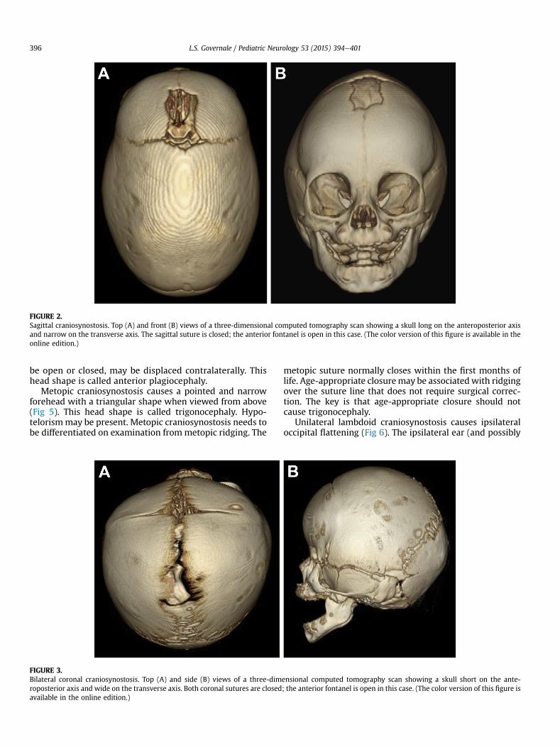
FIGURE 2.Sagittal craniosynostosis. Top (A) and front (B) views of a three-dimensional computed tomography scan showing a skull long on the anteroposterior axisand narrow on the transverse axis. The sagittal suture is closed; the anterior fontanel is open in this case. (The color version of this figure is available in theonline edition.)
L.S. Governale / Pediatric Neurology 53 (2015) 394e401396
be open or closed, may be displaced contralaterally. Thishead shape is called anterior plagiocephaly.
Metopic craniosynostosis causes a pointed and narrowforehead with a triangular shape when viewed from above(Fig 5). This head shape is called trigonocephaly. Hypo-telorismmay be present. Metopic craniosynostosis needs tobe differentiated on examination frommetopic ridging. The
FIGURE 3.Bilateral coronal craniosynostosis. Top (A) and side (B) views of a three-dimroposterior axis and wide on the transverse axis. Both coronal sutures are closedavailable in the online edition.)
metopic suture normally closes within the first months oflife. Age-appropriate closuremay be associatedwith ridgingover the suture line that does not require surgical correc-tion. The key is that age-appropriate closure should notcause trigonocephaly.
Unilateral lambdoid craniosynostosis causes ipsilateraloccipital flattening (Fig 6). The ipsilateral ear (and possibly
ensional computed tomography scan showing a skull short on the ante-; the anterior fontanel is open in this case. (The color version of this figure is

FIGURE 4.Unilateral coronal craniosynostosis. Top (A) and front (B) views of a three-dimensional computed tomography scan showing closure of one coronal suture.The ipsilateral forehead is flattened, the anterior fontanel is displaced contralaterally, the ipsilateral orbital roof and sphenoid wing are displaced superiorly,and the nasal root is deviated toward the side of the closed suture. The superior displacement of the ipsilateral orbital roof and sphenoid wing is termed aHarlequin eye deformity because, when viewed on a frontal x-ray, it resembles the shape of the similarly named masquerade mask. (The color version ofthis figure is available in the online edition.)
L.S. Governale / Pediatric Neurology 53 (2015) 394e401 397
forehead) is displaced posteriorly resulting in a trapezoidalhead shape. The ipsilateral mastoid elongates drawing theipsilateral ear inferiorly. Early closure of this suture is veryrare, and the head shape is called posterior plagiocephaly.
FIGURE 5.Metopic craniosynostosis. Top (A) and front (B) views of a three-dimensionaltriangular shape when viewed from above. Hypotelorism is present. The metoversion of this figure is available in the online edition.)
The most common cranial deformity is positional pla-giocephaly. It is important on examination to differentiatethis entity from craniosynostosis because they have vastlydifferent implications. Unlike craniosynostosis, positional
computed tomography scan showing a narrow, pointed forehead with apic suture is closed; the anterior fontanel is open in this case. (The color

FIGURE 6.Lambdoid craniosynostosis. Top (A) and back (B) views of a three-dimensional computed tomography scan showing closure of the right lambdoid suture.The skull has a trapezoidal shape with right posterior flattening and posterior displacement of the right forehead. If it were visible, the ear ipsilateral to theposterior flattening would be posteriorly displaced. Also seen is inferior elongation of the ipsilateral mastoid that would also displace the ipsilateral earinferiorly. (The color version of this figure is available in the online edition.)
L.S. Governale / Pediatric Neurology 53 (2015) 394e401398
plagiocephaly is not associated with a risk of head growthrestriction or increased intracranial pressure. As such,treatment is nonsurgical, usually with position changes,“tummy time,” and physical therapy for any torticollis thatmay be present. The flattening is likely to diminish as thechild obtains gross motor milestones and lies on the arealess. The use of molding helmets is controversial.
The cranial deformity associated with positional plagio-cephaly is predictable (Fig 7). When an infant lays his or herhead in a particular spot repeatedly, that area of the headtends to flatten. Because the recommendation to decrease
FIGURE 7.Positional plagiocephaly. Top view (A) of a three-dimensional computed tomogshape. There is right posterior flattening and anterior displacement of thedisplacement of the ear ipsilateral to the posterior flattening. The sutures are o
sudden infant death syndrome is for supine sleep, the flat-tened area is occipital. The flattened area is typically unilat-eral, where itmust be distinguished from unilateral lambdoidcraniosynostosis (a rare condition). In positional plagioce-phaly, the ear (and possibly forehead) ipsilateral to theflattening is displaced anteriorly compared with the contra-lateral ear, resulting in a parallelogram shape. In unilaterallambdoid craniosynostosis, the ear (and possibly forehead)ipsilateral to the flattening is displaced posteriorly comparedwith the contralateral ear, resulting in a trapezoid shape. Theipsilateral mastoid elongation and inferior displacement of
raphy scan (done for other reasons) showing a skull with a parallelogramright forehead. Axial computed tomography scan (B) showing anteriorpen. (The color version of this figure is available in the online edition.)

L.S. Governale / Pediatric Neurology 53 (2015) 394e401 399
the ipsilateral ear seen in unilateral lambdoid craniosynos-tosis is not seen in positional plagiocephaly. Symmetricbilateral flattening can mimic bilateral coronal craniosynos-tosis and may require imaging to make a distinction.
The diagnosis of craniosynostosis can often bemadewitha clinical examination of the head shape. In cases of diag-nostic uncertainty or for confirmation, radiographic imag-ing can be acquired. A simple initial method is a skull x-rayseries consisting of an anteroposterior, Townes, and twolateral views. If skull x-rays are not definitive, a noncontrasthead computed tomography scan with three-dimensionalreconstructions of the bone windows should be obtained.
Natural history
Left untreated, craniosynostosis can result in worsenedcranial deformity and, potentially, overall cranial growthrestriction with resultant increased intracranial pressure(ICP). The deformity may lead to psychosocial issues as thechild interacts with peers during development. In addition tocranial growth restriction, increased ICP may develop in thesyndromic patients because of venous outflow stenosis at thejugular foramina, elevated central venous pressures fromobstructive sleep apnea, and hydrocephalus from aqueductalstenosis or fourth ventricular outflow obstruction.7 Thesecomorbidities result in a higher risk of elevated ICP in syn-dromic cases and are themselves treated when possible withcontinuous positive airway pressure and endoscopic thirdventriculostomy. Cerebrospinal fluid shunts are avoidedwhenever possible because of the risk of slit ventricle syn-drome and hemorrhage in this patient population.
Although the risk of elevated ICP is more controversial inthe nonsyndromic cases, there are studies demonstrating it.Several studies reported in the 1990s showed an estimatedincidence of between 4.5% and 24%.8 A 2014 study fromOxford found a 44% incidence of increased ICP as measured
FIGURE 8.Craniosynostosis molding helmet. The orthosis consists of a rigid outer shell anless growth is desired. Open areas are positioned over skull regions where moreThe helmet may be decorated in an attempt to reduce social stigma associatedparents. (The color version of this figure is available in the online edition.)
by invasive ICP monitoring among 39 patients with isolatednonsyndromic sagittal craniosynostosis.8 A 2012 study bythe same group found high or borderline ICP in five of sevenpatients with isolated nonsyndromic unilateral coronalcraniosynostosis.9
Intracranial pressure is usually monitored by noninvasivemeans. These may include surveillance for classical symp-toms of elevated ICP (e.g., headache, nausea, emesis, upgazepalsy), measurement of the orbitofrontal circumference,palpation of the fontanel (if present), funduscopy to assessfor papilledema, and/or optical coherence tomography tomeasure the thickness of the retinal nerve fiber layer. Cranialimaging may show effacement of the cisterns or the con-vexity sulci, a secondary Chiari malformation, and/or a“copper-beaten” appearance to the skull (resulting frompressure-related gyral imprinting on the inner table of theskull). Lumbar puncture and/or use of a cranial pressuremonitor can be considered in cases of diagnostic uncertainty.
Treatment
Because of the risks associated with untreated cranio-synostosis, it is usually treated surgically soon after diag-nosis to unlock and reshape the bones. There are currentlyfour surgical methods: open calvarial reconstruction,minimally invasive strip craniectomy with use of post-operative molding helmet, minimally invasive strip cra-niectomy with spring implantation, and cranial distraction.One group has advocated the use of a molding helmetwithout surgery for sagittal craniosynostosis,10 but this ishighly controversial and not recommended.11
The traditional open calvarial reconstruction involvesremoval, reshaping, and replacement of the deformed por-tions of the bony convexity, including the fused suture. Forsagittal and lambdoid craniosynostosis, the posterior half ofthe convexity is reshaped, usually from the coronal sutures
d customizable inner foam padding. The foam contacts skull regions wheregrowth is desired. In this way, overall skull growth is strategically directed.with medical orthoses. This helmet has been well-adorned by the child’s
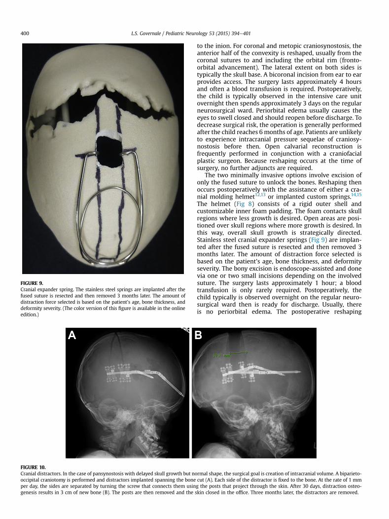
FIGURE 10.Cranial distractors. In the case of pansynostosis with delayed skull growth but nooccipital craniotomy is performed and distractors implanted spanning the boneper day, the sides are separated by turning the screw that connects them usingenesis results in 3 cm of new bone (B). The posts are then removed and the s
FIGURE 9.Cranial expander spring. The stainless steel springs are implanted after thefused suture is resected and then removed 3 months later. The amount ofdistraction force selected is based on the patient’s age, bone thickness, anddeformity severity. (The color version of this figure is available in the onlineedition.)
L.S. Governale / Pediatric Neurology 53 (2015) 394e401400
to the inion. For coronal and metopic craniosynostosis, theanterior half of the convexity is reshaped, usually from thecoronal sutures to and including the orbital rim (fronto-orbital advancement). The lateral extent on both sides istypically the skull base. A bicoronal incision from ear to earprovides access. The surgery lasts approximately 4 hoursand often a blood transfusion is required. Postoperatively,the child is typically observed in the intensive care unitovernight then spends approximately 3 days on the regularneurosurgical ward. Periorbital edema usually causes theeyes to swell closed and should reopen before discharge. Todecrease surgical risk, the operation is generally performedafter the child reaches 6 months of age. Patients are unlikelyto experience intracranial pressure sequelae of craniosy-nostosis before then. Open calvarial reconstruction isfrequently performed in conjunction with a craniofacialplastic surgeon. Because reshaping occurs at the time ofsurgery, no further adjuncts are required.
The two minimally invasive options involve excision ofonly the fused suture to unlock the bones. Reshaping thenoccurs postoperatively with the assistance of either a cra-nial molding helmet12,13 or implanted custom springs.14,15
The helmet (Fig 8) consists of a rigid outer shell andcustomizable inner foam padding. The foam contacts skullregions where less growth is desired. Open areas are posi-tioned over skull regions where more growth is desired. Inthis way, overall skull growth is strategically directed.Stainless steel cranial expander springs (Fig 9) are implan-ted after the fused suture is resected and then removed 3months later. The amount of distraction force selected isbased on the patient’s age, bone thickness, and deformityseverity. The bony excision is endoscope-assisted and donevia one or two small incisions depending on the involvedsuture. The surgery lasts approximately 1 hour; a bloodtransfusion is only rarely required. Postoperatively, thechild typically is observed overnight on the regular neuro-surgical ward then is ready for discharge. Usually, thereis no periorbital edema. The postoperative reshaping
rmal shape, the surgical goal is creation of intracranial volume. A biparieto-cut (A). Each side of the distractor is fixed to the bone. At the rate of 1 mmg the posts that project through the skin. After 30 days, distraction osteo-kin closed in the office. Three months later, the distractors are removed.

L.S. Governale / Pediatric Neurology 53 (2015) 394e401 401
adjuncts, however, do have some drawbacks. The helmetmust be worn 23 hours per day often until the child’s firstbirthday and requires frequent visits to an orthotist. Thesprings require a second surgery 3months later for removal.
The fourth surgical option is cranial distraction. In thecase of pansynostosis with delayed skull growth but normalshape, the surgical goal is creation of intracranial volume. Abiparieto-occipital craniotomy is performed and distractorsimplanted that span the bone cut (Fig 10). Each side of thedistractor is fixed to the bone. At the rate of 1 mm per day,the sides are separated by turning the screw that connectsthem using the posts that project through the skin. After30 days, distraction osteogenesis results in 3 cm of newbone. The posts are then removed and the skin closed in theoffice. Three months later, the distractors are removed.Distraction may also be used anteriorly to advance theanterior skull base and midface as a unit in a procedurecalled monobloc.
The decision of which procedure to perform is the resultof a discussion between the surgeon and the family, becauseeach has its positive and negative attributes. Key to beingable to offer all options, however, is early diagnosis andreferral. The age window to perform the helmet endoscopicoption is ideally 2.5-3.5 months of age, although some willattempt the procedure slightly later. The agewindow for thespring endoscopic option is ideally 3-6 months of age.
Syndromic andmultiple suture cases aremost frequentlytreated with open calvarial reconstruction, but applicationof the minimally invasive options to this patient populationis being investigated.16,17
Conclusions
Craniosynostosis may present in many different forms,but familiarity with associated head shapes can allowbedside diagnosis and differentiation from positional pla-giocephaly. Multiple surgical options for craniosynostosiscurrently exist, but early referral to a pediatric craniofacialcenter is needed to allow all options to be offered.
The author would like to thank Greg Pearson, MD, for the cranial expander springimage and Mark Proctor, MD, for the lambdoid craniosynostosis images.
References
1. Di Rocco F, Arnaud E, Renier D. Evolution in the frequency of non-syndromic craniosynostosis. J Neurosurg Pediatr. 2009;4:21-25.
2. Lajeunie E, Crimmins DW, Arnaud E, Renier D. Genetic consider-ations in nonsyndromic midline craniosynostoses: a study of twinsand their families. J Neurosurg. 2005;103:353-356.
3. Su N, Jin M, Chen L. Role of FGF/FGFR signaling in skeletal devel-opment and homeostasis: learning from mouse models. BoneResearch. 2014;2:1-24.
4. Lajeunie E, Barcik U, Thorne JA, El Ghouzzi V, Bourgeois M, Renier D.Craniosynostosis and fetal exposure to sodium valproate.J Neurosurg. 2001;95:778-782.
5. Sanchez-Lara PA, Carmichael SL, Graham Jr JM, et al. Fetal constraintas a potential risk factor for craniosynostosis. Am J Med Genet A.2010;152A:394-400.
6. Singh RP, Dhariwal D, Bhujel N, et al. Role of parental risk factors inthe aetiology of isolated non-syndromic metopic craniosynostosis.Br J Oral Maxillofac Surg. 2010;48:438-442.
7. Spruijt B, Joosten KF, Driessen C, et al. Algorithm for the manage-ment of intracranial hypertension in children with syndromic cra-niosynostosis. Plast Reconstr Surg. 2015;136:331-340.
8. Wall SA, Thomas GP, Johnson D, et al. The preoperative incidence ofraised intracranial pressure in nonsyndromic sagittal craniosynos-tosis is underestimated in the literature. J Neurosurg Pediatr. 2014;14:674-681.
9. Eley KA, Johnson D, Wilkie AO, Jayamohan J, Richards P, Wall SA.Raised intracranial pressure is frequent in untreated nonsyndromicunicoronal synostosis and does not correlate with severity ofphenotypic features. Plast Reconstr Surg. 2012;130:690e-697e.
10. Sood S, Rozzelle A, Shaqiri B, Sood N, Ham SD. Effect of moldinghelmet on head shape in nonsurgically treated sagittal craniosy-nostosis. J Neurosurg Pediatr. 2011;7:627-632.
11. Proctor MR, Rogers GF. Helmets and synostosis. J Neurosurg Pediatr.2012;9:680-681. author reply 681-682.
12. Jimenez DF, Barone CM. Endoscopic techniques for craniosynos-tosis. Atlas Oral Maxillofac Surg Clin North Am. 2010;18:93-107.
13. Berry-Candelario J, Ridgway EB, Grondin RT, Rogers GF, Proctor MR.Endoscope-assisted strip craniectomy and postoperative helmettherapy for treatment of craniosynostosis. Neurosurg Focus. 2011;31:E5.
14. Lauritzen CG, Davis C, Ivarsson A, Sanger C, Hewitt TD. The evolvingrole of springs in craniofacial surgery: the first 100 clinical cases.Plast Reconstr Surg. 2008;121:545-554.
15. van Veelen ML, Mathijssen IM. Spring-assisted correction of sagittalsuture synostosis. Childs Nerv Syst. 2012;28:1347-1351.
16. Jimenez DF, Barone CM. Multiple-suture nonsyndromic craniosy-nostosis: early and effective management using endoscopic tech-niques. J Neurosurg Pediatr. 2010;5:223-231.
17. Jimenez DF, Barone CM. Bilateral endoscopic craniectomies in thetreatment of an infant with Apert syndrome. J Neurosurg Pediatr.2012;10:310-314.


![Abstract arXiv:1211.3711v1 [cs.NE] 14 Nov 2012transducer’s construction from RNNs, with their abil-ity to extract features from raw data and their poten-tially unbounded range of](https://static.fdocuments.us/doc/165x107/5ea773f7e6d3a109e1760ffd/abstract-arxiv12113711v1-csne-14-nov-2012-transduceras-construction-from.jpg)