02 withdrawal of immunosuppressants in pediatric liver transplant
Upload
sidra-afzalCategory
view
59download
0
Pediatric Liver Masses: Part 1. Benign Tumors
DR SIDRA AFZALPGR
NEW RADIOLOGY DEPTSERVICES HOSPITAL LAHORE

• Benign hepatic tumors in children include leisions that are unique to the pediatric age group.
• About one third of the primary liver tumors in children are benign.

Infantile Hemangioendothelioma
• Or infantile hepatic hemangioma is a vascular neoplasm and the most common benign hepatic tumor of infancy
• About half of them are solitary and half are multifocal
• Nearly 90 percent of them are diagnosed in the first 6 months of life and one third in the first month.
• There is slight female predominance.

Clinical features
• Asymptomatic abdominal mass
• Serious complications may occur which include,
1. High output CHF due to associated large AV shunts
2. ‘Kassabach Merritt syndrome’ of coagulopathy due to intratumoral platelet sequestration.

Imaging features
• Depend upon whether leisions are focal, multifocal or diffuse.
• Typically evidence of high flow is apparent as manifest by enlargement of hepatic arteries and veins.
• Owing to the risk of bleeding, biopsy of these masses is avoided and diognosis is made on the basis of typical imaging findings.

Multifocal infantile hemangioendothelioma in a 6-month-old girl Transverse US image shows several small, well-demarcated, homogeneous hypoechoic lesions (arrowheads) in the liver.

Color Doppler image shows peripheral flow around some of the lesions.

Computed tomographic (CT) image obtained without intravenous contrast material shows that the lesions (arrowheads) are hypoattenuating relative to the liver

Contrast material–enhanced CT image shows that the nodules enhance intensely and uniformly (arrowheads).

Coronal fat-saturated T2-weighted magnetic resonance (MR) image shows numerous well-defined hyperintense nodules in the liver. Arrow = gallbladder.

Coronal T1-weighted MR image shows that the nodules (arrowheads) are hypointense relative to the liver.

Coronal gadolinium-enhanced fat-saturated T1-weighted MR image shows uniform enhancement of the nodules (arrowheads).

This is Plain radio-graph of the chest and abdomen of a 12 day old girl born prematurely at 31 weeks, shows an enlarged cardiac silhouette, paucity of bowel gas with lateral deviation of the stomach, and body wall edema.

Axial T2-weighted MR image shows a predominantly hyperintensemass (arrow) with a central hypointense area and adjacent flow voids (arrowhead), which represent enlarged hepatic veins.

Axial nonenhanced T1-weighted spoiled gradient-echo MR image shows that the mass (T) is hypointense relative to the liver.

Arterial phase gadolinium-enhanced T1-weighted MR image shows intense, peripheral, papillary enhancement (arrowheads) of the mass

Delayed phase gadolinium-enhanced T1- weighted MR image shows centripetal enhancement of the mass with a persistent hypointense area (arrowhead

Diffuse form of hemangioendothelioma in a 10-week-old girl with severe hypo-thyroidism.
Transverse US images show numerous large masses (* ) replacing the liver and compressing the inferior vena cava (arrow ). AO ,aorta.

Longitudinal color Doppler image shows a direct portal vein–to–hepatic vein shunt.

Contrast-enhanced CT image, obtained in the early portal venous phase, shows peripheral corrugated enhancement of the masses (arrowheads) and compression of the inferior vena cava (arrow).

Delayed phase CT image shows centripetal enhancement of the masses

Mesenchymal Hemartoma
• Is the second most common benign liver mass in children
• It is most commonly discovered in children younger than 2 yrs of age with nearly all lesions discovered by age 5.
• There is slight male predominance.

Clinical features
• The most common presentation, is painless abdominal distention. The abdominal enlargement is usually gradual, although distention can develop fairly rapidly .

Imaging features
• The gross appearance, which ranges from predominantly cystic to predominantly solid, determines the imaging features. The vast majority of mesenchymal hamartomas contain cysts.
• Cystic portions are avascular and stromalportions are relatively hypovascular.

Mesenchymal hamartoma in a 16-month-old girl.
Transverse US image shows cystic (arrowheads) and solid (T) portions of the tumor and adjacent normal liver (*).

Longitudinal color Doppler image shows no flow to the cystic component, which contains low-level echoes (arrowhead). Minimal flow is seen in the solid component (arrows).

Coronal CT image obtained with intravenous and oral contrast material shows the mixed cystic (arrowheads) and solid (T) tumor replacing the left hepatic lobe. * = normal liver.

Mesenchymal hamartoma of the liver in a 2-year-old boy.* normal liver,Trans-verse US image shows a well-defined cystic mass with multiple
septa in the liver.

Axial T2-weighted MR image shows the markedly hyperintense mass containing thin septa (arrows)

Coronal nonenhanced T1-weighted MR image shows that the mass (arrows) is homogeneously hypointenserelative to the liver.

Coronal contrast-enhanced T1-weighted MR image obtained at the same level shows that enhancement is limited to the septa (arrows).

Focal Nodular Hyperplasia
• FNH is most often seen in adult women but uncommonly occurs in young children and adolescents.
• it represents 2% of all primary hepatic tumors in children from birth to age 20 years . In the pediatric population, the lesion is typically diagnosed between the ages of 2 and 5 years
• A marked female predominance of the lesion is reported

Clinical features
• FNH is most commonly an incidental finding at imaging
• Symptoms of a mass lesion are described in 20% of cases .
• Abdominal pain is another common symptom
• Tumor rupture and hemorrhage are rare

Imaging features
• Because the mass is composed predominantly of hepatocytes, it appears similar to normal liver, and the lesion may be inapparent except for mass effect on adjacent structures.
• The presence of the central scar may aid identification of the mass on nonenhancedscans

FNH in a 6-year-old girl. Transverse US image shows the well-circumscribed, homogeneous, slightly hypoechoic mass (arrows) in the liver.

Color Doppler image shows flow in vessels radiating outward from the central scar.
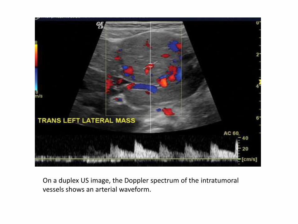
On a duplex US image, the Doppler spectrum of the intratumoralvessels shows an arterial waveform.

Arterial phase coronal CT image shows the tumor (arrow) enhancing more than the adjacent liver and lack of enhancement of the central scar (arrowhead).

Coronal CT delayed image, shows intensely enhancing vessels (arrow) adjacent to the tumor.

Hepatocellular Adenoma
• Hepatocellular adenoma, or hepatic adenoma, is a rare benign hepatic neoplasm that is etiologically associated with the use of steroids, especially oral contraceptives.
• Pediatric patients mainly consist of girls over 10 years old, most of whom have a history of oral contraceptive use
• It is also associated with androgen steroid therapy in fanconi anemia, glycogen storage disease types I and III, and also galactosemia and familial diabetes mellitus in pediatric patients.

Clinical features
• More commonly, patients are asymptomatic or present with an abdominal mass.
• Chronic and acute abdominal pain are other reported symptoms.
• The main clinical concern is intratumoralhemorrhage, which occurs in approximately 10% of patients.

Imaging features
• The appearance of hepatocellular adenoma varies depending on its pathologic composition.
• Those without hemorrhage are homogeneous and similar in appearance to adjacent normal liver.
• The presence of intratumoral hemorrhage or intracellular fat produces distinguishing imaging features.

Multiple hepatocellular adenomas in a 16-year-old girl In-phase axial T1-weighted gradient-echo MR image shows a heterogeneous, predominantly hypointense mass (arrowheads) with a small hyperintense focus consistent with hemorrhage. In addition, there is also an isointense mass (arrow).

Out-of-phase axial MR image shows a decrease in the signal intensity of both masses (arrowheads, arrow), a finding indicative of intralesional fat.

Nonenhanced axial T1-weighted spoiled gradient-echo MR image shows the well-defined masses (T). One is slightly hypointenserelative to the liver; the other is isointense.

Arterial phase axial T1-weighted MR image shows that both masses (T) enhance slightly more than the liver. An additional smaller lesion is visible (arrow

Portal venous phase MR image shows that the masses (T) are isointense to slightly hypointense relative to the liver. Straight arrow = additional smaller lesion, curved arrow = adjacent enlarged vein.

Nodular Regenerative Hyperplasia
• NRH may occur in patients of any age and has infrequently been reported in children
• NRH is characterized by regenerative nodules surrounded by atrophic liver in the absence of fibrosis. The nodules vary in size from a few millimeters to several centimeters.

Clinical features
• One-half of cases are found incidentally during studies performed for other indications, but one-half have signs and symptoms of portal hypertension
• NRH should be considered in young patients with portal hypertension and no evidence of portal vein thrombosis.

Imaging features
• The appearance of NRH at imaging is variable and depends in part on the size of the nodules.
• Diffuse tiny nodules are not detected, and the imaging appearance of the liver is normal.
• Nodules have a propensity to coalesce and may then become evident at imaging.

NRH, US image shows a well-circumscribed, homogeneous, hypoechoic hepatic mass (arrow).

On a contrast-enhanced CT image, the mass (arrow) diffusely enhances more than adjacent liver.

Axial T1-weighted MR image shows that the mass (arrow) is ill defined and slightly hypointense relative to the liver with a slightly hyperintense partial rim

Axial T2-weighted MR image shows the ill-defined hyperintense mass (arrow).

Thankyou