Pediatric Abdominal Pain - McGill
21
Pediatric Abdominal Pain An Emergency Medicine Perspective Jeremiah Smith, MD a, *, Sean M. Fox, MD b BACKGROUND Pediatric abdominal pain is a common complaint evaluated in emergency depart- ments (EDs). Although often due to benign causes, the varied and nonspecific presen- tations present a diagnostic challenge. Emergency care providers are tasked with the difficult job of remaining vigilant for the rare, yet devastating conditions while sorting through the much more common, benign causes of abdominal pain. This task is akin to finding the needle in the haystack. Diagnostic momentum can further threaten to divert the provider’s attention from the true cause. Pediatric abdominal pain is a challenging complaint to evaluate and deserves specific attention. EPIDEMIOLOGY Overall, 5% to 10% of all ED visits by pediatric patients are for abdominal pain. 1,2 In the United States alone, up to 38% of school-aged children complain of abdominal Disclosures: The authors have nothing to disclose. a Department of Emergency Medicine, Carolinas Medical Center, 1000 Blythe Boulevard, MEB Floor 3, Charlotte, NC 28203, USA; b Emergency Medicine Residency Program, Department of Emergency Medicine, Carolinas Medical Center, 1000 Blythe Boulevard, MEB Floor 3, Charlotte, NC 28203, USA * Corresponding author. E-mail address: [email protected] KEYWORDS Functional constipation Pyloric stenosis Necrotizing enterocolitis Appendicitis Incarcerated inguinal hernia Gonadal torsion Functional gastrointestinal disorder KEY POINTS Avoid diagnostic momentum, especially when evaluating functional constipation and functional gastrointestinal disorders. Bilious vomiting in a neonate is a surgical emergency until proven otherwise. Always consider gonadal torsion in a child with lower abdominal pain. Do not overlook the potential for psychosocial causes of abdominal pain. Constipation is not an innocuous condition. Emerg Med Clin N Am 34 (2016) 341–361 http://dx.doi.org/10.1016/j.emc.2015.12.010 emed.theclinics.com 0733-8627/16/$ – see front matter Ó 2016 Elsevier Inc. All rights reserved.
Transcript of Pediatric Abdominal Pain - McGill

Pediatric Abdominal PainAn Emergency Medicine Perspective
Jeremiah Smith, MDa,*, Sean M. Fox, MDb
KEYWORDS
� Functional constipation � Pyloric stenosis � Necrotizing enterocolitis � Appendicitis� Incarcerated inguinal hernia � Gonadal torsion � Functional gastrointestinal disorder
KEY POINTS
� Avoid diagnostic momentum, especially when evaluating functional constipation andfunctional gastrointestinal disorders.
� Bilious vomiting in a neonate is a surgical emergency until proven otherwise.
� Always consider gonadal torsion in a child with lower abdominal pain.
� Do not overlook the potential for psychosocial causes of abdominal pain.
� Constipation is not an innocuous condition.
BACKGROUND
Pediatric abdominal pain is a common complaint evaluated in emergency depart-ments (EDs). Although often due to benign causes, the varied and nonspecific presen-tations present a diagnostic challenge. Emergency care providers are tasked with thedifficult job of remaining vigilant for the rare, yet devastating conditions while sortingthrough the much more common, benign causes of abdominal pain. This task isakin to finding the needle in the haystack. Diagnostic momentum can further threatento divert the provider’s attention from the true cause. Pediatric abdominal pain is achallenging complaint to evaluate and deserves specific attention.
EPIDEMIOLOGY
Overall, 5% to 10% of all ED visits by pediatric patients are for abdominal pain.1,2 Inthe United States alone, up to 38% of school-aged children complain of abdominal
Disclosures: The authors have nothing to disclose.a Department of Emergency Medicine, Carolinas Medical Center, 1000 Blythe Boulevard, MEBFloor 3, Charlotte, NC 28203, USA; b Emergency Medicine Residency Program, Department ofEmergency Medicine, Carolinas Medical Center, 1000 Blythe Boulevard, MEB Floor 3, Charlotte,NC 28203, USA* Corresponding author.E-mail address: [email protected]
Emerg Med Clin N Am 34 (2016) 341–361http://dx.doi.org/10.1016/j.emc.2015.12.010 emed.theclinics.com0733-8627/16/$ – see front matter � 2016 Elsevier Inc. All rights reserved.

Smith & Fox342
pain weekly and up to 24% of them have had that pain for greater than 8 weeks.3,4
What makes finding the rare, but potentially life-threatening case of abdominal paineven more difficult is that only 5% to 10% of children with abdominal pain have under-lying organic disease and that the causes vary substantially with the age of the pa-tients (Table 1).3
HISTORY
The history of present illness and past medical history are the foundation on whichappropriate medical decisions are built. A thorough history helps pare down the largedifferential for abdominal pain. Although daunting in a busy ED, it is possible to obtaina thorough but efficient history.When taking the history, question both the caregiver and child themselves sepa-
rately, if age appropriate. Sitting or kneeling may help minimize anxiety in both childrenand parents. Interview the child where he or she is most comfortable. For olderchildren and adolescents, a history for sexual activity, drug use, possible abuse,and suicidal ideation is best obtained with the caregivers out of the room.
PHYSICAL EXAMINATION
A complete history should always be followed by an equally thorough physical exam-ination. Although the abdominal examination is the centerpiece, significant informationcan be gleaned from a full examination (Box 1). The patients’ general appearance andactivity level are also helpful in sorting out the potential causes, especially if infants arelethargic or inconsolable. Focusing only on the abdomen may lead to missing simpleclues to other causes.
Table 1Common causes of abdominal pain by age
Age <1 y 1–5 y 5–12 y >12 y
Commonor benign
Colic, GERD,milk proteinallergy
UTI,constipation
UTI, constipation,FGID, GAS
UTI, constipation,FGID, GAS
Urgent AGE,malrotationwithoutvolvulus
AGE, HSP,pneumonia,Meckeldiverticulum
AGE, IBD,pneumonia
AGE, IBD,pneumonia,hepatitis,pancreatitis,nephrolithiasis,PID
Emergent Trauma, NAT,midgutvolvulus, NEC,omphalitis,incarceratedhernia, pyloricstenosis,intussusception
Trauma,appendicitis,asthma
Trauma, appendicitis,gonadal torsion,DKA, asthma
Trauma,appendicitis,gonadaltorsion,ectopicpregnancy,DKA, asthma
Abbreviations:AGE, acute gastroenteritis; DKA, diabetic ketoacidosis; FGID, functional gastrointes-tinal disorders; GAS, group A strep; GERD, gastroesophageal reflux disease; HSP, Henoch-Schonleinpurpura; IBD, inflammatory bowel disease; NAT, nonaccidental trauma; NEC, necrotizing enteroco-litis; PID, pelvic inflammatory disease; UTI, urinary tract infection.

Box 1
Physical examination for a child with abdominal pain
General
� Play with the child and engage him or her in a fun activity before the examination.
� Use a stuffed animal to show what you will do and how easy it is.
� Attempt to perform as much of the examination as possible in the caregivers lap if possible.
� Use a distraction during the examination.
� Use child life if they are available at your institution.
Constitutional
� Observation of the child before entering the room can direct your examination.
� Check for the absence or presence of a fever.
� Check for any other vital sign abnormality (ie, tachycardia, tachypnea, hypoxia).
Abdominal examination
� Use visualization for distention, masses, visible peristalsis, or bruising.
� Use auscultation for bowel sounds.
Palpation
� Check for the location of maximal tenderness, masses, or guarding.
� Having patients bend their knees while lying will help relax abdominal muscles and improveyour examination.
� It is sometimes helpful to push with the stethoscope during auscultation to evaluate fortenderness.
Percussion
� It is possible to percuss for abdominal fluid.
� It can be helpful in evaluating for rebound tenderness.
� Asking patients to jump and give you a high 5 is a great way to assess for reboundtenderness.
Rectal
� This examination is not always necessary and should not be routine in all examinations.
� Directed reasons for a rectal examination are as follows: evaluate for bloody stool, possiblefecal impaction, and question of Hirschsprung disease.
Genitourinary examination
� A genital examination should be performed in all male patients with abdominal pain and, atleast, externally in all female patients.
� A complete gynecologic examination is sometimes required in sexually active femalepatients.
Remaining examination
� The remaining physical examination should not be skipped over.
� Evaluate for other causes of abdominal pain, such as pneumonia or pharyngitis.
Pediatric Abdominal Pain 343

Smith & Fox344
IMAGING
Judicious use of imaging is often integral to a complete evaluation of abdominal pain.It is important to know the benefit and potential limitations of each modality. Box 2highlights some of the important considerations of various imaging modalities.
PEDIATRIC CAUSES OF ABDOMINAL PAIN
Constipation is a ubiquitous problem with a worldwide prevalence of 3% to 5%6,7
(Box 3). In the United States, retrospective studies have shown constipation toaccount for 19.3% of all ED visits for abdominal pain and 0.4% of all visits to the ED.1,7
Box 2
Judicious use of imaging
Abdominal radiograph
� It is rarely useful because of low sensitivity and specificity.
� An acute abdominal series may show signs of obstruction or perforation.
� A fecalith in the right lower quadrant of a patient with appendicitis may occasionally beseen.
� It should not be routinely ordered for patients with constipation.
� It may show a basilar pneumonia.
Ultrasound
� It is often the image modality of choice for many diseases because it has no radiationexposure.
� It can be performed at the bedside.
� It may be very user dependent and is best at institutions that use it often.
� It is the imaging modality of choice for hydronephrosis from possible nephrolithiasis,gallstones, gonadal torsion, intussusception, pyloric stenosis, appendicitis, and FocusedAssessment with Sonography in Trauma examinations.
Computed tomography
� It has high sensitivity and specificity for many intra-abdominal diseases.
� Sensitivity and specificity are often maintained between community and academic facilities.
� It exposes children to ionizing radiation.� 25.8 to 33.9 cases of solid organ cancer per 10,000 abdomen/pelvis CTs in girls5
� 13.1 to 14.8 cases of solid organ cancer per 10,000 abdomen/pelvis CTs in boys5
� Children are more radiosensitive to ionizing radiation.
� Children have longer expected lifetime to manifest latent injury.
� There is greater potential for radiation overdose from inappropriate CT protocols.
� Helical computed tomography is the most sensitive test for nephrolithiasis in children.
MRI
� It has high sensitivity and specificity for many intra-abdominal diseases.
� It is expensive.
� It is time intensive.
� It is not readily available at many EDs.
� It may require sedation in children.

Box 3
Functional constipation
� Two or less defecations per week
� At least one episode per week of encopresis after potty training
� Excessive stool retention/retentive posturing
� Painful and hard bowel movements
� Large fecal mass in rectum or large-diameter stools that may obstruct toilet
� No pathologic cause
Data from Tabbers M, DiLorenzo C, Berger M, et al. Evaluation and treatment of functionalconstipation in infants and children: evidence-based recommendations from ESPGHAN andNASPGHAN. J Pediatr Gastroenterol Nutr 2014;58:258–74.
Pediatric Abdominal Pain 345
Diagnosis and Workup
Functional constipation is a diagnosis of exclusion, and the evaluation begins with athorough history and physical examination.Abdominal radiographs are often ordered to evaluate for constipation, but they only
have a reported sensitivity of 60% to 80% and should not be routinely ordered.6 Infact, there is no evidence to support routine testing of any sort if the child does nothave any concerning signs or symptoms (Box 4), yet it is important to remain vigilantfor other concealed conditions, such as Hirschsprung disease in the neonate withconstipation.
Management
The management of constipation can be broken up into 2 groups: ED managementand home management. The cornerstone of ED management for constipation beginswith setting reasonable expectations and explanation that this is a long-term process.An enema in the ED may be required, but daily osmotic laxatives (eg, polyethyleneglycol 3350) or glycerin suppositories at home, behavioral modifications, and closefollow-up with their primary care provider will keep them out of the ED.
PYLORIC STENOSIS
� The pylorus is a single unit of smooth muscle at the lower end of the stomach.� It connects to the duodenum via the pyloric sphincter.� Stenosis occurs with elongation and thickening of the pylorus.
Box 4
Important aspects of the physical examination for constipation
� Growth parameters (ie, look at their growth chart for failure to thrive)
� Abdominal distention and the presence of a fecal mass
� Soiling of their undergarments or skin in the perianal area
� Anal skin tags, anal fissures or tears, flat buttocks, or a sacral dimple/tuft of hair
� Complete neurologic examination with deep tendon reflexes and evaluating for saddleanesthesia
� No evidence for routine digital rectal examinations unless concern for an organic cause orunsure diagnosis6,8

Smith & Fox346
Gastric outlet obstruction occurs when the pyloric sphincter is unable to open.Pyloric stenosis is the most common surgical cause of nonbilious emesis in infants
less than 6 months of age and typically occurs around 4 to 6 weeks.9–11 Up to 43% ofpatients with pyloric stenosis are firstborn, and it is 4 to 5 times more common inmales.10–12
Diagnosis and Workup
Any infant with true vomiting is concerning and deserves a thorough evaluation. Mostclinicians will easily recognize the classic presentation of pyloric stenosis; however,not every presentation is classic. The physical examination can heighten suspicionfor pyloric stenosis as well as help sort through other causes of vomiting.If the infant has worsening projectile vomiting or failure to thrive, diagnostic testing
to evaluate for pyloric stenosis should be done.
� Laboratories
Figtes
� Classically, infants develop hypochloremic hypokalemic metabolic alkalosis.� With earlier diagnosis, less than 50% of infants will present with electrolyteabnormalities.13,14
� Electrolyte changes often after vomiting for greater than 1 week.13
� Abdominal ultrasound� It is the imaging modality of choice with a sensitivity of 98% to 100% and spec-ificity up to 100%.9,15
� Findings are consistent with pyloric stenosis: pylorus length greater than 14 to17 mm and a single-wall thickness greater than 3.0 to 4.5 mm9,13,16 (Fig. 1).
� ED physicians using point-of-care ultrasound had 100% sensitivity (95% con-fidence interval [CI] 66%–100%) and 100% specificity (95% CI 92%–100%)when able to identify the pylorus (wide CIs for sensitivity makes this a nonidealscreening test).9
� Upper gastrointestinal (GI) study� It is the former gold standard, but rarely used now.15
� It is useful if bilious vomiting is present as it also evaluates for malrotation andvolvulus.10,15
. 1. Length 25.1 mm, thickness 5.8 mm. (From Shah S. An update on common gastroin-tinal emergencies. Emerg Med Clin North Am 2013;31:775–93.)

Pediatric Abdominal Pain 347
� Findings are as follows:- String sign: a string of contrast through the elongated pyloric channel- Double-track sign: several linear tracks of contrast separated by redundant
mucosa� Abdominal radiographs
� It is rarely useful but may show gas in the stomach and a paucity distal to thepylorus.
Management
These infants may present ill appearing and may require stabilization with fluid resus-citation. It may be difficult to differentiate between sepsis and pyloric stenosis in aseverely dehydrated infant, and a sepsis evaluation may be necessary as well.Once volume resuscitated and stable, surgical correction is required. Being mindfulof the management will ideally help avoid potential pitfalls (Box 5).
INTUSSUSCEPTION
� Telescoping of one portion of intestines into itself� Most common cause of GI obstruction in children� Second most common abdominal surgical emergency in children
Intussusception is the most common cause of GI obstruction in children and is thesecond most common pediatric acute abdominal surgical emergency. Its peak inci-dence is at 5 to 10 months of age.17–19
Diagnosis and Workup
Often the need for evaluation will be predicated on the history and a high index ofsuspicion. The classic history of colicky abdominal pain interspersed with episodesof normal activity or lethargy is seen in only 7.5% to 50.0% of patients.17,20,21 Itmay even be painless in up to 40% of patients less than 4 months of age.22 Redcurrant jelly stools represents bowel ischemia. Up to 75% of children without thesegrossly bloody stools may still be hemoccult positive.23
� Using history and examination alone, physicians are better at determining pa-tients who do not have intussusception rather than who do (specificity of 85%and an negative predictive value of 94%).21
� Laboratoryevaluation isoftenunnecessary,althoughabnormalities like leukocytosis,elevated band count, and elevated lactate can be seen with bowel perforation.24
Abdominal radiographs
� A paucity of gas in the right lower quadrant, intracolonic mass, rim sign, or signsof small bowel obstruction may be seen.
Box 5
Pitfalls of pyloric stenosis
� Failing to perform a comprehensive history and physical in every vomiting infant
� Ruling out pyloric stenosis automatically in an infant with bilious vomiting
� Deciding an infant does not need a full diagnostic workup for pyloric stenosis because theydo not have an olivelike mass or hypochloremic hypokalemic metabolic alkalosis
� Forgetting to check a glucose level in a child with severely altered oral intake

Smith & Fox348
� Overall sensitivity for ileocolic intussusception is 74% to 90% and is 88% to100% sensitive if there is air in the ascending colon in all images of a 3-viewabdominal radiograph.17,25
� Up to 24% of children with intussusception may have normal radiographs.26
Abdominal ultrasound
� Evaluate for a target, donut, or pseudokidney sign (Fig. 2).� In the radiology department, ultrasound has a 97.9% to 100% sensitivity.25
� One study showed ED physician point-of-care ultrasound to have a sensitivity of85% and specificity of 97%.19
Abdominal computed tomography/MRI
� Rarely necessary and is not cost-effective� May be necessary to identify a pathologic lead point in an older child or for recur-rent intussusception
Fluoroscopy enema
� Diagnostic and therapeutic for intussusception� Avoid if you have a concern for peritonitis, perforation, or necrosis
Management
Once diagnosed, the intussusception must be reduced. The largest controversy asso-ciated with intussusception management has to do with final disposition. It is commonfor most hospitals to admit and observe children after successful nonoperative reduc-tion because of the 7.5% to 43.0% recurrence rate.18,27 A recent meta-analysisshowed that 2.2% to 5.3% of patients had a recurrence at 24 hours and 7.1% at48 hours.18,28 After 48 hours, recurrence was seen in 5.1% and there was rarelyadverse events.26 This finding, along with a growing body of supportive evidence,
Fig. 2. Sonographic image showing the target or donut sign associated with intussuscep-tion. (From Marin J, Alpern E. Abdominal pain in children. Emerg Med Clin North Am2011;29:401–28.)

Pediatric Abdominal Pain 349
has led many practitioners to begin observing patients for approximately 6 hours aftersuccessful nonoperative reduction and then discharging home with strict return pre-cautions and close follow-up.18,27,28 Naturally, disposition planning requires coordina-tion with the pediatric surgical team to help avoid potential pitfalls (Box 6).
MALROTATION WITH OR WITHOUT MIDGUT VOLVULUS
� Malrotation refers to a spectrum of abnormal rotation of the duodenum aroundthe superior mesenteric artery (SMA) axis.
� This abnormal rotation leads to a shortened mesenteric root and predisposes tomidgut volvulus.
� Additionally, fibrous peritoneal bands (ie, Ladd bands) can lead to volvulus orobstruction themselves.
Midgut volvulus is abnormal rotation and fixation of the midgut around the SMA axisthat impedes lymphatic drainage, venous outflow, and arterial blood flow leading tomassive bowel infarction.Although malrotation is traditionally thought of as a disease of infancy, up to 25% of
patients may not be diagnosed until 5 years of age.29 Sixty percent of patients withmalrotation, however, will present by 1 month and 90% with volvulus present withinthe first year of life.22,30,31
Diagnosis and Workup
Bilious vomiting is present in greater than 90% of neonates with volvulus and shouldalways be considered a surgical emergency until proven otherwise.32 Neonates withvolvulus are typically irritable because of poor feeding with vomiting, abdominalpain and/or distention, and hematochezia. Older children with bilious vomiting havea larger differential diagnosis, but there should always be a high index of suspicionfor malrotation with volvulus because 22% of children and 12% of adults with malro-tation present with a volvulus.32 They will often have a history of chronic abdominalpain or cyclic vomiting and present with abrupt worsening of abdominal pain, and50% will have nonbilious vomiting.30
Naturally, any patient with signs of decompensation requires aggressive resuscita-tion before further diagnostic testing. Surgical consultation may be required basedsolely on clinical suspicion if the child remains unstable with findings concerning forabdominal catastrophe. Once the child is stable, imaging should be performed to eval-uate for malrotation and volvulus.
� Abdominal radiograph
Box
Pitf
� Nin
� Fa
� M
� It is often the initial study of choice because it is quick and readily available.� Findings are as follows:
- Double bubble sign signifying duodenal obstruction- Lack of bowel gas distal to the duodenum
6
alls of intussusception
ot considering and evaluating for a possible pathologic lead point in an older child withtussusception or in cases of recurrence
iling to recognize that intussusception can lead to somnolence and lethargy
issing the diagnosis because the infant had fever, anorexia, or diarrhea initially

Fig. 3.does noProximaof duodemerge
Smith & Fox350
- Bowel malposition- Air fluid levels- Pneumatosis- The most common finding is “normal bowel gas pattern.”33
� Upper GI with small bowel follow-through� It is considered the gold standard and defines size, shape, rotation, and pres-ence of obstruction.
� Malrotation with or without volvulus is suggested if the duodenal-jejunal junc-tion (DJJ) is in low position, DJJ is not left of the vertebral body pedicle, thejejunum is on the right and coiled like a spring, there is duodenal redundancy,or there is a corkscrew appearance of the DJJ (Fig. 3).
� Sensitivity is 93% to 100%, but the false-positive rate is 15% and false-negative rate is 2% to 3%.34
� Equivocal findings are seen in up to 37% of patients.35
� Ultrasound� Normally, the superior mesenteric vein (SMV) should be right of the SMA.� With malrotation, the SMV will be anterior or leftward or the duodenum will notbe between the SMA and aorta.
� If volvulus is present, a whirlpool sign, whereby the SMV wraps around theSMA on color-flow Doppler, may be seen.
� There is a clinical spectrum of normal variant anatomy, and confirmatorytesting is often still necessary.
� Other imaging modalities� Barium enema may show the cecum in the right upper quadrant or in thecentral abdomen but is not a reliable sign for malrotation.
� Abdominal CT can evaluate the anatomic relationship between the SMA, SMV,and DJJ positioning with a sensitivity of 97.3% and specificity of 99.0%.33
� Abdominal MRI can identify malrotation as well but is often time and costprohibitive.
Upper GI series demonstrates inferior displacement of the DJJ to the right. DJJt pass to the left of the spine and does not rise to the level of the duodenal bulb.l small bowel appears on the right side of the abdomen. Likely corkscrew patternenum indicating volvulus. (From Shah S. An update on common gastrointestinalncies. Emerg Med Clin North Am 2013;31:775–93.)

Pediatric Abdominal Pain 351
Management
Management of malrotation depends on the presence of an associated midgutvolvulus. Midgut volvulus is a true surgical emergency. Immediate surgical consulta-tion and operative repair with a Ladd procedure is necessary. Aggressive resuscitationand correction of hypoglycemia is imperative. Additionally, gastric decompressionand initiation of broad-spectrum antibiotics that cover gut flora are vitally important.Unfortunately, even with prompt resuscitation and emergent surgical correction, themortality rate for a midgut volvulus is 3% to 9%. Asymptomatic pediatric patientswith malrotation but no midgut volvulus can be managed electively by pediatricsurgery (Box 7).
NECROTIZING ENTEROCOLITIS
� Classic triad of abdominal distention, GI bleeding, and pneumatosis on radiograph� Modified Bells staging: stage I suspected necrotizing enterocolitis (NEC), stage IImild NEC, and stage III severe NEC
NEC is often thought of as a disease of prematurity, but nearly 10% to 13% of ne-onates with NEC are full term within the first 10 days of life.36,37 Presentation can varyfrom being nonspecific with temperature instability and feeding intolerance to overtshock with grossly bloody stool. Often times, full-term NEC is associated with infec-tion, hypoxic event at birth, congenital heart disease or cardiac surgery, and umbilicalartery catherization.37
Diagnosis and Workup
Full-term NEC can often present in a nonspecific fashion, and the clinician needs tomaintain a high index of suspicion while sorting through the other potential diagnoses.Because these patients will often appear similar to a septic neonate, a full septic
workup including glucose level, complete blood count, comprehensive metabolicpanel, urinalysis, blood and urine cultures, and cerebrospinal fluid (CSF) studies isbeneficial. An abdominal radiograph, with either a cross-table lateral or decubitusview, may show an abnormal gas pattern, pneumatosis, free air, or portal gas. Abdom-inal ultrasound may show a pseudokidney sign. Additional studies are often requiredto evaluate for uncommon causes of NEC in a full-term neonate.
Management
These patients can become critically ill rapidly and may require cardiopulmonary andfluid resuscitation. Twenty percent to 30% of neonates with NEC have bacteremia,
Box 7
Pitfalls of malrotation with or without volvulus
� Not appreciating that an infant with bilious vomiting is a surgical emergency until provenotherwise
� Delaying surgical consultationbyobtaining time-intensive testing in anacutely ill neonate/childwith a suggestive history of malrotation with volvulus
� Dismissing the idea of malrotation with volvulus in a worrisome child because imaging wasnegative
� Always be vigilant of patients with chronic abdominal pain and vomiting who have beenlabeled cyclic vomiting—avoid diagnostic momentum because they patients may be anolder patient with undiagnosed malrotation.

Smith & Fox352
and broad-spectrum antibiotics (eg, ampicillin 1 cefotaxime 1 metronidazole) thatcover gut flora should be initiated promptly. The neonate should have nothing bymouth, and a nasogastric tube should be placed for gastric decompression. Earlycoordination with pediatric surgery is necessary. Neonates with pneumoperitoneum,an abdominal mass or stricture with obstruction, or signs of sepsis require operativeintervention37 (Box 8).
APPENDICITIS
� It is inflammation of the appendix.� It can affect all age groups but is difficult to diagnosis in the very young.� Children less than 3 years of age have the highest perforation risk.
Appendicitis is the most common pediatric surgical emergency, and 250,000 casesare seen annually with a lifetime risk of developing it of 8.6% for men and 6.7% inwomen.38–40 The perforation rate is 80% to 100% in children younger than 3 yearsand up to 38% in older children.24,41
Diagnosis and Workup
The diagnosis of appendicitis may be quite clear or rather confounded but typicallyinvolves a combination of physical examination findings, laboratories, and imaging.
� History and physical examination
B
P
��
��
� Studies have shown experienced practitioners in pediatric emergency medi-cine are able to accurately diagnose men with appendicitis at a rate of 78%to 92% and women at 58% to 85%.42
� Unfortunately, physical examination alone leads to a false-negative rate of9.8% when taken to the operating room (OR) directly based on examinationas opposed to 4.5% with imaging.43
� Overall, there is no single predictor highly indicating appendicitis; but reboundtenderness has a positive likelihood ratio (LR) of 2.3 to 3.9, and right lowerquadrant (RLQ) pain to percussion has a positive LR of 2.56.24,44
� As always, being mindful of the other potential causes of abdominal pain isnecessary.
� Laboratory values� A complete blood count (CBC) with differential is often ordered but hasrelatively low diagnostic yield. If it is greater than 10,000 mm3 it has a positiveLR of 1.77 and a sensitivity and specificity for appendicitis of 65% to 85% and32% to 83%, respectively.44,45
� Increased polymorphonuclear cells and bandemia may provide diagnosticclues but are not indicative by themselves.
� A C-reactive protein (CRP) greater than 10 mg/L is not sensitive or specific forappendicitis but may be a strong predictor of perforation.46
ox 8
itfalls of NEC
Failing to consider NEC as a possible diagnosis for abdominal pain in a full-term neonate
Not searching for uncommon causes associated with NEC in a full-term neonate, such ascongenital heart disease (eg, coarctation or patent ductus arteriosus)
Forgetting that sepsis may cause NEC and a full workup including CSF is needed
Not stopping feeds and starting antibiotics when you suspect NEC

Figcoibl(arNo
Pediatric Abdominal Pain 353
� Pooled laboratory tests may increase the predictive power and generate asensitivity of 98% to 99%, but they still have low specificity (only 6%–12%).45
� Overall, laboratory test results may help determine which patients are at lowrisk for appendicitis, but they do not help determine who actually needs togo the OR for an appendectomy.
� A pregnancy test in female patients should always be done.� Imaging
� Abdominal radiographs are rarely useful, showing an appendicolith less than5% of time.42
� Abdominal ultrasound is often used as the first imaging of choice when evalu-ating appendicitis (Fig. 4)
. 4mpe arowrth
- It has become the imaging modality of choice at many institutions and isrecommended by the American College of Emergency Physicians to diag-nose but not exclude.47
- It has a sensitivity of 72.5% to 94.0% and specificity of 89% to 98% whenthe appendix is visualized (25%–73% of the time).48,49
- False negatives can be seen with perforation or tip appendicitis.50
- The diagnostic accuracy increases with the duration of symptoms.49
� Abdominal computed tomography (CT) may be used when abdominal ultra-sound was non-diagnostic for appendicitis or other diagnoses are beingconsidered concurrently.- It is the imaging modality of choice for many institutions because it has a
sensitivity of 90% to 97% and specificity of 91% to 99%.25,51
- This high accuracy is maintained between large academic hospitals andsmall rural community hospitals.38
. Thickened wall, noncompressible. (A) Sonographic image of appendix withoutression. (B) Sonographic image of appendix with compression showing noncompress-ppendix. Sonographic image of the thickened wall of the appendix with appendicitis). (From Parks N, Schroeppel T. Update on imaging for acute appendicitis. Surg ClinAm 2011;91:141–54.)

Smith & Fox354
- Unfortunately, children are much more sensitive to the ionizing radiation andhave a lifetime radiation-induced cancer risk of 20.1 to 26.1 per 100,000.52
� Abdominal MRI has 100% sensitivity and 96% specificity without radiationexposure but is not readily available in all EDs.
� Scoring systems� The Alvarado scoring system was initially designed to use in adults but onlyhas a specificity of 59% to 100% in children.53,54
� The Pediatric Appendicitis Score was designed specifically for children butagain has a specificity range of 50% to 98%.54,55
� The a priori judgment of experienced practitioners in emergency medicine hasa specificity of 49.6% to 90.2% and is equal to these appendicitis scoringsystems.44
� Scoring systems seem to be most useful for trainees or as adjuncts to optimizeand standardize patient management.44
Management
The primary challenge for the emergency care provider is in considering and diag-nosing appendicitis. Its subsequent ED management is relatively straightforward aslong as potential pitfalls are appreciated (Box 9).
INCARCERATED INGUINAL HERNIA
� It is entrapment of peritoneal contents in an inguinal hernia.� Strangulation occurs when the hernia is tightly constricted and the vascularsupply of the herniated contents becomes severely compromised.
Inguinal hernias are the most common congenital abnormality that requires surgeryoccurring in 0.8% to 4.4% of the general population but up to 6% to 31% in pediatricpatients.56,57 It is 6 times more common in males and typically occurs on the rightside.58
Diagnosis and Workup
Incarcerated inguinal hernias are often diagnosable with history and physical exami-nation alone, which has a sensitivity of 84%.56 They typically present with an abruptbulge in the groin area that increases in size when the child is upset and disappearswhen calm. The child is usually comfortable appearing unless it is incarcerated. Ifincarcerated, the hernia is not easily reduced and the child is inconsolable. Bloodystools and bilious emesis occur with bowel strangulation.Bedside point-of-care ultrasound can augment the evaluation. If a hernia is present,
peristalsis with air or fluid within the lumen of the inguinal mass will be seen.
Box 9
Pitfalls of appendicitis
� Assuming a child who has a concerning examination does not have appendicitis because of anegative or indeterminate ultrasound
� Not ensuring adequate follow-up within 24 hours for children discharged home with aworrisome history but reassuring laboratory test results/imaging
� Failing to consider gonadal torsion in any child with lower abdominal pain and concern forappendicitis
� Relying on only one sign or symptom to diagnose or exclude appendicitis

Pediatric Abdominal Pain 355
Ultrasound in the radiology department has a sensitivity of 97.9% and can help differ-entiate between indirect and direct hernias.56 Formal sonographic evaluation shouldbe considered in female patients to evaluate for ovarian contents.
Management
Nonoperative management of incarcerated hernias with bedside reduction requiresadequate analgesia and possibly sedation (50% of children do not receive analgesiabefore this procedure).59 If bedside reduction fails, pediatric surgical consultation withpossible operative repair is necessary (Box 10).
GONADAL TORSION
� Ovarian torsion
� It is a rotated ovary on its pedicle leading to obstruction of venous outflow,lymphatic drainage, and arterial blood flow once the ovary is engorged andedematous.� Adnexal torsion occurs with torsion of the ovary and/or fallopian tube.� Testicular torsion
� It is the rotation of spermatic cord resulting in compromise of testicular bloodflow.
Ovarian torsion accounts for up to 2.7% of all cases of acute abdominal pain infemale patients, but only 15% of those are in children.60,61 Forty percent to 84% ofcases of ovarian pathology have some abnormal features upon histologic examinationbut this is rarely malignancy.62
Testicular torsion occurs in 3.8 to 4.5 males per 100,000, but only 3% to 38% ofmales with acute scrotal pain have testicular torsion.61–64 A bell-clapper deformity isa predisposing condition for torsion and occurs bilaterally in 12% of patients whodevelop testicular torsion.62
Diagnosis and Workup
Whether it is a female or male patient being evaluated, early diagnosis and a highclinical suspicion is important for gonadal salvage. There are obvious differencesbetween their respective diagnostic evaluations.
Ovarian torsion
� Diagnosis is difficult because of the nonspecific symptoms often seen, and up to38% are initially diagnosed with appendicitis.65
� Colicky RLQ pain is common with associated fever, nausea, vomiting, anddysuria.
� A previous history of torsion or ovarian mass is often seen.� On average, girls wait 2.5 times as long for imaging and 2.7 times as long to go tothe OR as males with testicular torsion.66
Box 10
Pitfalls of incarcerated inguinal hernia
� Failing to consider ovarian involvement in female patients
� Not providing adequate analgesia and sedation before nonoperative reduction
� Failing to evaluate for testes below the hernia, that is, not a retracted testicle
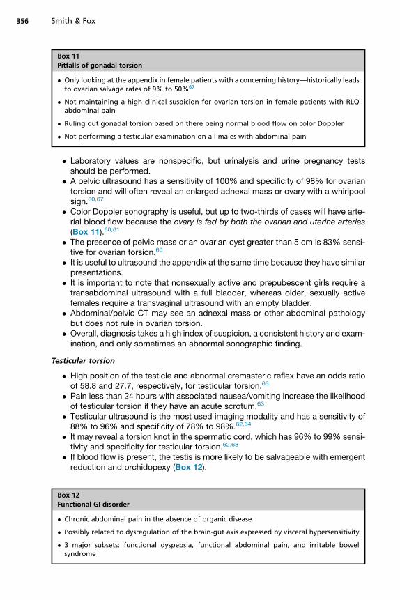
Box 11
Pitfalls of gonadal torsion
� Only looking at the appendix in female patients with a concerning history—historically leadsto ovarian salvage rates of 9% to 50%67
� Not maintaining a high clinical suspicion for ovarian torsion in female patients with RLQabdominal pain
� Ruling out gonadal torsion based on there being normal blood flow on color Doppler
� Not performing a testicular examination on all males with abdominal pain
Smith & Fox356
� Laboratory values are nonspecific, but urinalysis and urine pregnancy testsshould be performed.
� A pelvic ultrasound has a sensitivity of 100% and specificity of 98% for ovariantorsion and will often reveal an enlarged adnexal mass or ovary with a whirlpoolsign.60,67
� Color Doppler sonography is useful, but up to two-thirds of cases will have arte-rial blood flow because the ovary is fed by both the ovarian and uterine arteries(Box 11).60,61
� The presence of pelvic mass or an ovarian cyst greater than 5 cm is 83% sensi-tive for ovarian torsion.60
� It is useful to ultrasound the appendix at the same time because they have similarpresentations.
� It is important to note that nonsexually active and prepubescent girls require atransabdominal ultrasound with a full bladder, whereas older, sexually activefemales require a transvaginal ultrasound with an empty bladder.
� Abdominal/pelvic CT may see an adnexal mass or other abdominal pathologybut does not rule in ovarian torsion.
� Overall, diagnosis takes a high index of suspicion, a consistent history and exam-ination, and only sometimes an abnormal sonographic finding.
Testicular torsion
� High position of the testicle and abnormal cremasteric reflex have an odds ratioof 58.8 and 27.7, respectively, for testicular torsion.63
� Pain less than 24 hours with associated nausea/vomiting increase the likelihoodof testicular torsion if they have an acute scrotum.63
� Testicular ultrasound is the most used imaging modality and has a sensitivity of88% to 96% and specificity of 78% to 98%.62,64
� It may reveal a torsion knot in the spermatic cord, which has 96% to 99% sensi-tivity and specificity for testicular torsion.62,68
� If blood flow is present, the testis is more likely to be salvageable with emergentreduction and orchidopexy (Box 12).
Box 12
Functional GI disorder
� Chronic abdominal pain in the absence of organic disease
� Possibly related to dysregulation of the brain-gut axis expressed by visceral hypersensitivity
� 3 major subsets: functional dyspepsia, functional abdominal pain, and irritable bowelsyndrome

Box 13
Important aspects of the history for FGID
Functional abdominal pain
� Episodic or continuous periumbilical abdominal pain
IBS
� Diffuse abdominal pain
� Related to bowel movement frequency and improves after defecation
Functional dyspepsia
� Nausea
� Vomiting
� Symptoms consistent with gastroesophageal reflux disease
General
� Often have anxiety, depression, social isolation, and school absenteeism71
� Family history of celiac disease, IBD, peptic ulcers, FGID, and constipation
Pediatric Abdominal Pain 357
Functional GI disorder (FGID) is a common worldwide problem with a prevalence of1.6% to 41.2%. Up to 45% of these children will be diagnosed with irritable bowelsyndrome (IBS).69,70 Seventy percent of diagnosed cases of IBS occur in females,but there is more sex variability with the other subsets of FGID.69,71 It is no surprisethat this entity is the most common disease leading to consultation with a pediatricgastroenterologist.72
Diagnosis and Workup
The American Academy of Pediatrics recommends that evaluation for FGID shouldtake place in the primary care setting, but these children will often present to theED.3 When they do, a full history and physical examination (Box 13) should be per-formed to ensure that there are no red flags for organic disease, abuse, depression,or suicidal ideation (Box 14).If they have a reassuring history and physical examination, diagnostic testing is
often low yield.3 If there are red flags on examination, laboratory values, such asCBC, CRP, liver function tests, lipase, erythrocyte sedimentation rate, celiac serol-ogies, urinalysis, urine pregnancy test, and stool studies, may be indicated. Imagingthough is not routinely recommended. Without a concerning red flag, less than 1%
Box 14
Red flags for organic cause of abdominal pain in children
� Weight loss
� Severe vomiting
� Chronic severe diarrhea
� GI bleeding
� Hematemesis
� Fever
� Family medical history of inflammatory bowel disease

Box 15
Pitfalls of FGID
� Not considering psychosocial conditions like depression, suicidal ideation, or child abuse inyour differential—this may be how the child is reaching out for help
� Failing to recognize and workup worrisome red flags associated with FGID
� Failing to perform a thorough examination in child already labeled with chronic abdominalpain
Smith & Fox358
of children will have an abnormality on ultrasound.3 If further workup is needed, butchildren are stable and there is not concern for an emergent condition, the need forevaluation should be explained and then deferred to patients’ primary care provider.
Management
The mainstay of management for children with FGID is thorough anticipatory guidancediscussions and the setting of reasonable expectations (Box 15).
SUMMARY
Up to 10% of all visits to a pediatric ED are for abdominal pain.48 The astute clinicianneeds to have a high index of suspicion while evaluating any child with abdominalpain. The challenge is to remain vigilant for the rare, yet significant pathologic condi-tion, while not overtesting the more common, benign conditions.
REFERENCES
1. Caperell K, Pitetti R, Cross K. Race and acute abdominal pain in a pediatric emer-gency department. Pediatrics 2013;131:1098–106.
2. Pollack E. Pediatric abdominal surgical emergencies. Pediatr Ann 1996;25:448–57.
3. Romano C, Porcaro F. Currents issues in the management of pediatric functionalabdominal pain. Rev Recent Clin Trials 2014;9:13–20.
4. Saps M, Seshadri R, Sztainberg M, et al. A prospective school-based study ofabdominal pain and other common somatic complaints in children. J Pediatr2009;154:322–36.
5. Miglioretti D, Johnson E, Williams A, et al. Pediatric computed tomography andassociated radiation exposure and estimated cancer risk. JAMA Pediatr 2013;167:700–7.
6. Tabbers M, DiLorenzo C, Berger M, et al. Evaluation and treatment of functionalconstipation in infants and children: evidence-based recommendations fromESPGHAN and NASPGHAN. J Pediatr Gastroenterol Nutr 2014;58:258–74.
7. Diamanti A, Bracci F, Reale A, et al. Incidence, clinical presentation, andmanagement of constipation in a pediatric ED. Am J Emerg Med 2010;28:189–94.
8. Fox S. Recurrent abdominal pain. Charlotte (NC): Pediatric EM Morsels; 2014.
9. Sivitz A, Tejani C, Cohen S. Evaluation of hypertrophic pyloric stenosis by pedi-atric emergency physician sonography. Acad Emerg Med 2013;20:646–51.
10. Taylor N, Cass D, Holland A. Infantile hypertrophic pyloric stenosis: has anythingchanged? J Paediatr Child Health 2013;49:33–7.
Pediatric Abdominal Pain 359
11. Eberly M, Eide M, Thompson J, et al. Azithromycin in early infancy and pyloricstenosis. Pediatrics 2015;135:483–8.
12. Piroutek M, Brown L, Thorp A. Bilious vomiting does not rule out infantile hyper-trophic pyloric stenosis. Clin Pediatr 2012;51:214–8.
13. Glatstein M, Carbell G, Boddu S, et al. The changing clinical presentation ofhypertrophic pyloric stenosis: the experience of a large, tertiary care pediatrichospital. Clin Pediatr 2011;50:192–5.
14. Tutay G, Capraro G, Spirko B, et al. Electrolyte profile of pediatric patients withhypertrophic pyloric stenosis. Pediatr Emerg Care 2013;29:465–8.
15. Askew N. An overview of infantile hypertrophic pyloric stenosis. Paediatr Nurs2010;22:27–30.
16. Hernanz-Schulman M. Infantile hypertrophic pyloric stenosis. Radiology 2003;227:319–31.
17. Mandeville K, Chien M, Willyerd F, et al. Intussusception: clinical presentation andimaging characteristics. Pediatr Emerg Care 2012;28:842–4.
18. Gray M, Li S, Hoffmann R, et al. Recurrence rates after intussusception enemareduction: a meta-analysis. Pediatrics 2014;134:110–9.
19. Riera A, Hsiao A, Langhan M, et al. Diagnosis of intussusception by physiciannovice sonographers in the emergency department. Ann Emerg Med 2012;60:264–8.
20. Lam S, Wise A, Yenter C. Emergency bedside ultrasound for the diagnosis ofpediatric intussusception: a retrospective review. World J Emerg Med 2014;5:255–8.
21. Weihmiller S, Monuteaux M, Bachur R. Ability of pediatric physicians to judge thelikelihood of intussusception. Pediatr Emerg Care 2012;28:136–40.
22. Shah S. An update on common gastrointestinal emergencies. Emerg Med ClinNorth Am 2013;31:775–93.
23. Fleisher G. Textbook of pediatric emergency medicine. 6th edition. Philadelphia:Lippincott Williams & Wilkins; 2010. Print.
24. Pepper V, Stanfill A, Pearl R. Diagnosis and management of pediatric appendi-citis, intussusception, and Meckel diverticulum. Surg Clin North Am 2012;92:505–26.
25. Henderson A, Anupindi S, Servaes S, et al. Comparison of 2-view abdominalradiographs with ultrasound in children with suspected intussusception. PediatrEmerg Care 2013;29:145–50.
26. Hernandez J, Swischuk L, Angel C. Validity of plain films in intussusception.Emerg Radiol 2004;10:323–6.
27. Beres A, Baird R, Fung E, et al. Comparative outcome analysis of the manage-ment of pediatric intussusception with or without surgical admission. J PediatrSurg 2014;49:750–2.
28. Chien M, Willyerd F, Mandeville K, et al. Management of the child after enema-reduced intussusception: hospital or home? J Emerg Med 2013;44:53–7.
29. Aboagye J, Goldstein S, Salazar J. Age at presentation of common pediatric sur-gical conditions: reexamining dogma. J Pediatr Surg 2014;49:995–9.
30. Millar A, Rode H, Cywe S. Malrotation and volvulus in infancy and childhood.Semin Pediatr Surg 2003;12:229–36.
31. Sivitz A, Lyons R. Mid-gut volvulus identified by pediatric emergency ultrasonog-raphy. J Emerg Med 2013;45:e173–4.
32. Nehra D, Goldstein A. Intestinal malrotation: varied clinical presentation frominfancy through adulthood. Surgery 2011;149:386–93.

Smith & Fox360
33. Tackett J, Muise E, Cowles R. Malrotation: current strategies navigating the radio-logic diagnosis of a surgical emergency. World J Radiol 2014;6:730–6.
34. Applegate K, Anderson J, Klatte E. Intestinal malrotation in children: a problem-solving approach to the upper gastrointestinal series. Radiographics 2006;26:1485–500.
35. Lodwik D, Minneci P, Deans K. Current surgical management of intestinal rota-tional abnormalities. Curr Opin Pediatr 2015;27:383–8.
36. Short S, Papillon S, Berel D, et al. Late onset of necrotizing enterocolitis in thefull-term infant is associated with increased mortality: results from a two-centeranalysis. J Pediatr Surg 2014;49:950–3.
37. Sakellaris G, Partalis N, Dede O, et al. Gastrointestinal perforations in neonatalperiod: experience over 10 years. Pediatr Emerg Care 2012;28:886–8.
38. Parks N, Schroeppel T. Update on imaging for acute appendicitis. Surg ClinNorth Am 2011;91:141–54.
39. Paulson E, Kalady M, Pappas T. Clinical practice. Suspected appendicitis.N Engl J Med 2003;348:910–25.
40. Cole M, Maldonado N. Evidence-based management of suspected appendicitisin the emergency department. Emerg Med Pract 2011;13:1–32.
41. Lavine E, Saul T, Frasure S, et al. Point-of-care ultrasound in a patient with perfo-rated appendicitis. Pediatr Emerg Care 2014;30:665–7.
42. Old J, Dusing R, Yap W, et al. Imaging for suspected appendicitis. Am FamPhysician 2005;71:71–8.
43. The SCOAP Collaborative, Cuschieri J, Florence M, Flum DR, et al. Negativeappendectomy and imaging accuracy in the Washington State Surgical Careand Outcomes Assessment Program. Ann Surg 2008;248:557–63.
44. Fleischman R, Devine M, Yagapen M, et al. Evaluation of a novel pediatric appen-dicitis pathway using high- and low-risk scoring systems. Pediatr Emerg Care2013;29:1060–5.
45. Shogilev D, Duus N, Odom S, et al. Diagnosing appendicitis: evidence-basedreview of the diagnostic approach in 2014. West J Emerg Med 2014;7:859–71.
46. Wu H, Lin C, Chang C, et al. Predictive value of C-reactive protein at differentcutoff levels in acute appendicitis. Am J Emerg Med 2005;23:449–53.
47. Howell J, Eddy O, Lukens T, et al. Clinical policy: critical issues in the evaluationand management of emergency department patients with suspected appendi-citis. Ann Emerg Med 2010;55:71–116.
48. Mittal M, Dayan P, Macias C, et al. Performance of ultrasound in the diagnosis ofappendicitis in children in a multicenter cohort. Acad Emerg Med 2013;20:697–702.
49. Ross M, Liu H, Netherton S, et al. Outcomes of children with suspected appen-dicitis and incompletely visualized appendix on ultrasound. Acad Emerg Med2014;21:538–42.
50. Horn A, Ufberg J. Appendicitis, diverticulitis, and colitis. Emerg Med Clin NorthAm 2011;29:347–68.
51. Birnbaum B, Wilson S. Appendicitis at the millennium. Radiology 2000;215:337–48.
52. Hall E. Lessons we have learned from our children: cancer risks from diagnosticradiology. Pediatr Radiol 2002;32:700–6.
53. Escriba A, Gamell A, Fernandez Y, et al. Prospective validation of two systems ofclassification for the diagnosis of acute appendicitis. Pediatr Emerg Care 2011;27:165–9.

Pediatric Abdominal Pain 361
54. Pogorelic Z, Rak S, Mrklic I, et al. Prospective validation of Alvarado score andpediatric appendicitis score for the diagnosis of acute appendicitis in children.Pediatr Emerg Care 2015;31:164–8.
55. Goldman R, Carter S, Stephens D, et al. Prospective validation of the pediatricappendicitis score. J Pediatr 2008;153:278–82.
56. Till L, Kessler D. Rapid evaluation of an inguinal mass in a female infant usingpoint-of-care ultrasound. Pediatr Emerg Care 2014;30:366–7.
57. Lau S, Lee Y, Caty M. Current management of hernias and hydroceles. SeminPediatr Surg 2007;16:50–7.
58. Cascini V, Lisi G, Di Renzo D, et al. Irreducible indirect inguinal hernia containinguterus and bilateral adnexa in a premature female infant: report of an exceptionalcase and review of the literature. J Pediatr Surg 2013;48:E17–9.
59. Al-Ansari K, Sulowski C, Ratnapalan S. Analgesia and sedation practices forincarcerated inguinal hernias in children. Clin Pediatr 2008;47:766–9.
60. Appelbaum H, Abraham C, Choi-Rosen J, et al. Key clinical predictors in the earlydiagnosis of adnexal torsion in children. J Pediatr Adolesc Gynecol 2013;26:167–70.
61. Schmitt E, Ngai S, Gausche-Hill M, et al. Twist and shout! Pediatric ovarian torsionclinical update and case discussion. Pediatr Emerg Care 2013;29:518–26.
62. Baldisserotto M. Scrotal emergencies. Pediatr Radiol 2009;39:516–21.63. Beni-Israel T, Goldman M, Bar Chaim S, et al. Clinical predictors for testicular
torsion as seen in the pediatric ED. Am J Emerg Med 2010;28:786–9.64. Shah M, Caviness A, Mendez D. Prospective pilot derivation of a decision tool for
children at low risk for testicular torsion. Acad Emerg Med 2013;20:271–8.65. Ryan M, Desai B. Ovarian torsion in a 5-year old: a case report and review. Case
Rep Emerg Med 2012;2012:679121.66. Piper H, Oltmann S, Xu L, et al. Ovarian torsion: diagnosis of inclusion mandates
earlier intervention. J Pediatr Surg 2012;47:2071–6.67. Ochsner T, Roos J, Johnson A, et al. Ovarian torsion in a three-year-old girl.
J Emerg Med 2010;38:e27–30.68. Dajusta D, Granberg C, Villanueva C, et al. Contemporary review of testicular
torsion: new concepts, emerging technologies and potential therapeutics.J Pediatr Urol 2013;9:723–30.
69. Korterink J, Rutten J, Venmans L, et al. Pharmacologic treatment in pediatricfunctional abdominal pain disorders: a systematic review. J Pediatr 2015;166:424–31.
70. Korterink J, Diederen K, Benninga M, et al. Epidemiology of pediatric functionalabdominal pain disorders: a meta-analysis. PLoS One 2015;10(5):1–17.
71. Varni J, Shulman R, Self M, et al. Symptom profiles in patients with irritable bowelsyndrome or functional abdominal pain compared to healthy controls. J PediatrGastroenterol Nutr 2015;61(3):323–9.
72. Saps M, Biring H, Pusatcioglu C, et al. A comprehensive review of randomizedplacebo-controlled pharmacological clinical trials in children with functionalabdominal pain disorders. J Pediatr Gastroenterol Nutr 2015;60:645–53.