
Site-Directed C3a Receptor Antibodies from Phage … C3a Receptor Antibodies from Phage Display...
13
of May 10, 2018. This information is current as Phage Display Libraries Site-Directed C3a Receptor Antibodies from Axel Kola, Andreas Klos and Jörg Köhl Baensch, Bettina Sohns, Lubomir Arseniev, Wilfried Bautsch, Heiko Hawlisch, Ronald Frank, Meike Hennecke, Melanie http://www.jimmunol.org/content/160/6/2947 1998; 160:2947-2958; ; J Immunol References http://www.jimmunol.org/content/160/6/2947.full#ref-list-1 , 15 of which you can access for free at: cites 41 articles This article average * 4 weeks from acceptance to publication Fast Publication! • Every submission reviewed by practicing scientists No Triage! • from submission to initial decision Rapid Reviews! 30 days* • Submit online. ? The JI Why Subscription http://jimmunol.org/subscription is online at: The Journal of Immunology Information about subscribing to Permissions http://www.aai.org/About/Publications/JI/copyright.html Submit copyright permission requests at: Email Alerts http://jimmunol.org/alerts Receive free email-alerts when new articles cite this article. Sign up at: Print ISSN: 0022-1767 Online ISSN: 1550-6606. Immunologists All rights reserved. Copyright © 1998 by The American Association of 1451 Rockville Pike, Suite 650, Rockville, MD 20852 The American Association of Immunologists, Inc., is published twice each month by The Journal of Immunology by guest on May 10, 2018 http://www.jimmunol.org/ Downloaded from by guest on May 10, 2018 http://www.jimmunol.org/ Downloaded from
Transcript of Site-Directed C3a Receptor Antibodies from Phage … C3a Receptor Antibodies from Phage Display...

of May 10, 2018.This information is current as
Phage Display LibrariesSite-Directed C3a Receptor Antibodies from
Axel Kola, Andreas Klos and Jörg KöhlBaensch, Bettina Sohns, Lubomir Arseniev, Wilfried Bautsch, Heiko Hawlisch, Ronald Frank, Meike Hennecke, Melanie
http://www.jimmunol.org/content/160/6/29471998; 160:2947-2958; ;J Immunol
Referenceshttp://www.jimmunol.org/content/160/6/2947.full#ref-list-1
, 15 of which you can access for free at: cites 41 articlesThis article
average*
4 weeks from acceptance to publicationFast Publication! •
Every submission reviewed by practicing scientistsNo Triage! •
from submission to initial decisionRapid Reviews! 30 days* •
Submit online. ?The JIWhy
Subscriptionhttp://jimmunol.org/subscription
is online at: The Journal of ImmunologyInformation about subscribing to
Permissionshttp://www.aai.org/About/Publications/JI/copyright.htmlSubmit copyright permission requests at:
Email Alertshttp://jimmunol.org/alertsReceive free email-alerts when new articles cite this article. Sign up at:
Print ISSN: 0022-1767 Online ISSN: 1550-6606. Immunologists All rights reserved.Copyright © 1998 by The American Association of1451 Rockville Pike, Suite 650, Rockville, MD 20852The American Association of Immunologists, Inc.,
is published twice each month byThe Journal of Immunology
by guest on May 10, 2018
http://ww
w.jim
munol.org/
Dow
nloaded from
by guest on May 10, 2018
http://ww
w.jim
munol.org/
Dow
nloaded from

Site-Directed C3a Receptor Antibodies from Phage DisplayLibraries 1
Heiko Hawlisch,* Ronald Frank,‡ Meike Hennecke,* Melanie Baensch,* Bettina Sohns,*Lubomir Arseniev,† Wilfried Bautsch,* Axel Kola,* Andreas Klos,* and Jo rg Kohl 2*
Recent cloning of the human C3a receptor (C3aR) revealed that this receptor belongs to the large family of rhodopsin-typereceptors. A unique feature of the C3aR is the large second extracellular loop comprising about 175 amino acid residues. Weconstructed combinatorial phage Ab libraries expressing single chain Fv Abs from BALB/c mice immunized with the affinity-purified second extracellular loop of the C3aR, fused to glutathione-S-transferase. A panel of anti-C3aR single chain Fv fragments(scFvs) was selected after four rounds of panning using the second extracellular loop of the C3aR, fused to the maltose bindingprotein as Ag. Sequencing of the clones obtained revealed three different groups of scFvs, the epitopes of which were mapped totwo distinct regions within the loop, i.e., positions 185 to 193 and 218 to 226, representing the immunodominant domains of theloop. By flow cyotmetric analyses, the scFvs bound to RBL-2H3 cells transfected with the C3aR, but not to cells transfected withthe C5aR or to nontransfected RBL-2H3 cells. In addition, the scFvs bound to the human mast cell line HMC-1. Immunofluo-rescence studies showed C3aR expression on polymorphonuclear granulocytes and monocytes, but not on lymphocytes. In addi-tion, no C3aR expression was observed on human erythrocytes or platelets. Surprisingly, none of the scFvs alone or in combinationinhibited C3a-induced Ca21 mobilization from RBL-2H3 cells transfected with the C3aR. In addition, C3a did not displacebinding of the scFvs to the receptor, strongly suggesting that the N-terminal part of the second extracellular loop is not involvedin ligand binding. The Journal of Immunology,1998, 160: 2947–2958.
A ctivation of the complement system results in the releaseof a variety of potent proinflammatory mediators, suchas the anaphylatoxins C3a, C4a, C5a, and C5adesArg. In
the guinea pig system C3a exerts numerous biologic effects, suchas smooth muscle contraction probably induced by mast cell de-granulation, enhanced vascular permeability, and aggregation ofplatelets (reviewed in Ref. 1). However, the physiologic relevanceof the 77-amino acid C3a in humans has been an enigma for a longtime. Only in the last few years have biologic effects, such ashistamine release from IL-3-stimulated basophils (2), chemotaxisof eosinophils (3) and mast cells (4), or release of reactive oxygenmetabolites from polymorphonuclear leukocytes (5) and eosino-phils (6), been determined.
The pleiotropic effects of the anaphylatoxins are mediatedthrough specific receptor interactions. C3a receptors (C3aR)3 havebeen convincingly demonstrated on human leukemia-derived ba-
sophils (7), eosinophils (3, 6, 8), neutrophils, monocytes (9), andmast cells (4, 10, 11). Furthermore, C3a has been recently shownto modulate LPS-induced mRNA and protein synthesis for TNF-aand IL-1b in PBMC (12) and to suppress IgG, TNF-a, and IL-6production in activated tonsil-derived B cells (13).
The human C5a anaphylatoxin receptor (C5aR) and the C3aRbelong to the family of G protein-coupled receptors (14–18). Themost conspicuous feature of the C3aR is its large second extracel-lular loop, which is unique among the family of seven-transmem-brane receptors and which implies a functional role, e.g., in ligandbinding.
Northern blot hybridization revealed a broad expression of theC3aR in different tissues, such as lung, brain, spleen, thymus,lymph nodes, bone marrow, and peripheral blood leukocytes (16–18). This wide distribution of the C3aR suggests a role for C3aeven beyond its function as a pure proinflammatory mediator.
C3aR-specific mAbs would be useful to assess the expression ofthe C3aR on cells and tissues and might provide information aboutthe functional role of extracellular domains. In addition, they couldprove helpful in receptor mutagenesis studies. However, in gen-eral, it has been difficult to isolate mAbs to G protein-coupledreceptors. In fact, only two groups have reported isolation of mAbsto the C5aR, and one of these mAbs was an IgM (19, 20).
In the last few years methods have been developed to clone theentire repertoire of Ab genes from mice or human donors and toexpress the encoded Abs on filamentous bacteriophage (reviewedin Ref. 21). Phage displaying specific Ab fragments on their sur-face can be selected for binding on Ag-coated surfaces. Ab frag-ments specific for haptens or proteins, e.g., C5a (22), and severalhuman viruses have been recovered from such phage display com-binatorial libraries (21, 23).
*Institute of Medical Microbiology; and†Department of Hematology, HannoverMedical School, Hannover, Germany; and‡AG Molecular Recognition, Gesellschaftfur Biotechnologische Forschung, Braunschweig, Germany
Received for publication May 27, 1997. Accepted for publication November18, 1997.
The costs of publication of this article were defrayed in part by the payment of pagecharges. This article must therefore be hereby markedadvertisementin accordancewith 18 U.S.C. Section 1734 solely to indicate this fact.1 This work was supported by a grant from the Sonderforschungsbereich 244 (to J.K.;Project B8).2 Address correspondence and reprint requests to Dr. Jo¨rg Kohl, Institut fur Medi-zinische Mikrobiologie, Medizinische Hochschule Hannover, 30623 Hannover, Ger-many. E-mail address: [email protected] Abbreviations used in this paper: C3aR, complement 3a receptor; C5adesArg, com-plement 5a without C-terminal arginine residue; scFv, single chain Fv fragment; GST,glutathione-S-transferase; hC3aR, human complement 3a receptor; MBP, maltosebinding protein; IPTG, isopropylb-D-thiogalactoside; TYE/ap/glu, TYE plates con-taining ampicillin (100mg/ml) supplemented with 1% glucose; MPBS, phosphate-buffered saline containing 2% nonfat dry milk; SN, supernatant; PBS-BSA, phos-
phate-buffered saline supplemented with 1% bovine serum albumin; [Ca21]i,intracellular Ca21 concentration; CDR, complementarity-determining region.
Copyright © 1998 by The American Association of Immunologists 0022-1767/98/$02.00
by guest on May 10, 2018
http://ww
w.jim
munol.org/
Dow
nloaded from

Here, we report the selection of site-directed C3aR Abs fromscFv phage libraries derived from mice immunized with the sec-ond extracellular loop of the C3aR fused to glutathione-S-trans-ferase (GST). We obtained a panel of scFv Ab that could be di-vided into three groups according to their nucleotide sequences.The scFvs from the three different groups reacted with two distinctregions within the second extracellular loop of the C3aR. ThesescFvs were used 1) to analyze the role of the second extracellularloop in terms of C3a binding, and 2) to assess C3aR expression onhuman polymorphonuclear leukocytes, monocytes, lymphocytes,erythrocytes, and platelets.
Materials and MethodsAg preparation
The second extracellular loop of the hC3aR from amino acid positions 173to 332 (C3aR-(173–332)) was amplified from plasmid pTC12 (which ispcDNAI/Amp 1 cDNA hC3aR) (16) using the following primers: TC1,59-GGCATAG2AATTCGGCCACAAATTTGGTCTC-39; and TC2, 59-GGCATAG2TCGACTCATTAGGGTGTTGGCACTTGATC-39 (stopcodons are in boldface; restriction sites are in italics).
The DNA fragment was digested withEcoRI andSacI and ligated intovector pMAL-c (New England Biolabs, Beverly, MA), resulting in a fusionprotein composed of maltose binding protein (MBP) and the second ex-tracellular loop of the C3aR, MBP-C3aR-(173–332). In addition, the DNAwas ligated into vector pGEX-4T-1 (Pharmacia, Freiburg, Germany), re-sulting in a fusion protein composed of GST and the second extracellularloop of the C3aR (GST-C3aR-(173–332), using standard protocols (24).Escherichia coliBL21 cells (Pharmacia, Freiburg, Germany) were trans-fected with plasmids pMAL-c and C3aR-(173–332) or pGEX-4T-1 andC3aR-(173–332) by electroporation (25). Protein expression was inducedby adding IPTG (0.3 mM) to the bacterial culture at an OD600of 0.4 to 0.6.To purify the fusion proteins, bacteria were lysed by sonification, and thecytoplasmic extract containing MBP-C3aR-(173–332) was purified bychromatography using an amylose resin column (New England Biolabs).GST-C3aR-(173–332) was purified using a glutathione-Sepharose 4B col-umn (Pharmacia). The purified proteins were concentrated by filtrationthrough a Centriprep-10 membrane (Amicon, Danvers, MA). In silver-stained SDS gels, a band with the expected molecular mass of MBP-C3aR-(173–332) was observed at approximately 60 kDa, with an additional bandat 45 kDa. In the case of GST-C3aR-(173–332), a prominent band with theexpected size of 45 kDa was found, with a second band at 38 kDa. The lowmolecular mass bands probably represent degradation products yieldedduring the purification procedure (data not shown).
Immunization
Five BALB/c mice were immunized i.p. with 100mg of GST-C3aR-(172–332), with two booster injections 3 and 6 wk later. Four weeks after thesecond boost, a final boost (7.4mg of Ag) was applied directly into thespleen of three mice, which exhibited the highest Ab titer as determined byELISA using MBP-C3aR-(173–332). Total RNA was prepared from bothspleen and blood cells as previously described (26).
Construction of the scFv library
First-strand cDNA was generated from poly(A)1 RNA (obtained from theOligotex mRNA kit, Quiagen, Hilden, Germany) using Superscript II re-verse transcriptase (Life Technologies, Eggenstein, Germany) and an oli-go(dT)12–18 primer (Boehringer Mannheim, Ingelheim, Germany). Fromeach mouse Igg1 and Igg2a heavy chain repertoires as well as thek lightchain repertoire were amplified separately (Fig. 1).
In PCR I, g1 andg2a heavy chains andk light chains were amplifiedusing the following primer combinations:g1, primers MH1 to MH7/MH9;g2a, MH1 to MH7/JK10; andk, MH14 to MH20/MH21 (Table I).
As template, 1.8ml of cDNA was used. Ten microliters of Pfu buffer(10-fold concentrated), 0.8ml of dNTP mix (25 mM of each), and 2ml eachof the 39 and 59primers were added (10 pmol/ml), and the volume wasadjusted to 100ml with distilled water. The PCR cycles were as follows.Cycle I was 94°C for 300 s and XX°C for 60 s (XX5 64°C heavy chain;58°C light chain). After the first cycle, 2.5 U of Pfu polymerase wereadded. Cycle II was 72°C for 120 s, 94°C for 90 s, and XX°C for 120 s.The second cycle was repeated 30 times. Cycle III was 72°C for 660 s. Thecorrect length of the PCR products was assessed (g1 heavy chain5 378bp; g2a heavy chain5 705 bp;k light chain 5 348 bp) by agarose gelelectrophoresis. Bands with the correct size were purified using glass beads(Jetsorb gel extraction kit, Genomed, Bad Oeynhausen, Germany).
In PCR II, the seven purifiedg1 heavy chain,g2a heavy chain, andklight chain fragments were used as templates. Heavy chains were amplifiedwith the same 59 primers as those described for PCR I, however, they werenow combined with four different 39 primers, i.e., primers MH10 to MH13(Table I and Fig. 2), specific for the four different joining region genesegments. They have a long 59 overhang encoding for a part of the(Gly4Ser)3-linker at the 39 end of the heavy chain. Thek light chain wasamplified analogous to the heavy chains using primer combinations MH33to MH39/MH22 to MH25 (Table I, and Figs. 1 and 2).
Two nanograms of purified PCR I product (12.8ml PCR I product,diluted 1/100) was mixed with 10ml of Taq buffer (10-fold concentrated;Life Technologies), 3ml of MgCl2 (50 mM), 0.8ml of dNTP mix (25 mMeach), and 2ml each of the 39 and 59primers (10 pmol/ml) and adjusted toa final volume of 100ml with distilled water. PCR cycling was performedas described for PCR I, except that Taq polymerase (Life Technologies)was used. The annealing temperature was 55°C.
PCR II resulted in 28g1 heavy chain, 28g2a heavy chain, and 28klight chain fragments, with lengths of 377 bp (heavy chain) and 381 bp(light chain) (Fig. 2). Fragments of the correct size were purified as de-scribed above.
For the combination of heavy and light chains in PCR III, all PCR IIproducts amplified by the same 59 primer were pooled, resulting in seveng1 and seveng2a heavy chain pools, i.e., pools 1 to 7, each of whichcontained four different PCR II heavy chain products (Figs. 1 and 2). In thecase of the light chain, all PCR II products amplified with the same 39primer, e.g., all MH22 templates, were combined, leading to fourk lightchain pools, i.e., pool Jk1 to pool Jk4, each pool of which contained sevendifferent PCR II light chain products (Figs. 1 and 2).
As template for PCR III we combined 10 ng of each heavy chain pool(pools 1–7) with 10 ng of every light chain pool (pools Jk1-Jk4), resultingin 28 differentg1 and 28 differentg2a mixtures (Fig. 5). These mixtureswere cycled seven times with an annealing temperature of 60°C. Then, 2mlof both VH-specific 59primers (MH26–31) and VL-specific 39primers(MH22–25; each 10 pmol/ml) were added, and the mixture was cycled
FIGURE 1. Flow chart of the cloning scheme used to construct the scFvrepertoire.
2948 C3a RECEPTOR Abs FROM PHAGE DISPLAY LIBRARIES
by guest on May 10, 2018
http://ww
w.jim
munol.org/
Dow
nloaded from

another 25 times. After purification, 56 scFv gene fragments (28g11k and28g2a1k) with a length of 711 bp were obtained.
The 28 scFv gene fragments of each library and the vector DNA(pHEN-1) were digested with bothSfiI andNotI as previously described(21). The digested scFv DNA fragments were purified by phenol/chloro-form extraction and subsequently ethanol precipitated. Vector DNA waspurified from an agarose gel using glass beads (Jetsorb, Genomed). Five
hundred nanograms of vector DNA was ligated with 500 ng of scFv DNAin a total volume of 100ml with 8 ml of T4 ligase (400 U/ml; New EnglandBiolabs) for 72 h at 4°C. The mixture was heated to 70°C for 30 min to stopthe reaction. After phenol/chloroform extraction and ethanol precipitation,the phagemid DNA was dissolved in 10ml of distilled water and used forelectroporation ofE. coli TG1 cells (Stratagene, San Diego, CA) as pre-viously described (25). The bacteria were subsequently resuspended in 1
Table I. PCR primers used for PCR amplification of mouse Igg1, Igg2a, andk repertoirea
Primer Sequence
PCR I Backward primersJK10 (CH1 g2a) 59-GTT CTG ACT AGT GGG CAC TCT GGG CTC-39MH9 (CH1 g1) 59-AGG GGC CAG TGG ATA GAC AGA TGG-39MH21 (Ck) 59-GGA TAC AGT TGG TGC AGC ATC AGC-39
Foreward primers heavy-chainMH1 (I A1B) 59-CAG GTG CAG CTK MAG GAG TCA GG-39MH2 (II B) 59-CAG GTC CAR CTG CAG CAG YCT GG-39MH3 (II A 1C) 59-GAG GTY CAG CTG CAR CAG TCT GG-39MH4 (III A 1D) 59-GAR GTG AAG CTG GTG GAR TCT GG-39MH5 (III B) 59-GAG GTG AAG CTT CTC GAG TCT GG-39MH6 (III C) 59-GAA GTG AAG CTK GAK GAG TCT GG-39MH7 V 59-GAG GTY CAG CTT CAG CAG TCT-39
Forward primersk-light chainMH14 (I1III) 59-GAC ATT GTG MTG ACM CAR TCT-39MH15 (IV1VI) 59-CAA AWT GTK CTC ACC CAG TCT-39MH16 (II) 59-GAT GTT KTG ATG ACC CAR RCT CCA C-39MH17 (II) 59-GAT GTT KTG ATG ACC CAR ACT CCA C-39MH18 (II) 59-GAT RTT GTG ATG ACC CAA RCT CCA C-39MH19 (V) 59-GAC ATC MAG ATG ACM CAG TCT-39MH20 (V) 59-GAY ATT GTG ATG ACM CAG WCT-39
PCR II JH backward primers with linker (underlined)MH10 (JH1) 59-CC TCC AGA ACC TCC GCC TCC TGA TCC GCC ACC TCC
ACC TGA GGA GAC GGT GAC CGT GGT C-39MH11 (JH2) 59-CC TCC AGA ACC TCC GCC TCC TGA TCC GCC ACC TCC
ACC TGA GGA GAC TGT GAG AGT GGT-39MH12 (JH3) 59-CC TCC AGA ACC TCC GCC TCC TGA TCC GCC ACC TCC
ACC TGC AGA GAC AGT GAC CAG AGT-39MH13 (JH4) 59-CC TCC AGA ACC TCC GCC TCC TGA TCC GCC ACC TCC
ACC TGA GGA GAC GGT GAC TGA GGT T-39Foreward primers Vk with linker (underlined)
MH33 (I1III) 59-GGC GGA TCA GGA GGC GGA GGT TCT GGA GGA GGT GGG AGTGAC ATT GTG MTG ACM CAR TCT-39
MH34 (IV1VI) 59-GGC GGA TCA GGA GGC GGA GGT TCT GGA GGA GGT GGG AGTCAA AWT GTK CTC ACC CAG TCT-39
MH35 (II) 59-GGC GGA TCA GGA GGC GGA GGT TCT GGA GGA GGT GGG AGTGAT GTT KTG ATG ACC CAR RCT CCA C-39
MH36 (II) 59-GGC GGA TCA GGA GGC GGA GGT TCT GGA GGA GGT GGG AGTGAT GTT KTG ATG ACC CAR ACT CCA C-39
MH37 (II) 59-GGC GGA TCA GGA GGC GGA GGT TCT GGA GGA GGT GGG AGTGAT RTT GTG ATG ACC CAA RCT CCA C-39
MH38 (V) 59-GGC GGA TCA GGA GGC GGA GGT TCT GGA GGA GGT GGG AGTGAC ATC MAG ATG ACM CAG TCT-39
MH39 (V) 59-GGC GGA TCA GGA GGC GGA GGT TCT GGA GGA GGT GGG AGTGAY ATT GTG ATG ACM CAG WCT-39
Backward primers Jk with NotI restriction site (underlined)MH22 (Jk1) 59(GA)10 GC GGC CGCACG TTT GAT TTC CAG CTT GGT GCC-39MH23 (Jk2) 59(GA)10 GC GGC CGCACG TTT TAT TTC CAG CTT GGT CCC-39MH24 (Jk3) 59(GA)10 GC GGC CGCACG TTT TAT TTC CAA CTT TGT CCC-39MH25 (Jk4) 59(GA)10 GC GGC CGCACG TTT CAG CTC CAG CTT GGT CCC-39
Foreward primers heavy-chain withSfiI restriction site (underlined)MH26 (I A1B) 59(GA)10 G GCC CAG CCG GCCATG GCC CAG GTG CAG CTK MAG GAG TCA GG-39MH27 (II B) 59(GA)10 G GCC CAG CCG GCCATG GCC CAG GTC CAR CTG CAG CAG YCT GG-39MH28 (II A1C) 59(GA)10 G GCC CAG CCG GCCATG GCC GAG GTY CAG CTG CAR CAG TCT GG-39MH29 (III A 1D) 59(GA)10 G GCC CAG CCG GCCATG GCC GAR GTG AAG CTG GTG GAR TCT GG-39MH30 (III B) 59(GA)10 G GCC CAG CCG GCCATG GCC GAG GTG AAG CTT CTC GAG TCT GG-39MH31 (III C) 59(GA)10 G GCC CAG CCG GCCATG GCC GAA GTG AAG CTK GAK GAG TCT GG-39MH32 (V) 59(GA)10 G GCC CAG CCG GCCATG GCC GAG GTY CAG CTT CAG CAG TCT-39
SequencingprimersJK61 59-ATG AAA TAC CTA TTG CCT ACG GC-39JK75 59-CC TCC AGA ACC TCC GCC TCC-39JK76 59-GGC GGA TCA GGA GGC GGA GG-39JK77 59-CTA TGC GGC CCC ATT CA-39
a Primers used for sequencing are depicted. Primer degenerations are shown in bold face. The ambiguity codes used were the following: M5 A or C; R 5 A or G; W 5A or T; Y 5 C or T; K 5 G or T.
2949The Journal of Immunology
by guest on May 10, 2018
http://ww
w.jim
munol.org/
Dow
nloaded from

ml of SOC medium (2% tryptone; 0.5% yeast extract; 10 mM NaCl; 25mM KCl; 10 mM MgCl2; 10 mM MgSO4; 20 mM glucose) and incubatedfor 1 h at37°C. After incubation, bacteria from one library were plated onlarge TYE plates containing ampicillin (100mg/ml) supplemented with 1%glucose (TYE/ap/glu; Ø5 145 mm) and incubated overnight 37°C. Thetiter of the library was determined by plating 10-fold dilutions (1022-1027)of the electroporated bacteria on TYE/ap/glu plates. The bacteria grownwere scraped off the plates with 4 ml of 23 TY/ap/glu. This primary stockwas stored at220°C.
From the primary stock, 5ml was used to inoculate 50 ml of 23TY/ap/glu. The culture was shaken at 37°C until the OD600 reached 0.5.Then, 10 ml was removed and superinfected with 1011 pfu of VCS-M13 at37°C for 30 min without shaking. Cells were pelleted by centrifugation,subsequently resuspended in 300 ml of 23 TY/ap and kanamycin (25mg/ml), and grown overnight at 30°C with shaking (250 rpm). Phage par-ticles were purified and concentrated by polyethylene glycolprecipitation (24).
Affinity enrichment of C3aR-specific scFvs
In the first panning round, immunotubes (Nunc, Wiesbaden, Germany)were coated with MBP-C3aR-(173–332) (50mg/ml in PBS) and incubatedovernight at room temperature with gentle rotation. The tube was washedthree times with PBS and subsequently blocked with PBS containing 2%nonfat dry milk (MPBS) for 2 h at37°C. After three times washing withPBS, 63 1011 phages, that is 1011 phages from each library, were addedto 440ml of MPBS (2.2%) and incubated for 2 h atroom temperature withgentle rotation. The tube was washed 10 times with PBS/Tween-20 (0.1%)and 10 times with PBS. Bound phages were eluted with 1 ml of HCl/glycine (0.1 M; pH 2.2; plus 1% BSA) for 10 min at room temperature.Phages were immediately neutralized with 187.5ml of Tris/HCl (1 M; pH9.1) and used to infectE. coli TG1 cells. The numbers of phages before andafter panning were determined by plating infectedE. coli Tg1 cells ontoTYE/ap/glu plates.
During the next panning rounds, the stringency of the selection condi-tions was increased as follows. In panning rounds 2 and 3, 10mg/ml Agwere used. In round 4, the Ag concentration was reduced to 10 ng/ml, andpanning was performed in a microtiter plate. In panning rounds 3 and 4,both the tube and the microtiter plate were washed 20 times with PBS/-Tween-20 (0.1%) and 20 times with PBS.
Fingerprint analysis
To analyze the diversity of the scFv libraries during the selection process,individual clones were isolated after each panning round. The scFv DNA
of a particular clone was amplified by PCR using primers JK 61 (which sitswithin the c-Myc peptide fused C-terminal to the VL chain sequence) andJK 77 (which sits within the pelB sequence). The PCR fragment of the scFvgene was digested using the frequent cutter endonucleaseBstNI as previ-ously described (27).
Generation and partial purification of scFvs
E. coli strain HB2151 transfected with plasmid pHEN1 and scFv Ab wasgrown in 23 TY/amp supplemented with glucose (0.1%) until the OD600
reached 0.9. Protein expression was started by adding IPTG to a finalconcentration of 1 mM. After overnight culture at room temperature withshaking (200 rpm), bacteria were centrifuged (15 min, 11,3253 g, 4°C).The supernatant (SN) was concentrated in two steps using the ProVario-3filtration and concentration system (Filtron, Karlstein, Germany). All stepswere performed at 4°C. First, the SN was filtrated through an OMEGAmembrane (Filtron) (0.16mm). The filtrate was subsequently concentratedfrom 1 l to 50 ml byfiltration through a second OMEGA membrane (10kDa) and was finally dialyzed three times against PBS. The concentratedscFvs were quantified by densitometry using Herolab EasyImage software(HeroLab, Weisloch, Germany).
Rescue of phage or soluble scFvs from individual phagemidclones
Phage were rescued from single ampicillin-resistant colonies of infectedE.coli TG1 cells using the helper phage VCSM13 as described by Marks etal. (27). To produce soluble scFv Abs,E. coli HB 2151 cells were infectedwith monoclonal phage expressing a particular scFv fragment. The bacteriawere grown in 23 TY/ap supplemented with 0.1% glucose to OD600 0.9at 37°C. Subsequently, IPTG was added to a final concentration of 1 mM,and bacteria were grown overnight at room temperature with shaking (250rpm). SN containing either scFv phages or soluble scFvs were used forELISA analysis.
C3aR-(173–332) specific ELISA
MBP-C3aR-(173–332) (10mg/ml, 50 ml/well) was coated overnight on apolystyrene microtiter plate (Greiner), rinsed three times in PBS, and sat-urated in MPBS (2%) for 2 h. All steps were performed at room temper-ature. After washing three times with PBS, 25ml of 10% MBPS was addedfollowed by the addition of 100ml of phages or scFvs. To compare thebinding affinity of different phage Abs, the phage number was adjusted to3 3 109/100 ml. After incubation for 90 min, plates were washed threetimes with PBS/0.05% Tween-20, three times with PBS, and subsequentlyincubated with 100ml of a 1/1000 dilution of a rabbit anti-M13 serum (28).Before use, the antiserum (1/500 diluted in 4% MPBS) was incubated with2.5 ml of a cytoplasmic solution ofE. coli BL21 cells overnight at roomtemperature to prevent cross-reaction of the antiserum withE. coli proteinsin the Ag preparation. When scFvs were added, plates were incubated withmAb 9E10 (100ml/well) to detect the c-Myc peptide fused C-terminal tothe VL chain as previously described (21). Plates were washed three timeswith 50 mM Tris/0.15 M NaCl, pH 7.5; 100ml of alkaline phosphatase-conjugated goat anti-rabbit polyclonal Ab (Amersham, Braunschweig,Germany) diluted 1/2000 in 50 mM Tris buffer/0.15 M NaCl, pH 7.5, wasadded; and the plates were incubated for 90 min. To detect scFvs, 100ml/well peroxidase-conjugated anti-mouse Ab (Sigma, Munich, Germany)diluted 1/1000 in 50 mM Tris buffer/0.15 M NaCl, pH 7.5, was added andincubated for 60 min. Plates were either developed with 100ml of alkalinephosphatase substrate solution or, in the case of scFvs, with 100ml ofperoxidase substrate solution as previously described (28, 29).
Cell culture conditions
The rat basophilic leukemia cell-lines (RBL-2H3) stably transfected witheither the hC3aR cDNA or the hC5aR cDNA (30) were grown as describedpreviously (18, 31). The culture conditions used for the human mast cellline HMC-1 were previously described (10).
Isolation of human PMNs and mononuclear cells
Highly purified PMNs (with an erythrocyte contamination of,5% and aneosinophil contamination of 1–4%) and mononuclear cells (i.e., lympho-cytes and monocytes) were prepared by density centrifugation from citrate-anticoagulated blood of healthy human donors using Polymorphprep (Ny-comed, Oslo, Norway) according to the manufacturer’s instructions.Usually 1 to 23 107 cells were obtained from 40 ml of citrate-anticoag-ulated blood. The mononuclear cells were collected from the upper celllayer; the neutrophils were obtained from the lower layer. The cells werewashed twice with PBS, counted, and resuspended in PBS/BSA (1%) to
FIGURE 2. Summary of primer combinations used for PCR II and III.
2950 C3a RECEPTOR Abs FROM PHAGE DISPLAY LIBRARIES
by guest on May 10, 2018
http://ww
w.jim
munol.org/
Dow
nloaded from

5 3 106 cells/ml. Cells were assayed immediately after isolation. Purifi-cation of human platelets was performed exactly as described for guineapig platelets (32).
Immunofluorescence microscopy studies
All steps were performed in single wells of a 96-well microtiter plate.Wells were blocked with 250ml of PBS supplemented with 1% BSA(PBS-BSA) for 2 h at room temperature. Purified cells or cell lines werewashed once in 50 ml of PBS-BSA and were finally adjusted to a densityof 5 3 106 cells/ml. One hundred microliters of cells were transferred toeach well, and the plates were sealed (Dynatech Microtiter System, Den-kendorf, Germany). Cells were pelleted by centrifugation for 4 min at5143 g at room temperature (Minifuge RF, Heraeus Sepatech, Osterode,Germany). The SN was discarded. One hundred microliters of phage or 10mg/ml scFvs (100ml) were added to the cells. In competition bindingassays 1mM purified C3a (Advanced Research Technologies, La Jolla,CA) was added along with the scFvs. Cells were incubated for 60 min onice (all remaining incubation and centrifugation steps were performed at4°C). Cells were pelleted by centrifugation and were washed twice with150ml/well PBS-BSA. The pellet was resuspended in 100ml of PBS-BSAcontaining mAb 9E10 (1/1000), and the mixture was incubated for 90 min.When scFv phages were used, a rabbit anti-M13 antiserum (1/1000 inPBS-BSA) was added. After washing twice in PBS-BSA, pellets wereresuspended in 100ml of anti-mouse TRITC/anti-rabbit TRITC (Dianova;1/100 in PBS-BSA) and incubated for 30 min in the dark. Cells werewashed twice with PBS, resuspended in 10ml of mounting fluid (PBS/glycerol (50%)) and examined visually on a Zeiss III RS immunofluores-cence microscope (Zeiss, New York, NY).
Flow cytometric analysis of RBL-2H3 cells and the human mastcell line HMC-1
For flow cytometric measurements cells were treated as described for theimmunofluorescence microscopy studies with the following modifications:1) PBS was used for each washing step; 2) for detection of scFvs, 100mlof FITC-labeled anti-mouse antiserum (Becton Dickinson, Heidelberg,Germany; diluted 1/10 in PBS-BSA) was used; and 3) after the last washcells were resuspended in 150ml of PBS/formaldehyde (1%). Cells wereassessed in the flow cytometer FACScan and stored as list mode data forsubsequent analysis using CellQuest software (Becton Dickinson).
DNA sequencing
DNA from single colonies was prepared using standard protocols and se-quenced by PCR in a volume of 50ml using the following primers: JK61(a 59 primer that anneals to the pelB leader sequence), JK75 (a 39 primerthat anneals to the linker sequence), JK76 (a 59 primer that anneals to thelinker sequence), and JK77 (a 39 primer that anneals to the geneIII se-
quence; Table I). Sequencing was performed with the Sequenase PCRproduct sequencing kit and Sequenase version 2.0 DNA polymerase (U.S.Biochemical Corp., Cleveland, OH) according to the manufacturer’sinstructions.
Peptide synthesis and epitope mapping
Peptides were synthesized as arrays of N-terminally acylated and C-termi-nally covalently immobilized products on cellulose sheets derivatized withbAla-bAla dipeptide anchors by the spot synthesis technique as previouslydescribed (33), using a model ASP 222 spotting robot (ABIMED Anal-ysen-Technik GmbH, Langenfeld, Germany). Binding of the scFvs to pep-tide spots was assayed as previously described with a slight modification(34). After scFv binding, mAb 9E10 was used as the secondary Ab. Fordetection, an alkaline phosphatase-conjugated goat anti-mouse Ab wasused. Peptide arrays included overlapping pentadecapeptides, with an offsetof three amino acid residues, spanning the second extracellular loop of theC3aR from positions 173 to 332.
Measurement of [Ca21] i
Measurement of the change in [Ca21]i in fura-2/AM-loaded cells was per-formed exactly as described previously (35). Fluorescence data were con-verted to [Ca21]i by the formula of Grynkiewicz et al. (36).
ResultsConstruction of the scFv combinatorial libraries
Seven primers specific for mouse VH subgroups I to V and sevenprimers specific for mousek-chain subgroups I to VI were con-structed. From each subgroup, sequences available in the Kabatdatabase (37) were aligned, and the frequencies of codons encod-ing the first N-terminal eight amino acids were determined. Best fit
FIGURE 3. Number of positive phages through rounds of panning. Af-ter each cycle, between 106 and 126 different phage clones were assessedfor binding to MBP-C3aR-(173–332) using an ELISA. Arabic numerals atthe bottomof the columns give the number of cycles.
FIGURE 4. BstNI fingerprinting of clones before and after panning.Twelve clones before panning (A) and after the fourth panning round aredepicted. M, marker.
2951The Journal of Immunology
by guest on May 10, 2018
http://ww
w.jim
munol.org/
Dow
nloaded from

primers were designed to match the majority of codons of a sub-group with a maximum of two (VH primers) or three (Vk) degen-erations. To amplify the mouse repertoire, a three-step PCR ap-proach was performed similar to that described by Marks et al.(27), except that a two-fragment PCR assembly process was used(Fig. 1). In PCR I, heavy and light chain sequences were amplifiedusing variable region-specific primers combined with constant re-gion-specific primers, i.e.,g1 org2a in the case of the heavy chain,and k in the case of the light chain (Fig. 1). In PCR II, purifiedDNA fragments of PCR I were used to amplify the VH and the VLregions using joining region-specific primers (Fig. 1). In PCR III,VH and VL fragments were fused to scFvs by overlap extension oflinker segments introduced at the 39 end of the VH fragment and atthe 59end of the VL fragment during PCR II. In addition, restric-tion sites were introduced for cloning into the phagemid vectorpHEN1.
Selection and characterization of the C3aR Abs
Six combinatorial Ab phage libraries from three BALB/c micewere generated. Each library comprised either the Igg1 or theIgg2a heavy chain repertoire combined with thek light chain rep-ertoire. The sizes of the libraries ranged from 4.83 105 to 6.93106. From each library 1011 phages were mixed and incubated infour iterative cycles with MBP-C3aR-(173–332). After each roundof panning, between 106 and 126 clones were randomly pickedand assessed for binding to MBP-C3aR-(173–332). As shown inFigure 3, none of the clones rescued from panning round 1 bound
to the Ag. However, during progressive panning rounds, the num-ber of positive clones increased from nearly 1% after round 2 and15% after round 3 to.90% after round 4.
DNA fingerprinting of the clones recovered after the first roundof panning, usingBstNI, revealed a heterogeneous restriction pat-tern, indicating that numerous unique Abs were present in thispopulation. By the third round of panning, only three restrictionpatterns were detected (Fig. 4). Abs with the same restriction pat-tern were found to have a similar sequence, with only a few pointmutations within the framework or the complementarity-determin-ing regions (CDRs; Fig. 5). More than 50% of all clones belongedto group II (6 of 11). Three of the remaining clones belonged togroup III, and two clones belonged to group I.
Phage clones expressing MBP-C3aR-(173–332)-specific scFvswere used to infect cells of theE. coli nonsuppressor strainHB2151. Infected bacteria were induced with IPTG to expresssoluble scFv Abs. The SN of each individual clone was tested forreactivity with MBP-C3aR-(173–332). In Figure 6 the binding re-sults of both phage Abs and soluble scFvs with MBP-C3aR-(173–332) are depicted. The scFvs of clones 3G7 and 3F7 (group I) gavevery strong signals. In fact, from clone 3G7 both scFv phage andthe soluble scFvs exhibited the highest values of all clones tested.Clones 3G7 and 3F7 differed at six positions in the heavy chainand at two positions in the light chain CDRs (Fig. 5). From groupII, scFvs of clones 2A1 and 4H12 gave strong signals in theELISA. The reactivity of the phage Abs of these clones, however,differed substantially, i.e., the ELISA signal obtained with clone
FIGURE 5. A, Deduced protein sequences of heavy chain CDR1-CDR3.B, Deduced protein sequences of light chain CDR1-CDR3. X5 positions thathave not been sequenced or that could not be determined. Roman numerals depict the three different groups due to sequence homologies. The EMBLNucleotide Sequence Database accession numbers are: Clone 3G7 heavy chain5 AJ222590, 3G7 light chain5 AJ222591. Clone 3F7 heavy chain5AJ222592, 3F7 light chain5 AJ222593. Clone 2A1 heavy chain5 AJ222594, 2A1 light chain5 AJ222595. Clone 4H12 heavy chain5 AJ222596, 4H12light chain5 AJ222597. Clone 2E12 heavy chain5 AJ222598, 2E12 light chain5 AJ222599. Clone 3H4 heavy chain5 AJ222600, 3H4 light chain5AJ222601. Clone 4G2 heavy chain5 AJ222602, 4G2 light chain5 AJ222603. Clone 3D7 heavy chain5 AJ222604, 3D7 light chain5 AJ222605. Clone3A6 heavy chain5 AJ222606, 3A6 light chain5 AJ222607. Clone 3H8 heavy chain5 AJ222608, 3H8 light chain5 AJ222609. Clone 3C6 heavy chain5 AJ222610, 3C6 light chain5 AJ222611.
2952 C3a RECEPTOR Abs FROM PHAGE DISPLAY LIBRARIES
by guest on May 10, 2018
http://ww
w.jim
munol.org/
Dow
nloaded from

4H12 was 5 times higher than the signal obtained with clone 2A1.Sequence comparison of both clones revealed only one Y58F re-placement within the CDR2 of clone 4H12. Both clones shared thesame light chain. The majority of the phage Abs of the remainingclones of group II (2E12, 3H4, and 3D7) gave only faint signals inELISA, except clone 4G2, which gave a very strong signal (Fig.6). This increased reactivity of clone 4G2 toward MBP-C3aR-(173–332) is probably due to amino acid replacements withinCDR2 (at position 56) and CDR3 (at position 100J) of the heavychain, two amino acid substitutions that are distinct from the otherclones of group II. From group III, phage Abs of clone 3C6 ex-hibited the strongest signal. The differences in reactivity betweenclones 3C6 and 3A6 must be attributed to the heavy chains, sinceboth clones shared the same light chain. Indeed, these clones dif-fered at five positions within the CDRs.
Representative clones from each group were selected for immu-nofluorescence microscopy studies and flow cytometric analysis,i.e., clone 3G7 from group I, clone 2A1 from group II, and clone3C6 from group III.
The scFvs bind to RBL-2H3 cells stably expressing the C3aRand to the human mast cell line HMC-1
The scFvs were selected by binding to a fusion protein. To deter-mine whether the scFvs recognize the C3aR on cells, we per-formed both immunofluorescence microscopy studies and flow cy-tometric analysis using C3aR-transfected RBL-2H3 cells and thehuman mast cell line HMC-1, which naturally expressed the C3aR(4, 10, 11). To evaluate the specificity of C3aR binding, both bind-ing to C5aR-transfected RBL-2H3 cells and that to nontransfectedRBL-2H3 cells were investigated. The histograms shown in Figure7 demonstrate that scFv 3G7 binds solely to the C3aR-transfectedRBL-2H3 cells, not to C5aR-transfected or nontransfected cells.The same results were obtained for scFvs 2A1 and 3C6. In addi-tion, strong staining was observed for the HMC-1 cell line. As anegative control, each cell type (RBL-2H3 C3aR, RBL-2H3 C5aR,RBL-2H3, and HMC-1 cells) was incubated with a control scFv(directed against a cell surface Ag of CHO cells). No specificstaining could be detected with this scFv. The same results wereobtained in immunofluorescence microscopy studies (data notshown). However, when phage Abs instead of scFvs were used forimmunostaining, a high background was observed for C5aR-trans-fected or nontransfected RBL-2H3 as well as for HMC-1 cells.
C3aR is expressed on human neutrophils and monocytes, butnot on lymphocytes, platelets, or erythrocytes
Peripheral blood leukocytes were purified from citrate anticoagu-lated blood using Polymorphrep. This procedure results in two celllayers, the lower of which contains the neutrophils with a contam-ination of 1 to 4% eosinophils and 2 to 5% erythrocytes. In theupper layer, mononuclear cells are enriched, i.e., monocytes andlymphocytes contaminated with platelets. Purified cells from bothlayers were stained by the DiffQuick method (Merz1 Dade, AG,Dudingen, Switzerland) and examined by both phase contrast andimmunofluorescence microscopy (Fig. 8). In Figure 8A a typicalDiffQuick staining of cells isolated from the lower cell layer isdepicted, showing only neutrophils and some contaminating eryth-rocytes. In the phase contrast view (Fig. 8B), the polymorphonu-clear structure of the nucleus is visible. All neutrophils that can beseen in the phase contrast view exhibited strong staining in theimmunofluorescence view (Fig. 8C). No immunofluorescence sig-nal was obtained from erythrocytes. The DiffQuick staining of the
FIGURE 6. Comparison of ELISA signals obtained from phage Abs orsoluble scFvs. The Roman numerals delineate the three different groups ofclones that were tested. To compare the affinities of individual clones,phage numbers were adjusted to 33 109 for each clone.
FIGURE 7. Histograms of binding of C3aR scFvs to both RBL-2H3cells transfected with the hC3aR (A) and the human mast cell line HMC-1(B). C3aR-transfected RBL-2H3 cells (dashed area) and C5aR-transfectedRBL-2H3 cells were incubated with scFv 3G7 and subsequently mixed.Human mast cells were incubated with either scFv 3G7 (dashed area) or acontrol scFv. Data from one typical experiment of three performed isgiven. For each cell type, one typical experiment of a total of three per-formed is depicted.
2953The Journal of Immunology
by guest on May 10, 2018
http://ww
w.jim
munol.org/
Dow
nloaded from

upper cell layer revealed three different cell populations, i.e.,monocytes, lymphocytes, and platelets (Fig. 8D). In the immuno-fluorescence view, only a few cells were stained (Fig. 8F). Exam-ination of the cells by phase contrast microscopy demonstrated thatthe monocytes, but not the lymphocytes or platelets, were C3aRpositive (Fig. 8E). To verify that platelets were not stained by thescFvs, this cell population was also purified from platelet-richplasma. In this preparation platelets were contaminated by 2%erythrocytes. Again, no staining was observed (data not shown).
C3aR scFvs recognize two different epitopes of the secondextracellular loop
To assess whether the clones belonging to the three differentgroups recognize a common or different epitopes on the secondextracellular loop of the C3aR, a peptide scanning analysis wasperformed. Fifteen-mer peptides spanning the whole sequence ofC3aR-(173–332) were synthesized with an offset of three aminoacid residues. As shown in Figure 9 the scFvs 3G7 and 3C6, be-longing to groups 1 and 3, respectively, reacted with three differentpeptides from positions 179 to 199, i.e., peptides P3 to P5 (Fig. 9,A andB). The consensus sequence of these peptides is from po-sitions 185 to 193. The scFv 2A1 belonging to group 2 was pos-itive for three peptides from positions 212 to 232, i.e., peptides P14to P16, with a consensus motif from positions 218 to 226 and forpeptide P5 (positions 185–199).
The C3a-C3aR interaction is not inhibited by the C3aR scFvs
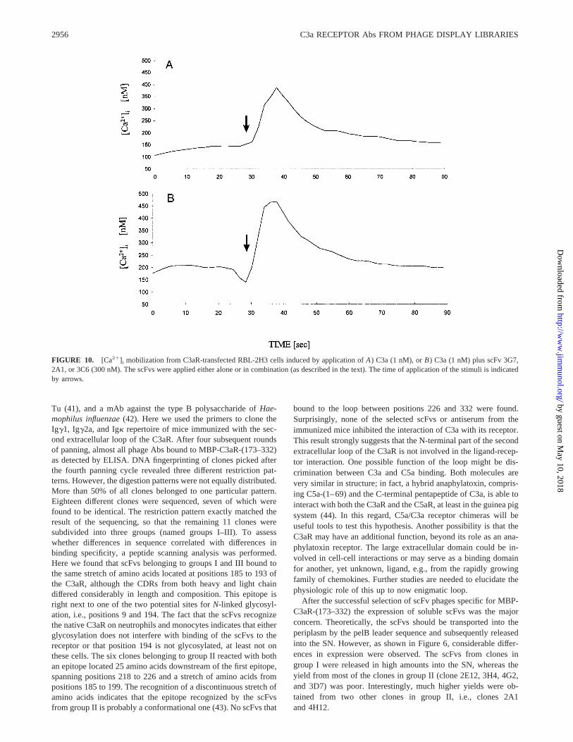
To analyze whether the scFvs are C3aR agonists, we tested theirability to directly stimulate Ca21 mobilization in RBL-2H3 C3aRcells. Conversely, to determine whether the scFvs are C3aR an-tagonists we tested the ability of the scFvs to inhibit C3a-stimu-lated Ca21 mobilization from RBL-2H3 C3aR cells.
First, the scFvs were incubated with the C3aR-transfected RBL-2H3, either individually or in combination (clone 3G71 2A1 orclone 3C61 2A1). The scFvs did not appear to function as re-ceptor agonists, as we did not observe any increase in [Ca21]i (datanot shown) from the cells challenged with the scFvs. Then, thescFvs were added to the cells either 5 min before addition of theC3a stimulus or simultaneously with C3a. None of the scFv Absaffected C3a-stimulated Ca21 mobilization in RBL-2H3 C3aRcells regardless of whether they were applied alone or in combi-nation (Fig. 10). In addition, we tested the antisera obtained fromthe three different mice used for the generation of the libraries(diluted 1/10). Again, no inhibition of the functional response wasobserved, indicating that the N-terminal part of the second extra-cellular loop is probably not involved in ligand binding. We alsotested the ability of C3a to reduce the fluorescence signal obtainedby the different scFvs in immunofluorescence microscopy usingC3aR-transfected RBL-2H3 cells. No detectable difference wasobserved between samples with or without competing C3a (data
FIGURE 8. The scFvs bind to human neutrophils and monocytes, but not to lymphocytes, platelets, or erythrocytes. Peripheral blood cells were purifiedusing Polymorphprep.A, DiffQuick stain of purified PMN.B, Phase contrast view of purified PMN.C, scFv immunostaining of purified PMN. Please notethat every PMN shown inB gives a positive signal, whereas the erythrocytes are not stained.D, DiffQuick staining of purified monocytes (long black arrow),lymphocytes (short black arrow), and platelets.E, Phase contrast view of purified monocytes (long black arrow), lymphocytes (short black arrow), andplatelets (white arrow).F, scFv immunostaining of monocytes, lymphocytes, and platelets. Only the monocytes were recognized by the scFvs, whereas noimmunofluorescence signal was obtained from lymphocytes or platelets.
2954 C3a RECEPTOR Abs FROM PHAGE DISPLAY LIBRARIES
by guest on May 10, 2018
http://ww
w.jim
munol.org/
Dow
nloaded from

not shown). The fact that a threefold molar excess of C3a (1mMvs 300 nM scFvs), which binds with high affinity to the C3aR (Kd
1 nM), did not displace the scFvs from the receptor implies that thebinding site of C3a and that of the scFvs are in distinct regions.
DiscussionThe hC3aR has recently been cloned by us and others (16, 17, 18).The cDNA clone encodes a protein with 482 amino acids and amolecular mass of 53,864 Da. In comparison, the C5aR contains350 amino acids with a calculated molecular mass of only 39,320Da. The difference in size between these two anaphylatoxin recep-tors is due to the difference in the length of the second extracellularloop, which comprises 175 amino acids in the C3aR, but only 22amino acids in the C5aR. This loop is unique within the largeseven-transmembrane domain receptor superfamily. In fact, no ho-mology to any known protein was found when scanning theSwissProt databank. This feature renders this part of the receptoran interesting candidate for the production of Abs, minimizing theprobability of cross-reactivities of such Abs to other proteins.
We decided to clone Ab fragments from combinatorial phagelibraries, since the generation of C5aR-specific mAbs using hy-bridoma technology had only limited success. The approach weused to generate anti-C3aR scFvs, immunization with a C3aR sec-
ond extracellular loop fusion protein, has successfully been used togenerate polyclonal Abs to this receptor (9, 17). Traditional hy-bridoma technology allows for only a limited sampling of the im-mune repertoire. In contrast, cloning of the repertoire inE. colipotentially allows for a much more extensive survey of the im-mune response, essential for the selection of rare Abs (21). Weconstructed combinatorial scFv phage libraries using primers de-signed to match the V region of murine heavy and light chainsubgroups (37). To cover a maximum of sequences with a mini-mum of different primers, up to two degenerations were introducedin each of the seven VH and VL primers. In contrast to the primersdescribed by Huse et al. (38), which cover only 58.5% of theavailable VH sequences at a homology level$82% (39), the newlydesigned primers cover 99% of both the VH and the VL sequenceswith a homology level.86%. Optimized primer sequences formouse scFv repertoires have been described previously (39). How-ever, since no degenerations were used, 10 or nine different VH- orVL-specific primers had to be designed to match the VH/VL geneswith high homologies. In consequence, more PCR reactions had tobe performed to construct the libraries.
We found the newly designed primers effective in PCR cloningof a panel of different mAbs, i.e., neoepitope-specific mAbsagainst both hC3a and hC5a (40), a mAb against elongation factor
FIGURE 9. Epitope mapping of scFv 3G7 (A; group I) and scFv 2A1 (C; group II) by SPOTscan analysis. Fifteen-mer peptides, which span the wholesequence of the second extracellular loop of the C3aR, i.e., C3aR-(173–332), were spotted on a cellulose sheet, i.e., 50 different peptides with an offset ofthree residues. For better orientation, the position of every 10th peptide spot is depicted inA andC by Arabic numerals. InB, both the sequence of the secondextracellular C3aR loop from positions 173 to 262 and the overlapping 15-mer peptides (P1–P26) are depicted. Peptides P27 to P50, spanning C3aR263–332, have been omitted, since none of the scFvs bound to peptides within the region (as shown inA andC). The consensus motifs recognized by thescFvs are indicated both in boldface and by shaded boxes. InA, peptides P3 to P5 are depicted (with the consensus motif in boldface and with shaded boxes)corresponding to the three dark spots recognized by scFv 3G7. The three faint spots inA (peptides P14–P16) are due to incomplete stripping of themembrane. The scFv 3C6 (group III) showed exactly the same binding pattern as that described for scFv 3G7 (data not shown). InC, the reactivity of scFv2A1 with the C3aR-derived peptides is shown. Four dark spots can be seen, marking peptide P5 (on theleft) and peptides P14 to P16 (on theright). Thesequences of peptides P14 to P16 (with the consensus motif indicated in boldface and by shaded boxes) are shown on thetop of the three dark spots.
2955The Journal of Immunology
by guest on May 10, 2018
http://ww
w.jim
munol.org/
Dow
nloaded from
Tu (41), and a mAb against the type B polysaccharide ofHae-mophilus influenzae(42). Here we used the primers to clone theIgg1, Igg2a, and Igk repertoire of mice immunized with the sec-ond extracellular loop of the C3aR. After four subsequent roundsof panning, almost all phage Abs bound to MBP-C3aR-(173–332)as detected by ELISA. DNA fingerprinting of clones picked afterthe fourth panning cycle revealed three different restriction pat-terns. However, the digestion patterns were not equally distributed.More than 50% of all clones belonged to one particular pattern.Eighteen different clones were sequenced, seven of which werefound to be identical. The restriction pattern exactly matched theresult of the sequencing, so that the remaining 11 clones weresubdivided into three groups (named groups I–III). To assesswhether differences in sequence correlated with differences inbinding specificity, a peptide scanning analysis was performed.Here we found that scFvs belonging to groups I and III bound tothe same stretch of amino acids located at positions 185 to 193 ofthe C3aR, although the CDRs from both heavy and light chaindiffered considerably in length and composition. This epitope isright next to one of the two potential sites forN-linked glycosyl-ation, i.e., positions 9 and 194. The fact that the scFvs recognizethe native C3aR on neutrophils and monocytes indicates that eitherglycosylation does not interfere with binding of the scFvs to thereceptor or that position 194 is not glycosylated, at least not onthese cells. The six clones belonging to group II reacted with bothan epitope located 25 amino acids downstream of the first epitope,spanning positions 218 to 226 and a stretch of amino acids frompositions 185 to 199. The recognition of a discontinuous stretch ofamino acids indicates that the epitope recognized by the scFvsfrom group II is probably a conformational one (43). No scFvs that
bound to the loop between positions 226 and 332 were found.Surprisingly, none of the selected scFvs or antiserum from theimmunized mice inhibited the interaction of C3a with its receptor.This result strongly suggests that the N-terminal part of the secondextracellular loop of the C3aR is not involved in the ligand-recep-tor interaction. One possible function of the loop might be dis-crimination between C3a and C5a binding. Both molecules arevery similar in structure; in fact, a hybrid anaphylatoxin, compris-ing C5a-(1–69) and the C-terminal pentapeptide of C3a, is able tointeract with both the C3aR and the C5aR, at least in the guinea pigsystem (44). In this regard, C5a/C3a receptor chimeras will beuseful tools to test this hypothesis. Another possibility is that theC3aR may have an additional function, beyond its role as an ana-phylatoxin receptor. The large extracellular domain could be in-volved in cell-cell interactions or may serve as a binding domainfor another, yet unknown, ligand, e.g., from the rapidly growingfamily of chemokines. Further studies are needed to elucidate thephysiologic role of this up to now enigmatic loop.
After the successful selection of scFv phages specific for MBP-C3aR-(173–332) the expression of soluble scFvs was the majorconcern. Theoretically, the scFvs should be transported into theperiplasm by the pelB leader sequence and subsequently releasedinto the SN. However, as shown in Figure 6, considerable differ-ences in expression were observed. The scFvs from clones ingroup I were released in high amounts into the SN, whereas theyield from most of the clones in group II (clone 2E12, 3H4, 4G2,and 3D7) was poor. Interestingly, much higher yields were ob-tained from two other clones in group II, i.e., clones 2A1and 4H12.
FIGURE 10. [Ca21]i mobilization from C3aR-transfected RBL-2H3 cells induced by application ofA) C3a (1 nM), orB) C3a (1 nM) plus scFv 3G7,2A1, or 3C6 (300 nM). The scFvs were applied either alone or in combination (as described in the text). The time of application of the stimuli is indicatedby arrows.
2956 C3a RECEPTOR Abs FROM PHAGE DISPLAY LIBRARIES
by guest on May 10, 2018
http://ww
w.jim
munol.org/
Dow
nloaded from

Recently, we and others have shown that C3a is able to induceboth chemotaxis and [Ca21]i release from human mast cells (4,11). In addition, C3aR have been demonstrated on the mast cellline HMC-1 by competitive binding studies (10). Using the se-lected scFvs, C3aR expression on HMC-1 cells could beconfirmed.
Using C3aR-specific scFvs, we could demonstrate C3aR-posi-tive cells at the single cell stage. Preparations of peripheral bloodleukocytes obtained using Polymorphprep are contaminated witherythrocytes, platelets, and eosinophils that may affect both[125I]C3a binding and functional studies. However, combiningthree different methods, i.e., DiffQuick cell staining, phase contrastmicroscopy, and immunofluorescence microscopy, binding ofC3aR specific scFvs to individual monocytes and neutrophilscould be detected.
Similar results were obtained in a parallel study, showing thatneutrophils and monocytes express the C3aR. In that study, func-tional assays and Northern blot hybridization on highly purifiedperipheral blood leukocytes, i.e., neutrophils, eosinophils, mono-cytes, and B and T lymphocytes were combined with flow cyto-metric analysis using a polyclonal rabbit C3aR second extracellu-lar loop antiserum (9).
The published data concerning the presence of C3aR on humanplatelets are inconclusive. While Polley and Nachmann (45) re-ported activation of human platelets by both C3a and C3adesArg,others have convincingly demonstrated that human platelets do notexpress the C3aR (46–48). Our data confirm the results obtainedin the latter studies and provide additional proof that no specificC3aR occurs on human platelets.
In addition, we found no C3aR expression on lymphocytes.These data are in accordance with the results of a recent study, inwhich we found no C3aRs on unchallenged, circulating T and Bcells at either the protein or the mRNA level (9). On the otherhand, Northern hybridization revealed C3aR mRNA in lymphnodes (16–18). The key to resolve these apparently inconsistentresults may be to look at the status of lymphocyte activation. It iswell known that Ag-activated B or T lymphocytes differ substan-tially in both their receptor and cytokine expression compared withresting lymphocytes.
Indeed, recent data suggest C3aR expression in activated tonsil-derived B cells (13). However, since both C3a and C3adesArgexhibited similar suppressive effects in these cells, it is question-able whether these effects are receptor mediated or due to unspe-cific ionic interactions with the cell surface.
Finally, we found that C3aR are not expressed on human eryth-rocytes. These cells do express a variety of different complementreceptors, such as decay accelerating factor (DAF) (CD55), ho-mologous restriction factor (HRF) and homologous restriction fac-tor 20 (HRF20) (CD59), and complement receptor 1 (CD35) (49).
In summary, we have generated a panel of C3aR-specific scFvfragments from combinatorial Ab phage libraries that bind to twodistinct regions within the large extracellular loop of the C3aR.They do not inhibit the binding or functional activity of C3a to itsreceptor, indicating that the N-terminus of the second extracellularloop is most likely not involved in ligand binding. In immunoflu-orescence studies the existence of C3aR on human neutrophils andmonocytes could be demonstrated. No C3aR were detected on hu-man lymphocytes, platelets, or erythrocytes.
These well-defined Ab fragments offer the opportunity to studyC3aR expression in different tissues under both physiologic andpathophysiologic conditions and may help elucidate the roles ofC3a and its receptor in the immune network.
AcknowledgmentsWe thank G. Winter for providing the pHEN-1 vector, J. H. Butterfield forproviding the HMC-1 cell line, R. S. Ames for providing the C3aR-trans-fected RBL-2H3 cell line, and P. Monk, M. D. Barker, and J. E. Pease forproviding the C5aR-transfected RBL-2H3 cells. In addition, we thank R. S.Ames for critical reading of the manuscript. We are very grateful for thecontinuous support from D. Bitter-Suermann.
References1. Hugli, T. E. 1990. Structure and function of C3a anaphylatoxin.Curr. Top. Mi-
crobiol. Immunol. 153:181.2. Bischoff, S. C., A. L. de-Weck, and C. A. Dahinden. 1990. Interleukin 3 and
granulocyte/macrophage-colony-stimulating factor render human basophils re-sponsive to low concentrations of complement component C3a.Proc. Natl. Acad.Sci. USA 87:6813.
3. Daffern, P. J., P. H. Pfeifer, J. A. Ember, and T. E. Hugli. 1995. C3a is a che-motaxin for human eosinophils but not for neutrophils. I. C3a stimulation ofneutrophils is secondary to eosinophil activation.J. Exp. Med. 181:2119.
4. Nilsson, G., M. Johnell, C. H. Hammer, H. L. Tiffany, D. Nilsson, D. D. Metcalfe,A. Siegbahn, and P. M. Murphy. 1996. C3a and C5a are chemotaxins for humanmast cells and act through distinct receptors via a pertussis toxin-sensitive signaltransduction pathway.J. Immunol. 157:1693.
5. Ehrengruber, M. U., T. Geiser, and D. A. Deranleau. 1994. Activation of humanneutrophils by C3a and C5A: comparison of the effects on shape changes, che-motaxis, secretion, and respiratory burst.FEBS Lett. 346:181.
6. Elsner, J., M. Oppermann, W. Czech, G. Dobos, E. Schopf, J. Norgauer, andA. Kapp. 1994. C3a activates reactive oxygen radical species production andintracellular calcium transients in human eosinophils.Eur. J. Immunol. 24:518.
7. Kretzschmar, T., A. Jeromin, C. Gietz, W. Bautsch, A. Klos, J. Kohl,G. Rechkemmer, and D. Bitter Suermann. 1993. Chronic myelogenous leukemia-derived basophilic granulocytes express a functional active receptor for the ana-phylatoxin C3a.Eur. J. Immunol. 23:558.
8. Takafuji, S., K. Tadokoro, K. Ito, and C. A. Dahinden. 1994. Degranulation fromhuman eosinophils stimulated with C3a and C5a.Int. Arch. Allergy Immunol.104:27.
9. Martin, U., D. Bock, L. Arseniev, M. A. Tornetta, R. S. Ames, W. Bautsch, J.Kohl, A. Ganser, and A. Klos. 1997. The human C3a-receptor is expressed onneutrophils and monocytes but not on B-or T-lymphocytes.J. Exp. Med. 186:199.
10. Legler, D. F., M. Loetscher, S. A. Jones, C. A. Dahinden, M. Arock, andB. Moser. 1996. Expression of high- and low-affinity receptors for C3a on thehuman mast cell line, HMC-1.Eur. J. Immunol. 26:753.
11. Hartmann, K., B. M. Henz, S. Kruger-Krasagakes, J. Kohl, R. Burger, S. Guhl,I. Haase, U. Lippert, and T. Zuberbier. 1997. C3a and C5a stimulate chemotaxisof human mast cells.Blood 89:2863.
12. Takabayashi, T., E. Vannier, B. D. Clark, N. H. Margolis, C. Dinarello,J. F. Burke, and J. A. Gelfand. 1996. A new biologic role for C3a and C3adesArg.J. Immunol. 156:3455.
13. Fischer, W. H., and T. E. Hugli. 1997. Regulation of B cell functions by C3a andC3adesArg. J. Immunol. 159:4279.
14. Gerard, N. P., and C. Gerard. 1991. The chemotactic receptor for human C5aanaphylatoxin.Nature 349:614.
15. Boulay, F., L. Mery, M. Tardif, L. Brouchon, and P. Vignais. 1991. Expressioncloning of a receptor for C5a anaphylatoxin on differentiated HL-60 cells.Bio-chemistry 30:2993.
16. Crass, T., U. Raffetseder, U. Martin, M. Grove, A. Klos, J. Kohl, and W. Bautsch.1996. Expression cloning of the human C3a anaphylatoxin receptor (C3aR) fromdifferentiated U-937 cells.Eur. J. Immunol. 26:1944.
17. Roglic, A., E. R. Prossnitz, S. L. Cavanagh, Z. Pan, A. Zou, and R. D. Ye. 1996.cDNA cloning of a novel G protein-coupled receptor with a large extracellularloop structure.Biochim. Biophys. Acta 1305:39.
18. Ames, R., Y. Li, H. M. Sarau, P. Nuthulaganti, J. J. Foley, C. Ellis, Z. Zeng,K. Su, A. J. Jurewicz, R. P. Hertzberg, D. J. Bergsma, and C. Kumar. 1996.Molecular cloning and characterization of the human anaphylatoxin C3a receptor.J. Biol. Chem. 271:20231.
19. Watanabe, H., M. Kuraya, R. Kasukawa, H. Yanagisawa, M. Yanagisawa, andM. Fujita. 1995. Analysis of C5a receptor by monoclonal antibody.J. Immunol.Methods 185:19.
20. Oppermann, M., U. Raedt, T. Hebell, B. Schmidt, B. Zimmermann, and O. Gotze.1993. Probing the human receptor for C5a anaphylatoxin with site-directed an-tibodies: identification of a potential ligand binding site on the NH2-terminaldomain.J. Immunol. 151:3785.
21. Winter, G., A. D. Griffiths, R. E. Hawkins, and H. R. Hoogenboom. 1994. Mak-ing antibodies by phage display technology.Annu. Rev. Immunol. 12:433.
22. Ames, R. S., M. A. Tornetta, C. S. Jones, and P. Tsui. 1994. Isolation of neu-tralizing anti-C5a monoclonal antibodies from a filamentous phage monovalentFab display library [published erratum appears in J. Immunol. 1994 Jul15;153(2):910].J. Immunol. 152:4572.
23. Burton, D., and C. F. Barbas III. 1994. Human antibodies from combinatoriallibraries.Adv. Immunol. 57:191.
24. Sambrook, J., E. F. Fritsch, and T. Maniatis. 1989.Molecular Cloning: A Lab-oratory Manual. Cold Spring Harbor Laboratory Press, Cold Spring Harbor, NY.
25. Dower, W. J., J. F. Miller, and C. W. Ragsdale. 1988. High efficiency transfor-mation ofE. coli by high voltage electroporation.Nucleic Acids Res. 16:6127.
2957The Journal of Immunology
by guest on May 10, 2018
http://ww
w.jim
munol.org/
Dow
nloaded from

26. Chomczynski, P., and J. Sacchi. 1987. Single-step method of RNA isolation byacid guanidinium thiocyanate-phenol-chloroform extraction.Anal. Biochem. 162:156.
27. Marks, J. D., H. R. Hoogenboom, T. P. Bonnert, J. McCafferty, A. D. Griffiths,and G. Winter. 1991. By-passing immunization: human antibodies from V-genelibraries displayed on phage.J. Mol. Biol. 222:581.
28. Hennecke, M., A. Kola, M. Baensch, A. Wrede, A. Klos, W. Bautsch, and J.Kohl. 1997. A selection system to study C5a-C5a-receptor interactions: phagedisplay of a novel C5a anaphylatoxin, Fos-C5aAla27. Gene 184:263.
29. Ward, E. S., D. Gussow, A. D. Griffith, P. T. Jones, and G. Winter. 1989. Bindingactivities of a repertoire of single immunoglobulin variable domains secretedfrom Escherichia coli.Nature 341:544.
30. Bock, D., U. Martin, S. Gartner, C. Rheinheimer, U. Raffetseder, L. Arseniev,M. D. Barker, P. N. Monk, W. Bautsch, J. Kohl, and A. Klos. 1997. The C-terminus of the human C5a receptor (CD88) is required for normal ligand-de-pendent receptor internalization.Eur. J. Immunol. 27:1522.
31. Monk, P. N., M. D. Barker, L. J. Partridge, and J. E. Pease. 1995. Mutation ofglutamate 199 of the human C5a receptor defines a binding site for ligand distinctfrom the receptor N terminus.J. Biol. Chem. 270:16625.
32. Zanker, B., H. Rasokat, U. Hadding, and D. Bitter-Suermann. 1982. C3a inducedactivation and stimulus specific reversible desensitization of guinea pig platelets.Agents Actions 11(Suppl.):147.
33. Frank, R. 1992. Spot-Synthesis: an easy technique for the positionally address-able, parallel chemical synthesis on a membrane support.Tetrahedron 48:9217.
34. Frank, R., and H. Overwin. 1996. SPOT synthesis: epitope analysis with arraysof synthetic peptides prepared on cellulose membranes. InMethods in MolecularBiology. G. E. Morris, ed. Humana Press, Totowa, NJ, p. 149.
35. Kohl, J. 1997. The anaphylatoxins. InComplement: A Practical Approach.A. W. Dodds and R. B. Sim, eds. IRL Press, Oxford, U.K., p. 135.
36. Grynkiewicz, G., M. Poenie, and R. Y. Tsien 1985:. A new generation of Ca21
indicators with greatly improved fluorescence properties.J. Biol. Chem. 260:3440.
37. Kabat, E. A., T. T. Wu, H. M. Perry, K. S. Gottesmann, and C. Foeller. 1991.Sequences of Proteins of Immunological Interest.U.S. Department of Health andHuman Services, Public Health Service, National Institute of Health, Bethesda,NIH publication 91–3242.
38. Huse, W. D., L. Sastry, S. A. Iverson, A. S. Kang, M. Alting-Mees, D. R. Burton,S. J. Benkovic, and R. Lerner. 1989. Generation of a large combinatorial libraryof the imunoglobulin repertoire in phage lambda.Science 246:1275.
39. Zhou, H., R. J. Fisher, and T. S. Papas. 1994. Optimization of primer sequencesfor mouse scFv repertoire display library construction.Nucleic Acids Res. 22:888.
40. Hartmann, H., B. Lubbers, M. Casaretto, W. Bautsch, A. Klos, and J. Kohl. 1993.Rapid quantification of C3a and C5a using a combination of chromatographic andimmunoassay procedures.J. Immunol. Methods 166:35.
41. Weber, S., F. Lottspeich, and J. Kohl. 1995. An epitope of elongation factor Tuis widely distributed within the bacterial and archaeal domains.J. Bacteriol.175:11.
42. Steinmetz, I., F. Albrecht, S. Hausler, and B. Brennecke. 1994. Monoclonal IgAclass-switch variants against bacterial surface antigens: molecular forms andtransport into murine respiratory secretions.Eur. J. Immunol. 24:2855.
43. Reineke, U., R. Sabat, A. Kramer, R.-D. Stigler, M. Seifert, T. Michel, H.-D.Volk, and J. Schneider-Mergener. 1996. Mapping protein-protein contact sitesusing cellulose bound peptide scans.Mol. Diversity 1:141.
44. Bautsch, W., T. Kretzschmar, T. Stuhmer, A. Kola, M. Emde, J. Kohl, A. Klos,and D. Bitter Suermann. 1992. A recombinant hybrid anaphylatoxin with dualC3a/C5a activity.Biochem. J. 288:261.
45. Polley, M. J., and R. L. Nachman. 1983. Human platelet activation by C3a andC3a des-arg.J. Exp. Med. 158:603.
46. Fukuoka, Y., and T. E. Hugli. 1988. Demonstration of a specific C3a receptor onguinea pig platelets.J. Immunol. 140:3496.
47. Kretzschmar, T., M. Pohl, M. Casaretto, M. Przewosny, W. Bautsch, A. Klos,D. Saunders, and J. Kohl. 1992. Synthetic peptides as antagonists of the anaphy-latoxin C3a.Eur. J. Biochem. 210:185.
48. Gerardy-Schahn, R., D. Ambrosius, D. Saunders, M. Casaretto, C. Mittler,G. Karwath, S. Gorgen, and D. Bitter-Suermann. 1989. Characterization of C3areceptor-proteins on guinea pig platelets and human polymorphonuclear leuko-cytes.Eur. J. Immunol. 19:1095.
49. Weiler, J. M. 1993. Introduction. InComplement in Health and Disease, 2nd Ed.K. Whaley, M. Loos, and J. M. Weiler, eds. Kluwer Academic Publishers, Dor-drecht, The Netherlands, p. 1.
2958 C3a RECEPTOR Abs FROM PHAGE DISPLAY LIBRARIES
by guest on May 10, 2018
http://ww
w.jim
munol.org/
Dow
nloaded from


![Preferential Identification of Agonistic OX40 Antibodies ... · phage display [6,7]. Recently, we invented a novel method for screening millions-diverse antibody repertoires using](https://static.fdocuments.us/doc/165x107/5ea899f26dff2a7ae712e1bc/preferential-identification-of-agonistic-ox40-antibodies-phage-display-67.jpg)

![Development of specific scFv antibodies to detect ...Phage clones displaying specific peptides to NC were obtained according to Ribeiro [12]. 2.3. scFv phage-display library Antibodies](https://static.fdocuments.us/doc/165x107/5eaa547bca83f15a83239fa6/development-of-specific-scfv-antibodies-to-detect-phage-clones-displaying-speciic.jpg)