
by Isabella Kung - Yeditepe University Faculty of … nerves emerge from the vertebral column with...
81
6. December.2013 Friday by Isabella Kung
Transcript of by Isabella Kung - Yeditepe University Faculty of … nerves emerge from the vertebral column with...

Vertebrae + intervertebtal (IV) discs Spine
Omurga
Onurğa
Wirbelsäule
العمود الفقري
Laf dhabar
Main part of the axial skeleton
Mgongo
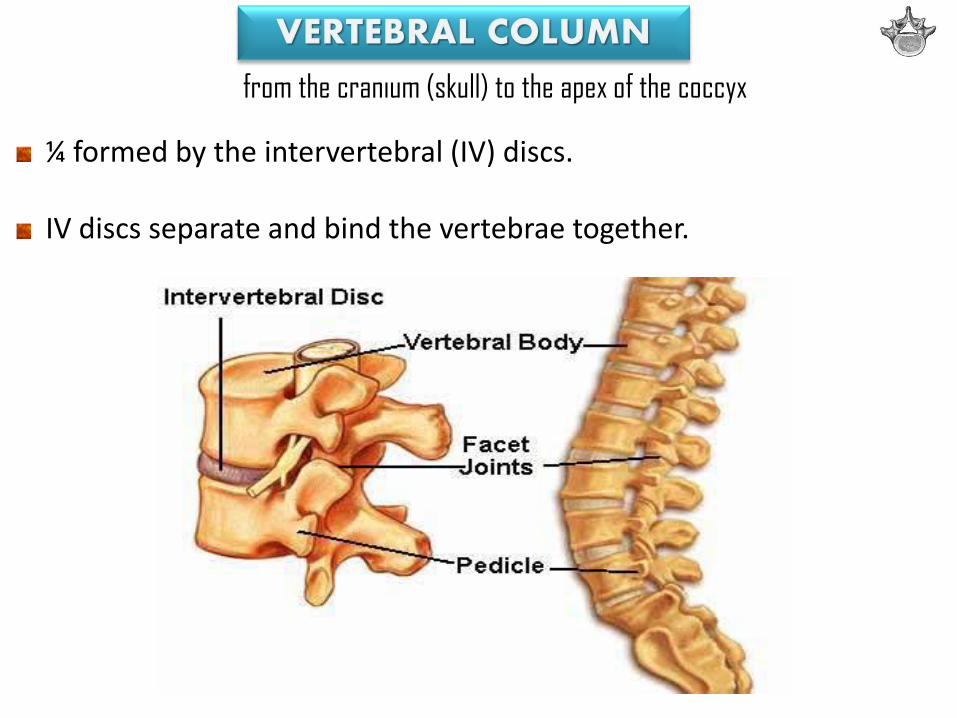
from the cranium (skull) to the apex of the coccyx
¼ formed by the intervertebral (IV) discs. IV discs separate and bind the vertebrae together.
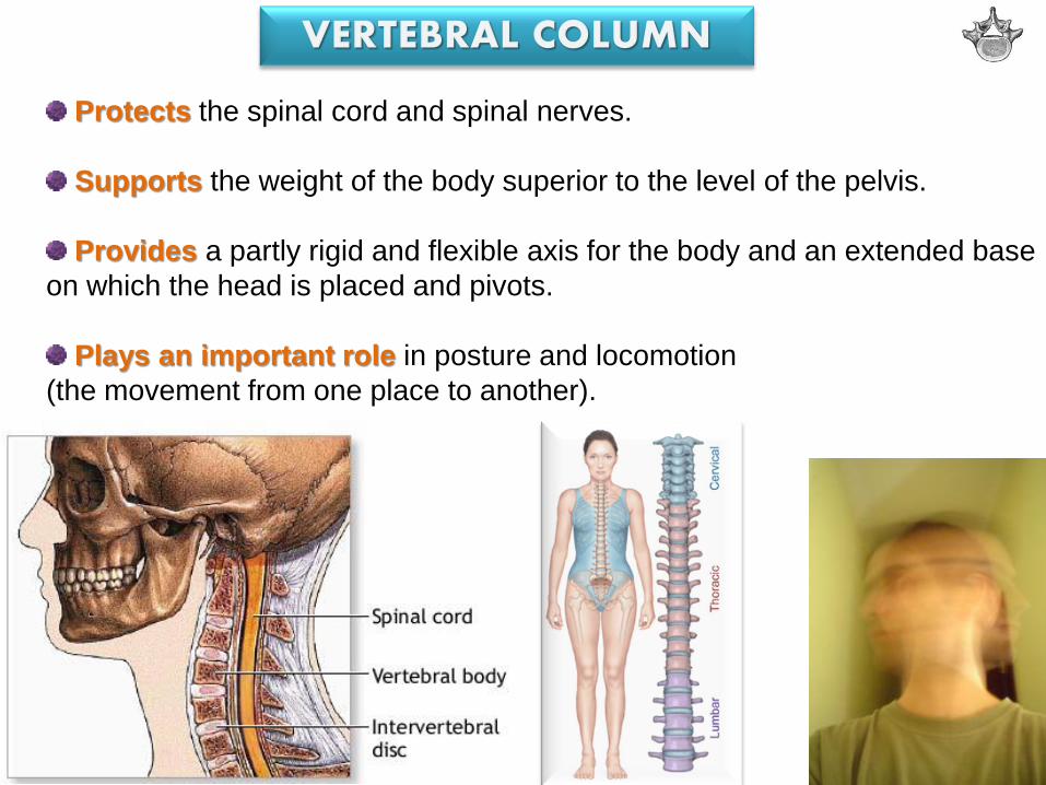
Protects the spinal cord and spinal nerves.
Supports the weight of the body superior to the level of the pelvis.
Provides a partly rigid and flexible axis for the body and an extended base
on which the head is placed and pivots.
Plays an important role in posture and locomotion
(the movement from one place to another).

vertebrae (singular = vertebra)
separated by resilient intervertebral (IV) discs.
Vertebral column flexible
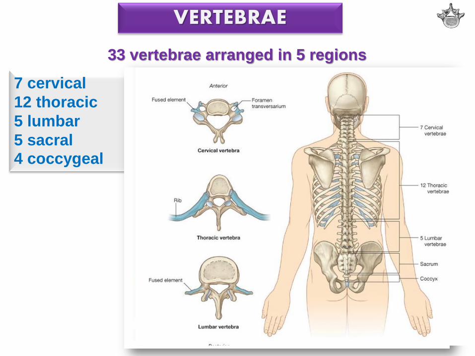
33 vertebrae arranged in 5 regions
7 cervical
12 thoracic
5 lumbar
5 sacral 4 coccygeal
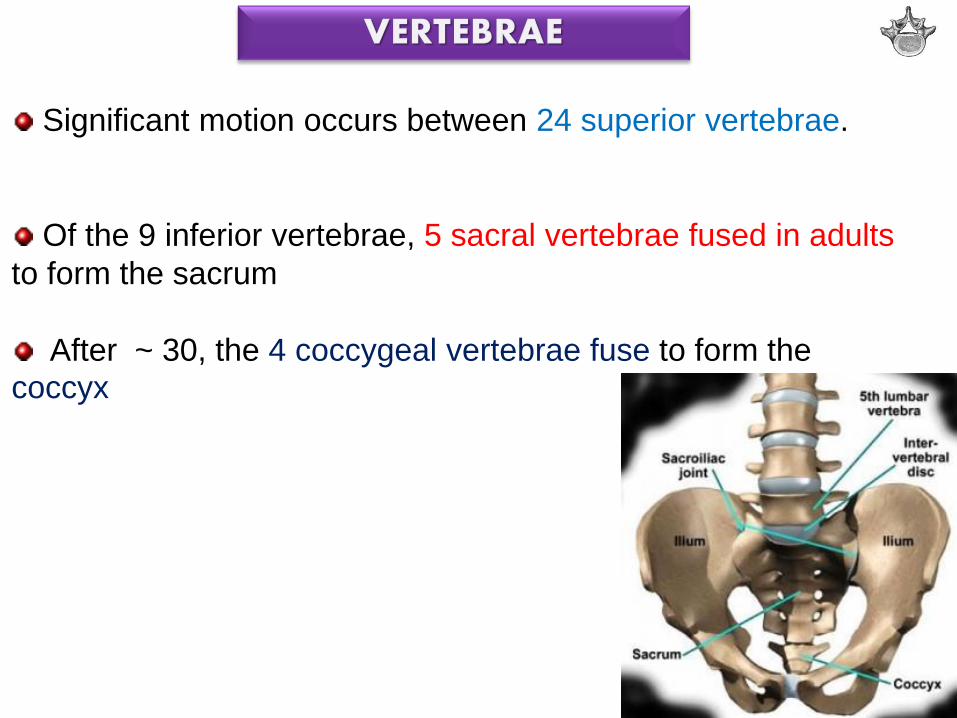
Significant motion occurs between 24 superior vertebrae.
Of the 9 inferior vertebrae, 5 sacral vertebrae fused in adults
to form the sacrum
After ~ 30, the 4 coccygeal vertebrae fuse to form the coccyx
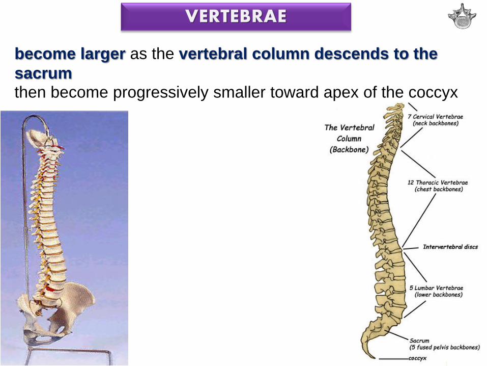
become larger as the vertebral column descends to the
sacrum then become progressively smaller toward apex of the coccyx
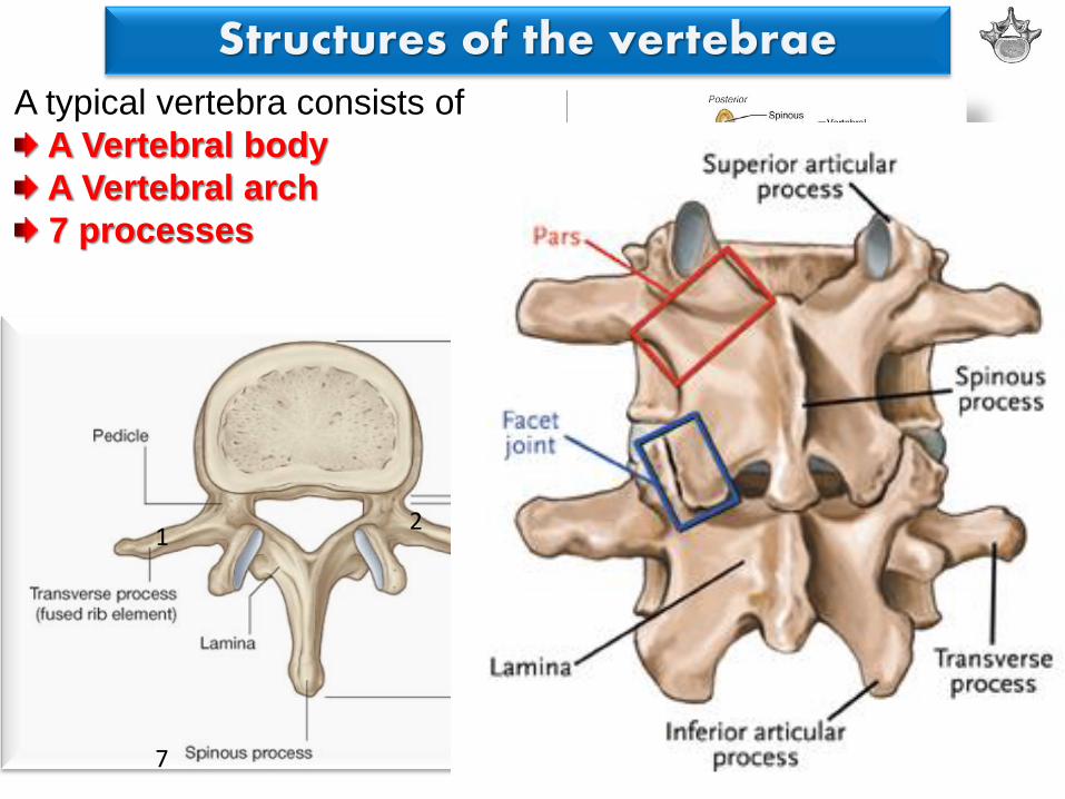
A typical vertebra consists of
A Vertebral body
A Vertebral arch
7 processes
1 2
3-4
5-6
7
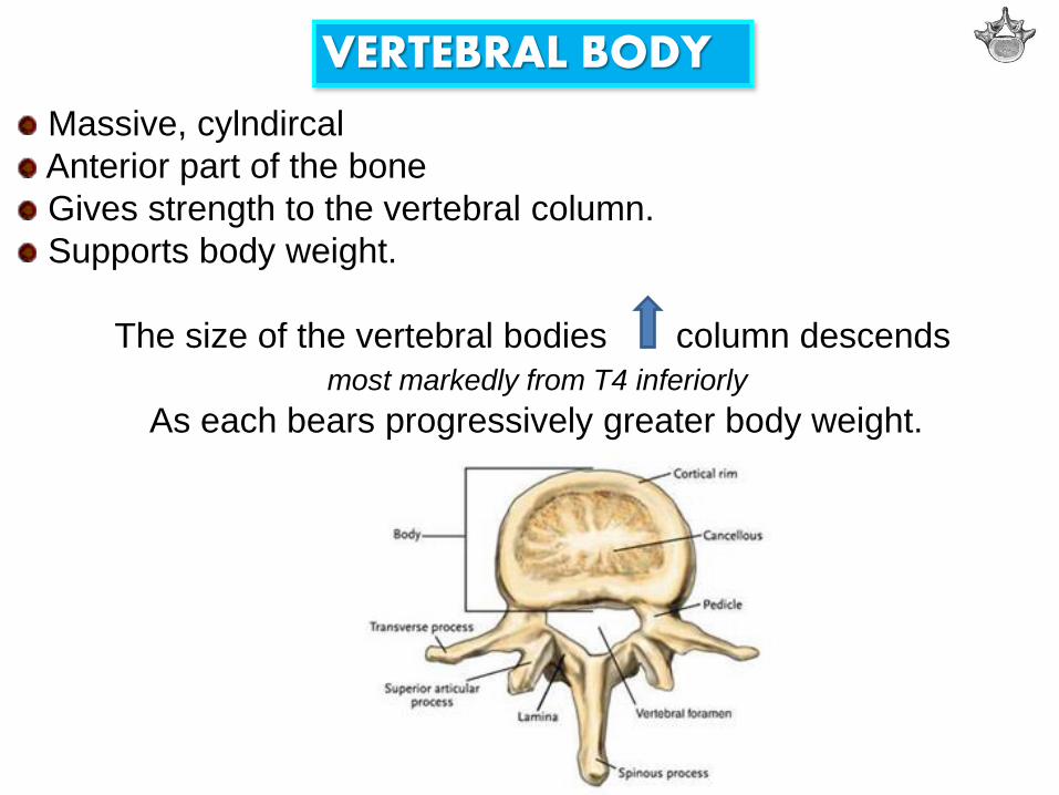
Massive, cylndircal
Anterior part of the bone
Gives strength to the vertebral column.
Supports body weight.
The size of the vertebral bodies column descends
most markedly from T4 inferiorly
As each bears progressively greater body weight.
VERTEBRAL BODY
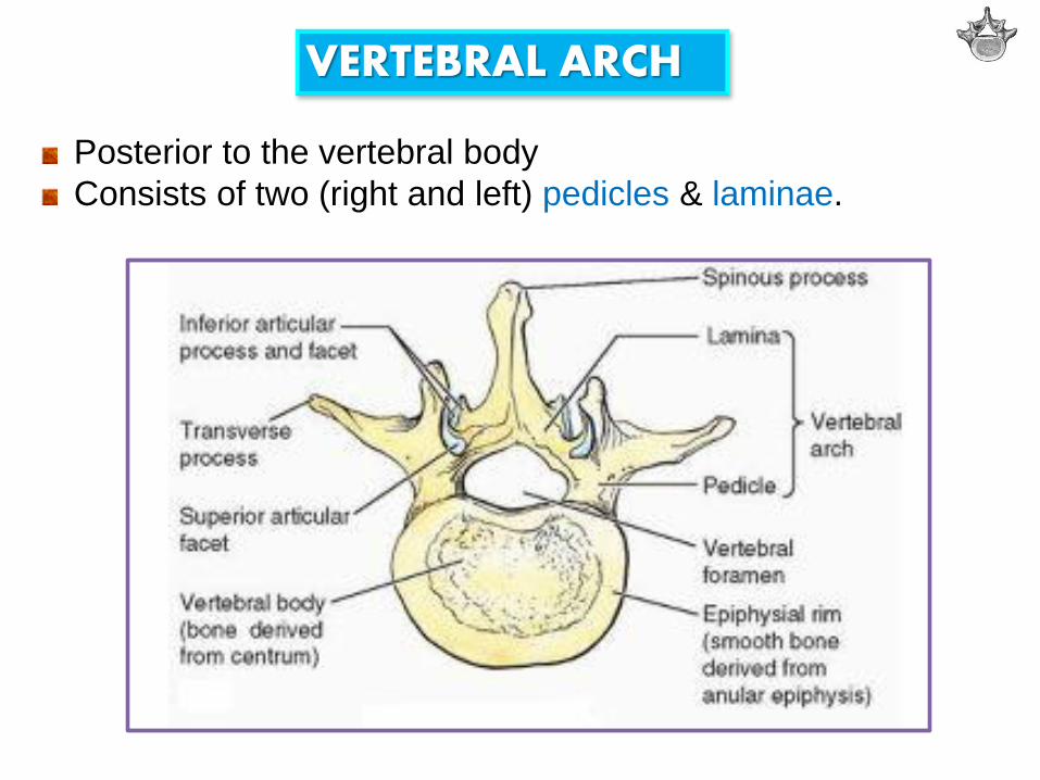
Posterior to the vertebral body
Consists of two (right and left) pedicles & laminae.
VERTEBRAL ARCH

vertebral arch + posterior surface of the vertebral body
walls of vertebral foramen

Succession of vertebral foramina
in the articulated vertebral column
forms
vertebral canal (spinal canal)

Vertebral notches (Incisura vertebralis)
Indentations observed in lateral views of the vertebrae
Superior and inferior to each pedicle
Between the superior and inferior articular processes posteriorly
Between the corresponding projections of the body anteriorly.

The superior and inferior vertebral notches of adjacent
vertebrae and the IV discs form intervertebral foramina
Intervertebral foramina
Spinal (posterior root) ganglia are located
Spinal nerves emerge from the vertebral column with their
accompanying vessels through these foramina.

vertebrae having foramina in their
transverse processes are cervical
vertebrae

articular facets orientation in each region different
Movement needed
articular facets of thoracic vertebrae nearly vertical,
define an arc centered in the IV disc
this arrangement permits rotation and lateral flexion of the vertebral column in this region.

Regional variations in size and shape of the vertebral canal accommodate the varying thickness of the spinal cord.

skeleton of the neck between the cranium & thoracic vertebrae

1) Smallest of the 24 movable vertebrae
2) Relatively larger intervertebral discs discs are thin, but relative to their small size; thick.
FEATURES TYPICAL FOR
CERVICAL VERTEBRAE
3) Greatest range & variety of movement of all the vertebral regions 4) foramen transversarium in the transverse process
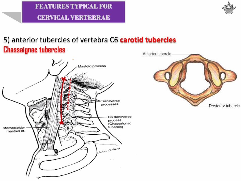
5) anterior tubercles of vertebra C6 carotid tubercles
Chassaignac tubercles
FEATURES TYPICAL FOR
CERVICAL VERTEBRAE
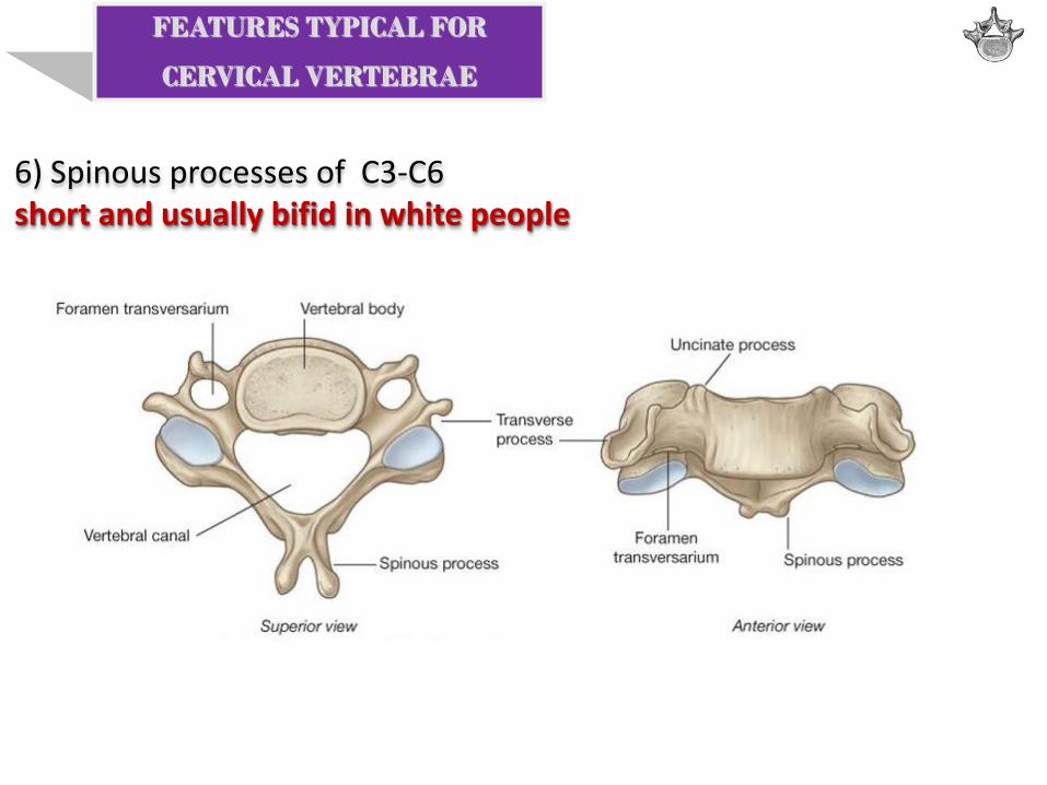
6) Spinous processes of C3-C6 short and usually bifid in white people
FEATURES TYPICAL FOR
CERVICAL VERTEBRAE

Vertebrae C3-C7 typical cervical vertebrae
Large vertebral foramina restricted rotation
superolateral margin uncus of the body uncinate process

C7- vertebra prominens A long spinous process
Most prominent spinous process in 70% of people

No body No spinous process Widest of the cervical vertebrae
The kidney-shaped, concave superior articular surfaces of the lateral masses articulate with occipital condyles.

Anterior and posterior arches a tubercle in the center of its external aspect
extend between the lateral masses forming a complete ring.
Posterior arch A wide groove for the vertebral artery on its superior surface. C1 nerve also runs in this groove.

strongest of the cervical vertebrae C1, carrying the cranium, rotates on C2 (e.g., when a person turns the head to indicate “no”).

The distinguishing feature blunt tooth-like dens Lies anterior to the spinal cord. Serves as the pivot about which the rotation of the head occurs.

large bifid spinous process


The thoracic skeleton includes: 12 pairs of ribs and associated costal cartilages 12 thoracic vertebrae and the intervertebral discs between them Sternum

FEATURES TYPICAL FOR
THORACIC VERTEBRAE
articulation with ribs.
1) Bilateral costal demifacets on the vertebral bodies for articulation with heads of ribs 2) Costal facets on the transverse processes for articulation with tubercles of ribs

FEATURES TYPICAL FOR
THORACIC VERTEBRAE
articulation with ribs.
3) Articular processes of thoracic vertebrae extend vertically with paired, nearly coronally oriented articular facets define an arc.
greatest degree of rotation is permitted here!

4) Heart-shaped bodies 5) Long, inferiorly slanting spinous processes
FEATURES TYPICAL FOR
THORACIC VERTEBRAE

T1-T4 vertebrae share some features of cervical vertebrae. The middle four thoracic vertebrae (T5-T8) demonstrate all the features typical of thoracic vertebrae.

T1 atypical 1. long, horizontal spinous process Vertebra prominens? No. 2. complete costal facet for the 1st rib
3. demifacet for the 2nd rib.
Typical pattern
1+1 costal facet @ transverse processes
0.5+0.5 demifacet
0.5+0.5 demifacet
1+0.5

[T9]-T10 vertebrae No inferior demifacet
1+1 costal facet @ transverse processes
0.5+0.5 demifacet
T11-T12 vertebrae No transverse costal facets 1 complete facet on each side
1+1 demifacet

superior half thoracic in character
costal facets & articular processes inferior half lumbar in character no costal facets articular processes that permit only flexion and extension.
T12

in the lower back between the thorax and sacrum
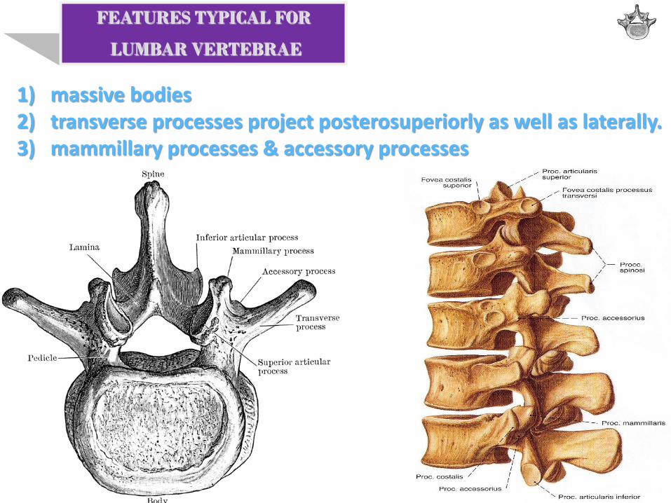
1) massive bodies 2) transverse processes project posterosuperiorly as well as laterally. 3) mammillary processes & accessory processes
FEATURES TYPICAL FOR
LUMBAR VERTEBRAE
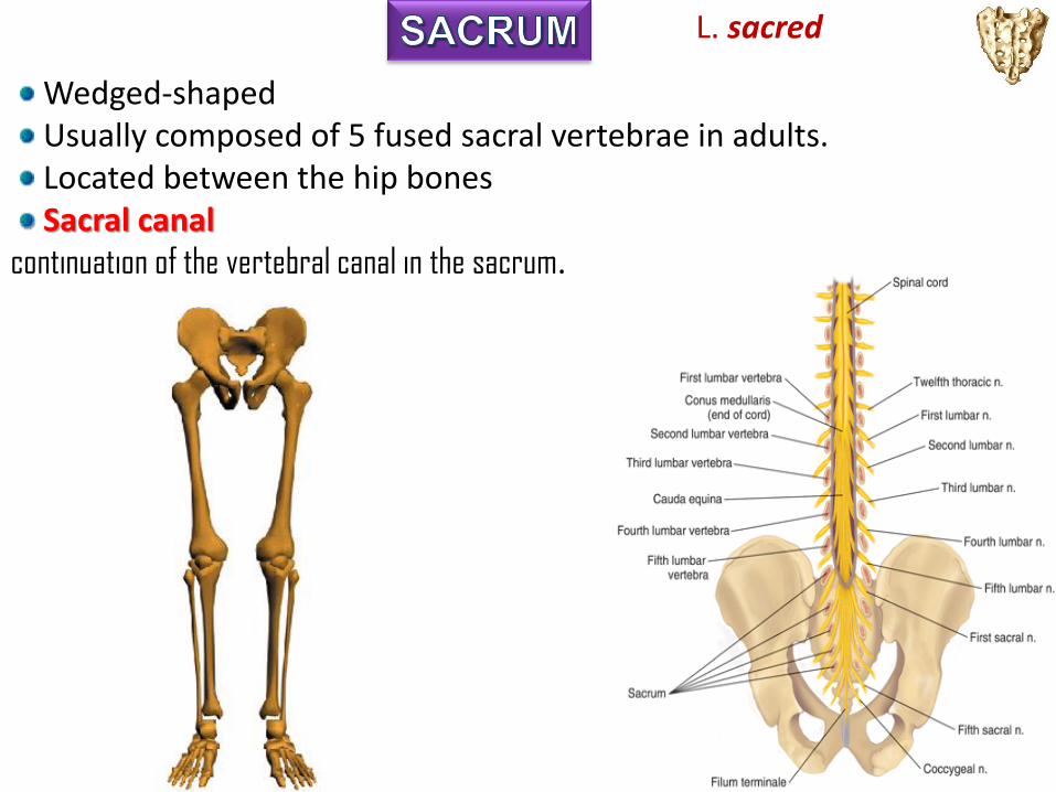
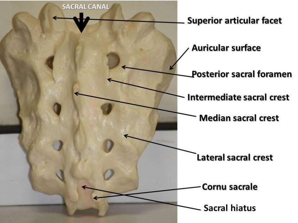
Wedged-shaped Usually composed of 5 fused sacral vertebrae in adults. Located between the hip bones Sacral canal
continuation of the vertebral canal in the sacrum.
L. sacred

On the pelvic and posterior surfaces of the sacrum four pairs of sacral foramina

Anterior projecting edge of the body of the S1 vertebra Sacral promontory (L. mountain ridge)
important obstetrical landmark

The sacrum supports the vertebral column and forms the posterior part of the bony pelvis. The sacrum is tilted so that it articulates with the L5 vertebra at the lumbosacral angle.
Eur Spine J. 2009 Feb;18(2):212-7. Epub 2008 Nov 18. Assessment of lumbosacral kyphosis in spondylolisthesis: a computer-assisted reliability study of six measurement techniques. Glavas P, Mac-Thiong JM, Parent S, de Guise JA, Labelle H.
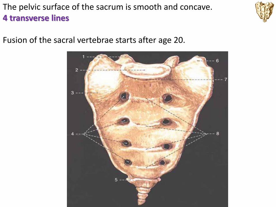
The pelvic surface of the sacrum is smooth and concave. 4 transverse lines Fusion of the sacral vertebrae starts after age 20.

The dorsal surface of the sacrum marked by five prominent longitudinal ridges.
median sacral crest fused rudimentary spinous processes of the superior three or four sacral vertebra

Intermediate sacral crests fused articular processes
Lateral sacral crests tips of the transverse processes of fused sacral vertebrae

Inverted U-shaped sacral hiatus Sacral cornua (L. Horns)
The sacral hiatus leads into the sacral canal. The sacral cornua, representing the inferior articular processes of S5 vertebra, project inferiorly on each side of the sacral hiatus and are a helpful guide to its location.

The superior part of the lateral surface of the sacrum auricular surface

A small triangular bone
Formed by fusion of 4 rudimentary coccygeal vertebrae.
Co1 may remain separate from the fused group.
Rudimentary articular processes @ post. surface

Last 3 coccygeal vertebrae often fuse during middle life
forming a beak-like coccyx
Aging- A single bone!
Muscular attachment!
No contribution to support of the body weight in standing!
Coccydynia

33 32 or 34
race, gender, and developmental factors (genetic and environmental)
32 34
Lumbar sacralization

A CRANIAL SHIFT • A cervical rib articulates with C7 • Rib 12 is small. • L5 partially "sacralized" . • S5 partially freed
B Common arrangement C CAUDAL SHIFT • Rib 12 is large. • A small lumbar rib is present. • S1 partially "lumbarized" . • Co1 is incorporated into the sacrum

1. The neck or cervical spine, curves gently inward (lordosis) 2. The mid back, or thoracic spine, curved outward (kyphosis) 3. The low back, or lumbar spine, also curves inward (lordosis) 4. Pelvic (Sacral) curvature

Ribs (L. costae) are curved, flat bones that form most of
the thoracic cage.
Remarkably light in weight yet highly resilient.
Each rib has a spongy interior containing bone marrow
(hematopoietic tissue), which forms blood cells.
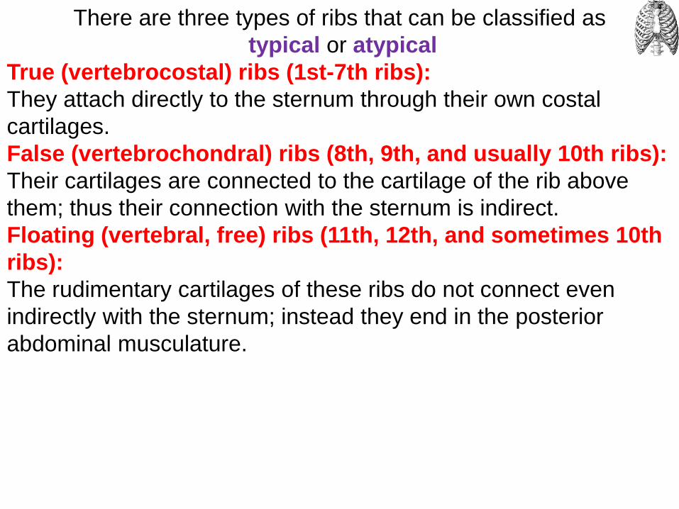
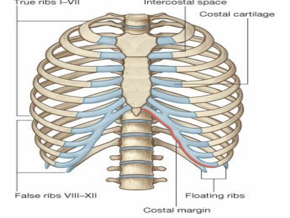
There are three types of ribs that can be classified as
typical or atypical
True (vertebrocostal) ribs (1st-7th ribs):
They attach directly to the sternum through their own costal
cartilages.
False (vertebrochondral) ribs (8th, 9th, and usually 10th ribs):
Their cartilages are connected to the cartilage of the rib above
them; thus their connection with the sternum is indirect.
Floating (vertebral, free) ribs (11th, 12th, and sometimes 10th
ribs):
The rudimentary cartilages of these ribs do not connect even
indirectly with the sternum; instead they end in the posterior
abdominal musculature.

Typical ribs (3rd-9th) have the following components:
Head: wedge-shaped and has two facets, separated by the crest
of the head; one facet for articulation with the numerically
corresponding vertebra and one facet for the vertebra
superior to it.
Neck: connects the head of the rib with the body at the level of
the tubercle.
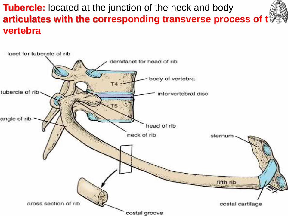
Tubercle: located at the junction of the neck and body
articulates with the corresponding transverse process of the
vertebra
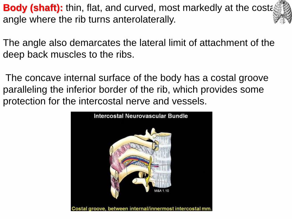
Body (shaft): thin, flat, and curved, most markedly at the costal
angle where the rib turns anterolaterally.
The angle also demarcates the lateral limit of attachment of the
deep back muscles to the ribs.
The concave internal surface of the body has a costal groove
paralleling the inferior border of the rib, which provides some
protection for the intercostal nerve and vessels.


Atypical ribs (1st, 2nd, and 10th-12th) are dissimilar:
The 1st rib is the broadest (i.e., its body is widest and nearly
horizontal), shortest, and most sharply curved of the 7 true ribs.
A single facet on its head for articulation with the T1 vertebra only
2 transversely directed grooves crossing its superior surface for
the subclavian vessels; the grooves are separated by a scalene
tubercle and ridge, to which the anterior scalene muscle is
attached. .

The 2nd rib is has a thinner, less curved body and is substantially
longer than the 1st rib.
Its head has two facets for articulation with the bodies of the T1
and T2 vertebrae.
Main atypical feature is, the tuberosity for serratus anterior a rough area on its upper surfacefrom which part of that muscle originates

10th-12th ribs, like the 1st rib, have only one facet on their
heads and articulate with a single vertebra.
11th and 12th ribs are short and have no neck or tubercle.

Costal cartilages
Prolong the ribs anteriorly
Contribute to the elasticity of the thoracic wall
Provide a flexible attachment for their anterior ends (tips).
The cartilages increase in length through the first 7 and then
gradually decrease.
.

Intercostal spaces Separate the ribs and their costal cartilages from one another. Named according to the rib forming the superior border of the space.
4th intercostal space lies between ribs 4 and 5.
11 intercostal spaces and 11 intercostal nerves intercostal muscles and membranes, and two sets (main and collateral) of intercostal blood vessels and nerves
identified by the same number assigned to the space.
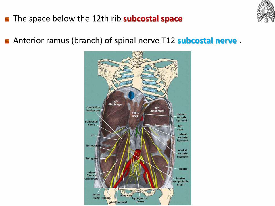
The space below the 12th rib subcostal space Anterior ramus (branch) of spinal nerve T12 subcostal nerve .

The intercostal spaces widest anterolaterally
widen further with inspiration further widened by extension and/or lateral flexion of the thoracic vertebral column to the contralateral side.

Flat, elongated bone Forms the middle of the anterior part of the thoracic cage. Affords protection for mediastinal viscera in general and much of the heart in particular.
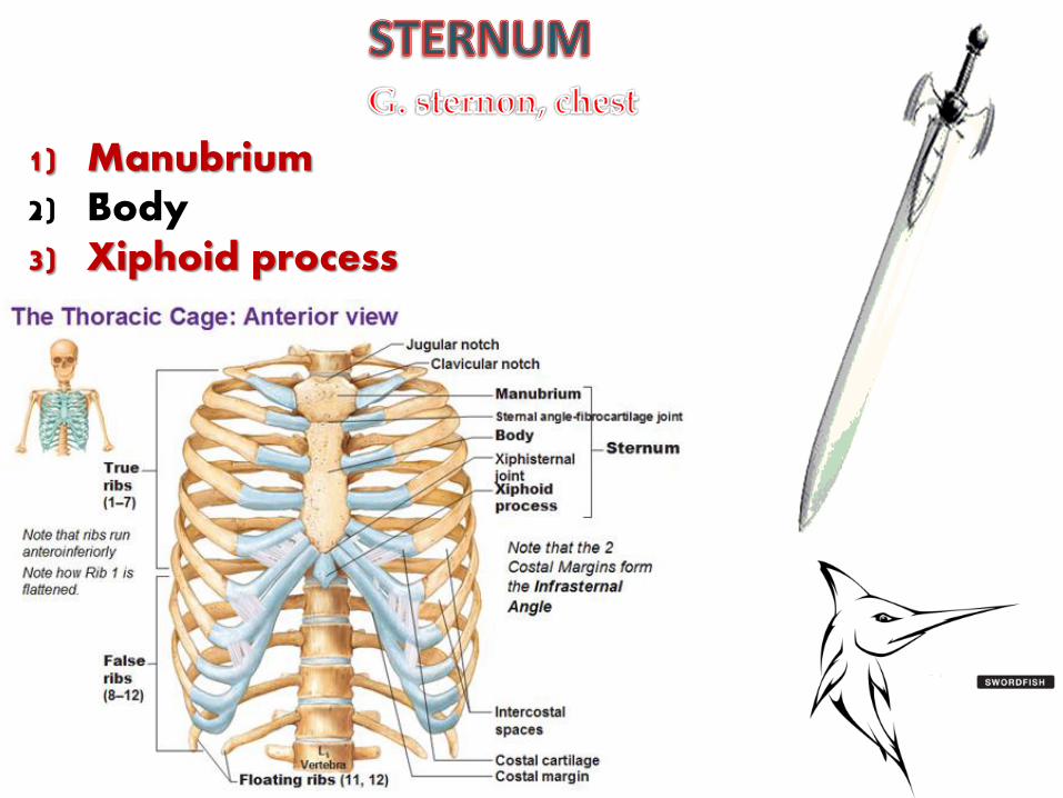
1) Manubrium 2) Body 3) Xiphoid process
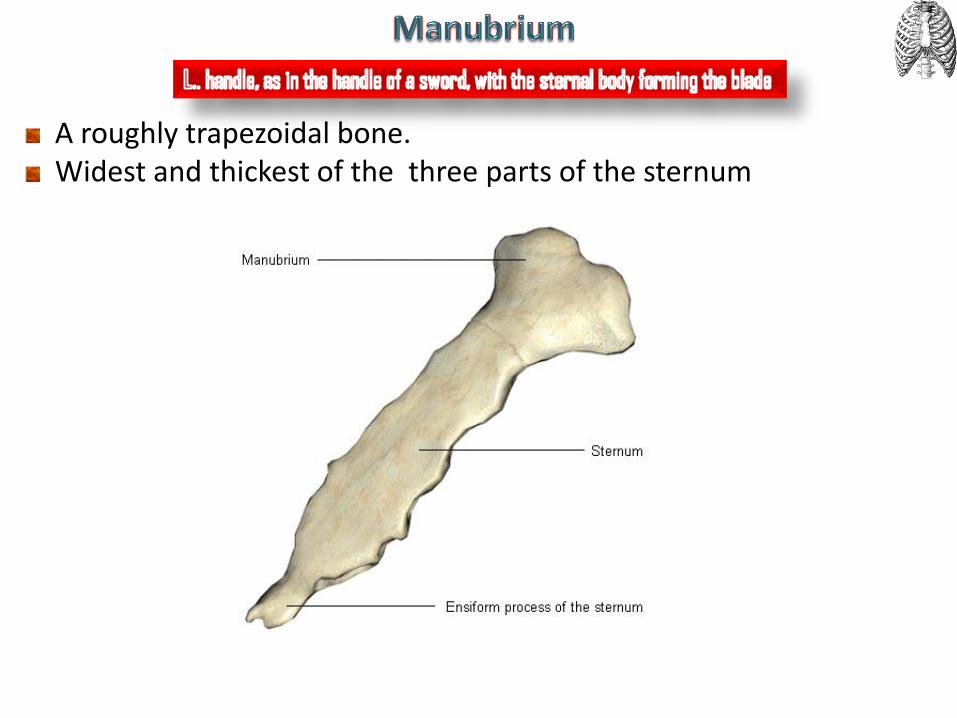
A roughly trapezoidal bone. Widest and thickest of the three parts of the sternum

jugular notch (suprasternal notch)
The easily palpated concave center of superior border of manubrium. Deepened by the medial (sternal) ends of the clavicles, which are much larger than the relatively small clavicular notches in the manubrium that receive them, forming the sternoclavicular (SC) joints.

Inferolateral to the clavicular notch, the costal cartilage of the 1st rib is tightly attached to the lateral border of the manubrium.
synchondrosis of the first rib
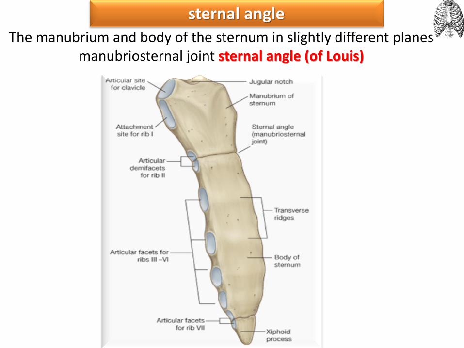
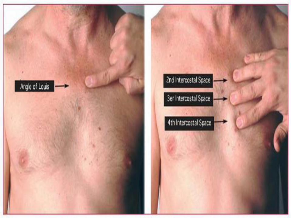
sternal angle
The manubrium and body of the sternum in slightly different planes manubriosternal joint sternal angle (of Louis)

Longer, narrower, and thinner than the manubrium. Located at the level of the T5-T9 vertebrae. Its width varies because of the scalloping of its lateral borders by the
costal notches. .
Body of the sternum (Corpus sterni)
Gladiolus

Smallest and most variable part of the sternum Thin and elongated Inferior end lies at the level of T10 vertebra.
Xiphoid process


79
Jugular (suprasternal)notch:T2 vertebra in male, T4 in female Sternal angle (of Louis) Th 4 vertebra • The border between superior and inferior mediastinum • Overlies the tracheal bifurcation and aortic arch • Useful for counting intercostal spaces (2nd ribs articulate here).

Xiphoid process an important landmark in the median plane
Its junction with the sternal body at the xiphisternal joint inferior limit of the central part of the thoracic cavity
Xiphisternal joint site of the infrasternal angle (subcostal angle)
formed by the right and left costal margins Midline marker for superior limit of the liver, central tendon of the
diaphragm, inferior border of the heart.