Patterns of T cell co-stimulation and co-inhibition in...
47
Universidade de Lisboa Faculdade de Ciências Departamento de Biologia Vegetal Patterns of T cell co-stimulation and co-inhibition in tumor microenvironment conditions, in vitro Dissertação de Mestrado orientada por: Prof. Dra. Maria Margarida Telhada Priv.Doz. Dr. med. habil. Frank Momburg Dissertação Luana Frias Guerra Mestrado em Biologia Molecular e Genética 2015
Transcript of Patterns of T cell co-stimulation and co-inhibition in...

Universidade de Lisboa
Faculdade de Ciências
Departamento de Biologia Vegetal
Patterns of T cell co-stimulation and co-inhibition in
tumor microenvironment conditions, in vitro
Dissertação de Mestrado orientada por:
Prof. Dra. Maria Margarida Telhada
Priv.Doz. Dr. med. habil. Frank Momburg
Dissertação
Luana Frias Guerra
Mestrado em Biologia Molecular e Genética
2015
I
Acknowledgements
Aos meus pais, um obrigada imenso por me darem as ferramentas sem o livro de instruções
ou o livro de instruções sem as ferramentas, mas nunca os dois em simultâneo.
À minha irmã, por ser parte de mim.
À família de cada dia, aos amigos de sempre e aos que se foram juntando a esse sempre, ao
longo do caminho, os que me inspiram e guiam. Não seria metade sem vocês.
To my new friends, the ones that I was lucky to meet and share each moment of my new life,
the reason why these months were pure bliss.
To Frank, who gave me the opportunity to work on what I am passionate about, for his
guidance and insight.
To Alexander and Mostafa, who were utterly patient and without whom I would have not been
able to complete this work.
À professora Margarida Telhada, por ter aceitado ser minha orientadora e pela sua
disponibilidade e apoio.
E, finalmente, mas como se viesse em primeiro, à professora Margarida Gama Carvalho, por
me ter iniciado nesta caminhada e por continuar presente e a acreditar.
II
Abstract
T cells (helper and cytotoxic) are present within the tumor microenvironment and constitute
viable targets for immunotherapeutic approaches against cancer, either by adoptive T cell
transfer or by the use of monoclonal antibodies. The activation, differentiation, function and
survival of those T cells comprehends a complex process and it is regulated by co-stimulatory
and co-inhibitory receptors which modulate the initial T cell receptor (TCR) signal, increasing
or hindering their anti-tumor function, respectively. Furthermore, various factors secreted by
tumor cells or non-lymphoid stromal cells are believed to mostly downmodulate T cell
responses.
In this work the patterns of expression of co-signaling molecules on T cells were assessed
using peripheral blood mononuclear cells (PBMCs) from healthy donors cultured with
transforming growth factor (TGF-β), in order to mimic one of the major immunosuppressive
stimuli of the tumor microenvironment. Noteworthy, all herein reported effects of TGF-β were
entirely dependent on the subsequent T cell activation by an antibody against the CD3ε chain
that is part of the TCR complex. While co-inhibitory receptors such as CTLA4 and PD1 were
upregulated, PD-L1, TIGIT and TIM3 (also co-inhibitory) had their expression decreased.
The transcription factor FOXP3, associated with the regulatory T cell phenotype, was
upregulated by TGF-β. For co-activating receptors, the same condition upregulated 4-1BB,
CD30 and CD28 expression, whereas it decreased OX40, ICOS and DNAM1 and did not it
interfere with CD27 or LIGHT expression. Moreover, alongside TGF-β and anti-CD3,
recombinant proteins, such as 4-1BB or OX40 ligand, were added, in order to simulate
potential therapeutic strategies using agonistic 4-1BB or OX40 antibodies. It was shown that
even though they could induce expression of co-stimulatory receptors such as OX40, CD27
and CD28, there was also CTLA4, PD-1 and TIGIT upregulation.
It seems crucial to unravel the details of the dynamics of the expression of co-signaling
molecules on T cells to improve existent immunotherapies and design new approaches able
to circumvent the immunosuppressive environment, created not only by tumor or stromal
cells, but also by immune cells themselves.
Keywords: cancer, TGF-β, T cells, co-stimulatory receptors, co-inhibitory receptors,
immunotherapy
III
Resumo
No microambiente tumoral, estão presentes linfócitos T (auxiliares ou citotóxicos) e estes
constituem alvos viáveis em imunoterapias para o cancro, quer por transferência adotiva de
linfócitos T, quer pela utilização de anticorpos monoclonais. A ativação, diferenciação, função
e sobrevivência destas células T compreende um processo complexo e é regulada por recetores
coestimuladores e coinibidores, os quais modelam o sinal inicial do recetor de células T
(TCR), amplificando ou diminuindo a sua atividade antitumoral, respetivamente.
Adicionalmente, fatores secretados por células tumorais e do estroma podem contribuir para
a supressão das respostas das células T.
Assim, o padrão de expressão de moléculas cossinalizadoras dos linfócitos T foi estudado
usando células mononucleares do sangue periférico (PBMC) de dadores saudáveis, colocadas
em cultura com TGF-β para mimetizar os estímulos supressivos do microambiente tumoral.
Todos os efeitos do TGF-β aqui reportados foram dependentes da subsequente ativação das
células T por um anticorpo contra a cadeia ε de CD3, que faz parte do complexo TCR.
Enquanto recetores coinibidores como CTLA4 e PD-1 tinham a sua expressão aumentada,
PD-L1, TIM3 e TIGIT (também coinibidores) encontravam-se subexpressos. O fator de
transcrição FOXP3 (associado ao fenótipo de células T reguladoras) estava aumentado na
presença de TGF-β. Já no caso dos recetores coestimuladores avaliados, a expressão de 4-
1BB, CD30 e CD28 aumentou nas células T, mas OX40, ICOS e DNAM1 encontravam-se
subexpressos. Já CD27 e LIGHT não foram afetados pela adição de TGF-β. Adicionalmente,
proteínas recombinantes, ligandos de 4-1BB e OX40, foram adicionadas, na presença de TGF-
β e anti-CD3, para a simulação de potenciais estratégias imunoterapêuticas. Concluiu-se que,
a diferentes níveis, apesar de se ter conseguido induzir a expressão OX40, CD27 e CD28 nas
células T, a expressão de CTLA4, PD-1 e TIGIT estava também aumentada.
Revelar os detalhes da dinâmica da expressão de moléculas cossinalizadoras em linfócitos T
é crucial para aperfeiçoar imunoterapias existentes e criar novas abordagens capazes de
minimizar os efeitos dos estímulos imunossupressivos do ambiente tumoral, criado não apenas
pelas células cancerígenas e do estroma, mas também pelas próprias células do sistema
imunitário.
Palavras-chave: cancro, TGF-β, linfócitos T, recetores coestimuladores, recetores
coinibidores, imunoterapia
IV
Resumo
O cancro é uma doença caracterizada por um crescimento anormal e não controlado de células
que, ao sofrerem mutações no seu DNA, adquirem características que lhes conferem uma
vantagem adaptativa de sobrevivência.
A função do sistema imunitário é contribuir para a homeostasia dos tecidos e, assim, os
linfócitos T exercem um papel crucial no cancro ao reconhecerem e eliminarem células
transformadas.
A ativação, diferenciação, função e sobrevivência das células T é um processo descrito por
um modelo de dois sinais. Isto é, há uma ativação primária através da ligação do recetor de
células T (TCR) ao complexo MHC (major histocompatibility complex) e este sinal é
complementado por recetores coestimuladores e coinibidores. São estas moléculas que,
colocalizadas com o complexo TCR-MHC na sinapse imunológica, ditam o destino das
células T ao modular o sinal do TCR – amplificando o sinal de ativação (estimuladores) ou
diminuindo-o (inibidores). O repertório destas moléculas cossinalizadoras é altamente
versátil, temporal e espacialmente, e responde a mudanças no ambiente dos diferentes tecidos.
Os recetores coestimuladores e coinibidores pertencem, na sua maioria, a duas superfamílias:
IgSF (immunoglobulin superfamily) e TNFRSF (tumor necrosis factor receptor superfamily).
À IgSF pertencem correcetores estimuladores como CD28 e outros coinibidores, como TIGIT
(T cell immunoreceptor with immunoglobulin and ITIM domains), para mencionar alguns
exemplos. Já a superfamília TNFRSF, compreende membros como 4-1BB ou OX40, ambos
coestimuladores.
Dada a capacidade destas moléculas cossinalizadoras modularem a ativação dos linfócitos T
no microambiente tumoral, estes constituem já alvos clínicos de imunoterapias contra o
cancro, como é o caso dos anticorpos monoclonais anti-CTLA4 (cytotoxic T-lymphocyte-
associated antigen) e anti-PD1 (programmed death protein 1), recentemente aprovados para
o tratamento de melanoma metastático. Ambos são recetores coinibidores e os anticorpos
antagonistas utilizados diminuem o fenótipo imunossupressor, nomeadamente ao impedirem
a ativação de células T reguladoras (Treg). No entanto, as possibilidades terapêuticas não se
esgotam aqui. Com a variedade de recetores descritos, há a hipótese de criar novos
tratamentos, recorrendo a combinações entre diferentes alvos, ou apenas melhorar a eficácia

V
e o perfil de toxicidade das terapias existentes. Para isso, é crucial conhecer detalhadamente
o fenótipo de ativação e inibição dos linfócitos T.
Assim, o objetivo deste trabalho é estudar os padrões de expressão de recetores
coestimuladores e coinibidores presentes em células T, inseridas num microambiente tumoral,
in vitro.
As células tumorais encontram-se, normalmente, num ambiente imunossupressor. Produzem
citoquinas, como TGF-β (transforming growth factor β), IL-10 ou prostaglandinas, que
recrutam células imunossupressoras - Tregs ou MDSC (myeloid derived suppressor cells) - as
quais impedem a função citotóxica das células T.
A estratégia experimental consistiu na utilização de células mononucleares do sangue
periférico (PMBC) isoladas de sangue de dadores saudáveis. Estas células foram pré-
cultivadas com TGF-β recombinante humano (rh), de modo a mimetizar o microambiente
tumoral. Posteriormente, adicionou-se um anticorpo anti-CD3ε, a cadeia ε do complexo TCR,
para que se desse a ativação policlonal das células T. A expressão de recetores
(coestimuladores e coinibidores) na superfície dos linfócitos T foi posteriormente avaliada por
citometria de fluxo. É de referir que todos os efeitos do TGF-β aqui descritos foram
dependentes da adição da ativação dos linfócitos T pela adição de anti-CD3.
O ambiente supressivo foi criado in vitro: a presença de TGF-β induziu a expressão do fator
de transcrição FOXP3 nas células T CD4+, o que está associado ao fenótipo de células T
reguladoras.
Observou-se que a expressão dos recetores coinibidores PD1 ou CTLA4 se encontrava
aumentada na presença de TGF-β, mediante a estimulação do TCR pela adição de anti-CD3,
o que se coaduna com o perfil de exaustão dos linfócitos T no ambiente pro-inflamatório
criado. Por outro lado, os correcetoresTIM3 (T cell immunoglobulin and mucin protein 3),
TIGIT (T cell immunoreceptor with Ig and ITIM domains), também coinibidores,
encontravam-se subregulados, enquanto a expressão de PD-L1 (programmed death-ligand 1)
não foi modificada pela presença de TGF-β, apesar de TIM3, TIGIT e PD-L1 estarem
descritos como sobrexpressos em linfócitos T infiltrados em tumores (TILs).
Já no que diz respeito a recetores coestimuladores, observou-se que membros da superfamília
de TNFR, 4-1BB, CD30, LIGHT OX40 e CD27, têm uma sensibilidade distinta ao ambiente
supressor criado in vitro. A expressão de 4-1BB e CD30 aumentou na presença de TGF-β e
VI
anti-CD3, apesar de OX40 ter a sua expressão diminuída e LIGHT e CD27 não terem
respondido à presença de TGF-β.
CD28, um dos principais recetores coestimuladores de células T, promove a ativação e
sobrevivência destas células através do aumento da produção de IL-2. Com a pré-cultura dos
PBMCs com TGF-β e adição do anticorpo anti-CD3, CD28 aumentou a sua expressão na
superfície dos linfócitos T. Já ICOS (inducible T cell costimulator), também um recetor
coestimulador, estruturalmente relacionado com CD28, diminuiu ligeiramente a sua expressão
nas mesmas condições. A expressão de ICOS, ao contrário de CD28, não induz a
superprodução de IL-2, mas sim de IL-10, descrita como pro-tumoral, podendo amplificar o
ambiente inflamatório criado pelo TGF-β.
DNAM1 (ou CD226), recetor coestimulador, na presença de TGF-β e anti-CD3, também
diminuiu a sua expressão.
Adicionalmente, para avaliar o efeito de potenciais imunoterapias que tenham como alvos
moléculas cossinalizadoras das células T, foram adicionadas, na presença de anti-CD3 e após
uma pré-cultura com TGF-β, proteínas recombinantes humanas, ligandos de recetores
coestimuladores da superfamília de TNFR – rh4-1BBL e rhOX40L, os quais promovem a
sobrevivência das células T. Sumariamente, observou-se que estas proteínas recombinantes
induziram a expressão do recetor coestimuladores OX40, CD27 e CD28, apesar de TIGIT,
CTLA4 e PD-1 e (coinibidores) terem também a sua expressão aumentada.
Evidentemente, o próximo passo deste trabalho seria realizar um estudo funcional. Avaliar-
se-iam as citoquinas produzidas pelos linfócitos T e a sua relação com os padrões de expressão
dos correcetores. Adicionalmente, poder-se-iam fazer, após a avaliação da expressão dos
correcetores, ensaios de citotoxicidade, com diferentes tipos de células tumorais como alvo e,
assim, relacionar a expressão destes com a eficácia das células T para cada tipo de tumor.
Seria, também, de grande importância estudar a expressão dos mesmos recetores
coestimuladores e coinibidores ex vivo, em TILs de doentes com cancro.
Concluindo, a soma destes resultados evidencia a importância de se conhecer o perfil de
ativação/inibição das células T. Conhecer o padrão de expressão das moléculas
cossinalizadoras – e poder compará-lo com o outcome funcional, anti- ou pro-tumoral, destes
linfócitos – é uma ferramenta importante, não só no que toca a combinações terapêuticas, mas
também, no que diz respeito a terapias específicas dirigidas a cada tipo de tumor, ou mesmo
na medicina personalizado.
Index
Acknowledgements ................................................................................................................... I
Abstract ................................................................................................................................... II
Resumo .................................................................................................................................. III
Resumo .................................................................................................................................. IV
Introduction .............................................................................................................................. 2
Cancer - Hallmarks............................................................................................................... 2
Cancer and the Immune System – a Paradox ....................................................................... 3
T lymphocytes - co-stimulation and co-inhibition ............................................................... 5
Cancer Immunotherapy - Targeting T cell co-signalling ..................................................... 6
Aims ..................................................................................................................................... 9
Materials and Methods ........................................................................................................... 10
Isolation of Peripheral Blood Mononuclear Cells .............................................................. 10
PBMCs Culture .................................................................................................................. 10
Flow Cytometry.................................................................................................................. 10
Results .................................................................................................................................... 12
A. Effect of TGF-β on the phenotype of CD4+ T cells from healthy donors’ PBMCs ..... 12
B. Effect of TGF-β on co-stimulatory receptors expression on T cells from healthy
donors’ PBMCs .................................................................................................................. 13
B.1. Effect of rhOX40L and rh4-1BBL on the expression of co-stimulatory receptors on
activated T cells cultured with TGF-β ............................................................................... 16
C. Effect of TGF-β on co-inhibitory receptors expression on T cells from healthy donors’
PBMCs ............................................................................................................................... 20
C.1. Effect of rhOX40L and rh4-1BBL on the expression of co-inhibitory receptors on
activated T cells cultured with TGF-β ............................................................................... 22
Discussion and Conclusion .................................................................................................... 24
References .............................................................................................................................. 30
Annex .................................................................................................................................... A1
Index of Figures
Figure 1 .................................................................................................................................. 13
Figure 2. ................................................................................................................................. 16
Figure 3. ................................................................................................................................. 19
Figure 4. ................................................................................................................................. 21
Figure 5. ................................................................................................................................. 23
Supplemental Figure 1. ......................................................................................................... A2
Supplemental Figure 2 .......................................................................................................... A3
2
Introduction
Cancer - Hallmarks
According to the World Health Organization, cancer is a leading cause of morbidity and
mortality worldwide. In 2012, 14 million new cases were diagnosed and this number is
expected to increase by 70% over the next two decades.3
Cancer is a disease characterized by an uncontrolled growth of transformed cells. Normal cells
have a synchronized cell cycle which guarantees the homeostasis of cell number and,
therefore, ensures the normal architecture and function of the tissue. On the contrary, due to a
deregulation of the production and release of growth-promoting signals, cancer cells have the
ability to sustain chronic proliferation. 4
The signals which orchestrate the entry into and the progression through the cell cycle are
mainly growth factors that bind to receptors on the cell surface. Thus, this sustained
proliferative signal might arise in different ways.4,5, For instance, transformed cells are able
to produce growth factor ligands that bind to cognate receptors on their own surface, which
creates an autocrine loop.6 Moreover, cancer cells may stimulate normal stromal cells in the
tumor microenvironment and those respond by providing additional growth factors.7 The
rising in the number of receptor proteins expressed on the cancer cell surface or alteration of
their structure (enabling ligand-independent signaling) constitute further ways of transformed
cells to reach the threshold of proliferative signals with otherwise-limiting amounts of growth-
factor ligand. A constitutive activation of components downstream of these receptors on the
signaling pathways avoids the need of ligand-mediated receptor stimulation, which makes
cancer cells independent of the stimuli of the surroundings.8
In addition to being able to induce and sustain proliferation, cancer cells are also programmed
to evade growth suppressor mechanisms, many of them dependent on the action of tumor
suppressor genes, such as retinoblastoma-associated (RB) and TP53 proteins.9,10 It is
important to mention that the growth inhibition signal provided by cell-to-cell contact is
abrogated throughout the tumorigenic process, which culminates in the loss of tissue integrity
and contributes to the epithelial-to-mesenchymal transition (EMT).11-13
The activation of the EMT program by transformed cells is one of the key events in cancer
progression, regulating invasion and metastasis.14 Epithelial cells, by activating a number of
transcriptional factors related to embryogenic migratory processes, e.g. Snail15, acquire a set
3
of traits which allows them to invade local tissues and enter the blood or lymphatic vessels,
to colonize distant tissues. Those cells loose adherens junctions and, as a consequence, their
morphology is reshaped, they express matrix-degrading enzymes, increase their motility and
become more resistant to apoptosis.16 However, the EMT program is not merely regulated in
a cell-autonomous way17: crosstalk between cancer cells and cells from the stroma (such as
tumor associated macrophages18, mesenchymal stem cells19 and fibroblasts20) are also
implicated in the development of invasive characteristics.
It is clear that plasticity is one of the most important traits of high-malignancy tumors: cancer
cells ought to reverse the mesenchymal traits so that they can form new tumor colonies for
metastatic dissemination.21
Complementary to the capacity of maintaining a proliferative signaling, cancer cells are also
resistant to cell death. These cells can circumvent apoptosis in several ways: loss of tumor
suppressor genes, e.g. TP53 22, downregulation of proaptotic factors, e.g. Bax, or increased
expression of antiapoptotic proteins, e.g. Bcl-2 23, and survival signals, e.g. Igf 1/224, and by
hindering the ligand-induced death pathway, e.g. Fas/FasL25.
Moreover, when necrotic cell death occurs, it is a pro-inflammatory process, which might
contribute to tumorigenesis as inflammatory cells (and the necrotic cells themselves) are able
to induce proliferation, invasiveness and angiogenesis.26
It is worth mentioning that the outgrowth of tumors is facilitated by the fact that cancer cells
have unlimited replicative potential due to the overexpression of telomerase, which enables
them to resist senescence and apoptosis.27
This outgrowth is sustained by the angiogenic switch which occurs during tumor
development.28 There is an increase of proangiogenic stimuli. Namely, vascular endothelial
growth factor (VEGF) expression is upregulated, inducing neovascularization.29 The switch
can occur via oncogenes that upregulate angiogenic factors28 or there might be an induction
through immune inflammatory cells30. The formation of new vessels allows the cancer cells
to have increased availability of nutrients essential for their growth.
Cancer and the Immune System – a Paradox
The mammalian immune system has evolved to maintain homeostasis of the tissues: to ensure
protection against foreign pathogens while remaining tolerant to self-antigens.
4
Innate immune cells (dendritic cells (DCs), natural killer (NK) cells, macrophages,
neutrophils, basophils, eosinophils and mast cells) constitute the first line of defense. When
tissue homeostasis is disturbed, DCs, macrophages and mast cells (which are distributed
through the different tissues) rapidly release soluble inflammatory mediators (cytokines,
chemokines, proteases, reactive oxygen species (ROS) and histamine) to recruit more
leukocytes to the damaged tissue. DCs take up foreign antigens and migrate to lymph nodes
in order to present the antigens to adaptive immune cells.
As T and B cells are antigen-specific, the recognition of the cognate antigen presented by DCs
and other professional antigen-presenting cells (APCs) in the lymph nodes results in clonal
expansion of the lymphocytes specific to fight the threat. A subset of these lymphocytes
differentiate to have a long-lasting memory phenotype, which guarantees a faster and more
efficient response upon subsequent exposure to the same antigen.31
Although the immune system should eliminate cancer cells, it may play a counterintuitive role
in tumor progression, as mentioned before. By promoting an inflammatory state within the
tumor microenvironment, immune cells may supply the cancer cells with growth factors that
maintain proliferation, survival factors that hamper cell death and proangiogenic factors and
extracellular matrix-degrading enzymes that enable invasion and metastasis. 26, 32-35
Furthermore, cancer cells create their own way of evading immune destruction. They are able
to secrete tumor growth factor β (TGF-β), IL-10 and prostaglandins36 and recruit
inflammatory cells that have an immunosuppressive phenotype, such as regulatory T cells
(Tregs)37 and myeloid derived suppressor cells (MDSCs)38, hindering the actions of cytotoxic
lymphocytes.
Additionally, there is a phenomenon named “immunoediting”, where highly immunogenic
cells are constantly being eliminated by a competent immune system, leaving mainly the
weakly immunogenic cells which have the ability to escape “immune surveillance”
(recognition and destruction of transformed cells before they grow into tumors).39, 40
The expression of immune mediators and modulators, the activation state of the different
immune cells within the tumor and the communication between the different cells that
compose the tumor microenvironment are the features that dictate whether there is tumor
promoting inflammation or if anti-tumor immunity will triumph.41
5
The most frequent immune cells invading the tumor microenvironment are tumor-associated
macrophages (TAMs) and T cells.
TAMs are mainly associated with tumor growth, angiogenesis, invasion and metastasis18 and,
thus, high rate of TAMs infiltration is related with poor prognosis. 42
Mature T cells are divided into two major groups, based on the receptor (TCR) they express:
γδ and αβ. αβ T cells have an additional classification according to their effector functions:
CD8+ cytotoxic T cells (CTLs) and CD4+ helper T cells (TH). The TH subsets include Th1,
Th2, Th9, Th17, Th22, natural killer T cells (NKT) and Treg cells.31 Dependent on their
effector functions, T cells may have tumor-suppressive or tumor-promoting activity. On one
hand, infiltration of the tumor by CD8+ T cells, Th1, Th2 and Th17 cells might be
contributing to cancerogenesis.43-45 But, on the other hand, there are reports that increased
CTL and Th1 cell numbers can also correlate with better survival in some cancers.46,47
T lymphocytes - co-stimulation and co-inhibition
CD8+ CTLs can exert their function in cancer by recognizing and killing potentially malignant
cells that express tumor antigens (mutant cellular or oncogenic viral proteins) in association
with class I MHC molecules. However, in most cases, specific responses of CTLs require
cross-presentation of tumor antigens by professional APCs, such as dendritic cells, given the
fact that co-stimulatory molecules necessary for T cell activation are normally expressed on
APCs but not on cancer cells.48
T cell activation, subset differentiation, effector function and survival are orchestrated by the
sum of two different signals –the “two-signal model” of T cell activation. The specific
recognition through TCR of cognate antigenic peptides presented by MHC molecules on
APCs is crucial. Nevertheless, co-signaling receptors (cell surface molecules) are required to
transduce signals into T cells by positively (co-stimulatory) or negatively (co-inhibitory)
modulating TCR signaling. These co-signaling receptors often co-localize with TCR
molecules at the immunological synapse and co-stimulatory and co-inhibitory molecules are
often expressed at the same time.49
The repertoire of co-signaling receptors expressed on T cells is extremely versatile and
responsive to changes in the tissue environment. Co-signaling ligands and counter-receptors
are most well characterized on APCs, as those are the primary drivers of T cell activation and
differentiation in lymphoid organs.50,51
6
The B7-CD28 interaction is the paradigm of the two-signal model. CD28 is constitutively
expressed on the cell surface of naïve CD4+ and CD8+ T cells and is an essential co-
stimulatory molecule for T cell growth and survival upon ligation by B7-1 (CD80), B7-2
(CD86) and B7-H2 (expressed on activated APCs). On the other hand, there is a co-inhibitory
molecule, cytotoxic T-lymphocyte-associated antigen 4 (CTLA4), which is induced upon T
cell activation that binds the same ligands as CD28. When CTLA4 expression is increased,
CD28 is downregulated and hence CTLA4 (which has greater affinity for B7 ligands than
CD28) interacts with the cognate counter-receptors and induces their trans-endocytosis,
abrogating co-stimulatory signaling and T cell responses.52
T cell activation is a highly dynamic process which provides multiple levels of spatiotemporal
regulation in order either to promote responses against non-self antigens or to limit aberrant
and autoreactive T cell responses. This regulation can occur at many levels: modulation of
cell surface expression, differential expression patterns of receptor-ligand pairs or distinct
interaction through multiple interfaces (reflects binding competition).50, 53
Most co-signaling molecules belong to the immunoglobulin superfamily (IgSF) and tumor
necrosis factor receptor superfamily (TNFRSF). 50
IgSF includes members from the families: CD28 and B754, type I transmembrane (or T cell)
immunoglobulin and mucin (TIM)55 as well as CD2/signaling lymphocytic activation
molecule (SLAM)56. Lymphocyte activation gene 3 protein (LAG3)57 and T cell
immunoreceptor with immunoglobulin and ITIM domains (TIGIT) are co-inhibitory
molecules that belong to IgSF. On the other hand, DNAX accessory molecule 1 (DNAM1,
CD226) and cytotoxic and regulatory T cell molecule (CRTAM) are examples of co-
stimulatory members of the IgSF.58
TNFRSF comprises also a broad range of members: herpes virus entry mediator (HVEM),
death receptor 3 (DR3), CD40, 4-1BB, OX40, glucocorticoid-induce TNFR-related protein
(GITR) are examples of elements with a co-stimulatory function that synergize with TCR
signaling to promote cell cycle progression, cytokine progression and T cell survival.59
Cancer Immunotherapy - Targeting T cell co-signaling
Mobilizing T cells for cancer therapies constitutes a particularly compelling method given the
fact that T lymphocytes exert an antigen-specific function, are able to differentiate into a
7
memory phenotype and their response is adaptable and thus can accommodate tumor
heterogeneity.60
T cell co-inhibitory molecules can also be named “immune checkpoints” and the expression
of these immune checkpoints can be dysregulated by tumors as an important mechanism of
immune evasion. The inhibitory ligands and receptors that regulate T cell effector functions
(and not activation) are usually overexpressed on tumor-cells or on other cells in the tumor
microenvironment. Membrane bound receptor-ligand co-signaling molecules are
“druggable” by the use of agonistic antibodies to enhance co-stimulatory pathways and
antagonistic antibodies to hinder inhibitory signals.61
CTLA4 was the first co-inhibitory receptor to be clinically targeted. It is exclusively expressed
on T cells, thus the strategy had the potential to work in different tumors. It seems to play a
major role not on activated CD8+ T cells but on the CD4+ subset – decreases helper T cell
activity and enhances Treg immunosuppressive function (as CTLA4 is linked to the
transcription factor FOXP3).62 Ipilimumab (anti-CTLA4 monoclonal antibody) was approved
by the US Food and Drug Administration (FDA) for the treatment of advanced melanoma in
2011.63 Alongside with the mean survival benefit, blocking of CTLA4 has a critical effect on
long-term survival, with recent reports stating a survival of ten years or more for a subset of
patients.64 The finding of ongoing responses and survival after the completion of the treatment
support the concept that immune-based therapies can re-educate the immune system and
enhance immune surveillance.60
It is important to mention that, in contrast with conventional chemotherapeutic agents,
response to immune checkpoint blockers is slower and delayed (up to 6 months after the
beginning of the treatment).61,65
Another co-inhibitory molecule-blocking strategy was recently approved by the FDA.
Pembrolizumab and nivolumab, antagonistic antibody against programmed death protein 1
(PD1), are being used in patients with metastatic melanoma and refractory or metastatic lung
cancer, respectively.66,67
PD1 is expressed on activated T cells. Its major role is to limit the activity of T cells in
peripheral tissues but only at the time of an inflammatory response. PD1 engagement with its
ligands (PDL1 and PDL2, both members of the B7 family) inhibits the TCR signal, which can
decrease the duration of T cell-APC or T cell-target cell contact, downmodulating T cell
activity.68, 69 PD1 is highly expressed on the Treg subset and its blocking is crucial in tumors
8
vastly infiltrated with these type of cells.70 PD1 is more broadly expressed than CTLA4,
therefore its blocking might also have effects on NK and B cells activity, stimulating the
production of antibodies specific for the tumor.71,72
PD1 and CTLA4 regulate different inhibitory pathways on T cells. In this way, a
combinational therapy using antibodies targeting both molecules has already advanced for
phase I clinical trials and the reports claim tumor regression in 50% of the melanoma patients
treated. 73
The blockade of the CTLA4 and PD1 axis is claimed to be the tip of the iceberg in the realm
of potential targets that can enhance anti-tumor responses.60 Several other immunological
pathways on T cells may be targeted (as monotherapy or in combination) for cancer treatment,
not only the inhibitory but also co-stimulatory molecules.
For LAG-3 (a co-inhibitory molecule), there is a fusion protein and an antibody in clinical
trials with some promising results.74 TIM-3 (co-expressed with PD1 on tumor-infiltrating
lymphocytes) has been subject to preclinical studies, where it is shown that combinational
therapies aiming these two pathways improves anti-tumor immunity.75 Other co-inhibitory
targets are being evaluated, such as B7-H3 (already in phase I clinical trial)76, B7-H477 or V-
domain Ig-suppressor of T cell activation (VISTA)78.
With regard to co-stimulatory molecules, efforts are being directed into agonistic antibodies
targeting OX40 (already in phase I clinical trial, with evidence of anti-tumor responses), 41BB
(with phase I/II studies in multiple cancers), both with an acceptable safety profile.79,80 Finally,
inducible co-stimulator ICOS, a member of the CD28/B7 family and whose expression is
increased upon T cell activation, is also being investigated. It is expected that ICOS can not
only serve as pharmacodynamics biomarker to indicate efficacy of anti-CTLA4 targeting
(ICOS+ effector T cells are increased upon treatment with anti-CTLA4, but not ICOS+ Treg81)
but also provide an important pathway to amplify T cell activation.82
However, as it happens with other cancer therapies, immune checkpoint therapies (the ones
clinically tested until now) may have side effects and toxicities, mainly related to
inflammatory conditions: dermatitis, colitis, hepatitis and pancreatitis, to name a few. This
was expected since this type of therapy does not elicit only tumor-specific responses. So far,
it has been manageable with corticosteroid therapy which does not seem to interfere with the
clinical benefit of the immune checkpoint blockade.83

9
Aims
In order to increase the efficacy of such therapies, improving their toxicity profile and design
new approaches, it is of great importance to understand the subtle mechanisms of T cell co-
signaling. It is essential to keep in mind that manipulation of a co-signaling molecule has
distinct effects in specific cell subsets.
Therefore, the aim of this work is to assess the pattern of expression of co-signaling molecules
present on T cells in conditions of the tumor microenvironment.
Thus, peripheral blood mononuclear cells (PBMC) from healthy donors were used and the
addition of recombinant human TGF-β (rhTGF-β) to the cell culture allowed the reproduction
of the tumor immunosuppressive environment resembling the tumor milieu where the immune
cells phenotype and function are shaped.
Additionally, ligands of the co-stimulatory receptors from the TNFR superfamily, 4-1BB and
OX40, were also used to induce co-activation of T cells within the generated suppressive
environment. After a pre-culture with TGF-β, 4-1BB and OX40 ligands were added alongside
anti-CD3 in order to test if activation of T cells could be enhanced and if it gave rise to a
phenotypic change in these lymphocytes.
Evaluating the expression of co-stimulatory T cell receptors such as 4-1BB, CD30, LIGHT,
OX40, CD27, ICOS, CD28 and DNAM1 or co-inhibitory surface molecules like TIM3,
TIGIT, CTLA4, PD1 and PD-L1 may help to decipher how the stimulation and inhibition of
these lymphocytes are finely structured through the modulation of co-signaling receptors. It
may clarify which ones are more sensitive to the inhibitory cytokines of the tumor
microenvironment or those worth targeting for therapies.
10
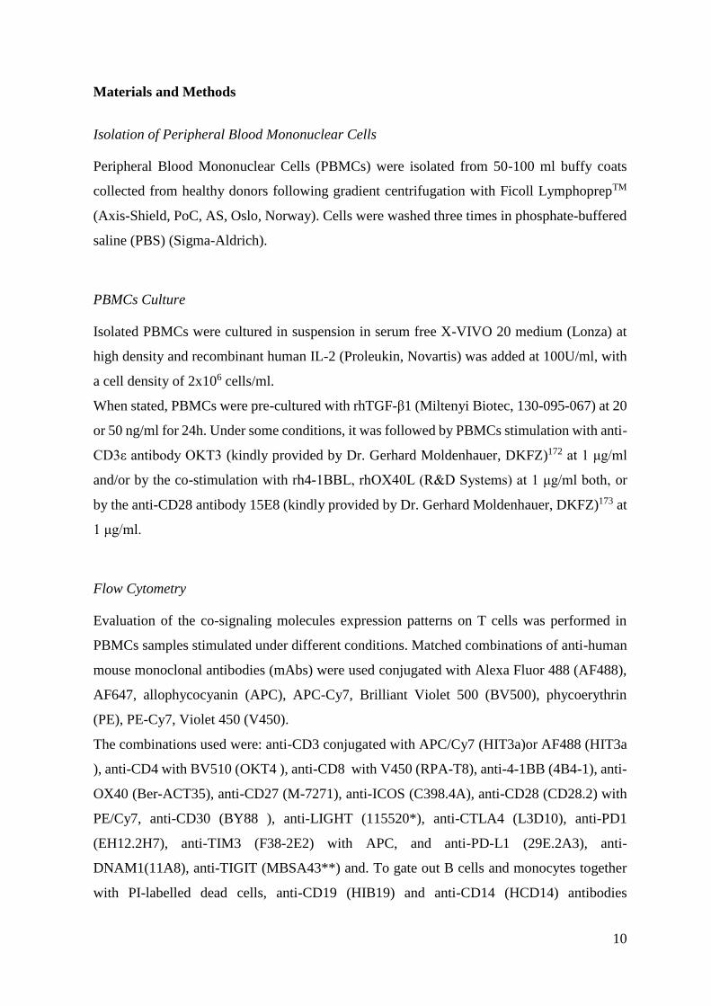
Materials and Methods
Isolation of Peripheral Blood Mononuclear Cells
Peripheral Blood Mononuclear Cells (PBMCs) were isolated from 50-100 ml buffy coats
collected from healthy donors following gradient centrifugation with Ficoll LymphoprepTM
(Axis-Shield, PoC, AS, Oslo, Norway). Cells were washed three times in phosphate-buffered
saline (PBS) (Sigma-Aldrich).
PBMCs Culture
Isolated PBMCs were cultured in suspension in serum free X-VIVO 20 medium (Lonza) at
high density and recombinant human IL-2 (Proleukin, Novartis) was added at 100U/ml, with
a cell density of 2x10⁶ cells/ml.
When stated, PBMCs were pre-cultured with rhTGF-β1 (Miltenyi Biotec, 130-095-067) at 20
or 50 ng/ml for 24h. Under some conditions, it was followed by PBMCs stimulation with anti-
CD3ε antibody OKT3 (kindly provided by Dr. Gerhard Moldenhauer, DKFZ)172 at 1 μg/ml
and/or by the co-stimulation with rh4-1BBL, rhOX40L (R&D Systems) at 1 μg/ml both, or
by the anti-CD28 antibody 15E8 (kindly provided by Dr. Gerhard Moldenhauer, DKFZ)173 at
1 μg/ml.
Flow Cytometry
Evaluation of the co-signaling molecules expression patterns on T cells was performed in
PBMCs samples stimulated under different conditions. Matched combinations of anti-human
mouse monoclonal antibodies (mAbs) were used conjugated with Alexa Fluor 488 (AF488),
AF647, allophycocyanin (APC), APC-Cy7, Brilliant Violet 500 (BV500), phycoerythrin
(PE), PE-Cy7, Violet 450 (V450).
The combinations used were: anti-CD3 conjugated with APC/Cy7 (HIT3a)or AF488 (HIT3a
), anti-CD4 with BV510 (OKT4 ), anti-CD8 with V450 (RPA-T8), anti-4-1BB (4B4-1), anti-
OX40 (Ber-ACT35), anti-CD27 (M-7271), anti-ICOS (C398.4A), anti-CD28 (CD28.2) with
PE/Cy7, anti-CD30 (BY88 ), anti-LIGHT (115520*), anti-CTLA4 (L3D10), anti-PD1
(EH12.2H7), anti-TIM3 (F38-2E2) with APC, and anti-PD-L1 (29E.2A3), anti-
DNAM1(11A8), anti-TIGIT (MBSA43**) and. To gate out B cells and monocytes together
with PI-labelled dead cells, anti-CD19 (HIB19) and anti-CD14 (HCD14) antibodies
11
conjugated with PerCP/Cy5.5 were used. Antibodies were purchased from BioLegend,
*Becton Dickinson (BD) or **eBiosciences.
For surface staining, PBMCs were incubated with the antibodies, at 1μg/2x105 cells, for 30
minutes, in the dark, at 4ºC, washed 2x with Fluorescent-Activated Cell Sorting (FACS
Buffer). FACS buffer was prepared with fetal bovine serum (FBS) (Biochrom AG) and PBS
(Sigma- Aldrich) at 1:50, respectively. Live/dead discrimination was done by using propidium
iodide (PI) (Sigma-Aldrich) at 1,6 μg/ml in FACS Buffer.
For FOXP3 intracellular staining the eBioscience FOXP3/Transcription Factor Staining
Buffer set (eBioscience) was used after the surface staining. PBMCs were first stained with
Live/Dead® Fixable Green Dead Cell Stain Kit (Life Techonologies) at 1 μl/ml FACS Buffer,
for 30 min on ice, for live/dead discrimination. Afterwards, PBMCs were fixed during 30
minutes at 4ºC with Fixation/Permeabilization Buffer (eBioscience) followed by
Permeabilization Buffer (eBioscience) buffer for 20 min. Buffers were prepared according to
manufacturer’s instructions (eBioscience). It was followed by intracellular staining with
antibody anti-FOXP3-PE (236A/E7) (eBioscience).
For flow cytometry a BD FACScan II flow cytometer (BD Biosciences) and BD FACS DIVA
software™ (BD Biosciences) was used. A total of 30000 cells/samples were acquired.
Lymphocytes were gated followed by selection against cell doublets and dead cells. Then,
CD3+ cells were gated followed by gating for CD4+ or CD8+ T cells.
12
Results
A. Effect of TGF-β on the phenotype of CD4+ T cells from healthy donors’ PBMCs
TGF-β is a pleiotropic cytokine secreted by immune cells and nonhematopoietic cells and it
is responsible for maintaining immune homeostasis by acting as an immune suppressor:
inducing differentiation of Treg cells, inhibiting proliferation, differentiation, activation and
effector function of immune cells. However, depending on the context, TGF-β may act also
as a pro-inflammatory cytokine, constituting a potent chemoattractant for neutrophils, driving
Th17 and Th9 cells differentiation (which are pro-inflammatory cells) and it can also inhibit
Th22 responses.
In cancer, in the beginning of the tumorigenic process, TGF-β inhibits the proliferation of
transformed cells. Nevertheless, once those become resistant to this cytokine, it starts inducing
angiogenesis and immune evasion, contributing to tumor growth and to the metastatic
process.84 Sources of TGF-β in tumors are cancer cells themselves85,86, mesenchymal cells,
platelets, Treg cells, neutrophils, NK cells, monocytes, macrophages and DCs.87 High levels
of this cytokine are found in the plasma of cancer patients and it is normally associated with
a poor prognosis.88 Thus, TGF-β is already being studied as a target for cancer treatment. 89
In human PBMCs, TGF-β induces FOXP3 expression and promotes differentiation of T
conventional to regulatory T cells.90 In vivo, TGF-β also converts CD4+ CD25- T cells to
induced FOXP3+ Tregs, which have a suppressive phenotype.91 Tregs are also able to secrete
TGF- β, hence the TGF-β signaling pathway is one of the mechanism through which
regulatory T cells mediate suppression of T effector activity.92, 93
In the tumor microenvironment, enzymes such as nitric oxide synthase or arginase contribute
to this suppressive effect; hypoxic conditions promote Treg expansion, which in turn induces
TGF-β expression and increases the immunoinhibitory milieu, creating a suppressive loop.94
Human Tregs are difficult to phenotype given their great diversity and the scarcity of
identified markers specific for different Treg subsets. Nevertheless, CD25 and FOXP3 still
remain the most frequently used phenotypic signature for human Tregs.95
In the experiments reported in this work, in the presence of TGF-β and anti-CD3 antibody,
CD4+ T cells upregulated the expression of the transcription factor FOXP3, as detected by
intracellular stainings. The percentage of CD4+ FOXP3+ T cells cells increased in the culture
(Fig. 1A). Moreover, it was observed that CD3+ CD4+ FOXP3+ cells had an augmented
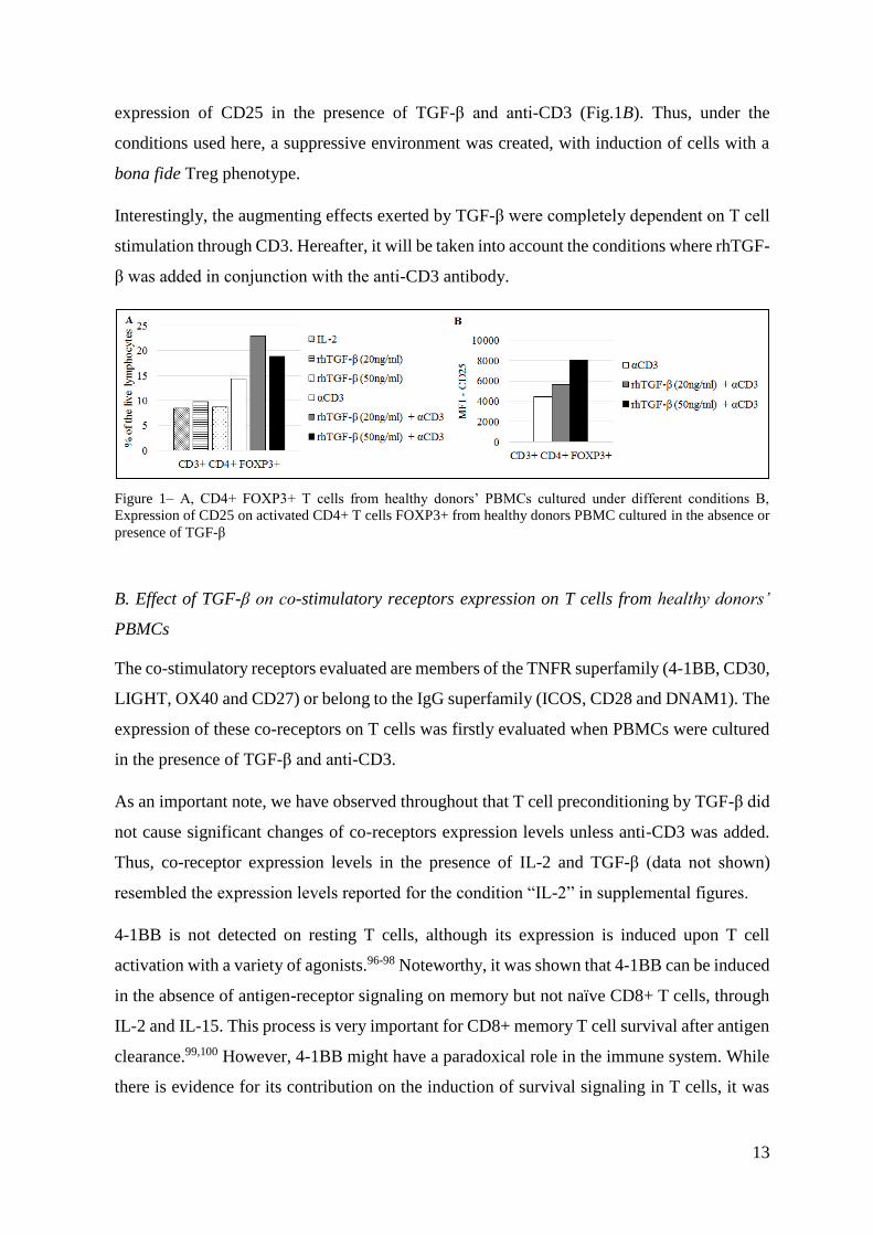
13
expression of CD25 in the presence of TGF-β and anti-CD3 (Fig.1B). Thus, under the
conditions used here, a suppressive environment was created, with induction of cells with a
bona fide Treg phenotype.
Interestingly, the augmenting effects exerted by TGF-β were completely dependent on T cell
stimulation through CD3. Hereafter, it will be taken into account the conditions where rhTGF-
β was added in conjunction with the anti-CD3 antibody.
Figure 1– A, CD4+ FOXP3+ T cells from healthy donors’ PBMCs cultured under different conditions B,
Expression of CD25 on activated CD4+ T cells FOXP3+ from healthy donors PBMC cultured in the absence or
presence of TGF-β
B. Effect of TGF-β on co-stimulatory receptors expression on T cells from healthy donors’
PBMCs
The co-stimulatory receptors evaluated are members of the TNFR superfamily (4-1BB, CD30,
LIGHT, OX40 and CD27) or belong to the IgG superfamily (ICOS, CD28 and DNAM1). The
expression of these co-receptors on T cells was firstly evaluated when PBMCs were cultured
in the presence of TGF-β and anti-CD3.
As an important note, we have observed throughout that T cell preconditioning by TGF-β did
not cause significant changes of co-receptors expression levels unless anti-CD3 was added.
Thus, co-receptor expression levels in the presence of IL-2 and TGF-β (data not shown)
resembled the expression levels reported for the condition “IL-2” in supplemental figures.
4-1BB is not detected on resting T cells, although its expression is induced upon T cell
activation with a variety of agonists.96-98 Noteworthy, it was shown that 4-1BB can be induced
in the absence of antigen-receptor signaling on memory but not naïve CD8+ T cells, through
IL-2 and IL-15. This process is very important for CD8+ memory T cell survival after antigen
clearance.99,100 However, 4-1BB might have a paradoxical role in the immune system. While
there is evidence for its contribution on the induction of survival signaling in T cells, it was
14
shown that 4-1BB-deficient T cells are hyperproliferative.100-102 Moreover, 4-1BB can
mediate CD8+ T cell suppression and CD4+ CD25+ Tregs may express 4-1BB.103
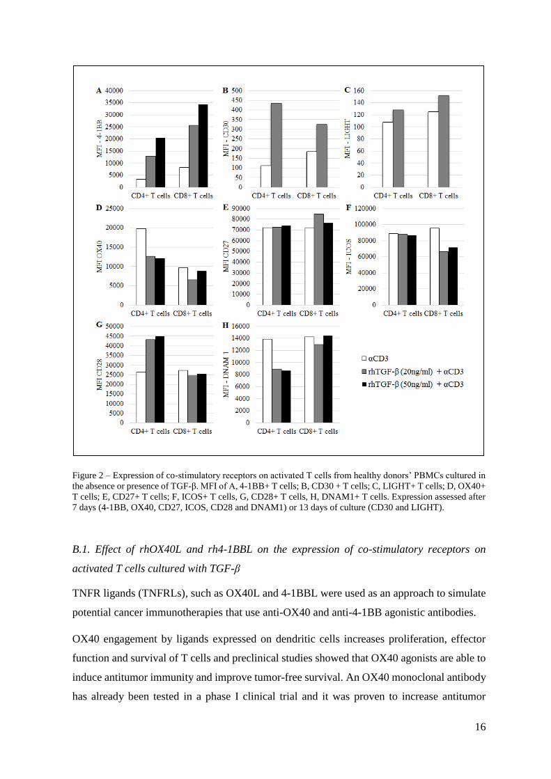
In our experiments, TGF-β-mediated induction of 4-1BB expression was dependent on the
TGF-β availability on the medium, where the higher the concentration of TGF-β, the higher
the expression of 4-1BB in both T cell subsets (Fig. 2A).
CD30 is not expressed on naïve T cells either – the peak of its expression happens after antigen
encounter and the CD28:CD80/CD86 engagement is able to further induce the expression of
this co-receptor.104 CD30 ligand is only expressed by professional APCs after activation.105
In vitro, crosslinking CD30 results in induction of cytokine production and proliferation of
activated T cells. However, it was shown that signaling by CD30 is also able to inhibit Th1
driven responses.106
TGF-β pre-culture of PBMCs and TCR stimulation triggered an upregulation of CD30,
particularly in CD4+ T cells (Fig. 2B).
With regard to LIGHT, besides being expressed on immature APCs and stimulating T cells
through HVEM107, it works as a co-stimulatory receptor that is expressed on T cells upon
activation.108 LIGHT was shown to play an important role on expansion of peripheral T cells
by a T cell-T cell-dependent manner.109
LIGHT expression was not significantly changed in the presence of TGF-β and anti-CD3 (Fig.
2C).
OX40, like 4-1BB and CD30, is not detectable on resting naïve T cells, although it is induced
on activated CD4+ and CD8+ T cells, as well as on Treg cells.110,111 TCR signals are sufficient
to induce OX40 expression. However, it is the CD28/B7-1/2 interaction that sustains this
expression and it can be modulated by T cell or APC-derived cytokines, such as IL-1, IL-2
and TNF.112 This sustained signal promotes antigen-specific T cell expansion and survival.
Furthermore, it was shown that OX40 is able to antagonize FOXP3 induction in naïve CD4+
T cells, hindering the generation of inducible Treg cells.113,114 This concomitant effect of
inducing effector activity and suppress immune suppression renders OX40 as a major player
on tumor immune surveillance.115,116
In the presence of TGF-β and anti-CD3, OX40 surface expression was slightly downregulated,
particularly in the CD4+ subset (Fig. 2D).
15
CD27 is expressed on naïve CD4+ and CD8+ T cells. But, unlike 4-1BB and OX40, its
expression increases with activation but it is subsequently downregulated after several cell
division cycles. This loss of CD27 is correlated with effector function of CD8+ T cells.117,118
CD27 is responsible for sustaining T cell survival, without influencing the cell division rate
in vitro.119,98
In the used conditions TGF-β had no additional effect on CD27 expression compared with the
level reached when anti-CD3 was added alone (Fig. 2E).
Regarding the evaluated members, ICOS and CD28 are homologous co-receptors. While
ICOS expression is restricted to activated T cells, CD28 is constitutively expressed in both
naïve and activated T cells.120
In vitro, it was established that ICOS is important to sustain T cell proliferation and that it
upregulates cell surface molecules and cytokine production. It also plays a role in T-cell
dependent humoral immunity, given the fact that ICOS ligand is highly expressed on B cells.
Upregulation of ICOS is strongly related with IL-10 production, thus, this receptor is also
expressed on FOXP3+ Treg cells, as well as on effector-memory T cells, being able to control
the pool size of both populations.121,122
In the used conditions, ICOS expression on CD4+ T cells was not affected with a TGF-β pre-
culture, although it was slightly decreased in the CD8+ subset (Fig. 2F).
CD28, likewise ICOS, is implicated in T cell expansion, survival and differentiation and it is
also necessary for proper IgG responses. The major difference between CD28 and ICOS is
that the former upregulates not IL-10 but IL-2 production.120
CD28 expression, unlike ICOS, was more affected in the CD4+ subset, where it was increased
in the presence of TGF-β and anti-CD3. On the contrary, CD28 expression on CD8+ T cells
was not changed when PBMCs were pre-cultured with TGF-β (Fig. 2G).
DNAM1, also named CD226, is also a member of the IgG superfamily and it is expressed in
both CD8+ and CD4+ T cells.123 The former constitutively express DNAM1, while the latter
only express this co-stimulatory receptor upon activation.124
Under the conditions used, in the presence of TGF-β and anti-CD3, DNAM1 expression was
diminished in the CD4+ subset, while TGF-β did not exert any effect on this co-stimulatory
receptor expression on CD8+ T cells (Fig. 2H).
16
Figure 2 – Expression of co-stimulatory receptors on activated T cells from healthy donors’ PBMCs cultured in
the absence or presence of TGF-β. MFI of A, 4-1BB+ T cells; B, CD30 + T cells; C, LIGHT+ T cells; D, OX40+
T cells; E, CD27+ T cells; F, ICOS+ T cells, G, CD28+ T cells, H, DNAM1+ T cells. Expression assessed after
7 days (4-1BB, OX40, CD27, ICOS, CD28 and DNAM1) or 13 days of culture (CD30 and LIGHT).
B.1. Effect of rhOX40L and rh4-1BBL on the expression of co-stimulatory receptors on
activated T cells cultured with TGF-β
TNFR ligands (TNFRLs), such as OX40L and 4-1BBL were used as an approach to simulate
potential cancer immunotherapies that use anti-OX40 and anti-4-1BB agonistic antibodies.
OX40 engagement by ligands expressed on dendritic cells increases proliferation, effector
function and survival of T cells and preclinical studies showed that OX40 agonists are able to
induce antitumor immunity and improve tumor-free survival. An OX40 monoclonal antibody
has already been tested in a phase I clinical trial and it was proven to increase antitumor
17
reactivity on T and B cells in patients with late-stage melanoma, increased T and B cells
responses to antigen immunization and drove a preferential upregulation of OX40 on FOXP3+
Treg cells, in TILs, with an acceptable toxicity profile.79
Regarding the importance of 4-1BB signaling pathway in cancer, it was already shown that 4-
1BB knockout mice have a higher rate of mortality when treated with B16.F10 melanoma
cells than 4-1BB wild-type (WT) mice. Furthermore, B16.F10-bearing 4-1BB WT mice
treated with an agonistic 4-1BB monoclonal antibody had a prolonged survival, which was
dependent on the effect of T cells and IFN-γ secretion. 4-1BB signaling, through agonistic
monoclonal antibodies or soluble 4-1BBL, causes T cell expansion, cytokine induction,
upregulation of anti-apoptotic genes and prevents activation-induced cell death. 96-98
The pattern of expression of T cell co-stimulatory receptors was assessed when PBMCs were
cultured with TNFRLs in order to test if these co-stimulatory molecules were able to enhance
T cell activation and if that was mirrored by phenotypic changes in these lymphocytes.
Firstly, the effect of recombinant proteins rhOX40L and rh4-1BBL was assessed in the
absence of TGF-β in order to evaluate if these recombinant proteins were able to induce co-
stimulation beyond the anti-CD3 antibody when the immunosuppressive stimuli were not
present. Those two conditions were compared with the co-stimulatory effect achieved with
the addition of anti-CD3 and anti-CD28.
Afterwards, rhOX40L and rh4-1BBL were added alongside anti-CD3 antibody after a pre-
culture of the PBMCs with rhTGF-β and the expression of the same range of co-stimulatory
receptors was evaluated.
The expression profile of the TNF members in response to the TNFR ligands (TNFRLs) was
modified in a distinct pattern for each molecule.
4-1BB expression on T cells was not changed when rhOX40L or rh4-1BBL were added
alongside anti-CD3 but the use of anti-CD3 and anti-CD28 resulted in an increase of 4-1BB
surface expression on CD4+ and CD8+ T cells (Sup. Fig. 1A, Annex).
In the presence of TGF-β (and anti-CD3), 4-1BB showed the same pattern of expression as in
the absence of the suppressive cytokine: anti-CD28 was the only co-stimulatory molecule able
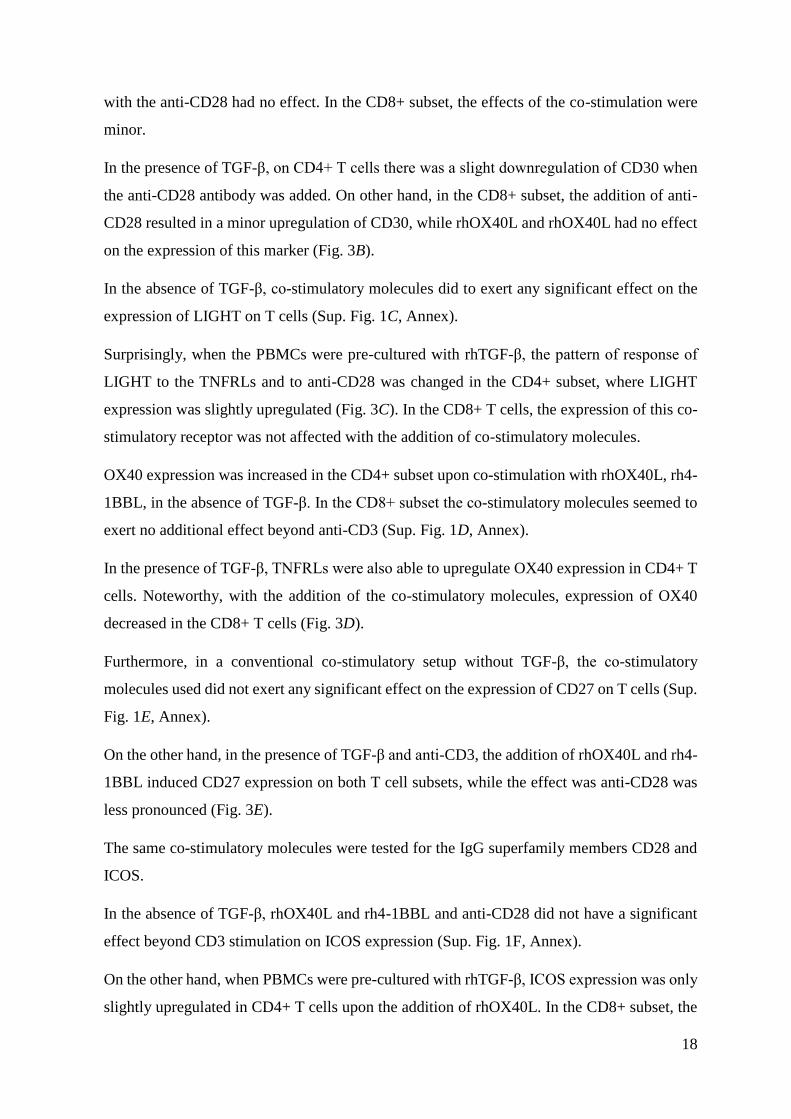
to upregulate this marker, what was particularly clear in the CD4+ T cell subset (Fig. 3A).
In the absence of TGF-β, CD30 expression was increased on CD4+ T cells when the rhOX40L
or rh4-1BBL were used (Sup. Fig. 1B, Annex). Interestingly, on the contrary, co-stimulation
18
with the anti-CD28 had no effect. In the CD8+ subset, the effects of the co-stimulation were
minor.
In the presence of TGF-β, on CD4+ T cells there was a slight downregulation of CD30 when
the anti-CD28 antibody was added. On other hand, in the CD8+ subset, the addition of anti-
CD28 resulted in a minor upregulation of CD30, while rhOX40L and rhOX40L had no effect
on the expression of this marker (Fig. 3B).
In the absence of TGF-β, co-stimulatory molecules did to exert any significant effect on the
expression of LIGHT on T cells (Sup. Fig. 1C, Annex).
Surprisingly, when the PBMCs were pre-cultured with rhTGF-β, the pattern of response of
LIGHT to the TNFRLs and to anti-CD28 was changed in the CD4+ subset, where LIGHT
expression was slightly upregulated (Fig. 3C). In the CD8+ T cells, the expression of this co-
stimulatory receptor was not affected with the addition of co-stimulatory molecules.
OX40 expression was increased in the CD4+ subset upon co-stimulation with rhOX40L, rh4-
1BBL, in the absence of TGF-β. In the CD8+ subset the co-stimulatory molecules seemed to
exert no additional effect beyond anti-CD3 (Sup. Fig. 1D, Annex).
In the presence of TGF-β, TNFRLs were also able to upregulate OX40 expression in CD4+ T
cells. Noteworthy, with the addition of the co-stimulatory molecules, expression of OX40
decreased in the CD8+ T cells (Fig. 3D).
Furthermore, in a conventional co-stimulatory setup without TGF-β, the co-stimulatory
molecules used did not exert any significant effect on the expression of CD27 on T cells (Sup.
Fig. 1E, Annex).
On the other hand, in the presence of TGF-β and anti-CD3, the addition of rhOX40L and rh4-
1BBL induced CD27 expression on both T cell subsets, while the effect was anti-CD28 was
less pronounced (Fig. 3E).
The same co-stimulatory molecules were tested for the IgG superfamily members CD28 and
ICOS.
In the absence of TGF-β, rhOX40L and rh4-1BBL and anti-CD28 did not have a significant
effect beyond CD3 stimulation on ICOS expression (Sup. Fig. 1F, Annex).
On the other hand, when PBMCs were pre-cultured with rhTGF-β, ICOS expression was only
slightly upregulated in CD4+ T cells upon the addition of rhOX40L. In the CD8+ subset, the
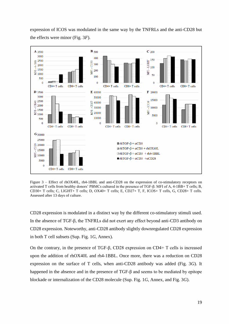
19
expression of ICOS was modulated in the same way by the TNFRLs and the anti-CD28 but
the effects were minor (Fig. 3F).
Figure 3 – Effect of rhOX40L, rh4-1BBL and anti-CD28 on the expression of co-stimulatory receptors on
activated T cells from healthy donors’ PBMCs cultured in the presence of TGF-β. MFI of A, 4-1BB+ T cells; B,
CD30+ T cells; C, LIGHT+ T cells; D, OX40+ T cells; E, CD27+ T, F, ICOS+ T cells, G, CD28+ T cells.
Assessed after 13 days of culture.
CD28 expression is modulated in a distinct way by the different co-stimulatory stimuli used.
In the absence of TGF-β, the TNFRLs did not exert any effect beyond anti-CD3 antibody on
CD28 expression. Noteworthy, anti-CD28 antibody slightly downregulated CD28 expression
in both T cell subsets (Sup. Fig. 1G, Annex).
On the contrary, in the presence of TGF-β, CD28 expression on CD4+ T cells is increased
upon the addition of rhOX40L and rh4-1BBL. Once more, there was a reduction on CD28
expression on the surface of T cells, when anti-CD28 antibody was added (Fig. 3G). It
happened in the absence and in the presence of TGF-β and seems to be mediated by epitope
blockade or internalization of the CD28 molecule (Sup. Fig. 1G, Annex, and Fig. 3G).
20
DNAM1 expression was not evaluated in what regards the effects of the addition of co-
stimulatory molecules, in the absence or presence of TGF-β.
C. Effect of TGF-β on co-inhibitory receptors expression on T cells from healthy donors’
PBMCs
Co-inhibitory receptors expressed on T cells were also evaluated within the suppressive
environment created in vitro. TIM3, TIGIT, PD1, CTLA4 and PD-L1 were the co-inhibitory
molecules whose expression was assessed when PBMCs were cultured in the presence of
TGF-β and anti-CD3.
TIM3 is described as a negative regulator of IFN-γ-secreting CD4+ Th1 and CD8+ cytotoxic
cells and it was shown to have an important role in CD8+ T cell exhaustion in cancer.148
Furthermore, it has a role in promoting MDSCs responses.125
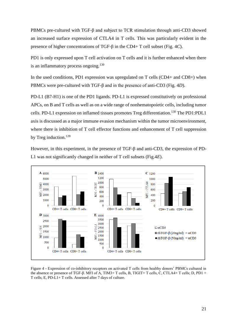
TIM3 was shown here to have its expression downregulated upon TCR stimulation and in the
presence of TGF-β, in both T cell subsets (Fig. 4A).
TIGIT is expressed on activated T cells. Its suppressive function relies on its engagement with
its ligand PVR (poliovirus receptor) on dendritic cells and subsequent induction of IL-10
production.126 TIGIT works as inhibitor of CD4+ T cell priming and CD8+ T cell anti-tumor
effector function.127
Despite being a co-inhibitory receptor, TIGIT expression was downregulated upon TCR
stimulation and in the presence of TGF-β. The effect seemed to be dependent on the TGF-β
concentration: the higher it is, the less TIGIT is available on the T cell surface (Fig. 4B).
As mentioned earlier, the CTLA4:CD80/CD86 and the PD1:PD-L1 axis constitute the first
approved targets of cancer immunotherapy. CTLA4 and PD1 have a distinct mechanism of
action. While CTLA4 rises the activation threshold on T cells and attenuates proliferation of
tumor-specific T lymphocytes, PD1 limits T cell effector function within the tumor
microenvironment.128
CTLA-4 expression is minimal in resting T cells and, as it counteracts the CD28-mediated
costimulatory signals, CTLA4 is enhanced through CD28/IL-2 co-stimulation after TCR
engagement. Antigen-experienced CD4+ and CD8+ T cells, as well as CD4+ Tregs express
CTLA4 constitutively.129
21
PBMCs pre-cultured with TGF-β and subject to TCR stimulation through anti-CD3 showed
an increased surface expression of CTLA4 in T cells. This was particularly evident in the
presence of higher concentrations of TGF-β in the CD4+ T cell subset (Fig. 4C).
PD1 is only expressed upon T cell activation on T cells and it is further enhanced when there
is an inflammatory process ongoing.130
In the used conditions, PD1 expression was upregulated on T cells (CD4+ and CD8+) when
PBMCs were pre-cultured with TGF-β and in the presence of anti-CD3 (Fig. 4D).
PD-L1 (B7-H1) is one of the PD1 ligands. PD-L1 is expressed constitutively on professional
APCs, on B and T cells as well as on a wide range of nonhematopoietic cells, including tumor
cells. PD-L1 expression on inflamed tissues promotes Treg differentiation.130 The PD1:PDL1
axis is discussed as a major immune evasion mechanism within the tumor microenvironment,
where there is inhibition of T cell effector functions and enhancement of T cell suppression
by Treg induction.128
However, in this experiment, in the presence of TGF-β and anti-CD3, the expression of PD-
L1 was not significantly changed in neither of T cell subsets (Fig.4E).
Figure 4 - Expression of co-inhibitory receptors on activated T cells from healthy donors’ PBMCs cultured in
the absence or presence of TGF-β. MFI of A, TIM3+ T cells, B, TIGIT+ T cells, C, CTLA4+ T cells; D, PD1 +
T cells; E, PD-L1+ T cells. Assessed after 7 days of culture.
22
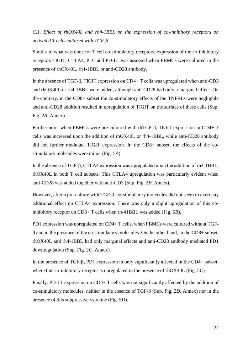
C.1. Effect of rhOX40L and rh4-1BBL on the expression of co-inhibitory receptors on
activated T cells cultured with TGF-β
Similar to what was done for T cell co-stimulatory receptors, expression of the co-inhibitory
receptors TIGIT, CTLA4, PD1 and PD-L1 was assessed when PBMCs were cultured in the
presence of rhOX40L, rh4-1BBL or anti-CD28 antibody.
In the absence of TGF-β, TIGIT expression on CD4+ T cells was upregulated when anti-CD3
and rhOX40L or rh4-1BBL were added, although anti-CD28 had only a marginal effect. On
the contrary, in the CD8+ subset the co-stimulatory effects of the TNFRLs were negligible
and anti-CD28 addition resulted in upregulation of TIGIT on the surface of these cells (Sup.
Fig. 2A, Annex).
Furthermore, when PBMCs were pre-cultured with rhTGF-β, TIGIT expression in CD4+ T
cells was increased upon the addition of rhOX40L or rh4-1BBL, while anti-CD28 antibody
did not further modulate TIGIT expression. In the CD8+ subset, the effects of the co-
stimulatory molecules were minor (Fig. 5A).
In the absence of TGF-β, CTLA4 expression was upregulated upon the addition of rh4-1BBL,
rhOX40L in both T cell subsets. This CTLA4 upregulation was particularly evident when
anti-CD28 was added together with anti-CD3 (Sup. Fig. 2B, Annex).
However, after a pre-culture with TGF-β, co-stimulatory molecules did not seem to exert any
additional effect on CTLA4 expression. There was only a slight upregulation of this co-
inhibitory receptor on CD8+ T cells when rh-41BBL was added (Fig. 5B).
PD1 expression was upregulated on CD4+ T cells, when PBMCs were cultured without TGF-
β and in the presence of the co-stimulatory molecules. On the other hand, in the CD8+ subset,
rhOX40L and rh4-1BBL had only marginal effects and anti-CD28 antibody mediated PD1
downregulation (Sup. Fig. 2C, Annex).
In the presence of TGF-β, PD1 expression in only significantly affected in the CD4+ subset,
where this co-inhibitory receptor is upregulated in the presence of rhOX40L (Fig. 5C)
Finally, PD-L1 expression on CD4+ T cells was not significantly affected by the addition of
co-stimulatory molecules, neither in the absence of TGF-β (Sup. Fig. 2D, Annex) nor in the
presence of this suppressive cytokine (Fig. 5D).
23
Figure 5 - Effect of rhOX40L, rh4-1BBL and anti-CD28 on the expression of the co-inhibitory receptors on
activated T cells from healthy donors’ PBMCs cultured in the presence of TGF-β. MFI of A, TIGIT+ T cells, B,
CTLA4+ T cells; C, PD1+ T cells; D, PD-L1+ T cells. Assessed after 13 days of culture.
24
Discussion and Conclusions
When PBMCs were pre-cultured with rhTGF-β for 24 hours and subsequently subjected to
polyclonal activation by the addition of anti-CD3, the percentage of CD3+ CD4+ FOXP3+
cells increased, as well as the expression of CD25 on CD3+ CD4+ FOXP3+ cells. This result
is consistent with induction of Treg cells, which are potentially able to inhibit anti-tumor
immunity. Thus, a suppressive microenvironment was created in vitro.
The concomitant presence of TGF-β and anti-CD3 has distinct effects on co-stimulatory
molecules, members of TNFRSF. While this condition upregulated 4-1BB and CD30
expression on T cells, LIGHT and CD27 did not significantly respond to the addition of TGF-
β and OX40 was downregulated in the presence of this cytokine.
It was shown that important co-stimulatory molecules, such as ICOS and CD28, from the B7
family of the IgSF members, had also a different pattern of expression under the same
conditions. In the presence of TGF-β and anti-CD3, CD28 expression was upregulated in
CD4+ T cells, while ICOS showed the tendency to be downmodulated by TGF-β in CD8+ T
cells. The CD28 signaling pathway induces IL-2 secretion, which might contribute to expand
T cells with maintenance of their functional activity or it might contribute to Treg cells
expansion, given the fact those need IL-2 to survive.95,131 On the other hand, ICOS expression
on Treg cells is related with an increment in the production of the immunosuppressive
cytokine IL-10.82,121 Thus, the expression levels of both co-receptors, CD28 and ICOS, might
not be conclusive by T cell phenotyping only, given the highly dynamic and contradictory
outcomes of cytokines whose secretion is induced by their signaling pathways. The inclusion
of blocking antibodies for IL-2 and IL-10 needs to be explored in the presence and absence of
TGF-β.
Moreover, in this suppressive environment, T cells upregulated exhaustion molecules, such
as PD-1 and CTLA4, co-inhibitory receptors that were reported to be upregulated on TILs of
cancer patients.132 However, TIM3 and TIGIT (also co-inhibitory) had their expression levels
decreased and PD-L1 expression did not change in the presence of rhTGF-β, albeit these
receptors are described as being expressed on T cells within the tumor microenvironment.75,127
Nevertheless, it remains to be explored how TGF-β suppresses TIGIT and TIM3 expression
and does not interfere with PD-L1 expression on activated T cells.
25
These intriguing results might be explained by a different action of TGF-β, other than inducing
Treg cells expansion, under the established conditions. It is described that TGF-β, besides
promoting the expression of FOXP3 and inducing Treg cells expansion, is also able to drive
Th17 cells differentiation.133,134 Additionally, it was reported that the capacity of TGF-β to
abrogate FOXP3 expression is dependent on NF-κB activity. NF-κB is a transcription factor
activated upon TCR/CD28 engagement and, at high doses of TCR stimulation, it can induce
the production of IL-17 and inhibit FoxP3 expression, driving the differentiation of naïve T
cells to Th17 cells, instead of induced Treg cells.135 Given the fact that we used high
concentrations of TGF-β (20 ng/ml or 50 ng/ml) and 1µg/ml of anti-CD3, the possibility that
a Th17 response was driven shall not be excluded.
Furthermore, Treg and Th17 subsets are plastic – there are reports in mice and in humans that
identify Th17 cells that transit into induced Treg cells136 as well as Foxp3+ induced Tregs that
have downmodulated RORγt and are able to transdifferentiate into Th17 cells, under pro-
inflammatory conditions such as the presence of the cytokine IL-6.137
This Th17-driven response would explain the remarkable upregulation of CD30 under the
presence of TGF-β (20ng/ml) and anti-CD3, given the fact that CD30:CD30L engagement
was shown to play a critical role in Th17 differentiation.138 Furthermore, it would also be a
reasonable explanation for a slight decrease on the percentage of FOXP3+ cells, when the
concentration of TGF-β was increased (from 23% to about 19%).
Nevertheless, Th17 cells have also a role in cancer, although it might be ambiguous. There
are reports on ovarian carcinomas-associated ascites that relate high Th17 cell density with
better overall survival. On the contrary, in malignant tumors (such as hepatocellular
carcinoma, colorectal cancer or pancreatic carcinoma), heavy infiltration of Th17 cells was
correlated with a poor prognosis.139
This dual role of Th17 cells is explained by their contradictory effects on the tumor
microenvironment. On one hand, Th17 responses are able to promote anti-tumor activity by
downregulation of Tregs, promotion of MHC-I and II expression and induction of CTL
activities. On the other hand, Th17 pro-tumor effects are likely underlined by the induction of
pro-inflammatory cytokines, chemokines and matrix metalloproteinases – there is induction
of MDSCs and inhibition of cytotoxic activity as well as promotion of angiogenesis.140 Thus,
even if a Th17 response was driven, the suppressive microenvironment could have still been
created in vitro.

26
However, this hypothesis shall be tested with the identification of a Th17 population under
the established conditions and with the execution of a cytokine profile, by intracellular
staining or ELISA. A titration of the TGF-β/anti-CD3 would allow to corroborate the results.
Concerning the approach to simulate immunotherapeutic approaches by the addition of
costimulatory molecules such as rh4-1BBL, rhOX40L or anti-CD28 antibody, it is important
to mention that it was not conclusive in the experiments shown here.
In summary, concerning the co-stimulation effect on co-stimulatory receptors, in the presence
of TGF-β and anti-CD3, LIGHT, OX40 and CD28 expression were particularly upregulated
on CD4+ T cells, while CD27 was increased in both subsets. On the other hand, 4-1BB, CD30
and ICOS expression had minor fluctuations with the addition of the TNFR ligands.
Noteworthy, 4-1BB expression was clearly upregulated in CD4+ T cells in the presence of the
anti-CD28 antibody, TGF-β and anti-CD3.
Regarding the co-inhibitory receptors tested, TIGIT was generally upregulated in the presence
of co-stimulatory molecules, TGF-β and anti-CD3, and CTLA4 expression was increased on
CD8+ T cells when rh4-1BBL was added. On the other hand, PD-1 and PD-L1 did not
significantly respond to the presence of the co-stimulatory molecules.
This inability to restore a full-fledged activation phenotype with the addition of TNFRLs
might rely on the late assessment of the expression of the co-receptors on T cells – most of
the flow cytometry evaluations were done after 7 or 13 days of culture. This long incubation
period with TGF-β/anti-CD3/rhOX40L/rh4-1BBL/anti-CD28 may result in T cell exhaustion
induced by all the activation stimuli present in the culture medium. Thus, it would be
important to study the kinetics of the expression of the co-signaling molecules and how they
influence the outcome of the immunotherapies. Furthermore, assessing the same markers also
with standard immunotherapies, monoclonal antibodies against CTLA4 and PD1, would
allow: first, a comparison between therapies considered effective and potentially new ones,
with regard to the expression of T cell co-receptors; second, a valid comparison between the
experiments and the available literature and, finally, a phenotypical characterization of T cell
responses under the established therapies.
It is worth mentioning that, although it has not been systematically studied, donor variability
may be an important issue. PBMCs were collected from different donors and donor variability
influences the pattern of expression of each molecule assigned.
27
First, it would be extremely important to assess the outcome of these results at a functional
level. Developing an intracellular assay to study how the pattern of expression of surface
molecules on T cells reflects the cytokines they produce would give a deeper insight about the
regulation of the anti-tumor functions of the different T cell subsets, as mentioned before.
Moreover, T cell cytotoxicity assays using a substantial diversity of types of cancer cells as
targets would allow exploring if the co-inhibitory and co-stimulatory molecules have different
outputs in what concerns killing of transformed cells.
Finally, if possible, the use of samples of tumor infiltrating lymphocytes (TILs) from cancer
patients would allow the study of co-signaling molecules on T cells ex vivo, which constitutes
a better model to investigate the suppressive effects of the transformed cells on the immune
system. Furthermore, the phenotyping could afterwards be correlated with the patients’
outcome.
Ultimately, in order to deeply study the patterns of expression of the co-signaling molecules
on T cells, one could use a novel technique called mass cytometry. This approach assemblies
flow cytometry and mass spectrometry, where cell markers are conjugated to metal ion tags
(rather than fluorophores). The labelled cells are ionized individually and the respective
atomic ion cloud is captured using time of flight mass spectrometry. While the flow cytometry
panels have a limitation of 8 different colors per panel, mass cytometry as a much higher
throughput and it can be incorporated up to 40 different markers. It can be used, not only for
cell surface markers, but also for intracellular molecules.141,142 Under the light of this
particular project, the use of this method would enable the simultaneous assessment of several
additional surface markers and intracellular molecules within the same cell, allowing a more
accurate phenotyping of the activation/inhibition status of each subset of T cells under
suppressive conditions (tumor microenvironment) or upon stimulation with monoclonal
antibodies (immunotherapies).
The immune system has certainly an intricate way of performing its role. And thus, in my
opinion, it is unrealistic to try to create a simplistic approach to design new anticancer
therapies.
It has been demonstrated that combinational therapies might have a synergistic effect on
antitumor responses. Attempts to have at least an additional effect when combining two
different immunotherapies seem promising and were already mentioned. Using anti-CTLA-4
and anti-PD-1 monoclonal antibodies for advanced melanoma treatment showed over 80%
28
tumor reductions on more than 50% of the patients and with a tolerable toxicity profile.73
Blocking of PD-1 and TIM-375 or LAG-3143 augmented antitumor efficacy when in
combination, although their effect as monotherapies is modest.144 Furthermore, it has been
shown that adoptive T cell therapy (gene-transduced T lymphocytes expressing transgenic
TCRs or chimeric antigen receptors – CARs) synergizes with anti PD-1 monoclonal
antibody.145
Unfortunately, the still prohibitive costs and toxicity profiles of immunotherapies set up limits
for the development of ground breaking approaches on cancer treatment.144 To begin with, the
dose and schedule of the new therapies ought to be tested and improved, in monotherapy
and/or in combination. 83 For example, despite the fact that the immune checkpoint blockade
produces durable responses in a fraction of patients (and therefore the mean survival is
improved), those responses are delayed in time. Combining this strategy with conventional
therapies, such as chemotherapy, radiotherapy or even with genomically targeted and
antiangiogenic therapies may enable, more than the synergistic effect, a rapid antitumor
response which, in turn, will be long lasting and sustained.60,144,146 Furthermore, combining
different tactics (targeting distinct hallmarks of cancer) allows to circumvent the myriad of
resistance mechanisms acquired by cancer cells.
In order to highlight the importance of combinatory approaches, there were some preclinical
studies that showed that chemotherapeutic agents capable of modulating the tumor
environment were powerful candidates to test with immune checkpoint inhibitors. For
example, gemcitabine (which targets Tregs and MDSCs147,148) seems to synergize with anti-
CTLA-4 in a transplantable tumor model.149 Moreover, radiotherapy was also shown to
synergize with CTLA-4, PDL1 and combined CD40/4-1BB targeting therapies to control the
growth of various tumor models.150 It is clear that strategies that induce a CD8+ T cell influx
and/or reduced FOXP3/CD8 ratio in tumors are valuable partners for checkpoint inhibitors.146
As a final remark, it seems to be of great relevance to further investigate, not only the
connections between all the cells that compose the tumor microenvironment, but also the
specificities of this network within each type of cancer. This will facilitate the planning of
combinatory therapies, which must consider that, for every cancer, different subsets of
immune cells exert a beneficial or antagonistic role on anticancer drugs. 146
Continuing to unravel the patterns of co-stimulation and co-inhibition of T cells might be of
great importance to determine the level of activation of the key players on antitumor

29
responses. As we evolve towards personalized cancer therapies, this would not only meet the
imperative need for biomarkers, but also would allow the creation of new immunotherapeutic
strategies based on the pattern of expression of co-signaling molecules in the tumor infiltrating
lymphocytes of each patient.
30
References
1. Hoffman, R.A., Kung P.C., Hansen, W.P., Goldstein, G., 1980, Simple and Rapid Measurement
of human peripheral lymphocytes and their subclasses in peripheral blood, Proc. Natl. Acad. Sci.
USA, 7: 4914-4917
2. Van Lier et al., 1988, Signals involved in T cell activation. T cell proliferation induced through
the synergistic action of anti-CD28 and anti-CD2 monoclonal antibodies, Eur. J. Immunol.,18:
167-172
3. GLOBOCAN 2012 v1.1, Cancer Incidence and Mortality Worldwide: IARC CancerBase No. 1,
Lyon, France: International Agency for Research on Cancer, 2014. Available from:
http://globocan.iarc.fr, accessed on 28/04/2015.
4. Hanahan, D. and Weinberg, R., 2011, Hallmarks of Cancer: The Next Generation, Cell, 144: 646-
674
5. Perona, R., 2006, Cell Signlling: growth factors and tyrosine kinase receptors, Clin. Transl.
Onco., 8: 77-82
6. Witsch,, E, Sela M. and Yarden, Y, 2010, Roles for growth factors in cancer progression,
Physiology (Bethesda), 25: 85-101
7. Bhowmick et al., 2004, Stromal Fibroblasts in cancer initiation and progression, Nature, 432:
332-337
8. Davies, M.A., and Samuels, Y., 2010, Analysis of the genome to personalize therapy for
melanoma, Oncogene, 29: 5545-5555
9. Burkhart, D.L., and Sage, J., 2008, Cellular mechanisms of tumor suppression by the
retinoblastoma gene, Nat. Rev. Cancer, 8: 671-682
10. Green, DR, and Kroemer G., 2009, Cytoplasmatic functions of the tumor suppressor p53, Nature,
458 (7242): 1127-1130
11. Curto, M., Cole, B.K., Lallemand, D., Liu, C. H. and McClactchey, A.I., 2007, Contact Dependent
Inhibition of EGFR signalling by NF2/Merlin, J. Cell Biol., 177: 893-903
12. Hezel, A.F., 2008, and Bardessy, N., LKB1; linking cell structure and tumor suppression,
Oncogene, 27: 6908-6919
13. Massagué, J., 2008, TGFβ in cancer, Cell, 134: 215-230
14. Filder, I.J., 2003, The pathogenesis of cancer metastasis: the ‘seed and soil’ hypothesis revisited,
Nat. Rev. Cancer, 3, 453-458
15. Barrallo-Gimeno, A., and Nieto, M.A., 2005, The Snail genes as inducers of cell movement and
survival: implications in development and cancer, Development, 132: 3151-3161
16. Peinado, H., Marin, F., Cubillo, E., Stark, H.J., Fusening, N., Nieto, M.A., and Cano, A., 2004,
Snail and E47 repressors of E-cadherin induce distinct invasive and angiogenic properties in vitro,
J. Cell Sci., 117: 2827-2839
17. Joyce, J.A., and Pollard, J.W., 2009, Microenvironmental regulation of metastasis, Nat. Rev.
Cancer, 9: 239-252
18. Quian, B.Z. and Pollard, J.W., 2010, Macrophage diversity enhances tumor progression and
metastasis, Cell, 141: 39-51
19. Karnoub, A.E. et al., 2007, Mesenchymal stem cells withing tumor stroma promote breast cancer
metastasis, Nature, 449: 557-563
20. Kalluri, R., and Zeisberg, M., 2006, Fibroblasts in Cancer, Nat. Rev. Cancer, 6: 392-401
21. Hugo, H., Ackland, M.L., Blick, T., Lawrence, M.G., Clements, J.A., Williams, E.D., and
Thompson, E.W., 2007, Epithelial-mesenchymal and mesenchymal-epithelial transition in
carcinoma progression, J. Cell Physiol., 213: 374-383
22. Lowe, S. W., Cepero, E., and Evan, G., 2004, Intrisic tumor suppression, Nature, 432: 307-315
23. Adams, J.M., and Cory, S., 2007, The Bcl-2 apoptotic switch in cancer development and therapy,
Oncogene, 26: 1324-1337
24. LeRoith, D., Roberts, C.T. Jr., 2003, The insulin-like growth factor system and cancer, Cancer
Letters, 195(2): 127-137
25. Reichmann, E., 2002, The biological role of the Fas/FasL system during tumor formation and
progression, Semin. Cancer Biol., 12(4): 309-315
31
26. Grivennikov, S.I., Greten, F.R., and Karin, M., 2010, Immunity, inflammation and cancer, Cell,
140: 883-899
27. Blasco, M.A., 2005, Telomeres and human disease: ageing, cancer and beyond, Nat. Rev. Genet.,
6: 611-622
28. Baeriswyl, V., and Cristofori, G., 2009, The angiogenic switch in carcinogenesis, Semin. Cancer
Biol., 19: 329-337
29. Ferrara, N., 2010, Pathways mediating VEGF-independent tumor angiogenesis, Cytokine Growth
Factor Rev., 21:21-26
30. Zumsteg, A., and Cristophori, G., 2009, Corrupt policemen: inflammatory cells promote tumor
angiogenesis, Curr. Opin. Oncol., 21: 60-70
31. Abul K. Abbas and Andrew H. Lichtman, Cellular and Molecular Immunology, Elsevier, 5th
Edition, 2005 (pp. 3-189)
32. Hanahan D., Coussens, L.M., 2012, Accessories to the crime: functions of cells recruited to the
tumor microenvironment, Cancer Cell, 21: 309-322
33. de Visser, K., Eichten, A., Coussens, L.M., 2006, Paradoxical roles of the immune system during
cancer development, Nat. Rev. Cancer, 6(1): 24-37
34. Coussens, L.M., Werb, Z., 2002, Inflammation and Cancer, Nature, 420(6917): 860-867
35. Balkwill, F., Charles KA, Mantovani, A., 2005, Smoldering and polarized inflammation and
promotion of malignant disease, Cancer Cell, 7(3): 211-217
36. Zou, W., 2005, Immunosuppressive networks in the tumor environment and their therapeutic
relevance, Nat. Rev. Cancer, 5: 263-274
37. Curiel, T.J., et al., Specific recruitment of regulatory T cells in ovarian carcinoma fosters immune
privilege and predicts reduced survival, Nature Med, 10: 942-994
38. Serafini, P. et al., 2004, Derangement of immune responses by myeloid suppressor cells, Cancer
Immunol. Immunother., 53: 64-72
39. Dunn, G.P., Bruce, A., Ikeda, H., Old, L., Schreiber, R., 2002, Cancer immunoediting: from
immunosurveillance to tumor escape, Nat. Immunol., 3: 991-998
40. Smyth, M.J., Dunn, G.P., and Schreiber R.D., 2006, Cancer immunosurveillance and
immunoediting: the roles of immunity in suppressing tumor development and shaping tumor
immunogenicity, Adv. Immunol., 90, 1-50
41. Lin, W. W., and Karin, M., 2007, A cytokine-mediated link between innate immunity,
inflammation and cancer, J. Clin. Invest., 117, 1175-1183
42. Murdoch, C., Muthana, M., Coffelt, S.B., and Lewis, C.E., 2008, The role of myeloid cells in the
promotion of tumor angiogenesis, Nat. Rev. Cancer, 8: 618-631.
43. Roberts, S. J., Ng, B.Y., Filler, R.B., Lewis, J., Glusac, E.J., Hayday, A.C., Tigelaar, R.E., and
Girardi, M., 2007, Characterizing tumor-promoting T cells in chemically induced cutaneous
carcinogenesis, Proc. Natl. Acad. Sci. USA, 104: 6770-6775
44. Hanada, T., et al., 2006, IFNgamma.dependent spontaneous development of colorectal
carcinomas in SOCS1-deficient mice, J. Exp. Med., 203: 1391-1397
45. Aspord, C., et al., 2007, Breast cancer instructs dendritic cells to prime interleukin 13-secreting
CD4+ T cells that facilitate tumor development, J. Exp. Med., 204: 1037-1047
46. Galon, J., et al., 2006, Type, density and location of immune cells with human colorectal tumors
predict clinical outcome, Science, 313: 1960-1964
47. Swan, J.B., and Smyth, M.J., 2007, Immune surveillance of tumors, J Clin Inv, 117: 1137-1146
48. Kurts, C., Robinson, B.W., Knolle, P.A., 2010, Cross Priming in health and disease, Nat. Rev.
Immunol., 10(6): 403-414
49. Mueller, D., Jenkins, M.K., and Schwartz, R.H., 1989, Clonal expansion vs. functional clonal
activation: a co-stimulatory signalling pathway determines the outcome of T cell antigen receptor
occupancy, Annu. Rev. Immunol., 7: 445-480
50. Chen, L., and Flies, D.B., 2013, Molecular mechanisms of T cell co-stimulation and co-inhibition,
Nat. Rev. Immunol., 13, 227-241
51. Liechtenstein, T., Dufait, I., Lanna, A., Breckport, K., and Escors, D., 2012, Modulating co-
stimulation during antigen presentation to enhance cancer immunotherapy, Immunol. Endocr.
Metab. Agents Med. Chem., 12: 224-235
32
52. Rudd, C.E., Taylor, A., Schneider, H., 2009, CD28 and CTLA-4 coreceptor expression and signal
transduction, Immunol. Rev., 229: 12-26
53. Zhu, Y., Yao, S., Chen, L., 2011, Cell surface signalling molecules in the control of immune
responses: a tide model, Immunity, 34: 466-478
54. Greenwald, R.J., Freeman, G.J., and Sharpe, A.H., 2005, The B7 family revisited, Annu. Rev.
Immunol., 23: 512-548
55. Kane, L.P., 2010, T cell Ig and mucin domain proteins and immunity, J. Immunol, 184: 2743-
2749
56. Cannons, J.L., Tangye, S.G., and Schawtzberg, P.L., 2011, SLAM family receptors and SAP
adaptors in immunity, Annu. Rev. Immunol, 29: 665-705
57. Sierro, S., Romero, P., and Speiser, D.E., 2011, The CD4-like molecule LAG3, biology, and
therapeutic applications, Expert Opin. Ther. Targets, 15: 91-101
58. Chan, C.J., Andrews, D.M., and Smyth, M.J., 2012, Receptors that interact with nectin and nectin-
like proteins in immunosurveillance and immunotherapy of cancer, Curr. Opin. Immunol., 24:
246-251
59. Croft, M. et al., 2012, TNF superfamily in inflammatory disease: translating basic insights, Trends
Immunol., 33, 144-152
60. Sharma, P., Allison, J.P., 2015, Immune checkpoint targeting in cancer therapy: toward
combination strategies with curative potential, Cell, 161, 205-214
61. Pardoll, D., 2012, The blockade of immune checkpoints in cancer immunotherapy, Nat. Rev.
Cancer, 12: 252-264
62. Peggs, K.S., Quezada, S.A., Chamber, C.A., Korman, A.J., and Allison, J.P., 2009, Blockade of
CTLA 4 on both effector and regulatory T cell compartments contributes to the antitumor activity
of anti-CTLA4 antibodies, J. Exp. Med., 206:17-1725
63. U.S. Food and Drug Administration – Center for Drug Evaluation and Research: Ipilimumab,
available
from:http://www.fda.gov/aboutfda/centersoffices/officeofmedicalproductsandtobacco/cder/ucm
248478.htm, accessed on 07/05/2015
64. Schadendorf, D., Hodi, F.S., Robert, C., Weber, J.S., Margolin, K., Hamid, O., Patt, D., Chen,
T.T., Berman, D.M., and Wolchol, J.D., 2015, Pooled analysis of long-term survival data from
phase II and phase III clinical trials of ipilimumab in unresectable or metastatic melanoma, J Clin.
Oncol., doi: 10.1200/JCO.2014.56.2736
65. Hoos, A. et al., 2010, Improved endpoints for cancer immunotherapy trials, J Natl. Cancer Inst.,
102: 1388-1397
66. U.S. Food and Drug Administration – Drugs,, Drug Approvals and Databases, Approved Drugs,
accessed from:
http://www.fda.gov/Drugs/InformationOnDrugs/ApprovedDrugs/ucm412861.htm, accessed on
07/05/2015
67. U.S. Food and Drug Administration – Drugs,, Drug Approvals and Databases, Approved Drugs,
accessed from
http://www.fda.gov/Drugs/InformationOnDrugs/ApprovedDrugs/ucm427807.htm, on
07/05/2015
68. Yshida, Y., Agata, Y., Shibahara, K., and Honjo T., 1992, Induced expression of PD-1, a novel
member of the immunoglobulin gene superfamily upon programmed cell death, EMBO J., 11:
3887-2895
69. Fife B., et al., 2009, Interactions between PD1 and PD-L1 promote tolerance by blocking TCR-
induced stop signal, Nature Immunol., 10: 1185-1192
70. Francisco, L.M., et al., 2009, PD-L1 regulates the development, maintenance and functions of
induced regulatory T cells, J. Exp. Med., 206: 3015-3029
71. Terme, M. et al., 2011, IL-18 induces PD-1 dependent immunosuppression in cancer, Cancer
Res., 71: 5393-5399
72. Fanoni, D. et al., 2011, New monoclonal antibodies against B cell antigens: possible new
strategies for diagnosis of primary cutaneous B cell lymphoma,, Immunol. Lett., 134: 157-160
73. Wolchok, J.D. et al., 2013, Nivolumab plus ipilimumab in advanced melanoma, N. Eng. J. Med.,
369: 122-133
33
74. Brigone, C., Escudier, B., Grygar, C., Marcu, M., and Triebel, F., 2009, A novel phase I
pharmacokinetic and biological correlative study of IMP321, a novel MHC Class II agonist, in
patients with advanced renal cell carcinoma, Clin. Cancer Res., 15: 2455-2465
75. Sakuishi, K., Apetoh, L., Sullivan, J.M., Blazar, B.R., Kuchroo, V.K., and Anderson, A.C., 2010,
Targeting TIM-3 and PD-1 pathways to reverse T cell exhaustion and restore anti-tumor
immunity, J. Exp. Med., 207: 2187-2194
76. ClinicalTrials.gov – A Service of the U.S. National Institute of Health - Safety Study of MGA271
in Refractory Cancer, accessed from https://clinicaltrials.gov/ct2/show/NCT01391143, on
07/05/2015
77. He, C., Quiao, H., Jiang, H., and Sun, X., 2011, The inhibitory role of B7-H4 in antitumor
immunity – association with cancer progression and survival, Clin. Dev. Immunol., doi:
10.1155/2011/695834
78. Le Mercier, I., Chen, W., Lines, J.L., Day, M., Li, J., Sergent, P., Noelle, R.J., and Wang, L.,
2014, VISTA regulates the development of protective antitumor immunity, Cancer Res., 74:
1933-1944
79. Curti, B.D., et al., 2013, OX40 is a potent immune-stimulating target in late-stage cancer patients,
Cancer Res., 73: 7189-7198
80. ClinicalTrials.gov – A Service of the U.S. National Institute of Health - Safety Study of MGA271
in Refractory Cancer, accessed from https://clinicaltrials.gov/ on 07/05/2015
81. Liakou, C.I., Kamat, A., Tang, D.N., Chen, H., Sun J., Troncoso, P., Logothesis, C., and Sharma
P., 2008, CTLA-4 blockade increases INFgamma-producing CD4+ICOShi cells to shift the ratio
of effector regulatory T cells in cancer patients, Proc. Natl. Acad. Sci. USA, 105: 14987-14992
82. Fan, X., Quezada, S.A., Sepulveda, M.A., Sharma, P., and Allison, J.P., 2014, Engagement of the
ICOS pathway markedly enchances efficacy of CTLA-4 blockade in cancer immunotherapy, J.
Exp. Med., 211: 715-725
83. Postow, M., Callahan, M.K., Wolchok, J.D, 2015, Immune Checkpoint in Cancer Therapy, J.
Clin. Onco., 33: 1974-1982
84. Mantel. P., Schmid-Weber, C., 2011, Transforming Growth Factor-Beta: recent advances in its
role in immune tolerance, Methods Mol. Bio, 677: 303-338
85. White, R.A., Malkoski, S.P., Wang, X.J., 2010, TGF-beta signaling in head and neck squamous
cell carcinoma, Oncogene, 29: 5437-5446
86. Weiping, Z., 2006, Regulatory T cells, tumour immunity and immunotherapy, Nature Rev.
Immunol., 6: 295-307
87. Teicher, B., 2007, Transforming growth factor-beta and the immune response to malignant
disease, Clin. Cancer Res., 13: 6247-6251
88. Ivanovic, V. et al., 2006, Elevated Plasma TGF-beta levels correlate with decreased survival of
metastatic breast cancer patients, Clin. Chim. Acta, 371: 191-193
89. Yang, L. and Moses, H., 2008, Transforming growth factor beta: tumor suppressor or promoter?
Are host immune cells the answer?, Cancer Res, 68: 9107-9111
90. Schuler, P.J., Schilling, B., Harasymczuk, M., Hoffmann T.K., Johnson, J. ,Lang, S., Whiteside,
T.L., 2012, Phenotypic and functional characteristics of CD4+ CD39+ FOXP3+ and CD4+
CD39+ and FOXP3neg T cell subsets in cancer patients, Eur. J. Immunol., 42:1876-1885
91. Chen, W., Jin., W., Hardegen, N., Lei, K., Li, L., Marinos, N., McGrady, G., Wahl, S.H., 2003,
TGF-β induces CD4+ CD25+ regulatory T cells through induction of Foxp3+, J. Exp. Med., 12,
1875-1886
92. Nakamura K., et al., 2003, TGF-beta 1 plays an important role in the mechanisms of CD4+CD25+
regulatory T cells by TGF-beta induction of Foxp3, J. Exp. Med., 198: 1875-1886
93. Yamagiwa, S., Gray, J.D., Hashimoto, S., Horwitz, D.A., 2001, A role for TGF-β in the generation
and expansion of CD4+CD25+ regulatory T cells from human peripheral blood, J. Immunol, 166:
7282-7289
94. Sitkovsky, MV., 2008, T regulatory cells: hypoxia-adenosinergic suppression an re-direction of
the immune response, Trends Immunol., 30: 102-108
95. Whiteside, T.L., 2014, Induced regulatory T cells in inhibitory microenvironments created by
cancer, Expert Opin Biol Ther., 14(10): 1411-1425
http://www.ncbi.nlm.nih.gov/pubmed/?term=Schilling%20B%5BAuthor%5D&cauthor=true&cauthor_uid=22585562
34
96. Vinay, D.S., Kwon, B.S., 1998, Role of 4-1BB in immune responses, Semin. Immunol., 10: 481-
489
97. Croft, M., 2003, Co-stimulatory members of the TNFR family: keys to effective T cell immunity?,
Nat. Rev. Immunol, 3: 609-620
98. Watts, T.H., 2005, TNF/TNFR family members in costimulation of T cell responses, Annu. Rev.
Immunol., 23: 23-68
99. Pulle, G., Vidric, M., Watts, T.H., 2006, IL-15 dependent induction of 4-1BB promotes Ag-
independent CD8 memory T cell survival, J. Immunol., 176: 2739-2748
100. Wang, C., Lin, G.H., McPherson, A.J., Watts, T.H., 2009, Immune regulation by 4-1BB and 4-
1BBL: complexities and challenges, Immunol. Rev., 229: 192-215
101. Lee, S.W., Vella, A.T., Kwon, B.S., Croft, M., 2005, Enhanced CD4 T cell responsiveness in the
absence of 4-1BB, J. Immunol., 174: 6803-6908
102. Kwon, B.S., et al., 2002, Immune responses in 4-1BB (CD137)-deficient mice, J. Immunol., 168:
5483-5490
103. McHugh, R.S., et al., 2002, CD4(+)CD25(+) immunoregulatory T cells: gene expression analysis
reveals a functional role for the glucocorticoid-induced TNF receptor, Immunity,16:311-323
104. Gilfillan, M.C., Noel, P.J., Podcack, E.R., Reiner, S.L., Thompson, S.L., 1998, Expression of the
co-stimulatory receptor CD30 is regulated by both CD28 and cytokines, J. Immunol., 160, 2180-
2187
105. Shanebeck, K.D., et al., 1995, Regulation of murine B cell growth and differentiation by CD30
ligand, Eur. J. Immunol, 25: 2147-2153
106. Del Prete, G. et al., 1995, CD30-mediated signaling promotes the development of human T helper
type 2-like T cells, J. Exp. Med., 182: 1655-1661
107. Mauri, D.N., Ebner, R., Montgomery, R.I., Kochel, K.D., Cheung, T.C., Yu, G.L., et al., 1998,
LIGHT- a new member of the TNF superfamily, and lymphotoxin alpha are ligands are ligands
for herpes virus entry mediator, Immunity, 8: 21-30
108. Tamada, K., Shimozaki, K., Chapoval, A.I., Zhai, Y., Su, J., Chen, S.F., et al., 2000, LIGHT, a
TNF-like molecules, costimulates T cell proliferation and is required from dendritic cell-mediated
allogeneic T-cell response, J. Immunol., 164:4105-4110
109. Wang, J., Lo, J.C., Foster, A., Yu, P., Chen, H.M., Wang, Y., et al., 2001, The regulation of T cell
homeostasis and autoimmunity by T-cell derived LIGHT, J. Clin. Invest., 108: 1771-1780
110. Mallett, S., Fossum, S., Barclay, A.N., 1990, Characterization of the MRC OX40 antigen of
activate CD4 positive T lymphocytes – a molecule related to nerve growth factor receptor, EMBO
J., 9: 1063-1068
111. Godfrey, W.R., Fagnoni, F.F., Harara, M.A., Buck, D., Engleman, E.G., 1993, Identification of a
human OX-40 ligand: a T cell activation marker that may mediate T-B cell interactions, J.
Immunol., 1251: 5261-5271
112. Walker, L.S., et al., 1999, Compromised OX40 function in CD28-deficient mice is linked with
failure to develop CXC chemokine receptor-5-positive CD4 cells and germinal centers, J. Exp.
Med., 190: 1115-1122
113. Vu, M.D., et al., 2007, OX40 costimulation turns off Foxp3+ Tregs, Blood, 110: 2501-2510
114. Ito, T., et al., 2006, OX40 ligand shuts down IL-10 producing regulatory T cells, PNAS USA, 103:
13138-13143
115. Weinberg, A.D., et al., 2000, Engagement of OX40 receptor in vivo enhances antitumor
immunity, J. Immunol., 164: 2160-2169
116. Croft, M., So, T., Duan, W., Soroosh, P., 2010, The significance of OX40 and OX40L to T cell
biology and immune disease, Immunol. Rev., 229: 173-191
117. Borst, J., Sluyser, C., De Vries, E., Klein, H., Melief, C.J., Van Lier, R.A., 1989, Alternative
molecular form of human T cell-specific antigen CD27 expressed upon T cell activation, Eur. J.
Immunol., 19: 357-364
118. De Jong, R., et al., 1991, Regulation of expression of CD27, a T cell-specific member of a novel
family of membrane receptors, J. Immunol., 146: 2488-2494
119. Henriks, J. Gravenstein, L.A., Tesselaar, K., van Lier, R.A.W., Schumacher, T.N.M., Borst, J.,
2000, CD27 is required for generation and long-term maintenance of T cell immunity, Nat.
Immunol., 1: 433-440
35
120. Van Berkel, M.E.A.T., Oosterwegel, M.A., 2006, CD28 and ICOS: similar or separate
costimulators of T cells?, Immunol. Letters, 105(2): 115-122
121. Löhning, M.A., Hutloff, T., Kallinich, H.W., Mages, K., Bonhagen, A., Radbruch, E.,
Hamelmann, and Kroczek, R.A., 2003, Expression of ICOS in vivo defines CD4+ effector T cells
with high inflammatory potential and a strong bias for secretion of interleukin 10, J. Exp. Med.,
197: 181-193
122. Burmeister, Y., Lischke, T., Dahler, A.C., Mages, H.W., Lam, K., Coyle, A. J., Kroczek, R.A.,
Hutloff, A., 2008, ICOS controls the pool size of effector-memory and regulatory T cells, J.
Immunology, 180: 774-782
123. Shibuya, A., D. et al., 1996, DNAM-1, a novel adhesion molecule involved in the cytolytic
function of T lymphocytes, Immunity.4:573–581
124. Dardalhon, V., et al., 2005, CD226 is specifically expressed on the surface of Th1 cells and
regulates their expansion and effector functions, J. Immunol., 175:1558–1565.
125. Dardalhon, V., 2010, Tim3/galectin-9-pathway: regulation of Th1 immunity through promotion
of CD11b+Ly-6G+ myeloid cells, J. Immunol., 185: 1383-1392
126. Yu, X., et al., 2009, The surface protein TIGIT suppresses T cell activation by promoting the
generation of mature immunoregulatory dendritic cells, Nat. Immunol., 10: 48-57
127. Johnston, R. et al., 2014, The immunoreceptor TIGIT regulates antitumor and antiviral CD8+ T
cell effector function, Cancer Cell, 26(6): 923-937
128. Intlekofer, A.M., Thompson, C.B., 2013, At the bench: preclinical rationale for CTLA-4 and PD-
1 blockade as cancer immunotherapy, J. Leukoc. Biol, 94(1): 25-39
129. Lindsten, T., et al., 1993, Characterization of CTLA-4 structure and expression on human T cells,
J. Immunol., 151: 3849-3499
130. Keir, M.E., Butte, M.J., Freeman, G.J., Sharpe, A.H., 2008 PD-1 and its ligands in tolerance and
immunity, Annu. Rev. Immunol., 26: 677-704
131. Rosenberg, S.A., 2014, IL-2: the first effective immunotherapy for cancer, J. Immunol., 192:
5451-5458
132. Ahmadzadeh, M., Johnson, L.A., Heemskerk, B., Wunderlich, J.R., Dudley, M.E., White, D.E.,
Rosenberg, S.A., 2009, Tumor antigen-specific CD8 T cells infiltrating the tumor express high
levels of PD-1 and are functionally impaired, Blood, 114(8): 1537-1544
133. Mangan, P.R., et al., 2006, Transforming growth factor-beta induces development of the T(H)17
lineage, Nature, 441:231-234
134. Zhu, J., Yamane, H., Paul, W.E., 2010, Differentiation of effector CD4 T cell populations, Annu.
Rev. Immunol., 28: 445-489
135. Molinero, L., Miller, M., Evaristo, C., Alegre, M.L., 2011, High TCR stimuli prevent induced
regulatory T cell differentiation in a NF-kB-dependent manner, J. Immunol., 186: 4609-4617
136. Hoechst, B., Gamrekelashvili, J., Manns, M.P., Greten, T.F., Korangy, F., 2000, Plasticity of
human Th17 cells and iTregs is orchestrated by different subsets of myeloid cells, Blood, 117:
6532-6541
137. Koenen, H.J., Smeets, R.L., Vink, P.M., van Rijssen, E., Boots, A.M., Joosten, I., 2008, Human
Cd25highFoxp3pos regulatory T cells differentiate into IL-17 producing cells, Blood, 112: 2340-
2352
138. Sun, X., Yamada, H., Shibata, K., Muta, H., Tani, K., Podack, E.R., Yoshikai, Y., 2010, CD3
0ligand/CD30 plays a critical role in Th17 differentiation in mice, J. Immunol., 185: 2222-2230
139. Kryczek, I. et al., 2009, Phenotype, distribution, generation and functional and clinical relevance
of Th17 in human tumor environments, Blood, 114: 1141-1149
140. Hemdan, N.Y.A., 2013, Anti-cancer vs. cancer-promoting effects on the interleukin-17-
producing T helper cells, Immunol. Letters, 149: 123-133
141. Martino, D., Allen, K., 2015, Meeting the challenges of measuring immune regulation, J. Immun.
Methods, doi: 10.1016/j.jim.2015.04.016
142. Bendall, S.C., Nolan, G.P., Roederer, M., Chatoopadhyay, P.K., 2012, A deep profiler’s guide to
cytometry, Trends in Immunology, 33: 323-332
143. Woo S.R., et al., 2012, Immune inhibitory molecules LAG-3 and PD-1 synergistically regulate
T-cell function to promote tumoral immune escape, Cancer Res., 72: 912-927
36
144. Melero, I., Perez-Gracia, J.L., Labiano, S., Rodriguez-Ruiz, M.E., Sanmamed, M.F., 2014,
Orchestrating imune check-point blockade for cancer immunotherapy in combinations, Curr.
Opin. Immun., 27: 89-97
145. John L.B. et al., 2013, Anti-PD-1 antibody therapy potently enhances the eradication of
established tumors by gene-modified T cells, Clin. Cancer Res., 19: 5636-5646
146. de Visser, K.E., and Coffelt, S.B., 2015, Immune-mediated mechanisms influencing the efficacy
of anticancertherapies, Trends in Immunol, 36: 198-216
147. Suzuki, E., et al., 2005, Gemcitabine selectively eliminates splenic Gr-1/CD11b+ myeloid derived
suppressor cells in tumor bearing animals and enhances antitumor immune activity, Clin. Cancer
Res., 11: 6713-6721
148. Suzuki, E., et al., 2007, Gemcitabine has significant immunomodulatory activity in murine tumor
models independent of its cytotoxic effects, Cancer Biol. Ther., 6: 880-885
149. Lesterhuis, W.J., et al., 2013, Synergistic effect of CTLA-4 blockade and cancer chemotherapy
in the induction of anti-tumor immunity, PLoS ONE, 8, e: e61895
150. Verbrugge, I., et al., 2012, Radiotherapy increases the permissiveness of established mammary
tumors to rejection by immunomodulatory antibodies, Cancer Res., 72: 3163

ANNEX Annex
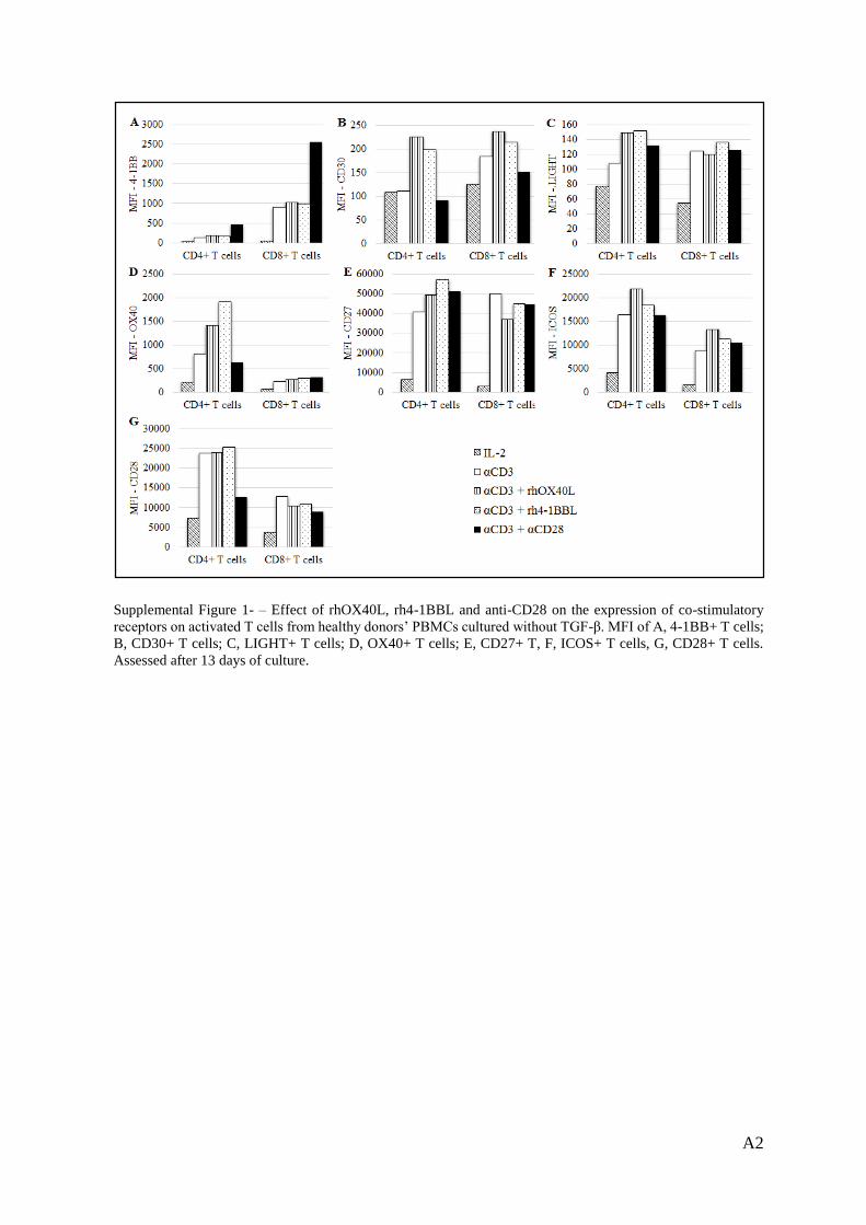
A2
Supplemental Figure 1- – Effect of rhOX40L, rh4-1BBL and anti-CD28 on the expression of co-stimulatory
receptors on activated T cells from healthy donors’ PBMCs cultured without TGF-β. MFI of A, 4-1BB+ T cells;
B, CD30+ T cells; C, LIGHT+ T cells; D, OX40+ T cells; E, CD27+ T, F, ICOS+ T cells, G, CD28+ T cells.
Assessed after 13 days of culture.
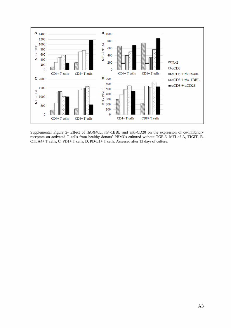
A3
Supplemental Figure 2- Effect of rhOX40L, rh4-1BBL and anti-CD28 on the expression of co-inhibitory
receptors on activated T cells from healthy donors’ PBMCs cultured without TGF-β. MFI of A, TIGIT, B,
CTLA4+ T cells; C, PD1+ T cells; D, PD-L1+ T cells. Assessed after 13 days of culture.