Pattern of distribution of serotonergic fibers to …vertes/sert-thalamus.pdfORIGINAL ARTICLE...
28
ORIGINAL ARTICLE Pattern of distribution of serotonergic fibers to the thalamus of the rat Robert P. Vertes • Stephanie B. Linley • Walter B. Hoover Received: 31 December 2009 / Accepted: 16 March 2010 / Published online: 13 April 2010 Ó Springer-Verlag 2010 Abstract It is well established that serotonergic (5-hydroxytryptamine, 5-HT) fibers, mainly originating from the dorsal and median raphe nuclei of the brainstem, distribute throughout the forebrain, most heavily to ‘lim- bic’ forebrain structures. Few reports have examined the distribution of 5-HT fibers to the thalamus and none to our knowledge using immunoprocedures for the detection of the serotonin transporter (SERT)—a very sensitive marker for 5-HT fibers. Using immunohistochemical methods for SERT, we examined the pattern of distribution of 5-HT fibers to the thalamus in the rat. We show that serotonergic fibers are heavily concentrated in midline, intralaminar and association nuclei of the thalamus, and with the exception of the lateral geniculate complex, weakly distributed to principal nuclei of thalamus. Specifically, we demonstrate that 5-HT fibers are densely concentrated in the antero- ventral, anteromedial and interanteromedial nuclei of the anterior thalamus, the paraventricular, rhomboid and reuniens nuclei of the midline thalamus, the central medial and central lateral nuclei of the intralaminar thalamus, the intermediodorsal nucleus, the lateral dorsal nucleus, and the dorsal and ventral lateral geniculate nuclei and inter- geniculate leaflet of the LGN complex. Less densely innervated sites include the mediodorsal, paracentral, parafascicular, lateral posterior and submedial nuclei of thalamus. Remaining regions of the thalamus, largely consisting of principal nuclei, contained few 5-HT fibers. This pattern of 5-HT innervation indicates that serotonin/ serotonergic fibers mainly affect thalamic nuclei with connections to ‘non-principal’ or limbic regions of the cortex (or forebrain). This suggests that serotonergic fibers to the thalamus may exert a significant influence on affective and cognitive functions, possibly complementing the actions of 5-HT fibers to other parts of the brain involved in emotional and cognitive behaviors. Keywords Midline thalamus Á Intralaminar thalamus Á Lateral geniculate complex Á Limbic forebrain Á Nucleus reuniens Á Paraventricular nucleus of thalamus Á Affective behavior Abbreviations 5-HT 5-Hydroxytryptamine, serotonin AD Anterodorsal nucleus of the thalamus AM Anteromedial nucleus of thalamus APN Anterior pretectal nucleus AV Anteroventral nucleus of thalamus CL Central lateral nucleus of the thalamus CM Central medial nucleus of thalamus COM Commissural nucleus, periaqueductal gray cpd Cerebral peduncle DR Dorsal raphe nucleus ec External capsule EC Entorhinal cortex fr Fasciculus retroflexus fx Fornix HD Head direction HF Hippocampus IAD Interanterodorsal nucleus of thalamus R. P. Vertes (&) Á W. B. Hoover Center for Complex Systems and Brain Sciences, Florida Atlantic University, Boca Raton, FL 33431, USA e-mail: [email protected] S. B. Linley Department of Psychology, Florida Atlantic University, Boca Raton, FL 33431, USA 123 Brain Struct Funct (2010) 215:1–28 DOI 10.1007/s00429-010-0249-x
-
Upload
nguyentruc -
Category
Documents
-
view
226 -
download
0
Transcript of Pattern of distribution of serotonergic fibers to …vertes/sert-thalamus.pdfORIGINAL ARTICLE...
ORIGINAL ARTICLE
Pattern of distribution of serotonergic fibers to the thalamusof the rat
Robert P. Vertes • Stephanie B. Linley •
Walter B. Hoover
Received: 31 December 2009 / Accepted: 16 March 2010 / Published online: 13 April 2010
� Springer-Verlag 2010
Abstract It is well established that serotonergic
(5-hydroxytryptamine, 5-HT) fibers, mainly originating
from the dorsal and median raphe nuclei of the brainstem,
distribute throughout the forebrain, most heavily to ‘lim-
bic’ forebrain structures. Few reports have examined the
distribution of 5-HT fibers to the thalamus and none to our
knowledge using immunoprocedures for the detection of
the serotonin transporter (SERT)—a very sensitive marker
for 5-HT fibers. Using immunohistochemical methods for
SERT, we examined the pattern of distribution of 5-HT
fibers to the thalamus in the rat. We show that serotonergic
fibers are heavily concentrated in midline, intralaminar and
association nuclei of the thalamus, and with the exception
of the lateral geniculate complex, weakly distributed to
principal nuclei of thalamus. Specifically, we demonstrate
that 5-HT fibers are densely concentrated in the antero-
ventral, anteromedial and interanteromedial nuclei of the
anterior thalamus, the paraventricular, rhomboid and
reuniens nuclei of the midline thalamus, the central medial
and central lateral nuclei of the intralaminar thalamus, the
intermediodorsal nucleus, the lateral dorsal nucleus, and
the dorsal and ventral lateral geniculate nuclei and inter-
geniculate leaflet of the LGN complex. Less densely
innervated sites include the mediodorsal, paracentral,
parafascicular, lateral posterior and submedial nuclei of
thalamus. Remaining regions of the thalamus, largely
consisting of principal nuclei, contained few 5-HT fibers.
This pattern of 5-HT innervation indicates that serotonin/
serotonergic fibers mainly affect thalamic nuclei with
connections to ‘non-principal’ or limbic regions of the
cortex (or forebrain). This suggests that serotonergic fibers
to the thalamus may exert a significant influence on
affective and cognitive functions, possibly complementing
the actions of 5-HT fibers to other parts of the brain
involved in emotional and cognitive behaviors.
Keywords Midline thalamus � Intralaminar thalamus �Lateral geniculate complex � Limbic forebrain �Nucleus reuniens � Paraventricular nucleus of thalamus �Affective behavior
Abbreviations
5-HT 5-Hydroxytryptamine, serotonin
AD Anterodorsal nucleus of the thalamus
AM Anteromedial nucleus of thalamus
APN Anterior pretectal nucleus
AV Anteroventral nucleus of thalamus
CL Central lateral nucleus of the thalamus
CM Central medial nucleus of thalamus
COM Commissural nucleus, periaqueductal gray
cpd Cerebral peduncle
DR Dorsal raphe nucleus
ec External capsule
EC Entorhinal cortex
fr Fasciculus retroflexus
fx Fornix
HD Head direction
HF Hippocampus
IAD Interanterodorsal nucleus of thalamus
R. P. Vertes (&) � W. B. Hoover
Center for Complex Systems and Brain Sciences,
Florida Atlantic University, Boca Raton, FL 33431, USA
e-mail: [email protected]
S. B. Linley
Department of Psychology, Florida Atlantic University,
Boca Raton, FL 33431, USA
123
Brain Struct Funct (2010) 215:1–28
DOI 10.1007/s00429-010-0249-x
IAM Interanteromedial nucleus of thalamus
ic Internal capsule
IGL Intergeniculate leaflet
IL Intralaminar thalamus
IMD Intermediodorsal nucleus of thalamus
LD Lateral dorsal nucleus of thalamus
LGNd Dorsal lateral geniculate nucleus
LGNv,m,l Ventral lateral geniculate nucleus, medial and
lateral divisions
LH Lateral habenula
LHy Lateral hypothalamus
LPl,m Lateral posterior nucleus of thalamus, lateral
and medial divisions
MB Mammillary bodies
MDc,1,m Mediodorsal nucleus of thalamus, central,
lateral, and medial divisions
MGN Medial geniculate nucleus
MH Medial habenula
ml Medial lemniscus
MPT Medial pretectal nucleus
MR Median raphe nucleus
MRF Mesencephalic reticular formation
mt Mammillothalamic tract
NOT Nucleus of optic tract
NPC Nucleus of posterior commissure
OP Olivary pretectal nucleus
PAG Periaqueductal gray
pc Posterior commissure
PCN Paracentral nucleus of thalamus
PF Parafascicular nucleus of thalamus
PFC Prefrontal cortex
PH Posterior hypothalamus
PO Posterior nucleus of thalamus
PR Peri-reuniens nucleus
PT Paratenial nucleus of thalamus
PVa,p Paraventricular nucleus of thalamus, anterior
and posterior divisions
RE Nucleus reuniens of thalamus
RH Rhomboid nucleus of thalamus
RSC Retrosplenial cortex
RT Reticular nucleus of thalamus
SC Superior colliculus
SCN Suprachiasmatic nucleus
SERT Serotonin transporter
sm Stria medullaris
SMT Submedial nucleus of thalamus
SPF Subparafascicular nucleus
st Stria terminalis
VAL Ventroanterior lateral complex of thalamus
VB Ventrobasal complex of the thalamus
VM Ventral medial nucleus of thalamus
ZI Zona incerta
Introduction
It is well recognized that serotonin-containing (5-hydroxy-
tryptamine, 5-HT) fibers are widely distributed throughout
the neuroaxis. Although 5-HT fibers reach virtually all
areas of the forebrain, they are concentrated in limbic
regions of the forebrain (Steinbusch 1981; Jacobs and
Azmitia 1992; Halliday et al. 2004; Vertes and Linley
2007, 2008; Lowry et al. 2008a).
The majority of ascending 5-HT fibers originate from
the dorsal (DR) and median raphe (MR) nuclei of the
brainstem. Several reports have described patterns of dis-
tribution of DR/MR fibers to the forebrain. With respect to
the thalamus, early studies indicated rather limited DR/MR
projections to the thalamus (Azmitia and Segal 1978;
Moore et al. 1978; Vertes and Martin 1988) but subsequent
reports, using improved tracing techniques, showed rela-
tively substantial DR and MR afferents to the thalamus.
DR/MR fibers mainly target ‘non-specific’ (or limbic)
nuclei of the thalamus as well as parts of the ‘visual thal-
amus’. This would primarily include the anterior nuclei, the
mediodorsal nucleus, the midline and intralaminar nuclei,
the habenula, the laterodorsal nucleus, and the lateral
geniculate (LGN) complex. With a few exceptions, specific
(principal) nuclei of the thalamus appear to lack input from
DR/MR (Vertes 1991; Morin and Meyer-Bernstein 1999;
Vertes et al. 1999).
In addition to 5-HT cells, the DR and MR contain other
types of ‘projection’ neurons including dopaminergic,
GABAergic, glutamatergic and various peptide containing
cells (Trulson et al. 1985; Melander et al. 1986; Austin
et al. 1997; Charara and Parent 1998; Day et al. 2004;
Waselus and Van Bockstaele 2007; Lowry et al. 2008a).
Accordingly, the extent to which DR/MR projections to the
thalamus originate specifically from 5-HT DR/MR cells
remains to be determined. Although no report has exam-
ined the overall distribution of serotonergic DR/MR fibers
to the thalamus, a few studies have described 5-HT DR/MR
projections to some nuclei of the thalamus. Specifically,
studies combining retrograde tracing with 5-HT immuno-
staining have demonstrated 5-HT DR or MR projections to
the anterodorsal and anteroventral nuclei of the anterior
thalamus (Gonzalo-Ruiz et al. 1995), to the lateral genic-
ulate nucleus and intergeniculate leaflet (Villar et al. 1988;
Meyer-Bernstein and Morin 1996; Harrington 1997; Morin
and Blanchard 1999) and to the paraventricular nucleus
(Otake and Ruggiero 1995; Hsu and Price 2009).
The pattern of distribution of serotonergic dorsal and
median raphe fibers to most nuclei of the thalamus remains
largely unknown. Despite this, the finding of two early
immunohistochemical analyses, one in rats (Cropper et al.
1984) and the other in monkeys (Lavoie and Parent 1991),
2 Brain Struct Funct (2010) 215:1–28
123

showed that 5-HT fibers spread widely throughout the
thalamus. These early studies, however, used immuno-
staining procedures for the detection of serotonin in cells/
fibers (Steinbusch 1981), and while still a widely utilized
technique, newer immunostaining procedures have been
developed for the identification of the serotonin transporter
protein (SERT) in 5-HT fibers (Sur et al. 1996; Zhou et al.
1996). Although both methods are useful and each has its
unique advantages, Aznar and colleagues (Nielsen et al.
2006) recently reported that SERT was preferable to 5-HT
as a marker for serotonergic fibers.
In particular, they showed that serotonin only began to
approach the quality of SERT for identifying 5-HT fibers
when 5-HT immunohistochemistry was combined with
pre-treatments, particularly the use of monoamine oxidase
inhibitors (MAOIs). For instance, they reported: (1) an
approximately 200% increase in the detection of 5-HT
immunolabeled fibers in rats pre-treated with MAOIs and
(2) *90% correspondence in the co-expression of 5-HT?
and SERT? fibers in MAOI-treated rats compared to only
a 30% correspondence in non-treated rats. The latter dif-
ference (30% correspondence) was attributed to the loss (or
inability to detect) of immunostained 5-HT fibers in
untreated animals.
Consistent with Nielsen et al. (2006), we found that
5-HT and SERT immunoreactive procedures labeled a
comparable set of 5-HT fibers in the thalamus, and that
SERT produced a stronger signal than did 5-HT. Accord-
ingly, we used SERT immunohistochemical techniques to
characterize the pattern of distribution of serotonergic
fibers to the thalamus of the rat.
In brief, we show that serotonergic fibers are densely
concentrated in midline nuclei, rostral intralaminar nuclei,
most of the anterior nuclei, the laterodorsal nucleus, and
the LGN complex. Of the midline group, 5-HT labeling
was most pronounced in the paraventricular, rhomboid and
reuniens nuclei. Although no region of the thalamus was
devoid of 5-HT fibers, labeling was very light in sensory
and motor nuclei of the thalamus and in the medial
geniculate nucleus.
Materials and methods
Ten (5 male, 5 female) naıve Sprague-Dawley rats (Harlan,
Indianapolis, IN) weighing 275–300 g were housed in pairs
on a 12:12 light dark cycle for 7 days during which food
and water were available ad libitum. Rats were then deeply
anesthetized with an intraperitoneal injection of sodium
pentobarbital (Nembutal, 75 mg/kg) and perfused tran-
scardially with 30 ml of cold heparinized 0.1 M phosphate
buffer saline (PBS), followed by 200–300 ml chilled 4%
paraformaldehyde in 0.1 M phosphate buffer (PB) at pH of
7.4. The brains were removed and postfixed overnight in
4% paraformaldehyde in 0.1 M PB. Brains were then
placed in a 30% sucrose solution for another 48 h. Fol-
lowing this, 50 lm coronal sections were taken on a
freezing microtome in a one in three series: one series of
sections through the thalamus were prepared for SERT
immunohistochemistry and a second series was stained
with cresyl violet. The experiments were approved by the
Florida Atlantic University Institutional Animal Care and
Use Committee and conform to all federal regulations and
National Institutes of Health guidelines for the care and use
of laboratory animals.
Immunohistochemistry
SERT
Sections were initially treated with a 30-min sodium
borohydride incubation (1% in 0.1 M PB) to remove
excess aldehydes. Following a copious PB wash, sections
were incubated for 1 h in 0.5% BSA in 0.1 M Tris buffered
saline (TBS) (pH 7.6). Sections were rinsed in 0.1 M PB
and then incubated in the primary antibody, rabbit anti-
SERT (Immunostar, Hudson, WI) at a concentration of
1:10,000 in a diluent of 0.1% bovine serum albumin (BSA)
in TBS containing 0.25% Triton X-100 for 48 h. Following
a 0.1 M PB wash, sections were placed in a secondary
antibody solution of biotinylated goat anti-rabbit immu-
noglobulin (Vector Labs) at a 1:500 concentration in dil-
uent for 2 h. This was followed by another PB wash.
Sections were then incubated for 2 h in a tertiary antibody
solution of biotinylated horse anti-goat immunoglobulin
(Vector Labs) at a 1:500 concentration in diluent. After
washing the tissue in 0.1 M PB, sections were incubated
for 60 min in a 1:200 dilution of a peroxidase–avidin
complex using the Vector Elite kit. Following a final 0.1 M
PB wash, SERT immunoreactive fibers were visualized by
placing them for approximately 3–4 min in a solution
containing 0.022% DAB (Aldrich, Milwaukee, WI) and
0.003% hydrogen peroxide in TBS. Sections were mounted
onto chrome-alum gelatin-coated slides, dehydrated using
graded methanols and coverslipped with permount.
Photomicroscopy
For depiction of SERT and 5-HT fibers, lightfield photo-
micrographs at 100 times magnification were captured
throughout the extent of the thalamus from representative
cases using a Nikon DXM1200 camera mounted onto a
Nikon Eclipse E600 microscope. The individual captures
were then compiled using Image Pro-Plus 4.5 (Media
Cybernetics, Silver Springs, MD) and imported into Adobe
Photoshop (CS 2.0; Mountain View, CA) where they were
Brain Struct Funct (2010) 215:1–28 3
123
adjusted for brightness and contrast. Representative sec-
tions throughout the thalamus were captured and illus-
trated. Files were imported into Adobe Illustrator (CS 2.0)
where borders were drawn around thalamic nuclei by
comparing immunostained sections to an adjacent series
of Nissl-stained sections and with the aid of the rat atlas
of Swanson (2003). Particularly noteworthy patterns of
labeling were illustrated with high magnification photo-
micrographs. Patterns of SERT labeling are described as
light, moderate and dense, with ‘light’ referring to a few
labeled fibers widely dispersed throughout a nucleus,
‘dense’ as a heavy concentration of labeled fibers generally
occupying a significant portion (or most) of a nucleus, and
‘moderate’ lying between these two patterns.
Results
The pattern of distribution of SERT immunoreactive fibers
in the thalamus of the rat is depicted with a series of 14
rostral to caudal transverse sections through the thalamus
(plates 1–14). We first describe patterns of labeling at each
rostral to caudal section and then compare patterns across
anatomical/functional groups of the thalamus.
A note on categories of thalamic nuclei
An early categorization of thalamic nuclei essentially
divided the thalamus into ‘relay’ nuclei and ‘non-specific’
nuclei (Dempsey and Morison 1942, 1943; Morison and
Dempsey 1942). The relay nuclei generally referred to
nuclei that transfer modality-specific information to dis-
crete regions and layers of the cortex, while ‘non-specific’
nuclei referred to nuclei that received multimodal infor-
mation and distribute to several regions of the cortex as
well as to subcortical sites. This categorization has been
revised in large part on the basis that ‘‘non-specific’’ nuclei
target very specific regions of the cortex and exhibit unique
functions (Bentivoglio et al. 1991; Groenewegen and
Berendse 1994).
Based on recent formulations (Price 1995; Groenewegen
and Witter 2004), thalamic nuclei will be categorized as
follows: (1) principal nuclei, consisting of the ventrobasal
complex (ventral posteromedial and ventral posterolateral
nuclei), the ventroanterior lateral complex, the ventrome-
dial nucleus, the posterior nucleus, the dorsal and ventral
lateral geniculate nuclei, the intergeniculate leaflet, and the
medial geniculate nucleus; (2) association nuclei, consist-
ing of the mediodorsal and intermediodorsal nuclei, the
submedial nucleus, the anterior nuclei (anterodorsal, ante-
roventral, anteromedial and interanteromedial), the lateral
dorsal nucleus and the lateral posterior nucleus; (3) midline
and intralaminar nuclei, consisting of nucleus reuniens,
rhomboid nucleus, paraventricular nucleus, paratenial
nucleus, the central medial nucleus, paracentral nucleus,
central lateral nucleus and the parafascicular nucleus; (4)
the reticular nucleus; and (5) the epithalamus, consisting of
the medial and lateral habenula.
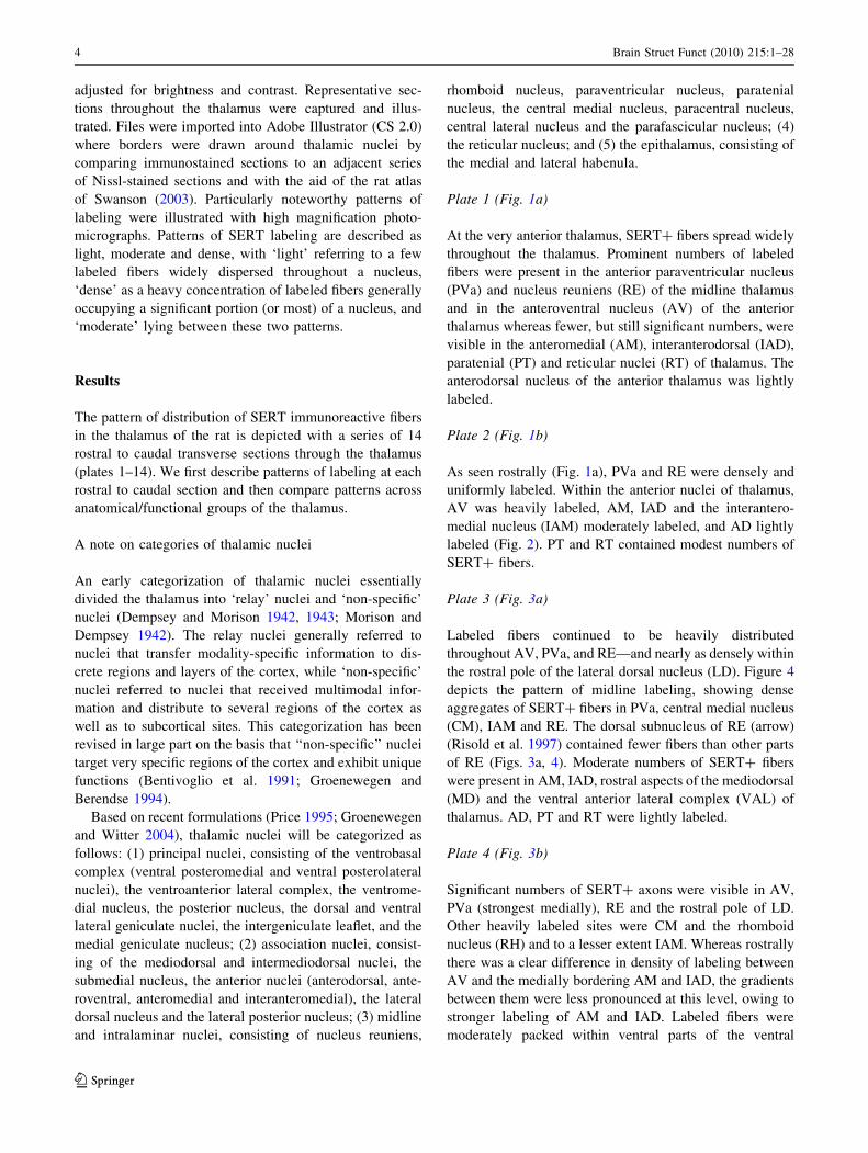
Plate 1 (Fig. 1a)
At the very anterior thalamus, SERT? fibers spread widely
throughout the thalamus. Prominent numbers of labeled
fibers were present in the anterior paraventricular nucleus
(PVa) and nucleus reuniens (RE) of the midline thalamus
and in the anteroventral nucleus (AV) of the anterior
thalamus whereas fewer, but still significant numbers, were
visible in the anteromedial (AM), interanterodorsal (IAD),
paratenial (PT) and reticular nuclei (RT) of thalamus. The
anterodorsal nucleus of the anterior thalamus was lightly
labeled.
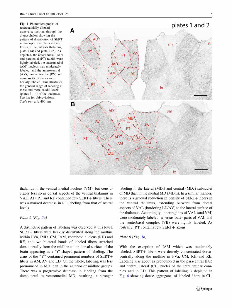
Plate 2 (Fig. 1b)
As seen rostrally (Fig. 1a), PVa and RE were densely and
uniformly labeled. Within the anterior nuclei of thalamus,
AV was heavily labeled, AM, IAD and the interantero-
medial nucleus (IAM) moderately labeled, and AD lightly
labeled (Fig. 2). PT and RT contained modest numbers of
SERT? fibers.
Plate 3 (Fig. 3a)
Labeled fibers continued to be heavily distributed
throughout AV, PVa, and RE—and nearly as densely within
the rostral pole of the lateral dorsal nucleus (LD). Figure 4
depicts the pattern of midline labeling, showing dense
aggregates of SERT? fibers in PVa, central medial nucleus
(CM), IAM and RE. The dorsal subnucleus of RE (arrow)
(Risold et al. 1997) contained fewer fibers than other parts
of RE (Figs. 3a, 4). Moderate numbers of SERT? fibers
were present in AM, IAD, rostral aspects of the mediodorsal
(MD) and the ventral anterior lateral complex (VAL) of
thalamus. AD, PT and RT were lightly labeled.
Plate 4 (Fig. 3b)
Significant numbers of SERT? axons were visible in AV,
PVa (strongest medially), RE and the rostral pole of LD.
Other heavily labeled sites were CM and the rhomboid
nucleus (RH) and to a lesser extent IAM. Whereas rostrally
there was a clear difference in density of labeling between
AV and the medially bordering AM and IAD, the gradients
between them were less pronounced at this level, owing to
stronger labeling of AM and IAD. Labeled fibers were
moderately packed within ventral parts of the ventral
4 Brain Struct Funct (2010) 215:1–28
123
thalamus in the ventral medial nucleus (VM), but consid-
erably less so in dorsal aspects of the ventral thalamus in
VAL. AD, PT and RT contained few SERT? fibers. There
was a marked decrease in RT labeling from that of rostral
levels.
Plate 5 (Fig. 5a)
A distinctive pattern of labeling was observed at this level.
SERT? fibers were heavily distributed along the midline
within PVa, IMD, CM, IAM, rhomboid nucleus (RH) and
RE, and two bilateral bands of labeled fibers stretched
dorsolaterally from the midline to the dorsal surface of the
brain appearing as a ‘Y’-shaped pattern of labeling. The
arms of the ‘‘Y’’ contained prominent numbers of SERT?
fibers in AM, AV and LD. On the whole, labeling was less
pronounced in MD than in the anterior or midline groups.
There was a progressive decrease in labeling from the
dorsolateral to ventromedial MD, resulting in stronger
labeling in the lateral (MDl) and central (MDc) subnuclei
of MD than in the medial MD (MDm). In a similar manner,
there is a gradual reduction in density of SERT? fibers in
the ventral thalamus, extending outward from dorsal
aspects of VAL (bordering LD/AV) to the lateral surface of
the thalamus. Accordingly, inner regions of VAL (and VM)
were moderately labeled, whereas outer parts of VAL and
the ventrobasal complex (VB) were lightly labeled. As
rostrally, RT contains few SERT? axons.
Plate 6 (Fig. 5b)
With the exception of IAM which was moderately
labeled, SERT? fibers were densely concentrated dorso-
ventrally along the midline in PVa, CM, RH and RE.
Labeling was about as pronounced in the paracentral (PC)
and central lateral (CL) nuclei of the intralaminar com-
plex and in LD. This pattern of labeling is depicted in
Fig. 6 showing dense aggregates of labeled fibers in CL,
Fig. 1 Photomicrographs of
rostrocaudally aligned
transverse sections through the
diencephalon showing the
pattern of distribution of SERT
immunopositive fibers at two
levels of the anterior thalamus,
plate 1 (a) and plate 2 (b). As
depicted, the anterodorsal (AD)
and paratenial (PT) nuclei were
lightly labeled, the anteromedial
(AM) nucleus was moderately
labeled, and the anteroventral
(AV), paraventricular (PV) and
reuniens (RE) nuclei were
heavily labeled. This illustrates
the general range of labeling at
these and more caudal levels
(plates 3–14) of the thalamus.
See list for abbreviations.
Scale bar a, b 400 lm
Brain Struct Funct (2010) 215:1–28 5
123
PC and LD—considerably more than present dorsomedi-
ally in MD or ventrolaterally in VAL. As observed ros-
trally, there was a gradual dorsal to ventral decline in
labeling from deep (inner) to superficial aspects of the
ventral thalamus (VAL to VB). VM and the medially
adjacent submedial nucleus (SMT) were moderately and
uniformly labeled. RT contained few SERT immunore-
active fibers.
Plate 7 (Fig. 7)
Three major clusters of SERT? fibers were visible at this
level: (1) a midline group, with labeling heaviest in PVa,
RH and RE and less pronounced in the intermediodorsal
nucleus (IMD); (2) an intralaminar group, with labeling
strongest in CM followed by CL and PC; and (3) the lateral
dorsal nucleus. Moderate numbers of SERT immunoreac-
tive fibers were also present in the medial (MH) and lateral
habenula (LH) and in SMT, but relatively few were seen
throughout the vast expanse of ventral thalamus (VM,
VAL, VB) and in RT. MD was lightly to moderately
labeled which contrasted with much stronger labeling of
adjacent regions, medially (PVa, IMD, CM) and laterally
(CL, PC). As depicted in Fig. 8, there were subtle differ-
ences in density of labeling across subdivisions of MD such
that the central nucleus (MDc) was more heavily labeled
than the medial (MDm) or lateral MD (MDl).
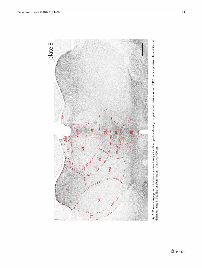
Plate 8 (Fig. 9)
SERT? fibers heavily invaded the midline and LD, but
were sparsely distributed over a large expanse of the ven-
tral thalamus in VB, VAL and RT. Within midline nuclei,
labeling was densest in PVp, RH and RE, but was also
pronounced in CM. There was a gradual decline in labeling
medially to laterally in the ventral thalamus, from VM to
VAL to VB. SMT contained moderate numbers of SERT?
fibers, intermediate between those of the laterally adjacent
VM and medially bordering RH/RE. MD was moderately
labeled, with slightly more labeled fibers in MDl and MDc
than in MDm.
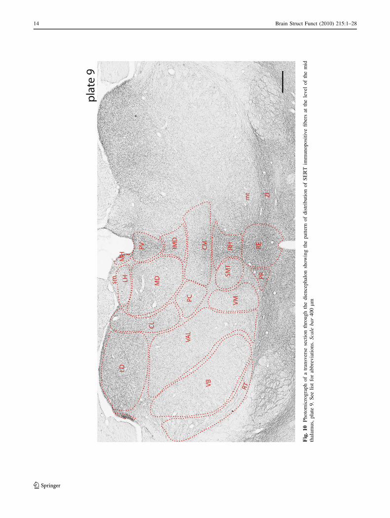
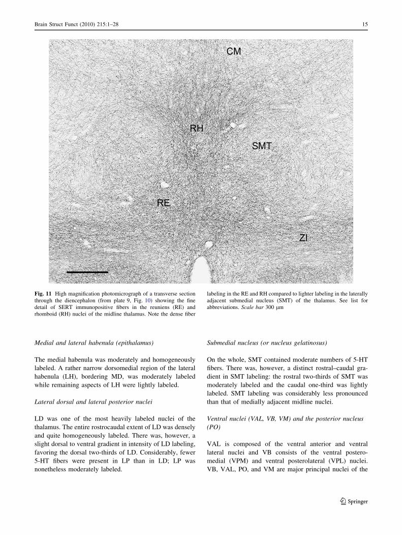
Plate 9 (Fig. 10)
There was a general reduction in labeling at caudal levels
of the thalamus due to the fact that a greater part of the
Fig. 2 High magnification
photomicrograph of a transverse
section through the rostral
diencephalon (from left side of
plate 2, Fig. 1b) showing the
fine detail of SERT
immunopositive fibers in the
anterior thalamus. Note dense
fiber labeling in the
anteroventral (AV)
paraventricular (PV) and
reuniens (RE) nuclei of the
thalamus. See list for
abbreviations. Scale bar150 lm
6 Brain Struct Funct (2010) 215:1–28
123

caudal thalamus is occupied by principal as opposed to
non-principal nuclei. Although labeling was still prominent
along the midline in PVp, IMD, CM, RH and RE, it was
less robust than seen rostrally within these nuclei and fairly
confined to a narrow band along the midline. Figure 11
shows a dense collection of labeled fibers within RE and
the dorsally adjacent RH at this level. Comparable to
midline labeling, LD was densely labeled and CL, PC and
MD (or lateral parts of MD) were moderately labeled.
Relatively few SERT? fibers were present throughout the
vast expanse of thalamus, mainly comprised of the ventral/
ventrolateral thalamus, i.e., within the posterior nucleus
(PO), VAL, VB and VM. The habenula was lightly labeled
(MH [ LH).
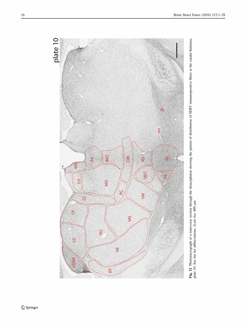
Plate 10 (Fig. 12)
Whereas rostrally in the thalamus labeling was described as
forming a ‘Y’-shaped configuration (plate 5, Fig. 5a), it
largely resembled a ‘T’-shaped pattern at this level. In
effect, this consisted of a vertical column of midline
labeling in MH, PVp, IMD, CM, RE, and lateral extensions
from the midline across the dorsal surface of the brain
within CL, the lateral posterior nucleus (LP), LD and the
Fig. 3 Photomicrographs of rostrocaudally aligned transverse sections through the diencephalon showing the pattern of distribution of SERT
immunopositive fibers at two levels of the anterior thalamus, plate 3 (a) and plate 4 (b). See list for abbreviations. Scale bar a, b 400 lm
Brain Struct Funct (2010) 215:1–28 7
123

dorsal lateral geniculate nucleus (LGNd). Of these sites,
labeling was densest in PVp, RE, LD and LGNd. Addi-
tionally, moderate numbers of SERT? fibers were present
in MD, PC, VM and SMT, but few were visible in most
remaining regions of the thalamus including PO, VAL, VB
and RT.
Plate 11 (Fig. 13)
As discussed, associated with the progressive switch from
midline/intralaminar and association nuclei to principal
nuclei (anterior to posterior thalamus), there was a corre-
sponding gradual reduction in numbers of SERT? fibers.
This is further exemplified at this level. Whereas the LGN
complex (LGNd, LGNv), PVp and RE were densely
labeled, relatively few SERT? fibers were present
throughout most of the thalamus in MH, IMD, CL, LP,
MD, CM (moderately labeled) as well in PO, VB, VM and
RT (lightly labeled). PO labeling was slightly stronger at
this level than rostrally—a trend that continued caudally in
the thalamus.
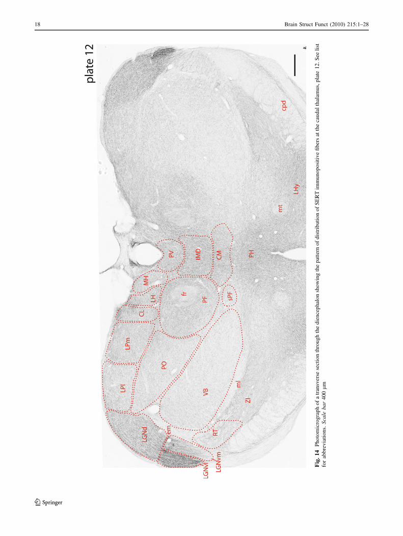
Plate 12 (Fig. 14)
Although labeling was still present along the midline
within PVp, IMD and CM, it was less pronounced at this
level than rostrally. The parafascicular nucleus (PF) lateral
to IMD was relatively densely labeled which set it off from
surrounding nuclei which were less heavily labeled. With
the exception of the LGN complex (LGNd and LGNv)
which was strongly labeled, SERT? fibers spread moder-
ately (and homogeneously) throughout the dorsal thalamus
to MH, LH, caudal CL, and LP. By contrast with the dorsal
thalamus, the ventral thalamus (mainly composed of PO
and VB) contained few SERT? fibers—more in PO than in
VB.
Plate 13 (Fig. 15)
At the caudal pole of the thalamus (or juncture between the
diencephalon and midbrain), thalamic nuclei are mainly
located laterally/dorsolaterally, lateral to the emerging
pretectal area. Within the dorsolateral thalamus, LGNd,
Fig. 4 High magnification
photomicrograph of a transverse
section through the rostral
diencephalon (from plate 3,
Fig. 3a) showing the fine detail
of SERT immunopositive fibers
in midline and adjacent nuclei at
the level of the anterior
thalamus. Note dense fiber
labeling in the paraventricular
(PV) and reuniens (RE) nuclei
of the midline thalamus and the
anteromedial (AM) and
interanteromedial nuclei of the
anterior thalamus. See list for
abbreviations. Scale bar300 lm
8 Brain Struct Funct (2010) 215:1–28
123

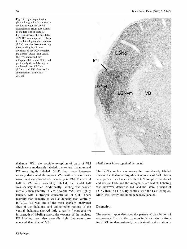
LGNv and the intergeniculate leaflet (IGL) were densely
labeled, PO, LP and PF were moderately labeled and VB
was lightly labeled. Figure 16 shows a dense collection of
SERT? fibers throughout the LGN complex and particu-
larly strong within IGL and the lateral division of the
ventral LGN.
Plate 14 (Fig. 17)
The caudal extent of the thalamus consists of visual and
auditory structures. Of these, labeling was pronounced in
the LGNd, LGNv and IGL, moderate in LP and light in the
medial geniculate nucleus (MGN).
Fig. 5 Photomicrographs of rostrocaudally aligned transverse sections through the diencephalon showing the pattern of distribution of SERT
immunopositive fibers at two levels of the mid thalamus, plate 5 (a) and plate 6 (b). See list for abbreviations. Scale bar a, b 400 lm
Brain Struct Funct (2010) 215:1–28 9
123

Functional/anatomical groups of the thalamus
Anterior nuclei
The anterior nuclei consist of the anterodorsal (AD),
anteroventral (AV), anteromedial (AM), interanterodorsal
(IAD) and interanteromedial (IAM) nuclei. On the whole,
the anterior group contained dense concentrations of 5-HT
fibers. AV was the most heavily labeled nucleus of the
anterior group and one of the most densely labeled sites of
the thalamus. Labeling was almost as prominent in AM,
IAD and IAM. By contrast, AD was lightly labeled. For the
most part, 5-HT fibers were homogeneously distributed
throughout the anterior thalamic nuclei.
Midline nuclei
The midline nuclei consist of the paraventricular (PV),
paratenial (PT), rhomboid (RH) and reuniens (RE) nuclei.
Other nuclei lying along the midline of the thalamus which
are not considered part of the midline nuclei, per se,
include the intermediodorsal (IMD), interanteromedial
(IAM), and central medial (CM) nuclei. Whereas the entire
dorsoventral extent of the midline thalamus (including
IMD, IAM and CM) was densely labeled, labeling was
heaviest within PV, RH and RE. In marked contrast to
other midline groups, PT was lightly labeled. Although
5-HT fibers generally distributed homogeneously through-
out the midline nuclei, there was some variation. Specifi-
cally, labeling was heavier in rostral than caudal regions of
PT, PV and RE, and somewhat denser in medial than
lateral aspects of PV and RE, particularly within the central
core compared to the lateral wings of RE.
Mediodorsal (MD) and intermediodorsal (IMD) nuclei
IMD lies on the midline and as a ‘midline-localized’ group
contained a dense concentration of 5-HT fibers—consid-
erably more than the laterally adjacent MD. On the whole,
MD contained moderate numbers of 5-HT fibers; fewer
Fig. 6 High magnification photomicrograph of a transverse section
through the diencephalon (from the right side of plate 5, Fig. 5a)
showing the fine detail of SERT immunopositive fibers in the
intralaminar and laterodorsal nuclei of the thalamus. Note the
relatively dense fiber labeling in the paracentral (PC), central lateral
(CL) and laterodorsal (LD) nuclei and comparatively lighter labeling
in the mediodorsal (MD) and ventral anterior lateral (VAL) nuclei of
thalamus. See list for abbreviations. Scale bar 300 lm
10 Brain Struct Funct (2010) 215:1–28
123
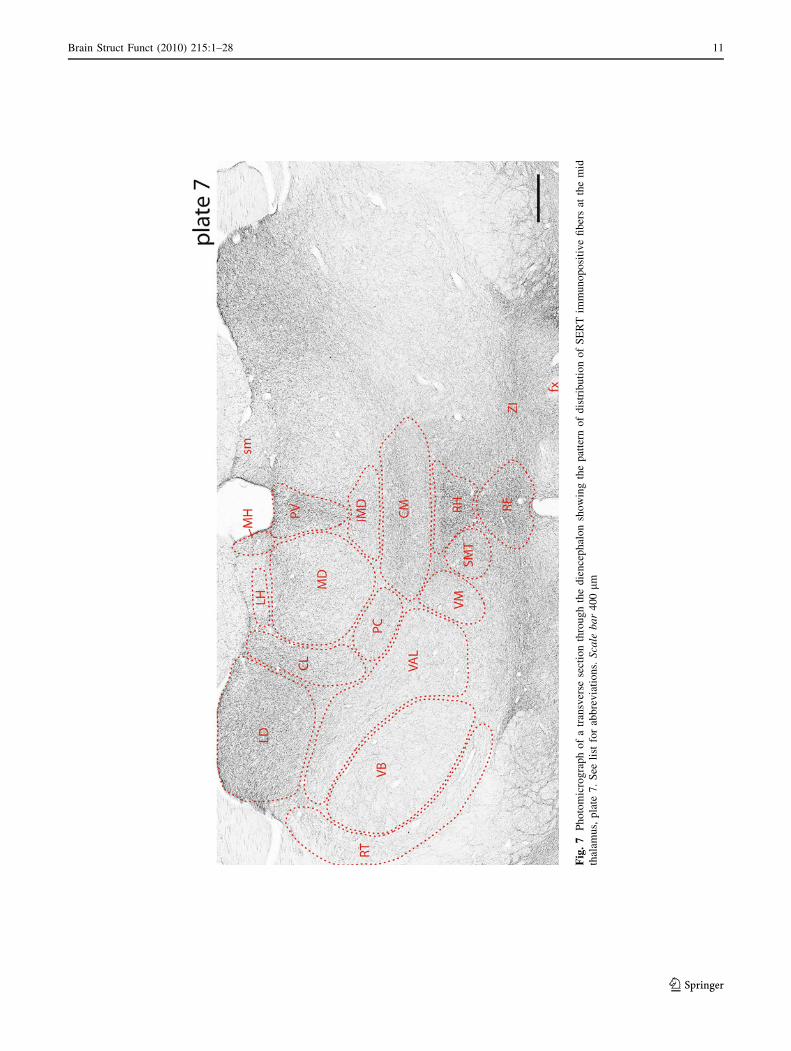
Fig
.7
Ph
oto
mic
rog
rap
ho
fa
tran
sver
sese
ctio
nth
rou
gh
the
die
nce
ph
alo
nsh
ow
ing
the
pat
tern
of
dis
trib
uti
on
of
SE
RT
imm
un
op
osi
tiv
efi
ber
sat
the
mid
thal
amu
s,p
late
7.
See
list
for
abb
rev
iati
on
s.S
cale
ba
r4
00
lm
Brain Struct Funct (2010) 215:1–28 11
123

than in bordering nuclei, medially (PV, IMD), laterally
(CL, PC), and perhaps dorsally (LH). There was a distinct
dorsolateral to ventromedial gradient in density of labeling
within the caudal two-thirds of MD resulting in denser
labeling in MDl and MDc than in MDm.
Intralaminar nuclei
The intralaminar (IL) nuclei consist of the central lateral (CL),
paracentral (PC) and central medial (CM) nuclei, rostrally,
and the parafascicular nucleus (PF), caudally. On the whole,
5-HT fibers were quite densely distributed throughout the
rostral intralaminar (IL) complex. As discussed with respect
to IMD, and perhaps owing to its position on the midline, CM
was the most heavily labeled nucleus of the rostral IL,
followed in order by CL and PC. Labeled fibers were
homogenously distributed rostrocaudally throughout CL, but
were more densely concentrated in lateral (adjacent to LD)
than in medial aspects of CL. In an analogous but reverse
manner, labeling was slightly denser medially in PC (on the
border with CM) than laterally in PC. PF was moderately to
heavily labeled—equivalent in density to that to CL.
Reticular nucleus
Somewhat surprisingly, with the exception of the rostral
pole of RT which was moderately labeled, RT contained
relatively few 5-HT fibers. In fact, labeling throughout
approximately the caudal two-thirds of RT was roughly
equivalent to that of the dorsally adjacent ventrobasal
complex which was among the most sparsely labeled sites
of the thalamus.
Fig. 8 High magnification photomicrograph of a transverse section
through the diencephalon (from the right side of plate 7, Fig. 7)
showing the fine detail of SERT immunopositive fibers in mediodor-
sal nucleus (MD) and surrounding regions of the thalamus. Note the
slightly denser labeling in the central (MDc) than the medial (MDm)
or lateral (MDl) divisions of MD. See list for abbreviations. Scale bar300 lm
12 Brain Struct Funct (2010) 215:1–28
123
Fig
.9
Ph
oto
mic
rog
rap
ho
fa
tran
sver
sese
ctio
nth
rou
gh
the
die
nce
ph
alo
nsh
ow
ing
the
pat
tern
of
dis
trib
uti
on
of
SE
RT
imm
un
op
osi
tiv
efi
ber
sat
the
mid
thal
amu
s,p
late
8.
See
list
for
abb
rev
iati
on
s.S
cale
ba
r4
00
lm
Brain Struct Funct (2010) 215:1–28 13
123
Fig
.1
0P
ho
tom
icro
gra
ph
of
atr
ansv
erse
sect
ion
thro
ug
hth
ed
ien
cep
hal
on
sho
win
gth
ep
atte
rno
fd
istr
ibu
tio
no
fS
ER
Tim
mu
no
po
siti
ve
fib
ers
atth
ele
vel
of
the
mid
thal
amu
s,p
late
9.
See
list
for
abb
rev
iati
on
s.S
cale
ba
r4
00
lm
14 Brain Struct Funct (2010) 215:1–28
123
Medial and lateral habenula (epithalamus)
The medial habenula was moderately and homogeneously
labeled. A rather narrow dorsomedial region of the lateral
habenula (LH), bordering MD, was moderately labeled
while remaining aspects of LH were lightly labeled.
Lateral dorsal and lateral posterior nuclei
LD was one of the most heavily labeled nuclei of the
thalamus. The entire rostrocaudal extent of LD was densely
and quite homogeneously labeled. There was, however, a
slight dorsal to ventral gradient in intensity of LD labeling,
favoring the dorsal two-thirds of LD. Considerably, fewer
5-HT fibers were present in LP than in LD; LP was
nonetheless moderately labeled.
Submedial nucleus (or nucleus gelatinosus)
On the whole, SMT contained moderate numbers of 5-HT
fibers. There was, however, a distinct rostral–caudal gra-
dient in SMT labeling: the rostral two-thirds of SMT was
moderately labeled and the caudal one-third was lightly
labeled. SMT labeling was considerably less pronounced
than that of medially adjacent midline nuclei.
Ventral nuclei (VAL, VB, VM) and the posterior nucleus
(PO)
VAL is composed of the ventral anterior and ventral
lateral nuclei and VB consists of the ventral postero-
medial (VPM) and ventral posterolateral (VPL) nuclei.
VB, VAL, PO, and VM are major principal nuclei of the
Fig. 11 High magnification photomicrograph of a transverse section
through the diencephalon (from plate 9, Fig. 10) showing the fine
detail of SERT immunopositive fibers in the reuniens (RE) and
rhomboid (RH) nuclei of the midline thalamus. Note the dense fiber
labeling in the RE and RH compared to lighter labeling in the laterally
adjacent submedial nucleus (SMT) of the thalamus. See list for
abbreviations. Scale bar 300 lm
Brain Struct Funct (2010) 215:1–28 15
123
Fig
.1
2P
ho
tom
icro
gra
ph
of
atr
ansv
erse
sect
ion
thro
ug
hth
ed
ien
cep
hal
on
sho
win
gth
ep
atte
rno
fd
istr
ibu
tio
no
fS
ER
Tim
mu
no
po
siti
ve
fib
ers
atth
eca
ud
alth
alam
us,
pla
te1
0.
See
list
for
abb
rev
iati
on
s.S
cale
ba
r4
00
lm
16 Brain Struct Funct (2010) 215:1–28
123

Fig
.1
3P
ho
tom
icro
gra
ph
of
atr
ansv
erse
sect
ion
thro
ug
hth
ed
ien
cep
hal
on
sho
win
gth
ep
atte
rno
fd
istr
ibu
tio
no
fS
ER
Tim
mu
no
po
siti
ve
fib
ers
atth
eca
ud
alth
alam
us,
pla
te1
1.
See
list
for
abb
rev
iati
on
s.S
cale
ba
r4
00
lm
Brain Struct Funct (2010) 215:1–28 17
123
Fig
.1
4P
ho
tom
icro
gra
ph
of
atr
ansv
erse
sect
ion
thro
ug
hth
ed
ien
cep
hal
on
sho
win
gth
ep
atte
rno
fd
istr
ibu
tio
no
fS
ER
Tim
mu
no
po
siti
ve
fib
ers
atth
eca
ud
alth
alam
us,
pla
te1
2.S
eeli
st
for
abb
rev
iati
on
s.S
cale
ba
r4
00
lm
18 Brain Struct Funct (2010) 215:1–28
123

Fig
.1
5P
ho
tom
icro
gra
ph
of
atr
ansv
erse
sect
ion
thro
ug
hth
ed
ien
cep
hal
on
sho
win
gth
ep
atte
rno
fd
istr
ibu
tio
no
fS
ER
Tim
mu
no
po
siti
ve
fib
ers
atth
eca
ud
alth
alam
us,
pla
te1
3.
See
list
for
abb
rev
iati
on
s.S
cale
ba
r4
00
lm
Brain Struct Funct (2010) 215:1–28 19
123
thalamus. With the possible exception of parts of VM
which were moderately labeled, the ventral thalamus and
PO were lightly labeled. 5-HT fibers were heteroge-
neously distributed throughout VM, with a marked var-
iation in density found rostrocaudally in VM. The rostral
half of VM was moderately labeled; the caudal half
was sparsely labeled. Additionally, labeling was heavier
medially than laterally in VM. Overall, VAL was lightly
labeled, with a stronger concentration of 5-HT fibers
rostrally than caudally as well as dorsally than ventrally
in VAL. VB was one of the most sparsely innervated
sites of the thalamus, and unlike other regions of the
ventral thalamus, showed little diversity (heterogeneity)
in strength of labeling across the expanse of the nucleus.
PO labeling was also generally light but more pro-
nounced than that of VB.
Medial and lateral geniculate nuclei
The LGN complex was among the most densely labeled
sites of the thalamus. Significant numbers of 5-HT fibers
were present in all nuclei of the LGN complex: the dorsal
and ventral LGN and the intergeniculate leaflet. Labeling
was, however, denser in IGL and the lateral division of
LGNv than in LGNd. By contrast with the LGN complex,
MGN was lightly and homogeneously labeled.
Discussion
The present report describes the pattern of distribution of
serotonergic fibers to the thalamus in the rat using antisera
for SERT. As demonstrated, there is significant variation in
Fig. 16 High magnification
photomicrograph of a transverse
section through the caudal
diencephalon (from just rostral
to the left side of plate 13,
Fig. 15) showing the fine detail
of SERT immunopositive fibers
in the lateral geniculate nucleus
(LGN) complex. Note the strong
fiber labeling in all three
divisions of the LGN complex,
the dorsal (LGNd) and ventral
(LGNv) nuclei and the
intergeniculate leaflet (IGL) and
particularly dense labeling in
the lateral part of LGNv
(LGNvl) and IGL. See list for
abbreviations. Scale bar250 lm
20 Brain Struct Funct (2010) 215:1–28
123

Fig
.1
7P
ho
tom
icro
gra
ph
of
atr
ansv
erse
sect
ion
thro
ug
hth
ed
ien
cep
hal
on
sho
win
gth
ep
atte
rno
fd
istr
ibu
tio
no
fS
ER
Tim
mu
no
po
siti
ve
fib
ers
atth
eca
ud
alth
alam
us,
pla
te1
4.
See
list
for
abb
rev
iati
on
s.S
cale
ba
r4
00
lm
Brain Struct Funct (2010) 215:1–28 21
123

the density of 5-HT-labeled fibers across nuclei of the
thalamus. On the whole, the thalamus could be partitioned
into regions of high and low density 5-HT innervation
corresponding on the one hand to midline/intralaminar and
association nuclei, and on the other, to principal nuclei of
the thalamus.
Serotonergic fibers distribute: (1) densely to the ante-
roventral, anteromedial and interanteromedial nuclei of
the anterior thalamus, the paraventricular, rhomboid and
reuniens nuclei of the midline thalamus, the central medial
and central lateral nuclei of the intralaminar thalamus, the
intermediodorsal nucleus, the lateral dorsal nucleus, and
the dorsal and ventral lateral geniculate nuclei and inter-
geniculate leaflet of the LGN complex; (2) moderately to
the paratenial, mediodorsal, paracentral, parafascicular,
lateral posterior and submedial nuclei; and (3) lightly to
remaining regions of the thalamus, largely consisting of
principal nuclei of the thalamus. The latter sites include
the posterior nucleus, the ventral anterior lateral complex,
the ventral medial nucleus, the ventral basal complex, the
reticular nucleus, and the medial geniculate nucleus. This
pattern of 5-HT innervation suggests that serotonin/sero-
tonergic fibers primarily affect anterior, midline and
intralaminar nuclei of thalamus—and consequently their
limbic forebrain targets.
Comparisons with previous examinations of 5-HT
innervation of the thalamus
Two previous reports, one in the rat (Cropper et al. 1984)
and the other in the monkey (Lavoie and Parent 1991),
described patterns of distribution of 5-HT fibers to the
thalamus. As discussed, these studies used immunostaining
procedures for serotonin which are reportedly less sensitive
than those for SERT for the identification of 5-HT fibers
(Nielsen et al. 2006).
Cropper et al. (1984) described a similar pattern of 5-HT
innervation of the thalamus as demonstrated here—with
some notable exceptions. Consistent with present findings,
they described relatively dense concentrations of 5-HT
fibers in the anteroventral nucleus, the midline thalamus
(PV, RH and RE), the lateral dorsal nucleus and the ventral
part of LGN, but few in somatomotor (VAL and VB) and
auditory (MGN) regions of the thalamus. Unlike our
results, however, they indicated that labeling was light in
several nuclei presently shown to be heavily labeled
including MD, AM, CM, SMT (nucleus gelatinosus) and
LGNd. For instance, they reported that MD and AM con-
tained few immunoreactive fibers which contrast with our
demonstration of pronounced labeling in these cell groups.
Finally, several sites were omitted from their description of
labeling such as IAM, IMD, the intergeniculate leaflet, and
the medial habenula—all of which were moderately to
densely labeled in the present study. These differences
could, in part, involve the superior sensitivity of immu-
noprocedures for SERT compared to those for serotonin
in the detection of 5-HT fibers (see ‘‘Introduction’’ and
Nielsen et al. 2006).
The pattern of distribution of 5-HT fibers to the thala-
mus in the monkey (Lavoie and Parent 1991) was in some
respects very similar, but in others, quite different than
presently shown for the rat. Specifically, in accord with us,
Lavoie and Parent (1991) demonstrated that 5-HT fibers in
monkeys strongly targeted the midline and intralaminar
nuclei of thalamus. They stated: ‘‘The densest 5-HT
innervation of the thalamus was observed in nuclei located
directly on the midline’’ (Lavoie and Parent 1991). By
comparison with the rat, midline nuclei of the squirrel
monkey consist of three main groups: the paraventricular,
central and reuniens nuclei. The central nucleus is located
between PV and RE, and depending on rostrocaudal levels,
this region would be comparable to IAM, IMD, or the
rhomboid nucleus in the rat. The central medial nucleus
(CM) in monkeys lies ventral to the central nucleus. All
cell groups lying along the midline (PV, central, CM and
RE) contained dense concentrations of 5-HT fibers in the
monkey.
Like here, Lavoie and Parent (1991) drew particular
attention to marked differences in overall density of
labeling between ‘non-specific’ and ‘relay’ nuclei of thal-
amus, noting that the ‘‘non-specific nuclei received the
heaviest innervation’’ and ‘‘by comparison the more spe-
cific relay nuclei and associated nuclei were less densely
labeled’’.
By contrast with present findings, however, Lavoie and
Parent (1991) reported that the reticular nucleus was one of
the most densely labeled sites of the thalamus (with
labeling comparable to that of midline and intralaminar
nuclei) and that AV, LD, LGNd, and the habenular com-
plex were weakly labeled. The medial and lateral habenula
were described as the most poorly innervated sites of the
thalamus. This contrasts with our demonstration that RT
(or the caudal RT) was lightly labeled and AV, LD, MH
and LGNd were heavily labeled. These discrepancies likely
involve species differences.
In a report primarily devoted to an examination of
ascending MR and DR projections, Morin and Meyer-
Bernstein (1999) also described patterns 5-HT innervation
of the forebrain (including the thalamus) in hamsters.
Consistent with present results, Morin and Meyer-Bern-
stein (1999) described a sharp decreasing gradient of
labeling in the transition from non-principal to principal
nuclei of the thalamus—or in their terms, a dense seroto-
nergic ring of labeling surrounding ‘‘a sparsely innervated
core consisting of the posterior and ventral nuclear com-
plexes and the reticular nucleus’’. More specifically, Morin
22 Brain Struct Funct (2010) 215:1–28
123
and Meyer-Bernstein (1999) reported that labeling was
prominent along the midline within PV, IMD, CM, RH,
and RE, in MH, in parts of the intralaminar thalamus (CM
and PF), and in the LGN complex. By contrast with them,
we observed considerably denser labeling in the anterior
nuclei, LD, LP and RH, but lighter labeling in PT.
The foregoing indicates, then, that 5-HT fibers of vari-
ous species (hamster, rat and monkey) strongly target
midline/intralaminar and association nuclei of the thala-
mus, and excluding the LGN complex, distribute lightly to
principal nuclei of the thalamus. Some species differences
exist with respect to the innervation of the visual-associ-
ated lateral nuclei (LD and LP), the habenula and RT, but
for the rat we found that LD was densely labeled, LP, MH
and LH moderately labeled and RT lightly labeled.
Correspondence between 5-HT innervation
of the thalamus and DR/MR projections to the thalamus
Generally consistent with present findings, previous
examinations of DR and MR projections (Vertes 1991;
Jacobs and Azmitia 1992; Morin and Meyer-Bernstein
1999; Vertes et al. 1999; Vertes and Linley 2007, 2008)
demonstrated that DR/MR fibers: (1) predominantly target
non-principal nuclei of the thalamus; (2) generally avoid
the central core of the thalamus including VAL, PO, VB
and VM; and (3) distribute lightly to RT.
Despite this correspondence, several regions of the
thalamus presently shown to contain relatively dense con-
centrations 5-HT fibers do not appear to receive significant
projections from DR or MR. These primarily include the
anterior nuclei, LP, LD, and parts of the LGN complex. It
seems possible that the anterograde tracers of earlier
reports were not optimally placed within subregions of DR/
MR giving rise to 5-HT projections to these thalamic sites.
In this regard, DR projections to LGN and LP appear to
almost entirely arise from the lateral wings of DR (Villar
et al. 1988; Waterhouse et al. 1993), those to IGL from
rostral aspects of DR (Meyer-Bernstein and Morin 1996)
and those to the anterior thalamus (AD and AV) from
ventromedial and ventrolateral regions of the rostral DR
(Gonzalo-Ruiz et al. 1995).
It could also be the case that the serotonergic innerva-
tion of thalamus may, at least in part, originate from 5-HT
neurons lying outside of DR/MR within rostral raphe nuclei
such as the supralemniscal nucleus (Vertes and Crane
1997) or caudal raphe nuclei (Peschanski and Besson 1984;
Krout et al. 2002). Finally, for some thalamic groups such
as CL and PF, DR/MR projections appear to exceed the
5-HT innervation. This suggests non-serotonergic DR/MR
projections to these sites.
Serotonergic innervation of nuclei lying along
the midline of the thalamus (PV, PT, IMD, IAM,
CM, RH, RE)
As described, the midline nuclei of the thalamus (PV, PT,
RH, RE), as well as other groups lying along the midline
but not classified as midline nuclei (IMD, IAM, CM),
contained dense concentrations of 5-HT fibers. Of these
nuclei, PV, CM and RE were the most heavily labeled; by
comparison PT was lightly labeled.
Unlike principal nuclei of the thalamus that target spe-
cific sensory/motor regions of the cortex, the midline nuclei
distribute almost exclusively to limbic subcortical and
cortical sites (Berendse and Groenewegen 1990, 1991; Su
and Bentivoglio 1990; Wouterlood et al. 1990; Bentivoglio
et al. 1991; Groenewegen and Berendse 1994; Van der
Werf et al. 2002; Vertes 2006; Vertes et al. 2006; Li and
Kirouac 2008; Vertes and Hoover 2008). Accordingly, the
midline nuclei occupy a pivotal position within limbic
forebrain circuitry; i.e., they receive a diverse array of
afferent projections (Krout et al. 2002; Van der Werf et al.
2002; McKenna and Vertes 2004; Kirouac et al. 2005,
2006) and distribute to restricted sets of (limbic) forebrain
structures which appear largely unique for each midline
nucleus (for review, Vertes 2006).
Among its functions, the midline thalamus is thought to
exert an activating or arousing effect on the limbic fore-
brain (Van der Werf et al. 2002; Vertes 2006). Regarding
the possible functional role of serotonergic afferents to the
midline thalamus, 5-HT fibers may represent a source of
excitatory drive to the midline thalamus in arousal/atten-
tion or alternatively may gate (enhance or impede) the flow
of information through the midline thalamus to other parts
of the limbic forebrain (Vertes 2006).
With respect to activating influence on the forebrain, the
dorsal raphe nucleus (and possibly MR) reportedly forms
part of a widespread ‘waking system’ of the brain that also
includes other monoaminergic nuclei, cholinergic cells of
the dorsolateral pontine tegmentum, and orexinergic neu-
rons of the lateral hypothalamus (Saper et al. 2005a, b;
Datta and MacLean 2007). A primary, but not sole, fore-
brain target for DR effects on arousal/wakefulness appears
to be the paraventricular nucleus of the midline thalamus.
PV is reciprocally connected with the suprachiasmatic
nucleus (SCN) (Moga et al. 1995; Novak et al. 2000a; Peng
and Bentivoglio 2004; Li and Kirouac 2008; Vertes and
Hoover 2008) and distributes strongly to the dorsomedial
nucleus of the hypothalamus (Vertes and Hoover 2008)—a
critical hub in sleep–wake control (Saper et al. 2005a, b).
PV cells show elevated levels of c-fos expression during
wakefulness (Peng et al. 1995; Novak et al. 2000b).
Brain Struct Funct (2010) 215:1–28 23
123
Anterior nuclei of the thalamus: anterodorsal,
anteroventral, anteromedial and interanteromedial
nuclei
Serotonergic fibers were densely concentrated in the ante-
rior nuclei of the thalamus. The anteroventral nucleus of
thalamus was most heavily labeled, followed by AM and
IAM and then AD. Comparatively, AD was lightly labeled.
It is well documented that lesions of the anterior thala-
mus in rats produce deficits in spatial learning, or specifi-
cally in tasks utilizing allocentric cues—or allocentric
spatial learning (Aggleton et al. 1996; Byatt and Dalrymple-
Alford 1996; Warburton et al. 1997; van Groen et al.
2002a). By contrast, lesions of structures bordering the
anterior thalamus including the mediodorsal or intralaminar
nuclei fail to produce spatial deficits (Mitchell and
Dalrymple-Alford 2005). The involvement of the anterior
nuclei in spatial behavior has been attributed to its close
connections with the hippocampus (Amaral and Witter
1995; Aggleton and Brown 1999). Aggleton and colleagues
have, in fact, proposed that the anterior thalamus forms part
of an extended network subserving hippocampal-dependent
memory, notably episodic memory in humans (Aggleton
and Brown 1999, 2006). Supporting this, alterations of the
anterior thalamus in humans produce the same severe def-
icits in episodic memory (diencephalic amnesia) as found
with hippocampal damage (von Cramon et al. 1985; Graff-
Radford et al. 1990; Van der Werf et al. 2000, 2003a, b).
Two parallel but separate sub-circuits have been iden-
tified within interconnected nuclei that were originally
described as forming Papez’s circuit (Papez 1937): a head
direction (HD) circuit and a theta rhythm circuit (Vann and
Aggleton 2004; Vertes et al. 2001, 2004; Vann 2009).
Within the anterior thalamus, HD cells are present in AD,
and ‘theta cells’ mainly in AV (Taube 1998, Vann and
Aggleton 2004; Vertes et al. 2004). It has been suggested
that theta bursting AV neurons may promote the transfer of
head direction information from AD to the retrosplenial
cortex thereby supporting spatial navigation/learning
(Vertes et al. 2004). As described, AV contains a dense
concentration of 5-HT fibers—among the heaviest in the
thalamus. Serotonergic input to AV could amplify the
effects of theta on HD circuitry. In this regard, theta
rhythmically firing neurons have been identified in DR and
MR, and may excite/drive ‘theta cells’ of AV (Kocsis and
Vertes 1992, 1996; Kocsis et al. 2006; Vertes 2010).
The rostral intralaminar nuclei (CL, PC and CM),
and the intermediodorsal (IMD) and mediodorsal (MD)
nuclei
As a group, the rostral intralaminar nuclei contain dense
concentrations of 5-HT fibers, but fewer than present in the
midline nuclei or AV. The central medial nucleus was
heavily labeled; PC and CL were moderately labeled.
There was a mediolateral gradient in density of labeling in
the intralaminar complex (CM [ PC/CL) as well as in
IMD and MD (IMD [ MD). MD contained moderate
numbers of 5-HT fibers.
Consistent with dense labeling of midline nuclei, the
midline ‘located’ CM and IMD contained strong concen-
trations of 5-HT fibers—denser than the laterally situated
cell groups of these complexes. This could involve the fact
that these medial nuclei (CM and IMD) mainly target
limbic forebrain structures, whereas the lateral cell groups
(PC/CL and central/lateral MD) primarily distribute to
motor regions of the forebrain (Van der Werf et al. 2002;
Groenewegen and Witter 2004; Vertes et al. 2006).
The intralaminar (IL) and MD complexes differ from
the anterior thalamus by their virtual lack of connections
with the hippocampus and parahippocampal structures
(Berendse and Groenewegen 1991; Van der Werf et al. 2002;
Groenewegen and Witter 2004). As such, the IL thalamus
(as well as MD/IMD) do not appear to participate in hip-
pocampal-dependent functions, but rather are involved
in prefrontal cortical-associated behaviors. Specifically, IL
or MD lesions produce little or no alteration on tasks
involving spatial memory (Hunt and Aggleton 1998a;
Bailey and Mair 2005; Mitchell and Dalrymple-Alford
2005; Wolff et al. 2008), but severely disrupt performance
on ‘prefrontally associated’ tasks, or those requiring shifts
in strategy or behavioral flexibility (Beracochea et al. 1989;
McAlonan et al. 1993; Hunt and Aggleton 1998b; Lacroix
et al. 2002; Floresco et al. 2008; Ghods-Sharifi et al. 2008;
Dolleman-van der Weel et al. 2009). The pronounced 5-HT
input to IL and MD/IMD could serve to coordinate the
activity of medial affective (CM and IMD) and lateral
motor (PC, CL and lateral MD) components of these sys-
tems, thereby providing emotional drive for complex motor
acts.
Lateral dorsal nucleus (LD) and the lateral geniculate
complex
Serotonergic fibers were densely concentrated in the lateral
dorsal nucleus and in the LGN complex: the dorsal and
ventral lateral geniculate nuclei and the intergeniculate
leaflet. Labeling was heaviest in IGL and LGNlv of the
LGN complex.
5-HT fibers were more heavily distributed within the
rostral than in the caudal LD. Rostral LD borders the
anterior nuclei and has been anatomically and functionally
linked with the anterior group (Groenewegen and Witter
2004; Jones 2007). Like the anterior nuclei, rostral LD is
strongly reciprocally connected with the retrosplenial cor-
tex and subiculum (of hippocampus) (van Groen and Wyss
24 Brain Struct Funct (2010) 215:1–28
123

1992; Groenewegen and Witter 2004; Shibata and Naito
2007), contains head direction cells (Mizumori and
Williams 1993), and participates in spatial learning/memory
(Mizumori et al. 1994; Wilton et al. 2001; van Groen et al.
2002b). Unlike the anterior nuclei, however, LD receives
fairly substantial input from subcortical visual structures
(Thompson and Robertson 1987; Kolmac et al. 2000). The
convergence of limbic and visual information in the lateral
dorsal nucleus has led to the suggestion that LD partici-
pates in visually guided spatial navigation/learning
(Mizumori et al. 1994; van Groen et al. 2002b; Bezdudnaya
and Keller 2008). The prominent serotonergic input to LD,
particularly to the rostral LD, may sharpen visuospatial
processing during conditions requiring focused attention.
The LGN complex in the rat consists of LGNd, LGNv
and IGL. LGNd is the main relay nucleus of the LGN
complex, conveying visual information from the retina to
the visual cortex (Price 1995; Groenewegen and Witter
2004). By comparison, the LGNv and IGL receive retinal
inputs (as well as afferents from several other nuclei of the
visual system) and do not project to visual cortices, but
rather to various visual and ‘non-visual’ structures of the
forebrain and brainstem (Kolmac and Mitrofanis 2000;
Moore et al. 2000; Vrang et al. 2003; Horowitz et al. 2004).
Among its sites of distribution, the LGNv projects signif-
icantly to midline (PV, RH and RE) and lateral nuclei (LP
and LD) of the thalamus (Kolmac et al. 2000; Moore et al.
2000). The visual information reaching these structures
would appear to participate in visually guided spatial
behavior (LP/LD) or visually elicited shifts in attention
(PV/RH/RE).
The LGNv consists of a lateral, magnocellular part and a
medial parvicellular division. Based in part on differential
sets of inputs and outputs (Kolmac and Mitrofanis 2000;
Kolmac et al. 2000), the lateral and medial LGNv have
been characterized, respectively, as visual (lateral) and non-
visual (medial) divisions of the LGNv complex (Kolmac
et al. 2000). As described, we showed that 5-HT fibers
were much more densely concentrated in the lateral than in
the medial LGNv, suggesting a greater serotonergic influ-
ence on LGNv-mediated visual than non-visual functions.
LGNd and LGNv receive afferent projections from the
dorsal raphe nucleus (Villar et al. 1988; Kolmac and Mi-
trofanis 2000; Horowitz et al. 2004). The serotonergic
input to LGNd may modulate the transfer of signals from
the retina to the visual cortex, whereas that to LGNv (and
hence to its targets in the midline and lateral thalamus) may
amplify the effect of visual information on spatial and
attentional processing.
The IGL receives afferents from photosensitive mela-
nopsin containing neurons of the retina and in turn
projects to the suprachiasmatic nucleus (SCN) of the
hypothalamus (Moore et al. 1995, 2000; Vrang et al.
2003; Horowitz et al. 2004). This indirect route from the
retina to SCN via IGL appears to fine-tune the effects of
light on circadian rhythmicity (Moore et al. 2000). For
instance, Morin and Pace (2002) demonstrated that neu-
rotoxic lesions of IGL in hamsters significantly attenuated
(by 50%) the lengthened circadian period produced by
constant light.
IGL receives significant 5-HT (and non-5-HT) projec-
tions from the dorsal raphe nucleus (Meyer-Bernstein and
Morin 1996; Horowitz et al. 2004), and DR stimulation has
been shown to produce circadian phase shifts, mediated by
the IGL (Glass et al. 2000). Glass et al. (2000) proposed
that based on its (DR) ‘‘functional linkages to the SCN and
intergeniculate leaflet, the DR could serve to provide
behavior/arousal state information to various sites com-
prising the brain circadian system’’.
Summary and conclusions
In summary, serotonergic fibers were found to be densely
concentrated in the midline/intralaminar and association
nuclei of the thalamus, and with the exception of the LGN
complex, sparsely distributed within principal nuclei and
the reticular nucleus of the thalamus. Specifically, sub-
stantial numbers of 5-HT fibers were present in the midline
nuclei (PV, RH, RE), the anterior nuclei (AV, AM, IAM),
the intralaminar nuclei (CM, PC, CL), mediodorsal
nucleus, lateral nuclei (LD, LP) and the medial and lateral
habenula. With a few exceptions, these structures might be
appropriately classified as the ‘limbic thalamus’; i.e., a
constellation of thalamic nuclei that predominantly target
limbic forebrain structures that subserve affective/cogni-
tive functions. Accordingly, through actions on the ‘limbic
thalamus’, serotonin/serotonergic axons may exert a sig-
nificant modulatory influence on emotional and cognitive
aspects of behavior, complementing 5-HT effects on other
forebrain structures involved in these functions (Cassel and
Jeltsch 1995; Cools et al. 2008).
This is consistent with the role of serotonin in a host of
‘limbic’ functions as well as its well recognized involve-
ment in affective disorders (Jacobs and Azmitia 1992;
Cools et al. 2008; Lowry et al. 2008b). Although the pre-
cise role served by serotonin in emotional and cognitive
behaviors remains to be fully determined, the present
findings showing that 5-HT fibers distribute densely to the
midline nuclei of thalamus suggest that serotonin may play
an important role in functions associated with the midline
thalamus such arousal/attention and response selection
(Vertes 2006).
Acknowledgments This research was supported by National Sci-
ence Foundation grant IOS 0820639 to RPV.
Brain Struct Funct (2010) 215:1–28 25
123

References
Aggleton JP, Brown MW (1999) Episodic memory, amnesia, and the
hippocampal-anterior thalamic axis. Behav Brain Sci 22:425–
444
Aggleton JP, Brown MW (2006) Interleaving brain systems for
episodic and recognition memory. Trends Cogn Sci 10:455–463
Aggleton JP, Hunt PR, Nagle S, Neave N (1996) The effects of
selective lesions within the anterior thalamic nuclei on spatial
memory in the rat. Behav Brain Res 81:189–198
Amaral DG, Witter MP (1995) Hippocampal formation. In: Paxinos G
(ed) The rat nervous system, 2nd edn. Academic Press, London,
pp 443–493
Austin MC, Rhodes JL, Lewis DA (1997) Differential distribution of
corticotropin-releasing hormone immunoreactive axons in
monoaminergic nuclei of the human brainstem. Neuropsycho-
pharmacology 17:326–341
Azmitia EC, Segal M (1978) An autoradiographic analysis of the
differential ascending projections of the dorsal and median raphe
nuclei in the rat. J Comp Neurol 179:641–667
Bailey KR, Mair RG (2005) Lesions of specific and nonspecific
thalamic nuclei affect prefrontal cortex-dependent aspects of
spatial working memory. Behav Neurosci 119:410–419
Bentivoglio M, Balercia G, Kruger L (1991) The specificity of the
nonspecific thalamus: the midline nuclei. Prog Brain Res 87:53–80
Beracochea DJ, Jaffard R, Jarrard LE (1989) Effects of anterior or
dorsomedial thalamic ibotenic lesions on learning and memory
in rats. Behav Neural Biol 51:364–376
Berendse HW, Groenewegen HJ (1990) Organization of the thalamo-
striatal projections in the rat, with special emphasis on the
ventral striatum. J Comp Neurol 299:187–228
Berendse HW, Groenewegen HJ (1991) Restricted cortical termina-
tion fields of the midline and intralaminar thalamic nuclei in the
rat. Neuroscience 42:73–102
Bezdudnaya T, Keller A (2008) Laterodorsal nucleus of the thalamus:
a processor of somatosensory inputs. J Comp Neurol 507:1979–
1989
Byatt G, Dalrymple-Alford JC (1996) Both anteromedial and
anteroventral thalamic lesions impair radial-maze learning in
rats. Behav Neurosci 110:1335–1348
Cassel JC, Jeltsch H (1995) Serotonergic modulation of cholinergic
function in the central nervous system: cognitive implications.
Neuroscience 69:1–41
Charara A, Parent A (1998) Chemoarchitecture of the primate dorsal
raphe nucleus. J Chem Neuroanat 15:111–127
Cools R, Roberts AC, Robbins TW (2008) Serotoninergic regulation
of emotional and behavioural control processes. Trends Cogn Sci
12:31–40
Cropper EC, Eisenman JS, Azmitia EC (1984) An immunocyto-
chemical study of the serotonergic innervation of the thalamus of
the rat. J Comp Neurol 224:38–50
Datta S, Maclean RR (2007) Neurobiological mechanisms for the
regulation of mammalian sleep–wake behavior: reinterpretation
of historical evidence and inclusion of contemporary cellular and
molecular evidence. Neurosci Biobehav Rev 31:775–824
Day HE, Greenwood BN, Hammack SE, Watkins LR, Fleshner M,
Maier SF, Campeau S (2004) Differential expression of 5HT-1A,
alpha 1b adrenergic, CRF-R1, and CRF-R2 receptor mRNA in
serotonergic, gamma-aminobutyric acidergic, and catecholam-
inergic cells of the rat dorsal raphe nucleus. J Comp Neurol
474:364–378
Dempsey EW, Morison RS (1942) The production of rhythmically
recurrent cortical potentials after localized thalamic stimulation.
Am J Physiol 135:293–300
Dempsey EW, Morison RS (1943) The electrical activity of a
thalamocortical relay system. Am J Physiol 138:283–298
Dolleman-van der Weel MJ, Morris RG, Witter MP (2009) Neuro-
toxic lesions of the thalamic reuniens or mediodorsal nucleus in
rats affect non-mnemonic aspects of watermaze learning. Brain
Struct Funct 213:329–342
Floresco SB, Block AE, Tse MT (2008) Inactivation of the medial
prefrontal cortex of the rat impairs strategy set-shifting, but not
reversal learning, using a novel, automated procedure. Behav
Brain Res 190:85–96
Ghods-Sharifi S, Haluk DM, Floresco SB (2008) Differential effects
of inactivation of the orbitofrontal cortex on strategy set-shifting
and reversal learning. Neurobiol Learn Mem 89:567–573
Glass JD, DiNardo LA, Ehlen JC (2000) Dorsal raphe nuclear
stimulation of SCN serotonin release and circadian phase-
resetting. Brain Res 859:224–232
Gonzalo-Ruiz A, Lieberman AR, Sanz-Anquela JM (1995) Organi-
zation of serotoninergic projections from the raphe nuclei to the
anterior thalamic nuclei in the rat: a combined retrograde tracing
and 5-HT immunohistochemical study. J Chem Neuroanat
8:103–115
Graff-Radford NR, Tranel D, Van Hoesen GW, Brandt JP (1990)
Diencephalic amnesia. Brain 113:1–25
Groenewegen HJ, Berendse HW (1994) The specificity of the
‘nonspecific’ midline and intralaminar thalamic nuclei. Trends
Neurosci 17:52–57
Groenewegen HJ, Witter MP (2004) Thalamus. In: Paxinos G (ed)
The rat nervous system, 3rd edn. Academic Press, New York, pp
407–453
Halliday G, Harding A, Paxinos G (2004) The serotonin and
tachykinin systems. In: Paxinos G (ed) The rat nervous system,
3rd edn. Academic Press, New York, pp 1205–1256
Harrington ME (1997) The ventral lateral geniculate nucleus and the
intergeniculate leaflet: interrelated structures in the visual and
circadian systems. Neurosci Biobehav Rev 21:705–727
Horowitz SS, Blanchard JH, Morin LP (2004) Intergeniculate leaflet
and ventral lateral geniculate nucleus afferent connections: an
anatomical substrate for functional input from the vestibulo-
visuomotor system. J Comp Neurol 474:227–245
Hsu DT, Price JL (2009) Paraventricular thalamic nucleus: subcor-
tical connections and innervation by serotonin, orexin, and
corticotropin-releasing hormone in macaque monkeys. J Comp
Neurol 512:825–848
Hunt PR, Aggleton JP (1998a) An examination of the spatial working
memory deficit following neurotoxic medial dorsal thalamic
lesions in rats. Behav Brain Res 97:129–141
Hunt PR, Aggleton JP (1998b) Neurotoxic lesions of the dorsomedial
thalamus impair the acquisition but not the performance of
delayed matching to place by rats: a deficit in shifting response
rules. J Neurosci 18:10045–10052
Jacobs BL, Azmitia EC (1992) Structure and function of the brain
serotonin system. Physiol Rev 72:165–229
Jones EG (2007) The thalamus, 2nd edn. Cambridge University Press,
Cambridge, UK
Kirouac GJ, Parsons MP, Li S (2005) Orexin (hypocretin) innervation
of the paraventricular nucleus of the thalamus. Brain Res
1059:179–188
Kirouac GJ, Parsons MP, Li S (2006) Innervation of the paraventric-
ular nucleus of the thalamus from cocaine- and amphetamine-
regulated transcript (CART) containing neurons of the hypo-
thalamus. J Comp Neurol 497:155–165
Kocsis B, Vertes RP (1992) Dorsal raphe neurons: synchronous
discharge with the theta rhythm of the hippocampus in the freely
behaving rat. J Neurophysiol 68:1463–1467
26 Brain Struct Funct (2010) 215:1–28
123

Kocsis B, Vertes RP (1996) Midbrain raphe cell firing and hippo-
campal theta rhythm urethane-anaesthetized rats. Neuroreport
7:2867–2872
Kocsis B, Varga V, Dahan L, Sik A (2006) Serotonergic neuron
diversity: identification of raphe neurons with discharges time-
locked to the hippocampal theta rhythm. Proc Natl Acad Sci
USA 2103:1059–1064
Kolmac C, Mitrofanis J (2000) Organization of brain stem afferents to
the ventral lateral geniculate nucleus of rats. Vis Neurosci
17:313–318
Kolmac CI, Power BD, Mitrofanis J (2000) Dorsal thalamic
connections of the ventral lateral geniculate nucleus of rats.
J Neurocytol 29:31–41
Krout KE, Belzer RE, Loewy AD (2002) Brainstem projections to
midline and intralaminar thalamic nuclei of the rat. J Comp
Neurol 448:53–101
Lacroix L, White I, Feldon J (2002) Effect of excitotoxic lesions of rat
medial prefrontal cortex on spatial memory. Behav Brain Res
133:69–81
Lavoie B, Parent A (1991) Serotoninergic innervation of the thalamus
in the primate: an immunohistochemical study. J Comp Neurol
312:1–18
Li S, Kirouac GJ (2008) Projections from the paraventricular nucleus
of the thalamus to the forebrain, with special emphasis on the
extended amygdala. J Comp Neurol 506:263–287
Lowry CA, Evans AK, Gasser PJ, Hale MW, Staub DR, Shekhar A
(2008a) Topographic organization and chemoarchitecture of the
dorsal raphe nucleus and the median raphe nucleus. In: Monti
JM, Pandi-Perumal SR, Jacobs BL, Nutt DJ (eds) Serotonin and
sleep: molecular, functional and clinical aspects. Birkhauser,
Basel, Switzerland, pp 25–67
Lowry CA, Hale MW, Evans AK, Heerkens J, Staub DR, Gasser PJ,
Shekhar A (2008b) Serotonergic systems, anxiety, and affective
disorder: focus on the dorsomedial part of the dorsal raphe
nucleus. Ann N Y Acad Sci 1148:86–94
McAlonan GM, Robbins TW, Everitt BJ (1993) Effects of medial
dorsal thalamic and ventral pallidal lesions on the acquisition of
a conditioned place preference: further evidence for the
involvement of the ventral striatopallidal system in reward-
related processes. Neuroscience 52:605–620
McKenna JT, Vertes RP (2004) Afferent projections to nucleus
reuniens of the thalamus. J Comp Neurol 480:115–142
Melander T, Hokfelt T, Rokaeus A, Cuello AC, Oertel WH,
Verhofstad A, Goldstein M (1986) Coexistence of galanin-like
immunoreactivity with catecholamines, 5-hydroxytryptamine,
GABA and neuropeptides in the rat CNS. J Neurosci 6:3640–
3654
Meyer-Bernstein EL, Morin LP (1996) Differential serotonergic
innervation of the suprachiasmatic nucleus and the intergenic-
ulate leaflet and its role in circadian rhythm modulation.
J Neurosci 16:2097–2111
Mitchell AS, Dalrymple-Alford JC (2005) Dissociable memory
effects after medial thalamus lesions in the rat. Eur J Neurosci
22:973–985
Mizumori SJ, Williams JD (1993) Directionally selective mnemonic
properties of neurons in the lateral dorsal nucleus of the thalamus
of rats. J Neurosci 13:4015–4028
Mizumori SJ, Miya DY, Ward KE (1994) Reversible inactivation of
the lateral dorsal thalamus disrupts hippocampal place represen-
tation and impairs spatial learning. Brain Res 644:168–174
Moga MM, Weis RP, Moore RY (1995) Efferent projections of the
paraventricular thalamic nucleus in the rat. J Comp Neurol
359:221–238
Moore RY, Halaris AE, Jones BE (1978) Serotonin neurons of the
midbrain raphe: ascending projections. J Comp Neurol 180:417–
438
Moore RY, Speh JC, Card JP (1995) The retinohypothalamic tract
originates from a distinct subset of retinal ganglion cells. J Comp
Neurol 352:351–366
Moore RY, Weis R, Moga MM (2000) Efferent projections of the
intergeniculate leaflet and the ventral lateral geniculate nucleus
in the rat. J Comp Neurol 420:398–418
Morin LP, Blanchard JH (1999) Forebrain connections of the hamster
intergeniculate leaflet: comparison with those of ventral lateral
geniculate nucleus and retina. Vis Neurosci 16:1037–1054
Morin LP, Meyer-Bernstein EL (1999) The ascending serotonergic
system in the hamster: comparison with projections of the dorsal
and median raphe nuclei. Neuroscience 91:81–105
Morin LP, Pace L (2002) The intergeniculate leaflet, but not the visual
midbrain, mediates hamster circadian rhythm response to
constant light. J Biol Rhythms 17:217–226
Morison RS, Dempsey EW (1942) A study of thalamocortical
relations. Am J Physiol 135:281–292
Nielsen K, Brask D, Knudsen GM, Aznar S (2006) Immunodetection
of the serotonin transporter protein is a more valid marker for
serotonergic fibers than serotonin. Synapse 59:270–276
Novak CM, Harris JA, Smale L, Nunez AA (2000a) Suprachiasmatic
nucleus projections to the paraventricular thalamic nucleus in
nocturnal rats (Rattus norvegicus) and diurnal nile grass rats
(Arviacanthis niloticus). Brain Res 874:147–157
Novak CM, Smale L, Nunez AA (2000b) Rhythms in Fos expression
in brain areas related to the sleep–wake cycle in the diurnal
Arvicanthis niloticus. Am J Physiol Regul Integr Comp Physiol
278:R1267–R1274
Otake K, Ruggiero DA (1995) Monoamines and nitric oxide are
employed by afferents engaged in midline thalamic regulation.
J Neurosci 15:1891–1911
Papez JW (1937) A proposed mechanism for emotion. Arch Neurol
Psychiatry 38:725–743
Peng ZC, Bentivoglio M (2004) The thalamic paraventricular nucleus
relays information from the suprachiasmatic nucleus to the
amygdala: a combined anterograde and retrograde tracing study
in the rat at the light and electron microscopic levels.
J Neurocytol 33:101–116
Peng ZC, Grassi-Zucconi G, Bentivoglio M (1995) Fos-related
protein expression in the midline paraventricular nucleus of the
rat thalamus: basal oscillation and relationship with limbic
efferents. Exp Brain Res 104:21–29
Peschanski M, Besson JM (1984) Diencephalic connections of the
raphe nuclei of the rat brainstem: an anatomical study with
reference to the somatosensory system. J Comp Neurol 224:509–
534
Price JL (1995) Thalamus. In: Paxinos G (ed) The rat nervous system,
2nd edn. Academic Press, New York, pp 629–648
Risold PY, Thompson RH, Swanson LW (1997) The structural
organization of connections between hypothalamus and cerebral
cortex. Brain Res Rev 24:197–254
Saper CB, Cano G, Scammell TE (2005a) Homeostatic, circadian,
and emotional regulation of sleep. J Comp Neurol 493:92–98
Saper CB, Scammell TE, Lu J (2005b) Hypothalamic regulation of
sleep and circadian rhythms. Nature 437:1257–1263
Shibata H, Naito J (2007) Organization of anterior cingulate and
frontal cortical projections to the anterior and laterodorsal
thalamic nuclei in the rat. Brain Res 1059:93–103
Steinbusch HW (1981) Distribution of serotonin-immunoreactivity in
the central nervous system of the rat-cell bodies and terminals.
Neuroscience 6:557–618
Su HS, Bentivoglio M (1990) Thalamic midline cell populations
projecting to the nucleus accumbens, amygdala, and hippocam-
pus in the rat. J Comp Neurol 297:582–593
Sur C, Betz H, Schloss P (1996) Immunocytochemical detection of
the serotonin transporter in rat brain. Neuroscience 73:217–231
Brain Struct Funct (2010) 215:1–28 27
123

Swanson LW (2003) Brain maps: structure of the rat brain. Elsevier,
New York
Taube JS (1998) Head direction cells and the neurophysiological basis
for a sense of direction. Prog Neurobiol 55:225–256
Thompson SM, Robertson RT (1987) Organization of subcortical
pathways for sensory projections to the limbic cortex. II.
Afferent projections to the thalamic lateral dorsal nucleus in
the rat. J Comp Neurol 265:189–202
Trulson ME, Cannon MS, Raese JD (1985) Identification of
dopamine-containing cell bodies in the dorsal and median raphe
nuclei of the rat brain using tyrosine hydroxylase immunochem-
istry. Brain Res Bull 15:229–234
Van der Werf YD, Witter MP, Uylings HB, Jolles J (2000)
Neuropsychology of infarctions in the thalamus: a review.
Neuropsychologia 38:613–627
Van der Werf YD, Witter MP, Groenewegen HJ (2002) The
intralaminar and midline nuclei of the thalamus. Anatomical
and functional evidence for participation in processes of arousal
and awareness. Brain Res Rev 39:107–140
Van der Werf YD, Jolles J, Witter MP, Uylings HB (2003a)
Contributions of thalamic nuclei to declarative memory func-
tioning. Cortex 39:1047–1062
Van der Werf YD, Scheltens P, Lindeboom J, Witter MP, Uylings
HB, Jolles J (2003b) Deficits of memory, executive functioning
and attention following infarction in the thalamus; a study of 22
cases with localised lesions. Neuropsychologia 41:1330–1344
van Groen T, Wyss JM (1992) Projections from the laterodorsal
nucleus of the thalamus to the limbic and visual cortices in the
rat. J Comp Neurol 324:427–448
van Groen T, Kadish I, Wyss JM (2002a) Role of the anterodorsal and
anteroventral nuclei of the thalamus in spatial memory in the rat.
Behav Brain Res 132:19–28
van Groen T, Kadish I, Wyss JM (2002b) The role of the laterodorsal
nucleus of the thalamus in spatial learning and memory in the
rat. Behav Brain Res 136:329–337
Vann SD (2010) Re-evaluating the role of the mammillary bodies in
memory. Neuropsychologia (in press)
Vann SD, Aggleton JP (2004) Deficits of memory, executive
functioning and attention following infarction in the thalamus;
a study of 22 cases with localised lesions. Nat Rev Neurosci
5:35–44
Vertes RP (1991) A PHA-L analysis of ascending projections of the
dorsal raphe nucleus in the rat. J Comp Neurol 313:643–668
Vertes RP (2006) Interactions among the medial prefrontal cortex,
hippocampus and midline thalamus in emotional and cognitive
processing in the rat. Neuroscience 142:1–20
Vertes RP (2010) Serotonergic regulation of rhythmical activity of the
brain, concentrating on the hippocampus. In: Muller C, Jacobs B
(eds) Handbook of the behavioral neurobiology of serotonin.
Academic, London, pp 277–292
Vertes RP, Crane AM (1997) Distribution, quantification, and
morphological characteristics of serotonin-immunoreactive cells
of the supralemniscal nucleus (B9) and pontomesencephalic
reticular formation in the rat. J Comp Neurol 378:411–424
Vertes RP, Hoover WB (2008) Projections of the paraventricular and
paratenial nuclei of the dorsal midline thalamus in the rat.
J Comp Neurol 508:212–237
Vertes RP, Linley SB (2007) Comparisons of projections of the dorsal
and median raphe nuclei, with some functional considerations.
In: Takai K (ed) The interdisciplinary conference on tryptophan
and related substances: chemistry, biology, and medicine.
International Congress Series, 1304. Elsevier, Oxford, pp 98–
120
Vertes RP, Linley SB (2008) Efferent and afferent connections of the
dorsal and median raphe nuclei in the rat. In: Monti JM, Pandi-
Perumal SR, Jacobs BL, Nutt DJ (eds) Serotonin and sleep:
molecular, functional and clinical aspects. Birkhauser, Basel,
Switzerland, pp 69–102
Vertes RP, Martin GF (1988) Autoradiographic analysis of ascending
projections from the pontine and mesencephalic reticular
formation and the median raphe nucleus in the rat. J Comp
Neurol 275:511–541
Vertes RP, Fortin WJ, Crane AM (1999) Projections of the median
raphe nucleus in the rat. J Comp Neurol 407:555–582
Vertes RP, Albo Z, Viana Di Prisco G (2001) Theta-rhythmically
firing neurons in the anterior thalamus: implications for
mnemonic functions of Papez’s circuit. Neuroscience 104:619–
625
Vertes RP, Hoover WB, Viana Di Prisco G (2004) Theta rhythm of
the hippocampus: subcortical control and functional significance.
Behav Cogn Neurosci Rev 3:173–200
Vertes RP, Hoover WB, Do Valle AC, Sherman A, Rodriguez JJ
(2006) Efferent projections of reuniens and rhomboid nuclei of
the thalamus in the rat. J Comp Neurol 499:768–796
Villar MJ, Vitale ML, Hokfelt T, Verhofstad AA (1988) Dorsal raphe
serotoninergic branching neurons projecting both to the lateral
geniculate body and superior colliculus: a combined retrograde
tracing-immunohistochemical study in the rat. J Comp Neurol
277:126–140
von Cramon DY, Hebel N, Schuri U (1985) A contribution to the
anatomical basis of thalamic amnesia. Brain 108:993–1008
Vrang N, Mrosovsky N, Mikkelsen JD (2003) Afferent projections to
the hamster intergeniculate leaflet demonstrated by retrograde
and anterograde tracing. Brain Res Bull 59:267–288
Warburton EC, Baird AL, Aggleton JP (1997) Assessing the
magnitude of the allocentric spatial deficit associated with
complete loss of the anterior thalamic nuclei in rats. Behav Brain
Res 87:223–232
Waselus M, Van Bockstaele EJ (2007) Co-localization of corticotro-
pin-releasing factor and vesicular glutamate transporters within
axon terminals of the rat dorsal raphe nucleus. Brain Res
1174:53–65
Waterhouse BD, Border B, Wahl L, Mihailoff GA (1993) Topo-
graphic organization of rat locus coeruleus and dorsal raphe
nuclei: distribution of cells projecting to visual system structures.
J Comp Neurol 336:345–361
Wilton LA, Baird AL, Muir JL, Honey RC, Aggleton JP (2001) Loss
of the thalamic nuclei for ‘‘head direction’’ impairs performance
on spatial memory tasks in rats. Behav Neurosci 115:861–869
Wolff M, Loukavenko EA, Will BE, Dalrymple-Alford JC (2008)
The extended hippocampal-diencephalic memory system:
enriched housing promotes recovery of the flexible use of spatial
representations after anterior thalamic lesions. Hippocampus
18:996–1007
Wouterlood FG, Saldana E, Witter MP (1990) Projection from the
nucleus reuniens thalami to the hippocampal region: light and
electron microscopic tracing study in the rat with the anterograde
tracer Phaseolus vulgaris-leucoagglutinin. J Comp Neurol
296:179–203
Zhou FC, Xu Y, Bledsoe S, Lin R, Kelley MR (1996) Serotonin
transporter antibodies: production, characterization, and locali-
zation in the brain. Mol Brain Res 43:267–278
28 Brain Struct Funct (2010) 215:1–28
123