patients - Szpiczak mnogi - dr hab. med. Artur … Leuk Lymph 2016.pdf · Cutaneous involvement in...
7
Full Terms & Conditions of access and use can be found at http://www.tandfonline.com/action/journalInformation?journalCode=ilal20 Download by: [Harvard Library] Date: 22 July 2016, At: 12:35 Leukemia & Lymphoma ISSN: 1042-8194 (Print) 1029-2403 (Online) Journal homepage: http://www.tandfonline.com/loi/ilal20 Cutaneous involvement in multiple myeloma: a multi-institutional retrospective study of 53 patients Artur Jurczyszyn, Magdalena Olszewska-Szopa, Vania Hungria, Edvan Crusoe, Tomas Pika, Michel Delforge, Xavier Leleu, Leo Rasche, Ajay K. Nooka, Agnieszka Druzd-Sitek, Jan Walewski, Julio Davila, Jo Caers, Vladimir Maisnar, Morie Gertz, Massimo Gentile, Dorotea Fantl, Giuseppe Mele, David H. Vesole, Andrew J. Yee, Chaim Shustik, Suzanne Lentzsch, Sonja Zweegman, Alessandro Gozzetti, Aleksander B. Skotnicki & Jorge J. Castillo To cite this article: Artur Jurczyszyn, Magdalena Olszewska-Szopa, Vania Hungria, Edvan Crusoe, Tomas Pika, Michel Delforge, Xavier Leleu, Leo Rasche, Ajay K. Nooka, Agnieszka Druzd-Sitek, Jan Walewski, Julio Davila, Jo Caers, Vladimir Maisnar, Morie Gertz, Massimo Gentile, Dorotea Fantl, Giuseppe Mele, David H. Vesole, Andrew J. Yee, Chaim Shustik, Suzanne Lentzsch, Sonja Zweegman, Alessandro Gozzetti, Aleksander B. Skotnicki & Jorge J. Castillo (2016) Cutaneous involvement in multiple myeloma: a multi-institutional retrospective study of 53 patients, Leukemia & Lymphoma, 57:9, 2071-2076, DOI: 10.3109/10428194.2015.1128542 To link to this article: http://dx.doi.org/10.3109/10428194.2015.1128542 View supplementary material Published online: 04 Jan 2016. Submit your article to this journal Article views: 226 View related articles View Crossmark data
Transcript of patients - Szpiczak mnogi - dr hab. med. Artur … Leuk Lymph 2016.pdf · Cutaneous involvement in...

Full Terms & Conditions of access and use can be found athttp://www.tandfonline.com/action/journalInformation?journalCode=ilal20
Download by: [Harvard Library] Date: 22 July 2016, At: 12:35
Leukemia & Lymphoma
ISSN: 1042-8194 (Print) 1029-2403 (Online) Journal homepage: http://www.tandfonline.com/loi/ilal20
Cutaneous involvement in multiple myeloma:a multi-institutional retrospective study of 53patients
Artur Jurczyszyn, Magdalena Olszewska-Szopa, Vania Hungria, EdvanCrusoe, Tomas Pika, Michel Delforge, Xavier Leleu, Leo Rasche, Ajay K.Nooka, Agnieszka Druzd-Sitek, Jan Walewski, Julio Davila, Jo Caers, VladimirMaisnar, Morie Gertz, Massimo Gentile, Dorotea Fantl, Giuseppe Mele,David H. Vesole, Andrew J. Yee, Chaim Shustik, Suzanne Lentzsch, SonjaZweegman, Alessandro Gozzetti, Aleksander B. Skotnicki & Jorge J. Castillo
To cite this article: Artur Jurczyszyn, Magdalena Olszewska-Szopa, Vania Hungria, EdvanCrusoe, Tomas Pika, Michel Delforge, Xavier Leleu, Leo Rasche, Ajay K. Nooka, AgnieszkaDruzd-Sitek, Jan Walewski, Julio Davila, Jo Caers, Vladimir Maisnar, Morie Gertz, MassimoGentile, Dorotea Fantl, Giuseppe Mele, David H. Vesole, Andrew J. Yee, Chaim Shustik, SuzanneLentzsch, Sonja Zweegman, Alessandro Gozzetti, Aleksander B. Skotnicki & Jorge J. Castillo(2016) Cutaneous involvement in multiple myeloma: a multi-institutional retrospective study of53 patients, Leukemia & Lymphoma, 57:9, 2071-2076, DOI: 10.3109/10428194.2015.1128542
To link to this article: http://dx.doi.org/10.3109/10428194.2015.1128542
View supplementary material Published online: 04 Jan 2016.
Submit your article to this journal Article views: 226
View related articles View Crossmark data

LEUKEMIA & LYMPHOMA, 2016VOL. 57, NO. 9, 2071–2076http://dx.doi.org/10.3109/10428194.2015.1128542
ORIGINAL ARTICLE: CLINICAL
Cutaneous involvement in multiple myeloma: a multi-institutional retro-spective study of 53 patients
Artur Jurczyszyna, Magdalena Olszewska-Szopab, Vania Hungriac, Edvan Crusoec, Tomas Pikad,Michel Delforgee, Xavier Leleuf, Leo Rascheg, Ajay K. Nookah, Agnieszka Druzd-Siteki, Jan Walewskii,Julio Davilaj, Jo Caersk, Vladimir Maisnarl, Morie Gertzm, Massimo Gentilen, Dorotea Fantlo, Giuseppe Melep,David H. Vesoleq, Andrew J. Yeer, Chaim Shustiks, Suzanne Lentzscht, Sonja Zweegmanu, Alessandro Gozzettiv,Aleksander B. Skotnickia and Jorge J. Castillow
aJagiellonian University Medical College, Cracow, Poland; bWroclaw Medical University, Wroclaw, Poland; cSanta Casa Medical School, SaoPaulo, Brazil; dUniversity Hospital Olomouc, Olomouc, Czech Republic; eDepartment of Hematology University Hospitals Leuven, Belgium;fHopital La Miletrie, CHU Poitiers, France; gDepartment of Internal Medicine II, University Hospital Wuerzburg, Wuerzburg, Germany;hWinship Cancer Institute, Emory University, Atlanta, GA, USA; iMaria Sklodowska-Curie Memorial Cancer Center and Institute of Oncology,Warsaw, Poland; jHospital Universitario De Salamanca, Salamanca, Spain; kCentre Hospitalier Universitaire De Liege, Liege, Belgium;lDepartment of Medicine – Haematology, Hradec Kralove, Czech Republic; mDivision of Hematology, Mayo Clinic, Rochester, MN, USA;nHematology Unit, Department of Onco-Hematology, A.O. of Cosenza, Cosenza, Italy; oHospital Italiano De Buenos Aires, Buenos Aires,Argentina; pHaematology, Ospedale A. Perrino, Brindisi, Italy; qJohn Theurer Cancer Center at Hackensack University Medical Center,Hackensack, NJ, USA; rMassachusetts General Hospital Cancer Center, Harvard Medical School, Boston, MA, USA; sRoyal Victoria Hospital,McGill University, Montreal, Canada; tColumbia University Medical Center, New York, NY, USA; uVU University Medical Center, Amsterdam,the Netherlands; vAzienda Ospedaliera Universitaria Senese, Siena, Italy; wDana-Farber Cancer Institute, Harvard Medical School, Boston,MA, USA
ABSTRACTSkin infiltration in multiple myeloma (skin MM) is a rare clinical problem. Only a few cases of skininvolvement have been reported, primarily in single case reports. We analyzed and present theclinical outcomes, immunohistochemistry and cytogenetic features, and relevant laboratory dataon 53 biopsy-proven skin MM cases. The median time from MM diagnosis to skin involvement was2 years. There appears to be an overrepresentation of immunoglobulin class A (IgA) and light chaindisease in skin MM. We found no correlation between CD56 negative MM and skin infiltration. Wefound that skin MM patients presented in all MM stages (i.e. ISS stages I to III), and there was nopreferential cytogenetic abnormality. Patients with skin MM carry a very poor prognosis with amedian overall survival (OS) of 8.5 months as time from skin involvement. Moreover, patients withIgA disease and plasmablastic morphology appear to have a worse OS.
ARTICLE HISTORY
Received 13 November 2015Revised 26 November 2015Accepted 29 November 2015
KEYWORDSCutaneous; diagnosis;myeloma; skin; treatment
Introduction
Multiple myeloma (MM) is a plasma cell dyscrasia
characterized by the accumulation of malignant
plasma cells, mainly in the bone marrow. The vast
majority of patients with MM present with anemia, renal
dysfunction, hypercalcemia, and/or lytic skeletal lesions.
Although the bone marrow is the most common site
affected by MM, extramedullary involvement of MM has
a strong prognostic value.[1–4]
Skin infiltration in MM (skin MM) is a rare clinical
problem, which usually manifests during end-stage
disease. To date, only a few series of skin MM have
been reported, and these are primarily case reports.
[5–10] Hence, little is known about the features and
outcomes of patients with skin MM. In this retrospective
study, we report on the clinical and pathological
characteristics, and outcomes of 53 pathologically con-
firmed skin MM cases.
Methods
Patient selection
This was a multi-institutional, retrospective study con-
ducted in 24 centers from 13 countries in Europe
(Belgium, Czech Republic, Denmark, France, Germany,
Italy, the Netherlands, Poland, and Spain), South America
(Argentina and Brazil), the United States, and Canada.
CONTACT Artur Jurczyszyn [email protected] Department of Hematology, Jagiellonian University Medical College, 17 Kopernika Str., 31-501Krakow, Poland
� 2015 Informa UK Limited, trading as Taylor & Francis Group
Dow
nloa
ded
by [
Har
vard
Lib
rary
] at
12:
35 2
2 Ju
ly 2
016

The institutional review boards at each of the participat-
ing institutions approved the study. Patients were
identified through a database search at each of the
participating institutions. Patients with a pathological
diagnosis of MM involving the skin were included in this
analysis.
Data gathering
Clinical data included: age at MM diagnosis, sex, time
from initial MM diagnosis to skin MM, number and type
of therapies for MM prior to skin involvement, symptoms
at skin MM diagnosis, number, and type of therapies
received for skin MM, overall survival (OS) from MM
diagnosis and skin MM diagnosis, and cause of death.
Laboratory data at the time of skin MM diagnosis
included: beta-2-microglobulin (B2M), albumin, and
lactate dehydrogenase (LDH) levels. Pathological data
included fluorescent in situ hybridization (FISH) abnorm-
alities prior to skin MM diagnosis. There are no standard
response criteria for skin MM. Therefore, for this study,
we considered complete response (CR) as the complete
resolution of skin lesions, partial response (PR) as a
decrease in size425% or number of lesions without
complete resolution, stable disease (SD) as no new
lesions or change in size of existing lesions, and
progressive disease (PD) as a425% increase in size or
number of lesions.
Statistical analysis
Continuous and categorical variables are presented using
descriptive statistics. Comparisons between categorical
variables were performed using the chi-square test. Time
from MM diagnosis to skin MM diagnosis (MM to skin
MM) was defined as the time in months from the date of
pathological diagnosis of MM to the date of a patho-
logical diagnosis of skin MM. OS was defined as the time
in months between the date of pathologic diagnosis of
skin MM and the date of death or last follow-up. Time
from MM to skin MM and OS were estimated using the
Kaplan-Meier method. The log-rank test was used to
compare OS estimates according to prognostic factors.
Univariate survival analyses were performed using the
log-rank test. p Values50.05 were considered statistically
significant. No multivariate analysis was attempted given
the small sample size. STATA 13.1 (StataCorp LP, College
Station, TX) was used for analysis.
Results
Fifty-three patients were included in this study; 32 (60%)
were from Europe, 12 (23%) from Latin America, and 9
(17%) from the United States and Canada. The median
time from MM to skin MM was 2.2 years (range 0–11
years; Figure 1). Clinical and laboratory characteristics of
the patients are shown in Table 1. The median burden of
bone marrow involvement at skin MM diagnosis was
21% (range 0–100%).
A third of the patients had more than five skin lesions,
and the largest skin MM mass was 10 cm� 15 cm. In 18
patients, the skin MM lesion diameter was 5 cm or larger.
The most common locations of skin MM were the chest,
lower extremities, back, and buttocks (Table 2). A
plasmablastic morphology could be recognized in skin
biopsies from 20 out of 34 (59%) patients. Pathological
characteristics of the patients are shown in Table 2.
The median number of therapies prior to skin MM was
3 (range 0–9). After a skin MM diagnosis, 52 patients
(98%) received therapy; 73% received chemotherapy,
48% received proteasome inhibitors, 29% received
immunomodulatory agents (IMIDs), 10% underwent
radiotherapy, and 12% had autologous stem cell trans-
plantation (SCT) (Table 3). Note: the numbers do not
sum up to 100% due to overlapping categories. After
first-line therapy, a total of 22% of investigated patients
showed CR to treatment, 35% of patients had a PR, 11%
of patients had SD, and 33% had PD. From the 52
patients who received initial therapy for skin MM, 38
patients (73%) went on to receive second-line therapy
for skin MM, as follows: 53% received chemotherapy,
50% received IMIDs, 42% received proteasome inhibi-
tors, 3% underwent radiotherapy, and 13% had autolo-
gous SCT. Responses to second-line therapy were as
follows: CR¼ 3%, PR¼ 24%, SD¼ 21%, and PD¼ 52%.
From the 38 patients who received second-line therapy
for skin MM, 29 patients (56%) received third line of
therapy; 52% received chemotherapy, 48% received
IMIDs, 45% received proteasome inhibitors, 7% under-
went radiotherapy, and 10% had autologous SCT.
Responses to third-line therapy were as follows:
CR¼ 9%, PR¼ 26%, and PD¼ 65%. The trend toward a
higher rate of PD with each line of therapy for skin MM
was statistically significant (p50.001). No treatment
modality was associated with higher rates of CR or CR/
PR (data not shown).
With a median follow-up of 24 months, the median
OS from time of skin MM diagnosis was 8.5 months
(range 0.4–108 months; Figure 2A). Causes of death were
MM progression in 83%, infection in 14%, and pancreatic
obstruction in 3% of the patients. In the univariate
analysis, patients with IgA heavy chain disease and
plasmablastic morphology were associated with worse
OS (Figure 2B and C, respectively). Age (log-rank
p¼ 0.18), sex (p¼ 0.58), light chain disease (p¼ 0.89),
International Scoring System (ISS) stage (p¼ 0.19),
2072 A. JURCZYSZYN ET AL.
Dow
nloa
ded
by [
Har
vard
Lib
rary
] at
12:
35 2
2 Ju
ly 2
016

number of previous lines of MM therapy (p¼ 0.37),
time from MM to skin MM (p¼ 0.48), CD56 expres-
sion (p¼ 0.50), complex cytogenetic abnormalities
(p¼ 0.89) at diagnosis, and location of the lesions
(p40.05 for each location) were not associated with
worse OS.
Discussion
Skin involvement is a rare complication in patients with
MM. This is the largest retrospective study focusing on
this rare clinical presentation. The most striking results of
our study are: (1) the median time from MM diagnosis to
skin involvement is 2.2 years, indicative of more aggres-
sive disease biology; (2) skin MM can be seen at any time
during the course of the disease and at any stage of
disease; (3) there is a trend toward a higher proportion
of IgA heavy chain in patients with skin MM; (4) the
median OS is short, at 8.5 months; (5) worse outcome is
seen in patients with IgA heavy chain disease and
plasmablastic morphology; and (6) there is an unmet
need for improved therapies for skin MM patients.
In our cohort, the median age at skin MM diagnosis
was 63, and there were no primary skin MM patients. The
median time from initial MM diagnosis to skin MM was
approximately 2 years, and a number of patients
developed skin involvement shortly after systemic MM
diagnosis. Also, 27.5% of our patients had ISS stage 1
disease at the time of skin MM diagnosis, suggesting no
association between staging and likelihood of develop-
ing skin MM. In three patients, skin infiltration was
accompanied by other extramedullary locations of MM.
Overall, our study shows that skin involvement by MM
might occur at any time during the course of the disease
and not necessarily only during the late stages of
disease. When it does occur earlier in the disease course,
it represents a more de-differentiated, aggressive form of
MM.
Plasmacytic infiltrates in the skin typically present as
red–violet spots, nodules or lumps, which can ulcerate,
or as dome-shaped plates having a smooth surface
(Figure 3).[11,12] In our study, a third of the patients had
more than five lesions and a lesion diameter of�5 cm.
The most common locations for skin MM lesions were
the chest, lower extremities, back and buttocks; some
lesions were located on the upper extremities and less
often on the face. In one of our patients, skin infiltration
appeared in the abdominal area where the patient had
frequently been given repeated subcutaneous injec-
tions. In another patient, skin lesions were found in the
amputation stump scar of the right leg. Therefore,
trauma might play a role in skin homing by plasma cells.
In most of our patients, skin lesions were just one of the
signs of disease progression; however, in more than 20%
of skin MM patients, the skin was the first or only site of
progression.
Previous studies have shown that histopathological
examination reveals the presence of clonal plasma cells
in different layers of the skin in MM, while the epidermis
is usually spared (Figure 4).[13,14] Often, cells with
Table 1. Clinical and laboratory characteristics of patients withcutaneous involvement of multiple myeloma.
CharacteristicN (%) or median
(range)
Median age at skin involvement (years) 63 (38–86)Male sex 32 (60%)Median lines of therapy before skin involvement 3 (0–9)
Chemotherapy 29 (58%)Immunomodulators 24 (48%)Proteasome inhibitors 21 (42%)Radiotherapy 7 (14%)Autologous stem cell transplant 17 (34%)Allogeneic stem cell transplant 4 (8%)
Heavy chain diseaseIgG 21 (40%)IgA 19 (36%)IgD 1 (2%)No heavy chain disease 12 (23%)
Light chain diseaseKappa 37 (70%)Lambda 16 (30%)
Laboratory dataSerum albumin (g/L) 3.6 (1.9–4.6)Serum beta-2-microglobulin (mg/dL) 3.1 (0.6–10.4)
Number of lesions1–5 lesions 34 (67%)45 lesions 17 (33%)
LocationChest 22 (44%)Lower extremities 12 (24%)Back/buttocks 11 (22%)Face/neck 10 (20%)Upper extremities 9 (18%)
International Scoring System stageStage I 11 (27.5%)Stage II 13 (32.5%)Stage III 16 (40%)
Figure 1. Kaplan–Meier estimates of time from multiple mye-loma (MM) diagnosis to skin involvement (skin MM).
CUTANEOUS MYELOMA 2073
Dow
nloa
ded
by [
Har
vard
Lib
rary
] at
12:
35 2
2 Ju
ly 2
016
atypical or immature morphology can be seen in skin
lesions, even though mature plasma cells are simultan-
eously present in the bone marrow. In our study, we
found a plasmablastic morphology in the skin infiltrates
in approximately 60% of our patients, suggesting the
aggressive nature of skin MM. This is consistent with
previous reports.[15,16] However, as this plasmablastic
morphology can be diagnostically misleading and may
suggest lymphoma or sarcoma, pertinent immunohis-
tochemistry, and molecular studies should be performed
to prove the clonal nature of the tumor.
Overrepresentation of rare types of monoclonal pro-
tein is remarkable in previously published descriptions of
skin MM. For example, some studies report a higher
incidence of IgA MM among cases with skin involve-
ment;[14,17] while others observed that the skin is more
frequently involved in IgD MM and light chain dis-
ease.[18] Our data also show a trend for IgA and light
chain disease cases, with kappa predominance, among
skin MM patients.
It has been hypothesized that lack of CD56 expression
might play a role in the pathophysiology of skin MM. In a
small study, 6 out of 7 patients with skin MM patients
showed a lack of CD56 expression.[19] In our study, we
assessed CD56 expression in 11 skin samples, and found
that 9 cases (82%) showed positive CD56 expression.
This argues against a role for negative CD56 expression
in the development of skin MM.
With a median follow-up of 24 months, the median OS
was 8.5 months from the time of skin involvement, which
is consistent with historical data.[14,19] The most
common cytogenetic adverse factor was complex karyo-
type, which was present in approximately 40% of the
patients. Almost half of the patients received proteasome
inhibitors (42%) and/or immunomodulators (48%) before
a skin MM diagnosis, and 58% of them were treated with
conventional chemotherapy. The median number of prior
lines of therapy was three, supporting the finding that
skin MM appears mainly in advanced disease in heavily
pretreated patients. In the univariate survival analysis, IgA
heavy chain disease, and plasmablastic morphology were
associated with worse patient outcomes. Both factors
have been previously associated with worse outcomes in
patients with MM.[20]
Nearly all patients received initial therapy for skin MM
(98%), 73% received second-line therapy, and 56%
received third line of therapy. There were some
responders observed in the later lines of therapy,
especially to novel drugs such as carfilzomib and
pomalidomide. However, no drug superiority was
observed in terms of response, although this may be
explained by the small sample size, the heterogeneity of
the treatments, and the significant trend toward a higher
rate of PD with each line of therapy. Single reports have
previously described transient effectiveness of trad-
itional chemotherapy in the treatment of skin
MM.[21,22] Novel agents, including bortezomib, thalido-
mide, and lenalidomide, have also had transient activity
in skin MM.[23,24] Moreover, the efficacy of autologous
or allogeneic SCT in skin MM, which was used by some
patients in our study, remains unclear.[11,15] Finally,
radiotherapy was not widely used by MM patients in our
cohort, and although responses were observed, they
were partial and transient. This is consistent with
previous reports.[25]
Conclusions
Skin involvement is a rare complication of MM. Our study
does not support an association with advanced disease
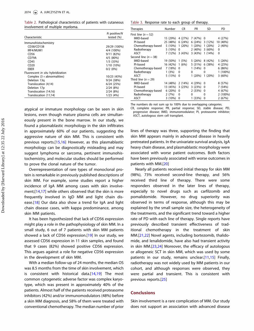
Table 3. Response rate to each group of therapy.
Therapies Number CR PR SD PD
First line (n¼ 52)IMID-based 15 (29%) 4 (27%) 7 (47%) 0 4 (27%)PI-based 25 (48%) 6 (24%) 6 (24%) 3 (12%) 10 (40%)Chemotherapy-based 5 (10%) 1 (20%) 1 (20%) 1 (20%) 2 (40%)Radiotherapy 5 (10%) 0 2 (40%) 3 (60%) 0ASCT 7 (12%) 3 (43%) 3 (43%) 1 (14%) 0
Second line (n¼ 38)IMID-based 19 (50%) 1 (5%) 5 (26%) 8 (42%) 5 (26%)PI-based 16 (42%) 1 (6%) 5 (31%) 6 (38%) 4 (25%)Chemotherapy-based 7 (18%) 0 1 (14%) 0 6 (86%)Radiotherapy 1 (3%) 0 0 0 1 (100%)ASCT 5 (13%) 0 1 (20%) 1 (20%) 3 (60%)
Third line (n¼ 29)IMID-based 14 (48%) 2 (14%) 4 (29%) 0 8 (57%)PI-based 13 (45%) 3 (23%) 3 (23%) 0 7 (54%)Chemotherapy-based 6 (20%) 0 2 (33%) 0 4 (67%)Radiotherapy 2 (7%) 0 0 0 2 (100%)ASCT 3 (10%) 0 1 (33%) 0 2 (67%)
The numbers do not sum up to 100% due to overlapping categories.CR, complete response; PR, partial response; SD, stable disease; PD,
progressive disease; IMID, immunomodulator; PI, proteasome inhibitor;ASCT, autologous stem cell transplant.
Table 2. Pathological characteristics of patients with cutaneousinvolvement of multiple myeloma.
CharacteristicN positive/N
tested (%)
ImmunohistochemistryCD38/CD138 29/29 (100%)IRF4/MUM1 4/4 (100%)CD56 9/11 (82%)CD79A 4/5 (80%)CD45 1/3 (33%)CD20 1/10 (10%)EBER 0/2 (0%)
Fluorescent in situ hybridizationComplex (3 + abnormalities) 10/23 (43%)Deletion 13q 9/24 (38%)Translocation (4;14) 6/24 (25%)Deletion 17p 2/24 (8%)Translocation (14;16) 2/24 (8%)Translocation (11;14) 1/23 (4%)
2074 A. JURCZYSZYN ET AL.
Dow
nloa
ded
by [
Har
vard
Lib
rary
] at
12:
35 2
2 Ju
ly 2
016

or high tumor burden at diagnosis. There seems to be an
overrepresentation of IgA heavy chain disease, light
chain disease, and plasmablastic morphology in skin
MM. Patients with skin MM carry a very poor prognosis
with a median OS of 8.5 months as time from skin
involvement. Currently, there is no standard of care for
patients with skin involvement by MM. Therefore, with a
median survival of less than a year from the time of
diagnosis, skin MM remains a therapeutic challenge with
poor prognosis.
Figure 2. Kaplan–Meier estimates of overall survival since skin myeloma diagnosis (A), and according to immunoglobulin (Ig) heavychain restriction (B), and plasmablastic morphology (C).
Figure 4. A dense infiltrate of CD138 + plasma cells in thedermis, with a normal epidermis.
Figure 3. Skin involvement in MM.
CUTANEOUS MYELOMA 2075
Dow
nloa
ded
by [
Har
vard
Lib
rary
] at
12:
35 2
2 Ju
ly 2
016

Potential conflict of interest: Disclosure forms provided
by the authors are available with the full text of this
article at http://dx.doi.org/10.3109/10428194.2015.11
28542.
References
1. An N, Li X, Shen M, et al. Analysis of clinical features,
treatment response, and prognosis among 61 elderly
newly diagnosed multiple myeloma patients: a single-
center report. World J Surg Oncol. 2015;13:2392. Deng S, Xu. Y, An. G, et al. Features of extramedullary disease
of multiple myeloma: high frequency of p53 deletion and
poor survival: a retrospective single-center study of 834
cases. Clin Lymphoma Myeloma Leuk. 2015;15:286–291.3. Lee SE, Kim JH, Jeon YW, et al. Impact of extramedullary
plasmacytomas on outcomes according to treatment
approach in newly diagnosed symptomatic multiple
myeloma. Ann Hematol. 2015;94:445–452.4. Short KD, Rajkumar SV, Larson D, et al. Incidence of
extramedullary disease in patients with multiple myeloma
in the era of novel therapy, and the activity of pomalidomide
on extramedullary myeloma. Leukemia 2011;25:906–908.5. Hedinger E. ZurFrage des Plasmacytomas. Frankfurter
Zeitschrift Fur Pathologie. 1911;7:343–350.6. Rodriguez JM, Lam S, Silber R. Multiple myeloma with
cutaneous involvement. JAMA 1977;237:2625–2626.7. Shpilberg O, Yaniv R, Levy Y, et al. Huge cutaneous
plasmacytomas complicating multiple myeloma. Clin Exp
Dermatol. 1994;19:324–326.8. Patel K, Carrington PA, Bhatnagar S, et al. IgD myeloma
with multiple cutaneous plasmacytomas. Clin Lab
Haematol. 1998;20:53–55.9. Guvenc B, Canataroglu A, Gumurdulu Y, et al. Multiple
myeloma with skin involvement. J Eur Acad Dermatol
Venereol. 2001;15:328–329.10. Santos G, Sousa L, Fernandes T, et al. Case for diagnosis.
Cutaneous involvement associated to multiple myeloma.
Anais Brasileiros De Dermatologia 2014;89:173–174.11. Araujo, C, Marques, H, Fernandes, JC, et al. Cutaneous
plasmacytomas secondary to nonsecretory multiple mye-
loma. J Dermatol Clin Res. 2014;2:1022.12. Serefhanoglu S, Haznedaroglu IC, Goker H, et al. Multiple
bulky cutaneous plasmacytomas with CNS relapse without
bone marrow involvement during the course of a lambda
light chain myeloma. Onkologie 2009;32:662–664.
13. Shah A, Klimo P, Worth A. Multiple myeloma first observedas multiple cutaneous plasmacytomas. Arch Dermatol.1982;118:922–924.
14. Requena L. Afectacion cutanea especıfica en pacientescon mieloma multiple. Estudio clınico-patologico,inmunohistoquımico y citogenetico de 40 casos. ActasDermosifiliograficas. 2005;96:424–440.
15. Messeguer F, Llombart B, Sanmartın O, et al. Cutaneousinvolvement in multiple myeloma: report of two cases.J Dermatol. 2012;39:806–808.
16. Nguyen SK, Dagnault A. Radiotherapy for multiple myelomawith skin involvement. Curr Oncol. 2010;17: 74–77.
17. Kato N, Kimura K, Yasukawa K, et al. Metastatic cutaneousplasmacytoma: a case report associated with IgA lambdamultiple myeloma and a review of the literature ofmetastatic cutaneous plasmacytomas associated withmultiple myeloma and primary cutaneous plasmacytomas.J Dermatol. 1999;26:587–594.
18. Bayer-Garner IB, Joseph L, Sanderson RD, et al. Expression ofsyndecan-1 is a sensitive marker for cutaneous plasmacy-toma. J Cutan Pathol. 2003;30:18–22.
19. Requena L, Kutzner H, Palmedo G, et al. Cutaneousinvolvement in multiple myeloma: a clinicopathologic,immunohistochemical, and cytogenetic study of 8 cases.Arch Dermatol. 2003;139:475–486.
20. Rajkumar SV, Fonseca R, Lacy MQ, et al. Plasmablasticmorphology is an independent predictor of poor survivalafter autologous stem-cell transplantation for multiplemyeloma. J Clin Oncol. 1999;17:1551–1557.
21. Subbiah S, O’Connell F, Kobraei KB, et al. Unusual multiplemyeloma cutaneous manifestation following nonmyeloa-blative allogeneic stem cell transplantation. Clin AdvHematol Oncol. 2009;7:800–802.
22. Rasche L, Strifler S, Duell J, et al. The lymphoma-likepolychemotherapy regimen ‘‘Dexa-BEAM’’ in advancedand extramedullary multiple myeloma. Ann Hematol.2014;93:1207–1214.
23. Karasawa T, Matsumoto T, Akiyama M. Metastatic skinlesions of multiple myeloma presenting as two extraor-dinarily large subcutaneous tumors. Int J Dermatol.2013;52:1568–1570.
24. Karimkhani C, Smith C. Extraosseous extension of multiplemyeloma: a cutaneous herald to systemic disease. J AmAcad Dermatol. 2014;71:e73–e74.
25. Liu J, Bakst R, Phelps R, et al. Radiation therapy forsecondary cutaneous plasmacytomas. Case Rep Hematol.2013;2013:739230
2076 A. JURCZYSZYN ET AL.
Dow
nloa
ded
by [
Har
vard
Lib
rary
] at
12:
35 2
2 Ju
ly 2
016