
Pathway complexity of Alzheimer's β-amyloid Aβ16−22 peptide ...
33
Pathway complexity of Alzheimer’s β -amyloid Aβ 16-22 peptide assembly S´ ebastien Santini 1 , Guanghong Wei 2 , Normand Mousseau 2 , Philippe Derreumaux 3 1 Information G´ enomique et Structurale, CNRS UPR 2589, 31 Chemin Joseph Aiguier, 13402 Marseille Cedex 20, France 2 D´ epartement de physique and Regroupement qu´ eb´ ecois sur les mat´ eriaux de pointe, Universit´ e de Montr´ eal, C.P. 6128, succ. centre-ville, Montr´ eal (Qu´ ebec), Canada 3 Laboratoire de Biochimie Th´ eorique, UPR 9080 CNRS, Institut de Biologie Physico- Chimique, 13 rue Pierre et Marie Curie, 75005 Paris, France Correspondence to [email protected] Keywords: amyloid fibrils, hydrogen bond registry, antiparallel and parallel β -sheets; folding simulations, reptation, Alzheimer’s disease 1
-
Upload
trinhthuan -
Category
Documents
-
view
222 -
download
2
Transcript of Pathway complexity of Alzheimer's β-amyloid Aβ16−22 peptide ...
Pathway complexity of Alzheimer’s β-amyloid Aβ16−22 peptide assembly
Sebastien Santini1, Guanghong Wei2, Normand Mousseau2, Philippe Derreumaux3
1Information Genomique et Structurale, CNRS UPR 2589, 31 Chemin Joseph Aiguier,
13402 Marseille Cedex 20, France
2Departement de physique and Regroupement quebecois sur les materiaux de pointe,
Universite de Montreal, C.P. 6128, succ. centre-ville, Montreal (Quebec), Canada
3Laboratoire de Biochimie Theorique, UPR 9080 CNRS, Institut de Biologie Physico-
Chimique, 13 rue Pierre et Marie Curie, 75005 Paris, France
Correspondence to [email protected]
Keywords: amyloid fibrils, hydrogen bond registry, antiparallel and parallel β-sheets;
folding simulations, reptation, Alzheimer’s disease
1

In the last few years, it has become clear that toxicity of amyloidogenic proteins lies in the
soluble oligomers rather than the insoluble fibrils, raising the interest in determining the
first steps of the assembly process. We have determined the aggregation mechanisms of
Aβ16−22 dimer using the activation-relaxation technique and an approximate free energy
model. Consistent with the NMR solid state analysis, the dimer of Aβ16−22 is predicted
to prefer an antiparallel β-sheet structure with the expected registry of intermolecular
hydrogen bonds. The simulations, however, locate three other antiparallel minima with
non-native β-sheet registries and one minimum with the β-strands parallel, slightly desta-
bilized with respect to the ground state. This result is significant because it can reconcile
a number of experimental data such as the dependency of β-sheet registry on pH con-
ditions and amino acid compositions. We also find that the assembly of Aβ16−22 into
dimers follows multiple routes starting from random coils or partially folded states and
often occurs through the reptation move of one strand of the β-sheet with respect to
the other, but α-helical conformations are not obligatory intermediates. This indicates
that destabilization of α-helical conformations is unlikely to abolish oligomerization of Aβ
peptides.
2
INTRODUCTION Past studies of the role of the Amyloid β-protein (Aβ) in Alzheimer’s
disease have linked the neurotoxicity of Aβ with its tendency to form amyloid fibrils (Selkoe,
1998). However, accumulating evidence suggests that the toxic species of Aβ might be
soluble Aβ oligomers (early aggregates) rather than fibrils (Walsh et al., 2002). This find-
ing supports an important role for Aβ oligomers in the aetiology of Alzheimer’s disease
and thus make Aβ oligomers attractive therapeutic targets (Wolfe, 2002). If effective drug
design strategies targeting Aβ oligomers are to be developed, it is essential to obtain a
detailed knowledge of the structures and assembly dynamics of these oligomers.
A detailed experimental characterization of these oligomeric intermediates is thus far
very difficult and only limited data are available because the intermediates are typically
short-lived and are present in a wide range of conformations and degrees of aggregation.
We know, however, that these oligomers tend to be small. Using photo-induced cross-
linker, the oligomer size distributions of aggregate-free, low molecular weight (LMW)
Aβ1−40 and Aβ1−42 could be assessed quantitatively (Bitan et al., 2003). This exper-
imental study revealed that LMW Aβ1−40 is a mixture of monomers, dimers, trimers
and tetramers, in rapid equilibrium, while LMW Aβ1−42 tends to exist in larger pen-
tamer/hexamer units. It is these structures that would self-assemble to form larger
oligomers (Bitan et al., 2003). We also have some limited information on the structure
of the intermediates. Studying the secondary structural changes of various Aβ1−40 and
Aβ1−42 fragments during fibrillogenesis, Walsh et al. and Kirkitadze et al. observed the
formation of an oligomeric intermediate containing 29-32% α-helix (Walsh et al., 1997,
1999; Kirkitadze et al., 2001). It was not until the α-helix formation process had begun,
as revealed by circular dichroism, that fibrils were detected by electron microscopy. But,
there is also experimental evidence that helix stabilization may facilitate as well as inhibit
fibril formation, depending on its strength (Fezoui & Teplow, 2002).
Recently, several structural models for Alzheimer’s β-amyloid fibrils based on exper-
imental constraints from solid state have been proposed. These studies showed that,
in Aβ10−35 (Benzinger et al., 2000) and Aβ10−40 (Petkova et al., 2002) fibrils, peptides
form parallel β-sheet structures. In this notation Aβn−m indicates residues n and m of
3
full-length β-amyloid protein. In contrast, an antiparallel β-sheet registry was found for
smaller fragments such as Aβ11−25 (Sikorski et al., 2003; Petkova et al., 2004), Aβ34−42 (Lans-
bury et al., 1995) and Aβ16−22 (Balbach et al., 2000). Because of its simplicity, Aβ16−22
of sequence KLVFFAE is an attractive system for molecular modelling studies. Further-
more, Aβ16−22 comprises the central hydrophobic core that is thought to be important
in full length Aβ assembly and contains four occurring Alzheimer’s disease-causing mu-
tations. They have been termed Flemish (A21G), Dutch (E22Q), Italian (E22K), and
Artic (E22G) (Miravalle et al., 2000). Aβ16−22 has also been investigated by molecu-
lar dynamics (MD) simulations with explicit solvent (Ma & Nussinov, 2002; Klimov &
Thirumalai, 2003). Ma and Nussinov studied the stability of octamer of Aβ16−22 packed
in different arrangements by MD at 330 K in explicit solvent (Ma & Nussinov, 2002).
They concluded that the antiparallel β-sheet/parallel layer model (each layer consisting
of four β-strands) is the most stable, but other supramolecular structures may be correct.
Although these simulations are important in understanding the supramolecular organiza-
tion of the fibrils, they do not provide any information on the oligomerisation process of
the peptides. Klimov and Thirumalai studied the folding of a trimer of Aβ16−22 by MD
at 300 K using an all-atom model of the protein, an explicit solvent model and a bias to
facilitate interactions between the peptides (Klimov & Thirumalai, 2003). They found
that the assembly of Aβ16−22 trimer occurs by multiple pathways with the formation of
an obligatory α-helical intermediate.
Because rapid equilibrium between monomers, dimers and larger units has been found
for Aβ1−40 (Bitan et al., 2003) and blocking the formation of dimers could inhibit fibril
formation, we have attempted recently to understand the folding mechanisms of Aβ16−22
dimer using the Activation Relaxation technique (ART) (Barkema & Mousseau, 1996;
Mousseau et al., 2001) coupled with OPEP, which provides for a detailed protein and un-
biased energy model (Derreumaux, 1999, 2000; Forcellino & Derreumaux, 2001). These
models are at variance with previous on-lattice studies (Istrail et al., 1999; Broglia et al.,
1998) and off-lattice studies using low-resolution models and Go-type potentials (Ding
et al., 2002; Jang et al., 2004) aimed at understanding aggregation in proteins. Pre-
4

liminary results were presented in a short communication (Santini et al., 2003). Here,
we offer a detailed description of the aggregation mechanisms starting from parallel β-
sheets. Furthermore, we present the results of new simulations starting from antiparallel
α-helices and the results of long explicit water LD simulations of some ART-predicted
configurations.
II. MATERIAL AND METHODS
We have simulated the folding of Aβ16−22 dimer. The N and C termini were neutral-
ized using acetyl and amine groups, respectively, as done experimentally (Balbach et al.,
2000). The energy surface was modeled using the OPEP energy model (Derreumaux,
1999, 2000; Forcellino & Derreumaux, 2001) and the folding trajectories were obtained by
the activation-relaxation technique (ART nouveau (Mousseau et al., 2001)). The stability
of some predicted arrangements was studied by all-atom MD simulations with an explicit
solvent model.
ART-OPEP simulations. A complete description of the ART-OPEP procedure has
been given elsewhere (Santini et al., 2003; Wei et al., 2002, 2003). In brief, the protein
model uses a simplified chain representation with all backbone atoms included (i.e., N, H,
Cα, C and O) and all side chains modeled by a bead with an appropriate van der Waals
radius and geometry. The OPEP (Optimized Potential for Efficient peptide-structure
Prediction) energy model (version 1.3) is expressed as a function of three types of in-
teractions: terms to satisfy stereo-chemistry, pairwise contact potential between main
chain particles, side chain – main chain and side chains (considering all 20 amino acid
types), and backbone two-body and four-body hydrogen bonding interactions (Santini
et al., 2003; Wei et al., 2002).
ART nouveau defines moves directly in the energy landscape. A basic event consists
of four steps. Starting from a minimum, the system is pushed outside the harmonic well
until a negative eigenvalue appears in the Hessian matrix. The system is then pushed
along the eigenvector associated with the negative eigenvalue until the total force is close
5

to zero, indicating a saddle point. The first two steps constitute the activation phase.
Subsequently, the configuration is pushed slightly over the saddle point and is relaxed to
a new local minimum, using standard minimization technique. Finally, the new config-
uration is accepted/rejected using the Metropolis criterion at the desired temperature.
This four-step procedure was repeated 18000 events for each simulation.
It is important to note that since ART events bring a conformation from a fully
relaxed state to a fully relaxed state, going through an activation barrier, the Metropolis
temperature cannot be directly associated with the real temperature. In particular, ART
neglects totally the vibrational contributions to the free energy of the oligomers. This
constitutes one major advantage for ART: it is possible to use a higher temperature than
in MD, for example, while still sampling the lowest-energy conformations.
In this work, we have performed 21 ART simulations of the dimer using OPEP: 12 start
from parallel β-sheets (R1-R12) and 9 start from antiparallel α-helices (S1-S9). Starting
from a low-energy parallel β-sheet allows us to verify that our procedure samples the con-
figurational space appropriately. Furthermore, this organisation is known to be preferred
for larger Aβ fragments. The second starting point with two α-helices is reminiscent of
the topological change that occurs in prion proteins (Prusiner, 1997). All runs starting
from the same configuration use different random-number seeds and are free of any bi-
ases to facilitate interactions between the peptides. This contrasts with the simulations
reported by Klimov and Thirumalai (Klimov & Thirumalai, 2003). In most simulations,
the Metropolis temperature is set to 1000 K because the parallel β-sheet is only desta-
bilized by 3 kcal/mol with respect to the antiparallel β-sheet. We emphasize that at
this temperature, the dimer is not stable and continues to evolve on the conformational
space. Finally, 3 runs were also carried out on the monomer to determine its equilibrium
conformations at TMetropolis = 300K starting from fully extended conformations.
OPEP was found recently to discriminate native from false positive conformations
for a series of peptides in monomeric forms (Derreumaux, 1999, 2000; Forcellino & Der-
reumaux, 2001). Because OPEP is an approximate free energy model, we know that
several factors can enhance the energy barriers estimated by more physically-based en-
6
ergy functions. In particular, solvent is not treated explicitly, and hydrogen bonding
interactions between the solvent and the chains certainly affect the relative energetics
and entropic effects (Derreumaux & Schlick, 1998); side chains are modeled by a bead
and may not capture the real complexity of hydrophobic and electrostatic interactions
between side chains; the dependence of the stability of polypeptides on pH conditions
is not considered. In spite of these limitations, we have found that the ART-generated
pathways using OPEP are generally kinetically possible: the folding of a helix model
through a transition state characterized by two α-helices connected by a loop was found
by ART using OPEP (Wei et al., 2002) and MD using all-atom model (Chowdhury et al.,
2003). Similarly, the folding of a β-hairpin model by ART identified two mechanisms
described by standard protocols, but also a reptation mechanism (Wei et al., 2003) which
was found recently by multicanonical simulations on another hairpin model using explicit
solvent (Ikeda & Higo, 2003).
Molecular dynamics simulations. The MD simulations used the GROMACS pro-
gram and the all-atom force field GROMOS96 (Berendsen et al., 1995). The all-atom
conformations discussed here were generated using the MOLMOL software (Koradi et al.,
1996). The models were solvated in a dodecaedric box of 30 A side containing ∼ 1700
SPC (simple point charge) water molecules under periodic boundary conditions. The Par-
ticle Mesh Ewald method was used with a cutoff distance of 12A for the van der Waals
and electrostatics interactions. The models were minimized by 1000 steps of conjugate
gradient and equilibrated at the desired temperature under Cα atom position restraints
for 10 ps. At this stage, the restraints were released and MD simulations were performed
in the canonical NPT (number of particles-pressure-temperature) ensemble at neutral pH
for 7 ns. Each model was subject to two simulations using different initial velocities at
330 K, i.e., at a T often used to incubate the amyloid in experiments. The time step for
dynamics was 2 fs using the SHAKE algorithm. Nonbonded interactions were updated
every 20 fs. Temperature was controlled using a weak coupling to a bath of constant T
(coupling time of 0.1 ps) and pressure by a weak coupling to a bath of constant P (1 atm,
7

coupling time of 0.5 ps). The density is ∼ 0.977 g/mL at 330 K. All runs described here
cover a total of 84 ns.
Trajectory Analysis. To analyse the dimer simulations, we used the radii of gyration
of the system (Rg), the hydrophobic core (Rg-core, using the side chains of Leu, Val
and the two Phe residues for each chain) and the Cα root mean square deviation (rmsd)
from the lowest energy conformation obtained in simulation R1, except when mentioned
otherwise. This antiparallel β-sheet structure can be described by a 16+k ⇐⇒ 22−k
β-sheet registry (i.e. intermolecular H-bonds between residues 16+k and 22−k of the two
chains, with k = 0, 2, 4 and 6, and the C=O...HN and NH..O=C bonds formed). A
H-bond is defined if it satifies DSSP conditions (Kabsch & Sander, 1983): the distance
between the carbonyl oxygen and amide hydrogen (O..H) is less than 2.5 A and the angle
NHO > 150◦. To follow the orientation of the chains, the scalar product (d12) between
the end-to-end unit vectors of each chain was calculated : d12 = 1 indicates parallel,
−1 antiparallel and 0 perpendicular register. The percentage of secondary structure in
each frame was determined using the DSSP program (Kabsch & Sander, 1983). We also
used the less stringent conditions used by Klimov and Thirumalai as described in their
experimental procedure: a conformation is a β-strand (or α-helix) if the φ and ψ of
any two consecutive residues are in the appropriate Ramachandran regions and no two
consecutive residues are in α-helix (β-strand) (Klimov & Thirumalai, 2003). The figures
were produced using the MOLMOL software (Koradi et al., 1996). In what follows, all
event numbers refer to accepted event numbers.
III. RESULTS AND DISCUSSION
Aβ16−22 monomer is random coil in solution
The solution structure of Aβ16−22 remains to be determined, but the structure of var-
ious peptides spanning this fragment has been characterized experimentally. Monomeric
forms of Aβ10−35 (Zhang et al., 2000), Aβ1−40 and Aβ1−42 (Zagorski et al., 2000) are essen-
8

tially unstructured in aqueous solution at neutral pH, while Aβ1−28 (Talafous et al., 1994),
Aβ1−40 and Aβ1−42 (Shao et al., 1999) adopt a predominantly α-helical conformation in
a membrane-like environment. Massy et al. found that the region 16-22 is essentially
random coil (RC) in 4-ns MD simulations of the wild-type and Dutch mutant forms of
Aβ10−35 in aqueous solution (Massi et al., 2002). Klimov and Thirumalai found that the
time-averaged populations of (RC, β-strand and α-helix) residues in Aβ16−22 monomers
are (56%, 33% and 11%) using 8-ns MD trajectories and their specific definitions for as-
signing conformational states to the residues (Klimov & Thirumalai, 2003). From our
ART-generated trajectories we find that the populations of (RC, β-strand and α-helix)
residues are (77%, 18% and 5%) using Klimov’s definitions and (96%, 2% and 2%) us-
ing DSSP. The lowest-energy structure, compact and random coil, generated by all ART
simulations is shown in Figure 1A. The energy spectrum of the calculated conformational
states in Figure 1B shows that the conformation of Aβ16−22 monomer within the dimeric
optimized form (Fig. 1C) is destabilized by 9 kcal/mol (15 kT ) with respect to its native
compact state. Our model is therefore consistent with the conformational change of Aβ
and prion proteins observed upon association.
Aβ16−22 can adopt different hydrogen bond β-sheet registries
ART search using OPEP indicates that the global energy minimum of the dimer
matches exactly the solid state NMR model proposed by Balbach et al. at pH 7.4, namely
the pattern of H-bonds depicted between layer II (middle) and layer III (bottom) in Figure
10 of (Balbach et al., 2000). This conformation, obtained starting from antiparallel α-
helices (event 5759 in run S1) or parallel β-sheets (events 10853 in R1, 2594 in R6 and
6467 in R9), is characterized by the 16+k ⇐⇒ 22−k β-sheet registry and an energy
of −44.4 kcal/mol (Fig. 1C). This pattern of H-bonds will be referred to as pattern I.
A partially folded pattern I of energy −43.3 kcal/mol is obtained in run R4 because the
residues A21 and E22 are disordered in both chains.
Surprisingly, the lowest-energy structures generated by the remaining 16 simulations
can be clustered into four distinct β-sheets (Fig.1D-G). Three β-sheets are antiparallel.
9
They are characterized by the 17+k ⇐⇒ 21−k β-sheet registry [pattern II, energy of
−43.4 kcal/mol, Fig. 1E, found at events 11546 in run R3 and 7484 in run S4, this H-bond
network follows exactly the second derived NMR pattern depicted between layers I (top)
and II (middle) in Figure 10 of (Balbach et al., 2000)]. Or they can be described by 16+k
⇐⇒ 21−k [pattern III, energy of −42.4 kcal/mol, runs R2, R10, S5 and S7, Fig. 1F] and
16+k ⇐⇒ 20−k [pattern IV, energy of −40.4 kcal/mol, run S3, Fig. 1G].
In contrast, the fourth β-sheet is parallel and characterized by the 16+k ⇐⇒ 16+k
β-sheet registry (Fig. 1D). This conformation, of energy −41.4 kcal/mol, is explored
many times, independently of the starting structure. We find that in runs R5, R7, R8
and R11 the chains easily escape from this state, but rarely explore antiparallel β-sheets;
in runs S2, S6 and S8, the chains pass from antiparallel α-helices to parallel β-sheets
permanently or quasi-permanently (see Figure 2 for runs R7 and S8) over the length of
our simulations. We know, however, that these runs would eventually reach the pattern
I if they were continued (Wei et al., 2003). Our parallel β-sheet structure differs from
that considered by Balbach et al. (Balbach et al., 2000) in two respects. Our model
contains 7 H-bonds vs. 6 H-bonds in Balbach’s model. This is to be compared with 8
H-bonds within pattern I and 6 H-bonds within patterns II, III and IV. Furthermore, the
charged residues Lys16 and Glu22 are on opposite sides of the β-sheet in our model and
not in close proximity as in Balbach’s model (Fig. 3A-B). All these models are stable
in MD simulations with water at 330K, independently of the initial velocities. In Figure
3C, we have selected to plot the Cα rms deviation of three models from their mimimized
energy structures as a function of time. We see that the Balbach’s model is less rigid than
our parallel structure and the pattern I, but all models are very stable within the 7 ns
timescale.
Taken together, our results indicate that several hydrogen bond β-sheet registries of
similar energy are available for the Aβ16−22 dimer. This finding complements recent MD
simulations of a trimer of the heptapeptide GNNQQNY from the yeast prion Sup35 which
point to three minima associated with parallel, antiparallel and mixed parallel-antiparallel
β-sheet structures (Gsponer et al., 2003).
10

Atomic assembly trajectories
Among a total of 21 runs, 8 runs from parallel β-sheets (R1-R4, R6, R9, R10 and R12)
and 6 runs from antiparallel α-helices (S1, S3-S5, S7 and S9) locate antiparallel β-sheet
structures of very low energy within the alloted number of events. In what follows, the
other runs, which essentially sample parallel β-sheets, are left out of the discussion.
The folding trajectory R1 was described in atomistic detail elsewhere (Santini et al.,
2003). We have shown that both chains rapidly loose their non-native H-bonds, then
rotate in respect to one another through a perpendicular registry with one chain partially
unfolded and the other fully extended, and finally adopt the antiparallel pattern I by
progressive zipping of the H-bonds. Here, we select to describe first the folding simulation
S1 starting from two antiparallel α-helices and then the general routes by which the two
chains assembly to their lowest-energy states.
Figure 4 shows the variation of several order parameters in the folding simulation
S1. During the first 1000 events, both chains remain helical but progressively rotate in
respect to one another (Fig. 4C). Then the chains, parallel in character, unfold and adopt
U-like conformations (see event 1650 in Fig. 4C). In this first phase, the rms deviation
with respect to the ground state, Rg and Rg-core do not vary much (Fig. 4A). At event
1700, four native H-bonds form (Fig. 4B). From this core of H-bonds, one chain extends,
whereas the other remains U-like (see cooperative decrease in rmsd and Rg-core and
increase in Rg around event 2500). The conformation at event 2500, shown in Fig. 4C, is
characterized by the N-terminal of chain 1 and the C-terminal of chain 2 antiparallel, but
the N-terminal of both chains parallel. From there, the chains find a way to locate the
ground state at event 5759. It is interesting to note that the structure at event 2500 is
explored, independently of the starting conformation. It is encountered (within 1.0 A rms
deviation) in runs R1 and S4.
By following the rms deviation of each chain with respect to its conformation within
the dimeric optimized form for each individual folding trajectories, we find that there
are four routes leading to the antiparallel β-sheets (Figure 5). In pathway I, both chains
unfold (U’); then one of the chains adopts its native state (N), whereas the other remains
11

unfolded (U’); subsequently the native assembly occurs (N2). In pathway II, the native
assembly of the dimer occurs directly from both chains unfolded, whereas in pathway III
it occurs from one chain in its native state and the other unfolded. Finally, in pathway
IV, native assembly occurs from both chains in their native states. All these routes are
not equiprobable. Pathway I is observed in 7/14 of the runs (R1, R2, R4, S1, S3, S5
and S9), pathway III in 4/14 (R3, R10, S4, S7), pathway IV in 2/14 (R6 and R9) and
pathway II in 1/14 (R12). Overall, our simulations are consistent with previous Monte
Carlo simulations using on-lattice protein models (Dima & Thirumalai, 2002; Harrison
et al., 2001). Our preferred pathway for assembly occurring directly from one of the chains
folded agrees with the general concept that protein aggregation arises from partially folded
intermediates (Kelly, 1996; Chiti et al., 1999). On the other hand, assembly from random
coils (pathway II) is supported by experimental studies on the two-state U1A and CI2
folders (Silow et al., 1999) and myoglobin (Fandrich et al., 2003).
H-bonds provide evidence for very complex aggregation routes
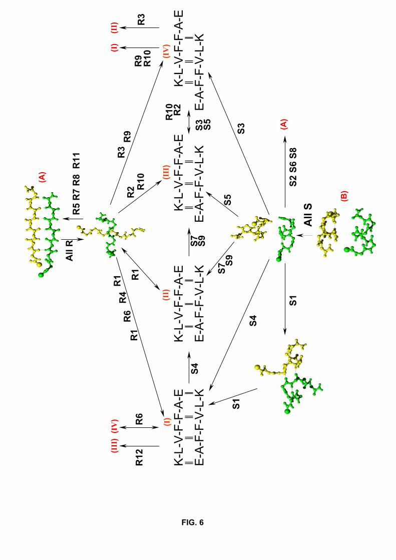
An important aspect that emerges from our simulations is that folding proceeds
through multiple very complex routes as indicated by the various networks of H-bonds
explored. Figure 6 summarizes all the folding routes starting from parallel β-sheets or
antiparallel α-helices. Consistent with the solid state NMR analysis, the patterns I and
II of H-bonds are highly populated, but the patterns III and IV have to be taken into
account. Interestingly, none of these patterns represent a dead end in our simulations. For
instance, in run S4, the dimer explores transient conformations of pattern I-type before
reaching the pattern II; in run R9, the dimer samples the pattern IV before reaching the
pattern I; in run S4, the dimer momentarily explores the pattern I and then switches to the
pattern II. The molecular mechanisms propagating the conformational changes between
the various patterns of H-bonds are listed in Table I and are described in atomistic detail
in Figure 7. These conformational changes can involve a rotation of one chain (Rot1)
or both chains (Rot2) by 180◦ around their strand axes, a reptation move (Wei et al.,
2003) of one strand of the β-sheet with respect to the other (RepX where X represents
12
the number of residues shifted) or a combination of these two mecanisms. Specifically,
we find that the dimer can exchange the patterns I and II (runs R1 and S4) by a Rot2
mechanism (Fig. 7C). Transitions between the patterns I and III (run R12), the patterns
II and III (runs S7 and S9) and the patterns III and IV (runs R2, R10, S3 and S5) occur
by a combined Rot1+Rep1 mechanism (Fig. 7A). Finally, the Rep2 mechanism, which
allows the exchange between the patterns II and IV (Fig. 7B) and does not need rotation
of the chains, is easily explored. It is encountered in runs R6, R9, R10, S1, S3 and S5.
We note that the transition between the patterns II and IV by a combined Rot2+Rep2
mechanism is not observed in this study, but this could not be totally excluded if we had
run 100 runs.
The transitions between the patterns can be fast or slow. Figure 8 shows the number
of H-bonds satisfying each pattern as a function of accepted events in the runs R2, R9
and S4. We see that the exchange between the patterns III and IV takes 34 events in run
R2 (Fig. 8B), but 1052 events in run R10. The patterns I and II interchange within 1350
events in run S4 (Fig. 8A) vs. 7100 events in run R1. The change between the patterns
IV and I takes 620 and 800 events in runs R9 (Fig. 8C) and R6, respectively. The barrier
height to go from one pattern to another without any bias (defined here by the difference
between the highest-energy minimum and the energy of one pattern during the transition)
is ∼ 20 kcal/mol, independently of the patterns involved. We recognize that these energy
barriers likely are much smaller using all-atom model and explicit solvent as discussed in
Material and Methods.
Non-helical conformations are possible intermediates
Using circular dichroism and electron microscopy, Kirkitadze et al. (Kirkitadze et al.,
2001) and Walsh and coworkers (Walsh et al., 1997, 1999) have identified an α-helix-
containing intermediate prior to the formation of β-sheet-rich assembly of Aβ1−40, Aβ1−42
and several variants. Based on MD simulations of Aβ16−22 trimer at 300K, Klimov and
Thirumalai have proposed recently that the antiparallel β-sheet could occur by multiple
pathways with the formation of an obligatory α-helical intermediate (Klimov & Thiru-
13
malai, 2003). But another interpretation is also possible: for instance, a mecanism involv-
ing rapid equilibrium between these α-rich oligomers incapable of maturing into fibrils and
smaller Aβ oligomers or Aβ monomers which could form fibrils through an alternative
pathway (Kirkitadze et al., 2001).
In order to detect whether α-helix conformations occur prior to optimal assembly of the
sheets, we have followed the secondary structure composition of the chains as a function
of events using both Klimov’s definitions and the DSSP program (Figure 9). Our 14 (8
R+ 6 S) folding simulations clearly show that the occurrence of a helical intermediate is
not an obligatory step for Aβ16−22 dimer using the DSSP program. Starting from parallel
β-sheets, intermediates containing 30% helix are observed in runs R1, R2, R4, R10 and
R12, but not in runs R3, R6 and R9. On the other hand, starting from antiparallel α-
helices, helical intermediates are detected in runs S1, S3, S5, S7 and S9, but not in S4.
In the analysis starting from helices, we consider a helical intermediate if it occurs after
1000 accepted events. It is interesting to note that by using Klimov’s definitions helical
intermediates are detected in R3 between events 4000-6000 (Fig 9B), in R6 around events
1000 (9C) and in R9, but not in S4.
Atomic analysis of the helical intermediates shows that the helix can be found in either
chains with a strong preference for encompassing residues VFF. For instance, in run R1,
the intermediate between events 3400 and 3660 has a α-helix spanning residues LVFF
in chain 2 (Fig. 9A) and the intermediate between events 5790 and 5820 has a α-helix
spanning residues VFFA in chain 1. In contrast, helix spans residues VFFA in chain 2
(events 3860 in R2, 1441 in S1 and 2576 in S9), residues LVFF in chain 1 (events 2722-
2741 in run S3, event 607 in run S4) or residues VFFA in chain 1 (events 6746 in R4, 3586
in R10, 10099 in R12, 2274 in S5 and 1428 in S7). We did not find evidence of structural
similarity between these intermediates, all structures deviating by more than 3.0 A rms
from each other.
IV. CONCLUSIONS
The present study has attempted to determine the folding pathways for Aβ16−22 as-
14
sembly into a dimer. Our analysis is based on simulations starting from parallel β-sheets
and antiparallel α-helices, but identical results are obtained from arbitrarily chosen con-
formations (data not shown). Our protocol is free of any biases to facilitate interactions
between the chains and uses a generic energy model which was found to work well on small
proteins adopting various secondary structures in their optimized monomeric forms.
Consistent with NMR solid state analysis at pH 7.4 (Balbach et al., 2000), the lowest
energy structure of the Aβ16−22 dimer is antiparallel in character with the 16+k ⇐⇒
22−k β-sheet registry. Hovever, the simulations also locate three alternative antiparallel
organisations with different β-sheet registries and one parallel β-sheet assembly, slightly
destabilized relative to the ground state. This result is significant because it helps clarify
the variation of β-sheet registry on pH conditions (Petkova et al., 2004) and amino acid
compositions (Tjernberg et al., 2002). In addition to finding these low energy minima,
our simulations also describe the molecular mechanisms propagating these conformational
changes and emphasize the crucial role of the reptation move of one strand of the β-
sheet with respect to the other. Again, this is consistent with very recent isotope-edited
infrared spectroscopy on the protein prion fragment spanning residues 109-122, which
points to the reptation move of the central strand within a trimer (Silva et al., 2003).
Finally, this study makes it clear that multiple aggregation routes are possible, but α-
helical conformations are not obligatory intermediates for the dimer. It is possible that a
minimum Aβ length is needed – there is no available circular dichroism study for Aβ16−22
as for Aβ1−40 (Kirkitadze et al., 2001) – or that a minimum oligomeric size – trimeric or
larger – is needed for the chains to gain stability by forming α-helices. ART simulations of
Aβ16−22 in trimeric and hexameric forms are underway to address these issues and provide
us with a more complete folding picture for fibril formation.
Acknowledgments
Sebastien Santini is supported by the Natural Sciences and Engineering Research Council
of Canada. The ART calculations were done on the computers of the Reseau quebecois
de calcul de haute performance and the MD simulations at IGS, Marseille. N.M. is a
15

Cottrell Scholar of Research Corporation.
16

References
Balbach, J., Ishii, Y., Antzutkin, O., Leapman, R., Rizzo, N., Dyda, F., Reed, J. and
Tycko, R. (2000). Amyloid fibril formation by Aβ16−22, a seven-residue fragment of the
Alzheimer’s β-amyloid peptide, and structural characterization by solid state NMR.
Biochemistry 39, 13748–13759.
Barkema, G.and Mousseau, N. (1996). Event-based relaxation of continuous disordered
systems. Phys. Rev. Lett. 77, 4358–4361.
Benzinger, T., Gregory, D., Burkoth, T., Miller-Auer, H., Lynn, D., Botto, R. and Mered-
ith, S. (2000). Two-dimensional structure of β-amyloid(10-35) fibrils. Biochemistry 39,
3491–3499.
Berendsen, H., van der Spoel, D. and van Drunen, R. (1995). GROMACS: a message-
passing parallel molecular dynamics implementation. Comp. Phys. Comm. 91, 43–56.
Bitan, G., Vollers, S. and Teplow, D. (2003). Elucidation of primary structure elements
controlling early amyloid β-protein oligomerization. J. Biol. Chem. 278, 34882–34889.
Broglia, R., Tiana, G., Pasquali, S., Roman, H. and Vigezzi, E. (1998). Folding and
aggregation of designed proteins. Proc. Natl. Acad. Sci. USA 95, 12930–12933.
Chiti, F., Webster, P., Taddei, N., Clark, A., Stefani, M., Ramponi, G. & Dobson, C.
(1999). Designing conditions for in vitro formation of amyloid protofilaments and fibrils.
Proc. Natl. Acad. Sci. USA 96, 3590–3594.
Chowdhury, S., Zhang, W., Wu, C., Xiong, G. and Duan, Y. (2003). Breaking non-native
hydrophobic clusters is the rate-limiting step in the folding of an alanine-based peptide.
Biopolymer 68, 63–75.
Derreumaux, P. (1999). From polypeptide sequences to structures using Monte Carlo
simulations and an optimized potential. J. Chem. Phys. 111, 2301–2310.
Derreumaux, P. (2000). Generating ensemble averages for small proteins from extended
conformations by Monte Carlo simulations. Phys. Rev. Lett. 85, 206–209.
Derreumaux, P.and Schlick, T. (1998). The loop opening/closing motion of the enzyme
triosephosphate isomerase. Biophys. J. 74, 72–81.
Dima, R.and Thirumalai, D. (2002). Exploring protein aggregation and self-propagation
using lattice models: phase diagram and kinetics. Protein Sci. 11, 1036–1049.
17
Ding, F., Dokholyan, N., Buldyrev, S.V. Stanley, H. and Shakhnovich, E. (2002). Molec-
ular dynamics simulation of the SH3 domain aggregation suggests a generic amyloido-
genesis mechanism. J. Mol. Biol. 324, 851–857.
Fandrich, M., Forge, V., Buder, K., Kittler, M., Dobson, C. and Diekmann, S. (2003).
Myoglobin forms amyloid fibrils by association of unfolded polypeptide segments. Proc.
Natl. Acad. Sci. USA 100, 15463–15468.
Fezoui, Y.and Teplow, D. (2002). Kinetic studies of amyloid β-protein fibril assembly. J.
Biol. Chem. 277, 36948–36954.
Forcellino, F.and Derreumaux, P. (2001). Computer simulations aimed at structure pre-
diction of supersecondary motifs in proteins. Proteins 45, 159–166.
Gsponer, J., Haberthur, U. and Caflisch, A. (2003). The role of side-chain interactions in
the early steps of aggregation: molecular dynamics simulations of an amyloid-forming
peptide from the yeast prion Sup35. Proc. Natl. Acad. Sci. USA 100, 5154–5159.
Harrison, P., Chan, H., Prusiner, S. and Cohen, F. (2001). Conformational propagation
with prion-like characteristics in a simple model of protein folding. Protein Sci. 10,
819–835.
Ikeda, K.and Higo, J. (2003). Free-energy landscape of a chameleon sequence in explicit
water and its inherent α/β bifacial property. Protein Sci. 12, 2542–2548.
Istrail, S., Schwartz, R. and King, J. (1999). Lattice simulations of aggregation funnels
for protein folding. J. Comp. Biol. 6, 143–162.
Jang, H., Hall, C. and Zhou, Y. (2004). Assembly and kinetic folding pathways of a
tetrameric β-sheet complex: molecular dynamics simulations on simplified off-lattice
protein models. Biophys. J. 86, 31–49.
Kabsch, W.and Sander, C. (1983). Dictionary of protein secondary structure : pattern
recognition of hydrogen-bonded and geometrical features. Biopolymers 22, 2577–2637.
Kelly, J. (1996). Alternative conformations of amyloidogenic proteins govern their behav-
ior. Curr. Opin. Struct. Biol. 6, 11–17.
Kirkitadze, M., Condron, M. and Teplow, D. (2001). Identification and characterization
of key kinetic intermediates in Amyloid β-protein fibrillogenesis. J. Mol. Biol. 312,
1103–1119.
Klimov, D.and Thirumalai, D. (2003). Dissecting the assembly of Aβ16−22 amyloid pep-
tides into antiparallel β-sheets. Structure (Camb) 11, 295–307.
18

Koradi, R., Billeter, M. and Wuthrich, K. (1996). Molmol : a program for display and
analysys of molecular structures. J. Mol. Graphics. 14, 51.
Lansbury, P. J., Costa, P., Griffiths, J., Simon, E., Auger, M., Halverson, K., Kocisko,
D., Hendsch, Z., Ashburn, T. and Spencer, R. e. a. (1995). Structural model for the
β-amyloid fibril based on interstrand alignment of an antiparallel-sheet comprising a
c-terminal peptide. Nat. Struct. Biol. 2, 990–998.
Ma, B.and Nussinov, R. (2002). Stabilities and conformations of Alzheimer’s β-amyloid
peptide oligomers (aβ16−22, aβ16−35, and aβ10−35): sequence effects. Proc. Natl. Acad.
Sci. USA 99, 14126–14131.
Massi, F., Klimov, D., Thirumalai, D. and Straub, J. (2002). Charge states rather than
propensity for β-structure determine enhanced fibrillogenesis in wild-type Alzheimer’s
β-amyloid peptide compared to E22Q Dutch mutant. Protein Sci. 11, 1639–1647.
Miravalle, L., Tokuda, T., Chiarle, R., Giaccone, G., Bugiani, O., Tagliavini, F., Fran-
gione, B. and Ghiso, J. (2000). Substitutions at codon 22 of Alzheimer’s Aβ peptide
induce diverse conformational changes and apoptotic effects in human cerebral endothe-
lial cells. J. Biol. Chem. 275, 27110–27116.
Mousseau, N., Derreumaux, P., Barkema, G. and Malek, R. (2001). Sampling activated
mechanisms in proteins with the activation-relaxation technique. J. Mol. Graph. Model.
19, 78–86.
Petkova, A., Buntkowsky, G., Dyda, F., Leapman, R., Yau, W. and Tycko, R. (2004).
Solid state NMR reveals a pH-dependent antiparallel β-sheet registry in fibrils formed
by a β-amyloid peptide. J. Mol. Biol. 335, 247–260.
Petkova, A., Ishii, Y., Balbach, J., Antzutkin, O., Leapman, R., Delaglio, F. and Tycko,
R. (2002). A structural model for Alzheimer’s β-amyloid fibrils based on experimental
constraints from solid state NMR. Proc. Natl. Acad. Sci. USA 99, 16742–16747.
Prusiner, S. (1997). Prion diseases and the BSE crisis. Science 278, 245–251.
Santini, S., Wei, G., Mousseau, N. and Derreumaux, P. (2003). Exploring the folding
pathways of proteins through energy landscape sampling: application to alzheimer’s
β-amyloid peptide. I.E.J.M.D. 2, 564–577.
Selkoe, D. (1998). The cell biology of β-amyloid precursor protein and presenilin in
Alzheimer’s disease. Trends. Cell. Biol. 8, 447–453.
19
Shao, H., Jao, S., Ma, K. and Zagorski, M. (1999). Solution structures of micelle-bound
amyloid β-(1-40) and β-(1-42) peptides of Alzheimer’s disease. J. Mol. Biol. 285, 755–
773.
Sikorski, P., Atkins, E. and Serpell, L. (2003). Structure and texture of fibrous crystals
formed by Alzheimer’s Aβ(11-25) peptide fragment. Structure 11, 915–926.
Silow, M., Tan, Y., Fersht, A. and Oliveberg, M. (1999). Formation of short-lived protein
aggregates directly from the coil in two-state folding. Biochemistry 38, 13006–13012.
Silva, R., Barber-Armstrong, W. and Decatur, S. (2003). The organization and assembly
of a β-sheet formed by a prion peptide in solution: an isotope-edited FTIR study. J.
Am. Chem. Soc. 125, 13674–13675.
Talafous, J., Marcinowski, K., Klopman, G. and Zagorski, M. (1994). Solution structure
of residues 1-28 of the amyloid β-peptide. Biochemistry 33, 7788–7796.
Tjernberg, L., Tjernberg, A., Bark, N., Shi, Y., Ruzsicska, B., Bu, Z., Thyberg, J. and
Callaway, D. (2002). Assembling amyloid fibrils from designed structures containing a
significant amyloid β-peptide fragment. Biochem. J. 366, 343–351.
Walsh, D., Hartley, D., Kusumoto, Y., Fezoui, Y., Condron, M., Lomakin, A., Benedek,
G., Selkoe, D. and Teplow, D. (1999). Amyloid β-protein fibrillogenesis. Structure and
biological activity of protofibrillar intermediates. J. Biol. Chem. 274, 25945–25952.
Walsh, D., Klyubin, I., Fadeeva, J., Cullen, W., Anwyl, R., Wolfe, M. & Rowan, M.
(2002). Naturally secreted oligomers of amyloid β-protein potently inhibit hippocampal
long-term potentiation in vivo. Nature 416, 483–484.
Walsh, D., Lomakin, A., Benedek, G., Condron, M. and Teplow, D. (1997). Amyloid
β-protein fibrillogenesis. Detection of a protofibrillar. J. Biol. Chem. 272, 22364–22372.
Wei, G., Derreumaux, P. and Mousseau, N. (2003). Sampling the complex energy land-
scape of a simple β-hairpin. J. Chem. Phys. 119, 6403–6406.
Wei, G., Mousseau, N. and Derreumaux, P. (2002). Exploring the energy landscape of
proteins: a characterization of the activation-relaxation technique. J. Chem. Phys. 117,
11379–11387.
Wolfe, M. (2002). Therapeutic strategies for Alzheimer’s disease. Nature Reviews, Drug
Discovery 1, 859–866.
20
Zagorski, M., Shao, H., Ma, K., Yang, J., Li, H., Zhang, Y. and Papolla, M. (2000).
Amyloid Aβ(1-40) and Aβ(1-42) adopt remarkably stable, monomeric, and extended
structures in water solution at neutral pH. Neurobiol. Aging 21, 10–11.
Zhang, S., Iwata, K., Lachenmann, M., Peng, J., Li, S., Stimson, E., Lu, Y., Felix, A.,
Maggio, J. and Lee, J. (2000). The Alzheimer’s peptide Aβ adopts a collapsed coil
structure in water. J. Struct. Biol. 130, 130–141.
21
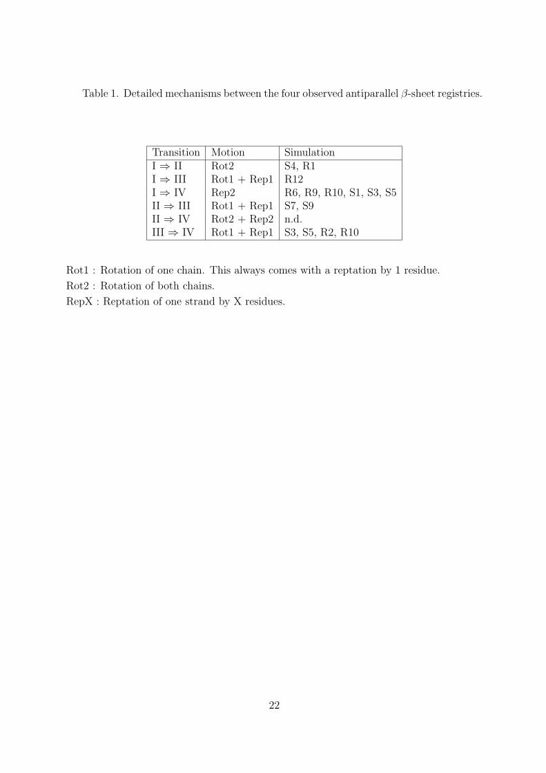
Table 1. Detailed mechanisms between the four observed antiparallel β-sheet registries.
Transition Motion SimulationI ⇒ II Rot2 S4, R1I ⇒ III Rot1 + Rep1 R12I ⇒ IV Rep2 R6, R9, R10, S1, S3, S5II ⇒ III Rot1 + Rep1 S7, S9II ⇒ IV Rot2 + Rep2 n.d.III ⇒ IV Rot1 + Rep1 S3, S5, R2, R10
Rot1 : Rotation of one chain. This always comes with a reptation by 1 residue.
Rot2 : Rotation of both chains.
RepX : Reptation of one strand by X residues.
22

Figure Captions
Fig.1. Lowest-energy structures of Aβ16−22 in monomeric and dimeric forms. (a) Native
state of the monomer. (b) Energy difference between the ground state of the monomer
and its conformation within the dimeric optimized form. (c) Ground state of the dimer,
antiparallel in character and described by the 16+k ⇐⇒ 22−k β-sheet registry. (d) Par-
allel β-sheet structure. (e-g) The three other antiparallel β-sheet organisations: pattern
II (e), pattern III (f) and pattern IV (g). In all figures, the N-terminal end in each chain
is located by a sphere.
Fig.2. Variation of the orientation of the chains (d12) as a function of accepted events in
run R7 (A) and run S8 (B).
Fig.3. Comparison of the parallel β-sheet structures: (a) our predicted structure with the
charged residues pointing to opposite directions above the plane formed by the sheets,
(b) the parallel β-sheet structure, as discussed in Balbach (Balbach et al., 2000), with
the K (and E) of both chains in close proximity. (c) RMS deviations (in A) of pattern I
(black), our parallel (red) and Balbach’s (green) models from their minimized structures
by 7-ns MD simulations at 330 K. The time-averaged percentage of H-bond formed is
79% (pattern I), 82% (our parallel model) and 75% (Balbach’s model). Identical RMS
deviations and percentages of H-bonds are obtained for the patterns II, III and IV (data
not shown).
Fig.4. Variation of order parameters in the folding simulation S1 leading to pattern I. (A)
RMS deviation from the ground state (black), Rg (red) and Rg-core (green) in A; (B)
number of native H-bonds; (C) d12 and (D) energy in kcal/mol. The structures at events
1000, 1650 and 2500 are shown. For clarity, the results are given until the ground state
is located at event 5759. Only accepted events are shown.
Fig.5. Four aggregation routes (P1-P4) leading to the native assembly of the dimer (N2).
The symbols U’ and N refer to the unfolded and native states of either chains. A chain
is considered native during folding if it deviates by less than 2.0 A from its conformation
in the dimeric optimized form.
Fig.6. Detailed aggregation motions leading to the patterns (I) to (IV). All the simu-
lations starting from the parallel β-structure (A) are denoted by R and pass through a
perpendicular state. All the simulations starting from the two parallel α-helices (B) are
denoted by S. One way transition observed in our simulations is represented by a single
arrow, two-way transition by a double arrow. Hydrogen bonding interactions between
residues are represented by vertical double bars.
23

Fig.7. Schematic representations of the observed mechanisms between the antiparallel
patterns. The patterns I and III (run R12), patterns II and III (runs S7 and S9) and
patterns III and IV (runs R2, R10, S3 and S5) can exchange through the rotation of one
chain, followed by the reptation of one strand by one residue. The I −− > III transition
is represented in A). A reptation move of one strand by two residues allows the chains to
exchange patterns I and IV (runs R6, R9, R10, S1, S3 and S5). The I −− > IV transition
is represented in B). Patterns I and II (run S4) interchange by rotating both chains (C).
Fig.8 Number of H-bonds satisfying patterns I-IV in runs S4 (A), R2 (B) et R9 (C) as
a function of accepted event numbers. Pattern I (black), pattern II (red), pattern III
(green) and pattern IV (blue).
Fig.9. Secondary structures as a function of accepted events during the folding simulations
R1 (A), R3(B), R6(C), S1(D), S5 (E) and S9 (F). They are obtained using Klimov’s rules
for the five inner residues (L17, V18, F19, F20, A21) (top panel). Blue indicates random
coil, green β-strand and red α-helix. Or they are obtained using the DSSP program
(bottom panel) and the percentages of β-sheet (green) and α-helix (red) are given. For
clarity, the results are shown until the ground state is located.
24

FIG. 1
15 kT
16
22
1622
16 22A) B) C)
D) E)
F) G)
16 22
16 22 17
20
17
21
16 20
17 20
16
21
16
21
0
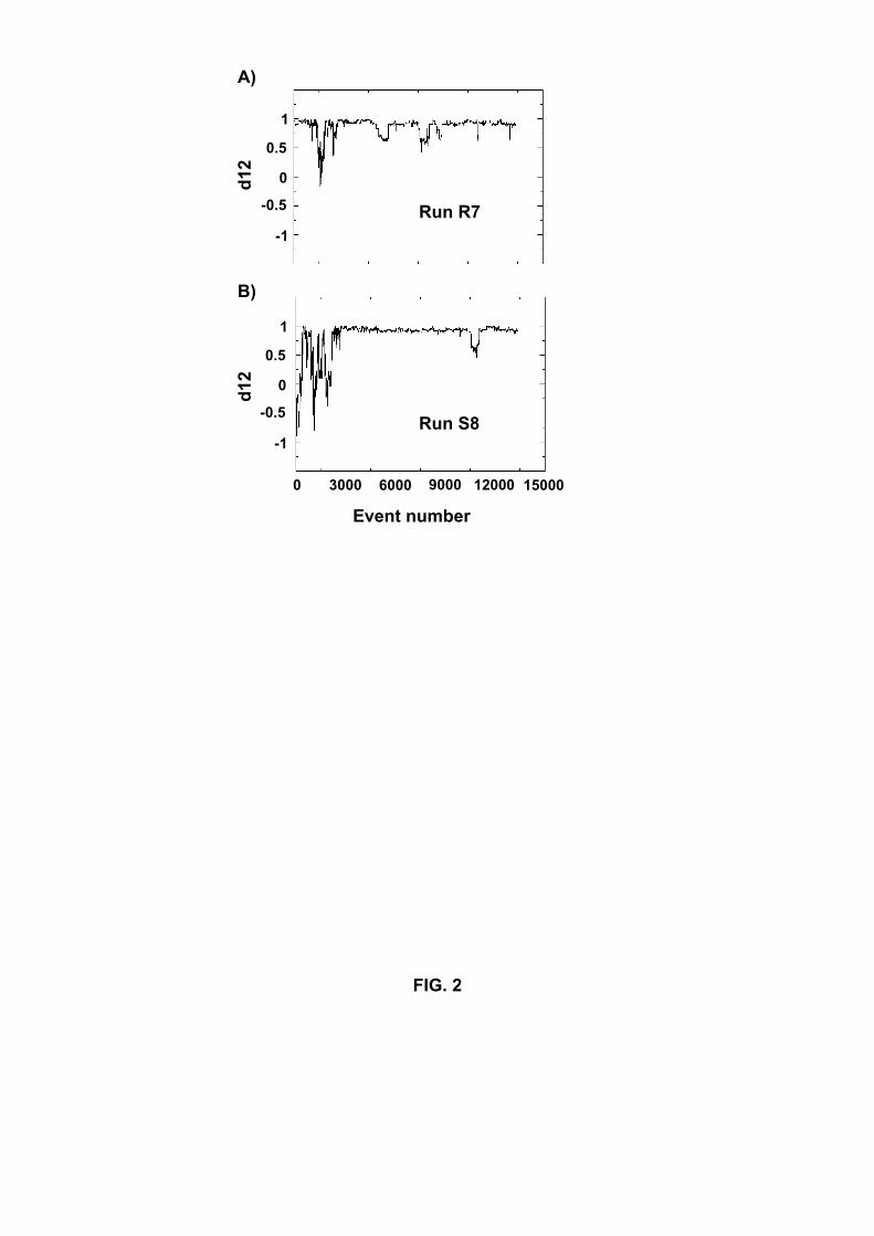
FIG. 2
d12
d12
3000 6000 9000 12000 15000
-1
1
-0.5
0
0
0.5
-1
1
-0.5
0
0.5
Event number
A)
B)
Run R7
Run S8
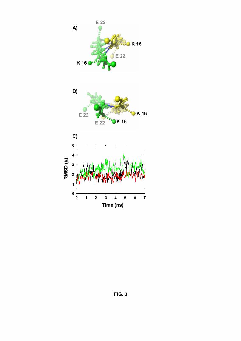
FIG. 3
A)
B)
C)
K 16
E 22
E 22
E 22 E 22
K 16
K 16 K 16
0 2 4 60
1
2
3
4
5
7531
Time (ns)
RM
SD (Å
)
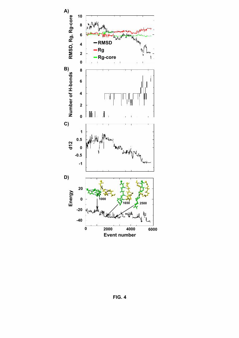
FIG. 4
d12
20
0
-20
-40
0
10
2
4
6
8
0
2
4
6
8
-1
1
-0.5
0
0.5
RM
SD, R
g, R
g-co
re
Num
ber o
f H-b
onds
En
ergy
Event number
A)
B)
C)
D)
RMSDRg Rg-core
0 2000 4000 6000
1000 1650 2500

FIG. 5
N + N
U’ + N U’ + U’
U’ + N
U’ + U’
U2 N2
(P1)
(P2)
(P4)
(P3)
All
S
S3
S3
S5
S1
S4
S4
S2 S
6 S8
S7 S9
S7
S9
R5
R7
R8
R11
R1
R4
R6
R1
R2 R
10
R3
R9
(III
)
R2
R10
(I)
(II)
R9
R10
R
3 R
12
R1
R6
(IV
)
S5
S1
(I)
K-L
-V-F
-F-A
-E
E-A
-F-F
-V-L
-K
(II)
K-L
-V-F
-F-A
-E
E-A
-F-F
-V-L
-K
(III
)
K-L
-V-F
-F-A
-E
E-A
-F-F
-V-L
-K
(IV
)
K-L
-V-F
-F-A
-E
E-A
-F-F
-V-L
-K
All
R
FIG. 6
(A)
(B)
(A)
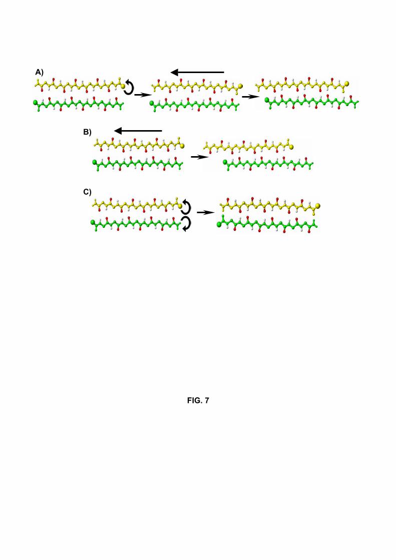
FIG. 7
A)
B)
C)

FIG. 8
3000 6000 9000 12000 15000 0 0
2
4
6
8
10
0
2
4
6
8
10
0
2
4
6
8
10
Event number
Num
ber o
f H-b
onds
N
umbe
r of H
-bon
ds
Num
ber o
f H-b
onds
A)
B)
C)
Run S4
Run R2
Run R9
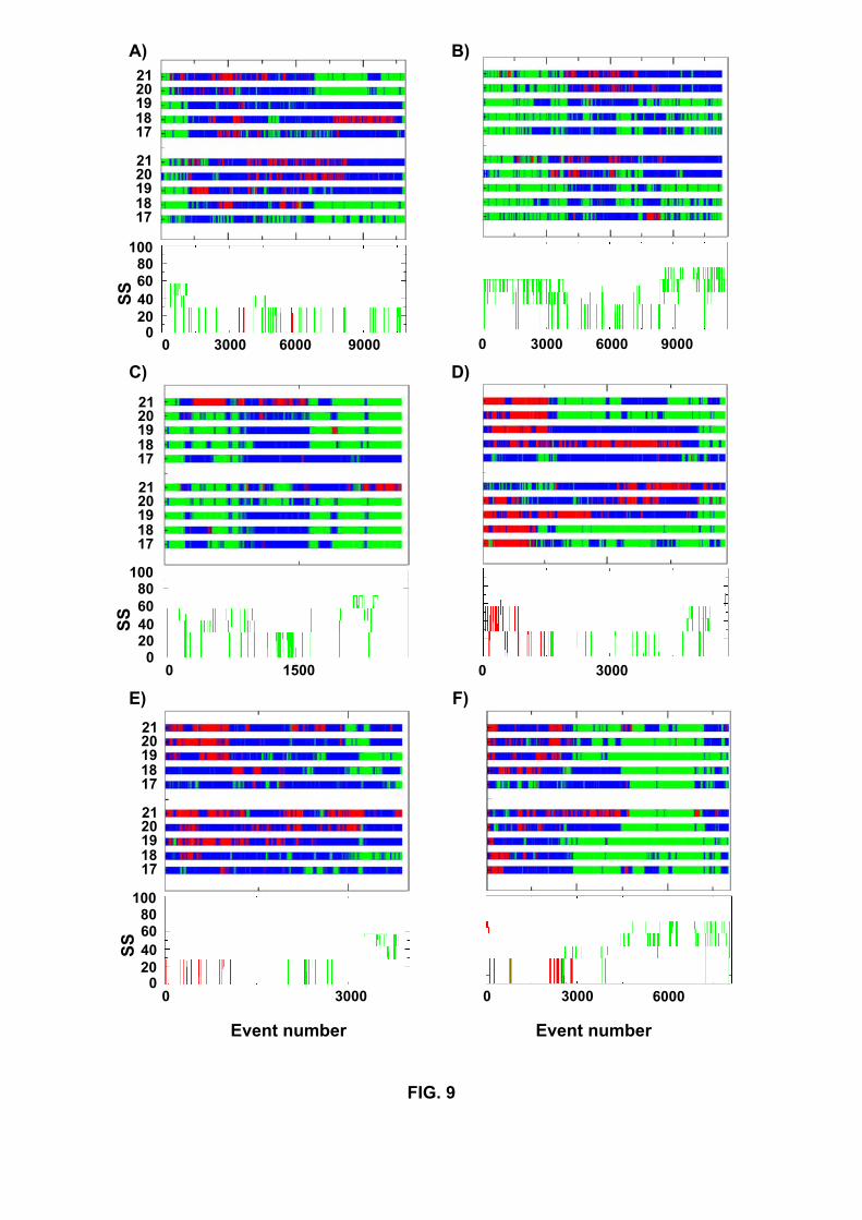
FIG. 9
C)
17
21 20 19 18
17
21 20 19 18
20
60
100
0
40
80
SS
1500 0
A)
17
21 20 19 18
17
21 20 19 18
0 20 40 60
100 80
SS
3000 6000 9000 0
B)
3000 6000 9000 0
F)
3000 6000 0
Event number
30000 0
60
100
E)
17
21 20 19 18
17
21 20 19 18
20 40
80
SS
Event number
D)
3000 0


![Alzheimer's disease - a neurospirochetosis. Analysis of ... · gles and amyloid deposition both occur in dementia paralytica [59,64-66]. Recent analysis of archival brain material](https://static.fdocuments.us/doc/165x107/6063ff9cd49b8c0275254d9b/alzheimers-disease-a-neurospirochetosis-analysis-of-gles-and-amyloid-deposition.jpg)


![Alpha-synucleinaltersdifferentlygeneexpressionofSirts ... · mers which subsequently can aggregate into soluble protofibrils and insoluble β-amyloid fibres [9]. Recent data haveindicated](https://static.fdocuments.us/doc/165x107/5e634eabc469ab7a0c1b0dc3/alpha-synucleinaltersdifferentlygeneexpressionofsirts-mers-which-subsequently.jpg)


