Pathology of subclinical paratuberculosis (Johne’s Disease ...vri.cz/docs/vetmed/55-12-590.pdf ·...
13
Original Paper Veterinarni Medicina, 55, 2010 (12): 590–602 590 Pathology of subclinical paratuberculosis (Johne’s Disease) in Awassi sheep with reference to its occurrence in Jordan N.Q. Hailat 1 , W. Hananeh 1 , A.S. Metekia 1 , J.R. Stabel 2 , A. Al-Majali 1 , S. Lafi 1 1 Faculty of Veterinary Medicine, Jordan University of Science and Technology, Irbid, Jordan 2 USDA-ARS, National Animal Disease Centre, Ames, USA ABSTRACT: In this study, the pathological lesions and occurrence of subclinical Johne’s disease in Awassi sheep is investigated using histopathological (HP) and immunohistochemical (IHC) examinations, Acid Fast Staining (AFS) of tissue sections, direct smears from tissues and culture. Histopathological examination of 202 ilea and the corresponding mesenteric lymph nodes (179) was conducted. In addition, IHC examination, using rabbit poly- clonal antiserum, of 134 ilea and 123 mesenteric lymph nodes was also conducted. The occurrence of the disease was observed in 50% and 93% of the ilea examined using histopathology and IHC techniques, respectively. Fifty nine percent of lymph nodes were positive by IHC. The histopathological lesions were graded from І–IV, I being the least severe, based on the type of cellular infiltrate (lymphocytes, macrophages and epithelioid cells) and the severity of the lesions. Grades III and IV (SP) were considered positive while I and II were considered suspected. Analysis of the results also revealed that the majority of suspected cases (grades I and II) reacted positive with the IHC. Furthermore, the IHC reactions were classified into three categories depending on the number of stained cells and the intensity of the staining (I-mild, II-moderate and III-strong). Direct smears, and tissue sections obtained from the ilea and stained with AFS revealed that out of 202 tissue samples, 53 (26%) and 22 (11%) were positive, respectively. Results of the culture revealed that 22 (11%) out of 202 were positive. These results showed that the occurrence of paratuberculosis (Johne’s disease) in Awassi sheep is very high in Jordan and needs further study in order to develop rational methods of control effective for the Jordanian sheep population. Keywords: sheep; paratuberculosis; histopathology; acid fast stain; ileum; immunohistochemistry Paratuberculosis (Johne’s disease) is an infectious disease of ruminants, considered by many research- ers to be one of the most serious infectious diseases currently plaguing the world’s sheep, cattle and goat industries (Hope et al., 2000). An increasing number of publication highlights the importance of this animal infection with a zoonotic potential (Kaevska and Hruska, 2010). The disease caused by Mycobacterium avium subsp. paratuberculosis (MAP), a gram-positive acid-fast organism shed in the faeces of infected animals, is characterized by chronic granulomatous enteritis (Jubb et al., 1993; Radostits et al., 1994; Adams et al., 1996). Contaminated feed and water, bedding and soiled udders are thought to be the major routes for spread of the organism and young animals less than six months of age are thought to be the most suscep- tible to infection. Cattle become infected early as young calves via faecal-oral transmission of the infectious agent (Stabel et al., 2002). Intrauterine infection in cattle has also been well document- ed (Martin and Aitken, 1991; Whittington and Windsor, 2009). Isolation of M. paratuberculosis from a sheep foetus has been confirmed, and anti- bodies to M. paratuberculosis have been demon- strated in colostrum-deprived lambs (Jones et al., 1997; Lambeth et al., 2004). Focus on paratuberculosis has been intensifying as it is becoming an economically important dis- ease for the livestock sector. It was reported that
Transcript of Pathology of subclinical paratuberculosis (Johne’s Disease ...vri.cz/docs/vetmed/55-12-590.pdf ·...

Original Paper Veterinarni Medicina, 55, 2010 (12): 590–602
590
Pathology of subclinical paratuberculosis (Johne’s Disease) in Awassi sheep with reference to its occurrence in Jordan
N.Q. Hailat1, W. Hananeh1, A.S. Metekia1, J.R. Stabel2, A. Al-Majali1, S. Lafi1
1Faculty of Veterinary Medicine, Jordan University of Science and Technology, Irbid, Jordan2USDA-ARS, National Animal Disease Centre, Ames, USA
ABSTRACT: In this study, the pathological lesions and occurrence of subclinical Johne’s disease in Awassi sheep is investigated using histopathological (HP) and immunohistochemical (IHC) examinations, Acid Fast Staining (AFS) of tissue sections, direct smears from tissues and culture. Histopathological examination of 202 ilea and the corresponding mesenteric lymph nodes (179) was conducted. In addition, IHC examination, using rabbit poly-clonal antiserum, of 134 ilea and 123 mesenteric lymph nodes was also conducted. The occurrence of the disease was observed in 50% and 93% of the ilea examined using histopathology and IHC techniques, respectively. Fifty nine percent of lymph nodes were positive by IHC. The histopathological lesions were graded from І–IV, I being the least severe, based on the type of cellular infiltrate (lymphocytes, macrophages and epithelioid cells) and the severity of the lesions. Grades III and IV (SP) were considered positive while I and II were considered suspected. Analysis of the results also revealed that the majority of suspected cases (grades I and II) reacted positive with the IHC. Furthermore, the IHC reactions were classified into three categories depending on the number of stained cells and the intensity of the staining (I-mild, II-moderate and III-strong). Direct smears, and tissue sections obtained from the ilea and stained with AFS revealed that out of 202 tissue samples, 53 (26%) and 22 (11%) were positive, respectively. Results of the culture revealed that 22 (11%) out of 202 were positive. These results showed that the occurrence of paratuberculosis (Johne’s disease) in Awassi sheep is very high in Jordan and needs further study in order to develop rational methods of control effective for the Jordanian sheep population.
Keywords: sheep; paratuberculosis; histopathology; acid fast stain; ileum; immunohistochemistry
Paratuberculosis (Johne’s disease) is an infectious disease of ruminants, considered by many research-ers to be one of the most serious infectious diseases currently plaguing the world’s sheep, cattle and goat industries (Hope et al., 2000). An increasing number of publication highlights the importance of this animal infection with a zoonotic potential (Kaevska and Hruska, 2010). The disease caused by Mycobacterium avium subsp. paratuberculosis (MAP), a gram-positive acid-fast organism shed in the faeces of infected animals, is characterized by chronic granulomatous enteritis (Jubb et al., 1993; Radostits et al., 1994; Adams et al., 1996).
Contaminated feed and water, bedding and soiled udders are thought to be the major routes for spread
of the organism and young animals less than six months of age are thought to be the most suscep-tible to infection. Cattle become infected early as young calves via faecal-oral transmission of the infectious agent (Stabel et al., 2002). Intrauterine infection in cattle has also been well document-ed (Martin and Aitken, 1991; Whittington and Windsor, 2009). Isolation of M. paratuberculosis from a sheep foetus has been confirmed, and anti-bodies to M. paratuberculosis have been demon-strated in colostrum-deprived lambs (Jones et al., 1997; Lambeth et al., 2004).
Focus on paratuberculosis has been intensifying as it is becoming an economically important dis-ease for the livestock sector. It was reported that

Veterinarni Medicina, 55, 2010 (12): 590–602 Original Paper
591
the economic losses due to Johne’s disease for the control and eradication program in one flock with a few cases would average $650 with most of that cost going for serological tests. The cost of disease for ovines is approximately $90 per clinical case and $250 per bovine clinical case (Menzies, 2010). The economic effect of Johne’s disease consists in the restriction of livestock marketing and contamina-tion of land. In Iceland, for example, measures to be taken for the control of paratuberculosis are expensive, involving the fencing of hundreds of kilometres to restrict sheep movement from the infected area. Laboratory tests used to detect in-fected animals are also costly and notwithstanding all these expenses it is very difficult to eradicate the disease (Fridriksdottir et al., 2000).
In addition, consumers of animal products are be-coming increasingly sensitive to the possible effect of livestock disease and residues on product qual-ity and human health. The potential link between Crohn’s disease (CD) and M. paratuberculosis is an example of such a potential relationship (Kennedy and Allworth, 2000; El-Zaatari et al., 2001). Recent data have strengthened the etiologic association between M. paratuberculosis and CD (Naser et al., 2000). Milk has been suggested as a possible vehicle of transmission of M. paratuberculosis to humans as M. paratuberculosis has been detected by PCR in row milk as well as from pasteurized milk from cattle, sheep and goats, repeatedly in England (Millar et al., 1996; Ellingson et al., 2005; Hruska et al., 2005; Zoi Dimareli-Malli, 2010).
It has been suggested that the diagnosis of sub-clinical cases of paratuberculosis is still a problem (Kurade et al., 2004). It was also reported that the diagnosis of subclinical cases of paratuberculosis is more difficult by histopathology as lesions may be subtle and organisms may be low in number (Buergelt et al., 2000). In sheep, on the other hand, it has been reported that histopathology was found to be a better indicator of paratuberculosis than bacteriology (Kurade et al., 2004). Furthermore, it was suggested that histopathological studies of tissues from infected animals can be considered as the gold standard method for the diagnosis of Johne’s disease and should be used to confirm the disease instead of other methods (Hope et al., 2000). However, expertise and diagnostic criteria may vary between institutions (Huda and Jensen, 2003). Therefore, accurate subclinical diagnosis needs more than one test to complement the his-topathological diagnosis.
To estimate and understand the occurrence of Johne’s disease in sheep, we conducted a field study of twenty sheep flocks, ranging from 50–500 heads and found that 80% of the flocks had a history of un-treatable emaciation, bottle jaw and diarrhea which were associated with animal losses. Therefore, this investigation was carried out to study the associat-ed pathological lesions of Johne’s disease in the ilea and their corresponding mesenteric lymph nodes, and its prevalence in apparently healthy young Awassi sheep in Jordan, using gross/histopatho-logical, immunohistochemistry (IHC), Acid Fast Stain (AFS) examinations and culture.
MATERIAL AND METHODS
Animals/Awassi sheep
Awassi sheep included in this study were locat-ed at 36°66'E, 32°04'N at about 616–686 m above sea level. During the period of this study, annual rainfall varied between 159 and 380 mm. Rainfall usually starts in November and ends by April. No precipitation occurs between May and November. Daily temperature is moderate with mean monthly maxima ranging between 12 and 32 °C and mean monthly minima between 2 and 14 °C. The mean monthly relative humidity ranged between 44 and 80%. The raising system of sheep in Jordan is mainly semi-intensive; farmers usually feed their animals chopped straw, barley and wheat bran during the winter season (October to February). During spring and summer seasons, the animals are maintained on grazing. Some farmers provide concentrate sup-plementation in seasons when the grazing grass is not adequate. The Awassi sheep breed is raised for meat, milk and wool production and it repre-sents almost 98% of the sheep breeds in the country (Ministry of Agriculture, 2000).
Two hundred and seventy nine apparently healthy Awassi sheep of both sexes, 8–24 months of age, raised in different flocks in Jordan, were slaugh-tered and examined in the slaughterhouses of the Central (represents 32% of the national flock of 1.9 million sheep) and Northern (represents 45% of the national flock) parts of the country (Ministry of Agriculture, 2000). The animals had no signs of emaciation, bottle jaw and diarrhea, alone or in combination. The site for animal selection in the slaughterhouses was based on the sheep popula-tions and distribution in Jordan. Animal movement

Original Paper Veterinarni Medicina, 55, 2010 (12): 590–602
592
is free and the sheep are bought from the sheep farmers in local animal markets by the local butch-ers or animal traders twice a week; on Mondays and Thursdays. Sheep are either sent directly to the slaughter house or fattened for some time before-hand. Different sheep farmers bring their animals from different sites of the country; in some cases they transport them about 150 Km or more to bring them to the animal markets. In some cases, sheep traders visit sheep farmers and buy sheep from the farms directly before sending them to the slaughter house or for slaughter in local butcheries.
Tissue sample collection
During a period of five months (August to December 2001), a total of 279 apparently healthy Awassi sheep were subjected to postmortem ex-amination in slaughterhouses where small and large intestines were examined thoroughly. The slaugh-terhouses were visited weekly to collect the sam-ples. Before slaughtering, complete information about the animals regarding age, sex, significant clinical signs and health status were recorded. The age of the sheep ranged from 8 to 24 months. The majority of the sheep examined were males.
Gross examination and samples transportation
Samples from small and large intestines and respective lymph nodes of the 279 animals were thoroughly examined for any apparent gross le-sions. Significant gross lesions including mucosal thickening, mucosal corrugations, prominent lym-phatics and intestinal discolorations were recorded. The size, shape and discoloration of the respec-tive lymph nodes were carefully described. Out of the 279 animals examined, 202 animals with some gross changes were sampled and tissues were col-lected from their ilea, ileocecal valves and respec-tive lymph nodes based on tissue changes seen in these organs. In the few cases in which gross lesions were observed in the large intestine, tissue samples from the cecum and colon were collected. Tissues samples were trimmed to a smaller size (4 mm to 1 cm thickness), fixed in 10% buffered formalin, and subjected to histopathological examination (202 ilea and 179 lymph nodes) and immunohis-tochemical staining (134 ilea and 123 lymph nodes).
Other tissue samples were transported on ice to the laboratory for direct smear (202 ilea) and for bac-terial culture (202 ilea). Samples for culture were kept at –20 °C until needed.
Direct smear staining with acid fast stain
Scrapping of the intestinal mucosa and lymph nodes parenchyma were carried out using scalpel blade and smears were done on microscopic slides. Slides were dried by air, fixed with methanol (100%) and stained with AFS method according to Coles (1986). Each slide was examined for 30 min us-ing oil immersion. The findings were registered according to the bacteria appearance, in which ob-servation of bacteria’s in clumps taken as positive, in dispersed form as suspected and as negative if neither of the two forms observed.
Culture
Media preparation. Middle Brook 7H10 agar base M199 (Himedia Laboratories, limited Mumbai (Bombay) 400086, India) was used with a supple-ment as a slant in tubes of 20 ml. The media was prepared as described in the OIE (2000). To 450 li-tres of distilled water, 9.73 g was added. Then, 5 ml of glycerol was added to this mixture which was boiled, autoclaved at 121 °C, 15 psi, for 15 min, and then cooled at 45 to 50 °C in a water bath (GFL 1083, Germany). Middlebrook OADC growth sup-plement (FD 018, India, 50 ml/l) was then added. For the suppression of contaminants, Penicillin (1000 000 IU) and the anti-fungal agent Nystatin (50 mg) were added to the media. The media con-taining mycobactin was prepared by adding myco-bactin J 2 mg/l (Allied Monitor IAC, Fayette, MO, USA) of the medium dissolved in 4 ml of ethyl al-cohol with the agar base before autoclaving.
Sample preparation and inoculation. Appro- ximately 3–5 g of scraped intestinal mucosa and lymph node parenchyma was taken and ground together using mortar, and incubated with 0.5% trypsin at 4 °C overnight at pH = 7.2 to 7.4 (adjusted using 4% NaOH). After 16 to 8 hours, the mixture was filtered using a gauze (folded), and centrifuged at 400 × g for 20 min (PK110, ALC, Italy). The su-pernatants were decanted and decontaminated and 5% oxalic acid was added to the pellet. The samples were then left for 24–30 h at room temperature.
Veterinarni Medicina, 55, 2010 (12): 590–602 Original Paper
593
Finally, 0.1ml (100 μl) of inoculum was taken care-fully from the bottom of the tube and evenly distrib-uted on the media. Each sample was inoculated in three tubes (one tube without mycobactin J and two with mycobactin J) and incubated at 37 °C (Binder GmbH, Germany) with a loss screw and inclined to facilitate the evaporation of excess moisture and inoculum fluid for one week. After one week tubes were returned to the vertical position with a tight-ened screw and incubated for 16 weeks.
Culture reading. Starting from eight weeks of inoculation, cultures were observed weekly for the presence of any growth. At 16 weeks, smears were taken from cultures that showed growth, and stained by AFS as described previously (Prophet et al., 1994). A culture was considered positive when white spot colonies were seen and this was con-firmed by AFS. Slides were observed under 100 × objectives for the detection of acid fast bacilli. Results were recorded considering the long incuba-tion period, the colony appearance, and acid-fast-ness of the bacteria. Nine tubes with micro-fungal growth and contamination were discarded.
Histopathological examination
All the trimmed samples were processed rou-tinely for histopathological examination accord-ing to a protocol described previously (Bancroft and Stevens 1990). Sections (4–5 µm) were cut and stained with Haematoxylin and Eosin (H&E). Staining of tissue sections by AFS was performed as described previously (Prophet et al., 1994).
Grading criteria for histopathological lesions
The H&E stained sections from 202 ilea and the corresponding lymph nodes (179) were observed
under 4×, 10×, and 40× objectives. Pathological lesions were recorded. The lesions were classified and graded into grades I, II, III and SP, depending on the type and amount of the cellular infiltrates (lymphocytes, macrophages and epithelioid cells). The intestinal tissue sections were considered posi-tive for paratuberculosis when there were macro-phage infiltrations or epithelioid cells obvious in the lamina propia of the villi and between crypts, and there was involvement of the Peyer’s patches as demonstrated by the presence of starry sky macro-phages/tingible body macrophages or micro-granu-loma. In cases where there was no microgranuloma, but macrophages and some epithelioid cells were present, these were considered suspected cases by histopathological criteria. The scoring of tissue le-sions was as shown in Table 1. Therefore, grades I and II were considered suspected while grades III and SP were considered positive.
Immunohistochemical (IHC) staining
Tissue samples from paraffin-wax embedded blocks were sectioned at 2–3 μ prior to immuno-histochemical staining. The sectioned tissue sam-ples were laid on Vectabond (DAKO A/S.Glostrup, Denmark) coated slides, air-dried and then heated in an oven at 55 °C for two hours. Tissue sections were deparaffinized in xylene and hydrated by sequential immersion of slides in decreasing concentrations of ethanol (100%, 95% and 70%) for one minute each. Tissue sections were washed in distilled water for 5 min, then washed in PBS, followed by immersion in citrate buffer solution (pH= 6, 10mM). Antigen retrieval was carried out by autoclaving the tissue section at 120 °C, 15 psi for 15 min (Express, Italy). The sections were cooled at room temperature and washed in PBS for 5 min. All of the subsequent in-cubations were performed at room temperature, and all washes were in PBS (pH 7.4, 3×; 5 min each).
Table 1. Criteria used for grading pathological lesions found in the ilea and lymph nodes
GradeType of inflammatory cells
lymphocytes macrophages epithelioid cells PP prol. and crypts replacement microgranuloma
I ++++ ++ + – –II +++ ++++ +++ moderate yes/noIII + +++ ++++ severe yes
SP + ++++ ++++ calcification, caseous necrosis in the LN
granuloma with giant cells
SP = special, PP prol. = Peyer’s patches proliferation, LN = lymph nodes

Original Paper Veterinarni Medicina, 55, 2010 (12): 590–602
594
Endogenous peroxidase was inactivated by im-mersion of the slides in a solution of 15% hydrogen peroxide in methanol for 30 min. After washing, non-specific adherence of proteins to tissue sec-tions was blocked by incubating with 1% bovine se-rum albumin (BSA), (Sigma Chemical Co., St.Louis, MO) for 2 h. The solution was drained from the slides and the rabbit polyclonal M. paratubercu-losis antiserum # 270, diluted 1 : 500 in PBS, was incubated on the slides for 2 h. The rabbit antibody was kindly provided by Dr. Stabel from the National Animal Disease Center, Ames, IA, USA. This was followed by further washing and incubation with biotinylated anti-goat, anti-rabbit, and anti-mouse immunoglobulin (DAKO A/S, Glostrup, Denmark), diluted at 1 : 20, as the secondary antibody for 15 min. After washing, streptavidin-biotinylated peroxidase conjugate (DAKO, A/S, and Glostrup, Denmark) was applied, and incubated on the tissue section for 15 min. The slides were washed and incubated with chromogen (3,3 diamino- benzi-din-4HCL; DAB, DAKO) at 1 mg/ml in PBS sup-plemented with hydrogen peroxide (10 ul of 3% hydrogen peroxide for 2 ml of DAB). Samples were incubated at room temperature for 3–5 min, and slides were then washed in distilled water for five minutes. Slides were then counter stained in hae-matoxylin for 2–3 min. The sections were washed in water and dehydrated in graded alcohol and cleared in xylene and mounted using DPX for fur-ther observation.
Slides were observed using 4×, 10× and 40× ob-jectives. Sections were considered positive accord-ing to the color observation that is an indication of antibody-antigen reaction, and manifested by intra-cytoplasm or extra-cellular brown colora-tion in different areas of the stained tissue sec-
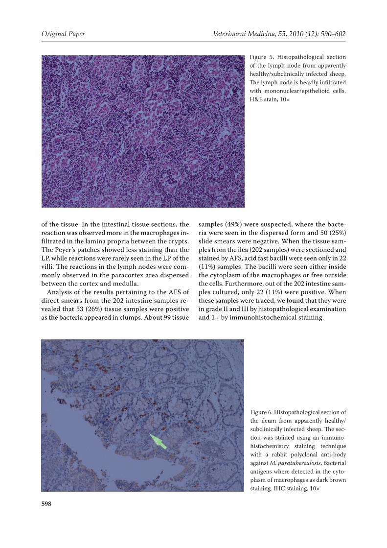
tion. The findings were registered by counting the number of reactions at 10×, accordingly starting from one cell reaction recorded as positive; 1–10 as + (mild), more than 10 but less than 50% cells reac-tion as ++ (moderate), reaction in 50% cells from one tissue section was graded as +++ (strong as in Figure 6). And more that 50% reaction in the macrophages of one section as ++++, but because only one sample was ++++, it was added to the +++ group. Additionally the intensity of the reac-tion was considered and in all cases only a strong brown color was recorded as a positive reaction. If the above criteria were found in at least one field, it was considered positive. At least one slide from each tissue section from the ilea sampled from each animal was tested. In some cases, more slides were taken from each tissue preparation.
In all cases, positive and negative control slides were processed together from the same known group of tissue sections, in order to avoid false positive and negative reaction. The positive control tissue was obtained from a sheep clinically suffered from MAP as diagnosed by histopathology, AFS staining, IHC and culture. The negative control was obtained from a three-month old lamb which was healthy and negative by all the four tests.
RESULTS
In this study, gross/histopathological, immuno-histopathological examinations, and AFS (tissue sections and scrapping) of the ilea and correspond-ing lymph nodes of 202 clinically healthy Awassi sheep, which were slaughtered during the study period, were used to assess, describe and grade the histopathological picture and examined the tissue
Table 2. Grade of histopathological findings, from ilea and corresponding lymph node H&E tissue sections in appar-ently healthy Awassi sheep aged 8 to 24 months in Jordan, 2002
Type of tissue Samples processed
Grade of the lesions
I II III SPTotal
+ve –ve
Ileum202 23 70 98 4 195 7
% (11) (35) (48) (2) (97) (3)
Lymph nodes179 10 35 76 4 125 54
% (5.5) (19.5) (42) (2.2) (69) (31 )
The percentage was taken by considering the decimal number > 5, adding one in all the results SP = special lesion, numbers in parentheses are percentiles

Veterinarni Medicina, 55, 2010 (12): 590–602 Original Paper
595
reaction regarding the presence of Mycobacterium avium subps. paratuberculosis for the purpose of di-agnosing the disease in its subclinical presentation. Culture was also used to confirm the diagnosis.
When the intestines and the corresponding lymph nodes were examined grossly in the slaughterhouses, we found that 202 intestines or lymph nodes out of the 270 animals examined harboured some sort of pathological changes. These changes ranged from very mild to severe congestion in the mucosa and thickening in the wall of the ilea. The wall of the ilea was very thick with corrugation in 13 animals (Figure 1). The surrounding mesenteric lymph nodes
were enlarged and oedematous in few cases, and they were connected with each other as a result of their tissue reaction and appeared as cords.
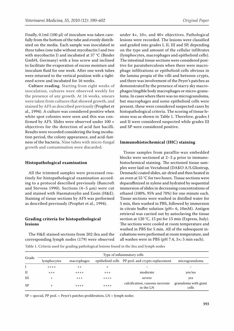
Histopathological examination of H&E tis-sue sections from the 202 ilea and correspond-ing lymph nodes revealed that 195 (97%) ilea had variable increases in the thickness and congestion of the mucosa due to inflammatory cell infiltra-tions (Table 3). The mucosa and less frequently the sub mucosa were infiltrated primarily with vari-able numbers of macrophages, lymphocytes and plasma cells (Figures 2 and 3). Occasional num-bers of epithelioid cells were also seen. They were present either as a scattered form in the lamina propria of the villi and between the crypts or as nests. Multinucleated giant cells were rarely seen in the affected tissues. In some cases the cellular in-filtrates were associated with caseous necrosis and/or mineralization. Variable degrees of Peyer’s patch hyperplasia were noted in many tissue sections with prominent starry sky macrophages/tingible body macrophages (Figure 3). In most cases, granuloma-tous lymphangitis with or without dilated lacteals were present (Figure 4). The villi exhibited different changes including: villous distortion and thickening by inflammatory cell infiltrations, villous atrophy and fusion. The lymph node changes consisted primarily of lymphofollicular hyperplasia and in-filtration of mononuclear cells predominately with epithelioid cells (Figure 5). The most severely af-fected lymph nodes were characterized by granulo-mas formation with mineralization. In less severely affected lymph nodes, epithelioid cells mixed with
Figure 2. Histopathological section of the ileum from apparently healthy/subclinically infected sheep. Payer’s patches lymphoid hyperplasia is seen throughout the section. H&E stain, 4×
Figure 1. Gross pathology of an ileum section from apparently healthy sheep. Corrugations and thickening of the mucosal wall are prominent

Original Paper Veterinarni Medicina, 55, 2010 (12): 590–602
596
other mononuclear cells and a few neutrophils were scattered throughout the lymph node sections.
Table 2 shows the grading of the lesions seen in the intestine and the lymph nodes. Out of the 202 intestinal H&E tissue sections examined, 23 (11%), 70 (35%), and 98 (48%) were graded as I, II and III, respectively, based upon the grading cri-teria described; namely the degree of lymphocyte, macrophage, and epithelioid cell infiltration; cel-lular proliferation within the Peyer’s patches and crypts; and the presence of microgranuloma. Four cases were considered as a special category where calcification with severe reaction and typical gran-ulomatous reaction with multinucleated Langhans giant cells were observed in the lymph nodes. The cases in grades III and SP (IV) were considered positive while the cases in grades I and II were considered suspected for the disease by histopatho-logical evaluation.
To further understand the cellular reaction in the intestine and in the respective mesenteric lymph nodes and to investigate whether this reaction was associated substantially with the presence of M. paratuberculosis, immunohistochemical exami-nation was used. A positive reaction was manifested by the presence of brown colour around the mac-rophage nucleus (perinuclear and intracytoplasmic reaction) (Figures 6 and 7). Table 3 shows the grading of the lesions seen in the intestine and the lymph nodes as the tissue samples were examined by im-munohistochemistry. We examined only 134 ilea tissue sections and 123 tissue sections from the corresponding lymph nodes, and because of the limitation in the reagents, we did not examine all tissue specimen. We found that 124 (93%) tissue sec-tions from the ilea were positive, ranging from mild (+) moderate (++) and strong (+++); 108 (81%), 9 (7%), 6 (4%) and 1 (1%) were graded as +, ++, +++
Figure 3. Histopathological section of the ileum from apparently healthy/sub-clinically infected sheep. Mononuclear cells infiltrate primarily the mucosa, and less frequently the submucosa. Dilation of some of the lacteals in the villi is also seen. H&E stain, 4×
Table 3. Immunohistochemical stain results from intestinal samples and lymph nodes in apparently healthy Awassi sheep, 8 to 24 months, in Jordan 2002
Type of tissue
Number of samples processed
Immunohistochemical stain
+ ++ +++total
+ve –ve
Ileum134 108 9 7 124 10
% (81) (7) (5) (93) (7)
Lymph node123 55 10 7 72 51
% (44) (8.1) (5.6) (58.5) (41.4)
Numbers in parentheses are percentiles

Veterinarni Medicina, 55, 2010 (12): 590–602 Original Paper
597
and ++++, respectively, based upon the number of cells which reacted positively with the antibody (Table 3). Because only one sample was in grade 4, it was added to grade 3. Out of the 123 tissue sections from the lymph nodes tested by IHC, 72 (58.5%) sections reacted positive (Table 3). Fifty five (44%) out of 123 lymph node tissue sections showed mild 1+, 10 (8%) moderate 2+ and 7 (6%) strong 3+. Interestingly, seven lymph node samples which
were negative by IHC were graded as 2+ and 3+ in the corresponding intestine by IHC. In either tissue sections of the ilea or their corresponding lymph nodes, if the tissue section shown 1+ or more, the tissue section (sheep) was considered positive for Johne’s disease by IHC.
The IHC staining of the tissue sections from the ilea was studied also to find the distribution of the stained macrophages in different histological parts
Table 4. Distribution of Histopathological (HP) (H&E), Immunohistochemical (IHC) and Acid Fast Stain (AFS) used on histopathological tissue sections (AFS/HP) and AFS Direct Smears (AFS/DS) and Culture results from intestinal specimens of apparently healthy Awassi sheep, 8 to 24 months, in Jordan 2002
Type of testNumber of samples
Totalpositive suspected negative
HP/H&E 102 93 7 202
% (50) (46) (4) (100)
IHC 124 0 10 134
% (92) (0) (8) (100)
AFS/HP 22 0 180 202
% (11) (0) (90) (100)
AFS/DS 53 99 50 202
% (26) (49) (25) (100)
Culture 22 0 180 202
% 11 0 89 (100)
Figure 4. Histopathological section of the ileum (muscularis and serosa) from apparently healthy/subclinically infected sheep. Granulomatous lymphangitis is shown where mononuclear cells infil-trate the wall of the lymphatics. H&E stain, L = lymphatic vessels, bar = 100 μ
Original Paper Veterinarni Medicina, 55, 2010 (12): 590–602
598
of the tissue. In the intestinal tissue sections, the reaction was observed more in the macrophages in-filtrated in the lamina propria between the crypts. The Peyer’s patches showed less staining than the LP, while reactions were rarely seen in the LP of the villi. The reactions in the lymph nodes were com-monly observed in the paracortex area dispersed between the cortex and medulla.
Analysis of the results pertaining to the AFS of direct smears from the 202 intestine samples re-vealed that 53 (26%) tissue samples were positive as the bacteria appeared in clumps. About 99 tissue
samples (49%) were suspected, where the bacte-ria were seen in the dispersed form and 50 (25%) slide smears were negative. When the tissue sam-ples from the ilea (202 samples) were sectioned and stained by AFS, acid fast bacilli were seen only in 22 (11%) samples. The bacilli were seen either inside the cytoplasm of the macrophages or free outside the cells. Furthermore, out of the 202 intestine sam-ples cultured, only 22 (11%) were positive. When these samples were traced, we found that they were in grade II and III by histopathological examination and 1+ by immunohistochemical staining.
Figure 5. Histopathological section of the lymph node from apparently healthy/subclinically infected sheep. The lymph node is heavily infiltrated with mononuclear/epithelioid cells. H&E stain, 10×
Figure 6. Histopathological section of the ileum from apparently healthy/subclinically infected sheep. The sec-tion was stained using an immuno-histochemistry staining technique with a rabbit polyclonal anti-body against M. paratuberculosis. Bacterial antigens where detected in the cyto-plasm of macrophages as dark brown staining. IHC staining, 10×

Veterinarni Medicina, 55, 2010 (12): 590–602 Original Paper
599
DISCUSSION
In the present study, the pathological lesions and the occurrence of subclinical Johne’s disease was studied in apparently healthy Awassi sheep between the ages of 8 to 24 months and with no history of clinical Johne’s disease using multiple diagnostic tests, histopathological and immunohistochemical examination, AFS and culture. Studies reporting previously on subclinical cases of Johne’s disease have demonstrated the importance of tissue sam-ple selection from different sites and it was found that sampling from limited foci can influence the results (Fraser et al., 1999; McDonald et al., 1999). Therefore, the selection of tissues from different sites along with the adjacent lymph nodes should be done to confirm the diagnosis of subclinical Johne’s disease, with the ileocecal valve as the first site to be selected. It was also reported that histopatho-logical examination should be considered as the first option for the diagnosis of subclinical cases. In our study, the last portion of the ileum and the ileocecal valve with their adjacent lymph nodes were utilized as the sample site and were used for all the implemented tests in this study.
Examination of the ilea and the corresponding lymph nodes from the clinically healthy Awassi sheep in the slaughter houses revealed that seven-ty-two percent (202 out of 279 examined) of them showed very mild to severe thickening and conges-tion in the intestine, especially in the last portion of the small intestine (Figure 1). Inflamed, oede-
matous, enlarged and corded mesenteric lymph nodes around the ileum and ileocecal valve were also evident. The histopathological findings such as the infiltration of the mucosa and submucosa with lymphocytes, macrophages, replacement of the crypts with macrophages, Peyer’s patches pro-liferation extending towards the mucosa and the presence of microgranuloma is partially in agree-ment with the results of others (Perez et al., 1996; Watkins et al., 2002). The grade 1 lesions noted in our study (11%; dominated by lymphocyte in-filtration) is in agreement with the asymptomatic form and to some extent with the paucibacillary form, while grades 2 and 3 (83%) are in agreement with the multibacillary form previously reported, keeping in mind that about 30% and 70% of the last two forms affected clinical cases, respectively (Watkins et al., 2002). This is in contrast to our study where the Awassi sheep were young and ap-parently healthy with no clinical signs in spite of the presence of marked macrophage infiltration and granulomatous reactions (42% of ilea) and with granuloma, Langhan giant cell formation and cal-cification (2% of animals) (Table 2). It has been reported that the paucibacillary form of the disease occurs particularly in sheep and in such animals M. paratuberculosis cannot be detected in the tis-sue microscopically and usually cannot be cultured (Hermon-Taylor, 1998). The grade 1 lesion in this study is close to the type 1 lesion in a study reported from South Africa although that study did not focus on lymphocyte infiltration and concentrated more
Figure 7. Histopathological section of the ileum from apparently healthy/subclinically infected sheep. This is a higher magnification view of figure six. IHC staining, 40×

Original Paper Veterinarni Medicina, 55, 2010 (12): 590–602
600
on the epithelioid cell nest in the mucosa and sub-mucosa (Micheal and Bastianello, 2002).
Paratuberculous lesions have been classified into three types (1, 2 and 3a, 3b, 3c) disregarding the clinical signs (Perez et al., 1996, 1999). In these classifications, epitheloid cells were not encoun-tered, and lymphocyte infiltrations were associated mainly with lesion type 3c. This classification was based mainly upon the presence of macrophages and acid-fast bacteria. In our study, only 22 (11%) of histopathological tissue sections revealed the presence of acid-fast bacilli and 26% were posi-tive on the direct smears (Table 4). Most of the 22 samples were grade 3 and a few were grade 2, with one, two or three intra-cytoplasmic bacilli observed within the macrophages. In some cases, extracel-lular acid-fast bacilli were also observed. In clinical cases it was reported that the presence of acid fast bacilli might be demonstrated intra- and extracel-lularly using the AFS stain (Kahn, 1997). However, in subclinical cases, it was reported that it may not be possible to find bacilli in tissues. Each tissue section was examined for a minimum of 30 min for the presence of bacilli as reported by others (Carbonell, 1998). Also, it was reported that it is very difficult in sub-clinical cases to detect bacteria by light microscopy due to the possible changes in the cell wall during the tissue preparation and processing (Micheal and Bastianello, 2002).
In a previous study, polyclonal antiserum which was raised by inoculating heat-killed M. paratuber-culosis, was shown to be useful for the detection of Johne’s disease in infected bovine tissue (Stabel et al., 1996). The specificity and the sensitivity of the immunohistochemical stain were high and can be used as a diagnostic tool. It was also evaluated for cross-reactivity with M. bovis antigens by immu-nohistochemical staining of tissues from infected pigs and cows. Tissues were devoid of positive re-activity when evaluated at the same dilutions that demonstrated positivity in M. paratuberculosis-infected tissues. In addition, cross-reactivity of the polyclonal antibody with M. avium infected tissue in pigs showed little positive staining. Furthermore, M. avium infection does not generally target the intestinal tract in ruminant species. Since we used a polyclonal antibody, it is unfair to consider this antibody completely specific for M. paratubercu-losis. This is the first study where we use this poly-clonal antibody in Awassi sheep and the tissues are from young and apparently healthy animals; it would be very interesting to find out how this
antibody reacts with tissues from sheep clinically infected with MAP. The 22 animals which were found by culture to be positive were also positive 1+ with this antibody by IHC. In tissues sections of ilea from apparently healthy cattle however, we found a significant correlation between the results of histopathological and IHC examinations; 66% of the ilea examined had lesions compatible with Johne’s disease and 65% reacted positive with the polyclonal antibody (Hailat et al., in preparation). Furthermore, others have also reported that poly-clonal and monoclonal antibodies could be used for the diagnosis of paratuberculosis in tissue sec-tions (Coetsier et al., 1999). Thus, it was concluded that the production of a polyclonal antibody to cell wall proteins of M. paratuberculosis resulted in a highly sensitive, species-specific tool for the de-tection of paratuberculosis in tissue sections. In addition, it was suggested that histopathological and immunohistopathological methods produced evidence of infection, which can be comparable with bacteriological examinations (Sigurdardottir et al., 1999).
In the present study, we used the same antisera, where a high percentage of the sheep tissue sec-tions were reactive within the infected macrophages and antibody reaction was also seen extracellularly (Table 3 and Figures 6 and 7). These results indicate that it is appropriate to use an immunohistochemical stain with a specific antiserum for the detection of M. paratuberculosis. The ultimate goal in determin-ing the etiologic relationship of an organism to a disease state is to demonstrate the association of the organism with the lesion. In our study, 93% of the ilea tissue sections (81% 1+) and 58% of the lymph node tissue sections (44% 1+) reacted with the polyclonal antiserum. Interestingly, 97% of the tissue sections from the ilea had mononuclear cellular infiltrates (50% grades III and SP) strongly suggesting an etio-logical relationship of the organism to the intesti-nal and lymph nodes lesions of Johne’s disease. In this study, it is also very obvious that the presence of an antigenic reaction by immunostaining in 108 (75%) out of 134 samples demonstrated by 1+, was characterised by high cellular infiltration in the LP and between the crypts with Peyer’s patches pro-liferation involving its border towards the mucosa. This indicates that at early stage of the disease or in subclinical cases, cell-mediated immune reactions could start with only few bacteria present in the in-testinal mucosa, as demonstrated by our results in these young animals.

Veterinarni Medicina, 55, 2010 (12): 590–602 Original Paper
601
We conclude that the occurrence of paratubercu-losis (Johne’s disease) is very high in Awassi sheep in Jordan. About 50% of clinically healthy Awassi sheep, 8 to 24 months of age, tested positive as they had histopathological lesions compatible with the disease. In addition, these cases were tested positive by IHC using a specific rabbit polyclonal antibody to M. paratuberculosis antigens. Thus, we consider paratuberculosis (Johne’s disease) to be a significant problem in Awassi sheep in Jordan and believe there is a great need for further studies on the prevalence and epidemiology of the disease in order to develop rational methods of control effec-tive on the Jordanian sheep population.
Acknowledgements
The authors would like to thank the Deanship of research for supporting this work as part of a graduate student thesis.
REfERENCES
Adams J, Collins MT, Czuprynski CJ (1996): Polymerase chain reaction analysis of alpha and iL-6 mRNA levels in whole blood from cattle naturally or experimentally infected with mycobacterium paratuberculosis. Jour-nal of Veterinary Research 60, 257–262.
Bancroft JD, Stevens A (1990): Theory and Practice of Histological Techniques. Churchill Livingston, Edin-burgh, London, Melbourne and New York. 21–119.
Carbonell P (1998): Histopathological diagnosis of Johne’s disease. Australian Veterinary Journal 76, 499.
Buergelt CD, Layton AW, Ginn PE, Taylor M, King JM, Habecker PL, Mauldin E, Whitlock R, Rossiter C, Col-lins MT (2000): The pathology of spontaneous paratu-berculosis in the North American Bison (Bison bison). Veterinary Pathology 37, 428–438.
Coetsier X, Havaux F, Matteland S, Sadatte F, Cormont K, Buergelt B, Limbourg D, Latinne M, Bazin JF, Denef C, Cocito C (1999): Detection of Mycobacterium avium subsp. paratuberculosis in infected tissue by new species-specific immunohistochemistry proce-
dures. Clinical Diagnosis Laboratory and Immunology 4, 446–451.
Ellingson JL, Anderson JL, Koziczkowski JJ, Radcliff RP, Sloan SJ, Allen SE, Sullivan NM (2005): Detection of Mycobacterium avium paratuberculosis in retail pas-teurized whole milk by two culture methods and PCR. Journal of Food Protection 5, 966–972.
El-Zaatari FA, Osato MS, Graham DY (2001): Etiology of Crohn’s disease: the role of Mycobacterium avium paratuberculosis. Trends in Molecular Medicine 7, 247–252.
Fraser CA, Marshall DJ, Ottaway SJ, Reddacliff LA, Whittington RJ (1999): Sensitivity of diagnosis of ovine Johne’s disease in New South Wales sheep flocks with a low prevalence of disease. In: Proceeding of the 6th International Colloquium on Paratuberculosis, Febru-ary 14–18, Melborn, Australia, 39 pp.
Fridriksdottir V, Gunnarsson E, Sigurdason S, Gud-mundsdottir KB (2000): Paratuberculosis in Iceland; Epidemiology and control measures past and present. Veterinary Microbiology 77, 263–267.
Hermon-Taylor J (1998): The causation of Crohn’s dis-ease and treatment with antimicrobial drugs. Italian Journal of Gastroenterol Hepatal 6, 607–610.
Hope AF, Kluver PF, Jones SL, Condron RJ (2000): Sen-sitivity and specificity of two serological tests for the detection of ovine paratuberculosis. Australian Vet-erinary Journal 87, 850–856.
Hruska K, Bartos M, Kralik P, Pavlik I (2005): Mycobac-terium avium subsp. paratuberculosis in powdered infant milk: paratuberculosis in cattle – the public health problem to be solved. Veterinarni Medicina 50, 327–335. http://vri.cz/docs/vetmed/50-8-327.pdf
Huda A, Jensen HE (2003): Comparison of histopathol-ogy, cultivation of tissues and rectal contents, and interferon-gamma and serum antibody responses for the diagnosis of bovine paratuberculosis. Journal of Comparative Pathology 129, 259–267.
Jones CT, Hunt DR, King WN (1997): Veterinary Pathol-ogy. 6th ed. Williams and Wilkins, UK. 498–500.
Jubb KV, Kennedy PC, Palmer N (1993): Pathology of Domestic Animals. 4th ed. Academic Press, USA, 157–158.
Kaevska M, Hruska K (2010): Analysis of publications on paratuberculosis from 1995 to 2009 with emphasis on
Editor-in-Chief quotes from the reviewer’s final recommendations
“I do believe that if the authors wish to pursue further work in this area, either further work to determine the specificity of this polyclonal antibody or additional confirmatory tests should be done to convince readers that their test results do identify MAP infected animals. Since they have cultured MAP from some sheep, it would be useful for them to try to determine the strain type (or types) that are represented.”

Original Paper Veterinarni Medicina, 55, 2010 (12): 590–602
602
the period from 2005 to 2009. Veterinarni Medicina, 55, 43-54. http://vri.cz/docs/vetmed/55-2-43.pdf
Kahn S (1997): Diagnosis of Johne’s disease in sheep, cattle and goats; Development of Microbiology and Immunol-ogy. James Cook University of North Queens land.
Kennedy DJ, Allworth MB (2000): Progress control and assurance programs of bovine Johne’s disease in Aus-tralia. Veterinary Microbiology 77, 443–451.
Kurade NP, Tripathi BN, Rajukumar K, Parihar NS (2004): Sequential development of histologic lesions and their relationship with bacterial isolation, faecal shedding, and immune responses during progressive stages of experimental infection of lambs with Myco-bacterium avium subsp. paratuberculosis. Veterinary Pathology 41, 378–387.
Lambeth C, Reddacliff LA, Windsor P, Abbott KA, Mc-Gregor H, Whittington RJ (2004): Intrauterine and transmammary transmission of Mycobacterium avium subsp. paratuberculosis in sheep. Australian Veteri-nary Journal 8, 504–508.
Martin WB, Aitken ID (1991): Diseases of Sheep. 2nd ed. Blackwell Scientific Publications, London. 59–98.
McDonald WL, Ridge SE, Hope AF, Condron RJ (1999): Evaluation of diagnostic tests for Johne’s disease in young cattle. Australian Veterinary Journal 77, 113–119.
Menzies P (2010): Johne’s disease in sheep. Department of Population Medicine, Ontario Veterinary College. www.omafra.gov.on.ca/english/livestock/sheep/facts/johnsdis
Micheal AL, Bastianello SS (2002): Paratuberculosis in sheep an emerging disease in South Africa. Veterinary Microbiology 77, 299–307.
Millar D, Ford J, Sandarson J, Withey S, Tizard M, Doran T, Hermon-Taylor J (1996): IS900 PCR to detect My-cobacterium paratuberculosis in retail supplies of whole pasteurized milk in England and Wales. Applied and Environmental Microbiology 62, 3446–3452.
Ministry of Agriculture Report (2000): Animal Wealth Report-Jordan. 5–20.
Naser SA, Hulten A, Shafran I, Graham DY, El-Zaatari FA (2000): Specific seroreactivity of Crohn’s disease patients against P53 and P36 antigens of M. avium subsp. paratu-berculosis. Veterinary Microbiology 77, 497–504.
OIE, Office International des Epizooties (2000): Paratu-berculosis (Johne’s Disease). In: Manual of Standards for Diagnostic Tests and Vaccines Terrestrial Animals. OIE, Paris, France. 1–19.
Perez V, Garcia Martin JF, Badiola JJ (1996): Description and classification of different types of lesion associated with natural paratuberculosis infection in sheep. Jour-nal of Comparative Pathology 2, 107–122.
Perez V, Tellechea J, Corpa JM, Guiterrez M, Garcia Martin JF (1999): Relation between pathologic findings and cellular immune responses in sheep with naturally acquired paratuberculosis. American Journal of Vet-erinary Research 60, 123–127.
Prophet EB, Mills B, Arrington JB, Sobin LH (1994): Laboratory Methods in Histopathology. Armed Force Institute of Pathology, Washington, D.C. 219–222.
Radostits OM, Blood DC, Gay CC (1994): Veterinary Med-icine, a Textbook of Diseases of Cattle, Sheep, Pigs, Goats and Horses. 8th ed. Bailliere Tindall, London. 841–849.
Sigurdardottir OG, Press CM, Saxegaard F, Evenson O (1999): Bacterial isolation, immunological response, and histopathological lesions during the early sub-clinical phase of experimental infection in goat kids with Mycobacterium avium subsp. paratuberculosis. Veterinary Pathology 36, 542–550.
Stabel JR, Ackerman MR, Goff JP (1996): Comparison of polyclonal antibodies to three different preparations of Mycobacterium paratuberculosis in cattle. Journal of Veterinary Diagnostic Investigation 4, 469–473.
Stabel JR, Wells SJ, Wagner BA (2002): Relationships between fecal culture, ELISA, and bulk tank milk test results for Johne’s disease in US dairy herds. Journal of Dairy Science 85, 525–531.
Watkins CA, Gossner A, Jones DG, Sharp JM, Hopkins J (2002): Comparative expression profiling in the three defined forms of ovine paratuberculosis. 7th Interna-tional Colloquium on Paratuberculosis, 11–14 June, Bilbao, Spain, 4 pp.
Whittington RJ, Windsor PA (2009): In utero infection of cattle with Mycobacterium avium subsp. paratu-berculosis: a critical review and meta-analysis. Vet-erinary Journal, 60–69.
Zoi Dimareli-Malli Z (2010): Detection of Mycobacterium avium subsp. paratuberculosis in milk from clinically affected sheep and goats. International Journal of Ap-plied Research in Veterinary Medicine 8, 44–50.
Received: 2010–07–14Accepted after corrections: 2010–10–22
Corresponding Author:
Nabil Qassem Hailat, Jordan University of Science and Technology, Faculty of Veterinary Medicine, Department of Pathology and Animal Health, Irbid, PO Box 3030, JordanTel. +962 0795885219, E-mail: [email protected]