PATHOLOGICAL AND THERAPEUTIC ANTIBODIES: NEW … · PATHOLOGICAL AND THERAPEUTIC ANTIBODIES: NEW...
100
PATHOLOGICAL AND THERAPEUTIC ANTIBODIES: NEW TOOLS FOR AUTOIMMUNITY DIAGNOSIS AND ANTICANCER THERAPY Annarita Tedesco Dottorato in Scienze Biotecnologiche – XX ciclo Indirizzo Biotecnologie Molecolari Università di Napoli Federico II
Transcript of PATHOLOGICAL AND THERAPEUTIC ANTIBODIES: NEW … · PATHOLOGICAL AND THERAPEUTIC ANTIBODIES: NEW...
PATHOLOGICAL AND THERAPEUTIC ANTIBODIES: NEW TOOLS FOR AUTOIMMUNITY DIAGNOSIS AND ANTICANCER THERAPY
Annarita Tedesco
Dottorato in Scienze Biotecnologiche – XX ciclo
Indirizzo Biotecnologie Molecolari Università di Napoli Federico II

Dottorato in Scienze Biotecnologiche – XX ciclo Indirizzo Biotecnologie Molecolari
Università di Napoli Federico II
PATHOLOGICAL AND THERAPEUTIC ANTIBODIES: NEW TOOLS FOR AUTOIMMUNITY DIAGNOSIS AND ANTICANCER THERAPY
Annarita Tedesco
Dottorando: Dott.ssa Annarita Tedesco
Relatore: Prof.ssa Renata Piccoli Coordinatore: Prof. Giovanni Sannia


Ai miei genitori

INDEX
ABBREVIATIONS pag. 1 Research activity pag. 3 ABSTRACT pag. 4 SOMMARIO pag. 5 INTRODUCTION pag. 11
Part A: THERAPEUTIC ANTIBODIES pag. 11 Immunotherapy pag. 11 Antibody engineering technology pag. 13 Tumour-Associated antigens pag. 16 Anti-ErbB2 antibodies pag. 17 Single chain variable fragment (scFv) pag. 19 Phage display technology pag. 20 Human anti-ErbB2 scFv (Erbicin) pag. 23
Part B: PATHOLOGICAL ANTIBODIES IN RHEUMATOID ARTHRITIS
pag. 25
Rheumatoid arthritis pag. 25 Pathogenesis of rheumatoid arthritis pag. 26 Rheumatoid factor pag. 28 Multiplex immunoassays pag. 31 Anti-CCP antibodies pag. 34
AIMS OF STUDY pag. 36
METHODS pag. 37
RESULTS pag. 47 Part A. THERAPEUTIC ANTIBODIES: A NOVEL ANTICANCER IMMUNOAGENT pag. 47
A1. The production of a novel immunoagent: a “compact” antibody
pag. 47
A2. Characterisation of the compact antibody pag. 54 A3. The prediction of the Erbicin structure pag. 57
Part B. A NEW DIAGNOSTIC STRATEGY FOR RHEUMATOID ARTHRITIS
pag. 66
Analyses of serum samples pag. 66 Rheumatoid factor detection pag. 67 Anti-CCP antibody assay pag. 69 Comparison of nephelometry versus FIDIS Rheuma test pag. 70 Comparison of nephelometry versus FIDIS Rheuma test coupled to anti-CCP test
pag. 71
Statistical analyses pag. 72
DISCUSSION pag. 75
REFERENCES pag. 79
PUBBLICATIONS AND COMUNICATIONS pag. 86
RINGRAZIAMENTI pag. 87
MANUSCRIPT
ABBREVIATIONS
ACR American College of Rheumatology
ACPA Anti-citrullinated protein antibody
ADCC Antibody-dependent cellular cytotoxicity
AKA Anti-keratin antibody
AmpR Ampicilline resistance
APF Anti-perinuclear factor antibody
BCA Bicinchoninic acid
BLAST Basic local alignment search tool
BSA Bovine serum albumine
C Connective tissue disease
CCP Cyclic citrullinated peptide
CDC Complement-dependent cytotoxicity
CDR Complementarity determining region
CH Constant domain of immunoglobulin
CHO Chinese hamster ovary
CMV Cytomegalovirus
CREST Calcinosis, Raynaud phenomenon, esophageal dysmotility,
sclerodactyly, and telangiectasia
dNTP Deossi-ribonucleosides triphosphate
ECD Extra cellular domain
EGF Epidermal growth factor
EGFR Epidermal growth factor receptor
ELISA Enzyme-linked immunosorbant assay
Fab Antigen-binding fragment
Fc Constant fragment
FCS Foetal calf serum
H Healthy people
HACA Human anti-chimera antibody
HAMA Human anti-mouse antibody
HCV Hepatitis C virus
- 1 -
HRP Horseradish peroxidase
IgG Immunoglobulin of G isotype
IgM Immunoglobulin of M isotype
IIF Indirect immunofluorescence
IU International unit
LB Luria-Bertani medium
mAb Monoclonal antibody
MCS Multiple cloning site
MDR Multi-drug resistance
OD Optical density
OriC Bacterial origin of replication
PAD Peptidyl-arginine deiminase
PBS Phosphate buffer saline
PCR Polymerase chain reaction
PE Phycoerythrin
PVDF Polyvinyldienefluoride
RA Rheumatoid arthritis
RF Rheumatoid factor
Rmsd Root mean square deviation
scFv Single chain Fv fragment
S.D. Standard deviation
SDS-PAGE Polyacrylammide gel electrophoresis in the presence of sodium
dodecyl-sulphate
TAA Tumour-associated antigen
TMB Tetra methyl benzidine
VH Heavy chain of the variable domain
VL Light chain of the variable domain
- 2 -
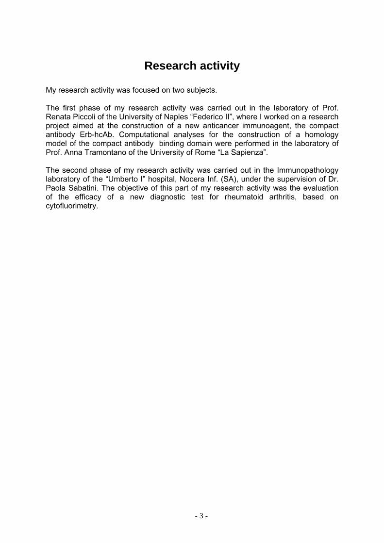
Research activity
My research activity was focused on two subjects. The first phase of my research activity was carried out in the laboratory of Prof. Renata Piccoli of the University of Naples “Federico II”, where I worked on a research project aimed at the construction of a new anticancer immunoagent, the compact antibody Erb-hcAb. Computational analyses for the construction of a homology model of the compact antibody binding domain were performed in the laboratory of Prof. Anna Tramontano of the University of Rome “La Sapienza”. The second phase of my research activity was carried out in the Immunopathology laboratory of the “Umberto I” hospital, Nocera Inf. (SA), under the supervision of Dr. Paola Sabatini. The objective of this part of my research activity was the evaluation of the efficacy of a new diagnostic test for rheumatoid arthritis, based on cytofluorimetry.
- 3 -
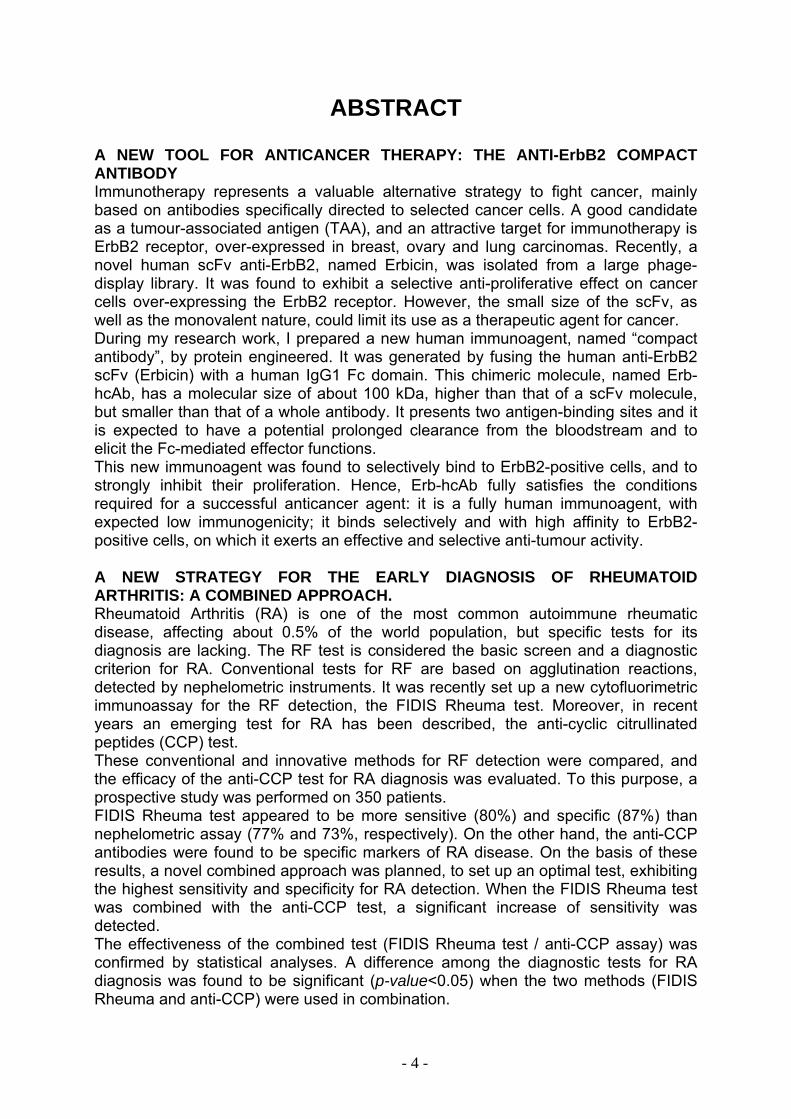
ABSTRACT A NEW TOOL FOR ANTICANCER THERAPY: THE ANTI-ErbB2 COMPACT ANTIBODY Immunotherapy represents a valuable alternative strategy to fight cancer, mainly based on antibodies specifically directed to selected cancer cells. A good candidate as a tumour-associated antigen (TAA), and an attractive target for immunotherapy is ErbB2 receptor, over-expressed in breast, ovary and lung carcinomas. Recently, a novel human scFv anti-ErbB2, named Erbicin, was isolated from a large phage-display library. It was found to exhibit a selective anti-proliferative effect on cancer cells over-expressing the ErbB2 receptor. However, the small size of the scFv, as well as the monovalent nature, could limit its use as a therapeutic agent for cancer. During my research work, I prepared a new human immunoagent, named “compact antibody”, by protein engineered. It was generated by fusing the human anti-ErbB2 scFv (Erbicin) with a human IgG1 Fc domain. This chimeric molecule, named Erb-hcAb, has a molecular size of about 100 kDa, higher than that of a scFv molecule, but smaller than that of a whole antibody. It presents two antigen-binding sites and it is expected to have a potential prolonged clearance from the bloodstream and to elicit the Fc-mediated effector functions. This new immunoagent was found to selectively bind to ErbB2-positive cells, and to strongly inhibit their proliferation. Hence, Erb-hcAb fully satisfies the conditions required for a successful anticancer agent: it is a fully human immunoagent, with expected low immunogenicity; it binds selectively and with high affinity to ErbB2-positive cells, on which it exerts an effective and selective anti-tumour activity. A NEW STRATEGY FOR THE EARLY DIAGNOSIS OF RHEUMATOID ARTHRITIS: A COMBINED APPROACH. Rheumatoid Arthritis (RA) is one of the most common autoimmune rheumatic disease, affecting about 0.5% of the world population, but specific tests for its diagnosis are lacking. The RF test is considered the basic screen and a diagnostic criterion for RA. Conventional tests for RF are based on agglutination reactions, detected by nephelometric instruments. It was recently set up a new cytofluorimetric immunoassay for the RF detection, the FIDIS Rheuma test. Moreover, in recent years an emerging test for RA has been described, the anti-cyclic citrullinated peptides (CCP) test. These conventional and innovative methods for RF detection were compared, and the efficacy of the anti-CCP test for RA diagnosis was evaluated. To this purpose, a prospective study was performed on 350 patients. FIDIS Rheuma test appeared to be more sensitive (80%) and specific (87%) than nephelometric assay (77% and 73%, respectively). On the other hand, the anti-CCP antibodies were found to be specific markers of RA disease. On the basis of these results, a novel combined approach was planned, to set up an optimal test, exhibiting the highest sensitivity and specificity for RA detection. When the FIDIS Rheuma test was combined with the anti-CCP test, a significant increase of sensitivity was detected. The effectiveness of the combined test (FIDIS Rheuma test / anti-CCP assay) was confirmed by statistical analyses. A difference among the diagnostic tests for RA diagnosis was found to be significant (p-value<0.05) when the two methods (FIDIS Rheuma and anti-CCP) were used in combination.
- 4 -
SOMMARIO
PROGETTO A: ANTICORPI TERAPEUTICI
UNA NUOVA MOLECOLA ANTI-CANCRO: L’ANTICORPO COMPATTO ANTI-ErbB2 Il mio progetto di ricerca si è incentrato sulla progettazione e costruzione di un nuovo immunoderivato dotato di attività tossica nei confronti di cellule tumorali. Un alto grado di specificità nel riconoscere le sole cellule cancerose e l’assenza di risposta immunitaria nei pazienti trattati sono tra i principali requisiti che un agente anti-cancro deve possedere. Uno dei principali limiti dei trattamenti convenzionali adottati nelle terapie del cancro, quali la chemioterapia e la radioterapia, è l’assenza di specificità nell’indurre la morte delle sole cellule tumorali. In particolare, l’uso di farmaci citotossici può risultare dannoso a causa degli effetti collaterali provocati sulle cellule normali, soprattutto quelle che in certi organi e tessuti si riproducono attivamente. Tra le nuove terapie adottate per la cura del cancro, chiamate anche terapie biologiche, una delle più promettenti è senza dubbio l’immunoterapia, ovvero l’insieme di trattamenti basati sull’utilizzo di componenti del sistema immunitario, quali anticorpi monoclonali o citochine. Tali molecole possono agire sia modulando l’attività del sistema immunitario, ovvero inducendo una più efficiente risposta immune effettrice nei confronti delle cellule tumorali, sia fungendo da “proiettili magici”, come avviene nel caso degli anticorpi monoclonali. Infatti, dato l’alto grado di specificità nel riconoscimento dell’antigene, gli anticorpi rappresentano potenziali agenti selettivi per le cellule tumorali. Inoltre, se ad essi sono legate molecole citotossiche, i risultanti anticorpi coniugati (immunotossine) possono funzionare da veicoli in grado di dirigere selettivamente la loro componente tossica verso le cellule neoplastiche. Nell’immunoterapia dei tumori, estrema importanza riveste la scelta del bersaglio verso cui indirizzare gli anticorpi ricombinanti prodotti. A questo proposito, negli ultimi anni sono stati isolati e caratterizzati diversi antigeni di superficie detti TAA (antigeni associati a tumori). Essi sono proteine espresse ad elevati livelli e presenti sulla superficie di cellule tumorali: per tali motivi possono essere considerati marcatori tumorali. Tra i TAA noti, il recettore transmembrana tirosina chinasico (RTK) ErbB2, omologo del recettore del fattore di crescita epidermica (EGFR), risulta essere un promettente bersaglio per una terapia anti-cancro. Esso, infatti, è espresso ad elevati livelli (fino a 2x10⁶ molecole per cellula) sulla superficie cellulare di carcinomi della mammella, dell’ovaio e del polmone. Nei tessuti normali, invece, il recettore è normalmente assente o espresso a bassi livelli solo su determinati tipi di cellule epiteliali. Per le caratteristiche di questo TAA, che lo rendono idoneo come bersaglio per l’immunoterapia, in diversi laboratori sono state preparate molecole anticorpali dirette contro ErbB2. Tra queste citiamo Herceptin, una versione umanizzata di un anticorpo monoclonale murino anti-ErbB2, che attualmente viene impiegato per il trattamento del cancro della mammella. Esistono però delle difficoltà correlate all’impiego di anticorpi nella terapia farmacologica. I principali ostacoli riguardano le caratteristiche fisiche proprie degli anticorpi. Infatti, avendo un peso molecolare di circa 155 kDa, gli anticorpi mostrano
- 5 -
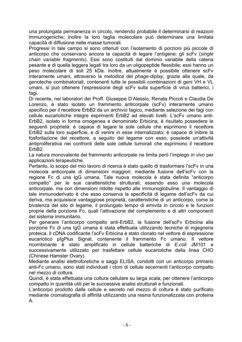
una prolungata permanenza in circolo, rendendo probabile il determinarsi di reazioni immunogeniche; inoltre la loro taglia molecolare può determinare una limitata capacità di diffusione nelle masse tumorali. Progressi in tale campo si sono ottenuti con l’isolamento di porzioni più piccole di anticorpo che conservano ancora la capacità di legare l’antigene: gli scFv (single chain variable fragments). Essi sono costituiti dal dominio variabile della catena pesante e di quella leggera legati tra loro da un oligopeptide flessibile; essi hanno un peso molecolare di soli 25 kDa. Inoltre, attualmente è possibile ottenere scFv interamente umani, attraverso la metodica del phage-diplay, grazie alla quale, da genoteche combinatoriali, contenenti tutte le possibili combinazioni di geni VH e VL umani, si può ottenere l’espressione degli scFv sulla superficie di virus batterici, i fagi. Di recente, nei laboratori dei Proff. Giuseppe D’Alessio, Renata Piccoli e Claudia De Lorenzo, è stato isolato un frammento anticorpale (scFv) interamente umano specifico per il recettore ErbB2 da un archivio fagico, mediante selezione dei fagi con cellule eucariotiche integre esprimenti ErbB2 ad elevati livelli. L’scFv umano anti-ErbB2, isolato in forma omogenea e denominato Erbicina, è risultato possedere le seguenti proprietà: è capace di legare le sole cellule che esprimono il recettore ErbB2 sulla loro superficie, e di venire in esse internalizzato; è capace di inibire la fosforilazione del recettore, a seguito del legame con esso; possiede un’attività antiproliferativa nei confronti delle sole cellule tumorali che esprimono il recettore ErbB2. La natura monovalente del frammento anticorpale ne limita però l’impiego in vivo per applicazioni terapeutiche. Pertanto, lo scopo del mio lavoro di ricerca è stato quello di trasformare l’scFv in una molecola anticorpale di dimensioni maggiori, mediante fusione dell’scFv con la regione Fc di una IgG umana. Tale nuova molecola è stata definita “anticorpo compatto” per le sue caratteristiche strutturali, essendo esso una molecola anticorpale, ma con dimensioni ridotte rispetto alle immunoglobuline. Il vantaggio di tale immunoderivato è che esso conserva la specificità di legame dell’scFv da cui deriva, ma acquisisce vantaggiose proprietà, caratteristiche di un anticorpo, come la bivalenza del sito di legame, il prolungato tempo di emivita in circolo e le funzioni proprie della porzione Fc, quali l’attivazione del complemento e di altri componenti del sistema immunitario. Per generare l’anticorpo compatto anti-ErbB2, la fusione dell’scFv Erbicina alla porzione Fc di una IgG umana è stata effettuata utilizzando tecniche di ingegneria proteica. Il cDNA codificante l’scFv Erbicina è stato clonato nel vettore di espressione eucariotico pIgPlus Signal, contenente il frammento Fc umano. Il vettore ricombinante è stato amplificato in cellule batteriche di E.coli JM101 e successivamente utilizzato per trasfettare cellule eucariotiche della linea CHO (Chinese Hamster Ovary). Mediante analisi elettroforetiche e saggi ELISA, condotti con un anticorpo primario anti-Fc umano, sono stati individuati i cloni di cellule secernenti l’anticorpo compatto nel mezzo di coltura. Quindi, è stata effettuata una coltura cellulare su larga scala, per ottenere l’anticorpo compatto in quantità utili per le successive analisi strutturali e funzionali. L’anticorpo prodotto dalle cellule e secreto nel mezzo di coltura è stato purificato mediante cromatografia di affinità utilizzando una resina funzionalizzata con proteina A.
- 6 -
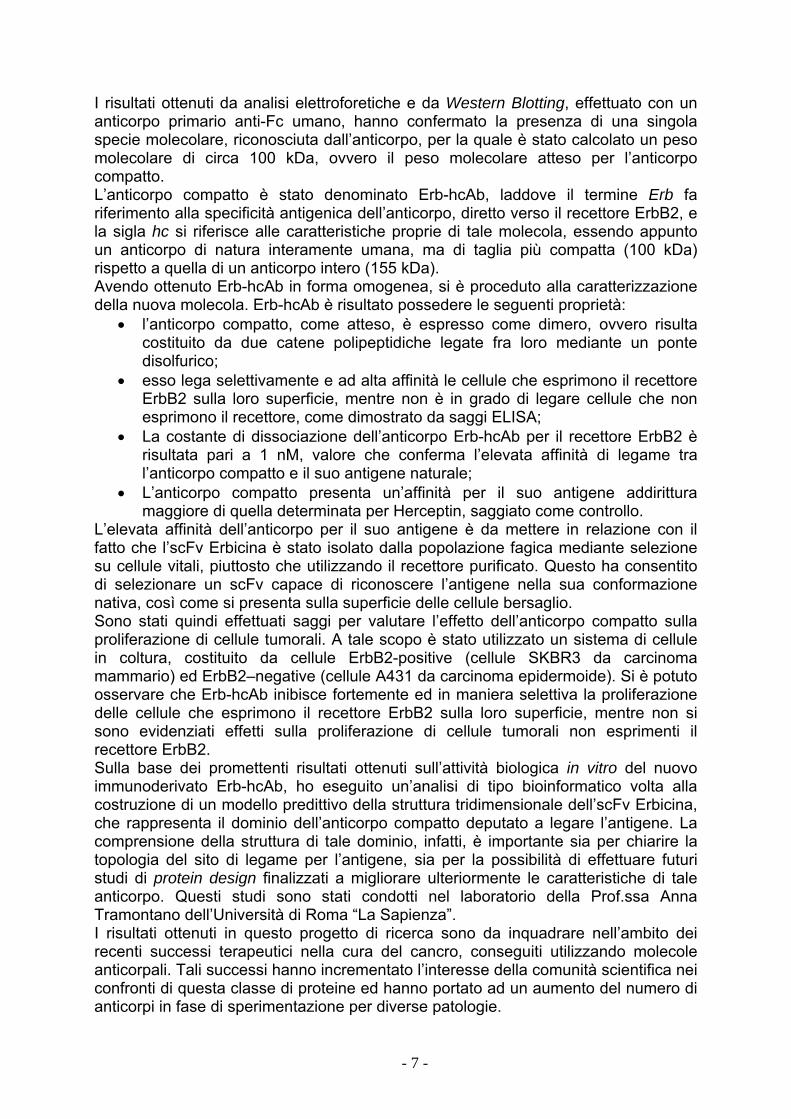
I risultati ottenuti da analisi elettroforetiche e da Western Blotting, effettuato con un anticorpo primario anti-Fc umano, hanno confermato la presenza di una singola specie molecolare, riconosciuta dall’anticorpo, per la quale è stato calcolato un peso molecolare di circa 100 kDa, ovvero il peso molecolare atteso per l’anticorpo compatto. L’anticorpo compatto è stato denominato Erb-hcAb, laddove il termine Erb fa riferimento alla specificità antigenica dell’anticorpo, diretto verso il recettore ErbB2, e la sigla hc si riferisce alle caratteristiche proprie di tale molecola, essendo appunto un anticorpo di natura interamente umana, ma di taglia più compatta (100 kDa) rispetto a quella di un anticorpo intero (155 kDa). Avendo ottenuto Erb-hcAb in forma omogenea, si è proceduto alla caratterizzazione della nuova molecola. Erb-hcAb è risultato possedere le seguenti proprietà:
• l’anticorpo compatto, come atteso, è espresso come dimero, ovvero risulta costituito da due catene polipeptidiche legate fra loro mediante un ponte disolfurico;
• esso lega selettivamente e ad alta affinità le cellule che esprimono il recettore ErbB2 sulla loro superficie, mentre non è in grado di legare cellule che non esprimono il recettore, come dimostrato da saggi ELISA;
• La costante di dissociazione dell’anticorpo Erb-hcAb per il recettore ErbB2 è risultata pari a 1 nM, valore che conferma l’elevata affinità di legame tra l’anticorpo compatto e il suo antigene naturale;
• L’anticorpo compatto presenta un’affinità per il suo antigene addirittura maggiore di quella determinata per Herceptin, saggiato come controllo.
L’elevata affinità dell’anticorpo per il suo antigene è da mettere in relazione con il fatto che l’scFv Erbicina è stato isolato dalla popolazione fagica mediante selezione su cellule vitali, piuttosto che utilizzando il recettore purificato. Questo ha consentito di selezionare un scFv capace di riconoscere l’antigene nella sua conformazione nativa, così come si presenta sulla superficie delle cellule bersaglio. Sono stati quindi effettuati saggi per valutare l’effetto dell’anticorpo compatto sulla proliferazione di cellule tumorali. A tale scopo è stato utilizzato un sistema di cellule in coltura, costituito da cellule ErbB2-positive (cellule SKBR3 da carcinoma mammario) ed ErbB2–negative (cellule A431 da carcinoma epidermoide). Si è potuto osservare che Erb-hcAb inibisce fortemente ed in maniera selettiva la proliferazione delle cellule che esprimono il recettore ErbB2 sulla loro superficie, mentre non si sono evidenziati effetti sulla proliferazione di cellule tumorali non esprimenti il recettore ErbB2. Sulla base dei promettenti risultati ottenuti sull’attività biologica in vitro del nuovo immunoderivato Erb-hcAb, ho eseguito un’analisi di tipo bioinformatico volta alla costruzione di un modello predittivo della struttura tridimensionale dell’scFv Erbicina, che rappresenta il dominio dell’anticorpo compatto deputato a legare l’antigene. La comprensione della struttura di tale dominio, infatti, è importante sia per chiarire la topologia del sito di legame per l’antigene, sia per la possibilità di effettuare futuri studi di protein design finalizzati a migliorare ulteriormente le caratteristiche di tale anticorpo. Questi studi sono stati condotti nel laboratorio della Prof.ssa Anna Tramontano dell’Università di Roma “La Sapienza”. I risultati ottenuti in questo progetto di ricerca sono da inquadrare nell’ambito dei recenti successi terapeutici nella cura del cancro, conseguiti utilizzando molecole anticorpali. Tali successi hanno incrementato l’interesse della comunità scientifica nei confronti di questa classe di proteine ed hanno portato ad un aumento del numero di anticorpi in fase di sperimentazione per diverse patologie.
- 7 -
I nostri risultati hanno dimostrato che Erb-hcAb: È un immunoderivato interamente umano, per il quale ci si attende una
ridotta o nulla immunogenicità nell’uomo Lega con elevata affinità uno dei più specifici marcatori tumorali, il
recettore ErbB2 Inibisce selettivamente la crescita di cellule tumorali esprimenti il recettore
ErbB2. Pertanto i nostri risultati hanno evidenziato che Erb-hcAb è un nuovo promettente agente anti-tumorale, ed un potenziale candidato per l’immunoterapia dei tumori. L’anticorpo Erb-hcAb è coperto da brevetto (Patent PCT/EP02/07671 on “Human mini-antibody cytotoxic for tumour cells which express the ErbB2 receptor” G.D’Alessio, R.Piccoli, C. De Lorenzo, D.B. Palmer, M.A. Ritter, owned by Biotechnol S.A., Oeiras, Portugal).
PROGETTO B: ANTICORPI PATOLOGICI
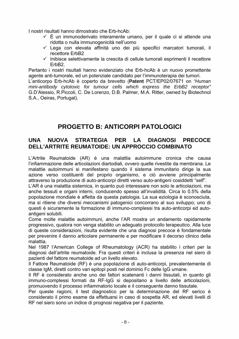
UNA NUOVA STRATEGIA PER LA DIAGNOSI PRECOCE DELL’ARTRITE REUMATOIDE: UN APPROCCIO COMBINATO L’Artrite Reumatoide (AR) è una malattia autoimmune cronica che causa l’infiammazione delle articolazioni diartodiali, ovvero quelle rivestite da membrana. Le malattie autoimmuni si manifestano quando il sistema immunitario dirige la sua azione verso costituenti del proprio organismo, e ciò avviene principalmente attraverso la produzione di auto-anticorpi diretti verso auto-antigeni cosiddetti “self”. L’AR è una malattia sistemica, in quanto può interessare non solo le articolazioni, ma anche tessuti e organi interni, conducendo spesso all’invalidità. Circa lo 0.5% della popolazione mondiale è affetta da questa patologia. La sua eziologia è sconosciuta, ma si ritiene che diversi meccanismi patogenici concorrano al suo sviluppo; uno di questi è sicuramente la formazione di immuno-complessi tra auto-anticorpi ed auto-antigeni solubili. Come molte malattie autoimmuni, anche l’AR mostra un andamento rapidamente progressivo, qualora non venga stabilito un adeguato protocollo terapeutico. Alla luce di queste considerazioni, risulta evidente che una diagnosi precoce è fondamentale per prevenire il danno articolare permanente e per modificare il decorso clinico della malattia. Nel 1987 l’American College of Rheumatology (ACR) ha stabilito i criteri per la diagnosi dell’artrite reumatoide. Fra questi criteri è inclusa la presenza nel siero di pazienti del fattore reumatoide ad un livello elevato. Il Fattore Reumatoide (RF) è una popolazione di auto-anticorpi, prevalentemente di classe IgM, diretti contro vari epitopi posti nel dominio Fc delle IgG umane. Il RF è considerato anche uno dei fattori scatenanti i danni tissutali, in quanto gli immuno-complessi formati da RF-IgG si depositano a livello delle articolazioni, promuovendo il processo infiammatorio locale e il conseguente danno tissutale. Per queste ragioni, il test diagnostico per la determinazione del RF serico è considerato il primo esame da effettuarsi in caso di sospetta AR, ed elevati livelli di RF nel siero sono un indice di prognosi negativa per il paziente.
- 8 -
Attualmente vengono di norma utilizzati due metodi di determinazione del RF serico: il test di agglutinazione di Waaler-Rose e il test nefelometrico. In entrambi i metodi, il siero del paziente viene miscelato con eritrociti di pecora rivestiti da IgG di coniglio (Waaler-Rose), oppure con particelle di latex rivestite da IgG umane (nefelometria). La presenza di RF nel siero induce la formazione di immuno-complessi che danno origine ad agglutinazione. La reazione di agglutinazione viene misurata con uno speciale apparecchio turbidimetrico, denominato nefelometro. Di recente è stata introdotta una nuova tecnologia per la misura del RF serico, il FIDIS Rheuma test, che si basa sulla citofluorimetria a flusso. Il sistema FIDIS utilizza la tecnologia Luminex, che comprende una miscela di 100 differenti tipi di microsfere fluorescenti di polistirene, tutte di uguali dimensioni, rivestite dall’antigene, un citometro a flusso interfacciato con un sistema di digitalizzazione dei segnali ed un hardware e un software per l’elaborazione dei dati. Tuttavia, il test del RF non è altamente specifico per l’artrite reumatoide. Infatti è possibile riscontrare, anche a titoli elevati, la presenza di questo auto-anticorpo nel siero di soggetti affetti da altre malattie autoimmuni, oppure in pazienti affetti da infezioni croniche e addirittura in soggetti sani, specialmente in età avanzata. Da pochi anni è stato identificato un nuovo gruppo di auto-anticorpi specifici per l’artrite reumatoide, gli anticorpi anti-proteine citrullinate (ACPA), che sono stati riscontrati per la prima volta nella liquido sinoviale di pazienti affetti da AR. La citrullinazione è una modifica post-traduzionale, ad opera dell’enzima peptidil-arginina deimminasi (PAD), che determina la sostituzione del gruppo imminico dell’arginina con un gruppo carbonilico, il cui atomo di ossigeno è il determinante antigenico di tali auto-anticorpi. Attualmente, la determinazione degli anticorpi ACPA viene effettuata mediante un test ELISA, in cui l’antigene è costituito da un peptide citrullinato ciclico (anti-CCP test). L’enorme importanza di una diagnosi precoce per l’artrite reumatoide rende necessaria la progettazione di un test altamente specifico e sensibile per la rapida individuazione della malattia. Sulla base di queste considerazioni, lo scopo del mio progetto di ricerca è stato quello di confrontare i metodi diagnostici attualmente in uso per la determinazione del RF serico e, parallelamente, di valutare l’efficacia degli anticorpi anti-citrullina nella diagnosi precoce dell’artrite reumatoide. A tale scopo, ho effettuato uno studio prospettico su una popolazione di 350 pazienti appartenenti a quattro categorie: 100 pazienti affetti da RA; 100 pazienti affetti da epatite cronica associata ad HCV; 53 pazienti affetti da altre malattie autoimmuni e 97 soggetti sani, come popolazione di controllo. L’intera popolazione è stata sottoposta al test per la determinazione del RF serico, sia mediante il convenzionale test nefelometrico sia mediante il nuovo saggio citofluorimetrico FIDIS. Inoltre, in tali pazienti è stato determinato anche il titolo degli anticorpi anti-CCP mediante saggi ELISA. I risultati dello studio hanno evidenziato che il metodo FIDIS Rheuma appare più sensibile del metodo nefelometrico nella determinazione del RF, in quanto identifica il 3% in più di pazienti affetti da artrite reumatoide. Inoltre, FIDIS Rheuma è risultato anche più specifico del test nefelometrico, in quanto nelle altre categorie di pazienti (HCV, C e H) identifica una percentuale minore di falsi positivi al test. Il metodo citofluorimetrico FIDIS Rheuma, dunque, ha mostrato una maggiore sensibilità (80%) e specificità (87%) rispetto al metodo nefelometrico (rispettivamente pari al 77% e 73%), per il dosaggio serico del RF.
- 9 -
E’ stato inoltre confermata l’elevata specificità diagnostica degli anticorpi anti-CCP nella diagnosi dell’AR, in quanto tale marcatore è stato identificato soltanto nel gruppo di pazienti affetti da artrite reumatoide. Sulla base di questi risultati, è stata proposta una nuova strategia diagnostica che prevede l’uso del test FIDIS Rheuma combinato a quello anti-CCP, allo scopo di approntare un saggio diagnostico che mostri la massima specificità e sensibilità nella diagnosi precoce dell’artrite reumatoide. Con tale approccio combinato è stata quindi analizzata la popolazione di pazienti descritta precedentemente. I risultati hanno indicato che tale test presenta la massima sensibilità (100%) e la più alta specificità finora riscontrata (87%) nella diagnosi di AR. L’efficacia del test combinato FIDIS Rheuma-anti-CCP è stata confermata anche da analisi statistiche, che hanno dimostrato che la differenza fra i dati ottenuti con i metodi diagnostici sopra riportati assume un valore significativo (p-value <0.05) soltanto se viene utilizzato il sistema combinato FIDIS Rheuma test - anti-CCP test. Dall’analisi dei dati si può quindi affermare che, nella diagnosi precoce dell’AR, il metodo di dosaggio del RF da preferire è quello basato sulla tecnologia citofluorimetrica FIDIS. Se a tale marcatore si associa anche il dosaggio degli anticorpi anti-peptidi ciclici citrullinati (anti-CCP), si incrementa la specificità diagnostica nei primissimi stadi della malattia, e si ottiene un validissimo ed efficace strumento diagnostico per l’artrite reumatoide.
- 10 -
INTRODUCTION PART A. THERAPEUTIC ANTIBODIES Conventional anticancer treatments, such as surgery, radiation and chemotherapy, being characterised by the lack of tumour cell specificity, not only may fail to cure the majority of neoplastic disease, but their employments also leads to severe and debilitating side effects. Another limit of anticancer therapies is the onset of the multi- drugs resistance phenotype (MDR). Cells that express MDR phenotype are able to maintain low levels of the anticancer drugs, such as alkaloids or antibiotics (1-2). The generally accepted mechanism of multi-drug resistance is that the MDR proteins actively expel the cytotoxic drugs from the cells, maintaining the drug levels below a cell-killing threshold. Different tumours with MDR protein over-expression often show primary (or intrinsic) resistance to cancer chemotherapy. In addition, cancer chemotherapy itself might induce the over-expression of these proteins, so that the multi-drug resistant clones become less sensitive to chemotherapy (secondary drug resistance). In the last few years, researchers, investigating alternative therapeutic strategies to chemotherapy, have developed new tools for cancer therapy. Among the newly acquired tools, immunotherapy represents a new and powerful weapon in the arsenal of anticancer treatments. IMMUNOTHERAPY Immunotherapy is an anticancer treatment that uses molecules involved of the immune system to fight disease, including cancer. This can be done in two ways:
• by stimulating the immune system to work harder or better; • by using components of the immune system, such as recombinant antibodies
or cytokines. The concept of immunotherapy is based on the body's natural defence system, which protects us against a variety of diseases. For many years, physicians believed that the immune system was effective only in combating infectious diseases caused by invading agents as bacteria and viruses. More recently, it has been showed that the immune system may play a central role in protecting the body against cancer and in combating cancer cells that have already developed. This latter role is not well understood, but there is evidence that in many cancer patients, the immune system slows down the growth and spread of tumours. One immediate goal of research in cancer immunology is the development of methods to harness and enhance the body's natural tendency to defend itself against malignant tumours. Immunotherapy of cancer began about one hundred years ago when Dr. William Coley, at the Sloan-Kettering Institute, showed that he could control the growth of some cancer cells and cure a few advanced cancers with injections of a mixed vaccine of streptococcal and staphylococcal bacteria known as Coley's toxin. The tuberculosis vaccine, Bacillus Calmette-Guerin (BCG), developed in 1922, is known to stimulate the immune system and is now used to treat bladder cancers. Many years of research have produced different successful examples of immunotherapies for cancer. Sometimes referred to as biological therapies, these new treatments, such as interferon and other cytokines, monoclonal antibodies, and
- 11 -
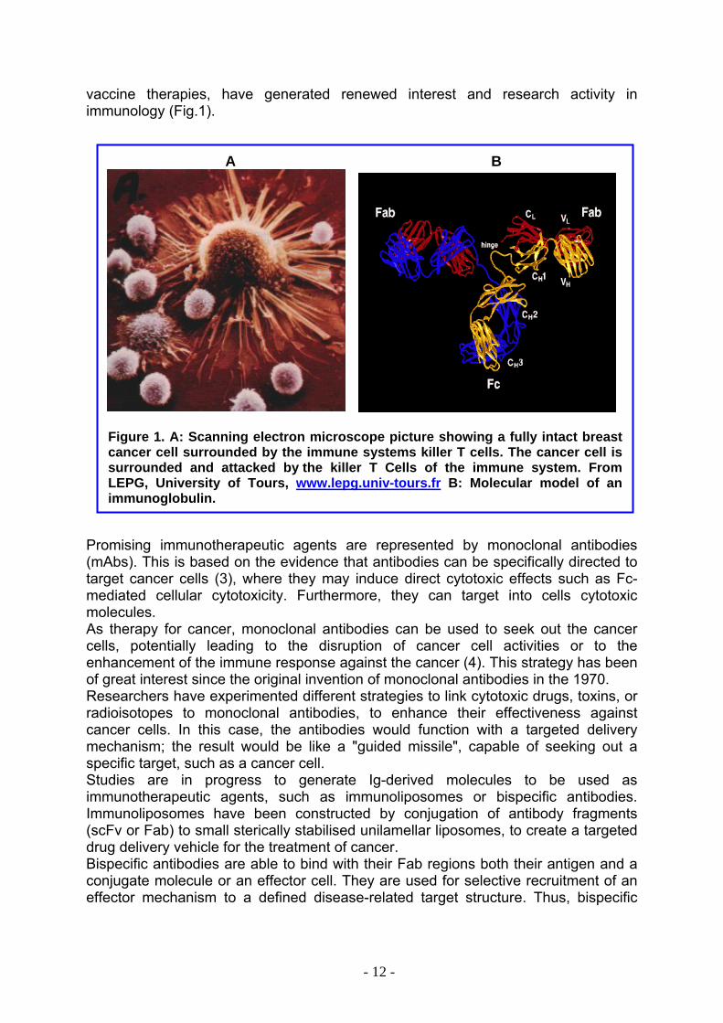
vaccine therapies, have generated renewed interest and research activity in immunology (Fig.1).
A B
Figure 1. A: Scanning electron microscope picture showing a fully intact breast cancer cell surrounded by the immune systems killer T cells. The cancer cell is surrounded and attacked by the killer T Cells of the immune system. From LEPG, University of Tours, www.lepg.univ-tours.fr B: Molecular model of an immunoglobulin.
Promising immunotherapeutic agents are represented by monoclonal antibodies (mAbs). This is based on the evidence that antibodies can be specifically directed to target cancer cells (3), where they may induce direct cytotoxic effects such as Fc-mediated cellular cytotoxicity. Furthermore, they can target into cells cytotoxic molecules. As therapy for cancer, monoclonal antibodies can be used to seek out the cancer cells, potentially leading to the disruption of cancer cell activities or to the enhancement of the immune response against the cancer (4). This strategy has been of great interest since the original invention of monoclonal antibodies in the 1970. Researchers have experimented different strategies to link cytotoxic drugs, toxins, or radioisotopes to monoclonal antibodies, to enhance their effectiveness against cancer cells. In this case, the antibodies would function with a targeted delivery mechanism; the result would be like a "guided missile", capable of seeking out a specific target, such as a cancer cell. Studies are in progress to generate Ig-derived molecules to be used as immunotherapeutic agents, such as immunoliposomes or bispecific antibodies. Immunoliposomes have been constructed by conjugation of antibody fragments (scFv or Fab) to small sterically stabilised unilamellar liposomes, to create a targeted drug delivery vehicle for the treatment of cancer. Bispecific antibodies are able to bind with their Fab regions both their antigen and a conjugate molecule or an effector cell. They are used for selective recruitment of an effector mechanism to a defined disease-related target structure. Thus, bispecific
- 12 -
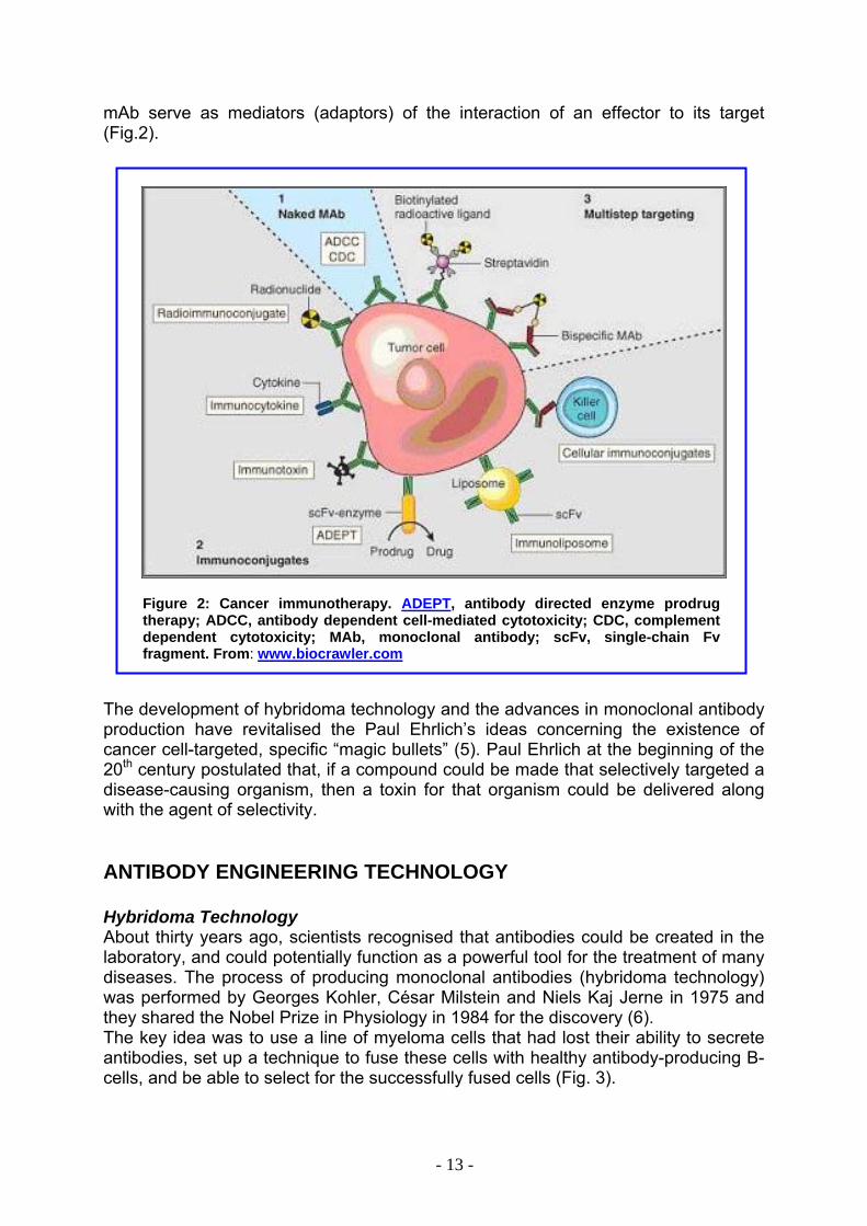
mAb serve as mediators (adaptors) of the interaction of an effector to its target (Fig.2).
Figure 2: Cancer immunotherapy. ADEPT, antibody directed enzyme prodrug therapy; ADCC, antibody dependent cell-mediated cytotoxicity; CDC, complement dependent cytotoxicity; MAb, monoclonal antibody; scFv, single-chain Fv fragment. From: www.biocrawler.com
The development of hybridoma technology and the advances in monoclonal antibody production have revitalised the Paul Ehrlich’s ideas concerning the existence of cancer cell-targeted, specific “magic bullets” (5). Paul Ehrlich at the beginning of the 20th century postulated that, if a compound could be made that selectively targeted a disease-causing organism, then a toxin for that organism could be delivered along with the agent of selectivity. ANTIBODY ENGINEERING TECHNOLOGY Hybridoma Technology About thirty years ago, scientists recognised that antibodies could be created in the laboratory, and could potentially function as a powerful tool for the treatment of many diseases. The process of producing monoclonal antibodies (hybridoma technology) was performed by Georges Kohler, César Milstein and Niels Kaj Jerne in 1975 and they shared the Nobel Prize in Physiology in 1984 for the discovery (6). The key idea was to use a line of myeloma cells that had lost their ability to secrete antibodies, set up a technique to fuse these cells with healthy antibody-producing B-cells, and be able to select for the successfully fused cells (Fig. 3).
- 13 -
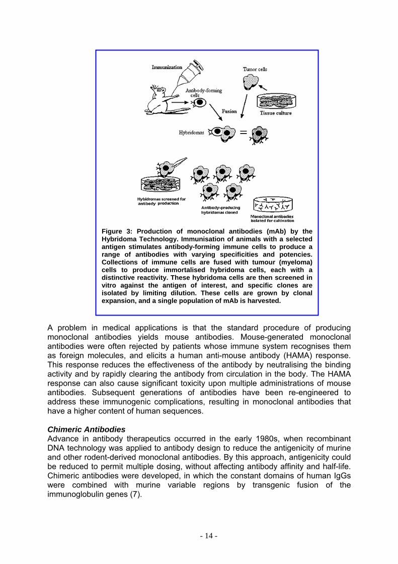
Figure 3: Production of monoclonal antibodies (mAb) by the Hybridoma Technology. Immunisation of animals with a selected antigen stimulates antibody-forming immune cells to produce a range of antibodies with varying specificities and potencies. Collections of immune cells are fused with tumour (myeloma) cells to produce immortalised hybridoma cells, each with a distinctive reactivity. These hybridoma cells are then screened in vitro against the antigen of interest, and specific clones are isolated by limiting dilution. These cells are grown by clonal expansion, and a single population of mAb is harvested.
A problem in medical applications is that the standard procedure of producing monoclonal antibodies yields mouse antibodies. Mouse-generated monoclonal antibodies were often rejected by patients whose immune system recognises them as foreign molecules, and elicits a human anti-mouse antibody (HAMA) response. This response reduces the effectiveness of the antibody by neutralising the binding activity and by rapidly clearing the antibody from circulation in the body. The HAMA response can also cause significant toxicity upon multiple administrations of mouse antibodies. Subsequent generations of antibodies have been re-engineered to address these immunogenic complications, resulting in monoclonal antibodies that have a higher content of human sequences. Chimeric Antibodies Advance in antibody therapeutics occurred in the early 1980s, when recombinant DNA technology was applied to antibody design to reduce the antigenicity of murine and other rodent-derived monoclonal antibodies. By this approach, antigenicity could be reduced to permit multiple dosing, without affecting antibody affinity and half-life. Chimeric antibodies were developed, in which the constant domains of human IgGs were combined with murine variable regions by transgenic fusion of the immunoglobulin genes (7).
- 14 -
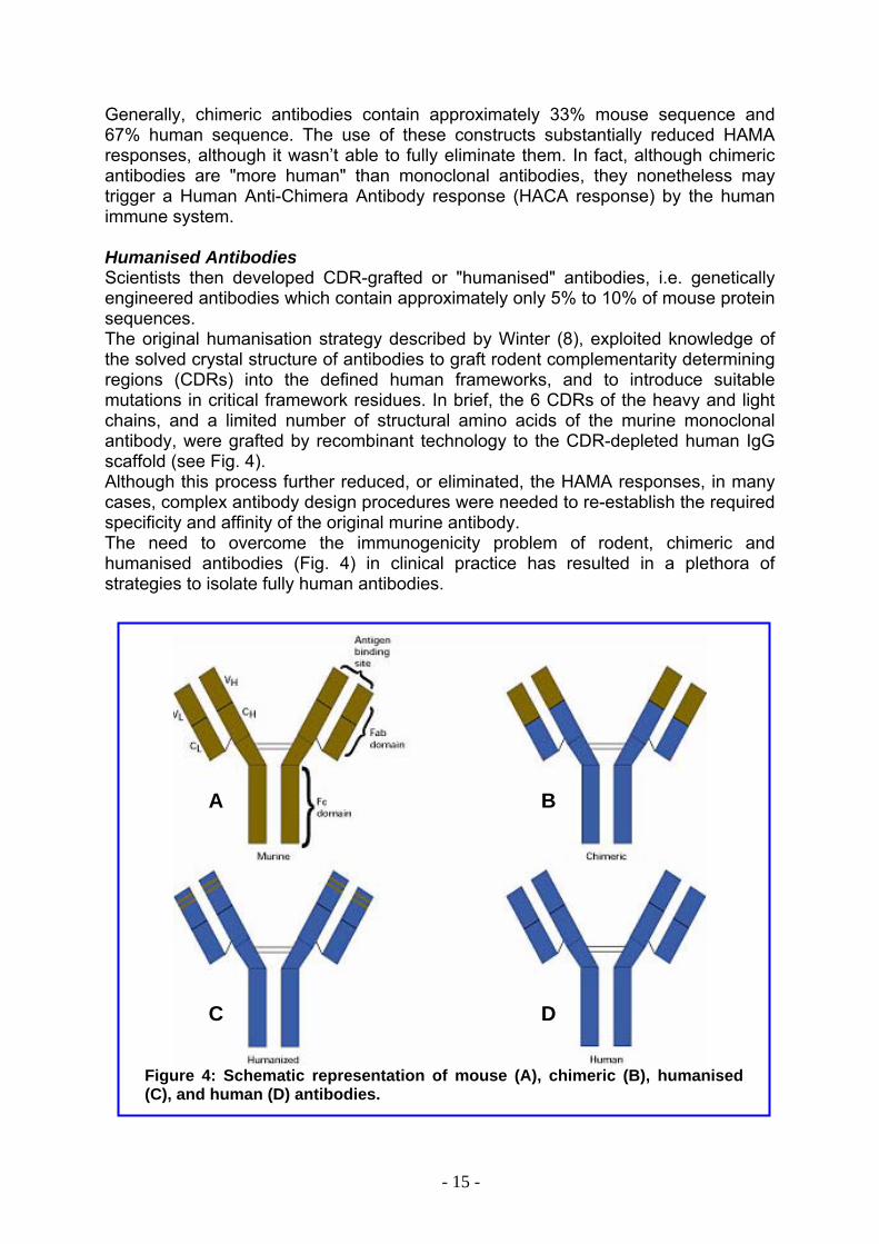
Generally, chimeric antibodies contain approximately 33% mouse sequence and 67% human sequence. The use of these constructs substantially reduced HAMA responses, although it wasn’t able to fully eliminate them. In fact, although chimeric antibodies are "more human" than monoclonal antibodies, they nonetheless may trigger a Human Anti-Chimera Antibody response (HACA response) by the human immune system. Humanised Antibodies Scientists then developed CDR-grafted or "humanised" antibodies, i.e. genetically engineered antibodies which contain approximately only 5% to 10% of mouse protein sequences. The original humanisation strategy described by Winter (8), exploited knowledge of the solved crystal structure of antibodies to graft rodent complementarity determining regions (CDRs) into the defined human frameworks, and to introduce suitable mutations in critical framework residues. In brief, the 6 CDRs of the heavy and light chains, and a limited number of structural amino acids of the murine monoclonal antibody, were grafted by recombinant technology to the CDR-depleted human IgG scaffold (see Fig. 4). Although this process further reduced, or eliminated, the HAMA responses, in many cases, complex antibody design procedures were needed to re-establish the required specificity and affinity of the original murine antibody. The need to overcome the immunogenicity problem of rodent, chimeric and humanised antibodies (Fig. 4) in clinical practice has resulted in a plethora of strategies to isolate fully human antibodies.
A B
C D
Figure 4: Schematic representation of mouse (A), chimeric (B), humanised (C), and human (D) antibodies.
- 15 -
Recently, the term "fully human" antibody has been used to indicate those antibodies derived from human cells, or from transgenic mice, in which mouse antibody gene expression is suppressed and effectively replaced with human antibody gene expression (Fig. 4). Human antibodies are currently produced by the following methods (9):
• fusion of mouse myeloma cells with human lymphocytes (blast cells fused to peripheral blood lymphocytes)
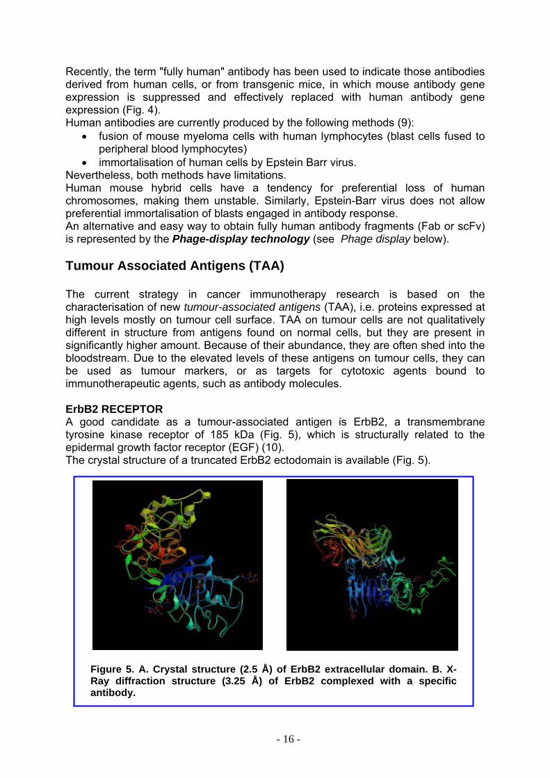
• immortalisation of human cells by Epstein Barr virus. Nevertheless, both methods have limitations. Human mouse hybrid cells have a tendency for preferential loss of human chromosomes, making them unstable. Similarly, Epstein-Barr virus does not allow preferential immortalisation of blasts engaged in antibody response. An alternative and easy way to obtain fully human antibody fragments (Fab or scFv) is represented by the Phage-display technology (see Phage display below). Tumour Associated Antigens (TAA) The current strategy in cancer immunotherapy research is based on the characterisation of new tumour-associated antigens (TAA), i.e. proteins expressed at high levels mostly on tumour cell surface. TAA on tumour cells are not qualitatively different in structure from antigens found on normal cells, but they are present in significantly higher amount. Because of their abundance, they are often shed into the bloodstream. Due to the elevated levels of these antigens on tumour cells, they can be used as tumour markers, or as targets for cytotoxic agents bound to immunotherapeutic agents, such as antibody molecules. ErbB2 RECEPTOR A good candidate as a tumour-associated antigen is ErbB2, a transmembrane tyrosine kinase receptor of 185 kDa (Fig. 5), which is structurally related to the epidermal growth factor receptor (EGF) (10). The crystal structure of a truncated ErbB2 ectodomain is available (Fig. 5).
Figure 5. A. Crystal structure (2.5 Å) of ErbB2 extracellular domain. B. X-Ray diffraction structure (3.25 Å) of ErbB2 complexed with a specific antibody.
- 16 -

The crystal structure of residues 1-509 of ErbB2 at 2.5 Å resolution (Fig. 5 A) reveals an activated conformation similar to that of the EGFR when complexed with its ligand and very different from that seen in the unactivated forms of ErbB3 or EGFR (11). The solved structure provides a molecular basis to explain the inability of ErbB2 to bind known ligands and to form homodimers. These data suggested a model in which ErbB2 is in the activated conformation, ready to interact with other ligand-activated ErbB-receptors. Actually, ErbB2 appears to be the major signalling partner for other ErbB receptors by forming heteromeric complexes with ErbB1, ErbB3, or ErbB4 (12).ErbB receptors are essential mediators of cell proliferation and differentiation during embryogenesis and in adult tissues, and their inappropriate activation is associated with the development and severity of many cancers. ErbB2 is highly expressed in breast, ovary and lung carcinomas (13, 14), as well as in salivary gland and gastric tumour-derived cell lines (15, 16). Its over expression, which occur most commonly via gene amplification, can reach as many as 2x10⁶ molecules/cell (see Fig.6). In normal tissues it is expressed at low levels only in certain epithelial cell types (17). ErbB2 amplification and over expression plays a central role in the initiation and progression of human breast cancer, and has been associated with poor prognosis, as it potentiates and prolongs the signal transduction cascades elicited by ligand activation of other tyrosine-kinase receptor (18, 19). Over-expression of ErbB2 may also increase resistance of tumour cells to host defences through the evasion of immune surveillance exerted by activated macrophages (20). The accessibility of ErbB2 on cell surface and its implications in the development of malignancy of these tumours, make it an attractive target for immunotherapy (Fig. 6).
Figure 6: ErbB2 gene amplification (21) detected by fluorescence in situ hybridization.
ANTI-ErbB2 ANTIBODIES Since the great relevance of ErbB2 receptor in the development and progression of various types of cancer, several groups of researchers have used this molecule as a target for immunotherapy, and have produced different types of antibodies directed to it.
- 17 -
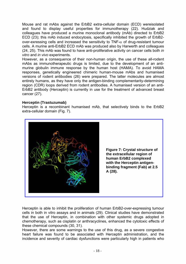
Mouse and rat mAbs against the ErbB2 extra-cellular domain (ECD) wereisolated and found to display useful properties for immunotherapy (22). Hudziak and colleagues have produced a murine monoclonal antibody (mAb) directed to ErbB2 ECD (23); this mAb induced endocytosis, specifically inhibited the growth of ErbB2-over-exressing cells and increased the sensitivity to TNF-α of drug-resistant tumour cells. A murine anti-ErbB2 ECD mAb was produced also by Harwerth and colleagues (24, 25). This mAb was found to have anti-proliferative activity on cancer cells both in vitro and in vivo experiments. However, as a consequence of their non-human origin, the use of these all-rodent mAbs as immunotherapeutic drugs is limited, due to the development of an anti-murine globulin immune response by the human host (HAMA). To avoid HAMA responses, genetically engineered chimeric human-mouse mAbs and humanised versions of rodent antibodies (26) were prepared. The latter molecules are almost entirely humans, as they have only the antigen-binding complementarity-determining region (CDR) loops derived from rodent antibodies. A humanised version of an anti-ErbB2 antibody (Herceptin) is currently in use for the treatment of advanced breast cancer (27). Herceptin (Trastuzumab) Herceptin is a recombinant humanised mAb, that selectively binds to the ErbB2 extra-cellular domain (Fig. 7).
Herceptin is able to inhibit the proliferation of hucells in both in vitro assays and in animals (29). that the use of Herceptin, in combination withchemotherapy, such as cisplatin or anthracyclinethese chemical compounds (30, 31). However, there are some warnings to the use oheart failure was found to be associated with incidence and severity of cardiac dysfunctions w
- 18 -
Figure 7: Crystal structure ofthe extracellular region of human ErbB2 complexed with the Herceptin antigen-binding fragment (Fab) at 2.5A (28).
man ErbB2-over-expressing tumour Clinical studies have demonstrated other systemic drugs adopted in s, enhanced the cytotoxic effects of
f this drug, as a severe congestive Herceptin administration, and the
ere particularly high in patients who
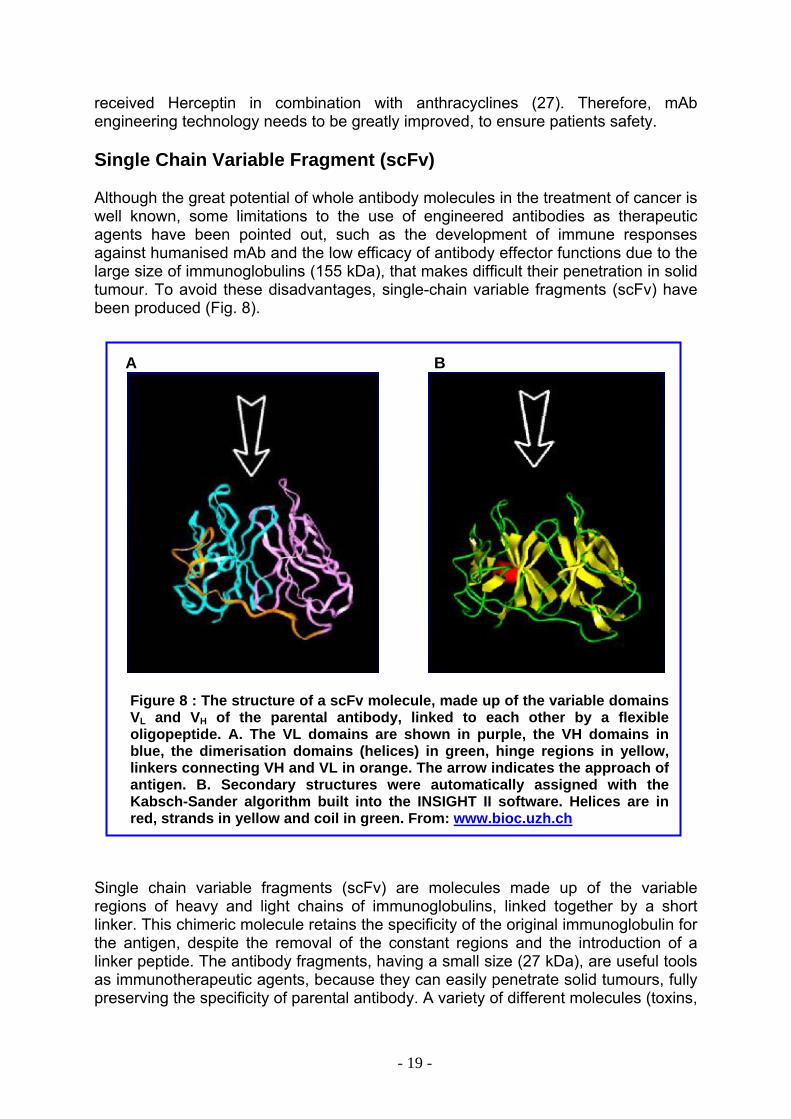
received Herceptin in combination with anthracyclines (27). Therefore, mAb engineering technology needs to be greatly improved, to ensure patients safety. Single Chain Variable Fragment (scFv) Although the great potential of whole antibody molecules in the treatment of cancer is well known, some limitations to the use of engineered antibodies as therapeutic agents have been pointed out, such as the development of immune responses against humanised mAb and the low efficacy of antibody effector functions due to the large size of immunoglobulins (155 kDa), that makes difficult their penetration in solid tumour. To avoid these disadvantages, single-chain variable fragments (scFv) have been produced (Fig. 8). A B
Figure 8 : The structure of a scFv molecule, made up of the variable domains VL and VH of the parental antibody, linked to each other by a flexible oligopeptide. A. The VL domains are shown in purple, the VH domains in blue, the dimerisation domains (helices) in green, hinge regions in yellow, linkers connecting VH and VL in orange. The arrow indicates the approach of antigen. B. Secondary structures were automatically assigned with the Kabsch-Sander algorithm built into the INSIGHT II software. Helices are in red, strands in yellow and coil in green. From: www.bioc.uzh.ch
Single chain variable fragments (scFv) are molecules made up of the variable regions of heavy and light chains of immunoglobulins, linked together by a short linker. This chimeric molecule retains the specificity of the original immunoglobulin for the antigen, despite the removal of the constant regions and the introduction of a linker peptide. The antibody fragments, having a small size (27 kDa), are useful tools as immunotherapeutic agents, because they can easily penetrate solid tumours, fully preserving the specificity of parental antibody. A variety of different molecules (toxins,
- 19 -
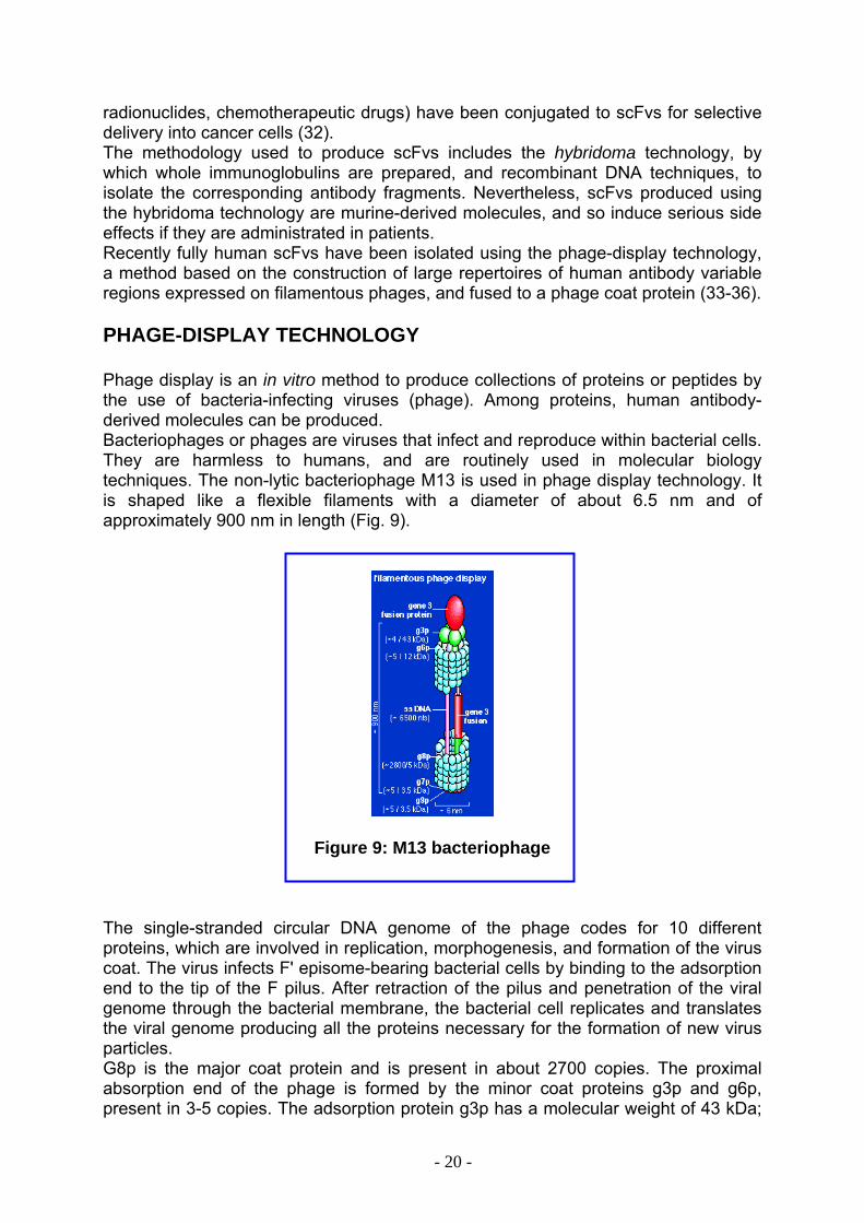
radionuclides, chemotherapeutic drugs) have been conjugated to scFvs for selective delivery into cancer cells (32). The methodology used to produce scFvs includes the hybridoma technology, by which whole immunoglobulins are prepared, and recombinant DNA techniques, to isolate the corresponding antibody fragments. Nevertheless, scFvs produced using the hybridoma technology are murine-derived molecules, and so induce serious side effects if they are administrated in patients. Recently fully human scFvs have been isolated using the phage-display technology, a method based on the construction of large repertoires of human antibody variable regions expressed on filamentous phages, and fused to a phage coat protein (33-36). PHAGE-DISPLAY TECHNOLOGY Phage display is an in vitro method to produce collections of proteins or peptides by the use of bacteria-infecting viruses (phage). Among proteins, human antibody-derived molecules can be produced. Bacteriophages or phages are viruses that infect and reproduce within bacterial cells. They are harmless to humans, and are routinely used in molecular biology techniques. The non-lytic bacteriophage M13 is used in phage display technology. It is shaped like a flexible filaments with a diameter of about 6.5 nm and of approximately 900 nm in length (Fig. 9).
Figure 9: M13 bacteriophage
The single-stranded circular DNA genome of the phage codes for 10 different proteins, which are involved in replication, morphogenesis, and formation of the virus coat. The virus infects F' episome-bearing bacterial cells by binding to the adsorption end to the tip of the F pilus. After retraction of the pilus and penetration of the viral genome through the bacterial membrane, the bacterial cell replicates and translates the viral genome producing all the proteins necessary for the formation of new virus particles. G8p is the major coat protein and is present in about 2700 copies. The proximal absorption end of the phage is formed by the minor coat proteins g3p and g6p, present in 3-5 copies. The adsorption protein g3p has a molecular weight of 43 kDa;
- 20 -
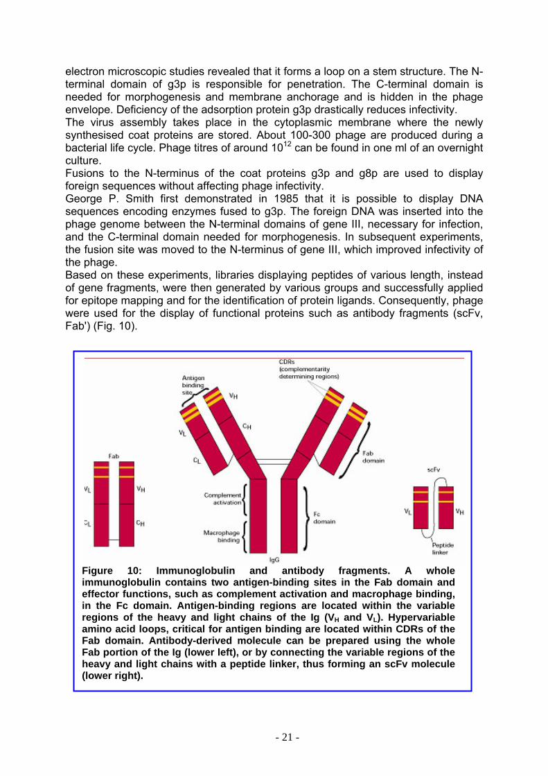
electron microscopic studies revealed that it forms a loop on a stem structure. The N-terminal domain of g3p is responsible for penetration. The C-terminal domain is needed for morphogenesis and membrane anchorage and is hidden in the phage envelope. Deficiency of the adsorption protein g3p drastically reduces infectivity. The virus assembly takes place in the cytoplasmic membrane where the newly synthesised coat proteins are stored. About 100-300 phage are produced during a bacterial life cycle. Phage titres of around 1012 can be found in one ml of an overnight culture. Fusions to the N-terminus of the coat proteins g3p and g8p are used to display foreign sequences without affecting phage infectivity. George P. Smith first demonstrated in 1985 that it is possible to display DNA sequences encoding enzymes fused to g3p. The foreign DNA was inserted into the phage genome between the N-terminal domains of gene III, necessary for infection, and the C-terminal domain needed for morphogenesis. In subsequent experiments, the fusion site was moved to the N-terminus of gene III, which improved infectivity of the phage. Based on these experiments, libraries displaying peptides of various length, instead of gene fragments, were then generated by various groups and successfully applied for epitope mapping and for the identification of protein ligands. Consequently, phage were used for the display of functional proteins such as antibody fragments (scFv, Fab') (Fig. 10).
Figure 10: Immunoglobulin and antibody fragments. A whole immunoglobulin contains two antigen-binding sites in the Fab domain and effector functions, such as complement activation and macrophage binding, in the Fc domain. Antigen-binding regions are located within the variable regions of the heavy and light chains of the Ig (VH and VL). Hypervariable amino acid loops, critical for antigen binding are located within CDRs of the Fab domain. Antibody-derived molecule can be prepared using the whole Fab portion of the Ig (lower left), or by connecting the variable regions of the heavy and light chains with a peptide linker, thus forming an scFv molecule (lower right).
- 21 -
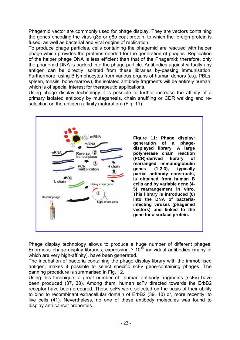
Phagemid vector are commonly used for phage display. They are vectors containing the genes encoding the virus g3p or g8p coat protein, to which the foreign protein is fused, as well as bacterial and viral origins of replication. To produce phage particles, cells containing the phagemid are rescued with helper phage which provides the proteins needed for the generation of phages. Replication of the helper phage DNA is less efficient than that of the Phagemid; therefore, only the phagemid DNA is packed into the phage particle. Antibodies against virtually any antigen can be directly isolated from these libraries by-passing immunisation. Furthermore, using B lymphocytes from various organs of human donors (e.g. PBLs, spleen, tonsils, bone marrow), the isolated antibody fragments will be entirely human, which is of special interest for therapeutic applications. Using phage display technology it is possible to further increase the affinity of a primary isolated antibody by mutagenesis, chain shuffling or CDR walking and re-selection on the antigen (affinity maturation) (Fig. 11).
Figure 11: Phage display: generation of a phage-displayed library. A large polymerase chain reaction (PCR)-derived library of rearranged immunoglobulin genes (1-2-3), typically partial antibody constructs, is obtained from human B cells and by variable gene (4-5) rearrangement in vitro. This library is introduced (6) into the DNA of bacteria-infecting viruses (phagemid vectors) and linked to the gene for a surface protein.
Phage display technology allows to produce a huge number of different phages. Enormous phage display libraries, expressing ≥ 1010 individual antibodies (many of which are very high-affinity), have been generated. The incubation of bacteria containing the phage display library with the immobilised antigen, makes it possible to select specific scFv gene-containing phages. The panning procedure is summarised in Fig. 12. Using this technique, a great number of human antibody fragments (scFv) have been produced (37, 38). Among them, human scFv directed towards the ErbB2 receptor have been prepared. These scFv were selected on the basis of their ability to bind to recombinant extracellular domain of ErbB2 (39, 40) or, more recently, to live cells (41). Nevertheless, no one of these antibody molecules was found to display anti-cancer properties.
- 22 -
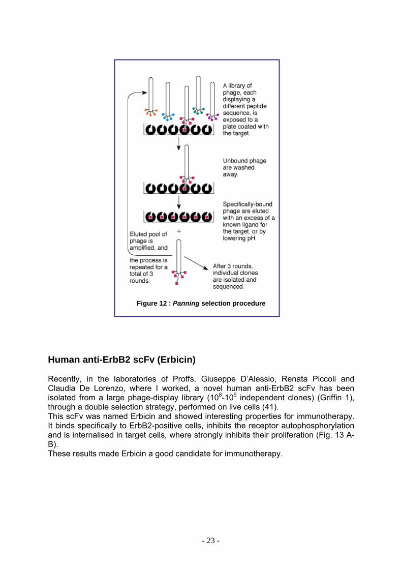
Figure 12 : Panning selection procedure
Human anti-ErbB2 scFv (Erbicin) Recently, in the laboratories of Proffs. Giuseppe D’Alessio, Renata Piccoli and Claudia De Lorenzo, where I worked, a novel human anti-ErbB2 scFv has been isolated from a large phage-display library (108-109 independent clones) (Griffin 1), through a double selection strategy, performed on live cells (41). This scFv was named Erbicin and showed interesting properties for immunotherapy. It binds specifically to ErbB2-positive cells, inhibits the receptor autophosphorylation and is internalised in target cells, where strongly inhibits their proliferation (Fig. 13 A-B). These results made Erbicin a good candidate for immunotherapy.
- 23 -
0
2040
60
80100
120
0 10 20 30 40
B
Empty symbols: ErbB2-negative cells Black symbols: ErbB2-positive cells
A KD ~ 4 nM 1
Abs
orba
nce
450
nm
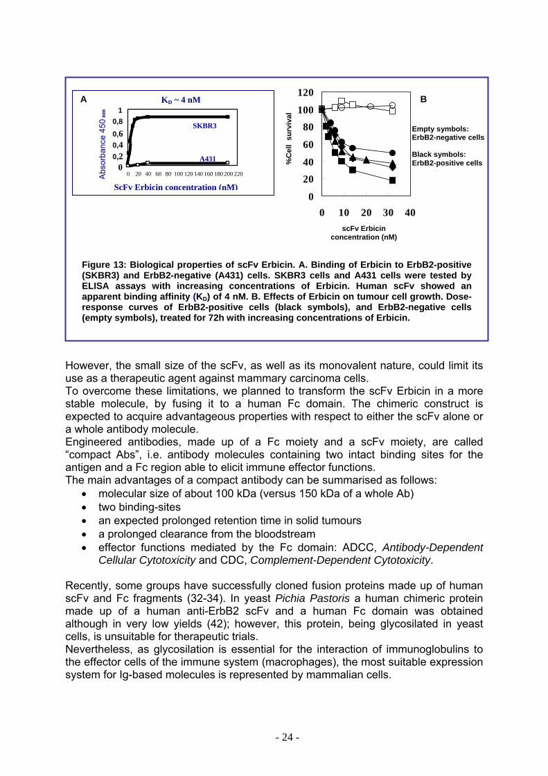
Figure 13: Biological properties of scFv Erbicin. A. Binding of Erbicin to ErbB2-positive (SKBR3) and ErbB2-negative (A431) cells. SKBR3 cells and A431 cells were tested by ELISA assays with increasing concentrations of Erbicin. Human scFv showed an apparent binding affinity (KD) of 4 nM. B. Effects of Erbicin on tumour cell growth. Dose-response curves of ErbB2-positive cells (black symbols), and ErbB2-negative cells (empty symbols), treated for 72h with increasing concentrations of Erbicin.
However, the small size of the scFv, as well as its monovalent nature, could limit its use as a therapeutic agent against mammary carcinoma cells. To overcome these limitations, we planned to transform the scFv Erbicin in a more stable molecule, by fusing it to a human Fc domain. The chimeric construct is expected to acquire advantageous properties with respect to either the scFv alone or a whole antibody molecule. Engineered antibodies, made up of a Fc moiety and a scFv moiety, are called “compact Abs”, i.e. antibody molecules containing two intact binding sites for the antigen and a Fc region able to elicit immune effector functions. The main advantages of a compact antibody can be summarised as follows:
• molecular size of about 100 kDa (versus 150 kDa of a whole Ab) • two binding-sites • an expected prolonged retention time in solid tumours • a prolonged clearance from the bloodstream • effector functions mediated by the Fc domain: ADCC, Antibody-Dependent
Cellular Cytotoxicity and CDC, Complement-Dependent Cytotoxicity.
Recently, some groups have successfully cloned fusion proteins made up of human scFv and Fc fragments (32-34). In yeast Pichia Pastoris a human chimeric protein made up of a human anti-ErbB2 scFv and a human Fc domain was obtained although in very low yields (42); however, this protein, being glycosilated in yeast cells, is unsuitable for therapeutic trials. Nevertheless, as glycosilation is essential for the interaction of immunoglobulins to the effector cells of the immune system (macrophages), the most suitable expression system for Ig-based molecules is represented by mammalian cells.
00,2 0,4 0,6
0,8
0 20 40 60 80 100 120 140 160 180 200 220
ScFv Erbicin concentration (nM)
A431
SKBR3
%C
ell
surv
ival
scFv Erbicin concentration (nM)
- 24 -

PART B. PATHOLOGICAL ANTIBODIES IN RHEUMATOID ARTHRITIS RHEUMATOID ARTHRITIS Rheumatoid Arthritis (RA) is a chronic debilitating autoimmune disease of unknown aetiology affecting diarthrodial joints. A joint is where two bones meet allowing movement of body parts. The joint inflammation of rheumatoid arthritis causes swelling, pain, stiffness, and redness in the joints. In a first phase of the disease, joints are usually affected asymmetrically; in a second phase they are affected in a symmetrical fashion (43-44) (Fig.14).
Figure 14: Schematic representation of normal joint (left) and inflamed joints (right), occurring in Rheumatoid Arthritis. The inflammation can also occur in tissues around the joints.
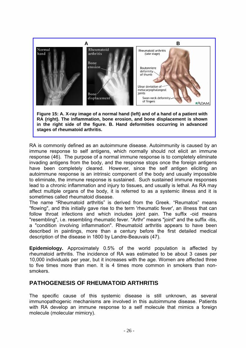
In some patients with RA, chronic inflammation leads to the destruction of the cartilage, bone and ligaments, causing deformity of the joints. Damage to the joints can occur early in the disease and be progressive. The fingers are typically deviated towards the little finger (ulnar deviation) and can assume unnatural shapes (45). Classical deformities in rheumatoid arthritis are the Boutonniere deformity (Hyper flexion at the proximal interphalangeal joint with hyperextension at the distal interphalangeal joint), swan neck deformity (Hyperextension at the proximal interphalangeal joint, hyper flexion at the distal interphalangeal joint). The thumb may develop a "Z-Thumb" deformity with fixed flexion and subluxation at the metacarpophalangeal joint (Fig.15).
- 25 -
A B
Figure 15: A. X-ray image of a normal hand (left) and of a hand of a patient with RA (right). The inflammation, bone erosion, and bone displacement is shown in the right side of the figure. B. Hand deformities occurring in advanced stages of rheumatoid arthritis.
RA is commonly defined as an autoimmune disease. Autoimmunity is caused by an immune response to self antigens, which normally should not elicit an immune response (46). The purpose of a normal immune response is to completely eliminate invading antigens from the body, and the response stops once the foreign antigens have been completely cleared. However, since the self antigen eliciting an autoimmune response is an intrinsic component of the body and usually impossible to eliminate, the immune response is sustained. Such sustained immune responses lead to a chronic inflammation and injury to tissues, and usually is lethal. As RA may affect multiple organs of the body, it is referred to as a systemic illness and it is sometimes called rheumatoid disease. The name “Rheumatoid arthritis” is derived from the Greek. “Reumatos” means "flowing", and this initially gave rise to the term 'rheumatic fever', an illness that can follow throat infections and which includes joint pain. The suffix -oid means "resembling", i.e. resembling rheumatic fever. “Arthr” means "joint" and the suffix -itis, a "condition involving inflammation". Rheumatoid arthritis appears to have been described in paintings, more than a century before the first detailed medical description of the disease in 1800 by Landre-Beauvais (47). Epidemiology. Approximately 0.5% of the world population is affected by rheumatoid arthritis. The incidence of RA was estimated to be about 3 cases per 10,000 individuals per year, but it increases with the age. Women are affected three to five times more than men. It is 4 times more common in smokers than non-smokers. PATHOGENESIS OF RHEUMATOID ARTHRITIS The specific cause of this systemic disease is still unknown, as several immunopathogenic mechanisms are involved in this autoimmune disease. Patients with RA develop an immune response to a self molecule that mimics a foreign molecule (molecular mimicry).
- 26 -
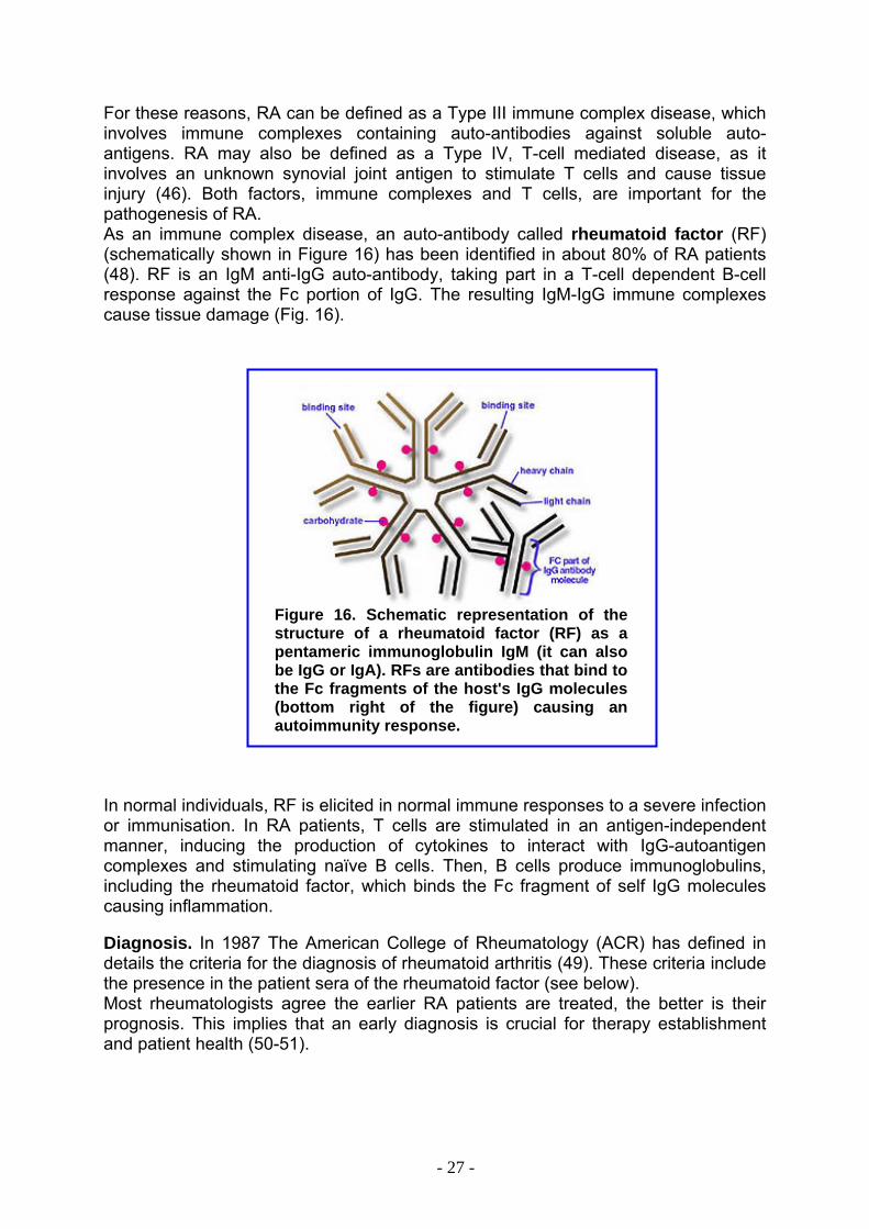
For these reasons, RA can be defined as a Type III immune complex disease, which involves immune complexes containing auto-antibodies against soluble auto-antigens. RA may also be defined as a Type IV, T-cell mediated disease, as it involves an unknown synovial joint antigen to stimulate T cells and cause tissue injury (46). Both factors, immune complexes and T cells, are important for the pathogenesis of RA. As an immune complex disease, an auto-antibody called rheumatoid factor (RF) (schematically shown in Figure 16) has been identified in about 80% of RA patients (48). RF is an IgM anti-IgG auto-antibody, taking part in a T-cell dependent B-cell response against the Fc portion of IgG. The resulting IgM-IgG immune complexes cause tissue damage (Fig. 16).
Figure 16. Schematic representation of the structure of a rheumatoid factor (RF) as a pentameric immunoglobulin IgM (it can also be IgG or IgA). RFs are antibodies that bind to the Fc fragments of the host's IgG molecules (bottom right of the figure) causing an autoimmunity response.
In normal individuals, RF is elicited in normal immune responses to a severe infection or immunisation. In RA patients, T cells are stimulated in an antigen-independent manner, inducing the production of cytokines to interact with IgG-autoantigen complexes and stimulating naïve B cells. Then, B cells produce immunoglobulins, including the rheumatoid factor, which binds the Fc fragment of self IgG molecules causing inflammation. Diagnosis. In 1987 The American College of Rheumatology (ACR) has defined in details the criteria for the diagnosis of rheumatoid arthritis (49). These criteria include the presence in the patient sera of the rheumatoid factor (see below). Most rheumatologists agree the earlier RA patients are treated, the better is their prognosis. This implies that an early diagnosis is crucial for therapy establishment and patient health (50-51).
- 27 -
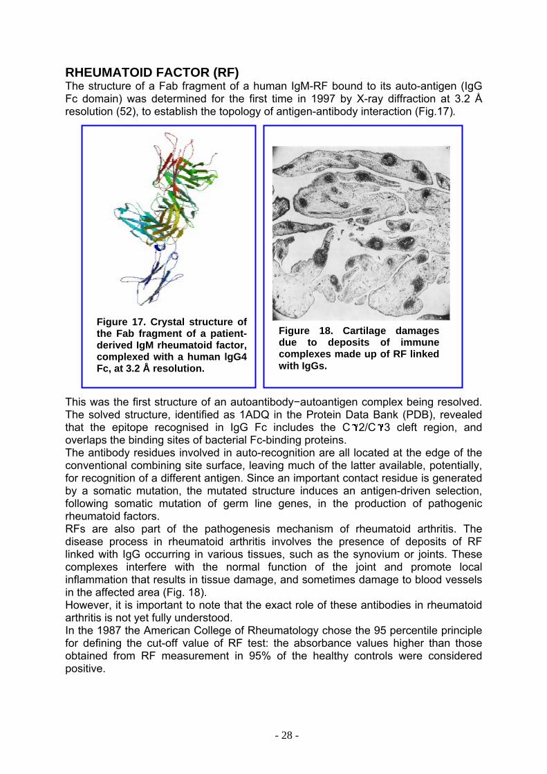
RHEUMATOID FACTOR (RF) The structure of a Fab fragment of a human IgM-RF bound to its auto-antigen (IgG Fc domain) was determined for the first time in 1997 by X-ray diffraction at 3.2 Å resolution (52), to establish the topology of antigen-antibody interaction (Fig.17).
Figure 18. Cartilage damages due to deposits of immune complexes made up of RF linked with IgGs.
Figure 17. Crystal structure of the Fab fragment of a patient-derived IgM rheumatoid factor, complexed with a human lgG4 Fc, at 3.2 Å resolution.
This was the first structure of an autoantibody−autoantigen complex being resolved. The solved structure, identified as 1ADQ in the Protein Data Bank (PDB), revealed that the epitope recognised in IgG Fc includes the C 2/C 3 cleft region, and overlaps the binding sites of bacterial Fc-binding proteins. The antibody residues involved in auto-recognition are all located at the edge of the conventional combining site surface, leaving much of the latter available, potentially, for recognition of a different antigen. Since an important contact residue is generated by a somatic mutation, the mutated structure induces an antigen-driven selection, following somatic mutation of germ line genes, in the production of pathogenic rheumatoid factors. RFs are also part of the pathogenesis mechanism of rheumatoid arthritis. The disease process in rheumatoid arthritis involves the presence of deposits of RF linked with IgG occurring in various tissues, such as the synovium or joints. These complexes interfere with the normal function of the joint and promote local inflammation that results in tissue damage, and sometimes damage to blood vessels in the affected area (Fig. 18). However, it is important to note that the exact role of these antibodies in rheumatoid arthritis is not yet fully understood. In the 1987 the American College of Rheumatology chose the 95 percentile principle for defining the cut-off value of RF test: the absorbance values higher than those obtained from RF measurement in 95% of the healthy controls were considered positive.
- 28 -
This means that up to 5% of middle-aged healthy blood donors can be expected to have a positive RF test. Increasing the cut-off limit, increases the diagnostic specificity for RA, but decreases the sensitivity. The RF test is considered the basic screen and hallmark for RA. It is considered a RA marker since its presence is associated with an increased risk of developing RA in people with mild arthritic symptoms. Higher levels are also detected in more severe forms of the disease, a condition that is a severe prognostic factor for patients (53). Approximately 80% of the people with rheumatoid arthritis have positive titres; 20% are considered sero-negative, which means they have no detectable RF. So, a negative RF does not rule out the existence of RA. RF may also occur in patients with other autoimmune conditions, such as systemic lupus erythematosus, mixed connective tissue disease Sjögren’s syndrome and occasionally scleroderma. The RF test may also be positive in other conditions such as chronic active hepatitis or other chronic infection, or in healthy subjects advanced in age (54). Actually, the amount of rheumatoid factor in blood can be measured in two ways: agglutination tests and nephelometric test. Agglutination tests. Among the agglutination methods routinely used in clinical chemistry we distinguish the Waaler-Rose haemagglutination test to identify the IgM class RF directed against rabbit antigenic determinants, and the latex agglutination test to identify the IgM class RF directed against human antigenic determinants. The Waaler–Rose haemagglutination assay (55-56), based on sheep erythrocytes coated with rabbit antibodies, was the prevailing method in the past to analyse RF. More recently, latex particle agglutination assays have been developed (57). In this last methods, the patient blood is mixed with tiny latex beads covered with human antibodies (IgG). The latex beads agglutinate if rheumatoid factor (IgM RF) is present (Fig. 19). This method is better used as a first-time screening test for rheumatoid arthritis.
Figure 19. Principle of the agglutination test. The antibody is mixed with the particulate antigen (blue spheres) coated on solid particles (red spheres). A positive test is indicated by the agglutination of the particulate antigen.
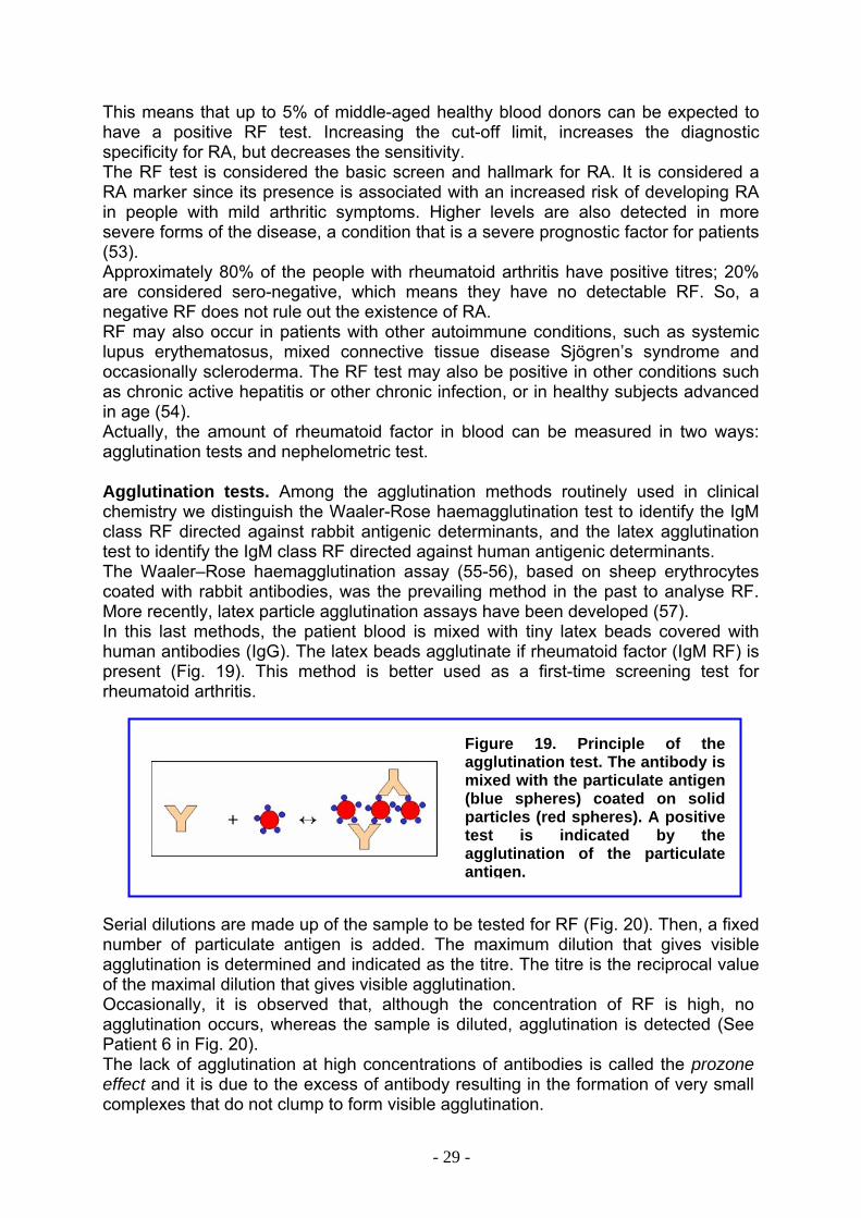
Serial dilutions are made up of the sample to be tested for RF (Fig. 20). Then, a fixed number of particulate antigen is added. The maximum dilution that gives visible agglutination is determined and indicated as the titre. The titre is the reciprocal value of the maximal dilution that gives visible agglutination. Occasionally, it is observed that, although the concentration of RF is high, no agglutination occurs, whereas the sample is diluted, agglutination is detected (See Patient 6 in Fig. 20). The lack of agglutination at high concentrations of antibodies is called the prozone effect and it is due to the excess of antibody resulting in the formation of very small complexes that do not clump to form visible agglutination.
- 29 -
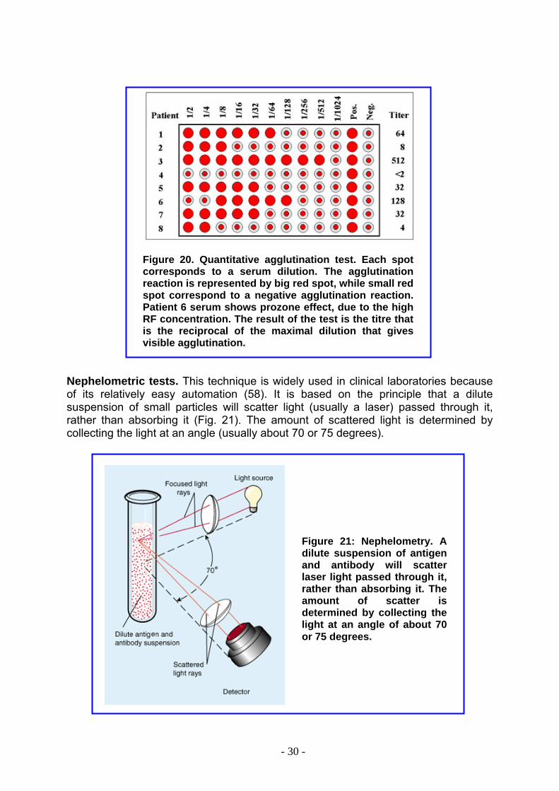
Figure 20. Quantitative agglutination test. Each spot corresponds to a serum dilution. The agglutination reaction is represented by big red spot, while small red spot correspond to a negative agglutination reaction. Patient 6 serum shows prozone effect, due to the high RF concentration. The result of the test is the titre that is the reciprocal of the maximal dilution that gives visible agglutination.
Nephelometric tests. This technique is widely used in clinical laboratories because of its relatively easy automation (58). It is based on the principle that a dilute suspension of small particles will scatter light (usually a laser) passed through it, rather than absorbing it (Fig. 21). The amount of scattered light is determined by collecting the light at an angle (usually about 70 or 75 degrees).
Figure 21: Nephelometry. A dilute suspension of antigen and antibody will scatter laser light passed through it, rather than absorbing it. The amount of scatter is determined by collecting the light at an angle of about 70 or 75 degrees.
- 30 -
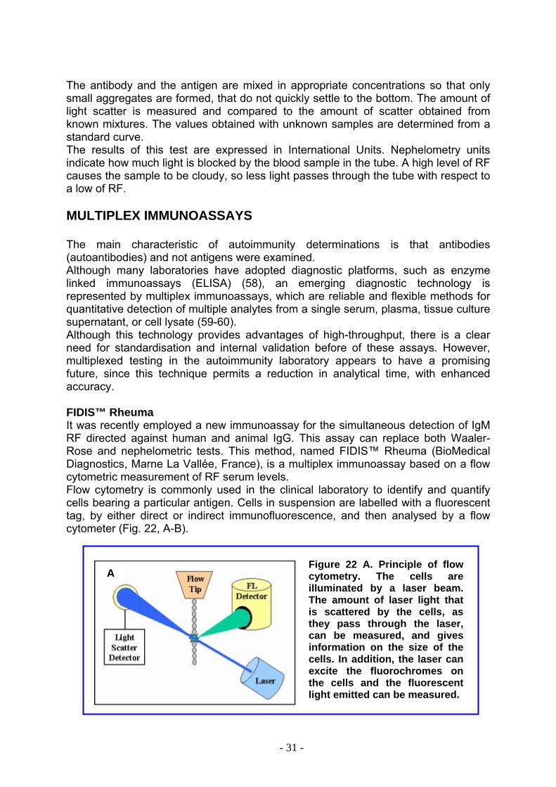
The antibody and the antigen are mixed in appropriate concentrations so that only small aggregates are formed, that do not quickly settle to the bottom. The amount of light scatter is measured and compared to the amount of scatter obtained from known mixtures. The values obtained with unknown samples are determined from a standard curve. The results of this test are expressed in International Units. Nephelometry units indicate how much light is blocked by the blood sample in the tube. A high level of RF causes the sample to be cloudy, so less light passes through the tube with respect to a low of RF. MULTIPLEX IMMUNOASSAYS The main characteristic of autoimmunity determinations is that antibodies (autoantibodies) and not antigens were examined. Although many laboratories have adopted diagnostic platforms, such as enzyme linked immunoassays (ELISA) (58), an emerging diagnostic technology is represented by multiplex immunoassays, which are reliable and flexible methods for quantitative detection of multiple analytes from a single serum, plasma, tissue culture supernatant, or cell lysate (59-60). Although this technology provides advantages of high-throughput, there is a clear need for standardisation and internal validation before of these assays. However, multiplexed testing in the autoimmunity laboratory appears to have a promising future, since this technique permits a reduction in analytical time, with enhanced accuracy. FIDIS™ Rheuma It was recently employed a new immunoassay for the simultaneous detection of IgM RF directed against human and animal IgG. This assay can replace both Waaler-Rose and nephelometric tests. This method, named FIDIS™ Rheuma (BioMedical Diagnostics, Marne La Vallée, France), is a multiplex immunoassay based on a flow cytometric measurement of RF serum levels. Flow cytometry is commonly used in the clinical laboratory to identify and quantify cells bearing a particular antigen. Cells in suspension are labelled with a fluorescent tag, by either direct or indirect immunofluorescence, and then analysed by a flow cytometer (Fig. 22, A-B).
Figure 22 A. Principle of flow cytometry. The cells are illuminated by a laser beam. The amount of laser light that is scattered by the cells, as they pass through the laser, can be measured, and gives information on the size of the cells. In addition, the laser can excite the fluorochromes on the cells and the fluorescent light emitted can be measured.
A
- 31 -
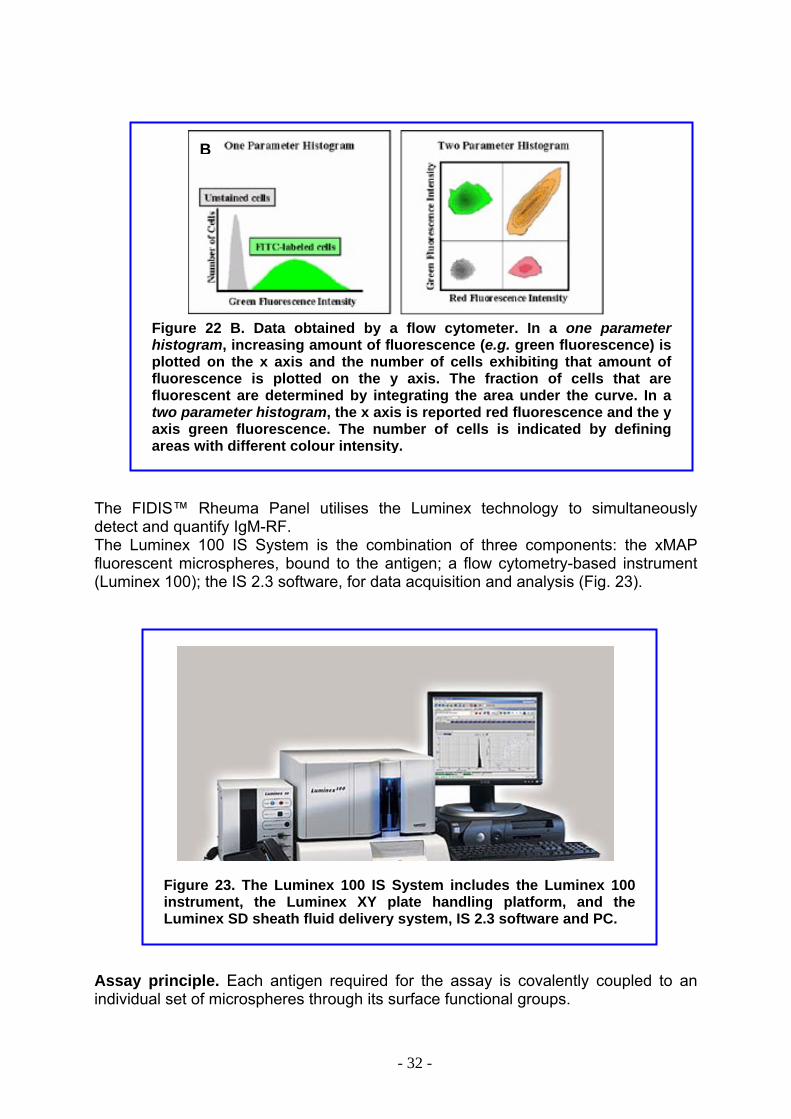
B
Figure 22 B. Data obtained by a flow cytometer. In a one parameter histogram, increasing amount of fluorescence (e.g. green fluorescence) is plotted on the x axis and the number of cells exhibiting that amount of fluorescence is plotted on the y axis. The fraction of cells that are fluorescent are determined by integrating the area under the curve. In a two parameter histogram, the x axis is reported red fluorescence and the y axis green fluorescence. The number of cells is indicated by defining areas with different colour intensity.
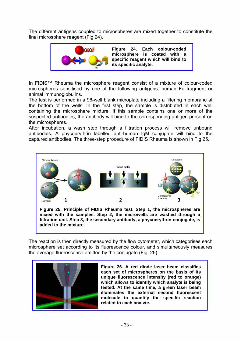
The FIDIS™ Rheuma Panel utilises the Luminex technology to simultaneously detect and quantify IgM-RF. The Luminex 100 IS System is the combination of three components: the xMAP fluorescent microspheres, bound to the antigen; a flow cytometry-based instrument (Luminex 100); the IS 2.3 software, for data acquisition and analysis (Fig. 23).
Figure 23. The Luminex 100 IS System includes the Luminex 100 instrument, the Luminex XY plate handling platform, and the Luminex SD sheath fluid delivery system, IS 2.3 software and PC.
Assay principle. Each antigen required for the assay is covalently coupled to an individual set of microspheres through its surface functional groups.
- 32 -
The different antigens coupled to microspheres are mixed together to constitute the final microsphere reagent (Fig.24).
Figure 24. Each colour-coded microsphere is coated with a specific reagent which will bind to its specific analyte.
In FIDIS™ Rheuma the microsphere reagent consist of a mixture of colour-coded microspheres sensitised by one of the following antigens: human Fc fragment or animal immunoglobulins. The test is performed in a 96-well blank microplate including a filtering membrane at the bottom of the wells. In the first step, the sample is distributed in each well containing the microsphere mixture. If this sample contains one or more of the suspected antibodies, the antibody will bind to the corresponding antigen present on the microspheres. After incubation, a wash step through a filtration process will remove unbound antibodies. A phycoerythrin labelled anti-human IgM conjugate will bind to the captured antibodies. The three-step procedure of FIDIS Rheuma is shown in Fig 25.
Figure 25. Principle of FIDIS Rheuma test. Step 1, the microspheres are mixed with the samples. Step 2, the microwells are washed through a filtration unit. Step 3, the secondary antibody, a phycoerythrin-conjugate, is added to the mixture.
The reaction is then directly measured by the flow cytometer, which categorises each microsphere set according to its fluorescence colour, and simultaneously measures the average fluorescence emitted by the conjugate (Fig. 26).
Figure 26. A red diode laser beam classifies each set of microspheres on the basis of its unique fluorescence intensity (red to orange) which allows to identify which analyte is being tested. At the same time, a green laser beam illuminates the external second fluorescent molecule to quantify the specific reaction related to each analyte.
1 2 3
- 33 -
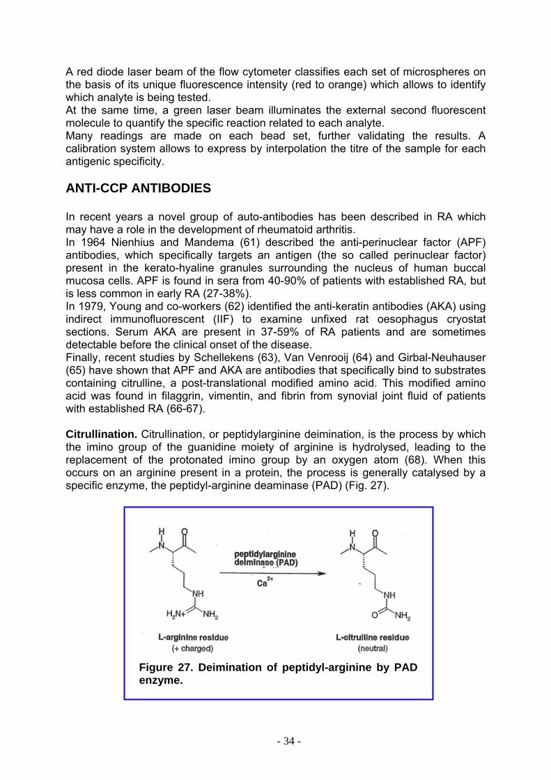
A red diode laser beam of the flow cytometer classifies each set of microspheres on the basis of its unique fluorescence intensity (red to orange) which allows to identify which analyte is being tested. At the same time, a green laser beam illuminates the external second fluorescent molecule to quantify the specific reaction related to each analyte. Many readings are made on each bead set, further validating the results. A calibration system allows to express by interpolation the titre of the sample for each antigenic specificity. ANTI-CCP ANTIBODIES In recent years a novel group of auto-antibodies has been described in RA which may have a role in the development of rheumatoid arthritis. In 1964 Nienhius and Mandema (61) described the anti-perinuclear factor (APF) antibodies, which specifically targets an antigen (the so called perinuclear factor) present in the kerato-hyaline granules surrounding the nucleus of human buccal mucosa cells. APF is found in sera from 40-90% of patients with established RA, but is less common in early RA (27-38%). In 1979, Young and co-workers (62) identified the anti-keratin antibodies (AKA) using indirect immunofluorescent (IIF) to examine unfixed rat oesophagus cryostat sections. Serum AKA are present in 37-59% of RA patients and are sometimes detectable before the clinical onset of the disease. Finally, recent studies by Schellekens (63), Van Venrooij (64) and Girbal-Neuhauser (65) have shown that APF and AKA are antibodies that specifically bind to substrates containing citrulline, a post-translational modified amino acid. This modified amino acid was found in filaggrin, vimentin, and fibrin from synovial joint fluid of patients with established RA (66-67). Citrullination. Citrullination, or peptidylarginine deimination, is the process by which the imino group of the guanidine moiety of arginine is hydrolysed, leading to the replacement of the protonated imino group by an oxygen atom (68). When this occurs on an arginine present in a protein, the process is generally catalysed by a specific enzyme, the peptidyl-arginine deaminase (PAD) (Fig. 27).
Figure 27. Deimination of peptidyl-arginine by PAD
enzyme.
- 34 -
Database searches reveal the existence of several isotypes of human PAD enzymes; these enzymes display tissue specific patterns and, in mice, they are stimulated by female sex hormones (69). Citrullinated extracellular fibrin in the RA synovium may be one of the major autoantigens driving the local immune response, since the production of anti-citrullinated filaggrin antibodies was found to occur in the joints (70). Moreover, the presence of anti-citrullinated protein antibodies (ACPA) predicts a more aggressive course of disease with unfavourable outcome (71). Anti-citrullinated protein antibodies (ACPA). The citrulline moiety is the determinant recognised by APF and AKA antibodies. Following the discovery of the crucial role of these epitopes, a biochemical test was developed to detect anti-citrulline antibodies in sera of RA patients. In principle, every citrullinated peptide could be used in serological tests to detect ACPA (72), so a number of linear peptides containing one citrulline residue were developed. However, since most linear peptides have shown low specificity for ACPA detection, several attempts were made to obtain a specific and reliable test. The use of cyclic peptides was found to increase the sensitivity and the specificity of the test. Cyclic citrullinated peptides (CCP) have a three-dimensional design that is optimally structured for recognition of the antigenic group by the heterogeneous population of RA autoantibodies. Actually, the biochemical assay routinely used to detect anti-citrullinated proteins antibodies in sera of RA patients, was developed by Walther van Venrooij and colleagues (70) as an enzyme linked immunosorbant assay (ELISA) named aCCP test. Anti-CCP antibodies were found to be present in about 80% of patients with rheumatoid arthritis. Synthesis and pathogenetic significance of anti-CCP antibodies. Recent data suggest that anti-CCP antibodies may be involved in the pathogenetic process of RA and that several RA-associated genetic factors might be related to the production of anti-cyclic citrullinated peptide antibodies or citrullinated antigens. Masson-Bessiere and co-workers found that the levels of IgG antibodies, directed towards citrullinated proteins, were several times higher in the pannus tissue than in synovial fluid or serum (73). Moreover, B cells from the synovial fluid of RA patients with anti-citrullinated protein antibodies spontaneously produce these antibodies, while peripheral blood B cells, or B cells from seronegative RA patients, do not (50). These results not only suggest an antigen-driven maturation of anti-citrullinated protein B cells at the site of inflammation in RA, but also indicate that the production of these antibodies is a local process occurring in the inflamed synovium (74). Several nuclear and cytoplasmic proteins, during programmed cellular death (apoptosis), undergoes post-translational modifications, and were fragmented and exposed on blebs membranes (apoptotic bodies). Some of these post-translationally modified proteins, during stress-induced apoptosis, are the targets of specific autoantibodies in autoimmune diseases (75-76). An attractive hypothesis could be the idea that citrullination of arginine residues in synovial cells during apoptosis generates an autoimmune response, as a response to an altered apoptotic pathway. The citrullinated peptides, derived from citrullinated protein fragmentation, being recognised by immune system as foreign components, become new autoantigens (77).
- 35 -
AIMS OF STUDY The aims of my research activity were: PART A. Construction, purification and characterisation of a new human anti-ErbB2 immunoagent, named “compact antibody”. This immunoagent will be generated by fusing the human anti-ErbB2 scFv (Erbicin) to a human Fc domain. This immuno-derivative will be directed towards cancer cells expressing the ErbB2 receptor, one of the most promising tumour associated antigen, hence a good candidate for targeted therapy. The antitumour potential of this immunoagent will be analysed. Furthermore, the elucidation of Erbicin structure was thought to be of great interest to describe in details the antigen binding site of the antibody, as well as to design strategies aimed at improving the immunoagent affinity for the ErbB2 receptor. Predictive analyses were planned using the homology modelling technique. Thus, the project can be summarised in the following steps:
1. Construction of the cDNA encoding the human compact antibody, by fusing the cDNA sequences encoding the two moieties of the antibody molecule;
2. Cloning of the chimeric cDNA into a suitable vector and transfection of eukaryotic cells;
3. Expression and purification of the human compact antibody; 4. Characterisation of the recombinant product by:
structural analyses binding assays to ErbB2 receptor on live cells assays of the effects of the compact Ab on tumour cell growth and
viability. 5. Prediction of Erbicin scFv structure by homology modelling.
PART B. The definition of a reliable test for rheumatoid arthritis. Being well known the great relevance of an early diagnosis of rheumatoid arthritis, a high-specific diagnostic test, able to predict a recent onset of the disease, and thus to prevent joint damage, is required. The multiplexed particle-based flow cytometric technology is a new tool for the diagnosis of autoimmune diseases, by combining the advantages of conventional methods to the possibility to quantitatively determine multiple autoantibodies in the same sample, simultaneously and rapidly. The aim of this part of my research activity was to assess the diagnostic value of the multiplexed technology for the diagnosis of rheumatoid disease, and to evaluate the potentiality of the FIDIS Rheuma assay as an effective diagnostic tool, in comparison with the conventional nephelometric test for IgM-RF detection. To improve the efficacy and reliability of the available tests, we will investigate the use of anti-cyclic citrullinated peptide (anti-CCP) antibodies as specific and early markers for rheumatoid arthritis. The population under study was constituted by 350 donors, belonging to different categories. Serum samples will be analysed, and statistically significant data will be evaluated to set up a new diagnostic strategy for early RA identification.
- 36 -
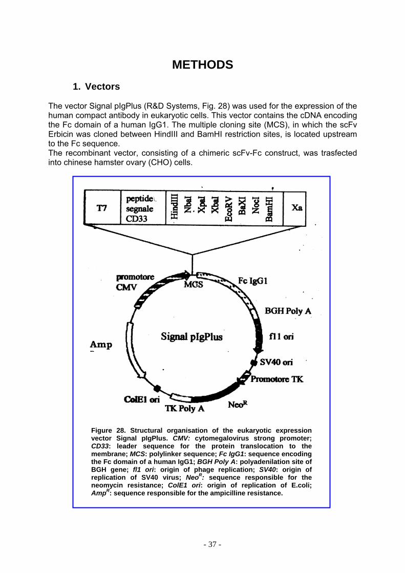
METHODS
1. Vectors
The vector Signal pIgPlus (R&D Systems, Fig. 28) was used for the expression of the human compact antibody in eukaryotic cells. This vector contains the cDNA encoding the Fc domain of a human IgG1. The multiple cloning site (MCS), in which the scFv Erbicin was cloned between HindIII and BamHI restriction sites, is located upstream to the Fc sequence. The recombinant vector, consisting of a chimeric scFv-Fc construct, was trasfected into chinese hamster ovary (CHO) cells.
AmpR
Figure 28. Structural organisation of the eukaryotic expression vector Signal pIgPlus. CMV: cytomegalovirus strong promoter; CD33: leader sequence for the protein translocation to the membrane; MCS: polylinker sequence; Fc IgG1: sequence encoding the Fc domain of a human IgG1; BGH Poly A: polyadenilation site of BGH gene; fl1 ori: origin of phage replication; SV40: origin of replication of SV40 virus; NeoR: sequence responsible for the neomycin resistance; ColE1 ori: origin of replication of E.coli; AmpR: sequence responsible for the ampicilline resistance.
- 37 -

2. Cell lines and antibodies
The following cell lines were used: SKBR3 cell line from human breast cancer; A431 cell line from human epidermoid carcinoma; Chinese hamster ovary (CHO) cells. All cell lines were provided by American Type Culture Collection, Rockville, MD and were cultured in RPMI 1640 (Gibco BRL, Life Technologies, Paisley, UK), supplemented with 10% foetal calf serum, containing 50 Units/ml penicillin, and 50 µg/ml streptomycin ( Gibco BRL). After transfection with the recombinant vector, CHO cells were grown in medium containing 1 mg/ml neomycin or G418 (Sigma). The antibodies used were: Herceptin (Genentech, South San Francisco, CA, USA); monoclonal anti-human IgG1 (Fc specific, Sigma, St Louis, MO, USA); horseradish peroxidase-conjugated goat anti-mouse immunoglobulins (Pierce, Rockford, IL, USA); horseradish peroxidase-conjugated goat anti-human affinity-isolated IgG (Fc specific) (Sigma).
3. Bacterial strains E. coli JM101 strain (78) was used for plasmid amplification.
4. Bacterial culture Culture medium LB (Luria-Bertani), used for the bacterial growth, was prepared as described by Sanbrook et al (80). The selection of transformed E. coli JM101 cells was obtained in the presence of 50 µg/ml ampicilline.
5. Chimeric cDNA construction The cDNA encoding the scFv Erbicin was amplified from vector pHEN2 by PCR, using, as forward and reverse primers, oligonucleotides containing at their 5’ end a Hind III site or a BamHI site, respectively. Oligonucleotides used for the PCR amplification reaction were: Forward primer: 5’CCCCCCAAGCTTCAGGTGCAGCTGTTG3’ (In red is shown HindIII restriction site) Reverse primer: 5’AACCGCGGATCCGCACCTAGGACGGTGAG3’ (In red is shown BamHI restriction site) Amplification reactions were performed using Pfx DNA polymerase from Pyrococcus (Invitrogen). The final reaction volume was 50 µl. The PCR reaction mixture contained:
• Erbicin cDNA, 20 ng
- 38 -
• Forward and reverse primers, 20 µM each • dNTP 0.2 mM • MgSO4 1mM • Pfx enzyme 5 U
30 amplification cycles were carried out under the following conditions: • 2 min denaturing at 95°C • 2 min annealing at 55°C • 2 min extension at 73°C.
The amplified products were purified by gel-elution using the “DNA purification system” kit (Promega). The isolated product was analysed by electrophoresis on 1% agarose gel.
6. Bacterial competent cells Single clones of JM101 E.coli strain were picked up from LB-agar plates and inoculated in 5 ml LB medium. Cells were grown at 37°C until they reached 0.5 OD600nm and were subsequently incubated at 0°C for 10 min. After centrifugation at 5000 rpm for 5 min, pelleted cells were resuspended in 50 mM CaCl2 and incubated for 20 min at 0°C. After centrifugation at 4000 rpm for 5 min, cells were resuspended in 50 mM CaCl2.
7. Cloning of the PCR amplified product into the Signal pIgPlus expression vector
The PCR amplified chimeric cDNA was then digested with HindIII and BamHI (New England Biolabs, Hertfordshire, UK) for cloning into the corresponding sites of plasmid Signal pIgPlus (R&D Systems, Minneapolis, USA). Upon cloning, the cDNA is positioned downstream to the leader sequence and upstream to the sequence coding for the hinge-CH2-CH3 region of a human IgG1 heavy chain constant region. The ligation reaction was performed by incubating at room temperature the digested scFv cDNA and pIgPlus Signal vector with “Fast-Ligase” enzyme (Promega). After 2h incubation, the ligation products were used to transform E.coli JM101 competent cells. The recombinant vector was purified from E.coli transformed cells using a mini-prep kit (Qiagen Valencia, CA) and analysed by restriction analysis on 1% agarose gel electrophoresis. The recombinant vector was then amplified in E. coli JM101 cells and fully sequenced to confirm the correct insertion of the scFv cDNA in pIg Plus Signal vector.
8. Transfection of CHO cells with the recombinant vector and expression of the human compact antibody
The chimeric protein was produced by transfecting CHO cells with the recombinant vector. Cells, grown in RPMI containing 10% FCS at 70-80% confluence, were trasfected with the recombinant vector.
- 39 -
Recombinant pIgPlus Signal vector (5 µg) was incubated for 10 min at room temperature with the Superfect reagent (Qiagen, Valencia, CA) in 0.2 ml RPMI medium. CHO cells, seeded in 6-well plates at a density of 5x10⁵ /well, were washed with 4 ml PBS, detached from the plates with the cell dissociation solution (Sigma), and resuspended in fresh RPMI medium containing 10% FCS. The mixture of recombinant vector and Superfect reagent was then added to the cells. After 2h incubation at 37°C, cells were harvested, extensively washed with PBS and resuspended in RPMI containing 10% FCS with antibiotics. Cells were then incubated at 37°C for 48h. Stable transfectants were selected in the presence of G418 (1 mg/ml). The selective medium was renewed every 3-4 days, until visible colonies were formed. After two weeks, resistant clones were picked up and transferred to plates containing fresh RPMI medium and G418. Expression of Erb-hcAb in the culture medium was determined by quantitative ELISA assays (see below). To obtain large-scale recombinant protein production, trasfected CHO cells were expanded by growing them in 10 cm plates near to confluence in RPMI medium containing neomycin, and then were grown for 3-4 days in serum-free medium.
9. Purification of human compact antibody by affinity chromatography
The recombinant Erb-hcAb antibody, secreted by trasfected CHO cells, was purified from the culture medium by affinity chromatography on a protein A-Ceramic hyper D F column (Biosepra S.A., Cergy-Saint-Christophe, France). To 300-500 ml of conditioned medium, previously clarified by centrifugation at 2000 rpm, 1 M TrisHCl at pH 8.0 was added (1/10 of medium volume). The medium was then loaded on the column, previously equilibrated in 0.1 M TrisHCl pH 8.0. The column was sequentially washed with 10 volumes of 100 mM Tris-HCl, pH 8.0 containing 0.5 M NaCl, and 10 volumes of 10 mM Tris-HCl, pH 8.0. The protein eluate, obtained with 50 mM glycine pH 3.0, was immediately neutralised by adding 0.1 volumes of 1 M Tris, pH 8.0.
10. SDS-PAGE and Western blotting analyses of the human compact antibody
The purity of the protein eluate was evaluated by SDS-PAGE analysis on 7.5 % polyacrylammide gels, following the procedure described by Laemmli (79). Protein bands were detected by Coomassie staining. The purified protein was analysed by Western blotting by transferring the protein species on PVDF membranes (Millipore, Bedford, MA). An anti-human IgG1 (Fc specific) antibody (Sigma) was used, followed by a goat anti-mouse horseradish peroxidase-conjugated mAb. The signal intensity of the reactive bands was quantitatively measured with a phosphorimager apparatus (GS-710, Bio-Rad, Hercules, CA). Protein concentration was determined using the BCA assays (Pierce), following the manufacturer’s instructions. Spectrophotometric determinations at 280 nm using E1% = 13 were also performed.
- 40 -
11. ELISA assays
ErbB2-positive SKBR3 cells and ErbB2-negative A431 cells were transferred to U-bottom microtiter plates (1 x 10⁵ cells/well). After blocking with PBS, containing 6% bovine serum albumin (BSA), cells were incubated for 90 min either with the conditioned medium or with the purified immunoagent in the ELISA buffer (PBS/BSA 3%). After centrifugation, supernatants were removed and the pelleted cells were washed twice in 200 µl of ELISA buffer, resuspended in 100 µl of ELISA buffer, and incubated with peroxidase-conjugated anti-human IgG (Fc-specific) antibodies (Sigma). After 1 hour, the plates were centrifuged and washed three times with ELISA buffer. The reaction was developed by adding 3,3’,5,5’-tetramethylbenzidine (TMB) (Sigma) and stopped with 1 M HCl. Binding values were obtained by determining the absorbance at 450 nm. The reported values are the mean of at least three determinations. The standard deviations were below 5%).
12. Cell growth inhibition assays Cells were seeded in 96-well, flat-bottom plates as follows: SKBR3 cells at a density of 1.5 x 10⁴/well; A431 at a density of 5 x 10³/well. After the addition of the immunoagent under test, viable cells were counted by the Trypan blue exclusion test at suitable time intervals (24-48-72 h). Cell survival was expressed as the percentage of viable cells in the presence of the protein under test, with respect to control cultures grown in the absence of the protein. Typically, cell survival values were obtained from at least three independent experiments in which triplicate counts were determined. Standard deviations were below 5%.
13. Molecular Modelling
• CDR (complementarity determining regions) The CDR regions were determined using KABAT numbering. The CDR canonical loop structures were determined using Chothia description.
• Database search Database search for VH (variable heavy chain) and VL (variable light chain) sequence homology was performed using the protein Blast (BLASTP) program at NCBI.
• Homology modelling and energy minimisation Insight II was used to model VH and VL chains separately. The VH and VL models were then connected by a synthetic peptide [(Gly4Ser)3] using BUILDER/Insight II, followed by energy minimisation cycles in a CFF91 force field.
- 41 -
14. Patients under test 350 unselected serum samples were tested by a prospective study. Four categories of patients were analysed:
1. RA: 100 patients who had been affected by RA 2. HCV: 100 patients who were affected by chronic infection of hepatitis C virus
3. C: 53 patients affected by other connective tissue diseases
4. H: 97 healthy blood donors
Serum samples from each patient were centrifuged and stored at -20°C, until use.
15. Agglutination test Principles of the Procedure Agglutination test for IgM-RF determination was performed using N Latex RF automated test (DADE BEHRING), using a nephelometric instrument (BN System, DADE BEHRING). Polystyrene particles coated with an immuno-complex consisting of human γ-globulin/anti-human γ-globulins from sheep, were agglutinate when mixed with samples containing RF. A beam of light is passed through the aggregates and scattered. The intensity of the scattered light is proportional to the concentration of analytes (RF) in the sample. The results were evaluated by comparison with a standard tested at known concentration. Assay protocol All steps necessary for the assays were performed automatically by the BN System instrument. Reagents provided by the manufacturer were lyophilised and reconstituted in distilled water about 15 minute before use. Serum samples were centrifuged prior tests, to avoid interference due to sample turbidity. Samples were automatically diluted 1:20 with serum diluent, provided by the manufacturer. Reference curves were obtained by multi-point calibration. Serial dilutions of RF standards were automatically prepared by the instrument. Results interpretation Results were expressed in international units per mL (IU/mL) according to the first British Standard for Rheumatoid Arthritis. Serum samples >15 U/mL were considered positive.
- 42 -
16. FIDISTM Rheuma Assay System Set up of the FIDISTM instrument A maintenance procedure has to be daily used, to assure the good performance of the system. Firstly, the optic system (laser beam) is warmed up, and this procedure requires thirty minutes. Then, any air bubbles in the Fidis microfluidic pathway were removed by the “prime” procedure. Then, the probe was cleaned by repetitive washing with isopropanol 70%. The pressure and temperature of the system were checked before testing samples. Calibration set-up was performed once per month systematically, and a new lot of sheath fluid reagent used, in order to ensure optimal instrument performance. Calibration was also performed when the temperature value was out of the operation range of the instrument. At the end of the analysis session, a sanitization was done, by using 70%sodium ipochlorite. Microspheres Each antigen (Fc fragment from human or rabbit IgG) was conjugated by a covalent bond to an individual set of microspheres through its surface functional carboxyl groups. The coupling procedure (activation, concentration of microspheres, quantity of coupling antigen-stabilising solution) was set up to ensure sensitivity of the assay and shelf life of the preparation. Following the coupling procedure of each set, the two antigen-coupled microsphere sets were mixed generating the final microsphere
reagent. Assay procedure
1. Microspheres dispensing. For each assay, a “reagent-blank” well, a positive control well, a negative control well, and a calibrator well were required. The microsphere reagent has to be mixed vigorously before dispensing 50 µl in each well to be used.
2. Samples incubation. 100 µl of TDL buffer, provided by the manufacturer,
were added to the reagent blank well. 100 µl of ready to use calibrator and of prediluted controls and samples were dispensed in the corresponding wells. Plates were covered to avoid direct light and incubated for 30 min at room temperature.
3. Wash step. Plates were washed with TDL buffer (2 cycles), using the filtration
unit. 200 µl of TDL buffer were dispensed in all wells. The filtration was repeated for, as described above. The residual buffer in the microwells was removed by blotting the plates with an absorbent towel. The plates were placed on a dry surface prior incubation.
4. Secondary antibody dispensing. 100 µl of prediluted secondary antibody
conjugated with phycoerythrin were dispensed in all microwells. Plates were incubated 30 min at room temperature.
5. Test reading. The excitation system of the FIDIS analyzer includes two solid-
states lasers. A reporter laser excites fluorescent molecules bound to biological reactants at the microsphere surface; a classification laser excites
- 43 -
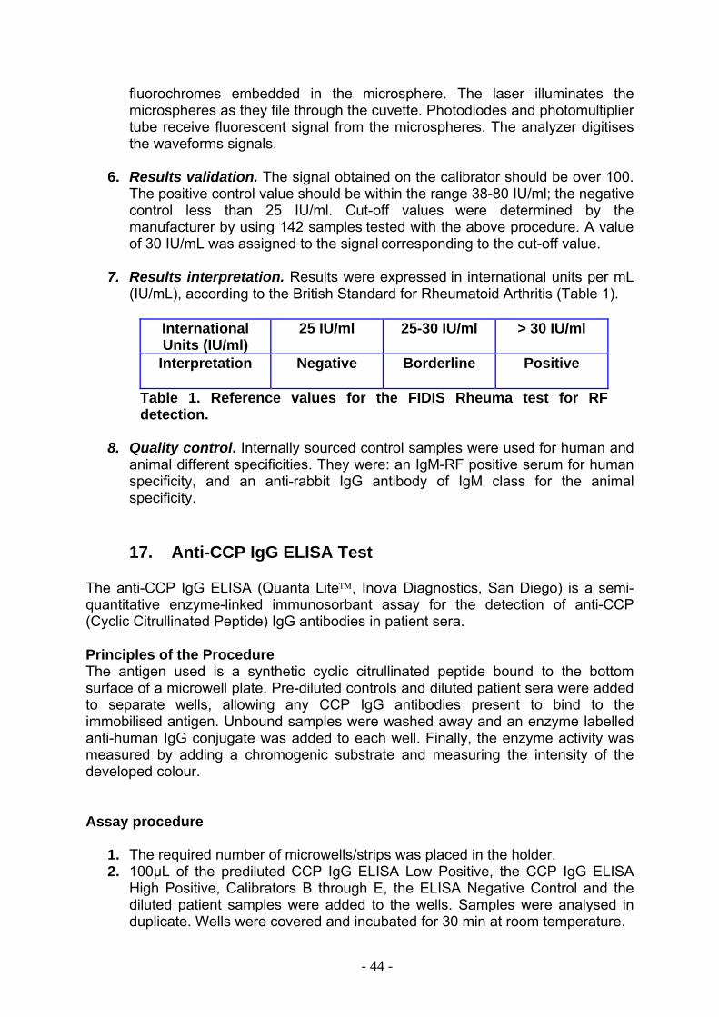
fluorochromes embedded in the microsphere. The laser illuminates the microspheres as they file through the cuvette. Photodiodes and photomultiplier tube receive fluorescent signal from the microspheres. The analyzer digitises the waveforms signals.
6. Results validation. The signal obtained on the calibrator should be over 100.
The positive control value should be within the range 38-80 IU/ml; the negative control less than 25 IU/ml. Cut-off values were determined by the manufacturer by using 142 samples tested with the above procedure. A value of 30 IU/mL was assigned to the signal corresponding to the cut-off value.
7. Results interpretation. Results were expressed in international units per mL
(IU/mL), according to the British Standard for Rheumatoid Arthritis (Table 1).
International Units (IU/ml)
25 IU/ml 25-30 IU/ml > 30 IU/ml
Interpretation Negative Borderline Positive
Table 1. Reference values for the FIDIS Rheuma test for RF detection.
8. Quality control. Internally sourced control samples were used for human and
animal different specificities. They were: an IgM-RF positive serum for human specificity, and an anti-rabbit IgG antibody of IgM class for the animal specificity.
17. Anti-CCP IgG ELISA Test The anti-CCP IgG ELISA (Quanta Lite™, Inova Diagnostics, San Diego) is a semi-quantitative enzyme-linked immunosorbant assay for the detection of anti-CCP (Cyclic Citrullinated Peptide) IgG antibodies in patient sera. Principles of the Procedure The antigen used is a synthetic cyclic citrullinated peptide bound to the bottom surface of a microwell plate. Pre-diluted controls and diluted patient sera were added to separate wells, allowing any CCP IgG antibodies present to bind to the immobilised antigen. Unbound samples were washed away and an enzyme labelled anti-human IgG conjugate was added to each well. Finally, the enzyme activity was measured by adding a chromogenic substrate and measuring the intensity of the developed colour. Assay procedure
1. The required number of microwells/strips was placed in the holder. 2. 100µL of the prediluted CCP IgG ELISA Low Positive, the CCP IgG ELISA
High Positive, Calibrators B through E, the ELISA Negative Control and the diluted patient samples were added to the wells. Samples were analysed in duplicate. Wells were covered and incubated for 30 min at room temperature.
- 44 -
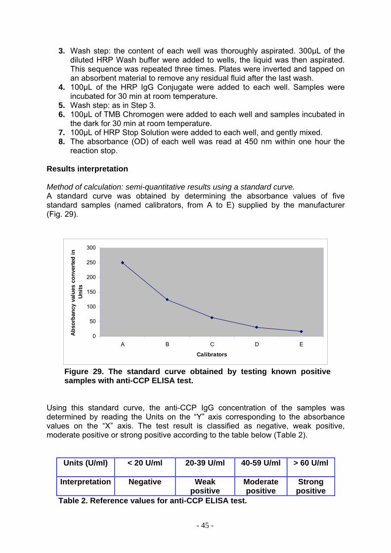
3. Wash step: the content of each well was thoroughly aspirated. 300µL of the diluted HRP Wash buffer were added to wells, the liquid was then aspirated. This sequence was repeated three times. Plates were inverted and tapped on an absorbent material to remove any residual fluid after the last wash.
4. 100µL of the HRP IgG Conjugate were added to each well. Samples were incubated for 30 min at room temperature.
5. Wash step: as in Step 3. 6. 100µL of TMB Chromogen were added to each well and samples incubated in
the dark for 30 min at room temperature. 7. 100µL of HRP Stop Solution were added to each well, and gently mixed. 8. The absorbance (OD) of each well was read at 450 nm within one hour the
reaction stop. Results interpretation Method of calculation: semi-quantitative results using a standard curve. A standard curve was obtained by determining the absorbance values of five standard samples (named calibrators, from A to E) supplied by the manufacturer (Fig. 29).
0
50
100
150
200
250
300
A B C D E
Calibrators
Abso
rban
cy v
alue
s co
nver
ted
in
Units
Figure 29. The standard curve obtained by testing known positive samples with anti-CCP ELISA test.
Using this standard curve, the anti-CCP IgG concentration of the samples was determined by reading the Units on the “Y” axis corresponding to the absorbance values on the “X” axis. The test result is classified as negative, weak positive, moderate positive or strong positive according to the table below (Table 2).
Units (U/ml) < 20 U/ml 20-39 U/ml 40-59 U/ml > 60 U/ml
Interpretation Negative Weak positive
Moderate positive
Strong positive
Table 2. Reference values for anti-CCP ELISA test.
- 45 -
Internal quality control As it is recommended by the GMP (good manufacturing practices) protocol, an internally sourced control sample was used for the anti-CCP IgG antibodies. An anti-CCP positive serum was used as the internal control sample.
18. STATISTICAL ANALYSES All the data obtained using the nephelometric or the FIDIS Rheuma tests on serum samples, were analysed by statistical methods. Statistical analyses were performed using the computer software R 2.4.1 (The R Development Core Team). McNemar's test was applied to a 2 × 2 contingency table, where each of the four cells contained one of the following samples: “positive with both methods”, “negative with both methods”, “positive with referral (nephelometric test) but negative with the experimental method (FIDIS Rheuma test)” and “negative with referral but positive with the experimental method”, respectively. The statistical null hypothesis was that the number of samples recognised as “positive” was the same in both methods. In a statistic test, null hypothesis are typically statements of no difference. The alternative hypothesis was that the number of samples recognised as “positive” was higher with the experimental FIDIS Rheuma method. Due to the small difference between the two methods, the Yate’s correction factor was inevitably applied to the McNemar's test.
- 46 -
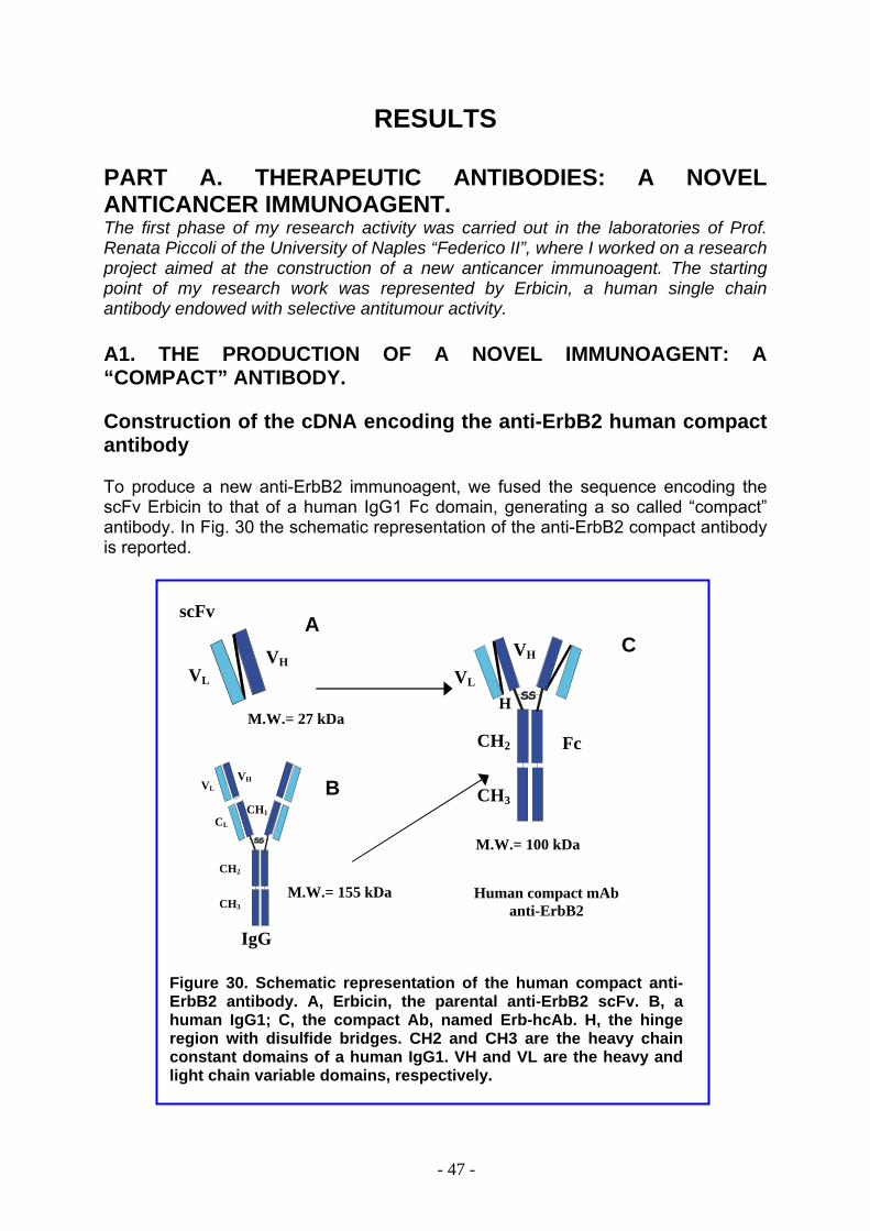
RESULTS PART A. THERAPEUTIC ANTIBODIES: A NOVEL ANTICANCER IMMUNOAGENT. The first phase of my research activity was carried out in the laboratories of Prof. Renata Piccoli of the University of Naples “Federico II”, where I worked on a research project aimed at the construction of a new anticancer immunoagent. The starting point of my research work was represented by Erbicin, a human single chain antibody endowed with selective antitumour activity. A1. THE PRODUCTION OF A NOVEL IMMUNOAGENT: A “COMPACT” ANTIBODY. Construction of the cDNA encoding the anti-ErbB2 human compact antibody To produce a new anti-ErbB2 immunoagent, we fused the sequence encoding the scFv Erbicin to that of a human IgG1 Fc domain, generating a so called “compact” antibody. In Fig. 30 the schematic representation of the anti-ErbB2 compact antibody is reported.
scFv
Figure 30. Schematic representation of the human compact anti-ErbB2 antibody. A, Erbicin, the parental anti-ErbB2 scFv. B, a human IgG1; C, the compact Ab, named Erb-hcAb. H, the hinge region with disulfide bridges. CH2 and CH3 are the heavy chain constant domains of a human IgG1. VH and VL are the heavy and light chain variable domains, respectively.
H
Fc
M.W.= 100 kDa
VH
VL
CH2
CH3
M.W.= 27 kDa
VH VL
A C
VH B VL
CH1 CL
CH2
M.W.= 155 kDa Human compact mAb CH3 anti-ErbB2
IgG
- 47 -
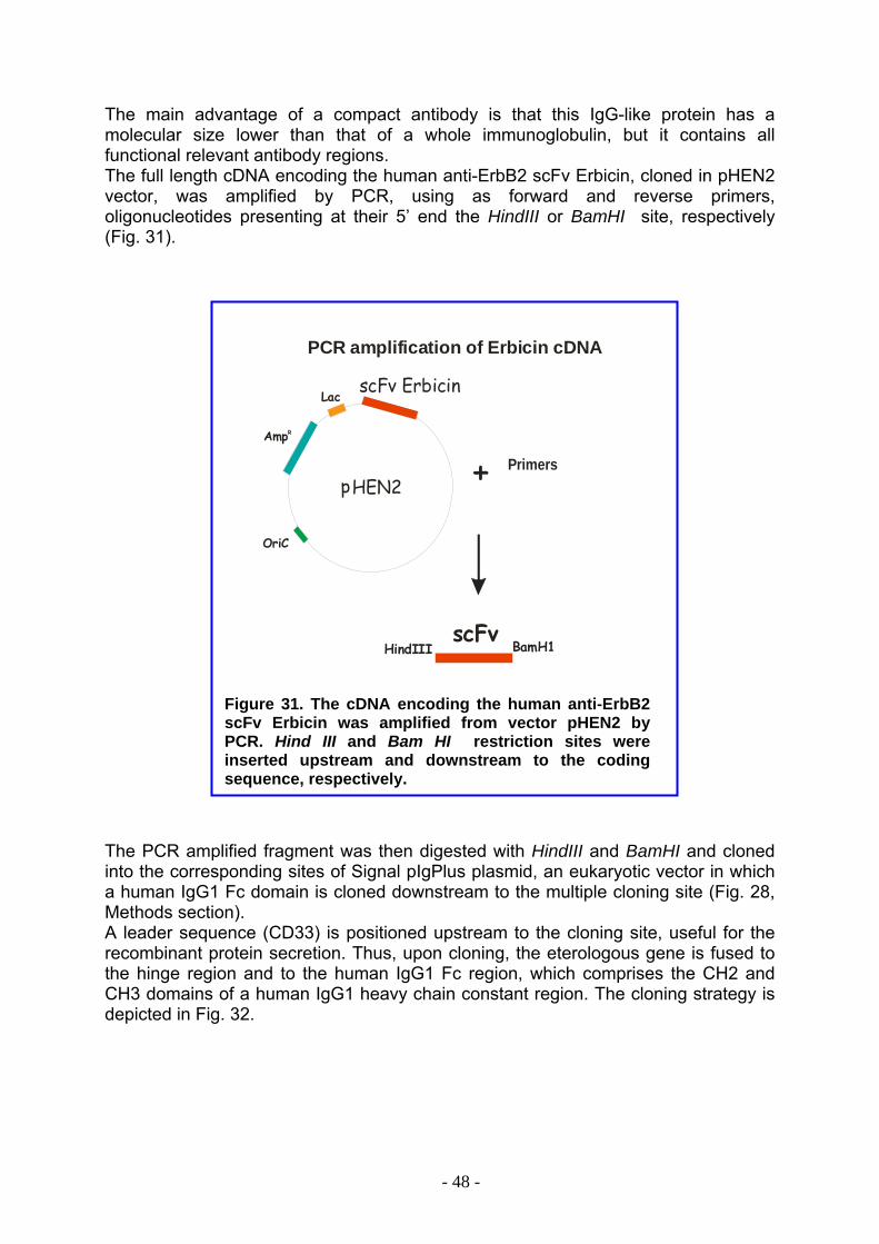
The main advantage of a compact antibody is that this IgG-like protein has a molecular size lower than that of a whole immunoglobulin, but it contains all functional relevant antibody regions. The full length cDNA encoding the human anti-ErbB2 scFv Erbicin, cloned in pHEN2 vector, was amplified by PCR, using as forward and reverse primers, oligonucleotides presenting at their 5’ end the HindIII or BamHI site, respectively (Fig. 31).
OriC
AmpR
scFv ErbicinLac
HEN2p
HindIII BamH1scFv
+ Primers
PCR amplification of Erbicin cDNA
Figure 31. The cDNA encoding the human anti-ErbB2 scFv Erbicin was amplified from vector pHEN2 by PCR. Hind III and Bam HI restriction sites were inserted upstream and downstream to the coding sequence, respectively.
The PCR amplified fragment was then digested with HindIII and BamHI and cloned into the corresponding sites of Signal pIgPlus plasmid, an eukaryotic vector in which a human IgG1 Fc domain is cloned downstream to the multiple cloning site (Fig. 28, Methods section). A leader sequence (CD33) is positioned upstream to the cloning site, useful for the recombinant protein secretion. Thus, upon cloning, the eterologous gene is fused to the hinge region and to the human IgG1 Fc region, which comprises the CH2 and CH3 domains of a human IgG1 heavy chain constant region. The cloning strategy is depicted in Fig. 32.
- 48 -
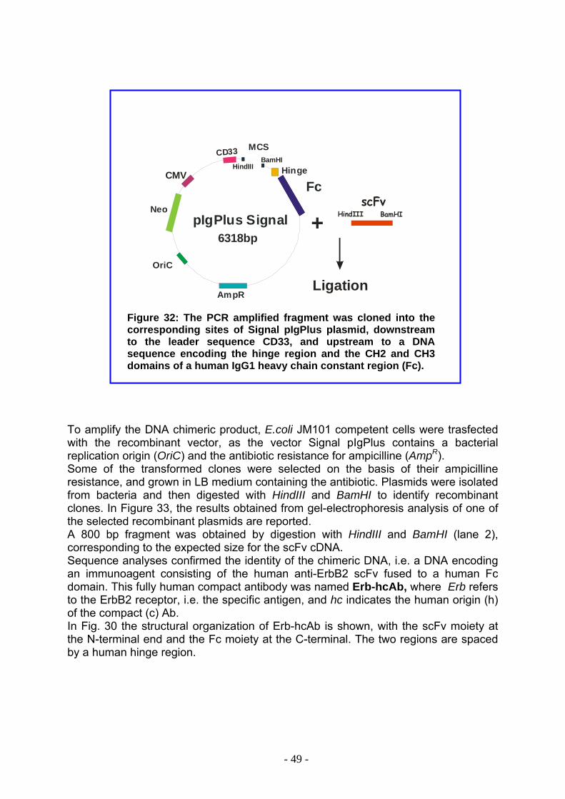
Fc
6318bp
Neo
OriC
Hinge
pIgPlus Signal
MCS
AmpR
CMVHindIII
BamHI
HindIII BamHIscFv
Ligation
CD33
+
Figure 32: The PCR amplified fragment was cloned into the corresponding sites of Signal pIgPlus plasmid, downstream to the leader sequence CD33, and upstream to a DNA sequence encoding the hinge region and the CH2 and CH3 domains of a human IgG1 heavy chain constant region (Fc).
To amplify the DNA chimeric product, E.coli JM101 competent cells were trasfected with the recombinant vector, as the vector Signal pIgPlus contains a bacterial replication origin (OriC) and the antibiotic resistance for ampicilline (AmpR). Some of the transformed clones were selected on the basis of their ampicilline resistance, and grown in LB medium containing the antibiotic. Plasmids were isolated from bacteria and then digested with HindIII and BamHI to identify recombinant clones. In Figure 33, the results obtained from gel-electrophoresis analysis of one of the selected recombinant plasmids are reported. A 800 bp fragment was obtained by digestion with HindIII and BamHI (lane 2), corresponding to the expected size for the scFv cDNA. Sequence analyses confirmed the identity of the chimeric DNA, i.e. a DNA encoding an immunoagent consisting of the human anti-ErbB2 scFv fused to a human Fc domain. This fully human compact antibody was named Erb-hcAb, where Erb refers to the ErbB2 receptor, i.e. the specific antigen, and hc indicates the human origin (h) of the compact (c) Ab. In Fig. 30 the structural organization of Erb-hcAb is shown, with the scFv moiety at the N-terminal end and the Fc moiety at the C-terminal. The two regions are spaced by a human hinge region.
- 49 -
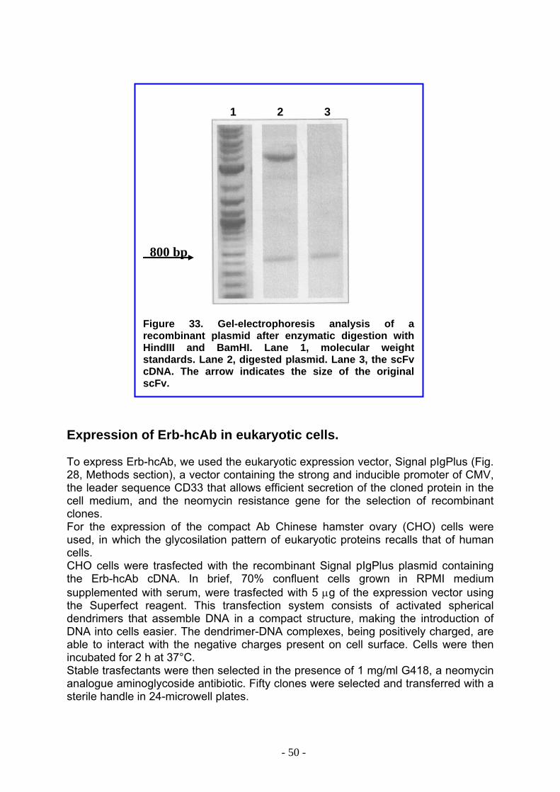
1 2 3
800 bp
Figure 33. Gel-electrophoresis analysis of a recombinant plasmid after enzymatic digestion with HindIII and BamHI. Lane 1, molecular weight standards. Lane 2, digested plasmid. Lane 3, the scFv cDNA. The arrow indicates the size of the original scFv.
Expression of Erb-hcAb in eukaryotic cells.
To express Erb-hcAb, we used the eukaryotic expression vector, Signal pIgPlus (Fig. 28, Methods section), a vector containing the strong and inducible promoter of CMV, the leader sequence CD33 that allows efficient secretion of the cloned protein in the cell medium, and the neomycin resistance gene for the selection of recombinant clones. For the expression of the compact Ab Chinese hamster ovary (CHO) cells were used, in which the glycosilation pattern of eukaryotic proteins recalls that of human cells. CHO cells were trasfected with the recombinant Signal pIgPlus plasmid containing the Erb-hcAb cDNA. In brief, 70% confluent cells grown in RPMI medium supplemented with serum, were trasfected with 5 µg of the expression vector using the Superfect reagent. This transfection system consists of activated spherical dendrimers that assemble DNA in a compact structure, making the introduction of DNA into cells easier. The dendrimer-DNA complexes, being positively charged, are able to interact with the negative charges present on cell surface. Cells were then incubated for 2 h at 37°C. Stable trasfectants were then selected in the presence of 1 mg/ml G418, a neomycin analogue aminoglycoside antibiotic. Fifty clones were selected and transferred with a sterile handle in 24-microwell plates.
- 50 -
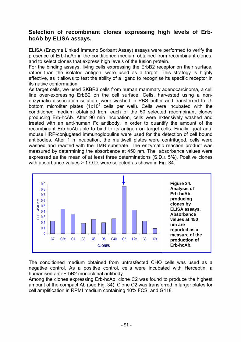
Selection of recombinant clones expressing high levels of Erb-hcAb by ELISA assays. ELISA (Enzyme Linked Immuno Sorbant Assay) assays were performed to verify the presence of Erb-hcAb in the conditioned medium obtained from recombinant clones, and to select clones that express high levels of the fusion protein. For the binding assays, living cells expressing the ErbB2 receptor on their surface, rather than the isolated antigen, were used as a target. This strategy is highly effective, as it allows to test the ability of a ligand to recognise its specific receptor in its native conformation. As target cells, we used SKBR3 cells from human mammary adenocarcinoma, a cell line over-expressing ErbB2 on the cell surface. Cells, harvested using a non-enzymatic dissociation solution, were washed in PBS buffer and transferred to U-bottom microtiter plates (1x10⁵ cells per well). Cells were incubated with the conditioned medium obtained from each of the 50 selected recombinant clones producing Erb-hcAb. After 90 min incubation, cells were extensively washed and treated with an anti-human Fc antibody, in order to quantify the amount of the recombinant Erb-hcAb able to bind to its antigen on target cells. Finally, goat anti-mouse HRP-conjugated immunoglobulins were used for the detection of cell bound antibodies. After 1 h incubation, the multiwell plates were centrifuged, cells were washed and reacted with the TMB substrate. The enzymatic reaction product was measured by determining the absorbance at 450 nm. The absorbance values were expressed as the mean of at least three determinations (S.D.≤ 5%). Positive clones with absorbance values > 1 O.D. were selected as shown in Fig. 34.
00,10,20,30,40,50,60,70,80,9
C7 C2x C1 C8 X6 X5 G40 C2 L2x C3 C9
CLONES
O.D
. 450
nm
Figure 34. Analysis of Erb-hcAb- producing clones by ELISA assays. Absorbance values at 450 nm are reported as a measure of the production of Erb-hcAb.
The conditioned medium obtained from untrasfected CHO cells was used as a negative control. As a positive control, cells were incubated with Herceptin, a humanised anti-ErbB2 monoclonal antibody. Among the clones expressing Erb-hcAb, clone C2 was found to produce the highest amount of the compact Ab (see Fig. 34). Clone C2 was transferred in larger plates for cell amplification in RPMI medium containing 10% FCS and G418.
- 51 -
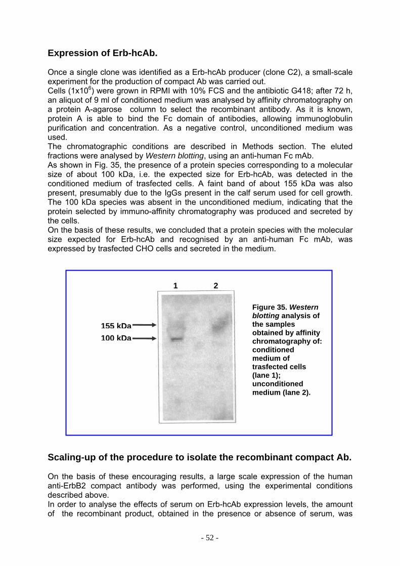
Expression of Erb-hcAb. Once a single clone was identified as a Erb-hcAb producer (clone C2), a small-scale experiment for the production of compact Ab was carried out. Cells (1x10⁶) were grown in RPMI with 10% FCS and the antibiotic G418; after 72 h, an aliquot of 9 ml of conditioned medium was analysed by affinity chromatography on a protein A-agarose column to select the recombinant antibody. As it is known, protein A is able to bind the Fc domain of antibodies, allowing immunoglobulin purification and concentration. As a negative control, unconditioned medium was used. The chromatographic conditions are described in Methods section. The eluted fractions were analysed by Western blotting, using an anti-human Fc mAb. As shown in Fig. 35, the presence of a protein species corresponding to a molecular size of about 100 kDa, i.e. the expected size for Erb-hcAb, was detected in the conditioned medium of trasfected cells. A faint band of about 155 kDa was also present, presumably due to the IgGs present in the calf serum used for cell growth. The 100 kDa species was absent in the unconditioned medium, indicating that the protein selected by immuno-affinity chromatography was produced and secreted by the cells. On the basis of these results, we concluded that a protein species with the molecular size expected for Erb-hcAb and recognised by an anti-human Fc mAb, was expressed by trasfected CHO cells and secreted in the medium. 1 2
Figure 35. Western blotting analysis of the samples obtained by affinity chromatography of: conditioned medium of trasfected cells (lane 1); unconditioned medium (lane 2).
155 kDa100 kDa
Scaling-up of the procedure to isolate the recombinant compact Ab. On the basis of these encouraging results, a large scale expression of the human anti-ErbB2 compact antibody was performed, using the experimental conditions described above. In order to analyse the effects of serum on Erb-hcAb expression levels, the amount of the recombinant product, obtained in the presence or absence of serum, was
- 52 -
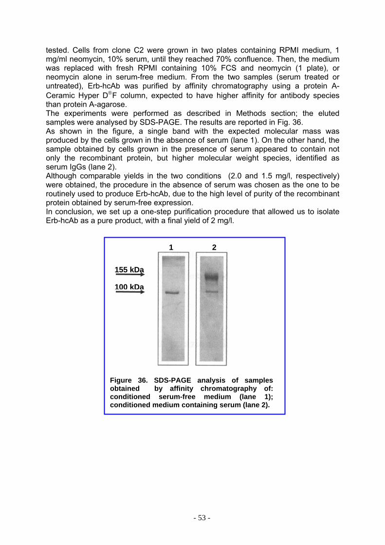
tested. Cells from clone C2 were grown in two plates containing RPMI medium, 1 mg/ml neomycin, 10% serum, until they reached 70% confluence. Then, the medium was replaced with fresh RPMI containing 10% FCS and neomycin (1 plate), or neomycin alone in serum-free medium. From the two samples (serum treated or untreated), Erb-hcAb was purified by affinity chromatography using a protein A-Ceramic Hyper D®F column, expected to have higher affinity for antibody species than protein A-agarose. The experiments were performed as described in Methods section; the eluted samples were analysed by SDS-PAGE. The results are reported in Fig. 36. As shown in the figure, a single band with the expected molecular mass was produced by the cells grown in the absence of serum (lane 1). On the other hand, the sample obtained by cells grown in the presence of serum appeared to contain not only the recombinant protein, but higher molecular weight species, identified as serum IgGs (lane 2). Although comparable yields in the two conditions (2.0 and 1.5 mg/l, respectively) were obtained, the procedure in the absence of serum was chosen as the one to be routinely used to produce Erb-hcAb, due to the high level of purity of the recombinant protein obtained by serum-free expression. In conclusion, we set up a one-step purification procedure that allowed us to isolate Erb-hcAb as a pure product, with a final yield of 2 mg/l. 1 2
155 kDa
100 kDa
Figure 36. SDS-PAGE analysis of samples obtained by affinity chromatography of: conditioned serum-free medium (lane 1); conditioned medium containing serum (lane 2).
- 53 -
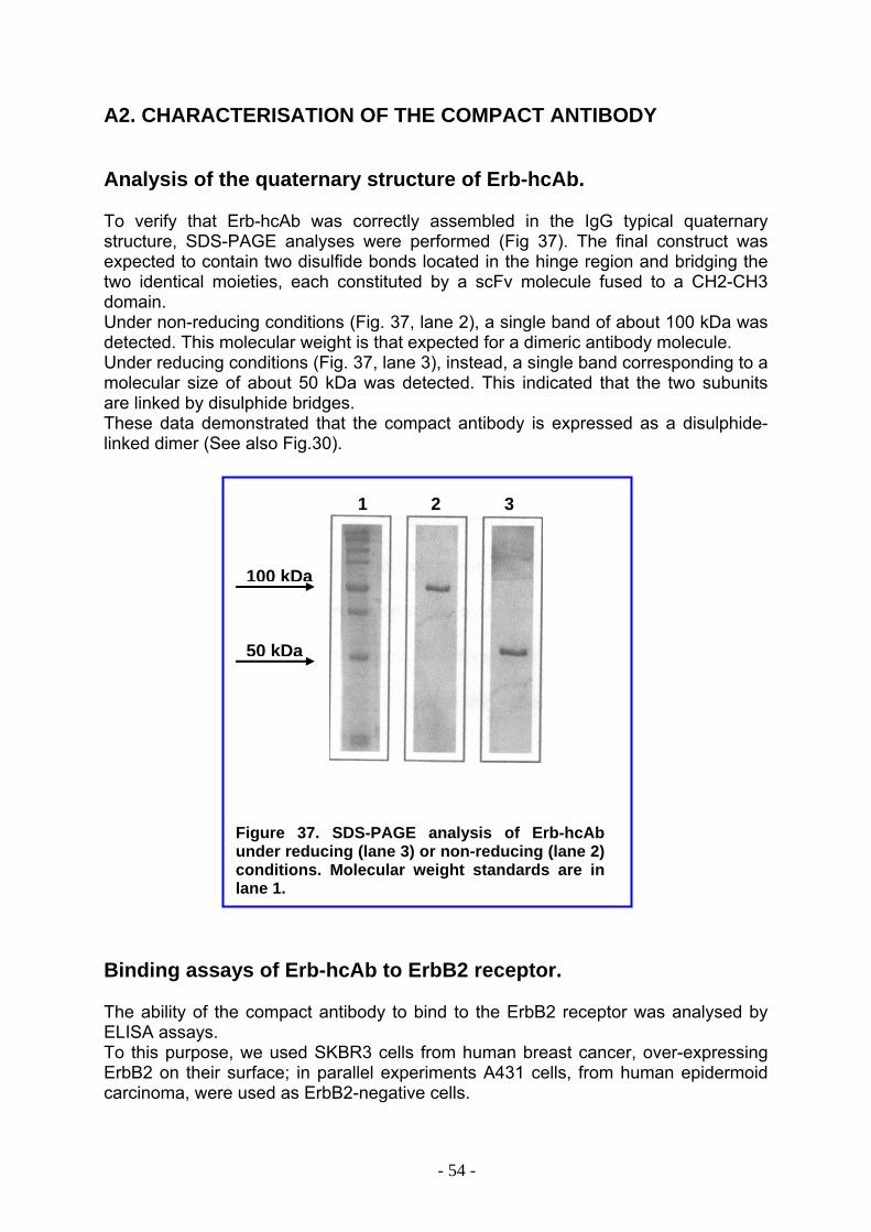
A2. CHARACTERISATION OF THE COMPACT ANTIBODY Analysis of the quaternary structure of Erb-hcAb. To verify that Erb-hcAb was correctly assembled in the IgG typical quaternary structure, SDS-PAGE analyses were performed (Fig 37). The final construct was expected to contain two disulfide bonds located in the hinge region and bridging the two identical moieties, each constituted by a scFv molecule fused to a CH2-CH3 domain. Under non-reducing conditions (Fig. 37, lane 2), a single band of about 100 kDa was detected. This molecular weight is that expected for a dimeric antibody molecule. Under reducing conditions (Fig. 37, lane 3), instead, a single band corresponding to a molecular size of about 50 kDa was detected. This indicated that the two subunits are linked by disulphide bridges. These data demonstrated that the compact antibody is expressed as a disulphide-linked dimer (See also Fig.30). 1 2 3
50 kDa
100 kDa
Figure 37. SDS-PAGE analysis of Erb-hcAb under reducing (lane 3) or non-reducing (lane 2) conditions. Molecular weight standards are in lane 1.
Binding assays of Erb-hcAb to ErbB2 receptor. The ability of the compact antibody to bind to the ErbB2 receptor was analysed by ELISA assays. To this purpose, we used SKBR3 cells from human breast cancer, over-expressing ErbB2 on their surface; in parallel experiments A431 cells, from human epidermoid carcinoma, were used as ErbB2-negative cells.
- 54 -
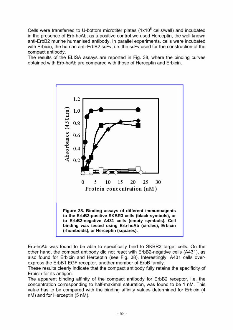
Cells were transferred to U-bottom microtiter plates (1x10⁵ cells/well) and incubated in the presence of Erb-hcAb; as a positive control we used Herceptin, the well known anti-ErbB2 murine humanised antibody. In parallel experiments, cells were incubated with Erbicin, the human anti-ErbB2 scFv, i.e. the scFv used for the construction of the compact antibody. The results of the ELISA assays are reported in Fig. 38, where the binding curves obtained with Erb-hcAb are compared with those of Herceptin and Erbicin.
Figure 38. Binding assays of different immunoagents to the ErbB2-positive SKBR3 cells (black symbols), or to ErbB2-negative A431 cells (empty symbols). Cell binding was tested using Erb-hcAb (circles), Erbicin (rhomboids), or Herceptin (squares).
Erb-hcAb was found to be able to specifically bind to SKBR3 target cells. On the other hand, the compact antibody did not react with ErbB2-negative cells (A431), as also found for Erbicin and Herceptin (see Fig. 38). Interestingly, A431 cells over-express the ErbB1 EGF receptor, another member of ErbB family. These results clearly indicate that the compact antibody fully retains the specificity of Erbicin for its antigen. The apparent binding affinity of the compact antibody for ErbB2 receptor, i.e. the concentration corresponding to half-maximal saturation, was found to be 1 nM. This value has to be compared with the binding affinity values determined for Erbicin (4 nM) and for Herceptin (5 nM).
- 55 -
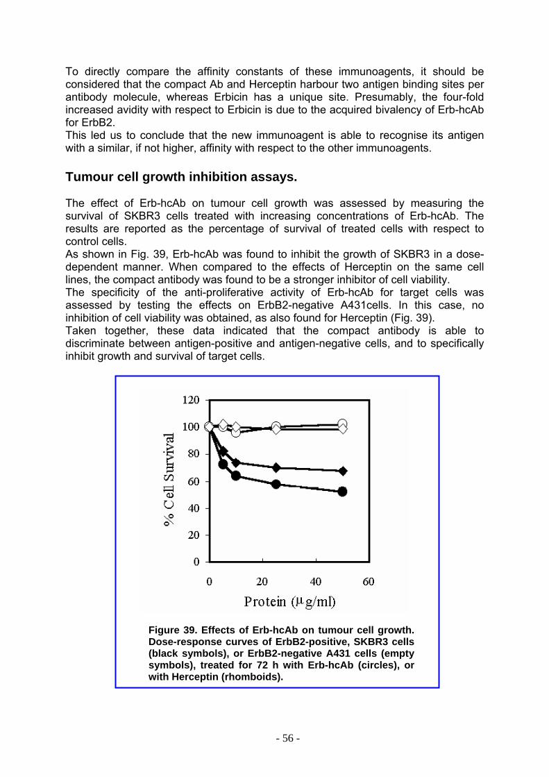
To directly compare the affinity constants of these immunoagents, it should be considered that the compact Ab and Herceptin harbour two antigen binding sites per antibody molecule, whereas Erbicin has a unique site. Presumably, the four-fold increased avidity with respect to Erbicin is due to the acquired bivalency of Erb-hcAb for ErbB2. This led us to conclude that the new immunoagent is able to recognise its antigen with a similar, if not higher, affinity with respect to the other immunoagents. Tumour cell growth inhibition assays.
The effect of Erb-hcAb on tumour cell growth was assessed by measuring the survival of SKBR3 cells treated with increasing concentrations of Erb-hcAb. The results are reported as the percentage of survival of treated cells with respect to control cells. As shown in Fig. 39, Erb-hcAb was found to inhibit the growth of SKBR3 in a dose-dependent manner. When compared to the effects of Herceptin on the same cell lines, the compact antibody was found to be a stronger inhibitor of cell viability. The specificity of the anti-proliferative activity of Erb-hcAb for target cells was assessed by testing the effects on ErbB2-negative A431cells. In this case, no inhibition of cell viability was obtained, as also found for Herceptin (Fig. 39). Taken together, these data indicated that the compact antibody is able to discriminate between antigen-positive and antigen-negative cells, and to specifically inhibit growth and survival of target cells.
Figure 39. Effects of Erb-hcAb on tumour cell growth. Dose-response curves of ErbB2-positive, SKBR3 cells (black symbols), or ErbB2-negative A431 cells (empty symbols), treated for 72 h with Erb-hcAb (circles), or with Herceptin (rhomboids).
- 56 -
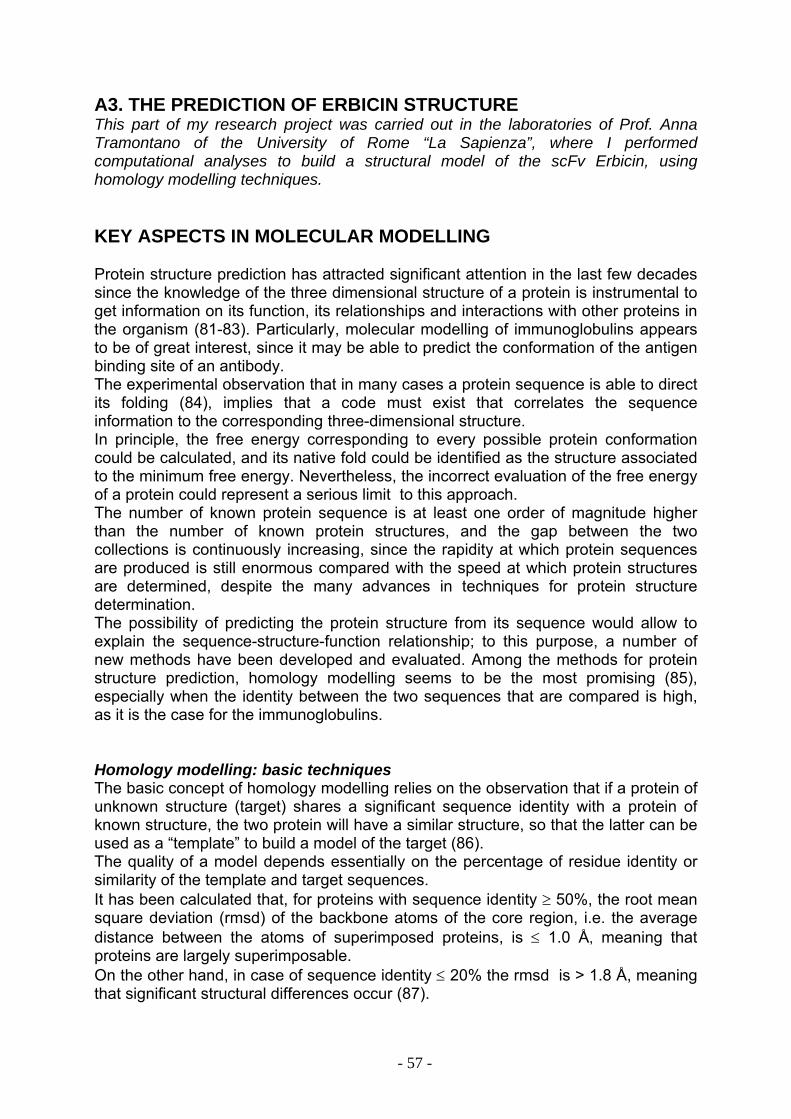
A3. THE PREDICTION OF ERBICIN STRUCTURE This part of my research project was carried out in the laboratories of Prof. Anna Tramontano of the University of Rome “La Sapienza”, where I performed computational analyses to build a structural model of the scFv Erbicin, using homology modelling techniques. KEY ASPECTS IN MOLECULAR MODELLING Protein structure prediction has attracted significant attention in the last few decades since the knowledge of the three dimensional structure of a protein is instrumental to get information on its function, its relationships and interactions with other proteins in the organism (81-83). Particularly, molecular modelling of immunoglobulins appears to be of great interest, since it may be able to predict the conformation of the antigen binding site of an antibody. The experimental observation that in many cases a protein sequence is able to direct its folding (84), implies that a code must exist that correlates the sequence information to the corresponding three-dimensional structure. In principle, the free energy corresponding to every possible protein conformation could be calculated, and its native fold could be identified as the structure associated to the minimum free energy. Nevertheless, the incorrect evaluation of the free energy of a protein could represent a serious limit to this approach. The number of known protein sequence is at least one order of magnitude higher than the number of known protein structures, and the gap between the two collections is continuously increasing, since the rapidity at which protein sequences are produced is still enormous compared with the speed at which protein structures are determined, despite the many advances in techniques for protein structure determination. The possibility of predicting the protein structure from its sequence would allow to explain the sequence-structure-function relationship; to this purpose, a number of new methods have been developed and evaluated. Among the methods for protein structure prediction, homology modelling seems to be the most promising (85), especially when the identity between the two sequences that are compared is high, as it is the case for the immunoglobulins. Homology modelling: basic techniques The basic concept of homology modelling relies on the observation that if a protein of unknown structure (target) shares a significant sequence identity with a protein of known structure, the two protein will have a similar structure, so that the latter can be used as a “template” to build a model of the target (86). The quality of a model depends essentially on the percentage of residue identity or similarity of the template and target sequences. It has been calculated that, for proteins with sequence identity ≥ 50%, the root mean square deviation (rmsd) of the backbone atoms of the core region, i.e. the average distance between the atoms of superimposed proteins, is ≤ 1.0 Å, meaning that proteins are largely superimposable. On the other hand, in case of sequence identity ≤ 20% the rmsd is > 1.8 Å, meaning that significant structural differences occur (87).
- 57 -
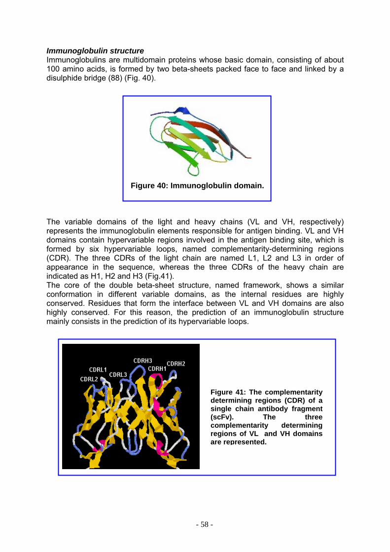
Immunoglobulin structure Immunoglobulins are multidomain proteins whose basic domain, consisting of about 100 amino acids, is formed by two beta-sheets packed face to face and linked by a disulphide bridge (88) (Fig. 40).
Figure 40: Immunoglobulin domain.
The variable domains of the light and heavy chains (VL and VH, respectively) represents the immunoglobulin elements responsible for antigen binding. VL and VH domains contain hypervariable regions involved in the antigen binding site, which is formed by six hypervariable loops, named complementarity-determining regions (CDR). The three CDRs of the light chain are named L1, L2 and L3 in order of appearance in the sequence, whereas the three CDRs of the heavy chain are indicated as H1, H2 and H3 (Fig.41). The core of the double beta-sheet structure, named framework, shows a similar conformation in different variable domains, as the internal residues are highly conserved. Residues that form the interface between VL and VH domains are also highly conserved. For this reason, the prediction of an immunoglobulin structure mainly consists in the prediction of its hypervariable loops.
Figure 41: The complementarity determining regions (CDR) of a single chain antibody fragment (scFv). The three complementarity determining regions of VL and VH domains are represented.
- 58 -
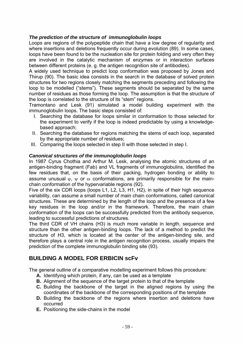
The prediction of the structure of immunoglobulin loops Loops are regions of the polypeptide chain that have a low degree of regularity and where insertions and deletions frequently occur during evolution (89). In some cases, loops have been found to be the nucleation site for protein folding and very often they are involved in the catalytic mechanism of enzymes or in interaction surfaces between different proteins (e. g. the antigen recognition site of antibodies). A widely used technique to predict loop conformation was proposed by Jones and Thirup (90). The basic idea consists in the search in the database of solved protein structures for two regions closely matching the segments preceding and following the loop to be modelled (“stems”). These segments should be separated by the same number of residues as those forming the loop. The assumption is that the structure of the loop is correlated to the structure of its “stem” regions. Tramontano and Lesk (91) simulated a model building experiment with the immunoglobulin loops. The basic steps consisted of:
I. Searching the database for loops similar in conformation to those selected for the experiment to verify if the loop is indeed predictable by using a knowledge-based approach;
II. Searching the database for regions matching the stems of each loop, separated by the appropriate number of residues;
III. Comparing the loops selected in step II with those selected in step I. Canonical structures of the immunoglobulin loops In 1987 Cyrus Chothia and Arthur M. Lesk, analysing the atomic structures of an antigen-binding fragment (Fab) and VL fragments of immunoglobulins, identified the few residues that, on the basis of their packing, hydrogen bonding or ability to assume unusual ϕ, ψ or ω conformations, are primarily responsible for the main-chain conformation of the hypervariable regions (92). Five of the six CDR loops (loops L1, L2, L3, H1, H2), in spite of their high sequence variability, can assume a small number of main chain conformations, called canonical structures. These are determined by the length of the loop and the presence of a few key residues in the loop and/or in the framework. Therefore, the main chain conformation of the loops can be successfully predicted from the antibody sequence, leading to successful predictions of structures. The third CDR of VH chains (H3) is much more variable in length, sequence and structure than the other antigen-binding loops. The lack of a method to predict the structure of H3, which is located at the center of the antigen-binding site, and therefore plays a central role in the antigen recognition process, usually impairs the prediction of the complete immunoglobulin binding site (93). BUILDING A MODEL FOR ERBICIN scFv The general outline of a comparative modelling experiment follows this procedure:
A. Identifying which protein, if any, can be used as a template B. Alignment of the sequence of the target protein to that of the template C. Building the backbone of the target in the aligned regions by using the
coordinates of the backbone of the corresponding positions of the template D. Building the backbone of the regions where insertion and deletions have
occurred E. Positioning the side-chains in the model
- 59 -
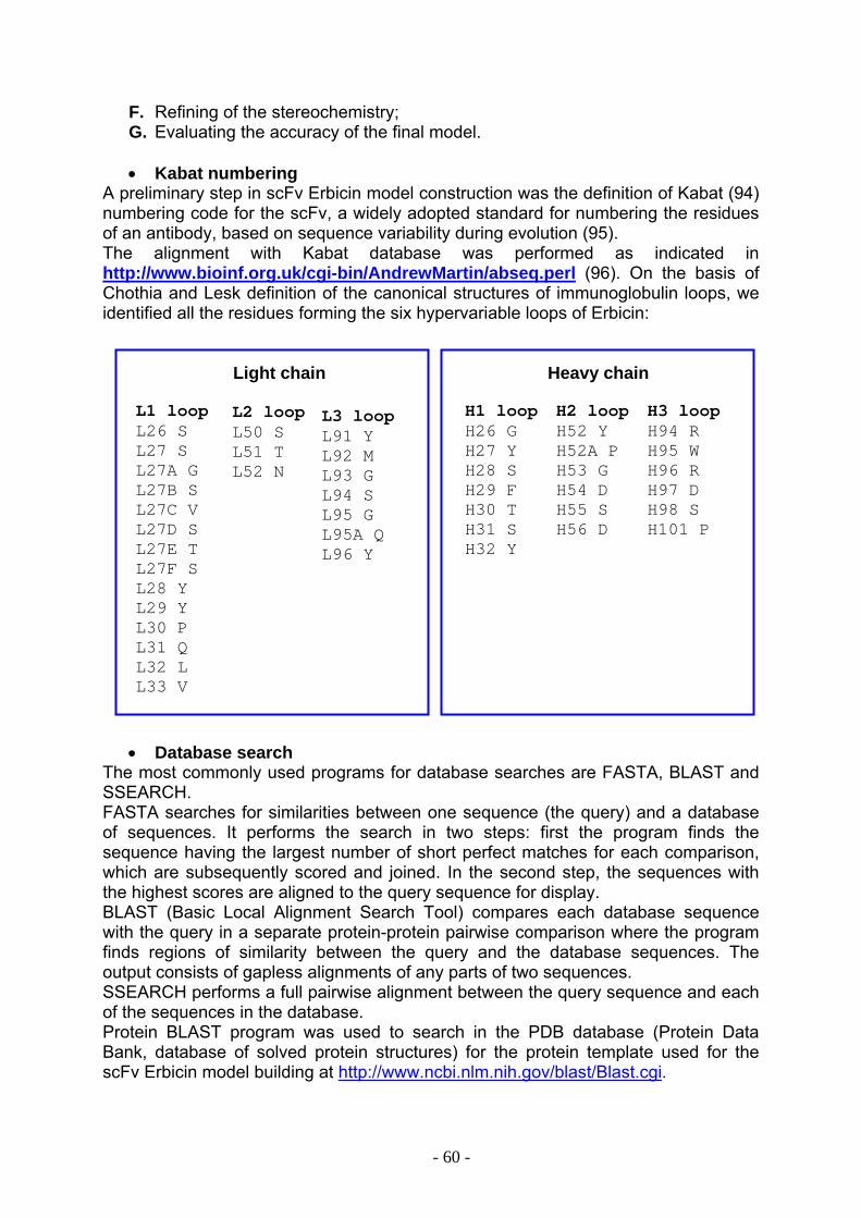
F. Refining of the stereochemistry; G. Evaluating the accuracy of the final model.
• Kabat numbering
A preliminary step in scFv Erbicin model construction was the definition of Kabat (94) numbering code for the scFv, a widely adopted standard for numbering the residues of an antibody, based on sequence variability during evolution (95). The alignment with Kabat database was performed as indicated in http://www.bioinf.org.uk/cgi-bin/AndrewMartin/abseq.perl (96). On the basis of Chothia and Lesk definition of the canonical structures of immunoglobulin loops, we identified all the residues forming the six hypervariable loops of Erbicin: Light chain Heavy chain
L1 loop L26 S L27 S L27A G L27B S L27C V L27D S L27E T L27F S L28 Y L29 Y L30 P L31 Q L32 L L33 V
H2 loop H52 Y H52A P H53 G H54 D H55 S H56 D
H3 loopH94 R H95 W H96 R H97 D H98 S H101 P
H1 loopH26 G H27 Y H28 S H29 F H30 T H31 S H32 Y
L2 loop L3 loopL91 Y L92 M L93 G L94 S L95 G L95A Q L96 Y
L50 S L51 T L52 N
• Database search
The most commonly used programs for database searches are FASTA, BLAST and SSEARCH. FASTA searches for similarities between one sequence (the query) and a database of sequences. It performs the search in two steps: first the program finds the sequence having the largest number of short perfect matches for each comparison, which are subsequently scored and joined. In the second step, the sequences with the highest scores are aligned to the query sequence for display. BLAST (Basic Local Alignment Search Tool) compares each database sequence with the query in a separate protein-protein pairwise comparison where the program finds regions of similarity between the query and the database sequences. The output consists of gapless alignments of any parts of two sequences. SSEARCH performs a full pairwise alignment between the query sequence and each of the sequences in the database. Protein BLAST program was used to search in the PDB database (Protein Data Bank, database of solved protein structures) for the protein template used for the scFv Erbicin model building at http://www.ncbi.nlm.nih.gov/blast/Blast.cgi.
- 60 -
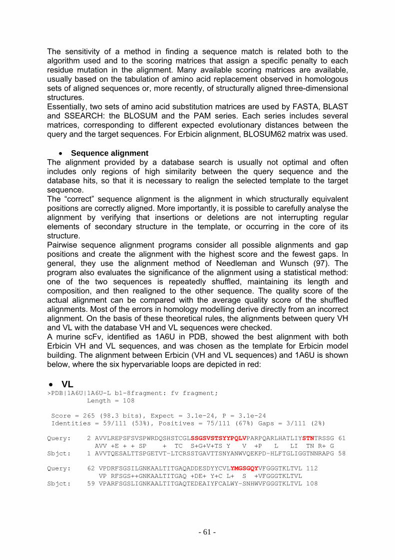
The sensitivity of a method in finding a sequence match is related both to the algorithm used and to the scoring matrices that assign a specific penalty to each residue mutation in the alignment. Many available scoring matrices are available, usually based on the tabulation of amino acid replacement observed in homologous sets of aligned sequences or, more recently, of structurally aligned three-dimensional structures. Essentially, two sets of amino acid substitution matrices are used by FASTA, BLAST and SSEARCH: the BLOSUM and the PAM series. Each series includes several matrices, corresponding to different expected evolutionary distances between the query and the target sequences. For Erbicin alignment, BLOSUM62 matrix was used.
• Sequence alignment The alignment provided by a database search is usually not optimal and often includes only regions of high similarity between the query sequence and the database hits, so that it is necessary to realign the selected template to the target sequence. The “correct” sequence alignment is the alignment in which structurally equivalent positions are correctly aligned. More importantly, it is possible to carefully analyse the alignment by verifying that insertions or deletions are not interrupting regular elements of secondary structure in the template, or occurring in the core of its structure. Pairwise sequence alignment programs consider all possible alignments and gap positions and create the alignment with the highest score and the fewest gaps. In general, they use the alignment method of Needleman and Wunsch (97). The program also evaluates the significance of the alignment using a statistical method: one of the two sequences is repeatedly shuffled, maintaining its length and composition, and then realigned to the other sequence. The quality score of the actual alignment can be compared with the average quality score of the shuffled alignments. Most of the errors in homology modelling derive directly from an incorrect alignment. On the basis of these theoretical rules, the alignments between query VH and VL with the database VH and VL sequences were checked. A murine scFv, identified as 1A6U in PDB, showed the best alignment with both Erbicin VH and VL sequences, and was chosen as the template for Erbicin model building. The alignment between Erbicin (VH and VL sequences) and 1A6U is shown below, where the six hypervariable loops are depicted in red: • VL >PDB|1A6U|1A6U-L b1-8fragment: fv fragment; Length = 108 Score = 265 (98.3 bits), Expect = 3.1e-24, P = 3.1e-24 Identities = 59/111 (53%), Positives = 75/111 (67%) Gaps = 3/111 (2%) Query: 2 AVVLREPSFSVSPWRDQSHSTCGLSSGSVSTSYYPQLVPARPQARLHATLIYSTNTRSSG 61 AVV +E + + SP + TC S+G+V+TS Y V +P L LI TN R+ G Sbjct: 1 AVVTQESALTTSPGETVT-LTCRSSTGAVTTSNYANWVQEKPD-HLFTGLIGGTNNRAPG 58 Query: 62 VPDRFSGSILGNKAALTITGAQADDESDYYCVLYMGSGQYVFGGGTKLTVL 112 VP RFSGS++GNKAALTITGAQ +DE+ Y+C L+ S +VFGGGTKLTVL Sbjct: 59 VPARFSGSLIGNKAALTITGAQTEDEAIYFCALWY-SNHWVFGGGTKLTVL 108
- 61 -
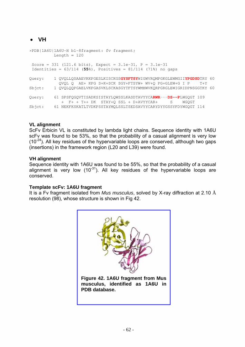
• VH >PDB|1A6U|1A6U-H b1-8fragment: fv fragment; Length = 120 Score = 331 (121.6 bits), Expect = 3.1e-31, P = 3.1e-31 Identities = 63/114 (55%), Positives = 81/114 (71%) no gaps Query: 1 QVQLLQSAAEVKKPGESLKISCKGSGYSFTSYWIGWVRQMPGKGLEWMGIIYPGDSDTRY 60 QVQL Q AE+ KPG S+K+SCK SGY+FTSYW+ WV+Q PG+GLEW+G I P T+Y Sbjct: 1 QVQLQQPGAELVKPGASVKLSCKASGYTFTSYWMHWVKQRPGRGLEWIGRIDPNSGGTKY 60 Query: 61 SPSFQGQVTISADKSISTAYLQWSSLKASDTAVYYCARWR---DS--PLWGQGT 109 + F+ + T++ DK STAY+Q SSL + D+AVYYCAR+ S WGQGT Sbjct: 61 NEKFKSKATLTVDKPSSTAYMQLSSLTSEDSAVYYCARYDYYGSSYFDYWGQGT 114
VL alignment ScFv Erbicin VL is constituted by lambda light chains. Sequence identity with 1A6U scFv was found to be 53%, so that the probability of a casual alignment is very low (10-24). All key residues of the hypervariable loops are conserved, although two gaps (insertions) in the framework region (L20 and L39) were found. VH alignment Sequence identity with 1A6U was found to be 55%, so that the probability of a casual alignment is very low (10-31). All key residues of the hypervariable loops are conserved. Template scFv: 1A6U fragment It is a Fv fragment isolated from Mus musculus, solved by X-ray diffraction at 2.10 Å resolution (98), whose structure is shown in Fig 42.
Figure 42. 1A6U fragment from Mus musculus, identified as 1A6U in PDB database.
- 62 -
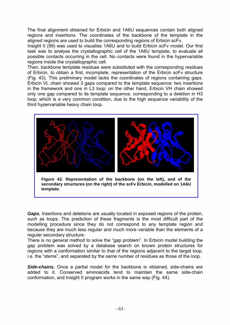
The final alignment obtained for Erbicin and 1A6U sequences contain both aligned regions and insertions. The coordinates of the backbone of the template in the aligned regions are used to build the corresponding regions of Erbicin scFv. Insight II (99) was used to visualise 1A6U and to build Erbicin scFv model. Our first task was to analyse the crystallographic cell of the 1A6U template, to evaluate all possible contacts occurring in the cell. No contacts were found in the hypervariable regions inside the crystallographic cell. Then, backbone template residues were substituted with the corresponding residues of Erbicin, to obtain a first, incomplete, representation of the Erbicin scFv structure (Fig. 43). This preliminary model lacks the coordinates of regions containing gaps. Erbicin VL chain showed 3 gaps compared to the template sequence: two insertions in the framework and one in L3 loop; on the other hand, Erbicin VH chain showed only one gap compared to its template sequence, corresponding to a deletion in H3 loop, which is a very common condition, due to the high sequence variability of the third hypervariable heavy chain loop.
Figure 43. Representation of the backbone (on the left), and of the secondary structures (on the right) of the scFv Erbicin, modelled on 1A6U template.
Gaps. Insertions and deletions are usually located in exposed regions of the protein, such as loops. The prediction of these fragments is the most difficult part of the modelling procedure since they do not correspond to any template region and because they are much less regular and much more variable than the elements of a regular secondary structure. There is no general method to solve the “gap problem”. In Erbicin model building the gap problem was solved by a database search on known protein structures for regions with a conformation similar to that of the regions adjacent to the target loop, i.e. the “stems”, and separated by the same number of residues as those of the loop. Side-chains. Once a partial model for the backbone is obtained, side-chains are added to it. Conserved aminoacids tend to maintain the same side-chain conformation, and Insight II program works in the same way (Fig. 44).
- 63 -
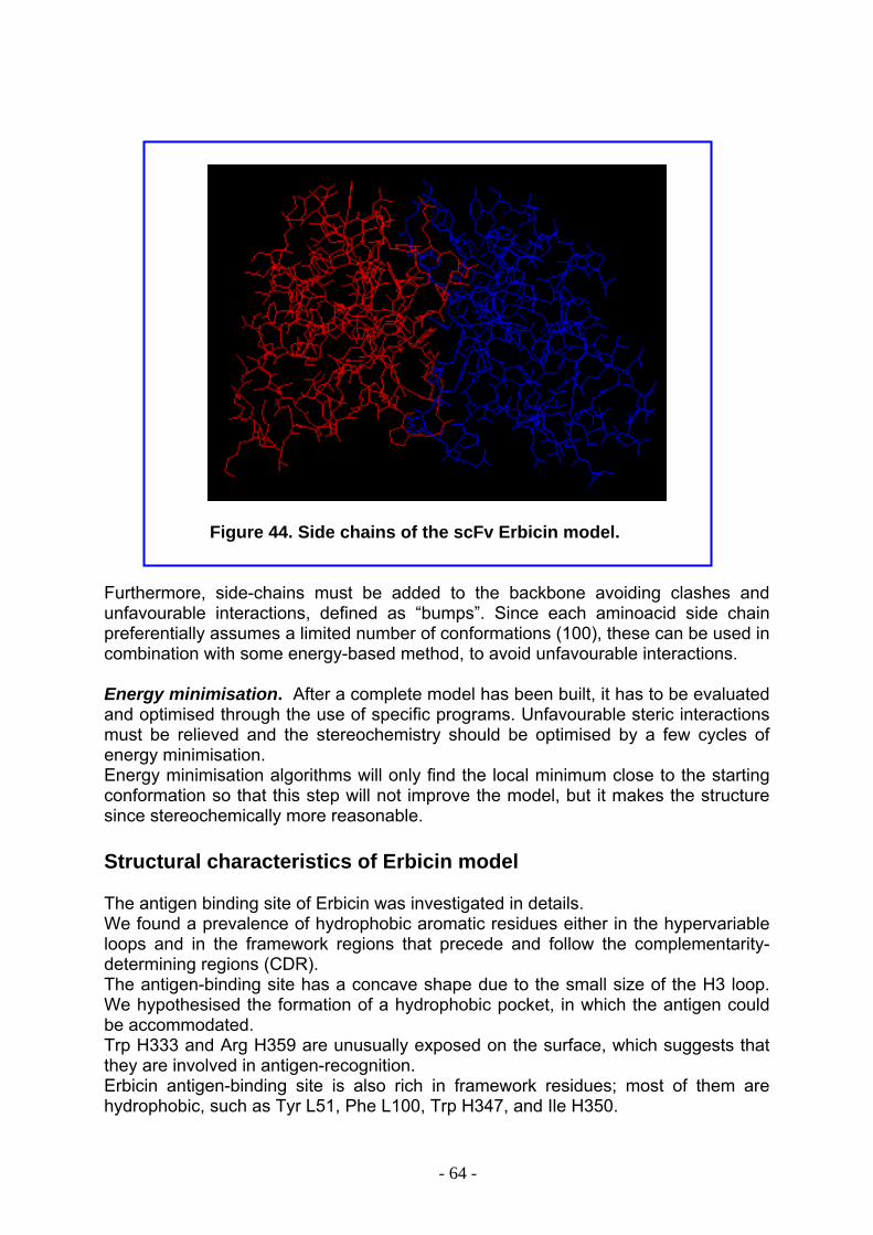
Figure 44. Side chains of the scFv Erbicin model. Furthermore, side-chains must be added to the backbone avoiding clashes and unfavourable interactions, defined as “bumps”. Since each aminoacid side chain preferentially assumes a limited number of conformations (100), these can be used in combination with some energy-based method, to avoid unfavourable interactions. Energy minimisation. After a complete model has been built, it has to be evaluated and optimised through the use of specific programs. Unfavourable steric interactions must be relieved and the stereochemistry should be optimised by a few cycles of energy minimisation. Energy minimisation algorithms will only find the local minimum close to the starting conformation so that this step will not improve the model, but it makes the structure since stereochemically more reasonable. Structural characteristics of Erbicin model The antigen binding site of Erbicin was investigated in details. We found a prevalence of hydrophobic aromatic residues either in the hypervariable loops and in the framework regions that precede and follow the complementarity-determining regions (CDR). The antigen-binding site has a concave shape due to the small size of the H3 loop. We hypothesised the formation of a hydrophobic pocket, in which the antigen could be accommodated. Trp H333 and Arg H359 are unusually exposed on the surface, which suggests that they are involved in antigen-recognition. Erbicin antigen-binding site is also rich in framework residues; most of them are hydrophobic, such as Tyr L51, Phe L100, Trp H347, and Ile H350.
- 64 -
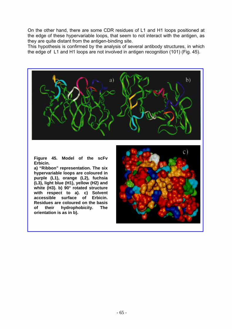
On the other hand, there are some CDR residues of L1 and H1 loops positioned at the edge of these hypervariable loops, that seem to not interact with the antigen, as they are quite distant from the antigen-binding site. This hypothesis is confirmed by the analysis of several antibody structures, in which the edge of L1 and H1 loops are not involved in antigen recognition (101) (Fig. 45).
Figure 45. Model of the scFv Erbicin. a) “Ribbon” representation. The six hypervariable loops are coloured in purple (L1), orange (L2), fuchsia (L3), light blue (H1), yellow (H2) and white (H3). b) 90° rotated structure with respect to a). c) Solvent accessible surface of Erbicin. Residues are coloured on the basis of their hydrophobicity. The orientation is as in b).
- 65 -
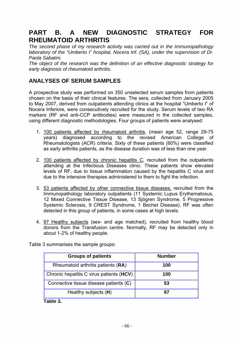
PART B. A NEW DIAGNOSTIC STRATEGY FOR RHEUMATOID ARTHRITIS The second phase of my research activity was carried out in the Immunopathology laboratory of the “Umberto I” hospital, Nocera Inf. (SA), under the supervision of Dr. Paola Sabatini. The object of the research was the definition of an effective diagnostic strategy for early diagnosis of rheumatoid arthritis. ANALYSES OF SERUM SAMPLES A prospective study was performed on 350 unselected serum samples from patients chosen on the basis of their clinical features. The sera, collected from January 2005 to May 2007, derived from outpatients attending clinics at the hospital “Umberto I” of Nocera Inferiore, were consecutively recruited for the study. Serum levels of two RA markers (RF and anti-CCP antibodies) were measured in the collected samples, using different diagnostic methodologies. Four groups of patients were analysed:
1. 100 patients affected by rheumatoid arthritis, (mean age 52, range 29-75 years) diagnosed according to the revised American College of Rheumatologists (ACR) criteria. Sixty of these patients (60%) were classified as early arthritis patients, as the disease duration was of less than one year.
2. 100 patients affected by chronic hepatitis C, recruited from the outpatients
attending at the Infectious Diseases clinic. These patients show elevated levels of RF, due to tissue inflammation caused by the hepatitis C virus and due to the intensive therapies administered to them to fight the infection.
3. 53 patients affected by other connective tissue diseases, recruited from the
Immunopathology laboratory outpatients (11 Systemic Lupus Erythematosus, 12 Mixed Connective Tissue Disease, 13 Sjögren Syndrome, 5 Progressive Systemic Sclerosis, 9 CREST Syndrome, 1 Bechet Disease). RF was often detected in this group of patients, in some cases at high levels.
4. 97 Healthy subjects (sex- and age matched), recruited from healthy blood
donors from the Transfusion centre. Normally, RF may be detected only in about 1-2% of healthy people.
Table 3 summarises the sample groups:
Groups of patients Number
Rheumatoid arthritis patients (RA) 100
Chronic hepatitis C virus patients (HCV) 100
Connective tissue disease patients (C) 53
Healthy subjects (H) 97
Table 3.
- 66 -
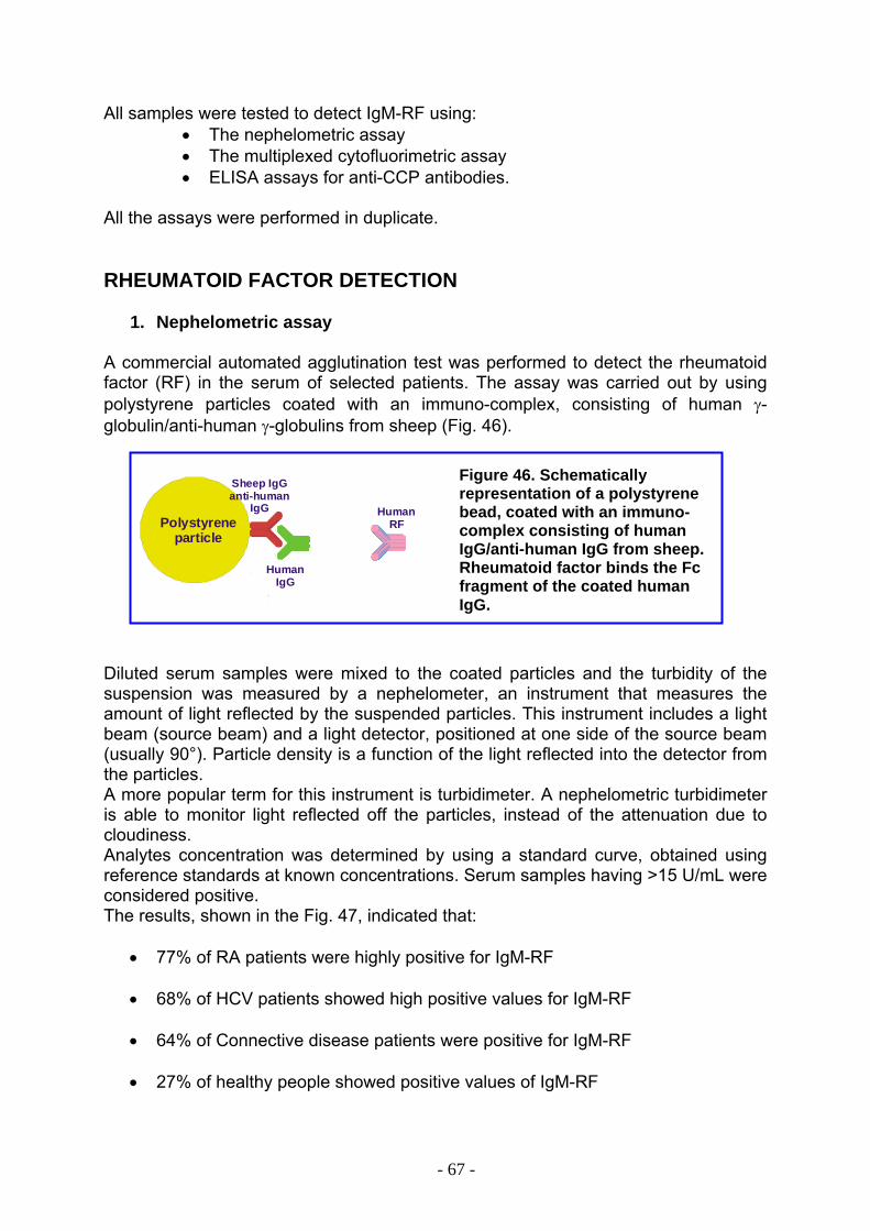
All samples were tested to detect IgM-RF using: • The nephelometric assay • The multiplexed cytofluorimetric assay • ELISA assays for anti-CCP antibodies.
All the assays were performed in duplicate.
RHEUMATOID FACTOR DETECTION
1. Nephelometric assay A commercial automated agglutination test was performed to detect the rheumatoid factor (RF) in the serum of selected patients. The assay was carried out by using polystyrene particles coated with an immuno-complex, consisting of human γ-globulin/anti-human γ-globulins from sheep (Fig. 46). Figure 46. Schematically
representation of a polystyrene bead, coated with an immuno-complex consisting of human IgG/anti-human IgG from sheep. Rheumatoid factor binds the Fc fragment of the coated human IgG.
Polystyrene particle
HumanIgG
Sheep IgGanti-human
IgG HumanRF
Diluted serum samples were mixed to the coated particles and the turbidity of the suspension was measured by a nephelometer, an instrument that measures the amount of light reflected by the suspended particles. This instrument includes a light beam (source beam) and a light detector, positioned at one side of the source beam (usually 90°). Particle density is a function of the light reflected into the detector from the particles. A more popular term for this instrument is turbidimeter. A nephelometric turbidimeter is able to monitor light reflected off the particles, instead of the attenuation due to cloudiness. Analytes concentration was determined by using a standard curve, obtained using reference standards at known concentrations. Serum samples having >15 U/mL were considered positive. The results, shown in the Fig. 47, indicated that:
• 77% of RA patients were highly positive for IgM-RF
• 68% of HCV patients showed high positive values for IgM-RF
• 64% of Connective disease patients were positive for IgM-RF
• 27% of healthy people showed positive values of IgM-RF
- 67 -
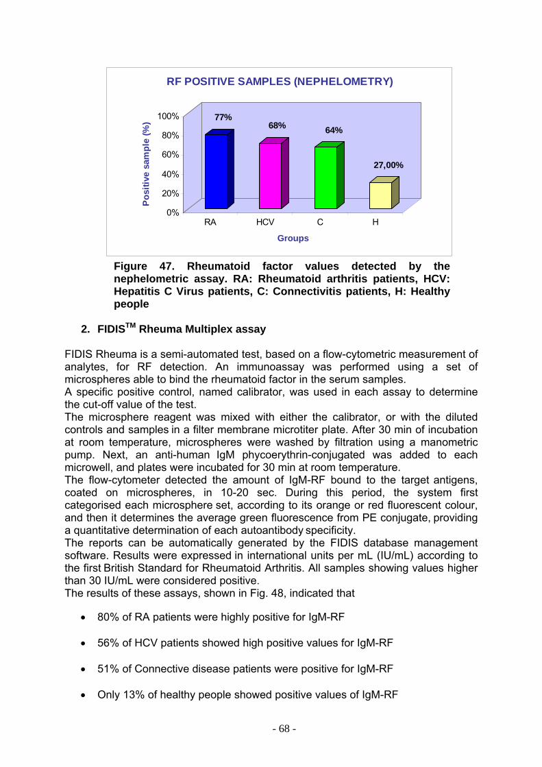
77%68% 64%
27,00%
0%
20%
40%
60%
80%
100%
Posi
tive
sam
ple
(%)
RA HCV C H
Groups
RF POSITIVE SAMPLES (NEPHELOMETRY)
Figure 47. Rheumatoid factor values detected by the nephelometric assay. RA: Rheumatoid arthritis patients, HCV: Hepatitis C Virus patients, C: Connectivitis patients, H: Healthy people
2. FIDISTM Rheuma Multiplex assay
FIDIS Rheuma is a semi-automated test, based on a flow-cytometric measurement of analytes, for RF detection. An immunoassay was performed using a set of microspheres able to bind the rheumatoid factor in the serum samples. A specific positive control, named calibrator, was used in each assay to determine the cut-off value of the test. The microsphere reagent was mixed with either the calibrator, or with the diluted controls and samples in a filter membrane microtiter plate. After 30 min of incubation
at room temperature, microspheres were washed by filtration using a manometric pump. Next, an anti-human IgM phycoerythrin-conjugated was added to each microwell, and plates were incubated for 30 min at room temperature. The flow-cytometer detected the amount of IgM-RF bound to the target antigens, coated on microspheres, in 10-20 sec. During this period, the system first categorised each microsphere set, according to its orange or red fluorescent colour, and then it determines the average green fluorescence from PE conjugate, providing a quantitative determination of each autoantibody specificity. The reports can be automatically generated by the FIDIS database management software. Results were expressed in international units per mL (IU/mL) according to the first British Standard for Rheumatoid Arthritis. All samples showing values higher than 30 IU/mL were considered positive. The results of these assays, shown in Fig. 48, indicated that
• 80% of RA patients were highly positive for IgM-RF
• 56% of HCV patients showed high positive values for IgM-RF
• 51% of Connective disease patients were positive for IgM-RF
• Only 13% of healthy people showed positive values of IgM-RF
- 68 -
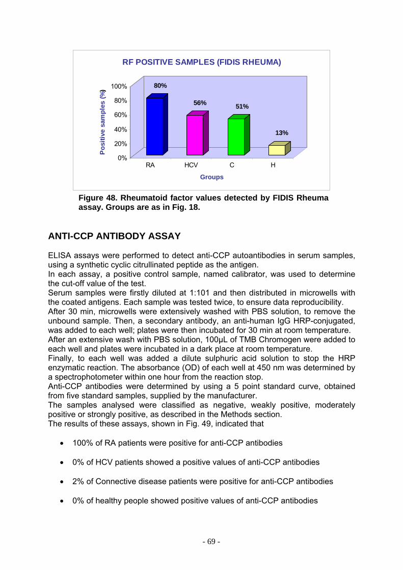
80%
56% 51%
13%
0%
20%
40%
60%
80%
100%
Pos
itive
sam
ples
(%)
RA HCV C H
Groups
RF POSITIVE SAMPLES (FIDIS RHEUMA)
Figure 48. Rheumatoid factor values detected by FIDIS Rheuma assay. Groups are as in Fig. 18.
ANTI-CCP ANTIBODY ASSAY ELISA assays were performed to detect anti-CCP autoantibodies in serum samples, using a synthetic cyclic citrullinated peptide as the antigen. In each assay, a positive control sample, named calibrator, was used to determine the cut-off value of the test. Serum samples were firstly diluted at 1:101 and then distributed in microwells with the coated antigens. Each sample was tested twice, to ensure data reproducibility. After 30 min, microwells were extensively washed with PBS solution, to remove the unbound sample. Then, a secondary antibody, an anti-human IgG HRP-conjugated, was added to each well; plates were then incubated for 30 min at room temperature. After an extensive wash with PBS solution, 100µL of TMB Chromogen were added to each well and plates were incubated in a dark place at room temperature. Finally, to each well was added a dilute sulphuric acid solution to stop the HRP enzymatic reaction. The absorbance (OD) of each well at 450 nm was determined by a spectrophotometer within one hour from the reaction stop. Anti-CCP antibodies were determined by using a 5 point standard curve, obtained from five standard samples, supplied by the manufacturer. The samples analysed were classified as negative, weakly positive, moderately positive or strongly positive, as described in the Methods section. The results of these assays, shown in Fig. 49, indicated that
• 100% of RA patients were positive for anti-CCP antibodies • 0% of HCV patients showed a positive values of anti-CCP antibodies
• 2% of Connective disease patients were positive for anti-CCP antibodies
• 0% of healthy people showed positive values of anti-CCP antibodies
- 69 -
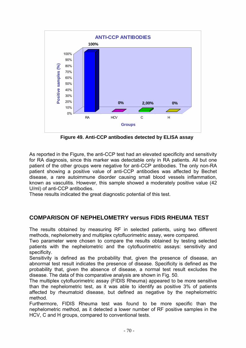
100%
0% 2,00% 0%
0%
10%
20%
30%
40%
50%
60%
70%
80%
90%
100%Po
sitiv
e sa
mpl
es (%
)
RA HCV C H
Groups
ANTI-CCP ANTIBODIES
Figure 49. Anti-CCP antibodies detected by ELISA assay
As reported in the Figure, the anti-CCP test had an elevated specificity and sensitivity for RA diagnosis, since this marker was detectable only in RA patients. All but one patient of the other groups were negative for anti-CCP antibodies. The only non-RA patient showing a positive value of anti-CCP antibodies was affected by Bechet disease, a rare autoimmune disorder causing small blood vessels inflammation, known as vasculitis. However, this sample showed a moderately positive value (42 U/ml) of anti-CCP antibodies. These results indicated the great diagnostic potential of this test. COMPARISON OF NEPHELOMETRY versus FIDIS RHEUMA TEST The results obtained by measuring RF in selected patients, using two different methods, nephelometry and multiplex cytofluorimetric assay, were compared. Two parameter were chosen to compare the results obtained by testing selected patients with the nephelometric and the cytofluorimetric assays: sensitivity and specificity. Sensitivity is defined as the probability that, given the presence of disease, an abnormal test result indicates the presence of disease. Specificity is defined as the probability that, given the absence of disease, a normal test result excludes the disease. The data of this comparative analysis are shown in Fig. 50. The multiplex cytofluorimetric assay (FIDIS Rheuma) appeared to be more sensitive than the nephelometric test, as it was able to identify as positive 3% of patients affected by rheumatoid disease, but defined as negative by the nephelometric method. Furthermore, FIDIS Rheuma test was found to be more specific than the nephelometric method, as it detected a lower number of RF positive samples in the HCV, C and H groups, compared to conventional tests.
- 70 -
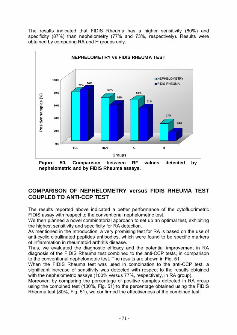
The results indicated that FIDIS Rheuma has a higher sensitivity (80%) and specificity (87%) than nephelometry (77% and 73%, respectively). Results were obtained by comparing RA and H groups only.
77%80%
68%
56%64%
51%
27%
13%
0%
20%
40%
60%
80%
100%
Posi
tive
sam
ples
(%)
RA HCV C H
Groups
NEPHELOMETRY vs FIDIS RHEUMA TEST
NEPHELOMETRYFIDIS RHEUMA
Figure 50. Comparison between RF values detected by nephelometric and by FIDIS Rheuma assays.
COMPARISON OF NEPHELOMETRY versus FIDIS RHEUMA TEST COUPLED TO ANTI-CCP TEST The results reported above indicated a better performance of the cytofluorimetric FIDIS assay with respect to the conventional nephelometric test. We then planned a novel combinatorial approach to set up an optimal test, exhibiting the highest sensitivity and specificity for RA detection. As mentioned in the Introduction, a very promising test for RA is based on the use of anti-cyclic citrullinated peptides antibodies, which were found to be specific markers of inflammation in rheumatoid arthritis disease. Thus, we evaluated the diagnostic efficacy and the potential improvement in RA diagnosis of the FIDIS Rheuma test combined to the anti-CCP tests, in comparison to the conventional nephelometric test. The results are shown in Fig. 51. When the FIDIS Rheuma test was used in combination to the anti-CCP test, a significant increase of sensitivity was detected with respect to the results obtained with the nephelometric assays (100% versus 77%, respectively, in RA group). Moreover, by comparing the percentage of positive samples detected in RA group using the combined test (100%, Fig. 51) to the percentage obtained using the FIDIS Rheuma test (80%, Fig. 51), we confirmed the effectiveness of the combined test.
- 71 -
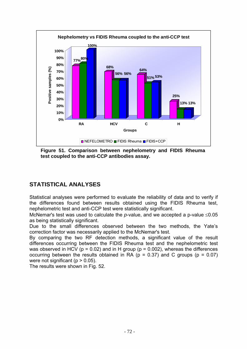
77%80%
100%
68%
56% 56%64%
51% 53%
25%
13% 13%
0%
10%
20%
30%
40%
50%
60%
70%
80%
90%
100%P
ositi
ve s
ampl
es (%
)
RA HCV C HGroups
Nephelometry vs FIDIS Rheuma coupled to the anti-CCP test
NEFELOMETRO FIDIS Rheuma FIDIS+CCP
Figure 51. Comparison between nephelometry and FIDIS Rheuma test coupled to the anti-CCP antibodies assay.
STATISTICAL ANALYSES Statistical analyses were performed to evaluate the reliability of data and to verify if the differences found between results obtained using the FIDIS Rheuma test, nephelometric test and anti-CCP test were statistically significant. McNemar's test was used to calculate the p-value, and we accepted a p-value ≤0.05 as being statistically significant. Due to the small differences observed between the two methods, the Yate’s correction factor was necessarily applied to the McNemar's test. By comparing the two RF detection methods, a significant value of the result differences occurring between the FIDIS Rheuma test and the nephelometric test was observed in HCV (p = 0.02) and in H group (p = 0.002), whereas the differences occurring between the results obtained in RA (p = 0.37) and C groups (p = 0.07) were not significant (p > 0.05). The results were shown in Fig. 52.
- 72 -
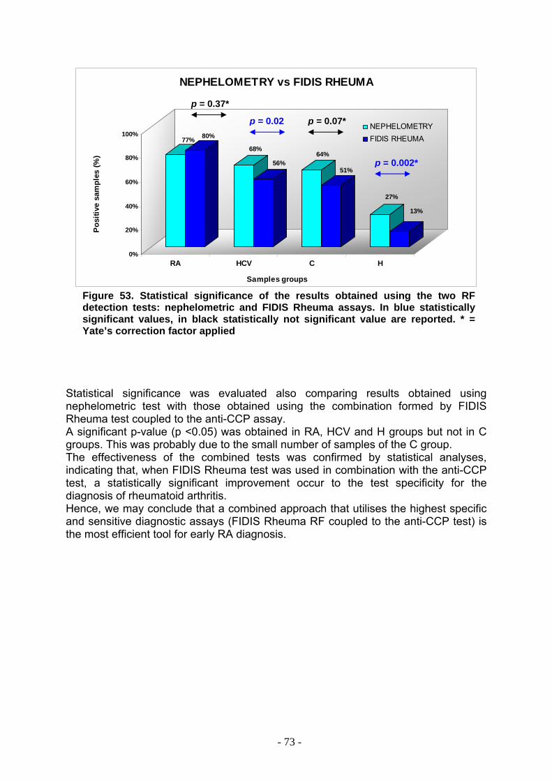
77% 80%
68%
56%64%
51%
27%
13%
0%
20%
40%
60%
80%
100%
Pos
itive
sam
ples
(%)
RA HCV C H
Samples groups
NEPHELOMETRY vs FIDIS RHEUMA
NEPHELOMETRYFIDIS RHEUMA
p = 0.37*
p = 0.02 p = 0.07*
p = 0.002*
Figure 53. Statistical significance of the results obtained using the two RF detection tests: nephelometric and FIDIS Rheuma assays. In blue statistically significant values, in black statistically not significant value are reported. * = Yate’s correction factor applied
Statistical significance was evaluated also comparing results obtained using nephelometric test with those obtained using the combination formed by FIDIS Rheuma test coupled to the anti-CCP assay. A significant p-value (p <0.05) was obtained in RA, HCV and H groups but not in C groups. This was probably due to the small number of samples of the C group. The effectiveness of the combined tests was confirmed by statistical analyses, indicating that, when FIDIS Rheuma test was used in combination with the anti-CCP test, a statistically significant improvement occur to the test specificity for the diagnosis of rheumatoid arthritis. Hence, we may conclude that a combined approach that utilises the highest specific and sensitive diagnostic assays (FIDIS Rheuma RF coupled to the anti-CCP test) is the most efficient tool for early RA diagnosis.
- 73 -
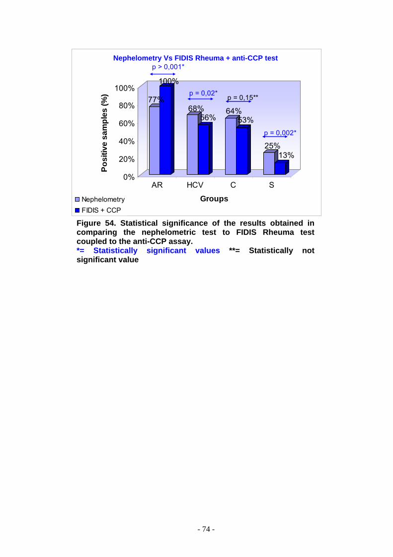
77%
100%
68%56%
64%53%
25%13%
0%
20%
40%
60%
80%
100%
Posi
tive
sam
ples
(%)
AR HCV C S
GroupsNephelometryFIDIS + CCP
p > 0,001*
p = 0,02* p = 0,15**
p = 0,002*
Nephelometry Vs FIDIS Rheuma + anti-CCP test
Figure 54. Statistical significance of the results obtained in comparing the nephelometric test to FIDIS Rheuma test coupled to the anti-CCP assay. *= Statistically significant values **= Statistically not significant value
- 74 -
DISCUSSION
A NEW TOOL FOR ANTICANCER THERAPY: THE ANTI-ErbB2 COMPACT ANTIBODY Conventional anti-cancer therapies, being characterised by the absence of specificity, may produce debilitating side effects, and sometimes may be ineffective. Immunotherapy represents a valuable alternative strategy to fight cancer, since it uses molecules of the immune system, mainly antibodies, specifically directed to selected cancer cells. Sometimes referred to as biological therapies, these new treatments have renewed interest and research activity in immunology. This is based on the evidence that antibodies can be specifically directed to target cancer cells, where they may induce direct cytotoxic effects, e.g. Fc-mediated cellular cytotoxicity, and they can also target cytotoxic molecules into cells. The current strategy in cancer immunotherapy research is based on the characterisation of new tumour-associated antigens (TAA), i.e. proteins expressed at high levels mostly on tumour cell surface. A good candidate as a tumour-associated antigen is the ErbB2 receptor, highly expressed in tumour cells of different origin, such as breast, ovary and lung carcinomas. The accessibility of ErbB2 on cell surface and its implications in the development of malignancy of these tumours make it an attractive target for immunotherapy. Several groups of researchers have produced different types of antibodies directed to ErbB2. A humanised version of an anti-ErbB2 antibody (Herceptin) was produced in 2000 and it is currently in use for the treatment of advanced breast cancer (27). Although the great potential of whole antibody molecules in the treatment of cancer, some limitations occurred in the use of engineered antibodies as therapeutic agents, such as the development of immune responses against humanised mAb and the low efficacy of antibody effector functions due to the large size of immunoglobulins (155 kDa), that makes difficult their penetration in solid tumours. To avoid these disadvantages, single-chain variable fragments (scFv) have been produced. These chimeric molecules retain the specificity of the original immunoglobulin, despite the removal of the constant regions. They are useful tools as immunotherapeutic agents, as they can easily penetrate solid tumours. Actually, fully human scFvs may be isolated using the phage-display technology, a method based on the construction of large repertoires of human antibody variable regions expressed on filamentous phages, and fused to a phage coat protein. Recently, in the laboratories of Proffs. Giuseppe D’Alessio, Renata Piccoli and Claudia De Lorenzo, a novel human anti-ErbB2 scFv has been isolated from a large phage-display library, through a double selection strategy, performed on live cells. This scFv, named Erbicin, showed interesting properties for immunotherapy. Erbicin was found to bind specifically to ErbB2-positive cells, to inhibit the receptor autophosphorylation and to be internalised in target cells where strongly inhibits their proliferation. However, the monovalent nature of the scFv as well as its supposed rapid clearance from the bloodstream, could limit its use as a therapeutic agent selective for mammary carcinoma cells.
- 75 -
To overcome these limitations, the aim of my research activity was to transform Erbicin scFv in a more stable molecule, by fusing Erbicin to a human Fc domain. The engineered antibody was defined “compact Ab”, i.e. an antibody molecule with the following features:
• molecular size of about 100 kDa, higher than a scFv molecule, but smaller than a whole antibody
• two binding-sites • potential prolonged clearance from the bloodstream • effector functions mediated by the Fc domain: ADCC, antibody-dependent
cellular cytotoxicity and CDC, complement-dependent cytotoxicity. To generate the compact antibody anti-ErbB2, the cDNA encoding the scFv Erbicin was cloned into the Signal pIgPlus plasmid, downstream to a human IgG1 Fc domain. The recombinant vector was then amplified in E.coli JM101 bacterial cells, and trasfected in CHO eukaryotic cells. Single clones, expressing the recombinant protein, were identified by electrophoresis analyses and ELISA assays, using anti-human Fc antibodies. A protein species, with the expected molecular weight of the chimeric protein and immuno-positive to anti-Fc antibodies, was identified. Large-scale experiments were then performed to produce the compact antibody in suitable amount to test its biological activity. The immunoagent was purified from the culture medium by a one-step purification procedure, using an immuno-affinity chromatography on a protein A-agarose column, and then analysed by SDS-PAGE and Western blotting. The compact antibody, isolated as a pure protein, was named Erb-hcAb. Analyses were then carried out to extensively characterise the compact Ab. Erb-hcAb features are summarised below:
• The compact antibody is expressed as a disulphide-linked dimer • Erb-hcAb binds selectively and with high affinity to ErbB2 over-expressing
cells • The apparent binding affinity of the compact antibody for the ErbB2 receptor
was found to be 1 nM, a value that confirms the high binding affinity of the compact Ab for its natural antigen
• When compared to Herceptin, a commonly used anticancer drug, Erb-hcAb was found to be able to recognise with higher affinity its antigen
When Erb-hcAb was tested on cell cultures, to analyse its effects on cell viability, it was found to be able to induce a strong and selective anti-proliferative action on target, i.e. ErbB2 expressing, cells. Erb-hcAb was found to be even more effective than Herceptin. No effects were instead detected when the compact Ab was tested on ErbB2-negative cell lines. On the basis of these results, we might conclude that Erb-hcAb has a high therapeutic potential, as it fully satisfies the conditions required for an effective anticancer agent:
I. it is a fully human immunoagent, thus low, if any, immunogenicity is expected to be elicited;
II. it recognises one of the most specific tumour-associated antigens (ErbB2) with high affinity;
- 76 -
III. it is effective in inhibiting tumour cell growth. This effect is selective for ErbB2-positive cells.
Moreover, I performed computational analyses aimed at building a model of Erbicin structure, which represents the antigen binding moiety of the compact Ab, using the homology modelling technique. This was relevant to define the topology of the antigen binding site, as well as to design strategies aimed at improving the immunoagent affinity for the ErbB2 receptor. These studies, carried out in the laboratories of Prof. Anna Tramontano, University of Rome, “La Sapienza”, provided an accurate model of the three dimensional structure of the scFv Erbicin. Taken together, these data suggest that Erb-hcAb is a promising new anticancer agent, and supports the concept that, following humanised monoclonal and scFvs, a third generation of immunoagents, the human compact antibodies, may represent the format of choice for the therapy of solid tumour. The new compact Ab was recently patented (Patent PCT/EP02/07671 on “Human mini-antibody cytotoxic for tumour cells which express the ErbB2 receptor” G.D’Alessio, R.Piccoli, C. De Lorenzo, D.B. Palmer, M.A. Ritter, owned by Biotechnol S.A., Oeiras, Portugal). A NEW STRATEGY FOR EARLY DIAGNOSIS OF RHEUMATOID ARTHRITIS. Rheumatoid Arthritis (RA) is an autoimmune disease that causes chronic inflammation of the joints. Autoimmune diseases are illnesses that occur when the body tissues are mistakenly attacked by its own immune system. Auto-antibodies are then produced, targeted to their own body tissues where they induce inflammation. Rheumatoid arthritis is referred to as a systemic illness, since it can affect multiple organs of the body. It affects approximately 0.5% of the world population. Although the cause of RA is still unknown, most rheumatologists agree that the earlier RA patients are treated, the better is their prognosis. This implies that an early diagnosis is crucial for therapy establishment and patient health. In 1987 the American College of Rheumatology (ACR) has defined in details the criteria for the diagnosis of rheumatoid arthritis. These criteria include the presence in the patient serum of the rheumatoid factor. Rheumatoid factor (RF) is a group of auto-antibodies reacting with various antigenic sites located on the Fc fragment of the IgG class immunoglobulins. The RF test is considered the basic screen and hallmark for RA, since approximately 80% of RA patients have positive RF values. Moreover, higher RF levels are also seen in more severe forms of the disease and this condition is a severe prognostic factor for patients. Actually, the amount of rheumatoid factor in blood is usually measured by the nephelometric test. Nephelometry is based on the principle that a dilute suspension of small particles will scatter light (usually a laser) passed through it. The amount of scattered light is determined by collecting the light at an angle of about 70 or 75 degrees. Recently, a new immunoassay for the simultaneous detection of IgM RF directed against human and animal IgG, the FIDIS Rheuma test, was set up. It is based on flow cytometric measurements of RF serum levels, using a set of 100 fluorescent uniform-size (5.5 µm) polystyrene microspheres, coated with the specific antigen.
- 77 -
Moreover, in recent years an emerging group of auto-antibodies has been described in RA, which may have a role in the development of rheumatoid arthritis: the anti-citrullinated peptides antibodies (ACPA). Citrullination, or peptidylarginine deimination, is a post-translational modification catalysed by a specific enzyme, peptidyl-arginine deaminase (PAD), in which the guanidine moiety of arginine is hydrolysed, and replaced by an oxygen atom. Due to the central role of these antibodies in RA, a biochemical test was developed to detect anti-citrulline antibodies in sera of RA patients: the anti-cyclic citrullinated peptides (anti-CCP) ELISA test. The aim of my research work was: (i) to evaluate the effectiveness of the FIDIS Rheuma test in comparison to the commercial nephelometric test for IgM-RF detection; (ii) to test the possibility to use anti-cyclic citrullinated peptide (anti-CCP) antibodies as early markers for rheumatoid arthritis. To do this, 350 unselected serum samples were tested by a prospective study to detect both RF, using multiplex and nephelometric methodologies, and anti-CCP antibodies. Four categories of patients were analysed: 100 patients affected by RA; 100 patients affected by hepatitis C virus chronic infection (HCV); 53 patients affected by other connective tissue disease (C); 97 healthy blood donors (H). Results indicated that the multiplex cytofluorimetric assay (FIDIS Rheuma) is more sensitive than the nephelometric test, as it was able to identify as positive 3% of patients affected by RA, but defined as negative by the nephelometric test. Furthermore, FIDIS Rheuma test resulted more specific than the nephelometric method, as it detects a lower number of RF positive samples in the HCV, C and H groups, compared to the conventional nephelometric test. Moreover, FIDIS Rheuma test shown higher sensitivity (80%) and specificity (87%) than nephelometry (77% and 73%, respectively). Furthermore, we demonstrated that anti-CCP test has elevated specificity and sensitivity for RA diagnosis, since this marker was detectable only in RA patients. This indicated that anti-CCP test is a very promising test for RA and a specific marker of inflammation. On the basis of these results, a novel combined approach was planned, to set up an optimal test, with the highest sensitivity and specificity for RA diagnosis. To this purpose, the FIDIS Rheuma test was combined to the anti-CCP test in order to improve their efficacy in RA diagnosis. The results indicated that, by combining the FIDIS Rheuma test to the anti-CCP test, a significant increase of sensitivity was detected with respect to the results obtained with the nephelometric assay (100% sensitivity versus 77%). Statistical analyses, performed using the McNemar's test, confirmed that a statistically significant (p-value <0.05) improvement to the test specificity for the diagnosis of rheumatoid arthritis occur when FIDIS Rheuma test was used in combination with the anti-CCP test. We conclude that the combined test (FIDIS Rheuma test coupled to anti-CCP test) is a successful approach to obtain the highest diagnostic specificity and sensitivity and to generate a useful tool for early RA diagnosis.
- 78 -
REFERENCES
1. Endicott, JA and Ling, V. (1989) The biochemistry of P-glycoprotein-mediated multidrug resistance. Annu. Rev. Biochem. 58,137-171
2. JM Ford and WN Hait. (1990) Pharmacology of drugs that alter multidrug resistance in cancer Pharmacol. Rev. 42, 155-199.
3. Trikha M, Yan L, Nakada MT. (2002) Monoclonal antibodies as therapeutics in oncology. Curr Opin Biotechnol. 13, 609-14.
4. Ross J, Gray K, Schenkein D, Greene B, Gray GS, Shulok J, Worland PJ, Celniker A, Rolfe M. (2003) Antibody-based therapeutics in oncology. Expert Rev Anticancer Ther. 3, 107-21.
5. Erlich P. Wiad Lek. (1967) 20, 1117-8. 6. Kohler G, Milstein C. (1975) Continuous cultures of fused cells secreting
antibody of predefined specificity. Nature 256, 495-7 7. Holliger P, Hoogenboom H. (1998) Antibodies come back from the brink.
Nat Biotechnol. 16, 1015-6. 8. Riechmann L, Clark M, Waldmann H, Winter G. (1988) Reshaping human
antibodies for therapy. Nature 332,323-7. 9. de Kruif J, van der Vuurst de Vries AR, Cilenti L, Boel E, van Ewijk W,
Logtenberg T. (1996) New perspectives on recombinant human antibodies. Immunol Today. 17, 453-5.
10. Yamamoto T, Ikawa S, Akiyama T, Semba K, Nomura N, Miyajima N, Saito T, Toyoshima K. (1986) Similarity of protein encoded by the human c-erb-B-2 gene to epidermal growth factor receptor. Nature. 319, 230-4.
11. Garrett TP, McKern NM, Lou M, Elleman TC, Adams TE, Lovrecz GO, Kofler M, Jorissen RN, Nice EC, Burgess AW, Ward CW. (2003) The crystal structure of a truncated ErbB2 ectodomain reveals an active conformation, poised to interact with other ErbB receptors. Mol Cell. 11,495-505.
12. Graus-Porta D, Beerli RR, Daly JM, Hynes NE. (1997) ErbB-2, the preferred heterodimerization partner of all ErbB receptors, is a mediator of lateral signaling. EMBO J. 16,1647-55.
13. CR King, MH Kraus, and SA Aaronson. (1985) Amplification of a novel v-erbB-related gene in a human mammary carcinoma. Science 4717, 974 – 976
14. DJ Slamon, W Godolphin, LA Jones, JA Holt, SG Wong, DE Keith, WJ Levin, SG Stuart, J Udove, A Ullrich, (1989) Studies of the HER-2/neu proto-oncogene in human breast and ovarian cancer. Science. 4905, 707 - 712
15. Fukushige S, Matsubara K, Yoshida M, Sasaki M, Suzuki T, Semba K, Toyoshima K, Yamamoto T. (1986) Localization of a novel v-erbB-related gene, c-erbB-2, on human chromosome 17 and its amplification in a gastric cancer cell line. Mol Cell Biol. 6, 955-8.
16. Semba K, Kamata N, Toyoshima K, Yamamoto T. (1985) A v-erbB-related protooncogene, c-erbB-2, is distinct from the c-erbB-1/epidermal growth factor-receptor gene and is amplified in a human salivary gland adenocarcinoma. Proc Natl Acad Sci U S A. 82, 6497-501.
- 79 -
17. Press MF, Cordon-Cardo C, Slamon DJ. (1990) Expression of the HER-2/neu proto-oncogene in normal human adult and fetal tissues.Oncogene. 5,953-62.
18. Tagliabue E, Centis F, Campiglio M, Mastroianni A, Martignone S, Pellegrini R, Casalini P, Lanzi C, Menard S, Colnaghi MI. (1991) Selection of monoclonal antibodies which induce internalization and phosphorylation of p185HER2 and growth inhibition of cells with HER2/NEU gene amplification. Int J Cancer. 47, 933-7.
19. Ullrich A, Schlessinger J. (1990) Signal transduction by receptors with tyrosine kinase activity. Cell. 61, 203-12.
20. Hudziak RM, Lewis GD, Shalaby MR, Eessalu TE, Aggarwal BB, Ullrich A, Shepard HM. (1988) Amplified expression of the HER2/ERBB2 oncogene induces resistance to tumor necrosis factor alpha in NIH 3T3 cells. Proc Natl Acad Sci U S A. 85, 5102-6.
21. Lee HR, Kim JH, Uhm HD, Ahn JB, Rha SY, Cho JY, Lee JI, Lee KH, Chung HC, Roh JK, Min JS, Lee KS, Shin DH, Kim BS, Hong SW, Choi JH. (1996) Overexpression of c-ErbB-2 protein in gastric cancer by immunohistochemical stain. Oncology. 53, 192-7.
22. Kita Y, Tseng J, Horan T, Wen J, Philo J, Chang D, Ratzkin B, Pacifici R, Brankow D, Hu S, Luo Y, Wen D, Arakawa T, Nicolson M. (1996) ErbB receptor activation, cell morphology changes, and apoptosis induced by anti-Her2 monoclonal antibodies. Biochem Biophys Res Commun. 226, 59-69.
23. Hudziak RM, Lewis GD, Winget M, Fendly BM, Shepard HM, Ullrich A. (1989) p185HER2 monoclonal antibody has antiproliferative effects in vitro and sensitizes human breast tumour cells to tumour necrosis factor . Mol Cell Biol. 9, 1165-72.
24. Harwerth IM, Wels W, Marte BM, Hynes NE. (1992) Monoclonal antibodies against the extracellular domain of the erbB-2 receptor function as partial ligand agonists. J Biol Chem. 267,15160-7.
25. Harwerth IM, Wels W, Schlegel J, Müller M, Hynes NE. (1993) Monoclonal antibodies directed to the erbB-2 receptor inhibit in vivo tumour cell growth. Br J Cancer. 68,1140-5.
26. Carter P, Presta L, Gorman CM, Ridgway JB, Henner D, Wong WL, Rowland AM, Kotts C, Carver ME, Shepard HM. (1992) Humanization of an anti-p185HER2 antibody for human cancer therapy. Proc Natl Acad Sci U S A. 89, 4285-9.
27. Carter P, Fendly BM, Lewis GD, Sliwkowski MX. (2000) Development of herceptin. Breast Dis. 11, 103-11.
28. Cho, H.S., Mason, K., Ramyar, K.X., Stanley, A.M., Gabelli, S.B., Denney Jr., D.W., Leahy, D.J. (2003) Structure of the Extracellular Region of HER2 alone and in complex with the Herceptin Fab. Nature 421, 756-760.
29. Lewis GD, Figari I, Fendly B, Wong WL, Carter P, Gorman C, Shepard HM. (1993) Differential responses of human tumor cell lines to anti-p185HER2 monoclonal antibodies. Cancer Immunol Immunother. 37, 255-63.
30. Baselga J, Norton L, Albanell J, Kim YM, Mendelsohn J. (1998) Recombinant humanized anti-HER2 antibody (Herceptin) enhances the antitumor activity of paclitaxel and doxorubicin against HER2/neu
- 80 -
overexpressing human breast cancer xenografts. Cancer Res. 58, 2825-2831.
31. Pegram MD, Lipton A, Hayes DF, Weber BL, Baselga JM, Tripathy D, Baly D, Baughman SA, Twaddell T, Glaspy JA, Slamon DJ. (1998) Phase II study of receptor-enhanced chemosensitivity using recombinant humanized anti-p185HER2/neu monoclonal antibody plus cisplatin in patients with HER2/neu-overexpressing metastatic breast cancer refractory to chemotherapy treatment. J Clin Oncol. 16, 2659-71.
32. De Lorenzo C, Nigro A, Piccoli R, D'Alessio G. (2002) A new RNase-based immunoconjugate selectively cytotoxic for ErbB2-overexpressing cells. FEBS Lett. 516, 208-12.
33. Sheets MD, Amersdorfer P, Finnern R, Sargent P, Lindquist E, Schier R, Hemingsen G, Wong C, Gerhart JC, Marks JD. (1998) Efficient construction of a large nonimmune phage antibody library: the production of high-affinity human single-chain antibodies to protein antigens. Proc. Natl. Acad. Sci. USA. 95, 6157-6162,
34. Poul MA, Becerril B, Nielsen UB, Morisson P, Marks JD. (2000) Selection of tumor-specific internalizing human antibodies from phage libraries. J. Mol. Biol. 301,1149-1161.
35. Marks JD, Hoogenboom HR, Bonnert TP, McCafferty J, Griffiths AD, Winter G. (1991) By-passing immunization. Human antibodies from V-gene libraries displayed on phage. J Mol Biol. 222, 581-97.
36. Griffiths AD, Williams SC, Hartley O, Tomlinson IM, Waterhouse P, Crosby WL, Kontermann RE, Jones PT, Low NM, Allison TJ, et al. (1994) Isolation of high affinity human antibodies directly from large synthetic repertoires. EMBO J. 13, 3245-60.
37. Li SL, Liang SJ, Guo N, Wu AM, Fujita-Yamaguchi Y. (2000) Single-chain antibodies against human insulin-like growth factor I receptor: expression, purification, and effect on tumor growth. Cancer Immunol Immunother. 49, 243-52.
38. Xu X, Clarke P, Szalai G, Shively JE, Williams LE, Shyr Y, Shi E, Primus FJ. (2000) Targeting and therapy of carcinoembryonic antigen-expressing tumors in transgenic mice with an antibody-interleukin 2 fusion protein. Cancer Res. 60, 4475-84.
39. Schier, R. et al, (1995) In vitro and in vivo characterisation of a human anti-c-erbB2 single chain Fv isolated from a filamentous phage antibody library. Immunotechnology. 1, 73-81.
40. Palmer DB, George AJ, Ritter MA. (1997) Selection of antibodies to cell surface determinants on mouse thymic epithelial cells using a phage display library. Immunology. 91, 473-8.
41. De Lorenzo C, Palmer DB, Piccoli R, Ritter MA, D'Alessio G. (2002) A new immunoreagent specific for ErbB2. Clin. Cancer Res. 8, 1710-1719.
42. Powers DB, Amersdorfer P, Poul M, Nielsen UB, Shalaby MR, Adams GP, Weiner LM, Marks JD. (2001) Expression of single-chain Fv-Fc fusions in Pichia pastoris. J Immunol Methods. 251, 123-35.
43. David Alan Isenberg. Oxford Textbook of Rheumatology. 2004.Third Edition. Edited by David Isenberg. Oxford University Press.
44. George C. Tsokos. 2000. Principles of Molecular Rheumatology. Humana Press.
- 81 -
45. Vliet Vlieland TP, van der Wijk TP, Jolie IM, Zwinderman AH, Hazes JM. (1996) Determinants of hand function in patients with rheumatoid arthritis. J Rheumatol. 5, 835-40.
46. Janeway C, Travers P, Walport M, Capra JD. (1999) Immunobiology: The Immune System in Health and Disease. New York, NY: Elsevier Science Ltd./Garland Publishing.
47. Landré-Beauvais AJ. The first description of rheumatoid arthritis. (2001) Unabridged text of the doctoral dissertation presented in 1800. Joint Bone Spine. 68, 130-43.
48. Goldbach-Mansky, R., J. Lee, A. McCoy, J. Hoxworth, C. Yarboro, J.S. Smolen, G. Steiner, A. Rosen, C. Zhang, H.A. Ménard, Z.J. Zhou, T. Palosuo, W.J. Van Venrooij, R.L. Wilder, J.H. Klippel, H.R. Schumacher, H.S. El-Gabalawy. (2000). Rheumatoid arthritis associated autoantibodies in patients with synovitis of recent onset. Arthritis Research. 2, 236-243.
49. Arnett F, Edworthy S, Bloch D, McShane D, Fries J, Cooper N, Healey L, Kaplan S, Liang M, Luthra H (1988). "The American Rheumatism Association 1987 revised criteria for the classification of rheumatoid arthritis.". Arthritis Rheum 31, 315-24.
50. Vital E, Emery P (2005). "Advances in the treatment of early rheumatoid arthritis.". Am Fam Physician 72, 1002-1004.
51. Savage C,St Clair EW. New therapeutics in rheumatoid arthritis. (2006) Rheum Dis Clin North Am. 32, 57-74.
52. Adam L. Corper, Maninder K. Sohi, Vincent R. Bonagura, Michael Steinitz, Royston Jefferis, Arnold Feinstein, Dennis Beale, Michael J. Taussig & Brian J. Sutton. (1997) Structure of human IgM rheumatoid factor Fab bound to its autoantigen IgG Fc reveals a novel topology of antibody—antigen interaction. Nature Structural Biology 4, 374 - 381
53. Egeland. T. & M. Munthe. (1983) The role of the laboratory in rheumatology: rheumatoid factors. Clin. Rheum. Dis. 9, 135-160.
54. Jones, M.G. et al. (1990) Specificity of rheumatoid factors in relation to the disease state in rheumatoid arthritis. Ann. Rheum. Dis. 49, 757-762.
55. Waaler, E. (1940) On the occurrence of a factor in human serum activating the specific agglutination of sheep blood corpuscles. Acta Pathol. Scand. 17, 172-178.
56. Rose, H.M. et al. (1948). Differential agglutination of normal and sensitized sheep erythrocytes by sera of patients with rheumatoid arthritis. Proc. Soc. Exp. Biol. Med. 68, 1-11.
57. Wolfe, F. et al. (1991) The latex test revisited. Arthritis Rheum. 34, 951-960.
58. Ailus, K. et al. (1991) Measuring rheumatoid factor in nonrheumatoid subjects: immunoturbidimetric assay, latex slide test, and enzyme linked immunosorbent assay compared. Clin. Chem. 37, 1766-1769.
59. González-Buitrago JM. (2006) Multiplexed testing in the autoimmunity laboratory. Clin Chem Lab Med. 44, 1169-74.
60. Fulton, R.J. et al. (1997) Advanced multiplexed analysis with the FlowMetrixTM system. Clin. Chem. 43, 1749-1756.
61. Nienhius RFL, Mandema EA. (1964) A new serum factor in patients with rheumatoid arthritis. The Antiperinuclear factor. Ann Rheum Dis. 23, 302-5.
62. Young BJ, Mallya RK, Leslie RD, Clark CJ, Hamblin TJ. (1979) Anti-keratin antibodies in rheumatoid arthritis. Br Med J. 2, 97-9.
- 82 -

63. Schellekens, G.A. et al. (2000) The diagnostic properties of rheumatoid arthritis antibodies recognizing a cyclic citrullinated peptide. Arthritis Rheum. 43, 155-163.
64. Schellekens GA, de Jong BA, van den Hoogen FH, van de Putte LB, van Venrooij WJ. (1998) Citrulline is an essential constituent of antigenic determinants recognized by rheumatoid arthritis-specific autoantibodies. J Clin Invest. 101, 273-81.
65. Girbal-Neuhauser E, Durieux JJ, Arnaud M, Dalbon P, Sebbag M, Vincent C, Simon M, Senshu T, Masson-Bessière C, Jolivet-Reynaud C, Jolivet M, Serre G. (1999) The epitopes targeted by the rheumatoid arthritis-associated antifilaggrin autoantibodies are posttranslationally generated on various sites of (pro)filaggrin by deimination of arginine residues. J Immunol. 162, 585-94.
66. Masson-Bessière C, Sebbag M, Girbal-Neuhauser E, Nogueira L, Vincent C, Senshu T, Serre G. (2001). The major synovial targets of the rheumatoid arthritis-specific antifilaggrin autoantibodies are deiminated forms of the alpha- and beta-chains of fibrin. J Immunol. 166, 4177-84.
67. Menard HA, Lapointe E, Rochdi MD, Zhou ZJ. (2000) Insights into rheumatoid arthritis derived from the Sa immune system. Arthritis Res. 2, 429-32.
68. van Venrooij WJ, Pruijn GJ. (2000) Citrullination: a small change for a protein with great consequences for rheumatoid arthritis.Arthritis Res. 2, 249-51.
69. Vossenaar ER, Nijenhuis S, Helsen MM, van der Heijden A, Senshu T, van den Berg WB, van Venrooij WJ, Joosten LA. (2003) Citrullination of synovial proteins in murine models of rheumatoid arthritis. Arthritis Rheum. 48, 2489-500.
70. van Venrooij WJ, Hazes JM, Visser H. (2002) Anticitrullinated protein/peptide antibody and its role in the diagnosis and prognosis of early rheumatoid arthritis. Neth J Med. 60, 383-8.
71. van Jaarsveld CH, ter Borg EJ, Jacobs JW, Schellekens GA, Gmelig-Meyling FH, van Booma-Frankfort C, de Jong BA, van Venrooij WJ, Bijlsma JW. (1999). The prognostic value of the antiperinuclear factor, anti-citrullinated peptide antibodies and rheumatoid factor in early rheumatoid arthritis. Clin Exp Rheumatol. Nov-Dec;17(6):689-97.
72. Paimela L, Palosuo T, Aho K, Lukka M, Kurki P, Leirisalo-Repo M, von Essen R. (2001). Association of autoantibodies to filaggrin with an active disease in early rheumatoid arthritis. Ann Rheum Dis. 60, 32-5.
73. Masson-Bessière C, Sebbag M, Durieux JJ, Nogueira L, Vincent C, Girbal-Neuhauser E, Durroux R, Cantagrel A, Serre G. (2000). In the rheumatoid pannus, anti-filaggrin autoantibodies are produced by local plasma cells and constitute a higher proportion of IgG than in synovial fluid and serum. Clin Exp Immunol. 119, 544-52.
74. Reparon-Schuijt CC, van Esch WJ, van Kooten C, Schellekens GA, de Jong BA, van Venrooij WJ, Breedveld FC, Verweij CL. (2001) Secretion of anti-citrulline-containing peptide antibody by B lymphocytes in rheumatoid arthritis. Arthritis Rheum. 44, 41-7.
75. Utz PJ, Hottelet M, Schur PH, Anderson P. (1997) Proteins phosphorylated during stress-induced apoptosis are common targets for autoantibody
- 83 -
production in patients with systemic lupus erythematosus. J Exp Med. 185, 843-54.
76. Asaga H, Yamada M, Senshu T. Selective deimination of vimentin in calcium ionophore-induced apoptosis of mouse peritoneal macrophages. (1998) Biochem Biophys Res Commun. 243, 641-6.
77. Erre GL, Tocco A, Faedda R, Cossu A, Carcassi A. (2004) Diagnostic and prognostic value of antibodies to cyclic citrullinated peptide (Anti-CCP) in rheumatoid arthritis. Reumatismo. 56, 118-23.
78. Messing, J., Recom. DNA Tech. Bull. (1979) 2 , 43 79. Laemmli, U. Nature 227, 680-685 (1970) 80. Sanbrook, K. et al., 1990. Molecular cloning: a laboratory manual. (Cold
Spring Harbor Laboratory Press, NY. 81. Salamov, A.A., and Solovyev, VV. (1995) Prediction of protein secondary
structure by combining nearest-neighbor algorithms and multiple sequence alignments. J. Mol. Biol. 247, 11-15.
82. Sippl, M.J., and Weitckus, S. (1992) Detection of native-like models for amino acid sequences of unknown three-dimensional structure in a data base of known protein conformations. Proteins 13, 258-271.
83. Godzik, A, Kolinski, A., and Skolnick, J. (1992) Topology fingerprint approach to the inverse protein folding problem. J. Mol. Biol. 227, 227-238
84. Anfinsen, C.B. (1973) Principles that govern the folding of protein chains. Science. 181, 223-230
85. Sanchez, R. and Sali, A. (1997) Advances in comparative protein-structure modelling. Curr. Opin. Struct. Biol. 7, 206-214
86. Chothia, C. and Lesk, A.M. (1986) The relation between the divergence of sequence and structure in proteins. EMBO J. 5, 823-826
87. Tramontano, A. (1998) Homology modelling with low sequence identity. Methods. 14, 293-300
88. Morea, V., Tramontano, A., Rustici, M., Chothia, C., Lesk, A.M. (1997) Antibody structure, prediction and redesign. Biophys. Chem. 68, 9-16
89. Pascarella S., & Argos, A. (1992) Analysis of insertion/deletions in protein structures. J. Mol. Biol. 224, 461-471.
90. Jones, T.A. & Thirup, S. (1986) Using known structures in protein model building and crystallography. EMBO J. 5, 819-822.
91. Tramontano, A. & Lesk, A.M. (1992), Common features of the conformations of the antigen-binding site in immunoglobulins and application to modelling loop conformations. Proteins. 13, 231-245.
92. Chothia, C. and Lesk, A.M. (1987) Canonical structures for the hypervariable regions of immunoglobulins. J. Mol. Biol. 196, 901-917.
93. Morea, V., Tramontano, A., Rustici, M., Chothia, C., Lesk, A.M. (1998) Conformations of the third hypervariable region in the VH domain of immunoglobulins. J. Mol. Biol. 275, 269-294.
94. Kabat, E.A., Wu, T.T., Bilofsky, H., Reid-Milner, M., Perry, H. Eds., Sequences of proteins of immunological interest. (U.S. Public Health Service, Washington, DC, 1983).
95. Johnson G, (2001) Kabat Database and its applications: future directions. Nucleic Acids Res. 29, 205-206.
96. Martin, A.C.R. (1996) Accessing the Kabat Antibody Sequence Database by Computer PROTEINS: Structure, Function and Genetics. 25, 130-133.
- 84 -
97. Needleman, S.B. and Wunsch, C.D. (1970) A general method applicable to the search for similarities in the amino acid sequence of two proteins. J. Mol. Biol. 48, 443-453
98. Simon,T. and Rajewsky,K. (1992) A functional antibody mutant with an insertion in the framework region 3 loop of the VH domain: implications for antibody engineering. Protein Eng. 5, 229-234
99. Dayringer HE, Tramontano A, Sprang SR, Fletterick RJ (1986). Interactive program for visualization and modeling of protein, nucleic acids and small molecules. J. Mol. Graphics 4, 82-87.
100. Dunbrack, R.L., ans Karplus, M. (1994) Conformational analysis of the backbone-dependent rotamer preferences of protein side-chains. Nature Struct. Biol. 1, 334-339.
101. Cohen, G.H., Silverton, E.W., Padlan, E.A., Dyda, F., Wibbenmeyer, J.A., Willson, R.C., Davies, D.R. (2005) Water molecules in the antibody-antigen interface of the structure of the Fab HyHEL-5-lysozyme complex at 1.7 A resolution: comparison with results from isothermal titration calorimetry. Acta Crystallogr.,Sect.D 61, 628-633.
- 85 -
Publications of Dr. Annarita Tedesco inherent to the Doctorate research activity
Publications: De Lorenzo C, Tedesco A, Terrazzano G, Cozzolino R, Laccetti P, Piccoli R, D’Alessio G. A human, compact, fully functional anti-ErbB2 antibody as a novel antitumour agent. (2004) Br. J. Cancer 91, 1200-1204. Comunications: 1. Diagnosi precoce dell’artrite reumatoide: verifica di una strategia attraverso
metodi diagnostici a confronto P.Sabatini , A.Tedesco, B.Sessa, P.Amato, P.Danise. 20° Congresso Nazionale SIMeL della Società italiana di Medicina di Laboratorio Altavilla Milicia (PA), 26-28 Ottobre 2006 2. Anticorpi anti-fattore intrinseco come marcatori per la diagnosi dell’anemia
perniciosa: un caso clinico Sabatini P., Tedesco A.R., Marchitiello R., Di Palma A., Esposito C., Aliperta A. 20° Congresso Nazionale SIMeL della Società italiana di Medicina di Laboratorio Altavilla Milicia (PA), 26-28 Ottobre 2006 3. The hemorrhagic lupus anticoagulant syndrome P. Sabatini, P. Danise, L.Clemente, A.Tedesco, C.Esposito, G. Rescigno,
G.Amendola. 12th International Congress on Antiphospholipid Antibodies Firenze, 18-20 Aprile 2007 4. Marcatori diagnostici nelle Early Arthritis Sabatini P., Tedesco A., D’Agostino D., Soriente I., Amato P. XLIV Congresso Nazionale SIR della Società Italiana di Reumatologia Venezia Lido, Italia 17-20 Ottobre 2007 5. Un insolito pattern fluoroscopico in pazienti trattati con Interferone di tipo I P.Sabatini , A. Tedesco, I. Soriente, L. Clemente, M. Corrado. 21° Congresso Nazionale SIMeL della Società italiana di Medicina di Laboratorio Riva del Garda (TN), 25-27 Ottobre 2007 6. Quanto sono frequenti le malattie del connettivo in pazienti con sclerosi
multipla? A. Tedesco, V. Brescia Morra, B.Sessa, M. Calabrese, P.Sabatini. 21° Congresso Nazionale SIMeL della Società italiana di Medicina di Laboratorio Riva del Garda (TN), 25-27 Ottobre 2007 7. Infezione da Epstein Barr virus- Mutazione V Leiden - Sindrome da Classe
economica: Un’ associazione esplosiva per la malattia tromboembolica. P. Danise, M. Maresca, G. Maresca, A. Tedesco, P.Sabatini 21° Congresso Nazionale SIMeL della Società italiana di Medicina di Laboratorio Riva del Garda (TN), 25-27 Ottobre 2007
- 86 -

RINGRAZIAMENTI
Desidero ringraziare calorosamente tutti coloro che hanno creduto in me, e che mi hanno aiutato affinché raggiungessi questo importantissimo traguardo. Ringrazio vivamente la Prof.ssa Renata Piccoli, per il sostegno costante e prezioso che in tutti questi anni mi ha sempre dimostrato, aiutandomi nei periodi più bui, regalandomi i suoi preziosissimi consigli ma soprattutto il suo affetto sincero. Ringrazio calorosamente la Dott.ssa Paola Sabatini, per essere stata una guida meravigliosa per me, durante il mio percorso formativo, e per l’affetto che mi ha sempre e in ogni modo dimostrato in questi tre anni di lavoro. Un ringraziamento speciale va alla Dott.ssa Claudia de Lorenzo, che è stata sempre un’eccellente guida ed una dolce ed affettuosa amica alla quale va tutta la mia riconoscenza e il mio profondo affetto. Un grazie di cuore è rivolto a tutti i Professori del dottorato, in modo particolare al Prof. Gennaro Marino e al Prof. Ettore Benedetti, che mi hanno sempre dimostrato fiducia, dandomi l’opportunità di continuare il mio lavoro, nonostante le varie difficoltà. Il ringraziamento più importante va però ai miei genitori, ai quali dedico questo mio lavoro, per dimostrargli quanto li amo e quanto gli sono grata per avermi consentito di essere quella che sono, e per ripagarli, anche se solo in misura infinitesimale, di tutto quello che hanno sempre fatto e continuano a fare per me.
- 87 -
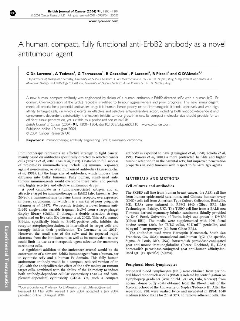
A human, compact, fully functional anti-ErbB2 antibody as a novelantitumour agent
C De Lorenzo1, A Tedesco1, G Terrazzano2, R Cozzolino1, P Laccetti1, R Piccoli1 and G D’Alessio*,1
1Department of Biological Chemistry, University of Naples Federico II, Via Mezzocannone 16, 80134 Naples, Italy; 2Department of Cellular andMolecular Biology and Pathology ‘L Califano’, University of Naples Federico II, via Pansini 5, 80131 Naples, Italy
A new human, compact antibody was engineered by fusion of a human, antitumour ErbB2-directed scFv with a human IgG1 Fcdomain. Overexpression of the ErbB2 receptor is related to tumour aggressiveness and poor prognosis. This new immunoagentmeets all criteria for a potential anticancer drug: it is human, hence poorly or not immunogenic; it binds selectively and with highaffinity to target cells, on which it exerts an effective and selective antiproliferative action, including both antibody-dependent andcomplement-dependent cytotoxicity; it effectively inhibits tumour growth in vivo. Its compact molecular size should provide for anefficient tissue penetration, yet suitable to a prolonged serum half-life.British Journal of Cancer (2004) 91, 1200–1204. doi:10.1038/sj.bjc.6602110 www.bjcancer.comPublished online 10 August 2004& 2004 Cancer Research UK
Keywords: immunotherapy; antibody engineering; ErbB2; mammary carcinoma
�����������������������������������
Immunotherapy represents an effective strategy to fight cancer,mainly based on antibodies specifically directed to selected cancercells (Trikha et al, 2002; Ross et al, 2003). Obstacles to full successof present-day immunotherapy include: (i) immune responsesagainst non-human, or even humanised antibodies (Kuus-Reichelet al, 1994); (ii) the large size of antibodies, which hinders theirdiffusion into bulky tumours. Fully human, small-sized anti-tumour immunoagents would overcome these risks, and providesafe, highly selective and effective antitumour drugs.
A good candidate as a tumour-associated antigen, and anattractive target for immunotherapy, is ErbB2 (also known as Her-2/Neu), a transmembrane tyrosine kinase receptor, overexpressedin breast carcinomas, for which it is a marker of poor prognosis(Slamon et al, 1987). We recently isolated a novel human anti-ErbB2 single-chain variable fragment (scFv) from a large phage-display library (Griffin 1) through a double selection strategyperformed on live cells (De Lorenzo et al, 2002). This scFv, namedErbicin, specifically binds to ErbB2-positive cells, inhibits thereceptor autophosphorylation, is internalised in target cells andstrongly inhibits their proliferation (De Lorenzo et al, 2002).However, the small size of the scFv and its expected rapidclearance from the bloodstream, as well as its monovalent nature,could limit its use as a therapeutic agent selective for mammarycarcinoma cells.
A significant addition to the anticancer arsenal would be theconstruction of a new anti-ErbB2 immunoagent from a human, perse cytotoxic scFv and a human Fc domain. This fully humanantitumour antibody would be a compact, reduced version of anIgG, with the antiproliferative effect of the scFv moiety on tumourtarget cells, combined with the ability of the Fc moiety to induceboth antibody-dependent cellular cytotoxicity (ADCC) and com-plement-dependent cytotoxicity (CDC). Yet, such a compact
antibody is expected to have (Demignot et al, 1990; Yokota et al,1993; Powers et al, 2001) a more protracted half-life and highertumour retention than the parental scFv, but improved penetrationproperties in solid tumours with respect to full-size IgG agents.
MATERIALS AND METHODS
Cell cultures and antibodies
The SKBR3 cell line from human breast cancer, the A431 cell linefrom human epidermoid carcinoma, and Chinese hamster ovary(CHO) cells (all from American Type Culture Collection, Rockville,MD, USA) were cultured in RPMI 1640 (Gibco BRL, LifeTechnologies, Paisley, UK). The TUBO cell line from a BALB-neuT mouse-derived mammary lobular carcinoma (kindly providedby Dr G Forni, University of Turin, Italy) was grown in DMEM(Gibco BRL). The media were supplemented with 10% foetalbovine serum (20% for TUBO cells), 50 U ml�1 penicillin, and50mg ml�1 streptomycin (all from Gibco BRL).
The antibodies used were: Herceptin (Genentech, South SanFrancisco, CA, USA); monoclonal anti-human IgG1 (Fc specific,Sigma, St Louis, MO, USA); horseradish peroxidase-conjugatedgoat anti-mouse immunoglobulins (Pierce, Rockford, IL, USA);horseradish peroxidase-conjugated goat anti-human affinity-iso-lated IgG (Fc specific) (Sigma).
Peripheral blood lymphocytes
Peripheral blood lymphocytes (PBL) were obtained from periph-eral blood mononuclear cells (PBMC) isolated by centrifugation onLymphoprep gradients (Axis Shield PoC AS, Oslo, Norway) fromnormal donor buffy coats obtained from the Blood Bank of theMedical School of the University of Naples ‘Federico II’. After theseparation, PBL were washed twice and incubated in RPMI 1640medium (Gibco BRL) for 2 h at 371C to remove adherent cells. The
Received 11 May 2004; revised 1 July 2004; accepted 2 July 2004;published online 10 August 2004
*Correspondence: Professor G D’Alessio; E-mail: [email protected]
British Journal of Cancer (2004) 91, 1200 – 1204
& 2004 Cancer Research UK All rights reserved 0007 – 0920/04 $30.00
www.bjcancer.com
Exp
erim
en
tal
Th
era
peu
tics
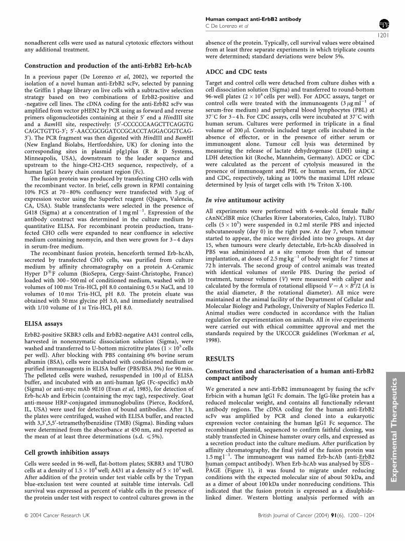
nonadherent cells were used as natural cytotoxic effectors withoutany additional treatment.
Construction and production of the anti-ErbB2 Erb-hcAb
In a previous paper (De Lorenzo et al, 2002), we reported theisolation of a novel human anti-ErbB2 scFv, selected by panningthe Griffin 1 phage library on live cells with a subtractive selectionstrategy based on two combinations of ErbB2-positive and-negative cell lines. The cDNA coding for the anti-ErbB2 scFv wasamplified from vector pHEN2 by PCR using as forward and reverseprimers oligonucleotides containing at their 50 end a HindIII siteand a BamHI site, respectively: (50-CCCCCCAAGCTTCAGGTGCAGCTGTTG-30; 50-AACCGCGGATCCGCACCTAGGACGGTCAG-30). The PCR fragment was then digested with HindIII and BamHI(New England Biolabs, Hertfordshire, UK) for cloning into thecorresponding sites in plasmid pIg1plus (R & D Systems,Minneapolis, USA), downstream to the leader sequence andupstream to the hinge-CH2-CH3 sequence, respectively, of ahuman IgG1 heavy chain constant region (Fc).
The fusion protein was produced by transfecting CHO cells withthe recombinant vector. In brief, cells grown in RPMI containing10% FCS at 70–80% confluency were transfected with 5 mg ofexpression vector using the Superfect reagent (Qiagen, Valencia,CA, USA). Stable transfectants were selected in the presence ofG418 (Sigma) at a concentration of 1 mg ml�1. Expression of theantibody construct was determined in the culture medium byquantitative ELISA. For recombinant protein production, trans-fected CHO cells were expanded to near confluence in selectivemedium containing neomycin, and then were grown for 3– 4 daysin serum-free medium.
The recombinant fusion protein, henceforth termed Erb-hcAb,secreted by transfected CHO cells, was purified from culturemedium by affinity chromatography on a protein A-CeramicHyper DsF column (BioSepra, Cergy-Saint-Christophe, France)loaded with 300– 500 ml of conditioned medium, washed with 10volumes of 100 mM Tris-HCl, pH 8.0 containing 0.5 M NaCl, and 10volumes of 10 mM Tris-HCl, pH 8.0. The protein eluate wasobtained with 50 mM glycine pH 3.0, and immediately neutralisedwith 1/10 volume of 1 M Tris-HCl, pH 8.0.
ELISA assays
ErbB2-positive SKBR3 cells and ErbB2-negative A431 control cells,harvested in nonenzymatic dissociation solution (Sigma), werewashed and transferred to U-bottom microtitre plates (1� 105 cellsper well). After blocking with PBS containing 6% bovine serumalbumin (BSA), cells were incubated with conditioned medium orpurified immunoagents in ELISA buffer (PBS/BSA 3%) for 90 min.The pelleted cells were washed, resuspended in 100 ml of ELISAbuffer, and incubated with an anti-human IgG (Fc-specific) mAb(Sigma) or anti-myc mAb 9E10 (Evan et al, 1985), for detection ofErb-hcAb and Erbicin (containing the myc tag), respectively. Goatanti-mouse HRP-conjugated immunoglobulins (Pierce, Rockford,IL, USA) were used for detection of bound antibodies. After 1 h,the plates were centrifuged, washed with ELISA buffer, and reactedwith 3,30,5,50-tetramethylbenzidine (TMB) (Sigma). Binding valueswere determined from the absorbance at 450 nm, and reported asthe mean of at least three determinations (s.d. p5%).
Cell growth inhibition assays
Cells were seeded in 96-well, flat-bottom plates; SKBR3 and TUBOcells at a density of 1.5� 104 well; A431 at a density of 5� 103 well.After addition of the protein under test viable cells by the Trypanblue-exclusion test were counted at suitable time intervals. Cellsurvival was expressed as percent of viable cells in the presence ofthe protein under test with respect to control cultures grown in the
absence of the protein. Typically, cell survival values were obtainedfrom at least three separate experiments in which triplicate countswere determined; standard deviations were below 5%.
ADCC and CDC tests
Target and control cells were detached from culture dishes with acell dissociation solution (Sigma) and transferred to round-bottom96-well plates (2� 104 cells per well). For ADCC assays, target orcontrol cells were treated with the immunoagents (3 mg ml�1 ofserum-free medium) and peripheral blood lymphocytes (PBL) at371C for 3 –4 h. For CDC assays, cells were incubated at 371C withhuman serum. Cultures were performed in triplicate in a finalvolume of 200 ml. Controls included target cells incubated in theabsence of effector, or in the presence of either serum orimmunoagent alone. Tumour cell lysis was determined bymeasuring the release of lactate dehydrogenase (LDH) using aLDH detection kit (Roche, Mannheim, Germany). ADCC or CDCwere calculated as the percent of cytolysis measured in thepresence of immunoagent and PBL or human serum, for ADCCand CDC, respectively, taking as 100% the maximal LDH releasedetermined by lysis of target cells with 1% Triton X-100.
In vivo antitumour activity
All experiments were performed with 6-week-old female Balb/cAnNCrlBR mice (Charles River Laboratories, Calco, Italy). TUBOcells (5� 105) were suspended in 0.2 ml sterile PBS and injectedsubcutaneously (day 0) in the right paw. At day 7, when tumourstarted to appear, the mice were divided into two groups. At day15, when tumours were clearly detectable, Erb-hcAb dissolved inPBS was administered at a site remote from that of tumourimplantation, at doses of 2.5 mg kg�1 of body weight for 7 times at72 h intervals. The second group of control animals was treatedwith identical volumes of sterile PBS. During the period oftreatment, tumour volumes (V) were measured with caliper andcalculated by the formula of rotational ellipsoid V¼A�B2/2 (A isthe axial diameter, B the rotational diameter). All mice weremaintained at the animal facility of the Department of Cellular andMolecular Biology and Pathology, University of Naples Federico II.Animal studies were conducted in accordance with the Italianregulation for experimentation on animals. All in vivo experimentswere carried out with ethical committee approval and met thestandards required by the UKCCCR guidelines (Workman et al,1998).
RESULTS
Construction and characterisation of a human anti-ErbB2compact antibody
We generated a new anti-ErbB2 immunoagent by fusing the scFvErbicin with a human IgG1 Fc domain. The IgG-like protein has areduced molecular weight, and contains all functionally relevantantibody regions. The cDNA coding for the human anti-ErbB2scFv was amplified by PCR and cloned into a eukaryoticexpression vector containing the human IgG1 Fc sequence. Therecombinant plasmid, sequenced to confirm faithful cloning, wasstably transfected in Chinese hamster ovary cells, and expressed asa secretion product into the culture medium. After purification byaffinity chromatography, the final yield of the fusion protein was1.5 mg l�1. The immunoagent was named Erb-hcAb (anti-ErbB2human compact antibody). When Erb-hcAb was analysed by SDS–PAGE (Figure 1), it was found to migrate under reducingconditions with the expected molecular size of about 50 kDa, andas a dimer of about 100 kDa under nonreducing conditions. Thisindicated that the fusion protein is expressed as a disulphide-linked dimer. Western blotting analysis performed with an
Human compact anti-ErbB2 antibody
C De Lorenzo et al
1201
British Journal of Cancer (2004) 91(6), 1200 – 1204& 2004 Cancer Research UK
Exp
eri
men
tal
Th
era
peu
tics
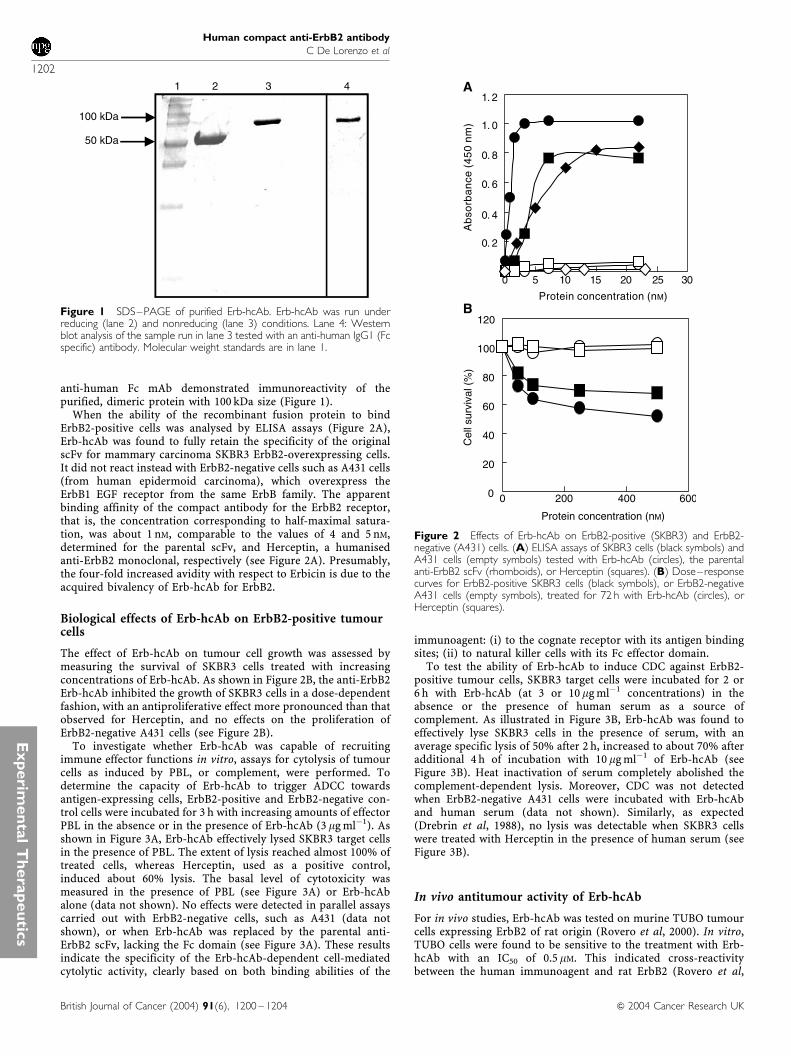
anti-human Fc mAb demonstrated immunoreactivity of thepurified, dimeric protein with 100 kDa size (Figure 1).
When the ability of the recombinant fusion protein to bindErbB2-positive cells was analysed by ELISA assays (Figure 2A),Erb-hcAb was found to fully retain the specificity of the originalscFv for mammary carcinoma SKBR3 ErbB2-overexpressing cells.It did not react instead with ErbB2-negative cells such as A431 cells(from human epidermoid carcinoma), which overexpress theErbB1 EGF receptor from the same ErbB family. The apparentbinding affinity of the compact antibody for the ErbB2 receptor,that is, the concentration corresponding to half-maximal satura-tion, was about 1 nM, comparable to the values of 4 and 5 nM,determined for the parental scFv, and Herceptin, a humanisedanti-ErbB2 monoclonal, respectively (see Figure 2A). Presumably,the four-fold increased avidity with respect to Erbicin is due to theacquired bivalency of Erb-hcAb for ErbB2.
Biological effects of Erb-hcAb on ErbB2-positive tumourcells
The effect of Erb-hcAb on tumour cell growth was assessed bymeasuring the survival of SKBR3 cells treated with increasingconcentrations of Erb-hcAb. As shown in Figure 2B, the anti-ErbB2Erb-hcAb inhibited the growth of SKBR3 cells in a dose-dependentfashion, with an antiproliferative effect more pronounced than thatobserved for Herceptin, and no effects on the proliferation ofErbB2-negative A431 cells (see Figure 2B).
To investigate whether Erb-hcAb was capable of recruitingimmune effector functions in vitro, assays for cytolysis of tumourcells as induced by PBL, or complement, were performed. Todetermine the capacity of Erb-hcAb to trigger ADCC towardsantigen-expressing cells, ErbB2-positive and ErbB2-negative con-trol cells were incubated for 3 h with increasing amounts of effectorPBL in the absence or in the presence of Erb-hcAb (3 mg ml�1). Asshown in Figure 3A, Erb-hcAb effectively lysed SKBR3 target cellsin the presence of PBL. The extent of lysis reached almost 100% oftreated cells, whereas Herceptin, used as a positive control,induced about 60% lysis. The basal level of cytotoxicity wasmeasured in the presence of PBL (see Figure 3A) or Erb-hcAbalone (data not shown). No effects were detected in parallel assayscarried out with ErbB2-negative cells, such as A431 (data notshown), or when Erb-hcAb was replaced by the parental anti-ErbB2 scFv, lacking the Fc domain (see Figure 3A). These resultsindicate the specificity of the Erb-hcAb-dependent cell-mediatedcytolytic activity, clearly based on both binding abilities of the
immunoagent: (i) to the cognate receptor with its antigen bindingsites; (ii) to natural killer cells with its Fc effector domain.
To test the ability of Erb-hcAb to induce CDC against ErbB2-positive tumour cells, SKBR3 target cells were incubated for 2 or6 h with Erb-hcAb (at 3 or 10mg ml�1 concentrations) in theabsence or the presence of human serum as a source ofcomplement. As illustrated in Figure 3B, Erb-hcAb was found toeffectively lyse SKBR3 cells in the presence of serum, with anaverage specific lysis of 50% after 2 h, increased to about 70% afteradditional 4 h of incubation with 10 mg ml�1 of Erb-hcAb (seeFigure 3B). Heat inactivation of serum completely abolished thecomplement-dependent lysis. Moreover, CDC was not detectedwhen ErbB2-negative A431 cells were incubated with Erb-hcAband human serum (data not shown). Similarly, as expected(Drebrin et al, 1988), no lysis was detectable when SKBR3 cellswere treated with Herceptin in the presence of human serum (seeFigure 3B).
In vivo antitumour activity of Erb-hcAb
For in vivo studies, Erb-hcAb was tested on murine TUBO tumourcells expressing ErbB2 of rat origin (Rovero et al, 2000). In vitro,TUBO cells were found to be sensitive to the treatment with Erb-hcAb with an IC50 of 0.5 mM. This indicated cross-reactivitybetween the human immunoagent and rat ErbB2 (Rovero et al,
1 3 4
100 kDa
50 kDa
2
Figure 1 SDS–PAGE of purified Erb-hcAb. Erb-hcAb was run underreducing (lane 2) and nonreducing (lane 3) conditions. Lane 4: Westernblot analysis of the sample run in lane 3 tested with an anti-human IgG1 (Fcspecific) antibody. Molecular weight standards are in lane 1.
0
20
40
60
80
100
120
0 200 400 600
0 5 10 15 20 25 30
Ab
sorb
an
ce (
45
0 n
m)
Protein concentration (nM)
0. 2
0. 4
0. 6
0. 8
1. 0
1. 2A
B
Protein concentration (nM)
Cel
l sur
viva
l (%
)
Figure 2 Effects of Erb-hcAb on ErbB2-positive (SKBR3) and ErbB2-negative (A431) cells. (A) ELISA assays of SKBR3 cells (black symbols) andA431 cells (empty symbols) tested with Erb-hcAb (circles), the parentalanti-ErbB2 scFv (rhomboids), or Herceptin (squares). (B) Dose–responsecurves for ErbB2-positive SKBR3 cells (black symbols), or ErbB2-negativeA431 cells (empty symbols), treated for 72 h with Erb-hcAb (circles), orHerceptin (squares).
Human compact anti-ErbB2 antibody
C De Lorenzo et al
1202
British Journal of Cancer (2004) 91(6), 1200 – 1204 & 2004 Cancer Research UK
Exp
erim
en
tal
Th
era
peu
tics
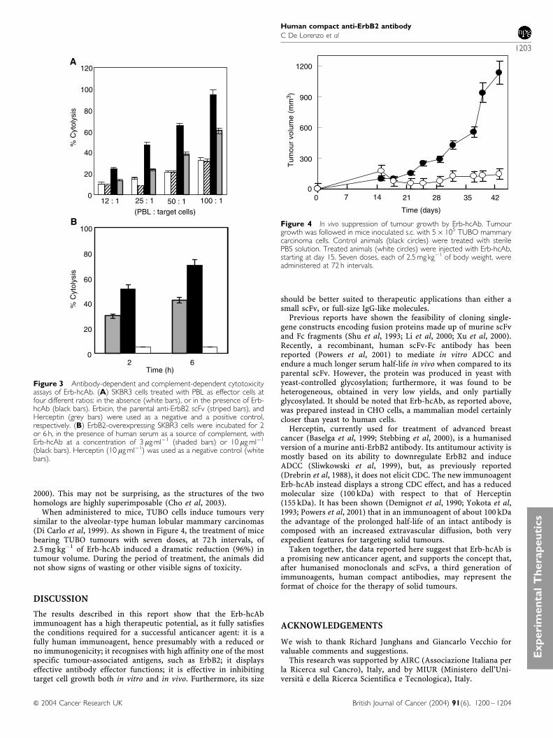
2000). This may not be surprising, as the structures of the twohomologs are highly superimposable (Cho et al, 2003).
When administered to mice, TUBO cells induce tumours verysimilar to the alveolar-type human lobular mammary carcinomas(Di Carlo et al, 1999). As shown in Figure 4, the treatment of micebearing TUBO tumours with seven doses, at 72 h intervals, of2.5 mg kg�1 of Erb-hcAb induced a dramatic reduction (96%) intumour volume. During the period of treatment, the animals didnot show signs of wasting or other visible signs of toxicity.
DISCUSSION
The results described in this report show that the Erb-hcAbimmunoagent has a high therapeutic potential, as it fully satisfiesthe conditions required for a successful anticancer agent: it is afully human immunoagent, hence presumably with a reduced orno immunogenicity; it recognises with high affinity one of the mostspecific tumour-associated antigens, such as ErbB2; it displayseffective antibody effector functions; it is effective in inhibitingtarget cell growth both in vitro and in vivo. Furthermore, its size
should be better suited to therapeutic applications than either asmall scFv, or full-size IgG-like molecules.
Previous reports have shown the feasibility of cloning single-gene constructs encoding fusion proteins made up of murine scFvand Fc fragments (Shu et al, 1993; Li et al, 2000; Xu et al, 2000).Recently, a recombinant, human scFv-Fc antibody has beenreported (Powers et al, 2001) to mediate in vitro ADCC andendure a much longer serum half-life in vivo when compared to itsparental scFv. However, the protein was produced in yeast withyeast-controlled glycosylation; furthermore, it was found to beheterogeneous, obtained in very low yields, and only partiallyglycosylated. It should be noted that Erb-hcAb, as reported above,was prepared instead in CHO cells, a mammalian model certainlycloser than yeast to human cells.
Herceptin, currently used for treatment of advanced breastcancer (Baselga et al, 1999; Stebbing et al, 2000), is a humanisedversion of a murine anti-ErbB2 antibody. Its antitumour activity ismostly based on its ability to downregulate ErbB2 and induceADCC (Sliwkowski et al, 1999), but, as previously reported(Drebrin et al, 1988), it does not elicit CDC. The new immunoagentErb-hcAb instead displays a strong CDC effect, and has a reducedmolecular size (100 kDa) with respect to that of Herceptin(155 kDa). It has been shown (Demignot et al, 1990; Yokota et al,1993; Powers et al, 2001) that in an immunoagent of about 100 kDathe advantage of the prolonged half-life of an intact antibody iscomposed with an increased extravascular diffusion, both veryexpedient features for targeting solid tumours.
Taken together, the data reported here suggest that Erb-hcAb isa promising new anticancer agent, and supports the concept that,after humanised monoclonals and scFvs, a third generation ofimmunoagents, human compact antibodies, may represent theformat of choice for the therapy of solid tumours.
ACKNOWLEDGEMENTS
We wish to thank Richard Junghans and Giancarlo Vecchio forvaluable comments and suggestions.
This research was supported by AIRC (Associazione Italiana perla Ricerca sul Cancro), Italy, and by MIUR (Ministero dell’Uni-versita e della Ricerca Scientifica e Tecnologica), Italy.
0
300
600
900
1200
0 14 21 28 35 42
Tum
our
volu
me
(mm
3 )
Time (days)
7
Figure 4 In vivo suppression of tumour growth by Erb-hcAb. Tumourgrowth was followed in mice inoculated s.c. with 5� 105 TUBO mammarycarcinoma cells. Control animals (black circles) were treated with sterilePBS solution. Treated animals (white circles) were injected with Erb-hcAb,starting at day 15. Seven doses, each of 2.5 mg kg�1 of body weight, wereadministered at 72 h intervals.
0
20
40
60
80
100
120
25 : 1 50 : 1 100 : 112 : 1
0
20
40
60
80
100
2 6
B
Time (h)
(PBL : target cells)
% C
ytol
ysis
A%
Cyt
olys
is
Figure 3 Antibody-dependent and complement-dependent cytotoxicityassays of Erb-hcAb. (A) SKBR3 cells treated with PBL as effector cells atfour different ratios: in the absence (white bars), or in the presence of Erb-hcAb (black bars). Erbicin, the parental anti-ErbB2 scFv (striped bars), andHerceptin (grey bars) were used as a negative and a positive control,respectively. (B) ErbB2-overexpressing SKBR3 cells were incubated for 2or 6 h, in the presence of human serum as a source of complement, withErb-hcAb at a concentration of 3 mg ml�1 (shaded bars) or 10 mg ml�1
(black bars). Herceptin (10 mg ml�1) was used as a negative control (whitebars).
Human compact anti-ErbB2 antibody
C De Lorenzo et al
1203
British Journal of Cancer (2004) 91(6), 1200 – 1204& 2004 Cancer Research UK
Exp
eri
men
tal
Th
era
peu
tics
REFERENCES
Baselga J, Tripathy D, Mendelsohn J, Baughman S, Benz CC, Dantis L,Sklarin NT, Seidman AD, Hudis CA, Moore J, Rosen PP, Twaddell T,Henderson IC, Norton L (1999) Phase II study of weekly intravenoustrastuzumab (Herceptin) in patients with HER2/neu-overexpressingmetastatic breast cancer. Semin Oncol 26: 78 – 83
Cho HS, Mason K, Ramyar KX, Stanley AM, Gabelli SB, Denney Jr DW,Leahy DJ (2003) Structure of the extracellular region of HER2 alone andin complex with the Herceptin Fab. Nature 421: 756 – 760
De Lorenzo C, Palmer DB, Piccoli R, Ritter MA, D’Alessio G (2002) A newhuman antitumor immunoreagent specific for ErbB2. Clin Cancer Res 8:1710 – 1719
Demignot S, Pimm MV, Baldwin RW (1990) Comparison of biodistribution of791T/36 monoclonal antibody and its Fab/c fragment in BALB/c mice andnude mice bearing human tumor xenografts. Cancer Res 50: 2936 – 2942
Di Carlo E, Diodoro MG, Boggio K, Modesti A, Modesti M, Nanni P, ForniG, Musiani P (1999) Analysis of mammary carcinoma onset andprogression in HER-2/neu oncogene transgenic mice reveals a lobularorigin. Lab Invest 79: 1261 – 1269
Drebrin JA, Link VC, Greene MI (1988) Monoclonal antibodies specific forthe neu oncogene product directly mediate anti-tumor effects in vivo.Oncogene 2: 387 – 394
Evan GI, Lewis GK, Ramsay G, Bishop JM (1985) Isolation of monoclonalantibodies specific for human c-myc proto-oncogene product. Mol CellBiol 5: 3610 – 3616
Kuus-Reichel K, Grauer LS, Karavodin LM, Knott C, Krusemeier M, Kay NE(1994) Will immunogenicity limit the use, efficacy, and futuredevelopment of therapeutic monoclonal antibodies? Clin Diagn LabImmunol 1: 365 – 372
Li SL, Liang SJ, Guo N, Wu AM, Fujita-Yamaguchi Y (2000) Single-chainantibodies against human insulin-like growth factor I receptor: expres-sion, purification, and effect on tumor growth. Cancer ImmunolImmunother 49: 243 – 252
Powers DB, Amersdorfer P, Poul M, Nielsen UB, Shalaby MR, Adams GP,Weiner LM, Marks JD (2001) Expression of single-chain Fv – Fc fusionsin Pichia pastoris. J Immunol Methods 251: 123 – 135
Ross J, Gray K, Schenkein D, Greene B, Gray GS, Shulok J, Worland PJ,Celniker A, Rolfe M (2003) Antibody-based therapeutics in oncology.Expert Rev Anticancer Ther 3: 107 – 121
Rovero S, Amici A, Carlo ED, Bei R, Nanni P, Quaglino E,Porcedda P, Boggio K, Smorlesi A, Lollini PL, Landuzzi L, ColomboMP, Giovarelli M, Musiani P, Forni, G (2000) DNA vaccination againstrat her-2/Neu p185 more effectively inhibits carcinogenesis thantransplantable carcinomas in transgenic BALB/c mice. J Immunol 165:5133 – 5142
Shu L, Qi CF, Schlom J, Kashmiri SV (1993) Secretion of a single-gene-encoded immunoglobulin from myeloma cells. Proc Natl Acad Sci USA90: 7995 – 7999
Slamon DJ, Clark GM, Wong SG, Levin WJ, Ullrich A, McGuire WL (1987)Human breast cancer: correlation of relapse and survival withamplification of the HER2/neu oncogene. Science 235: 177 – 182
Sliwkowski MX, Lofgren JA, Lewis GD, Hotaling TE, Fendly BM, Fox JA(1999) Nonclinical studies addressing the mechanism of action oftrastuzumab (Herceptin). Semin Oncol 26: 60 – 70
Stebbing J, Copson E, O’Reilly S (2000) Herceptin (trastuzumab) inadvanced breast cancer. Cancer Treat Rev 26: 287 – 290
Trikha M, Yan L, Nakada MT (2002) Monoclonal antibodies as therapeuticsin oncology. Curr Opin Biotechnol 13: 609 – 614
Workman P, Twentyman P, Balkwill F, Balmain A, Chaplin D, Double J,Embleton J, Newell D, raymond R, Stables J, Stephens T, Wallace J (1998)United Kingdom Co-ordinating Committee on Cancer Research(UKCCCR) Guidelines for the welfare of animals in experimentalneoplasia (Second Edition). Br J Cancer 77: 1 – 10
Xu X, Clarke P, Szalai G, Shively JE, Williams LE, Shyr Y, Shi E, Primus FJ(2000) Targeting and therapy of carcinoembryonic antigen-expressingtumors in transgenic mice with an antibody-interleukin 2 fusion protein.Cancer Res 60: 4475 – 4484
Yokota T, Milenic DE, Whitlow M, Wood JF, Hubert SL, Schlom J (1993)Microautoradiographic analysis of the normal organ distribution ofradioiodinated single-chain Fv and other immunoglobulin forms. CancerRes 53: 5832
Human compact anti-ErbB2 antibody
C De Lorenzo et al
1204
British Journal of Cancer (2004) 91(6), 1200 – 1204 & 2004 Cancer Research UK
Exp
erim
en
tal
Th
era
peu
tics