Pathologic Continuum of Acute Appendicitis - Emory EM · PDF filePATHOPHYSIOLOGY AND...
9
Pathologic Continuum of Acute Appendicitis Sonographic Findings and Clinical Management Implications Lauren Chan, BA,* Lewis K. Shin, MD,Þþ Reetesh K. Pai, MD,§ and R. Brooke Jeffrey, MDÞ Abstract: Appendicitis is one of the most common causes of the acute abdomen often requiring emergent surgery. Delayed diagnosis leads to the progression of uncomplicated appendicitis to compli- cated (gangrenous, perforated) appendicitis, often changing clinical management. Computed tomography and ultrasound are imaging modalities of choice to preoperatively diagnose appendicitis. Recent concerns of radiation exposure and cost have renewed interest in using ultrasound as an initial, diagnostic study. A sonographic pic- torial and histopathologic review of the continuum of appendicitis is presented. A comprehensive sonographic examination of the appen- dix should investigate the size (maximal diameter), the echogenic submucosal layer integrity, the mural color Doppler signature, the presence of a fecalith, and the periappendiceal changes. Features of an uncomplicated appendicitis include size greater than 6 to 7 mm, hyperemia on color Doppler, mural thickening, and an intact echo- genic submucosal layer. Gangrenous appendicitis is characterized by loss of the echogenic submucosal layer with absent color Doppler flow. Loculated pericecal fluid, prominent pericecal fat, and cir- cumferential loss of the submucosal layer are suggestive of perfora- tion. Sonographic staging can triage management of appendicitis by directing urgent laparoscopic appendectomy for uncomplicated appendicitis, open appendectomy for complicated appendicitis, and conservative management (antibiotics with percutaneous drainage) for perforated appendicitis with abscess formation. Key Words: appendicitis, sonography, abscess, phlegmon, complicated appendicitis, uncomplicated appendicitis (Ultrasound Quarterly 2011;27:71Y79) CLINICAL OVERVIEW Appendicitis is one of the most common causes of the acute abdomen in the Western world and is one of the most frequent indications for abdominal surgery. 1 It occurs in 7% to 12% of the general population, and although it may occur at any age, it is most common in the 10- to 19-year-old age group (233/100,000 population annually). 1 Each year, 280,000 appendectomies are performed in the United States to avoid the potential complications of perforated appendicitis. 1 Perforation rates are correlated with delayed diagnosis and age. Young children and the elderly have the highest rates of perforation (40%Y57% and 55%Y70%, respectively). 2 Of note is the fact that, in the past 2 decades, the negative appendectomy rate has been relatively constant with slight decline after 2000, but the rates for perforated appendicitis seem to be increasing. 2 The clinical diagnosis may be difficult to make without a classic presentation of periumbilical pain migrating to the right lower quadrant or in particular patient populations such as women of child bearing age whose gynecologic pathologies may present similarly. Owing to its increased morbidity, there is an ongoing need for early and accurate diagnosis of appendicitis before the onset of perforation. Depending on the pathologic stage of appendicitis, there are a variety of surgical and nonsurgical treatment options including laparoscopic or open appendectomy, primary anti- biotic therapy, or percutaneous drainage of periappendiceal abscesses. 3Y8 Appropriate patient management is based on distinguishing complicated (gangrenous or perforated ap- pendicitis) from uncomplicated appendicitis. In addition, in patients with perforated appendicitis, it is important to differentiate liquefied abscesses from indurated soft tissue masses (phlegmonous inflammation). For more than 20 years, computed tomography (CT) and sonography have been the primary imaging methods used in the diagnosis of appendicitis. 9 Despite its slightly higher sensitivity and specificity, there are increasing concerns about the radiation burden and cost of CT; thus, there is renewed interest in the role of sonography. 10 In this article, we review the sonographic findings along the pathologic continuum of acute appendicitis emphasizing important luminal, mural, and periappendiceal features that are helpful in differentiating complicated from uncomplicated appendicitis. THE APPENDIX The cecum and appendix develop from the midgut, which rotates and descends to its final position in the right iliac fossa. The mean length of the adult appendix measures approximately 9 cm and typically arises from the postero- medial border of the cecum approximately 1.7 to 2.5 cm below the terminal ileum. 11 The appendix can be found in almost any position relative to the cecum, and Wakeley 12 described 5 locations in a review of 10,000 anatomic cases: retrocecal (65.3%), descending/pelvic (directed downward on the psoas) (31%), subcecal in the iliac fossa (2.3%), preileal (1.0%), and postileal 0.4%. PICTORIAL ESSAY Ultrasound Quarterly & Volume 27, Number 2, June 2011 www.ultrasound-quarterly.com 71 Received for publication October 1, 2010; accepted January 5, 2011. *Stanford University School of Medicine, †Department of Radiology, Stan- ford University Medical Center, Stanford; ‡VA Palo Alto Health Care System, Palo Alto; §Department of Pathology, Stanford University Med- ical Center; Stanford, CA. The authors have disclosed that they have no interests in or significant rela- tionships with any commercial companies pertaining to this educational activity. Reprints: Lewis K. Shin, MD, Department of Radiology, Stanford University Medical Center/VA Palo Alto Health Care System, 300 Pasteur Dr H1307, Stanford, CA 94305-5105 (e-mail: [email protected]). Copyright * 2011 by Lippincott Williams & Wilkins Copyright © 2011 Lippincott Williams & Wilkins. Unauthorized reproduction of this article is prohibited.
Transcript of Pathologic Continuum of Acute Appendicitis - Emory EM · PDF filePATHOPHYSIOLOGY AND...
Pathologic Continuum of Acute AppendicitisSonographic Findings and Clinical Management Implications
Lauren Chan, BA,* Lewis K. Shin, MD,Þþ Reetesh K. Pai, MD,§ and R. Brooke Jeffrey, MDÞ
Abstract: Appendicitis is one of the most common causes of theacute abdomen often requiring emergent surgery. Delayed diagnosisleads to the progression of uncomplicated appendicitis to compli-cated (gangrenous, perforated) appendicitis, often changing clinicalmanagement. Computed tomography and ultrasound are imagingmodalities of choice to preoperatively diagnose appendicitis. Recentconcerns of radiation exposure and cost have renewed interest inusing ultrasound as an initial, diagnostic study. A sonographic pic-torial and histopathologic review of the continuum of appendicitis ispresented. A comprehensive sonographic examination of the appen-dix should investigate the size (maximal diameter), the echogenicsubmucosal layer integrity, the mural color Doppler signature, thepresence of a fecalith, and the periappendiceal changes. Features ofan uncomplicated appendicitis include size greater than 6 to 7 mm,hyperemia on color Doppler, mural thickening, and an intact echo-genic submucosal layer. Gangrenous appendicitis is characterized byloss of the echogenic submucosal layer with absent color Dopplerflow. Loculated pericecal fluid, prominent pericecal fat, and cir-cumferential loss of the submucosal layer are suggestive of perfora-tion. Sonographic staging can triage management of appendicitisby directing urgent laparoscopic appendectomy for uncomplicatedappendicitis, open appendectomy for complicated appendicitis, andconservative management (antibiotics with percutaneous drainage)for perforated appendicitis with abscess formation.
Key Words: appendicitis, sonography, abscess, phlegmon,complicated appendicitis, uncomplicated appendicitis
(Ultrasound Quarterly 2011;27:71Y79)
CLINICAL OVERVIEWAppendicitis is one of the most common causes of the
acute abdomen in the Western world and is one of the mostfrequent indications for abdominal surgery.1 It occurs in 7%to 12% of the general population, and although it may occurat any age, it is most common in the 10- to 19-year-old agegroup (233/100,000 population annually).1 Each year, 280,000
appendectomies are performed in the United States to avoid thepotential complications of perforated appendicitis.1 Perforationrates are correlated with delayed diagnosis and age. Youngchildren and the elderly have the highest rates of perforation(40%Y57% and 55%Y70%, respectively).2 Of note is the factthat, in the past 2 decades, the negative appendectomy rate hasbeen relatively constant with slight decline after 2000, but therates for perforated appendicitis seem to be increasing.2 Theclinical diagnosis may be difficult to make without a classicpresentation of periumbilical pain migrating to the right lowerquadrant or in particular patient populations such as women ofchild bearing age whose gynecologic pathologies may presentsimilarly. Owing to its increased morbidity, there is an ongoingneed for early and accurate diagnosis of appendicitis before theonset of perforation.
Depending on the pathologic stage of appendicitis, thereare a variety of surgical and nonsurgical treatment optionsincluding laparoscopic or open appendectomy, primary anti-biotic therapy, or percutaneous drainage of periappendicealabscesses.3Y8 Appropriate patient management is based ondistinguishing complicated (gangrenous or perforated ap-pendicitis) from uncomplicated appendicitis. In addition,in patients with perforated appendicitis, it is important todifferentiate liquefied abscesses from indurated soft tissuemasses (phlegmonous inflammation).
For more than 20 years, computed tomography (CT)and sonography have been the primary imaging methods usedin the diagnosis of appendicitis.9 Despite its slightly highersensitivity and specificity, there are increasing concerns aboutthe radiation burden and cost of CT; thus, there is renewedinterest in the role of sonography.10 In this article, we reviewthe sonographic findings along the pathologic continuum ofacute appendicitis emphasizing important luminal, mural, andperiappendiceal features that are helpful in differentiatingcomplicated from uncomplicated appendicitis.
THE APPENDIXThe cecum and appendix develop from the midgut,
which rotates and descends to its final position in the rightiliac fossa. The mean length of the adult appendix measuresapproximately 9 cm and typically arises from the postero-medial border of the cecum approximately 1.7 to 2.5 cm belowthe terminal ileum.11 The appendix can be found in almost anyposition relative to the cecum, and Wakeley12 described 5locations in a review of 10,000 anatomic cases: retrocecal(65.3%), descending/pelvic (directed downward on the psoas)(31%), subcecal in the iliac fossa (2.3%), preileal (1.0%), andpostileal 0.4%.
PICTORIAL ESSAY
Ultrasound Quarterly & Volume 27, Number 2, June 2011 www.ultrasound-quarterly.com 71
Received for publication October 1, 2010; accepted January 5, 2011.*Stanford University School of Medicine, †Department of Radiology, Stan-
ford University Medical Center, Stanford; ‡VA Palo Alto Health CareSystem, Palo Alto; §Department of Pathology, Stanford University Med-ical Center; Stanford, CA.
The authors have disclosed that they have no interests in or significant rela-tionships with any commercial companies pertaining to this educationalactivity.
Reprints: Lewis K. Shin, MD, Department of Radiology, Stanford UniversityMedical Center/VA Palo Alto Health Care System, 300 Pasteur Dr H1307,Stanford, CA 94305-5105 (e-mail: [email protected]).
Copyright * 2011 by Lippincott Williams & Wilkins
Copyright © 2011 Lippincott Williams & Wilkins. Unauthorized reproduction of this article is prohibited.
PATHOPHYSIOLOGY AND PATHOLOGICSTAGES OF ACUTE APPENDICITIS
The most commonly accepted pathogenesis of appen-dicitis is the obstruction of the appendiceal lumen by a fecalith,lymphoid hyperplasia, foreign bodies, parasites, or primary ormetastatic tumors.9 There may be a higher perforation riskwith the detection of an appendicolith as suggested by a studywhere 49% of perforated cases had an appendicolith com-pared with 27% in nonperforated cases with P = 0.049.13
However, other studies cite that an appendicolith is neitherhighly sensitive (50%) nor specific (70%) for the detection ofperforation and does not represent a statistically significantassociation.14,15
The early stage of appendicitis develops when obstruc-tion leads to fluid accumulation, elevation of intraluminalpressure, and luminal distention. On gross pathology, uncom-plicated appendicitis is characterized by a dull appearance ofthe normally glistening serosal surface and dilation of theserosal vessels, causing an injected appearance.16
Suppurative appendicitis (intramural infection withoutnecrosis) results from increasing intraluminal pressure thatexceeds capillary perfusion pressure, causing venous outflowobstruction and ultimately arterial compromise. This resultsin mucosal ischemia that allows bacterial invasion of theappendiceal wall. In some patients with early uncomplicatedappendicitis, these inflammatory changes are confined to thetip of the appendix due to a reduced blood supply from theterminal capillary branches of the appendiceal artery.16 Withthe onset of mural infection, there is edematous thickeningof the appendix wall due to intramural edema and infiltrationby inflammatory cells. This mural thickening is often notedin conjunction with dilation of the lumen. On gross pathol-ogy, the external appendix appears distended and hyper-emic, and there may be early edematous changes within themesoappendix.16
Unless diagnosed and surgically treated at an earlystage, appendiceal inflammation may ultimately progress togangrene and perforation. Gangrenous appendicitis resultswhen increasing intramural inflammatory change leads toappendiceal ischemia and transmural necrosis with serosalexudate. This is characterized by a friable serosa surface thatmay have a purple, green, or black discoloration. As withearly intramural inflammation, early gangrenous changesleading to perforation may be confined to the tip and or distalappendix.16 In many patients with appendiceal perforation,a liquefied abscess forms either in the peritoneal cavity (RLQor pelvis) or in the retroperitoneum (often the right anteriorpararenal space) with retrocecal appendicitis. The size and thedegree of ‘‘localization’’ of the abscesses are both highlyvariable. In other patients with perforated appendicitis, anindurated inflammatory mass (phlegmon) develops involvingthe surrounding soft tissues involving the mesoappendix,omentum, small bowel, and its adjacent mesentery.3
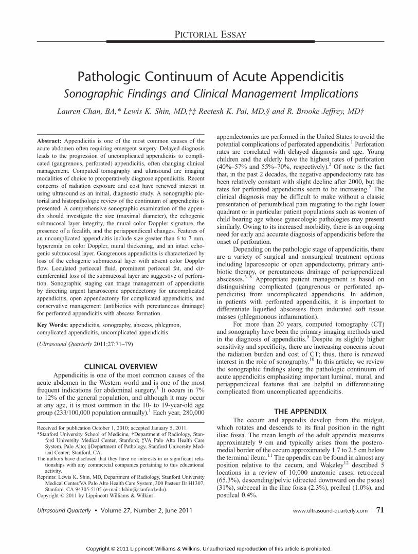
FIGURE 1. A and B, Normal appendix. Panel A is a longitudinalscan of the normal appendix. Note the well-preservedsubmucosal layer (long arrow), blind-ending tip (short arrow),and acoustic reflection from the collapsed luminal interface(arrowhead). Outer anteroposterior dimensions of appendixwere 4.4 mm, well within the reference range of less than5 mm. Panel B is a transverse scan of the normal appendixin the same patient with graded compression. Note theovoid shape of the normal appendix (long arrow) withreverberation echoes. Gas (short arrow) is seen within distaltip of appendix lateral to external iliac artery (arrowhead).
FIGURE 2. A and B, Early uncomplicated acute appendicitis.A, Longitudinal scan of the appendix, demonstrates distendedappendix (arrow) originating from the base of the cecal tip (‘‘C’’).The echogenic submucosal layer is preserved throughout. BTransverse color Doppler image of the same patient. Note theintramural hyperemia circumferentially seen within the wall ofthe appendix, indicating early appendicitis (arrow).
FIGURE 3. Early uncomplicated acute appendicitis.Longitudinal scan demonstrates distended appendix (arrows)with preserved echogenic submucosal layer throughout theentire length of the appendix.
Chan et al Ultrasound Quarterly & Volume 27, Number 2, June 2011
72 www.ultrasound-quarterly.com * 2011 Lippincott Williams & Wilkins
Copyright © 2011 Lippincott Williams & Wilkins. Unauthorized reproduction of this article is prohibited.
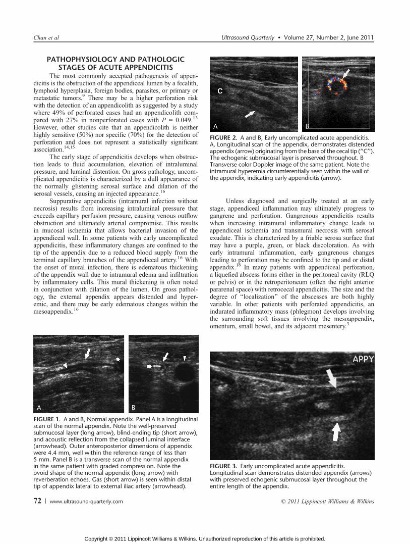
FIGURE 4. A and B, Early uncomplicated appendicitis with primary finding of distension. A, Transverse scan of RLQdemonstrating proximal and distal portions of the appendix cut in cross section (arrows) with luminal distension. There wereno other findings to indicate acute appendicitis, and the appendix measured 6.5 mm. B, Photomicrograph demonstratingearly, uncomplicated appendicitis with acute, luminal inflammation (black arrow) and mild, acute inflammation of the muscularispropria (white arrow) with diffuse wall thickening (double headed arrow).
FIGURE 5. A and B, Hyperemia in acute uncomplicated appendicitis. A, Longitudinal scan showing dilated appendix (longarrow) with multiple appendicoliths (short arrow). B, Longitudinal color Doppler scan of the same patient. Note the marked flowwithin the wall of the appendix, indicating early acute appendicitis.
FIGURE 6. A and B, Suppurative appendicitis with transmural inflammation and inflamed periappendiceal fat. A, Longitudinal scanof the appendix demonstrating dilated appendix (‘‘A’’) with increased echogenicity within periappendiceal fat (arrow)and poor delineation of echogenic submucosal layer, indicating transmural inflammation. B, Longitudinal color Doppler imageof the same patient. Note the slight increased flow within the inflamed periappendiceal fat (arrows).
Ultrasound Quarterly & Volume 27, Number 2, June 2011 Pathologic Continuum of Acute Appendicitis
* 2011 Lippincott Williams & Wilkins www.ultrasound-quarterly.com 73
Copyright © 2011 Lippincott Williams & Wilkins. Unauthorized reproduction of this article is prohibited.
ULTRASOUND AND CT OF THE APPENDIXGraded compression sonographic technique allows the
displacement of normal gas-filled bowel by slow sustainedpressure with the ultrasound (US) transducer, which helps apatient with peritonitis better tolerate the procedure.9 Scanningbegins at the edge of the right hepatic lobe, moving downwardinto the right iliac fossa to locate the ascending colon. Theileocecal valve can be identified by its fatty component andthe appendix should be located nearby. Although typicallyseen easily on transverse images, the origin of the appendixmay be better visualized arising from the cecum on longi-tudinal images.17 In cases when the appendix may extendinto the pelvis, transvaginal sonography may be a usefuladjunct to avoid missing a diagnosis of appendicitis. Criteriafor a normal appendix include a compressible, tubular, blindending structure, with wall thickness of 3 mm or less, absenceof peristalsis, and normal hyperechoic periappendiceal fatwithout inflammatory involvement of the cecum. It rarelyexceeds 5 mm in the outer anterior-posterior dimension andoften demonstrates an ovoid rather than a rounded shape(Fig. 1).18 The normal bowel wall layers of the appendix canbe distinguished progressing from the lumen to the serosaby alternating echogenicity: hypoechoic mucosa, hyperechoicmucosal interface and submucosa, hypoechoic muscularispropria, and hyperechoic serosa.17 These criteria are impor-tant to avoid false-positive studies where other structures (eg,dilated fallopian tube or dilated ureter) can mimic a dilatedappendix.
Sonography can be very limited in obese patients, andCT is often preferred in this population. Similarly, the normalappendix on CT appears as a tubular, pericecal structure that iscollapsed or filled with fluid, contrast, or air, with outer dia-meter that does not exceed 6 mm, wall thickness less than3 mm, and homogeneous periappendiceal fat. In appendicitis,CT demonstrates a distended, fluid-filled, blind-ending, tubu-lar structure arising from the cecum with mural thickeningand hyperenhancement. Linear fat stranding and local fascialthickening indicate periappendiceal inflammation. An appen-dicolith is variably present.9 For perforated appendicitis, thereare 5 CT findings that are 100% specific but have low sen-
sitivity: abscess, phlegmon, extraluminal air, extraluminalappendicolith, and focal defect in the appendix wall.13,19
US CORRELATION WITH PATHOLOGIC STAGE
Uncomplicated AppendicitisUncomplicated appendicitis may demonstrate a combi-
nation of luminal and mural changes. Using a thresholdmeasurement of 6 mm for the outer dimensions of a non-compressible appendix results in a sensitivity of 100% forappendicitis but a specificity of only 68%.20 If the size
FIGURE 7. A and B, Pelvic appendicitis adjacent to the ovary.A, Coronal endovaginal scan of the right adnexa demonstratingdilated appendix (‘‘A’’) immediately adjacent to the rightovary (‘‘OV’’). B, Endovaginal coronal color Doppler image.Note the slight hyperemia of the appendix with flow in theappendiceal wall on color Doppler (arrow). At surgery, earlyacute uncomplicated appendicitis was noted in the pelvicappendix immediately adjacent to the ovary.
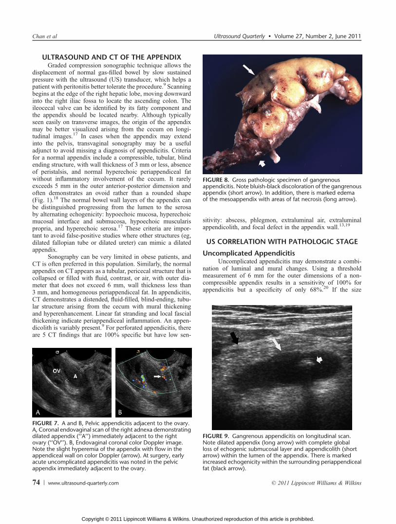
FIGURE 8. Gross pathologic specimen of gangrenousappendicitis. Note bluish-black discoloration of the gangrenousappendix (short arrow). In addition, there is marked edemaof the mesoappendix with areas of fat necrosis (long arrow).
FIGURE 9. Gangrenous appendicitis on longitudinal scan.Note dilated appendix (long arrow) with complete globalloss of echogenic submucosal layer and appendicolith (shortarrow) within the lumen of the appendix. There is markedincreased echogenicity within the surrounding periappendicealfat (black arrow).
Chan et al Ultrasound Quarterly & Volume 27, Number 2, June 2011
74 www.ultrasound-quarterly.com * 2011 Lippincott Williams & Wilkins
Copyright © 2011 Lippincott Williams & Wilkins. Unauthorized reproduction of this article is prohibited.
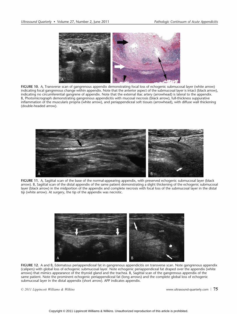
FIGURE 10. A, Transverse scan of gangrenous appendix demonstrating focal loss of echogenic submucosal layer (white arrow)indicating focal gangrenous change within appendix. Note that the anterior aspect of the submucosal layer is intact (black arrow),indicating no circumferential gangrene of appendix. Note that the external iliac artery (arrowhead) is lateral to the appendix.B, Photomicrograph demonstrating gangrenous appendicitis with mucosal necrosis (black arrow), full-thickness suppurativeinflammation of the muscularis propria (white arrow), and periappendiceal soft tissues (arrowhead), with diffuse wall thickening(double-headed arrow).
FIGURE 11. A, Sagittal scan of the base of the normal-appearing appendix, with preserved echogenic submucosal layer (blackarrow). B, Sagittal scan of the distal appendix of the same patient demonstrating a slight thickening of the echogenic submucosallayer (black arrow) in the midportion of the appendix and complete necrosis with focal loss of the submucosal layer in the distaltip (white arrow). At surgery, the tip of the appendix was necrotic.
FIGURE 12. A and B, Edematous periappendiceal fat in gangrenous appendicitis on transverse scan. Note gangrenous appendix(calipers) with global loss of echogenic submucosal layer. Note echogenic periappendiceal fat draped over the appendix (whitearrows) that mimics appearance of the thyroid gland and the trachea. B, Sagittal scan of the gangrenous appendix of thesame patient. Note the prominent echogenic periappendiceal fat (long arrows) and the complete global loss of echogenicsubmucosal layer in the distal appendix (short arrow). APP indicates appendix.
Ultrasound Quarterly & Volume 27, Number 2, June 2011 Pathologic Continuum of Acute Appendicitis
* 2011 Lippincott Williams & Wilkins www.ultrasound-quarterly.com 75
Copyright © 2011 Lippincott Williams & Wilkins. Unauthorized reproduction of this article is prohibited.
threshold is increased to 7 mm, however, the sensitivity dropsto 94%, but the specificity increases to 88%.20 When theappendix measures between 6 and 7 mm, demonstration ofintramural hyperemia on color Doppler may be a valuable aid inestablishing the diagnosis in borderline cases because there isusually no visualized flow in the normal appendix (Figs. 2Y7).9
On grayscale imaging, the normal wall of the appendixis less than 3 mm. Mural thickening is an important sono-graphic finding in appendicitis.9 Specifically, thickening ofthe echogenic submucosal layer is a sign of submucosaledema. Because appendicitis may be confined to the tip ofthe appendix, it is important to identify the ‘‘blind end’’ of theappendix in order not to miss early luminal or mural changes.Preservation of a normal submucosal echogenic layer, espe-cially in the distal and tip of the appendix, is an importantobservation indicating uncomplicated appendicitis due to lackof mural necrosis or gangrenous change.21Y23
The progressive enlargement of the intact submucosallayer signals suppurative appendicitis, with further transmu-ral inflammation causing appendiceal wall thickening, then
thinning from destruction of the mucosa, enlargement of themuscular layer, and an increase in distal appendiceal diameterdue to intraluminal accumulation of pus. Periappendiceal fatchanges are seen as well as false membranes from serosalinfection. Color Doppler shows severe hyperemia.24
Complicated Appendicitis (Gangrenous orPerforated Appendicitis)
Increasing intramural inflammation of the appendixultimately results in ischemia and appendiceal gangrene(Fig. 8). In patients with a strong clinical suspicion of perfo-rated appendicitis due to high fever and leukocytosis, contrastCT is often the imaging modality of choice because of thedifficulty in performing graded compression sonography inpatients with perforation and peritonitis. However, whentechnically satisfactory, sonography may demonstrate char-acteristic findings. Either focal or global loss of the echogenicsubmucosal layer is a key sonographic observation indicatinggangrenous appendicitis (Figs. 9Y12).25 Unlike the hyperemic
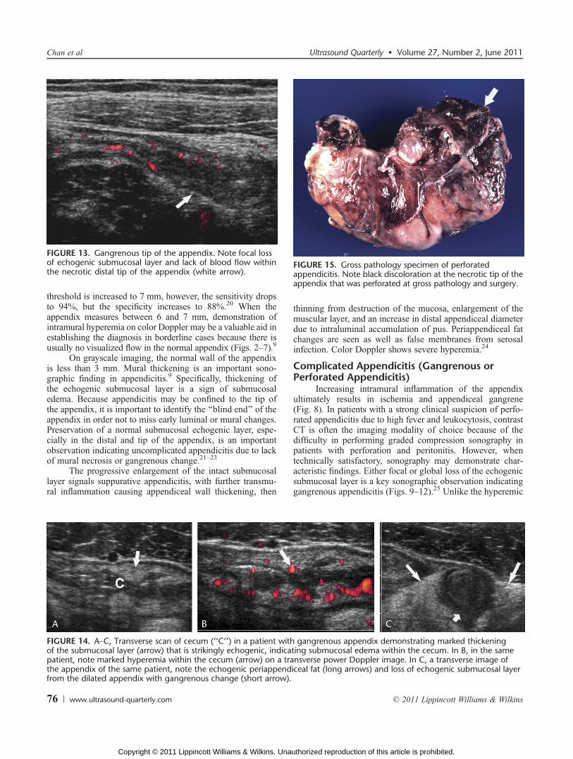
FIGURE 13. Gangrenous tip of the appendix. Note focal lossof echogenic submucosal layer and lack of blood flow withinthe necrotic distal tip of the appendix (white arrow).
FIGURE 14. AYC, Transverse scan of cecum (‘‘C’’) in a patient with gangrenous appendix demonstrating marked thickeningof the submucosal layer (arrow) that is strikingly echogenic, indicating submucosal edema within the cecum. In B, in the samepatient, note marked hyperemia within the cecum (arrow) on a transverse power Doppler image. In C, a transverse image ofthe appendix of the same patient, note the echogenic periappendiceal fat (long arrows) and loss of echogenic submucosal layerfrom the dilated appendix with gangrenous change (short arrow).
FIGURE 15. Gross pathology specimen of perforatedappendicitis. Note black discoloration at the necrotic tip of theappendix that was perforated at gross pathology and surgery.
Chan et al Ultrasound Quarterly & Volume 27, Number 2, June 2011
76 www.ultrasound-quarterly.com * 2011 Lippincott Williams & Wilkins
Copyright © 2011 Lippincott Williams & Wilkins. Unauthorized reproduction of this article is prohibited.
portions of the appendiceal wall that are merely inflamed butnot necrotic, areas of gangrenous necrosis are typically avas-cular on color Doppler because the blood supply to theseportions of the appendiceal wall is absent (Fig. 13).21 Trans-mural inflammatory exudate results in edema, first within thefat of the mesoappendix, then into the cecum and pericecalfat, and finally to the mesenteric or omental fat. The inflamedfat appears echogenic on grayscale imaging and hyperemic oncolor Doppler (Fig. 14).25,26 As previously emphasized, itis of critical importance to adequately visualize the tip ofthe appendix where gangrenous changes and perforationpreferentially occur.
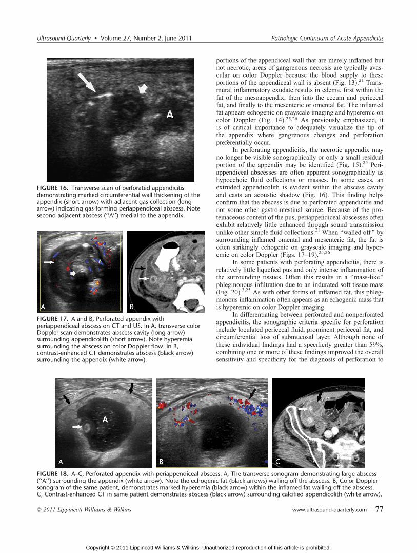
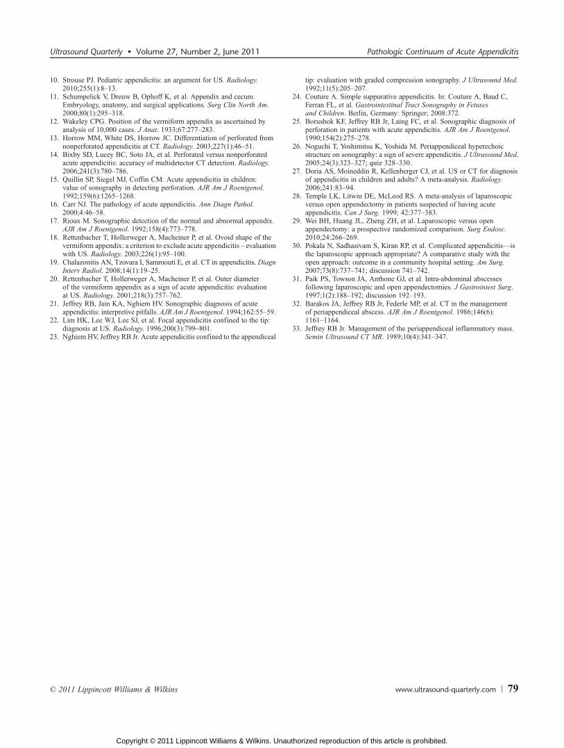
In perforating appendicitis, the necrotic appendix mayno longer be visible sonographically or only a small residualportion of the appendix may be identified (Fig. 15).25 Peri-appendiceal abscesses are often apparent sonographically ashypoechoic fluid collections or masses. In some cases, anextruded appendicolith is evident within the abscess cavityand casts an acoustic shadow (Fig. 16). This finding helpsconfirm that the abscess is due to perforated appendicitis andnot some other gastrointestinal source. Because of the pro-teinaceous content of the pus, periappendiceal abscesses oftenexhibit relatively little enhanced through sound transmissionunlike other simple fluid collections.21 When ‘‘walled off’’ bysurrounding inflamed omental and mesenteric fat, the fat isoften strikingly echogenic on grayscale imaging and hyper-emic on color Doppler (Figs. 17Y19).25,26
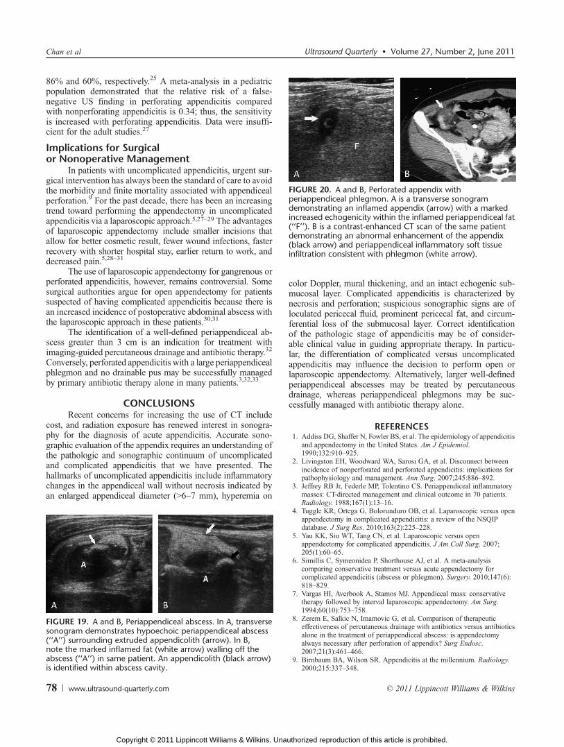
In some patients with perforating appendicitis, there isrelatively little liquefied pus and only intense inflammation ofthe surrounding tissues. Often this results in a ‘‘mass-like’’phlegmonous infiltration due to an indurated soft tissue mass(Fig. 20).3,25 As with other forms of inflamed fat, this phleg-monous inflammation often appears as an echogenic mass thatis hyperemic on color Doppler imaging.
In differentiating between perforated and nonperforatedappendicitis, the sonographic criteria specific for perforationinclude loculated pericecal fluid, prominent pericecal fat, andcircumferential loss of submucosal layer. Although none ofthese individual findings had a specificity greater than 59%,combining one or more of these findings improved the overallsensitivity and specificity for the diagnosis of perforation to
FIGURE 16. Transverse scan of perforated appendicitisdemonstrating marked circumferential wall thickening of theappendix (short arrow) with adjacent gas collection (longarrow) indicating gas-forming periappendiceal abscess. Notesecond adjacent abscess (‘‘A’’) medial to the appendix.
FIGURE 17. A and B, Perforated appendix withperiappendiceal abscess on CT and US. In A, transverse colorDoppler scan demonstrates abscess cavity (long arrow)surrounding appendicolith (short arrow). Note hyperemiasurrounding the abscess on color Doppler flow. In B,contrast-enhanced CT demonstrates abscess (black arrow)surrounding the appendix (white arrow).
FIGURE 18. AYC, Perforated appendix with periappendiceal abscess. A, The transverse sonogram demonstrating large abscess(‘‘A’’) surrounding the appendix (white arrow). Note the echogenic fat (black arrows) walling off the abscess. B, Color Dopplersonogram of the same patient, demonstrates marked hyperemia (black arrow) within the inflamed fat walling off the abscess.C, Contrast-enhanced CT in same patient demonstrates abscess (black arrow) surrounding calcified appendicolith (white arrow).
Ultrasound Quarterly & Volume 27, Number 2, June 2011 Pathologic Continuum of Acute Appendicitis
* 2011 Lippincott Williams & Wilkins www.ultrasound-quarterly.com 77
Copyright © 2011 Lippincott Williams & Wilkins. Unauthorized reproduction of this article is prohibited.
86% and 60%, respectively.25 A meta-analysis in a pediatricpopulation demonstrated that the relative risk of a false-negative US finding in perforating appendicitis comparedwith nonperforating appendicitis is 0.34; thus, the sensitivityis increased with perforating appendicitis. Data were insuffi-cient for the adult studies.27
Implications for Surgicalor Nonoperative Management
In patients with uncomplicated appendicitis, urgent sur-gical intervention has always been the standard of care to avoidthe morbidity and finite mortality associated with appendicealperforation.9 For the past decade, there has been an increasingtrend toward performing the appendectomy in uncomplicatedappendicitis via a laparoscopic approach.5,27Y29 The advantagesof laparoscopic appendectomy include smaller incisions thatallow for better cosmetic result, fewer wound infections, fasterrecovery with shorter hospital stay, earlier return to work, anddecreased pain.5,28Y31
The use of laparoscopic appendectomy for gangrenous orperforated appendicitis, however, remains controversial. Somesurgical authorities argue for open appendectomy for patientssuspected of having complicated appendicitis because there isan increased incidence of postoperative abdominal abscess withthe laparoscopic approach in these patients.30,31
The identification of a well-defined periappendiceal ab-scess greater than 3 cm is an indication for treatment withimaging-guided percutaneous drainage and antibiotic therapy.32
Conversely, perforated appendicitis with a large periappendicealphlegmon and no drainable pus may be successfully managedby primary antibiotic therapy alone in many patients.3,32,33
CONCLUSIONSRecent concerns for increasing the use of CT include
cost, and radiation exposure has renewed interest in sonogra-phy for the diagnosis of acute appendicitis. Accurate sono-graphic evaluation of the appendix requires an understanding ofthe pathologic and sonographic continuum of uncomplicatedand complicated appendicitis that we have presented. Thehallmarks of uncomplicated appendicitis include inflammatorychanges in the appendiceal wall without necrosis indicated byan enlarged appendiceal diameter (96Y7 mm), hyperemia on
color Doppler, mural thickening, and an intact echogenic sub-mucosal layer. Complicated appendicitis is characterized bynecrosis and perforation; suspicious sonographic signs are ofloculated pericecal fluid, prominent pericecal fat, and circum-ferential loss of the submucosal layer. Correct identificationof the pathologic stage of appendicitis may be of consider-able clinical value in guiding appropriate therapy. In particu-lar, the differentiation of complicated versus uncomplicatedappendicitis may influence the decision to perform open orlaparoscopic appendectomy. Alternatively, larger well-definedperiappendiceal abscesses may be treated by percutaneousdrainage, whereas periappendiceal phlegmons may be suc-cessfully managed with antibiotic therapy alone.
REFERENCES1. Addiss DG, Shaffer N, Fowler BS, et al. The epidemiology of appendicitis
and appendectomy in the United States. Am J Epidemiol.1990;132:910Y925.
2. Livingston EH, Woodward WA, Sarosi GA, et al. Disconnect betweenincidence of nonperforated and perforated appendicitis: implications forpathophysiology and management. Ann Surg. 2007;245:886Y892.
3. Jeffrey RB Jr, Federle MP, Tolentino CS. Periappendiceal inflammatorymasses: CT-directed management and clinical outcome in 70 patients.Radiology. 1988;167(1):13Y16.
4. Tuggle KR, Ortega G, Bolorunduro OB, et al. Laparoscopic versus openappendectomy in complicated appendicitis: a review of the NSQIPdatabase. J Surg Res. 2010;163(2):225Y228.
5. Yau KK, Siu WT, Tang CN, et al. Laparoscopic versus openappendectomy for complicated appendicitis. J Am Coll Surg. 2007;205(1):60Y65.
6. Simillis C, Symeonidea P, Shorthouse AJ, et al. A meta-analysiscomparing conservative treatment versus acute appendectomy forcomplicated appendicitis (abscess or phlegmon). Surgery. 2010;147(6):818Y829.
7. Vargas HI, Averbook A, Stamos MJ. Appendiceal mass: conservativetherapy followed by interval laparoscopic appendectomy. Am Surg.1994;60(10):753Y758.
8. Zerem E, Salkic N, Imamovic G, et al. Comparison of therapeuticeffectiveness of percutaneous drainage with antibiotics versus antibioticsalone in the treatment of periappendiceal abscess: is appendectomyalways necessary after perforation of appendix? Surg Endosc.2007;21(3):461Y466.
9. Birnbaum BA, Wilson SR. Appendicitis at the millennium. Radiology.2000;215:337Y348.
FIGURE 20. A and B, Perforated appendix withperiappendiceal phlegmon. A is a transverse sonogramdemonstrating an inflamed appendix (arrow) with a markedincreased echogenicity within the inflamed periappendiceal fat(‘‘F’’). B is a contrast-enhanced CT scan of the same patientdemonstrating an abnormal enhancement of the appendix(black arrow) and periappendiceal inflammatory soft tissueinfiltration consistent with phlegmon (white arrow).
FIGURE 19. A and B, Periappendiceal abscess. In A, transversesonogram demonstrates hypoechoic periappendiceal abscess(‘‘A’’) surrounding extruded appendicolith (arrow). In B,note the marked inflamed fat (white arrow) walling off theabscess (‘‘A’’) in same patient. An appendicolith (black arrow)is identified within abscess cavity.
Chan et al Ultrasound Quarterly & Volume 27, Number 2, June 2011
78 www.ultrasound-quarterly.com * 2011 Lippincott Williams & Wilkins
Copyright © 2011 Lippincott Williams & Wilkins. Unauthorized reproduction of this article is prohibited.
10. Strouse PJ. Pediatric appendicitis: an argument for US. Radiology.2010;255(1):8Y13.
11. Schumpelick V, Dreuw B, Ophoff K, et al. Appendix and cecum.Embryology, anatomy, and surgical applications. Surg Clin North Am.2000;80(1):295Y318.
12. Wakeley CPG. Position of the vermiform appendix as ascertained byanalysis of 10,000 cases. J Anat. 1933;67:277Y283.
13. Horrow MM, White DS, Horrow JC. Differentiation of perforated fromnonperforated appendicitis at CT. Radiology. 2003;227(1):46Y51.
14. Bixby SD, Lucey BC, Soto JA, et al. Perforated versus nonperforatedacute appendicitis: accuracy of multidetector CT detection. Radiology.2006;241(3):780Y786.
15. Quillin SP, Siegel MJ, Coffin CM. Acute appendicitis in children:value of sonography in detecting perforation. AJR Am J Roentgenol.1992;159(6):1265Y1268.
16. Carr NJ. The pathology of acute appendicitis. Ann Diagn Pathol.2000;4:46Y58.
17. Rioux M. Sonographic detection of the normal and abnormal appendix.AJR Am J Roentgenol. 1992;158(4):773Y778.
18. Rettenbacher T, Hollerweger A, Macheiner P, et al. Ovoid shape of thevermiform appendix: a criterion to exclude acute appendicitis Y evaluationwith US. Radiology. 2003;226(1):95Y100.
19. Chalazonitis AN, Tzovara I, Sammouti E, et al. CT in appendicitis. DiagnInterv Radiol. 2008;14(1):19Y25.
20. Rettenbacher T, Hollerweger A, Macheiner P, et al. Outer diameterof the vermiform appendix as a sign of acute appendicitis: evaluationat US. Radiology. 2001;218(3):757Y762.
21. Jeffrey RB, Jain KA, Nghiem HV. Sonographic diagnosis of acuteappendicitis: interpretive pitfalls. AJR Am J Roentgenol. 1994;162:55Y59.
22. Lim HK, Lee WJ, Lee SJ, et al. Focal appendicitis confined to the tip:diagnosis at US. Radiology. 1996;200(3):799Y801.
23. Nghiem HV, Jeffrey RB Jr. Acute appendicitis confined to the appendiceal
tip: evaluation with graded compression sonography. J Ultrasound Med.1992;11(5):205Y207.
24. Couture A. Simple suppurative appendicitis. In: Couture A, Baud C,Ferran FL, et al. Gastrointestinal Tract Sonography in Fetusesand Children. Berlin, Germany: Springer; 2008:372.
25. Borushok KF, Jeffrey RB Jr, Laing FC, et al. Sonographic diagnosis ofperforation in patients with acute appendicitis. AJR Am J Roentgenol.1990;154(2):275Y278.
26. Noguchi T, Yoshimitsu K, Yoshida M. Periappendiceal hyperechoicstructure on sonography: a sign of severe appendicitis. J Ultrasound Med.2005;24(3):323Y327; quiz 328Y330.
27. Doria AS, Moineddin R, Kellenberger CJ, et al. US or CT for diagnosisof appendicitis in children and adults? A meta-analysis. Radiology.2006;241:83Y94.
28. Temple LK, Litwin DE, McLeod RS. A meta-analysis of laparoscopicversus open appendectomy in patients suspected of having acuteappendicitis. Can J Surg. 1999; 42:377Y383.
29. Wei BH, Huang JL, Zheng ZH, et al. Laparoscopic versus openappendectomy: a prospective randomized comparison. Surg Endosc.2010;24:266Y269.
30. Pokala N, Sadhasivam S, Kiran RP, et al. Complicated appendicitisVisthe laparoscopic approach appropriate? A comparative study with theopen approach: outcome in a community hospital setting. Am Surg.2007;73(8):737Y741; discussion 741Y742.
31. Paik PS, Towson JA, Anthone GJ, et al. Intra-abdominal abscessesfollowing laparoscopic and open appendectomies. J Gastrointest Surg.1997;1(2):188Y192; discussion 192Y193.
32. Barakos JA, Jeffrey RB Jr, Federle MP, et al. CT in the managementof periappendiceal abscess. AJR Am J Roentgenol. 1986;146(6):1161Y1164.
33. Jeffrey RB Jr. Management of the periappendiceal inflammatory mass.Semin Ultrasound CT MR. 1989;10(4):341Y347.
Ultrasound Quarterly & Volume 27, Number 2, June 2011 Pathologic Continuum of Acute Appendicitis
* 2011 Lippincott Williams & Wilkins www.ultrasound-quarterly.com 79
Copyright © 2011 Lippincott Williams & Wilkins. Unauthorized reproduction of this article is prohibited.