Pathologic and clinicopathologic studies on chronic copper ... · nostic 9100, Baker Serono...
17
Egypt. J. Comp. Path. & Clinic. Path. Vol. 22 No. 1 (January) 2009; 1 - 18 1 Referred by Referred by Prof. Dr. Salah Deeb Deeb Professor of Pathology, Fac. Vet. Med., Beni Sueif University Prof. Dr. Mahmoud Samy Professor of Pathology, Fac. Vet. Med., Cairo University Pathologic and clinicopathologic studies on chronic copper toxicosis in a group of milking Ewes By Sameh Youssef * Dept. of Pathobiology, Ontario Veterinary College, Ontario, Canada *Home address: Dept of Pathology, Faculty of Vet medicine, Alexandria, Egypt SUMMARY D uring the summer of 2005, seventeen milking ewes from a flock of 200 ewes died over a period of 5 days with unknown disease. Clinical signs included anorexia, recumbency, and few ewes were acutely ataxic. Five ewes were sent for necropsy and histopathology. Le- sions included hepatocellular apoptosis and necrosis with marked acute renal tubular degeneration and necrosis. Three ewes showed brain le- sions typical of hepatic encephalopathy. A diagnosis of copper toxicity was made on the basis of the aforementioned lesions and on the high levels of copper in the liver and serum of affected ewes. Ewes were found to have been accidentally fed on a ration that had a mineral mix containing 650 ppm copper for 3 months prior to the onset of mortalities. The mineral mix was removed from the ration one day after the mortali- ties started. This paper describes also the detailed microscopic lesions occurred in the brains of some of these copper-intoxicated ewes. INTRODUCTION S heep have a strong ability to accumulate copper in their liv- ers and a reduced rate of bile clear- ance more readily than do other animal species (Smith, 1996 and Maxie, 2007). These two factors make sheep one of the most sus- ceptible species for copper toxic- ity. Acute toxicosis results from ingestion of very high amounts of copper is rare. The toxicity in this form is manifested by vomiting, diarrhea and acute death usually results from the direct irritant ef- fect of the copper on the gastroin- testinal tracts (Lewis et al., 1997; Roder, 2001 and Ortolani et al., 2004). In contrast, the most com- mon form of copper toxicosis in sheep is the chronic form which results from chronic ingestion of
Transcript of Pathologic and clinicopathologic studies on chronic copper ... · nostic 9100, Baker Serono...
Egypt. J. Comp. Path. & Clinic. Path. Vol. 22 No. 1 (January) 2009; 1 - 18
1
Referred byReferred by Prof. Dr. Salah Deeb Deeb Professor of Pathology, Fac. Vet. Med., Beni
Sueif University Prof. Dr. Mahmoud Samy Professor of Pathology, Fac. Vet. Med.,
Cairo University
Pathologic and clinicopathologic studies on chronic copper toxicosis in a group of milking Ewes
By Sameh Youssef*
Dept. of Pathobiology, Ontario Veterinary College, Ontario, Canada *Home address: Dept of Pathology, Faculty of Vet medicine, Alexandria,
Egypt
SUMMARY
D uring the summer of 2005, seventeen milking ewes from a flock of 200 ewes died over a period of 5 days with unknown disease.
Clinical signs included anorexia, recumbency, and few ewes were acutely ataxic. Five ewes were sent for necropsy and histopathology. Le-sions included hepatocellular apoptosis and necrosis with marked acute renal tubular degeneration and necrosis. Three ewes showed brain le-sions typical of hepatic encephalopathy. A diagnosis of copper toxicity was made on the basis of the aforementioned lesions and on the high levels of copper in the liver and serum of affected ewes. Ewes were found to have been accidentally fed on a ration that had a mineral mix containing 650 ppm copper for 3 months prior to the onset of mortalities. The mineral mix was removed from the ration one day after the mortali-ties started. This paper describes also the detailed microscopic lesions occurred in the brains of some of these copper-intoxicated ewes.
INTRODUCTION
S heep have a strong ability to accumulate copper in their liv-
ers and a reduced rate of bile clear-ance more readily than do other animal species (Smith, 1996 and Maxie, 2007). These two factors make sheep one of the most sus-ceptible species for copper toxic-ity. Acute toxicosis results from ingestion of very high amounts of
copper is rare. The toxicity in this form is manifested by vomiting, diarrhea and acute death usually results from the direct irritant ef-fect of the copper on the gastroin-testinal tracts (Lewis et al., 1997; Roder, 2001 and Ortolani et al., 2004). In contrast, the most com-mon form of copper toxicosis in sheep is the chronic form which results from chronic ingestion of
Egypt. J. Comp. Path. & Clinic. Path. Vol. 22 No. 1 (January) 2009; 1 - 18
2
copper in amounts that are slightly to moderately higher than normal dietary levels that sheep can toler-ate (Smith, 1996; Fuentealba and Aburto, 2003; Maxie, 2007 and Mendel et al., 2007). Copper is a powerful hepatotoxin and causes severe hemolysis due to its oxidant effect on the red blood cells (RBCs) and hepatocellular cell membranes (Fuentealba and Aburto, 2003; Christodoulopou-los and Roubies, 2007; McGavin and Zachary, 2007 and Mendel et al., 2007). Therefore, sheep chronically intoxicated with cop-per die either from hepatic failure or from hemolytic crisis. Sources of copper are multiple and include copper-containing medications, feed additives, consumption of for-age plants rich in copper or con-taminated with pesticides contain-ing copper, and from water ponds contaminated with copper contain-ing pesticides and algicides (Lewis, 1997; McGavin and Zachary, 2007 and Mendel et al., 2007). Cooper accumulation also increases if the dietary molybde-num level is low (Smith, 1996; Roder, 2001 and Maxie, 2007). Molybdenum, in the presence of sufficient sulfate, forms insoluble complexes with copper in the in-testine and liver, making the cop-per biologically inactive. Hepatic insufficiency from any etiology can lead to severe brain lesions in a clinical condition known as he-
patic encephalopathy (HE) (Sargison et al., 2001; Bobe et al., 2004; Johns et al., 2007 and McGavin and Zachary, 2007). Liver has a major detoxifying role which prevents the accumulation of ammonia and other metabolites. The inability of diseased liver to do this function leads to the accu-mulation of these metabolites which can cause severe brain physiological and histological changes leading in the end to HE (Maxie, 2007; McGavin and Zachary, 2007 and Zielińska et al., 2007). Sheep intoxicated with cooper usually die before develop-ing HE. Reported here, the lesions and the biochemical changes asso-ciated with chronic copper toxicity in a group of milking ewes. MATERIALS AND METHODS Clinical history:
During the summer of 2005, seventeen milking ewes from a flock of 200 ewes suddenly died over a period of five days (days 1 through 5) of unknown disease. Dead ewes were recumbent, ano-rexic and showed acute ataxia shortly before death. Dry ewes and lambs were clinically healthy and no mortalities were reported among them. All animals were vaccinated up to date and being treated with Ivermectin twice a year. Feed was a complete dairy ration and hay. Two animals (SH1 and SH2) that died at day 1 and
Egypt. J. Comp. Path. & Clinic. Path. Vol. 22 No. 1 (January) 2009; 1 - 18
3
three (SHs 3, 4, 5) that died at day 4 were sent to the Animal health laboratory, University of Guelph, Ontario, Canada for investigation of the cause of death. A complete pathologic investigation was car-ried out. Clinical pathology and toxicol-ogy:
Blood and sera were col-lected from dying ewes and from those showed clinical signs. Whole blood was analyzed with an auto-mated cell counter (Serono Diag-nostic 9100, Baker Serono Diag-nostics, Allentown, PA). Serum
biochemistry was measured with an automated chemistry analyzer
(Beckmann Synchron CX-5 chem-istry analyzer with Beckman re-agents and following the manufac-ture's recommendations, Beckman Diagnostics, Brea, CA). Quantita-tive mineral analysis for Copper and other minerals in livers were estimated by flame atomic absorp-tion spectrophotometry. In brief, the liver specimens were dehy-drated and dried overnight at 85ºC, ashed overnight at 600ºC, dis-solved in NHNO3, and diluted for analysis (Helrich, 1990 and Schultheiss et al., 2002). Levels of copper in the ration was analyzed using an atomic absorption spec-trophotometer as described previ-ously (Saito et al., 1988). Molyb-denum, and other trace elements levels in the ration were analyzed
also by the atomic absorption spec-trophotometer using the standard operation protocols of the Animal Health Laboratory, University of Guelph, Guelph, Ontario, Canada.
Pathologic studies:
Selected specimens from different organs at time of ne-cropsy were collected and fixed in 10% neutral buffered formalin. The fixed specimens were proc-essed according to standard proto-cols, embedded in paraffin wax (56 °C) using an automatic appara-tus, sectioned on rotary microtome at thickness of 4 microns then stained with routine hematoxyline and eosin (H&E). Selected liver and kidney sections were stained for copper and some brain sections were stained for myelin (Prophet et al., 1992).
RESULTS
Gross pathology: A- Sheep died at day 1:
Two sheep (SH1 and SH2) were necropsied and unless other-wise described, both had similar lesions. Mucous membranes were pale to mildly congested. Hydra-tion, body fat, and musculature were within the normal limits. The abdominal cavity of SH1 con-tained approximately 250 ml of clear ascetic fluid. The rumenal contents were semisolid and com-posed of roughages, grains, and light grey fluid. Feces was formed
Egypt. J. Comp. Path. & Clinic. Path. Vol. 22 No. 1 (January) 2009; 1 - 18
4
but small intestine was gas and fluid filled. The livers of both ani-mals were diffusely paler but had normal hepatoid texture. The gall bladders were markedly distended with bile. The lungs were diffusely moderately edematous. Kidneys and spleens were diffusely se-verely congested. The renal corti-ces had few tiny (0.1 to 0.2 cm) fairly circumscribed pale grey ar-eas. Menengial blood vessels were markedly congested.
B- Sheep died at day 4: All ewes had excellent body
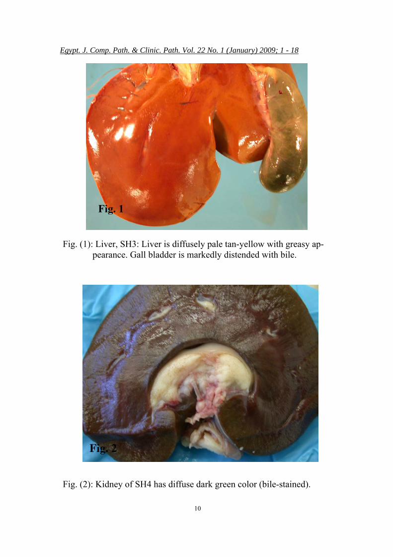
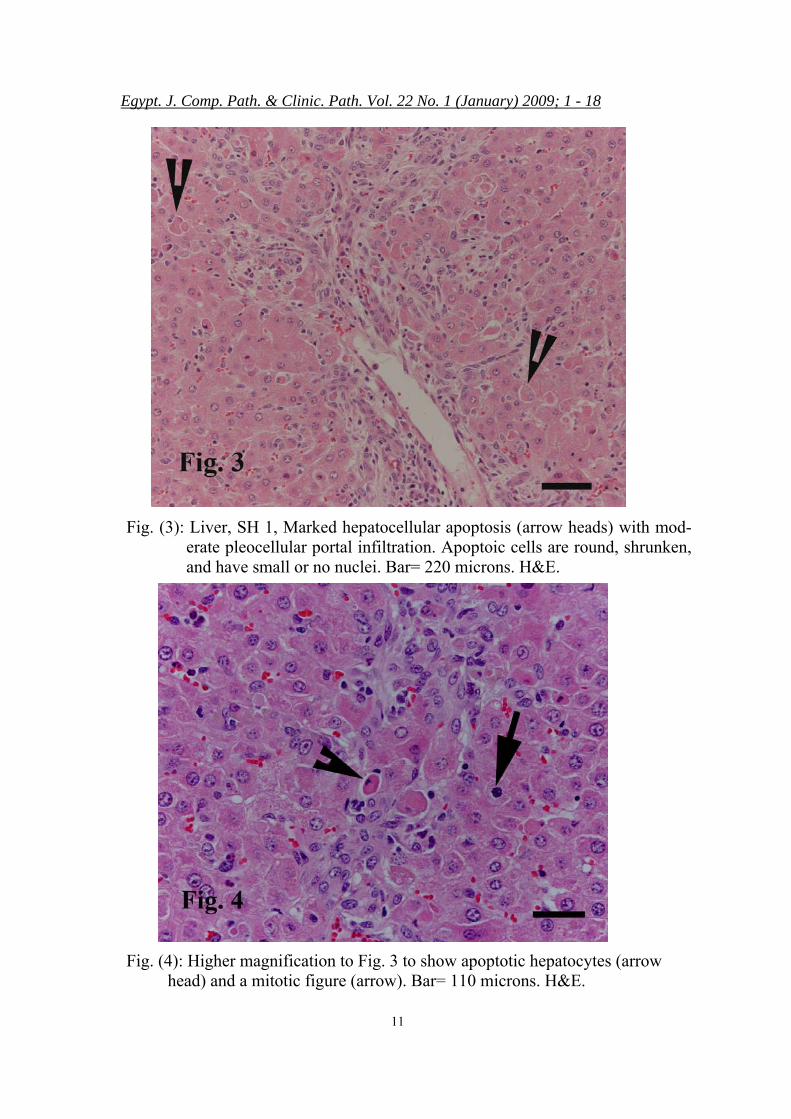
condition and were slightly obese. Unless otherwise indicated, the gross pathology of the three ne-cropised ewes (SH3, SH4, and SH5) in this group was similar. Body fat and mucous membranes were mildly icteric. The livers of all animals were diffusely tan yel-low and had greasy texture (Fig.1). Occasionally, the liver cut surface had enhanced lobular pattern. Kid-neys of SH3 and SH4 were dif-fusely dark green (bilirubin-stained) (Fig. 2). The kidney of SH 5 was diffusely dark red. The heart of SH3 had multifocal ecchymotic hemorrhages in epicardium and endocardium, as well as extensive hemorrhage within the papillary muscles and adjacent septal and ventricular free wall of the left ventricle. Lung of all animals were diffusely dark red, wet and heavy. Menengial blood vessels were markedly congested.
Histopathology: Multiple organs were col-lected for histopathology. How-ever, significant lesions were pre-sent only in the livers and kidneys of all examined sheep that died ei-ther at day 1 or day 4 and the brain of sheep died at day 4. The brains of sheep died at day 1 were not ex-amined histologically.
A- Sheep died at day 1: Unless otherwise described
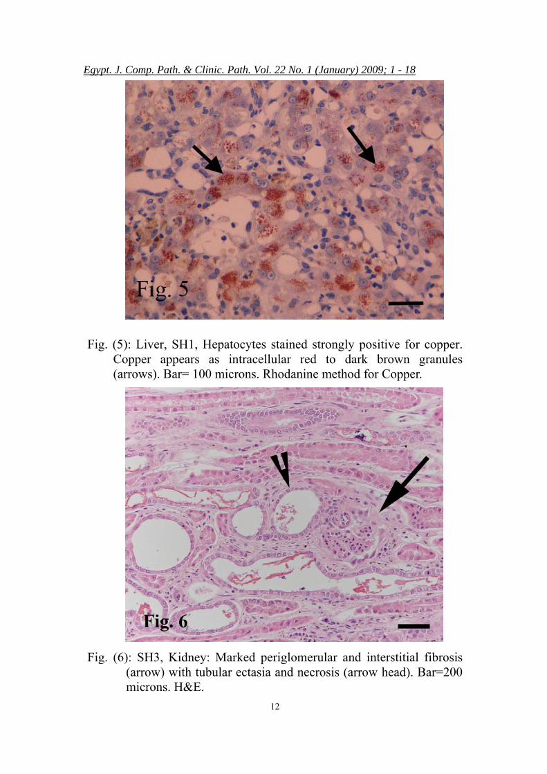
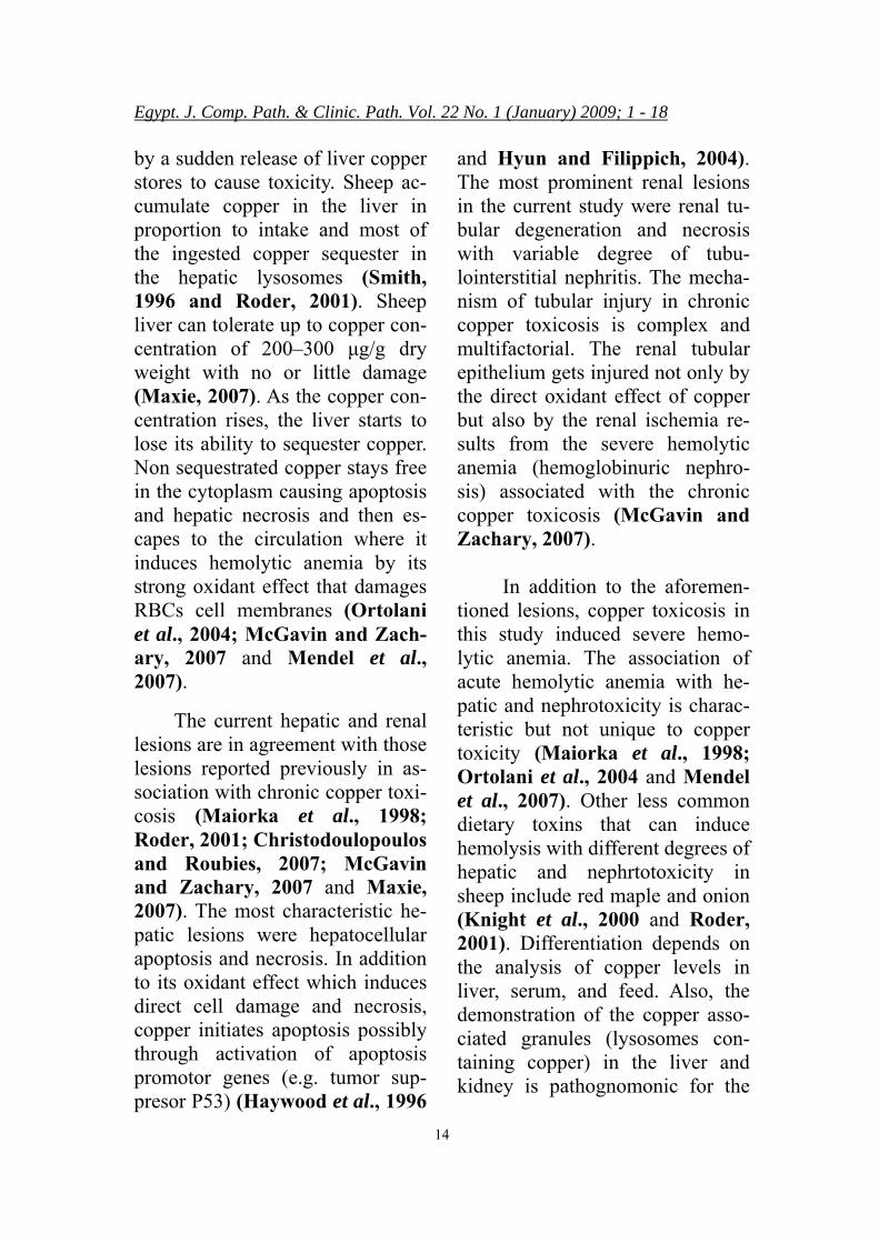
the microscopic lesions were simi-lar in both sheep examined (SH1 and SH2). Liver: There was a marked hepa-tocellular apoptosis affecting nu-merous individual hepatocytes in all zones (Fig.3). Apoptotic cells appeared round, had hypereosino-philic homogenous cytoplasm with copper-positive cytoplasmic gran-ules, and pyknotic nuclei (Figs 4 and 5). Mitotic figures were fre-quently seen (indicative of in-creased wear and tear). Kupffer cells had swollen nuclei and their cytoplasm was moderately filled with brown globular pigment that was stained positive for copper. The portal areas were mildly to moderately infiltrated with lym-phocytes, plasma cells, macro-phages, and fewer neutrophils. Oc-casionally, tiny multifocal random areas of parenchymal loss (necr-otic areas) were present and char-acterized by replacement of the he-patic parenchyma with necrotic de-
Egypt. J. Comp. Path. & Clinic. Path. Vol. 22 No. 1 (January) 2009; 1 - 18
5
bris and pleocellular inflammatory exudates. Few hepatocytes had in-tracellular fat vacuoles (fatty change).
Kidney: There were marked multi-focal tubular ectasia, attenuation, and necrosis affecting mostly the proximal convoluted tubules but was present in all tubular levels. Affected tubules appeared more basophilic, dilated, had wider lu-mens, and were lined with flat-tened to low cuboidal cells or by necrotic cells. Occasionally, the lumens of the affected tubules con-tained hemolysed blood or light golden brown globular pigment consistent with bile. Few finely granular copper-positive granules were present in the cytoplasm of both necrotic and intact proximal tubules. Rarely, sporadic mineral-ized tubules were present particu-larly in the corticomedullary junc-tion. Random multifocal areas of interstitial fibrosis with mild infil-tration of lymphocytes and plasma cells were present. Glomeruli were generally spared and had no sig-nificant lesions. B- Sheep died at day 4:
Unless otherwise described the microscopic lesions were simi-lar in the 3 sheep examined (SH3, SH4 and SH5) from this group.
Liver: The most characteristic le-sions were present in the portal ar-eas and appeared in form of mild biliary proliferation with mild to
moderate infiltration of portal ar-eas with lymphocytes, plasma cells, and fewer copper-laden macrophages. Mild periacinar ne-crosis affecting periacinar hepato-cytes was present. Occasional hepatocellular apoptosis was also present. Moderate numbers of cop-per-positive cytoplasmic granules were present in hepatocytes (parti-cularly in necrotic hepatocytes), portal macrophages and within the Kupffer cells. Kidney: In addition to the tubular changes described above, there was a mild interstitial fibroplasia, edema, and infiltration with mono-nuclear cells (Fig.6). The lumens of the degenerated/necrotic tubules contained red granular casts. Occa-sionally, the cytoplasm of some tubules either in cortex or medulla contained few yellowish green granules that were consistent with bilirubin.
Brain: The meningeal blood ves-sels were mildly congested. There was mild perivascular edema af-fecting the capillaries of the cere-bral cortex. The white matter of the cerebellum and to lesser extend the cerebral cortex was markedly paler (neuropilar palor) and had severe well-developed spongy vacuolation of myelin (status spongiosis). Vacuoles were multi-ple, symmetrical, and varied in size (10- 150 microns) (Fig.7). Us-ing myelin stain, the vacuolated areas appeared as an empty area

Egypt. J. Comp. Path. & Clinic. Path. Vol. 22 No. 1 (January) 2009; 1 - 18
6
devoid of myelin with no hypo-myelination or myelin degenera-tion. Mild diffuse astrocytosis with rare satellitosis were present in the external laminar cerebral cortical layer. Few astrocytes with large and pale nuclei (Alzheimer type II astrocytes) were present in the external cortical laminar layer. No significant neuronal changes were present.
Clinical Pathology: Blood and serum analysis
were evaluated only in SHs 1, 2, and 3. Blood and serum from SHs 4 and 5 were collected but not evaluated.
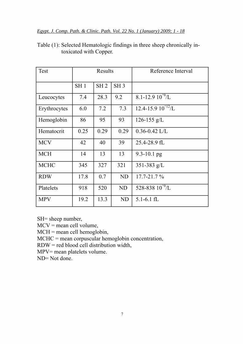
Blood analysis of all tested sheep revealed the presence of acute hemolytic anemia (Tab. 1) as indicated by red plasma, hyper-bilirubinemia, hemoglobinemia, increased mean corpuscular hemo-globin concentration (MCHC), and erythrocytopenia. Serum biochem-istry panel (Tab. 2) indicated marked liver and renal injury with severe electrolytes imbalance. The liver injury was detected mainly by increased activity of aspartate ami-notransferase (AST), and gamma glutamyl transferase (GGT). Se-rum indicators of renal abnormali-ties included azotemia (increased urea concentration) and increased cretinine concentration.
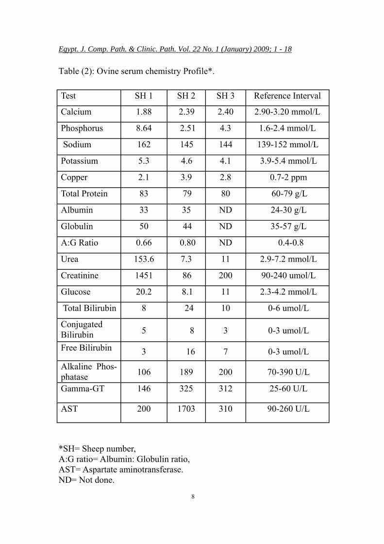
Toxicologic studies: Feed analysis revealed the
presence of copper at concentra-
tion of 650 ppm. The liver copper
concentrations were markedly in-creased (Tab. 3) in all examined animals. Hepatic copper concen-tration measured in SH1, SH 2, SH 3, SH 4 and SH5 were 380, 840, 400, 312, and 520 ppm respec-tively dry liver weight (normal range is 25-100 ppm). No other abnormalities were present in the mean hepatic heavy metal screens. Also, the serum copper concentra-tions of the in SH1, SH2 and SH3 were 2.1, 3.9, and 2.8 ppm respec-tively (normal value 0.7-2 ppm) (Tab. 1)
Egypt. J. Comp. Path. & Clinic. Path. Vol. 22 No. 1 (January) 2009; 1 - 18
7
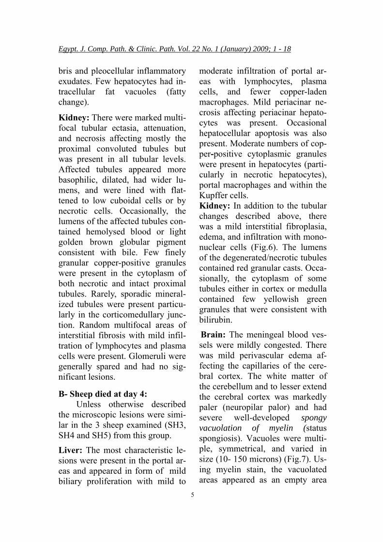
Table (1): Selected Hematologic findings in three sheep chronically in-toxicated with Copper.
Test Results Reference Interval
SH 1 SH 2 SH 3
Leucocytes 7.4 28.3 9.2 8.1-12.9 10^9/L
Erythrocytes 6.0 7.2 7.3 12.4-15.9 10^12/L
Hemoglobin 86 95 93 126-155 g/L
Hematocrit 0.25 0.29 0.29 0.36-0.42 L/L
MCV 42 40 39 25.4-28.9 fL
MCH 14 13 13 9.3-10.1 pg
MCHC 345 327 321 351-383 g/L
RDW 17.8 0.7 ND 17.7-21.7 %
Platelets 918 520 ND 528-838 10^9/L
MPV 19.2 13.3 ND 5.1-6.1 fL
SH= sheep number, MCV = mean cell volume, MCH = mean cell hemoglobin, MCHC = mean corpuscular hemoglobin concentration, RDW = red blood cell distribution width, MPV= mean platelets volume. ND= Not done.
Egypt. J. Comp. Path. & Clinic. Path. Vol. 22 No. 1 (January) 2009; 1 - 18
8
Table (2): Ovine serum chemistry Profile*.
Test SH 1 SH 2 SH 3 Reference Interval
Calcium 1.88 2.39 2.40 2.90-3.20 mmol/L
Phosphorus 8.64 2.51 4.3 1.6-2.4 mmol/L
Sodium 162 145 144 139-152 mmol/L
Potassium 5.3 4.6 4.1 3.9-5.4 mmol/L
Copper 2.1 3.9 2.8 0.7-2 ppm
Total Protein 83 79 80 60-79 g/L
Albumin 33 35 ND 24-30 g/L
Globulin 50 44 ND 35-57 g/L
A:G Ratio 0.66 0.80 ND 0.4-0.8
Urea 153.6 7.3 11 2.9-7.2 mmol/L
Creatinine 1451 86 200 90-240 umol/L
Glucose 20.2 8.1 11 2.3-4.2 mmol/L
Total Bilirubin 8 24 10 0-6 umol/L
Conjugated Bilirubin 5 8 3 0-3 umol/L
Free Bilirubin 3 16 7 0-3 umol/L
Alkaline Phos-phatase
106 189 200 70-390 U/L
Gamma-GT 146 325 312 25-60 U/L
AST 200 1703 310 90-260 U/L
*SH= Sheep number, A:G ratio= Albumin: Globulin ratio, AST= Aspartate aminotransferase. ND= Not done.
Egypt. J. Comp. Path. & Clinic. Path. Vol. 22 No. 1 (January) 2009; 1 - 18
9
Table (3): Heavy metal screen on liver of all examined sheep.
Tests* Results
SH 1 SH 2 SH 3 SH 4 SH 5
Copper 380 840 400 312 520 25.0 100.0 ppm
Iron 190 73 150 142 142 30 200 ppm
Manganese 2.8 3.3 2.9 2.7 2.6 2.0 4.4 ppm
Molybde-
num 2.0 2.0 2.9 1.9 2.0 1.5 6.0 ppm
Zinc 25 47 74 45 38 30.0 75.0 ppm
Selenium 1.1 1.5 0.74 1.5 1.3 0.25 1.5 ppm
Reference In-
tervals
* In addition to the metals mentioned in the Tab. 3, no significant levels of arsenic, cadmium, cobalt, chromium and lead were present in any of the examined livers. SH= sheep number.
Egypt. J. Comp. Path. & Clinic. Path. Vol. 22 No. 1 (January) 2009; 1 - 18
10
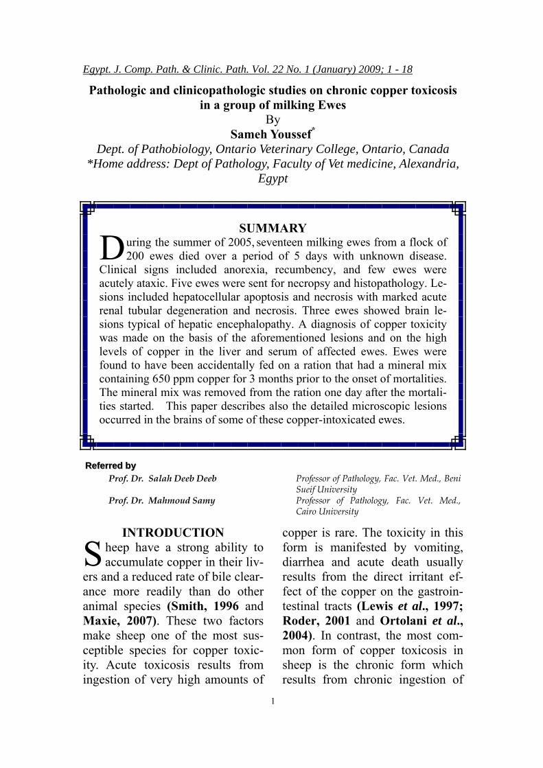
Fig. (1): Liver, SH3: Liver is diffusely pale tan-yellow with greasy ap-pearance. Gall bladder is markedly distended with bile.
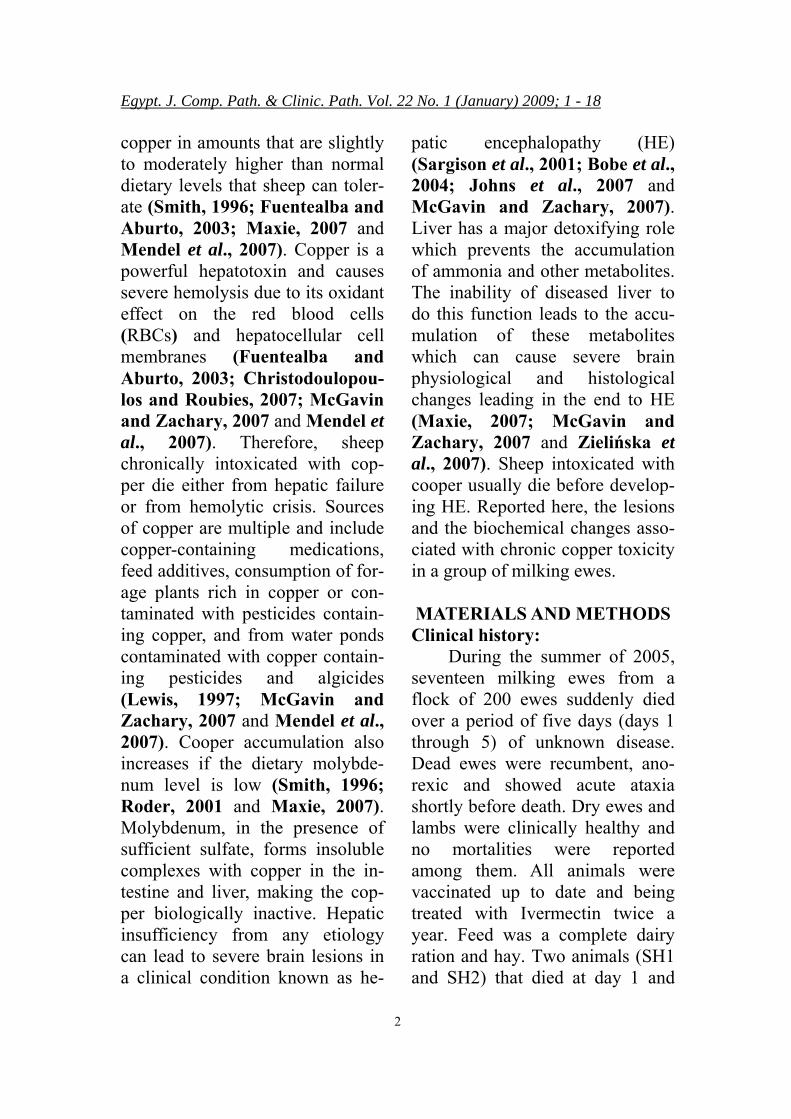
Fig. (2): Kidney of SH4 has diffuse dark green color (bile-stained).
Egypt. J. Comp. Path. & Clinic. Path. Vol. 22 No. 1 (January) 2009; 1 - 18
11
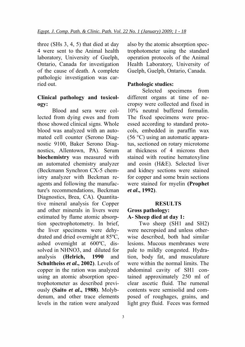
Fig. (3): Liver, SH 1, Marked hepatocellular apoptosis (arrow heads) with mod-erate pleocellular portal infiltration. Apoptoic cells are round, shrunken, and have small or no nuclei. Bar= 220 microns. H&E.
Fig. (4): Higher magnification to Fig. 3 to show apoptotic hepatocytes (arrow head) and a mitotic figure (arrow). Bar= 110 microns. H&E.
Egypt. J. Comp. Path. & Clinic. Path. Vol. 22 No. 1 (January) 2009; 1 - 18
12
Fig. (5): Liver, SH1, Hepatocytes stained strongly positive for copper. Copper appears as intracellular red to dark brown granules (arrows). Bar= 100 microns. Rhodanine method for Copper.
Fig. (6): SH3, Kidney: Marked periglomerular and interstitial fibrosis (arrow) with tubular ectasia and necrosis (arrow head). Bar=200 microns. H&E.

Egypt. J. Comp. Path. & Clinic. Path. Vol. 22 No. 1 (January) 2009; 1 - 18
13
Fig. (7): SH 4: Brain stem: Marked edema and vacuolation of the white matter (status spongiosis). Bar= 220 microns. Luxol fast blue.
DISCUSSION:
I n this paper, we have reported the clinical signs, lesions, and
the biochemical and hematologic changes that occurred in a group of milking ewes that were chronically intoxicated with dietary copper. These ewes were found to have been accidentally fed on a ration that had a mineral mix containing 650 ppm copper for 3 months prior to the onset of acute mortalities. Dry ewes and lambs were in a dif-ferent feeding system and they did not receive this mineral mix, there-fore, no mortalities or clinical signs were observed on them. Gen-erally, sheep require about 5 ppm (parts per million) of copper in
their total diet and toxicity can oc-cur at levels above 25 ppm (NRC, 1985; Smith, 1996 and Roder, 2001). The current mortalities and clinical course were sudden and acute, a finding typical of chronic copper toxicosis in sheep which almost always appears acutely or suddenly despite the fact that the ingestion of copper was chronic (Soli, 1980; Smith, 1996; Roder, 2001; Fuentealba and Aburto, 2003; Maxie, 2007 and Mendel et al., 2007). In other words, copper toxicity in sheep usually results from the accumulation of copper in the liver over a period of a few weeks to more than a year with no clinical signs this usually follow
Egypt. J. Comp. Path. & Clinic. Path. Vol. 22 No. 1 (January) 2009; 1 - 18
14
by a sudden release of liver copper stores to cause toxicity. Sheep ac-cumulate copper in the liver in proportion to intake and most of the ingested copper sequester in the hepatic lysosomes (Smith, 1996 and Roder, 2001). Sheep liver can tolerate up to copper con-centration of 200–300 μg/g dry weight with no or little damage (Maxie, 2007). As the copper con-centration rises, the liver starts to lose its ability to sequester copper. Non sequestrated copper stays free in the cytoplasm causing apoptosis and hepatic necrosis and then es-capes to the circulation where it induces hemolytic anemia by its strong oxidant effect that damages RBCs cell membranes (Ortolani et al., 2004; McGavin and Zach-ary, 2007 and Mendel et al., 2007).
The current hepatic and renal lesions are in agreement with those lesions reported previously in as-sociation with chronic copper toxi-cosis (Maiorka et al., 1998; Roder, 2001; Christodoulopoulos and Roubies, 2007; McGavin and Zachary, 2007 and Maxie, 2007). The most characteristic he-patic lesions were hepatocellular apoptosis and necrosis. In addition to its oxidant effect which induces direct cell damage and necrosis, copper initiates apoptosis possibly through activation of apoptosis promotor genes (e.g. tumor sup-presor P53) (Haywood et al., 1996
and Hyun and Filippich, 2004). The most prominent renal lesions in the current study were renal tu-bular degeneration and necrosis with variable degree of tubu-lointerstitial nephritis. The mecha-nism of tubular injury in chronic copper toxicosis is complex and multifactorial. The renal tubular epithelium gets injured not only by the direct oxidant effect of copper but also by the renal ischemia re-sults from the severe hemolytic anemia (hemoglobinuric nephro-sis) associated with the chronic copper toxicosis (McGavin and Zachary, 2007).
In addition to the aforemen-tioned lesions, copper toxicosis in this study induced severe hemo-lytic anemia. The association of acute hemolytic anemia with he-patic and nephrotoxicity is charac-teristic but not unique to copper toxicity (Maiorka et al., 1998; Ortolani et al., 2004 and Mendel et al., 2007). Other less common dietary toxins that can induce hemolysis with different degrees of hepatic and nephrtotoxicity in sheep include red maple and onion (Knight et al., 2000 and Roder, 2001). Differentiation depends on the analysis of copper levels in liver, serum, and feed. Also, the demonstration of the copper asso-ciated granules (lysosomes con-taining copper) in the liver and kidney is pathognomonic for the

Egypt. J. Comp. Path. & Clinic. Path. Vol. 22 No. 1 (January) 2009; 1 - 18
15
copper toxicity.
Hepatic encephalopathy is rarely described in sheep and only few cases were reported mostly in association with white liver dis-ease (cobalt deficiency) (Sargison et al., 2001). To the best of our knowledge, hepatic encephalopa-thy due to copper toxicity in sheep is only reported once and the le-sions were not fully described (Mason et al., 1984). In the cur-rent paper, we described in details the histopathologic changes in the brain of three sheep that chroni-cally intoxicated with copper. The brain of these sheep had marked status spongiosis with the presence of rare Alzheimer type II cells. These lesions are typical to those lesions observed previously either in sheep or other ruminant suffered from hepatic encephalopathy (Maxie, 2007 and McGavein and Zachary, 2007). The pathogenesis of hepatic encephalopathy is pro-bably multifactorial, although the predominant causative agent ap-pears to be ammonia (Maddison, 1992). The ammonia hypothesis suggests that a high concentration of ammonia associated with liver disease, alters membranes and functions of neurons, glial cells, and the blood brain barrier which result in signs of neurologic dis-ease (Maddison, 1992, Jones 2002). Other hypotheses include the amino acid hypothesis and the false neurotransmitter hypothesis.
These hypotheses suggest that en-hanced catabolism or low intake of branch chain amino acids by dis-eased liver results in a compensa-tory rise in aromatic amino acids, which leads to synthesis of inhibi-tory neurotransmitters (e.g. trypto-phan, serotonin, benzodiazepam-like substances). Inhibitory neuro-transmitters cause low excitatory receptor activity, high inhibitory receptor function, weakened blood brain barrier, and gradual degen-erative changes, leading in the end to hepatic encephalopathy (Albrecht and Jones, 1999; Albrecht and Zielińska, 2002 and Zielińska et al. 2007).
In brief, chronic copper toxi-cosis is not uncommon and can cause severe economic losses to the sheep industry. Initial diagnosis depends on the demonstration of the characteristic lesions and hemolytic anemia. Confirmation of the diagnosis is by analyzing the copper levels in liver, serum and feed or environment. Chronic cop-per toxicosis should be considered as on of the conditions that can lead to hepatic encephalopathy in sheep.
REFERENCES Albrecht, J. and Jones, E.A.
(1999): "Hepatic encephalo-pathy: molecular mechanisms underlying the clinical syn-
Egypt. J. Comp. Path. & Clinic. Path. Vol. 22 No. 1 (January) 2009; 1 - 18
16
drome." J. Neurol. Sci., 170 (2): 138-46.
Albrecht, J. and Zielińska, M. (2002): "The role of inhibi-tory amino acidergic neuro-transmission in hepatic en-cephalopathy: a critical over-view." Brain Dis., 17 (4): 283-294.
Bobe, G.; Young, J.W. and Beitz, D.C. (2004): "Pathology, eti-ology, prevention, and treat-ment of fatty liver in dairy cows." J Dairy Sci., 87 (10): 3105-3124.
Christodoulopoulos, G. and Roubies, N. (2007): "Diagnosis and treatment of copper poisoning caused by accidental feeding on poultry litter in a sheep flock." Aust Vet J. , 85 (11):451-3.
Fuentealba, I.C. and Aburto, E.M. (2003): "Animal models of copper-associated liver dis-ease." Comp Hepatol., 3,2 (1): 5.
Haywood, S.; Fuentealba, I.C.; Foster, J. and Ross, G. (1996): "Pathobiology of cop-p e r -i n d u c e d i n j u r y i n Bedlington terriers: ultrastruc-tural and microanalytical studies." Anal Cell Pathol., 10 (3): 229-241.
Helrich K. (1990): "Official meth-ods of analysis of the Asso-ciation of Official Analytical
Chemists.", 15th ed. Associa-tion of Official Analytical Chemists, Arlington, VA. USA.
Hyun, C. and Filippich, L.J. (2004): "Inherited canine cop-per toxicosis in Australian Bedlington Terriers." J Vet Sci., 5(1):19-28.
Johns, I.C.; Del Piero, F. and Wi lk ins , P .A . (2007) : "Hepatic encephalopathy in a pregnant mare: identification of histopathological changes in the brain of a mare and fe-tus." Aust Vet J., 85(8): 337-340.
Jones, E.A. (2002): "Ammonia, the GABA neurotransmitter system and hepatic encepha-lopathy." Metab Brain Dis., 17(4): 275-281.
Knight, A.P.; Lassen, D.; McBride, T.; Marsh, D.; Kimberling, C.; Delgado, M.G. and Gould, D. (2000): "Adaptation of pregnant ewes to an exclusive onion diet.” Vet. Hum. Toxicol., 42(1):1-4.
Lewis, N.J.; Fallah-Rad, A.H. and Connor, M.L. (1997): "Copper toxicity in confine-ment-housed ram lambs." Can Vet J., 38(8): 496–498.
Maddison, J.E. (1992): "Hepatic encephalopathy. Current con-cepts of the pathogenesis." J
Egypt. J. Comp. Path. & Clinic. Path. Vol. 22 No. 1 (January) 2009; 1 - 18
17
Vet Intern Med., 6(6): 341-353.
Maiorka, P.C.; Massoco, C.O.; de Almeida, S.D.; Gorniak, S.L. and Dagli, M.L. (1998): "Copper toxicosis in sheep: a case report." Vet Hum Toxi-col., 40 (2): 99-100.
Mason, R.W.; McManus, T.J.; Henri, D.; Middleton, M. and Sloan, C. (1984): "Death in sheep following dosing with copper diethylamine oxyquinoline sulphonate as a commercial injectable copper preparation." Aust Vet J., 61 (2): 38-40.
Maxie, M.G. (2007): "Pathology of Domestic animals.", 5th ed., Saunders-Elsevier, Philadel-phia, PA, 19103-2899. USA.
McGavin, M.D. and Zachary, J.F. (2007): "Pathologic basis of veterinary disease." 4th ed., Mosby, St. Louis, Missouri 63146.
Mendel, M.; Chłopecka, M. and D z i e k a n , N . ( 2 0 0 7 ) : "Haemolytic crisis in sheep as a result of chronic exposure to copper." Pol. J. Vet. Sci., 10 (1): 51-56.
NRC (National research council) (1985): "Nutrient require-ments of sheep." National Academy Press, Washington D.C.
Ortolani, E.L.; Antonelli, A.C.
and de Souza Sarkis, J.E. (2004): "Acute sheep poison-ing from a copper sulfate footbath." Vet Hum Toxicol., 46 (6): 315-318.
Prophet, E.B.; Mills, B.; Arring-ton, J.B. and Sobin, L.H. (1992): "American Rgistry of path." AFIP, Washington, D.C. 20306-6000.
Roder, J.D. (2001): "Veterinary Toxicology." Butterworth-heinemann, 225 Wildwood avenue, Woburn, MA 01801-2041.
Saito, I.; Oshima, H.; Kawa-mura, N. and Yamada, M. (1988): "Screening method for determination of high lev-els of cadmium, lead, and copper in foods by polarized Zeeman atomic absorption spectrometry using discrete nebulization technique." J. Assoc. Official Anal. Chem., 71(4):829-32.
Sargison, N.D.; Scott, P.R.; Wil-son, D.J.; Bell, G.J.; Mauch-line, S. and Rhind, S.M. (2001): "Hepatic encephalo-pathy associated with cobalt deficiency and white liver dis-ease in lambs." Vet Rec., 149 (25): 770-772.
Schultheiss, P.C.; Bedwell, C.L.; Hamar, D.W. and Fettman, M.J. (2002): "Canine liver iron, copper, and zinc concen-


![Fluorescence quenching of the SYBR Green I-dsDNA complex ... · Introduction DNA analysis is central to many applications in clinical diag-nostics [1], personalized medicine [2],](https://static.fdocuments.us/doc/165x107/5f0a53a17e708231d42b1b78/fluorescence-quenching-of-the-sybr-green-i-dsdna-complex-introduction-dna-analysis.jpg)