Pathogenicity of Mycobacterium fortuitum Mycobacterium … · Pathogenicity of Mycobacterium...
14
Pathogenicity of Mycobacterium fortuitum and Mycobacterium smegmatis to goldfish, Carassius auratus Adel M. Talaat a,b,1 , Michele Trucksis a,c , Andrew S. Kane b , Renate Reimschuessel b,* a Center for Vaccine Development, Division of Geographic Medicine, Department of Medicine, University of Maryland School of Medicine, Baltimore, MD 21201, USA b Department of Pathology, University of Maryland School of Medicine, Baltimore, MD 21201, USA c Medical Service, Veterans’ Affairs Medical Center, Baltimore, MD 21201, USA Received 3 June 1998; accepted 22 December 1998 Abstract Despite the ubiquitous presence of atypical mycobacteria in the environment and the potential risk of infection in humans and animals, the pathogenesis of diseases caused by infection with atypical mycobacteria has been poorly characterized. In this study, goldfish, Carassius auratus were infected either with the rapidly growing fish pathogen, Mycobacterium fortuitum or with another rapidly growing mycobacteria, Mycobacterium smegmatis. Bacterial persistence and pathological host response to mycobacterial infection in the goldfish are described. Mycobacteria were recovered from a high percentage of inoculated fish that developed a characteristic chronic granulomatous response similar to that associated with natural mycobacterial infection. Both M. fortuitum and M. smegmatis were pathogenic to fish. Fish infected with M. smegmatis ATCC 19420 showed the highest level of giant cell recruitment compared to fish inoculated with M. smegmatis mc 2 155 and M. fortuitum. Of the three strains of mycobacteria examined, M. smegmatis ATCC 19420 was the most virulent strain to goldfish followed by M. fortuitum and M. smegmatis mc 2 155, respectively. # 1999 Elsevier Science B.V. All rights reserved. Keywords: Fish; Virulence; Mycobacteria; Mycobacterium fortuitum; Mycobacterium smegmatis; Pathogenesis Veterinary Microbiology 66 (1999) 151–164 * Corresponding author. Present address. Food and Drug Administration, Center for Veterinary Medicine, Office of Research, 4801 Muirkirk Road, Laurel, MD 20708, USA. Tel.: +1-301-827-8025; e-mail: [email protected] 1 Present address: University of Texas Southwestern medical center, Department of internal medicine, 5323 Harry Hines Blvd., Dallas, TX 75235, USA. 0378-1135/99/$ – see front matter # 1999 Elsevier Science B.V. All rights reserved. PII:S0378-1135(99)00002-4
-
Upload
phungtuyen -
Category
Documents
-
view
226 -
download
0
Transcript of Pathogenicity of Mycobacterium fortuitum Mycobacterium … · Pathogenicity of Mycobacterium...
Pathogenicity of Mycobacterium fortuitum and
Mycobacterium smegmatis to goldfish,
Carassius auratus
Adel M. Talaata,b,1, Michele Trucksisa,c, Andrew S. Kaneb,Renate Reimschuesselb,*
aCenter for Vaccine Development, Division of Geographic Medicine, Department of Medicine,
University of Maryland School of Medicine, Baltimore, MD 21201, USAbDepartment of Pathology, University of Maryland School of Medicine, Baltimore, MD 21201, USA
cMedical Service, Veterans' Affairs Medical Center, Baltimore, MD 21201, USA
Received 3 June 1998; accepted 22 December 1998
Abstract
Despite the ubiquitous presence of atypical mycobacteria in the environment and the potential
risk of infection in humans and animals, the pathogenesis of diseases caused by infection with
atypical mycobacteria has been poorly characterized. In this study, goldfish, Carassius auratus were
infected either with the rapidly growing fish pathogen, Mycobacterium fortuitum or with another
rapidly growing mycobacteria, Mycobacterium smegmatis. Bacterial persistence and pathological
host response to mycobacterial infection in the goldfish are described. Mycobacteria were
recovered from a high percentage of inoculated fish that developed a characteristic chronic
granulomatous response similar to that associated with natural mycobacterial infection. Both M.
fortuitum and M. smegmatis were pathogenic to fish. Fish infected with M. smegmatis ATCC 19420
showed the highest level of giant cell recruitment compared to fish inoculated with M. smegmatis
mc2155 and M. fortuitum. Of the three strains of mycobacteria examined, M. smegmatis ATCC
19420 was the most virulent strain to goldfish followed by M. fortuitum and M. smegmatis mc2155,
respectively. # 1999 Elsevier Science B.V. All rights reserved.
Keywords: Fish; Virulence; Mycobacteria; Mycobacterium fortuitum; Mycobacterium smegmatis; Pathogenesis
Veterinary Microbiology 66 (1999) 151±164
* Corresponding author. Present address. Food and Drug Administration, Center for Veterinary Medicine,Office of Research, 4801 Muirkirk Road, Laurel, MD 20708, USA. Tel.: +1-301-827-8025; e-mail:[email protected]
1 Present address: University of Texas Southwestern medical center, Department of internal medicine, 5323
Harry Hines Blvd., Dallas, TX 75235, USA.
0378-1135/99/$ ± see front matter # 1999 Elsevier Science B.V. All rights reserved.
PII: S 0 3 7 8 - 1 1 3 5 ( 9 9 ) 0 0 0 0 2 - 4
1. Introduction
Mycobacterium fortuitum and Mycobacterium smegmatis have been placed in one class
of rapidly growing atypical mycobacteria (Runyon, 1959). These species of mycobacteria
are isolated from multiple sources in the environment including soil and water (Goslee
and Wolinsky, 1976; Kamala et al., 1994). M. fortuitum has been considered a pathogen
for both animals and humans since its first isolation from a human abscess in 1938 (Cruz,
1938). M. fortuitum, as well as Mycobacterium marinum and Mycobacterium chelonae,
are the mycobacterial species commonly associated with fish tuberculosis (Belas et al.,
1995). Fish tuberculosis is a systemic, chronic disease characterized by the presence of
granulomatous reaction in visceral organs accompanied by continuing mortalities in the
infected stock (Hedrick et al., 1987; Daoust et al., 1989; Wallace et al., 1994). M.
fortuitum has also been implicated in cases of cattle and sheep mastitis (Richardson,
1971), canine pulmonary and subcutaneous abscesses (Jang et al., 1984; Fox et al., 1995),
feline cutaneous granulomas (Wilkinson et al., 1978) as well as the mouse neurological
disorder, `spinning disease' (Saito and Tasaka, 1969).
Nosocomial infections with M. fortuitum have been traced to contaminated water
sources in hospitals (Brown, 1985). Most M. fortuitum infections are either wound
infections resulting in abscesses or postoperative infections such as sternotomy wound
infections and prosthetic valve endocarditis (Woods and Washington II, 1987; Yew et al.,
1993).
In humans, M. smegmatis was first isolated from syphilitic chancres (Bloom, 1885)
and normal genital secretions (Alvarez, 1885). After its initial isolation from
the genitourinary tract, these bacilli have been recognized as environmental saprophytes
(Kamala et al., 1994). In an animal model, an oil suspension of M. smegmatis
produced a clinical mastitis in sheep after intramammary infusion (Richardson, 1971).
Similarly, M. smegmatis-induced granulomatous mastitis was seen in a dairy herd
after intramammary treatment (Thomson et al., 1988). Recently, the organism has
been implicated in systemic granulomatous lesions in an immunocompromised
dog (Grooters et al., 1995). M. smegmatis has been implicated in clinical cases of
sternal wound infection, breast abscesses, endocarditis, lymphadenitis, osteomyelitis,
cellulitis as well as lipoid and aspiration pneumonia (Wallace Jr. et al., 1988;
Newton Jr. et al., 1993; Newton Jr. and Weiss, 1994). Even though M. smegmatis is
pathogenic to animals and humans under some circumstances, this organism is generally
considered to be a non-pathogenic species for the frog- and the tissue culture- models of
infections (Shepard, 1957; Falcone et al., 1994; Barker et al., 1996; Ramakrishnan et al.,
1997).
Although M. fortuitum is pathogenic to animals and humans, neither the molecular
pathogenesis of this organism nor the virulence determinants have been identified.
Previously, we developed goldfish and M. marinum as a model system for studying
pathogenesis of the slowly growing mycobacteria (Talaat et al., 1998). In this study, we
compared the virulence of the rapidly growing mycobacteria, M. fortuitum and M.
smegmatis using goldfish, Carassius auratus. We were able to reproduce the
characteristic features of mycobacterial infection after inoculating goldfish with either
M. smegmatis or M. fortuitum.
152 A.M. Talaat et al. / Veterinary Microbiology 66 (1999) 151±164
2. Materials and methods
2.1. Animal
Goldfish (4±5 inches, 20±30 g) were obtained from a local commercial fish farm
(Hunting Creek Fisheries, Hunting Creek, MD). The fish were quarantined (with a
photoperiod of 16 h light and 8 h dark) in flow-through tanks (50 l), for at least 2 weeks
prior to use in the study. Fish were treated twice with addition of 100 ppm (part per
million) of formalin 37% to the tank for 1 h, as a prophylactic program against parasitic
infestation. During quarantine, fish were examined for parasitic or bacterial infection by
skin scrapes, gill biopsy, fecal examination and bacteriological culturing prior to
inoculation. The fish were fed a pelleted fish chow (36% protein) purchased from a
commercial supplier (Ziegler Bros., Gardner, PA).
2.2. Bacteria
M. fortuitum ATCC 6841 and M. smegmatis ATCC 19420 were purchased
from American Type Culture Collection (ATCC, Rockville, MD). M. smegmatis
mc2155 was a gift from Dr. R. Belas (University of Maryland Biotechnology
Institute). M. smegmatis mc2155 is non-pathogenic (Barker et al., 1996; Ramakrishnan
et al., 1997) and transformation-efficient strain of mycobacteria derived from M.
smegmatis mc26 (Snapper et al., 1990). M. smegmatis mc2155 is commonly used as a
cloning host for the study of genes from virulent mycobacteria (Parish and Stoker, 1995).
M. fortuitum was grown in Dubos broth (Difco, Detroit, MI) at 378C while M. smegmatis
(ATCC 19420 and mc2155) was cultured in 7H9 broth supplemented with albumin-
dextrose complex (ADC) (Difco) at 378C (Jacobs Jr. et al., 1991). Inocula for animals
were obtained from the mid-exponential growth phase cultures and concentrated or
diluted to the specified colony forming units per milliliter (cfu/ml). The number of cfu/ml
was determined by plating on 7H10 agar (Difco) at 378C. Clumps of bacteria were
dissociated by sonication for 3 min (power level 3) while cooling in a cup horn accessory
attached to a cell disrupter (model W-220 F, Heat Systems; Ultrasonics, Farmingdale,
NY).
2.3. Experimental design
After the quarantine period, fish were transferred from the quarantine room to the
experimental room, and randomly housed in separate tanks (6±10 fish/50 l tank). Groups
of goldfish (total of 166 fish, 6±10 fish per group) were intraperitoneally inoculated
through the lateral abdominal musculature with 0.5 ml of varying concentrations of either
M. fortuitum, M. smegmatis or phosphate buffered saline (PBS, control fish group) by
using a 25-gauge needle and tuberculin syringe (Talaat et al., 1998). A total of 45 fish
were inoculated with PBS in four separate experiments. Each group of fish was housed in
a separate aquarium. Between four to eight fish were sacrificed at 2, 4, 6 and 8 weeks
post-inoculation, or when they became moribund. At sacrifice, 100 mg of liver, spleen
and kidney were collected under aseptic conditions for bacteriological examination, to
A.M. Talaat et al. / Veterinary Microbiology 66 (1999) 151±164 153
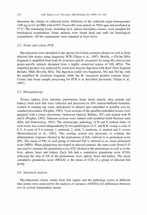
determine the cfu/gm of collected tissue. Dilutions of the collected organ homogenates
(100 mg in 0.5 ml PBS with 0.05% Tween-80) were plated on 7H10 agar and incubated at
378C. The remaining tissue, including liver, spleen and kidney tissues, were sampled for
histological examination. Some animals were found dead and unfit for histological
examination. All the experiments were repeated at least twice.
2.4. Tissue and colony PCR
Mycobacteria were identified to the species level from isolated colonies as well as from
infected fish tissues using diagnostic PCR (Talaat et al., 1997). Briefly, a 924 bp DNA
fragment is amplified from both M. fortuitum and M. smegmatis by using Mycobacterium
genus-specific primers designed from a highly conserved region of 16S rRNA. The
amplified product was analyzed by restriction enzyme digestion with BanI (New England
Biolabs, NEB, Beverly, MA). The digestion yields two fragments, 562 and 362 bp, with
the amplified M. fortuitum fragment, while the M. smegmatis product remains intact.
Colony and tissue sample processing for PCR is as described previously (Talaat et al.,
1997).
2.5. Histopathology
Tissues (spleen, liver, intestine, peritoneum, brain, heart, muscle, skin, gonads and
kidney) from each fish were collected and processed in 10% neutral-buffered formalin,
washed in running tap water, dehydrated in ethanol and embedded in paraffin wax by
standard procedures (Prophet, 1992). 5 mm sections of the paraffin embedded tissues were
prepared with a rotary microtome (American Optical, Buffalo, NY) and stained with H
and E (Prophet, 1992). Selected sections were stained with modified Ziehl-Neelsen stain
(Ellis and Zabrowarny, 1993). The microscopic pathology of H and E stained slides of
each tissue was scored independently by two pathologists (A.T. and R.R.) using a scale of
0±5. A score of 0 is normal; 1, minimal; 2, mild; 3, moderate; 4, marked and 5, severe
(Reimschuessel et al., 1992). This scoring system was necessary to evaluate the
inflammatory response elicited in the peritoneum of fish, referred to as peritoneal score
(PS). The mean of PSs of each group of infected fish is referred to as, mean peritoneal
score (MPS). When granulomas developed in infected animals, the same scale (from 0±5)
was used to estimate the granuloma score (GS) elicited in the peritoneum as well as in the
liver, spleen, heart and kidney. Each fish had a cumulative granuloma score (CGS)
reflecting the sum of GS of the peritoneum, liver, spleen, heart and kidney. The mean
cumulative granuloma score (MCGS) is the mean of CGS of a group of infected fish
group.
2.6. Statistical analysis
Mycobacterial colony counts from fish organs and the pathology scores at different
time points were analyzed by the analysis of variance (ANOVA) for differences between
two or several independent means.
154 A.M. Talaat et al. / Veterinary Microbiology 66 (1999) 151±164
3. Results
3.1. Recovery of M. fortuitum and M. smegmatis from organs
To determine the persistence of M. fortuitum and M. smegmatis mc2155 in fish organs,
we inoculated goldfish with 107 cfu of each mycobacterial species and monitored the
mycobacterial colony counts over an 8-week period in the liver, spleen and kidney of
infected animals. Mycobacteria were recovered from at least one organ of all animals
examined. A representative number of colonies were further identified by diagnostic PCR
and/or Ziehl-Neelsen staining.
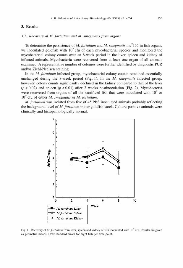
In the M. fortuitum infected group, mycobacterial colony counts remained essentially
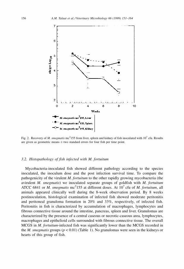
unchanged during the 8-week period (Fig. 1). In the M. smegmatis infected group,
however, colony counts significantly declined in the kidney compared to that of the liver
(p < 0.02) and spleen (p < 0.01) after 2 weeks postinoculation (Fig. 2). Mycobacteria
were recovered from organs of all the sacrificed fish that were inoculated with 108 or
109 cfu of either M. smegmatis or M. fortuitum.
M. fortuitum was isolated from five of 45 PBS inoculated animals probably reflecting
the background level of M. fortuitum in our goldfish stock. Culture-positive animals were
clinically and histopathologically normal.
Fig. 1. Recovery of M. fortuitum from liver, spleen and kidney of fish inoculated with 107 cfu. Results are given
as geometric means � two standard errors for eight fish per time point.
A.M. Talaat et al. / Veterinary Microbiology 66 (1999) 151±164 155
3.2. Histopathology of fish infected with M. fortuitum
Mycobacteria-inoculated fish showed different pathology according to the species
inoculated, the inoculum dose and the post infection survival time. To compare the
pathogenicity of the virulent M. fortuitum to the other rapidly growing mycobacteria (the
avirulent M. smegmatis) we inoculated separate groups of goldfish with M. fortuitum
ATCC 6841 or M. smegmatis mc2155 at different doses. At 107 cfu of M. fortuitum, all
animals appeared clinically well during the 8-week observation period. By 8 weeks
postinoculation, histological examination of infected fish showed moderate peritonitis
and peritoneal granuloma formation in 20% and 33%, respectively, of infected fish.
Peritonitis in fish is characterized by accumulation of macrophages, lymphocytes and
fibrous connective tissue around the intestine, pancreas, spleen and liver. Granulomas are
characterized by the presence of a central caseous or necrotic-caseous area, lymphocytes,
macrophages and epithelioid cells surrounded with fibrous connective tissue. The overall
MCGS in M. fortuitum-infected fish was significantly lower than the MCGS recorded in
the M. smegmatis groups (p < 0.01) (Table 1). No granulomas were seen in the kidneys or
hearts of this group of fish.
Fig. 2. Recovery of M. smegmatis mc2155 from liver, spleen and kidney of fish inoculated with 107 cfu. Results
are given as geometric means � two standard errors for four fish per time point.
156 A.M. Talaat et al. / Veterinary Microbiology 66 (1999) 151±164
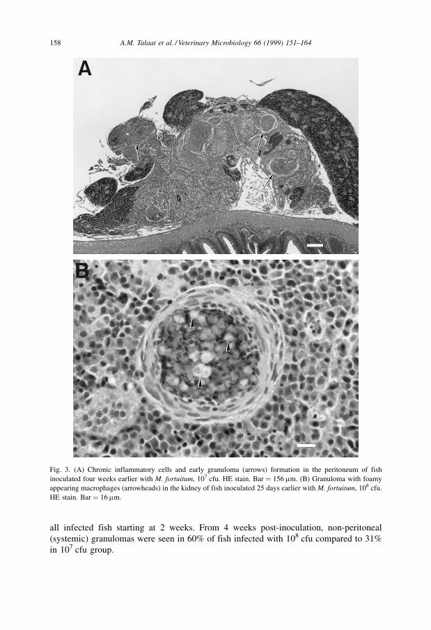
Fish inoculated with 108 cfu of M. fortuitum suffered from severe mycobacteriosis with
high peritoneal scores, at 2 weeks, followed by a peritoneal chronic granulomatous
reaction starting 4 weeks postinfection (Fig. 3(A)). Both necrotizing and caseous
granulomatous reactions were seen in M. fortuitum-infected fish. Granulomas filled with
foamy-appearing macrophages (Fig. 3(B)) were also seen. Peritoneal melanomacrophage
centers were occasionally seen in this group of fish while giant cells were rarely present.
The GS in the peritoneum was significantly higher (p < 0.001) than those recorded in the
liver, spleen, kidney and heart. Non-peritoneal, systemic granulomas were seen in only
17% of infected animals.
A severe mycobacteriosis was produced in fish inoculated with 109 cfu of M. fortuitum
and all fish died within 8 days post-infection with severe peritonitis (peritoneal score,
PS � 5). No granulomas were seen in this group of animals (Table 1).
3.3. Histopathology of fish infected with M. smegmatis
Fish inoculated with M. smegmatis mc2155 showed a severe granulomatous response
despite the general belief that it is an avirulent mycobacterial strain (Falcone et al., 1994;
Ramakrishnan et al., 1997). At 107 cfu inoculum of M. smegmatis mc2155, fish showed
moderate peritonitis (PS ranged from 0 to 3) and early granuloma formation when fish
examined at 2 weeks post-infection. Fish examined at 4, 6 and 8 weeks postinfection
showed histopathological lesions in 85% with more granulomas seen in the peritoneum
(62%) compared to the liver, spleen, kidney and heart (31%).
At 108 cfu of M. smegmatis mc2155, fish showed a similar pathology (Fig. 4(A)) to
those infected with 107 cfu except that greater granuloma scores (Table 1) were seen in
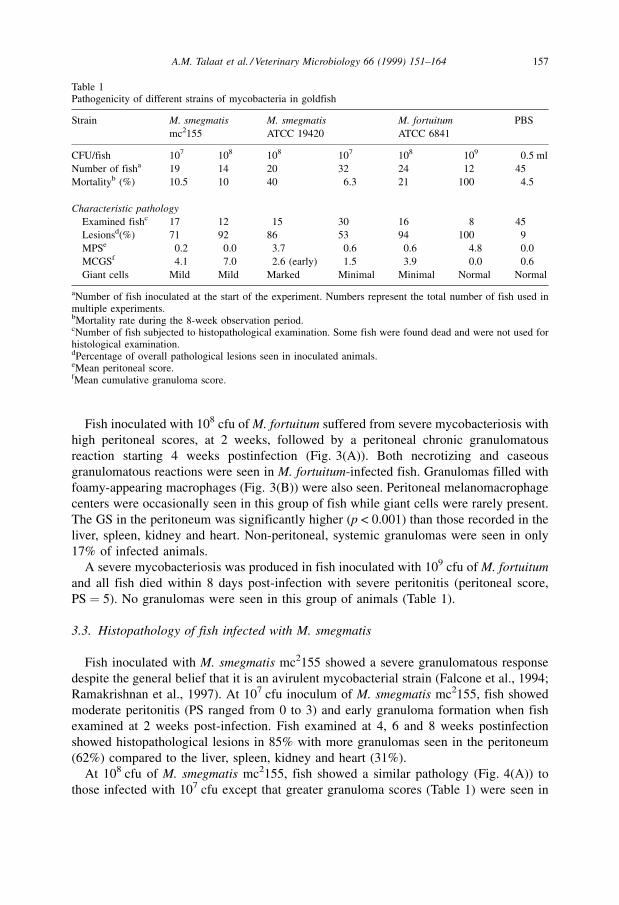
Table 1Pathogenicity of different strains of mycobacteria in goldfish
Strain M. smegmatis
mc2155
M. smegmatis
ATCC 19420
M. fortuitum
ATCC 6841
PBS
CFU/fish 107 108 108 107 108 109 0.5 ml
Number of fisha 19 14 20 32 24 12 45
Mortalityb (%) 10.5 10 40 6.3 21 100 4.5
Characteristic pathology
Examined fishc 17 12 15 30 16 8 45
Lesionsd(%) 71 92 86 53 94 100 9
MPSe 0.2 0.0 3.7 0.6 0.6 4.8 0.0
MCGSf 4.1 7.0 2.6 (early) 1.5 3.9 0.0 0.6
Giant cells Mild Mild Marked Minimal Minimal Normal Normal
aNumber of fish inoculated at the start of the experiment. Numbers represent the total number of fish used inmultiple experiments.bMortality rate during the 8-week observation period.cNumber of fish subjected to histopathological examination. Some fish were found dead and were not used forhistological examination.dPercentage of overall pathological lesions seen in inoculated animals.eMean peritoneal score.fMean cumulative granuloma score.
A.M. Talaat et al. / Veterinary Microbiology 66 (1999) 151±164 157
all infected fish starting at 2 weeks. From 4 weeks post-inoculation, non-peritoneal
(systemic) granulomas were seen in 60% of fish infected with 108 cfu compared to 31%
in 107 cfu group.
Fig. 3. (A) Chronic inflammatory cells and early granuloma (arrows) formation in the peritoneum of fish
inoculated four weeks earlier with M. fortuitum, 107 cfu. HE stain. Bar � 156 mm. (B) Granuloma with foamy
appearing macrophages (arrowheads) in the kidney of fish inoculated 25 days earlier with M. fortuitum, 108 cfu.
HE stain. Bar � 16 mm.
158 A.M. Talaat et al. / Veterinary Microbiology 66 (1999) 151±164
To test the pathogenicity of an alternative strain to M. smegmatis mc2155,
we inoculated goldfish with M. smegmatis ATCC 19420. While the infection with
108 cfu of M. smegmatis mc2155 resulted in high granuloma scores, the infection
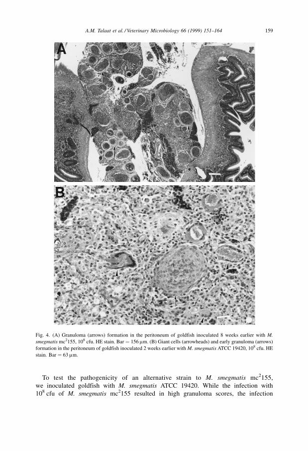
Fig. 4. (A) Granuloma (arrows) formation in the peritoneum of goldfish inoculated 8 weeks earlier with M.
smegmatis mc2155, 108 cfu. HE stain. Bar � 156 mm. (B) Giant cells (arrowheads) and early granuloma (arrows)
formation in the peritoneum of goldfish inoculated 2 weeks earlier with M. smegmatis ATCC 19420, 108 cfu. HE
stain. Bar � 63 mm.
A.M. Talaat et al. / Veterinary Microbiology 66 (1999) 151±164 159
with 108 cfu M. smegmatis ATCC 19420 resulted in marked mean peritoneal
inflammation (MPS � 3.7). In addition to the intense inflammatory response, there were
numerous giant cells in the peritoneum during early granuloma formation (Fig. 4(B)).
Giant cells were not a prominent feature in the M. smegmatis mc2155 or M. fortuitum
infected fish (Table 1).
The majority of control goldfish inoculated with PBS were histopathologically
unremarkable. Granulomas were seen in four of 45 fish. Such granulomas in fish are
usually caused by previous infection with parasites (e.g. myxozoan sp.), fungi (e.g.
Ichthyophonus-like spores) or bacteria (e.g. corynebacteria or mycobacteria infection)
(Balouet and Baudinlaurencin, 1986). These fish showed no clinical manifestation of
mycobacteriosis nor were mycobacteria recovered from their organs.
3.4. Virulence of M. fortuitum and M. smegmatis
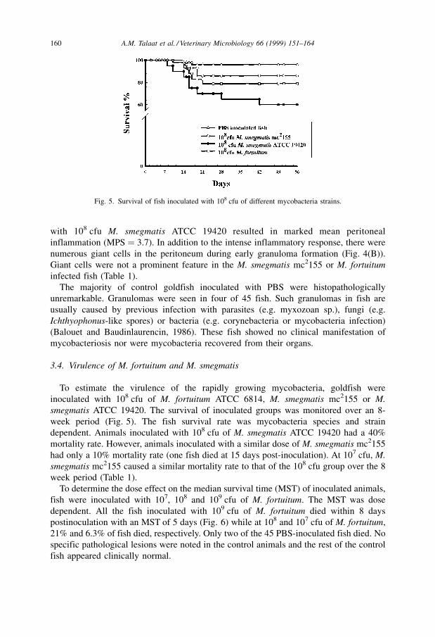
To estimate the virulence of the rapidly growing mycobacteria, goldfish were
inoculated with 108 cfu of M. fortuitum ATCC 6814, M. smegmatis mc2155 or M.
smegmatis ATCC 19420. The survival of inoculated groups was monitored over an 8-
week period (Fig. 5). The fish survival rate was mycobacteria species and strain
dependent. Animals inoculated with 108 cfu of M. smegmatis ATCC 19420 had a 40%
mortality rate. However, animals inoculated with a similar dose of M. smegmatis mc2155
had only a 10% mortality rate (one fish died at 15 days post-inoculation). At 107 cfu, M.
smegmatis mc2155 caused a similar mortality rate to that of the 108 cfu group over the 8
week period (Table 1).
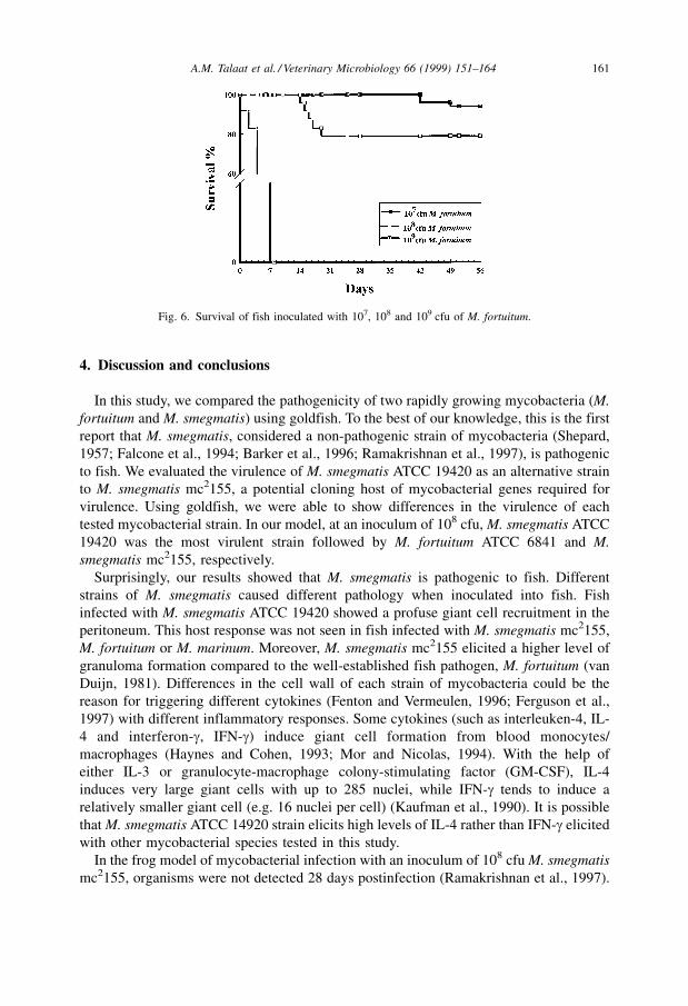
To determine the dose effect on the median survival time (MST) of inoculated animals,
fish were inoculated with 107, 108 and 109 cfu of M. fortuitum. The MST was dose
dependent. All the fish inoculated with 109 cfu of M. fortuitum died within 8 days
postinoculation with an MST of 5 days (Fig. 6) while at 108 and 107 cfu of M. fortuitum,
21% and 6.3% of fish died, respectively. Only two of the 45 PBS-inoculated fish died. No
specific pathological lesions were noted in the control animals and the rest of the control
fish appeared clinically normal.
Fig. 5. Survival of fish inoculated with 108 cfu of different mycobacteria strains.
160 A.M. Talaat et al. / Veterinary Microbiology 66 (1999) 151±164
4. Discussion and conclusions
In this study, we compared the pathogenicity of two rapidly growing mycobacteria (M.
fortuitum and M. smegmatis) using goldfish. To the best of our knowledge, this is the first
report that M. smegmatis, considered a non-pathogenic strain of mycobacteria (Shepard,
1957; Falcone et al., 1994; Barker et al., 1996; Ramakrishnan et al., 1997), is pathogenic
to fish. We evaluated the virulence of M. smegmatis ATCC 19420 as an alternative strain
to M. smegmatis mc2155, a potential cloning host of mycobacterial genes required for
virulence. Using goldfish, we were able to show differences in the virulence of each
tested mycobacterial strain. In our model, at an inoculum of 108 cfu, M. smegmatis ATCC
19420 was the most virulent strain followed by M. fortuitum ATCC 6841 and M.
smegmatis mc2155, respectively.
Surprisingly, our results showed that M. smegmatis is pathogenic to fish. Different
strains of M. smegmatis caused different pathology when inoculated into fish. Fish
infected with M. smegmatis ATCC 19420 showed a profuse giant cell recruitment in the
peritoneum. This host response was not seen in fish infected with M. smegmatis mc2155,
M. fortuitum or M. marinum. Moreover, M. smegmatis mc2155 elicited a higher level of
granuloma formation compared to the well-established fish pathogen, M. fortuitum (van
Duijn, 1981). Differences in the cell wall of each strain of mycobacteria could be the
reason for triggering different cytokines (Fenton and Vermeulen, 1996; Ferguson et al.,
1997) with different inflammatory responses. Some cytokines (such as interleuken-4, IL-
4 and interferon-g, IFN-g) induce giant cell formation from blood monocytes/
macrophages (Haynes and Cohen, 1993; Mor and Nicolas, 1994). With the help of
either IL-3 or granulocyte-macrophage colony-stimulating factor (GM-CSF), IL-4
induces very large giant cells with up to 285 nuclei, while IFN-g tends to induce a
relatively smaller giant cell (e.g. 16 nuclei per cell) (Kaufman et al., 1990). It is possible
that M. smegmatis ATCC 14920 strain elicits high levels of IL-4 rather than IFN-g elicited
with other mycobacterial species tested in this study.
In the frog model of mycobacterial infection with an inoculum of 108 cfu M. smegmatis
mc2155, organisms were not detected 28 days postinfection (Ramakrishnan et al., 1997).
Fig. 6. Survival of fish inoculated with 107, 108 and 109 cfu of M. fortuitum.
A.M. Talaat et al. / Veterinary Microbiology 66 (1999) 151±164 161
In contrast, goldfish infection with a similar inoculum resulted in persistence of
mycobacteria in fish organs to the end of the experiment (56 days) with a marked
granulomatous reaction.
In comparison to fish infected with the slowly growing, M. marinum, fewer
caseous granulomas were seen in fish infected with M. smegmatis and M. fortuitum.
By 8 weeks postinoculation, systemic granulomas (kidney and heart granulomas)
were detected in 20% of M. smegmatis-inoculated fish compared to 88% in the M.
marinum group (Talaat et al., 1998). On the other hand, M. fortuitum inoculated fish
showed no systemic granulomas during the same time period using a similar inoculum
(107 cfu/fish).
Fish infected with M. fortuitum showed no sign of nervous system involvement, but
histopathological lesions were found in the peritoneum, pancreas, liver, spleen, heart in
addition to the kidney, especially with the 108 cfu inoculum. In the mouse model, M.
fortuitum induced kidney and nervous system (brain and inner ear) lesions (Saito and
Tasaka, 1969). In both the animals (mice and fish) granulomas were seen in the examined
organs.
Using goldfish, we have shown varying pathological responses among fish groups
infected with different species and strains of mycobacteria. These differences suggest
differences in the virulence of each mycobacterial strains. This model system of
mycobacterial infection will be useful for further studies into the pathogenesis of atypical
mycobacteria. It is an excellent model for dissecting the genetic basis of the virulence of
the rapidly growing mycobacteria.
Acknowledgements
This work was supported in part by the Office of Research and Development, Medical
Research Service, Department of Veteran Affairs and University of Maryland School of
Medicine (M.T.). We thank Tim Conn and Richard Milanich for expert technical
assistance.
References
Alvarez, T., 1885. Recherches sur les bacille de Lustgarten. Arch Physiol. Normal Pathol. 2, 203±221.
Balouet, G., Baudinlaurencin, F., 1986. Granulomatous nodules in fish: An experimental assessment in rainbow
trout, Salmo gairdneri Richardson, and turbot, Scophthalmus maximus (L.). J. Fish Dis. 9, 417±429.
Barker, K., Fan, H.X., Carroll, C., Kaplan, G., Barker, J., Hellmann, W., Cohn, Z.A., 1996. Nonadherent cultures
of human monocytes kill Mycobacterium smegmatis, but adherent cultures do not. Infect. Immun. 64, 428±
433.
Belas, R., Faloon, P., Hannaford, A., 1995. Potential applications of molecular biology to the study of fish
mycobacteriosis. Ann. Rev. Fish Dis. 5, 133±173.
Bloom, J.M., 1885. The bacillus of syphilis. Lancet. 1, 609±610.
Brown, T.H., 1985. The rapidly growing mycobacteria ± Mycobacterium fortuitum and Mycobacterium chelonei.
Infec. Contr. 6, 283±288.
Cruz, J.C., 1938. Mycobacterium fortuitum, um novo bacilo acido-resistente patogenico para o Homem. Acta
Med. Rio de Jan. 1, 297±301.
162 A.M. Talaat et al. / Veterinary Microbiology 66 (1999) 151±164
Daoust, P.Y., Larson, B.E., Johnson, G.R., 1989. Mycobacteriosis in Yellow Perch (Perca flavescens) from two
lakes in Alberta. J. Wildl. Dis. 25, 31±37.
Ellis, R.C., Zabrowarny, L.A., 1993. Safer staining method for acid fast bacilli. J. Clin. Pathol. 46, 559±560.
Falcone, V., Bassey, E.B., Toniolo, A., Conaldi, P.G., Collins, F.M., 1994. Differential release of tumor necrosis
factor-alpha from murine peritoneal macrophages stimulated with virulent and avirulent species of
mycobacteria. FEMS Immunol. Med. Microbiol. 8, 225±232.
Fenton, M.J., Vermeulen, M.W., 1996. Immunopathology of tuberculosis: Roles of macrophages and monocytes.
Infec. Immun. 64, 683±690.
Ferguson, J.S., Gaynor, C.D., Schlesinger, L.S., 1997. Mononuclear phagocytes in tuberculosis pathogenesis.
Curr. Opin. Infect. Dis. 10, 190±195.
Fox, L.E., Kunkle, G.A., Homer, B.L., Manella, C., Thompson, J.P., 1995. Disseminated subcutaneous
Mycobacterium fortuitum infection in a dog. J. Am. Vet. Med. Assoc. 206, 53±55.
Goslee, S., Wolinsky, E., 1976. Water as a source of potentially pathogenic mycobacteria. Am. Rev. Resp. Dis.
113, 287±292.
Grooters, A.M., Couto, C.G., Andrews, J.M., Johnson, S.E., Kowalski, J.J., Esplin, R.B., 1995. Systemic
Mycobacterium smegmatis infection in a dog. J. Am. Vet. Med. Assoc. 206, 200±202.
Haynes, L., Cohen, N., 1993. Transforming growth factor beta (TGF beta) is produced by and influences the
proliferative response of Xenopus laevis lymphocytes. Develop. Immunol. 3, 223±230.
Hedrick, R.P., McDowell, T., Groff, J., 1987. Mycobacteriosis in cultured striped bass from California. J. Wildl.
Dis. 22, 391±395.
Jacobs Jr., W.R., Kalpana, G.V., Cirillo, J.D., Pascopella, L., Snapper, S.B., Udani, R.A., Jones, W., Barletta,
R.G., Bloom, B.R., 1991. Genetic systems for mycobacteria. Methods. Enzymol. 204, 537±555.
Jang, S.S., Eckhaus, M.A., Saunders, G., 1984. Pulmonary Mycobacterium fortuitum infection in a dog. J. Am.
Vet. Med. Assoc. 184, 96±98.
Kamala, T., Paramasivan, C.N., Herbert, D., Venkatesan, P., Prabhakar, R., 1994. Isolation and Identification of
environmental mycobacteria in the Mycobacterium bovis BCG trial area of south India. Appl. Environ.
Microbiol. 60, 2180±2183.
Kaufman, J., Skjoedt, K., Salomonsen, J., 1990. The MHC molecules of nonmammalian vertebrates. Immunol.
Rev. 113, 83±117.
Mor, A., Nicolas, P., 1994. Isolation and structure of novel defensive peptides from frog skin. Eur. J. Bioch. 219,
145±154.
Newton Jr., J.A., Weiss, P.J., Bowler, W.A., Oldfield 3d, E.C., 1993. Soft-tissue infection due to Mycobacterium
smegmatis: Report of two cases. Clin. Infect. Dis. 16, 531±533.
Newton Jr., J.A., Weiss, P.J., 1994. Aspiration pneumonia caused by Mycobacterium smegmatis. Mayo Clinic
Proc. 69, 296.
Parish, T., Stoker, N.G., 1995. Electroporation of mycobacteria. Methods. Molec. Biol. 47, 237±252.
Prophet, E.B., 1992. Tissue processing: dehydration, clearing, and infiltration. In: Prophet, E.B., Mills, B.,
Arrington, J.B., Sobin, L.H. (Eds.), Laboratory Methods in Histotechnology. American Registry of
Pathology, Washington, DC, pp. 29±32.
Ramakrishnan, L., Valdivia, R.H., McKerrow, J.H., Falkow, S., 1997. Mycobacterium marinum causes both
long-term subclinical infection and acute disease in the leopard frog (Rana pipiens). Infect. Immun. 65,
767±773.
Reimschuessel, R., Bennett, R.O., Lipsky, M.M., 1992. A classification system for histological lesions. J. Aquat.
Anim. Health. 4, 135±143.
Richardson, A., 1971. The experimental production of mastitis in sheep by Mycobacterium smegmatis and
Mycobacterium fortuitum. Cornell. Veterinarian. 61, 640±646.
Runyon, E.H., 1959. Anonymous mycobacteria in pulmonary disease. Med. Clin. North. Am. 43, 273±290.
Saito, H., Tasaka, H., 1969. Comparison of the pathogenicity for mice of Mycobacterium fortuitum and
Mycobacterium abscessus. J. Bacteriol. 99, 851±855.
Shepard, C.C., 1957. Growth characteristics of tubercle bacilli and certain other mycobacteria in HeLa cells. J.
Exp. Med. 105, 39±55.
Snapper, S.B., Melton, R.E., Mustafa, S., Kieser, T., Jacobs Jr., W.R., 1990. Isolation and characterization of
efficient plasmid transformation mutants of Mycobacterium smegmatis. Mol. Microbiol. 4, 1911±1919.
A.M. Talaat et al. / Veterinary Microbiology 66 (1999) 151±164 163
Talaat, A., Reimschuessel, R., Trucksis, M., 1998. Goldfish, Carassius auratus, a novel animal model for the
study of Mycobacterium marinum pathogenesis. Infect. Immun. 66, 2938±2942.
Talaat, A.M., Reimschuessel, R., Trucksis, M., 1997. Identification of mycobacteria infecting fish to the species
level using polymerase chain reaction and restriction enzyme analysis. Vet. Microbiol. 58, 229±237.
Thomson, J.R., Mollison, N., Matthews, K.P., 1988. An investigation of mastitis due to S. agalactiae, S. uberis
and M. smegmatis in a dairy herd. Vet. Rec. 122, 271±274.
van Duijn, C., 1981. Tuberculosis in fish. J. Sm. An. Pract. 22, 391±411.
Wallace, R.G.J., Silcox, V., Brown, B.A., 1994. Taxonomy of rapidly growing mycobacteria. Clin. Infect. Dis.
18, 121±122.
Wallace Jr., R.J., Nash, D.R., Tsukamura, M., Blacklock, Z.M., Silcox, V.A., 1988. Human disease due to
Mycobacterium smegmatis. J. Infect. Dis. 158, 52±59.
Wilkinson, G.T., Kelly, W.R., O'Boyle, D., 1978. Cutaneous granulomas associated with Mycobacterium
fortuitum in a cat. J. Sm. An. Pract. 19, 357±362.
Woods, G.L., Washington II, J.A., 1987. Mycobacteria other than Mycobacterium tuberculosis: review of
microbiological and clinical aspects. Rev. Infect. Dis. 9, 275±294.
Yew, W.-W., Wong, P.-c., Woo, H.-s., Yip, C.-w., Chan, C.-y., Cheng, F.-b., 1993. Characterization of
Mycobacterium fortuitum isolates from sternotomy wounds by antimicrobial susceptibilities, plasmid
profiles, and ribosomal ribonucleic acid gene restriction patterns. Diagn. Microbiol. Infect. Dis. 17, 111±
117.
164 A.M. Talaat et al. / Veterinary Microbiology 66 (1999) 151±164