
Pathogenicity of a minimal organism: Role of protein ...
275
Pathogenicity of a minimal organism: Role of protein phosphorylation in Mycoplasma pneumoniae Dissertation zur Erlangung des mathematisch-naturwissenschaftlichen Doktorgrades „Doctor rerum naturalium“ der Georg-August-Universität Göttingen vorgelegt von Sebastian Schmidl aus Bad Hersfeld Göttingen 2010
Transcript of Pathogenicity of a minimal organism: Role of protein ...

Pathogenicity of a minimal organism:
Role of protein phosphorylation in
Mycoplasma pneumoniae
Dissertation
zur Erlangung des mathematisch-naturwissenschaftlichen Doktorgrades
„Doctor rerum naturalium“
der Georg-August-Universität Göttingen
vorgelegt von
Sebastian Schmidl
aus Bad Hersfeld
Göttingen 2010

Mitglieder des Betreuungsausschusses:
Referent: Prof. Dr. Jörg Stülke
Koreferent: PD Dr. Michael Hoppert
Tag der mündlichen Prüfung: 02.11.2010

“Everything should be made as simple as possible, but not simpler.”
(Albert Einstein)
Danksagung
Zunächst möchte ich mich bei Prof. Dr. Jörg Stülke für die Ermöglichung dieser
Doktorarbeit bedanken. Nicht zuletzt durch seine freundliche und engagierte Betreuung
hat mir die Zeit viel Freude bereitet. Des Weiteren hat er mir alle Freiheiten zur
Verwirklichung meiner eigenen Ideen gelassen, was ich sehr zu schätzen weiß.
Für die Übernahme des Korreferates danke ich PD Dr. Michael Hoppert sowie
Prof. Dr. Heinz Neumann, PD Dr. Boris Görke, PD Dr. Rolf Daniel und Prof. Dr. Botho
Bowien für das Mitwirken im Thesis-Komitee. Der Studienstiftung des deutschen
Volkes gilt ein besonderer Dank für die finanzielle Unterstützung dieser Arbeit, durch
die es mir unter anderem auch möglich war, an Tagungen in fernen Ländern
teilzunehmen.
Prof. Dr. Michael Hecker und der Gruppe von Dr. Dörte Becher (Universität
Greifswald) danke ich für die freundliche Zusammenarbeit bei der Durchführung von
zahlreichen Proteomics-Experimenten. Ein ganz besonderer Dank geht dabei an Katrin
Gronau, die mich in die Feinheiten der 2D-Gelelektrophorese eingeführt hat. Außerdem
möchte ich mich bei Andreas Otto für die zahlreichen Proteinidentifikationen in den
letzten Monaten bedanken. Nicht zu vergessen ist auch meine zweite Außenstelle an der
Universität in Barcelona. Dr. Maria Lluch-Senar und Dr. Jaume Piñol gilt ein
besonderer Dank bei der morphologischen Untersuchungen von Mutanten sowie
zahlreichen lustigen Abenden auf Tagungen.
Für die gute Arbeitsatmosphäre und tatkräftige Unterstützung im Labor möchte
ich mich bei Julia Busse bedanken. Speziell natürlich für die unzähligen Slot Blots. Ein
besonderer Dank geht auch an Hinnerk Eilers für die vielen anregenden Diskussionen
rund um die kleinen Biester. Viel Erfolg in Umeå! Meinem Nachfolger Arne Schmeisky
wünsche ich alles Gute und hoffe, dass er das Mycoplasmen-Mekka weiterhin gut
vertreten wird. Dr. Petra Neumann-Staubitz danke ich für die vielen unterhaltsamen
Gespräche im S2-Labor. Mal sehen, ob ihr Schilderwahn einmal ein Ende nimmt!
Weiterhin bedanke ich mich bei meinen alteingesessenen sowie neuen Mitstreitern
Katrin Gunka, Christine Diethmaier, Christoph Wrede, Nico Pietack, Lope A. Flórez,
Martin Lehnik, Frederik Meyer und Fabian Rothe, die in den letzten 3 Jahren viel zu
einer entspannten und kreativen Arbeitsatmosphäre beigetragen haben. Ein großer Dank
für die Erleichterung des Laboraltages geht auch an unsere „Pufferella“ Bärbel Herbst.
Möge sie wieder zu ihrer alten Stärke zurückfinden! Bei der Praktikantin Sandra Appelt,

den Bachelor-Studenten Stephanie Großhennig, Miriam Bothe und Daniel Reuß sowie
den Diplomanden Meike Ridderbusch und Pavel Dutow bedanke ich mich für die
tatkräftige Unterstützung bei vielen Projekten. Hoffentlich haben sie meinen kleinen
Exkurs in die „Advanced and Applied Mycoplasmology“ gut überstanden!? Außerdem
danke ich noch all jenen, die ich nicht namentlich erwähnen konnte, die mich aber
dennoch bei der Erstellung dieser Arbeit unterstützt haben.
Mein ganz spezieller Dank gilt meiner Familie. Ohne die moralische
Unterstützung meiner Eltern und meiner Schwester hätte ich weder mein Studium noch
meine Doktorarbeit schaffen können. Dafür ein riesengroßes Dankeschön! Zum Schluss
gilt mein Dank meiner Freundin Anke, die mich immer wieder neu motiviert und so
sehr an mich glaubt.
CONTENTS EDITORIAL
I
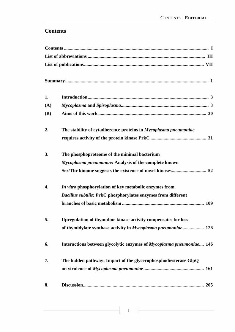
Contents
Contents .......................................................................................................................... I
List of abbreviations ................................................................................................... III
List of publications ..................................................................................................... VII
Summary ......................................................................................................................... 1
1. Introduction ...................................................................................................... 3
(A) Mycoplasma and Spiroplasma .......................................................................... 3
(B) Aims of this work ........................................................................................... 30
2. The stability of cytadherence proteins in Mycoplasma pneumoniae
requires activity of the protein kinase PrkC ............................................... 31
3. The phosphoproteome of the minimal bacterium
Mycoplasma pneumoniae: Analysis of the complete known
Ser/Thr kinome suggests the existence of novel kinases ............................. 52
4. In vitro phosphorylation of key metabolic enzymes from
Bacillus subtilis: PrkC phosphorylates enzymes from different
branches of basic metabolism ..................................................................... 109
5. Upregulation of thymidine kinase activity compensates for loss
of thymidylate synthase activity in Mycoplasma pneumoniae .................. 128
6. Interactions between glycolytic enzymes of Mycoplasma pneumoniae .... 146
7. The hidden pathway: Impact of the glycerophosphodiesterase GlpQ
on virulence of Mycoplasma pneumoniae ................................................... 161
8. Discussion ...................................................................................................... 205

CONTENTS EDITORIAL
II
9. References ..................................................................................................... 219
10. Appendix ....................................................................................................... 245
Curriculum vitae ........................................................................................................ 263

ABBREVIATIONS EDITORIAL
III
List of abbreviations
5FdUMP 5-fluorodeoxyuridine monophosphat A adenine Å ångström ABC adenosine 5’-triphosphate binding cassette ADP adenosine 5’-diphosphate Amp ampicillin Asn asparagine ATP adenosine 5’-triphosphate B2H bacterial two-hybrid bp base pair BSA bovine serum albumin C cytosine Ca2+ calcium cation Cam chloramphenicol cAMP cyclic adenosine 5’-monophosphate CDP-Star disodium 2-chloro-5-(4-methoxyspiro {1,2-dioxetane-3,2’-(5’-chloro)
tricyclo[3.3.1.13,7]decan}-4-yl) phenyl phosphate CH3CN acetonitrile CHAPS 3-[(3-cholamidopropyl)dimethylammonio]-1-propanesulfonate ChIP-chip chromatin immunoprecipitation with microarray technology Cho choline Ci curie cm centimeter CM cell membrane cm2 square centimeter CMP cytidine 5’-monophosphate co control CO2 carbon dioxide CoA coenzyme A COG cluster of orthologous groups of proteins Cys cysteine Da dalton DHAP dihydroxyacetone phosphate DIG digoxigenin DNA deoxyribonucleic acid dT thymidine dTDP thymidine 5’-diphosphate dTMP thymidine 5’-monophosphate DTT dithiothreitol dTTP thymidine 5’-triphosphate dU deoxyuridine dUMP deoxyuridine 5’-monophosphate EDTA ethylenediaminetetraacetic acid Erm erythromycine ESI electrospray ionization et al. and others FAD flavin adenine dinucleotide
ABBREVIATIONS EDITORIAL
IV
Fig. figure G guanine g gram G3P glycerol 3-phosphate Glc glucose Glu glutamic acid Gly glycerol Gm gentamicin GPC glycerophosphorylcholine GTP guanosine 5’-triphosphate h hour H hydrogen H2O water HCl hydrochloric acid HEPES 4-(2-hydroxyethyl)-1-piperazineethanesulfonic acid His histidine HIV human immunodeficiency virus HMW high molecular weight HPLC high performance liquid chromatography HPrK HPr kinase i.d. in diameter i.e. that is IPG immobilized power of hydrogen gradient IPTG isopropyl-β-D-1-thiogalactopyranoside IS insertion sequence Kan kanamycin kb kilobase KCl potassium chloride KClO4 potassium perchlorate kDa kilodalton KOH potassium hydroxide kPa kilopascal l liter LB lysogeny broth LC liquid chromatography Leu leucine M marker M molar m/z mass-to-charge ratio mg milligram Mg2+ magnesium cation MgCl2 magnesium chloride µg microgram µl microliter µm micrometer µM micromolar min minute ml milliliter mm millimeter mM millimolar
ABBREVIATIONS EDITORIAL
V
mmol millimole MMR multiple mutation reaction Mn2+ manganese cation MnCl2 manganese chloride MPN Mycoplasma pneumoniae mRNA messenger ribonucleic acid MS mass spectrometry MW molecular weight na not available NaCl sodium chloride NAD+/NADH nicotinamide adenine dinucleotide (oxidized/reduced form) NADPH nicotinamide adenine dinucleotide phosphate (reduced form) NaF sodium fluoride NaH2PO4 monosodium phosphate NaOH sodium hydroxide nd not detectable ng nanogram NH4HCO3 ammonium bicarbonate Ni2+ nickel cation nl nanoliter nm nanometer nmol nanomole ns no significant difference N-terminal amino-terminal OH hydroxyl group ORF open reading frame P phosphate p.s.i. pound per square inch PAGE polyacrylamide gel electrophoresis PBS phosphate buffered saline PCA perchloric acid PCR polymerase chain reaction PEI polyethylenimine PEP phosphoenolpyruvate Pfu Pyrococcus furiosus pH power of hydrogen Phe phenylalanine pI isoelectric point ppm parts per million PrkC protein kinase C PrpC protein phosphatase of the PP2C family PRPP phosphoribosyl pyrophosphate pS phosphoserine pT phosphothreonine PTS phosphotransferase system RNA ribonucleic acid RNAse ribonuclease rRNA ribosomal ribonucleic acid s second sa similar amount
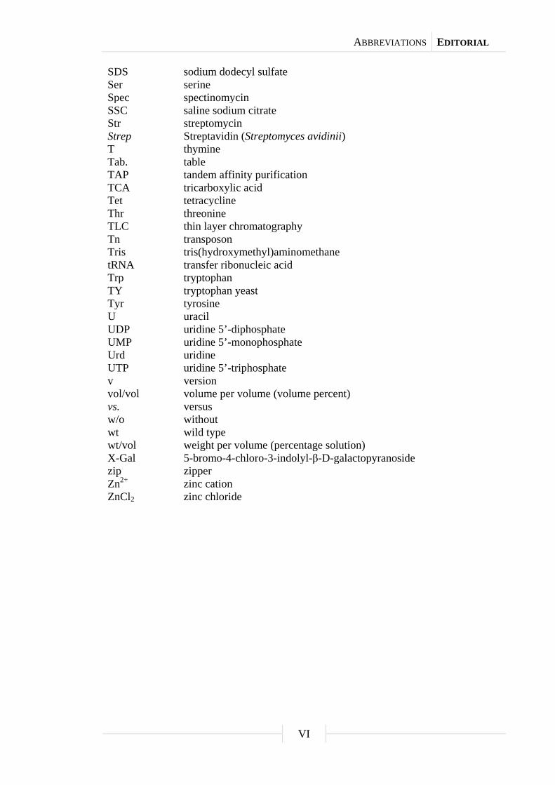
ABBREVIATIONS EDITORIAL
VI
SDS sodium dodecyl sulfate Ser serine Spec spectinomycin SSC saline sodium citrate Str streptomycin Strep Streptavidin (Streptomyces avidinii) T thymine Tab. table TAP tandem affinity purification TCA tricarboxylic acid Tet tetracycline Thr threonine TLC thin layer chromatography Tn transposon Tris tris(hydroxymethyl)aminomethane tRNA transfer ribonucleic acid Trp tryptophan TY tryptophan yeast Tyr tyrosine U uracil UDP uridine 5’-diphosphate UMP uridine 5’-monophosphate Urd uridine UTP uridine 5’-triphosphate v version vol/vol volume per volume (volume percent) vs. versus w/o without wt wild type wt/vol weight per volume (percentage solution) X-Gal 5-bromo-4-chloro-3-indolyl-β-D-galactopyranoside zip zipper Zn2+ zinc cation ZnCl2 zinc chloride
PUBLICATIONS EDITORIAL
VII
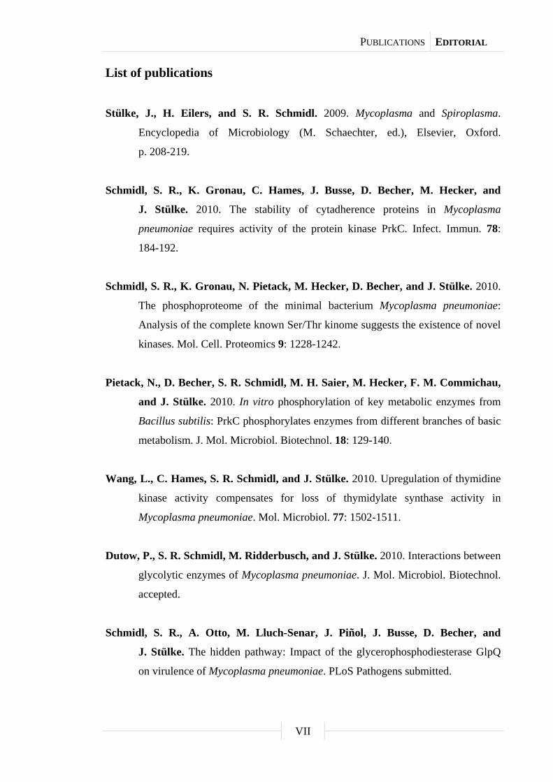
List of publications
Stülke, J., H. Eilers, and S. R. Schmidl. 2009. Mycoplasma and Spiroplasma.
Encyclopedia of Microbiology (M. Schaechter, ed.), Elsevier, Oxford.
p. 208-219.
Schmidl, S. R., K. Gronau, C. Hames, J. Busse, D. Becher, M. Hecker, and
J. Stülke. 2010. The stability of cytadherence proteins in Mycoplasma
pneumoniae requires activity of the protein kinase PrkC. Infect. Immun. 78:
184-192.
Schmidl, S. R., K. Gronau, N. Pietack, M. Hecker, D. Becher, and J. Stülke. 2010.
The phosphoproteome of the minimal bacterium Mycoplasma pneumoniae:
Analysis of the complete known Ser/Thr kinome suggests the existence of novel
kinases. Mol. Cell. Proteomics 9: 1228-1242.
Pietack, N., D. Becher, S. R. Schmidl, M. H. Saier, M. Hecker, F. M. Commichau,
and J. Stülke. 2010. In vitro phosphorylation of key metabolic enzymes from
Bacillus subtilis: PrkC phosphorylates enzymes from different branches of basic
metabolism. J. Mol. Microbiol. Biotechnol. 18: 129-140.
Wang, L., C. Hames, S. R. Schmidl, and J. Stülke. 2010. Upregulation of thymidine
kinase activity compensates for loss of thymidylate synthase activity in
Mycoplasma pneumoniae. Mol. Microbiol. 77: 1502-1511.
Dutow, P., S. R. Schmidl, M. Ridderbusch, and J. Stülke. 2010. Interactions between
glycolytic enzymes of Mycoplasma pneumoniae. J. Mol. Microbiol. Biotechnol.
accepted.
Schmidl, S. R., A. Otto, M. Lluch-Senar, J. Piñol, J. Busse, D. Becher, and
J. Stülke. The hidden pathway: Impact of the glycerophosphodiesterase GlpQ
on virulence of Mycoplasma pneumoniae. PLoS Pathogens submitted.

SUMMARY PROLOGUE
1
Summary
Mycoplasma pneumoniae is a human pathogen that belongs to the Mollicutes, a
group of bacteria with the smallest genomes that are capable of independent life. The
reductive evolution of the Mollicutes is reflected by their limited regulatory features for
gene expression. Thus, posttranslational regulation might be important for
M. pneumoniae to adapt to environmental changes. Among the very few regulatory
proteins retained is the HPr kinase (HPrK), which phosphorylates the phosphocarrier
protein HPr at the Ser-46 residue. This phosphorylation event is a major signal to trigger
carbon catabolite repression in less degenerated bacteria. However, the function of
HPr(Ser-P) in M. pneumoniae is unknown. For the protein phosphatase PrpC, an
implication in the dephosphorylation of HPr(Ser-P) could be shown. In addition to
HPrK, the M. pneumoniae prkC gene encodes another serine/threonine protein kinase C.
The determination of the complete phosphoproteome of M. pneumoniae by
two-dimensional gel electrophoresis and mass spectrometry allowed the detection of
63 phosphorylated proteins, including many enzymes of central carbon metabolism and
proteins related to host cell adhesion. It was also possible to detect 16 phosphorylation
sites, among them 8 serine and 8 threonine residues. However, a comparison with the
phosphoproteomes of other bacteria revealed that there is only a weak conservation of
phosphorylation sites, even if the same proteins are phosphorylated in related
organisms. There is only one exception: The phosphorylation of phosphosugar mutases
on a conserved serine residue, which could be detected in all studied organisms from
archaea and bacteria to man. In the case of the phosphosugar mutase ManB in
M. pneumoniae, it could be shown that this protein undergoes autophosphorylation. In
conclusion, the results indicate that protein phosphorylation seems to be highly specific
for each individual organism.
For a more detailed analysis of the phosphorylation network in M. pneumoniae,
the phosphoproteomes of the wild type strain and of three isogenic mutants that are
affected in the two protein kinases HPrK and PrkC and in the protein phosphatase PrpC
were compared. Examination of the phosphorylation profile of the hprK mutant
revealed that only HPr is phosphorylated by HPrK, whereas six proteins, including the
major adhesin P1 and two cytadherence proteins HMW1 and HMW3, were affected by
the loss of PrkC. In contrast, inactivation of PrpC that antagonizes PrkC-dependent
phosphorylation resulted in more intensive phosphorylation of the same target proteins.
SUMMARY PROLOGUE
2
The phenotypic characterization of prkC mutant cells revealed a nonadherent growth
type along with a loss of cytotoxicity toward HeLa cells. Thus, posttranslational
modification of cytadherence proteins by PrkC is essential for cell adhesion and
virulence in M. pneumoniae.
The phosphoproteomic analysis demonstrated that several glycolytic enzymes
are subject to phosphorylation. M. pneumoniae uses glycolysis as the major pathway for
the generation of energy by substrate-level phosphorylation. Using a bacterial
two-hybrid approach, the enolase was identified as the central glycolytic enzyme of
M. pneumoniae due to its ability to directly interact with all other glycolytic enzymes.
Moreover, most of the glycolytic enzymes performed self-interactions. The results
support the idea that glycolysis proceeds in a well structured manner even in a minimal
organism.
In its natural habitat, M. pneumoniae thrives on pulmonary surfaces that are
mainly composed of phosphatidylcholine. This phospholipid can be integrated directly
into the cell membrane or serve as precursor for cellular processes. M. pneumoniae
possesses two potential glycerophosphodiesterases, MPN420 (GlpQ) and MPN566, that
are able to cleave deacylated phospholipids to glycerol 3-phosphate and choline. Further
glycerol 3-phosphate utilization by enzymes of the glycerol metabolism is crucial for
the cytotoxicity of M. pneumoniae due to hydrogen peroxide release. Biochemical
studies showed that GlpQ is active as a glycerophosphodiesterase, whereas MPN566
has no enzymatic activity in vitro. Mutants affected in either glycerophosphodiesterase
revealed that inactivation of mpn566 did not result in any phenotype. In contrast, the
glpQ mutant exhibited a growth defect in glucose-supplemented medium. Moreover, the
lack of GlpQ resulted in an absence of hydrogen peroxide formation in the presence of
deacylated phospholipids and a loss of cytotoxicity toward HeLa cells. These
observations imply that GlpQ is important for the pathogenicity of M. pneumoniae, but
also for other functions in the cell. Indeed, proteomic and transcriptomic analyses of the
wild type and the glpQ mutant strain suggested a GlpQ-dependent transcription
regulation, which led to higher or lower protein amounts of the glycerol facilitator, a
subunit of a metal ion ABC transporter, and three lipoproteins. Interestingly, all genes
subject to GlpQ-dependent control have a conserved potential cis-acting element
upstream of the coding region. Nevertheless, it is open for speculation whether GlpQ or
a transcription factor that is controlled by GlpQ is responsible for this regulation.

Chapter 1
Introduction
(A) Mycoplasma and Spiroplasma
This chapter is part of the following publication:
Stülke, J., H. Eilers, and S. R. Schmidl. 2009. Mycoplasma and Spiroplasma.
Encyclopedia of Microbiology (M. Schaechter, ed.), Elsevier, Oxford. p. 208-219.
Author contributions:
This review was written by JS, HE, and SRS. SRS performed the systematics of the
Mollicutes, on which the first two chapters are based as well as partial researches on
biochemistry, genetics, and molecular biology of the Mollicutes.

INTRODUCTION CHAPTER 1
4
Defining statement
Mycoplasma and Spiroplasma species are bacteria that lack a cell wall (the
Mollicutes). These organisms evolved in close association with their eukaryotic hosts,
resulting in an extreme genome reduction. In this article, the biology of the Mollicutes
is discussed with special emphasis on their pathogenicity, cell biology, and molecular
biology.
Introduction
Mycoplasmas and spiroplasmas are two important genera of the bacterial group
called Mollicutes. The name Mollicutes - soft skin - reflects the major collective
characteristic of these bacteria - the lack of a cell wall - which at the same time
distinguishes them from all other bacteria with the exception of the chlamydiae. The
lack of a cell wall is caused by the absence of genes encoding enzymes for
peptidoglycan biosynthesis. The lack of a cell wall is closely linked to another
characteristic feature of the Mollicutes - their cells are usually pleomorphic. Again,
there is no rule without exception: The cells of the genus Spiroplasma have a helical
shape (see “Cytology of the Mollicutes”).
Another important feature of the Mollicutes is their close association with
eukaryotic host organisms. In nature, Mollicutes are never found as free-living
organisms. Hosts are either animals including humans (Mycoplasma, Ureaplasma) or
plants and insects (Spiroplasma, Phytoplasma) (Table 1). Mycoplasma species usually
cause mild diseases such as atypical pneumonia (Mycoplasma pneumoniae) or
nongonococcal urethritis (Mycoplasma genitalium). However, there is an interesting
exception: Mycoplasma alligatoris, a pathogen of alligators, causes lethal infections.
Although the infections caused by Mollicutes are rarely lethal, Mollicutes pathogenic
for plants and animals cause a significant economic loss in agriculture. This is true for
cattle in Africa that are infected by Mycoplasma mycoides as well as for rice crops in
some regions of Southeast Asia that are infected by phytoplasmas. These losses not only
have an economic dimension but also a significant effect on human nutrition in the
affected regions. Mycoplasma species such as Mycoplasma hyorhinis or Acholeplasma
laidlawii are major sources of cell culture contamination and have gained increasing

INTRODUCTION CHAPTER 1
5
interest. These infections are often discovered only late in the course of an experiment
and can invalidate the scientific research.
The close association of Mollicutes with eukaryotic hosts and their adaptation to
habitats with a good nutrient supply and relatively constant growth conditions led to a
remarkable process of reductive genome evolution. The organism with the smallest
known genome capable of independent life (if provided with rich artificial medium) is
M. genitalium, a human pathogen. This organism has a genome size of only 580 kb and
encodes about 480 proteins, as compared to about 4 million bp and 4000 genes for
bacteria such as Escherichia coli or Bacillus subtilis. These small genomes made the
Mollicutes important tools for the new discipline of synthetic biology (see “Genomic
comparisons of Mollicutes”).
The systematics of the Mollicutes
Evolution of the Mollicutes. The analysis and comparison of 16S rRNA
sequences revealed that the Mollicutes belong to the Gram-positive bacteria with
genomes of low GC content. Ironically, most members of this phylum are characterized
by their thick Gram-positive cell wall, and the group is therefore referred to as the
Firmicutes. This bacterial phylum includes the lactic acid bacteria (such as
Streptococcus and Lactobacillus), spore-forming bacteria (Bacillus and Clostridium)
and their close relatives (Listeria and Staphylococcus). As can be seen in the
phylogenetic tree of the Firmicutes (Fig. 1), the Mollicutes form a sister group to the
large Bacillus/lactic acid bacteria group. It is believed that the first Mollicutes emerged
some 600 million years ago and that significant loss of ancestral genomic sequences
was a major force in the evolution of the Mollicutes.
The Mollicutes are subdivided in several ways. Three traditional classifications
rely on genetic or physiological properties of the bacteria, whereas more recent
classification schemes are based on the similarity of the 16S rRNA or conserved protein
families.

INTRODUCTION CHAPTER 1
6
Tab. 1. The systematic groups of the Mollicutes.
Order Genus Genome size Sterol requirement Characteristics Habitat
Mycoplasmatales Mycoplasma 580-1350 kb Yes Growth optimum: 37°C
UGA as Trp codon
Humans, animals
Ureaplasma 760-1170 kb Yes Urea hydrolysis
UGA as Trp codon
Humans, animals
Entomoplasmatales Entomoplasma 790-1140 kb Yes Growth optimum: 30°C Insects, plants
Mesoplasma 870-1100 kb No Growth optimum: 30°C
UGA as Trp codon
Insects, plants
Spiroplasma 780-2200 kb Yes Growth optimum: 30-37°C
UGA as Trp codon
Helical motile filaments
Insects, plants
Anaeroplasmatales Anaeroplasma 1500-1600 kb Yes Obligate anaerobes Bovine/ovine rumen
Asteroleplasma 1500 kb No Obligate anaerobes Bovine/ovine rumen
Acholeplasmatales Acholeplasma 1500-1650 kb No Growth optimum: 30-37°C
UGA as stopp codon
Animals, plants, insects
Phytoplasma 640-1185 kb Not known Uncultured in vitro
UGA as stopp codon
Insects, plants
INTRODUCTION CHAPTER 1
7
Two large groups of Mollicutes can be distinguished based on their host
organisms. Although most Mollicutes infect exclusively animal hosts, there are other
representatives (Spiroplasma and Phytoplasma) that are capable of infecting both plant
and insect hosts. Another conventional way of classifying the Mollicutes is based on
their requirement for sterols. Most genera need sterols for growth, whereas this is not
the case for the members of the genus Acholeplasma (see Table 1). However, this
requirement can only be determined for those Mollicutes that can be cultivated, and
many (perhaps most) representatives have not yet been cultured, including all species of
the genus Phytoplasma. Another peculiarity of most Mollicutes is their codon usage:
They use the UGA codon to specify tryptophan rather than as a stop codon as in the
universal genetic code. Only the genera Acholeplasma and Phytoplasma among the
Mollicutes use UGA as a stop codon. Because this is the ancestral property, it can be
assumed that Acholeplasma and Phytoplasma represent the more ancestral Mollicutes.
This conclusion is supported by a phylogenetic tree based on a concatenated alignment
of 30 protein families present in all Mollicutes that places the genus Phytoplasma at the
bottom of the tree (Fig. 1). The genus Acholeplasma is not included in this analysis
because of the lack of genome sequence information. It is interesting to note that the
genus Mycoplasma is paraphyletic, and that genera such as Spiroplasma, Mesoplasma,
and Ureaplasma have specific relatives among the different Mycoplasma clades
(Fig. 1).
For practical reasons, the Mollicutes are grouped in four orders that do not
represent the phylogenetic relationships. An overview of these taxa is provided in
Table 1.
Mycoplasma. As mentioned earlier, the genus Mycoplasma is a paraphyletic
collection of Mollicutes that are widespread in nature as parasites of humans, mammals,
birds, reptiles, and fish. The first representative of the genus Mycoplasma was identified
in 1898 as the causative agent of contagious bovine pleuropneumonia (M. mycoides).
The human pathogens Mycoplasma hominis and M. pneumoniae were discovered in
1937 and 1944, respectively. Even now, new species are being identified: In 1981,
M. genitalium was isolated from a patient suffering from nongonococcal urethritis, and
more recently, Mycoplasma penetrans and Mycoplasma fermentans were found to be
associated with HIV infections.
INTRODUCTION CHAPTER 1
8
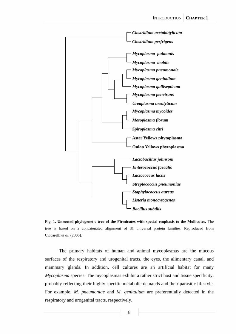
Fig. 1. Unrooted phylogenetic tree of the Firmicutes with special emphasis to the Mollicutes. The
tree is based on a concatenated alignment of 31 universal protein families. Reproduced from
Ciccarelli et al. (2006).
The primary habitats of human and animal mycoplasmas are the mucous
surfaces of the respiratory and urogenital tracts, the eyes, the alimentary canal, and
mammary glands. In addition, cell cultures are an artificial habitat for many
Mycoplasma species. The mycoplasmas exhibit a rather strict host and tissue specificity,
probably reflecting their highly specific metabolic demands and their parasitic lifestyle.
For example, M. pneumoniae and M. genitalium are preferentially detected in the
respiratory and urogenital tracts, respectively.
Onion Yellows phytoplasma
Mycoplasma mycoides
Mycoplasma mobile
Ureaplasma urealyticum
Mycoplasma penetrans
Mesoplasma florum
Spiroplasma citri
Mycoplasma gallisepticum
Mycoplasma pneumonaie
Mycoplasma pulmonis
Mycoplasma genitalium
Aster Yellows phytoplasma
Lactobacillus johnsoni
Enterococcus faecalis
Lactococcus lactis
Streptococcus pneumoniae
Staphylococcus aureus
Listeria monocytogenes
Bacillus subtilis
Clostridium perfrigens
Clostridium acetobutylicum

INTRODUCTION CHAPTER 1
9
If cultivated in the laboratory, mycoplasmas as well as other Mollicutes require
complex media containing sugars, amino acids, nucleotides, and vitamins. It has so far
been impossible to cultivate them on chemically defined media.
The complete genome sequences of ten species of the genus Mycoplasma have
been determined so far. This large interest in the variability of the Mycoplasma genetic
complement is stimulated by the interest in creating artificial organisms based on the
Mycoplasma species (i.e., synthetic biology; see “Genomic comparisons of
Mollicutes”). The genome sequences revealed the reason for the complex nutritional
requirements of the mycoplasmas: They lack the genes for many biosynthetic pathways
and are thus dependent on their host or on the artificial medium to provide these
required nutrients. Another interesting feature revealed by genome sequences is that
only very few known regulatory proteins are present. Again, this is reflective of their
close adaptation to one single natural habitat and a result of the reductive evolution:
While a metabolically versatile bacterium such as Pseudomonas aeruginosa that is
capable of thriving in a wide variety of environments reserves as much as 10% of its
genome for regulatory genes, only a handful of these genes is found in the mycoplasmas
(see “Gene expression in the Mollicutes”).
Pathogenicity has been most intensively studied with M. pneumoniae. In contrast
to most other pathogenic bacteria, M. pneumoniae and other Mollicutes do not seem to
produce any exo- or endotoxins. However, a recent study suggests the formation of a
protein similar to ADP-ribosylating and vacuolating cytotoxin. However, this
observation has not been confirmed by other groups. A major factor contributing to
cytotoxicity and thus to pathogenicity of M. pneumoniae is the formation of hydrogen
peroxide. The synthesis of hydrogen peroxide by mycoplasmas is most strongly
increased if the bacteria are supplied with glycerol. This can be attributed to the oxidase
activity of the enzyme that oxidizes glycerol 3-phosphate. This enzyme,
glycerol-3-phosphate oxidase, uses water rather than NAD+ (as in typical
glycerol-3-phosphate dehydrogenases) as the electron acceptor. The hydrogen peroxide
formed by M. pneumoniae acts in concert with endogenous toxic oxygen molecules
generated by the host cells and induces oxidative stress in the respiratory epithelium.
The effects of the peroxide on the host cells include loss of reduced glutathione,
denaturation of hemoglobin, peroxidation of erythrocyte lipids, and eventually the lysis
of the cells. Another result of infection by M. pneumoniae is the release of
INTRODUCTION CHAPTER 1
10
proinflammatory cytokines by the host cells. It has been suggested that cytokine
production leads to chronic pulmonary diseases such as bronchial asthma.
The significance of glycerol metabolism in hydrogen peroxide production and
virulence has been convincingly demonstrated by a series of studies that started with an
analysis of the differences between European and African strains of M. mycoides, the
causative agent of contagious bovine pleuropneumonia. Glycerol transport is highly
efficient in the African isolates, whereas it is barely detectable in the European isolates.
Because glycerol catabolism gives rise to the formation of hydrogen peroxide, it is not
surprising that hydrogen peroxide production is high in the African strains but low in
the European isolates of M. mycoides. In consequence, the African strains are highly
virulent to cattle, whereas their European relatives are harmless. It has been
hypothesized that intracellular formation of large quantities of hydrogen peroxide would
be toxic for the producing cells themselves. Accordingly, the cellular localization of the
responsible enzyme, GlpO, was studied in M. mycoides and it was found to be located
in the cell membrane. The inactivation of GlpO by antibodies results in the loss of
cytotoxicity of M. mycoides toward bovine epithelial cells. Given that hydrogen
peroxide in concentrations similar to those produced by M. mycoides is not cytotoxic, it
was concluded that GlpO is not only inserted in the bacterial cell membrane, but also in
the membrane of the host cell to inject the cytotoxic hydrogen peroxide directly into the
epithelial cells. This may cause oxidative stress and subsequent cell death.
Plant pathogenic Mollicutes: Spiroplasma and Phytoplasma. The genera
Spiroplasma and Phytoplasma contain plant pathogenic Mollicutes that shuttle between
plant and insect hosts. Spiroplasma citri was identified in 1971 as a causative agent of
citrus stubborn disease. Phytoplasmas were first described in 1967 as the probable cause
of plant yellow diseases. Originally, it was speculated that these diseases are of viral
origin, and only in 1967 it became clear that these pathogens are Mycoplasma-like
organisms. While spiroplasmas can be cultivated in the laboratory, no cultivation of any
representative of the phytoplasmas has been reported. Therefore, no valid species
description for members of the genus Phytoplasma is available. Moreover, Spiroplasma
cells have a spiral morphology, whereas phytoplasmas are pleomorphic.
INTRODUCTION CHAPTER 1
11
Spiroplasma species live in the phloem sieve tubes of their host plants. They are
transmitted by insect vectors that feed on the phloem sap. Multiplication of the bacteria
occurs both in the plant and in the insect hosts. The most intensively studied
representative of the genus, S. citri, infects periwinkle (Catharanthus roseus) and its
vector, the leafhopper Circulifer haematoceps. Unfortunately, no genome sequences of
any Spiroplasma species are so far publicly available, although the
Spiroplasma kunkelii genome has recently been sequenced.
The spiroplasmas are unique among the Mollicutes for their helical cell
morphology, and also by their unique mechanism of locomotion. The genetic
determinants for this distinct morphology and movement are so far unknown. Although
the spiroplasmas have a shape that is similar to that of the members of the genus
Spirillum, they are different because they do not possess flagella. Propulsion is
generated by a propagation of kink pairs down the length of the cell, caused by a
processive change of cell helicity. In addition, these waves of kinks seem to be initiated
always by the same end of the cell suggesting cell polarity. Cell polarity can also be
concluded from the results of diverse microscopic studies that showed heterogeneity of
both ends: One end is tapered with a tip-like structure called terminal organelle and the
other one is blunt or round.
An interesting aspect of the S. citri lifecycle is the differential utilization of
carbohydrates as source of carbon and energy in the two hosts. S. citri possesses the
genetic equipment for the utilization of sorbitol, trehalose, glucose, and fructose as
carbon sources, which are mainly catabolized to acetate. The two habitats of S. citri
differ significantly in their carbon source availability. While glucose and fructose are
predominant in phloem sieve tubes of plants, trehalose is the major sugar in the
hemolymph of the vector insect, the leafhopper C. haematoceps. The glucose and
trehalose permeases of the S. citri phosphotransferase system (PTS) share a common
IIA domain encoded by the crr gene, which might be involved in the rapid
physiological adaptation to changing carbon supplies. The glucose and fructose found in
the plant sieve tubes are both derived from the cleavage of sucrose by the plant enzyme
invertase. A transposon mutagenesis study with S. citri revealed that mutants devoid of
a functional fruR gene encoding the transcriptional activator of the fructose utilization
operon are no longer phytopathogenic. The fructose operon of S. citri contains three
genes, fruR, fruA, and fruK encoding the transcription activator, the fructose-specific
INTRODUCTION CHAPTER 1
12
permease of the PTS, and the fructose-1-phosphate kinase, respectively. Mutations in
the fruA and fruK genes also resulted in decreased phytopathogenicity. However, these
mutant strains could revert, and this reversion also restored severe symptoms upon plant
infection. Thus, fructose utilization and pathogenicity are intimately linked in S. citri. In
contrast to mutations affecting fructose utilization, a ptsG mutation abolishing glucose
transport into the cell does not result in reduced pathogenicity of S. citri. The reason for
the differential implication of the two sugars in pathogenicity was studied by nuclear
magnetic resonance analysis and it turned out that the bacteria use fructose
preferentially, whereas the glucose accumulated in the leaf cells of the infected plants.
This led to the following model. In noninfected plants, both fructose and glucose are
formed by invertase. Fructose inhibits this enzyme resulting in a very low activity. In
contrast, no inhibition occurs in infected plants because of fructose utilization by
S. citri. The accumulating glucose that is not used by the bacteria results in inhibition of
photosynthesis and thus in the different symptoms.
Transmission from an infected plant to an insect vector occurs by the uptake of
bacteria along with the phloem sap. Inside the leafhopper, the bacteria have to pass the
intestine midgut lining to multiply in the hemolymph, and then infect the salivary
glands. Infection of the salivary glands is important because transmission from the
insect to a host plant occurs by inoculation of the saliva into the damaged plant during
feeding. It was shown that certain adhesins are necessary for transmissibility of S. citri
from an infected plant to a vector, and that the genes coding for these adhesins are
located on plasmids not existing in all S. citri strains.
In contrast to the spiroplasmas whose members are pathogenic to a broad range
of plants and insects, the phytoplasmas form their own group among the Mollicutes that
is strictly pathogenic to plants. Like the plantpathogenic spiroplasmas, they inhabit the
phloem sieve tubes of their host plants after infection by an insect vector (usually
belonging to the family of Cicadelli), but they depend completely on their host and so
far it has been impossible to cultivate them in vitro. However, the genome sequences of
three members of this group, Candidatus Phytoplasma asteris onion yellows strain
(OY-M), aster yellows Phytoplasma strain witches broom (AY-WB), and
Candidatus Phytoplasma australiense have been determined.

INTRODUCTION CHAPTER 1
13
Compared to other members of the Mollicutes, the phytoplasmas have some
unique features. They exhibit shapes that range from rounded pleomorphic cells, with
an average diameter of 200-800 µm, to filaments. Their genomes lack all known genes
coding for cytoskeleton or flagellum elements, suggesting that translocation of cells
in planta is a passive event caused by the flow of phloem sap. As other Mollicutes, the
phytoplasmas lack genes for the de novo synthesis of amino acids, fatty acids, or
nucleotides, but they also lack some genes considered to be essential in all bacteria,
such as ftsZ encoding a tubulin-like protein. As FtsZ is involved in cell division, the
mechanism of division in the phytoplasmas lacking it must be completely different from
that of other bacteria. Although living in an environment that is rich in carbon sources,
neither of the sequenced Phytoplasma possesses genes coding for sugar-specific
components of the PTS. In contrast, S. citri and S. kunkelii, which thrive in the same
environment as the phytoplasmas, contain three PTS for the import of glucose, fructose,
and the insect-specific sugar trehalose (see earlier). However, Phytoplasma possesses
the maltose-binding protein MalE. This protein may bind other sugars as well, but genes
for enzymes making these sugars available for glycolysis are absent. Sucrose, the main
sugar in the phloem sap of plants, could be used as a source of carbon and energy, but in
sequenced phytoplasmas the gene for sucrose phosphorylase, which is important for
sucrose degradation, is absent or fragmented. In general, phytoplasmas possess fewer
genes related to carbon metabolism than the other Mollicutes. Energy generation in
phytoplasmas seems to be restricted to glycolysis because ATP synthases are absent.
OY-M Phytoplasma contains a P2C-ATPase, which is common in eukaryotic cells but
unique among prokaryotes. Another remarkable feature that makes the phytoplasmas
unique among the Mollicutes is their ability to synthesize phospholipids, supporting a
closer phylogenetic relationship to Acholeplasma, which do not require sterols.
Biochemistry of the Mollicutes
Cytology of the Mollicutes. The Mollicutes differ from other bacteria not only
because they lack a cell wall but also by dint of their small cell sizes. A typical cell of
M. pneumoniae is 1-2 µm long and 0.1-0.2 µm wide (Fig. 2). In contrast, a typical
rod-shaped bacterial cell (such as E. coli or B. subtilis) is 1-4 µm in length and
0.5-1 µm in diameter.

INTRODUCTION CHAPTER 1
14
The absence of a cell wall has serious consequences for the osmotic stability of
the Mollicute cells. They are much more sensitive to changes of the osmotic conditions
than bacteria possessing a cell wall. The parasitic lifestyle of the Mollicutes may be
directly related to their osmotic sensitivity: The hosts provide them with osmotically
constant conditions that would not be found in the external environment. For example,
M. genitalium is a parasite of the human urogenital tract, and its transmission by sexual
contact ensures minimal exposure of the bacteria to an external, osmotically variable,
environment. With the exception of the phytoplasmas and acholeplasmas, the
Mollicutes are unable to produce fatty acids for membrane biosynthesis and are
therefore dependent on exogenously provided fatty acids, which are then used for
phospholipid synthesis. The lack of fatty acid synthesis is accompanied by the absence
of a fatty acid desaturase, which is required to adapt the membrane fluidity to lower
temperatures. To overcome this difficulty, most Mollicutes incorporate large amounts of
sterols, which serve as a very effective buffer of membrane fluidity (see Table 1).
The lack of a cell wall has also consequences for the cellular morphology of the
Mollicutes. The cells are pleomorphic; however, they are not small amoebas! The
Mollicutes exhibit a variety of morphologies, such as pear-shaped cells, flask-shaped
cells with terminal tip structures (see below), filaments of various lengths, and in the
case of Spiroplasma species the cells are helical.
The mycoplasmas have a flask- or club-like shape with a terminal organelle, the
so-called tip structure (see Fig. 2). This tip structure is a complex and specialized
attachment organelle that has evolved to facilitate the parasitic existence of the
mycoplasmas. The tip structure is made up of a network of adhesins, interactive
proteins, and adherence accessory proteins, which cooperate structurally and
functionally to mobilize and concentrate adhesins at the tip of the cell. The major
adhesin of M. pneumoniae is the 170 kDa P1 protein that is responsible for the
interaction of the bacteria with the host cells. In addition, the tip structure is important
for the internalization of intracellular Mollicutes such as M. penetrans and
M. genitalium. M. penetrans is capable of actively entering different types of animal
cells, even those with minimal phagocytic activity. This may protect the bacterial cells
against the host immune system. The formation of the tip structure in M. pneumoniae
depends on the activity of the P41 protein that serves as an anchor protein. In the
absence of this protein, multiple terminal organelles form at lateral sites of the cell and

INTRODUCTION CHAPTER 1
15
the terminal organelles are not attached to the body of the cell. In Mycoplasma mobile,
there is also a terminal structure that is referred to as the “jellyfish” structure made up of
a “bell” with dozens of flexible tentacles. Several components of this structure have
been identified. With the exception of the glycolytic enzyme phosphoglycerate kinase,
these M. mobile proteins are all absent from the genome of M. pneumoniae suggesting
that the two species found individual solutions for the assembly of the terminal
organelle.
Mycoplasma species are able to glide on solid surfaces with the help of their
terminal attachment organelle. Terminal organelles that are detached from the body of
the M. pneumoniae cell are released by some mutants. These detached organelles are
still capable of gliding demonstrating that this organelle acts as a novel engine that
allows cellular movement. The fastest gliding Mycoplasma species, M. mobile, contains
a dedicated 349 kDa “leg” protein that is required for gliding. This protein is composed
of an oval base with three successive flexible extensions that may support movement.
Movement is thought to occur by repeated catching and releasing of sialic acid on solid
surfaces and is driven by the hydrolysis of ATP. This ATP hydrolysis may be catalyzed
by the glycolytic enzyme phosphoglycerate kinase that is part of the terminal organelle
in M. mobile.
As other bacteria, the Mollicutes divide by binary fission. Again, the terminal
organelle seems to be very important for this process: Cell division in M. pneumoniae is
preceded by the formation of a second tip structure adjacent to the existing one. The two
terminal organelles then separate leading eventually to cytokinesis. Among the proteins
known to be important for bacterial cell division is the tubulin-like GTP-hydrolyzing
FtsZ protein that forms a ring at the division site. Until recently, FtsZ proteins were
found in any newly analyzed genome, and the ftsZ gene is essential in most bacteria,
including E. coli and B. subtilis. Therefore, FtsZ was considered to be indispensable for
all life. However, it recently turned out that some Mollicutes such as M. mobile,
Ureaplasma urealyticum, and the two sequenced phytoplasmas lack ftsZ genes,
suggesting that its function is dispensable at least in some Mollicutes. In many bacteria,
the FtsA protein is required for the recruitment of the proteins that form the septum for
cell division. Interestingly, this protein is absent from all the pleomorphic Mollicutes,
whereas it has been detected in S. kunkelii. This may be related to the helical
morphology of these bacteria.

INTRODUCTION CHAPTER 1
16
Fig. 2. Electron micrograph of a cell of M. pneumoniae. The terminal organelle (also called the tip
structure) is visible in the upper part of the cell. Scale bar, 200 nm.
Metabolism of the Mollicutes. The reductive evolution of the Mollicutes is
reflected in their limited metabolic properties. Of the central metabolic pathways, that
is, glycolysis, the pentose phosphate shunt, and the tricarboxylic acid (TCA) cycle, only
glycolysis seems to be operative in most Mollicutes. Most striking is the lack of many
energy-yielding systems in the Mollicutes. No quinones or cytochromes were found in
any representative. The electron transport system is flavin-terminated. Thus, ATP is
produced by substrate-level phosphorylation, a less efficient mechanism as compared to
oxidative phosphorylation.
As observed for M. genitalium glyceraldehyde-3-phosphate dehydrogenase, the
glycolytic kinases of several Mollicute species have functions in addition to that in
glycolysis. These enzymes can use not only ADP/ATP but also other nucleoside
diphosphate/triphosphate couples. Thus, these enzymes (phosphofructokinase,
phosphoglycerate kinase, pyruvate kinase, and acetate kinase) compensate for the lack
of the normally essential ndk gene encoding nucleoside diphosphate kinase that is
required for nucleotide biosynthesis.

INTRODUCTION CHAPTER 1
17
Glycolysis is not the only source of ATP formation by substrate level
phosphorylation in the Mollicutes. Pyruvate can be oxidized to acetyl-CoA by pyruvate
dehydrogenase. Acetyl-CoA can be further catabolized by phosphotransacetylase and
acetate kinase in an additional substrate-level phosphorylation resulting in the formation
of acetate. An alternative pathway of pyruvate consumption is its reduction to lactate,
leading to the regeneration of NAD+.
A recent study with M. pneumoniae demonstrated that glucose is the carbon
source allowing the fastest growth of these bacteria. In addition, M. pneumoniae can
utilize glycerol and fructose. Interestingly, mannitol is not used even though the genetic
equipment to utilize this carbohydrate seems to be complete. Obviously, one or more of
the required genes are not expressed or inactive.
Glucose and fructose are transported into the cells by the PTS. This system is
made up of general soluble components and sugar-specific membrane-bound permeases.
The general components, enzyme I and HPr, transfer a phosphate group from
phosphoenolpyruvate to the sugar permease, which phosphorylates the sugar
concomitant to its transport.
The arginine dihydrolase pathway can be found also in some Spiroplasma and
Mycoplasma species. Arginine hydrolysis by this pathway results in the production of
ornithine, ATP, CO2, and ammonia. The pathway uses three enzymes: Arginine
deiminase, ornithine carbamoyl transferase, and carbamate kinase. The degradation of
arginine is coupled to equimolar generation of ATP by substrate-level phosphorylation.
The role of this pathway as a sole energy-generating source in mycoplasmas is
questionable. However, the existence of an arginine-ornithine antiport system in
Spiroplasma melliferum requiring no ATP for arginine import into the cells supports an
energetic advantage in arginine utilization.
Mollicutes possess very limited metabolic and biosynthetic activities for amino
acids, carbohydrates, and lipids as compared to “conventional” bacteria. M. pneumoniae
scavenges nucleic acid precursors and does not synthesize purines or pyrimidines
de novo. These may be provided by RNA and DNA that have been degraded by potent
mycoplasmal nucleases. Furthermore, both M. genitalium and M. pneumoniae lack all
the genes involved in amino acid synthesis, making them totally dependent on the
exogenous supply of amino acids from the host or from the artificial culture medium.
The mycoplasmas have also lost most of the genes involved in cofactor biosynthesis;

INTRODUCTION CHAPTER 1
18
therefore, to cultivate them in vitro, the medium has to be supplemented with essentially
all the vitamins.
Being dependent on the exogenous supply of many nutrients would predict that
mycoplasmas need many transport systems. Surprisingly, M. genitalium and
M. pneumoniae possess a only small number of transport proteins (34 and 44 proteins,
respectively) compared to the 281 transport and binding proteins annotated in E. coli
and almost 400 in B. subtilis. The apparent low substrate specificity of some of the
Mollicute transport systems, such as those for amino acids, may also contribute to the
significant gene reduction observed.
Although Mollicutes produce hydrogen peroxide, M. pneumoniae and
M. genitalium lack the genes dealing with oxidative stress, such as those encoding
catalase, peroxidase, and superoxide dismutase. A thioredoxin reductase system,
identified in the mycoplasmas, may protect them from reactive oxygen compounds.
A major problem for the research with Mollicutes is the difficulty of cultivating
them in vitro. Only a minority of the Mollicutes existing in nature have been cultivated
so far. For example, none of the phytoplasmas infecting insects or plants has been
cultivated in vitro. To overcome the metabolic deficiencies of the mycoplasmas,
complex media are used for their cultivation. The media are usually based on beef heart
infusion, peptone, yeast extract, and serum with various supplements. Serum has been
shown to provide, among other nutrients, fatty acids and sterols that are required for
membrane synthesis. The requirement for sterols has served as an important taxonomic
criterion distinguishing the sterol-nonrequiring mycoplasmas, particularly the
Acholeplasma species, from the sterol-requiring ones. For most mycoplasmas, the pH is
adjusted to a slightly alkaline value, conditions that imitate those in the eukaryotic host.
A common approach to improve in vitro cultivation of fastidious mycoplasmas is based
on coculture with eukaryotic cell lines (cell-assisted growth). In this way, some
spiroplasmas, such as the Colorado potato beetle Spiroplasma, were first successfully
cocultivated with insect cell lines.
Genetics and molecular biology of the Mollicutes
Gene expression in the Mollicutes. The basic mechanisms of gene expression
have been studied poorly in the Mollicutes. They possess a conventional bacterial RNA
polymerase, but unlike most other bacteria, they encode only one sigma factor of the

INTRODUCTION CHAPTER 1
19
RNA polymerase. Thus, diversity of promoters and RNA polymerase holoenzymes are
not used for regulatory purposes in the Mollicutes. The transcription start sites have
been identified for several M. pneumoniae genes, and it turned out that the -10 region of
these promoters is similar to that recognized by the housekeeping sigma factors of other
bacteria such as E. coli or B. subtilis. In contrast, there is no conserved -35 region.
These observations were confirmed by a recent analysis of the sequence determinants
that are required for promoter activity in front of the M. pneumoniae ldh gene encoding
lactate dehydrogenase. The -10 region is essential for transcription initiation, whereas
the -35 region could be mutated without any consequences. Thus, the single
M. pneumoniae RNA polymerase holoenzyme recognizes only the -10 region for
promoter recognition.
Another peculiarity of the M. pneumoniae transcription machinery is the lack of
the termination factor Rho, and correspondingly, the absence of Rho-dependent
transcription terminators. Surprisingly, a bioinformatic analysis of bacterial genomes
and the free energy values of RNAs around the end of open reading frames suggest that
the Mollicutes do also not contain functional Rho-independent transcription terminators.
This raises the important question of how transcription is terminated in the Mollicutes
or whether it is terminated at all. The answer came from Northern blot experiments
aimed at the identification of in vivo transcripts, and this answer is ambiguous. Indeed,
defined transcripts were observed in a few cases, such as the M. genitalium and
M. pneumoniae ftsZ gene clusters or the M. pneumoniae ptsH gene. The existence of
these defined transcripts implies that there are also defined transcription terminators
present. However, these terminators may be very rare. This might explain the
observation that unrelated genes are expressed as parts of one transcription unit in the
Mollicutes. Moreover, most attempts to determine transcript sizes by Northern blot
analysis in the Mollicutes have failed. This is probably the result of mRNA length
polymorphisms, which prevent the detection of clearly defined RNA species.
Most genes in the Mollicutes have the same orientation on the chromosome, and
the intergenic regions are usually quite short if present at all. The transcription of most
of these large gene clusters is colinear with replication. This genome organization also
favors polycistronic transcription of large gene clusters.
INTRODUCTION CHAPTER 1
20
The lack of defined mRNA species results not only from the absence of
transcription terminators but also from the weak conservation of sequences that mediate
transcription initiation: A -10 region made up of only Ts and As is statistically
overrepresented in the AT-rich Mollicute genome. Indeed, the -10 regions predicted
from the analysis of many start points occur about 2900 times in the 816 kb genome of
M. pneumoniae. This large number of possible transcription initiation sites is also
reflected by the observation of substantial antisense transcription in both M. genitalium
and M. pneumoniae.
In bacteria, regulation is usually exerted at the level of transcription. In the
Mollicutes, only one example of transcription regulation is clearly documented: This is
the regulation of the S. citri fructose operon by the transcription activator FruR (see
“Plant pathogenic Mollicutes: Spiroplasma and Phytoplasma”). Moreover, the induction
of chaperone-encoding genes at elevated temperatures was demonstrated in several
Mycoplasma species. By analogy to the mechanism of heat shock regulation by the
repressor protein HrcA and the DNA operator element CIRCE, it was proposed that
heat shock genes are under the control of HrcA in the Mollicutes. In addition to HrcA,
the genomes of M. genitalium and M. pneumoniae encode only two other potential
transcription factors that belong to the GntR and the Fur family, respectively.
Unfortunately, the function of these regulators has so far not been studied.
It is interesting to note that M. pneumoniae contains only three potential
regulators (less than 0.5% of all open reading frames), whereas environmental bacteria
such as Streptomyces coelicolor and P. aeruginosa reserve about 10% of their genetic
capacity to encode transcription factors. The low number of transcription factors in the
Mollicutes and the weak stringency of transcription signals in the Mollicutes might
therefore reflect their close adaptation to specific habitats that provide a good supply of
nutrients and protect the bacteria from harmful environmental conditions. Moreover, the
good supply of nutrients from external sources, that is, the host, may abolish the need
for transcription regulation, that is, to switch off the expression of genes if their
products are not required.
An additional mechanism of regulation is provided by riboswitches and
regulatory RNAs. A guanine-specific riboswitch was detected in the untranslated region
of the Mesoplasma florum guaAB operon suggesting that this RNA element governs the
regulation of this operon via guanine.
INTRODUCTION CHAPTER 1
21
Translation is one of the most prominent activities of the Mollicute cell: As
much as 15% of the genome of the Mollicutes is devoted to translation-related
functions. The principal mechanisms of translation in the Mollicutes are identical to
those found in other bacteria. Because of the low genomic GC content, the codon usage
is strongly biased toward AT-rich codons. With the exception of Phytoplasma and
Acholeplasma, the Mollicutes decode the UGA codon as tryptophan instead of using it
as a stop codon as in the universal genetic code. This poses severe problems for the
expression of Mollicute proteins in heterologous hosts (see “Molecular biology and
genetic tools for the Mollicutes”).
The mechanisms of translation initiation seem to differ among the Mollicutes. In
some organisms such as Mycoplasma capricolum and S. citri, the open reading frames
are preceded by canonical Shine-Dalgarno sequences that form base pairs with the
3’ end of the 16S rRNA. In contrast, many genes of M. pneumoniae and M. genitalium
lack such a sequence, and moreover, leaderless mRNAs are common in these bacteria.
The molecular mechanisms of translation initiation in M. pneumoniae and its close
relatives still await elucidation.
Posttranslational protein modification. In many bacteria including the
mycoplasmas, the HPr protein of the PTS cannot only be phosphorylated by enzyme I,
but is also the target of a regulatory phosphorylation on Ser-46 by a
metabolite-activated protein kinase, HPrK. The phosphorylation of HPr on Ser-46 in
“less degenerated” Firmicutes leads to carbon catabolite repression. So far, the functions
of HPrK and ATP-dependent phosphorylation of HPr have not been studied in the
Mollicutes. In contrast, much work has been devoted to the biochemical
characterization of HPrK from M. pneumoniae. Unlike its equivalent from other
bacteria, this protein is active at very low ATP concentrations. As in related proteins, it
contains an essential Walker A motif for ATP binding. Mutations in this region severely
affect both the kinase and the phosphatase activities of the protein. Fluorescence studies
revealed that the M. pneumoniae HPrK has a significantly higher affinity for ATP than
any other HPrK studied so far. This may explain why it is active even at low ATP
concentrations. The M. pneumoniae HPrK was crystallized and its structure determined.
As observed for homologous proteins, it forms a hexamer with the C-terminal domains
in the active center.

INTRODUCTION CHAPTER 1
22
In addition to HPrK, there is one other protein kinase in M. pneumoniae and
many other Mollicutes, PrkC. The corresponding gene is clustered with the gene
encoding a protein phosphatase of the PP2C family, PrpC. It was shown that PrpC is
implicated in the dephosphorylation of HPr(Ser-P). PrkC is known to phosphorylate a
wide variety of proteins in other Firmicutes; however, its targets and the role of
PrkC-dependent phosphorylation in the Mollicutes remain to be studied.
Protein phosphorylation seems to be important for the biology of the Mollicutes.
An analysis of the M. genitalium proteome revealed that each identified protein is
present at an average of 1.22 spots on a two-dimensional gel, suggesting
posttranslational modification of about 25% of all proteins. Given the importance of
protein phosphorylation in all other living organisms, it seems safe to assume that a
large portion of these modified proteins is actually phosphorylated. A phosphoproteome
analysis of M. genitalium and M. pneumoniae identified 5 and 3% of the total protein
complement of these bacteria, respectively, as phosphoproteins. Among these proteins
are not only enzymes of central carbon metabolism such as enolase and pyruvate
dehydrogenase subunits but also several cytoskeleton and cytadherence proteins. It is
tempting to speculate that PrkC may catalyze these phosphorylation events.
As in other bacteria, there is protein secretion in the Mollicutes. While some
exported proteins carry typical signal peptides at their N-termini, there is no signal
peptidase I present in the genome of the Mollicutes. This raises the possibility that so
far uncharacterized proteins are active in protein secretion in the Mollicutes.
Genomic comparisons of Mollicutes. One of the questions that have been of
interest to humans since its early days is the problem of what constitutes life. Only
today, in the era of genome research, are we able to attempt an answer to this question.
A major milestone in defining life was the identification of key features that
characterize all living things and differentiate them from nonliving matter such as
viruses and prions. Among these features are metabolism, autonomous replication,
communication, and evolution. With the availability of genome sequences, it has
become possible to determine the genetic equipment required for independent life. The
Mollicutes are of special interest in this respect because they have the smallest genomes
that allow independent life, at least under laboratory conditions.

INTRODUCTION CHAPTER 1
23
Genome research with the Mollicutes is driven by two major challenges: (1) The
identification of the minimal set of genes that is required for independent life and (2) the
creation of artificial organisms that are based on this minimal gene set. The simplicity
of the Mollicutes and the broad body of knowledge on their biology makes them ideal
starting points for these research areas.
Several different strategies have been applied to identify the minimal gene set
required for life. The most simple approach is based on the comparison of sequenced
genomes of different organisms. It seems safe to assume that those genes that are
conserved in different organisms are more important than those that appear only in
certain species. The smallest genome of any independent living organism known so far
is that of M. genitalium. This bacterium has a genome of 580 kb with
482 protein-coding genes and 39 genes coding for RNAs. M. pneumoniae has a genome
of 816 kb with 779 genes coding for proteins and 40 RNA-coding genes. A comparison
of the two genomes reveals an overlap of 477 genes common to both species. This
suggests that M. pneumoniae is an “extended version” of M. genitalium. It is tempting
to speculate that M. genitalium is further advanced on the pathway of reductive genome
evolution. Indeed, some genes present in M. pneumoniae but not in M. genitalium such
as the mannitol utilization genes are known to be nonfunctional in the former organism.
Thus, M. genitalium seems to be very close to a true minimal organism.
A comparison of all sequenced Mollicute genomes reveals that only a small
subset of their genes is part of a common gene pool. Only 156 genes are common to all
Mollicute genomes that have so far been sequenced. This represents about one-third of
the 482 open reading frames of M. genitalium. Interestingly, of the 156 genes of the
Mollicute core genome, the large majority, that is, 124 genes, are shared by all
Firmicutes. Thus, there is only a small set of 32 genes that is conserved in all Mollicutes
but not in all Firmicutes. However, even these genes are shared by many members of
the Firmicutes thus precluding the idea of a gene set unique to the Mollicutes.
Moreover, a large fraction of the common Mollicute gene set forms the core genome of
all bacteria (about 100 genes). Thus, the genome reduction of the Mollicutes obviously
went down to a minimum that is absolutely required for cellular life. This is becoming
clear if one takes into account that even unrelated bacteria such as E. coli
(γ-proteobacterium) and B. subtilis (Firmicute) share about 1000 genes.

INTRODUCTION CHAPTER 1
24
The core gene set of the Mollicutes is made up mainly of genes encoding
proteins involved in essential cellular functions such as DNA topology, replication and
repair, transcription, RNA modification and degradation, translation, protein folding,
secretion, modification or degradation (Table 2). In addition, seven genes encoding
potential GTP-binding proteins are conserved in all Mollicute genomes. A few
conserved metabolic genes encode proteins involved in glycolysis, metabolite and ion
transport, nucleotide, lipid, phosphate, and amino acid metabolism. Interestingly, not a
single protein of completely unknown function is conserved among all Mollicutes.
Moreover, the genes common to all Mollicutes act in the central processes of life. This
implies that there are no genes common to all Mollicutes that are required for
Mollicute-specific activities such as the formation of the terminal organelle. This is in
good agreement with earlier studies that demonstrated a large variability in the protein
composition of this organelle.
A second approach to determine the minimal gene set required for life uses an
experimental setup. Global transposon mutagenesis studies with M. genitalium and
M. pneumoniae revealed dispensable genes. For M. genitalium, about 100 genes could
be disrupted. This implies that the remaining 382 genes are essential. In addition, five
genes that are part of groups of redundant genes seem to be essential. It is believed that
these 387 genes (plus the RNA-coding genes) constitute the essential gene set of
M. genitalium. The difference between the 156 genes in the core gene set of the
Mollicutes and the 387 genes that are essential for M. genitalium suggests that many of
the additional genes are important under the specific ecological conditions of
M. genitalium. This idea is supported by the presence of 110 genes of unknown function
among the essential genes. This finding clearly demonstrates how much remains to be
learned about the biology of M. genitalium, and surely about the other Mollicutes as
well.
With information on the minimal gene set in hand, the logical next step will be
to construct artificial organisms with this set of genes. In 2007 and 2008, two important
technological steps have been made on the way to the construction of such minimal
artificial life: First, the replacement of one genome by another, a process called genome
transplantation, was demonstrated. Genomic DNA of M. mycoides large colony was
used to replace the genome of M. capricolum by polyethylene glycol-mediated
transformation. The second major achievement was the chemical synthesis and

INTRODUCTION CHAPTER 1
25
assembly of the M. genitalium chromosome. Thus, an artificial chromosome can be
synthesized and this DNA can be introduced into a living cell to provide the
environment for the expression of this genome. The generation of an artificial minimal
Mycoplasma-derived organism (“Mycoplasma laboratorium”) would be the logical next
step and the ultimate proof of both these technologies and of our understanding for the
minimal equipment of a living cell.
Tab. 2. The core gene set of the Mollicutes.
Function Number of genes
Information pathways - Protein
Ribosomal proteins 38
Translation factors 11
Amino acyl tRNA synthetases 19
Chaperones 2
Proteolysis 3
Protein modification 1
Protein secretion 5
Information pathways - RNA
Transcription 7
RNA modification 8
RNA degradation and maturation 5
Information pathways - DNA
Replication 7
Repair 8
DNA topology 3
Metabolism
Basic carbon and energy metabolism 8
Amino acid metabolism 1
Nucleotide biosynthesis 6
Pyrophosphatase 1
Lipid metabolism 1
Miscellaneous functions
Transport 7
GTP-binding proteins 7
Unknown proteins 7 (MG_009, 056, 132, 222, 366, 505, and 516)

INTRODUCTION CHAPTER 1
26
Molecular biology and genetic tools for the Mollicutes. The detailed genetic
analysis of the Mollicutes has been hampered for a long time by the lack of genetic
tools that allow the efficient expression of UGA-containing Mollicute genes in
heterologous hosts for purification and subsequent biochemical analysis, the stable
introduction of foreign genetic material into a Mollicute cell, and either the targeted
construction or the targeted isolation of desired mutant strains. During the past few
years considerable progress has been made in the field of Mollicute genetics, making
these organisms accessible for genetic studies.
The occurrence of UGA codons in the genes of Mollicutes has often prevented
their expression in heterologous hosts for detailed biochemical analysis, because they
serve as stop codons in E. coli and other expression hosts. To circumvent this problem,
a variety of different but rather dissatisfying strategies had been employed, including
the expression of UGA-containing genes in opal suppressor strains of E. coli, or in
S. citri that also reads UGA as a tryptophan codon. As long as only few UGA codons
are present in a gene, their sequential replacement by standard site-directed mutagenesis
strategies might also be taken into consideration. However, the latter approach is
time-consuming and cost-intensive with an increasing number of UGA codons.
Recently, a strategy referred to as multiple mutation reaction (MMR) allowing the
simultaneous replacement of multiple UGA codons in a single-step reaction was
developed. This strategy is based on the use of 5’-phosphorylated oligonucleotides
containing the desired mutations in a polymerase chain reaction (PCR). During the
elongation steps, the external amplification primers are extended. As the mutation
primers are designed to hybridize more strongly to their targets, the elongated
amplification primers can then be ligated to the 5’ ends of the mutation primer by a
thermostable DNA ligase, yielding a DNA strand that contains the desired mutation.
With this strategy, the simultaneous introduction of up to nine mutations in one single
step is possible.
The majority of genetic tools that are well established in model organisms are
unavailable for Mollicutes. Therefore, transposons are in common use for a variety of
purposes. In combination with smart screening systems, they were used for the
disruption of genes but also as carriers for the introduction of genetic material into the
chromosome. The transposons Tn916 and Tn4001 and their improved derivatives can
be used in Mollicutes. These transposons were originally isolated from Enterococcus
INTRODUCTION CHAPTER 1
27
faecalis and Staphylococcus aureus, respectively, and have a broad host range. Tn916 is
a conjugative 18 kb transposable element that contains the xis-Tn/int-Tn genes for
excision/integration, followed by the tetM tetracycline resistance determinant and a set
of genes (tra) required for intercellular transfer. Tn916 does not generate target
duplications at its integration site, because it transposes by an excision/integration
mechanism that is based on staggered nicks in the donor DNA. Tn4001 is a 4.5 kb
composite transposon consisting of two identical IS256 elements flanking the
gentamicin/kanamycin/tobramycin resistance conferring aac-aphD gene. Tn4001 has
been used for transforming several Mycoplasma species. To increase the stability of
transposon insertion mutants, mini-transposons on the basis of Tn4001 were constructed
that have the transposase gene outside the transposable elements to prevent reexcision
of the transposon after the first transposition event.
Until very recently, the targeted construction of gene knockout mutants via
homologous recombination has only been reported in a few Mollicutes such as
M. genitalium, Mycoplasma gallisepticum, Mycoplasma pulmonis, and A. laidlawii. In
the absence of homologous recombination, the only remaining way to obtain gene
knockouts is transposon mutagenesis. Because of the randomness of integration, the
screening of large transposon mutant libraries for the loss or gain of a specific
phenotype is required to isolate a gene knockout of interest. If no screenable phenotype
can be expected to be associated with a gene of interest, the only known feature of the
desired gene knockout is the specific DNA junction between the gene of interest and the
transposon. Based on this idea, a strategy referred to as “haystack mutagenesis” has
been designed that allows the targeted isolation of any viable transposon insertion strain
out of an ordered library of transposon mutants. The concept of haystack mutagenesis is
based on a saturating transposon mutagenesis to ensure that each dispensable gene is
disrupted at a desired confidence level. Once the required number of transposon mutants
has been isolated, they are arranged in pools of a reasonable size. These pools can then
be screened by PCR using a gene-specific oligonucleotide and another one specific to
the transposon for identifying the pool that contains the desired insertion. Subsequently,
a similar screen at the level of the individual clones of the positive pool will identify the
mutant of interest. This strategy has already been used for the isolation of several
M. pneumoniae mutants. Alternatively, transposon mutant libraries can be screened for
mutants that exhibit an interesting phenotype, such as loss of gliding motility.
INTRODUCTION CHAPTER 1
28
The use of transposons is accompanied by the problem of changes of the genetic
context at the site of integration that may cause undesired side effects. To avoid this
problem, autonomously replicating plasmids have always been the vehicle of choice.
Some early studies reported the isolation of naturally occurring plasmids from
M. mycoides. These are small cryptic plasmids with a size in the range of 1.7-1.9 kb
coding for replication functions only. Based on one of these plasmids,
M. mycoides-E. coli shuttle vectors were developed. Further developments of artificial
plasmid vectors were stimulated, when the first genome sequences became available
that allowed the determination of the origins of replication of Mycoplasma
chromosomes. Plasmid replicons have been constructed that contain the oriC sequences
from M. mycoides, M. capricolum, and Mycoplasma agalactiae. Remarkably, a certain
host specificity was observed for oriC plasmids, hampering the prediction the oriC
compatibility between different Mycoplasma species and the derived plasmids.
Nevertheless, with the genome sequence of many mycoplasmas at hand, the
construction of stably replicating oriC plasmids for any desired Mycoplasma can be
expected in the near future.
In the past there have been a couple of studies aimed at the definition of
mycoplasmal promoters. The lack of clarity concerning the nature of gene
expression/regulation signals in Mollicutes (see “Gene expression in the Mollicutes”)
can only be answered in experiments that make use of promoter reporter systems. Such
reporter systems based on the promoterless lacZ gene or on fluorescent proteins have
been developed and used. They are used in two ways: The reporter genes can be
randomly introduced into the chromosome to isolate random fusions with promoters;
alternatively, the fusions can be prepared on plasmid vectors before their introduction
into the genome. This second possibility allows the analysis of mutant promoter
variants.
At present all required tools for the application of standard genetics to
mycoplasmas are available. The biochemical in vitro analysis of individual proteins is
no longer hampered by the genetic code of these organisms. Thus, interesting proteins
can be easily studied. Similarly, antigenic surface proteins, which are often very large
and thus contain many UGA codons can now easily be produced in heterologous hosts
in sufficient amounts to be tested as vaccine candidates. Using the existing reporter
systems, it will be possible to refine the mycoplasmal promoter concept, to discover

INTRODUCTION CHAPTER 1
29
regulatory DNA sequences and, ultimately, unravel the signal transduction mechanisms
that mediate the adaptive responses seen in a wide variety of DNA microarray analyses,
but which are not yet understood at the molecular level. To confirm in vitro findings
with purified proteins, targeted disruption of desired genes can presently be carried out
in various representatives of the genus Mycoplasma, either by homologous
recombination or by facilitated screening methods such as haystack mutagenesis.
Accompanying feature
Additional resources on the Mollicutes (key references, genome information,
labs working on the Mollicutes, and information on important methods) can be found on
an accompanying web page (http://tinyurl.com/3vw8ca).
Further reading
Barré et al. (2004) Nucleic Acids Res. 32: D307-D310.
Bové et al. (2003) Annu. Rev. Phytopathol. 41: 483-500.
Christensen et al. (2005) Trends Plant Sci. 10: 526-535.
Ciccarelli et al. (2006) Science 311: 1283-1287.
Gibson et al. (2008) Science 319: 1215-1220.
Glass et al. (2006) Proc. Natl. Acad. Sci. USA 103: 425-430.
Halbedel and Stülke (2007) Int. J. Med. Microbiol. 297: 37-44.
Krause and Balish (2004) Mol. Microbiol. 51: 917-924.
Lee et al. (2000) Annu. Rev. Microbiol. 54: 221-255.
Pollack (2001) Trends Microbiol. 9: 169-175.
Razin and Herrmann (2002) Kluwer Academic/Plenum Publishers, New York.
Sirand-Pugnet et al. (2007) Res. Microbiol. 158: 754-766.
Waites and Talkington (2004) Clin. Microbiol. Rev. 17: 697-728.
INTRODUCTION CHAPTER 1
30
(B) Aims of this work
The close adaptation of M. pneumoniae to human mucosal surfaces did not only
result in a reductive evolution of metabolic capabilities but also affected the regulation
of protein biosynthesis and activity. The most prominent regulatory modification of a
protein both in bacteria and in eukaryotes is posttranslational modification via protein
phosphorylation (Boekhorst et al., 2008; Jers et al., 2008).
In M. pneumoniae, the phosphorylation of HPr by HPrK was the first regulatory
event and the first covalent protein modification described for any Mycoplasma strain
(Halbedel et al., 2004; Merzbacher et al., 2003; Steinhauer et al., 2002). Later on, the
implication of PrpC in HPr(Ser-P) dephosphorylation was discovered (Halbedel et al.,
2006) and phylogenetic studies suggested that PrkC may be another protein kinase in
M. pneumoniae. A recent publication demonstrated that many proteins of
M. pneumoniae are subject to protein phosphorylation (Su et al., 2007). Interestingly,
the phosphorylation of HPr escaped the authors of this study. Moreover, these authors
do not have the mutants affected in kinases and the phosphatase.
To understand the role of HPrK and other enzymes implicated in protein
phosphorylation/dephosphorylation, it was therefore intended to analyze the
phosphoproteome of the M. pneumoniae wild type strain and of three isogenic mutants
that are affected in the two protein kinases HPrK and PrkC and in the protein
phosphatase PrpC using two-dimensional gel electrophoresis. Similarly, the regulatory
output of protein phosphorylation was studied more directly by unraveling the
phosphorylation effect on cell morphology and virulence.
In a second part of this work two proteins encoding paralogous
glycerophosphodiesterases (GlpQ/MPN420 and MPN566) were to be characterized.
These enzymes cleave glycerol 3-phosphate diesters present in phospholipids to
produce glycerol 3-phosphate that can be utilized by enzymes of the glycerol
metabolism (Hames et al., 2009). A first experiment was the analysis of the biochemical
activity of the two glycerophosphodiesterases in vitro. To analyze the impact of
glycerophosphodiesterases on the pathogenicity of M. pneumonaie, glpQ and mpn566
mutant strains had to be isolated. These mutants allowed the verification of hydrogen
peroxide production and their cytotoxicity toward HeLa cells. Furthermore, a detailed
proteome analysis of the mutants should provide the basis for a better understanding of
regulatory mechanisms of gene expression in M. pneumoniae.

Chapter 2
The stability of cytadherence proteins in
Mycoplasma pneumoniae requires activity of the
protein kinase PrkC
The work described in this chapter was published in:
Schmidl, S. R., K. Gronau, C. Hames, J. Busse, D. Becher, M. Hecker, and
J. Stülke. 2010. The stability of cytadherence proteins in Mycoplasma pneumoniae
requires activity of the protein kinase PrkC. Infect. Immun. 78: 184-192.
Author contributions:
This study was designed and interpreted by SRS, CH, and JS. CH isolated the
prkC transposon mutant. SRS performed all experiments, but JB contributed
substantially to the expression analysis of cytadherence genes. The proteomic analysis
was done in collaboration with KG, DB, and MH, University of Greifswald. SRS and JS
wrote the paper.
MYCOPLASMA PNEUMONIAE PROTEIN KINASE PRKC CHAPTER 2
32
Abstract
Mycoplasma pneumoniae belongs to the Mollicutes, a group of bacteria that
have strongly reduced genomes, but that are nevertheless capable of independent life.
With only three transcription factors, the regulatory features of these bacteria are very
limited. Thus, posttranslational regulation might be important for M. pneumoniae. In
addition to the highly specific HPr kinase, the M. pneumoniae prkC gene encodes the
serine/threonine protein kinase C. In order to study the function(s) of this kinase, we
isolated an M. pneumoniae mutant affected in PrkC. This mutation resulted in
nonadherent growth and loss of cytotoxicity. Examination of the phosphorylation
profile of the prkC mutant suggested that phosphorylation of cytadherence proteins was
affected by the loss of this kinase. In contrast, inactivation of the prpC gene affecting
the protein phosphatase that antagonizes PrkC-dependent phosphorylation resulted in
more intensive phosphorylation of the cytadherence proteins HMW1 and HMW3 of the
major adhesin P1 and of the surface protein MPN474. Moreover, loss of PrkC affects
not only the phosphorylation state of the cytadherence proteins but also their
intracellular accumulation. However, the expression of the corresponding genes was not
affected by PrkC, suggesting that PrkC-dependent phosphorylation results in
stabilization of the cytadherence proteins. The HMW proteins and P1 are part of the
so-called terminal organelle of M. pneumoniae that is involved in gliding motility, cell
division, and adhesion to host epithelial tissues. Our observations suggest that the
posttranslational modification of cytadherence proteins by PrkC is essential for the
development and function of the M. pneumoniae terminal organelle.
Introduction
Mycoplasma pneumoniae belongs to the Mollicutes, i.e., cell wall-less bacteria.
These organisms are among the smallest self-replicating living beings capable of a
host-independent existence. M. pneumoniae is a pathogenic bacterium that causes
atypical pneumonia and extrapulmonary infections, such as autoimmune disorders,
asthma, and arthritis (Atkinson et al., 2008; Stülke et al., 2009; Waites and Talkington,
2004).
MYCOPLASMA PNEUMONIAE PROTEIN KINASE PRKC CHAPTER 2
33
M. pneumoniae and its close relative Mycoplasma genitalium have recently
attracted much attention, not only with respect to the elucidation of pathogenicity
mechanisms, but also because the small genomes of these bacteria define the lower limit
of naturally existing independent life. The analysis of the minimal gene complement of
M. pneumoniae and M. genitalium is one of the sources of synthetic biology, a new
discipline of biology (Gibson et al., 2008; Glass et al., 2006).
However, life is not static and not determined only by a defined set of genes. A
very important feature is the control of biological activities in response to changing
environmental conditions. Only this control enables organisms to adapt to different
habitats and to survive suboptimal conditions. In M. pneumoniae, the regulatory
potential seems to be rather limited. In bacteria, regulation of gene expression is
achieved mainly at the level of transcription, by alternative sigma factors of the RNA
polymerase, by transcription repressors and activators, or by RNA switches. The
annotation of the M. pneumoniae genome revealed only one sigma factor and three
putative transcriptional repressors. No two-component regulatory system is present
(Dandekar et al., 2000). This set of transcription factors corresponds to only 0.5% of the
protein-coding genes of M. pneumoniae. In contrast, versatile environmental bacteria,
such as Streptomyces coelicolor and Pseudomonas aeruginosa, reserve as much as 10%
of their coding capacity for transcription regulation (Greenberg, 2000).
A second way to control biochemical activities is the posttranslational
modification of proteins, and protein phosphorylation is the most common protein
modification. For a long time, it has been thought that bacteria prefer to phosphorylate
proteins on histidine and aspartate residues, whereas eukaryotes phosphorylate serine,
threonine, and tyrosine residues. In the past few years, protein phosphorylation on these
amino acids was found to be common in bacteria as well (Jers et al., 2008).
In M. pneumoniae, protein phosphorylation has been studied with the
phosphotransferase system (PTS). This system uses the phosphate group of
phosphoenolpyruvate to phosphorylate consecutively its components, enzyme I, HPr,
the glucose permease, and the incoming glucose (Halbedel et al., 2004). The HPr
protein of the PTS is not only subject to enzyme I-dependent phosphorylation on
His-15, but it is also phosphorylated on Ser-46 by the HPr kinase at the expense of ATP
(Steinhauer et al., 2002). The molecular role of HPr phosphorylation on Ser-46 has not
yet been identified with M. pneumoniae; in other bacteria this phosphorylation is crucial
MYCOPLASMA PNEUMONIAE PROTEIN KINASE PRKC CHAPTER 2
34
for carbon catabolite repression (Görke and Stülke, 2008). Dephosphorylation of
HPr(Ser-P) in M. pneumoniae requires the protein phosphatase PrpC, the product of the
MPN247 gene (Halbedel et al., 2006). This gene is clustered with another gene that
potentially encodes a protein kinase (PrkC). This gene cluster is conserved in all
Firmicutes, i.e., in gram-positive bacteria with a low GC content of their genomic DNA
(this group includes the Mollicutes). In other Firmicutes, PrkC has been shown to be a
protein kinase involved in many functions, such as phosphorylation of glycolytic
enzymes, virulence, and germination (Faucher et al., 2008; Kristich et al., 2007;
Lomas-Lopez et al., 2007; Shah et al., 2008). However, the potential protein kinase
PrkC of M. pneumoniae has so far not been the subject of any studies.
To date, phosphorylation of HPr by the HPr kinase is the best-studied protein
phosphorylation event of M. pneumoniae (Allen et al., 2003; Halbedel and Stülke,
2005; Halbedel et al., 2006; Merzbacher et al., 2004; Steinhauer et al., 2002). However,
in addition to HPr, several other proteins are phosphorylated in these bacteria. Among
these proteins are HMW1 and HMW2, large cytadherence proteins that were shown to
be phosphorylated on serine and threonine residues (Dirksen et al., 1994; Krebes et al.,
1995). Recently, a proteomic approach was used to identify phosphorylated proteins in
M. genitalium and M. pneumoniae (Su et al., 2007). This study identified
18 phosphoproteins in M. pneumoniae, among them the surface protein MPN474, the
cytadherence protein HMW3, and several metabolic enzymes. Unfortunately, none of
the previously identified phosphoproteins was found by the phosphoproteome approach.
Until now, it has not been known, which kinases are responsible for these
phosphorylation events. Moreover, the function of these phosphorylations has not been
addressed.
In this work, we have studied the function of PrkC in M. pneumoniae by genetic
and proteomic methods. The analysis of a prkC mutant revealed that this kinase is
required for adherent growth of the cells on solid surfaces and for cytotoxicity toward
eukaryotic cells. Moreover, we provide evidence that PrkC-dependent protein
phosphorylation is crucial for the stability of a group of high molecular weight
cytadherence proteins.

MYCOPLASMA PNEUMONIAE PROTEIN KINASE PRKC CHAPTER 2
35
Materials and Methods
Bacterial strains, oligonucleotides, and growth conditions. The
M. pneumoniae strains used in this study were M. pneumoniae M129 (ATCC 29342) in
the 32nd broth passage and its isogenic mutant derivatives GPM11 (prkC::mini-Tn,
GmR), GPM51 (hprK::mini-Tn, GmR) (Halbedel et al., 2006), GPM68 (prpC::mini-Tn,
GmR) (Halbedel et al., 2006), and GPM70 (mpn474::mini-Tn, GmR) (Hegermann et al.,
2008). The oligonucleotides used in this study are listed in Table S1 in the supplemental
material. M. pneumoniae was grown at 37°C in 150-cm2 tissue culture flasks containing
100 ml of modified Hayflick medium with glucose [1% (wt/vol)] as the carbon source
as described previously (Halbedel et al., 2004). Strains harboring transposon insertions
were cultivated in the presence of 80 g/ml gentamicin.
Preparation of whole cell extracts. A 100-ml culture was washed twice with
cold phosphate-buffered saline (PBS) (137 mM NaCl, 2.7 mM KCl, 10 mM sodium
hydrogen phosphate, 2 mM potassium dihydrogenphosphate, pH 7.4). The cells were
harvested with 1.5 ml PBS by scraping them off the surface of the flask. The cells were
then centrifuged for 4 min at 15,000 × g at 4°C. The pellet was resuspended in 150 µl of
PBS. The protein concentration was determined and adjusted to 1 µg/l in PBS
containing sodium dodecyl sulfate-polyacrylamide gel electrophoresis (SDS-PAGE)
loading dye. Cells of the M. pneumoniae prkC mutant GPM11 were harvested by
centrifugation of the culture. The pellets were washed twice with 1.5 ml PBS, and the
cells were resuspended as described above.
Visualization of protein phosphorylation. In order to detect phosphorylated
proteins, cell extracts were separated on 6% SDS polyacrylamide gels. The gels were
subsequently stained with Pro-Q Diamond (Invitrogen) and Flamingo fluorescent dye
(Bio-Rad) to visualize the phosphoproteins and all proteins, respectively (Eymann et al.,
2007). Pro-Q staining was detected using a Molecular Imager FX (Bio-Rad). Flamingo
signals were detected using a Typhoon 9400 image scanner. Relative quantification of
the phosphosignal intensities was performed using Image J software v1.42
(Abramoff et al., 2004).
Protein identification by MS. Gel pieces were washed twice with 200 µl
20 mM NH4HCO3/30% (vol/vol) acetonitrile for 30 min at 37°C and dried in a vacuum
centrifuge (Concentrator 5301, Eppendorf). Trypsin solution (10 ng/µl trypsin in
20 mM ammonium bicarbonate) was added until gel pieces stopped swelling, and
MYCOPLASMA PNEUMONIAE PROTEIN KINASE PRKC CHAPTER 2
36
digestion was allowed to proceed for 16 to 18 h at 37°C. Peptides were extracted from
gel pieces by incubation in an ultrasonic bath for 15 min in 20 µl high-performance
liquid chromatography-grade water and transferred into micro vials for mass
spectrometric analysis. Peptides were separated by liquid chromatography and measured
online by electrospray ionization mass spectrometry (MS) using a nanoAcquity
ultra-performance liquid chromatography system (Waters, Milford, MA) coupled to an
LTQ Orbitrap mass spectrometer (Thermo Fisher Scientific, Waltham, MA). Peptides
were desalted onto a trap column (Symmetry C18, Waters). Elution was performed in an
analytical column (BEH130 C18, Waters) with a binary gradient of buffer A
[0.1% (vol/vol) acetic acid] and B [100% (vol/vol) acetonitrile, 0.1% (vol/vol) acetic
acid] over a period of 80 min with a flow rate of 400 nl/min. The LTQ Orbitrap was
operated in data-dependent tandem MS (MS-MS) mode using multistage activation for
phospho-relevant masses. Proteins were identified by searching all MS-MS spectra in
.dta format against all M. pneumoniae proteins (extracted from the NCBI database
[http://www.ncbi.nlm.nih.gov/sites/entrez?Db=genome&Cmd=Retrieve&dopt=Protein+
Table&list_uids=113]) using SEQUEST (Bioworks 3.3.1/Sequest version 2.7, revision
11; Thermo Electron). Initial mass tolerances for peptide identification on MS and
MS-MS peaks were 10 ppm and 1 Da, respectively. Up to two missed tryptic cleavages
were allowed. Methionine oxidation (+15.99492 Da) was set as a variable modification.
Proteins were identified by at least two peptides by applying a stringent SEQUEST
filter (Xcorr versus charge state of 1.90 for singly charged ions, 2.2 for doubly charged
ions, 3.3 for triply charged ions, and 3.75 for higher-charged ions). Phosphorylated
peptides that passed this filter were examined manually and accepted only when b- or
y-ions confirmed the phosphorylation site.
Western blot analysis. For Western blot analysis, M. pneumoniae cell extracts
were separated on SDS polyacrylamide gels (6% to 12%). After electrophoresis, the
proteins were transferred onto a polyvinylidene difluoride membrane (Bio-Rad) by
electroblotting. The proteins of interest were detected with polyclonal antibodies raised
against these proteins. The antibodies used in this study are listed in Table S2 in the
supplemental material. Antibodies were visualized by using anti-rabbit immunoglobulin
G–alkaline phosphatase secondary antibodies (Promega) and the CDP-Star detection
system (Roche Diagnostics). The quantification was performed using Image J software
v1.42 (Abramoff et al., 2004).

MYCOPLASMA PNEUMONIAE PROTEIN KINASE PRKC CHAPTER 2
37
Southern blot analysis. For the preparation of M. pneumoniae chromosomal
DNA, cells of a 100-ml culture were harvested as described previously (Halbedel et al.,
2004). The cell pellet was resuspended in 750 µl 50 mM Tris-HCl, pH 8.0, and
25 mM EDTA, and RNase A was added to a final concentration of 25 µg/ml. After an
incubation step at 37°C for 15 min, 50 µl proteinase K (25 mg/ml) and 75 µl 10% SDS
were added. The mixture was incubated at 50°C until the lysate was clarified and
subsequently cooled down on ice. To precipitate debris, 300 µl 5 M NaCl was added,
and the mixture was incubated for 20 min on ice. The precipitate was pelleted by
centrifugation (25 min, 15,000 × g, 4°C), and the resulting supernatant was mixed with
500 µl isopropanol to precipitate the chromosomal DNA. The DNA pellet was washed
with 70% ethanol and finally resolved in 300 µl Tris-EDTA buffer. Digests of
chromosomal DNA were separated using 1% agarose gels, transferred onto a positively
charged nylon membrane (Roche Diagnostics) (Sambrook et al., 1989), and probed with
digoxigenin (DIG)-labeled riboprobes obtained by in vitro transcription with T7 RNA
polymerase (Roche Diagnostics) using PCR-generated fragments as templates. Primer
pairs for the amplification of prkC and aac-ahpD gene fragments were CH74/SH89 and
SH62/SH63, respectively (see Table S1 in the supplemental material). The reverse
primers contained a T7 RNA polymerase recognition sequence. In vitro RNA labeling,
hybridization, and signal detection were carried out according to the manufacturer’s
instructions (DIG RNA labeling kit and detection chemicals, Roche Diagnostics).
Analysis of mRNA amounts. Preparation of total M. pneumoniae RNA was
done as described previously (Halbedel et al., 2004). For slot blot analysis, serial
twofold dilutions of the RNA extract in 10× SSC (1× SSC is 0.15 M NaCl plus
0.015 M sodium citrate) (2 µg to 0.25 µg) were blotted onto a positively charged nylon
membrane using a PR 648 slot blot manifold (Amersham Biosciences). Equal amounts
of yeast tRNA (Roche) and M. pneumoniae chromosomal DNA served as controls.
DIG-labeled riboprobes were obtained by in vitro transcription from PCR products that
cover open reading frame internal sequences using T7 RNA polymerase (Roche). The
reverse primers used to generate the PCR products contained a T7 promoter sequence
(see Table S1 in the supplemental material). The quantification was performed using
Image J software v1.42 (Abramoff et al., 2004).
MYCOPLASMA PNEUMONIAE PROTEIN KINASE PRKC CHAPTER 2
38
HeLa cell cytotoxicity assay. HeLa cells were grown in 24-well plates with
2.5 × 10⁴ cells per well in 700 µl Dulbecco modified Eagle medium for 24 h at 37°C
and 5% CO2. The M. pneumoniae cultures were grown for 96 h at 37°C. The
M. pneumoniae cells were then washed three times with 67.6 mM HEPES, pH 7.3;
140 mM NaCl; and 7 mM MgCl2. The M. pneumoniae cells were resuspended with a
0.4- by 20-mm needle. Depending on the size of the pellet, they were resuspended in
5 to 8 ml buffer. The cell suspensions were adjusted to an A550 of 0.1 and centrifuged
for 5 min at 15,000 × g at 4°C. The pellet was resuspended in 125 µl of modified
Hayflick medium with a 0.4- by 20-mm needle. The cells were then pipetted onto the
lawn of HeLa cells and incubated for 2 h at 37°C and 5% CO2. Then the supernatant
was removed and replaced by 700 µl Dulbecco modified Eagle medium, and the cells
were incubated. The cytotoxicity assays were performed in triplicate.
Results
Isolation of an M. pneumoniae prkC mutant. To get more insights into the
function of the protein kinase PrkC (MPN248), we attempted to isolate prkC mutants.
This was done using “haystack mutagenesis” (Halbedel et al., 2006). This strategy is
based on an ordered collection of pooled random transposon insertion mutants that can
be screened for junctions between the transposon and the gene of interest due to
transposon insertion. Sixty-four pools containing 2,976 individual mutants
(Halbedel et al., 2006) were used in a PCR to detect junctions between the prkC gene
and the mini-transposon using the oligonucleotides CH35 and SH29 for prkC and the
mini-transposon, respectively (Fig. 3A). For one pool that gave a positive signal, colony
PCR with the 50 individual mutants resulted in the identification of one prkC mutant.
The presence of the transposon insertion in prkC was verified by Southern blot analysis
using a prkC-specific probe (Fig. 3B). To test whether this strain contained only a
unique transposon insertion, we did another Southern blotting using a probe specific for
the aac-aphD resistance gene present on the mini-transposon. As shown in Fig. 3B,
only a single band hybridizing with this probe was detected; moreover, this fragment
had the same size as that of the XhoI fragment hybridizing to the prkC probe (Fig. 3B).
The isolated prkC mutant strain was designated GPM11. The position of the transposon
insertion in the prkC gene of M. pneumoniae GPM11 was determined by DNA
sequencing. The prkC gene was disrupted between nucleotides 313 and 314, resulting in
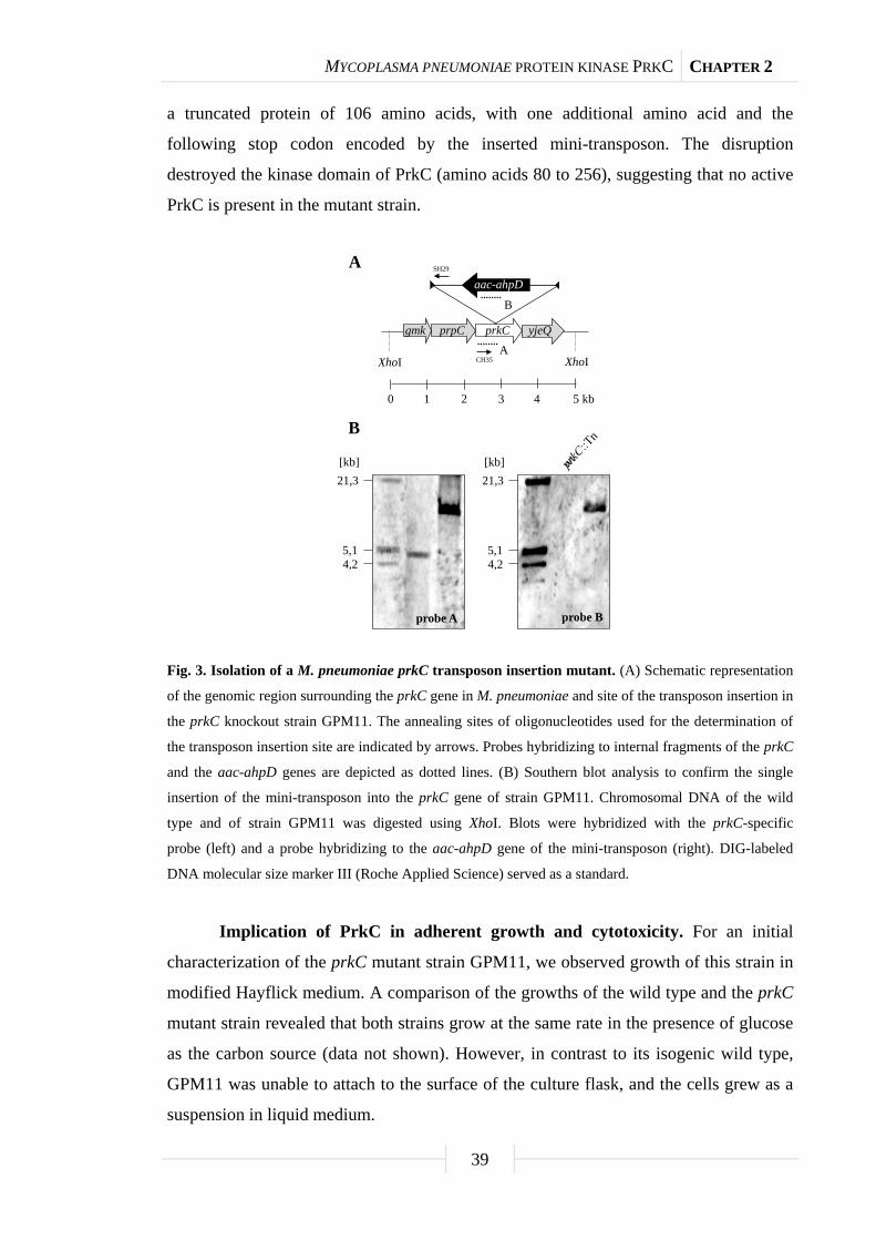
MYCOPLASMA PNEUMONIAE PROTEIN KINASE PRKC CHAPTER 2
39
a truncated protein of 106 amino acids, with one additional amino acid and the
following stop codon encoded by the inserted mini-transposon. The disruption
destroyed the kinase domain of PrkC (amino acids 80 to 256), suggesting that no active
PrkC is present in the mutant strain.
Fig. 3. Isolation of a M. pneumoniae prkC transposon insertion mutant. (A) Schematic representation
of the genomic region surrounding the prkC gene in M. pneumoniae and site of the transposon insertion in
the prkC knockout strain GPM11. The annealing sites of oligonucleotides used for the determination of
the transposon insertion site are indicated by arrows. Probes hybridizing to internal fragments of the prkC
and the aac-ahpD genes are depicted as dotted lines. (B) Southern blot analysis to confirm the single
insertion of the mini-transposon into the prkC gene of strain GPM11. Chromosomal DNA of the wild
type and of strain GPM11 was digested using XhoI. Blots were hybridized with the prkC-specific
probe (left) and a probe hybridizing to the aac-ahpD gene of the mini-transposon (right). DIG-labeled
DNA molecular size marker III (Roche Applied Science) served as a standard.
Implication of PrkC in adherent growth and cytotoxicity. For an initial
characterization of the prkC mutant strain GPM11, we observed growth of this strain in
modified Hayflick medium. A comparison of the growths of the wild type and the prkC
mutant strain revealed that both strains grow at the same rate in the presence of glucose
as the carbon source (data not shown). However, in contrast to its isogenic wild type,
GPM11 was unable to attach to the surface of the culture flask, and the cells grew as a
suspension in liquid medium.
21,3
5,14,2
[kb]21,3
5,14,2
[kb]
prpC yjeQgmk
XhoI
prkC
aac-ahpD
A
B
XhoI
0 1 32 5 kb4
A
B
probe A probe B
SH29
CH35
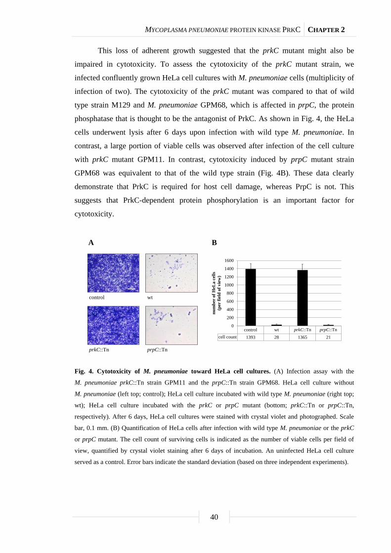
MYCOPLASMA PNEUMONIAE PROTEIN KINASE PRKC CHAPTER 2
40
This loss of adherent growth suggested that the prkC mutant might also be
impaired in cytotoxicity. To assess the cytotoxicity of the prkC mutant strain, we
infected confluently grown HeLa cell cultures with M. pneumoniae cells (multiplicity of
infection of two). The cytotoxicity of the prkC mutant was compared to that of wild
type strain M129 and M. pneumoniae GPM68, which is affected in prpC, the protein
phosphatase that is thought to be the antagonist of PrkC. As shown in Fig. 4, the HeLa
cells underwent lysis after 6 days upon infection with wild type M. pneumoniae. In
contrast, a large portion of viable cells was observed after infection of the cell culture
with prkC mutant GPM11. In contrast, cytotoxicity induced by prpC mutant strain
GPM68 was equivalent to that of the wild type strain (Fig. 4B). These data clearly
demonstrate that PrkC is required for host cell damage, whereas PrpC is not. This
suggests that PrkC-dependent protein phosphorylation is an important factor for
cytotoxicity.
Fig. 4. Cytotoxicity of M. pneumoniae toward HeLa cell cultures. (A) Infection assay with the
M. pneumoniae prkC::Tn strain GPM11 and the prpC::Tn strain GPM68. HeLa cell culture without
M. pneumoniae (left top; control); HeLa cell culture incubated with wild type M. pneumoniae (right top;
wt); HeLa cell culture incubated with the prkC or prpC mutant (bottom; prkC::Tn or prpC::Tn,
respectively). After 6 days, HeLa cell cultures were stained with crystal violet and photographed. Scale
bar, 0.1 mm. (B) Quantification of HeLa cells after infection with wild type M. pneumoniae or the prkC
or prpC mutant. The cell count of surviving cells is indicated as the number of viable cells per field of
view, quantified by crystal violet staining after 6 days of incubation. An uninfected HeLa cell culture
served as a control. Error bars indicate the standard deviation (based on three independent experiments).
A B
wtcontrol
prkC::Tn prpC::Tn
HeLa wt prkC::Tn prpC::Tncell count 1393 28 1365 21
0
200
400
600
800
1000
1200
1400
1600
num
ber
of H
eLa
cells
(per
fiel
d of
vie
w)
prkC::Tn prpC::Tncontrol

MYCOPLASMA PNEUMONIAE PROTEIN KINASE PRKC CHAPTER 2
41
Identification of proteins that are subject to PrkC-dependent
phosphorylation. The loss of adherent growth and cytotoxicity in the prkC mutant
suggested that proteins that are phosphorylated by PrkC might be involved in these
functions. To identify such proteins, we separated whole cell extracts of M. pneumoniae
by SDS-PAGE. The proteins were consecutively stained with Pro-Q Diamond, a dye for
the detection of Ser/Thr/Tyr-phosphorylated proteins, and with Flamingo fluorescence
stain for the detection of the total proteins. For this analysis, we compared the wild type
strain M129 and the prkC mutant GPM11. As a control, we used the prpC mutant
GPM68. Since PrpC is thought to be the antagonist of PrkC, we expected to observe a
more intense phosphorylation of PrkC target proteins with this mutant. In addition, we
used a mutant affected in the other identified protein kinase of M. pneumoniae, HPrK
(GPM51), and a mutant deficient in the surface protein MPN474, which is not thought
to be implicated in protein phosphorylation (GPM70) (Krebes et al., 1995). Several of
the proteins that are involved in cytadherence in M. pneumoniae are very large proteins,
such as HMW1, HMW2, and HMW3. It has been shown that these proteins are
phosphorylated in M. pneumoniae (Krebes et al., 1995; Su et al., 2007). Therefore, we
decided to analyze specifically the phosphorylation pattern of large proteins by using
6% SDS-polyacrylamide gels. A comparison of the total protein patterns in the wild
type and the prkC mutant strain revealed that the quantity of four proteins was severely
reduced (Fig. 5A). These protein bands were identified by MS as the three high
molecular weight proteins HMW1, HMW2, and HMW3 and the major adhesin P1. The
amounts of these four proteins in the prpC mutant were indistinguishable from those of
the wild type strain, suggesting that PrkC is necessary for their accumulation in the cell.
The quantitative analysis of the Pro-Q Diamond signal intensities for the detection of
phosphorylated proteins revealed that the bands of these proteins were absent or much
weaker in the prkC mutant (Fig. 5B and C). In contrast, the phosphorylation signals of
HMW1, HMW3, and P1 were more intense in the prpC phosphatase mutant than in the
wild type (Fig. 5C). This observation indicated the accumulation of phosphorylated
protein that cannot be dephosphorylated by the cognate protein phosphatase. In turn,
this suggests that these proteins are targets of PrkC-dependent phosphorylation and that
phosphorylation plays a role in their accumulation. In addition, one protein with an
apparent molecular mass of 160 kDa accumulates in the prpC mutant and shows also a
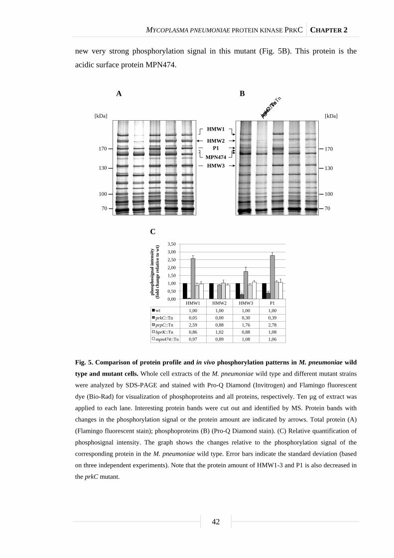
MYCOPLASMA PNEUMONIAE PROTEIN KINASE PRKC CHAPTER 2
42
new very strong phosphorylation signal in this mutant (Fig. 5B). This protein is the
acidic surface protein MPN474.
Fig. 5. Comparison of protein profile and in vivo phosphorylation patterns in M. pneumoniae wild
type and mutant cells. Whole cell extracts of the M. pneumoniae wild type and different mutant strains
were analyzed by SDS-PAGE and stained with Pro-Q Diamond (Invitrogen) and Flamingo fluorescent
dye (Bio-Rad) for visualization of phosphoproteins and all proteins, respectively. Ten µg of extract was
applied to each lane. Interesting protein bands were cut out and identified by MS. Protein bands with
changes in the phosphorylation signal or the protein amount are indicated by arrows. Total protein (A)
(Flamingo fluorescent stain); phosphoproteins (B) (Pro-Q Diamond stain). (C) Relative quantification of
phosphosignal intensity. The graph shows the changes relative to the phosphorylation signal of the
corresponding protein in the M. pneumoniae wild type. Error bars indicate the standard deviation (based
on three independent experiments). Note that the protein amount of HMW1-3 and P1 is also decreased in
the prkC mutant.
70
100
130
170
[kDa]
70
100
130
170
[kDa]
A B
HMW1
HMW2P1
HMW3MPN474
HMW1 HMW2 HMW3 P1wt 1,00 1,00 1,00 1,00prkC::Tn 0,05 0,00 0,30 0,39prpC::Tn 2,59 0,88 1,76 2,78hprK::Tn 0,86 1,02 0,88 1,08mpn474::Tn 0,97 0,89 1,08 1,06
0,00
0,50
1,00
1,50
2,00
2,50
3,00
3,50
phos
phos
igna
lint
ensi
ty(f
old
chan
ge r
elat
ive
to w
t)
C
prkC::TnprpC::TnhprK::Tnmpn474::Tn

MYCOPLASMA PNEUMONIAE PROTEIN KINASE PRKC CHAPTER 2
43
Impact of PrkC on the accumulation of cytadherence proteins. It has been
shown that HMW2 mutants of M. pneumoniae are impaired in the accumulation of
other cytadherence proteins, such as HMW1, HMW3, and MPN309 (P65) (Balish et al.,
2003). Since the amounts of the HMW proteins were reduced in the prkC mutant, we
determined the cellular amounts of other cytadherence proteins by use of specific
polyclonal antibodies raised against these proteins (Table 3 provides a summary of the
results). As controls, we used the elongation factor G (FusA) and the MPN567 (P200)
protein, which is required for gliding motility (Jordan et al., 2007). The amounts of both
proteins were virtually identical in all strains tested in this study (Fig. 6C).
Tab. 3. Summary of Western blot analyses with a prkC::Tn mutant (GPM11).
M. pneumoniae
locus tags
SwissProt
accession no. Protein
Signal in Western
blot (%)a Protein function
MPN141 P11311 P1 35 Adhesin
MPN142 Q50341 P40 40 Involved in cytadherence
MPN227 P75544 FusA 100 Elongation factor G
MPN309 P53663 P65 9 Adhesin-related protein
MPN310 P75471 HMW2 0 Cytadherence high
molecular weight protein 2
MPN310 P75471 HMW2-S
(P28)
0 Adhesin-related protein,
HMW2 fragment
MPN311 P75470 P41 8 Adhesin-related protein
MPN312 P75469 P24 41 Adhesin-related protein
MPN447 Q50365 HMW1 20 Cytadherence high
molecular weight protein 1
MPN452 Q50360 HMW3 22 Cytadherence high
molecular weight protein 3
MPN453 P75330 P30 34 Adhesin
MPN474 P75310 MPN474 104 Coiled coil protein,
putative structural protein
involved in cytosceleton
MPN567 P75211 P200 96 Involved in cytadherence a The protein amount in the prkC mutant relative to that in the wild type strain (100%) is indicated.

MYCOPLASMA PNEUMONIAE PROTEIN KINASE PRKC CHAPTER 2
44
P65 HMW2-SHMW2 P24P41
α-P65
α-HMW2-S(P28)
α-P41
α-P24
α-HMW2
A
α-P65 α-HMW2 α-HMW2-s (P28) α-P41 α-P24
wt 100 100 100 100 100prkC::Tn 9 0 0 8 41prpC::Tn 97 105 102 98 104hprK::Tn 103 102 99 99 98mpn474::Tn 104 100 104 101 101
0
20
40
60
80
100
120
prot
ein
leve
ls (%
wt)
prkC::TnprpC::TnhprK::Tnmpn474::Tn
HMW2-S(P28)
α-HMW3
α-HMW1
α-P30
HMW1 P30HMW3
B
α-HMW1 α-HMW3 α-P30wt 100 100 100prkC::Tn 20 22 34prpC::Tn 105 98 97hprK::Tn 102 95 101mpn474::Tn 98 103 98
0
20
40
60
80
100
120
prot
ein
leve
ls (%
wt)
prkC::TnprpC::TnhprK::Tnmpn474::Tn
α-MPN474
α-P40
α-P200
α-FusA
α-P1
P1 P90P40
C
α-P1 α-P40 α-MPN474 α-P200 α-FusAwt 100 100 100 100 100prkC::Tn 35 40 104 96 100prpC::Tn 96 102 100 100 103hprK::Tn 100 103 103 104 99mpn474::Tn 102 105 0 97 101
0
20
40
60
80
100
120
prot
ein
leve
ls (%
wt)
prkC::TnprpC::TnhprK::Tnmpn474::Tn

MYCOPLASMA PNEUMONIAE PROTEIN KINASE PRKC CHAPTER 2
45
Fig. 6. Western blot analyses of expression profiles of M. pneumoniae cytadherence proteins.
Whole cell extracts of the M. pneumoniae wild type and different mutant strains were separated by
SDS-PAGE and blotted onto a polyvinylidene difluoride membrane. Cytadherence proteins were detected
by using polyclonal rabbit antibodies raised against proteins of the P65 operon (A), the HMW gene
cluster (B), and major adhesins (C). A polyclonal rabbit antibody raised against M. pneumoniae
elongation factor G (FusA) was used as the control. Ten µg of extract was applied to each lane. The
names of antibodies are given next to each blot. α, anti. HMW1, HMW2, HMW3, MPN474, P1, and P200
were analyzed by 6% SDS-PAGE, and the other proteins by 12% SDS-PAGE. The graphs show the
quantification of Western blot signals relative to that for the M. pneumoniae wild type. Error bars indicate
the standard deviation (based on three independent experiments). The organization of the different
operons is shown by arrows. Note that there are four genes between hmw3 and hmw1, which is indicated
(panel B). Moreover, mpn474, p200, and fusA are not part of operons (panel C).
First, we studied the proteins encoded by one gene cluster containing the gene
encoding HMW2 (the so-called P65 operon). As shown in Fig. 6A, the cellular amounts
of all these proteins are significantly reduced in the prkC mutant. In fact, the HMW2
protein and HMW2-S (also called P28), a second translation product of the same
mRNA (Boonmee et al., 2009), are nearly completely absent in the prkC mutant strain.
The accumulation of all the proteins encoded by the gene cluster is not affected by a
disruption of the prpC gene (Fig. 6A). Thus, the phosphorylation of these proteins
seems to be essential for their accumulation. The so-called HMW gene cluster encodes
the HMW1, HMW3, and MPN453 (P30) proteins. As observed for HMW2, the two
high molecular weight proteins encoded by this gene cluster are much less abundant in
the prkC mutant, whereas none of the other tested mutations had any effect. The
accumulation of MPN453 was also somewhat reduced but to a lesser extent (Fig. 6B).
Finally, we assayed the amounts of the major adhesins MPN141 (P1) and MPN142
(P40) and of the surface protein MPN474, which was shown to be phosphorylated by
PrkC (Fig. 5). As shown for the large cytadherence proteins, many fewer adhesins were
detected in the prkC mutant than in the wild type or all other strains. In contrast, the
cellular amounts of MPN474 were not affected by the prkC mutation. As expected, the
antibodies do not cross-react with any protein in the MPN247 mutant strain GPM70.
Transcription of the genes encoding cytadherence proteins in mutants
affecting protein phosphorylation. The virtual absence of cytadherence proteins in the
prkC mutant can be explained by two alternative scenarios: (1) The expression of the
corresponding genes and the accumulation of their mRNAs might require some kind of

MYCOPLASMA PNEUMONIAE PROTEIN KINASE PRKC CHAPTER 2
46
PrkC-dependent protein phosphorylation and (2) the proteins might be translated to a
lesser extent or be subject to rapid degradation. To distinguish between these
possibilities, we compared the amounts of the mRNAs of the cytadherence genes in the
wild type strain M129 and in our set of mutants that are affected in protein
(de)phosphorylation. For this purpose, we isolated RNA from cultures grown in
modified Hayflick medium supplemented with glucose and performed slot blot
analyses.
Fig. 7. Expression of cytadherence genes in M. pneumoniae wild type and mutant cells. Slot blot
analyses with whole RNA extracts of the M. pneumoniae wild type and different mutant strains. A
dilution series of RNA extracts was blotted onto a positively charged nylon membrane and probed with a
DIG-labeled riboprobe specific for an internal part of a particular open reading frame. Names of
riboprobes are given next to each blot. Signals obtained with 1 µg of RNA are shown. Yeast tRNA and
M. pneumoniae chromosomal DNA served as controls. Expression profiles of the genes of the P65 operon
are shown (A), and the analyses of cytadherence genes of the HMW gene cluster and the major adhesins
are also shown (B). The graphs show the quantification of transcript levels relative to that for the
M. pneumoniae wild type. Error bars indicate the standard deviation (based on three independent
experiments).
P65 HMW2 HMW2-s (P28) P41 P24
wt 100 100 100 100 100prkC::Tn 101 102 100 27 34prpC::Tn 98 99 101 95 96hprK::Tn 104 102 105 101 95mpn474::Tn 100 103 100 104 102
0
20
40
60
80
100
120
tran
scri
pt le
vels
(% w
t)
HMW2
P65
P41
P24
HMW2-S(P28)
A
prkC::TnprpC::TnhprK::Tnmpn474::Tn
HMW2-S(P28)
HMW3
HMW1
P1
P40
P30
B
HMW1 HMW3 P30 P1 P40wt 100 100 100 100 100prkC::Tn 95 101 98 101 101prpC::Tn 100 142 158 97 102hprK::Tn 100 105 104 100 104mpn474::Tn 95 101 97 99 100
020406080
100120140160180
tran
scri
pt le
vels
(% w
t)
prkC::TnprpC::TnhprK::Tnmpn474::Tn

MYCOPLASMA PNEUMONIAE PROTEIN KINASE PRKC CHAPTER 2
47
For the P65 operon, we observed similar amounts of mRNA for the two
proximal genes of the operon, MPN309 (encoding P65) and hmw2 (encoding HMW2)
in the wild type and in the kinase and phosphatase mutants (Fig. 7A). This observation
is in sharp contrast to the strongly reduced amounts of the corresponding proteins in the
prkC mutant (Fig. 7A). For the two distal genes of the operon, MPN311 (P41) and
MPN312 (P24), we observed a slight reduction of mRNA amounts in the prkC mutant
(Fig. 7A). These reduced mRNA levels might explain the weak reduction of P24 protein
accumulation, but they are not sufficient to account for the strongly reduced P41
amounts in the prkC mutant strain GPM11.
The genes of the HMW and the adhesin (P1 and P40) operons were all expressed
at similar levels in the wild type as well as in the prkC, hprK, and prpC mutant strains
(Fig. 7B). Again, the mRNA amounts were not affected by the prkC mutation, whereas
the protein amounts were strongly reduced (Fig. 6C). This is in good agreement with the
observations for the P65 operon and supports the conclusion that PrkC-dependent
protein phosphorylation is implicated in the stability of the cytadherence and adhesion
proteins in M. pneumoniae.
Discussion
Protein phosphorylation is a major mechanism to control the activities of
proteins. This posttranslational modification has been observed with all organisms, for
which it has been studied, including the minimal bacteria M. genitalium and
M. pneumoniae (Su et al., 2007; this work). However, the physiological consequences
of protein phosphorylation events in bacteria, as well as the responsible kinases, have
remained largely unknown.
So far, only a few classes of serine/threonine protein kinases have been
identified and characterized for bacteria. M. pneumoniae contains two of these protein
kinases, HPrK and PrkC (Steinhauer et al., 2002; this work). While HPrK is generally
thought to phosphorylate only one specific substrate, the HPr protein of the
phosphotransferase system, a variety of substrates have been proposed for PrkC and its
homologues in different gram-positive bacteria. In Bacillus subtilis, PrkC was shown to
phosphorylate the elongation factors Tu and G, the small-ribosome-associated GTPase
CpgA, and the potential stressosome component YezA (Absalon et al., 2009;
Shah et al., 2008). PrkC is required for germination of B. subtilis spores in response to

MYCOPLASMA PNEUMONIAE PROTEIN KINASE PRKC CHAPTER 2
48
muropeptides (Shah et al., 2008). The extracellular PASTA domain of PrkC senses
these muropeptides (Shah et al., 2008). Since M. pneumoniae neither sporulates nor
possesses a peptidoglycan cell wall, it is not surprising that the PrkC protein of this
bacterium does not contain a PASTA domain. Thus, it is safe to assume that
M. pneumoniae PrkC has a function(s) that differs completely from that described for
B. subtilis. Indeed, this work provides evidence that PrkC is required for adhesive
growth of M. pneumoniae, and a prkC mutant has lost cytotoxicity. This is reminiscent
of observations that have been made with several other pathogenic Firmicutes. In
Streptococcus pyogenes, the homolog of PrkC is required for adherence to host cells
and for invasion (Jin and Pancholi, 2006). Similarly, the kinase of Enterococcus faecalis
is implicated in persistence in the intestine of mice (Kristich et al., 2007). In contrast,
PrkC of Staphylococcus aureus was reported to phosphorylate glycolytic enzymes
(Lomas-Lopez et al., 2007).
While the importance of PrkC-dependent protein phosphorylation is well
established for many bacteria, the molecular consequences of the primary
phosphorylation events have not been studied so far. Most often, protein
phosphorylation results in a shift of a protein’s activity (Johnson and Barford, 1993).
In this work, we observed that PrkC-dependent phosphorylation of large
cytadherence proteins is required for the stability of these complexes. It is unknown,
which of these proteins is responsible for the destabilization of cytadherence proteins in
the prkC mutant, but our studies revealed that HMW1, HMW3, the major adhesin P1,
and the surface protein MPN474 are subject to PrkC-dependent phosphorylation. This
conclusion is supported not only by the absence of these proteins in cell extracts of the
prkC mutant but also by the increased intensity of phosphorylation signals for these
proteins in the prpC mutant strain. PrpC is the protein phosphatase that reverses
PrkC-dependent protein phosphorylation. Thus, our work confirms and extends earlier
reports on the phosphorylation of cytadherence proteins (Krebes et al., 1995; Su et al.,
2007). We cannot exclude, however, the possibility that an additional kinase(s) might
phosphorylate HMW3 and P1 (Fig. 5C). It is well established that the cytadherence
proteins show reciprocal dependency in their stabilities, i.e., mutations in one of the
corresponding genes result in destabilization and delocalization of the other proteins of
the attachment structure (Balish and Krause, 2006; Popham et al., 1997; Willby et al.,
2004). Specifically, HMW1 was shown to be required for HMW2 and P1 localization

MYCOPLASMA PNEUMONIAE PROTEIN KINASE PRKC CHAPTER 2
49
and stability. In turn, HMW2 is necessary for the stabilization of HMW3 and P65. As
shown in this work, it has been suggested previously that the interdependence of the
proteins of the attachment organelle does not involve reduced transcription
(Popham et al., 1997). Taken together, these findings establish that phosphorylation of
the HMW proteins by PrkC is a prerequisite for their stability and perhaps for their
proper localization in the attachment structure. This is the first time that protein
phosphorylation was shown to increase the stability of a protein.
M. pneumoniae has only limited regulatory potential. These bacteria may use, in
addition to three potential transcription regulators, protein phosphorylation to adapt to
changes in environmental conditions. An analysis of the M. pneumoniae genome
revealed the presence of two genes for serine/threonine kinases, i.e., hprK and prkC,
encoding the HPr kinase and protein kinase C (PrkC), respectively. Based on our
present knowledge, it appears that these two proteins represent the total serine/threonine
kinome of M. pneumoniae. It will be interesting to study how PrkC activity is triggered
and whether other proteins are targets of this pleiotropic protein kinase.
Acknowledgments
We are grateful to Richard Herrmann for the gift of antibodies. Hinnerk Eilers is
acknowledged for helpful discussions. This work was supported by Deutsche
Forschungsgemeinschaft and the Fonds der Chemischen Industrie. S.R.S. was supported
by a personal grant from the Studienstiftung des Deutschen Volkes.
MYCOPLASMA PNEUMONIAE PROTEIN KINASE PRKC CHAPTER 2
50
Supplemental material
Tab. S1. Oligonucleotides used in this study.
Oligonucleotide Sequence (5’→3’)a
CH35 GCTCAACTTGATTAATTTAAAACAATGG
CH74 CTAATACGACTCACTATAGGGAGAGGTGTTCAAAACTGTGGAGG
SH29 ATGAGTGAGCTAACTCACAG
SH62 TAGAATTTTATGGTGGTAGAG
SH63 CTAATACGACTCACTATAGGGAGAACACTATCATAACCACTACC
SH89 TAGAGCTCGATGGCACTAAATTTAAAGATTGG
SS60 GCTACTGCATACGATCCCAATC
SS61 CTAATACGACTCACTATAGGGAGACATAGTAAGCGTTGGGATCGG
SS62 GACACTGGGTTTGATGATGGG
SS63 CTAATACGACTCACTATAGGGAGACCTGGTTAGCTGCAATCTGTTC
SS64 GAAGCGCAACCAACTAACGC
SS65 CTAATACGACTCACTATAGGGAGAGTTGTTGGGACAGATCAGCAC
SS66 GATGATGAAGCTGACATCATCATAG
SS67 CTAATACGACTCACTATAGGGAGAGTGTATTCCGTGCCACCAATAAC
SS68 CTAACACTAAAACGGGTACGAATTG
SS69 CTAATACGACTCACTATAGGGAGAGCCACAAGTGTTTCTGATGTCAC
SS70 CTGAGGAAAATCCCGAACAGATC
SS71 CTAATACGACTCACTATAGGGAGAGCTGGTACATCTGCTTCCAAAC
SS72 GCCAACAGCGACATTAACAGC
SS73 CTAATACGACTCACTATAGGGAGACAGTATCACTGCAGAGCTTATAAAC
SS74 GTATGGTAAGCTAGCACAAAAGATC
SS75 CTAATACGACTCACTATAGGGAGAGTTGATCAGCTTGCTGTTCGG
SS76 CTTGGATTCTCATCCTCACCG
SS77 CTAATACGACTCACTATAGGGAGAGGTGCAGCCCCACTCAAAC
SS78 GCGAGCGGGTGGTTCG
SS79 CTAATACGACTCACTATAGGGAGAGCTTGTAAGCACTCGCTTCC
SS80 CCTGAATGCTAGTGCTGTTCAG
SS81 CTAATACGACTCACTATAGGGAGAGTTGAGGTGGGAAACCAGTAC
SS86 GCACGCATCTTAAAGCTGTTGG
SS87 CTAATACGACTCACTATAGGGAGACACTGTCAGCGTGCTCTGC
SS92 GCTAGCTTCTTCACTTACCGG
SS93 CTAATACGACTCACTATAGGGAGAGTTAACGTTCCTTCGTTACCATTG a The sequence of the T7-promotor is underlined.
MYCOPLASMA PNEUMONIAE PROTEIN KINASE PRKC CHAPTER 2
51
Tab. S2. Antibodies used in this study.
M. pneumoniae locus tags Protein Antibody number Dilution
MPN141 P1 449 1:1000
MPN146 P40 66450 1:250
MPN227 FusA 66708 1:3000
MPN309 P65 66578 1:3000
MPN310 HMW2 28050 1:2000
MPN310 HMW2-S (P28) 86236 1:1000
MPN311 P41 86630 1:1000
MPN312 P24 85910 1:250
MPN447 HMW1 84267 1:10000
MPN452 HMW3 65376 1:5000
MPN453 P30 123 1:50000
MPN474 MPN474 41804 1:20000
MPN567 P200 65020 1:10000

Chapter 3
The phosphoproteome of the minimal bacterium
Mycoplasma pneumoniae:
Analysis of the complete known Ser/Thr kinome
suggests the existence of novel kinases
The work described in this chapter was published in:
Schmidl, S. R., K. Gronau, N. Pietack, M. Hecker, D. Becher, and J. Stülke. 2010.
The phosphoproteome of the minimal bacterium Mycoplasma pneumoniae: Analysis of
the complete known Ser/Thr kinome suggests the existence of novel kinases. Mol. Cell.
Proteomics 9: 1228-1242.
Author contributions:
This study was designed and interpreted by SRS and JS. SRS performed the
phosphoproteome experiments in M. pneumonaie. NP contributed to the investigation of
the autophosphorylation event of phosphosugar mutases, especially in B. subtilis. The
phosphoproteome analysis was done in collaboration with KG, MH, and DB, University
of Greifswald. KG and DB helped substantially with the evaluation of the
phosphorylation sites. SRS and JS wrote the paper.

PHOSPHOPROTEOME OF MYCOPLASMA PNEUMONIAE CHAPTER 3
53
Abstract
Mycoplasma pneumoniae belongs to the Mollicutes, the group of organisms with
the smallest genomes that are capable of host-independent life. These bacteria show
little regulation in gene expression, suggesting an important role for the control of
protein activities. We have studied protein phosphorylation in M. pneumoniae to
identify phosphorylated proteins. Two-dimensional gel electrophoresis and mass
spectrometry allowed the detection of 63 phosphorylated proteins, many of them
enzymes of central carbon metabolism and proteins related to host cell adhesion. We
identified 16 phosphorylation sites, among them 8 serine and 8 threonine residues,
respectively. A phosphoproteome analysis with mutants affected in the two annotated
protein kinase genes or in the single known protein phosphatase gene suggested that
only one protein (HPr) is phosphorylated by the HPr kinase, HPrK, whereas four
adhesion-related or surface proteins were targets of the protein kinase C, PrkC. A
comparison with the phosphoproteomes of other bacteria revealed that protein
phosphorylation is evolutionarily only poorly conserved. Only one single protein with
an identified phosphorylation site, a phosphosugar mutase (ManB in M. pneumoniae), is
phosphorylated on a conserved serine residue in all studied organisms from archaea and
bacteria to man. We demonstrate that this protein undergoes autophosphorylation. This
explains the strong conservation of this phosphorylation event. For most other proteins,
even if they are phosphorylated in different species, the actual phosphorylation sites are
different. This suggests that protein phosphorylation is a form of adaptation of the
bacteria to the specific needs of their particular ecological niche.
Introduction
Bacteria of the group called Mollicutes are unique among all living organisms
because of their small genome that nevertheless allows them to grow independently
from any host cell. The two most intensively studied representatives of the Mollicutes
are Mycoplasma genitalium and Mycoplasma pneumoniae with genome sizes of 580
and 816 kb, respectively. The approximately 475 protein-coding and 43 RNA-coding
genes of M. genitalium define the lower limit of the genetic equipment that permits
independent life. The small genomes of the Mollicutes reflect their adaptation to rarely
changing ecosystems, such as lung epithelia for M. pneumoniae (Stülke et al., 2009).

PHOSPHOPROTEOME OF MYCOPLASMA PNEUMONIAE CHAPTER 3
54
Furthermore, M. pneumoniae has only limited metabolic capabilities: This bacterium
can utilize only a few carbon sources (glucose, fructose, and glycerol) (Halbedel et al.,
2004), and its only way to produce ATP is by substrate level phosphorylation in
glycolysis. The citric acid cycle, respiration, and most anabolic reactions are not carried
out by M. pneumoniae (Halbedel et al., 2007).
M. pneumoniae is a human pathogen that causes usually mild infections such as
atypical pneumonia; however, the infections may be severe in children and elderly
people. In addition, M. pneumoniae is involved in extrapulmonary complications, such
as erythema multiforme and pediatric encephalitis (Atkinson et al., 2008; Jacobs, 1997;
Waites and Talkington, 2004).
The close adaptation of M. pneumoniae to human mucosal surfaces did not only
result in a reductive evolution of metabolic capabilities but also affected the regulation
of protein biosynthesis and activity: In contrast to versatile environmental bacteria such
as Pseudomonas aeruginosa that reserve about 10% of their coding capacity for
regulators of gene expression, M. pneumoniae encodes only three potential transcription
regulators (less than 0.5% of all genes) (Stülke et al., 2009). This absence of regulation
at the level of gene expression suggests that the control of protein activities might play
an important role in M. pneumoniae and other Mollicutes.
An important way to control the activity of a protein is posttranslational
modification with protein phosphorylation being the most prominent regulatory
modification both in bacteria and in eukaryotes (Boekhorst et al., 2008; Jers et al.,
2008). Studies on the phosphoproteome have been performed for several bacteria,
including the model organisms Escherichia coli and Bacillus subtilis. In both bacteria,
about 5% of all proteins can be phosphorylated on Ser, Thr, or Tyr residues
(Eymann et al., 2007; Lévine et al., 2006; Macek et al., 2008; Macek et al., 2007).
Interestingly, there is a high level of species specificity with respect to the
phosphoproteome: A comparison of the phosphoproteomes of B. subtilis, E. coli, and
Lactococcus lactis revealed only eight proteins that are phosphorylated in all three
species. Six of these proteins are involved in sugar metabolism, and two are translation
factors. Of these eight proteins, only one, phosphoglucosamine mutase, is
phosphorylated at a conserved residue (Soufi et al., 2008).
In M. pneumoniae, only the phosphorylation of the HPr protein of the
phosphoenolpyruvate:sugar phosphotransferase system by the HPr kinase has been

PHOSPHOPROTEOME OF MYCOPLASMA PNEUMONIAE CHAPTER 3
55
studied to some detail (Allen et al., 2003; Halbedel et al., 2004; Steinhauer et al., 2002).
Once phosphorylated, HPr can be dephosphorylated by a protein phosphatase, PrpC
(Halbedel et al., 2006). The corresponding prpC gene is in many Gram-positive bacteria
clustered with the gene for a protein kinase, prkC, suggesting that PrkC and PrpC form
a functional couple with opposing activities. Evidence for this idea was provided by the
observation that the two proteins from B. subtilis have identical targets in vitro
(Absalon et al., 2009). The prpC/prkC gene cluster also exists in M. pneumoniae;
however, the targets of the corresponding proteins are so far unknown. Previous studies
with M. pneumoniae demonstrated that several proteins implicated in adhesion to the
host cell are subject to protein phosphorylation in vivo (Dirksen et al., 1994;
Krebes et al., 1995; Schmidl et al., 2010). These proteins, HMW1 and HMW2, are
important for the virulence of M. pneumoniae because adhesion is the first step in the
interaction with the host. A recent proteomics study resulted in the identification of
18 phosphorylated proteins in M. pneumoniae, among them adhesion and surface
proteins and metabolic enzymes (Su et al., 2007). Unfortunately, none of the previously
known phosphoproteins were detected in this study. Moreover, Ser-46 in HPr is still the
only known phosphorylation site in M. pneumoniae (with the exception of
phosphorylated His and Cys residues in phosphotransferase system proteins), and the
(de)phosphorylation of HPr is the only event, for which the kinase and the phosphatase
are known. Finally, nothing is known about protein tyrosine phosphorylation in
M. pneumoniae and other Mollicutes.
We are interested in the elucidation of the molecular networks that allow life of
M. pneumoniae as a minimal organism. The analysis of these bacteria is hampered by
their slow growth and by the lack of efficient genetic systems that allow the elucidation
of gene functions. We have established a technique, haystack mutagenesis, that can be
used to isolate mutants of M. pneumoniae (Halbedel et al., 2006). In this work, we have
studied the phosphoproteome of the M. pneumoniae wild type strain and of three
isogenic mutants that are affected in the two protein kinases, HPr kinase (HPrK) and
protein kinase C (PrkC), and in the protein phosphatase PrpC. We identified
63 phosphorylated proteins. However, most of these proteins are phosphorylated by so
far unknown kinases. An in-depth analysis of the only universally conserved
phosphoprotein, a phosphosugar mutase, revealed that this enzyme autophosphorylates.
PHOSPHOPROTEOME OF MYCOPLASMA PNEUMONIAE CHAPTER 3
56
Experimental procedures
Bacterial strains and growth conditions. The M. pneumoniae strains used in
this study were M. pneumoniae M129 (ATCC 29342) in the 32nd broth passage and its
isogenic mutant derivatives GPM11 (prkC::mini-Tn, GmR) (Schmidl et al., 2010),
GPM51 (hprK::mini-Tn, GmR) (Halbedel et al., 2006), and GPM68 (prpC::mini-Tn,
GmR) (Halbedel et al., 2006). M. pneumoniae was grown at 37°C in 150-cm2 tissue
culture flasks containing 100 ml of modified Hayflick medium with glucose
(1%, wt/vol) as the carbon source as described previously (Halbedel et al., 2004).
Strains harboring transposon insertions were cultivated in the presence of 80 µg/ml
gentamicin. B. subtilis 168 (trpC2; laboratory collection) was grown in LB and in
minimal medium containing succinate and glutamate/ammonium as basic sources of
carbon and nitrogen, respectively (Faires et al., 1999). The media were supplemented
with tryptophan (at 50 mg/liter) and glucose (0.5%, wt/vol). B. subtilis was transformed
with plasmid DNA or PCR products according to the two-step protocol
(Kunst and Rapoport, 1995). Transformants were selected on sporulation medium plates
(Faires et al., 1999) containing erythromycin plus lincomycin (2 and 25 µg/ml,
respectively) and/or spectinomycin (100 µg/ml).
Preparation of cell extracts. After 4 days of incubation at 37°C, the culture
medium was removed from the M. pneumoniae cell layer, and the cells were washed
twice with 20 ml of cold PBS. After washing, cells were collected by scraping into
1.5 ml of PBS and subsequent centrifugation (5 min, 15,000 × g, 4°C). The cell pellet
was resuspended in 500 µl of 10 mM Tris-HCl, pH 7.5, and the cells were disrupted by
sonication (3 × 10 s, 50 watts, 4°C). Cell debris were sedimented by centrifugation
(10 min, 15,000 × g, 4°C), and the resulting supernatant was centrifuged again (30 min,
24,000 × g, 4°C) to remove disturbing particles. The protein concentration of the
supernatant was determined using the Bio-Rad dye binding assay with bovine serum
albumin as the standard.
Two-dimensional SDS-polyacrylamide gel electrophoresis. Two-dimensional
separation of proteins was performed as described previously (Halbedel et al., 2007).
For separation in the first dimension, IPG strips with a linear pH range of 4-7, 4.5-5.5,
or 6-11 were used (ImmobilineTM DryStrip, Amersham Biosciences). The gels were
subsequently stained with Pro-Q Diamond (Invitrogen) and FlamingoTM fluorescent dye
(Bio-Rad) to visualize the phosphoproteins and all proteins, respectively. Image

PHOSPHOPROTEOME OF MYCOPLASMA PNEUMONIAE CHAPTER 3
57
analysis, spot quantification, and determination of putative phosphorylated protein spots
were performed as described previously (Eymann et al., 2007). The determination of
phosphorylated proteins was based on the Pro-Q/Flamingo log ratio. Phosphoprotein
spots were cut from the gel and identified by mass spectroscopy.
Protein identification by mass spectrometry. Gel pieces were washed twice
with 200 µl of 20 mM NH4HCO3, 30% (v/v) CH3CN for 30 min at 37°C and dried in a
vacuum centrifuge (Concentrator 5301, Eppendorf). Trypsin solution (10 ng/µl trypsin
in 20 mM ammonium bicarbonate) was added until gel pieces stopped swelling, and
digestion was allowed to proceed for 16-18 h at 37°C. Peptides were extracted from gel
pieces by incubation in an ultrasonic bath for 15 min in 20 µl of HPLC grade water and
transferred into microvials for mass spectrometric analysis. Peptides were separated by
liquid chromatography and measured on line by ESI-mass spectrometry using a
nanoACQUITY UPLCTM system (Waters) coupled to an LTQ OrbitrapTM mass
spectrometer (Thermo Fisher Scientific, Waltham, MA). Peptides were desalted onto a
trap column (Symmetry® C18, Waters). Elution was performed onto an analytical
column (BEH130 C18, Waters) by a binary gradient of buffer A [0.1% (v/v) acetic acid]
and B [100% (v/v) acetonitrile, 0.1% (v/v) acetic acid] over a period of 50 min with a
flow rate of 400 nl/min. The LTQ Orbitrap was operated in data-dependent MS/MS
mode using multistage activation for phosphorelevant masses. Proteins were identified
by searching all MS/MS spectra in .dta format against all 689 M. pneumoniae proteins
(extracted from the NCBI database) using SorcererTM-SEQUEST® (SEQUEST
version 2.7 revision 11, Thermo Electron) including Scaffold_2_05_02 (Proteome
Software Inc., Portland, OR). SEQUEST was searched with a fragment ion mass
tolerance of 1.00 Da and a parent ion tolerance of 10 ppm. Up to two missed tryptic
cleavages were allowed. Methionine oxidation (+15.99492 Da), cysteine
carbamidomethylation (+57.021465 Da), and phosphorylation (+79.966331 Da) were
set as variable modifications. For protein identification, a stringent SEQUEST filter for
peptides was used (Xcorr versus charge state: 1.90 for singly, 2.2 for doubly, and 3.3 for
triply charged ions and ∆Cn value greater than 0.10), and at least two peptides per
proteins were required. With this method, all identified proteins had a protein
probability score greater than 99.9%. Protein probabilities were assigned by the Protein
Prophet algorithm (Nesvizhskii et al., 2003). Phosphorylated peptides that had a peptide
probability score greater than 95.0% as specified by the Peptide Prophet algorithm

PHOSPHOPROTEOME OF MYCOPLASMA PNEUMONIAE CHAPTER 3
58
(Keller et al., 2002) were examined manually and accepted only when b- or y-ions
confirmed the identification. All spectra with Scaffold-annotated b- and y-ions series are
provided as screen shots in the supplemental material (see supplemental Fig. S3).
Analysis of protein conservation and structure prediction. To address the
potential conservation of phosphorylation sites, multiple sequence alignments were
performed using the EXPRESSO alignment server that takes available structures into
account (Armougom et al., 2006). Structural models of the phosphoproteins were
created using SWISS-MODEL in the automated mode and Swiss-PdbViewer v4.0.1
(Guex and Peitsch, 1997; Kiefer et al., 2009). Ordered and disordered regions of
proteins were predicted using the DisEMBL v1.5 web server (Linding et al., 2003).
Cloning procedures. E. coli DH5α (Sambrook et al., 1989) was used for
cloning experiments and protein expression. E. coli was grown in LB medium.
LB plates were prepared by addition of 17 g of Bacto agar/liter (Difco) to LB
(Sambrook et al., 1989). Transformation of E. coli and plasmid DNA extraction were
performed using standard procedures (Sambrook et al., 1989). Restriction enzymes,
T4 DNA ligase, and DNA polymerases were used as recommended by the
manufacturers. DNA fragments were purified from agarose gels using the QIAquick Gel
Extraction kit (Qiagen). Pfu DNA polymerase was used for the polymerase chain
reaction as recommended by the manufacturer. DNA sequences were determined using
the dideoxy chain termination method (Sambrook et al., 1989). Standard procedures
were used to transform E. coli (Sambrook et al., 1989), and transformants were selected
on LB plates containing ampicillin (100 µg/ml).
Plasmid constructions. Plasmids for the overexpression and purification of the
phosphosugar mutases GlmM and ManB from B. subtilis and M. pneumoniae,
respectively, were constructed as follows. The coding sequence of the genes was
amplified by PCR with gene-specific primers (listed in supplemental Table S3) using
chromosomal DNA of B. subtilis 168 and M. pneumoniae M129 as the template. The
PCR products were digested as shown in supplemental Table S4 and cloned into the
appropriately linearized expression vector pWH844 (Schirmer et al., 1997). These
plasmids allowed the expression of the phosphosugar mutases carrying an N-terminal
His6-tag. The resulting plasmids pGP1401 and pGP656 are listed in supplemental
Table S4. In M. pneumoniae, the UGA specifies a tryptophan; however, in E. coli, it is a
stop codon. The manB gene contains three UGA codons that were replaced by the
PHOSPHOPROTEOME OF MYCOPLASMA PNEUMONIAE CHAPTER 3
59
multiple mutation reaction (Hames et al., 2005) using the phosphorylated mutagenic
oligonucleotides SS109, SS110, and SS111 (see supplemental Table S3) and plasmid
pGP656 as the template. The resulting plasmid was pGP657 (see supplemental
Table S4). The phosphorylation sites of GlmM and ManB were replaced by alanine
residues. For this purpose, the combined chain reaction (Bi and Stambrook, 1998) was
applied with the mutagenic primers NP116 and SS112 (see supplemental Table S3) and
plasmids pGP1401 and pGP657. The resulting plasmids are pGP1405 (glmM) and
pGP658 (manB) (see supplemental Table S4). All plasmid inserts were verified by DNA
sequencing.
The plasmids pGP400 and pGP1403 were used to express the wild type and
S100A mutant forms of GlmM, respectively, in B. subtilis. For the construction of
pGP400, the glmM gene was amplified using the oligonucleotides NP108/NP109. The
PCR product was digested with BamHI and SalI and cloned into pBQ200
(Martin-Verstraete et al., 1994) digested with the same enzymes. For the construction of
the plasmid encoding the S100A GlmM variant, mutagenesis was performed using the
combined chain reaction with NP116 as the mutagenic primer and NP108/NP109. The
resulting PCR product was cloned into pBQ200 as described for the wild type allele.
The resulting plasmid was pGP1403.
Overexpression and purification of recombinant proteins. E. coli DH5α
transformed with the appropriate expression vector was used as host for the
overexpression of recombinant proteins. Expression was induced by the addition of
isopropyl 1-thio-β-D-galactopyranoside (final concentration, 1 mM) to exponentially
growing cultures (A600 of 0.6). Cells were lysed using a French press (20,000 p.s.i.,
138,000 kilopascals; Spectronic Instruments). After lysis, the crude extracts were
centrifuged at 15,000 × g for 1 h. For purification of His6-tagged proteins, the resulting
supernatants were passed over a Ni2+ HiTrap chelating column (4-ml bed volume;
GE Healthcare) followed by elution with an imidazole gradient (from 0 to
500 mM imidazole in a buffer containing 10 mM Tris-HCl, pH 7.5, 600 mM NaCl,
10 mM β-mercaptoethanol) over 30 ml at a flow rate of 0.5 ml/min. After elution, the
fractions were tested for the desired protein using 12.5% SDS-PAGE. The relevant
fractions were combined and dialyzed overnight. Protein concentration was determined
using the Bio-Rad dye binding assay where bovine serum albumin served as the
standard.
PHOSPHOPROTEOME OF MYCOPLASMA PNEUMONIAE CHAPTER 3
60
Assays for protein phosphorylation. Protein phosphorylation assays were
carried out with purified proteins in assay buffer [25 mM Tris-HCl, pH 7.6,
1 mM dithiothreitol, 0.4 mM [γ-32P]ATP (480 Ci/mmol), divalent cations as indicated]
using purified target proteins. The assays were carried out at 37°C for 30 min followed
by thermal inactivation of the protein (10 min at 95°C). The assay mixtures were
analyzed on 16% SDS-polyacrylamide gels. Proteins were visualized by Coomassie
staining. Radioactive protein spots were detected using a phosphorimaging system
(Storm 860, GE Healthcare).
Allelic replacement of B. subtilis glmM gene. To delete the chromosomal copy
of the glmM gene, the long flanking homology PCR technique was used (Wach, 1996).
Briefly, a cassette carrying the spc resistance gene was amplified from plasmid
pDG1726 (Guérout-Fleury et al., 1995). DNA fragments of about 1,000 bp flanking the
glmM gene at its 5’ and 3’ ends were amplified. The 3’ end of the upstream fragment as
well as the 5’ end of the downstream fragment extended into glmM in a way that all
expression signals of genes up- and downstream of the glmM remained intact. The
joining of the two fragments to the resistance cassette was performed in a second PCR
as described previously (Jordan et al., 2006). In this reaction, we used the primer pair
spec-fwd/spec-rev (Jordan et al., 2006) for the amplification and joining of the
spc cassette. The PCR product was directly used to transform B. subtilis carrying
plasmids that express different alleles of glmM. The integrity of the regions flanking the
integrated resistance cassette was verified by sequencing PCR products of about
1,000 bp amplified from chromosomal DNA of the resulting mutants.
Results
Phosphoproteome of growing M. pneumoniae cells. In a previous study on
protein phosphorylation in M. pneumoniae, no phosphorylation sites were determined,
and no mutants affected in the players of protein phosphorylation were studied
(Su et al., 2007). To get a more comprehensive insight into protein phosphorylation in
M. pneumoniae, we decided to analyze the phosphoproteome of the wild type strain
M129 and of three isogenic mutants defective in the two annotated protein kinases and
the single annotated protein phosphatase of M. pneumoniae.

PHOSPHOPROTEOME OF MYCOPLASMA PNEUMONIAE CHAPTER 3
61
Fig. 8. Dual channel image of total protein amount (Flamingo fluorescent dye-stained; green) and
phosphorylated proteins (Pro-Q Diamond-stained; red) of stationary phase M. pneumoniae cells.
About 125 µg of a total protein extract of M. pneumoniae grown in modified Hayflick medium
supplemented with glucose were subjected to two-dimensional gel electrophoresis using either an
18-cm IPG strip with a linear pH gradient of pI 6–11 (A) or pI 4–7 (B) in the first dimension. Dual
channel images were obtained using the software DECODON Delta2D v3.6. Spots with high
Pro-Q Diamond/Flamingo log ratios representing phosphorylated proteins (Eymann et al., 2007) are
indicated on the gel. Multiple protein spots corresponding to the same original protein are distinguished
by appended digits (e.g. Tuf-1 and Tuf-2). Protein spots were cut from the gel and identified by MS/MS
(see Table 4 and supplemental Table S5).
The wild type strain M129 was grown in modified Hayflick medium with
glucose as the principal carbon source, and cells were harvested in the late logarithmic
phase. Protein extracts were prepared as described under “Experimental procedures”
and the proteins were separated by two-dimensional gel electrophoresis (see Fig. 8).
Traditional two-dimensional gels cover only the neutral and acidic proteins
(pH range, 4-7). However, the theoretical proteome of M. pneumoniae shows a high
number of more basic proteins that might escape detection (see supplemental Fig. S1).
To include these basic proteins, we performed the isoelectric focusing in two pH ranges,
4-7 and 6-11. The gels were stained with Pro-Q Diamond and Flamingo fluorescent dye
pI 11.0 6.0 pI 7.0 4.0
BA
NrdE-1
Tig-1
PdhC-4PdhC-1
[PdhA Pgk]
FusA-1 FusA-2
NusA-2FusA-3
NrdE-3
[AtpA Pgm]
NusA-1DnaK-2
Tig-2Nox
Tig-3NrdE-2GroL-2GroL-1 PdhC-2 PdhC-3
Eno-1 Eno-3Eno-2 Eno-4AtpD-1 AtpD-2Tuf-1 Tuf-3
Tuf-2Tuf-4
PdhA DnaN
[DnaN PdhA]
[LplA FtsY]NrdF
[Pta PdhB]Pta [PdhB Pta]-1
[PdhB Pta]-2Fba [Fba Soj]
MPN295-2
MPN295-1 GlpQ
GrpETpiA[DoeD TpiA]
MPN555-1MPN555-2
Frr-1 Frr-2
Ppa
MPN517
[UlaB HPr]
MPN625
DnaK-1
AckA-4
Wild type controlFlamingo gel stain
Wild type controlPro-Q Diamond gel stain
NrdE-1
NrdE-3NrdE-2
TopAMPN547
EI-1
Tkt-2Tkt-3
EI-2MPN280-1
MPN280-2
Tkt-1
Pyk
GlyA
PepP-1PepP-2
GapA-1GapA-2 [GapA Xdj1]-1
[GapA Xdj1]-2
MPN677
MPN638-1 MPN638-2
DeoC
Frr-1
Pth
NoxPdhD-1GlpK-1
GlpK-2 PepA-1 PepA-2
PepA-3
AckA-1AckA-4
AckA-2 AckA-3
MPN295-1MPN349
GroS
Ldh-2
PfkA
PdhD-2
Ldh-1
ManB
PHOSPHOPROTEOME OF MYCOPLASMA PNEUMONIAE CHAPTER 3
62
to detect the phosphorylated proteins and the all proteins, respectively. The presence of
more than 700 protein spots on the gels revealed that the proteome of M. pneumoniae
was completely covered by our system (Jaffe et al., 2004; Regula et al., 2000). The
identification of phosphorylated proteins revealed 58 phosphoproteins (see Table 4). We
observed 36 phosphorylated proteins in the pH range from 4 to 7 and
27 phosphoproteins in the pH range from 6 to 11 with only five proteins (i.e. AckA, Frr,
MPN295, Nox, and NrdE) detected in both gel systems. Of the proteins previously
known to be phosphorylated, we detected HPr and, with the exception of the ribosomal
protein S2 and the adhesin-related protein P65, all 17 phosphoproteins reported by
Su et al. (2007).
In a next step, we tried to identify the phosphorylation sites in all phosphorylated
peptides that had a peptide probability score greater than 95.0% as specified by the
Peptide Prophet algorithm (see “Experimental procedures”). This analysis identified
12 phosphorylation sites in 10 phosphoproteins (see Table 4). These phosphorylations
occurred on serine (eight sites) and threonine (four sites). Two proteins possessing two
phosphorylation sites, NrdE and PepA, were phosphorylated on both a serine and a
threonine residue. It is interesting to note that the individual NrdE molecules were
phosphorylated only on one site: The two phosphopeptides for this protein were
identified in different protein spots. In contrast, both PepA phosphopeptides were
present in the protein from one spot, suggesting that this molecule was indeed doubly
phosphorylated.
Phosphoproteome in prpC mutant. The M. pneumoniae genome encodes one
protein phosphatase, PrpC. Previously, we have shown that PrpC dephosphorylates
HPr(Ser-P) (Halbedel et al., 2006). We considered that the analysis of the
phosphoproteome in a prpC mutant might allow the identification of additional
phosphorylated proteins that may have escaped our attention in the experiments with the
wild type strain. Therefore, we cultivated the prpC mutant strain GPM68 as described
above for the wild type and analyzed the protein phosphorylation pattern by
two-dimensional gel electrophoresis. A comparison of the phosphoproteomes of the two
strains revealed that all phosphoproteins detected in the wild type were also present in
the prpC mutant. In addition, a few additional phosphoprotein spots were detected in the
prpC mutant. These proteins had isoelectric points between 4 and 5 (data not shown).
Therefore, we increased the resolution for these proteins by performing the isoelectric
PHOSPHOPROTEOME OF MYCOPLASMA PNEUMONIAE CHAPTER 3
63
focusing in the pH range from 4.5 to 5.5. In the prpC mutant, we observed five
phosphoprotein spots that were not present in the wild type strain. The corresponding
proteins were RpoE, the δ subunit of RNA polymerase, the adhesin-related protein P41,
the high molecular weight cytadherence protein HMW3, the uncharacterized protein
MPN256, and the cell surface protein MPN474 (see Table 4 and Fig. 9). With the
exception of RpoE, phosphorylation of these proteins seemed likely in the wild type
strain, but was at the detection limit.
The analysis of the phosphorylated peptides that occur exclusively in the
prpC mutant led to the identification of four additional phosphorylation sites. For P41,
MPN256, and MPN474, phosphorylation occurred on a threonine residue. In addition,
two different phosphorylation sites (Ser-87 and Thr-93) could be validated
independently on the same phosphopeptide in the case of RpoE. However, the exact site
could not be determined unambiguously, so it could only be assumed that
phosphorylation occurred most likely on the threonine residue.
Effect of inactivation of Ser/Thr kinases on phosphoproteome.
M. pneumoniae encodes two known serine/threonine kinases, i.e. the HPr kinase HPrK
and the protein kinase C PrkC. To analyze the impact of these kinases on protein
phosphorylation on a global level, we analyzed the phosphoproteome in the
corresponding mutant strains. As described for the wild type strain M129, the isogenic
mutants GPM51 (hprK) and GPM11 (prkC) were grown in modified Hayflick medium,
and cell extracts separated by two-dimensional gel electrophoresis.
In the hprK mutant, only one phosphoprotein spot appeared to be less intense as
compared with the wild type (see Fig. 10). In the wild type strain, this spot contains two
phosphorylated proteins, HPr and UlaB, a putative EIIB component of the
phosphotransferase system. In the hprK mutant GPM51, this spot contained only the
phosphorylated UlaB protein, whereas HPr was absent. This observation is in good
agreement with the previous report that HPrK is the only kinase for HPr
(Halbedel et al., 2006). On the other hand, HPr is the only substrate that is
phosphorylated by HPrK.

PHOSPHOPROTEOME OF MYCOPLASMA PNEUMONIAE CHAPTER 3
64
Fig. 9. PrpC/PrkC-dependent modification of cytadherence proteins in M. pneumoniae. Shown are
sections of dual channel images of Flamingo fluorescent dye- (green) and Pro-Q Diamond-stained (red)
two-dimensional gels representing selected proteins in the M. pneumoniae wild type, prpC::Tn mutant,
and prkC::Tn mutant. Proteins were separated on an 18-cm IPG strip with a linear pH gradient of
pI 4.5-5.5 in the first dimension. The proteins HMW3, MPN474, P65 (A), MPN256, RpoE (B), and
P41 (C) are shown. Putative phosphorylated proteins in the M. pneumoniae wild type as well as proteins
with increased or new phosphorylation spots in the prpC::Tn mutant are indicated. Missing
phosphorylation spots in the M. pneumoniae wild type and the prkC::Tn mutant are highlighted by a box.
Protein amounts of HMW3, MPN256, and P41 (all represented by a triple protein spot) seem to be
reduced as well as for the P65 protein (underlined) in the prkC::Tn mutant. Note that the P41 spot “2*” is
only putative because of a low Pro-Q Diamond/Flamingo log ratio in the analyzed strains. Protein spots
were cut from the gel and identified by MS/MS (see Table 4 and supplemental Table S5).
A
wt
prpC::Tn
prkC::Tn
HMW3
P65MPN474
MPN474
MPN474
HMW3
HMW3
P65
P65
wt prpC::Tn prkC::Tn
wt prpC::Tn prkC::Tn
B
C
P41-1
Flamingo Pro-Q Diamond
RpoE
MPN256-2MPN256-1
MPN256-2MPN256-1
RpoERpoE
MPN256-1MPN256-2
P41-2*P41-1
P41-2*P41-1
P41-2*
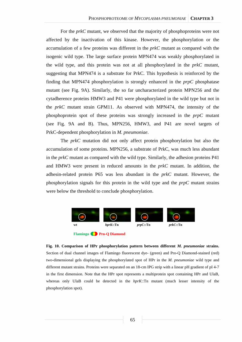
PHOSPHOPROTEOME OF MYCOPLASMA PNEUMONIAE CHAPTER 3
65
For the prkC mutant, we observed that the majority of phosphoproteins were not
affected by the inactivation of this kinase. However, the phosphorylation or the
accumulation of a few proteins was different in the prkC mutant as compared with the
isogenic wild type. The large surface protein MPN474 was weakly phosphorylated in
the wild type, and this protein was not at all phosphorylated in the prkC mutant,
suggesting that MPN474 is a substrate for PrkC. This hypothesis is reinforced by the
finding that MPN474 phosphorylation is strongly enhanced in the prpC phosphatase
mutant (see Fig. 9A). Similarly, the so far uncharacterized protein MPN256 and the
cytadherence proteins HMW3 and P41 were phosphorylated in the wild type but not in
the prkC mutant strain GPM11. As observed with MPN474, the intensity of the
phosphoprotein spot of these proteins was strongly increased in the prpC mutant
(see Fig. 9A and B). Thus, MPN256, HMW3, and P41 are novel targets of
PrkC-dependent phosphorylation in M. pneumoniae.
The prkC mutation did not only affect protein phosphorylation but also the
accumulation of some proteins. MPN256, a substrate of PrkC, was much less abundant
in the prkC mutant as compared with the wild type. Similarly, the adhesion proteins P41
and HMW3 were present in reduced amounts in the prkC mutant. In addition, the
adhesin-related protein P65 was less abundant in the prkC mutant. However, the
phosphorylation signals for this protein in the wild type and the prpC mutant strains
were below the threshold to conclude phosphorylation.
Fig. 10. Comparison of HPr phosphorylation pattern between different M. pneumoniae strains.
Section of dual channel images of Flamingo fluorescent dye- (green) and Pro-Q Diamond-stained (red)
two-dimensional gels displaying the phosphorylated spot of HPr in the M. pneumoniae wild type and
different mutant strains. Proteins were separated on an 18-cm IPG strip with a linear pH gradient of pI 4-7
in the first dimension. Note that the HPr spot represents a multiprotein spot containing HPr and UlaB,
whereas only UlaB could be detected in the hprK::Tn mutant (much lesser intensity of the
phosphorylation spot).
wt hprK::Tn prpC::Tn prkC::Tn
Flamingo Pro-Q Diamond
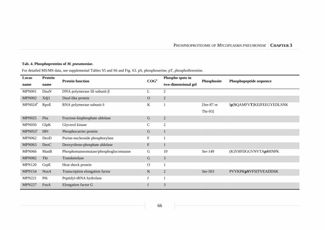
PHOSPHOPROTEOME OF MYCOPLASMA PNEUMONIAE CHAPTER 3
66
Tab. 4. Phosphoproteins of M. pneumoniae.
For detailed MS/MS data, see supplemental Tables S5 and S6 and Fig. S3. pS, phosphoserine; pT, phosphothreonine.
Locus
name
Protein
name Protein function COGa
Phospho spots in
two-dimensional gel Phosphosite Phosphopeptide sequence
MPN001 DnaN DNA polymerase III subunit β L 2
MPN002 Xdj1 DnaJ-like protein O 2
MPN024b RpoE RNA polymerase subunit δ K 1 [Ser-87 or
Thr-93]
Ip[SQAMFVT]KEIFEEGYEDLSNK
MPN025 Fba Fructose-bisphosphate aldolase G 2
MPN050 GlpK Glycerol kinase C 2
MPN053c HPr Phosphocarrier protein G 1
MPN062 DeoD Purine-nucleoside phosphorylase F 1
MPN063 DeoC Deoxyribose-phosphate aldolase F 1
MPN066 ManB Phosphomannomutase/phosphoglucomutase G 10 Ser-149 (K)YHFDGGVNVTApSHNPK
MPN082 Tkt Transketolase G 3
MPN120 GrpE Heat shock protein O 1
MPN154 NusA Transcription elongation factor K 2 Ser-503 PVVKPKpSVFSITVEADDSK
MPN221 Pth Peptidyl-tRNA hydrolase J 1
MPN227 FusA Elongation factor G J 3
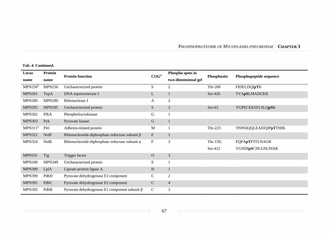
PHOSPHOPROTEOME OF MYCOPLASMA PNEUMONIAE CHAPTER 3
67
Tab. 4. Continued.
Locus
name
Protein
name Protein function COGa
Phospho spots in
two-dimensional gel Phosphosite Phosphopeptide sequence
MPN256b MPN256 Uncharacterized protein S 2 Thr-200 FIDELDQIpTK
MPN261 TopA DNA topoisomerase I L 1 Ser-426 TVApSLMADCKK
MPN280 MPN280 Ribonuclease J A 2
MPN295 MPN295 Uncharacterized protein S 2 Ser-63 YGPECEKSFLSLQpSK
MPN302 PfkA Phosphofructokinase G 1
MPN303 Pyk Pyruvate kinase G 1
MPN311b P41 Adhesin-related protein M 1 Thr-223 TNNSIQQLEAEIQIPpTTHIK
MPN322 NrdF Ribonucleoside-diphosphate reductase subunit β F 1
MPN324 NrdE Ribonucleoside-diphosphate reductase subunit α F 3 Thr-159,
Ser-412
FQPApTPTFLNAGR
VGNDIpSCNLGSLNIAK
MPN331 Tig Trigger factor O 3
MPN349 MPN349 Uncharacterized protein S 1
MPN389 LplA Lipoate-protein ligase A H 1
MPN390 PdhD Pyruvate dehydrogenase E3 component C 2
MPN391 PdhC Pyruvate dehydrogenase E2 component C 4
MPN392 PdhB Pyruvate dehydrogenase E1 component subunit β C 3

PHOSPHOPROTEOME OF MYCOPLASMA PNEUMONIAE CHAPTER 3
68
Tab. 4. Continued.
Locus
name
Protein
name Protein function COGa
Phospho spots in
two-dimensional gel Phosphosite Phosphopeptide sequence
MPN393 PdhA Pyruvate dehydrogenase E1 component subunit α C 3 Ser-205 TKLEpSAVSDLSTK
MPN394 Nox NADH oxidase H 1
MPN420 GlpQ Glycerophosphoryldiester phosphodiesterase C 1
MPN425 FtsY Cell division protein homolog U 1
MPN428 Pta Phosphotransacetylase C 4
MPN429 Pgk Phosphoglycerate kinase G 1
MPN430 GapA Glyceraldehyde-3-phosphate dehydrogenase G 4 Ser-245 VPVLTGpSIVELCVALEK
MPN434 DnaK Chaperone protein O 2
MPN452b HMW3 Cytadherence high molecular weight protein 3 M 1
MPN470 PepP Xaa-Pro aminopeptidase E 2 Thr-317 LLCENAVIpTIEPGIYIPSVGGIR
MPN474b MPN474 Coiled coil surface protein M 1 Thr-773 QNNEELpTDKCSNIQNELHDLNR
MPN495 UlaB Ascorbate-specific phosphotransferase enzyme IIB
component
G 1
MPN517 MPN517 Reductase homolog C 1
MPN528 Ppa Inorganic pyrophosphatase P 1
MPN533 AckA Acetate kinase C 4

PHOSPHOPROTEOME OF MYCOPLASMA PNEUMONIAE CHAPTER 3
69
Tab. 4. Continued.
Locus
name
Protein
name Protein function COGa
Phospho spots in
two-dimensional gel Phosphosite Phosphopeptide sequence
MPN547 MPN547 Dihydroxyacetone kinase C 1
MPN555 MPN555 Uncharacterized protein S 2
MPN572 PepA Cytosol aminopeptidase E 3 Ser-249,
Thr-420
YDMSGAAIVCpSTVLALAK
EGVPLIHCDIASpTASIQDLGQGVLVR
MPN573 GroL 60-kDa chaperonin O 2
MPN574 GroS 10-kDa chaperonin O 1
MPN576 GlyA Serine hydroxymethyltransferase E 1
MPN598 AtpD ATP synthase subunit β C 2
MPN600 AtpA ATP synthase subunit α C 1
MPN606 Eno Enolase G 4
MPN625 MPN625 Osmotically inducible protein C-like protein O 1
MPN627 EI Phosphotransferase system enzyme I G 2
MPN628 Pgm Phosphoglycerate mutase G 1
MPN629 TpiA Triose-phosphate isomerase G 2
MPN636 Frr Ribosome-recycling factor J 2
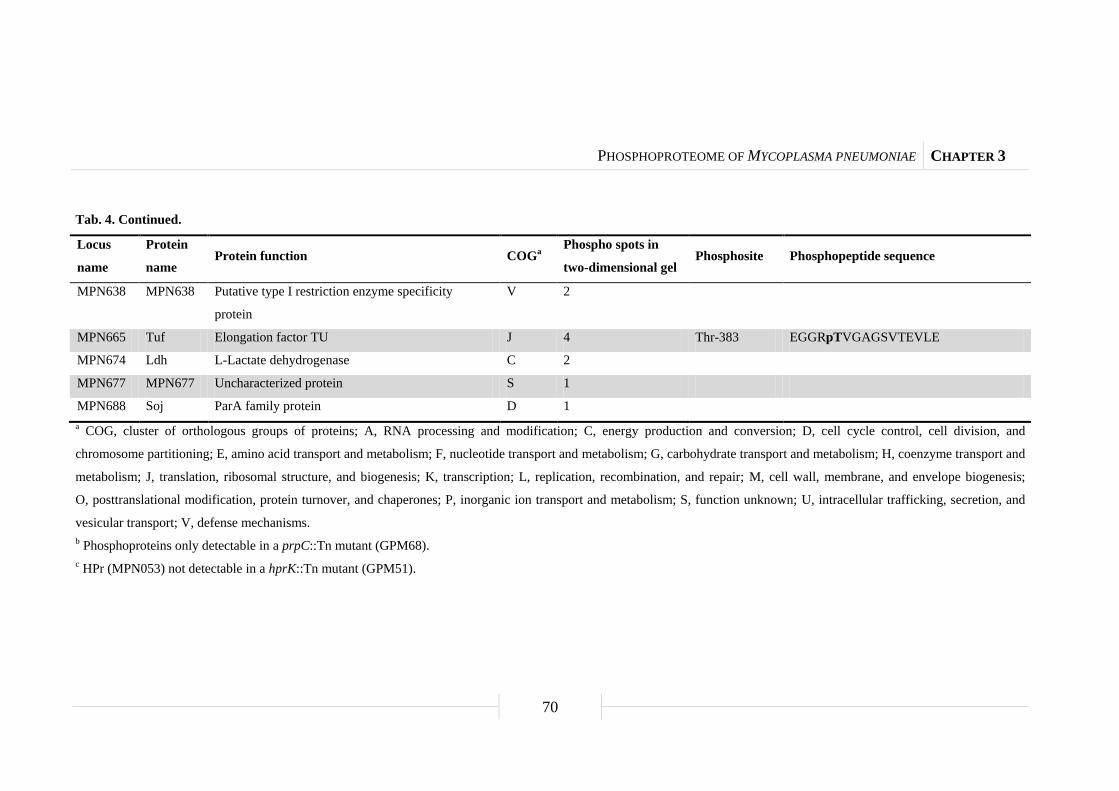
PHOSPHOPROTEOME OF MYCOPLASMA PNEUMONIAE CHAPTER 3
70
Tab. 4. Continued.
Locus
name
Protein
name Protein function COGa
Phospho spots in
two-dimensional gel Phosphosite Phosphopeptide sequence
MPN638 MPN638 Putative type I restriction enzyme specificity
protein
V 2
MPN665 Tuf Elongation factor TU J 4 Thr-383 EGGRpTVGAGSVTEVLE
MPN674 Ldh L-Lactate dehydrogenase C 2
MPN677 MPN677 Uncharacterized protein S 1
MPN688 Soj ParA family protein D 1 a COG, cluster of orthologous groups of proteins; A, RNA processing and modification; C, energy production and conversion; D, cell cycle control, cell division, and
chromosome partitioning; E, amino acid transport and metabolism; F, nucleotide transport and metabolism; G, carbohydrate transport and metabolism; H, coenzyme transport and
metabolism; J, translation, ribosomal structure, and biogenesis; K, transcription; L, replication, recombination, and repair; M, cell wall, membrane, and envelope biogenesis;
O, posttranslational modification, protein turnover, and chaperones; P, inorganic ion transport and metabolism; S, function unknown; U, intracellular trafficking, secretion, and
vesicular transport; V, defense mechanisms. b Phosphoproteins only detectable in a prpC::Tn mutant (GPM68). c HPr (MPN053) not detectable in a hprK::Tn mutant (GPM51).

PHOSPHOPROTEOME OF MYCOPLASMA PNEUMONIAE CHAPTER 3
71
Classes of phosphorylated proteins. To get an impression of, which cellular
processes are affected by protein phosphorylation, we grouped the proteins according to
their functional categories (cluster of orthologous groups of proteins) (see Table 4). This
analysis revealed that most phosphoproteins (36 proteins) are metabolic enzymes or
transporters. 13 phosphorylated proteins are involved in cellular processes such as
protein folding or envelope biogenesis. Nine of the phosphorylated proteins are required
for the storage and processing of the genetic information, and the functions of five
proteins are unknown.
Among the phosphorylated enzymes and transporters, a large number are
involved in the central carbon metabolism (see Fig. 11). Of the glycolytic enzymes, all
but the phosphoglucose isomerase are phosphorylated in M. pneumoniae. Moreover, all
subunits of the pyruvate dehydrogenase and all three enzymes of overflow metabolism
(Ldh, AckA, and Pta) are phosphoproteins. Of the enzymes of the pentose phosphate
pathway, only the transketolase (Tkt) is phosphorylated in M. pneumoniae. In addition
to glucose, M. pneumoniae can utilize glycerol and fructose. Although the enzymes that
introduce fructose in the central metabolism are not phosphorylated, two
glycerol 3-phosphate-forming enzymes are phosphoproteins.
Glycolytic enzymes are phosphorylated in many bacteria (see Table 5). A
comparison of the phosphorylation pattern of these enzymes in different bacteria,
however, revealed specific sets of phosphorylated glycolytic enzymes in each species.
None of the glycolytic enzymes are phosphorylated in all species, and the
phosphofructokinase is only phosphorylated in M. pneumoniae but not in any other
bacterium studied so far. For some of the glycolytic phosphoproteins, the
phosphorylation sites have been determined in multiple species. It is interesting to note
that even if the same enzyme is phosphorylated in different species, the phosphorylation
sites are not conserved. An exception is the pyruvate kinase that is phosphorylated on
Ser-36 in a Gram-positive and in a Gram-negative bacterium (B. subtilis and E. coli,
respectively) (Macek et al., 2008; Macek et al., 2007). However, phosphorylation of
this enzyme was not detected in L. lactis (Soufi et al., 2008), a close relative of both
B. subtilis and M. pneumoniae (see “Discussion”).
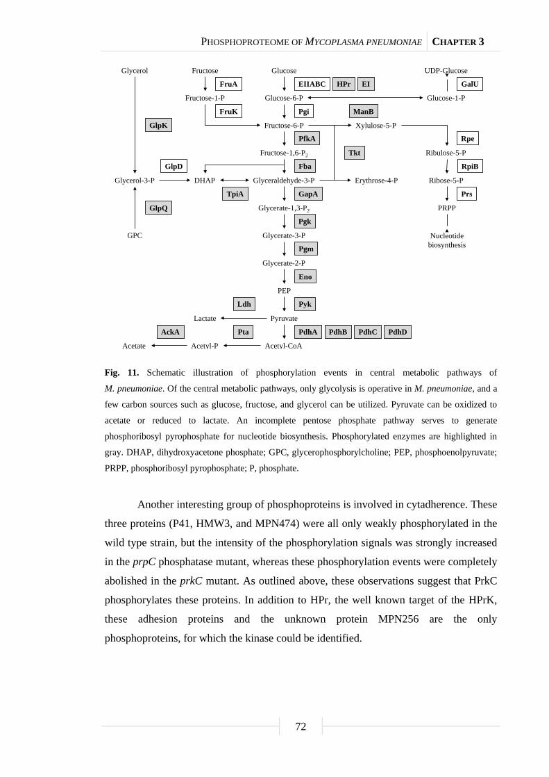
PHOSPHOPROTEOME OF MYCOPLASMA PNEUMONIAE CHAPTER 3
72
Fig. 11. Schematic illustration of phosphorylation events in central metabolic pathways of
M. pneumoniae. Of the central metabolic pathways, only glycolysis is operative in M. pneumoniae, and a
few carbon sources such as glucose, fructose, and glycerol can be utilized. Pyruvate can be oxidized to
acetate or reduced to lactate. An incomplete pentose phosphate pathway serves to generate
phosphoribosyl pyrophosphate for nucleotide biosynthesis. Phosphorylated enzymes are highlighted in
gray. DHAP, dihydroxyacetone phosphate; GPC, glycerophosphorylcholine; PEP, phosphoenolpyruvate;
PRPP, phosphoribosyl pyrophosphate; P, phosphate.
Another interesting group of phosphoproteins is involved in cytadherence. These
three proteins (P41, HMW3, and MPN474) were all only weakly phosphorylated in the
wild type strain, but the intensity of the phosphorylation signals was strongly increased
in the prpC phosphatase mutant, whereas these phosphorylation events were completely
abolished in the prkC mutant. As outlined above, these observations suggest that PrkC
phosphorylates these proteins. In addition to HPr, the well known target of the HPrK,
these adhesion proteins and the unknown protein MPN256 are the only
phosphoproteins, for which the kinase could be identified.
Glucose
Glucose-6-P
Fructose-6-P
Fructose-1,6-P2
Glyceraldehyde-3-PDHAP
Glycerate-1,3-P2
Glycerate-3-P
Glycerate-2-P
PEP
Pyruvate
Acetyl-CoAAcetyl-PAcetate
Glycerol-3-P
Glycerol
Pgi
PfkA
Fba
GapA
Pgk
Pgm
Eno
Pyk
PdhA
Lactate
Ldh
PtaAckA
Fructose
Fructose-1-P
FruK
FruA EIIABC
TpiA
GlpK
GlpD
Xylulose-5-P
Erythrose-4-P
Ribulose-5-P
Ribose-5-P
Tkt
PRPP
Nucleotidebiosynthesis
Rpe
RpiB
Prs
HPr EI
PdhB PdhC PdhD
Glucose-1-P
ManB
UDP-Glucose
GalU
GPC
GlpQ
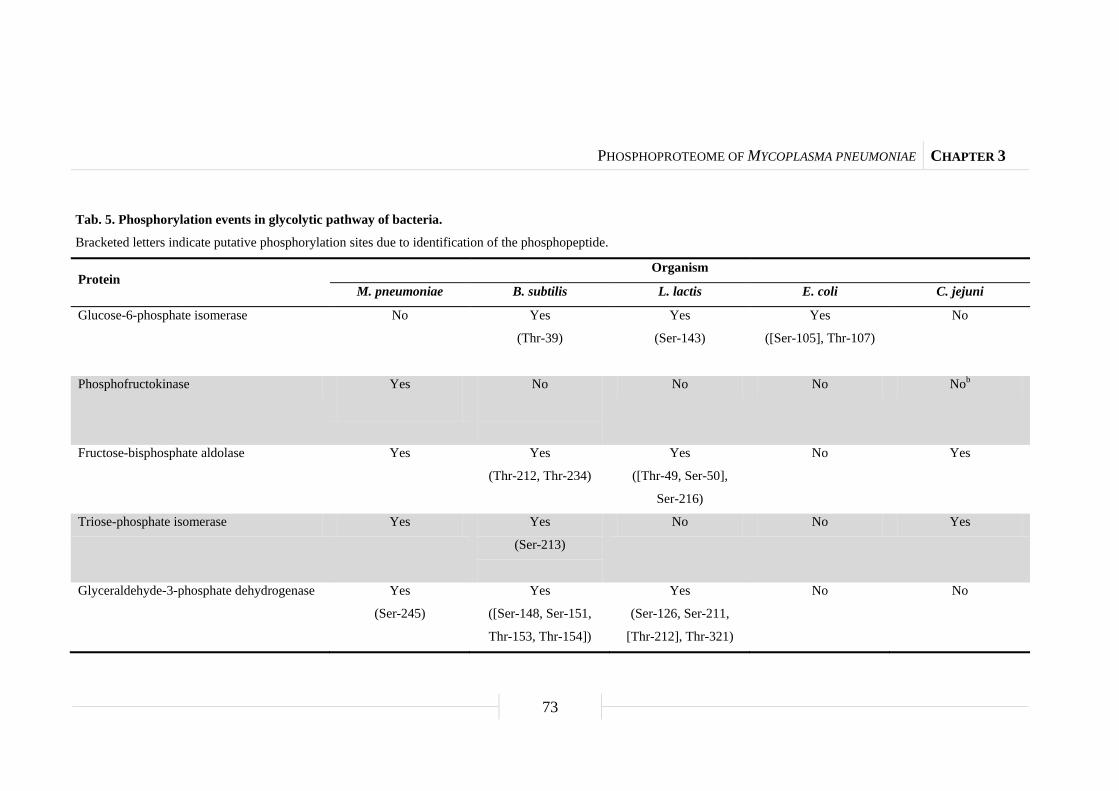
PHOSPHOPROTEOME OF MYCOPLASMA PNEUMONIAE CHAPTER 3
73
Tab. 5. Phosphorylation events in glycolytic pathway of bacteria.
Bracketed letters indicate putative phosphorylation sites due to identification of the phosphopeptide.
Protein Organism
M. pneumoniae B. subtilis L. lactis E. coli C. jejuni
Glucose-6-phosphate isomerase No Yes
(Thr-39)
Yes
(Ser-143)
Yes
([Ser-105], Thr-107)
No
Phosphofructokinase Yes
No
No No Nob
Fructose-bisphosphate aldolase Yes Yes
(Thr-212, Thr-234)
Yes
([Thr-49, Ser-50],
Ser-216)
No Yes
Triose-phosphate isomerase Yes Yes
(Ser-213)
No No Yes
Glyceraldehyde-3-phosphate dehydrogenase Yes
(Ser-245)
Yes
([Ser-148, Ser-151,
Thr-153, Thr-154])
Yes
(Ser-126, Ser-211,
[Thr-212], Thr-321)
No No

PHOSPHOPROTEOME OF MYCOPLASMA PNEUMONIAE CHAPTER 3
74
Tab. 5. Continued.
Protein Organism
M. pneumoniae B. subtilis L. lactis E. coli C. jejuni
Phosphoglycerate kinase Yes Yes
(Ser-183, Thr-299)
Yes
(Ser-217)
Yes
(Ser-192, Thr-196,
Thr-199)
No
Phosphoglycerate mutase Yes Yes
(Ser-62)
Yes
(T94, S144)
Yes
(S146)
No
Enolase Yes Yes
(Thr-141, Ser-259,
Tyr-281, Ser-325)
No Yes
([Ser-372, Thr-375,
Thr-379])
Yes
([Ser-180, Tyr-185])
Pyruvate kinase Yes Yes
(Ser-36)
No Yes
(Ser-36)
Yes
a C. jejuni does not encode for the glycolytic enzyme phosphofructokinase.

PHOSPHOPROTEOME OF MYCOPLASMA PNEUMONIAE CHAPTER 3
75
Autophosphorylation of single universally conserved phosphoprotein. As
mentioned above, there is little conservation of the phosphorylation sites even if the
same proteins are phosphorylated in fairly closely related organisms. There is only one
exception to this notion, the phosphosugar mutase ManB (according to the
nomenclature for M. pneumoniae). Phosphosugar mutases are phosphorylated in all
domains of life from the archaeon Halobacterium salinarum and bacteria such as
M. pneumoniae and E. coli to eukaryotes such as yeast, Drosophila melanogaster, and
humans (Aivaliotis et al., 2009; Li et al., 2007; Macek et al., 2008; Yu et al., 2007;
Zhai et al., 2008).
The phosphoproteome analysis showed the presence of 10 phosphospots that do
all correspond to ManB. The analysis of the phosphorylation site revealed that Ser-149
was phosphorylated in ManB of M. pneumoniae (see Fig. 12). This site was detected in
all 10 spots, suggesting that ManB is subject to different posttranslational modifications
that affect the isoelectric point of the protein. An alignment of the regions surrounding
the phosphorylation site of these proteins revealed a very strong conservation of the
region around the phosphorylated serine residue (see Fig. 13A). The motif
(T/S)ASHN(P/R) is present in all proteins. This strong similarity suggests phylogenetic
conservation of the phosphorylation site and also implies conservation of the
phosphorylation mechanism (the kinase) and of the biological function of the
phosphorylation event.
Because there are no conserved protein kinases in the three domains of life, we
considered the possibility that ManB might autophosphorylate as it had been shown for
the phosphoglucosamine mutase from E. coli (Jolly et al., 2000). To test this idea, we
purified ManB carrying an N-terminal His6-tag and performed phosphorylation assays.
As shown in Fig. 13B, the protein did indeed autophosphorylate in the presence of
manganese. Only faint phosphorylation signals were observed in the presence of other
divalent cations. A protein in which the phosphorylated serine residue was replaced by a
non-phosphorylatable alanine did not autophosphorylate (Fig. 13C). This result
demonstrates that ManB is phosphorylated on the same site in vivo and in vitro and
supports the hypothesis of autophosphorylation. To demonstrate that
autophosphorylation is not limited to the enzyme of M. pneumoniae, we also purified
the phosphosugar mutase GlmM from B. subtilis and observed autophosphorylation for
this protein as well. Again, no phosphorylation was detectable if the phosphorylated
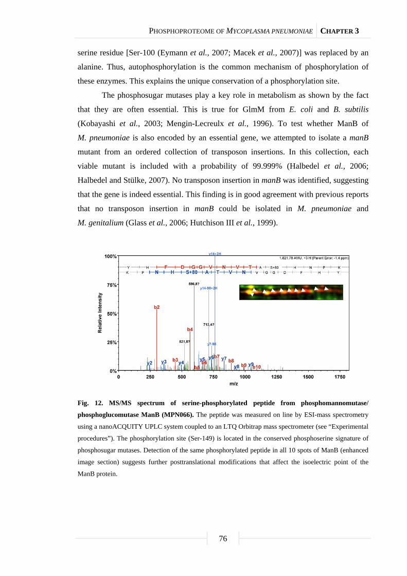
PHOSPHOPROTEOME OF MYCOPLASMA PNEUMONIAE CHAPTER 3
76
serine residue [Ser-100 (Eymann et al., 2007; Macek et al., 2007)] was replaced by an
alanine. Thus, autophosphorylation is the common mechanism of phosphorylation of
these enzymes. This explains the unique conservation of a phosphorylation site.
The phosphosugar mutases play a key role in metabolism as shown by the fact
that they are often essential. This is true for GlmM from E. coli and B. subtilis
(Kobayashi et al., 2003; Mengin-Lecreulx et al., 1996). To test whether ManB of
M. pneumoniae is also encoded by an essential gene, we attempted to isolate a manB
mutant from an ordered collection of transposon insertions. In this collection, each
viable mutant is included with a probability of 99.999% (Halbedel et al., 2006;
Halbedel and Stülke, 2007). No transposon insertion in manB was identified, suggesting
that the gene is indeed essential. This finding is in good agreement with previous reports
that no transposon insertion in manB could be isolated in M. pneumoniae and
M. genitalium (Glass et al., 2006; Hutchison III et al., 1999).
Fig. 12. MS/MS spectrum of serine-phosphorylated peptide from phosphomannomutase/
phosphoglucomutase ManB (MPN066). The peptide was measured on line by ESI-mass spectrometry
using a nanoACQUITY UPLC system coupled to an LTQ Orbitrap mass spectrometer (see “Experimental
procedures”). The phosphorylation site (Ser-149) is located in the conserved phosphoserine signature of
phosphosugar mutases. Detection of the same phosphorylated peptide in all 10 spots of ManB (enhanced
image section) suggests further posttranslational modifications that affect the isoelectric point of the
ManB protein.

PHOSPHOPROTEOME OF MYCOPLASMA PNEUMONIAE CHAPTER 3
77
Fig. 13. Autophosphorylation on universally conserved serine residue in active site of phosphosugar
mutases. (A) Multiple alignment of the conserved phosphoserine signature of phosphosugar mutases
from all domains of life. Amino acids with similarities in at least two of the sequences are highlighted in
gray, whereas amino acids that are identical in at least two of the sequences are depicted in a black
background. The UniProtKB entry names of the aligned sequences are MANB_MYCPN (ManB,
M. pneumoniae), GLMM_BACSU (GlmM; B. subtilis), GLMM_LACLA (GlmM; L. lactis),
ALGC_PSEAE (AlgC; P. aeruginosa), MANB_ECOLI (ManB; E. coli), Q0P8K7_CAMJE (ManB;
C. jejuni), B0R7B7_HALS3 (PMM1; H. salinarum), AGM1_YEAST (AGM1; Saccharomyces
cerevisiae), PGM_DROME (PGM; D. melanogaster), AGM1_MOUSE (AGM1; Mus musculus), and
PGM1_HUMAN (PGM1; Homo sapiens). The conserved active site phosphoserine is indicated by an
arrow. (B) M. pneumoniae ManB and B. subtilis GlmM autophosphorylation assay in the presence of
various divalent cations. About 5 µg of purified His6-tagged ManB (lanes 1-4) or GlmM (lanes 4-8) were
incubated in the presence of [γ32P]ATP in an autophosphorylation assay (see “Experimental procedures”).
Each reaction mixture was analyzed by SDS-PAGE and phosphorimaging analysis. Divalent
cations (5 mM) used in the assays are indicated below the lanes. Bsu, B. subtilis; Mpn, M. pneumoniae.
(C) Autophosphorylation assay of M. pneumoniae ManB (wild type (wt) and S149A) and B. subtilis
GlmM (wild type and S100A) recombinant proteins. The four recombinant proteins were checked for
autophosphorylation as described in (B). Autophosphorylation reactions were conducted in the presence
of Mn2+ (5 mM) as divalent cation as it showed the strongest signal in (B).
wt
S149
A
wt
S100
A
ManB (Mpn) GlmM (Bsu)
Mg2+ Mn2+ Ca2+ Zn2+ Mg2+ Mn2+ Ca2+ Zn2+
MANB_MYCPN 129 PLVSYVIKKYHFDGGVNVTASHNPKTDN---GFKIYDGHGAQLLGLMM_BACSU 80 PGVSYLTKAMDAEAGVMISASHNPVQDN---GIKFFGGDGFKLSGLMM_LACLA 81 PGVAYLVKKDGASAGVMISASHNPALDN---GIKFFGGDGYKLEALGC_PSEAE 88 PVLYYAANVLEGKSGVMLTGSHNPPDYN---GFKIVVAGETLANMANB_ECOLI 82 PAVAYLTRTFRAEAGIVISASHNPFYDN---GIKFFSIDGTKLPQ0P8K7_CAMJE 87 LGYFSLYEGLKFDANVMITGSHNPKDYN---GFKITINKESFFGB0R7B7_HALS3 74 TPATQAYAERAGVPAVMVTASHNPSQYN---GVKLVGSDGVELAAGM1_YEAST 47 LAVLRSLKLQGQYVGVMITASHNPYQDN---GVKIVEPDGSMLLPGM_DROME 96 PAVSSLIRHNKALGGIVLTASHNPGGPENDFGIKFNCEN---GGAGM1_MOUSE 44 LLAVLRSKQTRSTIGVMVTASHNPEEDN---GVKLVDPLGEMLAPGM1_HUMAN 97 PAVSCIIRKIKAIGGIILTASHNPGGPNGDFGIKFNISN---GGconsensus ... . . .. .......**** .. *.*. . .
A
B C
Mn2+
ManB GlmM

PHOSPHOPROTEOME OF MYCOPLASMA PNEUMONIAE CHAPTER 3
78
The universal conservation of the phosphorylation site in phosphosugar mutases
led us to conclude that this phosphorylation event might be essential. This hypothesis
could not be addressed in M. pneumoniae because of the lack of genetic tools
(Halbedel and Stülke, 2007). Therefore, we studied the role of this phosphorylation site
for GlmM B. subtilis. The glmM gene and its variant encoding the S100A protein were
established on multicopy plasmids in B. subtilis. In a strain expressing the wild type
glmM gene from a plasmid, the chromosomal copy could be deleted by homologous
recombination using a PCR fragment obtained by long flanking homology PCR. In
contrast, a resistance cassette could not replace the chromosomal copy of glmM when
the glmM-S100A allele was expressed from the plasmid. Thus, the phosphorylation of
GlmM on Ser-100 seems to be essential for its function and thus for the survival of the
bacteria.
Discussion
The analysis of the phosphoproteome of M. pneumoniae revealed substantial
protein serine/threonine/tyrosine phosphorylation even in this minimal organism. We
identified 63 phosphoproteins that account for nearly 10% of all proteins encoded by
M. pneumoniae. This exceeds the percentage of phosphorylated proteins in other
bacteria by far. For example, only 2.5% of all proteins of B. subtilis are known to be
phosphorylated on a serine, threonine or tyrosine residue (Eymann et al., 2007;
Lévine et al., 2006; Macek et al., 2007). This raises the question whether protein
phosphorylation is more common in M. pneumoniae than in other bacteria. It is
tempting to speculate that the high rate of protein phosphorylation compensates for the
obvious lack of transcriptional regulation in M. pneumoniae. However, this lack of
regulation of gene expression allows the detection of the complete phosphoproteome in
a single (or a few) experiment(s). Indeed, we detected about 500 CHAPS-soluble
proteins that correspond to 80% of all 635 proteins that are in the pI range covered in
this study (see supplemental Fig. S1). In other species with complex transcriptional
regulation, only about 1,000 proteins are detectable under any specific condition,
whereas the rest are required under specific growth conditions (Eymann et al., 2004).
For B. subtilis or E. coli, this corresponds to only 25% of all proteins. Thus, the higher
coverage in our study is most probably due to the lack of transcription regulation in
M. pneumoniae, but does not seem to reflect a higher impact of protein phosphorylation.
PHOSPHOPROTEOME OF MYCOPLASMA PNEUMONIAE CHAPTER 3
79
A striking feature of the phosphoproteomes of different bacteria is the
phosphorylation of enzymes of the central metabolic pathways. In M. pneumoniae,
metabolism is strongly reduced, and the major pathways are depicted in Fig. 11. The
citric acid cycle and pathways of lipid, amino acid, and nucleotide metabolisms are
absent in these bacteria. As in the other Firmicutes, most glycolytic enzymes are
phosphorylated in M. pneumoniae. Unfortunately, the functions of these
phosphorylations as well as the responsible kinases are so far unknown. It is interesting
that all four subunits of the pyruvate dehydrogenase are phosphorylated in
M. pneumoniae. In B. subtilis, there are different results concerning the pyruvate
dehydrogenase. One study reports phosphorylation of PdhB (Macek et al., 2007),
whereas PdhC and PdhD were found to be phosphoproteins in another work
(Eymann et al., 2007). In contrast, none of the subunits is phosphorylated in E. coli,
Campylobacter jejuni, and L. lactis (Macek et al., 2008; Soufi et al., 2008; Voisin et al.,
2007).
This work is the first global analysis of the effects of mutations in the complete
set of annotated protein kinases on the phosphoproteome. We have shown that the HPr
kinase has only one substrate, HPr. The phosphorylation of HPr on Ser-46 is a major
signal to trigger carbon catabolite repression of catabolic genes and operons in
B. subtilis and other Firmicutes (Görke and Stülke, 2008). If HPr(Ser-P) had the same
function in M. pneumoniae, one would have expected major changes in the proteome of
the hprK mutant as compared with the wild type strain. However, with the exception of
HPr phosphorylation (see Fig. 10), no differences were detectable. This suggests that
HPr phosphorylation on Ser-46 has a function different from that in the other bacteria.
This conclusion is supported by three observations. First, HPr(Ser-P) acts as the
cofactor for the pleiotropic transcription factor CcpA to cause catabolite repression. No
protein similar to CcpA is encoded in the genome of any Mycoplasma species. Second,
the phosphorylation of HPr on Ser-46 interferes with its participation in sugar transport
due to the inhibition of enzyme I-dependent phosphorylation of His-15. This inhibition
is relevant even in Gram-negative bacteria that also do not possess an equivalent of
CcpA (Krausse et al., 2009). However, in M. pneumoniae, these two phosphorylation
events are not mutually exclusive (Halbedel and Stülke, 2005). Finally, although the
activity of the HPr kinase of B. subtilis and most other bacteria is strongly controlled by
the ATP and fructose 1,6-bisphosphate concentrations, the enzyme of M. pneumoniae is

PHOSPHOPROTEOME OF MYCOPLASMA PNEUMONIAE CHAPTER 3
80
constitutively active due to its high affinity to ATP (Merzbacher et al., 2004). Thus, the
phosphorylation of HPr on Ser-46 seems to serve other purposes in M. pneumoniae.
In addition to the HPr kinase, M. pneumoniae encodes only one protein kinase,
PrkC. This kinase is implicated in a multitude of cellular functions in different bacteria,
among them spore germination, virulence, and control of glycolysis (Kristich et al.,
2007; Lomas-Lopez et al., 2007; Shah et al., 2008). We found that PrkC has at least
four targets in M. pneumoniae, the cell adhesion proteins HMW3 and P41, the cell
surface protein MPN474, and the uncharacterized protein MPN256. These proteins are
not phosphorylated in a prkC mutant, but their phosphorylation is enhanced if the
protein phosphatase PrpC, the antagonist of PrkC, is not expressed (see Fig. 9).
Moreover, HMW1 and P1, two very large proteins that were out of the detection
window applied in this study, are likely to be phosphorylated by PrkC (Schmidl et al.,
2010). This suggests that PrkC might be required for cell adhesion in M. pneumoniae.
Indeed, a prkC mutant strain is not capable of adherent growth and has lost toxicity to
human host cells, most likely due to the defect in cell adhesion (Schmidl et al., 2010).
Thus, PrkC has a specific function in M. pneumoniae and phosphorylates a specific set
of proteins.
As stated above, HPrK and PrkC are the only two annotated protein kinases in
M. pneumoniae. However, they account for only five of 63 identified protein
phosphorylation events. This suggests the existence of other, yet to be discovered
protein kinases in M. pneumoniae. Alternatively, autophosphorylation might be more
relevant than previously anticipated. Indeed, the only phosphorylation site that is
universally conserved from archaea via bacteria such as M. pneumoniae to eukaryotes is
the result of an autophosphorylation event in the catalytic site of phosphosugar mutases
such as GlmM and ManB (Jolly et al., 2000; Mengin-Lecreulx et al., 1996; this work).
This autophosphorylation is essential for the enzymatic activity of these enzymes, thus
providing an explanation for the universal but highly exceptional conservation of the
phosphorylation event at a conserved site. Interestingly, another ubiquitous protein, the
heat shock protein DnaK (also called Hsp70) is also present as a phosphoprotein in all
organisms that were studied so far. In E. coli and mycobacteria, DnaK is capable of
autophosphorylating at a strongly conserved threonine residue (McCarty and Walker,
1991; Peake et al., 1998; Preneta et al., 2004; Zylicz et al., 1983). This site is also
present in the DnaK protein of M. pneumoniae (Thr-182), but it is not conserved in the
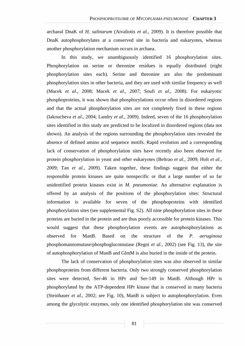
PHOSPHOPROTEOME OF MYCOPLASMA PNEUMONIAE CHAPTER 3
81
archaeal DnaK of H. salinarum (Aivaliotis et al., 2009). It is therefore possible that
DnaK autophosphorylates at a conserved site in bacteria and eukaryotes, whereas
another phosphorylation mechanism occurs in archaea.
In this study, we unambiguously identified 16 phosphorylation sites.
Phosphorylation on serine or threonine residues is equally distributed (eight
phosphorylation sites each). Serine and threonine are also the predominant
phosphorylation sites in other bacteria, and they are used with similar frequency as well
(Macek et al., 2008; Macek et al., 2007; Soufi et al., 2008). For eukaryotic
phosphoproteins, it was shown that phosphorylations occur often in disordered regions
and that the actual phosphorylation sites are not completely fixed in these regions
(Iakoucheva et al., 2004; Landry et al., 2009). Indeed, seven of the 16 phosphorylation
sites identified in this study are predicted to be localized in disordered regions (data not
shown). An analysis of the regions surrounding the phosphorylation sites revealed the
absence of defined amino acid sequence motifs. Rapid evolution and a corresponding
lack of conservation of phosphorylation sites have recently also been observed for
protein phosphorylation in yeast and other eukaryotes (Beltrao et al., 2009; Holt et al.,
2009; Tan et al., 2009). Taken together, these findings suggest that either the
responsible protein kinases are quite nonspecific or that a large number of so far
unidentified protein kinases exist in M. pneumoniae. An alternative explanation is
offered by an analysis of the positions of the phosphorylation sites: Structural
information is available for seven of the phosphoproteins with identified
phosphorylation sites (see supplemental Fig. S2). All nine phosphorylation sites in these
proteins are buried in the protein and are thus poorly accessible for protein kinases. This
would suggest that these phosphorylation events are autophosphorylations as
observed for ManB. Based on the structure of the P. aeruginosa
phosphomannomutase/phosphoglucomutase (Regni et al., 2002) (see Fig. 13), the site
of autophosphorylation of ManB and GlmM is also buried in the inside of the protein.
The lack of conservation of phosphorylation sites was also observed in similar
phosphoproteins from different bacteria. Only two strongly conserved phosphorylation
sites were detected, Ser-46 in HPr and Ser-149 in ManB. Although HPr is
phosphorylated by the ATP-dependent HPr kinase that is conserved in many bacteria
(Steinhauer et al., 2002; see Fig. 10), ManB is subject to autophosphorylation. Even
among the glycolytic enzymes, only one identified phosphorylation site was conserved

PHOSPHOPROTEOME OF MYCOPLASMA PNEUMONIAE CHAPTER 3
82
between two species (Ser-36 in the pyruvate kinases from B. subtilis and E. coli).
Because this latter site is conserved in M. pneumoniae, it is tempting to speculate that it
is also phosphorylated in the Mollicutes. The observations reported in this work have
important implications for the evolution of protein phosphorylation in bacteria: Protein
phosphorylation events seem to be highly specific for each individual bacterial species;
even among members of one bacterial phylum such as the Firmicutes, there is little
conservation of protein phosphorylation. Moreover, one protein kinase may
phosphorylate distinct sets of proteins in the different species. Finally, there may be a
large number of protein kinases that have so far escaped discovery, and these kinases
are likely to be specific for small groups of bacteria. In conclusion, protein
phosphorylation may be one of the major features in the adaptation of bacteria to their
individual ecological niche.
Acknowledgments
We are grateful to Julia Busse for excellent technical assistance. Robert Hertel is
acknowledged for help with some experiments. This work was supported by grants of
the Deutsche Forschungsgemeinschaft and the Fonds der Chemischen Industrie (to
D. B., M. H., and J. S.). S. R. S. was supported by the Studienstiftung des Deutschen
Volkes.

PHOSPHOPROTEOME OF MYCOPLASMA PNEUMONIAE CHAPTER 3
83
Supplemental material
Tab. S3. Primers used in this study.
Primer Sequence (5’→3’)ab Mutation Resulting plasmid
NP108 AAAGGATCCCTCACTTATTTAAAGGAGGAAA
CAATCATGGGCAAGTATTTTGGAACAGACG
None pGP400, pGP1403
NP109 TTTGTCGACCTATCACTCTAATCCCATTTCTG
ACCGG
None pGP400, pGP1401,
pGP1403, pGP1405
NP110 GGCTATTCTCCGGAGCAGCCGATTGTCA None -
NP111 CAGGAACGGACGACCAAAAGTTTTCCCG None -
NP112 CCTATCACCTCAAATGGTTCGCTGTAAAGGC
CAGCTCAGGTGTAAGCTC
None -
NP113 CCGAGCGCCTACGAGGAATTTGTATCGAGCG
AAGACGAAAGAGCTGTGCGA
None -
NP114 CATCGCCATCGCATCAGATGCTACGACGT None -
NP115 TATAAGACGCACGTGTAATCACGTCACCATC None -
NP116 P_GCAGAGGCGGGCGTCATGATTTCCGCTGC
CCATAACCCAGTGCAGGATAACGGCATCAA
T298G pGP1403, pGP1405
NP117 AAAGGATCCATGGGCAAGTATTTTGGAACAG
ACG
None pGP1401, pGP1405
spec-fwd
(kan)
CAGCGAACCATTTGAGGTGATAGGGACTGGC
TCGCTAATAACGTAACGTGACTGGCAAGAG
None -
spec-rev
(kan)
CGATACAAATTCCTCGTAGGCGCTCGGCGTA
GCGAGGGCAAGGGTTTATTGTTTTCTAAAAT
CTG
None -
SH29 ATGAGTGAGCTAACTCACAG None -
SH30 CAATACGCAAACCGCCTC None -
SS107 AAAGGATCCATGAACAGTAACGCATACTTGG
AAGC
None pGP656-pGP658
SS108 TATACTGCAGCTAAGCTTTATCGAGGTTGAG
CAATC
None pGP656-pGP658
SS109 P_GAAAGTAAGGATACCTGGGAGTTAGCGCG A846G pGP657
SS110 P_CTTTGCGATTGCCGAATGGAATCCGCAAA
C
A936G pGP657
SS111 P_GATTACTATAACTGGACGGTACCACACAC
CATTC
A1356G pGP657
SS112 P_CGGTGAATGTCACTGCTGCTCATAATCCTA
AAACC
A445G,
G446C
pGP658

PHOSPHOPROTEOME OF MYCOPLASMA PNEUMONIAE CHAPTER 3
84
Tab. S3. Continued.
Primer Sequence (5’→3’)ab Mutation Resulting plasmid
SS113 CAAGGCCACAATTGATTATGTCTTTG None -
SS114 GGTAACTTAGTGGAAGTCCACTTAAC None - a Restriction sites are underlined. b The “P” at the 5’ end of primer sequences indicates phosphorylation.
Tab. S4. Plasmids used for analysis of phosphosugar mutases.
Plasmid Relevant characteristics Used restriction
sites Reference
pBQ200 Allows expression of proteins in B. subtilis - Martin-Verstraete et al.
(1994)
pGP400 pBQ200-glmM BamHI + SalI This work
pGP656 pWH844-manB BamHI + PstI This work
pGP657 pWH844-manBa BamHI + PstI This work
pGP658 pWH844-manB(S149A) BamHI + PstI This work
pGP1401 pWH844-glmM BamHI + SalI This work
pGP1403 pBQ200-glmM(S100A) BamHI + SalI This work
pGP1405 pWH844-glmM(S100A) BamHI + SalI This work
pWH844 Allows overexpression of N-terminal
His6-tag fusion proteins in E. coli DH5α
- Schirmer et al. (1997)
a The three TGA opal codons in the wild type manB gene are mutated to TGG.

PHOSPHOPROTEOME OF MYCOPLASMA PNEUMONIAE CHAPTER 3
85
Tab. S5. Complete list of phosphospots identified from M. pneumoniae two-dimensional gels.
For general overview, see Table 4. pS, phosphoserine; pT, phosphothreonine.
# Spot Protein
name
Locus
name
UniProtKB
accession
number
Protein function Molecular
weight (Da)
Isoelectric
point (pI)
Protein
identification
probability (%)
Number
of unique
peptides
Coverage
(%)
Phosphorylation
site Phosphopeptide sequence Experimenta
1 AckA-1 AckA MPN533 P75245 Acetate kinase 43672.68 8.05 100.00 23 58.97 1, 2, 3, 4
2 AckA-2 AckA MPN533 P75245 Acetate kinase 43672.68 8.05 100.00 22 55.38 1, 2, 3, 4
3 AckA-3 AckA MPN533 P75245 Acetate kinase 43672.68 8.05 100.00 18 50.77 1, 2, 3, 4
4 AckA-4 AckA MPN533 P75245 Acetate kinase 43672.68 8.05 100.00 16 43.85 1, 2, 3, 4
5 [AtpA Pgm] AtpA MPN600 Q50329 ATP synthase subunit α 57339.01 6.14 100.00 19 41.12 1, 2, 3, 4
Pgm MPN628 P75167 Phosphoglycerate mutase 56339.53 6.08 100.00 14 36.22 1, 2, 3, 4
6 AtpD-1 AtpD MPN598 Q50331 ATP synthase subunit β 52204.62 5.36 100.00 28 86.32 1, 2, 3, 4
7 AtpD-2 AtpD MPN598 Q50331 ATP synthase subunit β 52204.62 5.36 100.00 22 71.79 1, 2, 3, 4
8 DeoC DeoC MPN063 P09924 Deoxyribose-phosphate aldolase 24861.89 8.34 100.00 9 53.13 1, 2, 3, 4
9 [DoeD TpiA] DeoD MPN062 P75053 Purine-nucleoside phosphorylase 26266.66 6.78 100.00 15 77.73 1, 2, 3, 4
TpiA MPN629 P78010 Triosephosphate isomerase 26941.33 7.34 100.00 11 55.33 1, 2, 3, 4
10 DnaK-1 DnaK MPN434 P75344 Chaperone protein 65058.30 5.28 100.00 34 51.09 1, 2, 3, 4
11 DnaK-2 DnaK MPN434 P75344 Chaperone protein 65058.30 5.28 100.00 18 35.97 1, 2, 3, 4
12 DnaN DnaN MPN001 Q50313 DNA polymerase III subunit β 43829.01 5.74 100.00 23 59.74 1, 2, 3, 4
13 [DnaN PdhA] DnaN MPN001 Q50313 DNA polymerase III subunit β 43829.01 5.74 100.00 28 76.05 1, 2, 3, 4
PdhA MPN393 P75390 Pyruvate dehydrogenase E1 component
subunit α
40568.44 6.22 100.00 10 30.45 1, 2, 3, 4
14 EI-1 EI MPN627 P75168 Phosphotransferase system enzyme I 63909.70 7.92 100.00 31 47.03 1, 2, 3, 4
15 EI-2 EI MPN627 P75168 Phosphotransferase system enzyme I 63909.70 7.92 100.00 12 27.62 1, 2, 3, 4
16 Eno-1 Eno MPN606 P75189 Enolase 49197.47 6.11 100.00 24 64.69 1, 2, 3, 4
17 Eno-2 Eno MPN606 P75189 Enolase 49197.47 6.11 100.00 25 70.18 1, 2, 3, 4
18 Eno-3 Eno MPN606 P75189 Enolase 49197.47 6.11 100.00 20 60.96 1, 2, 3, 4
19 Eno-4 Eno MPN606 P75189 Enolase 49197.47 6.11 100.00 17 55.26 1, 2, 3, 4
20 Fba Fba MPN025 P75089 Fructose-bisphosphate aldolase 31049.17 6.44 100.00 6 23.26 1, 2, 3, 4
21 [Fba Soj] Fba MPN025 P75089 Fructose-bisphosphate aldolase 31049.17 6.44 100.00 12 53.82 1, 2, 3, 4
Soj MPN688 Q50314 ParA family protein 30061.83 7.26 100.00 8 27.78 1, 2, 3, 4
22 Frr-1 Frr MPN636 P75161 Ribosome-recycling factor 21653.51 8.87 100.00 12 63.04 1, 2, 3, 4
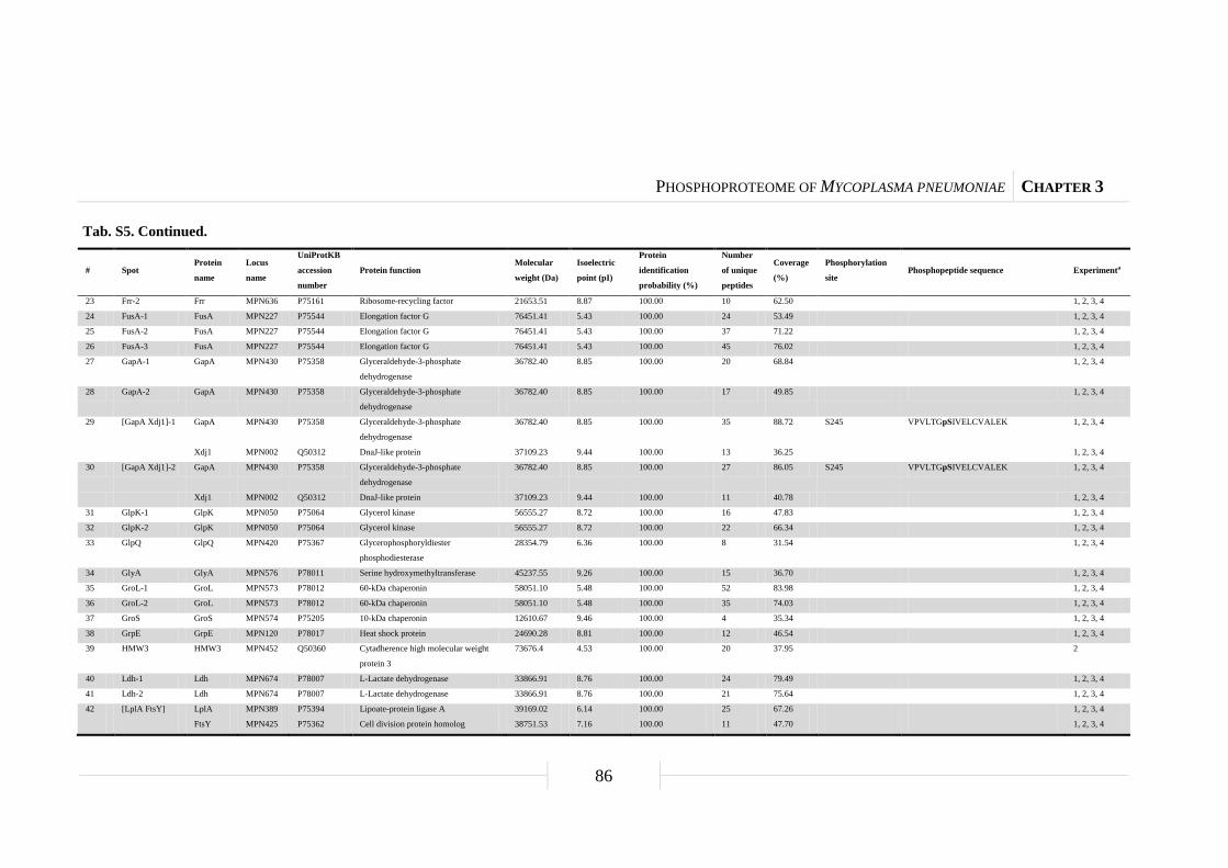
PHOSPHOPROTEOME OF MYCOPLASMA PNEUMONIAE CHAPTER 3
86
Tab. S5. Continued.
# Spot Protein
name
Locus
name
UniProtKB
accession
number
Protein function Molecular
weight (Da)
Isoelectric
point (pI)
Protein
identification
probability (%)
Number
of unique
peptides
Coverage
(%)
Phosphorylation
site Phosphopeptide sequence Experimenta
23 Frr-2 Frr MPN636 P75161 Ribosome-recycling factor 21653.51 8.87 100.00 10 62.50 1, 2, 3, 4
24 FusA-1 FusA MPN227 P75544 Elongation factor G 76451.41 5.43 100.00 24 53.49 1, 2, 3, 4
25 FusA-2 FusA MPN227 P75544 Elongation factor G 76451.41 5.43 100.00 37 71.22 1, 2, 3, 4
26 FusA-3 FusA MPN227 P75544 Elongation factor G 76451.41 5.43 100.00 45 76.02 1, 2, 3, 4
27 GapA-1 GapA MPN430 P75358 Glyceraldehyde-3-phosphate
dehydrogenase
36782.40 8.85 100.00 20 68.84 1, 2, 3, 4
28 GapA-2 GapA MPN430 P75358 Glyceraldehyde-3-phosphate
dehydrogenase
36782.40 8.85 100.00 17 49.85 1, 2, 3, 4
29 [GapA Xdj1]-1 GapA MPN430 P75358 Glyceraldehyde-3-phosphate
dehydrogenase
36782.40 8.85 100.00 35 88.72 S245 VPVLTGpSIVELCVALEK 1, 2, 3, 4
Xdj1 MPN002 Q50312 DnaJ-like protein 37109.23 9.44 100.00 13 36.25 1, 2, 3, 4
30 [GapA Xdj1]-2 GapA MPN430 P75358 Glyceraldehyde-3-phosphate
dehydrogenase
36782.40 8.85 100.00 27 86.05 S245 VPVLTGpSIVELCVALEK 1, 2, 3, 4
Xdj1 MPN002 Q50312 DnaJ-like protein 37109.23 9.44 100.00 11 40.78 1, 2, 3, 4
31 GlpK-1 GlpK MPN050 P75064 Glycerol kinase 56555.27 8.72 100.00 16 47.83 1, 2, 3, 4
32 GlpK-2 GlpK MPN050 P75064 Glycerol kinase 56555.27 8.72 100.00 22 66.34 1, 2, 3, 4
33 GlpQ GlpQ MPN420 P75367 Glycerophosphoryldiester
phosphodiesterase
28354.79 6.36 100.00 8 31.54 1, 2, 3, 4
34 GlyA GlyA MPN576 P78011 Serine hydroxymethyltransferase 45237.55 9.26 100.00 15 36.70 1, 2, 3, 4
35 GroL-1 GroL MPN573 P78012 60-kDa chaperonin 58051.10 5.48 100.00 52 83.98 1, 2, 3, 4
36 GroL-2 GroL MPN573 P78012 60-kDa chaperonin 58051.10 5.48 100.00 35 74.03 1, 2, 3, 4
37 GroS GroS MPN574 P75205 10-kDa chaperonin 12610.67 9.46 100.00 4 35.34 1, 2, 3, 4
38 GrpE GrpE MPN120 P78017 Heat shock protein 24690.28 8.81 100.00 12 46.54 1, 2, 3, 4
39 HMW3 HMW3 MPN452 Q50360 Cytadherence high molecular weight
protein 3
73676.4 4.53 100.00 20 37.95 2
40 Ldh-1 Ldh MPN674 P78007 L-Lactate dehydrogenase 33866.91 8.76 100.00 24 79.49 1, 2, 3, 4
41 Ldh-2 Ldh MPN674 P78007 L-Lactate dehydrogenase 33866.91 8.76 100.00 21 75.64 1, 2, 3, 4
42 [LplA FtsY] LplA MPN389 P75394 Lipoate-protein ligase A 39169.02 6.14 100.00 25 67.26 1, 2, 3, 4
FtsY MPN425 P75362 Cell division protein homolog 38751.53 7.16 100.00 11 47.70 1, 2, 3, 4
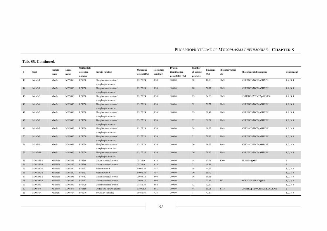
PHOSPHOPROTEOME OF MYCOPLASMA PNEUMONIAE CHAPTER 3
87
Tab. S5. Continued.
# Spot Protein
name
Locus
name
UniProtKB
accession
number
Protein function Molecular
weight (Da)
Isoelectric
point (pI)
Protein
identification
probability (%)
Number
of unique
peptides
Coverage
(%)
Phosphorylation
site Phosphopeptide sequence Experimenta
43 ManB-1 ManB MPN066 P75050 Phosphomannomutase/
phosphoglucomutase
63175.24 8.39 100.00 10 18.23 S149 YHFDGGVNVTApSHNPK 1, 2, 3, 4
44 ManB-2 ManB MPN066 P75050 Phosphomannomutase/
phosphoglucomutase
63175.24 8.39 100.00 20 52.17 S149 YHFDGGVNVTApSHNPK 1, 2, 3, 4
45 ManB-3 ManB MPN066 P75050 Phosphomannomutase/
phosphoglucomutase
63175.24 8.39 100.00 23 54.69 S149 KYHFDGGVNVTApSHNPK 1, 2, 3, 4
46 ManB-4 ManB MPN066 P75050 Phosphomannomutase/
phosphoglucomutase
63175.24 8.39 100.00 32 59.57 S149 YHFDGGVNVTApSHNPK 1, 2, 3, 4
47 ManB-5 ManB MPN066 P75050 Phosphomannomutase/
phosphoglucomutase
63175.24 8.39 100.00 25 60.47 S149 YHFDGGVNVTApSHNPK 1, 2, 3, 4
48 ManB-6 ManB MPN066 P75050 Phosphomannomutase/
phosphoglucomutase
63175.24 8.39 100.00 22 66.61 S149 YHFDGGVNVTApSHNPK 1, 2, 3, 4
49 ManB-7 ManB MPN066 P75050 Phosphomannomutase/
phosphoglucomutase
63175.24 8.39 100.00 24 66.25 S149 YHFDGGVNVTApSHNPK 1, 2, 3, 4
50 ManB-8 ManB MPN066 P75050 Phosphomannomutase/
phosphoglucomutase
63175.24 8.39 100.00 21 58.12 S149 YHFDGGVNVTApSHNPK 1, 2, 3, 4
51 ManB-9 ManB MPN066 P75050 Phosphomannomutase/
phosphoglucomutase
63175.24 8.39 100.00 26 66.25 S149 YHFDGGVNVTApSHNPK 1, 2, 3, 4
52 ManB-10 ManB MPN066 P75050 Phosphomannomutase/
phosphoglucomutase
63175.24 8.39 100.00 36 58.12 S149 YHFDGGVNVTApSHNPK 1, 2, 3, 4
53 MPN256-1 MPN256 MPN256 P75518 Uncharacterized protein 25722.9 4.18 100.00 14 67.71 T200 FIDELDQIpTK 2
54 MPN256-2 MPN256 MPN256 P75518 Uncharacterized protein 25722.9 4.18 100.00 7 48.88 2
55 MPN280-1 MPN280 MPN280 P75497 Ribonuclease J 64045.33 7.57 100.00 18 44.29 1, 2, 3, 4
56 MPN280-2 MPN280 MPN280 P75497 Ribonuclease J 64045.33 7.57 100.00 16 39.72 1, 2, 3, 4
57 MPN295-1 MPN295 MPN295 P75482 Uncharacterized protein 25684.16 8.08 100.00 16 60.91 1, 2, 3, 4
58 MPN295-2 MPN295 MPN295 P75482 Uncharacterized protein 25684.16 8.08 100.00 22 73.18 S63 YGPECEKSFLSLQpSK 1, 2, 3, 4
59 MPN349 MPN349 MPN349 P75429 Uncharacterized protein 31411.30 8.83 100.00 12 52.67 1, 2, 3, 4
60 MPN474 MPN474 MPN474 P75310 Coiled coil surface protein 118006.4 4.81 100.00 44 61.08 T773 QNNEELpTDKCSNIQNELHDLNR 2
61 MPN517 MPN517 MPN517 P75270 Reductase homolog 18856.85 7.26 100.00 7 42.17 1, 2, 3, 4

PHOSPHOPROTEOME OF MYCOPLASMA PNEUMONIAE CHAPTER 3
88
Tab. S5. Continued.
# Spot Protein
name
Locus
name
UniProtKB
accession
number
Protein function Molecular
weight (Da)
Isoelectric
point (pI)
Protein
identification
probability (%)
Number
of unique
peptides
Coverage
(%)
Phosphorylation
site Phosphopeptide sequence Experimenta
62 MPN547 MPN547 MPN547 P75231 Dihydroxyacetone kinase 62398.49 8.20 100.00 39 75.63 1, 2, 3, 4
63 MPN555-1 MPN555 MPN555 P75223 Uncharacterized protein 22419.68 5.51 100.00 7 41.97 1, 2, 3, 4
64 MPN555-2 MPN555 MPN555 P75223 Uncharacterized protein 22419.68 5.51 100.00 11 55.44 1, 2, 3, 4
65 MPN625 MPN625 MPN625 P75170 Osmotical inducible protein C-like
protein
15458.91 6.60 100.00 4 31.21 1, 2, 3, 4
66 MPN638-1 MPN638 MPN638 P75159 Putative type I restriction enzyme
specificity protein
42623.25 8.51 100.00 24 66.67 1, 2, 3, 4
67 MPN638-2 MPN638 MPN638 P75159 Putative type I restriction enzyme
specificity protein
42623.25 8.51 100.00 15 43.47 1, 2, 3, 4
68 MPN677 MPN677 MPN677 P75115 Uncharacterized protein 50554.18 8.89 100.00 12 36.94 1, 2, 3, 4
69 NrdE-1 NrdE MPN324 P78027 Ribonucleoside-diphosphate reductase
subunit α
82322.96 6.66 100.00 66 69.90 T159 FQPApTPTFLNAGR 1, 2, 3, 4
70 NrdE-2 NrdE MPN324 P78027 Ribonucleoside-diphosphate reductase
subunit α
82322.96 6.66 100.00 54 61.86 S412 VGNDIpSCNLGSLNIAK 1, 2, 3, 4
71 NrdE-3 NrdE MPN324 P78027 Ribonucleoside-diphosphate reductase
subunit α
82322.96 6.66 100.00 17 23.02 1, 2, 3, 4
72 NrdF NrdF MPN322 P75461 Ribonucleoside-diphosphate reductase
subunit β
39388.04 5.36 100.00 19 71.98 1, 2, 3, 4
73 Nox Nox MPN394 P75389 NADH oxidase 52841.36 6.70 100.00 11 29.85 1, 2, 3, 4
74 NusA-1 NusA MPN154 P75591 Transcription elongation factor 60235.03 5.77 100.00 43 80.00 S503 PVVKPKpSVFSITVEADDSK 1, 2, 3, 4
75 NusA-2 NusA MPN154 P75591 Transcription elongation factor 60235.03 5.77 100.00 19 48.15 1, 2, 3, 4
76 P41 P41 MPN311 P75470 Adhesin-related protein 40555.6 4.64 100.00 9 42.58 T223 TNNSIQQLEAEIQIPpTTHIK 2
77 PdhA PdhA MPN393 P75390 Pyruvate dehydrogenase E1 component
subunit α
40568.44 6.22 100.00 19 53.63 1, 2, 3, 4
78 [PdhA Pgk] PdhA MPN393 P75390 Pyruvate dehydrogenase E1 component
subunit α
40568.44 6.22 100.00 14 38.27 S205 TKLEpSAVSDLSTK 1, 2, 3, 4
Pgk MPN429 P78018 Phosphoglycerate kinase 44184.27 7.26 100.00 9 26.65 1, 2, 3, 4

PHOSPHOPROTEOME OF MYCOPLASMA PNEUMONIAE CHAPTER 3
89
Tab. S5. Continued.
# Spot Protein
name
Locus
name
UniProtKB
accession
number
Protein function Molecular
weight (Da)
Isoelectric
point (pI)
Protein
identification
probability (%)
Number
of unique
peptides
Coverage
(%)
Phosphorylation
site Phosphopeptide sequence Experimenta
79 [PdhB Pta]-1 PdhB MPN392 P75391 Pyruvate dehydrogenase E1 component
subunit β
35891.67 6.54 100.00 15 57.19 1, 2, 3, 4
Pta MPN428 P75359 Phosphotransacetylase 35196.23 7.16 100.00 9 38.75 1, 2, 3, 4
80 [PdhB Pta]-2 PdhB MPN392 P75391 Pyruvate dehydrogenase E1 component
subunit β
35891.67 6.54 100.00 14 50.76 1, 2, 3, 4
Pta MPN428 P75359 Phosphotransacetylase 35196.23 7.16 100.00 7 27.50 1, 2, 3, 4
81 PdhC-1 PdhC MPN391 P75392 Pyruvate dehydrogenase E2 component 42370.45 5.43 100.00 13 37.06 1, 2, 3, 4
82 PdhC-2 PdhC MPN391 P75392 Pyruvate dehydrogenase E2 component 42370.45 5.43 100.00 15 37.06 1, 2, 3, 4
83 PdhC-3 PdhC MPN391 P75392 Pyruvate dehydrogenase E2 component 42370.45 5.43 100.00 11 33.58 1, 2, 3, 4
84 PdhC-4 PdhC MPN391 P75392 Pyruvate dehydrogenase E2 component 42370.45 5.43 100.00 8 18.41 1, 2, 3, 4
85 PdhD-1 PdhD MPN390 P75393 Pyruvate dehydrogenase E3 component 49406.02 8.14 100.00 15 47.05 1, 2, 3, 4
86 PdhD-2 PdhD MPN390 P75393 Pyruvate dehydrogenase E3 component 49406.02 8.14 100.00 11 38.29 1, 2, 3, 4
87 PepA-1 PepA MPN572 P75206 Cytosol aminopeptidase 48758.12 8.23 100.00 38 75.73 S249
T420
YDMSGAAIVCpSTVLALAK
EGVPLIHCDIASpTASIQDLGQGVLVR
1, 2, 3, 4
88 PepA-2 PepA MPN572 P75206 Cytosol aminopeptidase 48758.12 8.23 100.00 22 56.18 1, 2, 3, 4
89 PepA-3 PepA MPN572 P75206 Cytosol aminopeptidase 48758.12 8.23 100.00 18 49.44 1, 2, 3, 4
90 PepP-1 PepP MPN470 P75313 Xaa-Pro aminopeptidase 39598.88 8.81 100.00 34 88.70 T317 LLCENAVIpTIEPGIYIPSVGGIR 1, 2, 3, 4
91 PepP-2 PepP MPN470 P75313 Xaa-Pro aminopeptidase 39598.88 8.81 100.00 23 64.12 1, 2, 3, 4
92 PfkA PfkA MPN302 P75476 Phosphofructokinase 35966.53 7.30 100.00 6 24.39 1, 2, 3, 4
93 Ppa Ppa MPN528 P75250 Inorganic pyrophosphatase 21355.08 5.38 100.00 8 35.87 1, 2, 3, 4
94 Pta Pta MPN428 P75359 Phosphotransacetylase 35196.23 7.16 100.00 16 57.19 1, 2, 3, 4
95 [Pta PdhB] Pta MPN428 P75359 Phosphotransacetylase 35196.23 7.16 100.00 16 60.63 1, 2, 3, 4
PdhB MPN392 P75391 Pyruvate dehydrogenase E1 component
subunit β
35891.67 6.54 100.00 11 47.71 1, 2, 3, 4
96 Pth Pth MPN221 P78034 Peptidyl-tRNA hydrolase 21403.30 9.91 100.00 6 38.83 1, 2, 3, 4
97 Pyk Pyk MPN303 P78031 Pyruvate kinase 57232.04 9.45 100.00 28 63.19 1, 2, 3, 4
98 RpoE RpoE MPN024 P75090 RNA polymerase subunit δ 17395.3 4.17 100.00 11 70.55 [S87 or T93] Ip[SQAMFVT]KEIFEEGYEDLSNK 2
81 PdhC-1 PdhC MPN391 P75392 Pyruvate dehydrogenase E2 component 42370.45 5.43 100.00 13 37.06 1, 2, 3, 4
99 Tig-1 Tig MPN331 P75454 Trigger factor 51321.25 6.08 100.00 27 66.67 1, 2, 3, 4
PHOSPHOPROTEOME OF MYCOPLASMA PNEUMONIAE CHAPTER 3
90
Tab. S5. Continued.
# Spot Protein
name
Locus
name
UniProtKB
accession
number
Protein function Molecular
weight (Da)
Isoelectric
point (pI)
Protein
identification
probability (%)
Number
of unique
peptides
Coverage
(%)
Phosphorylation
site Phosphopeptide sequence Experimenta
100 Tig-2 Tig MPN331 P75454 Trigger factor 51321.25 6.08 100.00 25 52.03 1, 2, 3, 4
101 Tig-3 Tig MPN331 P75454 Trigger factor 51321.25 6.08 100.00 16 30.18 1, 2, 3, 4
102 TopA TopA MPN261 P78032 DNA topoisomerase I 81914.21 9.52 100.00 48 64.84 S426 TVApSLMADCKK 1, 2, 3, 4
103 TpiA TpiA MPN629 P78010 Triose-phosphate isomerase 26941.33 7.34 100.00 12 56.97 1, 2, 3, 4
104 Tkt-1 Tkt MPN082 P75611 Transketolase 72332.86 7.20 100.00 25 51.23 1, 2, 3, 4
105 Tkt-2 Tkt MPN082 P75611 Transketolase 72332.86 7.20 100.00 20 37.35 1, 2, 3, 4
106 Tkt-3 Tkt MPN082 P75611 Transketolase 72332.86 7.20 100.00 15 38.12 1, 2, 3, 4
107 Tuf-1 Tuf MPN665 P23568 Elongation factor TU 43122.32 6.04 100.00 30 65.23 T383 EGGRpTVGAGSVTEVLE 1, 2, 3, 4
108 Tuf-2 Tuf MPN665 P23568 Elongation factor TU 43122.32 6.04 100.00 24 64.97 T383 EGGRpTVGAGSVTEVLE 1, 2, 3, 4
109 Tuf-3 Tuf MPN665 P23568 Elongation factor TU 43122.32 6.04 100.00 19 47.21 T383 EGGRpTVGAGSVTEVLE 1, 2, 3, 4
110 Tuf-4 Tuf MPN665 P23568 Elongation factor TU 43122.32 6.04 100.00 7 22.08 T383 EGGRpTVGAGSVTEVLE 1, 2, 3, 4
111 [UlaB HPr] UlaB MPN495 Q9EXD8 Ascorbate-specific phosphotransferase
enzyme IIB component
10320.41 8.61 100.00 4 55.79 1, 2, 3, 4
HPr MPN053 P75061 Phosphocarrier protein 9489.23 9.22 100.00 3 30.68 1, 2, 3
a Phosphospot detected in different M. pneumoniae strains. 1, wild type; 2, prpC::Tn mutant; 3, prkC::Tn mutant; 4, hprK::Tn mutant.

PHOSPHOPROTEOME OF MYCOPLASMA PNEUMONIAE CHAPTER 3
91
Tab. S6. List of phosphopeptides.
For general overview, see Table 4. oM, oxidised methionine; pS, phosphoserine; pT, phosphothreonine.
# Spot Protein
name
Locus
name
UniProtKB
accession
number
Phosphopeptide sequence Start aa Stop aa SEQUEST
Xcorr
SEQUEST
deltaCn
Observed
m/z
Peptide
mass
(AMU)
Charge
Peptide
identification
probability (%)
Position Mod. aa
1 [GapA Xdj1]-1 GapA MPN430 P75358 VPVLTGpSIVELCVALEK 239 255 2.1403 0.7004 925.4895 1848.9634 2 95.00 245 S
2 [GapA Xdj1]-2 GapA MPN430 P75358 VPVLTGpSIVELCVALEK 239 255 2.8309 0.7830 925.4899 1848.9641 2 95.00 245 S
3 ManB-1 ManB MPN066 P75050 YHFDGGVNVTApSHNPK 138 153 3.4808 0.7661 608.2682 1821.7811 3 95.00 149 S
4 ManB-2 ManB MPN066 P75050 YHFDGGVNVTApSHNPK 138 153 3.4107 0.8276 608.2686 1821.7824 3 95.00 149 S
5 ManB-3 ManB MPN066 P75050 KYHFDGGVNVTApSHNPK 137 153 4.1654 0.4348 488.4794 1949.8865 4 95.00 149 S
6 ManB-4 ManB MPN066 P75050 YHFDGGVNVTApSHNPK 138 153 3.0082 0.7313 608.2675 1821.7791 3 95.00 149 S
7 ManB-5 ManB MPN066 P75050 YHFDGGVNVTApSHNPK 138 153 3.3379 0.7312 608.2715 1821.7910 3 95.00 149 S
8 ManB-6 ManB MPN066 P75050 YHFDGGVNVTApSHNPK 138 153 3.5166 0.7365 608.2682 1821.7811 3 95.00 149 S
9 ManB-7 ManB MPN066 P75050 YHFDGGVNVTApSHNPK 138 153 3.4299 0.7130 608.2709 1821.7891 3 95.00 149 S
10 ManB-8 ManB MPN066 P75050 YHFDGGVNVTApSHNPK 138 153 3.5219 0.6994 608.2709 1821.7893 3 95.00 149 S
11 ManB-9 ManB MPN066 P75050 YHFDGGVNVTApSHNPK 138 153 3.1959 0.7766 608.2673 1821.7783 3 95.00 149 S
12 ManB-10 ManB MPN066 P75050 YHFDGGVNVTApSHNPK 138 153 3.2742 0.7367 608.2702 1821.7871 3 95.00 149 S
13 MPN256-1 MPN256 MPN256 P75518 FIDELDQIpTK 192 201 1.8676 0.4646 651.3056 1300.5955 2 95.00 200 T
14 MPN295-2 MPN295 MPN295 P75482 YGPECEKSFLSLQpSK 50 64 1.8583 0.6774 898.4016 1794.7875 2 95.00 63 S
15 MPN474 MPN474 MPN474 P75310 QNNEELpTDKCSNIQNELHDLNR 767 788 3.8806 0.6714 903.0639 2706.1682 3 95.00 773 T
16 NrdE-1 NrdE MPN324 P78027 FQPApTPTFLNAGR 155 167 1.9105 0.6332 750.3589 1498.7022 2 95.00 159 T
17 NrdE-2 NrdE MPN324 P78027 VGNDIpSCNLGSLNIAK 407 422 2.1525 0.3036 849.3960 1696.7763 2 95.00 412 S
18 NusA-1 NusA MPN154 P75591 PVVKPKpSVFSITVEADDSK 497 515 3.2040 0.6335 709.3649 2125.0713 3 95.00 503 S
19 P41 P41 MPN311 P75470 TNNSIQQLEAEIQIPpTTHIK 208 227 3.8887 0.7207 786.7265 2357.1560 3 95.00 223 T
20 [PdhA Pgk] PdhA MPN393 P75390 TKLEpSAVSDLSTK 201 213 3.4406 0.2305 729.8603 1457.7050 2 95.00 205 S
21 PepA-1 PepA MPN572 P75206 YDoMSGAAIVCpSTVLALAK 239 256 3.2690 0.6324 954.9456 1907.8756 2 95.00 249 S
22 PepA-1 PepA MPN572 P75206 EGVPLIHCDIASpTASIQDLGQGVLVR 408 433 2.1490 0.3303 924.4661 2770.3749 3 95.00 420 T
23 PepP-1 PepP MPN470 P75313 LLCENAVIpTIEPGIYIPSVGGIR 309 331 2.7570 0.7431 627.5862 2506.3136 4 95.00 317 T
24 RpoE RpoE MPN024 P75090 IpSQAoMFVTKEIFEEGYEDLSNK 86 107 4.5815 0.6970 892.0717 2673.1917 3 95.00 87 S
25 RpoE RpoE MPN024 P75090 ISQAMFVpTKEIFEEGYEDLSNK 86 107 4.9395 0.7017 886.7402 2657.1970 3 95.00 93 T
26 TopA TopA MPN261 P78032 TVApSLMADCKK 423 433 1.9215 0.5958 623.7813 1245.5470 2 95.00 426 S
27 Tuf-1 Tuf MPN665 P23568 EGGRpTVGAGSVTEVLE 379 394 3.4741 0.7097 820.8821 1639.7486 2 95.00 383 T
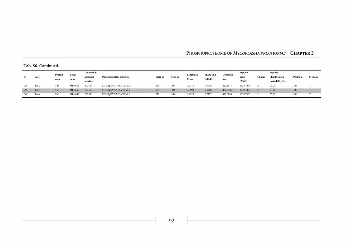
PHOSPHOPROTEOME OF MYCOPLASMA PNEUMONIAE CHAPTER 3
92
Tab. S6. Continued.
# Spot Protein
name
Locus
name
UniProtKB
accession
number
Phosphopeptide sequence Start aa Stop aa SEQUEST
Xcorr
SEQUEST
deltaCn
Observed
m/z
Peptide
mass
(AMU)
Charge
Peptide
identification
probability (%)
Position Mod. aa
28 Tuf-2 Tuf MPN665 P23568 EGGRpTVGAGSVTEVLE 379 394 3.2175 0.7339 820.8817 1639.7478 2 95.00 383 T
29 Tuf-3 Tuf MPN665 P23568 EGGRpTVGAGSVTEVLE 379 394 2.9993 0.6668 820.8795 1639.7433 2 95.00 383 T
30 Tuf-4 Tuf MPN665 P23568 EGGRpTVGAGSVTEVLE 379 394 3.2929 0.7727 820.8883 1639.7609 2 95.00 383 T

PHOSPHOPROTEOME OF MYCOPLASMA PNEUMONIAE CHAPTER 3
93
Fig. S1. Virtual two-dimensional protein map of M. pneumoniae. Based on the calculated molecular
weight (MW) and theoretical isoelectric point (pI), each protein is localized in this virtual
two-dimensional protein map of M. pneumoniae using the software JVirGel v2.2.3b
(http://www.jvirgel.de/). NCBI reference sequence used for predicting MWs and pIs of the 689 proteins is
NC_000912 (M. pneumoniae strain M129). The pI ranged from 4-11 and the molecular weight is adjusted
from 8-125 kDa. About 635 proteins could be visualized with this set-up.
11.0 9.0 8.0 7.0 6.0 5.0 4.010.0
kDa
10.0
20.0
40.0
80.0
100.0
120.0
60.0
pI
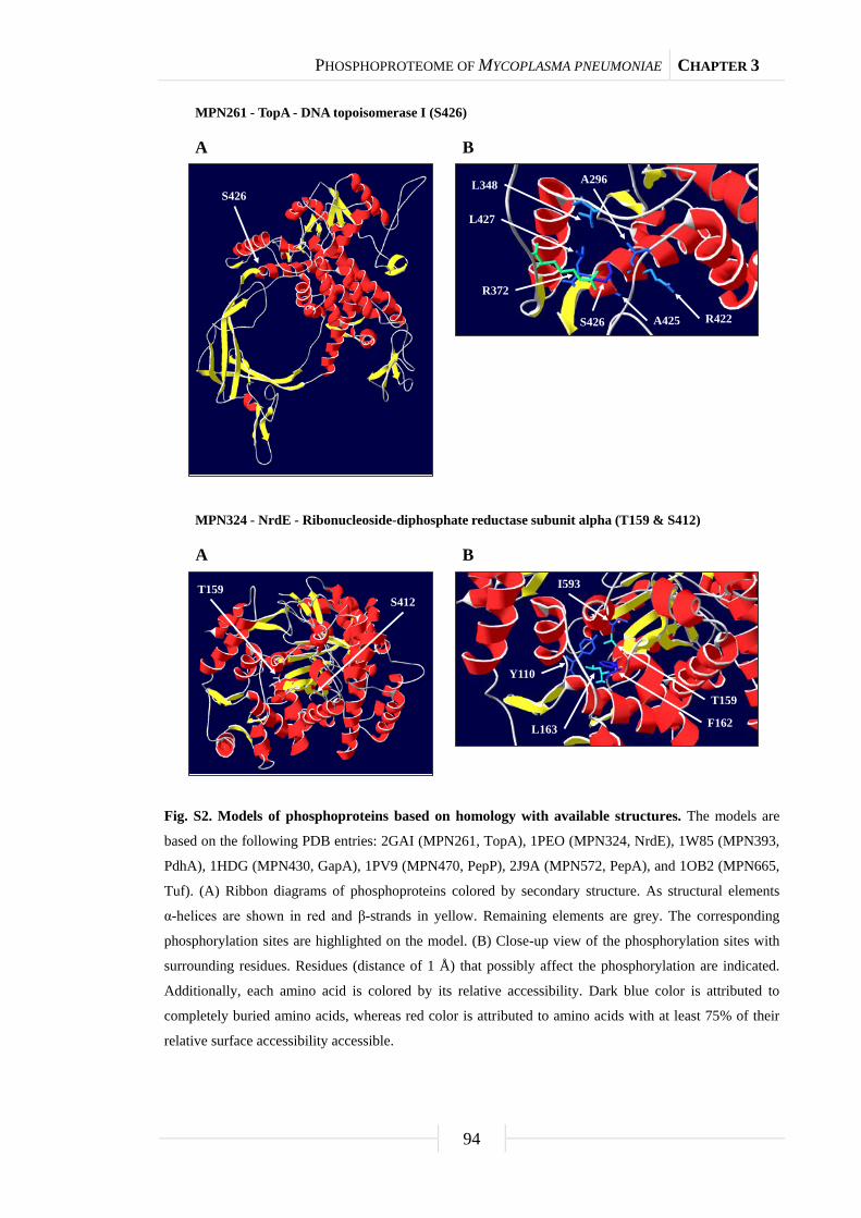
PHOSPHOPROTEOME OF MYCOPLASMA PNEUMONIAE CHAPTER 3
94
Fig. S2. Models of phosphoproteins based on homology with available structures. The models are
based on the following PDB entries: 2GAI (MPN261, TopA), 1PEO (MPN324, NrdE), 1W85 (MPN393,
PdhA), 1HDG (MPN430, GapA), 1PV9 (MPN470, PepP), 2J9A (MPN572, PepA), and 1OB2 (MPN665,
Tuf). (A) Ribbon diagrams of phosphoproteins colored by secondary structure. As structural elements
α-helices are shown in red and β-strands in yellow. Remaining elements are grey. The corresponding
phosphorylation sites are highlighted on the model. (B) Close-up view of the phosphorylation sites with
surrounding residues. Residues (distance of 1 Å) that possibly affect the phosphorylation are indicated.
Additionally, each amino acid is colored by its relative accessibility. Dark blue color is attributed to
completely buried amino acids, whereas red color is attributed to amino acids with at least 75% of their
relative surface accessibility accessible.
MPN261 - TopA - DNA topoisomerase I (S426)
S426A296L348
R422
R372
S426 A425
L427
BA
S412T159
Y110
F162L163
I593
T159
BA
MPN324 - NrdE - Ribonucleoside-diphosphate reductase subunit alpha (T159 & S412)

PHOSPHOPROTEOME OF MYCOPLASMA PNEUMONIAE CHAPTER 3
95
Fig. S2. Continued.
S412T159
Y177
N460
I456
V174
L450
S412
N414S448
BA
MPN324 - NrdE - Ribonucleoside-diphosphate reductase subunit alpha (T159 & S412)
S205
V207L210
BA
E204
S205
T167
K213
MPN393 - PdhA - Pyruvate dehydrogenase E1 component subunit alpha (S205)

PHOSPHOPROTEOME OF MYCOPLASMA PNEUMONIAE CHAPTER 3
96
Fig. S2. Continued.
S245
T182
H184S245
A185
Y317
N319
BA
MPN430 - GapA - Glyceraldehyde-3-phosphate dehydrogenase (S245)
H290
E333D224
T317
V292
V302T317
BA
E319
MPN470 - PepP - Xaa-Pro aminopeptidase (T317)
BA
F157
L252
S249
V432
S249T420
A137L140
A245
MPN572 - PepA - Cytosol aminopeptidase (S249 & T420)

PHOSPHOPROTEOME OF MYCOPLASMA PNEUMONIAE CHAPTER 3
97
Fig. S2. Continued.
BA
V430I417
Q428
L431
T338T420
S419
S249 T420
T341
MPN572 - PepA - Cytosol aminopeptidase (S249 & T420)
T383
L121
Q125
L312S376
T383
L122
R383
BA
MPN665 - Tuf - Elongation factor TU (T383)
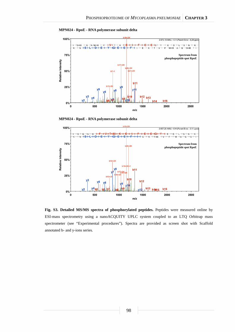
PHOSPHOPROTEOME OF MYCOPLASMA PNEUMONIAE CHAPTER 3
98
Fig. S3. Detailed MS/MS spectra of phosphorylated peptides. Peptides were measured online by
ESI-mass spectrometry using a nanoACQUITY UPLC system coupled to an LTQ Orbitrap mass
spectrometer (see “Experimental procedures”). Spectra are provided as screen shot with Scaffold
annotated b- and y-ions series.
MPN024 - RpoE - RNA polymerase subunit delta
Spectrum fromphosphopeptide spot RpoE
MPN024 - RpoE - RNA polymerase subunit delta
Spectrum fromphosphopeptide spot RpoE

PHOSPHOPROTEOME OF MYCOPLASMA PNEUMONIAE CHAPTER 3
99
Fig. S3. Continued.
MPN066 - ManB - Phosphomannomutase/phosphoglucomutase
Spectrum fromphosphopeptide spot ManB-1
MPN066 - ManB - Phosphomannomutase/phosphoglucomutase
Spectrum fromphosphopeptide spot ManB-2
MPN066 - ManB - Phosphomannomutase/phosphoglucomutase
Spectrum fromphosphopeptide spot ManB-3
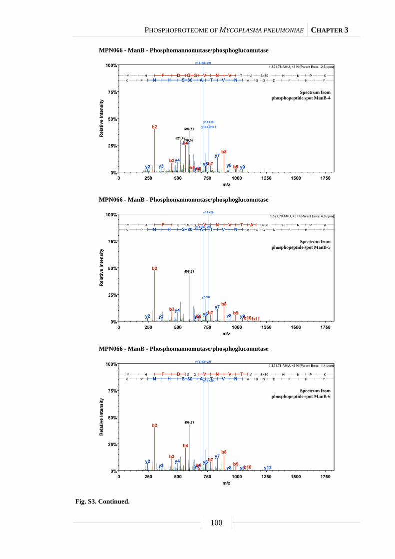
PHOSPHOPROTEOME OF MYCOPLASMA PNEUMONIAE CHAPTER 3
100
Fig. S3. Continued.
MPN066 - ManB - Phosphomannomutase/phosphoglucomutase
Spectrum fromphosphopeptide spot ManB-4
MPN066 - ManB - Phosphomannomutase/phosphoglucomutase
Spectrum fromphosphopeptide spot ManB-5
MPN066 - ManB - Phosphomannomutase/phosphoglucomutase
Spectrum fromphosphopeptide spot ManB-6

PHOSPHOPROTEOME OF MYCOPLASMA PNEUMONIAE CHAPTER 3
101
Fig. S3. Continued.
MPN066 - ManB - Phosphomannomutase/phosphoglucomutase
Spectrum fromphosphopeptide spot ManB-7
MPN066 - ManB - Phosphomannomutase/phosphoglucomutase
Spectrum fromphosphopeptide spot ManB-8
MPN066 - ManB - Phosphomannomutase/phosphoglucomutase
Spectrum fromphosphopeptide spot ManB-9
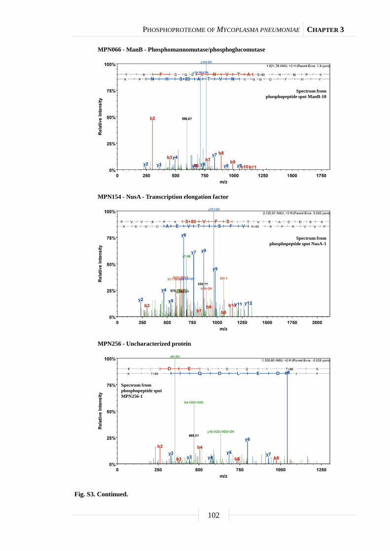
PHOSPHOPROTEOME OF MYCOPLASMA PNEUMONIAE CHAPTER 3
102
Fig. S3. Continued.
MPN066 - ManB - Phosphomannomutase/phosphoglucomutase
Spectrum fromphosphopeptide spot ManB-10
MPN154 - NusA - Transcription elongation factor
Spectrum fromphosphopeptide spot NusA-1
MPN256 - Uncharacterized protein
Spectrum fromphosphopeptide spotMPN256-1

PHOSPHOPROTEOME OF MYCOPLASMA PNEUMONIAE CHAPTER 3
103
Fig. S3. Continued.
MPN261 - TopA - DNA topoisomerase I
Spectrum fromphosphopeptide spot TopA
MPN295 - Uncharacterized protein
Spectrum fromphosphopeptide spot MPN295-2
MPN311 - P41 - Adhesin-related protein
Spectrum fromphosphopeptide spot P41

PHOSPHOPROTEOME OF MYCOPLASMA PNEUMONIAE CHAPTER 3
104
Fig. S3. Continued.
MPN324 - NrdE - Ribonucleoside-diphosphate reductase subunit alpha
Spectrum fromphosphopeptide spot NrdE-1
MPN324 - NrdE - Ribonucleoside-diphosphate reductase subunit alpha
Spectrum fromphosphopeptide spot NrdE-2
MPN393 - PdhA - Pyruvate dehydrogenase E1 component subunit alpha
Spectrum fromphosphopeptide spot [PdhA Pgk]
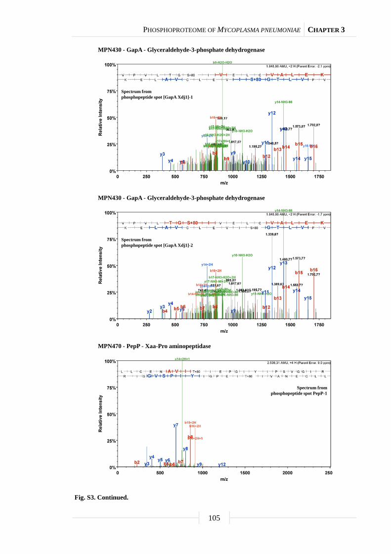
PHOSPHOPROTEOME OF MYCOPLASMA PNEUMONIAE CHAPTER 3
105
Fig. S3. Continued.
MPN430 - GapA - Glyceraldehyde-3-phosphate dehydrogenase
Spectrum fromphosphopeptide spot [GapA Xdj1]-1
MPN430 - GapA - Glyceraldehyde-3-phosphate dehydrogenase
Spectrum fromphosphopeptide spot [GapA Xdj1]-2
MPN470 - PepP - Xaa-Pro aminopeptidase
Spectrum fromphosphopeptide spot PepP-1

PHOSPHOPROTEOME OF MYCOPLASMA PNEUMONIAE CHAPTER 3
106
Fig. S3. Continued.
MPN474 - Coiled coil surface protein
Spectrum fromphosphopeptide spot MPN474
MPN572 - PepA - Cytosol aminopeptidase
Spectrum fromphosphopeptide spot PepA-1
MPN572 - PepA - Cytosol aminopeptidase
Spectrum fromphosphopeptide spot PepA-1
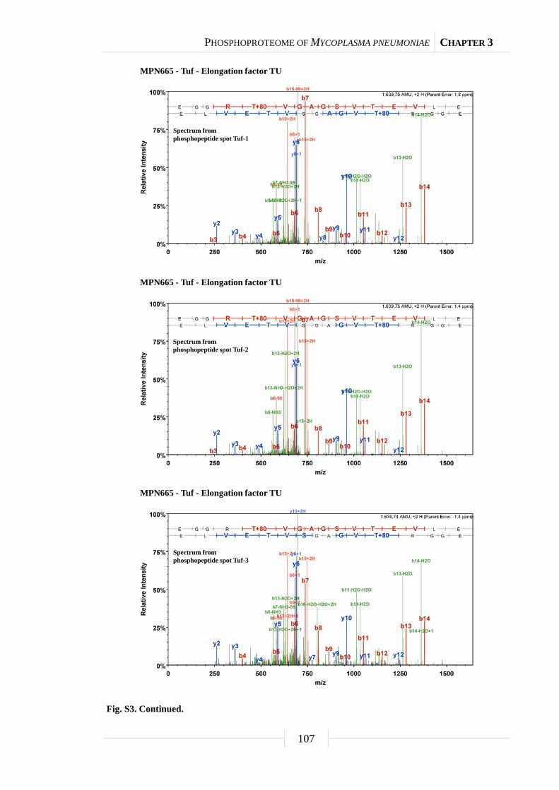
PHOSPHOPROTEOME OF MYCOPLASMA PNEUMONIAE CHAPTER 3
107
Fig. S3. Continued.
MPN665 - Tuf - Elongation factor TU
Spectrum fromphosphopeptide spot Tuf-1
MPN665 - Tuf - Elongation factor TU
Spectrum fromphosphopeptide spot Tuf-2
MPN665 - Tuf - Elongation factor TU
Spectrum fromphosphopeptide spot Tuf-3

PHOSPHOPROTEOME OF MYCOPLASMA PNEUMONIAE CHAPTER 3
108
Fig. S3. Continued.
MPN665 - Tuf - Elongation factor TU
Spectrum fromphosphopeptide spot Tuf-4

Chapter 4
In vitro phosphorylation of key metabolic
enzymes from Bacillus subtilis:
PrkC phosphorylates enzymes from different
branches of basic metabolism
The work described in this chapter was published in:
Pietack, N., D. Becher, S. R. Schmidl, M. H. Saier, M. Hecker, F. M. Commichau,
and J. Stülke. 2010. In vitro phosphorylation of key metabolic enzymes from Bacillus
subtilis: PrkC phosphorylates enzymes from different branches of basic metabolism.
J. Mol. Microbiol. Biotechnol. 18: 129-140.
Author contributions:
This study was designed and interpreted by NP, FMC, and JS. NP performed all in vitro
phosphorylation experiments. SRS performed the phylogenetic analysis of PrkC
homologues during an internship at the lab of MHS, University of California,
San Diego. Determination of phosphorylation sites was done in collaboration with DB
and MH, University of Greifswald. NP and JS wrote the paper.
BACILLUS SUBTILIS PROTEIN KINASE PRKC CHAPTER 4
110
Abstract
Phosphorylation is an important mechanism of protein modification. In the
Gram-positive soil bacterium Bacillus subtilis, about 5% of all proteins are subject to
phosphorylation, and a significant portion of these proteins is phosphorylated on serine
or threonine residues. We were interested in the regulation of the basic metabolism in
B. subtilis. Many enzymes of the central metabolic pathways are phosphorylated in this
organism. In an attempt to identify the responsible protein kinase(s), we identified four
candidate kinases, among them the previously studied kinase PrkC. We observed that
PrkC is indeed able to phosphorylate several metabolic enzymes in vitro. Determination
of the phosphorylation sites revealed a remarkable preference of PrkC for threonine
residues. Moreover, PrkC often used several phosphorylation sites in one protein. This
feature of PrkC-dependent protein phosphorylation resembles the multiple
phosphorylations often observed in eukaryotic proteins. The HPr protein of the
phosphotransferase system is one of the proteins phosphorylated by PrkC, and PrkC
phosphorylates a site (Ser-12) that has recently been found to be phosphorylated in vivo.
The agreement between in vivo and in vitro phosphorylation of HPr on Ser-12 suggests
that our in vitro observations reflect the events that take place in the cell.
Introduction
Adaptation to changing environmental conditions is essential for life. The
Gram-positive soil bacterium Bacillus subtilis is permanently exposed to changing
conditions, such as nutrient limitations or various physical stresses. In principle, there
are three different mechanisms by which bacteria can adapt their metabolism to the
environment: (1) alterations of gene expression, (2) changing the stabilities of proteins,
and (3) modulation of enzymatic activities. Regulation of gene expression is by far the
most intensively studied regulatory mechanism. There is a huge collection of studies
concerning the response of B. subtilis to environmental changes at the proteome and/or
transcriptome levels (Blencke et al., 2003; Eymann et al., 2002; Moreno et al., 2001). In
the past few years, when transcriptome and proteome studies became very popular, most
regulatory effects were attributed to changes in transcription. However, changes in
transcription are not always accompanied by corresponding changes in metabolic fluxes

BACILLUS SUBTILIS PROTEIN KINASE PRKC CHAPTER 4
111
and vice versa (Schilling et al., 2007). Thus, posttranslational regulatory mechanisms
require our attention to fully understand regulatory phenomena.
In the past few years, it turned out that extensive protein phosphorylation not
only on histidine, but also on serine, threonine, and tyrosine residues occurs in bacteria
(Deutscher and Saier, 2005; Kennelly and Potts, 1996). In B. subtilis, phosphorylation
on histidine residues occurs in the phosphotransferase system (PTS) as well as in
PTS-controlled transcription regulators and glycerol kinase. The sensor kinases of the
two-component regulatory systems constitute a second class of His-phosphorylated
proteins (Fabret et al., 1999; Reizer et al., 1999). In the PTS, some phosphotransfer
domains are phosphorylated on cysteine residues from which the phosphate is
transferred to the incoming sugar. In contrast, the response regulators of the
two-component systems are phosphorylated on aspartate residues by their cognate
sensor kinases (Fabret et al., 1999; Reizer et al., 1999). Phosphorylation on serine
residues has long been studied for the HPr protein of the PTS that is phosphorylated on
Ser-46 by the metabolite-controlled HPr kinase/phosphorylase. HPr(Ser-P) then binds to
the transcription factor CcpA and acts as a corepressor in carbon regulation
(Görke and Stülke, 2008). Regulatory phosphorylation of proteins on serine (and
threonine) residues is also well established for the modulators of sigma factor activity in
sporulation and stress response (Min et al., 1993; Yang et al., 1996). Protein tyrosine
phosphorylation is implicated in the control of the transcription repressor CtsR, of
UDP-glucose dehydrogenases and of the single-stranded DNA-binding proteins
(Grangeasse et al., 2007). In total, about 200 proteins, representing 5% of all B. subtilis
proteins, are now known to be phosphorylated (Eymann et al., 2007; Lévine et al.,
2006; Macek et al., 2007).
With the large number of proteins phosphorylated on serine, threonine or
tyrosine, the identification of the responsible kinase(s) is getting into the spotlight. So
far, the HPr kinase and the three kinases involved in controlling sigma factor activities
have been studied. All these kinases seem to phosphorylate clearly defined sites and are
therefore not candidates for the phosphorylation of novel targets. In addition, the protein
kinase PrkC can phosphorylate several proteins, including elongation factor G and PrkC
itself (Gaidenko et al., 2002; Madec et al., 2002). PrkC is a located in the cell
membrane of vegetative cells and in the inner spore membrane of spores (Madec et al.,
2002; Shah et al., 2008). The prkC gene is part of the prpC-prkC-cpgA operon that is

BACILLUS SUBTILIS PROTEIN KINASE PRKC CHAPTER 4
112
constitutively expressed throughout growth (Iwanicki et al., 2005). Recently,
stimulation of PrkC activity by muropeptides was reported. In spores, this activation of
PrkC triggers germination, and prkC mutants are unable to germinate in response to
muropeptides (Shah et al., 2008). Homologues of PrkC are widespread in both
Gram-positive and Gram-negative bacteria (Fig. 14). This kinase is implicated in the
phosphorylation of glycolytic enzymes and histone-like proteins as well as in
pathogenicity in other Gram-positive bacteria (Jin and Pancholi, 2006; Kristich et al.,
2007; Lomas-Lopez et al., 2007). It is interesting to note that prkC is encoded in an
operon with a corresponding protein phosphatase, PrpC (Madec et al., 2002). This
phosphatase is able to dephosphorylate HPr(Ser-P) in B. subtilis and Mycoplasma
pneumoniae (Halbedel et al., 2006; Singh et al., 2007).
We were interested in the regulation of carbon and nitrogen metabolism in
B. subtilis. In an analysis of the transcriptome and metabolic flux responses to organic
acids, we noticed that there are some cases where the changes in transcription do not
match those in the metabolic fluxes. This is exemplified by the expression of the alsSD
operon encoding acetolactate synthase and acetolactate decarboxylase and the
corresponding acetoin production. Therefore, we considered posttranscriptional events
to be important for metabolic adaptation (Schilling et al., 2007). At the same time,
phosphoproteome analyses demonstrated that indeed many enzymes of basic
metabolism are subject to phosphorylation in B. subtilis (Eymann et al., 2007;
Lévine et al., 2006; Macek et al., 2007). Thus, protein phosphorylation might indeed
play an important role for the adaptation of B. subtilis metabolism to changing
conditions. However, the kinases responsible for these phosphorylation events have so
far not been identified.
In this study, we intended to identify the kinase(s) for some of the enzymes of
central metabolism by in vitro analysis of protein phosphorylation. Our work
demonstrates that PrkC is able to phosphorylate several enzymes of different metabolic
pathways. Moreover, PrkC was also responsible for a novel modification of the HPr
protein of the PTS on Ser-12, an event previously observed in vivo (Macek et al., 2007).
Thus, PrkC may play an important role in phosphorylating proteins with different
functions in B. subtilis.
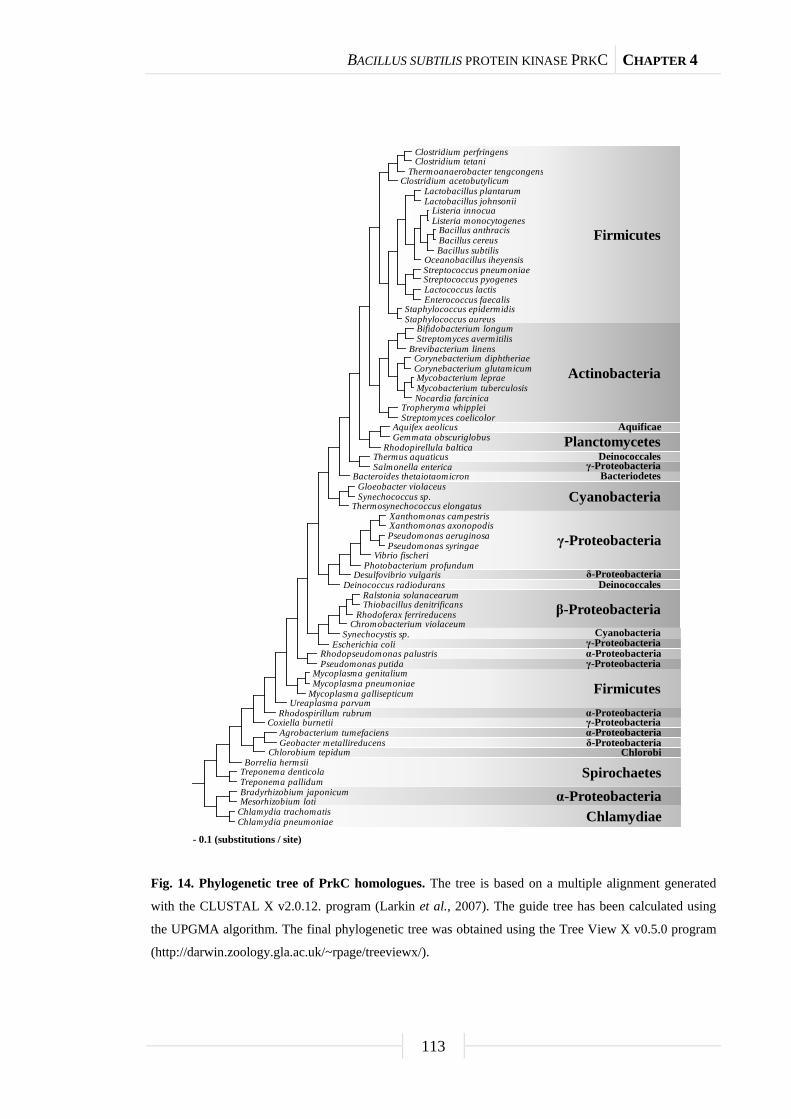
BACILLUS SUBTILIS PROTEIN KINASE PRKC CHAPTER 4
113
Fig. 14. Phylogenetic tree of PrkC homologues. The tree is based on a multiple alignment generated
with the CLUSTAL X v2.0.12. program (Larkin et al., 2007). The guide tree has been calculated using
the UPGMA algorithm. The final phylogenetic tree was obtained using the Tree View X v0.5.0 program
(http://darwin.zoology.gla.ac.uk/~rpage/treeviewx/).
Clostridium perfringensClostridium tetani
Thermoanaerobacter tengcongensClostridium acetobutylicum
Lactobacillus plantarumLactobacillus johnsonii
Listeria innocuaListeria monocytogenes
Bacillus anthracisBacillus cereusBacillus subtilis
Oceanobacillus iheyensisStreptococcus pneumoniaeStreptococcus pyogenesLactococcus lactisEnterococcus faecalis
Staphylococcus epidermidisStaphylococcus aureus
Bifidobacterium longumStreptomyces avermitilis
Brevibacterium linensCorynebacterium diphtheriaeCorynebacterium glutamicumMycobacterium lepraeMycobacterium tuberculosisNocardia farcinica
Tropheryma whippleiStreptomyces coelicolor
Aquifex aeolicusGemmata obscuriglobus
Rhodopirellula balticaThermus aquaticusSalmonella enterica
Bacteroides thetaiotaomicronGloeobacter violaceusSynechococcus sp.
Thermosynechococcus elongatusXanthomonas campestrisXanthomonas axonopodisPseudomonas aeruginosaPseudomonas syringae
Vibrio fischeriPhotobacterium profundum
Desulfovibrio vulgarisDeinococcus radiodurans
Ralstonia solanacearumThiobacillus denitrificans
Rhodoferax ferrireducensChromobacterium violaceum
Synechocystis sp.Escherichia coli
Rhodopseudomonas palustrisPseudomonas putida
Mycoplasma genitaliumMycoplasma pneumoniae
Mycoplasma gallisepticumUreaplasma parvum
Rhodospirillum rubrumCoxiella burnetii
Agrobacterium tumefaciensGeobacter metallireducens
Chlorobium tepidumBorrelia hermsii
Treponema denticolaTreponema pallidumBradyrhizobium japonicumMesorhizobium loti
Chlamydia trachomatisChlamydia pneumoniae
0.1
Firmicutes
Actinobacteria
AquificaePlanctomycetes
Deinococcalesγ-Proteobacteria
Bacteriodetes
Cyanobacteria
γ-Proteobacteria
δ-ProteobacteriaDeinococcales
β-ProteobacteriaCyanobacteria
γ-Proteobacteriaα-Proteobacteriaγ-Proteobacteria
Firmicutesα-Proteobacteriaγ-Proteobacteriaα-Proteobacteriaδ-Proteobacteria
Chlorobi
Spirochaetesα-Proteobacteria
Chlamydiae- 0.1 (substitutions / site)

BACILLUS SUBTILIS PROTEIN KINASE PRKC CHAPTER 4
114
Tab. 6. Plasmids used in this work.
Plasmid Gene Protein Affinity tag Oligonucleotides Vector References
pAG2 ptsH HPr His6-tag - - Galinier et al. (1997)
pGP174 glnA Glutamine synthetase Strep-tag - - Heinrich et al. (2006)
pGP205 hprK HPr kinase His6-tag - - Hanson et al. (2002)
pGP371 ptsHa HPr-S46A His6-tag SH82/SH83 pWH844 This work
pGP563 eno Enolase His6-tag eno forw1/eno rev pWH844 This work
pGP819 ywjH Transaldolase Strep-tag NP11/NP12 pGP172 This work
pGP820 tkt Transketolase Strep-tag NP05/NP06 pGP172 This work
pGP821 prkD Protein kinase Strep-tag NP09/NP10 pGP172 This work
pGP822 alsD α-acetolactate decarboxylase Strep-tag NP01/NP02 pGP172 This work
pGP823 yabT Protein kinase Strep-tag NP07/NP08 pGP172 This work
pGP824 yxaL Protein kinase Strep-tag NP13/NP14 pGP172 This work
pGP825 prkC Protein kinase Strep-tag NP03/NP04 pGP172 This work
pGP931 icd Isocitrate dehydrogenase His6-tag FC17/FC18 pWH844 This work
pGP1100 pyk Pyruvate kinase His6-tag pyk forw1/pyk rev pWH844 This work a Indicates mutant alleles.

BACILLUS SUBTILIS PROTEIN KINASE PRKC CHAPTER 4
115
Results
Identification of potential serine/threonine protein kinases in B. subtilis. The
genome of B. subtilis encodes several proteins that are known or suspected to have
serine/threonine kinase activity, among them the intensively studied HPr
kinase/phosphorylase and the kinases involved in the control of sigma factor activities.
In addition, the PrkC serine/threonine kinase has been studied before. Analysis of the
annotated genome sequence revealed the presence of three additional potential kinases:
PrkD that is similar to the kinase domain of PrkC (Madec et al., 2002), YxaL and YabT.
Purification of selected potential protein kinases and phosphorylation
targets. The four genes encoding the selected proteins were cloned into the expression
vector pGP172, fusing them to an N-terminal Strep-tag. The proteins were expressed
and purified using their affinity for Streptactin Sepharose. PrkC and PrkD were purified
to apparent homogeneity. In contrast, we were unable to purify detectable amounts of
YxaL. This may be due to the fact that YxaL is a secreted protein carrying a signal
peptide (Tjalsma et al., 2004). Similarly, only small amounts of YabT were obtained.
Therefore, we concentrated our subsequent analyses on PrkC and PrkD.
Among the phosphoproteins that are involved in central metabolism, we selected
glutamine synthetase (GlnA), enolase (Eno), transaldolase (YwjH), transketolase (Tkt),
α-acetolactate decarboxylase (AlsD), isocitrate dehydrogenase (Icd), and pyruvate
kinase (Pyk). These proteins were found to be phosphorylated in vivo on serine,
threonine or tyrosine (Eymann et al., 2007; Lévine et al., 2006; Macek et al., 2007).
The corresponding genes were cloned into the expression vectors pWH844 and pGP172
to fuse the coding sequences to an N-terminal His6- or Strep-tag, respectively (Table 6).
All these proteins were expressed in Escherichia coli and were purified to apparent
homogeneity by affinity chromatography.
Autophosphorylation of PrkC and PrkD. First, we analyzed the
autophosphorylation of the selected serine/threonine kinases, PrkC and PrkD. For this
purpose, we incubated the two proteins in the presence of [γ-32P]ATP and analyzed
them by SDS-PAGE. As shown in Fig. 15, both proteins exhibited autophosphorylation
activity. This observation is in excellent agreement with a previous report on the
autophosphorylation of PrkC (Madec et al., 2003). To characterize these
autophosphorylation events in more detail, we determined the phosphorylation sites by
mass spectrometry using an LTQ Orbitrap mass spectrometer. For PrkC, we detected
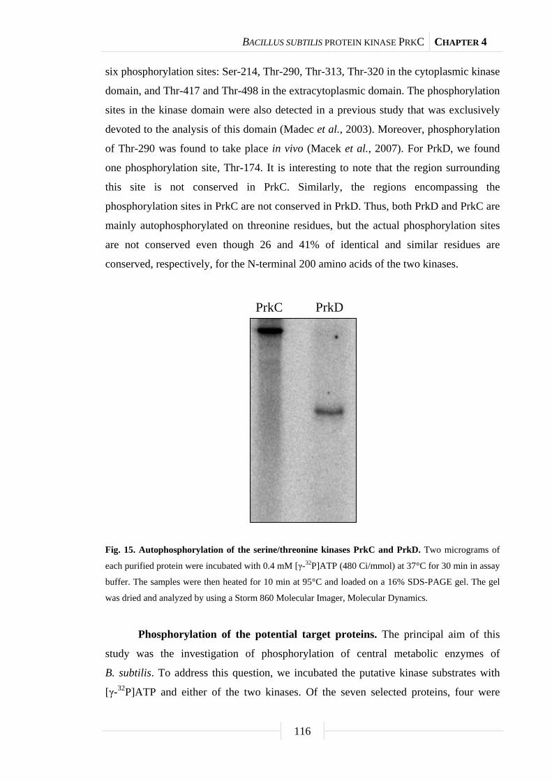
BACILLUS SUBTILIS PROTEIN KINASE PRKC CHAPTER 4
116
six phosphorylation sites: Ser-214, Thr-290, Thr-313, Thr-320 in the cytoplasmic kinase
domain, and Thr-417 and Thr-498 in the extracytoplasmic domain. The phosphorylation
sites in the kinase domain were also detected in a previous study that was exclusively
devoted to the analysis of this domain (Madec et al., 2003). Moreover, phosphorylation
of Thr-290 was found to take place in vivo (Macek et al., 2007). For PrkD, we found
one phosphorylation site, Thr-174. It is interesting to note that the region surrounding
this site is not conserved in PrkC. Similarly, the regions encompassing the
phosphorylation sites in PrkC are not conserved in PrkD. Thus, both PrkD and PrkC are
mainly autophosphorylated on threonine residues, but the actual phosphorylation sites
are not conserved even though 26 and 41% of identical and similar residues are
conserved, respectively, for the N-terminal 200 amino acids of the two kinases.
Fig. 15. Autophosphorylation of the serine/threonine kinases PrkC and PrkD. Two micrograms of
each purified protein were incubated with 0.4 mM [γ-32P]ATP (480 Ci/mmol) at 37°C for 30 min in assay
buffer. The samples were then heated for 10 min at 95°C and loaded on a 16% SDS-PAGE gel. The gel
was dried and analyzed by using a Storm 860 Molecular Imager, Molecular Dynamics.
Phosphorylation of the potential target proteins. The principal aim of this
study was the investigation of phosphorylation of central metabolic enzymes of
B. subtilis. To address this question, we incubated the putative kinase substrates with
[γ-32P]ATP and either of the two kinases. Of the seven selected proteins, four were
PrkC PrkD

BACILLUS SUBTILIS PROTEIN KINASE PRKC CHAPTER 4
117
phosphorylated by PrkC (Fig. 16). These were the transaldolase YwjH, the glutamine
synthetase GlnA, the isocitrate dehydrogenase Icd, and the α-acetolactate decarboxylase
AlsD. In contrast, enolase, transketolase, and the pyruvate kinase PykA were not
substrates for PrkC-dependent phosphorylation (data not shown). However, the
phosphorylation efficiency differed for the PrkC substrates: The phosphorylation
efficiency of YwjH, GlnA, and Icd by PrkC was similar to that observed for
HPrK-dependent phosphorylation of HPr (Fig. 16). In contrast, only weak signals were
observed with AlsD (Fig. 16). This might be caused by differences in the number of
phosphorylation sites or in different affinities of PrkC for its respective substrates. The
kinase assays with PrkD revealed that only Icd and GlnA were phosphorylated by PrkD
(data not shown). However, these phosphorylation signals were extremely faint, which
questions their biological relevance. Thus, our analysis revealed selective
phosphorylation of four enzymes of central metabolism by PrkC.
Determination of phosphorylation sites in the target proteins. In order to get
new insights into PrkC substrates, we determined the phosphorylation sites of the
proteins phosphorylated by PrkC. As observed for the autophosphorylation of PrkC,
multiple phosphorylations with threonine as the major phosphoacceptor were observed.
For the transaldolase YwjH, we observed six sites that were phosphorylated by PrkC.
The phosphorylated amino acids were scattered throughout the protein. There was one
phosphorylated serine residue, Ser-55, and five phosphothreonines, i.e. Thr-26, Thr-82,
Thr-125, Thr-159, and Thr-184. Similar results were obtained with glutamine
synthetase. This enzyme was phosphorylated on one serine residue, Ser-207, and three
threonine residues, Thr-26, Thr-147, and Thr-286. It is interesting to note that Thr-26 is
located in the N-terminal β-grasp domain of the enzyme that is thought to be involved in
binding of small molecules (Burroughs et al., 2007), whereas the other phosphorylation
events take place in the catalytic domain of glutamine synthetase. Isocitrate
dehydrogenase is phosphorylated by PrkC on three threonine residues, Thr-138,
Thr-147, and Thr-396. In contrast to the other PrkC substrates (including PrkC
autophosphorylation), the α-acetolactate decarboxylase AlsD was phosphorylated by
PrkC on a single serine residue, Ser-88.

BACILLUS SUBTILIS PROTEIN KINASE PRKC CHAPTER 4
118
Fig. 16. Phosphorylation of potential target proteins by PrkC. Ten micrograms of the purified
enzymes YwjH, GlnA, Icd, and AlsD were incubated with 0.4 mM [γ-32P]ATP (480 Ci/mmol) in the
absence and in the presence of 2 µg PrkC at 37°C for 30 min in assay buffer. The samples were heated for
10 min at 95°C and loaded on a 16% SDS-PAGE gel. To obtain the autoradiogram, the gel was dried and
analyzed using a Storm 860 Molecular Imager, Molecular Dynamics. (A) In addition, the proteins were
visualized by Coomassie staining. (B) co = Control (HPr phosphorylated by HPrK).
PrkCYwjH
A
BM co
1208647
34
26
20
coGlnA Icd AlsD
PrkC
GlnAIcd
YwjH
- + - + - + - +
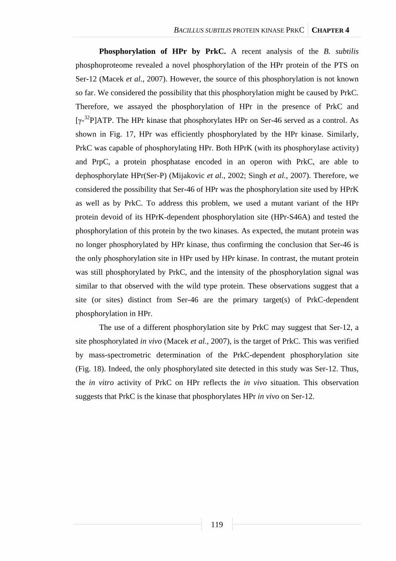
BACILLUS SUBTILIS PROTEIN KINASE PRKC CHAPTER 4
119
Phosphorylation of HPr by PrkC. A recent analysis of the B. subtilis
phosphoproteome revealed a novel phosphorylation of the HPr protein of the PTS on
Ser-12 (Macek et al., 2007). However, the source of this phosphorylation is not known
so far. We considered the possibility that this phosphorylation might be caused by PrkC.
Therefore, we assayed the phosphorylation of HPr in the presence of PrkC and
[γ-32P]ATP. The HPr kinase that phosphorylates HPr on Ser-46 served as a control. As
shown in Fig. 17, HPr was efficiently phosphorylated by the HPr kinase. Similarly,
PrkC was capable of phosphorylating HPr. Both HPrK (with its phosphorylase activity)
and PrpC, a protein phosphatase encoded in an operon with PrkC, are able to
dephosphorylate HPr(Ser-P) (Mijakovic et al., 2002; Singh et al., 2007). Therefore, we
considered the possibility that Ser-46 of HPr was the phosphorylation site used by HPrK
as well as by PrkC. To address this problem, we used a mutant variant of the HPr
protein devoid of its HPrK-dependent phosphorylation site (HPr-S46A) and tested the
phosphorylation of this protein by the two kinases. As expected, the mutant protein was
no longer phosphorylated by HPr kinase, thus confirming the conclusion that Ser-46 is
the only phosphorylation site in HPr used by HPr kinase. In contrast, the mutant protein
was still phosphorylated by PrkC, and the intensity of the phosphorylation signal was
similar to that observed with the wild type protein. These observations suggest that a
site (or sites) distinct from Ser-46 are the primary target(s) of PrkC-dependent
phosphorylation in HPr.
The use of a different phosphorylation site by PrkC may suggest that Ser-12, a
site phosphorylated in vivo (Macek et al., 2007), is the target of PrkC. This was verified
by mass-spectrometric determination of the PrkC-dependent phosphorylation site
(Fig. 18). Indeed, the only phosphorylated site detected in this study was Ser-12. Thus,
the in vitro activity of PrkC on HPr reflects the in vivo situation. This observation
suggests that PrkC is the kinase that phosphorylates HPr in vivo on Ser-12.
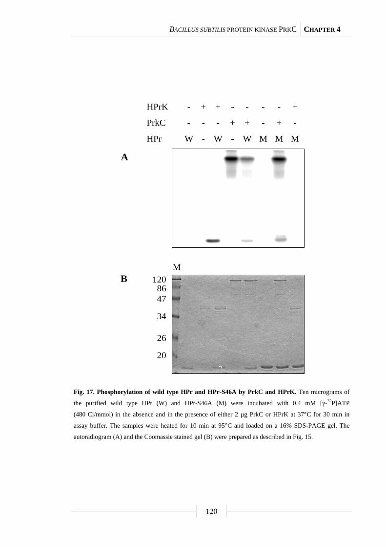
BACILLUS SUBTILIS PROTEIN KINASE PRKC CHAPTER 4
120
Fig. 17. Phosphorylation of wild type HPr and HPr-S46A by PrkC and HPrK. Ten micrograms of
the purified wild type HPr (W) and HPr-S46A (M) were incubated with 0.4 mM [γ-32P]ATP
(480 Ci/mmol) in the absence and in the presence of either 2 µg PrkC or HPrK at 37°C for 30 min in
assay buffer. The samples were heated for 10 min at 95°C and loaded on a 16% SDS-PAGE gel. The
autoradiogram (A) and the Coomassie stained gel (B) were prepared as described in Fig. 15.
HPrK
PrkC
HPr
-
-
W
+
-
-
+
-
W
-
+
-
-
+
W
-
-
M
-
+
M
+
-
M
A
B 1208647
34
26
20
M

BACILLUS SUBTILIS PROTEIN KINASE PRKC CHAPTER 4
121
Fig. 18. Identification of the site of PrkC-dependent phosphorylation in HPr. The fragment ion
spectrum of the HPr T2 peptide (VTADSGIHAR) with a precursor ion mass 544.7 [M + 2 H]2+ indicates
a phosphate residue. The corresponding b- and y-ions and the formation of the ion corresponding to the
neutral loss of phosphoric acid from the doubly-charged precursor ion are highlighted and demonstrate
the Ser-12 phosphorylation of HPr.
Discussion
Protein phosphorylation is a very common event in all organisms
(Eymann et al., 2007; Ficarro et al., 2002; Levine et al., 2006; Macek et al., 2007;
Macek et al., 2008). As much as about 5% of all proteins can be phosphorylated in
organisms so different as the sporulating bacterium B. subtilis and the genome
minimalist Mycoplasma genitalium (Su et al., 2007). Two major questions remain to be
answered: (1) Which kinases are responsible for these phosphorylation events, and
(2) what is the effect of the phosphorylation on the activity of the target proteins?
200 300 400 500 600 700 800 900 1000 1100m/z
05
10
1520
2530
3540
455055
6065
7075
8085
9095
100
Rel
ativ
e A
bund
ance
y8+2
454.0yy3+1
383.3y4+1
496.3yy8
+1
906.3yy6+1
720.4yy2+1
246.1yy7
+1
835.3bb2+1
201.1bb9
+1
932.2bb6
+1
611.6yy1
+1
175.2yy9
+1
1007.6bb3
+1
271.7
504.9
544.7
808.4
495.7
V T A D pS G I H A Ry1
b9
y2
b8
y9
b1
y8
b2
y7
b3
y6
b4
y5
b5
y4
b6
y3
b7
Precursor ion: 544.7 [M + 2 H]2+
[M2+ - H3PO4]

BACILLUS SUBTILIS PROTEIN KINASE PRKC CHAPTER 4
122
In this work, we addressed the first of these questions. In B. subtilis, only five
serine/threonine kinases have been studied so far. These are HPr kinase, RsbT kinase,
RsbW kinase, and SpoIIAB kinase, involved in the control of sigma factor activities and
PrkC (Reizer et al., 1998; Madec et al., 2002; Min et al., 1993). In this work, we
focused on PrkC and its relative, PrkD. Indeed, our in vitro experiments revealed that
PrkC is able to phosphorylate several of the proteins that were found to exist as
phosphoproteins in vivo.
PrkC is implicated in sporulation, biofilm formation, and germination
(Gaidenko et al., 2002; Madec et al., 2002; Shah et al., 2008), but only two substrates
(EF-G and PrkC itself) have been identified. Our work confirms that PrkC might be of
global importance as it phosphorylates enzymes from different pathways, such as
transaldolase, glutamine synthetase, isocitrate dehydrogenase, α-acetolactate
decarboxylase, and HPr. However, among the eight phosphoproteins tested for
phosphorylation by PrkC, three were not targets of this kinase. A global role for PrkC is
supported by studies with other bacteria. In Staphylococcus aureus, PrkC
phosphorylates glycolytic enzymes (Lomas-Lopez et al., 2007), whereas the α-subunit
of RNA polymerase and the phosphoglucosamine mutase are substrates of the
Streptococcus pneumoniae PrkC homolog, StkP (Novákováa et al., 2005). Moreover,
PrkC homologs are important for virulence development and global gene expression in
S. pneumoniae, S. aureus, M. pneumoniae, and Bacillus anthracis (Débarbouillé et al.,
2009; Sasková et al., 2007; Schmidl et al., 2010; Shakir et al., 2010). Thus, PrkC can
phosphorylate a wide range of proteins involved in a variety of cellular functions.
Another remarkable feature of PrkC is its obvious preference for threonine. This
study presents a significant increase of the known phosphorylation sites used by PrkC.
Of the sites identified here, 16 are threonines and only 5 are serine residues. In a
previous study on PrkC autophosphorylation, 7 phosphorylated threonine residues and
one serine residue were reported (Madec et al., 2003). Our results confirm the
autophosphorylation of Ser-214 as well as of Thr-290, Thr-313, and Thr-320. We failed
to detect the phosphorylation of a stretch of multiple threonine residues in the
N-terminal part of PrkC, but instead we detected two phosphorylation sites in the
extracellular domain of PrkC that was not included in the previous study (Madec et al.,
2003). In vivo phosphorylation of PrkC was observed on Thr-290 (Macek et al., 2007),
thus providing a confirmation for our in vitro study. Studies on protein phosphorylation
BACILLUS SUBTILIS PROTEIN KINASE PRKC CHAPTER 4
123
by PrkC proteins from Streptococcus pyogenes also revealed the use of threonine as the
preferred phosphorylation target by PrkC (Jin and Pancholi, 2006). The kinase domains
of PrkC and PrkD are very similar to each other (about 30% identical residues).
However, the sites of autophosphorylation were not conserved between the two proteins
although the only phosphorylation site identified for PrkD was also a threonine. A
comparison of the regions around the PrkC-dependent phosphorylation sites revealed
little conservation of the primary sequences, suggesting that it is not the primary
sequence context that serves for recognition by PrkC.
An interesting aspect of this work is the identification of HPr as a novel target
for PrkC. HPr is a paradigm of protein phosphorylation with its His-15 and Ser-46
phosphorylated by enzyme I of the PTS and by the ATP-dependent HPr kinase,
respectively (Reizer et al., 1998). Analysis of the B. subtilis phosphoproteome revealed
the phosphorylation of yet another site, Ser-12. We could attribute this phosphorylation
event to the activity of PrkC. This finding suggests that our in vitro observations
regarding PrkC-dependent protein phosphorylation reflect the in vivo activity of PrkC
and the physiological interactions with the different target proteins. However, it is not
known whether this phosphorylation affects the biological activity of HPr. The weak
phosphorylation of Ser-12 by PrkC suggests that this phosphorylation does not affect
the properties of the cellular HPr. Similar results have been obtained for the
transcription antiterminator GlcT, which can be phosphorylated and inactivated to a
minor degree by HPr under conditions in which its full activity is required. Again, this
phosphorylation affects only a small fraction of the cellular GlcT pool and, therefore,
does not influence GlcT-mediated regulation (Schmalisch et al., 2003).
Our work suggests that PrkC is a protein kinase, which may have global
importance in B. subtilis. However, important questions remain to be answered: Which
other proteins does PrkC phosphorylate? Phosphoproteome studies with prkC mutants
might be helpful to address this question. Does PrkC-dependent phosphorylation serve
for fine-tuning of the metabolism, does it tag the proteins for degradation, or does it
serve completely different purposes? Finding the answers to these questions is
challenging, but these answers will have a significant impact on our understanding of
microbial physiology.

BACILLUS SUBTILIS PROTEIN KINASE PRKC CHAPTER 4
124
Materials and Methods
Bacterial strains, oligonucleotides, and growth conditions. B. subtilis 168
(wild type, laboratory collection) and QB5223 (ptsH1) (Martin-Verstraete et al., 1995)
were used to isolate chromosomal DNA for the amplification of the desired genes.
E. coli DH5 and BL21(DE3)/pLysS (Sambrook et al., 1989) were used for cloning
experiments and protein expression, respectively. B. subtilis was grown in TY medium
(1% wt/vol tryptone, 0.5% wt/vol yeast extract, and 1% wt/vol NaCl). E. coli was
grown in lysogeny broth medium (LB medium), and transformants were selected on
plates containing ampicillin (100 g/ml). LB plates were prepared by the addition of
17 g Bacto agar (Difco) per liter of LB medium. The oligonucleotides used in this study
are listed in Table 7.
DNA manipulation. Transformation of E. coli and plasmid DNA extraction
were performed using standard procedures (Sambrook et al., 1989). Restriction
enzymes, T4 DNA ligase, and DNA polymerases were used as recommended by the
manufacturers. DNA fragments were purified from agarose gels using the QIAquick
PCR Purification kit (Qiagen, Germany). Pfu DNA polymerase was used for the
polymerase chain reaction as recommended by the manufacturer. DNA sequences were
determined using the dideoxy chain termination method (Sambrook et al., 1989). All
plasmid inserts derived from PCR products were verified by DNA sequencing.
Chromosomal DNA of B. subtilis was isolated as described (Martin-Verstraete et al.,
1995).
Plasmid constructions. The plasmids used for the expression of tagged
B. subtilis proteins in E. coli were constructed using the expression vectors pWH844
(Schirmer et al., 1997) or pGP172 (Merzbacher et al., 2004) that allow the fusion of the
cloned proteins to an N-terminal His6- or Strep-tag, respectively. These plasmids are
listed in Table 6.
For the expression of the S46A mutant variant of the HPr proteins, plasmid
pGP371 was constructed by cloning of the ptsH1 DNA fragment from B. subtilis
QB5223 (Table 6). B. subtilis HPr kinase and wild type HPr were expressed using
pGP205 and pAG2, respectively (Galinier et al., 1997; Hanson et al., 2002).

BACILLUS SUBTILIS PROTEIN KINASE PRKC CHAPTER 4
125
Tab. 7. Oligonucleotides used in this study.
Oligonucleotide Sequence (5’→3’)a
CD13 AAACATATGGCTAGCTGGAGCCACCCGCAGTTC
eno forw1 AAGGATCCCCATACATTGTTGATGTTTATGCAC
eno rev TTTCTGCAGTTACTTGTTTAAGTTGTAGAAAGAGTTG
FC17 AAAGGATCCGTGGCACAAGGTGAAAAAATTAC
FC18 TTTCTGCAGTTATTAGTCCATGTTTTTGAT
JS39 TCTATCAACAGGAGTCCAAGC
NP01 AAAGAGCTCCGATGAAACGAGAAAGCAACATTC
NP02 TTTGGATCCCTATTATTCAGGGCTTCCTTCAGT
NP03 AAAGAGCTCCGGTGCTAATCGGCAAGC
NP04 TTTAGATCTTCATTATTCATCTTTCGGATACTCAAT
NP05 AAAGAGCTCGATGGATACAATTGAAAAGAAATCAG
NP06 TTTGGATCCTCATTACTTATTGATTAATGCCTTAAC
NP07 AAAGAGCTCGATGATGAACGACGCTTTGAC
NP08 TTTAGATCTTCATTAGATTAAGAAAAAGATAATA
NP09 AAAGAGCTCGATGGCATTAAAACTTCTAAAAAA
NP10 TTTGGATCCTCATTATGTGACCGATTGAATGGC
NP11 AAAGAGCTCGATGTTATTCTTTGTTGATACAG
NP12 TTTGGATCCTCATTATTTGTTCCAGTCTGCC
NP13 AAAGAGCTCGATGGTCAAGTCATTTCG
NP14 TTTGGATCCTCATTATTTCCCAAAAGCCATCAG
NP15 CCGGTGTTCACGGTGCGCCGCTTGG
NP16 CTCACCTGATTCTTTAACAGCTACAGCG
NP17 CTTGCTAAAGCTGCTGGCGAAAAGAAGCTG
NP18 ATCGTGAGCAAAACCCACGGCCAC
NP19 ATGTTCACGCGATCAAACCGACGTC
NP20 GCAGCAGCCAACTCAGCTTCCTTTCGGGC
SH82 ATGGATCCATGGCACAAAAAACATTTAAAG
SH83 ATAAAGCTTCTCGCCGAGTCCTTCG
pWH844 fw TATGAGAGGATCGCATCACCAT
pyk forw1 AAGGATCCAGAAAAACTAAAATTGTTTGTACCATCGG
pyk rev TTTCTGCAGTTAAAGAACGCTCGCACGG a The restrictions sites are underlined.
BACILLUS SUBTILIS PROTEIN KINASE PRKC CHAPTER 4
126
Overexpression and purification of recombinant proteins.
E. coli BL21(DE3)/pLysS was used as host for the overexpression of recombinant
proteins. Expression was induced by the addition of IPTG (final concentration 1 mM) to
exponentially growing cultures (OD600 of 0.8). Cells were lysed using a French press
(20,000 p.s.i., 138,000 kPa; Spectronic Instruments, UK). After lysis, the crude extracts
were centrifuged at 15,000 g for 1 h. For purification of His6-tagged proteins, the
resulting supernatants were passed over an Ni2+ HiTrap chelating column (5 ml bed
volume, Pharmacia) followed by elution with an imidazole gradient (from 0 to
500 mM imidazole in a buffer containing 10 mM Tris-HCl pH 7.5, 600 mM NaCl,
10 mM β-mercaptoethanol) over 30 ml at a flow rate of 0.5 ml/min. For proteins
carrying an N-terminal Strep-tag, the crude extract was passed over a Streptactin
column (IBA, Göttingen, Germany). The recombinant protein was eluted with
desthiobiotin (Sigma, final concentration 2.5 mM).
After elution, the fractions were tested for the desired protein using
12.5% SDS-PAGE gels. The relevant fractions were combined and dialyzed overnight.
Protein concentration was determined using the Bio-Rad dye-binding assay with bovine
serum albumin serving as the standard.
Assays for protein phosphorylation. Activity assays of potential protein
kinases were carried out with purified proteins in assay buffer [10 mM MgCl2,
25 mM Tris-HCl pH 7.6, 1 mM dithiothreitol, 0.4 mM [γ-32P]ATP (480 Ci/mmol)]
using purified target proteins. For assays of HPr kinase activity,
fructose 1,6-bisphosphate was added to a final concentration of 25 mM. The assays
were carried out at 37°C for 30 min followed by thermal inactivation of the enzyme
(10 min at 95°C). The assay mixtures were analyzed on 16% SDS-PAGE gels. Proteins
were visualized by Coomassie staining.
Protein identification by mass spectrometry. Peptide preparation was carried
out as recently described (Eymann et al., 2007). Excised polyacrylamide gel pieces of
stained protein bands were washed with 100 µl of 200 mM NH4HCO3 in 50% CH3CN
for 20 min at 37°C and dried in a vacuum centrifuge for 20 min. In-gel digestion with
trypsin (Promega, Madison, Wisc., USA) was performed overnight at 37°C. After
addition of 15 µl formic acid (5%, v/v), samples were incubated in an ultrasonic bath for
20 min. The supernatant was transferred into microsample vials and stored for
LC-MS/MS analysis at 4°C.

BACILLUS SUBTILIS PROTEIN KINASE PRKC CHAPTER 4
127
LC-MS/MS analysis was performed using either an LTQ Orbitrap mass
spectrometer (Thermo Electron, San Jose, Calif., USA) in conjunction with a
nanoACQUITY UPLCTM system (Waters, Milford, Mass., USA) or a Q-Star Pulsar
mass spectrometer (Applied Biosystems MDS Sciex) in conjunction with the Ettan
MDLC system (GE Healthcare, Munich, Germany).
In the nanoACQUITY UPLC system, peptides were loaded onto a trapping
column (nanoAcquity UPLC Column, Symmetry® C18 5 µm, 180 µm × 20 mm, Waters)
and washed for 3 min with 99% buffer A (0.1% v/v formic acid) at a flow rate of
10 µl/min and separated on an analytical column (BEH130 C18 1.7 µm,
100 µm × 100 mm, Waters) in a linear gradient from 99% buffer A to 60% buffer B
(90% v/v acetonitrile, 0.1% v/v acetic acid) in 80 min. The flow rate during elution was
set to 1 µl/min. In the Ettan MDLC system, the peptides were loaded onto a trapping
column (µ-Precolumn, PepMapTM, C18, 300-µm i.d. × 5 mm, LC Packings), washed for
15 min with buffer A and separated onto the analytical column (PepMap, C18,
75-µm i.d. × 15 cm, LC Packings) by formation of a 70-min gradient of 99% buffer A
to 60% buffer B at a flow rate of 200 nl/min.
The LTQ Orbitrap was employed to acquire a full Orbitrap survey scan in the
range m/z 300-2,000. Subsequently, MS/MS experiments of the three most abundant
precursor ions were carried out in the LTQ. Meanwhile, the masses of the precursor ions
were determined with high accuracy via single-ion mode scans in the Orbitrap cell of
the mass spectrometer. The Q-Star system was used to carry out a survey scan in the
mass range of m/z 230-2,000 in the first step. In a second step, up to four precursor ions
were selected for a fragmentation in MS/MS experiments. Product ions were detected in
the range of m/z 70-2,000. The data were analyzed as described (Eymann et al., 2007).
Acknowledgments
Oliver Schilling, Sven Halbedel, and Christina Herzberg are acknowledged for
their help with some experiments. Finally, we wish to express our gratitude to Josef
Deutscher and Boris Görke for their interest in this work and their valuable suggestions.
This work was supported by grants of the Deutsche Forschungsgemeinschaft, the Fonds
der Chemischen Industrie, and the BMBF (SYSMO, PtJ-BIUO/0313978D) to J. S.
Chapter 5
Upregulation of thymidine kinase activity
compensates for loss of thymidylate synthase
activity in Mycoplasma pneumoniae
The work described in this chapter was published in:
Wang, L., C. Hames, S. R. Schmidl, and J. Stülke. 2010. Upregulation of thymidine
kinase activity compensates for loss of thymidylate synthase activity in Mycoplasma
pneumoniae. Mol. Microbiol. 77: 1502-1511.
Author contributions:
This study was designed and interpreted by LW. CH isolated the thyA transposon
mutant. LW performed all experiments and SRS contributed to the slot blot analysis of
the thymidine kinase gene. LW wrote the paper.
ENZYMES IN MYCOPLASMA PNEUMONIAE DTTP SYNTHESIS CHAPTER 5
129
Summary
Thymidylate, an essential building block of DNA, is synthesized either from
deoxyuridylate by thymidylate synthase (TS) or thymidine (dT) by thymidine kinase
(TK). Thymidylate kinase (TMPK) phosphorylates dTMP to dTTP. Thymidine
phosphorylase (TP) catalyses reversible phosphorolysis of dT. Using transposon
mutagenesis, Mycoplasma pneumoniae TS gene (thyA/mpn320) was interrupted and
requirement of this enzyme was studied. We found that TK activity and transcript levels
and TP activity, but not TMPK or TS activity, are growth-phase-regulated, with
induction at the exponential growth phase and a decline after the stationary phase.
Inactivation of thyA results in upregulation of TK transcript and a 10-fold increase in
TK activity, reduced TMPK level and it had no effect on TP activity. The level of
[3H]-dT uptake and incorporation into DNA in the thyA mutant correlates with increases
in TK activity, suggesting that dT uptake and metabolism is TK-dependent and that
upregulation of TK activity in the thyA mutant compensates for the lack of ThyA
activity. [3H]-dU uptake was low compared with dT, and incorporation of radioactivity
into DNA in the thyA mutant indicates the presence of an alternative TS. Our results
suggest that TK and TMPK are potential targets for the development of
Mycoplasma-specific antibiotics.
Introduction
Mycoplasmas, of the class Mollicutes, phylogenetically belong to the family of
Gram-positive bacteria and lack a cell wall. Several species are pathogenic to humans,
including M. pneumoniae, Mycoplasma genitalium and Ureaplasma urealyticum.
M. pneumoniae is spread through respiratory droplet transmission. Once attached to the
mucosa, M. pneumoniae can cause pharyngitis, bronchitis, and pneumonia.
M. pneumoniae has also been associated with a wide variety of extrapulmonary
diseases, including central nerve system manifestation, such as encephalitis, aseptic
meningitis, acute transverse myelitis, stroke, and polyradiculopathy (Narita, 2009;
Narita, 2010; Tsiodras et al., 2005; Waites and Talkington, 2004). Because of the lack
of a cell wall, mycoplasmas are unaffected by antibiotics targeting cell wall synthesis,
such as penicillin.
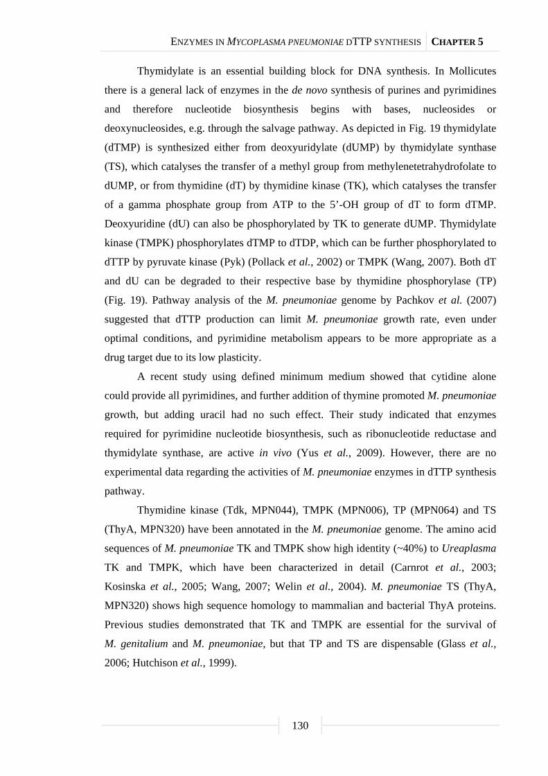
ENZYMES IN MYCOPLASMA PNEUMONIAE DTTP SYNTHESIS CHAPTER 5
130
Thymidylate is an essential building block for DNA synthesis. In Mollicutes
there is a general lack of enzymes in the de novo synthesis of purines and pyrimidines
and therefore nucleotide biosynthesis begins with bases, nucleosides or
deoxynucleosides, e.g. through the salvage pathway. As depicted in Fig. 19 thymidylate
(dTMP) is synthesized either from deoxyuridylate (dUMP) by thymidylate synthase
(TS), which catalyses the transfer of a methyl group from methylenetetrahydrofolate to
dUMP, or from thymidine (dT) by thymidine kinase (TK), which catalyses the transfer
of a gamma phosphate group from ATP to the 5’-OH group of dT to form dTMP.
Deoxyuridine (dU) can also be phosphorylated by TK to generate dUMP. Thymidylate
kinase (TMPK) phosphorylates dTMP to dTDP, which can be further phosphorylated to
dTTP by pyruvate kinase (Pyk) (Pollack et al., 2002) or TMPK (Wang, 2007). Both dT
and dU can be degraded to their respective base by thymidine phosphorylase (TP)
(Fig. 19). Pathway analysis of the M. pneumoniae genome by Pachkov et al. (2007)
suggested that dTTP production can limit M. pneumoniae growth rate, even under
optimal conditions, and pyrimidine metabolism appears to be more appropriate as a
drug target due to its low plasticity.
A recent study using defined minimum medium showed that cytidine alone
could provide all pyrimidines, and further addition of thymine promoted M. pneumoniae
growth, but adding uracil had no such effect. Their study indicated that enzymes
required for pyrimidine nucleotide biosynthesis, such as ribonucleotide reductase and
thymidylate synthase, are active in vivo (Yus et al., 2009). However, there are no
experimental data regarding the activities of M. pneumoniae enzymes in dTTP synthesis
pathway.
Thymidine kinase (Tdk, MPN044), TMPK (MPN006), TP (MPN064) and TS
(ThyA, MPN320) have been annotated in the M. pneumoniae genome. The amino acid
sequences of M. pneumoniae TK and TMPK show high identity (~40%) to Ureaplasma
TK and TMPK, which have been characterized in detail (Carnrot et al., 2003;
Kosinska et al., 2005; Wang, 2007; Welin et al., 2004). M. pneumoniae TS (ThyA,
MPN320) shows high sequence homology to mammalian and bacterial ThyA proteins.
Previous studies demonstrated that TK and TMPK are essential for the survival of
M. genitalium and M. pneumoniae, but that TP and TS are dispensable (Glass et al.,
2006; Hutchison et al., 1999).

ENZYMES IN MYCOPLASMA PNEUMONIAE DTTP SYNTHESIS CHAPTER 5
131
Fig. 19. Simplified dTTP synthesis pathway in mycoplasmas. In Mollicutes genomes nucleoside
diphosphate kinase (Ndk), an enzyme responsible for the phosphorylation of nucleoside diphosphates to
triphosphates has not been identified; however, it has been shown experimentally that glycolytic enzymes,
such as Pyk (Pollack et al., 2002), or TMPK (Wang, 2007) are able to phosphorylate dTDP to dTTP, and
they were suggested to replace Ndk function in Mollicutes. dT, thymidine; T, thymine; dTMP, thymidine
5’-monophosphate; dTDP, thymidine 5’-diphosphate; dTTP, thymidine 5’-triphosphate; dU,
deoxyuridine; U, uracil; dUMP, deoxyuridine 5’-monophosphate; TK, thymidine kinase; TMPK,
thymidylate kinase; TS, thymidylate synthase; TP, thymidine phosphorylase; Pyk, pyruvate kinase.
The transposon mutagenesis technique was used to interrupt the M. pneumoniae
TS gene (thyA/mpn320) to study the interactions of enzymes in the dTTP synthesis
pathway in relation to M. pneumoniae growth. We found that TK and TP activities are
closely regulated to M. pneumoniae growth but not TS and TMPK. Inactivation of the
thyA gene resulted in upregulation of tdk (TK) gene transcription and increased TK
activity and reduced TS and TMPK activity. Metabolic labelling with [3H]-dT shows a
high level of [3H]-dT uptake and incorporation in the thyA mutant. However, [3H]-dU
was primarily metabolized to ribonucleotides and incorporation into DNA was low.
These data suggest that dT salvage through TK and TMPK is rate-limiting in dTTP
synthesis and upregulation of TK activity compensated for the loss of ThyA activity in
the thyA mutant.
Results
Characterization of the thyA mutant. The thyA (mpn320) mutant
M. pneumoniae strain was generated by the global transposon mutagenesis procedure,
as previously described (Halbedel et al., 2006). Transposon insertion site into the thyA
locus was after nucleotide 653. Wild type and the mutant strain were cultured according
to standard protocol using modified Hayflick medium. Total proteins were extracted
and TS activity was determined using [32P]-dUMP as the substrate. TS activity was
detected in both strains (Fig. 20A).
T dT dTMP dTDP dTTPTP TK TMPK Pyk/TMPK
U dU dUMPTP TK
TS
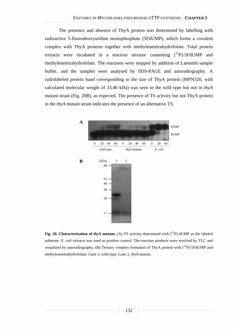
ENZYMES IN MYCOPLASMA PNEUMONIAE DTTP SYNTHESIS CHAPTER 5
132
The presence and absence of ThyA protein was determined by labelling with
radioactive 5-fluorodeoxyuridine monophosphate (5FdUMP), which forms a covalent
complex with ThyA proteins together with methylenetetrahydrofolate. Total protein
extracts were incubated in a reaction mixture containing [32P]-5FdUMP and
methylenetetrahydrofolate. The reactions were stopped by addition of Laemmli sample
buffer, and the samples were analysed by SDS-PAGE and autoradiography. A
radiolabeled protein band corresponding to the size of ThyA protein (MPN320, with
calculated molecular weight of 33.46 kDa) was seen in the wild type but not in thyA
mutant strain (Fig. 20B), as expected. The presence of TS activity but not ThyA protein
in the thyA mutant strain indicates the presence of an alternative TS.
Fig. 20. Characterization of thyA mutant. (A) TS activity determined with [32P]-dUMP as the labeled
substrate. E. coli extracts was used as positive control. The reaction products were resolved by TLC and
visualized by autoradiography. (B) Ternary complex formation of ThyA protein with [32P]-5FdUMP and
methylenetetrahydrofolate. Lane 1, wild type; Lane 2, thyA mutant.
17
28
38
62
[kDa]
A
B 21
49
98
0 20 40 60 0 20 40 60 0 20 60
dTMP
dUMP
wild type thyA mutant E. coli

ENZYMES IN MYCOPLASMA PNEUMONIAE DTTP SYNTHESIS CHAPTER 5
133
The levels of TK, TP, TS, and TMPK activity. The activities of enzymes in
dTTP synthesis were determined in protein extracts from both M. pneumoniae strains.
TK activity was elevated (> 10-fold) in the thyA mutant, compared with the wild type
(Table 8). TMPK activity was lower in the mutant strain as compared with wild type
cells. The difference in TP activity in the thyA mutant and wild type strain was not
statistically significant. There was also detectable TS activity in the thyA knockout
strain (Table 8). We have also observed great variation in TK and TP activity in cultures
that were harvested at different time points and/or had a different number of cells in the
starting culture (inoculum size) (data not shown).
Tab. 8. Specific activities of enzymes in dTTP synthesis (nmol/(min mg)).
Wild type thyA mutant P-valuea
TK 1.10 ± 0.07 13.20 ± 0.30 < 0.001
TMPK 0.14 ± 0.01 0.083 ± 0.003 0.002
TP 2.65 ± 0.20 3.27 ± 0.09 0.170
TS 1.14 ± 0.10 0.0012 ± 0.0004 < 0.001 a Statistical analysis was performed by Student’s t-test and P-value < 0.05 indicates significant difference.
Results are mean ± standard error from four to six measurements.
Activities of dTTP synthesis enzymes in relation to M. pneumoniae growth.
The above results warranted a further study of the levels of these enzymes at different
phases of M. pneumoniae cell growth. Wild type and thyA mutant cells were cultured
over an extended period (8 days) and harvested at 24 h intervals. M. pneumoniae growth
was monitored daily by color change of the culture medium measured by OD550 value,
pH value of the medium, and cell pellet wet weight at harvest. There was no observable
difference in growth between the two strains. Total proteins were extracted and enzyme
activities and proteins levels were determined.
Thymidine kinase activity and transcript levels. There was a sharp increase in
TK activity from day 2 to day 3 in wild type cells and activity peaked at day 4,
decreasing to basal levels thereafter (Fig. 21A). In thyA mutant cells, TK activity
increased at day 2 and reached the maximum level at day 3, remaining at this level until
decreasing at day 6 (Fig. 21A). Interestingly, TK activity at each time point was
4-5 times higher in thyA mutant cells compared with wild type.

ENZYMES IN MYCOPLASMA PNEUMONIAE DTTP SYNTHESIS CHAPTER 5
134
Thymidine kinase transcript levels, determined by slot blot analysis of total
RNA, showed a similar pattern as TK activity profiles in both wild type and thyA
mutant (Table 9 and Fig. 21B). In wild type cells there was a sharp increase in TK
transcript from day 2, peaked at day 3, and then decreased. In thyA mutant cells TK
transcript level increased from day 2, reached maximum at day 3, remained at relative
high level at day 4 and 5, and then decreased to basal level at day 6 (Table 9 and
Fig. 21B). At day 1 and day 2 TK transcripts in thyA mutant were at similar levels as
wild type and upregulation of TK transcript levels in thyA mutant was observed from
day 3 to day 6 with the exception that at day 4 the levels were similar in both strains
(Table 9).
By comparing TK transcript and activity profiles, we observed that in wild type
cells TK transcript levels peaked at day 3, but activity reached maximum at day 4 while
in thyA mutant cells both TK transcript and activity reached maximum levels at day 3
and remained at high levels from day 3 through day 5, at day 6 TK transcript decreased
to basal level, but TK activity still remained at highest level. These observations
indicated that there is a clear difference in transcriptional and translational regulation as
well as TK protein degradation in wild type and thyA mutant.
Thymidylate kinase activity. The TMPK activity increased approximately 25%
from day 1 to 3 in wild type cells and stayed at a similar level throughout the culture
period. In contrast, TMPK activity began decreasing in thyA mutant strains from day 3
resulting in about a 25% decrease from starting levels by the end of the culture period
(Fig. 21C), and thus the mutant strain had lower TMPK activity at the end of the culture
period compared with wild type cells (Fig. 21C). In general, TMPK activity was much
lower compared with TK activity in both strains, suggesting that the second and third
phosphorylation by TMPK is rate-limiting in dTTP synthesis.
The TMPK reaction products were analysed by the TLC technique and revealed
that the majority (~75%) of the products were dTTP (data not shown). These data
suggest that TMPK is capable of functioning as a nucleoside diphosphate kinase, as was
demonstrated earlier with the Ureaplasma TMPK (Wang, 2007).
Thymidine phosphorylase activity. In both strains, TP activity increased
during the exponential growth phase, reached a peak value at day 3, and then decreased
to the end of the culture period. In wild type and thyA mutant cells the levels of TP
activity were similar (Fig. 21D).
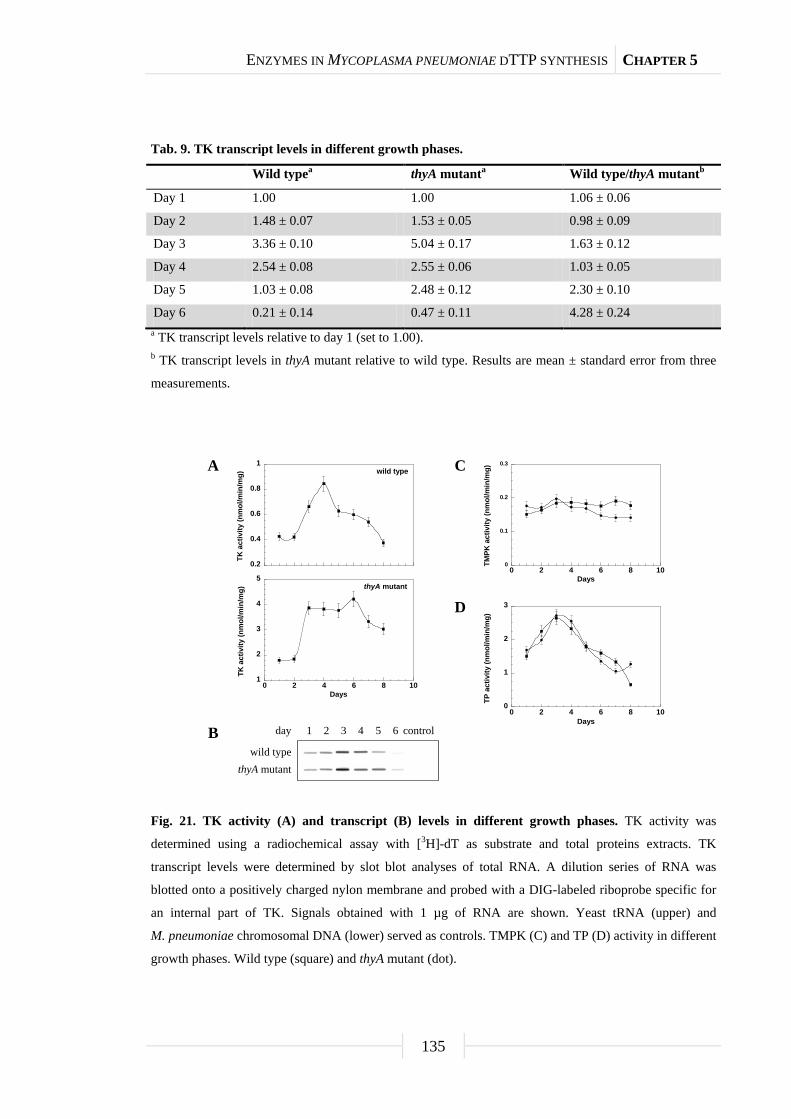
ENZYMES IN MYCOPLASMA PNEUMONIAE DTTP SYNTHESIS CHAPTER 5
135
Tab. 9. TK transcript levels in different growth phases.
Wild typea thyA mutanta Wild type/thyA mutantb
Day 1 1.00 1.00 1.06 ± 0.06
Day 2 1.48 ± 0.07 1.53 ± 0.05 0.98 ± 0.09
Day 3 3.36 ± 0.10 5.04 ± 0.17 1.63 ± 0.12
Day 4 2.54 ± 0.08 2.55 ± 0.06 1.03 ± 0.05
Day 5 1.03 ± 0.08 2.48 ± 0.12 2.30 ± 0.10
Day 6 0.21 ± 0.14 0.47 ± 0.11 4.28 ± 0.24 a TK transcript levels relative to day 1 (set to 1.00). b TK transcript levels in thyA mutant relative to wild type. Results are mean ± standard error from three
measurements.
Fig. 21. TK activity (A) and transcript (B) levels in different growth phases. TK activity was
determined using a radiochemical assay with [3H]-dT as substrate and total proteins extracts. TK
transcript levels were determined by slot blot analyses of total RNA. A dilution series of RNA was
blotted onto a positively charged nylon membrane and probed with a DIG-labeled riboprobe specific for
an internal part of TK. Signals obtained with 1 µg of RNA are shown. Yeast tRNA (upper) and
M. pneumoniae chromosomal DNA (lower) served as controls. TMPK (C) and TP (D) activity in different
growth phases. Wild type (square) and thyA mutant (dot).
0.2
0.4
0.6
0.8
1
TK a
ctiv
ity (n
mol
/min
/mg) wild type
1
2
3
4
5
0 2 4 6 8 10
TK a
ctiv
ity (n
mol
/min
/mg)
Days
thyA mutant
0
1
2
3
0 2 4 6 8 10
TP a
ctiv
ity (n
mol
/min
/mg)
Days
0
0.1
0.2
0.3
0 2 4 6 8 10
TMPK
act
ivity
(nm
ol/m
in/m
g)
Days
day 1 2 3 4 5 6
wild typethyA mutant
control
A
B
C
D
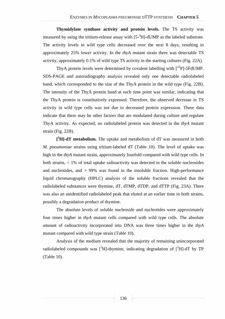
ENZYMES IN MYCOPLASMA PNEUMONIAE DTTP SYNTHESIS CHAPTER 5
136
Thymidylate synthase activity and protein levels. The TS activity was
measured by using the tritium-release assay with [5-3H]-dUMP as the labeled substrate.
The activity levels in wild type cells decreased over the next 8 days, resulting in
approximately 25% lower activity. In the thyA mutant strain there was detectable TS
activity, approximately 0.1% of wild type TS activity in the starting cultures (Fig. 22A).
ThyA protein levels were determined by covalent labelling with [32P]-5FdUMP.
SDS-PAGE and autoradiography analysis revealed only one detectable radiolabeled
band, which corresponded to the size of the ThyA protein in the wild type (Fig. 22B).
The intensity of the ThyA protein band at each time point was similar, indicating that
the ThyA protein is constitutively expressed. Therefore, the observed decrease in TS
activity in wild type cells was not due to decreased protein expression. These data
indicate that there may be other factors that are modulated during culture and regulate
ThyA activity. As expected, no radiolabeled protein was detected in the thyA mutant
strain (Fig. 22B).
[3H]-dT metabolism. The uptake and metabolism of dT was measured in both
M. pneumoniae strains using tritium-labeled dT (Table 10). The level of uptake was
high in the thyA mutant strain, approximately fourfold compared with wild type cells. In
both strains, < 1% of total uptake radioactivity was detected in the soluble nucleosides
and nucleotides, and > 99% was found in the insoluble fraction. High-performance
liquid chromatography (HPLC) analysis of the soluble fractions revealed that the
radiolabeled substances were thymine, dT, dTMP, dTDP, and dTTP (Fig. 23A). There
was also an unidentified radiolabeled peak that eluted at an earlier time in both strains,
possibly a degradation product of thymine.
The absolute levels of soluble nucleoside and nucleotides were approximately
four times higher in thyA mutant cells compared with wild type cells. The absolute
amount of radioactivity incorporated into DNA was three times higher in the thyA
mutant compared with wild type strain (Table 10).
Analysis of the medium revealed that the majority of remaining unincorporated
radiolabeled compounds was [3H]-thymine, indicating degradation of [3H]-dT by TP
(Table 10).
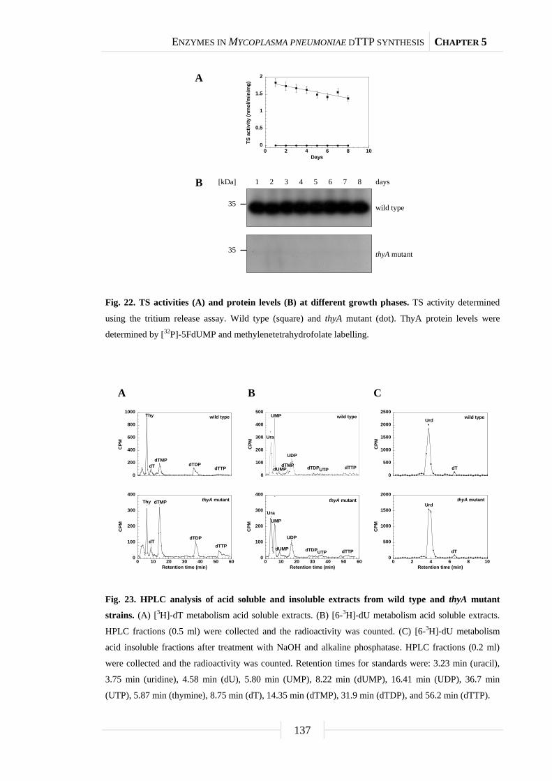
ENZYMES IN MYCOPLASMA PNEUMONIAE DTTP SYNTHESIS CHAPTER 5
137
Fig. 22. TS activities (A) and protein levels (B) at different growth phases. TS activity determined
using the tritium release assay. Wild type (square) and thyA mutant (dot). ThyA protein levels were
determined by [32P]-5FdUMP and methylenetetrahydrofolate labelling.
Fig. 23. HPLC analysis of acid soluble and insoluble extracts from wild type and thyA mutant
strains. (A) [3H]-dT metabolism acid soluble extracts. (B) [6-3H]-dU metabolism acid soluble extracts.
HPLC fractions (0.5 ml) were collected and the radioactivity was counted. (C) [6-3H]-dU metabolism
acid insoluble fractions after treatment with NaOH and alkaline phosphatase. HPLC fractions (0.2 ml)
were collected and the radioactivity was counted. Retention times for standards were: 3.23 min (uracil),
3.75 min (uridine), 4.58 min (dU), 5.80 min (UMP), 8.22 min (dUMP), 16.41 min (UDP), 36.7 min
(UTP), 5.87 min (thymine), 8.75 min (dT), 14.35 min (dTMP), 31.9 min (dTDP), and 56.2 min (dTTP).
0
0.5
1
1.5
2
0 2 4 6 8 10
TS a
ctiv
ity (n
mol
/min
/mg)
Days
[kDa] 1 2 3 4 5 6
wild type
thyA mutant
days
A
B 7 8
35
35
0
500
1000
1500
2000
0 2 4 6 8 10
CPM
Retention time (min)
thyA mutantUrd
dT
0
500
1000
1500
2000
2500
CPM
wild typeUrd
dT
0
100
200
300
400
0 10 20 30 40 50 60
CPM
Retention time (min)
UraUMP
dUMP
UDP
UTP dTTPdTDP
thyA mutant
0
100
200
300
400
0 10 20 30 40 50 60
CPM
Retention time (min)
thyA mutant
dTTPdTDP
dTMP
dT
Thy
0
100
200
300
400
500
CPM
UDP
dTMPdUMP
UMP
Ura
UTP dTTPdTDP
wild type
0
200
400
600
800
1000
CPM
wild typeThy
dTdTMP
dTDPdTTP
A CB

ENZYMES IN MYCOPLASMA PNEUMONIAE DTTP SYNTHESIS CHAPTER 5
138
Tab. 10. [3H]-dT metabolism in wild type and thyA mutant M. pneumoniae strainsa.
Wild type thyA mutant
Total uptake (%) 19 ± 5 82 ± 2
PCA-soluble extracts (total counts) 16,830 ± 1,600 60,264 ± 6,500
% of total uptake 0.6 ± 0.3 0.7 ± 0.2
NaOH-soluble fraction (total counts) 1,455,915 ± 15,000 4,768,860 ± 45,000
% of total uptake 99.4 ± 0.3 99.3 ± 0.2
Medium analysis after removal of cells
[3H]-dT (%) 31 ± 13 15 ± 5
[3H]-Thymine (%) 69 ± 12 85 ± 5 a Data were from four independent incubations and represented as the mean ± standard error. Total uptake
was calculated as radioactivity recovered in total extracts (soluble and insoluble) as compared with total
radioactivity in the medium before incubation start.
[6-3H]-dU metabolism. A similar approach was performed using [6-3H]-dU to
determine the role of TS in dTTP synthesis in wild type and thyA mutant strains
(Table 11). The level of uptake was low in the thyA mutant strain at approximately
0.57%, while the level of uptake in wild type cells was at approximately 2.6%. In wild
type strain, approximately 5% of total uptake was found in the soluble nucleosides and
nucleotides, and approximately 95% was found in the insoluble fraction. In the thyA
mutant strain, 10% of total uptake was incorporated into soluble nucleoside and
nucleotides and 90% of the total radioactivity was recovered in the insoluble fraction.
HPLC analysis of soluble nucleotides revealed that the radiolabeled compounds were
predominantly uracil, UMP, and UDP, indicating that dU was degraded to uracil and
further metabolized to ribonucleotides. Tritium-labeled dUMP and dTMP were detected
at low levels in the wild type strain, but there was no detectable tritium-labeled dTMP
in the thyA mutant strain (Fig. 23B). There was no [3H]-dU detected in the soluble
fractions. Trace amounts of radiolabeled dTDP and dTTP were also detected (Fig. 23B).
The absolute levels of soluble nucleotides were low in thyA mutant compared
with wild type cells (Table 10). The absolute amount of radioactivity in the insoluble
fraction was six times lower in the thyA mutant than in wild type cells (Table 11).
The PCA-insoluble fractions were solubilized with NaOH, neutralized and
treated with alkaline phosphatase before HPLC analysis. Two radioactive peaks were
detected in both strains, one with a retention time of 3.95 min (Urd) and the other with a
retention time of 6.35 min (dT), indicating that there was thymidylate incorporated into

ENZYMES IN MYCOPLASMA PNEUMONIAE DTTP SYNTHESIS CHAPTER 5
139
DNA even in the thyA knockout strain (Fig. 23C). The amount of [3H]-dT recovered
from HPLC analysis of PCA-insoluble fractions in both wild type and the thyA mutant
were only ~1% of the total radioactivity in these fractions, which indicated that 99% of
the radioactivity originated from [6-3H]-dU was incorporated into RNA as uridine
ribonucleotide.
Analysis of the medium showed that the remaining radiolabeled compounds
were [3H]-uracil and [3H]-dU, indicating that [3H]-dU had been degraded (Table 11).
Tab. 11. [3H]-dU metabolism in wild type and thyA mutant M. pneumoniae strainsa.
Wild type thyA mutant
Total uptake (%) 2.6 ± 0.1 0.57 ± 0.10
PCA-soluble extracts (total counts) 15,510 ± 987 6,435 ± 650
% of total uptake 4.3 ± 0.5 10.3 ± 0.2
NaOH-soluble fraction (total counts) 276,750 ± 2540 46,230 ± 2,347
% of total uptake 95.7 ± 0.7 90.3 ± 0.4
Medium analysis after removal of cells
[3H]-dU (%) 34 ± 6 48 ± 5
[3H]-Uracil (%) 66 ± 7 52 ± 5 a Data were from three independent experiments and given as mean ± standard error. Total uptake was
calculated as radioactivity recovered in total extracts (soluble and insoluble) as compared with total
radioactivity in the medium before incubation start.
Discussion
In mycoplasmas there are only a few enzymes involved in the biosynthesis of
dTTP (Fig. 19), an essential precursor for DNA synthesis, as compared with their hosts.
The corresponding mammalian enzymes, including TK, TMPK, TP, and TS, have been
well studied, and their activity levels have been shown to be tightly regulated
throughout the cell cycle, with the highest levels in S-phase (Gasparri et al., 2009;
Ke and Chang, 2004). However, little is known regarding cell cycle regulation of
bacterial enzymes. Here we show that M. pneumoniae TK, at both activity and transcript
levels, is regulated during the growth phase, with a clear induction in the exponential
growth phase and a drastic decline during the stationary phase. The M. pneumoniae TK
activity profile in wild type cells correlates with the rate of DNA synthesis (Yus et al.,
2009). Compared with wild type both TK activity and transcript levels were upregulated
in thyA mutant, and the differences seen in TK expression profiles in wild type and thyA

ENZYMES IN MYCOPLASMA PNEUMONIAE DTTP SYNTHESIS CHAPTER 5
140
mutant indicate that there is a cross-talk in transcriptional and translational regulation of
tdk (TK) and thyA genes. Inactivation of thyA resulted in the upregulation of tdk gene
transcription, which leads to induction of TK protein and increased TK activity, as a
mechanism to compensate the loss of thyA. The upregulation and prolonged period of
high TK activity in the thyA mutant strain indicates the importance of TK activity in
dTTP synthesis and DNA replication in compensating for the lack of ThyA activity. In
contrast to mammalian enzymes, M. pneumoniae TMPK activity varies only slightly
during the culture period in both wild type and the thyA mutant strain. In wild type
ThyA protein remained at similar at levels during culture; however, TS activity
decreased over time. In the thyA mutant a very low level of TS activity was detected,
indicating the presence of an alternative TS enzyme. Similar to TK, TP activity was
induced at the exponential growth phase and decreased at the stationary phase. The level
of TP activity was similar in wild type and thyA mutant cells. Collectively, these results
suggest that ThyA and TMPK are constitutively expressed and inactivation of the thyA
gene results in the induction of TK.
Increased salvage of dT in thyA mutant compensates for the lack of ThyA
activity. Metabolic labelling studies with dT and dU showed that both nucleosides or
bases can be taken up, metabolized, and incorporated into DNA, confirming the activity
of additional enzymes in the dTTP synthesis pathway. Furthermore, there was higher
level of dT than dU uptake in wild type cells, indicating M. pneumoniae salvages dT
more efficiently than dU. Radiolabeled dT was converted to the corresponding
nucleotides and efficiently incorporated into DNA. However, [3H]-dU was recovered
mainly as ribonucleotides and the amount of radioactivity in the form of dUMP and
dTMP was < 5% of the total soluble fraction and total incorporation into DNA was only
about 1%. These data indicate that the efficiency of ribonucleotide reduction and
conversion of dUMP to dTMP was low.
The level of dT uptake and incorporation into DNA in the thyA mutant correlates
with increases in TK activity, suggesting that dT uptake and metabolism is
TK-dependent and upregulation of TK activity in thyA mutant cells is apparently
compensating for the lack of ThyA activity. Our results also indicate that the efficiency
of dTTP synthesis via UDP reduction was low compared with the direct salvage of dT,
explaining why thyA is not essential.

ENZYMES IN MYCOPLASMA PNEUMONIAE DTTP SYNTHESIS CHAPTER 5
141
Metabolism of dU and thymidylate synthases. The uptake of [3H]-dU was low
compared with dT uptake in wild type cells, and uptake and incorporation of [3H]-dU
into macromolecules were greatly reduced in the thyA knockout strain. HPLC analysis
revealed that the radiolabeled compounds were mainly ribonucleotides and no
detectable [3H]-dU was present in the soluble fractions, suggesting that [3H]-dU has
been degraded to uracil and subsequently taken up and converted to ribonucleotides via
uracil phosphoribosyltransferase (MPN033). The low level of uracil uptake in the thyA
mutant indicates that inactivation of the thyA gene may affect uracil uptake and/or uracil
phosphoribosyltransferase activity. This is the first experimental proof that connects the
pentose phosphate pathway to nucleotide metabolism in M. pneumoniae. These results
also explain why dU uptake is not TK-dependent.
The TS activity in the thyA mutant was only approximately 0.1% of activity in
wild type cells. Incorporation of radioactive thymidylate into DNA originated from
[3H]-dU in thyA mutant cells, indicating the presence of an alternative TS in
M. pneumoniae. Nonetheless, our study indicates that dTTP synthesis via ribonucleotide
reduction and methylation of dUMP by ThyA is not efficient in M. pneumoniae.
Thymidine/thymine, but not uracil, promotes M. pneumoniae growth.
Yus et al. (2009) showed that purine and pyrimidine composition in the minimum
defined medium can be replaced by ribonucleosides, but not deoxyribonucleosides, and
that thymidine can be metabolized to some extent, but not thymine. Furthermore,
addition of thymine increases M. pneumoniae growth when added with cytidine. In our
metabolic study, we did not observe any difference in growth rate since we used
enriched medium (modified Hayflick medium). When the medium was analysed at the
end of the culture period most of the radiolabeled dT and dU were degraded; therefore,
both thymine and uracil were present. We showed that dT, thymine, and uracil, but not
dU, were taken up and metabolized. These data suggest that there may be specific
transporters for bases and ribonucleosides and M. pneumoniae may lack such
transporters for deoxynucleosides. Our study also indicates that the expression of TP
may be influenced by nutrition status, since M. pneumoniae apparently was not able to
degrade dT or dU and/or salvage thymine in the minimum defined medium (Yus et al.,
2009). The uptake of uracil was less efficient as compared with dT/thymine and the
level of dTTP synthesized through UDP reduction was very low. However, thymine/dT
can be efficiently salvaged to thymidine nucleotides (dTTP) through the combined

ENZYMES IN MYCOPLASMA PNEUMONIAE DTTP SYNTHESIS CHAPTER 5
142
action of TP, TK, and TMPK, and increased dTTP synthesis promotes M. pneumoniae
growth. Our results suggest that the slow division rate of M. pneumoniae is most likely
due to inefficient dTTP synthesis.
Inhibition of M. pneumoniae dTTP synthesis as a therapeutic target.
Mycoplasmas cause a wide variety of diseases and have also been associated with
cancer (Narita, 2009; Narita, 2010; Waites and Talkington, 2004). Persistent
Mycoplasma infection induced malignant transformation of human cells, as shown in a
recent study (Namiki et al., 2009). Mycoplasma infections can reduce the effect of
anticancer nucleoside analogues due to degradation by Mycoplasma enzymes
(Liekens et al., 2009). Therefore, it may be beneficial to treat Mycoplasma infections in
cancer patients who receive nucleoside analogues as chemotherapeutic agents. Current
antibiotics used in treating Mycoplasma infections are effective but clinical antibiotic
resistant Mycoplasma strains are emerging (Morozumi et al., 2010). In addition,
mycoplasmas are slow growing and therefore long-term treatment is required, which
may lead to the emergence of antibiotic resistance in other bacteria species. Therefore,
new targets for developing specific antibiotics against Mycoplasma are needed.
DNA synthesis requires dTTP, which is an essential building block, thus,
blocking dTTP synthesis would lead to thymine deficiency-induced cell death.
Inhibition of pyrimidine uptake and/or conversion to thymidine nucleotides would
impair Mycoplasma dTTP synthesis and lead to cell death. Therefore, TK and TMPK
are potential targets for future development of antibiotics against Mycoplasma
infections.
Experimental procedures
Materials. Radiolabeled compounds [methyl-3H]-thymidine (dT, 20 Ci/mmol)
and [γ-32P]-ATP (3,000 Ci/mmol) were purchased from PerkinElmer. [5-3H]-dUMP
(deoxyuridine 5’-monophosphate, 47 Ci/mmol) and [6-3H]-deoxyuridine (dU,
45 Ci/mmol) were bought from Moravek Biochemicals. [32P]-5FdUMP, [32P]-dUMP,
and [3H]-dTMP were synthesized enzymatically using Ureaplasma thymidine kinase
with [32P]-ATP or [3H]-dT as the labeled substrate and purified (Carnrot et al., 2003).
Rabbit anti-MPN358 antibodies were produced by using synthetic peptides chosen from
MPN358 sequence as antigens (GenScript Corp.) and antisera were used directly in
Western blot analysis without further purification.

ENZYMES IN MYCOPLASMA PNEUMONIAE DTTP SYNTHESIS CHAPTER 5
143
Mycoplasma culture and protein extraction. M. pneumoniae wild type and the
thyA mutant strains were cultured in modified Hayflick medium, as described
previously (Halbedel et al., 2004).
Time-course study. Culture was started (day 0) by the addition of 1 ml stock
(OD550, ~0.7) to a 25-cm2 flask containing 25 ml fresh medium and incubated at 37°C
with 5% CO2. Culture optical density was monitored at 550 nm daily and cells
harvested at 24 h intervals by centrifugation at 7,000 g for 30 min. Pellets were washed
three times with phosphate-buffered saline (PBS) and then resuspended in protein
extraction buffer. Total proteins were extracted as previously described (Wang et al.,
2001). Protein concentration was determined by Bio-Rad protein assay using BSA as a
standard, according to the manufacturer’s protocol.
Metabolic labelling with [methyl-3H]-dT and [6-3H]-dU. A stationary phase
culture was resuspended in 6 ml fresh medium in a 25-cm2 flask and used as stock. One
milliliter stock culture was added to a 25-cm2 flask containing 25 ml fresh medium with
1 µCi/ml [methyl-3H]-dT or [6-3H]-dU (1 µM dT or dU) and incubated at 37°C. After
5 days the cultures were harvested by centrifugation, washed three times with PBS, and
divided into three aliquots and saved at -20°C for further analysis. Soluble nucleotides
were extracted with 100 µl of 10% PCA (perchloric acid), followed by centrifuge at
16,000 g for 15 min. The supernatant was transferred to a new tube and neutralized with
KOH and the insoluble salt (KClO4) was removed by centrifugation. The supernatant
containing nucleotides and nucleosides was analysed directly by TLC and/or HPLC to
determine the amount of radiolabeled nucleoside tri-, di-, and monophosphates. To
extract nucleotides from the insoluble fraction, pellets were washed twice with
10% PCA, followed by addition of 100 µl 1 M NaOH and incubation at 60°C for 2 h
with shaking, and incorporation of radioactivity was then counted using a liquid
scintillation counter (Beckman-Coulter). The NaOH-soluble fraction was treated with
alkaline phosphatase after neutralization and analysed by HPLC and TLC.
Non-radioactive nucleosides and/or nucleotides were used as internal standards in
HPLC analysis. Culture medium before and after incubation was counted for
radioactivity and the radioactive components were analysed by TLC and/or HPLC.
Total uptake was calculated as radioactivity recovered in total extracts (soluble and
insoluble) as compared with total radioactivity in the medium before incubation start.

ENZYMES IN MYCOPLASMA PNEUMONIAE DTTP SYNTHESIS CHAPTER 5
144
The HPLC analysis was performed using reverse phase column (Source 5RPC,
GE Healthcare) by isocratic elution in buffer containing 50 mM ammonium phosphate,
pH 6.5, 5 mM tetrabutylammonium hydrogen sulphate and 12% methanol at a flow rate
of 1 ml/min. Soluble nucleotide extracts were mixed with HPLC buffer (1:4) before
injection. Fractions (0.5 ml or 0.2 ml) were collected and mixed with 2 ml scintillation
fluid and counted. The radioactive peaks were compared with non-radioactive
standards. Retention times for standards were 3.23 min (uracil), 3.75 min (uridine),
4.58 min (dU), 5.80 min (UMP), 8.22 min (dUMP), 16.41 min (UDP), 36.7 min (UTP),
5.87 min (thymine), 8.75 min (dT), 14.35 min (dTMP), 31.9 min (dTDP), and 56.2 min
(dTTP). TLC analysis was performed with PEI-cellulose and developed in
0.1 M NaH2PO4 for separation of thymidine nucleotides and
isobutyric acid/H2O/ammonia (66:33:1) for separation of Urd, dU from other
nucleosides and nucleotides as previously described (Wang, 2007).
Labelling of ThyA protein with [32P]-5FdUMP. M. pneumoniae protein
extracts (30 mg) were added to a reaction mixture containing 25 mM MgCl2,
0.2 mM methylenetetrahydrofolate, 0.2 mM CMP, 20 mM DTT, and
5 µM [32P]-5FdUMP in a total volume of 25 ml. The mixtures were incubated at 37°C
for 2 h. The reaction was stopped by addition of Laemmli sample buffer (5 µl) and 15 µl
(~15 µg protein) was resolved by 12% SDS-PAGE. After electrophoresis, gels were
fixed in methanol/acetic acid and the radiolabeled protein bands were visualized by
autoradiography.
Enzyme assays. The TK activity was assayed by a radiochemical method using
[3H]-dT as a substrate, essentially as described (Wang et al., 2001). TMPK activity was
determined by using [3H]-dTMP as a substrate in a reaction mixture containing
50 mM Tris-HCl pH 8.0, 0.5 mg/ml BSA, 10 mM DTT, 5 mM MgCl2, 2 mM ATP, and
10 µM [3H]-dTMP. The reaction was started by addition of M. pneumoniae protein
extracts and incubated at 37°C. At 0, 10, 20, and 30 min intervals a 10 µl aliquot was
removed and spotted onto DE-81 filter paper and allowed to dry. The unutilized
substrate was removed by washing the filter papers three times in 50 mM ammonium
formate solution. Finally, the products were eluted with 0.5 ml of 0.1 M HCl and
0.2 M KCl, and mixed with 2 ml scintillation fluid and the radioactivity counted. The
specific activity was calculated as nmol/(min mg) protein.
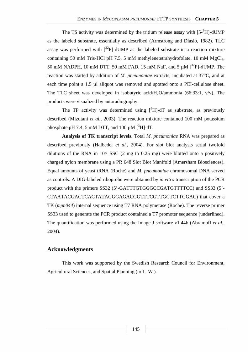
ENZYMES IN MYCOPLASMA PNEUMONIAE DTTP SYNTHESIS CHAPTER 5
145
The TS activity was determined by the tritium release assay with [5-3H]-dUMP
as the labeled substrate, essentially as described (Armstrong and Diasio, 1982). TLC
assay was performed with [32P]-dUMP as the labeled substrate in a reaction mixture
containing 50 mM Tris-HCl pH 7.5, 5 mM methylenetetrahydrofolate, 10 mM MgCl2,
50 mM NADPH, 10 mM DTT, 50 mM FAD, 15 mM NaF, and 5 µM [32P]-dUMP. The
reaction was started by addition of M. pneumoniae extracts, incubated at 37°C, and at
each time point a 1.5 µl aliquot was removed and spotted onto a PEI-cellulose sheet.
The TLC sheet was developed in isobutyric acid/H2O/ammonia (66:33:1, v/v). The
products were visualized by autoradiography.
The TP activity was determined using [3H]-dT as substrate, as previously
described (Mizutani et al., 2003). The reaction mixture contained 100 mM potassium
phosphate pH 7.4, 5 mM DTT, and 100 µM [3H]-dT.
Analysis of TK transcript levels. Total M. pneumoniae RNA was prepared as
described previously (Halbedel et al., 2004). For slot blot analysis serial twofold
dilutions of the RNA in 10× SSC (2 mg to 0.25 mg) were blotted onto a positively
charged nylon membrane using a PR 648 Slot Blot Manifold (Amersham Biosciences).
Equal amounts of yeast tRNA (Roche) and M. pneumoniae chromosomal DNA served
as controls. A DIG-labeled riboprobe were obtained by in vitro transcription of the PCR
product with the primers SS32 (5’-GATTTGTGGGCCGATGTTTTCC) and SS33 (5’-
CTAATACGACTCACTATAGGGAGACGGTTTCGTTGCTCTTGGAC) that cover a
TK (mpn044) internal sequence using T7 RNA polymerase (Roche). The reverse primer
SS33 used to generate the PCR product contained a T7 promoter sequence (underlined).
The quantification was performed using the Image J software v1.44b (Abramoff et al.,
2004).
Acknowledgments
This work was supported by the Swedish Research Council for Environment,
Agricultural Sciences, and Spatial Planning (to L. W.).

Chapter 6
Interactions between glycolytic enzymes of
Mycoplasma pneumoniae
The work described in this chapter was published in:
Dutow, P., S. R. Schmidl, M. Ridderbusch, and J. Stülke. 2010. Interactions between
glycolytic enzymes of Mycoplasma pneumoniae. J. Mol. Microbiol. Biotechnol.
accepted.
Author contributions:
This study was designed by SRS and JS. MR constructed the mutated glycolytic
enzyme alleles and PD performed the bacterial two-hybrid analysis. SRS designed the
oligonucleotides and supervised the experimental work that was accomplished by MR
and PD during their diploma theses. All authors were involved in the interpretation of
the collected data. SRS and JS wrote the paper.

INTERACTIONS OF GLYCOLYTIC ENZYMES IN MYCOPLASMA PNEUMONIAE CHAPTER 6
147
Abstract
With only 688 protein-coding genes, Mycoplasma pneumoniae is one of the
smallest self-replicating organisms. These bacteria use glycolysis as the major pathway
for ATP production by substrate-level phosphorylation suggesting that this pathway
must be optimized to high efficiency. In this study, we have investigated the interactions
between glycolytic enzymes using the bacterial adenylate cyclase-based two-hybrid
system. We demonstrate that most of the glycolytic enzymes perform self-interactions
suggesting that they form dimers or other oligomeric forms. In addition, enolase was
identified as the central glycolytic enzyme of M. pneumoniae due to its ability to
directly interact with each other glycolytic enzyme. Our results support the idea of the
formation of a glycolytic complex in M. pneumoniae and we suggest that the formation
of this complex might ensure higher fluxes through the glycolytic pathway than would
be possible with isolated non-interacting enzymes.
Introduction
Mycoplasma pneumoniae, a cell wall-less pathogenic bacterium, is the
best-studied representative of the class Mollicutes. These bacteria are derived from
Gram-positive ancestors that are related to Bacillus subtilis and lactic acid bacteria. The
evolution of the Mollicutes is characterized by substantial genome degeneration that
reflects adaptation of the bacteria to a life in close association with eukaryotic hosts
(Razin et al., 1998; Stülke et al., 2009).
M. pneumoniae causes usually mild diseases such as pneumonias: However,
implication of this bacterium in arthritic, cutaneous or auto-immune diseases as well as
in encephalitis was reported (Atkinson et al., 2008; Domenech et al., 2009; Jacobs,
1997; Schalock and Dinulos, 2009; Waites and Talkington, 2004). Although
M. pneumoniae is highly adapted to a life on mucosal surfaces, the bacteria are able to
live independently of host cells. With less than 700 genes, M. pneumoniae belongs to
the smallest organisms that are capable of autonomous life (Himmelreich et al., 1996).
The small genome of M. pneumoniae is also reflected by its limited metabolic
capabilities. The bacteria can utilize glucose, fructose or glycerol as carbon sources
(Halbedel et al., 2004; Yus et al., 2009). These carbohydrates are metabolized via the
glycolytic pathway, leading to the concomitant generation of ATP by substrate-level

INTERACTIONS OF GLYCOLYTIC ENZYMES IN MYCOPLASMA PNEUMONIAE CHAPTER 6
148
phosphorylation. However, M. pneumoniae does not possess an active citric acid cycle;
similarly the pentose phosphate shunt is incomplete. While M. pneumoniae can produce
ATP, nearly all anabolic pathways have been lost during degenerative evolution. Thus,
M. pneumoniae depends heavily on the host cells or on organic nutrient supply from the
artificial medium (Halbedel et al., 2007; Miles, 1992).
We are interested in the glycolytic pathway. M. pneumoniae encodes all
glycolytic enzymes. Interestingly, some of these enzymes have multiple functions: The
glycolytic kinases are also active in nucleotide phosphorylation thus replacing the
nucleoside diphosphate kinase (Pollack et al., 2002). Moreover, several glycolytic
enzymes are subject to phosphorylation. However, it is not known how these
phosphorylation events are catalyzed and how they affect the enzymatic activities
(Schmidl et al., 2010). For the regeneration of NAD+, M. pneumoniae possesses a
lactate dehydrogenase; alternatively the NAD+ oxidase may directly oxidize NADH,
and the latter reaction is coupled to the production of hydrogen peroxide, the major
virulence factor of M. pneumoniae (Cole et al., 1968; Halbedel et al., 2007;
Low and Zimkus, 1973).
For a long time it was thought that metabolic pathways take place in an
unorganized chaotic way in bacterial cells. However, at the same time there have been
data indicating that, e.g. tryptophan metabolism in Escherichia coli involves protein
complexes (Yanofsky and Rachmeler, 1958). Similarly, glycolytic protein complexes
were reported for E. coli as well as for eukaryotic cells (Mowbray and Moses, 1976;
Campanella et al., 2005). The accumulating recent evidence suggests that enzymes of
one pathway may be clustered to allow efficient metabolism. This was shown for purine
biosynthesis and branched-chain amino acid catabolism in human cells as well as for
glycolysis in B. subtilis (An et al., 2008; Commichau et al., 2009; Islam et al., 2007).
Moreover, a recent in silico study supports the idea that glycolytic flux is much more
efficient if the enzymes form a complex as compared to free floating enzymes
(Amar et al., 2008). The analysis of protein complexes in M. pneumoniae suggests that
glycolytic enzymes might also interact in this organism (Kühner et al., 2009).
The analysis of primary protein-protein interactions in M. pneumoniae and other
Mollicutes is hampered by an alteration of the genetic code in these organisms: They
use the UGA codon for the incorporation of tryptophan, whereas it specifies a stop in
most other organisms. Due to this problem, the interactions between Mycoplasma

INTERACTIONS OF GLYCOLYTIC ENZYMES IN MYCOPLASMA PNEUMONIAE CHAPTER 6
149
proteins have so far never been studied using a two-hybrid system. We have developed
a method for the quick replacement of stop codons (Hames et al., 2005). Based on this
method, we have studied the interactions between all glycolytic enzymes of M.
pneumoniae using a bacterial two-hybrid (B2H) system. Our results support the idea
that these enzymes interact, and that metabolism is well structured even in a seemingly
primitive organism.
Results
Organization of glycolytic genes in M. pneumoniae. In B. subtilis and other
Firmicutes, genes encoding glycolytic enzymes from phosphotransferase system (PTS)
proteins for glucose transport and phosphorylation to the pyruvate kinase are clustered
in five operons (Ludwig et al., 2001). In contrast, the corresponding M. pneumoniae
genes are scattered on the chromosome (Fig. 24). This is even the case for the PTS
genes in M. pneumoniae that are clustered in most bacteria with a functional PTS sugar
transport system (Barabote and Saier, 2005). In M. pneumoniae, three gene clusters with
glycolytic genes are present: The pfkA-pyk cluster encodes the two glycolytic kinases.
These genes also form an operon in B. subtilis. The second cluster is made up of the pgk
and gapA genes encoding phosphoglycerate kinase and glyceraldehyde-3-phosphate
dehydrogenase, respectively. These genes are clustered in most bacteria, and it has been
suggested that the clustering of these genes might reflect physical interaction of the
encoded enzymes, which catalyze consecutive reactions (Dandekar et al., 1998). The
third glycolytic cluster is formed by the ptsI, pgm, and tpiA genes encoding enzyme I of
the PTS, phosphoglycerate mutase, and triose-phosphate isomerase, respectively. The
pgm and tpiA genes are also part of one operon in B. subtilis (Ludwig et al., 2001).
Interactions between glycolytic enzymes. In order to identify possible primary
interactions between glycolytic enzymes, we made use of the B2H system. In this
system, the T25 and the T18 fragments of the catalytic domain of the Bordetella
pertussis adenylate cyclase were fused to full-length copies of all glycolytic enzymes of
M. pneumoniae. The leucine zipper of the yeast GCN4 transcription factor served as a
control (Karimova et al., 1998). The results of the B2H analysis are shown in Fig. 25.
As expected, the leucine zipper of GCN4 showed strong self-interaction.
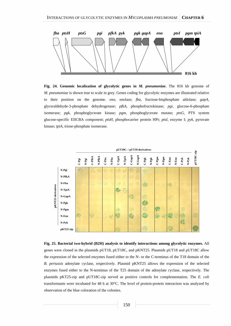
INTERACTIONS OF GLYCOLYTIC ENZYMES IN MYCOPLASMA PNEUMONIAE CHAPTER 6
150
Fig. 24. Genomic localization of glycolytic genes in M. pneumoniae. The 816 kb genome of
M. pneumoniae is shown true to scale in grey. Genes coding for glycolytic enzymes are illustrated relative
to their position on the genome. eno, enolase; fba, fructose-bisphosphate aldolase; gapA,
glyceraldehyde-3-phosphate dehydrogenase; pfkA, phosphofructokinase; pgi, glucose-6-phosphate
isomerase; pgk, phosphoglycerate kinase; pgm, phosphoglycerate mutase; ptsG, PTS system
glucose-specific EIICBA component; ptsH, phosphocarrier protein HPr; ptsI, enzyme I; pyk, pyruvate
kinase; tpiA, triose-phosphate isomerase.
Fig. 25. Bacterial two-hybrid (B2H) analysis to identify interactions among glycolytic enzymes. All
genes were cloned in the plasmids pUT18, pUT18C, and pKNT25. Plasmids pUT18 and pUT18C allow
the expression of the selected enzymes fused either to the N- or the C-terminus of the T18 domain of the
B. pertussis adenylate cyclase, respectively. Plasmid pKNT25 allows the expression of the selected
enzymes fused either to the N-terminus of the T25 domain of the adenylate cyclase, respectively. The
plasmids pKT25-zip and pUT18C-zip served as positive controls for complementation. The E. coli
transformants were incubated for 48 h at 30°C. The level of protein-protein interaction was analyzed by
observation of the blue coloration of the colonies.
816 kb
fba ptsH ptsG pgi pfkA pyk pgk gapA eno ptsI pgm tpiA
N-Pgi
N-PfkA
N-Fba
N-TpiA
N-GapA
N-Pgm
N-Eno
N-Pyk
pKT25-zip
pKN
T25
deri
vativ
es
N-Pgk
N-P
gi
N-P
fkA
N-F
ba
N-T
piA
N-G
apA
pUT18C- / pUT18 derivatives
C-P
gi
C-P
fkA
C-F
ba
C-T
piA
C-G
apA
pUT1
8C-z
ip
N-P
gk
N-P
gm
N-E
no
N-P
yk
C-P
gk
C-P
gm
C-E
no
C-P
yk

INTERACTIONS OF GLYCOLYTIC ENZYMES IN MYCOPLASMA PNEUMONIAE CHAPTER 6
151
As shown in Fig. 25, we observed self-interactions for all glycolytic enzymes
with the exception of phosphofructokinase and fructose-bisphosphate aldolase. In
addition to self-interactions, we detected several interactions between different
glycolytic enzymes (see Fig. 25, Fig. 26). Of all glycolytic enzymes, enolase is involved
in the largest number of interactions with other enzymes. We observed interactions of
enolase with all other glycolytic enzymes. The strongest interactions were detected
between enolase and triose-phosphate isomerase, glyceraldehyde-3-phosphate
dehydrogenase, phosphoglycerate kinase, phosphoglycerate mutase, and pyruvate
kinase. A strong interaction was also detected between phosphoglycerate kinase and
fructose-bisphosphate aldolase. Weaker interactions were observed between enolase and
glucose-6-phosphate isomerase, phosphofructokinase, and fructose-bisphosphate
aldolase. Moreover, we observed weak interactions between
glyceraldehyde-3-phosphate dehydrogenase and phosphoglycerate kinase as well as
between triose-phosphate isomerase and phosphoglycerate mutase. These latter
interactions reflect the genomic organization with pgk-gapA and pgm-tpiA gene clusters
(see Fig. 24). Finally, faint interactions were observed between phosphoglycerate
mutase on the one hand and glyceraldehyde-3-phosphate dehydrogenase,
phosphoglycerate kinase, and pyruvate kinase on the other. A very weak interaction was
also detectable between phosphoglycerate kinase and glucose-6-phosphate isomerase.
Thus, all glycolytic enzymes are involved in interactions with other enzymes of
the pathway suggesting that glycolysis proceeds in a well organized and structured
manner in M. pneumoniae.
Discussion
Glycolysis is the major pathway for the generation of energy by substrate-level
phosphorylation in M. pneumoniae and most other Mollicutes. In good agreement with
both the importance for cellular metabolism and the ancient origin of the glycolytic
pathway is the observation that the genes encoding glycolytic enzymes are essential in
many organisms and that these enzymes have additional functions that are completely
unrelated to the biochemical pathway (Canback et al., 2002; Commichau et al., 2009;
Kim and Kang, 2005).

INTERACTIONS OF GLYCOLYTIC ENZYMES IN MYCOPLASMA PNEUMONIAE CHAPTER 6
152
Fig. 26. Schematic summary of the outcome of the B2H analysis. The interaction network of
glycolytic enzymes is depicted with the Enolase (Eno) as the core protein. Proteins that are able to form
homomultimeric complexes are highlighted. Dashed arrows indicate weak interactions, whereas regular
or bold black arrows point out moderate and strong interactions, respectively.
In this study, we have addressed the interactions between the glycolytic enzymes
themselves. A recent proteome-level analysis of protein complexes in M. pneumoniae
(Kühner et al., 2009) as well as experiments with the Gram-positive model bacterium
B. subtilis (Commichau et al., 2009) provided indications for such interactions. In order
to identify the primary interactions between the glycolytic enzymes, we applied the
technique of two-hybrid analysis for the first time to proteins from a Mycoplasma
species. Our studies revealed self-interactions that may correspond to the formation of
dimers or other homo-oligomers for all glycolytic enzymes except phosphofructokinase
and fructose-bisphosphate aldolase. This observation is in good agreement with the
known structures of glycolytic enzymes that indicate the formation of dimers or
tetramers (Erlandsen et al., 2000).
Our study indicates that enolase is of central importance in the organisation of
glycolysis in M. pneumoniae since this is the only enzyme that is capable of interacting
directly with any other glycolytic enzyme (see Fig. 26). Moreover, most of the strongest
interactions involve enolase. Another interesting conclusion from this work is that the
Eno
GapA
Pgm
Pyk
TpiA
PfkA
Pgi
Pgk
Fba
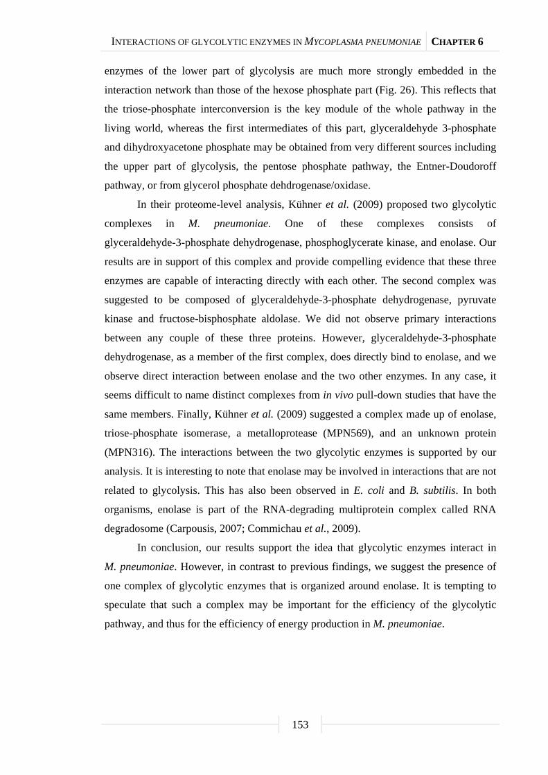
INTERACTIONS OF GLYCOLYTIC ENZYMES IN MYCOPLASMA PNEUMONIAE CHAPTER 6
153
enzymes of the lower part of glycolysis are much more strongly embedded in the
interaction network than those of the hexose phosphate part (Fig. 26). This reflects that
the triose-phosphate interconversion is the key module of the whole pathway in the
living world, whereas the first intermediates of this part, glyceraldehyde 3-phosphate
and dihydroxyacetone phosphate may be obtained from very different sources including
the upper part of glycolysis, the pentose phosphate pathway, the Entner-Doudoroff
pathway, or from glycerol phosphate dehdrogenase/oxidase.
In their proteome-level analysis, Kühner et al. (2009) proposed two glycolytic
complexes in M. pneumoniae. One of these complexes consists of
glyceraldehyde-3-phosphate dehydrogenase, phosphoglycerate kinase, and enolase. Our
results are in support of this complex and provide compelling evidence that these three
enzymes are capable of interacting directly with each other. The second complex was
suggested to be composed of glyceraldehyde-3-phosphate dehydrogenase, pyruvate
kinase and fructose-bisphosphate aldolase. We did not observe primary interactions
between any couple of these three proteins. However, glyceraldehyde-3-phosphate
dehydrogenase, as a member of the first complex, does directly bind to enolase, and we
observe direct interaction between enolase and the two other enzymes. In any case, it
seems difficult to name distinct complexes from in vivo pull-down studies that have the
same members. Finally, Kühner et al. (2009) suggested a complex made up of enolase,
triose-phosphate isomerase, a metalloprotease (MPN569), and an unknown protein
(MPN316). The interactions between the two glycolytic enzymes is supported by our
analysis. It is interesting to note that enolase may be involved in interactions that are not
related to glycolysis. This has also been observed in E. coli and B. subtilis. In both
organisms, enolase is part of the RNA-degrading multiprotein complex called RNA
degradosome (Carpousis, 2007; Commichau et al., 2009).
In conclusion, our results support the idea that glycolytic enzymes interact in
M. pneumoniae. However, in contrast to previous findings, we suggest the presence of
one complex of glycolytic enzymes that is organized around enolase. It is tempting to
speculate that such a complex may be important for the efficiency of the glycolytic
pathway, and thus for the efficiency of energy production in M. pneumoniae.

INTERACTIONS OF GLYCOLYTIC ENZYMES IN MYCOPLASMA PNEUMONIAE CHAPTER 6
154
Experimental procedures
Bacterial strains and growth conditions. E. coli XL1-Blue (Stratagene) and
BTH101 (Karimova et al., 1998) were used for cloning experiments and B2H analyses,
respectively. E. coli was grown in LB medium. LB plates were prepared by the addition
of 18 g Bacto agar/liter (Difco) to LB medium.
DNA manipulation and transformation. Transformation of E. coli and
plasmid DNA extraction were performed using standard procedures (Sambrook et al.,
1989). Restriction enzymes, T4 DNA ligase, and DNA polymerases were used as
recommended by the manufacturers. DNA fragments were purified from agarose gels
using the Nucleospin Extract Kit (Macherey and Nagel, Germany). Phusion DNA
polymerase was used for the polymerase chain reaction as recommended by the
manufacturer. All primer sequences are listed in the supplemental Table S7. DNA
sequences were determined using the dideoxy chain termination method
(Sambrook et al., 1989). All plasmid inserts derived from PCR products were verified
by DNA sequencing. E. coli transformants were selected on LB plates containing
ampicillin (100 µg/ml) or kanamycin (50 µg/ml).
Plasmid constructions. The genes encoding the glycolytic enzymes from
M. pneumoniae were first subcloned as follows. The coding sequence of each gene was
amplified by PCR with gene specific primers using chromosomal DNA of
M. pneumonia M129 as the template. The PCR products were digested as appropriate
with BamHI and HindIII or with BamHI and PstI and cloned into the appropriately
linearized expression vector pWH844 (Schirmer et al., 1997). These plasmids allowed
the expression of the glycolytic enzymes carrying a N-terminal His6-tag. The resulting
plasmids are listed in the supplemental Table S8. In M. pneumoniae, the UGA specifies
a tryptophan; however, in E. coli it is a stop codon. With exception of pfkA and pgm, the
glycolytic genes contain one to three UGA codons that were replaced by the multiple
mutation reaction (Hames et al., 2005) using phosphorylated mutagenic
oligonucleotides and the former plasmids as template. The resulting final plasmids are
listed in the supplemental material (see Table S8).
To obtain the plasmids for the B2H analyses, the coding sequences of the
glycolytic genes were amplified by PCR using the latter plasmids. The PCR products
were digested as shown in the supplemental material, and the resulting fragments were
cloned into each of the three plasmids pKNT25, pUT18, and pUT18C (Claessen et al.,
INTERACTIONS OF GLYCOLYTIC ENZYMES IN MYCOPLASMA PNEUMONIAE CHAPTER 6
155
2008; Karimova et al., 1998), digested with the same enzymes. The resulting plasmids
used for the B2H analyses are shown in the supplemental Table S8. All plasmid inserts
were verified by DNA sequencing.
Bacterial two-hybrid analysis. Primary protein-protein interactions were
identified by B2H analysis (Karimova et al., 1998). The B2H system is based on the
interaction-mediated reconstruction of adenylate cyclase (CyaA) activity from
B. pertussis in E. coli. The CyaA enzyme consists of two complementary fragments T18
and T25 that are not active when physically separated. Fusion of these fragments to
interacting proteins results in functional complementation between the T18 and T25
fragments and the synthesis of cAMP. cAMP production can be monitored by
measuring the β-galactosidase activity of the cAMP-CAP-dependent promoter of the
E. coli lac operon. Thus, a high β-galactosidase activity reflects the interaction between
the hybrid proteins. Plasmids pUT18 and pKNT25 allow the expression of proteins
fused to the N-terminus of the T18 and T25 fragments of the CyaA protein,
respectively, and the plasmid pUT18C allows the expression of proteins fused to the
C-terminus of the T18 fragment of the CyaA protein, respectively (Claessen et al.,
2008; Karimova et al., 1998). The plasmids pKT25-zip and pUT18C-zip served as
positive controls for complementation. These plasmids express T18-zip and T25-zip
fusion proteins that can associate due to the leucine zipper motifs resulting in an active
CyaA enzyme and a high β-galactosidase activity. The plasmids constructed for the
B2H assay (see Table S8) were used for cotransformations of E. coli BTH101 and the
protein-protein interactions were then analyzed by plating the cells on LB plates
containing ampicillin (100 µg/ml), kanamycin (50 µg/ml), X-Gal (40 µg/ml)
and IPTG (0.5 mM). The plates were incubated for a maximum of 60 h at 30°C. The
B2H assays were performed in triplicate.
Acknowledgments
We are grateful to Julia Busse, Henrike Pförtner, Martin Arnold, and Frederik
Meyer for helpful discussions. This work was supported by Deutsche
Forschungsgemeinschaft and the Fonds der Chemischen Industrie. S. R. S. was
supported by a personal grant from the Studienstiftung des Deutschen Volkes.

INTERACTIONS OF GLYCOLYTIC ENZYMES IN MYCOPLASMA PNEUMONIAE CHAPTER 6
156
Supplemental material
Tab. S7. Primers used in this study.
Primer Sequence (5’→3’)ab Mutation Resulting plasmid
Subcloning for multiple mutation reaction
JS39 TCTATCAACAGGAGTCCAAGC None
MR04 AAAGGATCCATGGTTGACTTTAAAACAG
TCCAAGC
None pGP1006, pGP1010
MR05 TATAAAGCTTTTACTTTTTTTGAATATCG
CTAATTCCCAC
None pGP1006, pGP1010
MR06 P_GTTAGCACAGTTCTGGGCATCATTGGG A441G pGP1010
MR07 P_CCCAATGGCCAGCAGAACTTCAGG A894G pGP1010
MR08 AAAGGATCCATGCATAAGAAGGTCTTAC
TAGCC
None pGP1007
MR09 TATACTGCAGTTACTTTTTACTTATTAAA
AGGGAGTTTGCG
None pGP1007
MR10 AAAGGATCCATGAGTGCACAAACTGGA
ACCGATCTTTTCAAG
None pGP1008
MR11 TATACTGCAGTTAAGCTTTTTGCGGTTTA
ATGTTTTTAAAGGTGTTCCAACC
A1335G pGP1008
MR19 AAAGGATCCATGGAAAGTAAATGGTTAA
CAGTTGAC
None pGP756, pGP762
MR20 TATAAAGCTTCTATTCACGACCAAGTTT
AGCAAAC
None pGP756, pGP762
MR21 P_GGGTAAATCATGGGCTTTGGTAGTAA
CTTC
A393G pGP762
MR22 P_CTGCTTTGCGTCACTGGCTTTACAC A756G pGP762
MR23 AAAGGATCCATGAGTCCAAAAACAACC
AAAAAAATTGCC
None pGP757
MR24 TATAAAGCTTTTAAATAATATTTTTATTA
ATGACTGCAATTAAAGCGC
None pGP757
MR25 AAAGGATCCATGCTAGTAAACATCAAAC
AAATGTTGCAAC
None pGP758, pGP763
MR26 TATAAAGCTTCTAAGCCTTATTGGTTGA
ACCACAG
None pGP758, pGP763
MR27 P_GGCTTTACCCCGACAACTGGAAGGG A552G pGP763
MR28 AAAGGATCCATGCGTACGAAATACCTAA
TTGGTAACTGGAAG
A30G pGP759, pGP764
INTERACTIONS OF GLYCOLYTIC ENZYMES IN MYCOPLASMA PNEUMONIAE CHAPTER 6
157
Tab. S7. Continued.
Primer Sequence (5’→3’)ab Mutation Resulting plasmid
MR29 TATACTGCAGTTATGCATATACTTGTGCC
ATTACTAAAAAGTCG
None pGP759, pGP764
MR30 P_GTGATTGCTTACGAACCAATTTGGGCA
ATTGGTACGG
A489G pGP764
MR31 AAAGGATCCATGCTAGCAAAGAGTAAG
ACTATCC
None pGP760, pGP765
MR32 TATACTGCAGTTAAAGCTTGGCACAATA
GTTAACTAC
None pGP760, pGP765
MR33 P_GCTAACCTGCCATGGGCAGAACAC A270G pGP765
MR34 AAAGGATCCATGATTCACCACCTAAAAC
GCAC
None pGP761, pGP766
MR35 TATACTGCAGTTATAAGCTAATGATCTT
ATTGTGAAATACTC
None pGP761, pGP766
MR36 P_GCTATGGACGCTAGCAATGTTAGATG
AC
A69G pGP766
MR37 P_CAAGGTGCCTTACTGGCAACGGTAC A864G pGP766
MR38 P_CAGTGAGTTTTGGAAGCAGGTGGTG A1167G pGP766
pWH844 fw TATGAGAGGATCGCATCACCAT None
Bacterial two-hybrid analysis
FC146 CGATGCGTTCGCGATCCAGGC None -
FC147 CCAGCCTGATGCGATTGCTGCAT None -
FC148 GTCACCCGGATTGCGGCGG None -
FC149 GCTGGCTTAACTATGCGGCATCAGA None -
M13 puc rev GGAAACAGCTATGACCATG None -
PD05 AAATCTAGAGATGGAAAGTAAATGGTTA
ACAGTTGACAC
None pGP1553, pGP1563,
pGP1573
PD06 TTTGGTACCCGTTCACGACCAAGTTTAG
CAAACATTAAC
None pGP1553, pGP1563,
pGP1573
PD07 AAATCTAGAGATGAGTCCAAAAACAACC
AAAAAAATTGCC
None pGP1554, pGP1564,
pGP1574
PD08 TTTGGTACCCGAATAATATTTTTATTAAT
GACTGCAATTAAAGCGC
None pGP1554, pGP1564,
pGP1574
PD09 AAATCTAGAGATGCTAGTAAACATCAAA
CAAATGTTGCAAC
None pGP1555, pGP1565,
pGP1575
PD10 TTTGAGCTCCGAGCCTTATTGGTTGAACC
ACAGAG
None pGP1555, pGP1565,
pGP1575

INTERACTIONS OF GLYCOLYTIC ENZYMES IN MYCOPLASMA PNEUMONIAE CHAPTER 6
158
Tab. S7. Continued.
Primer Sequence (5’→3’)ab Mutation Resulting plasmid
PD11 AAATCTAGAGATGCGTACGAAATACCTA
ATTGGTAAC
None pGP1556, pGP1566,
pGP1576
PD12 TTTGGTACCCGTGCATATACTTGTGCCAT
TACTAAAAAGTC
None pGP1556, pGP1566,
pGP1576
PD13 AAATCTAGAGATGCTAGCAAAGAGTAA
GACTATCCG
None pGP1557, pGP1567,
pGP1577
PD14 TTTGAATTCCGAAGCTTGGCACAATAGT
TAACTACCC
None pGP1557, pGP1567,
pGP1577
PD15 AAATCTAGAGATGGTTGACTTTAAAACA
GTCCAAGC
None pGP1558, pGP1568,
pGP1578
PD16 TTTGAATTCCGCTTTTTTTGAATATCGCT
AATTCCCACTAG
None pGP1558, pGP1568,
pGP1578
PD17 AAATCTAGAGATGCATAAGAAGGTCTTA
CTAGCC
None pGP1559, pGP1569,
pGP1579
PD18 TTTGAATTCCGCTTTTTACTTATTAAAAG
GGAGTTTGCG
None pGP1559, pGP1569,
pGP1579
PD19 AAATCTAGAGATGAGTGCACAAACTGGA
ACCG
None pGP1560, pGP1570,
pGP1580
PD20 TTTGAATTCCGAGCTTTTTGCGGTTTAAT
GTTTTTAAAGGTG
None pGP1560, pGP1570,
pGP1580
PD21 AAATCTAGAGATGATTCACCACCTAAAA
CGCAC
None pGP1561, pGP1571,
pGP1581
PD22 TTTGGTACCCGTAAGCTAATGATCTTATT
GTGAAATACTCC
None pGP1561, pGP1571,
pGP1581 a Restriction sites are underlined. b The “P” at the 5’ end of primer sequences indicates phosphorylation.
INTERACTIONS OF GLYCOLYTIC ENZYMES IN MYCOPLASMA PNEUMONIAE CHAPTER 6
159
Tab. S8. Plasmids used for analysis of glycolytic enzymes.
Plasmid Relevant characteristicsa Used restriction
sites Reference
Subcloning for multiple mutation reaction
pGP756 pWH844-pgi BamHI + HindIII This work
pGP757 pWH844-pfkA BamHI + HindIII This work
pGP758 pWH844-fba BamHI + HindIII This work
pGP759 pWH844-tpiA(A30G) BamHI + PstI This work
pGP760 pWH844-gapA BamHI + PstI This work
pGP761 pWH844-pyk BamHI + PstI This work
pGP762 pWH844-pgi(A393G+A756G) BamHI + HindIII This work
pGP763 pWH844-fba(A552G) BamHI + HindIII This work
pGP764 pWH844-tpiA(A30G+A489G) BamHI + PstI This work
pGP765 pWH844-gapA(A270G) BamHI + PstI This work
pGP766 pWH844-pyk(A69G+A864G+A1167G) BamHI + PstI This work
pGP1006 pWH844-pgk BamHI + HindIII This work
pGP1007 pWH844-pgm BamHI + PstI This work
pGP1008 pWH844-eno(A1335G) BamHI + PstI This work
pGP1010 pWH844-pgk(A441G+A894G) BamHI + HindIII This work
pWH844 Allows overexpression of N-terminal
His6-tag fusion proteins in E. coli DH5α
(AmpR)
- Schirmer et al. (1997)
Bacterial two-hybrid analysis
pGP1553 pUT18C-pgi(A393G+A756G) XbaI + KpnI This work
pGP1554 pUT18C-pfkA XbaI + KpnI This work
pGP1555 pUT18C-fba(A552G) XbaI + SacI This work
pGP1556 pUT18C-tpiA(A30G+A489G) XbaI + KpnI This work
pGP1557 pUT18C-gapA(A270G) XbaI + EcoRI This work
pGP1558 pUT18C-pgk(A441G+A894G) XbaI + EcoRI This work
pGP1559 pUT18C-pgm XbaI + EcoRI This work
pGP1560 pUT18C-eno(A1335G) XbaI + EcoRI This work
pGP1561 pUT18C-pyk(A69G+A864G+A1167G) XbaI + KpnI This work
pGP1563 pUT18-pgi(A393G+A756G) XbaI + KpnI This work
pGP1564 pUT18-pfkA XbaI + KpnI This work
pGP1565 pUT18-fba(A552G) XbaI + SacI This work
pGP1566 pUT18-tpiA(A30G+A489G) XbaI + KpnI This work
pGP1567 pUT18-gapA(A270G) XbaI + EcoRI This work
pGP1568 pUT18-pgk(A441G+A894G) XbaI + EcoRI This work
pGP1569 pUT18-pgm XbaI + EcoRI This work
INTERACTIONS OF GLYCOLYTIC ENZYMES IN MYCOPLASMA PNEUMONIAE CHAPTER 6
160
Tab. S8. Continued.
Plasmid Relevant characteristicsa Used restriction
sites Reference
pGP1570 pUT18-eno(A1335G) XbaI + EcoRI This work
pGP1571 pUT18-pyk(A69G+A864G+A1167G) XbaI + KpnI This work
pGP1573 pKNT25-pgi(A393G+A756G) XbaI + KpnI This work
pGP1574 pKNT25-pfkA XbaI + KpnI This work
pGP1575 pKNT25-fba(A552G) XbaI + SacI This work
pGP1576 pKNT25-tpiA(A30G+A489G) XbaI + KpnI This work
pGP1577 pKNT25-gapA(A270G) XbaI + EcoRI This work
pGP1578 pKNT25-pgk(A441G+A894G) XbaI + EcoRI This work
pGP1579 pKNT25-pgm XbaI + EcoRI This work
pGP1580 pKNT25-eno(A1335G) XbaI + EcoRI This work
pGP1581 pKNT25-pyk(A69G+A864G+A1167G) XbaI + KpnI This work
pKNT25 Plac-mcs-cyaA (KanR) - Claessen et al. (2008)
pKT25-zip Plac-cyaA-zip (KanR) - Karimova et al. (1998)
pUT18 Plac-mcs-cyaA (AmpR) - Karimova et al. (1998)
pUT18C Plac-cyaA-mcs (AmpR) - Karimova et al. (1998)
pUT18C-zip Plac-cyaA-zip (AmpR) - Karimova et al. (1998) a Resistance gene abbreviations as follows: Amp, ampicillin; Kan, kanamycin.

Chapter 7
The hidden pathway:
Impact of the glycerophosphodiesterase GlpQ
on virulence of Mycoplasma pneumoniae
The work described in this chapter was submitted for publication in:
Schmidl, S. R., A. Otto, M. Lluch-Senar, J. Piñol, J. Busse, D. Becher, and
J. Stülke. The hidden pathway: Impact of the glycerophosphodiesterase GlpQ on
virulence of Mycoplasma pneumoniae. PLoS Pathogens submitted.
Author contributions:
This study was designed and interpreted by SRS. The cell morphology experiments
were accomplished by MLS and JP, University of Barcelona. SRS performed all
biochemical and physiological researches, but JB contributed substantially to the slot
blot analyses. The proteomic analysis was done in collaboration with AO and DB,
University of Greifswald. SRS and JS wrote the paper.

ENZYMES IN MYCOPLASMA PNEUMONIAE PHOSPHOLIPID METABOLISM CHAPTER 7
162
Abstract
Mycoplasma pneumoniae is the causative agent of atypical pneumonia. The
formation of hydrogen peroxide, a product of glycerol metabolism, is the major factor
that damages the host cells. Phosphatidylcholine is the major carbon source available on
lung epithelia, and their utilization requires the cleavage of deacylated phospholipids to
glycerol 3-phosphate and choline. M. pneumoniae possesses two potential
glycerophosphodiesterases, MPN420 (GlpQ) and MPN566. In this work, the function of
these proteins was analyzed by biochemical, genetic, and physiological studies. The
results indicate that only GlpQ is an active glycerophosphodiesterase. MPN566 has no
enzymatic activity and the inactivation of the gene did not result in any detectable
phenotype. Inactivation of the glpQ gene resulted in reduced growth in medium with
glucose as the carbon source, in loss of hydrogen peroxide production when
phosphatidylcholine was present, and in a complete loss of cytotoxicity toward HeLa
cells. Moreover, the glpQ mutant strain exhibited a reduced gliding velocity. A
comparison of the proteomes of the wild type and the glpQ mutant strain revealed that
this enzyme is also implicated in the control of gene expression. Several proteins were
present in higher or lower amounts in the mutant. This regulation by GlpQ is exerted at
the level of transcription as determined by mRNA slot blot analyses. All genes subject
to GlpQ-dependent control have a conserved potential cis-acting element upstream of
the coding region. This element overlaps the promoter in the case of the genes that are
repressed in a GlpQ-dependent manner and it is located upstream of the promoter for
GlpQ-activated genes. We may suggest that GlpQ acts as a trigger enzyme that
measures the availability of its product glycerol 3-phosphate and uses this information
to differentially control gene expression.
Author summary
Mycoplasma pneumoniae serves as a model organism for bacteria with very
small genomes that are nonetheless independently viable. These bacteria infect the
human lung and cause a specific pneumonia. The major virulence determinant of
M. pneumoniae is hydrogen peroxide that is generated during the utilization of
glycerol 3-phosphate, which might be derived from free glycerol or from the
degradation of phospholipids. Indeed, lecithin is the by far most abundant carbon source

ENZYMES IN MYCOPLASMA PNEUMONIAE PHOSPHOLIPID METABOLISM CHAPTER 7
163
on lung epithelia. In this study, we made use of the recent availability of methods to
isolate mutants of M. pneumoniae and characterized the enzyme that generates
glycerol 3-phosphate from deacylated lecithin (glycerophosphorylcholine). This
enzyme, called GlpQ, is essential for the formation of hydrogen peroxide when the
bacteria are incubated with glycerophosphorylcholine. Moreover, M. pneumoniae is
unable to cause any damage to the host cells in the absence of GlpQ. This underlines the
important role of phospholipid metabolism for the virulence of M. pneumoniae. We
observed that GlpQ in addition to its enzymatic activity does also control the expression
of several genes, among them the glycerol transporter. Thus, GlpQ is central to the
normal physiology and to pathogenicity of the minimal pathogen M. pneumoniae.
Introduction
Pathogenic bacteria have developed a large battery of enzymes and mechanisms
for extracting nutrients from their hosts, and the requirement for nutrient acquisition can
be regarded as one of the driving forces for virulence (Eisenreich et al., 2010;
Görke and Stülke, 2008; Sonenshein, 2007). In consequence, the metabolic capabilities
of a pathogen reflect its adaptation to a particular niche in a particular host.
Mycoplasma pneumoniae is the causative agent of atypical pneumonia and
several additional infections including encephalitis, aseptic meningitis, acute transverses
myelitis, stroke, and polyradiculopathy (Narita, 2009; Narita, 2010; Tsiodras et al.,
2005; Waites and Talkington, 2004). These bacteria are members of the phylogenetic
group of Mollicutes that are characterized by an extreme reductive evolution that results
in the smallest genomes that allow independent life. Moreover, the Mollicutes have lost
the cell wall and most metabolic pathways, since they obtain the building blocks for
their cellular macromolecules from the host tissue. However, even in these minimal
pathogens, there is a close relation between metabolism and virulence [for review see
Halbedel et al. (2007)]. M. pneumoniae thrives at the surfaces of lung epithelia. Thus,
these bacteria must have evolved to utilize the carbon sources present in this niche. The
pulmonary surfactant is composed of about 90% phospholipids and 10% proteins
(Veldhuizen et al., 1998). This suggests that phospholipids play a major role in the
nutrition of M. pneumoniae.

ENZYMES IN MYCOPLASMA PNEUMONIAE PHOSPHOLIPID METABOLISM CHAPTER 7
164
Glycerophospholipids, the major building blocks of the cell membrane in
bacteria and eukaryotes, are degraded in several steps. First, the fatty acids are cleaved
off from the phospholipids resulting in the formation of glycerophosphodiesters. In
these molecules, the phosphate group of glycerol 3-phosphate is linked to another
compound, called the head group. In eukaryotes, choline is by far the most abundant
head group, and lecithin, the choline-containing phospholipid accounts for about 80% of
all phospholipids in human lung cells (Veldhuizen et al., 1998). In the second step, the
choline head group is cleaved off due to the activity of a glycerophosphodiesterase
resulting in the formation of glycerol 3-phosphate that can feed into glycolysis after
oxidation to dihydroxyacetone phosphate (see Fig. 27). In M. pneumoniae, the latter
reaction is catalyzed by the glycerol-3-phosphate oxidase GlpD (Hames et al., 2009).
GlpD transfers the electrons to water resulting in the formation of hydrogen peroxide,
the major virulence factor of M. pneumoniae (Somerson et al., 1965). In consequence,
the virulence of M. pneumoniae glpD mutant cells is severely attenuated (Hames et al.,
2009).
While the metabolism of glycerol has been well studied in M. pneumoniae and
other Mollicutes such as Mycoplasma mycoides (Bischof et al., 2009; Hames et al.,
2009; Pilo et al., 2005), only little is known about the glycerophosphodiesterases
required for lipid utilization. Many bacteria encode multiple glycerophosphodiesterases.
In Escherichia coli, both enzymes are enzymatically active in lipid degradation;
however, they are differentially regulated, with GlpQ and UgpQ being induced in the
presence of glycerol 3-phosphate and under conditions of phosphate starvation,
respectively (Ohshima et al., 2008; Wong and Kwan, 1992). In Bacillus subtilis, out of
three putative glycerophosphodiesterases, only GlpQ has been studied. The
corresponding gene is under dual control and its expression is induced when phosphate
becomes limiting and glycerol is available (Antelmann et al., 2000; Nilsson et al.,
1994). Moreover, glpQ expression is repressed if more favourable carbon sources such
as glucose are present (Blencke et al., 2003). In Haemophilus influenzae, another
bacterium thriving in the respiratory tract, the glycerophosphodiesterase is involved in
pathogenicity. The enzyme generates choline, which in turn is used for the biosynthesis
of the bacterial lipopolysaccharide layer - a major virulence determinant of
Gram-negative bacteria (Fan et al., 2001; Forsgren et al., 2008). Similarly,
glycerophosphodiesterase activity is implicated on virulence of different Borrelia
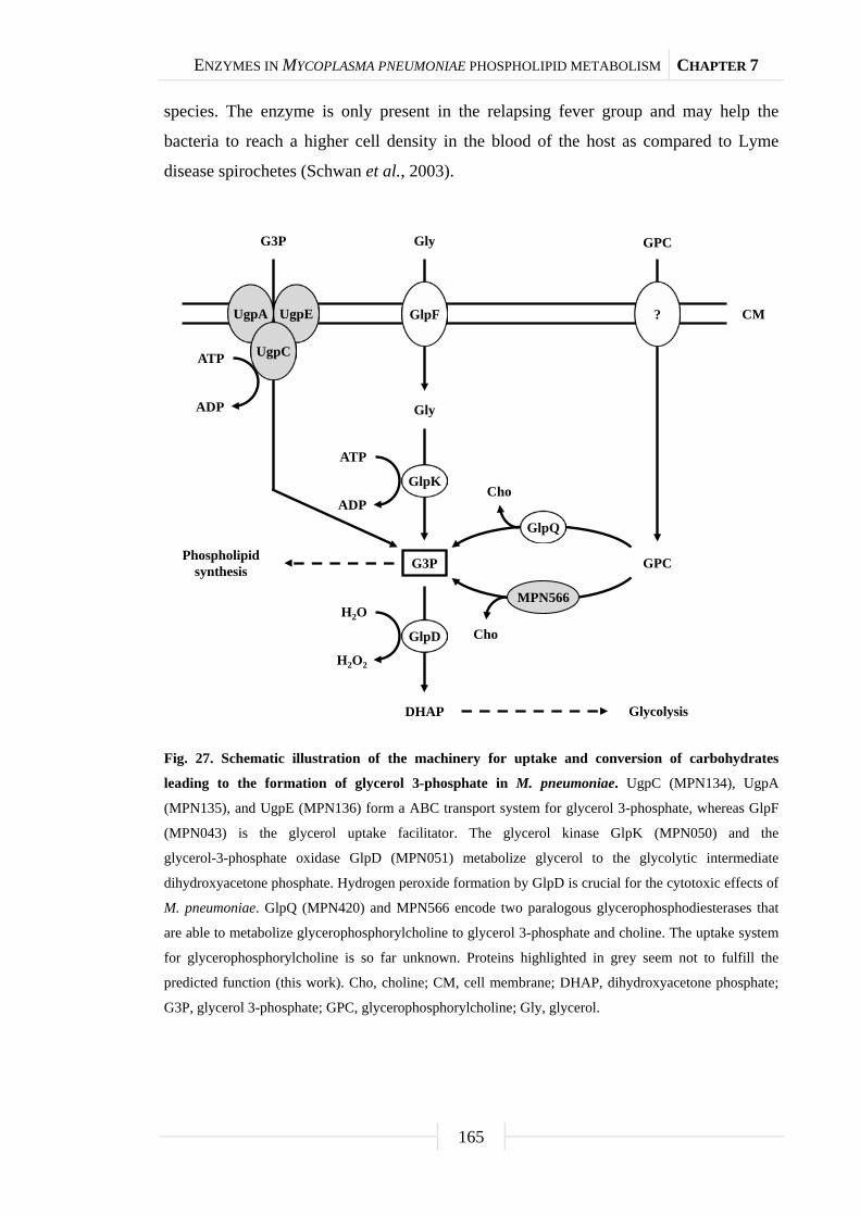
ENZYMES IN MYCOPLASMA PNEUMONIAE PHOSPHOLIPID METABOLISM CHAPTER 7
165
species. The enzyme is only present in the relapsing fever group and may help the
bacteria to reach a higher cell density in the blood of the host as compared to Lyme
disease spirochetes (Schwan et al., 2003).
Fig. 27. Schematic illustration of the machinery for uptake and conversion of carbohydrates
leading to the formation of glycerol 3-phosphate in M. pneumoniae. UgpC (MPN134), UgpA
(MPN135), and UgpE (MPN136) form a ABC transport system for glycerol 3-phosphate, whereas GlpF
(MPN043) is the glycerol uptake facilitator. The glycerol kinase GlpK (MPN050) and the
glycerol-3-phosphate oxidase GlpD (MPN051) metabolize glycerol to the glycolytic intermediate
dihydroxyacetone phosphate. Hydrogen peroxide formation by GlpD is crucial for the cytotoxic effects of
M. pneumoniae. GlpQ (MPN420) and MPN566 encode two paralogous glycerophosphodiesterases that
are able to metabolize glycerophosphorylcholine to glycerol 3-phosphate and choline. The uptake system
for glycerophosphorylcholine is so far unknown. Proteins highlighted in grey seem not to fulfill the
predicted function (this work). Cho, choline; CM, cell membrane; DHAP, dihydroxyacetone phosphate;
G3P, glycerol 3-phosphate; GPC, glycerophosphorylcholine; Gly, glycerol.
Gly
Gly
G3P
DHAP
CM
GPC
Glycolysis
Phospholipidsynthesis
GlpF
MPN566
GlpQ
GPC
?
H2O
GlpD
H2O2
GlpK
ATP
ADPCho
Cho
UgpA UgpE
UgpC
G3P
ATP
ADP

ENZYMES IN MYCOPLASMA PNEUMONIAE PHOSPHOLIPID METABOLISM CHAPTER 7
166
In many bacteria, central enzymes of metabolism do not only serve their
catalytic function, but in addition, they are also involved in signal transduction. In this
way, the information on the availability of important metabolites can be directly
determined by the enzyme in charge of their conversion, and this information is then
often transferred to the transcription machinery. Collectively, such enzymes were
termed trigger enzymes (Commichau and Stülke, 2008). They can control gene
expression by directly acting as DNA- or RNA-binding transcription factors as the
E. coli proline dehydrogenase and the aconitase or by controlling the activity of
transcription factors by covalent modification or a regulatory protein-protein interaction
as observed for several sugar permeases of the bacterial phosphotransferase system and
the B. subtilis glutamate dehydrogenase, respectively (Beinert et al., 1996;
Commichau et al., 2007; Schmalisch et al., 2003; Zhu and Becker, 2003).
In this work, we have analyzed the role of the two potential
glycerophosphodiesterases encoded in the genome of M. pneumoniae. Biochemical and
physiological studies demonstrate that one of the two proteins, GlpQ, is a functional
glycerophosphodiesterase. GlpQ is essential for hydrogen peroxide formation in the
presence of deacylated phospholipids as the carbon source and, in consequence, for
cytotoxicity. Moreover, GlpQ may act as a trigger enzyme by controlling the expression
of a set of genes encoding lipoproteins, the glycerol facilitator, and a metal ion ABC
transporter.
Results
Identification of M. pneumoniae genes encoding potential
glycerophosphodiesterases. Since phospholipids are the most abundant potential
carbon sources for M. pneumoniae living at lung epithelial surfaces, we considered the
possibility that these bacteria synthesize enzymes that cleave the polar head groups from
the glycerophosphodiesters to produce glycerol 3-phosphate that can be utilized by the
enzymes of glycerol metabolism (Hames et al., 2009) (Fig. 27). Two genes that
potentially encode such enzymes are present in the genome of M. pneumoniae, i.e.
mpn420 (renamed to glpQ) and mpn566. An alignment of the corresponding proteins to
glycerophosphodiesterases from other bacteria is shown in the supporting information
(Fig. S4).

ENZYMES IN MYCOPLASMA PNEUMONIAE PHOSPHOLIPID METABOLISM CHAPTER 7
167
Enzymatic activities of the potential glycerophosphodiesterases. In order to
assess the biochemical properties and physiological relevance of the putative
glycerophosphodiesterases, their corresponding genes, glpQ and mpn566, were cloned
into the expression vector pGP172, thus allowing a fusion of the proteins to a
N-terminal Strep-tag facilitating purification. The recombinant proteins were purified
and the activities were first determined using glycerophosphorylcholine (GPC) as the
substrate and a set of divalent cations. As shown in Fig. 28, purified GlpQ was active
against GPC, and the activity was highest in the presence of magnesium ions (10 mM).
Manganese and zinc ions did also support activity, although to a lesser extent (Fig. 28).
In contrast, the enzyme was inactive in the presence of calcium and cobalt ions (data not
shown). The activity assay with purified MPN566 revealed no activity with GPC,
irrespective of the cation present in the assay (data not shown). We did also test the
activity of both proteins with glycerophosphorylethanolamine and
glycerophosphorylglycerol. However, neither protein was active with any of these
substrates. Thus, our data demonstrate that GlpQ is active as a
glycerophosphodiesterase, whereas MPN566 does not exhibit such an activity.
The two proteins GlpQ and MPN566 share ~58% identical residues. Thus, it
seems surprising that MPN566 was inactive in the enzymatic assay. However, several
residues that are known to be important for the activity of glycerophosphodiesterases
are conserved in GlpQ but not in MPN566 (Fig. S4). These residues include Trp-36 and
Glu-38 as well as the conserved HD motif (Asn-51 and Leu-52 in MPN566) and
Phe-110. Interestingly, a similar arrangement with two GlpQ-like proteins is also
observed in Mycoplasma genitalium, and as in M. pneumoniae, one protein has all the
conserved residues characteristic for glycerophosphodiesterases, whereas the second
protein has similar deviations from the consensus as MPN566 (Fig. S4). In order to test
whether a restoration of the conserved residues would also convert MPN566 to a
biologically active glycerophosphodiesterase, we replaced the five amino acids that
differ from the consensus by those residues present in GlpQ. The resulting mutant allele
was cloned into pGP172, and the protein purified. Unfortunately, this protein was
highly unstable and purification was impossible.

ENZYMES IN MYCOPLASMA PNEUMONIAE PHOSPHOLIPID METABOLISM CHAPTER 7
168
Fig. 28. Effect of divalent cations on GlpQ activity. GlpQ activity was measured in the presence of
0.5 mM glycerophosphorylcholine and various concentrations of divalent cations. Error bars indicate
standard deviation (based on three independent experiments).
Construction of glpQ and mpn566 mutants. The analysis of mutants is one of
the most powerful tools for studying gene functions and bacterial physiology. The
isolation of desired M. pneumoniae mutants became possible only recently by the
introduction of the “Haystack mutagenesis” (Halbedel and Stülke, 2007). To get more
insights into the physiological role of the glycerophosphodiesterases GlpQ and its
paralogous MPN566, we attempted to isolate mutants affected in the corresponding
genes.
The strategy of “Haystack mutagenesis” is based on an ordered collection of
pooled random transposon insertion mutants that can be screened for junctions between
the transposon and the gene of interest due to transposon insertion. The 64 pools were
used in a PCR to detect junctions between the glpQ or mpn566 genes and the
mini-transposon using the oligonucleotides SS35 and SS40 (for the respective genes)
and SH30 (for the mini-transposon) (Halbedel et al., 2006) (Fig. 29). Positive signals
were obtained for both genes. From pools that gave a positive signal, colony PCR with
the 50 individual mutants resulted in the identification of the desired glpQ and mpn566
mutants. The presence of the transposon insertion in both genes was verified by
Southern blot analysis (Fig. 29). To test whether these strains contained only unique
transposon insertions, we did another Southern blot using a probe specific for the
0
5
10
15
20
25
30
35
0.1 0.5 1 2 5 10 15
Act
ivity
[U/m
g pr
otei
n]
Divalent cation [mM]
MgCl2 MnCl2 ZnCl2
ENZYMES IN MYCOPLASMA PNEUMONIAE PHOSPHOLIPID METABOLISM CHAPTER 7
169
aac-aphD resistance gene present on the mini-transposon. As shown in Fig. 29, only
one single band hybridizing with this probe was detected in each strain, moreover, this
fragment had the same size as the AgeI or PstI/SacI fragment hybridizing to the glpQ
and mpn566 probe, respectively (Fig. 29). The isolated glpQ and mpn566 mutant strains
were designated as GPM81 and GPM82. The position of the transposon insertion in the
two genes was determined by DNA sequencing. The glpQ gene was disrupted between
nucleotides 517 and 518, resulting in a truncated protein of 172 amino acids with one
additional amino acid and the following stop codon encoded by the inserted
mini-transposon. The disruption of the mpn566 gene was located between nucleotides
157 and 158, resulting in a truncated protein of 52 amino acids with one additional
amino acid and the following stop codon.
Contributions of GlpQ and MPN566 to growth and motility. First, we
compared the ability of the wild type strain and the two mutant strains to utilize glucose
and glycerol as the single carbon sources (Fig. 30). As an additional control, we used
the glpD mutant strain GPM52. This strain is defective in glycerol-3-phosphate oxidase
and therefore unable to utilize glycerol as the only carbon source (Hames et al., 2009).
As shown in Fig. 30A, the wild type and the glpD and mpn566 mutant strains grew well
with glucose. In contrast, the glpQ mutant GPM81 grew more slowly and did not reach
the final biomass as compared to the other strains. As reported previously, the wild type
strain exhibited very slow growth with glycerol as the only carbon source
(Halbedel et al., 2004). In this respect, the glpQ and mpn566 mutants were
indistinguishable from the wild type. As reported previously, the glpD mutant strain did
not grow at all in glycerol-containing medium (Hames et al., 2009). In conclusion, the
active glycerophosphodiesterase GlpQ is required for maximal growth in the presence
of glucose, whereas its absence does not interfere with the slow growth in the presence
of glycerol. It is therefore tempting to speculate that some glycerophosphodiesters in the
Hayflick medium support growth (see “Discussion”).
Since the disruption of glpQ affected the growth properties of the bacteria, we
wondered whether this might reflect changes in cell morphology and in the movement
of the bacteria. The morphology of the wild type and mutant bacteria was analyzed by
scanning electron microscopy, and no significant differences were detected (Fig. S5).
An analysis of the gliding velocities of the three strains revealed that the wild type strain
glided with a velocity of 0.32 ± 0.09 µm/s, whereas the glpQ and mpn566 mutants

ENZYMES IN MYCOPLASMA PNEUMONIAE PHOSPHOLIPID METABOLISM CHAPTER 7
170
exhibited velocities of 0.2 ± 0.08 µm/s and 0.3 ± 0.1µm/s, respectively. Thus, the active
glycerophosphodiesterase GlpQ is required for efficient gliding movement of the
bacteria.
Fig. 29. Isolation of M. pneumoniae glycerophosphodiesterases transposon insertion mutants.
(A, C) Schematic representation of the genomic region surrounding the glpQ and mpn566 gene (both
designated as glycerophosphodiesterases) in M. pneumoniae and site of the transposon insertion in the
knockout strains GPM81 and GPM82, respectively. The annealing sites of oligonucleotides used for the
determination of the transposon insertion site are indicated by arrows. Probes hybridizing to internal
fragments of the glycerophosphodiesterases and the aac-ahpD genes are depicted as dotted lines.
(B, D) Southern blot analysis to confirm the single insertion of the mini-transposon into the glpQ and
mpn566 gene of the strains GPM81 and GPM82, respectively. Chromosomal DNAs of the wild type and
both glycerophosphodiesterases were digested as indicated. Blots were hybridized with the respective
glycerophosphodiesterase-specific probe (left) and a probe hybridizing to the aac-ahpD gene of the
mini-transposon (right).
21,2
5,14,3
[kb]
probe A
alaS 421
AgeI
glpQ
aac-ahpD
A
B
AgeI
0 1 32 54 6 kb
p200
SacI
566
aac-ahpD
A
B
PstI
0 1 32 54 6 kb
3,5
2,0probe B
adh 565
probe A probe B
21,2
5,14,3
[kb]
3,5
2,0
21,2
5,14,3
[kb]
3,5
2,0
21,2
5,14,3
[kb]
3,5
2,0
A CSH30 SH29
SS35
SH30 SH29
SS40
B D

ENZYMES IN MYCOPLASMA PNEUMONIAE PHOSPHOLIPID METABOLISM CHAPTER 7
171
Fig. 30. Growth of M. pneumoniae in modified Hayflick medium containing different carbon
sources. One hundred milliliters of medium were inoculated with 5 mg of M. pneumoniae wild type (wt),
glpQ::Tn, mpn566::Tn, and glpD::Tn [control; Hames et al. (2009)] mutant cells and incubated for up to
six days at 37°C in 150-cm2 cell culture flasks. Glucose (A) and glycerol (B) were added to a final
concentration of 1% (wt/vol). Attached cells were collected by scraping and growth was monitored by
determination of the wet weight of the cell pellets. All measurements were done three times. Results are
from a representative experiment.
Implication of GlpQ and MPN566 in hydrogen peroxide production and
cytotoxicity. The utilization of glycerol or glycerophosphodiesters results in the
generation of hydrogen peroxide, the major cytotoxic product of M. pneumoniae. We
asked therefore whether the glpQ and mpn566 disruptions would affect hydrogen
peroxide formation and if so, whether it also affects cytotoxicity. Hydrogen peroxide
formation was assayed in M. pneumoniae cultures that contained glucose, glycerol,
GPC, glycerol 3-phosphate or no carbon source. In the absence of an added carbon
source, neither the wild type strain nor the mutants formed substantial amounts of
hydrogen peroxide (Fig. 31). Similarly, essentially no hydrogen peroxide was produced
in the presence of glucose. If glycerol was available, maximal hydrogen peroxide
formation (9.5 mg/l) was observed in the wild type strain. In the glpD mutant that
served as a control, no hydrogen peroxide was formed. This is in good agreement with
previous reports on the increase of hydrogen peroxide generation in the presence of
glycerol and its dependence on a functional glycerol-3-phosphate oxidase (Hames et al.,
2009). The hydrogen peroxide production in the glpQ and mpn566 mutants was similar
to that observed in the wild type strain. This result reflects that the metabolite glycerol
is downstream from the glycerophosphodiesterase activity. In the presence of GPC, the
1
10
100
0 1 2 3 4 5 6
Wet
wei
ght [
mg]
Time [d]
1
10
100
0 1 2 3 4 5 6
Wet
wei
ght [
mg]
Time [d]
wt
glpQ::Tn
mpn566::Tn
glpD::Tn
A B
wt
glpQ::Tn
mpn566::Tn
glpD::Tn

ENZYMES IN MYCOPLASMA PNEUMONIAE PHOSPHOLIPID METABOLISM CHAPTER 7
172
wild type strain produced similar amounts of hydrogen peroxide (9 mg/l) as in the
presence of glycerol. In contrast, no hydrogen peroxide formation was detected for the
glpQ mutant GPM81, whereas the disruption of mpn566 did not have any effect on the
production of hydrogen peroxide (Fig. 31). This result is in good agreement with the
enzymatic activities of the two proteins: GlpQ is the only active
glycerophosphodiesterase in M. pneumoniae, and no glycerol 3-phosphate, the substrate
of GlpD, can be formed in its absence, whereas MPN566 is dispensable for the
utilization of GPC. We also tested the ability of the M. pneumoniae strains to form
hydrogen peroxide in the presence of glycerophosphorylethanolamine and
glycerophosphorylglycerol. These compounds did not stimulate hydrogen peroxide in
any of the strains tested (data not shown). This is in excellent agreement with the result
of the enzyme assay that suggested that neither GlpQ nor MPN566 is able to degrade
these substances. Finally, we tested whether hydrogen peroxide was formed in the
presence of glycerol 3-phosphate. As shown in Fig. 31, no significant formation of
hydrogen peroxide was observed in any of the strains tested. This suggests that the
uptake of glycerol 3-phosphate is rather inefficient.
Fig. 31. Examination of M. pneumoniae hydrogen peroxide release. Hydrogen peroxide production of
M. pneumoniae wild type (wt), glpQ::Tn, mpn566::Tn, and glpD::Tn [control; Hames et al. (2009)]
mutant strains was measured in the presence of different carbon sources (100 µM) after 2 h. Error bars
indicate standard deviation (based on three independent experiments). G3P, glycerol 3-phosphate; GPC,
glycerophosphorylcholine; Glc, glucose; Gly, glycerol; w/o, without addition of any carbon source.
0
2
4
6
8
10
12
w/o Glc Gly GPC G3P
Hyd
roge
n pe
roxi
dere
leas
e[m
g/l]
Carbon source [100 mM]
wt
glpQ::Tn
mpn566::Tn
glpD::Tn

ENZYMES IN MYCOPLASMA PNEUMONIAE PHOSPHOLIPID METABOLISM CHAPTER 7
173
To assess the cytotoxicity of the different M. pneumoniae strains, we infected
confluently grown HeLa cell cultures with M. pneumoniae cells (multiplicity of
infection: 2). The cytotoxicity of the mutants was compared to that of the wild type
strain and M. pneumoniae GPM52 that is affected in glpD. As shown in Fig. 32, the
HeLa cells had undergone lysis after four days upon infection with wild type
M. pneumoniae. As observed previously, a large portion of viable cells was observed
after infection of the cell culture with the glpD mutant GPM52 (Hames et al., 2009).
For the glpQ mutant GPM81, nearly all HeLa cells had survived the infection
suggesting that GlpQ is essential for cytotoxicity. In contrast, cytotoxicity induced by
the mpn566 mutant strain GPM82 was equivalent to that of the wild type strain
(Fig. 32). These data clearly demonstrate that the active glycerophosphodiesterase GlpQ
is required for host cell damage, whereas the inactive enzyme MPN566 is not.
Moreover, they support the assumption that hydrogen peroxide formation is the major
factor that contributes to host cell damage.
Fig. 32. Cytotoxicity of M. pneumoniae toward HeLa cell cultures. (A) Infection assay to verify
cytotoxic effects of M. pneumoniae glpQ::Tn and mpn566::Tn mutant strains. HeLa cells were infected
with M. pneumoniae wild type (wt), glpQ::Tn, and mpn566::Tn mutant cells. As control served two HeLa
cell cultures: One without addition of M. pneumoniae cells and another after infection with the glpD::Tn
mutant strain (Hames et al., 2009). After four days, HeLa cell cultures were stained with crystal violet
and photographed. All pictures are shown at the same magnification. Scale bar, 0.1 mm.
(B) Quantification of HeLa cells after infection with different M. pneumoniae strains. The cell count of
surviving cells is indicated in percent as the number of viable cells per field of view, quantified by crystal
violet staining after four days of incubation. An uninfected HeLa cell culture served as control. Error bars
indicate standard deviation (based on three independent experiments).
wt glpQ::Tn
mpn566::Tn glpD::Tn
control
A B
0
20
40
60
80
100
120
Surv
ivin
gce
lls[%
]
Strain
ENZYMES IN MYCOPLASMA PNEUMONIAE PHOSPHOLIPID METABOLISM CHAPTER 7
174
The role of GlpQ in gene expression. As reported above, the glpQ mutant
exhibits multiple phenotypes related to motility, metabolism, and pathogenicity. We
asked therefore whether some of the effects are due to changes in the proteome of the
glpQ mutant GPM81. To answer this question, we compared the total protein profiles of
the wild type strain and the glpQ and mpn566 mutants, GPM81 and GPM82,
respectively, after growth in glucose and glycerol (Fig. 33). While the protein patterns
in the mpn566 mutant were indistinguishable from the wild type strain under both
conditions, several differences were noted for the glpQ mutant.
To identify those proteins that exhibit altered accumulation in the glpQ mutant,
the total proteins of the wild type and the glpQ mutant strains were identified by mass
spectrometry. For the protein extracts from glucose-grown cells, 532 different proteins
were identified. This corresponds to about 77% of the theoretical proteome of
M. pneumoniae. In the presence of glycerol, 473 proteins corresponding to 69% of the
theoretical proteome were identified. The differences in protein expression between
glucose- and glycerol-grown cells as well as proteins that could not be detected at all are
summarized in Tables S9 and S10. A detailed list of the differences of the protein
profiles between the wild type strain and the glpQ mutant is presented in Tables S11
and S12. As expected, the GlpQ protein was detected in the protein extracts of the wild
type strain but not in those of the glpQ mutant strain. In glucose-grown cells, 33 and
21 proteins were in elevated and reduced amounts, respectively, in the glpQ mutant. The
strongest increase was observed for the glycerol facilitator GlpF and the uncharacterized
lipoprotein MPN162. A strongly reduced accumulation was observed for the lipoprotein
MPN506. In the presence of glycerol, five induced and five repressed proteins were
detected. These proteins were subject to a similar regulation as in glucose-grown cells
and they are the only proteins with identical regulation under both conditions
(Table 12).
It has been shown before that changes at the proteome level may result from
altered gene expression or from changes in protein stability (Halbedel et al., 2007;
Schmidl et al., 2010). Therefore, we studied the expression of the genes corresponding
to the most prominently regulated proteins and of genes encoding potential regulators,
transport systems, and potential pathogenicity factors. For this purpose, we isolated
RNA from cultures grown in modified Hayflick medium supplemented with glucose
and performed slot blot analyses (Fig. 34 and S6).

ENZYMES IN MYCOPLASMA PNEUMONIAE PHOSPHOLIPID METABOLISM CHAPTER 7
175
Table 12. Proteins with GlpQ-dependent expression pattern.
For detailed information on proteome and transcript changes in the glpQ mutant GPM81, see Tables S11 and S12.
Locus name Protein name Protein function Fold-change in the presence of glucosea Fold-change in the presence of glycerola
Protein level Transcript level Protein level Transcript level
MPN043 GlpF Glycerol uptake facilitator 10.79 ± 1.06 5.69 ± 0.36 5.83 ± 0.76 3.32 ± 0.34
MPN162 - Uncharacterized lipoprotein 9.96 ± 0.97 5.23 ± 0.23 5.41 ± 0.83 2.81 ± 0.47
MPN284 - Uncharacterized lipoprotein ns 0.25 ± 0.05 0.36 ± 0.02 0.35 ± 0.04
MPN433 CbiO Metal ion ABC transporter ATP-binding protein ns 7.56 ± 0.39 5.66 ± 0.49 2.92 ± 0.24
MPN506 - Uncharacterized lipoprotein 0.10 ± 0.03 0.07 ± 0.05 0.18 ± 0.04 0.16 ± 0.02 a Fold-change cut off ≥ 2.0 and ≤ 0.5, respectively (glpQ mutant strain vs. wild type). Abbreviation: ns, no significant difference.
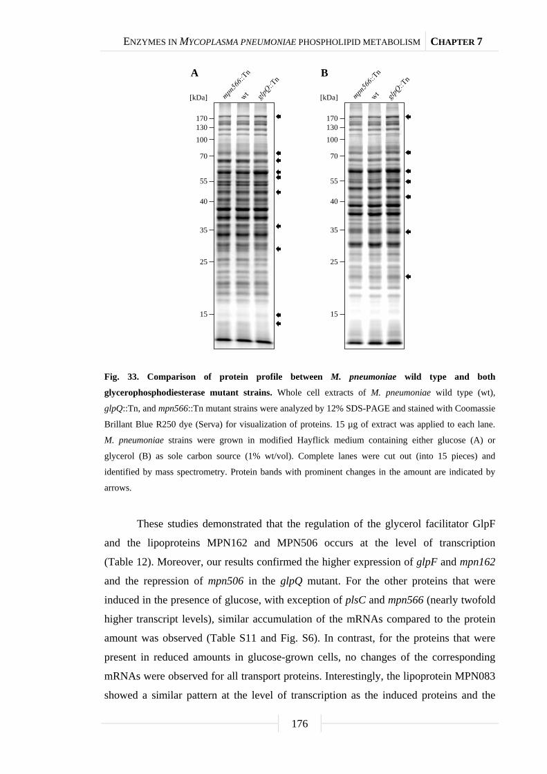
ENZYMES IN MYCOPLASMA PNEUMONIAE PHOSPHOLIPID METABOLISM CHAPTER 7
176
Fig. 33. Comparison of protein profile between M. pneumoniae wild type and both
glycerophosphodiesterase mutant strains. Whole cell extracts of M. pneumoniae wild type (wt),
glpQ::Tn, and mpn566::Tn mutant strains were analyzed by 12% SDS-PAGE and stained with Coomassie
Brillant Blue R250 dye (Serva) for visualization of proteins. 15 µg of extract was applied to each lane.
M. pneumoniae strains were grown in modified Hayflick medium containing either glucose (A) or
glycerol (B) as sole carbon source (1% wt/vol). Complete lanes were cut out (into 15 pieces) and
identified by mass spectrometry. Protein bands with prominent changes in the amount are indicated by
arrows.
These studies demonstrated that the regulation of the glycerol facilitator GlpF
and the lipoproteins MPN162 and MPN506 occurs at the level of transcription
(Table 12). Moreover, our results confirmed the higher expression of glpF and mpn162
and the repression of mpn506 in the glpQ mutant. For the other proteins that were
induced in the presence of glucose, with exception of plsC and mpn566 (nearly twofold
higher transcript levels), similar accumulation of the mRNAs compared to the protein
amount was observed (Table S11 and Fig. S6). In contrast, for the proteins that were
present in reduced amounts in glucose-grown cells, no changes of the corresponding
mRNAs were observed for all transport proteins. Interestingly, the lipoprotein MPN083
showed a similar pattern at the level of transcription as the induced proteins and the
[kDa]
170130100
70
55
40
35
25
15
A B
[kDa]
170130100
70
55
40
35
25
15

ENZYMES IN MYCOPLASMA PNEUMONIAE PHOSPHOLIPID METABOLISM CHAPTER 7
177
ribonucleoside-diphosphate reductase (encoded by nrdFIE) was the only protein with
reduced mRNA amounts, however changes in transcript level were not significant
(Table S11 and Fig. S6).
Fig. 34. Transcription analysis of GlpQ-dependent genes in M. pneumoniae. Slot blots were
performed with whole RNA extracts of M. pneumoniae wild type (wt), glpQ::Tn, mpn566::Tn, and
glpD::Tn [control; Hames et al. (2009)] mutant strains grown in modified Hayflick medium containing
either glucose or glycerol as sole carbon source (1% wt/vol). A dilution series of RNA extracts was
blotted onto a positively charged nylon membrane and probed with a DIG-labeled riboprobe specific for
an internal part of a particular open reading frame. Names of riboprobes are given next to each blot.
Signals obtained with 1 µg of RNA are shown. Yeast tRNA and M. pneumoniae chromosomal DNA
served as controls. For detailed information on differences of transcript levels see Tables 12, S11, and
S12.
Identification of a potential target site for GlpQ-dependent regulation. The
proteome and transcription analyses identified three genes that are significantly
regulated - either induced or repressed - in a GlpQ-dependent manner. An inspection of
the upstream region of these genes revealed the presence of a common palindromic
DNA motif (Fig. 35). To exclude the possibility that this motif is randomly distributed
in the genome of M. pneumoniae because of the extremely AT-rich consensus sequence,
we tested its presence in the genome using the GLAM2SCAN algorithm (Frith et al.,
glpF
cbiO
mpn162
mpn506
mpn284
Glucose Glycerol
Act
ivat
ing
effe
ctof
Glp
QR
epre
ssin
gef
fect
ofG
lpQ

ENZYMES IN MYCOPLASMA PNEUMONIAE PHOSPHOLIPID METABOLISM CHAPTER 7
178
2008). In 15 cases (matching score cut off ≥ 20), this potential motif was located
upstream of open reading frames, among them the three genes mentioned above.
Therefore, the expression of the remaining 12 genes was tested by slot blot analyses,
and for two of these genes, cbiO and mpn284, a significant accumulation and reduction
of the mRNA, respectively, was observed (data not shown; Fig. 34). Interestingly, the
corresponding proteins, a subunit of a metal ion ABC transporter CbiO and the
uncharacterized lipoprotein MPN284 were found to be present in higher or lower
amounts in the glpQ mutant in glycerol-grown cell. Thus, there is a very good
agreement between the regulatory effect of GlpQ at the proteome level, the regulation at
the level of transcription, and the presence of the cis-acting element.
Fig. 35. Transcriptional organization of GlpQ-dependent genes in M. pneumoniae. (A) Nucleotide
sequence of promoter regions of M. pneumoniae glpF (mpn043), mpn162, cbiO (mpn433), mpn284, and
mpn506 genes. Promoter sequences are numbered relative to the 5’ end. Directions of the open reading
frames (ORF) are indicated by dotted arrows and predicted ATG/TTG start codons are highlighted by
bold type. The respective -10 motifs are underlined. GlpQ-dependent cis-acting elements (palindromic
DNA motifs) are indicated by grey shading. (B) Consensus sequence of the GlpQ-dependent palindromic
DNA motif in M. pneumoniae. The sequence logo was created using WebLogo v2.8.2 (Crooks et al.,
2004) based on all five cis-acting elements mentioned in A.
GGTAGAAATTAGTTTCCAAAAACGGTTTTAAATTAGAGGCTTATTTTTAAAAATTCAAAATAAGCAAATATAATTAAACCGTCTTTTATCTGAATATATGAAACT
cis-acting element
-10-100
mpn162 ORF+5
TTTGACCTGTAATAATGGTTTATAGACGTCTTTAAGCACTTTTTAAACCATTTTTTCAATTAGTCTTATTTTTAAAATTTAAAAATAAAAACTTAAATTGCTCAA
cis-acting element
-10-100 +5
cbiO ORF
GTTTTATTTTGAAAAATTCTAAATAAGAGATATAATTTTACTGATAGCTTTTCTTAAAAAATAACTTTTTGAGAGGGCGAAAATTCTTTTTACTAATATGTTTAA
-10
cis-acting element
-100 +5glpF ORF
A
AGTTACTTTTTAAAAAATCAAAAGAAAGGAGGATTTGACC-N478-AACAAGTACAAACTTTGTTAATATTTCTCCAAATTTCGGGTTTTTAGTGTTTATGAAATT
cis-acting element
-10-570
mpn506 ORF+5
AGTTATTTTTTAAAAAATCAAAAGAAAGGAGGATTTAAAC-N454-TTAAAAACTTAAACTTTTTTTAAGATTCTGTAGATTTTCTGGGTTTTAGTGCATGAAATT
-10
cis-acting element
-546 +5mpn284 ORF
B
Negative regulation by GlpQ
Positive regulation by GlpQ

ENZYMES IN MYCOPLASMA PNEUMONIAE PHOSPHOLIPID METABOLISM CHAPTER 7
179
Discussion
This work establishes that the glycerophosphodiesterase GlpQ of
M. pneumoniae is essential for cytotoxicity of these bacteria. This is in excellent
agreement with previous reports that carbon metabolism is intimately linked to
virulence in pathogenic bacteria, including M. pneumoniae and other Mollicutes
(Görke and Stülke, 2008; Halbedel et al., 2007; Sonenshein, 2007). The utilization of
glycerol and phospholipids plays a particularly important role in virulence of
Mycoplasma species: Hydrogen peroxide, the only known cytotoxic substance produced
by these bacteria, is generated as a product of glycerol metabolism, and both glpD and
glpQ mutants are severely affected in pathogenicity (Hames et al., 2009; this work). In
M. mycoides, pathogenicity is associated with the presence of a highly efficient ABC
transporter for glycerol. Non-pathogenic strains of M. mycoides rely on the less efficient
glycerol facilitator for glycerol uptake (Vilei and Frey, 2001).
In M. pneumoniae, GlpQ is not only important for virulence but also for growth
in the commonly used medium in the laboratory, i.e. modified Hayflick medium with
glucose as the added carbon source (see Fig. 30A). This observation is in good
agreement with a recent analysis of the M. pneumoniae metabolism that suggested that
glycerol is essential for growth of M. pneumoniae (Yus et al., 2009). Accordingly, no
difference between the wild type strain and the glpQ mutant was observed during
growth in the presence of glycerol (see Fig. 30B).
In addition to GlpQ, M. pneumoniae encodes a second paralogous protein.
However, as shown in this work, this protein does not exhibit enzymatic activity nor
does the inactivation of the corresponding gene (mpn566) cause any detectable
phenotypes. This lack of detectable activity of MPN566 is easily explained by the lack
of conservation of amino acid residues that are essential for the activity as a
glycerophosphodiesterase. Interestingly, a very similar arrangement with two glpQ-like
genes is also present in M. genitalium and Mycoplasma alligatoris. Based on the
conservation of the catalytically important residues (see Fig. S4), there is an active and
an inactive enzyme in M. genitalium, as observed here for M. pneumoniae. In
M. alligatoris, both potential glycerophosphodiesterases contain all the important amino
acids suggesting that both proteins are enzymatically active. It is tempting to speculate
that the possession of two active glycerophosphodiesterases is related to the fact that
M. alligatoris is the only Mollicute that causes fatal infections (Brown et al., 2001). In
ENZYMES IN MYCOPLASMA PNEUMONIAE PHOSPHOLIPID METABOLISM CHAPTER 7
180
the syphilis spirochete, Treponema pallidum, one glpQ-like gene is present, however,
the encoded protein is not active as a glycerophosphodiesterase. Again, the inactivity is
most likely caused by the lack of conservation of functionally important amino acids
(Shevchenko et al., 1997; Stebeck et al., 1997). The presence of inactive GlpQ-like
proteins in several pathogens, including a spirochete and M. genitalium, the bacterium
with the smallest genome, suggests that these pseudo-enzymes have other functions that
have yet to be identified. Unfortunately, the experiments reported in this study did not
give any hints as to a putative function of MPN566.
Many proteins have activities in addition to their primary functions. On one
hand, this allows gene duplication and specialization to non-related functions of similar
proteins. On the other hand, a protein may acquire a second useful activity and act as a
so-called moonlighting protein (Jeffery, 1999). The former is very common and might
apply to the putative functional specialization of GlpQ and MPN566. In contrast, the
latter phenomenon is true for all trigger enzymes that measure the availability of their
respective metabolites and transduce this information to the regulatory machinery of the
cell. In mammals, a glycerophosphodiesterase controls the development of skeletal
muscles independent from its enzymatic activity (Okazaki et al., 2010). Our results
suggest that GlpQ might also have such a second activity. Indeed, the expression of the
glycerol facilitator GlpF, a lipoprotein, and the ATP-binding subunit of a metal ion
ABC transporter are strongly overexpressed in the glpQ mutant, whereas two
uncharacterized lipoproteins are less expressed in the mutant. Interestingly, the genes
that are under negative control of GlpQ are more strongly expressed in the presence of
glycerol as the carbon source (as compared to glucose). In contrast, the two lipoprotein
genes mpn284 and mpn506 that require GlpQ for expression are only weakly expressed
in the presence of glycerol, but they are strongly induced if glucose is used as the
carbon source. These observations might be explained as follows: In the presence of
glucose, only little glycerol or glycerol 3-phosphate (the product of the reaction
catalyzed by GlpQ) is present in the cell. Free GlpQ might then directly bind DNA or
trigger the DNA-binding activity of another, yet unknown transcription factor, resulting
in repression or activation of the two sets of genes. In the presence of glycerol,
glycerol 3-phosphate would be formed due to the activity of glycerol kinase, and this
metabolite might then prevent GlpQ from its regulatory activity. As a result, those genes
that are subject to GlpQ-dependent repression (glpF, mpn162, and cbiO) are stronger
ENZYMES IN MYCOPLASMA PNEUMONIAE PHOSPHOLIPID METABOLISM CHAPTER 7
181
expressed than in the presence of glucose, whereas the GlpQ-activated genes (mpn284
and mpn506) would be less expressed. Finally, in the glpQ mutant, the former set of
GlpQ-repressed genes is highly constitutively expressed, and only a very low level of
transcription can be detected for the two GlpQ-dependent lipoprotein genes. Since
glycerol 3-phosphate is the product of the glycerophosphodiesterase reaction, this
metabolite is an excellent candidate for detection by GlpQ. Moreover, the glpQ gene is
constitutively expressed and the GlpQ protein was detected in M. pneumoniae cells
irrespective of the carbon source used in similar amounts in this study (Güell et al.,
2009; this study). Thus, GlpQ is available in the cell under all conditions to cause
regulation. In a recent study on the phosphoproteome of M. pneumoniae,
phosphorylation of GlpQ was observed (Schmidl et al., 2010), however, the functional
relevance of this modification is unknown.
As observed for several other transcription regulators, GlpQ causes both
transcription repression and transcription activation. The location of the putative
cis-acting element correlates perfectly with the regulatory effect: Those genes that are
subject to negative control by GlpQ have this element overlapping or in the very close
vicinity of the -10 region of the promoters. This element is the only conserved promoter
element in M. pneumoniae and it is sufficient for transcription initiation (Güell et al.,
2009; Halbedel et al., 2007). Binding of GlpQ (or of a transcription factor that is
controlled by GlpQ) would prevent a productive interaction with RNA polymerase and
therefore cause transcription repression. On the other hand, the cis-acting elements that
may be involved in the regulation of the GlpQ-activated genes are located upstream of
the promoters. This is usually the case for binding sites of transcription activators and
fits perfect with the observed regulation.
Our future work will focus on the elucidation of the mechanism(s) by which
GlpQ controls gene expression. Moreover, we will address the functions of the
lipoproteins that are subject by glycerol- and GlpQ-dependent regulation.

ENZYMES IN MYCOPLASMA PNEUMONIAE PHOSPHOLIPID METABOLISM CHAPTER 7
182
Materials and Methods
Bacterial strains and growth conditions. The M. pneumoniae strains used in
this study were M. pneumoniae M129 (ATCC 29342) in the 32nd broth passage, and its
isogenic mutant derivatives GPM52 (glpD::mini-Tn, GmR) (Hames et al., 2009),
GPM81 (glpQ::mini-Tn, GmR), and GPM82 (mpn566::mini-Tn, GmR). M. pneumoniae
was grown at 37°C in 150-cm2 tissue culture flasks containing 100 ml of modified
Hayflick medium as described previously (Halbedel et al., 2004). Carbon sources were
added to a final concentration of 1% (wt/vol). Growth curves were obtained by
determining the wet weight of M. pneumoniae cultures as described previously
(Halbedel et al., 2004). Strains harboring transposon insertions were cultivated in the
presence of 80 µg/ml gentamicin. Escherichia coli DH5α and BL21(DE3)/pLysS
(Sambrook et al., 1989) were used as host for cloning and recombinant protein
expression, respectively. The sequences of the oligonucleotides used in this study are
listed in Table S13.
Construction of plasmids for the expression of potential
glycerophosphodiesterases. The M. pneumoniae genes encoding proteins similar to
glycerophosphodiesterases (glpQ and mpn566) were amplified with chromosomal DNA
as the template and the primer pairs SS34/SS35 and SS39/SS40, respectively. The PCR
products were digested with SacI and BamHI and cloned into the expression vector
pGP172 that allows the fusion of the target proteins to a Strep-tag at their N-terminus
(Merzbacher et al., 2004). The resulting plasmids were pGP1018 and pGP1020. Since
the glpQ gene contains three TGA codons that are recognized as stop codons in E. coli,
these codons were replaced by TGG specifying tryptophan as in M. pneumoniae. For
this purpose we applied the multiple mutation reaction (Hames et al., 2005) using the
phosphorylated mutagenesis primers SS36, SS37, and SS38 and the external primers
SS34 and SS35. The PCR product was digested and cloned into pGP172 as described
above. The resulting expression vector was pGP1019. The plasmids pGP1019 and
pGP1020 allowed the purification of the potential M. pneumoniae
glycerophosphodiesterases (GlpQ and MPN566) carrying a N-terminal Strep-tag.
A mutant variant of MPN566 was obtained by the multiple mutation reaction
using pGP1020 as the template and the phosphorylated mutagenesis primers SS192,
SS193, and SS194 and the external primers SS39 and SS40. The PCR product was
cloned into pGP172 as described above and the resulting plasmid was pGP661.

ENZYMES IN MYCOPLASMA PNEUMONIAE PHOSPHOLIPID METABOLISM CHAPTER 7
183
Protein purification. The potential glycerophosphodiesterases were
overexpressed in E. coli BL21(DE3)/pLysS. Expression was induced by the addition of
IPTG (final concentration 1 mM) to exponentially growing cultures (OD600 of 0.8).
Cells were lysed using a french press (20.000 p.s.i., 138,000 kPa, two passes, Spectronic
Instruments, UK). After lysis the crude extracts were centrifuged at 15,000 g for
60 min. The crude extract was passed over a Streptactin column (IBA, Göttingen,
Germany). The recombinant proteins were eluted with desthiobiotin (IBA, final
concentration 2.5 mM).
After elution the fractions were tested for the desired protein using
12% SDS-PAGE. The relevant fractions were combined and dialyzed overnight. Protein
concentration was determined according to the method of Bradford using the Bio-Rad
dye-binding assay where Bovine serum albumin served as the standard.
Assays of glycerophosphodiesterase enzymatic activity.
Glycerophosphodiesterase activity was measured in a coupled spectrophotometric assay
as described previously (Larson et al., 1983). The enzyme assay is based on the
formation of glycerol 3-phosphate and the subsequent oxidation by the
glycerol-3-phosphate dehydrogenase and the formation of NADH. Briefly, 5 µg of
glycerophosphodiesterase were incubated with 20 U of rabbit muscle
glycerol-3-phosphate dehydrogenase (Sigma) in a 0.9 M glycine-hydrazine buffer
containing 0.5 mM glycerophosphodiesters and 0.5 mM NAD+ in a volume of 1 ml.
Divalent cations were added as indicated. NADH formation was determined
photospectrometrically at 340 nm.
Determination of in vivo hydrogen peroxide production. The hydrogen
peroxide production in M. pneumoniae was determined using the Merckoquant peroxide
test (Merck, Darmstadt, Germany) as previously described (Hames et al., 2009).
Briefly, growing cells were resuspended in assay buffer and after incubation for 1 h at
37°C, glucose, glycerol, glycerol 3-phosphate or glycerophosphodiesters (final
concentration 100 µM) were added to one aliquot. An aliquot without any added carbon
source served as the control. The test strips were dipped into the suspensions for 1 s and
subsequently read.

ENZYMES IN MYCOPLASMA PNEUMONIAE PHOSPHOLIPID METABOLISM CHAPTER 7
184
Preparation and separation of whole cell extracts. Whole cell extracts of the
different M. pneumoniae strains were prepared as described previously (Schmidl et al.,
2010). In order to analyze the complete proteome, 15 µg of the cell extracts were
separated by one-dimensional 12% SDS-PAGE and the gels subsequently stained with
Coomassie Brillant Blue R250 dye (Serva). For protein identification, each running lane
was cut out into 15 pieces followed by a separate analysis by mass spectrometry. The
proteome analyses were performed in triplicate.
Protein identification by mass spectrometry. Gel pieces were washed twice
with 200 µl 20 mM NH4HCO3/30% (v/v) acetonitrile for 30 min, at 37°C and dried in a
vacuum centrifuge (Concentrator 5301, Eppendorf). Trypsin solution (10 ng/µl trypsin
in 20 mM ammonium bicarbonate) was added until gel pieces stopped swelling and
digestion was allowed to proceed for 16 to 18 h at 37°C. Peptides were extracted from
gel pieces by incubation in an ultrasonic bath for 15 min in 20 µl HPLC grade water and
transferred into micro vials for mass spectrometric analysis.
The tryptic digested proteins obtained from the one-dimensional SDS-PAGE gel
pieces were subjected to a reversed phase column chromatography (Waters BEH
1.7 µm, 100-µm i.d. × 100 mm, Waters Corporation, Milford, Mass., USA) operated on
a nanoACQUITY UPLCTM (Waters Corporation, Milford, Mass., USA). Peptides were
first concentrated and desalted on a trapping column (Waters nanoACQUITY UPLC
column, Symmetry C18, 5 µm, 180 µm × 20 mm, Waters Corporation, Milford, Mass.,
USA) for 3 min at a flow rate of 1 ml/min with 0.1% acetic acid. Subsequently the
peptides were eluted and separated with a non-linear 80-min gradient from
5-60% acetonitrile in 0.1% acetic acid at a constant flow rate of 400 nl/min. MS and
MS/MS data were acquired with the LTQ OrbitrapTM mass spectrometer (Thermo
Fisher, Bremen, Germany) equipped with a nanoelectrospray ion source. After a survey
scan in the Orbitrap (r = 30,000), MS/MS data were recorded for the five most intensive
precursor ions in the linear ion trap. Singly charged ions were not taken into account for
MS/MS analysis.
Tandem mass spectra were extracted using SorcererTM v3.5 (Sage-N Research).
All MS/MS samples were analyzed using SEQUEST® (Thermo Fisher Scientific, San
Jose, CA, USA; version 2.7, revision 11). Database searching was performed against a
target decoy database of M. pneumoniae with added common laboratory contaminant
proteins. Cleavage specificity for full tryptic cleavage and a maximum of 2 missed

ENZYMES IN MYCOPLASMA PNEUMONIAE PHOSPHOLIPID METABOLISM CHAPTER 7
185
cleavages was assumed. SEQUEST was run with a fragment ion mass tolerance of
1.00 Da and a parent ion tolerance of 10 ppm. Oxidation of methionine (+15.99492 Da)
and phosphorylation of serine/threonine/tyrosine (+79.966331 Da) were specified in
SEQUEST as variable modifications. Proteins were identified by at least two peptides
applying a stringent SEQUEST filter (Xcorr vs. charge state: 1.8 for singly, 2.2 for
doubly, 3.3 for triply, and 3.5 for higher charged ions). To address protein amount
differences between the M. pneumoniae wild type and mutant strains, fold-changes were
calculated by comparing number of assigned spectra for each protein (mutant vs. wild
type strain).
Southern blot analysis. M. pneumoniae chromosomal DNA was prepared as
described previously (Halbedel et al., 2006). Finally, digests of chromosomal DNA
were separated using 1% agarose gels and transferred onto a positively charged nylon
membrane (Roche Diagnostics) (Sambrook et al., 1989) and probed with Digoxigenin
(DIG)-labeled riboprobes obtained by in vitro transcription with T7 RNA polymerase
(Roche Diagnostics) using PCR-generated fragments as templates. Primer pairs for the
amplification of glpQ, mpn566, and aac-ahpD gene fragments were SS42/SS43,
SS44/SS45, and SH62/ SH63, respectively (Table S13). The reverse primers contained
a T7 RNA polymerase recognition sequence. In vitro RNA labelling, hybridisation, and
signal detection were carried out according to the manufacturer’s instructions (DIG
RNA labeling Kit and detection chemicals, Roche Diagnostics).
Analysis of mRNA amounts. Preparation of total M. pneumoniae RNA was
done as previously described (Halbedel et al., 2004). For slot blot analysis, serial
twofold dilutions of the RNA extract in 10× SSC (2 µg - 0.25 µg) were blotted onto a
positively charged nylon membran using a PR 648 Slot Blot Manifold (Amersham
Biosciences). Equal amounts of yeast tRNA (Roche) and M. pneumoniae chromosomal
DNA served as controls. DIG-labeled riboprobes were obtained by in vitro transcription
from PCR products that cover ORF internal sequences using T7 RNA polymerase
(Roche). The reverse primers used to generate the PCR products contained a T7
promoter sequence (Table S13). The quantification was performed using the Image J
software v1.44c (Abramoff et al., 2004).
ENZYMES IN MYCOPLASMA PNEUMONIAE PHOSPHOLIPID METABOLISM CHAPTER 7
186
HeLa cell cytotoxicity assay. Infection of HeLa cell cultures with
M. pneumoniae cells were done as described previously (Hames et al., 2009;
Schmidl et al., 2010). After four days upon infection, HeLa cells cultures were stained
with crystal violet and photographed. The cytotoxicity assays were performed in
triplicate.
Scanning electron microscopy. After growing of M. pneumoniae cultures in
5 ml culture to mid-log phase, the cells were scraped off and passed ten times through a
syringe. Then, 20 µl of this cell suspension were inoculated to 2 ml of modified
Hayflick medium in a Lab-Tek chamber slide (Nunc). After growing cells to mid-log
phase, the medium was removed and the cells were washed three times with PBS and
fixed with 1% glutaraldehyde for 1 h. The samples were washed three times with PBS
and then dehydrated sequentially with 30, 50, 70, 90, and 100% ethanol for 10 min
each. Immediately, the critical point dried of samples was performed (K850 critical
point drier; Emitech Ashfort, United Kingdom) and sputter coated with 20 nm of gold.
Samples were observed using a Hitachi S-570 (Tokyo, Japan) scanning electron
microscope.
Microcinematography. After passing through a syringe, cells grown in a 5 ml
culture, 20 µl of disaggregated cells were inoculated to 2 ml of modified Hayflick
medium including 3% gelatine in 14 mm glass bottom culture dishes plates (MatTek).
Cell movement was examined at 37°C using a Nikon Eclipse TE 2000-E microscope,
and images were captured at intervals of 2 s for a total of 2 min with a digital sight
DS-SMC Nikon camera controlled by NIS-Elements BR software. Tracks from
50 individual motile cells corresponding to 2 min of observation and 2 separated
experiments were analyzed to determine the gliding velocity and gliding motile
patterns.
Acknowledgments
We are grateful to Stephanie Großhennig and Daniel Reuß for the help with
some experiments. This work was supported by grants from the Deutsche
Forschungsgemeinschaft and the Fonds der Chemischen Industrie. S. R. S. was
supported by a personal grant from the Studienstiftung des Deutschen Volkes.

ENZYMES IN MYCOPLASMA PNEUMONIAE PHOSPHOLIPID METABOLISM CHAPTER 7
187
Supporting information
Table S9. Proteins with differential expression pattern.
Locus
name
Protein
name
UniProtKB
accession
number
Locus
name
Protein
name
UniProtKB
accession
number
Locus
name
Protein
name
UniProtKB
accession
number
Only identified in the presence of glucose
MPN012 - P75101 MPN293 LspA P75484 MPN502 - P75285
MPN030 - P75084 MPN333 - P75445 MPN504 - P75282
MPN035 - P75079 MPN339 - P75439 MPN505 - P75281
MPN068 SecE P75048 MPN345 HsdR P75433 MPN507 - P75279
MPN074 SmpB P75043 MPN350 PlsY P75428 MPN512 - P75274
MPN095 - P75597 MPN351 - P75427 MPN537 MucB P75241
MPN096 - P75596 MPN385 - P75397 MPN564 Adh P75214
MPN100 - P75592 MPN404 - P75380 MPN569 - P75209
MPN116 RpmI P75447 MPN406 - P75378 MPN575 - P75204
MPN130 - P75345 MPN411 - P75373 MPN582 - P75198
MPN136 UgpE P75262 MPN414 - P75372 MPN588 - Q50339
MPN137 - P75261 MPN431 - P75357 MPN589 - Q50338
MPN138 - P75260 MPN435 - P75343 MPN603 AtpE Q59550
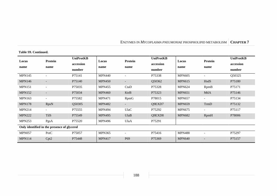
ENZYMES IN MYCOPLASMA PNEUMONIAE PHOSPHOLIPID METABOLISM CHAPTER 7
188
Table S9. Continued.
Locus
name
Protein
name
UniProtKB
accession
number
Locus
name
Protein
name
UniProtKB
accession
number
Locus
name
Protein
name
UniProtKB
accession
number
MPN145 - P75141 MPN440 - P75338 MPN605 - Q50325
MPN146 - P75140 MPN450 - Q50362 MPN615 HsdS P75180
MPN151 - P75035 MPN455 CtaD P75328 MPN624 RpmB P75171
MPN152 - P75034 MPN460 KtrB P75323 MPN651 MtlA P75146
MPN163 - P75582 MPN471 RpmG P78015 MPN657 - P75134
MPN178 RpsN Q50305 MPN482 - Q9EXD7 MPN659 TrmD P75132
MPN214 - P75555 MPN494 UlaC P75292 MPN675 - P75117
MPN222 TilS P75549 MPN495 UlaB Q9EXD8 MPN682 RpmH P78006
MPN253 PgsA P75520 MPN496 UlaA P75291
Only identified in the presence of glycerol
MPN057 PotC P75057 MPN365 - P75416 MPN488 - P75297
MPN114 Cpt2 P75448 MPN417 P69 P75369 MPN640 - P75157
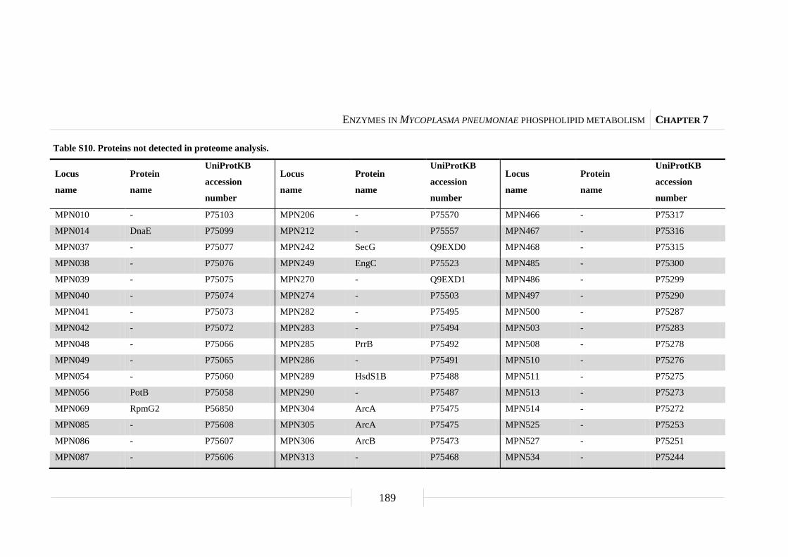
ENZYMES IN MYCOPLASMA PNEUMONIAE PHOSPHOLIPID METABOLISM CHAPTER 7
189
Table S10. Proteins not detected in proteome analysis.
Locus
name
Protein
name
UniProtKB
accession
number
Locus
name
Protein
name
UniProtKB
accession
number
Locus
name
Protein
name
UniProtKB
accession
number
MPN010 - P75103 MPN206 - P75570 MPN466 - P75317
MPN014 DnaE P75099 MPN212 - P75557 MPN467 - P75316
MPN037 - P75077 MPN242 SecG Q9EXD0 MPN468 - P75315
MPN038 - P75076 MPN249 EngC P75523 MPN485 - P75300
MPN039 - P75075 MPN270 - Q9EXD1 MPN486 - P75299
MPN040 - P75074 MPN274 - P75503 MPN497 - P75290
MPN041 - P75073 MPN282 - P75495 MPN500 - P75287
MPN042 - P75072 MPN283 - P75494 MPN503 - P75283
MPN048 - P75066 MPN285 PrrB P75492 MPN508 - P75278
MPN049 - P75065 MPN286 - P75491 MPN510 - P75276
MPN054 - P75060 MPN289 HsdS1B P75488 MPN511 - P75275
MPN056 PotB P75058 MPN290 - P75487 MPN513 - P75273
MPN069 RpmG2 P56850 MPN304 ArcA P75475 MPN514 - P75272
MPN085 - P75608 MPN305 ArcA P75475 MPN525 - P75253
MPN086 - P75607 MPN306 ArcB P75473 MPN527 - P75251
MPN087 - P75606 MPN313 - P75468 MPN534 - P75244

ENZYMES IN MYCOPLASMA PNEUMONIAE PHOSPHOLIPID METABOLISM CHAPTER 7
190
Table S10. Continued.
Locus
name
Protein
name
UniProtKB
accession
number
Locus
name
Protein
name
UniProtKB
accession
number
Locus
name
Protein
name
UniProtKB
accession
number
MPN088 - P75605 MPN334 BcrA P75444 MPN535 RuvA P75243
MPN089 HsdS P75604 MPN335 - P75443 MPN536 RuvB P75242
MPN091 - P75602 MPN343 - P75435 MPN540 RpmF P75238
MPN092 - P75600 MPN346 - P75432 MPN565 - P75213
MPN093 - P75599 MPN347 HsdR P75431 MPN570 - P75208
MPN097 - P75595 MPN363 - P75418 MPN571 LcnDR3 P75207
MPN098 - P75594 MPN364 - P75417 MPN577 - P75203
MPN099 - P75593 MPN366 - P75415 MPN578 - P75202
MPN101 - P75568 MPN367 - P75414 MPN579 - P75201
MPN102 - P75567 MPN369 - P75412 MPN580 - P75200
MPN103 - P75566 MPN370 - P75411 MPN581 - P75199
MPN107 - P75562 MPN371 - P75410 MPN583 - P75197
MPN108 - P75561 MPN373 - P75408 MPN584 - P75196
MPN110 - P75452 MPN374 - P75407 MPN586 - P75194
MPN111 - P75451 MPN375 - P75406 MPN587 - P75193
MPN112 - P75450 MPN388 - Q9EXD4 MPN590 - Q50337

ENZYMES IN MYCOPLASMA PNEUMONIAE PHOSPHOLIPID METABOLISM CHAPTER 7
191
Table S10. Continued.
Locus
name
Protein
name
UniProtKB
accession
number
Locus
name
Protein
name
UniProtKB
accession
number
Locus
name
Protein
name
UniProtKB
accession
number
MPN113 - P75449 MPN403 - P75381 MPN593 - Q50334
MPN127 - P75348 MPN405 - P75379 MPN594 - P75191
MPN128 - P75347 MPN409 - P75375 MPN612 - P75183
MPN129 - P75346 MPN412 - Q9EXD5 MPN613 - P75182
MPN131 - P75267 MPN413 - Q9EXD6 MPN614 - P75181
MPN132 - P75266 MPN437 - P75341 MPN626 - P75169
MPN143 - P75143 MPN438 - P75340 MPN633 - P75164
MPN144 - P75142 MPN439 - P75339 MPN634 - P75163
MPN147 - P75139 MPN441 - P75337 MPN635 - P75162
MPN149 - P75037 MPN442 - P75336 MPN637 CdsA P75160
MPN150 - P75036 MPN448 - Q50364 MPN644 - P75153
MPN160 - P75585 MPN451 Come3 Q50361 MPN648 - P75149
MPN188 RpmJ P52864 MPN457 - P75326 MPN649 - P75148
MPN201 - Q50287 MPN458 - P75325 MPN650 - P75147
MPN202 - Q50286 MPN462 - P75321 MPN654 - P75137
MPN203 - Q50284 MPN463 - P75320 MPN666 - P75125

ENZYMES IN MYCOPLASMA PNEUMONIAE PHOSPHOLIPID METABOLISM CHAPTER 7
192
Table S10. Continued.
Locus
name
Protein
name
UniProtKB
accession
number
Locus
name
Protein
name
UniProtKB
accession
number
Locus
name
Protein
name
UniProtKB
accession
number
MPN204 - P75572 MPN464 - P75319 MPN676 - P75116
MPN205 - P75571 MPN465 - P75318 MPN681 RnpA P75111

ENZYMES IN MYCOPLASMA PNEUMONIAE PHOSPHOLIPID METABOLISM CHAPTER 7
193
Table S11. Summary of proteome and transcript analysis in the glpQ mutant GPM81 in the presence of glucose.
Locus
name
Protein
name
UniProtKB
accession
number
Protein function COGa
Molecular
weight
(kDa)
Isoelectric
point
(pI)
Fold-changeb
Protein
level
Transcript
level
Induced expression
MPN023 MetG P75091 Methionyl-tRNA synthetase J 59.26 6.63 2.25 ± 0.22 na
MPN043 GlpF P75071 Glycerol uptake facilitator G 28.31 9.33 10.79 ± 1.06 5.69 ± 0.36
MPN060 MetK P78003 S-adenosylmethionine synthetase J 42.56 6.07 2.33 ± 0.11 na
MPN162 - P75583 Uncharacterized lipoprotein S 36.10 6.19 9.96 ± 0.97 5.23 ± 0.23
MPN179 RpsH Q50304 30S ribosomal protein S8 J 15.88 10.32 2.63 ± 0.33 na
MPN191 RpoA Q50295 RNA polymerase subunit α K 36.66 6.98 2.30 ± 0.04 2.18 ± 0.17
MPN209 PacL P78036 Cation-transporting P-type ATPase P 94.97 6.38 2.49 ± 0.26 na
MPN221 Pth P78034 Peptidyl-tRNA hydrolase J 21.42 9.52 2.43 ± 0.14 na
MPN223 HPrK P75548 HPr kinase phosphorylase O 35.24 8.89 2.90 ± 0.29 2.83 ± 0.09
MPN239 - P75532 HTH-type transcriptional regulator K 25.85 9.50 2.53 ± 0.17 3.16 ± 0.14
MPN244 DisA P75528 Uncharacterized protein L 22.73 9.38 2.72 ± 0.30 3.39 ± 0.19
MPN247 PrpC P75525 Protein phosphatase O 29.69 8.57 2.43 ± 0.18 2.91 ± 0.12
MPN248 PrkC P75524 Serine threonine-protein kinase O 44.88 9.08 2.33 ± 0.11 na
MPN254 CinA Q9EXC9 Uncharacterized protein S 16.96 8.81 2.60 ± 0.27 2.11 ± 0.10
MPN266 Spx P75509 Transcriptional regulator K 16.81 9.81 2.71 ± 0.13 2.23 ± 0.07

ENZYMES IN MYCOPLASMA PNEUMONIAE PHOSPHOLIPID METABOLISM CHAPTER 7
194
Table S11. Continued.
Locus
name
Protein
name
UniProtKB
accession
number
Protein function COGa
Molecular
weight
(kDa)
Isoelectric
point
(pI)
Fold-changeb
Protein
level
Transcript
level
MPN279 LepA P75498 GTP-binding protein J 66.16 7.58 2.34 ± 0.18 na
MPN299 PlsC P75479 1-acyl-sn-glycerol-3-phosphate acyltransferase I 30.42 9.79 3.20 ± 0.35 6.43 ± 0.31
MPN300 ScpA P75478 Segregation and condensation protein A U 59.52 8.15 2.45 ± 0.18 na
MPN338 - P75440 Uncharacterized protein S 74.34 5.60 2.51 ± 0.23 na
MPN340 PcrA P75438 DNA helicase II L 60.53 6.23 2.43 ± 0.21 na
MPN357 LigA P78021 DNA ligase L 73.97 8.40 2.58 ± 0.30 na
MPN359 - P75421 Uncharacterized protein S 30.56 9.67 2.34 ± 0.24 na
MPN372 - P75409 ADP-ribosylating toxin CARDS V 68.06 5.63 2.91 ± 0.21 2.72 ± 0.20
MPN407 - P75377 Lipase I 101.09 5.81 2.13 ± 0.11 na
MPN408 - P75376 Uncharacterized lipoprotein S 83.35 9.16 3.57 ± 0.26 na
MPN425 FtsY P75362 Cell division protein homolog D 38.78 6.85 2.34 ± 0.21 na
MPN433 CbiO P75355 Metal ion ABC transporter ATP-binding protein P 30.77 8.41 ns 7.56 ± 0.39
MPN456 - P75327 Uncharacterized lipoprotein S 110.52 6.33 2.35 ± 0.18 na
MPN518 - P75269 Uncharacterized protein S 40.72 8.93 3.42 ± 0.12 na
MPN544 - P75234 Uncharacterized protein S 76.77 7.50 5.14 ± 0.48 na
MPN552 - P75226 Uncharacterized protein S 30.91 5.66 4.04 ± 0.41 na
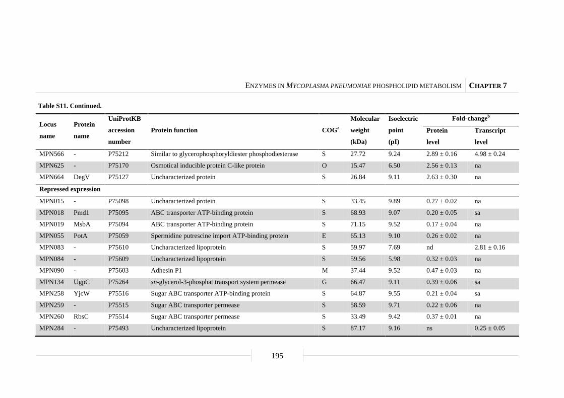
ENZYMES IN MYCOPLASMA PNEUMONIAE PHOSPHOLIPID METABOLISM CHAPTER 7
195
Table S11. Continued.
Locus
name
Protein
name
UniProtKB
accession
number
Protein function COGa
Molecular
weight
(kDa)
Isoelectric
point
(pI)
Fold-changeb
Protein
level
Transcript
level
MPN566 - P75212 Similar to glycerophosphoryldiester phosphodiesterase S 27.72 9.24 2.89 ± 0.16 4.98 ± 0.24
MPN625 - P75170 Osmotical inducible protein C-like protein O 15.47 6.50 2.56 ± 0.13 na
MPN664 DegV P75127 Uncharacterized protein S 26.84 9.11 2.63 ± 0.30 na
Repressed expression
MPN015 - P75098 Uncharacterized protein S 33.45 9.89 0.27 ± 0.02 na
MPN018 Pmd1 P75095 ABC transporter ATP-binding protein S 68.93 9.07 0.20 ± 0.05 sa
MPN019 MsbA P75094 ABC transporter ATP-binding protein S 71.15 9.52 0.17 ± 0.04 na
MPN055 PotA P75059 Spermidine putrescine import ATP-binding protein E 65.13 9.10 0.26 ± 0.02 na
MPN083 - P75610 Uncharacterized lipoprotein S 59.97 7.69 nd 2.81 ± 0.16
MPN084 - P75609 Uncharacterized lipoprotein S 59.56 5.98 0.32 ± 0.03 na
MPN090 - P75603 Adhesin P1 M 37.44 9.52 0.47 ± 0.03 na
MPN134 UgpC P75264 sn-glycerol-3-phosphat transport system permease G 66.47 9.11 0.39 ± 0.06 sa
MPN258 YjcW P75516 Sugar ABC transporter ATP-binding protein S 64.87 9.55 0.21 ± 0.04 sa
MPN259 - P75515 Sugar ABC transporter permease S 58.59 9.71 0.22 ± 0.06 na
MPN260 RbsC P75514 Sugar ABC transporter permease S 33.49 9.42 0.37 ± 0.01 na
MPN284 - P75493 Uncharacterized lipoprotein S 87.17 9.16 ns 0.25 ± 0.05

ENZYMES IN MYCOPLASMA PNEUMONIAE PHOSPHOLIPID METABOLISM CHAPTER 7
196
Table S11. Continued.
Locus
name
Protein
name
UniProtKB
accession
number
Protein function COGa
Molecular
weight
(kDa)
Isoelectric
point
(pI)
Fold-changeb
Protein
level
Transcript
level
MPN322 NrdF P75461 Ribonucleoside-diphosphate reductase subunit β F 39.41 5.47 0.38 ± 0.05 ns
MPN323 NrdI P75460 Ribonucleoside-diphosphate reductase stimulatory protein F 17.15 7.79 0.29 ± 0.06 na
MPN324 NrdE P78027 Ribonucleoside-diphosphate reductase subunit α F 82.38 6.63 0.40 ± 0.05 na
MPN420 GlpQ P75367 Glycerophosphoryldiester phosphodiesterase C 28.37 6.32 nd na
MPN449 - Q50363 Uncharacterized protein S 50.55 9.32 0.10 ± 0.04 na
MPN498 UlaF P75289 L-ribulose-5-phosphate 4-epimerase C 27.09 6.44 0.20 ± 0.04 sa
MPN506 - P75280 Uncharacterized lipoprotein S 87.50 9.24 0.10 ± 0.03 0.07 ± 0.05
MPN684 - P75109 ABC transporter permease S 209.45 8.23 0.32 ± 0.03 sa
MPN685 CysA Q50316 ABC transport ATP-binding protein S 32.24 9.69 0.21 ± 0.04 na a Abbreviations: COG, Cluster of orthologous groups of proteins; C, Energy production and conversion; D, Cell cycle control, cell division, and chromosome partitioning;
E, Amino acid transport and metabolism; F, Nucleotide transport and metabolism; G, Carbohydrate transport and metabolism; I, Lipid metabolism; J, Translation, ribosomal
structure, and biogenesis; K, Transcription; L, Replication, recombination, and repair; M, Cell wall, membrane, and envelope biogenesis; O, Posttranslational modification,
protein turnover, and chaperones; P, Inorganic ion transport and metabolism; S, Function unknown; U, Intracellular trafficking, secretion, and vesicular transport; V, Defense
mechanisms. b Fold-change cut off ≥ 2.0 and ≤ 0.5, respectively (glpQ mutant strain vs. wild type). Abbreviations: na, not available; nd, not detectable; ns, no significant difference; sa, similar
amount.

ENZYMES IN MYCOPLASMA PNEUMONIAE PHOSPHOLIPID METABOLISM CHAPTER 7
197
Table S12. Summary of proteome and transcript analysis in the glpQ mutant GPM81 in the presence of glycerol.
Locus
name
Protein
name
UniProtKB
accession
number
Protein function COGa
Molecular
weight
(kDa)
Isoelectric
point
(pI)
Fold-changeb
Protein
level
Transcript
level
Induced expression
MPN043 GlpF P75071 Glycerol uptake facilitator G 28.31 9.33 5.83 ± 0.76 3.32 ± 0.34
MPN162 - P75583 Uncharacterized lipoprotein S 36.10 6.19 5.41 ± 0.83 2.81 ± 0.47
MPN433 CbiO P75355 Metal ion ABC transporter ATP-binding protein P 30.77 8.41 5.66 ± 0.49 2.92 ± 0.24
MPN444 - P75334 Uncharacterized lipoprotein S 146.28 7.89 2.74 ± 0.09 na
MPN489 - P75296 Uncharacterized lipoprotein S 143.06 9.14 2.40 ± 0.20 na
Repressed expression
MPN284 - P75493 Uncharacterized lipoprotein S 87.17 9.16 0.36 ± 0.02 0.35 ± 0.04
MPN288 - P75489 Uncharacterized lipoprotein S 86.89 9.07 0.29 ± 0.03 na
MPN420 GlpQ P75367 Glycerophosphoryldiester phosphodiesterase C 28.37 6.32 nd na
MPN506 - P75280 Uncharacterized lipoprotein S 87.50 9.24 0.18 ± 0.04 0.16 ± 0.02
MPN673 - P75118 Uncharacterized protein S 19.47 9.16 0.21 ± 0.03 na a Abbreviations: COG, Cluster of orthologous groups of proteins; C, Energy production and conversion; G, Carbohydrate transport and metabolism; P, Inorganic ion transport and
metabolism; S, Function unknown. b Fold-change cut off ≥ 2.0 and ≤ 0.5, respectively (glpQ mutant strain vs. wild type). Abbreviations: na, not available; nd, not detectable.
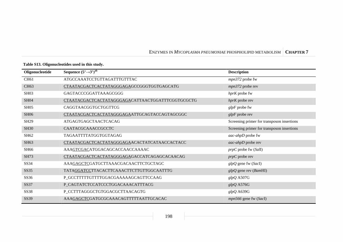
ENZYMES IN MYCOPLASMA PNEUMONIAE PHOSPHOLIPID METABOLISM CHAPTER 7
198
Table S13. Oligonucleotides used in this study.
Oligonucleotide Sequence (5’→3’)ab Description
CH61 ATGCCAAATCCTGTTAGATTTGTTTAC mpn372 probe fw
CH63 CTAATACGACTCACTATAGGGAGAGCCGGGTGGTGAGCATG mpn372 probe rev
SH03 GAGTACCCGGATTAAAGCGGG hprK probe fw
SH04 CTAATACGACTCACTATAGGGAGACATTAACTGGATTTCGGTGCGCTG hprK probe rev
SH05 CAGGTAACGGTGCTGGTTCG glpF probe fw
SH06 CTAATACGACTCACTATAGGGAGAATTGCAGTACCAGTAGCGGC glpF probe rev
SH29 ATGAGTGAGCTAACTCACAG Screening primer for transposon insertions
SH30 CAATACGCAAACCGCCTC Screening primer for transposon insertions
SH62 TAGAATTTTATGGTGGTAGAG aac-ahpD probe fw
SH63 CTAATACGACTCACTATAGGGAGAACACTATCATAACCACTACC aac-ahpD probe rev
SH66 AAAGTCGACATGGACAGCACCAACCAAAAC prpC probe fw (SalI)
SH73 CTAATACGACTCACTATAGGGAGAGACCATCAGAGCACAACAG prpC probe rev
SS34 AAAGAGCTCGATGCTTAAACGACAACTTCTGCTAGC glpQ gene fw (SacI)
SS35 TATAGGATCCTTACACTTCAAACTTCTTGTTGGCAATTTG glpQ gene rev (BamHI)
SS36 P_GCCTTTTTGTTTTGGACGAAAAAGCAGTTCCAAG glpQ A507G
SS37 P_CAGTATCTCCATCCCTGGACAAACATTTACG glpQ A576G
SS38 P_CCTTTAGGGCTGTGGACGCTTAACAGTG glpQ A639G
SS39 AAAGAGCTCGATGCGCAAACAGTTTTTAATTGCACAC mpn566 gene fw (SacI)

ENZYMES IN MYCOPLASMA PNEUMONIAE PHOSPHOLIPID METABOLISM CHAPTER 7
199
Table S13. Continued.
Oligonucleotide Sequence (5’→3’)ab Description
SS40 TATAGGATCCTTAGTAAAGTTGTGCTGCTATTTGAAATTTAAC mpn566 gene rev (BamHI)
SS42 CAACTTCTGCTAGCACACCG glpQ probe fw
SS43 CTAATACGACTCACTATAGGGAGAGCTATTTGGTAGTTGGGGTTAATG glpQ probe rev
SS44 GCAAACAGTTTTTAATTGCACACCG mpn566 probe fw
SS45 CTAATACGACTCACTATAGGGAGAGCTCTTTAACTTTTCGTTGAGGTAC mpn566 probe rev
SS123 GAATCAGTTTCTCCCTTAGAATATGC nrdF probe fw
SS124 CTAATACGACTCACTATAGGGAGAGTCTTTCCCGGTGTAATAGGG nrdF probe rev
SS127 CCAACAGCGCTTTTATTCTCGG mpn083 probe fw
SS128 CTAATACGACTCACTATAGGGAGAGGACATTAGGTTTGGTGTACTTAC mpn083 probe rev
SS129 CCTTGTTAGTTGCGCCACAC mpn162 probe fw
SS130 CTAATACGACTCACTATAGGGAGACACCTTCGTGGTGATCATGATC mpn162 probe rev
SS131 GGCTTAGTCATCCACACTTGG ulaF probe fw
SS132 CTAATACGACTCACTATAGGGAGACCACTGCATCCTTGCCATTC ulaF probe rev
SS135 GGTTTTACCCGTTTTTGTGTTAATGC plsC probe fw
SS136 CTAATACGACTCACTATAGGGAGAGCCCGATTTAAACTCACCAATTTG plsC probe rev
SS137 CCGTTACATTCTCCTTAAAATTCAAAG mpn239 probe fw
SS138 CTAATACGACTCACTATAGGGAGAGCTCTACCACAATGCCGTTG mpn239 probe rev
SS139 ATGCTTAAGAAAAAAGTTAATAATGATGCTG spx probe fw
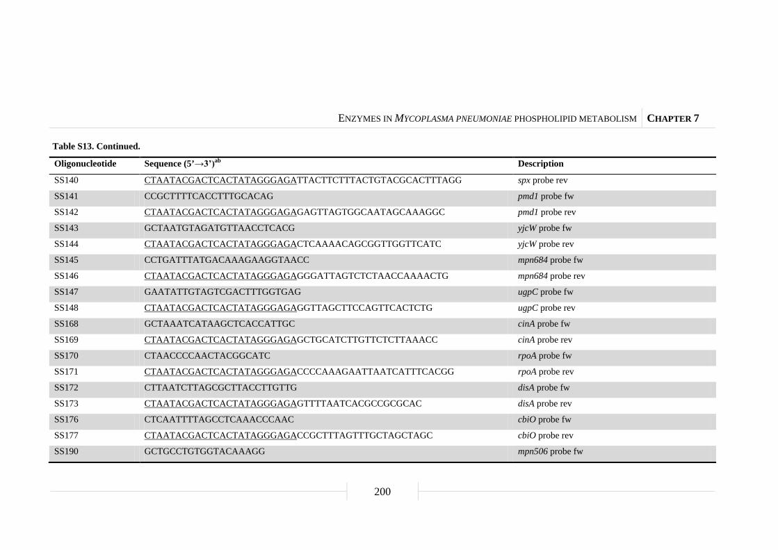
ENZYMES IN MYCOPLASMA PNEUMONIAE PHOSPHOLIPID METABOLISM CHAPTER 7
200
Table S13. Continued.
Oligonucleotide Sequence (5’→3’)ab Description
SS140 CTAATACGACTCACTATAGGGAGATTACTTCTTTACTGTACGCACTTTAGG spx probe rev
SS141 CCGCTTTTCACCTTTGCACAG pmd1 probe fw
SS142 CTAATACGACTCACTATAGGGAGAGAGTTAGTGGCAATAGCAAAGGC pmd1 probe rev
SS143 GCTAATGTAGATGTTAACCTCACG yjcW probe fw
SS144 CTAATACGACTCACTATAGGGAGACTCAAAACAGCGGTTGGTTCATC yjcW probe rev
SS145 CCTGATTTATGACAAAGAAGGTAACC mpn684 probe fw
SS146 CTAATACGACTCACTATAGGGAGAGGGATTAGTCTCTAACCAAAACTG mpn684 probe rev
SS147 GAATATTGTAGTCGACTTTGGTGAG ugpC probe fw
SS148 CTAATACGACTCACTATAGGGAGAGGTTAGCTTCCAGTTCACTCTG ugpC probe rev
SS168 GCTAAATCATAAGCTCACCATTGC cinA probe fw
SS169 CTAATACGACTCACTATAGGGAGAGCTGCATCTTGTTCTCTTAAACC cinA probe rev
SS170 CTAACCCCAACTACGGCATC rpoA probe fw
SS171 CTAATACGACTCACTATAGGGAGACCCCAAAGAATTAATCATTTCACGG rpoA probe rev
SS172 CTTAATCTTAGCGCTTACCTTGTTG disA probe fw
SS173 CTAATACGACTCACTATAGGGAGAGTTTTAATCACGCCGCGCAC disA probe rev
SS176 CTCAATTTTAGCCTCAAACCCAAC cbiO probe fw
SS177 CTAATACGACTCACTATAGGGAGACCGCTTTAGTTTGCTAGCTAGC cbiO probe rev
SS190 GCTGCCTGTGGTACAAAGG mpn506 probe fw
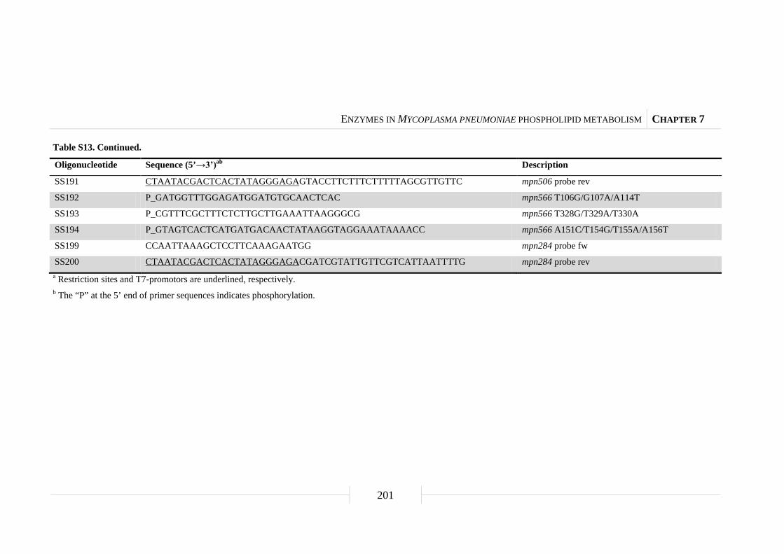
ENZYMES IN MYCOPLASMA PNEUMONIAE PHOSPHOLIPID METABOLISM CHAPTER 7
201
Table S13. Continued.
Oligonucleotide Sequence (5’→3’)ab Description
SS191 CTAATACGACTCACTATAGGGAGAGTACCTTCTTTCTTTTTAGCGTTGTTC mpn506 probe rev
SS192 P_GATGGTTTGGAGATGGATGTGCAACTCAC mpn566 T106G/G107A/A114T
SS193 P_CGTTTCGCTTTCTCTTGCTTGAAATTAAGGGCG mpn566 T328G/T329A/T330A
SS194 P_GTAGTCACTCATGATGACAACTATAAGGTAGGAAATAAAACC mpn566 A151C/T154G/T155A/A156T
SS199 CCAATTAAAGCTCCTTCAAAGAATGG mpn284 probe fw
SS200 CTAATACGACTCACTATAGGGAGACGATCGTATTGTTCGTCATTAATTTTG mpn284 probe rev a Restriction sites and T7-promotors are underlined, respectively. b The “P” at the 5’ end of primer sequences indicates phosphorylation.
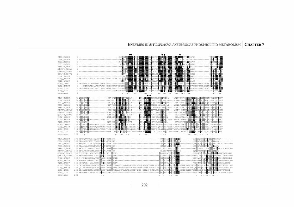
ENZYMES IN MYCOPLASMA PNEUMONIAE PHOSPHOLIPID METABOLISM CHAPTER 7
202
Y420_MYCPN 1 ---------------------------------MLKRQLLLAHRGYSDIAPENTQLAFELAFQYRFDGVELDVHLTKDGELVIIHDETTTRTALVDK------------TIELETLASLKY566_MYCPN 1 ----------------------------------MRKQFLIAHRGYSTIAPENTHLAFAAAQLFGFDGLWMEVQLTKDKQIVVTNLDNYK-VGNKTH------------KLSDINLVNLKY293_MYCGE 1 ---------------------------------MHNKQLLLAHRGYSFIAPENTKLAFDLAFEYCFDGIELDVHLTKDEQLVIIHDETTLRTALVNK------------EVEFESLVSLKY385_MYCGE 1 ----------------------------------MRKQFLIAYRGYSSIAPENTKLAFQAAQVFDFDGIWIDIQLTKDKQIVVTHKENFK-VSNKNL------------NLNQINLVDLKD4XVJ7_9MOLU 1 ---------------------------------MNKKIYKIAHRGYSSLYPENTKLAFSKALDSGFDGVEIDLHLTKDNQIVIIHDNNTKRLTDKNL------------VVKNSNLEELKD4XVF1_9MOLU 1 -----------------------------------MKQILLAHRGYSGIAPENTRLAFDAALIFGFDGFEIDVHQTKDNVLVVIHDETTNRTAKSNR------------TVGKSTYKELLQ8XNB7_CLOPE 1 ----------------------------------MKKILNVAHRGLSGLYPENTMIAFRKAIEADCDGIEMDVHLTRDGVPVVIHDEEVDRTTNGSG------------YVKDFTYEQLCQ8XJ84_CLOPE 1 -----------------------------------MKVF--AHRGFSYKYPENTLLAFKEALKLDIYGIELDVHKSKDGKLVIIHDEDIKRTFKGEG------------KVKDYTFEELRYHDW_BACSU 1 -------------------------------------MYIIAHRGASGYAPENTIAAFDLAVKMNADMIELDVQLTKDRQIVVIHDDRVDRTTNGSG------------FVKDFTLEELQGLPQ_BACSU 1 MRKNRILALFVLSLGLLSFMVTPVSAASKGNLLSPDRILTVAHRGASGYVPEHTILSYETAQKMKADFIELDLQMTKDGKLIVMHDEKLDRTTNGMG------------WVKDHTLADIKYQIK_BACSU 1 ----------------------------------MTKIF--AHRGASGQFPENTMLAFEKGIEAGADGIELDVQLTKDGRIVVIHDERLNRTTSLKG------------FVKDTAYDEVKGLPQ_TREPA 1 -MRGTYCVTLWGGVFAALVAGCAS------------ERMIVAYRGAAGYVPEHTFASKVLAFAQGADYLQQDVVLSKDNQLIVAQSHILDNMTDVAEKFPRRQRADGHFYVIDFTVEELSGLPQ_HAEIN 1 ---MKLKTLALSLLAAGVLAGCSSHSSNMANTQMKSDKIIIAHRGASGYLPEHTLESKALAFAQHSDYLEQDLAMTKDGRLVVIHDHFLDGLTDVAKKFPYRHRKDGRYYVIDFTLKEIQGLPQ_ECOLI 1 -MKLTLKNLSMAIMMSTIVMGSSAMAADSN------EKIVIAHRGASGYLPEHTLPAKAMAYAQGADYLEQDLVMTKDDNLVVLHDHYLDRVTDVADRFPDRARKDGRYYAIDFTLDEIKUGPQ_ECOLI 1 -------------------------------MSNWPYPRIVAHRGGGKLAPENTLASIDVGAKYGHKMIEFDAKLSKDGEIFLLHDDNLERTSNGWG------------VAGELNWQDLLconsensus 1 . ...*.** ... **.*.... . ....... ...* ....... . ... . . .. ...
Y420_MYCPN 76 QDDHSAFFK---------------FKTQPQPIMTLKEFFDQYLD------KFQLINVEIKTDQKEYPGIEAKIDA----LAQQYGKKVIEKVVFSSFNFASLQRLYDINPNYQIAFLFWTY566_MYCPN 74 KINLAKQFK---------------VNVQEQTILTLKEVLMEFLT------PFRFLLLFIKGEEEQQNQVLVEQLA----QLLEGFELAKEKLILLSSHFSTIKYLNEKLKSFKTSFVFNSY293_MYCGE 76 RDDHSAFFH---------------LKIQFQSILTLKEFLDLYLD------KFKLINIEIKTDQKPYLGIEKKLVD----LVKGYGKKAIDKILFSSFNFESLQKVYDLDNSYKKGFLFWTY385_MYCGE 74 KVNLASEFK---------------LKVTSQQIQTLKEVLTQFIQ------PFKYLLIHIKDDKEN-NNSLLEQLN----LLCKDFVLAKEKMILLSSNFHIIKLINETFKGFKTGFVIAND4XVJ7_9MOLU 76 KLNLAAKYK---------------EIGLFEQILTIEEFLELYVN------KFDLINLEIKVTKQKICKFVKILLN----KLSP-YLNNINKFVFSSFNYDVLYELNKNDKNLKLAFLCKTD4XVF1_9MOLU 74 EDDHSLFMG---------------KNLPIQRILTLEELLDLYIE------HVQYLNIEIKTDVTEYEGIEENINN----LMSKY-KKHWNKIIFSSFNYNSLRRLYVLNPKLRYGFLWWKQ8XNB7_CLOPE 75 NLDAGIKF---------------SDEFKGEKIPTLKEFFDFMKD------NNKLINIELKNSIIHYEGLEEKVY------KEIENYGFEDRVIISSFDHYSIRKCIRLNKNIKTGILYWDQ8XJ84_CLOPE 72 NFKCNKEG---------------FENNDDCKISLLEDVFNLIKD------KDIVLNIEIKNDVIDYENIEKDVL------DLIKEYNLERKILISSFNHKALEKVKKLNGDIKLGVLYEKYHDW_BACSU 72 KLDAGSWY---------------GPAFQGERIPTLEAVLKRYH-------KKIGLLIELK-GHPSQVGIEEEVG------QLLGQFSFSINNIVQSFQFRSVQRFRELYPSIPTAVITRPGLPQ_BACSU 109 KLDAGSWFNE-------AYPEKAKPQYVGLKVPTLEEVLDRFG-------KHANYYIETK-SPDTYPGMEEKLIASLQKHKLLGKHSKPGQVIIQSFSKESLVKVHQLQPNLPTVQLLEAYQIK_BACSU 73 TANAAAGH---------------DQAYSDIKVPLLEDVLSWAVK------KDFLINIELKNSVIRYEGMEEKVL------EAVKRFNVEDRVILSTFNHDSLALCARLAPHIERAVLTSDGLPQ_TREPA 108 LLRATNSFYTRGKRHTPVYGQRFPLWKPGFRLHTFEEELQFIRGLEQTTGKKIGIYSEIKVPWFHHQEGKDIAALTLALLKKYGYQSRSDLVYVQTYDFNELKRIKRELLPKYEMNVKLIGLPQ_HAEIN 118 SLEMTENFETKDGKQAQVYPNRFPLWKSHFRIHTFEDEIEFIQGLEKSTGKKVGIYPEIKAPWFHHQNGKDIATETLKVLKKYGYDKKTDMVYLQTFDFNELKRIKTELLPQMGMDLKLVGLPQ_ECOLI 114 SLKFTEGFDIENGKKVQTYPGRFPMGKSDFRVHTFEEEIEFVQGLNHSTGKNIGIYPEIKAPWFHHQEGKDIAAKTLEVLKKYGYTGKDDKVYLQCFDADELKRIKNELEPKMGMELNLVUGPQ_ECOLI 78 RVDAGSWYS---------------KMFKGEPLPLLSQVAERCRE------HGMMANIEIKPTTGTGPLTGKMVAL-----AARELWAGMTPPLLSSFEIDALEAAQQAAPELPRGLLLDEconsensus 121 .. . . .. ....... . . .....* ... . .......... .. .. . . . .
Y420_MYCPN 171 KKQFQAVDALKIKQVCQYLHPWTNIYEKFP--------------------------------------DMVLSLQLPLGLWTLNS----------EVKFHQFRQDRMVYAQIANKKFEV----------Y566_MYCPN 169 KKQLVHLTKDEITQNCRFLAPNDSFYHKNC--------------------------------------AELQSYGLPIILWVIKG----------LLRYQFYENDRFVKFQIAAQLY------------Y293_MYCGE 171 KKQFETISTARIQKICQFLHPWTKIYEKYP--------------------------------------QMIKKLNLPLNLWTVNS----------QNKFQQFLADNHVYAQIANKKFEIKIN-------Y385_MYCGE 168 KKALLVASRDELMRYCRFLVPNESFYHKNC--------------------------------------KEIQNLGLPVILWVIKG----------LLRYQFYENDRFVKFQITAQIY------------D4XVJ7_9MOLU 170 KKALLNISKFNLKIIT-YLAPSLNSFRINK--------------------------------------KFYDSYNKYYLFWTLKT----------KKSFLKYSKFLNTFGLILHKPFWYKKQRNNKK--D4XVF1_9MOLU 168 ESQLKNINVSEMLSFCEFLHPWLPMYEKDK--------------------------------------ELYLSWNKKFCLWTIKS----------IQNFDKYKNDKNVFAQISNYKY------------Q8XNB7_CLOPE 168 CIFEPHN--YCKVVGSSALHPEFNS--ITEEI-----------------------------------VRDAHEDFQQVNVYTVN-----------EKKDMEKMIDLNVDMIITNYPNILKEVLESKNK-Q8XJ84_CLOPE 165 EMPNILD--IAKSLNAYAIHPAKSL--VSEEL-----------------------------------VEKAHNEGVKVNVYTVN-----------EKEDIDKLKNYGVDAIFTDQAKMALEYLK-----YHDW_BACSU 163 N-FGMLSRNQMKAFRSFANYVNIKHTRLNRLM-----------------------------------IGSINKNGLNIFAWTVN-----------NQKTAAKLQAMGVDGIVTDYPDFIIKDGKHENI-GLPQ_BACSU 214 KQMASMTDAALEEIKTYAVGAGPDYKALNQEN-----------------------------------VRMIRSHGLLLHPYTVN-----------NEADMHRLLDWGVTGVFTNYPDLFHKVKKGY---YQIK_BACSU 166 VLYQADR--YIASIPASGYHPKINSPGVTDEV-----------------------------------LKKMRNGLIKVRPYTVN-----------RPEDMKRLIEAGADGMFTDFPEKASALLKNE---GLPQ_TREPA 228 QRVAYTDQRETQEKDSRGKWINYNYNWMFEPGGMQKIAKYADGVGPDWRMLIENEWSKVGAVRLSPMVSAIQDAKLECHVHTVRKETLPSYARTMDEMFSILFKQTGANVVLTDFPDLGVKFLGKPARYGLPQ_HAEIN 238 QLIAYTDWKETQEKDPKGYWVNYNYDWMFKPGAMAEVVKYADGVGPGWYMLVNKEESKPDNIVYTPLVKELAQYNVEVHPYTVRKDALPEFFTDVNQMYDALLNKSGATGVFTDFPDTGVEFLKGIK--GLPQ_ECOLI 234 QLIAYTDWNETQQKQPDGSWVNYNYDWMFKPGAMKQVAEYADGIGPDYHMLI-EETSQPGNIKLTGMVQDAQQNKLVVHPYTVRSDKLPEYTPDVNQLYDALYNKAGVNGLFTDFPDKAVKFLNKE---UGPQ_ECOLI 172 WRDDWRELTARLGCVSIHLNHKLLNKAR---------------------------------------VMQLKDAGLRILVYTVNK-----------PQRAAELLRWGVDCICTDAIDVIGPNFTAQ---consensus 241 .. . . . . . . ..... . . . .. .... ..
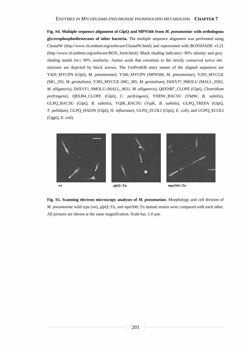
ENZYMES IN MYCOPLASMA PNEUMONIAE PHOSPHOLIPID METABOLISM CHAPTER 7
203
Fig. S4. Multiple sequence alignment of GlpQ and MPN566 from M. pneumoniae with orthologous
glycerophosphodiesterases of other bacteria. The multiple sequence alignment was performed using
ClustalW (http://www.ch.embnet.org/software/ClustalW.html) and represented with BOXSHADE v3.21
(http://www.ch.embnet.org/software/BOX_form.html). Black shading indicates ≥ 80% identity and grey
shading stands for ≥ 80% similarity. Amino acids that constitute to the strictly conserved active site
structure are depicted by black arrows. The UniProtKB entry names of the aligned sequences are
Y420_MYCPN (GlpQ, M. pneumoniae), Y566_MYCPN (MPN566, M. pneumoniae), Y293_MYCGE
(MG_293, M. genitalium), Y385_MYCGE (MG_385, M. genitalium), D4XVJ7_9MOLU (MALL_0582,
M. alligatoris), D4XVF1_9MOLU (MALL_0631, M. alligatoris), Q8XNB7_CLOPE (GlpQ, Clostridium
perfringens), Q8XJ84_CLOPE (GlpQ, C. perfringens), YHDW_BACSU (YhdW, B. subtilis),
GLPQ_BACSU (GlpQ, B. subtilis), YQIK_BACSU (YqiK, B. subtilis), GLPQ_TREPA (GlpQ,
T. pallidum), GLPQ_HAEIN (GlpQ, H. influenzae), GLPQ_ECOLI (GlpQ, E. coli), and UGPQ_ECOLI
(UgpQ, E. coli).
Fig. S5. Scanning electron microscopy analyses of M. pneumoniae. Morphology and cell division of
M. pneumoniae wild type (wt), glpQ::Tn, and mpn566::Tn mutant strains were compared with each other.
All pictures are shown at the same magnification. Scale bar, 1.0 µm.
wt glpQ::Tn mpn566::Tn

ENZYMES IN MYCOPLASMA PNEUMONIAE PHOSPHOLIPID METABOLISM CHAPTER 7
204
Fig. S6. Transcription analysis of interesting genes with protein amount changes in the glpQ
mutant. Slot blots were performed with whole RNA extracts of M. pneumoniae wild type (wt), glpQ::Tn,
mpn566::Tn, and glpD::Tn [control; Hames et al. (2009)] mutant strains grown in modified Hayflick
medium containing either glucose or glycerol as sole carbon source (1% wt/vol). A dilution series of
RNA extracts was blotted onto a positively charged nylon membrane and probed with a DIG-labeled
riboprobe specific for an internal part of a particular open reading frame. Names of riboprobes are given
next to each blot. Signals obtained with 1 µg of RNA are shown. Yeast tRNA and M. pneumoniae
chromosomal DNA served as controls. Genes which had a significant higher protein amount in the
glpQ::Tn mutant with glucose as carbon source are shown in (A) and genes with significant lower protein
amounts in (B). For detailed information on changes of transcript levels see Table S11 and S12.
rpoA
mpn239
hprK
prpC
disA
cinA
plsC
spx
mpn372
mpn566
pmd1
ugpC
mpn083
nrdF
yjcW
ulaF
mpn684
A B
Glucose Glycerol
Glucose Glycerol

Chapter 8
Discussion
DISCUSSION CHAPTER 8
206
Protein phosphorylation in a minimal organism and its impact on
virulence
Although much work has been devoted to the Mollicutes in the past few years,
only little is known about their regulatory mechanisms. One major question is, how
Mycoplasma pneumoniae cells recognize whether they are approaching a host tissue or
not, and which changes in protein activities and/or gene expressions are caused. The
most prominent regulatory mechanism both in bacteria and in eukaryotes is the control
of protein activity by protein phosphorylation (Boekhorst et al., 2008; Jers et al., 2008).
Bacteria possess two different classes of phosphorylation events. In addition to histidine
and aspartate phosphorylation as part of two-component signal transduction (Hoch,
2000; Stock et al., 2000), serine/threonine/tyrosine phosphorylation is also present and
comes more and more into the spotlight. Phosphoproteome studies have been performed
for several bacteria, including the model organisms Escherichia coli and Bacillus
subtilis (Eymann et al., 2007; Lévine et al., 2006; Macek et al., 2008; Macek et al.,
2007) or pathogens like Klebsiella pneumoniae or Streptococcus pneumoniae (Lin et al.,
2009; Sun et al., 2010).
The analysis of the phosphoproteome of M. pneumoniae revealed substantial
protein serine/threonine/tyrosine phosphorylation even in this minimal organism. In
total 63 phosphorylated proteins could be identified that account for nearly 10% of all
proteins encoded by M. pneumoniae. Compared to only 2.5% of all proteins of
B. subtilis that are known to be phosphorylated on a serine, threonine or tyrosine residue
(Eymann et al., 2007; Lévine et al., 2006; Macek et al., 2007), it can be speculated that
protein phosphorylation occurs more frequently in M. pneumoniae than in other
bacteria. This might be related to the lack of an obvious transcription regulation
machinery in M. pneumoniae (Dandekar et al., 2000; Himmelreich et al., 1996).
Nevertheless, the size of all studied bacterial phosphoproteomes so far is remarkably
similar suggesting a conservation of phosphorylated proteins. A comparison of the
phosphoproteomes revealed a high proportion of phosphoproteins, including glycolytic
enzymes and elongation factors that are conserved throughout the bacterial kingdom
(Soufi et al., 2008; this work). However, phosphorylation sites are hardly conserved and
only one site, a serine residue of phosphosugar mutases, is actually identical in all living
organisms from archaea and bacteria to man (Soufi et al., 2008; this work). This
observation might be due to the fact that site specific phosphorylation coevolved with
DISCUSSION CHAPTER 8
207
the adaptation of an organism to an individual ecological niche. Furthermore, the
phosphorylation process might also partially result from gene transfer as many
eukaryote-like kinases have been found in bacteria by metagenomic approaches
(Yooseph et al., 2007). This suggests that regulation via protein phosphorylation
developed relatively late in the evolution. In general, eukaryotic phosphosites are more
conserved throughout the eukaryotic domain (Gnad et al., 2010). An explanation for
this could be that in eukaryotes phosphorylation regulates many key processes including
cell growth, proliferation, differentiation, and immune response (Hubbard and Miller,
2007; Pawson and Scott, 2005), processes which are not present in bacteria.
The knowledge about phosphorylation events and their regulation is crucial to
understand the functional biology of an organism. M. pneumoniae contains two protein
kinases, the HPr kinase (HPrK) and a serine/threonine protein kinase C (PrkC)
(Steinhauer et al., 2002; this work). For HPrK, only an implication in the
phosphorylation of the HPr protein could be observed. In B. subtilis and other
Firmicutes, the phosphorylation of HPr on Ser-46 is a major signal to trigger carbon
catabolite repression (Görke and Stülke, 2008). In this case, HPr(Ser-P) acts as the
cofactor for the pleiotropic transcription factor CcpA to regulate the expression of
catabolic genes and operons. However, no such protein is encoded in the genome of
M. pneumoniae suggesting a different role for HPr(Ser-P). This idea is supported by the
fact that also the proteome of the hprK mutant was similar to the wild type strain.
The kinase PrkC is implicated in various cellular processes in different bacteria,
among them spore germination, virulence, and control of glycolysis (Kristich et al.,
2007; Lomas-Lopez et al., 2007; Shah et al., 2008). In M. pneumoniae, six target
proteins could be identified, including the major adhesin P1, two large cytadherence
proteins HMW1 and HMW2, the adhesin-related protein P41, the coiled coil surface
protein MPN474, and a protein of unknown function MPN256 (Fig. 36A).
Phosphorylation of most of these proteins was reported earlier (Krebes et al., 1995;
Su et al., 2007). However, the corresponding kinase had not been identified. The HMW
proteins and P1 are part of the so-called terminal organelle of M. pneumoniae that is
involved in gliding motility, cell division, and adhesion to host epithelial tissues
(Balish and Krause, 2006; Miyata, 2010). PrkC-dependent phosphorylation seems to be
required for the stability of this protein complex due to the observation that an
inactivation of the kinase led not only to a loss of protein phosphorylation, but also to a

DISCUSSION CHAPTER 8
208
decrease of the protein amount of the target proteins and also additional cytadherence
proteins. It is known that the cytadherence proteins show a reciprocal dependency in
their stabilities (Balish and Krause, 2006). Specifically, HMW1 was shown to be
required for HMW2 and P1 localization and stability (Willby et al., 2004). In turn,
HMW2 is necessary for the stabilization of HMW3 and P65 (Fisseha et al., 1999). As
shown in this work, it has been suggested previously that the interdependence of the
proteins of the attachment organelle is regulated posttranslationally (Popham et al.,
1997). In conclusion, PrkC is essential for cell adhesion in M. pneumoniae, which is
also reflected by a nonadherent growth type of the prkC mutant strain. Furthermore, this
mutant has lost cytotoxicity toward HeLa cells, most likely due to defects in cell
adhesion. Interestingly, it has been reported that the prpC mutant has a reduced gliding
velocity (Hasselbring et al., 2006). Indeed, phosphorylation of the six PrkC target
proteins was enhanced in the prpC mutant strain suggesting that PrpC is the protein
phosphatase that reverses PrkC-dependent protein phosphorylation in M. pneumoniae
(see Fig. 36A). Thus, the antagonistic PrkC/PrpC pair has a specific function in
M. pneumoniae and phosphorylates/dephosphorylates a specific set of proteins.
Nevertheless, PrpC is implicated in two more dephosphorylation events. First in the
dephosphorylation of HPr(Ser-P) (Halbedel et al., 2006) and in addition, PrpC seems
also to dephosphorylate RpoE, the RNA polymerase subunit δ (Fig. 36B).
Unfortunately, the role of both dephosphorylation events is unknown.
As already mentioned, HPrK and PrkC are the only two annotated protein
kinases in M. pneumoniae. However, these known kinases were not responsible for the
majority of phosphorylation events, since only seven phosphoproteins were affected by
the loss of one of these two kinases. This suggests the existence of other, yet to be
discovered protein kinases in M. pneumoniae. Alternatively, autophosphorylation of
proteins might be more relevant than previously anticipated. Indeed, it could be shown
in this work that the only universally conserved phosphorylation event in all living
organisms is the result of an autophosphorylation in the catalytic site of phosphosugar
mutases such as B. subtilis GlmM (Ser-100) and M. pneumoniae ManB (Ser-149). For
the orthologue of both phosphosugar mutases in E. coli, it was established that the
protein is only active in the phosphorylated form (Jolly et al., 2000;
Mengin-Lecreulx et al., 1996). Thus, autophosphorylation is essential for the enzymatic
activity of these enzymes. In general, mutases are widespread over all phyla of life
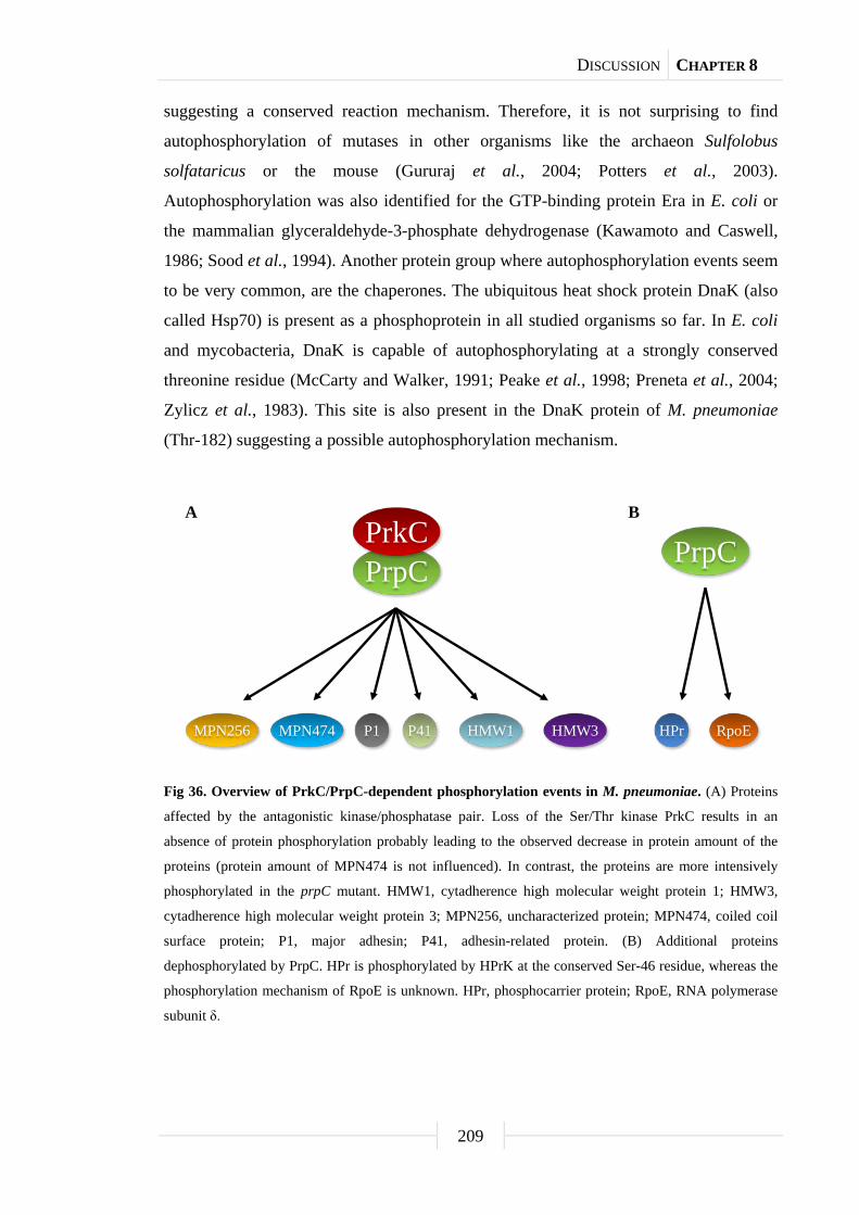
DISCUSSION CHAPTER 8
209
suggesting a conserved reaction mechanism. Therefore, it is not surprising to find
autophosphorylation of mutases in other organisms like the archaeon Sulfolobus
solfataricus or the mouse (Gururaj et al., 2004; Potters et al., 2003).
Autophosphorylation was also identified for the GTP-binding protein Era in E. coli or
the mammalian glyceraldehyde-3-phosphate dehydrogenase (Kawamoto and Caswell,
1986; Sood et al., 1994). Another protein group where autophosphorylation events seem
to be very common, are the chaperones. The ubiquitous heat shock protein DnaK (also
called Hsp70) is present as a phosphoprotein in all studied organisms so far. In E. coli
and mycobacteria, DnaK is capable of autophosphorylating at a strongly conserved
threonine residue (McCarty and Walker, 1991; Peake et al., 1998; Preneta et al., 2004;
Zylicz et al., 1983). This site is also present in the DnaK protein of M. pneumoniae
(Thr-182) suggesting a possible autophosphorylation mechanism.
Fig 36. Overview of PrkC/PrpC-dependent phosphorylation events in M. pneumoniae. (A) Proteins
affected by the antagonistic kinase/phosphatase pair. Loss of the Ser/Thr kinase PrkC results in an
absence of protein phosphorylation probably leading to the observed decrease in protein amount of the
proteins (protein amount of MPN474 is not influenced). In contrast, the proteins are more intensively
phosphorylated in the prpC mutant. HMW1, cytadherence high molecular weight protein 1; HMW3,
cytadherence high molecular weight protein 3; MPN256, uncharacterized protein; MPN474, coiled coil
surface protein; P1, major adhesin; P41, adhesin-related protein. (B) Additional proteins
dephosphorylated by PrpC. HPr is phosphorylated by HPrK at the conserved Ser-46 residue, whereas the
phosphorylation mechanism of RpoE is unknown. HPr, phosphocarrier protein; RpoE, RNA polymerase
subunit δ.
PrpCPrkC
RpoEHPr
PrpC
HMW3MPN474 HMW1MPN256 P41P1
A B

DISCUSSION CHAPTER 8
210
Glycerophosphodiesterases: Multiple roles for universal proteins
M. pneumoniae exhibits a hemolytic activity. While hemolysis by bacterial
pathogens is usually caused by extracellular enzymes, the hemolysin of M. pneumoniae
was identified as hydrogen peroxide (Somerson et al., 1965). The synthesis of hydrogen
peroxide by M. pneumoniae is strongly increased if the cells are supplied with glycerol
(Low, 1971). This can be attributed to the oxidase activity of the glycerol-3-phosphate
oxidase GlpD that oxidizes glycerol 3-phosphate (G3P). This enzyme uses molecular
oxygen rather than NAD+ (as in typical glycerol-3-phosphate dehydrogenases) as the
sink for electrons (Hames et al., 2009). Three potential pathways for the acquisition of
G3P exist in M. pneumoniae. Glycerol may cross the cell membrane, mediated by the
glycerol facilitator GlpF, and be phosphorylated by the glycerol kinase GlpK.
Furthermore, M. pneumoniae can acquire G3P by direct uptake via the ABC transport
system UgpAEC or hydrolysis of glycerophosphodiesters, an abundant degradation
product of phospholipids, by glycerophosphodiesterase activity. While the first pathway
is thought to be the most prominent one, it could be shown in this work that direct
uptake of G3P seems not to occur in M. pneumoniae. Nevertheless, two potential
glycerophosphodiesterases are present in the genome of M. pneumoniae, i.e. glpQ
(mpn420) and mpn566 (Dandekar et al., 2000; Himmelreich et al., 1996). Interestingly,
the pulmonary surfactant, the natural habitat of M. pneumoniae, is composed of about
90% phospholipids and 10% proteins (Veldhuizen et al., 1998) indicating that
glycerophosphodiesters might play a major role in the life of this minimal organism.
A first attempt to characterize the two potential glycerophosphodiesterases of
M. pneumoniae was to assess the biochemical properties of both enzymes. Enzymatic
assays with the purified enzymes revealed that M. pneumoniae GlpQ has an enzymatic
activity rather similar to the UgpQ of E. coli instead of the GlpQ of this organism
(Larson et al., 1983; Ohshima et al., 2008; this work). In contrast, MPN566 was not
active at all. M. pneumoniae GlpQ and E. coli UgpQ activity is highest in the presence
of magnesium ions suggesting that this is the preferred cation (Ohshima et al., 2008;
this work). In general, glycerophosphodiesterases exhibit a calcium-dependent
enzymatic activity as it has been reported for the GlpQ of E. coli and different Borrelia
species or the so-called protein D of Haemophilus influenzae (Larson et al., 1983;
Munson and Sasaki, 1993; Schwan et al., 2003). Nevertheless, the E. coli GlpQ is also
active in the presence of magnesium ions but to a much lesser extent (Larson et al.,

DISCUSSION CHAPTER 8
211
1983), whereas M. pneumoniae GlpQ and E. coli UgpQ are completely inactive with
calcium ions (Ohshima et al., 2008; this work). While E. coli UgpQ accumulates under
conditions of phosphate starvation (Ohshima et al., 2008), both M. pneumoniae
enzymes are constitutively expressed in the wild type strain (Güell et al., 2009). Similar
regulations compared to the UgpQ of E. coli were observed for the
glycerophosphodiesterases of B. subtilis, Corynebacterium glutamicum, and
Streptomyces coelicolor suggesting that the proteins allow the utilization of
glycerophosphodiesters as a source of phosphate (Antelmann et al., 2000; Ishige et al.,
2003; Santos-Beneit et al., 2009). Moreover, B. subtils glpQ expression is also induced
when glycerol is available and repressed if a more favorable carbon sources such as
glucose is present, respectively (Blencke et al., 2003; Nilsson et al., 1994). In
Staphylococcus aureus, the GlpQ orthologue is regulated by the two-component system
SaeRS, which is known to act on virulence gene expression (Rogasch et al., 2006).
M. pneumoniae GlpQ and MPN566 share ~58% identical residues. It could be
shown for the glycerophosphodiesterase of Thermoanaerobacter tengcongensis that
amino acid mutations in the active site affect protein activity by interfering with metal
ion or substrate binding (Shi et al., 2007). In contrast to M. pneumoniae GlpQ, the
active site structure is only poorly conserved in MPN566, not including all three
essential metal ion binding residues. Thus, MPN566 does not seem to act as a
glycerophosphodiesterase suggesting a different function in M. pneumoniae or the
protein itself reflects the reductive evolution of this organism. The same phenomena
were observed for the glycerophosphodiesterase of Treponema pallidum, whose active
site is also poorly conserved (Shevchenko et al., 1997; Stebeck et al., 1997). It has been
proposed that the T. pallidum GlpQ orthologue is a periplasmic lipoprotein associated
with the spirochete’s peptidoglycan-cytoplasma membrane complex (Shevchenko et al.,
1999). Interestingly, most studied glycerophosphodiesterases are cytoplasmic or
periplasmic enzymes. So far there are only two exceptions: Protein D of H. influenzae,
which is membrane bound, and the extracellular GlpQ of B. subtilis (Janson et al., 1992;
Voigt et al., 2009). This unusual localization could be explained by the fact that both
proteins are synthesized as a precursor with a signal peptide (Wu and Tokunaga, 1986).
In case of Protein D, the signal peptide is modified by covalent binding of fatty acids to
a cysteine residue, which becomes the amino terminus after cleavage of the signal
sequence (Janson et al., 1992). In eukaryotes, glycerophosphodiesterases are also

DISCUSSION CHAPTER 8
212
membrane proteins that form a large family with roles in motor neuron differentiation,
modulation of cell growth or amino acid homeostasis (Corda et al., 2009; Kopp et al.,
2010; Van der Rest et al., 2004; Yanaka, 2007).
To get more insights into the physiological role of the two paralogous
glycerophosphodiesterases of M. pneumoniae, mutants lacking the ability to express
either GlpQ or MPN566 were isolated in this work. No phenotypic differences could be
detected between the M. pneumoniae wild type and the mpn566 mutant strain. In
contrast, GlpQ is crucial for the pathogenicity of M. pneumoniae, since a glpQ mutant
exhibited a complete loss of cytotoxicity toward HeLa cells. This might be due to the
enzymatic activity itself because hydrogen peroxide release in the presence of
glycerophosphorylcholine was completely gone in this mutant suggesting rather
deacylated phospholipids than glycerol are used for G3P production in vivo. An
implication of glycerophosphodiesterases in the pathogenicity has also been reported for
different Borrelia species (Schwan et al., 2003). It is assumed that
glycerophosphodiesterase activity permits only the relapsing-fever group of spirochetes
to acquire G3P from phospholipids. This metabolic advantage may contribute in some
extent to their ability to achieve higher cell densities in the blood than do the Lyme
disease spirochetes (Schwan et al., 2003). Similar effects are observed in H. influenzae.
Here, protein D is involved in the pathogenesis of upper respiratory tract infections due
to nontypeable H. influenzae (Forsgren et al., 2008). A proposed mechanism is that the
protein D glycerophosphodiesterase activity itself is the virulence factor, in this way
that protein D hydrolyzes degraded phosphatidylcholine from host epithelia cells to
obtain free choline for lipopolysaccharides on the bacteria cell surface that in turn
contribute to pathogenicity (Fan et al., 2001). In M. pneumoniae, G3P rather than
choline is important for the pathogenicity of this organism but also for phospholipid
synthesis. Two major phospholipids were detected in the membrane of M. pneumoniae,
phosphatidylcholine and phosphatidylglycerol (Plackett et al., 1969). However, only
phosphatidylglycerol is synthesized de novo using G3P as precursor, whereas
phosphatidylcholine seems to be directly integrated into the M. pneumoniae cell
membrane from artificial media or host cells (Plackett et al., 1969). The importance of
G3P in phospholipid synthesis is also reflected by the proteome changes in
M. pneumoniae glpQ mutant cells growing with glucose as sole carbon source. Under
this condition, 33 and 21 proteins were present in elevated and reduced amounts,

DISCUSSION CHAPTER 8
213
respectively. In contrast, only five induced and five repressed proteins were detected in
the presence of glycerol suggesting a complementation effect compared to
glucose-grown cells. Indeed, nearly all proteins with reduced protein amounts in
medium supplemented with glucose as carbon source are transport proteins indicating
defects in cell membrane assembly, since transcription regulation of these proteins did
not appear. Moreover, the M. pneumoniae glpQ mutant strain exhibited a growth defect
in the presence of glucose and did not reach the final biomass as compared to the wild
type strain, whereas growth with glycerol as sole carbon source was not affected. An
implication of the glycerophosphodiesterase-like protein SHV3 in cell wall organization
was already shown for the plant Arabidopsis thaliana (Hayashi et al., 2008).
The protein amount of five proteins was up- or down-regulated in the
M. pneumoniae glpQ mutant strain in a carbon source independent manner, including
the glycerol facilitator GlpF, a subunit of a metal ion ABC transporter CbiO, and three
lipoproteins (MPN162, MPN287, and MPN506). For these proteins, it could be shown
that regulation appears on transcription level indicating a GlpQ-dependent regulon in
M. pneumoniae. A possible explanation for this result could be that M. pneumoniae
GlpQ acts as a trigger enzyme (Commichau and Stülke, 2008), which measures the
availability of its product G3P and uses this information to differentially control gene
expression by direct binding to DNA operator elements. Alternatively, the protein may
bind as a cofactor to a transcription regulator to regulate gene expression similar to the
role of HPr(Ser-P) in carbon catabolite repression in the Firmicutes (Görke and Stülke,
2008). Indeed, GlpQ was identified in the M. pneumoniae phosphoproteome analysis.
However, the phosphorylation site and the function of this phosphorylation event is
unknown. In addition, a recent analysis of protein-protein-interactions in
M. pneumoniae provided no clear evidence for an interaction of GlpQ with other
proteins. Only a self-interaction of this protein was reported (Kühner et al., 2009).
Non-enzymatic functions of glycerophosphodiesterases have also been reported for the
mammalian GDE5. In this case, the protein negatively regulates skeletal muscle
development even without enzymatic activity (Okazaki et al., 2010).
DISCUSSION CHAPTER 8
214
GlpQ-dependent transcription regulation in M. pneumoniae:
Mechanism and function?
In bacteria, regulation mostly appears on the transcription level (Campbel et al.,
2008; Hoskisson and Rigali, 2008; Segal and Ron, 1998). However, only two examples
of transcription regulation are reported in the Mollicutes so far. This is the regulation of
the Spiroplasma citri fructose operon by the transcription activator FruR
(Gaurivaud et al., 2001) and the control of heat shock genes in M. pneumoniae by the
repressor protein HrcA and the DNA operator element CIRCE (Chang et al., 2008;
Weiner III et al., 2003). Moreover, the induction of chaperone-encoding genes at
elevated temperatures was also observed in several other Mycoplasma species
(Dascher et al., 1990; Madsen et al., 2006; Musatovova et al., 2006).
The analysis of the M. pneumoniae glpQ mutant strain revealed several
differences in the protein amount of proteins, many triggered by changes at the
transcription level. However, it was proposed that the higher proportion of regulatory
events with glucose as sole carbon source are mostly due to a lack of G3P instead of the
missing glycerophosphodiesterase GlpQ. Interestingly, the overlap of proteins and the
corresponding genes, respectively, that exhibited a regulation independent from the
carbon source, was relatively low. As mentioned before, these proteins were the
glycerol facilitator GlpF, a subunit of a metal ion ABC transporter CbiO, and the three
lipoproteins MPN162, MPN287, and MPN506. The observation that another way to
acquire G3P from glycerol was strongly up-regulated in the M. pneumoniae glpQ
mutant strain confirms the crucial factor of G3P accumulation in the life of this
organism. Furthermore, CbiO and MPN162 showed an increase in transcription level,
whereas the two other lipoproteins were down-regulated in the M. pneumoniae glpQ
mutant strain. An inspection of the upstream region of all five genes revealed the
presence of a common palindromic DNA motif, which has strong similarity to the motif
of the virulence gene regulator CovR in group A Streptococcus (Churchward, 2007). A
possible explanation for this differential regulation pattern of the five genes could be
suspected in the localization of the inverted repeat sequence in relation to the -10 region
of the gene. For all up-regulated genes, the motif adjoins or overlaps with the
-10 region, whereas a ~500 bases spacer is present in case of the down-regulated ones.
In addition to HrcA, the genome of M. pneumoniae encodes only two other typical
transcription factors that belong to the GntR and the Fur family, respectively
DISCUSSION CHAPTER 8
215
(Dandekar et al., 2000; Himmelreich et al., 1996). However, it is open for speculation
whether these two proteins are involved in this regulation process.
It is interesting to note that M. pneumoniae contains almost 70 potential
lipoproteins (10% of all open reading frames) (Hallamaa et al., 2006), whereas
environmental bacteria such as E. coli and B. subtilis reserve less than one percent of
their genetic capacity to encode these proteins (Blattner et al., 1997; Kunst et al., 1997).
The high number of lipoproteins in M. pneumoniae might therefore reflect the close
adaptation to the human host. It is reported that M. pneumoniae lipoproteins act as
surface antigens because of their modulin activity, which makes them a preferred target
of the host immune response (Chambaud et al., 1999; You et al., 2006; Zuo et al.,
2009). Similar lipoprotein functions were also documented in S. aureus, where
lipoproteins seem to play an important role in pathogen recognition and host innate
defense mechanisms against infections (Bubeck Wardenburg et al., 2006). For
M. pneumoniae MPN162 and its homologue in Mycoplasma genitalium, it could already
be shown that these lipoproteins activate the nuclear factor ĸB, a transcription regulator
involved in immune response, through toll-like receptors (Shimizu et al., 2007;
Shimizu et al., 2008). Moreover, MslA, the homologue of MPN284 and MPN506,
which share a similarity of 76%, plays a role in Mycoplasma gallisepticum virulence,
although the mechanism is unknown (Szczepanek et al., 2010). Further studies of
lipoproteins in the Mollicutes suggest functions in adhesion and invasion of mammalian
cells as well (Liu et al., 2006; Zeiman et al., 2008; Zimmermann et al., 2010).
Nevertheless, Mycoplasma lipoproteins seem not to be constitutively expressed as
different expression patterns after contact with human host cells could be observed
(Cecchini et al., 2007; Hallamaa et al., 2008; Madsen et al., 2008).
In a first attempt to identify functions of GlpQ-dependent regulated proteins,
M. pneumoniae mutant strains lacking the lipoproteins MPN161, MPN284 or MPN506
were isolated, while glpF and cbiO are essential (Hames et al., 2009; unpublished data).
It turned out that the mpn284 mutant possessed a decreased cytotoxicity toward HeLa
cells (Fig. 37A and B). Interestingly, this phenotype was very similar to that of the
M. pneumoniae glpD mutant (Hames et al., 2009). In addition, only the mpn284 mutant
exhibited an effect in hydrogen peroxide release, in that way that no formation occurred
with glycerol as carbon source (Fig. 37C). This raises the question if decreased
cytotoxicity toward HeLa cells is due to defects in hydrogen peroxide release with

DISCUSSION CHAPTER 8
216
glycerol. Nevertheless, deacylated phospholipids rather than glycerol are most likely
available upon infection. A recent analysis of protein-protein-interactions in
M. pneumoniae revealed also two potential interaction partners of MPN284, including
the thymidylate synthase ThyA and a putative phosphosugar facilitator MPN076
(Kühner et al., 2009). However, it is not known, how these three proteins are related to
each other. Summing up, the first results showed that the lipoprotein MPN284 has a role
in the pathogenicity of M. pneumoniae.
Fig. 37. Analysis of the pathogenicity of M. pneumoniae lipoprotein mutants. (A) Infection assay to
verify cytotoxic effects of M. pneumoniae lipoprotein mutants. HeLa cells were infected with
M. pneumoniae wild type (wt), mpn162::Tn, mpn284::Tn, and mpn506::Tn mutant cells. As control
served a HeLa cell culture without addition of M. pneumoniae cells. After four days, HeLa cell cultures
were stained with crystal violet and photographed. All pictures are shown at the same magnification.
Scale bar, 0.1 mm. (B) Quantification of HeLa cells after infection with different M. pneumoniae strains.
The cell count of surviving cells is indicated in percent as the number of viable cells per field of view,
quantified by crystal violet staining after four days of incubation. An uninfected HeLa cell culture served
as control. Error bars indicate standard deviation (based on three independent experiments).
(C) Examination of M. pneumoniae hydrogen peroxide release. Hydrogen peroxide production of
M. pneumoniae wild type (wt), mpn162::Tn, mpn284::Tn, and mpn506::Tn mutant strains were measured
in the presence of different carbon sources (100 µM) after 2 h. Error bars indicate standard deviation
(based on three independent experiments). GPC, glycerophosphorylcholine; Glc, glucose; Gly, glycerol;
w/o, without addition of any carbon source.
0
2
4
6
8
10
12
w/o Glc Gly GPC
Hyd
roge
n pe
roxi
dere
leas
e[m
g/l]
Carbon source [100 mM]
mpn162::Tnmpn284::Tn
wt
mpn506::Tn
wt mpn162::Tn mpn284::Tn mpn506::Tncontrol
A
B C
0
20
40
60
80
100
120
Surv
ivin
gce
lls[%
]
Strain
DISCUSSION CHAPTER 8
217
Outlook
The significance of PrkC/PrpC-dependent protein phosphorylation in the
pathogenicity of M. pneumoniae was demonstrated in this work. In addition to one
HPrK target protein, six proteins are phosphorylated by the kinase PrkC. However, this
is only a small group of proteins compared to over 60 identified phosphoproteins in
total. Thus, the genome of M. pneumoniae has to encode far more than the two
annotated kinases or autophosphorylation as shown for the phosphosugar mutase ManB
is more common as thought so far. To identify rather cryptic kinases or potential
proteins with moonlighting functions in phosphorylation, it could be helpful to analyze
the phosphoproteome of the smaller ancestor M. genitalium. The phosphoproteome
overlap between these two minimal organisms might give hints for the identification of
other phosphorylation mechanisms. An evidence for a prominent role of moonlighting
proteins in minimal organisms give the current smallest cellular genome of the insect
symbiont Hodgkinia cicadicola, which consists of just about 144 kb
(McCutcheon et al., 2009). Interestingly, the genome encodes for only 15 tRNAs to
assemble the 20 amino acids required for protein synthesizes. It is difficult to
understand, how this could work, but probably several tRNAs fulfill multiple functions
in this organism.
A potential candidate of a moonlighting protein, in this special case a trigger
enzyme, is the glycerophosphodiesterase GlpQ in M. pneumoniae. The current results
indicate beside the enzymatic function a role in transcription regulation of a putative
virulence regulon. Indeed, it could be shown that the protein binds DNA in vitro, but
rather unspecific (unpublished data). Therefore, a useful approach should be the direct
expression of a recombinant version of the protein in M. pneumoniae in order to
elucidate the DNA-binding activity of the enzyme by techniques such as ChIP-chip. The
use of a smaller protein-tag as the TAP-tag by Kühner et al. (2009) may also allow the
identification of GlpQ interaction partners if the protein is not the direct transcription
regulator. Moreover, the purification of GlpQ enhances the possibility to detect the
phosphorylation site, which might allow a general knowledge of the regulation process
itself.

DISCUSSION CHAPTER 8
218
For a more detailed understanding of the interconnection between the lipoprotein
MPN284 and the pathogenicity of M. pneumoniae, the glycerol uptake system of
M. pneumoniae should be reconstituted with or without MPN284 in a B. subtilis glpF
mutant. This approach allows the verification, whether MPN284 modulates directly the
glycerol uptake or later regulations are responsible for the absence of hydrogen peroxide
formation with glycerol as carbon source. Furthermore, it might be interesting to
compare the proteome of mpn284 mutant cells grown in the presence of glycerol to the
M. pneumoniae wild type strain.

Chapter 9
References

REFERENCES CHAPTER 9
220
References
Abramoff, M. D., P. J. Magelhaes, and S. J. Ram. 2004. Image processing with
ImageJ. Biophotonics Internat. 11: 36-42.
Absalon, C., M. Obuchowski, E. Madec, D. Delattre, I. B. Holland, and S. J. Séror.
2009. CpgA, EF-Tu and the stressosome protein YezB are substrates of the
Ser/Thr kinase/phosphatase couple, PrkC/PrpC, in Bacillus subtilis.
Microbiology 155: 932-943.
Aivaliotis, M., B. Macek, F. Gnad, P. Reichelt, M. Mann, and D. Oesterhelt. 2009.
Ser/Thr/Tyr protein phosphorylation in the archaeon Halobacterium salinarum -
A representative of the third domain of life. PLoS ONE 4: e4777.
Allen, G. S., K. Steinhauer, W. Hillen, J. Stülke, and R. G. Brennan. 2003. Crystal
structure of HPr kinase/phosphatase from Mycoplasma pneumonia. J. Mol. Biol.
326: 1203-1217.
Amar, P., G. Legent, M. Thellier, C. Ripoll, G. Bernot, T. Nystrom,
M. H. Saier Jr., and V. Norris. 2008. A stochastic automaton shows how
enzyme assemblies may contribute to metabolic efficiency. BMC Syst. Biol. 2:
27.
An, S., R. Kumar, E. D. Sheets, and S. J. Benkovic. 2008. Reversible
compartmentalization of de novo purine biosynthetic complexes in living cells.
Science 320: 103-106.
Antelmann, H., C. Scharf, and M. Hecker. 2000. Phosphate starvation-inducible
proteins of Bacillus subtilis: Proteomics and transcriptional analysis.
J. Bacteriol. 182: 4478-4490.
Armougom, F., S. Moretti, O. Poirot, S. Audic, P. Dumas, B. Schaeli, V. Keduas,
and C. Notredame. 2006. Expresso: Automatic incorporation of structural
information in multiple sequence alignments using 3D-Coffee. Nucleic Acids
Res. 34: W604-608.
Armstrong, R. D., and R. B. Diasio. 1982. Improved measurement of thymidylate
synthetase activity by a modified tritium-release assay. J. Biochem. Biophys.
Methods 6: 141-147.
Atkinson, T. P., M. F. Balish, and K. B. Waites. 2008. Epidemiology, clinical
manifestations, pathogenesis and laboratory detection of Mycoplasma
pneumoniae infections. FEMS Microbiol. Rev. 32: 956-973.

REFERENCES CHAPTER 9
221
Balish, M. F., and D. C. Krause. 2006. Mycoplasmas: A distinct cytoskeleton for
wall-less bacteria. J. Mol. Microbiol. Biotechnol. 11: 244-255.
Balish, M. F., S. M. Ross, M. Fisseha, and D. C. Krause. 2003. Deletion analysis
identifies key functional domains of the cytadherence-associated protein HMW2
of Mycoplasma pneumonia. Mol. Microbiol. 50: 1507-1516.
Barabote, R. D., and M. H. Saier Jr. 2005. Comparative genomic analyses of the
bacterial phosphotransferase system. Microbiol. Mol. Biol. Rev. 69: 608-634.
Barré, A., A. de Daruvar, and A. Blanchard. 2004. MolliGen, a database dedicated to
the comparative genomics of Mollicutes. Nucleic Acids Res. 32: D307-D310.
Beinert, H., M. C. Kennedy, and C. D. Stout. 1996. Aconitase as iron-sulfur protein,
enzyme, and iron-regulatory protein. Chem. Rev. 96: 2335-2374.
Beltrao, P., J. C. Trinidad, D. Fiedler, A. Roguev, W. A. Lim, K. M. Shokat,
A. L. Burlingame, and N. J. Krogan. 2009. Evolution of phosphoregulation:
Comparison of phosphorylation patterns across yeast species. PLoS Biol. 7:
e1000134.
Bi, W., and P. J. Stambrook. 1998. Site-directed mutagenesis by combined chain
reaction. Anal. Biochem. 256: 137-140.
Bischof, D. F., E. M. Vilei, and J. Frey. 2009. Functional and antigenic properties of
GlpO from Mycoplasma mycoides subsp. mycoides SC: Characterization of a
flavine adenine dinucleotide-binding site deletion mutant. Vet. Res. 40: 35.
Blattner, F. R., G. Plunkett III, C. A. Bloch, N. T. Perna, V. Burland, M. Riley,
J. Collado-Vides, J. D. Glasner, C. K. Rode, G. F. Mayhew, et al. 1997. The
complete genome sequence of Escherichia coli K-12. Science 277: 1453-1462.
Blencke, H. M., G. Homuth, H. Ludwig, U. Mäder, M. Hecker, and J. Stülke. 2003.
Transcriptional profiling of gene expression in response to glucose in Bacillus
subtilis: Regulation of the central metabolic pathways. Metab. Engn. 5: 133-149.
Boekhorst, J., B. van Breukelen, A. J. Heck Jr., and B. Snel. 2008. Comparative
phosphoproteomics reveals evolutionary and functional conservation of
phosphorylation across eukaryotes. Genome Biol. 9: R144.
Boonmee, A., T. Ruppert, and R. Herrmann. 2009. The gene mpn310 (hmw2) from
Mycoplasma pneumoniae encodes two proteins, HMW2 and HMW2-s, which
differ in size but use the same reading frame. FEMS Microbiol. Lett. 290:
174-181.

REFERENCES CHAPTER 9
222
Bové, J. M., J. Renaudin, C. Saillard, X. Foissac, and M. Garnier. 2003.
Spiroplasma citri, a plant pathogenic Mollicute: Relationship with its two hosts,
the plant and the leafhopper vector. Annu. Rev. Phytopathol. 41: 483-500.
Brown, D. R., J. M Farley, L. A. Zacher, J. M. Carlton, T. L. Clippinger,
J. G. Tully, and M. B. Brown. 2001. Mycoplasma alligatoris sp. nov., from
American alligators. Int. J. Syst. Evol. Microbiol. 51: 419-424.
Bubeck Wardenburg, J., W. A. Williams, and D. Missiakas. 2006. Host defenses
against Staphylococcus aureus infection require recognition of bacterial
lipoproteins. Proc. Natl. Acad. Sci. USA 103: 13831-13836.
Burroughs, A. M., S. Balaji, L. M. Iyer, and L. Aravind. 2007. Small but versatile:
The extraordinary functional and structural diversity of the beta-grasp fold. Biol.
Direct. 2: 18.
Campanella, M. E., H. Chu, and P. S. Low. 2005. Assembly and regulation of a
glycolytic enzyme complex on the human erythrocyte membrane. Proc. Natl.
Acad. Sci. USA 102: 2402-2407.
Campbel, E. A., L. F. Westblade, and S. A. Darst. 2008. Regulation of bacterial RNA
polymerase sigma factor activity: A structural perspective. Curr. Opin.
Microbiol. 11: 121-127.
Canback, B., S. G. Andersson, and C. G. Kurland. 2005. The global phylogeny of
glycolytic enzymes. Proc. Natl. Acad. Sci. USA 99: 6097-6102.
Carnrot, C., R. Wehelie, S. Eriksson, G. Bölske, and L. Wang. 2003. Molecular
characterization of thymidine kinase from Ureaplasma urealyticum: Nucleoside
analogues as potent inhibitors of Mycoplasma growth. Mol. Microbiol. 50:
771-780.
Carpousis, A. J. 2007. The RNA degradosome of Escherichia coli: An
mRNA-degrading machine assembled on RNase E. Annu. Rev. Microbiol. 61:
71-87.
Cecchini, K. R., T. S. Gorton, and S. J. Geary. 2007. Transcriptional responses of
Mycoplasma gallisepticum strain R in association with eukaryotic cells.
J. Bacteriol. 189: 5803-5807.
Chambaud, I., H. Wróblewski, and A. Blanchard. 1999. Interactions between
Mycoplasma lipoproteins and the host immune system. Trends Microbiol. 7:
493-499.

REFERENCES CHAPTER 9
223
Chang, L. J., W. H. Chen, F. C. Minion, and D. Shiuan. 2008. Mycoplasmas regulate
the expression of heat-shock protein genes through CIRCE-HrcA interactions.
Biochem. Biophys. Res. Commun. 367: 213-218.
Christensen, N. M., K. B. Axelsen, M. Nicolaisen, and A. Schulz. 2005.
Phytoplasmas and their interactions with hosts. Trends Plant Sci. 10: 526-535.
Churchward, G. 2007. The two faces of Janus: Virulence gene regulation by CovR/S
in group A streptococci. Mol. Microbiol. 64: 34-41.
Ciccarelli, F. D., T. Doerks, C. von Mering, C. J. Creevey, B. Snel B, and P. Bork.
2006. Toward automatic reconstruction of a highly resolved tree of life.
Science 311: 1283-1287.
Claessen, D., R. Emmins, L. W. Hamoen, R. A. Daniel, J. Errington, and
D. H. Edwards. 2008. Control of the cell elongation-division cycle by shuttling of
PBP1 protein in Bacillus subtilis. Mol. Microbiol. 68: 1029-1046.
Cole, B. C., J. R. Ward, and C. H. Martin. 1968. Hemolysin and peroxide activity of
Mycoplasma species. J. Bacteriol. 95: 2022-2030.
Commichau, F. M., and J. Stülke. 2008. Trigger enzymes: Bifunctional proteins
active in metabolism and in controlling gene expression. Mol. Microbiol. 67:
692-702.
Commichau, F. M., C. Herzberg, P. Tripal, O. Valerius, and J. Stülke. 2007. A
regulatory protein-protein interaction governs glutamate biosynthesis in Bacillus
subtilis: The glutamate dehydrogenase RocG moonlights in controlling the
transcription factor GltC. Mol. Microbiol. 65: 642-654.
Commichau, F. M., F. M. Rothe, C. Herzberg, E. Wagner, D. Hellwig,
M. Lehnik-Habrink, E. Hammer, U. Völker, and J. Stülke. 2009. Novel
activities of glycolytic enzymes in Bacillus subtilis: Interactions with essential
proteins involved in mRNA processing. Mol. Cell. Proteomics 8: 1350-1360.
Corda, D., T. Kudo, P. Zizza, C. Iurisci, E. Kawai, N. Kato, N. Yanaka, and
S. Mariggiò. 2009. The developmentally regulated osteoblast phosphodiesterase
GDE3 is glycerophosphoinositol-specific and modulates cell growth. J. Biol.
Chem. 284: 24848-24856.
Crooks, G. E., G. Hon, J. M. Chandonia, and S. E. Brenner. 2004. WebLogo: A
sequence logo generator. Genome Res. 14: 1188-1190.
REFERENCES CHAPTER 9
224
Dandekar, T., B. Snel, M. Huynen, and P. Bork. 1998. Conservation of gene order: A
fingerprint of proteins that physically interact. Trends Biochem. Sci. 23: 324-328.
Dandekar, T., M. Huynen, J. T. Regula, B. Ueberle, C. U. Zimmermann,
M. A. Andrade, T. Doerks, L. Sánchez-Pulido, B. Snel, M. Suyama, et al.
2000. Re-annotating the Mycoplasma pneumoniae genome sequence: Adding
value, function and reading frames. Nucl. Acids Res. 28: 3278-3288.
Dascher, C. C., S. K. Poddar, and J. Maniloff. 1990. Heat shock response in
mycoplasmas, genome-limited organisms. J. Bacteriol. 172: 1823-1827.
Débarbouillé, M., S. Dramsi, O. Dussurget, M. A. Nahori, E. Vaganay, G. Jouvion,
A. Cozzone, T. Msadek, and B. Duclos. 2009. Characterization of a
serine/threonine kinase involved in virulence of Staphylococcus aureus.
J. Bacteriol. 191: 4070-4081.
Deutscher, J., and M. H. Saier Jr. 2005. Ser/Thr/Tyr phosphorylation in bacteria - For
long time neglected, now well established. J. Mol. Microbiol. Biotechnol. 9:
125-131.
Dirksen, L. B., K. A. Krebes, and D. C. Krause. 1994. Phosphorylation of
cytadherence-accessory proteins in Mycoplasma pneumonia. J. Bacteriol. 176:
7499-7505.
Domenech, C., N. Leveque, B. Lina, F. Najioullah, and D. Floret. 2009. Role of
Mycoplasma pneumoniae in pediatric encephalitis. Eur. J. Clin. Microbiol. Infect.
Dis. 28: 91-94.
Dutow, P., S. R. Schmidl, M. Ridderbusch, and J. Stülke. 2010. Interactions between
glycolytic enzymes of Mycoplasma pneumoniae. J. Mol. Microbiol. Biotechnol.
accepted.
Eisenreich, W., T. Dandekar, J. Heesemann, and W. Goebel. 2010. Carbon
metabolism of intracellular bacterial pathogens and possible links to virulence.
Nat. Rev. Microbiol. 8: 401-412.
Erlandsen, H., E. E. Abola, and R. C. Stevens. 2000. Combining structural genomics
and enzymology: Completing the picture in metabolic pathways and enzyme
active sites. Curr. Opin. Struct. Biol. 10: 719-730.
Eymann, C., A. Dreisbach, D. Albrecht, J. Bernhardt, D. Becher, S. Gentner,
T. Tam Le, K. Büttner, G. Buurman, C. Scharf, et al. 2004. A comprehensive
proteome map of growing Bacillus subtilis cells. Proteomics 4: 2849-2876.

REFERENCES CHAPTER 9
225
Eymann, C., D. Becher, J. Bernhardt, K. Gronau, A. Klutzny, and M. Hecker.
2007. Dynamics of protein phosphorylation on Ser/Thr/Tyr in Bacillus subtilis.
Proteomics 7: 3509-3526.
Eymann, C., G. Homuth, C. Scharf, and M. Hecker. 2002. Bacillus subtilis
functional genomics: Global characterization of the stringent response by
proteome and transcriptome analysis. J. Bacteriol. 184: 2500-2520.
Fabret, C., V. A. Feher, and J. A. Hoch. 1999. Two-component signal transduction in
Bacillus subtilis: How one organism sees its world. J. Bacteriol. 181: 1975-1983.
Faires, N., S. Tobisch, S. Bachem, I. Martin-Verstraete, M. Hecker, and J. Stülke.
1999. The catabolite control protein CcpA controls ammonium assimilation in
Bacillus subtilis. J. Mol. Microbiol. Biotechnol. 1: 141-148.
Fan, X., H. Goldfine, E. Lysenko, and J. N. Weiser. 2001. The transfer of choline
from the host to the bacterial cell surface requires glpQ in Haemophilus
influenzae. Mol. Microbiol. 41: 1029-1036.
Faucher, S. P., C. Viau, P. P. Gros, F. Daigle, and H. Le Moual. 2008. The prpZ
gene cluster encoding eukaryotic-type Ser/Thr protein kinases and phosphatases
is repressed by oxidative stress and involved in Salmonella enterica serovar
Typhi survival in human macrophages. FEMS Microbiol. Lett. 281: 160-166.
Ficarro, S. B., M. L. McClelland, P. T. Stukenberg, D. J. Burke, M. M. Ross,
J. Shabanowitz, D. F. Hunt, and F. M. White. 2002. Phosphoproteome
analysis by mass spectrometry and its application to Saccharomyces cerevisiae.
Nat. Biotechnol. 20: 301-305.
Fisseha, M., H. W. Göhlmann, R. Herrmann, and D. C. Krause. 1999. Identification
and complementation of frameshift mutations associated with loss of
cytadherence in Mycoplasma pneumoniae. J. Bacteriol. 181: 4404-4410.
Forsgren, A., K. Riesbeck, and H. Janson. 2008. Protein D of Haemophilus
influenzae: A protective nontypeable H. influenzae antigen and a carrier for
pneumococcal conjugate vaccines. Clin. Infect. Dis. 46: 726-731.
Frith, M. C., N. F. Saunders, B. Kobe, and T. L. Bailey. 2008. Discovering sequence
motifs with arbitrary insertions and deletions. PLoS Comput. Biol. 4: e1000071.
Gaidenko, T. A., T. J. Kim, and C. W. Price. 2002. The PrpC serine-threonine
phosphatase and PrkC kinase have opposing physiological roles in
stationary-phase Bacillus subtilis cells. J. Bacteriol. 184: 6109-6114.

REFERENCES CHAPTER 9
226
Galinier, A., J. Haiech, M. C. Kilhoffer, M. Jaquinod, J. Stülke, J. Deutscher, and
I. Martin-Verstraete. 1997. The Bacillus subtilis crh gene encodes a HPr-like
protein involved in carbon catabolite repression. Proc. Natl. Acad. Sci. USA 94:
8439-8444.
Gasparri, F., N. Wang, S. Skog, A. Galvani, and S. Eriksson. 2009. Thymidine
kinase 1 expression defines an activated G1 state of the cell cycle as revealed
with site-specific antibodies and ArrayScan assays. Eur. J. Cell Biol. 88:
779-785.
Gaurivaud, P., F. Laigret, M. Garnier, and J. M. Bové. 2001. Characterization of
FruR as a putative activator of the fructose operon of Spiroplasma citri. FEMS
Microbiol. Lett. 198: 73-78.
Gibson, D. G., G. A. Benders, C. Andrews-Pfannkoch, E. A. Denisova,
H. Baden-Tillson, J. Zaveri, T. B. Stockwell, A. Brownley, D. W. Thomas,
M. A. Algire, et al. 2008. Complete chemical synthesis, assembly, and cloning
of a Mycoplasma genitalium genome. Science 319: 1215-1220.
Glass, J. I., N. Assad-Garcia, N. Alperovich, S. Yooseph, M. R. Lewis, M. Maruf,
C. A. Hutchison III, H. O. Smith, and J. C. Venter. 2006. Essential genes of a
minimal bacterium. Proc. Natl. Acad. Sci. USA 103: 425-430.
Gnad, F., F. Forner, D. F. Zielinska, E. Birney, J. Gunawardena, and M. Mann.
2010. Evolutionary constraints of phosphorylation in eukaryotes, prokaryotes,
and mitochondria. Mol. Cell. Proteomics in press (PMID: 20688971).
Görke , B., and J. Stülke. 2008. Carbon catabolite repression in bacteria: Many ways
to make the most out of nutrients. Nat. Rev. Microbiol. 6: 613-624.
Grangeasse, C., A. J. Cozzone, J. Deutscher, and I. Mijakovic. 2007. Tyrosine
phosphorylation: An emerging regulatory device of bacterial physiology. Trends
Biochem. Sci. 32: 86-94.
Greenberg, E. P. 2000. Bacterial genomics. Pump up the versatility. Nature 406:
947-948.
Güell, M., V. van Noort, E. Yus, W. H. Chen, J. Leigh-Bell, K. Michalodimitrakis,
T. Yamada, M. Arumugam, T. Doerks, S. Kühner, et al. 2009. Transcriptome
complexity in a genome-reduced bacterium. Science 326: 1268-1271.
Guérout-Fleury, A. M., K. Shazand, N. Frandsen, and P. Stragier. 1995. Antibiotic
resistance cassettes for Bacillus subtilis. Gene 167: 335-336.
REFERENCES CHAPTER 9
227
Guex, N., and M. C. Peitsch. 1997. SWISS-MODEL and the Swiss-PdbViewer: An
environment for comparative protein modeling. Electrophoresis 18: 2714-2723.
Gururaj, A., C. J. Barnes, R. K. Vadlamudi, and R. Kumar. 2004. Regulation of
phosphoglucomutase 1 phosphorylation and activity by a signaling kinase.
Oncogene 23: 8118-8127.
Halbedel, S., and J. Stülke. 2005. Dual phosphorylation of Mycoplasma pneumoniae
HPr by Enzyme I and HPr kinase suggests an extended phosphoryl group
susceptibility of HPr. FEMS Microbiol. Lett. 247: 193-198.
Halbedel, S., and J. Stülke. 2006. Probing in vivo promoter activities in Mycoplasma
pneumoniae: A system for generation of single-copy reporter constructs. Appl.
Environ. Microbiol. 72: 1696-1699.
Halbedel, S., and J. Stülke. 2007. Tools for the genetic analysis of Mycoplasma. Int. J.
Med. Microbiol. 297: 37-44.
Halbedel, S., C. Hames, and J. Stülke. 2004. In vivo activity of enzymatic and
regulatory components of the phosphoenolpyruvate:sugar phosphotransferase
system in Mycoplasma pneumoniae. J. Bacteriol. 186: 7936-7943.
Halbedel, S., C. Hames, and J. Stülke. 2007. Regulation of carbon metabolism in the
Mollicutes and its relation to virulence. J. Mol. Microbiol. Biotechnol. 12:
147-154.
Halbedel, S., C. Hames, and J. Stülke. 2007. Regulation of carbon metabolism in the
Mollicutes and its relation to virulence. J. Mol. Microbiol. Biotechnol. 12:
147-154.
Halbedel, S., H. Eilers, B. Jonas, J. Busse, M. Hecker, S. Engelmann, and J. Stülke.
2007. Transcription in Mycoplasma pneumoniae: Analysis of the promoters of
the ackA and ldh genes. J. Mol. Biol. 371: 596-607.
Halbedel, S., J., Busse, S. R. Schmidl, and J. Stülke. 2006. Regulatory protein
phosphorylation in Mycoplasma pneumoniae: A PP2C-type phosphatase serves
to dephosphorylate HPr(Ser-P). J. Biol. Chem. 281: 26253-26259.
Hallamaa, K. M., G. F. Browning, and S. L. Tang. 2006. Lipoprotein multigene
families in Mycoplasma pneumoniae. J. Bacteriol. 188: 5393-5399.
REFERENCES CHAPTER 9
228
Hallamaa, K. M., S. L. Tang, N. Ficorilli, and G. F. Browning. 2008. Differential
expression of lipoprotein genes in Mycoplasma pneumoniae after contact with
human lung epithelial cells, and under oxidative and acidic stress. BMC
Microbiol. 8: 124.
Hames, C., S. Halbedel, and J. Stülke. 2005. Multiple-mutation reaction: A method
for simultaneous introduction of multiple mutations into the glpK gene of
Mycoplasma pneumoniae. Appl. Environ. Microbiol. 71: 4097-4100.
Hames, C., S. Halbedel, M. Hoppert, J. Frey, and J. Stülke. 2009. Glycerol
metabolism is important for cytotoxicity of Mycoplasma pneumoniae. J.
Bacteriol. 191: 747-753.
Hanson, K. G., K. Steinhauer, J. Reizer, W. Hillen, and J. Stülke. 2002. HPr
kinase/phosphatase of Bacillus subtilis: Expression of the gene and effects of
mutations on enzyme activity, growth, and carbon catabolite repression.
Microbiology 148: 1805-1811.
Hasselbring, B. M., C. A. Page, E. S. Sheppard, and D. C. Krause. 2006.
Transposon mutagenesis identifies genes associated with Mycoplasma
pneumoniae gliding motility. J. Bacteriol. 188: 6335-6345.
Hayashi, S., T. Ishii, T. Matsunaga, R. Tominaga, T. Kuromori, T. Wada,
K. Shinozaki, and T. Hirayama. 2008. The glycerophosphoryl diester
phosphodiesterase-like proteins SHV3 and its homologs play important roles in
cell wall organization. Plant Cell Physiol. 49: 1522-1535.
Hegermann, J., S. Halbedel, R. Dumke, J. Regula, R. R. Gaboulline, F. Mayer,
J. Stülke, and R. Herrmann. 2008. The acidic, glutamine-rich MPN474 protein
of Mycoplasma pneumoniae is surface exposed and covers the complete cell.
Microbiology 154: 1185-1192.
Heinrich, A., K. Woyda, K. Brauburger, G. Meiss, C. Detsch, J. Stülke, and
K. Forchhammer. 2006. Interaction of the membrane-bound GlnK-AmtB
complex with the master regulator of nitrogen metabolism TnrA in Bacillus
subtilis. J. Biol. Chem. 281: 34909-34917.
Himmelreich, R., H. Hilbert, H. Plagens, E. Pirkl, B. C. Li, and R. Herrmann. 1996.
Complete sequence analysis of the genome of the bacterium Mycoplasma
pneumoniae. Nucleic Acids Res. 24: 4420-4449.

REFERENCES CHAPTER 9
229
Hoch, J. A. 2000. Two-component and phosphorelay signal transduction. Curr. Opin.
Microbiol. 3: 165-170.
Holt, L. J., B. B. Tuch, J. Villén, A. D. Johnson, S. P. Gygi, and D. O. Morgan.
2009. Global analysis of Cdk1 substrate phosphorylation sites provides insights
into evolution. Science 325: 1682-1686.
Hoskisson, P. A., and S. Rigali. 2008. Chapter 1: Variation in form and function the
helix-turn-helix regulators of the GntR superfamily. Adv. Appl. Microbiol. 69:
1-22.
Hubbard, S. R., and W. T. Miller. 2007. Receptor tyrosine kinases: Mechanisms of
activation and signaling. Curr. Opin. Cell Biol. 19: 117-123.
Hutchison III, C. A., S. C. Peterson, S. R. Gill, R. T. Cline, O. White, C. M. Fraser,
H. O. Smith, and J. C. Venter. 1999. Global transposon mutagenesis and a
minimal Mycoplasma genome. Science 286: 2165-2169.
Iakoucheva, L. M., P. Radivojac, C. J. Brown, T. R. O’Connor, J. G. Sikes,
Z. Obradovic, and A. K. Dunker. 2004. The importance of intrinsic disorder
for protein phosphorylation. Nucleic Acids Res. 32: 1037-1049.
Ishige, T., M. Krause, M. Bott, V. F. Wendisch, and H. Sahm. 2003. The phosphate
starvation stimulon of Corynebacterium glutamicum determined by DNA
microarray analyses. J. Bacteriol. 185: 4519-4529.
Islam, M. M., R. Wallin, R. M. Wynn, M. Conway, H. Fujii, J. A. Mobley,
D. T. Chuang, and S. M. Hutson. 2007. A novel branched-chain amnio acid
metabolon. Protein-protein interactions in a supramolecular complex. J. Biol.
Chem. 282: 11893-11903.
Iwanicki, A., K. Hinc, S. Seror, G. Węgrzyn, and M. Obuchowski. 2005.
Transcription in the prpC-yloQ region in Bacillus subtilis. Arch. Microbiol. 183:
421-430.
Jacobs, E. 1997. Mycoplasma infections of the human respiratory tract. Wien. Klin.
Wochenschr. 109: 574-577.
Jaffe, J. D., H. C. Berg, and G. M. Church. 2004. Proteogenomic mapping as a
complementary method to perform genome annotation. Proteomics 4: 59-77.
Janson, H., L. O. Hedén, and A. Forsgren. 1992. Protein D, the immunoglobulin
D-binding protein of Haemophilus influenzae, is a lipoprotein. Infect. Immun.
60: 1336-1342.
REFERENCES CHAPTER 9
230
Jeffery, C. J. 1999. Moonlighting proteins. Trends Biochem. Sci. 24: 8-11.
Jers, C., B. Soufi, C. Grangeasse, J. Deutscher, and I. Mijakovic. 2008.
Phosphoproteomics in bacteria: Towards a systemic understanding of bacterial
phosphorylation networks. Exp. Rev. Proteomics 5: 619-627.
Jin, H., and V. Pancholi. 2006. Identification and biochemical characterization of a
eukaryotic-type serine/threonine kinase and its cognate phosphatase in
Streptococcus pyogenes: Their biological functions and substrate identification.
J. Mol. Biol. 357: 1351-1372.
Johnson, L. N., and D. Barford. 1993. The effects of phosphorylation on the structure
and function of proteins. Annu. Rev. Biophys. Biomol. Struct. 22: 199-232.
Jolly, L., F. Pompeo, J. van Heijenoort, F. Fassy, and D. Mengin-Lecreulx. 2000.
Autophosphorylation of phosphoglucosamine mutase from Escherichia coli.
J. Bacteriol. 182: 1280-1285.
Jordan, J. L., H. Y. Chang, M. F. Balish, L. S. Holt, S. R. Bose, B. M. Hasselbring,
R. H. Waldo III, T. M. Krunkosky, and D. C. Krause. 2007. Protein P200 is
dispensable for Mycoplasma pneumoniae hemadsorption but not gliding motility
or colonization of differentiated bronchial epithelium. Infect. Immun. 75:
518-522.
Jordan, S., A. Junker, J. D. Helmann, and T. Mascher. 2006. Regulation of
LiaRS-dependent gene expression in Bacillus subtilis: Identification of inhibitor
proteins, regulator binding sites, and target genes of a conserved cell envelope
stress-sensing two-component system. J. Bacteriol. 188: 5153-5166.
Karimova, G., J. Pidoux, A. Ullmann, and D. Ladant. 1998. A bacterial two-hybrid
system based on a reconstituted signal transduction pathway. Proc. Natl. Acad.
Sci. USA 95: 5752-5756.
Kawamoto, R. M., and A. H. Caswell. 1986. Autophosphorylation of
glyceraldehydephosphate dehydrogenase and phosphorylation of protein from
skeletal muscle microsomes. Biochemistry 25: 657-661.
Ke, P. Y., and Z. F. Chang. 2004. Mitotic degradation of human thymidine kinase 1 is
dependent on the anaphase-promoting complex/cyclosome-CDH1-mediated
pathway. Mol. Cell. Biol. 24: 514-526.

REFERENCES CHAPTER 9
231
Keller, A., A. I. Nesvizhskii, E. Kolker, and R. Aebersold. 2002. Empirical statistical
model to estimate the accuracy of peptide identifications made by MS/MS and
database search. Anal. Chem. 74: 5383-5392.
Kennelly, P. J., and M. Potts. 1996. Fancy meeting you here! A fresh look at
“prokaryotic” protein phosphorylation. J. Bacteriol. 178: 4759-4764.
Kiefer, F., K. Arnold, M. Künzli, L. Bordoli, and T. Schwede. 2009. The
SWISS-MODEL Repository and associated resources. Nucleic Acids Res. 37:
D387-D392.
Kim, J. W., and C. V. Dang. 2005. Multifaceted roles of glycolytic enzymes. Trends
Biochem. Sci. 30: 142-150.
Kobayashi, K., S. D. Ehrlich, A. Albertini, G. Amati, K. K. Andersen, M. Arnaud,
K. Asai, S. Ashikaga, S. Aymerich, P. Bessieres, et al. 2003. Essential Bacillus
subtilis genes. Proc. Natl. Acad. Sci. USA 100: 4678-4683.
Kopp, F., T. Komatsu, D. K. Nomura, S. A. Trauger, J. R. Thomas, G. Siuzdak,
G. M. Simon, and B. F. Cravatt. 2010. The glycerophospho metabolome and
its influence on amino acid homeostasis revealed by brain metabolomics of
GDE1(-/-) mice. Chem. Biol. 17: 831-840.
Kosinska, U., C. Carnrot, S. Eriksson, L. Wang, and H. Eklund. 2005. Structure of
the substrate complex of thymidine kinase from Ureaplasma urealyticum and
investigations of possible drug targets for the enzyme. FEBS J. 272: 6365-6372.
Krause, D. C., and M. F. Balish. 2004. Cellular engineering in a minimal microbe:
Structure and assembly of the terminal organelle of Mycoplasma pneumoniae.
Mol. Microbiol. 51: 917-924.
Krauße, D., K. Hunold, B. Kusian, O. Lenz, J. Stülke, B. Bowien, and J. Deutscher.
2009. Essential role of the hprK gene in Ralstonia eutropha H16. J. Mol.
Microbiol. Biotechnol. 17: 146-152.
Krebes, K. A., L. B. Dirksen, and D. C. Krause. 1995. Phosphorylation of
Mycoplasma pneumoniae cytadherence-accessory proteins in cell extracts.
J. Bacteriol. 177: 4571-4574.
Kristich, C. J., C. L. Wells, and G. M. Dunny. 2007. A eukaryotic-type Ser/Thr
kinase in Enterococcus faecalis mediates antimicrobial resistance and intestinal
persistence. Proc. Natl. Acad. Sci. USA 104: 3508-3513.

REFERENCES CHAPTER 9
232
Kühner, S., V. van Noort, M. J. Betts, A. Leo-Macias, C. Batisse, M. Rode,
T. Yamada, T. Maier, S. Bader, P. Beltran-Alvarez, et al. 2009. Proteome
organization in a genome-reduced bacterium. Science 326: 1235-1240.
Kunst, F., and G. Rapoport. 1995. Salt stress is an environmental signal affecting
degradative enzyme synthesis in Bacillus subtilis. J. Bacteriol. 177: 2403-2407.
Kunst, F., N. Ogasawara, I. Moszer, A. M. Albertini, G. Alloni, V. Azevedo,
M. G. Bertero, P. Bessières, A. Bolotin, S. Borchert, et al. 1997. The complete
genome sequence of the Gram-positive bacterium Bacillus subtilis. Nature 390:
249-256.
Landry, C. R., E. D. Levy, and S. W. Michnick. 2009. Weak functional constraints on
phosphoproteomes. Trends Genet. 25: 193-197.
Larkin, M. A., G. Blackshields, N. P. Brown, R. Chenna, P. A. McGettigan,
H. McWilliam, F. Valentin, I. M. Wallace, A. Wilm, R. Lopez,
et al. 2007. Clustal W and Clustal X version 2.0. Bioinformatics 23: 2947-2948.
Larson, T. J., M. Ehrmann, and W. Boos. 1983. Periplasmic glycerophosphodiester
phosphodiesterase of Escherichia coli, a new enzyme of the glp regulon. J. Biol.
Chem. 258: 5428-5432.
Lee, I. M., R. E. Davis, and D. E. Gundersen-Rindal. 2000. Phytoplasma:
Phytopathogenic Mollicutes. Annu. Rev. Microbiol. 54: 221-255.
Lévine, A., F. Vannier, C. Absalon, L. Kuhn, P. Jackson, E. Scrivener, V. Labas,
J. Vinh, P. Courtney, J. Garin, and S. J. Séror. 2006. Analysis of the dynamic
Bacillus subtilis Ser/Thr/Tyr phosphoproteome implicated in a wide variety of
cellular processes. Proteomics 6: 2157-2173.
Li, X., S. A. Gerber, A. D. Rudner, S. A. Beausoleil, W. Haas, J. Villén, J. E. Elias,
and S. P. Gygi. 2007. Large-scale phosphorylation analysis of
alpha-factor-arrested Saccharomyces cerevisiae. J. Proteome Res. 6: 1190-1197.
Liekens, S., A. Bronckaers, and J. Balzarini. 2009. Improvement of purine and
pyrimidine antimetabolite-based anticancer treatment by selective suppression of
Mycoplasma-encoded catabolic enzymes. Lancet Oncol. 10: 628-635.
Lin, M. H., T. L. Hsu, S. Y. Lin, Y. J. Pan, J. T. Jan, J. T. Wang, K. H. Khoo, and
S. H. Wu. 2009. Phosphoproteomics of Klebsiella pneumoniae NTUH-K2044
reveals a tight link between tyrosine phosphorylation and virulence. Mol. Cell.
Proteomics 8: 2613-2623.

REFERENCES CHAPTER 9
233
Linding, R., L. J. Jensen, F. Diella, P. Bork, T. J. Gibson, and R. B. Russell. 2003.
Protein disorder prediction: Implications for structural proteomics. Structure 11:
1453-1459.
Liu, W. B., J. Z. Zhang, B. H. Jiang, T. T. Ren, M. M. Gong, L. Meng, and
C. C. Shou. 2006. Lipoprotein p37 from Mycoplasma hyorhinis inhibiting
mammalian cell adhesion. J. Biomed. Sci. 13: 323-331.
Lomas-Lopez, R., P. Paracuellos, M. Riberty, A. J. Cozzone, and B. Duclos. 2007.
Several enzymes of the central metabolism are phosphorylated in
Staphylococcus aureus. FEMS Microbiol. Lett. 272: 35-42.
Low, I. E. 1971. Effect of medium on H2O2 levels and peroxidase-like activity by
Mycoplasma pneumoniae. Infect. Immun. 3: 80-86.
Low, I. E., and S. M. Zimkus. 1973. Reduced nicotinamide adenine dinucleotide
oxidase activity and H2O2 formation of Mycoplasma pneumoniae. J. Bacteriol.
116: 346-354.
Ludwig, H., G. Homuth, M. Schmalisch, F. M. Dyka, M. Hecker, and J. Stülke.
2001. Transcription of glycolytic genes and operons in Bacillus subtilis: Evidence
for the presence of multiple levels of control of the gapA operon. Mol. Microbiol.
41: 409-422.
Macek, B., F. Gnad, B. Soufi, C. Kumar, J. V. Olsen, I. Mijakovic, and M. Mann.
2008. Phosphoproteome analysis of E. coli reveals evolutionary conservation of
bacterial Ser/Thr/Tyr phosphorylation. Mol. Cell. Proteomics 7: 299-307.
Macek, B., I. Mijakovic, J. V. Olsen, F. Gnad, C. Kumar, P. R. Jensen, and
M. Mann. 2007. The serine/threonine/tyrosine phosphoproteome of the model
bacterium Bacillus subtilis. Mol. Cell. Proteomics 6: 697-707.
Madec, E., A. Laszkiewicz, A. Iwanicki, M. Obuchowski, and S. Séror. 2002.
Characterization of a membrane-linked Ser/Thr protein kinase in Bacillus
subtilis, implicated in developmental processes. Mol. Microbiol. 46: 571-586.
Madec, E., A. Stensballe, S. Kjellström, L. Cladière, M. Obuchowski, O. N. Jensen,
and S. Séror. 2003. Mass spectrometry and site-directed mutagenesis identify
several autophosphorylated residues required for the activity of PrkC, a Ser/Thr
kinase from Bacillus subtilis. J. Mol. Biol. 330: 459-472.
REFERENCES CHAPTER 9
234
Madsen, M. L., D. Nettleton, E. L. Thacker, R. Edwards, and F.C. Minion. 2006.
Transcriptional profiling of Mycoplasma hyopneumoniae during heat shock
using microarrays. Infect. Immun. 74: 160-166.
Madsen, M. L., S. Puttamreddy, E. L. Thacker, M. D. Carruthers, and
F. C. Minion. 2008. Transcriptome changes in Mycoplasma hyopneumoniae
during infection. Infect. Immun. 76: 658-663.
Martin-Verstraete, I., J. Stülke, A. Klier, and G. Rapoport. 1995. Two different
mechanisms mediate catabolite repression of the Bacillus subtilis levanase
operon. J. Bacteriol. 177: 6919-6927.
Martin-Verstraete, I., M. Débarbouillé, A. Klier, and G. Rapoport. 1994.
Interaction of wild-type truncated LevR of Bacillus subtilis with the upstream
activating sequence of the levanase operon. J. Mol. Biol. 241: 178-192.
McCarty, J. S., and G. C. Walker. 1991. DnaK as a thermometer: Threonine-199 is
site of autophosphorylation and is critical for ATPase activity. Proc. Natl. Acad.
Sci. USA 88: 9513-9517.
McCutcheon, J. P., B. R. McDonald, and N. A. Moran. 2009. Convergent evolution
of metabolic roles in bacterial co-symbionts of insects. Proc. Natl. Acad. Sci.
USA 106: 15394-15399.
Mengin-Lecreulx, D., and J. van Heijenoort. 1996. Characterization of the essential
gene glmM encoding phosphoglucosamine mutase in Escherichia coli. J. Biol.
Chem. 271: 32-39.
Merzbacher, M., C. Detsch, W. Hillen, and J. Stülke. 2004. Mycoplasma
pneumoniae HPr kinase/phosphorylase: Assigning functional roles to the P-loop
and the HPrK/P signature sequence motif. Eur. J. Biochem. 271: 367-374.
Mijakovic, I., S. Poncet, A. Galiner, V. Monedero, S. Fieulaine, J. Janin, S. Nessler,
J. A. Marquez, K. Scheffzek, S. Hasenbein, et al. 2002.
Pyrophosphate-producing protein dephosphorylation by HPr
kinase/phosphorylase: A relic of early life? Proc. Natl. Acad. Sci. USA 99:
13442-13447.
Miles, R. J. 1992. Catabolism in Mollicutes. J. Gen. Microbiol. 138: 1773-1783.
Min, K. T., C. M. Hilditch, B. Diederich, J. Errington, and M. D. Yudkin. 1993.
Sigma F, the first compartment-specific transcription factor of B. subtilis, is
regulated by an anti-sigma factor that is also a protein kinase. Cell 27: 735-742.

REFERENCES CHAPTER 9
235
Miyata, M. 2010. Unique centipede mechanism of Mycoplasma gliding. Annu. Rev.
Microbiol. in press (PMID: 20533876).
Mizutani, Y., H. Wada, O. Yoshida, M. Fukushima, M. Nakao, and T. Miki. 2003.
The significance of thymidine phosphorylase/platelet-derived endothelial cell
growth factor activity in renal cell carcinoma. Cancer 98: 730-736.
Moreno, M. S., B. L. Schneider, R. R. Maile, W. Weyler, and M. H. Saier Jr. 2001.
Catabolite repression mediated by the CcpA protein in Bacillus subtilis: Novel
modes of regulation revealed by whole-genome analysis. Mol. Microbiol. 39:
1366-1381.
Morozumi, M., T. Takahashi, and K. Ubukata. 2010. Macrolide-resistant
Mycoplasma pneumoniae: Characteristics of isolates and clinical aspects of
community-acquired pneumonia. J. Infect. Chemother. 16: 78-86.
Mowbray, J., and V. Moses. 1976. The tentative identification in Escherichia coli of a
multi-enzyme complex with glycolytic activity. Eur. J. Biochem. 66: 25-36.
Munson Jr., R. S., and K. Sasaki. 1993. Protein D, a putative immunoglobulin
D-binding protein produced by Haemophilus influenzae, is
glycerophosphodiester phosphodiesterase. J. Bacteriol. 175: 4569-4571.
Musatovova, O., S. Dhandayuthapani, and J. B. Baseman. 2006. Transcriptional
heat shock response in the smallest known self-replicating cell, Mycoplasma
genitalium. J. Bacteriol. 188: 2845-2855.
Namiki, K., S. Goodison, S. Porvasnik, R. W. Allan, K. A. Iczkowski, C. Urbanek,
L. Reyes, N. Sakamoto, and C. J. Rosser. 2009. Persistent exposure to
Mycoplasma induces malignant transformation of human prostate cells. PLoS
ONE 4: e6872.
Narita, M. 2009. Pathogenesis of neurologic manifestations of Mycoplasma
pneumoniae infection. Pediatr. Neurol. 41: 159-166.
Narita, M. 2010. Pathogenesis of extrapulmonary manifestations of Mycoplasma
pneumoniae infection with special reference to pneumonia. J. Infect. Chemother.
16: 162-169.
Nesvizhskii, A. I., A. Keller, E. Kolker, and R. Aebersold. 2003. A statistical model
for identifying proteins by tandem mass spectrometry. Anal. Chem. 75:
4646-4658.
REFERENCES CHAPTER 9
236
Nilsson, R. P., L. Beijer, and B. Rutberg. 1994. The glpT and glpQ genes of the
glycerol regulon in Bacillus subtilis. Microbiology 140: 723-730.
Nováková, L., L. Sasková, P. Pallová, J. Janecek, J. Novotná, A. Ulrych,
J. Echenique, M. C. Trombe, and P. Branny. 2005. Characterization of a
eukaryotic type serine/threonine protein kinase and protein phosphatase of
Streptococcus pneumoniae and identification of kinase substrates. FEBS J. 272:
1243-1254.
Ohshima, N., S. Yamashita, N. Takahashi, C. Kuroishi, Y. Shiro, and K. Takio.
2008. Escherichia coli cytosolic glycerophosphodiester phosphodiesterase
(UgpQ) requires Mg2+, Co2+ or Mn2+ for its enzyme activity. J. Bacteriol. 190:
1219-1223.
Okazaki, Y., N. Ohshima, I. Yoshizawa, Y. Kamei, S. Mariggiò, K. Okamoto,
M. Maeda, Y. Nogusa, Y. Fujioka, T. Izumi, et al. 2010. A novel
glycerophosphodiester phosphodiesterase, GDE5, controls skeletal muscle
development via a non-enzymatic mechanism. J. Biol. Chem. 285: 27652-27663.
Pachkov, M., T. Dandekar, J. Korbel, P. Bork, and S. Schuster. 2007. Use of
pathway analysis and genome context methods for functional genomics of
Mycoplasma pneumoniae nucleotide metabolism. Gene 396: 215-225.
Pawson, T., and J. D. Scott. 2005. Protein phosphorylation in signaling - 50 years and
counting. Trends Biochem. Sci. 30: 286-290.
Peake, P., N. Winter, and W. Britton. 1998. Phosphorylation of Mycobacterium
leprae heat-shock 70 protein at threonine 175 alters its substrate binding
characteristics. Biochim. Biophys. Acta 1387: 387-394.
Pietack, N., D. Becher, S. R. Schmidl, M. H. Saier, M. Hecker, F. M. Commichau,
and J. Stülke. 2010. In vitro phosphorylation of key metabolic enzymes from
Bacillus subtilis: PrkC phosphorylates enzymes from different branches of basic
metabolism. J. Mol. Microbiol. Biotechnol. 18: 129-140.
Pilo, P., E. M. Vilei, E. Peterhans, L. Bonvin-Klotz, M. H. Stoffel, D. Dobbelaere,
and J. Frey. 2005. A metabolic enzyme as a primary virulence factor of
Mycoplasma mycoides subsp. mycoides small colony. J. Bacteriol. 187:
6824-6831.

REFERENCES CHAPTER 9
237
Plackett, P., B. P. Marmion, E. J. Shaw, and R. M. Lemcke. 1969. Immunochemical
analysis of Mycoplasma pneumoniae. 3. Separation and chemical identification
of serologically active lipids. Aust. J. Exp. Biol. Med. Sci. 47: 171-195.
Pollack, J. D. 2001. Ureaplasma urealyticum: An opportunity for combinatorial
genomics. Trends Microbiol. 9: 169-175.
Pollack, J. D., M. A. Myers, T. Dandekar, and R. Herrmann. 2002. Suspected utility
of enzymes with multiple activities in the small genome Mycoplasma species:
The replacement of the missing “household” nucleoside diphosphate kinase gene
and activity by glycolytic kinases. OMICS 6: 247-258.
Popham, P. L., T. W. Hahn, K. A. Krebes, and D. C. Krause. 1997. Loss of HMW1
and HMW3 in noncytadhering mutants of Mycoplasma pneumoniae occurs
post-translationally. Proc. Natl. Acad. Sci. USA 94: 13979-13984.
Potters, M. B., B. T. Solow, K. M. Bischoff, D. E. Graham, B. H. Lower, R. Helm,
and P. J. Kennelly. 2003. Phosphoprotein with phosphoglycerate mutase
activity from the archaeon Sulfolobus solfataricus. J. Bacteriol. 185: 2112-2121.
Preneta, R., K .G. Papavinasasundaram, A. J. Cozzone, and B. Duclos. 2004.
Autophosphorylation of the 16 kDa and 70 kDa antigens (Hsp 16.3 and Hsp 70)
of Mycobacterium tuberculosis. Microbiology 150: 2135-2141.
Razin, S., and R. Herrmann. 2002. Molecular biology and pathogenicity of
mycoplasmas (eds.), Kluwer Academic/Plenum Publishers, New York.
Razin, S., D. Yogev, and Y. Naot. 1998. Molecular biology and pathogenicity of
mycoplasmas. Microbiol. Mol. Biol. Rev. 62: 1094-1156.
Regni, C., P. A. Tipton, and L. J. Beamer. 2002. Crystal structure of PMM/PGM: An
enzyme in the biosynthetic pathway of P. aeruginosa virulence factors. Structure
10: 269-279.
Regula, J. T., B. Ueberle, G. Boguth, A. Görg, M. Schnölzer, R. Herrmann, and
R. Frank. 2000. Towards a two-dimensional proteome map of Mycoplasma
pneumoniae. Electrophoresis 21: 3765-3780.
Reizer, J., C. Hoischen, F. Titgemeyer, C. Rivolta, R. Rabus, J. Stülke,
D. Karamata, M. H. Saier Jr., and W. Hillen. 1998. A novel bacterial protein
kinase that controls carbon catabolite repression. Mol. Microbiol. 27:
1157-1169.
REFERENCES CHAPTER 9
238
Reizer, J., S. Bachem, A. Reizer, M. Arnaud, M. H. Saier Jr., and J. Stülke. 1999.
Novel phosphotransferase system genes revealed by genome analysis - The
complete complement of PTS proteins encoded within the genome of Bacillus
subtilis. Microbiology 145: 3419-3429.
Rogasch, K., V. Rühmling, J. Pané-Farré, D. Höper, C. Weinberg, S. Fuchs,
M. Schmudde, B. M. Bröker, C. Wolz, M. Hecker, and S. Engelmann. 2006.
Influence of the two-component system SaeRS on global gene expression in two
different Staphylococcus aureus strains. J. Bacteriol. 188: 7742-7758.
Sambrook, J., E. F. Fritsch, and T. Maniatis. 1989. Molecular cloning: A laboratory
manual, 2nd ed. Cold Spring Harbor Laboratory, Cold Spring Harbor, N.Y.
Santos-Beneit, F., A. Rodríguez-García, A. K. Apel, and J. F. Martín. 2009.
Phosphate and carbon source regulation of two PhoP-dependent
glycerophosphodiester phosphodiesterase genes of Streptomyces coelicolor.
Microbiology 155: 1800-1811.
Sasková, L., L. Nováková, M. Basler, and P. Branny. 2007. Eukaryotic-type
serine/threonine protein kinase StkP is a global regulator of gene expression in
Streptococcus pneumoniae. J. Bacteriol. 189: 4168-4179.
Schalock, P. C., and J. G. Dinulos. 2009. Mycoplasma pneumoniae-induced cutaneous
disease. Int. J. Dermatol. 48: 673-680.
Schilling, O., O. Frick, C. Herzberg, A. Ehrenreich, E. Heinzle, C. Wittmann, and
J. Stülke. 2007. Transcriptional and metabolic responses of Bacillus subtilis to
the availability of organic acids: Transcription regulation is important but not
sufficient to account for metabolic adaptation. Appl. Env. Microbiol. 73:
499-507.
Schirmer, F., S. Ehrt, and W. Hillen. 1997. Expression, inducer spectrum domain
structure, and function of MopR, the regulator of phenol degradation in
Acinetobacter calcoaceticus NCIB8250. J. Bacteriol. 179: 1329-1336.
Schmalisch, M. H., S. Bachem, and J. Stülke. 2003. Control of the Bacillus subtilis
antiterminator protein GlcT by phosphorylation: Elucidation of the
phosphorylation chain leading to inactivation of GlcT. J. Biol. Chem. 278:
51108-51115.
REFERENCES CHAPTER 9
239
Schmidl, S. R., K. Gronau, C. Hames, J. Busse, D. Becher, M. Hecker, and
J. Stülke. 2010. The stability of cytadherence proteins in Mycoplasma
pneumoniae requires activity of the protein kinase PrkC. Infect. Immun. 78:
184-192.
Schmidl, S. R., K. Gronau, N. Pietack, M. Hecker, D. Becher, and J. Stülke. 2010.
The phosphoproteome of the minimal bacterium Mycoplasma pneumoniae:
Analysis of the complete known Ser/Thr kinome suggests the existence of novel
kinases. Mol. Cell. Proteomics 9: 1228-1242.
Schwan, T. G., J. M Battisti, S. F. Porcella, S. J. Raffel, M. E. Schrumpf,
E. R. Fischer, J. A. Carroll, P. E. Stewart, P. Rosa, and G. A. Somerville.
2003. Glycerol-3-phosphate acquisition in spirochetes: Distribution and
biological activity of glycerophosphodiester phosphodiesterase (GlpQ) among
Borrelia species. J. Bacteriol. 185: 1346-1356.
Segal, G., and E. Z. Ron. 1998. Regulation of heat-shock response in bacteria. Ann.
NY Acad. Sci. 851: 147-151.
Shah, I. M., M. H. Laaberki, D. L. Popham, and J. Dworkin. 2008. A
eukaryotic-like Ser/Thr kinase signals bacteria to exit dormancy in response to
peptidoglycan fragments. Cell 135: 486-496.
Shakir, S. M., K. M. Bryant, J. L. Larabee, E. E. Hamm, J. Lovchik, C. R. Lyons,
and J. D. Ballard. 2010. Regulatory interactions of a virulence-associated
serine/threonine phosphatase-kinase pair in Bacillus anthracis. J. Bacteriol. 192:
400-409.
Shevchenko, D. V., D. R. Akins, E. J. Robinson, M. Li, O. V. Shevchenko, and
J. D. Radolf. 1997. Identification of homologs for thioredoxin, peptidyl prolyl
cis-trans isomerase, and glycerophosphodiester phosphodiesterase in outer
membrane fractions from Treponema pallidum, the syphilis spirochete. Infect.
Immun. 65: 4179-4189.
Shevchenko, D. V., T. J. Sellati, D. L. Cox, O. V. Shevchenko, E. J. Robinson, and
J. D. Radolf. 1999. Membrane topology and cellular location of the Treponema
pallidum glycerophosphodiester phosphodiesterase (GlpQ) ortholog. Infect.
Immun. 67: 2266-2276.

REFERENCES CHAPTER 9
240
Shi, L., J. F. Liu, X. M. An, and D. C. Liang. 2007. Crystal structure of
glycerophosphodiester phosphodiesterase (GDPD) from Thermoanaerobacter
tengcongensis, a metal ion-dependent enzyme: Insight into the catalytic
mechanism. Proteins 72: 280-288.
Shimizu, T., Y. Kida, and K. Kuwano. 2007. Triacylated lipoproteins derived from
Mycoplasma pneumoniae activate nuclear factor-kappaB through toll-like
receptors 1 and 2. Immunology 121: 473-483.
Shimizu, T., Y. Kida, and K. Kuwano. 2008. A triacylated lipoprotein from
Mycoplasma genitalium activates NF-kappaB through Toll-like receptor 1
(TLR1) and TLR2. Infect Immun. 76: 3672-3678.
Singh, K. D., S. Halbedel, B. Görke, and J. Stülke. 2007. Control of the
phosphorylation state of the HPr protein of the phosphotransferase system in
Bacillus subtilis: Implication of the protein phosphatase PrpC. J. Mol. Microbiol.
Biotechnol. 13: 165-171.
Sirand-Pugnet, P., C. Citti, A. Barré, and A. Blanchard. 2007. Evolution of
Mollicutes: Down a bumpy road with twists and turns. Res. Microbiol. 158:
754-766.
Somerson, N. L., B. E. Walls, and R. M. Chanock. 1965. Hemolysin of Mycoplasma
pneumoniae: Tentative identification as the peroxide. Science 150: 226-228.
Somerson, N. L., D. Taylor-Robinson, and R. M. Chanock. 1963. Hemolyin
production as an aid in the identification and quantitation of Eaton agent
(Mycoplasma pneumoniae). Am. J. Hyg. 77: 122-128.
Sonenshein, A. L. 2007. Control of key metabolic intersections in Bacillus subtilis.
Nat. Rev. Microbiol. 5: 917-927.
Sood, P., C. G. Lerner, T. Shimamoto, Q. Lu, and M. Inouye. 1994. Characterization
of the autophosphorylation of Era, an essential Escherichia coli GTPase. Mol.
Microbiol. 12: 201-208.
Soufi, B., F. Gnad, P. R. Jensen, D. Petranovic, M. Mann, I. Mijakovic, and
B. Macek. 2008. The Ser/Thr/Tyr phosphoproteome of Lactococcus lactis
IL1403 reveals multiply phosphorylated proteins. Proteomics 8: 3486-3493.
REFERENCES CHAPTER 9
241
Stebeck, C. E., J. M. Shaffer, T. W. Arroll, S. A. Lukehart, and
W. C. van Voorhis. 1997. Identification of the Treponema pallidum subsp.
pallidum glycerophosphodiester phosphodiesterase homologue. FEMS
Microbiol. Lett. 154: 303-310.
Steinhauer, K., T. Jepp, W. Hillen, and J. Stülke. 2002. A novel mode of control of
Mycoplasma pneumoniae HPr kinase/phosphatase activity reflects its parasitic
lifestyle. Microbiology 148: 3277-3284.
Stock, A. M., V. L. Robinson, and P. N. Goudreau. 2000. Two-component signal
transduction. Annu. Rev. Biochem. 69: 183-215.
Stülke, J., H. Eilers, and S. R. Schmidl. 2009. Mycoplasma and Spiroplasma.
Encyclopedia of Microbiology (M. Schaechter, ed.), Elsevier, Oxford.
p. 208-219.
Su, H. C., C. A. Hutchison III, and M. C. Giddings. 2007. Mapping phosphoproteins
in Mycoplasma genitalium and Mycoplasma pneumoniae. BMC Microbiol. 7:
63.
Sun, X., F. Ge, C. L. Xiao, X. F. Yin, R. Ge, L. H. Zhang, and Q. Y. He. 2010.
Phosphoproteomic analysis reveals the multiple roles of phosphorylation in
pathogenic bacterium Streptococcus pneumoniae. J. Proteome Res. 9: 275-282.
Szczepanek, S. M., S. Frasca Jr., V. L. Schumacher, X. Liao, M. Padula,
S. P. Djordjevic, and S. J. Geary. 2010. Identification of lipoprotein MslA as a
neoteric virulence factor of Mycoplasma gallisepticum. Infect. Immun. 78:
3475-3483.
Tan, C. S., B. Bodenmiller, A. Pasculescu, M. Jovanovic, M. O. Hengartner,
C. Jørgensen, G. D. Bader, R. Aebersold, T. Pawson, and R. Linding. 2009.
Comparative analysis reveals conserved protein phosphorylation networks
implicated in multiple diseases. Sci. Signal. 2: ra39.
Tjalsma, H., H. Antelmann, J. D. H. Jongbloed, P. G. Braun, E. Darmon,
R. Dorenbos, J. Y. Dubois, H. Westers, G. Zanen, W. J. Quax, et al. 2004.
Proteomics of protein secretion by Bacillus subtilis: Separating the “secrets” of
the secretome. Microbiol. Mol. Biol. Rev. 68: 207-233.
Tsiodras, S., I. Kelesidis, T. Kelesidis, E. Stamboulis, and H. Giamarellou. 2005.
Central nervous system manifestations of Mycoplasma pneumoniae infection.
J. Infect. 51: 343-354.

REFERENCES CHAPTER 9
242
Van der Rest, B., N. Rolland, A. M. Boisson, M. Ferro, R. Bligny, and R. Douce.
2004. Identification and characterization of plant glycerophosphodiester
phosphodiesterase. Biochem. J. 379: 601-607.
Veldhuizen, R., K. Nag, S. Orgeig, and F. Possmayer. 1998. The role of lipids in
pulmonary surfactant. Biochim. Biophys. Acta 1408: 90-108.
Vilei, E. M., and J. Frey. 2001. Genetic and biochemical characterization of glycerol
uptake in Mycoplasma mycoides subsp. mycoides SC: Its impact on H2O2
production and virulence. Clin. Diagn. Lab. Immunol. 8: 85-92.
Voigt, B., H. Antelmann, D. Albrecht, A. Ehrenreich, K. H. Maurer, S. Evers,
G. Gottschalk, J. M. van Dijl, T. Schweder, and M. Hecker. 2009. Cell
physiology and protein secretion of Bacillus licheniformis compared to Bacillus
subtilis. J. Mol. Microbiol. Biotechnol. 16: 53-68.
Voisin, S., D. C. Watson, L. Tessier, W. Ding, S. Foote, S. Bhatia, J. F. Kelly, and
N. M. Young. 2007, The cytoplasmic phosphoproteome of the Gram-negative
bacterium Campylobacter jejuni: Evidence for modification by unidentified
protein kinases. Proteomics 7: 4338-4348.
Wach, A. 1996. PCR-synthesis of marker cassettes with long flanking homology
regions for gene disruptions in S. cerevisiae. Yeast 12: 259-265.
Waites, K. B., and D. F. Talkington. 2004. Mycoplasma pneumoniae and its role as a
human pathogen. Clin. Microbiol. Rev. 17: 697-728.
Wang, L. 2007. The role of Ureaplasma nucleoside monophosphate kinases in the
synthesis of nucleoside triphosphates. FEBS J. 274: 1983-1990.
Wang, L., C. Hames, S. R. Schmidl, and J. Stülke. 2010. Upregulation of thymidine
kinase activity compensates for loss of thymidylate synthase activity in
Mycoplasma pneumoniae. Mol. Microbiol. 77: 1502-1511.
Wang, L., J. Westerberg, G. Bölske, and S. Eriksson. 2001. Novel
deoxynucleoside-phosphorylating enzymes in mycoplasmas: Evidence for
efficient utilization of deoxynucleosides. Mol. Microbiol. 42: 1065-1073.
Weiner III, J., C. U. Zimmerman, H. W. Göhlmann, and R. Herrmann. 2003.
Transcription profiles of the bacterium Mycoplasma pneumoniae grown at
different temperatures. Nucleic Acids Res. 31: 6306-6320.
REFERENCES CHAPTER 9
243
Welin, M., U. Kosinska, N. E. Mikkelsen, C. Carnrot, C. Zhu, L. Wang,
S. Eriksson, B. Munch-Petersen, and H. Eklund. 2004. Structures of
thymidine kinase 1 of human and mycoplasmic origin. Proc. Natl. Acad. Sci.
USA 101: 17970-17975.
Willby, M. J., M. F. Balish, S. M. Ross, K. K. Lee, J. L. Jordan, and D. C. Krause.
2004. HMW1 is required for stability and localization of HMW2 to the
attachment organelle of Mycoplasma pneumoniae. J. Bacteriol. 186: 8221-8228.
Wong, K. K., and H. S. Kwan. 1992. Transcription of glpT of Escherichia coli K12 is
regulated by anaerobiosis and fnr. FEMS Microbiol. Lett. 73: 15-18.
Wu, H. C., and M. Tokunaga. 1986. Biogenesis of lipoproteins in bacteria. Curr. Top.
Microbiol. Immunol. 125: 127-157.
Yanaka, N. 2007. Mammalian glycerophosphodiester phosphodiesterases. Biosci.
Biotechnol. Biochem. 71: 1811-1818.
Yang, X., C. M. Kang, M. S. Brody, and C. W. Price. 1996. Opposing pairs of serine
protein kinases and phosphatases transmit signals of environmental stress to
activate a bacterial transcription factor. Genes Dev. 10: 2265-2275.
Yanofsky, C., and M. Rachmeler. 1958. The exclusion of free indole as an
intermediate in the biosynthesis of tryptophan in Neurospora crassa. Biochim.
Biophys. Acta 28: 640-641.
Yooseph, S., G. Sutton, D. B. Rusch, A. L. Halpern, S. J. Williamson,
K. Remington, J. A. Eisen, K. B. Heidelberg, G. Manning, W. Li, et al. 2007.
The “Sorcerer II” global ocean sampling expedition: Expanding the universe of
protein families. PLoS Biol. 5: e16.
You, X. X., Y. H. Zeng, and Y. M. Wu. 2006. Interactions between Mycoplasma
lipid-associated membrane proteins and the host cells. J. Zhejiang Univ. Sci. B
7: 342-350.
Yu, L. R., Z. Zhu, K. C. Chan, H. J. Issaq, D. S. Dimitrov, and T. D. Veenstra.
2007. Improved titanium dioxide enrichment of phosphopeptides from HeLa
cells and high confident phosphopeptide identification by cross-validation of
MS/MS and MS/MS/MS spectra. J. Proteome Res. 6: 4150-4162.
REFERENCES CHAPTER 9
244
Yus, E., T. Maier, K. Michalodimitrakis, V. van Noort, T. Yamada, W. H. Chen,
J. A. Wodke, M. Güell, S. Martínez, R. Bourgeois, et al. 2009. Impact of
genome reduction on bacterial metabolism and its regulation. Science 326:
1263-1268.
Zeiman, E., M. Tarshis, and S. Rottem. 2008. Mycoplasma penetrans under
nutritional stress: Influence on lipid and lipoprotein profiles and on the binding
to and invasion of HeLa cells. FEMS Microbiol. Lett. 287: 243-249.
Zhai, B., J. Villén, S. A. Beausoleil, J. Mintseris, and S. P. Gygi. 2008.
Phosphoproteome analysis of Drosophila melanogaster embryos. J. Proteome
Res. 7: 1675-1682.
Zhu, W., and D. F. Becker. 2003. Flavin redox state triggers conformational changes
in the PutA protein from Escherichia coli. Biochemistry 42: 5469-5477.
Zimmerman, C. U., and R. Herrmann. 2005. Synthesis of a small, cysteine-rich,
29 amino acids long peptide in Mycoplasma pneumoniae. FEMS Microbiol.
Lett. 253: 315-321.
Zimmermann, L., E. Peterhans, and J. Frey. 2010. RGD motif of lipoprotein T,
involved in adhesion of Mycoplasma conjunctivae to lamb synovial tissue cells.
J. Bacteriol. 192: 3773-3779.
Zuo, L. L., Y. M. Wu, and X. X. You. 2009. Mycoplasma lipoproteins and Toll-like
receptors. J. Zhejiang Univ. Sci. B 10: 67-76.
Zylicz, M., J. H. LeBowitz, R. McMacken, and C. Georgopoulos. 1983. The DnaK
protein of Escherichia coli possesses an ATPase and autophosphorylating
activity and is essential in an in vitro DNA replication system. Proc. Natl. Acad.
Sci. USA 80: 6431-6435.

Chapter 10
Appendix
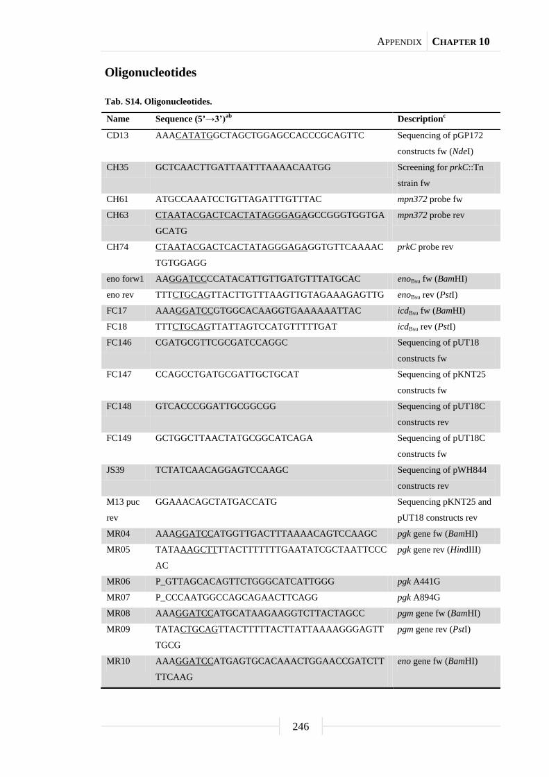
APPENDIX CHAPTER 10
246
Oligonucleotides
Tab. S14. Oligonucleotides.
Name Sequence (5’→3’)ab Descriptionc
CD13 AAACATATGGCTAGCTGGAGCCACCCGCAGTTC Sequencing of pGP172
constructs fw (NdeI)
CH35 GCTCAACTTGATTAATTTAAAACAATGG Screening for prkC::Tn
strain fw CH61 ATGCCAAATCCTGTTAGATTTGTTTAC mpn372 probe fw
CH63 CTAATACGACTCACTATAGGGAGAGCCGGGTGGTGA
GCATG
mpn372 probe rev
CH74 CTAATACGACTCACTATAGGGAGAGGTGTTCAAAAC
TGTGGAGG
prkC probe rev
eno forw1 AAGGATCCCCATACATTGTTGATGTTTATGCAC enoBsu fw (BamHI)
eno rev TTTCTGCAGTTACTTGTTTAAGTTGTAGAAAGAGTTG enoBsu rev (PstI)
FC17 AAAGGATCCGTGGCACAAGGTGAAAAAATTAC icdBsu fw (BamHI)
FC18 TTTCTGCAGTTATTAGTCCATGTTTTTGAT icdBsu rev (PstI)
FC146 CGATGCGTTCGCGATCCAGGC Sequencing of pUT18
constructs fw FC147 CCAGCCTGATGCGATTGCTGCAT Sequencing of pKNT25
constructs fw FC148 GTCACCCGGATTGCGGCGG Sequencing of pUT18C
constructs rev FC149 GCTGGCTTAACTATGCGGCATCAGA Sequencing of pUT18C
constructs fw JS39 TCTATCAACAGGAGTCCAAGC Sequencing of pWH844
constructs rev
M13 puc
rev
GGAAACAGCTATGACCATG Sequencing pKNT25 and
pUT18 constructs rev MR04 AAAGGATCCATGGTTGACTTTAAAACAGTCCAAGC pgk gene fw (BamHI)
MR05 TATAAAGCTTTTACTTTTTTTGAATATCGCTAATTCCC
AC
pgk gene rev (HindIII)
MR06 P_GTTAGCACAGTTCTGGGCATCATTGGG pgk A441G
MR07 P_CCCAATGGCCAGCAGAACTTCAGG pgk A894G
MR08 AAAGGATCCATGCATAAGAAGGTCTTACTAGCC pgm gene fw (BamHI)
MR09 TATACTGCAGTTACTTTTTACTTATTAAAAGGGAGTT
TGCG
pgm gene rev (PstI)
MR10 AAAGGATCCATGAGTGCACAAACTGGAACCGATCTT
TTCAAG
eno gene fw (BamHI)
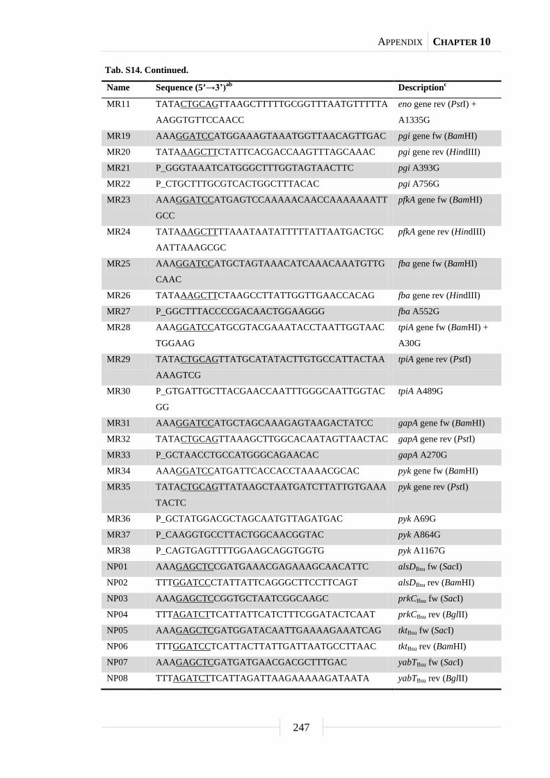
APPENDIX CHAPTER 10
247
Tab. S14. Continued.
Name Sequence (5’→3’)ab Descriptionc
MR11 TATACTGCAGTTAAGCTTTTTGCGGTTTAATGTTTTTA
AAGGTGTTCCAACC
eno gene rev (PstI) +
A1335G
MR19 AAAGGATCCATGGAAAGTAAATGGTTAACAGTTGAC pgi gene fw (BamHI)
MR20 TATAAAGCTTCTATTCACGACCAAGTTTAGCAAAC pgi gene rev (HindIII)
MR21 P_GGGTAAATCATGGGCTTTGGTAGTAACTTC pgi A393G
MR22 P_CTGCTTTGCGTCACTGGCTTTACAC pgi A756G
MR23 AAAGGATCCATGAGTCCAAAAACAACCAAAAAAATT
GCC
pfkA gene fw (BamHI)
MR24 TATAAAGCTTTTAAATAATATTTTTATTAATGACTGC
AATTAAAGCGC
pfkA gene rev (HindIII)
MR25 AAAGGATCCATGCTAGTAAACATCAAACAAATGTTG
CAAC
fba gene fw (BamHI)
MR26 TATAAAGCTTCTAAGCCTTATTGGTTGAACCACAG fba gene rev (HindIII)
MR27 P_GGCTTTACCCCGACAACTGGAAGGG fba A552G
MR28 AAAGGATCCATGCGTACGAAATACCTAATTGGTAAC
TGGAAG
tpiA gene fw (BamHI) +
A30G
MR29 TATACTGCAGTTATGCATATACTTGTGCCATTACTAA
AAAGTCG
tpiA gene rev (PstI)
MR30 P_GTGATTGCTTACGAACCAATTTGGGCAATTGGTAC
GG
tpiA A489G
MR31 AAAGGATCCATGCTAGCAAAGAGTAAGACTATCC gapA gene fw (BamHI)
MR32 TATACTGCAGTTAAAGCTTGGCACAATAGTTAACTAC gapA gene rev (PstI)
MR33 P_GCTAACCTGCCATGGGCAGAACAC gapA A270G
MR34 AAAGGATCCATGATTCACCACCTAAAACGCAC pyk gene fw (BamHI)
MR35 TATACTGCAGTTATAAGCTAATGATCTTATTGTGAAA
TACTC
pyk gene rev (PstI)
MR36 P_GCTATGGACGCTAGCAATGTTAGATGAC pyk A69G
MR37 P_CAAGGTGCCTTACTGGCAACGGTAC pyk A864G
MR38 P_CAGTGAGTTTTGGAAGCAGGTGGTG pyk A1167G
NP01 AAAGAGCTCCGATGAAACGAGAAAGCAACATTC alsDBsu fw (SacI)
NP02 TTTGGATCCCTATTATTCAGGGCTTCCTTCAGT alsDBsu rev (BamHI)
NP03 AAAGAGCTCCGGTGCTAATCGGCAAGC prkCBsu fw (SacI)
NP04 TTTAGATCTTCATTATTCATCTTTCGGATACTCAAT prkCBsu rev (BglII)
NP05 AAAGAGCTCGATGGATACAATTGAAAAGAAATCAG tktBsu fw (SacI)
NP06 TTTGGATCCTCATTACTTATTGATTAATGCCTTAAC tktBsu rev (BamHI)
NP07 AAAGAGCTCGATGATGAACGACGCTTTGAC yabTBsu fw (SacI)
NP08 TTTAGATCTTCATTAGATTAAGAAAAAGATAATA yabTBsu rev (BglII)

APPENDIX CHAPTER 10
248
Tab. S14. Continued.
Name Sequence (5’→3’)ab Descriptionc
NP09 AAAGAGCTCGATGGCATTAAAACTTCTAAAAAA prkDBsu fw (SacI)
NP10 TTTGGATCCTCATTATGTGACCGATTGAATGGC prkDBsu rev (BamHI)
NP11 AAAGAGCTCGATGTTATTCTTTGTTGATACAG ywjHBsu fw (SacI)
NP12 TTTGGATCCTCATTATTTGTTCCAGTCTGCC ywjHBsu rev (BamHI)
NP13 AAAGAGCTCGATGGTCAAGTCATTTCG yxaLBsu fw (SacI)
NP14 TTTGGATCCTCATTATTTCCCAAAAGCCATCAG yxaLBsu rev (BamHI)
NP15 CCGGTGTTCACGGTGCGCCGCTTGG tktBsu sequencing fw
NP16 CTCACCTGATTCTTTAACAGCTACAGCG tktBsu sequencing rev
NP17 CTTGCTAAAGCTGCTGGCGAAAAGAAGCTG prkCBsu sequencing fw
NP18 ATCGTGAGCAAAACCCACGGCCAC prkCBsu sequencing rev
NP19 ATGTTCACGCGATCAAACCGACGTC yxaLBsu sequencing fw
NP20 GCAGCAGCCAACTCAGCTTCCTTTCGGGC Sequencing of pGP172
constructs rev
NP108 AAAGGATCCCTCACTTATTTAAAGGAGGAAACAATC
ATGGGCAAGTATTTTGGAACAGACG
glmMBsu + RBSgapA fw
(BamHI)
NP109 TTTGTCGACCTATCACTCTAATCCCATTTCTGACCGG glmMBsu rev (SalI)
NP110 GGCTATTCTCCGGAGCAGCCGATTGTCA glmMBsu LFH-PCR fw
(up-fragment)
NP111 CAGGAACGGACGACCAAAAGTTTTCCCG glmMBsu LFH-PCR fw
(up-fragment,
sequencing)
NP112 CCTATCACCTCAAATGGTTCGCTGTAAAGGCCAGCTC
AGGTGTAAGCTC
glmMBsu LFH-PCR rev
(up-fragment)
NP113 CCGAGCGCCTACGAGGAATTTGTATCGAGCGAAGAC
GAAAGAGCTGTGCGA
glmMBsu LFH-PCR fw
(down- fragment)
NP114 CATCGCCATCGCATCAGATGCTACGACGT glmMBsu LFH-PCR rev
(down-fragment)
NP115 TATAAGACGCACGTGTAATCACGTCACCATC glmMBsu LFH-PCR rev
(down-fragment,
sequencing)
NP116 P_GCAGAGGCGGGCGTCATGATTTCCGCTGCCCATAA
CCCAGTGCAGGATAACGGCATCAA
glmMBsu T298G
NP117 AAAGGATCCATGGGCAAGTATTTTGGAACAGACG glmMBsu fw (BamHI)
PD05 AAATCTAGAGATGGAAAGTAAATGGTTAACAGTTGA
CAC
pgi gene fw (XbaI)
PD06 TTTGGTACCCGTTCACGACCAAGTTTAGCAAACATTA
AC
pgi gene rev (KpnI)
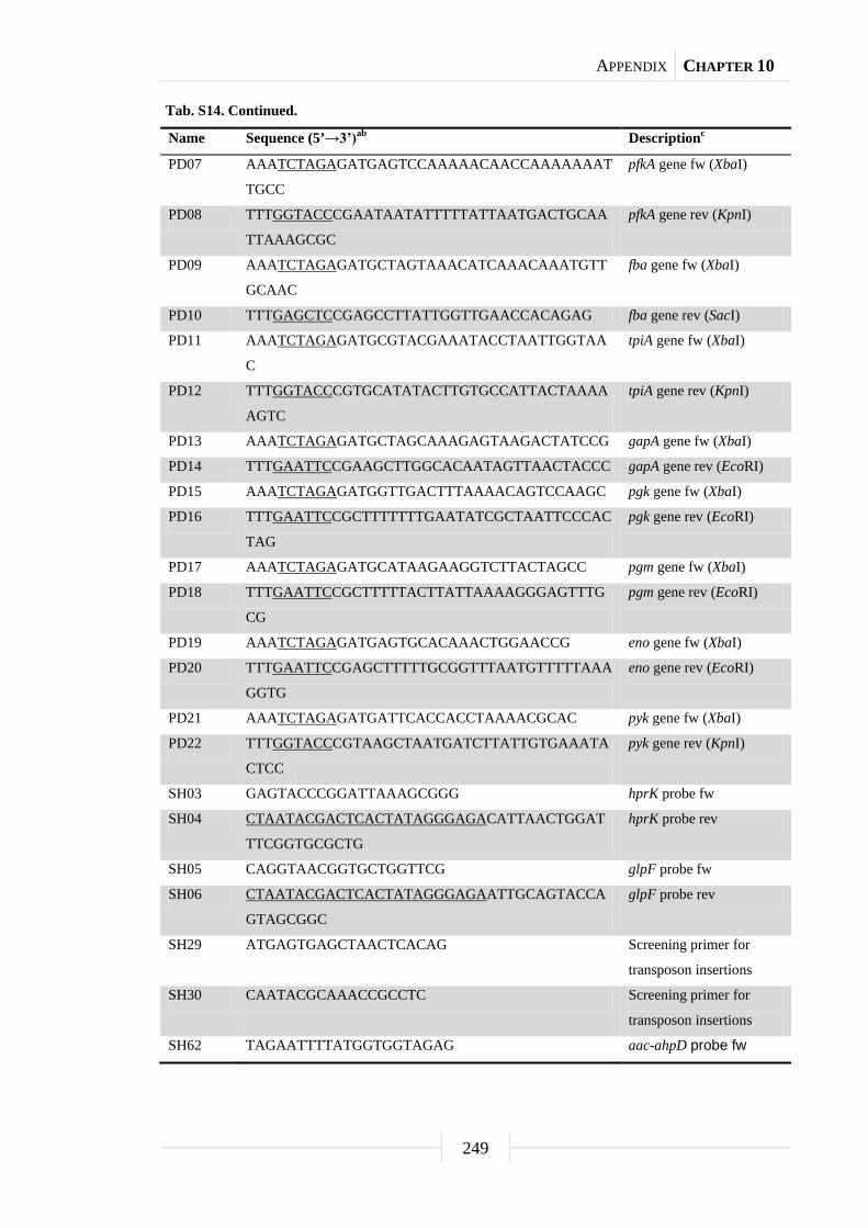
APPENDIX CHAPTER 10
249
Tab. S14. Continued.
Name Sequence (5’→3’)ab Descriptionc
PD07 AAATCTAGAGATGAGTCCAAAAACAACCAAAAAAAT
TGCC
pfkA gene fw (XbaI)
PD08 TTTGGTACCCGAATAATATTTTTATTAATGACTGCAA
TTAAAGCGC
pfkA gene rev (KpnI)
PD09 AAATCTAGAGATGCTAGTAAACATCAAACAAATGTT
GCAAC
fba gene fw (XbaI)
PD10 TTTGAGCTCCGAGCCTTATTGGTTGAACCACAGAG fba gene rev (SacI)
PD11 AAATCTAGAGATGCGTACGAAATACCTAATTGGTAA
C
tpiA gene fw (XbaI)
PD12 TTTGGTACCCGTGCATATACTTGTGCCATTACTAAAA
AGTC
tpiA gene rev (KpnI)
PD13 AAATCTAGAGATGCTAGCAAAGAGTAAGACTATCCG gapA gene fw (XbaI)
PD14 TTTGAATTCCGAAGCTTGGCACAATAGTTAACTACCC gapA gene rev (EcoRI)
PD15 AAATCTAGAGATGGTTGACTTTAAAACAGTCCAAGC pgk gene fw (XbaI)
PD16 TTTGAATTCCGCTTTTTTTGAATATCGCTAATTCCCAC
TAG
pgk gene rev (EcoRI)
PD17 AAATCTAGAGATGCATAAGAAGGTCTTACTAGCC pgm gene fw (XbaI)
PD18 TTTGAATTCCGCTTTTTACTTATTAAAAGGGAGTTTG
CG
pgm gene rev (EcoRI)
PD19 AAATCTAGAGATGAGTGCACAAACTGGAACCG eno gene fw (XbaI)
PD20 TTTGAATTCCGAGCTTTTTGCGGTTTAATGTTTTTAAA
GGTG
eno gene rev (EcoRI)
PD21 AAATCTAGAGATGATTCACCACCTAAAACGCAC pyk gene fw (XbaI)
PD22 TTTGGTACCCGTAAGCTAATGATCTTATTGTGAAATA
CTCC
pyk gene rev (KpnI)
SH03 GAGTACCCGGATTAAAGCGGG hprK probe fw
SH04 CTAATACGACTCACTATAGGGAGACATTAACTGGAT
TTCGGTGCGCTG
hprK probe rev
SH05 CAGGTAACGGTGCTGGTTCG glpF probe fw
SH06 CTAATACGACTCACTATAGGGAGAATTGCAGTACCA
GTAGCGGC
glpF probe rev
SH29 ATGAGTGAGCTAACTCACAG Screening primer for
transposon insertions
SH30 CAATACGCAAACCGCCTC Screening primer for
transposon insertions
SH62 TAGAATTTTATGGTGGTAGAG aac-ahpD probe fw

APPENDIX CHAPTER 10
250
Tab. S14. Continued.
Name Sequence (5’→3’)ab Descriptionc
SH63 CTAATACGACTCACTATAGGGAGAACACTATCATAA
CCACTACC
aac-ahpD probe rev
SH66 AAAGTCGACATGGACAGCACCAACCAAAAC prpC probe fw (SalI)
SH73 CTAATACGACTCACTATAGGGAGAGACCATCAGAGC
ACAACAG
prpC probe rev
SH82 ATGGATCCATGGCACAAAAAACATTTAAAG ptsHBsu fw (BamHI)
SH83 ATAAAGCTTCTCGCCGAGTCCTTCG ptsHBsu rev (HindIII)
SH89 TAGAGCTCGATGGCACTAAATTTAAAGATTGG prkC probe fw
spec-fwd
(kan)
CAGCGAACCATTTGAGGTGATAGGGACTGGCTCGCT
AATAACGTAACGTGACTGGCAAGAG
Construction of B. subtilis
mutants by LFH-PCR fw
spec-rev
(kan)
CGATACAAATTCCTCGTAGGCGCTCGGCGTAGCGAG
GGCAAGGGTTTATTGTTTTCTAAAATCTG
Construction of B. subtilis
mutants by LFH-PCR rev
SS29 AAAGAGCTCGATGATTAACATCGATCCCCATTTTATT
C
mpn549 gene fw (SacI)
SS30 TATAGGATCCTTAGCTGTTGACATGCTTTTGTTTGG mpn549 gene rev
(BamHI)
SS31 P_GAAGACATTAAGAAGTGGATTGGTTCCATTCG mpn549 A807G
SS32 GATTTGTGGGCCGATGTTTTCC tdk probe fw
SS33 CTAATACGACTCACTATAGGGAGACGGTTTCGTTGCT
CTTGGAC
tdk probe rev
SS34 AAAGAGCTCGATGCTTAAACGACAACTTCTGCTAGC glpQ gene fw (SacI)
SS35 TATAGGATCCTTACACTTCAAACTTCTTGTTGGCAAT
TTG
glpQ gene rev (BamHI)
SS36 P_GCCTTTTTGTTTTGGACGAAAAAGCAGTTCCAAG glpQ A507G
SS37 P_CAGTATCTCCATCCCTGGACAAACATTTACG glpQ A576G
SS38 P_CCTTTAGGGCTGTGGACGCTTAACAGTG glpQ A639G
SS39 AAAGAGCTCGATGCGCAAACAGTTTTTAATTGCACA
C
mpn566 gene fw (SacI)
SS40 TATAGGATCCTTAGTAAAGTTGTGCTGCTATTTGAAA
TTTAAC
mpn566 gene rev
(BamHI)
SS41 CGCAACAATTTAAAGAACTGACTTAGC Screening for mpn549::Tn
strain rev
SS42 CAACTTCTGCTAGCACACCG glpQ probe fw
SS43 CTAATACGACTCACTATAGGGAGAGCTATTTGGTAG
TTGGGGTTAATG
glpQ probe rev
SS44 GCAAACAGTTTTTAATTGCACACCG mpn566 probe fw
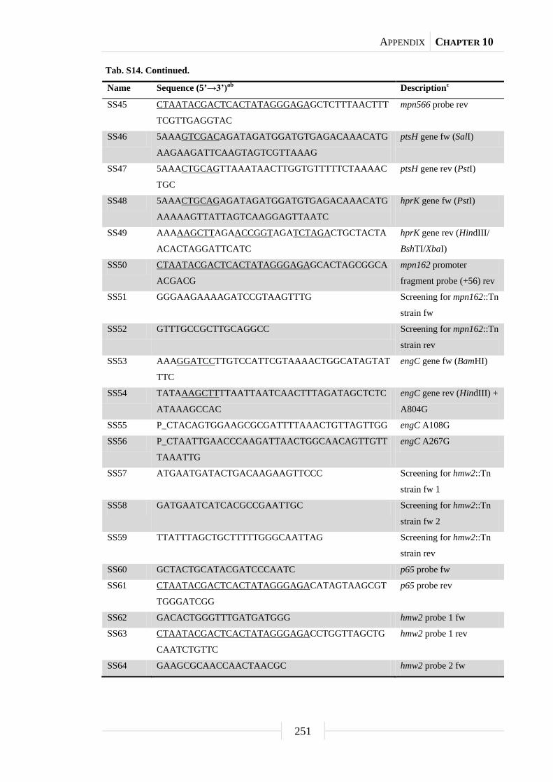
APPENDIX CHAPTER 10
251
Tab. S14. Continued.
Name Sequence (5’→3’)ab Descriptionc
SS45 CTAATACGACTCACTATAGGGAGAGCTCTTTAACTTT
TCGTTGAGGTAC
mpn566 probe rev
SS46 5AAAGTCGACAGATAGATGGATGTGAGACAAACATG
AAGAAGATTCAAGTAGTCGTTAAAG
ptsH gene fw (SalI)
SS47 5AAACTGCAGTTAAATAACTTGGTGTTTTTCTAAAAC
TGC
ptsH gene rev (PstI)
SS48 5AAACTGCAGAGATAGATGGATGTGAGACAAACATG
AAAAAGTTATTAGTCAAGGAGTTAATC
hprK gene fw (PstI)
SS49 AAAAAGCTTAGAACCGGTAGATCTAGACTGCTACTA
ACACTAGGATTCATC
hprK gene rev (HindIII/
BshTI/XbaI)
SS50 CTAATACGACTCACTATAGGGAGAGCACTAGCGGCA
ACGACG
mpn162 promoter
fragment probe (+56) rev
SS51 GGGAAGAAAAGATCCGTAAGTTTG Screening for mpn162::Tn
strain fw SS52 GTTTGCCGCTTGCAGGCC Screening for mpn162::Tn
strain rev SS53 AAAGGATCCTTGTCCATTCGTAAAACTGGCATAGTAT
TTC
engC gene fw (BamHI)
SS54 TATAAAGCTTTTAATTAATCAACTTTAGATAGCTCTC
ATAAAGCCAC
engC gene rev (HindIII) +
A804G
SS55 P_CTACAGTGGAAGCGCGATTTTAAACTGTTAGTTGG engC A108G
SS56 P_CTAATTGAACCCAAGATTAACTGGCAACAGTTGTT
TAAATTG
engC A267G
SS57 ATGAATGATACTGACAAGAAGTTCCC Screening for hmw2::Tn
strain fw 1
SS58 GATGAATCATCACGCCGAATTGC Screening for hmw2::Tn
strain fw 2 SS59 TTATTTAGCTGCTTTTTGGGCAATTAG Screening for hmw2::Tn
strain rev SS60 GCTACTGCATACGATCCCAATC p65 probe fw
SS61 CTAATACGACTCACTATAGGGAGACATAGTAAGCGT
TGGGATCGG
p65 probe rev
SS62 GACACTGGGTTTGATGATGGG hmw2 probe 1 fw
SS63 CTAATACGACTCACTATAGGGAGACCTGGTTAGCTG
CAATCTGTTC
hmw2 probe 1 rev
SS64 GAAGCGCAACCAACTAACGC hmw2 probe 2 fw

APPENDIX CHAPTER 10
252
Tab. S14. Continued.
Name Sequence (5’→3’)ab Descriptionc
SS65 CTAATACGACTCACTATAGGGAGAGTTGTTGGGACA
GATCAGCAC
hmw2 probe 2 rev
SS66 GATGATGAAGCTGACATCATCATAG p41 probe fw
SS67 CTAATACGACTCACTATAGGGAGAGTGTATTCCGTG
CCACCAATAAC
p41 probe rev
SS68 CTAACACTAAAACGGGTACGAATTG p24 probe fw
SS69 CTAATACGACTCACTATAGGGAGAGCCACAAGTGTT
TCTGATGTCAC
p24 probe rev
SS70 CTGAGGAAAATCCCGAACAGATC hmw1 probe 1 fw
SS71 CTAATACGACTCACTATAGGGAGAGCTGGTACATCT
GCTTCCAAAC
hmw1 probe 1 rev
SS72 GCCAACAGCGACATTAACAGC hmw1 probe 2 fw
SS73 CTAATACGACTCACTATAGGGAGACAGTATCACTGC
AGAGCTTATAAAC
hmw1 probe 2 rev
SS74 GTATGGTAAGCTAGCACAAAAGATC hmw3 probe fw
SS75 CTAATACGACTCACTATAGGGAGAGTTGATCAGCTT
GCTGTTCGG
hmw3 probe rev
SS76 CTTGGATTCTCATCCTCACCG p1 probe 1 fw
SS77 CTAATACGACTCACTATAGGGAGAGGTGCAGCCCCA
CTCAAAC
p1 probe 1 rev
SS78 GCGAGCGGGTGGTTCG p1 probe 2 fw
SS79 CTAATACGACTCACTATAGGGAGAGCTTGTAAGCAC
TCGCTTCC
p1 probe 2 rev
SS80 CCTGAATGCTAGTGCTGTTCAG p30 probe fw
SS81 CTAATACGACTCACTATAGGGAGAGTTGAGGTGGGA
AACCAGTAC
p30 probe rev
SS82 CAAAGGTAGTTAATGTAGATCAGGTAG engC probe fw
SS83 CTAATACGACTCACTATAGGGAGAGTTGGGCTGCTA
ATTGTTTGAGG
engC probe rev
SS84 GTTGCAGTAGATCAATAAAAAGAGGG Screening for cbiO::Tn
strain fw SS85 GCTTTTATCAACAAAGACACTAAGATTG Screening for cbiO::Tn
strain rev SS86 GCACGCATCTTAAAGCTGTTGG p28 probe fw
SS87 CTAATACGACTCACTATAGGGAGACACTGTCAGCGT
GCTCTGC
p28 probe rev
SS88 GCTTCTCCAATTCGCGGCG hmw2 sequencing rev 1

APPENDIX CHAPTER 10
253
Tab. S14. Continued.
Name Sequence (5’→3’)ab Descriptionc
SS89 CGCCGCGAATTGGAGAAGC hmw2 sequencing fw 1
SS90 GTTCGATTTGCTGGAACTCCTG hmw2 sequencing rev 2
SS91 CGCCAGTCTTTAGAGCATGAC hmw2 sequencing fw 2
SS92 GCTAGCTTCTTCACTTACCGG p40 probe fw
SS93 CTAATACGACTCACTATAGGGAGAGTTAACGTTCCTT
CGTTACCATTG
p40 probe rev
SS94 GCGTCAAAGTAGTACTTGGGAG Screening for mpn256::Tn
strain fw SS95 GACATCAGGGTTAATTTGAGCAATC Screening for mpn256::Tn
strain rev SS96 CGACTGATGGTTGGCTCTAC Screening for glpQ::Tn
strain fw SS97 GCTAACCGTTTACTCGGTGG Screening for glpQ::Tn
strain rev SS98 CAGTTAATAGGCAACGAAGACTATG Screening for mpn566::Tn
strain fw SS99 CACTAGTAAGAACCACAGTAGTAC Screening for mpn566::Tn
strain rev SS100 GAGTAGCTCACAATGACCCTG Screening for glpK::Tn
strain fw SS101 GGTGGAGCAGAAGATAAGCAG Screening for glpK::Tn
strain rev SS102 GTTAATTAATGGGAGGATAAGCAAGC Screening for glpF::Tn
strain fw SS103 CCGACAAGATTTGTAAGTTAGATGC Screening for glpF::Tn
strain rev SS104 AAAGGATCCATGGCACTAAATTTAAAGATTGGTGAC prkC gene fw (BamHI)
SS105 TATAAAGCTTTTATAGGTAATTACTTACCATTAGTTC
ACG
prkC catalytic domain rev
(HindIII)
SS106 TATAAAGCTTTTACGAATGGACAACTACCCCAG prkC gene rev (HindIII)
SS107 AAAGGATCCATGAACAGTAACGCATACTTGGAAGC manB gene fw (BamHI)
SS108 TATACTGCAGCTAAGCTTTATCGAGGTTGAGCAATC manB gene rev (PstI)
SS109 P_GAAAGTAAGGATACCTGGGAGTTAGCGCG manB A846G
SS110 P_CTTTGCGATTGCCGAATGGAATCCGCAAAC manB A936G
SS111 P_GATTACTATAACTGGACGGTACCACACACCATTC manB A1356G
SS112 P_CGGTGAATGTCACTGCTGCTCATAATCCTAAAACC manB A445G/G446C

APPENDIX CHAPTER 10
254
Tab. S14. Continued.
Name Sequence (5’→3’)ab Descriptionc
SS113 CAAGGCCACAATTGATTATGTCTTTG Screening for manB::Tn
strain fw SS114 GGTAACTTAGTGGAAGTCCACTTAAC Screening for manB::Tn
strain rev SS115 GAATTAAACAGTAAGCTAACCGGG mpn256 probe fw
SS116 CTAATACGACTCACTATAGGGAGACAATAAACTTAG
AAAAGTCCATGGTTG
mpn256 probe rev
SS117 GTATTTTGAGAGTTAGTGCCTTTCC Screening for ftsY::Tn
strain fw SS118 CTCGCGTAATTGGGGATCTG Screening for ftsY::Tn
strain rev SS119 GATCAGGAGACAAATACATTCGAAC Screening for mpn349::Tn
strain fw SS120 GTGCCTTGGTTAGATTACCTTTAC Screening for mpn349::Tn
strain rev SS121 CACTAAGGTGTACGGTGATTTTAAG nrdE probe fw
SS122 CTAATACGACTCACTATAGGGAGAGACCAATTGATT
CCATGTTGTCTTC
nrdE probe rev
SS123 GAATCAGTTTCTCCCTTAGAATATGC nrdF probe fw
SS124 CTAATACGACTCACTATAGGGAGAGTCTTTCCCGGTG
TAATAGGG
nrdF probe rev
SS125 GATATCAAAATAGTCGACGCTAGTG nrdI probe fw
SS126 CTAATACGACTCACTATAGGGAGACTTGTTCTACATC
GTTTTTCGTGC
nrdI probe rev
SS127 CCAACAGCGCTTTTATTCTCGG mpn083 probe fw
SS128 CTAATACGACTCACTATAGGGAGAGGACATTAGGTT
TGGTGTACTTAC
mpn083 probe rev
SS129 CCTTGTTAGTTGCGCCACAC mpn162 probe fw
SS130 CTAATACGACTCACTATAGGGAGACACCTTCGTGGT
GATCATGATC
mpn162 probe rev
SS131 GGCTTAGTCATCCACACTTGG ulaF probe fw
SS132 CTAATACGACTCACTATAGGGAGACCACTGCATCCTT
GCCATTC
ulaF probe rev
SS133 CTGCTGTAGCTCATGTGGAAC licA probe fw
SS134 CTAATACGACTCACTATAGGGAGACTCAAAGTCAAT
TAAAACGACCTGC
licA probe rev
SS135 GGTTTTACCCGTTTTTGTGTTAATGC plsC probe fw
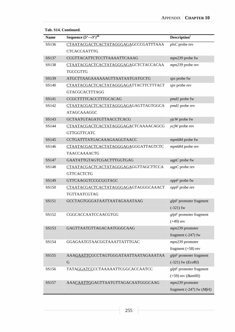
APPENDIX CHAPTER 10
255
Tab. S14. Continued.
Name Sequence (5’→3’)ab Descriptionc
SS136 CTAATACGACTCACTATAGGGAGAGCCCGATTTAAA
CTCACCAATTTG
plsC probe rev
SS137 CCGTTACATTCTCCTTAAAATTCAAAG mpn239 probe fw
SS138 CTAATACGACTCACTATAGGGAGAGCTCTACCACAA
TGCCGTTG
mpn239 probe rev
SS139 ATGCTTAAGAAAAAAGTTAATAATGATGCTG spx probe fw
SS140 CTAATACGACTCACTATAGGGAGATTACTTCTTTACT
GTACGCACTTTAGG
spx probe rev
SS141 CCGCTTTTCACCTTTGCACAG pmd1 probe fw
SS142 CTAATACGACTCACTATAGGGAGAGAGTTAGTGGCA
ATAGCAAAGGC
pmd1 probe rev
SS143 GCTAATGTAGATGTTAACCTCACG yjcW probe fw
SS144 CTAATACGACTCACTATAGGGAGACTCAAAACAGCG
GTTGGTTCATC
ycjW probe rev
SS145 CCTGATTTATGACAAAGAAGGTAACC mpn684 probe fw
SS146 CTAATACGACTCACTATAGGGAGAGGGATTAGTCTC
TAACCAAAACTG
mpn684 probe rev
SS147 GAATATTGTAGTCGACTTTGGTGAG ugpC probe fw
SS148 CTAATACGACTCACTATAGGGAGAGGTTAGCTTCCA
GTTCACTCTG
ugpC probe rev
SS149 GTTCAAGGTCCGCGGTAGC oppF probe fw
SS150 CTAATACGACTCACTATAGGGAGAGTAGGGCAAACT
TGTTAATCGTAG
oppF probe rev
SS151 GCCTAGTGGGATAATTAATAGAAATAAG glpF promoter fragment
(-321) fw
SS152 CGGCACCAATCCAACGTGG glpF promoter fragment
(+49) rev
SS153 GAGTTAATGTTAGACAATGGGCAAG mpn239 promoter
fragment (-247) fw
SS154 GGAGAATGTAACGGTAAATTATTTGAC mpn239 promoter
fragment (+58) rev
SS155 AAAGAATTCGCCTAGTGGGATAATTAATAGAAATAA
G
glpF promoter fragment
(-321) fw (EcoRI)
SS156 TATAGGATCCCCTAAAAATTCGGCACCAATCC glpF promoter fragment
(+59) rev (BamHI)
SS157 AAACAATTGGAGTTAATGTTAGACAATGGGCAAG mpn239 promoter
fragment (-247) fw (MfeI)

APPENDIX CHAPTER 10
256
Tab. S14. Continued.
Name Sequence (5’→3’)ab Descriptionc
SS158 TATAGGATCCCTTTGAATTTTAAGGAGAATGTAACG
G
mpn239 promoter
fragment (+71) rev
(BamHI)
SS159 TATAGCGGCCGCTTACACTTCAAACTTCTTGTTGGCA
ATTTGTG
glpQ gene rev (NotI)
SS160 TATAGCGGCCGCTTAGTAAAGTTGTGCTGCTATTTGA
AATTTAACAAAG
mpn566 gene rev (NotI)
SS161 AAAGGATCCGCAGGTTAGCTGATAAGTTTAGGAG glpQ promoter fragment
(-495) fw (BamHI)
SS162 AAAGGATCCGGGGAATGGGTATGAGCTGG mpn566 promoter
fragment (-492) fw
(BamHI)
SS163 AAAGGATCCGTTAATAATGATGATTGAAGC ackA promoter fragment
(-224) fw (BamHI)
SS164 AAACTGCAGATGCTTAAACGACAACTTCTGCTAGC glpQ gene fw (PstI)
SS165 AAACTGCAGATGCGCAAACAGTTTTTAATTGCACAC mpn566 gene fw (PstI)
SS166 CTTCTACCGGGTTTTCCATGC Screening for deoA::Tn
strain fw SS167 CGCACTACGTTCCGCACAC Screening for deoA::Tn
strain rev SS168 GCTAAATCATAAGCTCACCATTGC cinA probe fw
SS169 CTAATACGACTCACTATAGGGAGAGCTGCATCTTGTT
CTCTTAAACC
cinA probe rev
SS170 CTAACCCCAACTACGGCATC rpoA probe fw
SS171 CTAATACGACTCACTATAGGGAGACCCCAAAGAATT
AATCATTTCACGG
rpoA probe rev
SS172 CTTAATCTTAGCGCTTACCTTGTTG disA probe fw
SS173 CTAATACGACTCACTATAGGGAGAGTTTTAATCACG
CCGCGCAC
disA probe rev
SS174 GAAATTTAAGTATGGTGCTACCC mpn363 probe fw
SS175 CTAATACGACTCACTATAGGGAGAGTTAGGTGATAA
CTTGGAGAGATC
mpn363 probe rev
SS176 CTCAATTTTAGCCTCAAACCCAAC cbiO probe fw
SS177 CTAATACGACTCACTATAGGGAGACCGCTTTAGTTTG
CTAGCTAGC
cbiO probe rev
SS178 GTTGGTGCCTTCTTTACCCTC mpn407 probe fw
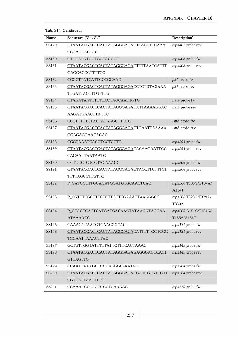
APPENDIX CHAPTER 10
257
Tab. S14. Continued.
Name Sequence (5’→3’)ab Descriptionc
SS179 CTAATACGACTCACTATAGGGAGACTTACCTTCAAA
CCGAGCACTAG
mpn407 probe rev
SS180 CTGCATGTGGTGCTAGGGG mpn408 probe fw
SS181 CTAATACGACTCACTATAGGGAGACTTTTAATCATTT
GAGCACCGTTTTCC
mpn408 probe rev
SS182 CCGCTTATCATTCCCGCAAC p37 probe fw
SS183 CTAATACGACTCACTATAGGGAGACCTCTGTAGAAA
TTGATTAGTTTGTTTG
p37 probe rev
SS184 CTAGATAGTTTTTTACCAGCAATTGTG mtlF probe fw
SS185 CTAATACGACTCACTATAGGGAGACATTAAAAGGAC
AAGATGAACTTAGCC
mtlF probe rev
SS186 CCCTTTTTGTACTATAAGCTTGCC lspA probe fw
SS187 CTAATACGACTCACTATAGGGAGACTGAATTAAAAA
GGAGAGGAACAGAC
lspA probe rev
SS188 CGCCAAATCACGTCCTGTTC mpn294 probe fw
SS189 CTAATACGACTCACTATAGGGAGACACAAGAATTGG
CACAACTAATAATG
mpn294 probe rev
SS190 GCTGCCTGTGGTACAAAGG mpn506 probe fw
SS191 CTAATACGACTCACTATAGGGAGAGTACCTTCTTTCT
TTTTAGCGTTGTTC
mpn506 probe rev
SS192 P_GATGGTTTGGAGATGGATGTGCAACTCAC mpn566 T106G/G107A/
A114T
SS193 P_CGTTTCGCTTTCTCTTGCTTGAAATTAAGGGCG mpn566 T328G/T329A/
T330A
SS194 P_GTAGTCACTCATGATGACAACTATAAGGTAGGAA
ATAAAACC
mpn566 A151C/T154G/
T155A/A156T
SS195 CAAAGCCAATGTCAACGGCAC mpn131 probe fw
SS196 CTAATACGACTCACTATAGGGAGACATTTTTGGTCGG
TGGAATTAAACTTAC
mpn131 probe rev
SS197 GCTGTTGGTATTTTTATTCTTTCACTAAAC mpn149 probe fw
SS198 CTAATACGACTCACTATAGGGAGAGAGGGAGCCACT
GTTAGTTG
mpn149 probe rev
SS199 CCAATTAAAGCTCCTTCAAAGAATGG mpn284 probe fw
SS200 CTAATACGACTCACTATAGGGAGACGATCGTATTGTT
CGTCATTAATTTTG
mpn284 probe rev
SS201 CCAAACCCCAATCCCTCAAAAC mpn370 probe fw
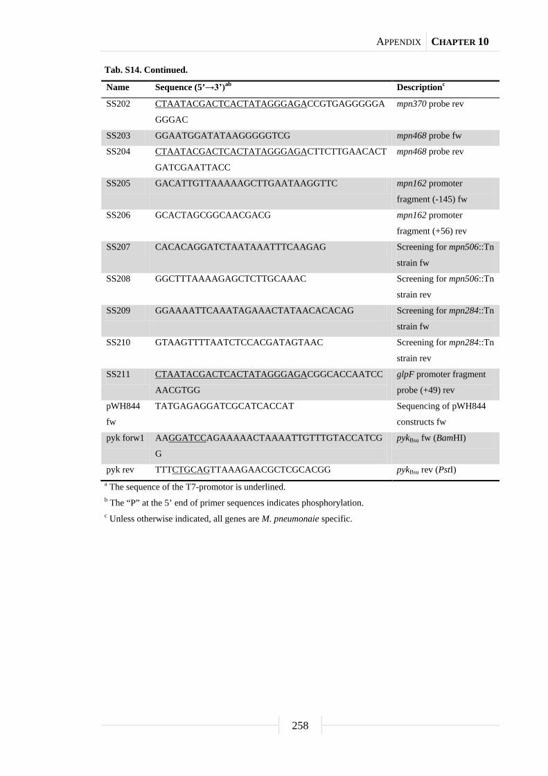
APPENDIX CHAPTER 10
258
Tab. S14. Continued.
Name Sequence (5’→3’)ab Descriptionc
SS202 CTAATACGACTCACTATAGGGAGACCGTGAGGGGGA
GGGAC
mpn370 probe rev
SS203 GGAATGGATATAAGGGGGTCG mpn468 probe fw
SS204 CTAATACGACTCACTATAGGGAGACTTCTTGAACACT
GATCGAATTACC
mpn468 probe rev
SS205 GACATTGTTAAAAAGCTTGAATAAGGTTC mpn162 promoter
fragment (-145) fw
SS206 GCACTAGCGGCAACGACG mpn162 promoter
fragment (+56) rev
SS207 CACACAGGATCTAATAAATTTCAAGAG Screening for mpn506::Tn
strain fw SS208 GGCTTTAAAAGAGCTCTTGCAAAC Screening for mpn506::Tn
strain rev SS209 GGAAAATTCAAATAGAAACTATAACACACAG Screening for mpn284::Tn
strain fw SS210 GTAAGTTTTAATCTCCACGATAGTAAC Screening for mpn284::Tn
strain rev SS211 CTAATACGACTCACTATAGGGAGACGGCACCAATCC
AACGTGG
glpF promoter fragment
probe (+49) rev
pWH844
fw
TATGAGAGGATCGCATCACCAT Sequencing of pWH844
constructs fw
pyk forw1 AAGGATCCAGAAAAACTAAAATTGTTTGTACCATCG
G
pykBsu fw (BamHI)
pyk rev TTTCTGCAGTTAAAGAACGCTCGCACGG pykBsu rev (PstI) a The sequence of the T7-promotor is underlined. b The “P” at the 5’ end of primer sequences indicates phosphorylation. c Unless otherwise indicated, all genes are M. pneumonaie specific.
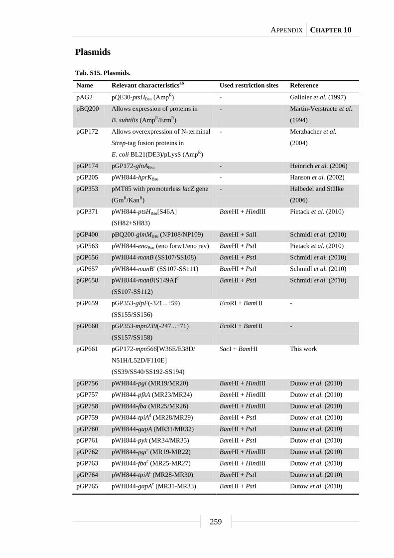
APPENDIX CHAPTER 10
259
Plasmids
Tab. S15. Plasmids.
Name Relevant characteristicsab Used restriction sites Reference
pAG2 pQE30-ptsHBsu (AmpR) - Galinier et al. (1997)
pBQ200 Allows expression of proteins in
B. subtilis (AmpR/ErmR)
- Martin-Verstraete et al.
(1994)
pGP172 Allows overexpression of N-terminal
Strep-tag fusion proteins in
E. coli BL21(DE3)/pLysS (AmpR)
- Merzbacher et al.
(2004)
pGP174 pGP172-glnABsu - Heinrich et al. (2006)
pGP205 pWH844-hprKBsu - Hanson et al. (2002)
pGP353 pMT85 with promoterless lacZ gene
(GmR/KanR)
- Halbedel and Stülke
(2006)
pGP371 pWH844-ptsHBsu[S46A]
(SH82+SH83)
BamHI + HindIII Pietack et al. (2010)
pGP400 pBQ200-glmMBsu (NP108/NP109) BamHI + SalI Schmidl et al. (2010)
pGP563 pWH844-enoBsu (eno forw1/eno rev) BamHI + PstI Pietack et al. (2010)
pGP656 pWH844-manB (SS107/SS108) BamHI + PstI Schmidl et al. (2010)
pGP657 pWH844-manBc (SS107-SS111) BamHI + PstI Schmidl et al. (2010)
pGP658 pWH844-manB[S149A]c
(SS107-SS112)
BamHI + PstI Schmidl et al. (2010)
pGP659 pGP353-glpF(-321...+59)
(SS155/SS156)
EcoRI + BamHI -
pGP660 pGP353-mpn239(-247...+71)
(SS157/SS158)
EcoRI + BamHI -
pGP661 pGP172-mpn566[W36E/E38D/
N51H/L52D/F110E]
(SS39/SS40/SS192-SS194)
SacI + BamHI This work
pGP756 pWH844-pgi (MR19/MR20) BamHI + HindIII Dutow et al. (2010)
pGP757 pWH844-pfkA (MR23/MR24) BamHI + HindIII Dutow et al. (2010)
pGP758 pWH844-fba (MR25/MR26) BamHI + HindIII Dutow et al. (2010)
pGP759 pWH844-tpiAd (MR28/MR29) BamHI + PstI Dutow et al. (2010)
pGP760 pWH844-gapA (MR31/MR32) BamHI + PstI Dutow et al. (2010)
pGP761 pWH844-pyk (MR34/MR35) BamHI + PstI Dutow et al. (2010)
pGP762 pWH844-pgic (MR19-MR22) BamHI + HindIII Dutow et al. (2010)
pGP763 pWH844-fbac (MR25-MR27) BamHI + HindIII Dutow et al. (2010)
pGP764 pWH844-tpiAc (MR28-MR30) BamHI + PstI Dutow et al. (2010)
pGP765 pWH844-gapAc (MR31-MR33) BamHI + PstI Dutow et al. (2010)

APPENDIX CHAPTER 10
260
Tab. S15. Continued.
Name Relevant characteristicsab Used restriction sites Reference
pGP766 pWH844-pykc (MR34-MR38) BamHI + PstI Dutow et al. (2010)
pGP819 pGP172-ywjHBsu (NP11/NP12) SacI + BamHI Pietack et al. (2010)
pGP820 pGP172-tktBsu (NP05/NP06) SacI + BamHI Pietack et al. (2010)
pGP821 pGP172-prkDBsu (NP09/NP10) SacI + BamHI Pietack et al. (2010)
pGP822 pGP172-alsDBsu (NP01/NP02) SacI + BamHI Pietack et al. (2010)
pGP823 pGP172-yabTBsu (NP07/NP08) SacI + BamHI Pietack et al. (2010)
pGP824 pGP172-yxaLBsu (NP13/NP14) SacI + BamHI Pietack et al. (2010)
pGP825 pGP172-prkCBsu (NP03/NP04) SacI + BamHI Pietack et al. (2010)
pGP931 pWH844-icdBsu (FC17/FC18) BamHI + PstI Pietack et al. (2010)
pGP1006 pWH844-pgk (MR04/MR05) BamHI + HindIII Dutow et al. (2010)
pGP1007 pWH844-pgm (MR08/MR09) BamHI + PstI Dutow et al. (2010)
pGP1008 pWH844-enoc (MR10/MR11) BamHI + PstI Dutow et al. (2010)
pGP1010 pWH844-pgkc (MR04-MR07) BamHI + HindIII Dutow et al. (2010)
pGP1016 pGP172-mpn549 (SS29/SS30) SacI + BamHI -
pGP1017 pGP172-mpn549c (SS29-SS31) SacI + BamHI -
pGP1018 pGP172-glpQ (SS34/SS35) SacI + BamHI This work
pGP1019 pGP172-glpQc (SS34-SS38) SacI + BamHI This work
pGP1020 pGP172-mpn566 (SS39/SS40) SacI + BamHI This work
pGP1100 pWH844-pykBsu (pyk forw1/pyk rev) BamHI + PstI Pietack et al. (2010)
pGP1401 pWH844-glmMBsu (NP109/NP117) BamHI + SalI Schmidl et al. (2010)
pGP1403 pBQ200-glmMBsu[S100A]
(NP108/NP109/NP116)
BamHI + SalI Schmidl et al. (2010)
pGP1405 pWH844-glmMBsu[S100A]
(NP108/NP116/NP117)
BamHI + SalI Schmidl et al. (2010)
pGP1553 pUT18C-pgic (PD05/PD06) XbaI + KpnI Dutow et al. (2010)
pGP1554 pUT18C-pfkA (PD07/PD08) XbaI + KpnI Dutow et al. (2010)
pGP1555 pUT18C-fbac (PD09/PD10) XbaI + SacI Dutow et al. (2010)
pGP1556 pUT18C-tpiAc (PD11/PD12) XbaI + KpnI Dutow et al. (2010)
pGP1557 pUT18C-gapAc (PD13/PD14) XbaI + EcoRI Dutow et al. (2010)
pGP1558 pUT18C-pgkc (PD15/PD16) XbaI + EcoRI Dutow et al. (2010)
pGP1559 pUT18C-pgm (PD17/PD18) XbaI + EcoRI Dutow et al. (2010)
pGP1560 pUT18C-enoc (PD19/PD20) XbaI + EcoRI Dutow et al. (2010)
pGP1561 pUT18C-pykc (PD21/PD22) XbaI + KpnI Dutow et al. (2010)
pGP1563 pUT18-pgic (PD05/PD06) XbaI + KpnI Dutow et al. (2010)
pGP1564 pUT18-pfkA (PD07/PD08) XbaI + KpnI Dutow et al. (2010)
pGP1565 pUT18-fbac (PD09/PD10) XbaI + SacI Dutow et al. (2010)
pGP1566 pUT18-tpiAc (PD11/PD12) XbaI + KpnI Dutow et al. (2010)

APPENDIX CHAPTER 10
261
Tab. S15. Continued.
Name Relevant characteristicsab Used restriction sites Reference
pGP1567 pUT18-gapAc (PD13/PD14) XbaI + EcoRI Dutow et al. (2010)
pGP1568 pUT18-pgkc (PD15/PD16) XbaI + EcoRI Dutow et al. (2010)
pGP1569 pUT18-pgm (PD17/PD18) XbaI + EcoRI Dutow et al. (2010)
pGP1570 pUT18-enoc (PD19/PD20) XbaI + EcoRI Dutow et al. (2010)
pGP1571 pUT18-pykc (PD21/PD22) XbaI + KpnI Dutow et al. (2010)
pGP1573 pKNT25-pgic (PD05/PD06) XbaI + KpnI Dutow et al. (2010)
pGP1574 pKNT25-pfkA (PD07/PD08) XbaI + KpnI Dutow et al. (2010)
pGP1575 pKNT25-fbac (PD09/PD10) XbaI + SacI Dutow et al. (2010)
pGP1576 pKNT25-tpiAc (PD11/PD12) XbaI + KpnI Dutow et al. (2010)
pGP1577 pKNT25-gapAc (PD13/PD14) XbaI + EcoRI Dutow et al. (2010)
pGP1578 pKNT25-pgkc (PD15/PD16) XbaI + EcoRI Dutow et al. (2010)
pGP1579 pKNT25-pgm (PD17/PD18) XbaI + EcoRI Dutow et al. (2010)
pGP1580 pKNT25-enoc (PD19/PD20) XbaI + EcoRI Dutow et al. (2010)
pGP1581 pKNT25-pykc (PD21/PD22) XbaI + KpnI Dutow et al. (2010)
pKNT25 Plac-mcs-cyaA (KanR) - Claessen et al. (2008)
pKT25-
zip
Plac-cyaA-zip (KanR) - Karimova et al. (1998)
pMT85 Mini-transposon delivery vector for
M. pneumoniae (GmR/KanR)
- Zimmerman and
Herrmann (2005)
pUT18 Plac-mcs-cyaA (AmpR) - Karimova et al. (1998)
pUT18C Plac-cyaA-mcs (AmpR) - Karimova et al. (1998)
pUT18C-
zip
Plac-cyaA-zip (AmpR) - Karimova et al. (1998)
pWH844 Allows overexpression of N-terminal
His6-tag fusion proteins in
E. coli DH5α (AmpR)
- Schirmer et al. (1997)
a Resistance abbreviations as follows: Amp, ampicillin; Erm, erythromycin; Gm, gentamicin; Kan,
kanamycin. b Unless otherwise indicated, all genes are M. pneumonaie specific. c All TGA opal codons in the wild type gene are mutated to TGG. d Only one TGA opal codon in the forward primer region is mutated to TGG.
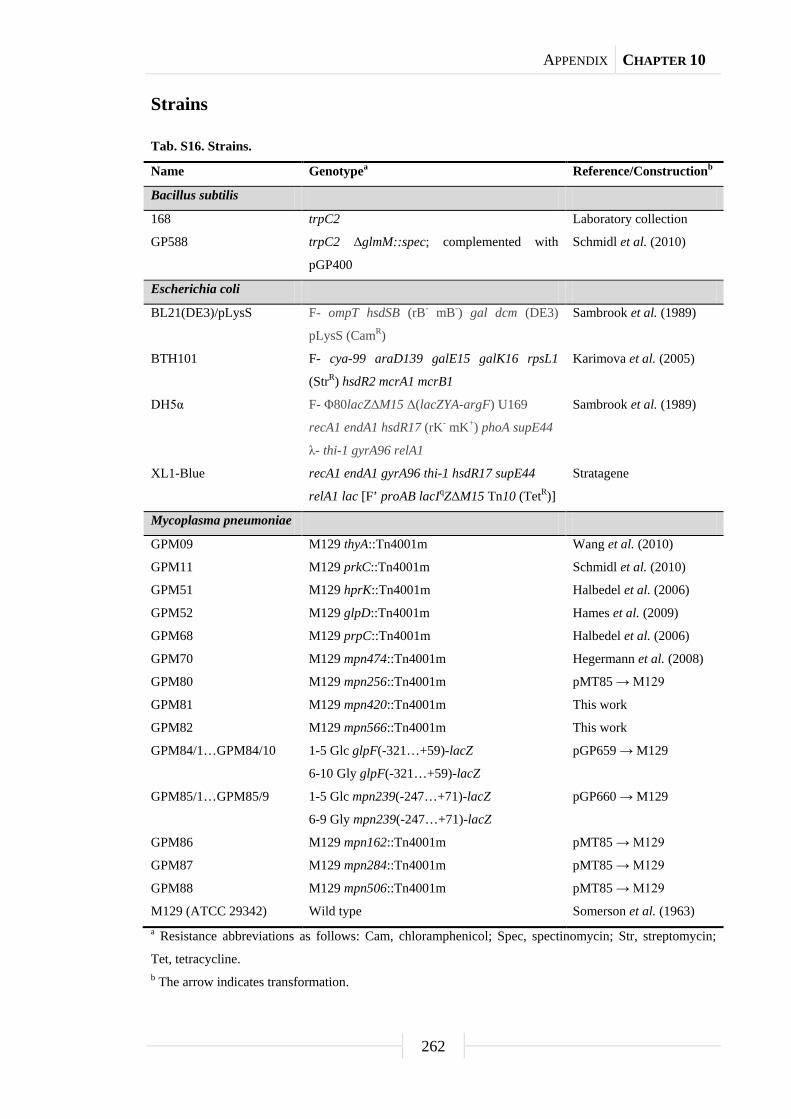
APPENDIX CHAPTER 10
262
Strains
Tab. S16. Strains.
Name Genotypea Reference/Constructionb
Bacillus subtilis
168 trpC2 Laboratory collection
GP588 trpC2 ΔglmM::spec; complemented with
pGP400
Schmidl et al. (2010)
Escherichia coli
BL21(DE3)/pLysS F- ompT hsdSB (rB- mB-) gal dcm (DE3)
pLysS (CamR)
Sambrook et al. (1989)
BTH101 F- cya-99 araD139 galE15 galK16 rpsL1
(StrR) hsdR2 mcrA1 mcrB1
Karimova et al. (2005)
DH5α F- Φ80lacZΔM15 Δ(lacZYA-argF) U169
recA1 endA1 hsdR17 (rK- mK+) phoA supE44
λ- thi-1 gyrA96 relA1
Sambrook et al. (1989)
XL1-Blue recA1 endA1 gyrA96 thi-1 hsdR17 supE44
relA1 lac [F’ proAB lacIqZΔM15 Tn10 (TetR)]
Stratagene
Mycoplasma pneumoniae
GPM09 M129 thyA::Tn4001m Wang et al. (2010)
GPM11 M129 prkC::Tn4001m Schmidl et al. (2010)
GPM51 M129 hprK::Tn4001m Halbedel et al. (2006)
GPM52 M129 glpD::Tn4001m Hames et al. (2009)
GPM68 M129 prpC::Tn4001m Halbedel et al. (2006)
GPM70 M129 mpn474::Tn4001m Hegermann et al. (2008)
GPM80 M129 mpn256::Tn4001m pMT85 → M129
GPM81 M129 mpn420::Tn4001m This work
GPM82 M129 mpn566::Tn4001m This work
GPM84/1…GPM84/10 1-5 Glc glpF(-321…+59)-lacZ
6-10 Gly glpF(-321…+59)-lacZ
pGP659 → M129
GPM85/1…GPM85/9 1-5 Glc mpn239(-247…+71)-lacZ
6-9 Gly mpn239(-247…+71)-lacZ
pGP660 → M129
GPM86 M129 mpn162::Tn4001m pMT85 → M129 GPM87 M129 mpn284::Tn4001m pMT85 → M129 GPM88 M129 mpn506::Tn4001m pMT85 → M129 M129 (ATCC 29342) Wild type Somerson et al. (1963) a Resistance abbreviations as follows: Cam, chloramphenicol; Spec, spectinomycin; Str, streptomycin;
Tet, tetracycline. b The arrow indicates transformation.

CURRICULUM VITAE ADDENDUM
263
Curriculum vitae
Personal information
Name: Sebastian Schmidl
Date of birth: 27th July, 1981
Place of Birth: Bad Hersfeld, Germany
Nationality: German
Parents: Roland Schmidl and Petra Schmidl, née Haas
Sibling: Saskia Schmidl
School education
1988 - 1992 Grundschule Friedlos, Ludwigsau
1992 - 1998 Gesamtschule Geistal, Bad Hersfeld
1998 - 2001 Modellschule Obersberg, Bad Hersfeld
06/2001 University-entrance diploma (Abitur)
Community service
2001-2002 Deutsches Rotes Kreuz, Bad Hersfeld
Scientific education
10/2002 - 09/2007 Study of biology, University of Göttingen
Diploma thesis in the group of Prof. Dr. Jörg Stülke,
University of Göttingen
Thesis title: “Control of the HPr kinase activity of
Mycoplasma pneumoniae”
11/2007 - 11/2010 PhD project:
“Pathogenicity of a minimal organism: Role of protein
phosphorylation in Mycoplasma pneumoniae”
Dept. of General Microbiology (Prof. Dr. Jörg Stülke),
University of Göttingen
03/2008 - 11/2010 Awarded a grant from the Studienstiftung des deutschen
Volkes