Structure and regulon of Campylobacter jejuni ferric uptake ...

PATHOGENIC MECHANISMS OF Campylobacter jejuni:
REGULATION OF Campylobacter INVASION ANTIGEN B
By
GARY ANDREW FLOM
A thesis submitted in partial fulfillment ofthe requirements for the degree of
MASTER OF SCIENCE IN MICROBIOLOGY
WASHINGTON STATE UNIVERSITYSchool of Molecular Biosciences
May 2004

ii
To the Faculty of Washington State University:
The members of the Committee appointed to examine the thesis of GARY ANDREW
FLOM find it satisfactory and recommend that it be accepted.
___________________________________________ Chair
___________________________________________
___________________________________________

iii
ACKNOWLEDGEMENTS
I would like to thank Dr. Michael Konkel for all of the guidance he has given me throughout my
graduate studies. I am indebted to him for all of the time and patience he extended to me to
complete my degree. I am so grateful to Lt. Commander Marshall Monteville for taking me
under his wing and helping me get through all of the challenges I faced in my time here at WSU.
I would like to express thanks to Dr. John Klena and Dr. Brian Raphael for all of their assistance
and the great discussions we had. Thank you, to the current and past members of the Konkel lab,
Joe, Randy, Vanessa, Joey, Dr. Biswas, Nicole, Amy, Pretti, and Dr. House. I also thank the
members of my committee, both present and past, Dr. Michael Kahn, Dr. Anthony Garza, Dr.
Philip Mixter, and Dr. Scott Minnich.

iv
PATHOGENIC MECHANISMS OF CAMPYLOBACTER JEJUNI: REGULATION OF
Campylobacter INVASION ANTIGEN B
Abstract
by Gary Andrew Flom, M.S.Washington State University
May 2004
Chair: Michael E. Konkel
Campylobacter jejuni, a Gram-negative, spiral shaped bacterium is the leading cause of
gastroenteritis in humans worldwide. However, little is known regarding the mechanisms
Campylobacter employs to cause disease. Previous work has shown that Campylobacter jejuni
secretes a set of proteins known as the Campylobacter invasion antigens (Cia proteins). Recent
research has shown that the flagella apparatus acts as a type III secretion system allowing the
delivery of the Cia proteins to intestinal epithelial cells. Furthermore, research has shown that
the expression of the cia genes is upregulated in the presence of bile salts and fetal bovine serum.
Given that an environmental signal was required to induce the expression of the cia genes, I
sought to determine how the cia genes were regulated. I generated single crossover mutants of
ten of the response regulators and one AraC-like transcriptional activator that are present in the
Campylobacter NCTC 11168 genome. Upon subjecting the single crossover mutants to
secretion assays, I determined that Cj0890c, Cj1024c (flgR), and Cj1042c (AraC-like) were
secretion negative and may play a role in the regulation of the cia genes. Next, I generated
double crossover mutants to validate this secretion negative phenotype. In addition I generated
double crossover mutants of s28 and s54, Cj0061c (fliA) and Cj0670 (rpoN). Secretion assays of

v
the double crossover mutants revealed that Cj0061c (fliA), Cj0890c, and Cj1042c (AraC-like)
were secretion positive and do not play a role in the regulation of the cia genes. Cj0670 (rpoN)
and Cj1024c (flgR) were found to be secretion negative, suggesting that Cj0670 (rpoN) and
Cj1024c (flgR) may play a role in the regulation of the cia genes. Upon subjecting Cj0670
(rpoN) and Cj1024c (flgR) to RT-PCR and immunoblot analysis we observed that the ciaB was
still transcribed and translated in the Cj0670 (rpoN) and Cj1024c (flgR) and therefore do not play
a role in the regulation of ciaB. This finding strengthens the result that the flagella apparatus
serves as the type III secretion system for the export of the Cia proteins. Collectively these data
suggest that ciaB is under the control of s70.

vi
TABLE OF CONTENTS
ACKNOWLEDGMENTS………………………...…………………………………..……….....iii
ABSTRACT………………………………………………………………………………….…..iv
TABLE OF CONTENTS…………………………………………………………………….…..vi
LIST OF TABLES…………………………………………………………………….………..viii
LIST OF FIGURES……………………………………………………………………………....ix
DEDICATION………………………………………………………………………………….....x
CHAPTER 1
INTRODUCTION……………………………….……………………………………......1
1.1 Background………………………………………………………...……….....1
1.2 C. jejuni binding and internalization……………………………………….....6
1.3 Virulence gene regulation…………………………………………………....11
REFERENCES…………………………………………………………………………..19
CHAPTER 2
Regulation of Campylobacter invasion antigen B…………………….……..…….……30
INTRODUCTION…………………………….………………………………………....31
MATERIALS AND METHODS………………………………………………………..35
Bacterial isolates and growth conditions………………………………………...35
Construction of single crossover response regulator and AraC-like
mutants.….………………………………………………………..…..….35
Construction of Cj0890c chromosomal mutant…………………………...…......36
Construction of double crossover response regulator, AraC-like, and
sigma factor mutants……………………………………………….....….36
vii
Complementation of the fliA double crossover mutant……………..…………....37
Motility assay………………………………………………………………….....37
Preparation of secreted proteins……………………………………………….....37
One dimensional gel electrophoresis………………………………………...…..38
Deoxycholate sensitivity assay……………………………………….…..……...38
Transmission electron microscopy………………………………………..…......39
RNA extractions and RT-PCR analysis………………….………………...…….39
Immunoblot analysis…………………………………………….……..…….…..40
RESULTS…………………………………………………………………...….………..41
Generation of C. jejuni single crossover response regulator
and AraC-like mutants……………………………………..….……..…..41
Deoxycholate resistance and sensitivity…………………………….…...……....43
Generation of C. jejuni double crossover response regulator,
AraC-like, and sigma factor mutants…………………………..…..….....44
Secretion of the Cia proteins requires s54 and FlgR………………………..........45
ciaB is transcribed and translated in s54 and FlgR knockouts……………….......47
DISCUSSION………………………………………………………………………..…..48
ACKNOWLEGEMENTS………………………………………………………………..51
REFERENCES……………………………………………...………………….….…….52
CHAPTER 3
CONCLUSION AND FUTURE DIRECTIONS……………………………....….……..69
REFERENCE……………………………………………………………………….……71
APPENDIX………………………………………………………………………………72

viii
LIST OF TABLES
Table 1. Oligonucleotides used to generate the C. jejuni single crossover
mutants………………………………………………….……………………......56
Table 2. Oligonucleotides used to generate the C. jejuni double crossover
mutants………………………………………………………………….……..…57
Table 3A. Phenotypes displayed by the C. jejuni response regulator and
AraC-like transcriptional factor single crossover mutants…………...…….…....58
Table 3B. Phenotypes displayed by the C. jejuni double crossover mutants……….…………..58

ix
LIST OF FIGURES
Figure 1. Assessment of C. jejuni double crossover mutant motility on
MH medium supplemented with 0.4% Bacto Agar after 48 hr……………….....60
Figure 2. Transmission electron microscopy examination of C. jejuni
wild-type F38011, fliA double crossover mutant, and fliA
complement isolates…………………….……………………………………..…62
Figure 3. C. jejuni Cia protein secretion requires Cj0670 (rpoN) and Cj1024c (flgR)………….64
Figure 4. Cj0061c (fliA) mutant secretion profile is identical to that of
wild-type C. jejuni F38011 …………..……………………………………….…66
Figure 5. ciaB is transcribed in Cj0670 (rpoN) and Cj1024c (flgR) mutants…………………...68

x
DEDICATION
To my loving wife, Karen.

1
Chapter 1
INTRODUCTION
1.1 Background
The genus Campylobacter, a member of the family Campylobacteraceae, is comprised of 16
species and 6 subspecies (On, 2001). These species were identified primarily by performing
partial 16S rRNA gene sequencing. Campylobacter was originally recognized as a species of
Vibrio, but in 1963 it was proposed by Sebald and Véron that it be reclassified in 1963 (Sebald
and Véron, 1963). The reasoning behind the reclassification was that Campylobacter organisms
displayed genetic differences from Vibrio in mol% (G+C) DNA content and also lacked
saccharolytic enzymes (Walker et al., 1986). The first Campylobacter species that was identified
was Campylobacter fetus, which causes abortion and infectious infertility in cattle, sheep, and
humans (Sauerwein et al., 1993; On, 2001). However, Campylobacter did not gain much
recognition until the 1970’s when it was shown to be highly prevalent in humans with diarrhea
(Dekeyser et al., 1972; Butzler et al., 1973). Numerous species of Campylobacter (C. concisus,
C. curvus, C. gracilis, C. rectus, C. showae, and C. sputorum) have been isolated from the oral
cavities of humans (Macuch and Tanner, 2000). The most prevalent disease-causing species in
humans are Campylobacter jejuni and C. coli, which are responsible for 95% of Campylobacter
infections (Park, 2002).
Campylobacter sp. are Gram-negative, non-spore forming, spiral shaped rods, that are 1.5 to 6
mm long and 0.2 to 0.5 mm wide (Ketley, 1997). The organism is motile via unipolar or bipolar
flagella. The growth Campylobacter is optimal under microaerobic conditions, requiring 5-10%

2
oxygen and 3-10% carbon dioxide, and at temperatures ranging from 30 to 42°C (Walker et al.,
1986).
Campylobacter jejuni, C. coli, and C. lari are recognized as the leading agents of Campylobacter
infections in humans (Koenraad et al., 1997). C. jejuni accounts for up to 95% of all
Campylobacter infections (Lane and Baker, 1993), and is the leading cause of bacterial
gastroenteritis in the United States, with an estimated 2.4 million cases per year (Allos, 2001).
Recently, it has been suggested that other Campylobacter species, such as C. upsaliensis, C.
fetus, and C. concisus may be significantly underdiagnosed as causes of gastrointestinal disorders
due to inappropriate isolation and identification methods (Engberg et al., 2000). Moreover,
some species, such as C. concisus, C. sputorum, C. curvus, C. rectus, and C. hyointestinalis,
require incubation in a hydrogen-enriched microaerobic atmosphere to allow for recovery
(Engberg et al., 2000). Due to the complexity of isolating Campylobacter sp., it is unclear as to
the prevalence of these taxa in causing infection.
In developed countries, a majority of Campylobacter infections result from handling and
consumption of undercooked poultry. Infections are also acquired from drinking unpasteurized
milk and contaminated water. The majority of infections are sporadic in nature (Friedman et al.,
2000). While Campylobacter infections may occur throughout the year, there is a marked peak
in summer and early fall (Blaser, 1997). It has been suggested that this seasonality of infection is
due to an increase in picnics and barbeques (Berndtson, 1996). Infection of individuals with
Campylobacter in developing countries does not appear to display a strong pattern of seasonality
(Ketley, 1997). Campylobacter infection can occur in all age groups; however, the age of

3
individuals infected differs between developed and developing countries. In developed countries
the highest level of reported cases occurs in infants and adults ranging from 15-30 years in age
(Blaser, 1997). In contrast, Campylobacter infections in developing countries occur more often
in children under the age of two (Blaser, 1997). The lack of symptomatic infection seen in the
adult population may be from protective immunity acquired through repeated childhood
exposure (Blaser, 1997).
Symptoms of C. jejuni infections depend upon numerous variables, including the virulence of the
Campylobacter isolate, infectious dose, host immune status, age, and history of infection. Due to
this wide variety of factors, the symptoms and disease progression associated with
Campylobacter infections are highly variable ranging from asymptomatic to sepsis and death.
For example, studies with human volunteers have shown that as few as 500 organisms are
required for infection and that the number of infected individuals increases with dose (Black et
al., 1988). In a typical Campylobacter infection, the standard incubation period after ingestion
ranges from 24 to 72 hours; however, incubation periods greater than one week have been
observed (Skirrow and Blaser, 1995). Prodromal symptoms may include headache, myalgias,
chills, fever, nausea, and acute abdominal pain so severe that they may be mistaken for
appendicitis (Blaser, 1997). Symptoms progress to profuse watery diarrhea with more than eight
bowel movements per day, often leading to inflammatory diarrhea containing both blood and
leukocytes (Blaser et al., 1982). Biopsies have revealed that diffuse inflammatory colitis and
enteritis may occur (Blaser, 1997). The majority of Campylobacter infections are self-limiting
and last less than one week; however, up to 20% of individuals infected with C. jejuni may
experience a relapse or a prolonged illness (Blaser et al., 1983). Individuals expressing mild

4
symptoms usually do not require treatment. Some patients may need fluid and electrolyte
treatment in order counteract dehydration (Allos and Blaser, 1995). Studies have shown that
antimicrobial therapy can speed the rate of recovery if administered at the onset of symptoms
(Salazar-Lindo et al., 1986; Allos and, 1995). The antibiotic of choice for most cases of
Campylobacter enteritis is erythromycin (Allos and Blaser, 1995). However, erythromycin-
resistant strains of Campylobacter are beginning to emerge (Reina et al., 1992).
Campylobacter infections have been implicated in several sequelae aside from acute
gastroenteritis, the most prominate being Guillain-Barré syndrome (GBS) and Miller Fisher
syndrome (MFS). GBS is considered to be the common cause of acute flaccid paralysis in the
post polio era (Ho et al., 1998). GBS is an acute post-infectious immune-mediated disorder
associated with demylinating polyneuropathy that leads to flaccid paralysis (Nachamkin, 2002).
Approximately one in every 1000 individuals infected with C. jejuni will develop GBS (Allos,
1997). Through culture and serological methods it has been estimated that 30-40% of patients
with GBS display evidence of C. jejuni infection (Nachamkin, 2002). Common symptoms
include symmetrical weakness in the limbs, weakness of the respiratory muscles and areflexia
(loss of reflexes) (Nachamkin et al., 1998). Generally, the weakness begins in the legs spreading
upward to the trunk of the body. In severe cases, up to 20% of patients may require mechanical
ventilation (Hadden and Gregson, 2001). GBS is often self-limiting, with most individuals
recovering muscle strength completely anywhere from a few weeks to months (Nachamkin et al.,
1998). However, 15-20% of individuals with GBS are left with severe neurologic deficits and 2-
3% of these individuals die despite respiratory care (Nachamkin et al., 1998). MFS is
considered a rare variant of GBS, characterized by the acute onset of ophthalmoplegia (inability

5
to move the move eyes), ataxia (unsteady movements and staggering gait), and areflexia
(Nachamkin et al., 1998). Studies have shown that C. jejuni isolates resulting in post infection
sequelae induce the production of cross-reactive autoimmune IgG antibodies directed against
various human gangliosides. For example, individuals with GBS produce antibodies against
GM1, while those with MFS generate antibodies toward GQ1b (Hadden and Gregson, 2001).
The genomes of C. coli and C. jejuni are approximately 1.6 to 1.7 Mbp (Chang and Taylor,
1990). In contrast, the C. upsaliensis genome is approximately 2 Mbp. This variance in size
may have arisen due to a large duplication of chromosomal sequences (Bourke et al., 1995). The
small size of the Campylobacter genome helps to explain their requirement for a complex growth
media, and their inability to ferment carbohydrates and to degrade complex substances (Griffiths
and Park, 1990; Parkhill et al., 2000). The complete genome of Campylobacter jejuni
subspecies jejuni NCTC 11168 was sequenced at the Sanger Centre and published in 2000
(Parkhill et al., 2000). Its genome is comprised of a circular chromosome of 1,641,481 base
pairs [30.6 mol% (G+C) content] that encodes 1,654 proteins and 54 stable RNA species
(Parkhill et al., 2000). The genome of C. jejuni is unique in that it contains relatively few
insertion sequences and phage-associated sequences and almost no repetitive sequences (Parkhill
et al., 2000). One interesting feature that was discovered was the presence of hypervariable
sequences (Parkhill et al., 2000). Evidence for extrachromosomal elements has been shown in
Campylobacter in the form of bacteriophages and plasmids (Taylor, 1992). Bacon et al. (2000)
reported that C. jejuni strain 81-176 contains two previously undescribed plasmids. One plasmid
was termed pVir due to regions of DNA that encode a putative type IV secretion system similar
to that in Helicobacter pylori. The second plasmid carries a tetO gene that confers tetracycline

6
resistance. Others have also reported the presence of small cryptic plasmids harbored by C.
jejuni (Luo and Zhang, 2001; Alfredson and Korolik, 2003).
1.2 C. jejuni binding and internalization
The pathogenic mechanisms that Campylobacter utilizes are the subject of intense research;
however, the understanding these factors is still in its infancy. There are numerous virulence
factors that have been implicated, including motility and chemotaxis, adhesion and invasion, and
toxin production. While no one model of infection can be agreed upon, it is believed that upon
entering the host’s intestine, Campylobacter display positive chemotaxic behavior toward mucin.
Mucin lines the crypts of intestinal epithelial cells. The migration of the organism to the crypts
allows adhesins, which are located on the surface of Campylobacter organisms, to bind to
receptors situated on intestinal epithelial cells. Invasion of Campylobacter into intestinal
epithelial cells can then occur by either a direct method through invasions (Campylobacter
invasion antigens) or an indirect method through toxin (Cytolethal distending toxin) production.
Adhesins are known to play a major role in the pathogenesis of organisms such as Salmonella
sp., Shigella sp., and enteropathogenic Escherichia coli (EPEC). The binding of Campylobacter
organisms to non-professional phagocytic cells serves as primary method of evading peristalsis,
and allows Campylobacter to colonize the host. Fauchère et al. (1986) reported that C. jejuni
recovered from individuals displaying fever and diarrhea exhibited a greater binding affinity to
epithelial cells than isolates recovered from asymptomatic individuals.

7
In vitro adherence assays have been used extensively to ascertain the identity of Campylobacter
adhesins. Early assays demonstrated that heat-killed and sodium azide-killed C. jejuni bound to
epithelial cells at levels equivalent to metabolically active organisms (Konkel and Cieplak,
1992). Additionally, C. jejuni treated with chloramphenicol (inhibitor of bacterial protein
synthesis) had no effect on adherence (Konkel and Cieplak, 1992). Taken together these data
indicated that the adhesins employed by Campylobacter are constitutively synthesized. Several
molecules that have been proposed to play a role in adherence include the flagellum,
lipooligosaccharides, the major outer membrane protein, and pili (Konkel et al., 2001). The
more widely recognized and studied adhesions include PEB1 (Pei and Blaser, 1990; Pei et al.,
1998), CadF (Konkel et al., 1997; Konkel et al., 1999; Ziprin et al., 1999; Ziprin et al., 2001;
Monteville and Konkel, 2002; Monteville et al., 2003), and JlpA (Jin et al., 2001; Jin et al.,
2003).
Work by de Melo and Pechère (1990) laid the groundwork for the identification and
characterization of Campylobacter adhesins. Using a ligand-binding assay de Melo and Pechère
(1990) identified four outer membrane proteins from C. jejuni, with molecular masses of 28, 32,
36, and 42 kDa, that were able to bind to HEp-2 cells. Pei and Blaser (1990) went on to clone a
gene, which codes for a protein with a molecular mass of 28,181 Da by screening a C. jejuni
genomic lgt11 library with an antibody raised against the purified 28 kDa protein. The 28 kDa
protein was termed PEB1, and is a product of the peb1A gene (Pei and Blaser, 1990). A null
mutant of peb1A exhibited a 50- to 100-fold reduction in binding to HeLa cells when compared
to wild-type C. jejuni, and a 15-fold reduction in invasion into INT 407 cells when compared to

8
the paternal isolate (Pei et al., 1998). The peb1A mutant also displayed a reduction in the
duration of mouse intestinal colonization compared to wild-type C. jejuni (Pei et al., 1998).
Konkel et al. (1997) observed, via scanning electron microscopy, that C. jejuni appear to posses
the ability to bind to the extracellular matrix of INT 407 cells. Studies also revealed that a 37
kDa outer membrane protein of C. jejuni was capable of binding fibronectin (Konkel et al.,
1997). The gene encoding the 37 kDa protein was termed cadF (Campylobacter adhesion to
fibronectin). The cadF gene is highly conserved among C. jejuni and C. coli isolates (Konkel et
al., 1999; Bang et al., 2003). Studies conducted on new hatched chickens revealed that wild-
type isolates of C. jejuni can readily colonize the cecum of chickens, whereas a cadF null mutant
failed to colonize the cecum, indicating that CadF is required to establish colonization in newly
hatched leghorn chickens (Ziprin et al., 1999). Additional work by Ziprin et al. (2001) has
shown that chickens challenged with the cadF null mutant provided protection from a
subsequent challenge of wild-type C. jejuni. Recent work involving CadF has shown that C.
jejuni preferentially invaded the basolateral surface of intestinal epithelial cells (Monteville and
Konkel, 2002) and that CadF is required for maximal adherence and invasion of intestinal
epithelial cells (Monteville et al., 2003). Additionally, the binding of CadF to fibronectin has
been shown to stimulate a signal transduction pathway allowing for microfilament reorganization
(Monteville et al., 2003).
Jin et al. (2001) identified a 42.3 kDa lipoprotein, termed JlpA (jejuni lipoprotein A). Adherence
to HEp-2 cells by a jlpA null mutant was reduced 18-19.4% when compared to wild-type C.
jejuni; however, no difference in invasion was observed (Jin et al., 2001). Recent work by Jin et

9
al. (2003) reported that JlpA interacts with heat shock protein (Hsp) 90a on the surface on HEp-2
cells. The adherence of JlpA to Hsp90a leads to the activation of NF-kB and p38 MAP kinase,
suggesting that JlpA triggers inflammatory/immune responses in host cells following C. jejuni
infection (Jin et al., 2003).
The mechanism that Campylobacter utilizes to invade epithelial cells, though widely studied, is
still poorly characterized. Konkel and Cieplak (1992) demonstrated that C. jejuni treated with
chloramphenicol were significantly hindered in their ability to invade intestinal epithelial cells.
In addition, metabolically inactive (heat-killed and sodium azide-killed) C. jejuni failed to
internalize (Konkel and Cieplak, 1992). Taken together these data suggest that C. jejuni produce
proteins required for entry. Metabolic-labeling assays revealed the presence of at least 14 de
novo synthesized proteins when C. jejuni were incubated with INT 407 cells or in INT 407 cell
conditioned media as judged by two dimensional gel electrophoresis (Konkel and Cieplak, 1992;
Konkel et al., 1993). Konkel et al. (1993) went on to develop two antiserums: one against C.
jejuni cultured in the presence of INT 407 cells (Cj + INT) and one against C. jejuni cultured in
the absence of INT 407 cells (Cj – INT). The former antiserum was capable of inhibiting
internalization in a dose dependent manner, but did not hinder adherence of C. jejuni to INT 407
cells (Konkel et al., 1993). This antiserum was used to clone a gene termed ciaB
(Campylobacter invasion antigen B), which codes for a 73kDa protein (Konkel et al., 1999). A
ciaB null mutant maintained a similar adherence level as wild-type C. jejuni to INT 407 cells;
however, internalization was significantly reduced (Konkel et al., 1999). The CiaB protein
displays a low level of similarity with Salmonella SipB, Shigella IpaB, and Yersinia YopB
(Konkel et al., 1999). These proteins lack a typical signal sequence and are translocated from

10
bacterial cells to eukaryotic cells via type III secretion systems (Hueck et al., 1998). Konkel et
al. (2004) have recently reported that the secretion of the Cia proteins is dependent upon a
functional flagellar export apparatus.
An antiserum generated against a recombinant CiaB protein revealed through
immunofluorescence microscopy that CiaB was translocated into the cytoplasm of host cells
(Konkel et al., 1999). Metabolic labeling experiments, coupled with one-dimensional gel
electrophoresis, revealed 8 proteins in the supernatant fluids of C. jejuni co-cultured with INT
407 conditioned media (Konkel et al., 1999). These proteins were designated CiaA-CiaH
ranging in size from 108-12.8 kDa (Konkel et al., 1999). Interestingly, a ciaB null mutant
contained no secreted proteins in the supernatant fluid (Konkel et al., 1999). To date, the
remaining Cia proteins have not been identified.
Studies by Rivera-Amill et al. (2001) demonstrated that bile salts (deoxycholate, cholate,
chenodeoxycholate) induce the expression of the cia genes. In addition, a component of fetal
bovine serum allowed for the synthesis and secretion of the Cia proteins (Rivera-Amill et al.,
2001). Culturing C. jejuni on bile salt supplemented plates retarded the inhibitory effect of
chloramphenicol on C. jejuni invasion (Rivera-Amill et al., 2001).
In vivo studies have revealed that a ciaB null mutant failed to colonize the cecum of chickens
(Ziprin et al., 2001). Moreover, the initial exposure of chicks to the ciaB null mutant followed by
challenge with a C. jejuni parental strain did not provide significant protection against
colonization in chicks (Ziprin et al., 2001). Whereas newborn piglets infected with the wild-type

11
C. jejuni developed diarrhea within 24 hours post-infection, the ciaB null mutant did not develop
diarrhea until 72 hours post-inoculation (Konkel et al., 2001).
1.3 Virulence gene regulation
Even with the availability of the C. jejuni NCTC 11168 genome (Parkhill et al., 2000) and
emerging new mutagenesis strategies (Bleumink-Pluym et al., 1999; Golden et al., 2000;
Colegio et al., 2001; Hendrixson et al., 2001), there is still relatively little understood about gene
regulation in C. jejuni. Based on the genome there are three sigma factors present in C. jejuni,
s70, s54, and s28. s70 is known as the house-keeping sigma factor and is encoded by rpoD. s54
(rpoN) and s28 (fliA) are involved in the transcription of flagellar genes.
One major obstacle in gene regulation in Campylobacter has been the determination of the
promoter regions recognized by the three sigma factors. Wösten et al. (1998) attempted to
characterize the s70 promoter and concluded the promoter was unusual and poorly conserved.
Recently, Peterson et al. (2003) used a hidden Markov model (HMM) to predict the consensus
sequence of the s70 promoter. Using the HMM a TATA box at the –10 promotor region,
preceded by a conserved TG at the –16 promotor region was observed (Peterson et al., 2003).
This conserved –16 promotor region is similar the –16 promotor region found in Gram-positive
bacteria (Peterson et al., 2003). No conserved –35 promotor region was present in C. jejuni
(Peterson et al., 2003). Research has shown that flaB, which encodes a minor component of the
flagellin filament, flgE (hook) and flhB (flagellar biosynthesis) are under the control of a
promoter recognized by s54 (Guerry et al., 1990; Kinsella et al., 1997; Matz et al., 2002). FlaA,
the major component of the flagellar filament encoded by flaA, is controlled by a

12
promoter recognized by s28 (Guerry et al., 1990). Hendrixson and DiRita (2003) have shown
that FlaA transcription is also regulated by s70. Recently, Carrillo et al. (2004) performed
genome-wide expression analyses of two variant Campylobacter jejuni NCTC 11168 strains.
Microarray profiles of the two variants revealed differences in the expression of several flagellar
genes as while as virulence factors (Carrillo et al., 2004). Examination of these genes lead to the
identification of putative s54 and s28 promoters for several of genes encoding the flagellar
structural components and virulence factors including those involved in flagellar glycosylation
and cytolethal distending toxin production (Carrillo et al., 2004). Additionally work by Wösten
et al. (2004) has identified promoter sequences to several of the Campylobacter flagellar genes.
The flagellar apparatus of Campylobacter has been implicated by Konkel et al. (2004) to serve as
a type III secretion apparatus for the Campylobacter invasion antigens (Cia). Work by Young et
al. (1999) has demonstrated that Yersinia secretes flagellar outer proteins (Fops) via the flagellar
type III secretion apparatus. In addition, the expression of the Fops was coregulated with the
flagella system; moreover, the expression of yplA, a member of the Fops, is dependent upon s28
and the flhDC flagella master regulon (Young et al., 1999). Studies in other bacteria, such as
Salmonella, Vibrio, and Shigella, have shown that virulence gene regulation and type III
secretion systems are coregulated (Prouty and Gunn, 2000; Gupta and Chowdhury, 1997;
Schuhmacher and Klose, 1999; Konkel and Tilly, 2000).
The flagellum of C. jejuni is composed of a basal body, hook, and filament. The filament is
comprised of two proteins, FlaA and FlaB, with FlaA being the major component. Until recently
flagellar gene regulation was poorly understood in C. jejuni due to the lack of a recognizable
master regulon (flhDC) and an anti-sigma factor (flgM) as seen in other organisms, such as

13
Salmonella. However, recent work by several labs has started to shed light on the regulation of
flagellar apparatus in Campylobacter. Colland et al. (2001) have suggested that Cj1464 may
severe as an anti-sigma factor in C. jejuni. Hendrixson and DiRita (2003) have shown that FlgM
(Cj1464) only slightly reduces flaA expression and does not appear to play a significant role in
the regulation of the s28 dependent transcription of flaA as seen in other organisms. s54, s28, and
FlgR have been implicated in the regulation of the flagella (Jagannathan et al., 2001). FlaB
(filament), FlgE (hook), and FlhB (flagellar biosynthesis) are regulated by a s54 promoter
(Guerry et al., 1990; Kinsella et al., 1997; Matz et al., 2002), whereas FlaA (filament) is under
the control of a s28 promoter (Guerry et al., 1990). FlgR, a member of the NtrC family of
transcriptional regulators as well as a response regulator, serves as a s54 enhancer element
binding upstream of s54 promoters. Deletion of flgR in C. jejuni abolished flagella production
(Jagannathan et al., 2001). Recently, Wösten et al. (2004) demonstrated that the two component
regulatory system FlgS/FlgR is regulated by s70, and is at the top of the Campylobacter flagellar
hierarchy. Along with FlgS/FlgR, the other s70 regulated genes include rpoN (s54), the genes
involved in production of the secretion apparatus (flhA, B; fliH, I, O, P, Q, R), the genes involved
in the rotor (fliF, G, M, N, Y), and the motor genes (motA, B). Together these genes make up the
Class I flagellar genes (Carrillo et al., 2004; Wösten et al., 2004). FlgR and RpoN work in
conjunction to regulate the Class II flagellar genes, which make up the basal body (flgF, G, H, I),
the hook (flgE, D, K, L), and the filament gene flaB (Carrillo et al., 2004; Wösten et al., 2004).
Other Class II genes are under the regulation of s70. These include the sigma factor fliA (s28)
and the basal body genes (flgB, C) (Carrillo et al., 2004). The Class III genes encode for the
anti-sigma factor flgM, the major filament flaA, and the filament cap fliD. The Class III genes are
primarily under the control of s28; however, fliD and flgM are dually regulated by s54 and s28

14
promoters. The assembly of the flagellar apparatus begins with the construction of the secretion
apparatus and the motor. Next, the basal body and hook are constructed. Upon the completion
of the hook it is believed that FlgM is exported allowing for increased production of FliA, which
in turn allows the filament (flaA) and filament cap genes to be transcribed. Unlike other
organisms, such as Salmonella, FlgM in Campylobacter does not completely repress the
transcription of the s28 dependent genes (Hendrixson and DiRita, 2003). A better understanding
of the regulational control of the flagellar apparatus may help reveal how the Cia proteins are
regulated and exported by the flagellar apparatus.
Bile salts play an important role in the disease progression of several enteric bacteria. Under
normal conditions in the human intestine bile salt concentrations range from 0.2-2% (Gunn,
2000). Bile salts act as a detergent to disaggregate the lipid bilayer structure of the cellular
membranes, thus acting as an effective antimicrobial (Gunn, 2000). Bile salts can enter directly
through the outer membrane of bacteria or by passage through porins (OmpF in Escherichia coli)
(Thanassi et al., 1997). Enteric pathogens have developed mechanisms to resist the damaging
effects of bile salts. For example, C. jejuni encodes a multidrug resistance efflux pump termed
CmeABC, which confers the organism’s resistance to bile salts and enables the organism to
colonize the intestine (Lin et al., 2003).
Bile salts are known to play an important role in the regulation of virulence genes in many
enteric pathogens. In Shigella, bile salts increase Ipa (invasion plasmid antigens) protein
secretion as while as increase invasion of epithelial cells due to enhanced adherence (Pope et al.,
1995). In addition, enhanced invasion was not observed when chloramphenical was used to stop

15
protein synthesis prior to growth in bile salts (Pope et al., 1995). How bile salts are sensed in
Shigella and how bile salts influence the regulation of virulence genes is unknown. What is
known is that virulence genes in Shigella are controlled by transcriptional activator and repressor
proteins and indirectly via the two component regulatory system EnvZ/OmpR, which detects
changes in osmolarity (Maurelli et al., 1992; Konkel and Tilly, 2000). At 30°C and low
osmolarity, the invasion proteins and a type III secretion system are repressed by VirR, which
binds to the operator region of virF (Konkel and Tilly, 2000). However, if the temperature is
raised to 37°C and the osmolarity of the external environment is increased, VirR will dissociate
from virF causing the derepression of virF (Konkel and Tilly, 2000). VirF acts as a
transcriptional activator, which helps recruit RNA polymerase and allows the expression of virB
(Konkel and Tilly, 2000). The transcriptional activator VirB allows for the expression of the
Shigella invasion proteins (Ipa) and a type III secretion system (Konkel and Tilly, 2000).
As mentioned earlier, Campylobacter synthesizes the Cia proteins in response to growth in the
presence of deoxycholate (Rivera-Amill et al., 2001). Bile salts have also been shown to
upregulate flaA promoter activity in C. jejuni (Allen and Griffiths, 2001). Recently, Konkel et
al. (unpublished data) observed that Campylobacter grown on deoxycholate supplemented plates
were able to invade epithelial cells immediately upon binding.
The methods that bacteria utilize to sense the presence of bile may include two component
regulatory systems and AraC-like regulatory factors. Two component regulatory (TCR) systems
allow for global gene regulation by bacteria, plants, and lower forms of eukaryotes such as
Saccharomyces cerevisiae. Global gene regulation allows organisms to adapt to numerous
environmental changes such as temperature, pH, nutrients, and osmolarity (Hoch and Silhavy,

16
1995). A typical two-component regulatory system is comprised of two subunits: a
histidine/sensor kinase (HK) and a response regulator (RR) (Hoch and Silhavy, 1995). A
standard HK consists of a transmembrane protein that contains two functional domains: an N-
terminal signal or input domain and a C-terminal sensor kinase. The N-terminal portion of the
signal domain of the HK usually resides within the periplasmic space and either directly interacts
with an external signal or another protein that serves to relay the external signal to the input
domain. Upon receiving the external stimulus the signal domain will activate the C-terminal
sensor kinase domain, located in the cytoplasm. The activated sensor kinase hydrolyzes ATP,
causing the phosphorylation of a conserved histidine residue in the sensor kinase domain. The
second subunit of a typical TCR system is the RR. The RR is localized in the cytoplasm and
contains an N-terminal response regulator domain and a C-terminal output domain. Once the
HK is phosphorylated, a conformational change occurs allowing the docking of the N-terminus
of the RR with the C-terminus of the HK. Docking allows the HK to transfer the phosphate, via
a phosphorelay system, to a conserved aspartic acid residue located in the N-terminal response
regulator domain. Phosphorylation of the RR will activate the C-terminal output domain. A
typical phosphorylated/activated RR binds to DNA, via a DNA-binding motif, stimulating the
induction or repression genes controlled by that RR. Some RRs lack the C-terminal output
domain and are unable to bind to DNA; however, these RRs are capable of binding other
proteins. Phosphorylated CheY (RR), a member of the TCR system that controls chemotaxis,
will bind to FliM and reverse the direction of flagellar rotation (McEvoy et al., 1999). In other
TCR systems, a phosphorylated RR can transfer its phosphate to another HK.

17
Studies in pathogenic organisms (Salmonella and Vibrio) have shown that bile salts act as an
external signal to regulate TCR systems that control the expression of virulence genes (Gunn,
2000). In Salmonella, the PhoQ/P TCR system is necessary for enhanced resistance to bile salts
(Van Velkinburgh and Gunn, 1999). The presence of bile salts acts on the Salmonella TCR
system, BarA/SirA, to repress the invasion regulatory proteins SirC, SirB, HilD, HilA, and InvF,
thus preventing the transcription of Salmonella pathogenicity island 1 (Prouty and Gunn, 2000).
Salmonella pathogenicity island 1 codes for a type III secretion system and Salmonella secreted
proteins. Bile salts have also been shown to serve as an external signal to stimulate motility in
Vibrio cholerae (Gupta and Chowdhury, 1997). Furthermore, bile salts negatively regulate ToxT
(RR)-dependent transcription of genes encoding for cholera toxin and the toxin-coregulated pilus
(Gupta and Chowdhury, 1997; Schuhmacher and Klose, 1999).
An alternative method that has been suggested for bacteria to sense bile salts is through the use
of AraC-like transcriptional regulators. These transcriptional regulators respond to
environmental signals such as oxidative stress, temperature, osmolarity, calcium concentration,
and pH (Tobes and Ramos, 2002). Some contain a signal-receptor domain in addition to the
DNA-binding domain (Tobes and Ramos, 2002). In a second group of AraC family
transcriptional regulators, an unlinked signal receptor protein controls the synthesis of the AraC
regulator protein (Tobes and Ramos, 2002). The proteins belonging to the AraC regulator family
share three main regulatory functions: carbon metabolism, stress response and pathogenesis
(Tobes and Ramos, 2002). Recently, Rosenberg et al. (2003) have shown that Rob, a member of
the AraC family, is able to bind bile salts thus allowing Rob to mediate the transcription of an
AcrAB efflux pump in E. coli and protect the bacteria from the harmful effects of bile salts.

18
Analysis of the C. jejuni genome has identified seven histidine kinases (HK) and eleven response
regulators (RR), as well as an AraC family transcriptional regulator. Three genes were found
that encode two component regulatory proteins involved with chemotaxis (cheA, cheY, and
cheV). Work by Yao et al. (1997) demonstrated that a C. jejuni cheY null mutant was able to
adhere to and invade INT 407 cells at a three-fold higher level than C. jejuni parental strain 81-
176. In addition, a C. jejuni cheY null mutant was unable to colonize and cause disease in mice
and ferrets (Yao et al., 1997). A two component regulatory system involving racS (HK) and
racR (RR) was identified as a temperature-dependent signaling pathway in C. jejuni (Brás et al.,
1999). A racR null mutant resulted in the reduction of colonization in the intestines of chickens
(Brás et al., 1999). The racR null mutant displayed temperature dependent changes in its protein
profile and growth characteristics (Brás et al., 1999). FlgR, a s54 associated response regulator
has been shown to be involved in the regulation of flagellin expression in C. jejuni (Jagannathan
et al., 2001; Hendrixson and DiRita, 2003; Wöston et al., 2004 ). Recently, Wöston et al. (2004)
have shown that the two component system FlgS/FlgR is involved in the gene regulation of the
flagellar of apparatus. A better understanding of the functions of C. jejuni two component
regulatory systems and AraC family transcriptional regulators will allow researchers to better
understand how virulence factors in C. jejuni are regulated.

19
REFERENCES
Alfredson, D.A., Korolik, V. (2003) Sequence analysis of a cryptic plasmid pCJ419 from
Campylobacter jejuni and construction of an Escherichia coli-Campylobacter shuttle
vector. Plasmid 50, 152-60.
Allen, K.J., Griffiths, M.W. (2001) Effect of environmental and chemotactic stimuli on
the activity of the Campylobacter jejuni flaA s28promoter. FEMS Microbiol Lett
205, 43-8.
Allos, B.M. (1997) Association between Campylobacter infection and Guillain-Barré
syndrome. J Infect Dis 176, S125-8.
Allos, B.M. (2001) Campylobacter jejuni infections: update on emerging issues and
trends. Clin Infect Dis 32, 1201-6.
Allos, B.M., Blaser, M.J. (1995) Campylobacter jejuni and the expanding spectrum of
related infections. Clin Infect Dis 20, 1092-1101.
Bacon, D.J., Alm, R.A., Burr, D.H., Hu, L., Kopecko, D.J., Ewing, C.P., Trust, T.J.,
Guerry, P. (2000) Involvement of a plasmid in virulence of Campylobacter jejuni 81-176.
Infect Immun 68, 4384-90.
Bang, D.D., Nielsen, E.M., Scheutz, F., Pedersen, K., Handberg, K., Madsen, M. (2003)
PCR detection of seven virulence and toxin genes of Campylobacter jejuni and
Campylobacter coli isolates from Danish pigs and cattle and cytolethal distending toxin
production of the isolates. J Appl Microbiol 94, 1003-14.
Berndtson, E. (1996) Campylobacter in broiler chickens. Thesis. Swedish University of
Agricultural Sciences, Uppsala, Sweden.

20
Black, R.E., Levine, M.M., Clements, M.L., Hughes, T.P., Blaser, M.J. (1988)
Experimental Campylobacter jejuni infection in humans. J Infect Dis 157, 472-9.
Blaser, M.J. (1997) Epidemiologic and clinical features of Campylobacter jejuni
infections. J Infect Dis 176, S103-5.
Blaser, M.J., Reller, L.B., Luechtefled, N.W., Wang, W.L.L. (1982) Campylobacter
enteritis in Dever. West J Med 136, 287-90.
Blaser, M.J., Taylor, D.E., Feldman, R.A. (1983) Epidemiology of Campylobacter jejuni
infections. Epidemiol Rev 5, 157-175.
Bleumink-Pluym, N.M.C., Verschoor, F., Gaastra, W., van der Zeijst, B.A.M., Fry, B.N.
(1999) A novel approach for the construction of a Campylobacter mutant library.
Microbiol 145, 2145-51.
Bourke, B., Sherman, P., Louie, H., Hani, E., Islur, P., Chan, V.L. (1995) Physical and
genetic map of the genome of Campylobacter upsaliensis. Microbiol 141,
2417-24.
Brás, A.M., Chatterjee, S., Wren, B.W., Newell, D.G., Ketley, J.M. (1999) A novel
Campylobacter jejuni two-component regulatory system important for temperature-
dependent growth and colonization. J Bacteriol 181, 3298-302.
Butzler, J.P., Dekeyser, P., Detrain, M., Dehaen, F. (1973) Related vibrio in stools.
J Pediatr 82, 493-5.

21
Carrillo, C.D., Taboada, E., Nash, J.H.E., Lanthier, P., Kelly, J., Lau, P., Verhulp, R.,
Mykytczuk, O., Sy, J., Findlay, W.A., Amoako, K., Gomis, S., Willson, P., Austin, J.W.,
Potter, A., Babiuk, L., Allan, B., Szymanski, C.M. (2004) Genome-wide expression
analyses of Campylobacter jejuni NCTC11168 reveals coordinate regulation of motility
and virulence by flhA. J Biol Chem Feb. 25 [Epub ahead of print].
Chang, N., Taylor, D.E. (1990) Use of pulsed-field agarose gel electrophoresis to size
genomes of Campylobacter species and to construct a SalI map of Campylobacter jejuni
UA580. J Bacteriol 172, 5211-7.
Colland, F., Rain, J.C., Gounon, P., Labigne, A., Legrain, P., De Reuse H. (2001)
Identification of the Helicobacter pylori anti-s28 factor. Mol Microbiol
41, 477-87.
Colegio, O.R., Griffin, T.J. 4th., Grindley, N.D.F., Galán, J.E. 2001 In vitro transposition
system for efficient generation of random mutants of Campylobacter jejuni.
J Bacteriol 183, 2384-8.
de Melo, M.A., Pechère, J-C. (1990) Identification of Campylobacter jejuni surface
proteins that bind to Eucaryotic cells in vitro. Infect Immun 58, 1749-56.
Dekeyser, P., Gossuin-Detrain, M., Butzler, J.P., Sternon, J. (1972) Acute enteritis due to
related vibrio: first positive stool cultures. J Infect Dis 125, 390-2.
Engberg, J., On, S.L.W., Harrington, C.S., Gerner-Smidt, P. (2000) Prevalence of
Campylobacter, Arcobacter, Helicobacter, and Sutterella spp. in human fecal samples as
estimated by a reevaluation of isolation methods for Campylobacters. J Clin Microbiol
38, 286-91.

22
Fauchère, J.L., Rosenau, A., Véron, M., Moyen, E.N., Richard, S., Pfister, A. (1986)
Association with HeLa cells of Campylobacter jejuni and Campylobacter coli isolated
from human feces. Infect Immun 54, 283-7.
Friedman, C., Neimann, J., Wegener, H., Tauxe, R. (2000) Epidemiology of
Campylobacter jejuni infections in the United States and other industrialized nations. In
Campylobacter 2nd Ed. Nachamkin, I., Blaser, M.J. eds. American Society for
Microbiology, Washington, D.C. pp. 121-138.
Golden, N.J., Camilli, A., Acheson, D.W.K. (2000) Random transposon mutagenesis of
Campylobacter jejuni. Infect Immun 68, 5450-3.
Griffiths, P.L., Park, R.W. (1990) Campylobacters associated with human diarrhoeal
disease. J Appl Bacteriol 69, 281-301.
Guerry, P., Logan, S.M., Thornton, S., Trust, T.J. (1990) Genomic organization and
expression of Campylobacter flagellin genes. J Bacteriol 172, 1853-60.
Gunn, J.S. (2000) Mechanisms of bacterial resistance and response to bile. Microbes
Infect 2, 907-13.
Gupta, S., Chowdhury, R. (1997) Bile affects production of virulence factors and motility
of Vibrio cholerae. Infect Immun 65, 1131-4.
Hadden, R.D., Gregson, N.A. (2001) Guillain-Barré syndrome and Campylobacter jejuni
infection. Symp Ser Soc Appl Microbiol 90, 145S-54S.
Hendrixson, D.R., Akerley, B.J., DiRita, V.J. (2001) Transposon mutagenesis of
Campylobacter jejuni identifies a bipartite energy taxis system required for motility. Mol
Microbiol 40, 214-24.

23
Hendrixson, D.R., DiRita, V.J. (2003) Transcription of s54-dependent but not s28-
dependent flagellar genes in Campylobacter jejuni is associated with formation of the
flagellar secretory apparatus. Mol Microbiol 50, 687-702.
Hueck, C.J. (1998) Type III protein secretion systems in bacterial pathogens of animals
and plants. Microbiol Mol Biol Rev 62, 379-433.
Ho, T.W., McKhann, G.M., Griffin, J.W. (1998) Human autoimmune neuropathies. Annu
Rev Neurosci 21, 187-226.
Hoch, J.A., Silhavy, T.J. (1995) Two-component signal transduction. ASM Press,
Washington, D.C.
Jagannathan, A., Constantinidou, C., Penn, C.W. (2001) Roles of rpoN, fliA, and flgR in
expression of flagella in Campylobacter jejuni. J Bacteriol 183, 2937-42.
Jin, S., Joe, A., Lynett, J., Hani, E.K., Sherman, P., Chan, V.L. (2001) JlpA, a novel
surface-exposed lipoprotein specific to Campylobacter jejuni, mediates adherence to host
epithelial cells. Mol Microbiol 39, 1225-36.
Jin, S., Song, Y.C., Emili, A., Sherman, P.M., Chan, V.L. (2003) JlpA of Campylobacter
jejuni interacts with surface-exposed heat shock protein 90a and triggers signalling
pathways leading to the activation of NF-kB and p38 MAP kinase in epithelial cells. Cell
Microbiol 5, 165-74.
Ketley, J.M. (1997) Pathogenesis of enteric infection by Campylobacter. Microbiol
143, 5-21.
Kinsella, N., Guerry, P., Cooney, J., Trust, T.J. (1997) The flgE gene of Campylobacter
coli is under the control of the alternative sigma factor s54. J Bacteriol
179, 4647-53.

24
Koenraad, P.M.F.J., Rombouts, F.M., Notermans, S.H.W. (1997) Epidemiological
aspects of thermophilic Campylobacter in water-related environments: a review. Water
Environ Res 69, 52-63.
Konkel, M.E., Cieplak, W., Jr. (1992) Altered synthetic response of Campylobacter
jejuni to cocultivation with human epithelial cells is associated with enhanced
internalization. Infect Immun 60, 4945-9.
Konkel, M.E., Garvis, S.G., Tipton, S.L., Anderson, D.E., Jr., Cieplak, W., Jr. (1997)
Identification and molecular cloning of a gene encoding a fibronectin-binding protein
(CadF) from Campylobacter jejuni. Mol Microbiol 24, 953-63.
Konkel, M.E., Gray, S.A., Kim, B.J., Garvis, S.G., Yoon, J. (1999) Identification of the
enteropathogens Campylobacter jejuni and Campylobacter coli based on the cadF
virulence gene and its product. J Clin Microbiol 37, 510-7.
Konkel, M.E., Kim, B.J., Rivera-Amill, V., Garvis, S.G. (1999) Bacterial secreted
proteins are required for the internalization of Campylobacter jejuni into cultured
mammalian cells. Mol Microbiol 32, 691-701.
Konkel, M.E., Klena, J.D., Rivera-Amill, V., Monteville, M.R., Biswas, D., Raphael, B.,
Mickelson, J. (2004) Secretion of virulence proteins from Campylobacter jejuni is
dependent on a functional flagellar export apparatus. J Bacteriol 186.
Konkel, M.E., Mead, D.J., Cieplak, W., Jr. (1993) Kinetic and antigenic characterization
of altered protein synthesis by Campylobacter jejuni during cultivation with human
epithelial cells. J Infect Dis 168, 948-54.
Konkel, M.E., Monteville, M.R., Rivera-Amill, V., Joens, L.A. (2001) The pathogenesis
of Campylobacter jejuni-mediated enteritis. Curr Issues Intest Microbiol 2, 55-71.
25
Konkel, M.E., Tilly, K. (2000) Temperature-regulated expression of bacterial virulence
genes. Microbes Infect 2, 157-66.
Lane, L., Baker, M. (1993) Are we experiencing an epidemic of Campylobacter
infection? Communicable Disease New Zealand 93, 57-69.
Lin, J., Sahin, O., Michel, L.O., Zhang, Q. (2003) Critical role of multidrug efflux pump
CmeABC in bile resistance and in vivo colonization of Campylobacter jejuni. Infect
Immun 71, 4250-9.
Luo, N., Zhang, Q. (2001) Molecular characterization of a cryptic plasmid from
Campylobacter jejuni. Plasmid 45, 127-33.
Macuch, P.J., Tanner, A.C. (2000) Campylobacter species in health, gingivitis, and
periodontitis. J Dent Res 79, 785-92.
Matz, C., van Vliet, A.H.M., Ketley, J.M., Penn, C.W. (2002) Mutational and
transcriptional analysis of the Campylobacter jejuni flagellar biosynthesis gene flhB.
Microbiol 148, 1679-85.
Maurelli, A.T., Hromockyj, A.E., Bernardini, M.L. (1992) Environmental regulation
of Shigella virulence. Curr Top Microbiol Immun 180, 95-116.
McEvoy, M.M., Bren, A., Eisenbach, M., Dahlquist, F.W. (1999) Identification of the
binding interfaces on CheY for its targets, the phosphatase CheZ and the flagellar switch
protein FliM. J Mol Biol 289, 1423-33.
Monteville, M.R., Konkel, M.E. (2002) Fibronectin-facilitated invasion of T84
eukaryotic cells by Campylobacter jejuni occurs preferentially at the basolateral cell
surface. Infect Immun 70, 6665-71.

26
Monteville, M.R., Yoon, J.E., Konkel, M.E. (2003) Maximal adherence and invasion of
INT 407 cells by Campylobacter jejuni requires the CadF outer-membrane protein and
microfilament reorganization. Microbiol 149, 153-65.
Nachamkin, I. (2002) Chronic effects of Campylobacter infection. Microbes Infect
4, 399-403.
Nachamkin, I, Allos, B.M., Ho, T. (1998) Campylobacter species and Guillain-Barré
syndrome. Clin Microbiol Rev 11, 555-67.
On, S.L.W. (2001) Taxonomy of Campylobacter, Arcobacter, Helicobacter, and related
bacteria: current status, future prospects, and immediate concerns. Symp Ser Soc Appl
Microbiol 90, 1S-15S.
Park, S.F. (2002) The physiology of Campylobacter species and its relevance to their role
as foodborne pathogens. Int J Food Microbiol 74, 177-88.
Parkhill, J., Wren, B.W., Mungall, K., Ketley, J.M., Churcher, C., Basham, D.,
Chillingworth, T., Davies, R.M., Feltwell, T., Holroyd, S., Jagels, K., Karlyshev, A.V.,
Moule, S., Pallen, M.J., Penn, C.W., Quail, M.A., Rajandream, M-A., Rutherford, K.M.,
van Vliet, A.H., Whitehead, S., Barrell, B.G. (2000) The genome sequence of the food-
borne pathogen Campylobacter jejuni reveals hypervariable sequences. Nature 403,
665-8.
Pei, Z., Blaser, M.J. (1993) PEB1, the major cell-binding factor of Campylobacter jejuni,
is a homolog of the binding component in gram-negative nutrient transport systems. J
Biol Chem 268, 18717-25.

27
Pei, Z., Burucoa, C., Grignon, B., Baqar, S., Huang, X-Z., Kopecko, D.J., Bourgeois,
A.L., Fauchere, J-L., Blaser, M.J. (1998) Mutation in the peb1A locus of Campylobacter
jejuni reduces interactions with epithelial cells and intestinal colonization of mice. Infect
Immun 66, 938-43.
Petersen, L., Larsen, T.S., Ussery, D.W., On, S.L.W., Krogh, A. (2003) RpoD promoters
in Campylobacter jejuni exhibit a strong periodic signal instead of a -35 box. J Mol Biol
326, 1361-72.
Pope, L.M., Reed, K.E., Payne, S.M. (1995) Increased protein secretion and adherence to
HeLa cells by Shigella spp. following growth in the presence of bile salts. Infect Immun
63, 3642-8.
Prouty, A.M., Gunn, J.S. (2000) Salmonella enterica serovar typhimurium invasion is
repressed in the presence of bile. Infect Immun 68, 6763-9.
Reina, J., Borrell, N., Serra, A. (1992) Emergence of resistance to erythromycin and
fluoroquinolones in thermotolerant Campylobacter strains isolated from feces 1987-1991.
Eur J Clin Microbiol Infect Dis 11, 1163-6.
Rivera-Amill, V., Kim, B.J., Seshu, J., Konkel, M.E. (2001) Secretion of the virulence-
associated Campylobacter invasion antigens from Campylobacter jejuni requires a
stimulatory signal. J Infect Dis 183, 1607-16.
Rosenberg, E.Y., Bertenthal, D., Nilles, M.L., Bertrand, K.P., Nikaido, H. (2003) Bile
salts and fatty acids induce the expression of Escherichia coli AcrAB multidrug efflux
pump through their interaction with Rob regulatory protein. Mol Microbiol 48, 1609-19.

28
Salazar-Lindo, E., Sack, R.B., Chea-Woo, E., Kay, B.A., Piscoya, Z.A., Leon-Barua, R.,
Yi, A. (1986) Early treatment with erythromycin of Campylobacter jejuni-associated
dysentery in children. J Pediatr 109, 355-60.
Sauerwein, R.W., Bisseling, J., Horrevorts, A.M. (1993) Septic abortion associated with
Campylobacter fetus subspecies fetus infection: case report and review of the literature.
Infection 21, 331-3.
Schuhmacher, D.A., Klose, K.E. (1999) Environmental signals modulate ToxT-
dependent virulence factor expression in Vibrio cholerae. J Bacteriol
181, 1508-14.
Sebald, M., Véron, M. (1963) Teneur en bases da l’ADN et classification des
vibrions. Annales de L’institut Pasteur (Paris) 105, 897-910.
Skirrow, M.B., Blaser, M.J. (1995) Campylobacter jejuni. In Infections of the
gastrointestinal tract. Blaser, M.J., Smith, P.D., Ravdin, J.I., Greenberg, H.B., Guerrant,
R.L. eds. Raven Press, New York City. pp. 285-48.
Taylor, D.E. (1992) Genetics of Campylobacter and Helicobacter. Annu Rev Microbiol
46, 35-64.
Thanassi, D.G., Cheng, L.W., Nikaido, H. (1997) Active efflux of bile salts by
Escherichia coli. J Bacteriol 179, 2512-8.
Tobes, R., Ramos, J.L. (2002) AraC-XylS database: a family of positive transcriptional
regulators in bacteria. Nucleic Acids Res 30, 318-21.
Van Velkinburgh, J.C., Gunn, J.S. (1999) PhoP-PhoQ-regulated loci are required for
enhanced bile resistance in Salmonella spp. Infect Immun 67, 1614-22.

29
Walker, R. I., Caldwell, M.B., Lee, E.C., Guerry, P., Trust, T.J., Ruiz-Palacios, G.M.
(1986) Pathophysiology of Campylobacter enteritis. Microbiol Rev 50, 81-94.
Wösten, M.M.S.M., Boeve, M., Koot, M.G.A., van Nuenen, A.C., van der Zeijst, B.A.M.
(1998) Identification of Campylobacter jejuni promoter sequences. J Bacteriol 180,
594-9.
Wösten, M.M.S.M., Wagenaar, J.A., Van Putten, J.P.M. (2004) The FlgS/FlgR two-
component signal transduction system regulates the fla regulon in Campylobacter jejuni.
J Biol Chem Feb. 11 [Epub ahead of print].
Yao, R., Burr, D.H., Guerry, P. (1997) CheY-mediated modulation of Campylobacter
jejuni virulence. Mol Microbiol 23, 1021-31.
Young, G.M., Schmiel, D.H., Miller, V.L. (1999) A new pathway for the secretion of
virulence factors by bacteria: the flagellar export apparatus functions as a protein-
secretion system. Proc Natl Acad Sci USA 96, 6456-61.
Ziprin, R.L., Young, C.R., Byrd, J.A., Stanker, L.H., Hume, M.E., Gray, S.A., Kim, B.J.,
Konkel, M.E. (2001) Role of Campylobacter jejuni potential virulence genes in cecal
colonization. Avian Dis 45, 549-57.
Ziprin, R.L., Young, C.R., Stanker, L.H., Hume, M.E., Konkel, M.E. (1999) The absence
of cecal colonization of chicks by a mutant of Campylobacter jejuni not expressing
bacterial fibronectin-binding protein. Avian Dis 43, 586-9.

30
Chapter 2
REGULATION OF Campylobacter INVASION ANTIGEN B
Running Title: Regulation of ciaB
Gary A. Flom and Michael E. Konkel
School of Molecular Biosciences, Washington State University, Pullman, Washington 99164
Manuscript in preparation

31
INTRODUCTION
Campylobacter jejuni, a Gram-negative bacterium motile via bipolar flagella is considered a
major cause of gastroenteritis. Individuals infected with C. jejuni display symptoms such as
fever, severe abdominal cramping, and diarrhea often containing blood and leukocytes. Studies
by Rivera-Amill et al. (2001) demonstrated that bile salts induce the expression the genes
encoding the Campylobacter invasion antigens (Cia). In addition, a component of fetal bovine
serum allowed for the synthesis and secretion of the Cia proteins (Rivera-Amill et al., 2001).
Recently, work by Konkel et al. (2004) has demonstrated that the flagellum of C. jejuni serves as
a type III export apparatus for the secretion of the Cia proteins. Furthermore, studies in other
pathogenic organisms have revealed that type III secretion apparatus and virulence gene
expression are coregulated (Young et al., 1999; Konkel and Tilly, 2000; Prouty and Gunn,
2000).
The flagellum of C. jejuni is composed of a basal body, hook, and filament. The filament is
comprised of two proteins, FlaA and FlaB, with FlaA being the major component. Flagellar
gene regulation was poorly understood in C. jejuni due to the lack of a recognizable master
regulon (flhDC) and an anti-sigma factor (flgM) as seen in other organisms, such as Salmonella.
However, recent findings have begun to shed light on the regulation of flagellar genes. Colland
et al. (2001) suggested that Cj1464 may serve as an anti-sigma factor in C. jejuni. Recently,
Hendrixson and DiRita (2003) have shown that Cj1464 (flgM) only slightly reduces flaA
expression and does not appear to play a significant role in the regulation of the s28 dependent
transcription of flaA as seen in other organisms. s54, s28, and FlgR have been implicated in the
regulation of the flagella (Jagannathan et al., 2001). flaB (filament), flgE (hook), and flhB

32
(flagellar biosynthesis) are regulated by a s54 promoter (Guerry et al., 1990; Kinsella et al.,
1997; Matz et al., 2002), whereas flaA (filament) is under the control of a s28 promoter (Guerry
et al., 1990). Recently, Carrillo et al. (2004) suggested census sequences for s54 and s28 by
using the flagellar genes. FlgR, a member of the NtrC family of transcriptional regulators as
well as a response regulator, serves as a s54 enhancer element binding upstream of s54
promoters. Deletion of flgR in C. jejuni abolished flagella production (Jagannathan et al., 2001).
Furthermore, research by Wösten et al. (2004) has shown that the FlgS/FlgR two component
regulatory system under the control of s70, is at the top of the flagellar gene hierarchy. A model
has been set forth dividing the regulation of the flagellar genes into three classes (Wösten et al.,
2004; Carrillo et al., 2004). The Class I genes are regulated by s70 and comprise the secretion
apparatus (flhA, B; fliH, I, O, P, Q, R), the rotor (fliF, G, M, N, Y) and the motor (motA, B).
Class II genes are primarily under the control of s54 working in conjunction with FlgR. These
genes encode the basal body (flgF, G, H, I), the hook (flgE, D, K, L), and the filament flaB. The
flgB and flgC genes that encode part of the basal body are transcribed by s70. The Class III
genes are under the regulation of s28 and include the filament flaA, the filament cap fliD, and
flgM. fliD and flgM transcription is also controlled by s54.
Virulence genes regulation in C. jejuni is poorly understood; however, in other enteric pathogens
virulence genes are regulated via environmental signals such as temperature, pH, osmolarity, and
bile salts (Hoch and Silhavy, 1995; Gunn, 2000). These environmental signals are sensed by
two-component regulatory (TCR) systems and AraC-like transcriptional factors. Studies in
pathogenic organisms (Salmonella and Vibrio) have shown that bile salts act as an external signal
to regulate TCR systems that control the expression of virulence genes (Gunn, 2000). The

33
presence of bile salts acts on the Salmonella TCR system, BarA/SirA, to repress the invasion
regulatory proteins SirC, SirB, HilD, HilA, and InvF, thus preventing the transcription of
Salmonella pathogenicity island 1 (Prouty and Gunn, 2000). Salmonella pathogenicity island 1
encodes for a type III secretion system and Salmonella secreted proteins. Bile salts have also
been shown to serve as an external signal to stimulate motility in Vibrio cholerae (Gupta and
Chowdhury, 1997). Furthermore, bile salts negatively regulate ToxT (response regulator)-
dependent transcription of genes encoding for cholera toxin and the toxin-coregulated pilus
(Gupta and Chowdhury, 1997; Schuhmacher and Klose, 1999). Virulence gene expression and a
type III secretion apparatus in Shigella are controlled by transcriptional activator and repressor
proteins and indirectly via the two component regulatory system EnvZ/OmpR, which detects
changes in osmolarity (Maurelli et al., 1992; Konkel and Tilly, 2000). In Shigella, VirR will
dissociate from virF (AraC-like regulatory factor) causing the derepression of virF, when the
temperature is raised from 30°C to 37°C and the osmolarity of the external environment is
increased. VirF acts as a transcriptional activator, which helps recruit RNA polymerase and
allows the expression of virB and in turn allows for the expression of the Shigella invasion
proteins (Ipa) and a type III secretion system. Young et al. (1999) demonstrated that Yersinia
secretes flagellar outer proteins (Fops) via the flagellar type III secretion apparatus. In addition,
the expression of the Fops was coregulated with the flagella system; moreover, the expression of
yplA, a member of the Fops, is dependent upon s28 and the flhDC flagella master regulon (Young
et al., 1999).
Due to the fact that virulence gene regulation and type III secretion systems are coregulated in
other bacteria (Salmonella, Vibrio, Shigella, and Yersinia), a study was undertaken to determine

34
if the Cia proteins are coregulated with the flagella export apparatus. To accomplish this we
sought to identify the sigma factor controlling the transcription of ciaB. In addition, we wanted
to determine if a two component regulatory system or an AraC-like transcriptional factor
regulates the expression of ciaB.

35
MATERIALS AND METHODS
Bacterial isolates and growth conditions. C. jejuni isolate F38011 was cultured on Mueller-
Hinton (MH) agar plates containing 5% bovine citrated blood (MH-blood) under microaerobic
conditions at 37°C. Where appropriate, MH-blood plates were supplemented with 12.5 mg/ml
tetracycline, 15 mg/ml chloramphenicol, and 200 mg/ml kanamycin. Campylobacter isolates
were routinely passed every 24-48 hours. For certain experiments, C. jejuni was harvested from
MH agar plates supplemented with sodium deoxycholate (0.1% wt/vol). Escherichia coli XL-1
Blue MRF (Stratagene, La Jolla, CA) and InvaF’ (Invitrogen, Carlsbad, CA) were cultured in
Luria-Bertani (LB) broth (10 g Bacto-tryptone, 5 g yeast extract, and 15 g Bacto-agar/L) in a
37°C incubator. LB plates were supplemented with 12.5 mg/ml tetracycline, 15 mg/ml
chloramphenicol, and 50 mg/ml kanamycin as appropriate.
Construction of single crossover response regulator and AraC-like mutants. C. jejuni strain
NCTC 11168 Cj0285c (cheV), Cj0355c, Cj0643, Cj0890c, Cj1024c (flgR), Cj1042c (AraC-like),
Cj1223c, Cj1227c, Cj1261 (racR), Cj1491c, Cj1608, Cj0670 (rpoN), and Cj0061c (fliA) gene
sequences were obtained from the Sanger Centre website (http://www.sanger.ac.uk /Projects/C.
jejuni). The Cj0285c (cheV), Cj0355c, Cj0643, Cj0890c, Cj1024c (flgR), Cj1042c (AraC-like),
Cj1223c, Cj1227c, Cj1261 (racR), Cj1491c, and Cj1608 genes in C. jejuni F38011 were
disrupted by recombination via a single crossover event between the chromosomal gene and an
internal fragment of the homologous gene in a suicide vector harboring the aphA-3 gene
encoding kanamycin resistance (Konkel et al., 1997). The primers used to amplify the internal
gene fragments are listed in Table 1.

36
Construction of Cj0890c chromosomal mutant. Chromosomal DNA isolated from the Cj0890
Kan single crossover mutant was digested with BglII and electroporated into C. jejuni parental
strain F38011. Cj0890c chromosomal mutants were selected on kanamycin plates and Cj0890c
disruption and aphA-3 gene acquisition was confirmed by PCR.
Construction of double crossover response regulator, AraC-like, and sigma factor mutants.
The Cj0890c, Cj1024c (flgR), Cj1042c (AraC-like), Cj0670 (rpoN), and Cj0061c (fliA) genes in
C. jejuni F38011 were disrupted by allelic replacement via a double crossover event between the
chromosomal gene and a homologous gene containing an internal deletion and the cat gene
(encodes chloramphenicol resistance) in a suicide vector harboring the aphA-3 gene (Konkel et
al., 1997). The primers used to amplify the 5’ and 3’ ends of the genes [Cj0890c, Cj1024c
(flgR), Cj1042c (AraC-like), Cj0670 (rpoN), and Cj0061c (fliA)] as well as the cat gene are listed
in Table 2. The forward primer for the 5’ end of the genes contained a BamHI restriction site
and the reverse primer contained NheI and SacII restriction sites. The forward primer for the 3’
end of the genes contained a NheI restriction site; while the reverse primer contained a SacII
restriction site. Following the ligation of the DNA fragments harboring the 5’ and 3’ regions of
the genes; the cat gene was ligated into the NheI restriction site. The recombinant vectors were
introduced into C. jejuni F38011 by electroporation. C. jejuni F38011 mutants were identified
by acquisition of kanamycin or chloramphenicol resistance/kanamycin sensitivity. Additionally,
specific gene disruption was confirmed by PCR.

37
Complementation of the fliA double crossover mutant. A 2000 bp fragment containing the
entire fliA gene and flanking DNA sequences was amplified from C. jejuni F38011 by PCR using
the following primers: 5’ -TTATTGAAAGATTTTAGC (forward primer) and 5’ -
GCATCAATTTCTTCTTGG (reverse primer). The forward primer is 348 bp upstream of the
AUG methionine initiation codon and the reverse primer extends 34 bp beyond UAA stop codon.
Following an intermediate cloning step into pCR2.1 (Invitrogen), the gel-purified insert was
ligated into the BamHI site of pMEK80 (Rivera-Amill et al., 2001). The resultant shuttle
plasmid was introduced into C. jejuni strain F38011 fliA- mutant by electroporation.
Transformants were identified by resistance to tetracycline and plasmid carriage was confirmed
by PCR.
Motility assay. Motility assays were preformed using MH medium supplemented with 0.4%
Bacto Agar. A 10 ml suspension of each bacterial isolate was spotted on the surface of the
medium. Motility plates were incubated under microaerophilic conditions for 48 hr and then
scored for motility on whether the isolate migrated from the center spot.
Preparation of secreted proteins. C. jejuni were harvested from MH-blood agar plates in
Minimal Essential Medium (MEM), pelleted by centrifugation at 6000 x g, washed twice in
MEM. The pellets were resuspended in MEM or MEM with 1% fetal bovine serum (FBS;
Hyclone Laboratories Inc., Logan, UT) to an optical density (OD540) of 0.3 [approximately 5x10⁸
colony forming units (CFU)]. Metabolic labeling experiments were performed in 3 ml of MEM
without methionine (ICN Biomedicals Inc., Aurora, OH) and [35S]-methionine (Perkin Elmer
Life Sciences Inc., Boston, MA) as described in Konkel and Cieplak (1992). For some

38
experiments, albumin was removed from the FBS using a Swell Gel Albumin removal kit
(Pierce, Rockford, IL). Following a 3 hr labeling period, bacterial cells were pelleted by
centrifugation at 6000 x g and supernatant fluids collected.
One dimensional gel electrophoresis. For some experiments, supernatant fluids were
concentrated 4-fold by the addition of 5 volumes of ice-cold 1 mM HCl-acetone. For
experiments where the albumin was removed from the FBS, the supernatant fluids were
concentrated 20-fold. The pellets were air dried, resuspended in water, and mixed with an equal
volume of double strength electrophoresis sample buffer (Konkel and Cieplak, 1992). Prior to
electrophoresis, the samples were heated to 95°C for 5 min and cooled to room temperature.
Proteins were resolved in sodium dodecylsulfate (SDS)-12.5% polyacrylamide gels using the
discontinuous buffer system (Laemmli, 1970). Gels were treated with Entensify (Life Sciences
Products, Boston, MA) according to the supplier’s instructions and dried. Labeled bacterial
proteins were visualized by autoradiography using Kodak BioMax MR film at -70°C.
Deoxycholate sensitivity assay. Equivalent amounts (OD540 = 0.18) of the single crossover
mutants [Cj0285c (cheV), Cj0355c, Cj0643, Cj0890c, Cj1024c (flgR), Cj1042c (AraC-like),
Cj1223c, Cj1227c, Cj1261 (racR), Cj1491c, and Cj1608], the double crossover mutants
[Cj0061c (fliA), Cj0670 (rpoN), Cj0890c, Cj1024c (flgR), Cj1042c (AraC-like)], Cj0890c
chromosomal mutant, and C. jejuni parental strain F38011 were plated on MH agar plates
supplemented with sodium deoxycholate (0.1% wt/vol) and incubated under microaerobic
conditions overnight. The plates were then scored for growth or no growth.

39
Transmission electron microscopy. C. jejuni bacterial suspensions were prepared from MH-
blood agar plates using phosphate buffered saline and were added dropwise to formvar-coated
copper grids. Bacteria were stained with 1% phosphotungstic acid. Samples were analyzed with
a 1200 EX transmission electron microscope (JOEL).
RNA extractions and RT-PCR analysis. Cultures were grown overnight in a CO2 incubator in
MH broth supplemented with 0.1% deoxycholate, pelleted, and resuspended in 200 ml of TE (10
mM Tris Cl, 1 mM EDTA pH 7.4) buffer. The samples were then treated with the following:
100 ml of 50 mM glucose, 25 mM Tris Cl, 10 mM EDTA, 200 ml of 0.1M NaOH, 1% SDS and
150 ml of 5 M potassium acetate and centrifuged 5 min at 13,000 x g. All solutions were
pretreated with 0.1% diethyl pyrocarbonate (Sigma, St. Louis, MO). The supernatant was treated
with an equal volume of (1:1) phenol/chloroform. The resulting supernatant was added to an
equal volume of 90% isopropanol and centrifuged for 5 min at 13,000 x g. The pellet was
washed in 70% isopropanol, repelleted, dried by vacuum aspiration and treated with 50 U of
RNase-free DNase (Roche, Indianapolis, IN) for 15 min at 37°C. After DNase treatment, an
equal volume 7.5 M sodium acetate was added as well as two volumes of 90% isopropanol and
centrifuged for 20 min at 4°C, and washed in 70% isopropanol and re-centrifuged. The pellet
was resuspended in 50 ml of RNase-free water (Maniatis et al., 1982).
Ten mg of the resulting RNA was used for cDNA preparation using 3 ml of the provided random
primers from the ProSTAR First Strand RT-PCR kit (Stratagene) according to the
manufacturer’s instructions. Then, 2.5 ml of the cDNA was amplified by PCR using primers
within the coding region of ciaB (Forward 5’- CTA TGC TAG CCA TAC TTA GGC; Reverse

40
5’-GCC CGC CTT AGA ACT TAC) and previously described primers for the aspA gene were
used as a control (Dingle et al., 2001). The cycling conditions were as follows: 1 cycle of 5 min
at 94°C; 5 cycles of 30 sec at 94°C, 30 sec at 45°C, and 1 min at 72°C; 20 cycles at 94°C, 30 sec
at 49°C, and 1 min at 72°C; with a final extension of 5 min at 72°C. The resulting products were
resolved by electrophoresis through 1% agarose in Tris-borate-EDTA buffer, and bands
visualized by UV light after ethidium bromide staining.
Immunoblot analysis. Following SDS-PAGE, proteins were electrophoretically transferred to
polyvinylidene fluoride membranes (Immobilon P; Millipore Corp., Bedford, MA). The
membranes were washed three times in phosphate buffered saline (PBS)-Tween (0.1%) and
incubated 18 hr at 4°C with a 1:250 dilution of rabbit anti-C. jejuni CiaB antibody (#407) in 20%
FBS/PBS-Tween 0.1%. Bound antibodies were detected using peroxidase-conjugated rabbit
anti-goat IgG (Sigma) and 4-chloro-1-napthol (Sigma) as the chromgenic substrate.

41
RESULTS
Generation of C. jejuni single crossover response regulator and AraC-like mutants.
Previous work in our laboratory has shown that the expression of the ciaB gene is induced by
bile salts and a component of FBS (Rivera-Amill et al., 2001). These data suggests that a two
component regulatory (TCR) system or an AraC-like transcriptional factor may sense bile salts
or a component of FBS and induce the transcription of the Cia proteins. Therefore, experiments
were undertaken to determine if the genes encoding the Cia proteins are regulated by a TCR
system or an AraC-like transcriptional factor. Upon searching the C. jejuni NCTC 11168
genome, 7 histidine kinases (HK), 11 response regulators (RR), and 1 AraC-like transcriptional
factor were found. Mutants were generated for 10 of the RRs [Cj0285c (cheV), Cj0355c,
Cj0643, Cj0890c, Cj1024c (flgR), Cj1223c, Cj1227c, Cj1261 (racR), Cj1491c, and Cj1608] and
the one AraC-like transcriptional factor, Cj1042c. The eleventh RR, cheY, was not disrupted
because a deletion in its HK partner, cheA, does not affect secretion of the Cia proteins
(unpublished data). Mutants were generated using homologous recombination via a single
crossover event between the chromosomal gene and an internal fragment of the homologous
gene. After generation of the mutants, we assessed whether each mutant was motile and if the
Cia proteins were secreted.
Motility assays were initially performed to determine if a RR or an AraC-like transcriptional
factor played a role in the expression of the flagella. The expression of flagellar genes is critical,
since a functional flagellar export apparatus is required for the secretion of the Cia proteins
(Konkel et al., 2004). Mutants in genes encoding the response regulators Cj0355c, Cj0643,
Cj1261 (racR), Cj1491c, and Cj1608 were deficient in motility as judged by lack of migration on
semi-solid agar (Table 3). Conversely, disruption of the following response regulators genes

42
displayed positive motility: Cj0285c (cheV), Cj0890c, Cj1024c (flgR), Cj1223c, and Cj1227c
(Table 3). The Cj1042c (AraC-like) mutant was also motile (Table 3). Interestingly, the
Cj1024c (flgR) mutant was motile. This result is in contrast with Jagannathan et al. (2001) and
indicated that the flgR mutant could still aid in the transcription of the flagella s54 genes, leading
us to question the validity of our mutant.
The mutants were also subject to secretion assays to determine if a RR or an AraC-like
transcriptional factor played a role in the expression of the cia genes. Konkel et al. (2004) has
shown that the flagella apparatus is required for the secretion of the Cia proteins. Furthermore, a
ciaB null mutant resulted in a secretion deficient phenotype (Konkel et al., 1999). We therefore
used a secretion negative profile as an initial marker to determine if a response regulator or an
AraC-like transcriptional factor played a role in the regulation of the secreted proteins. Initially,
the C. jejuni RR single crossover and AraC-like mutants were plated on MH agar supplemented
with sodium deoxycholate (0.1% wt/vol) to upregulate the synthesis of the Cia proteins (Rivera-
Amill et al., 2001). We observed that on the plates containing Cj0890c, Cj1223c, and Cj1608
little or no growth had occurred; therefore, the bacteria used for this experiment were harvested
from MH blood plates. Metabolic labeling experiments were preformed on the single crossover
mutants with labeling medium in the presence and absence of FBS. FBS serves as an artificial
signal to stimulate the synthesis and secretion of the Cia proteins (Rivera-Amill et al., 2001). A
negative secretion profile was observed in all single crossover mutants when incubated in
labeling medium minus FBS (Table 3). A positive secretion profile was observed in 8 of the 10
response regulator mutants in the presence of 1% FBS: Cj0285c (cheV), Cj0355c, Cj0643,
Cj1223c, Cj1227c, Cj1261 (racR), Cj1491c, and Cj1608 (Table 3). The Cj0890c and Cj1024c

43
(flgR) response regulator mutants were secretion negative in the presence of 1% FBS (Table 3).
Additionally, the Cj1042c (AraC-like) mutant was secretion negative in the presence of 1% FBS
(Table 3). The following results suggest that Cj0890c, Cj1024c (flgR), and Cj1042c (AraC-like)
may play a role in the regulation of the cia genes.
Deoxycholate resistance and sensitivity. The deoxycholate sensitive phenotype observed in the
Cj0890c, Cj1223c, and Cj1608 single crossover mutants was interesting, since previous work has
shown that C. jejuni mutants sensitive to bile salts fail to colonize chickens (Lin et al., 2003). To
confirm the deoxycholate sensitive phenotype associated with the mutation in the Cj0890c,
Cj1223c, and Cj1608 genes, additional isolates of the Cj0890c, Cj1223, and Cj1608 single
crossover mutants were tested for sensitivity to deoxycholate. Upon plating equivalent amounts
(OD540 = 0.180) onto MH agar supplemented with sodium deoxycholate (0.1% wt/vol), 2/6
isolates of Cj0890c displayed no growth; whereas, 4/6 isolates of Cj0890c a lawn of bacteria was
present (Table 3). A deoxycholate sensitive phenotype was observed in 6/6 isolates of Cj1223c
and Cj1608 (Table 3). To determine the true phenotype of the Cj0890c RR mutant, a
chromosomal Cj0890c mutant was generated using chromosomal DNA from a deoxycholate
sensitive Cj0890c single crossover mutant. The chromosomal DNA from the Cj0890c single
crossover mutant was digested with BglII and elecroporated into C. jejuni F38011 wild-type
isolate. The restriction enzyme BglII was chosen because this restriction enzyme site was not
contained within the internal Cj0890c gene fragment or the aphA-3 gene in the suicide vector
used to generate the Cj0890c single crossover mutant. Upon confirming the disruption of the
Cj0890c gene and the acquisition of aphA-3 gene, the new Cj0890c chromosomal mutant was
tested for deoxycholate sensitivity. Equivalent amounts (OD540 = 0.180) of the C. jejuni F38011

44
wild-type isolate, Cj0890c chromosomal mutant, and Cj0890c single crossover (deoxys) mutant
were plated on MH agar supplemented with sodium deoxycholate (0.1% wt/vol). No growth was
displayed by the Cj0890c single crossover (deoxys) mutant, but a lawn of bacteria was observed
on the plates containing the C. jejuni F38011 wild-type isolate and the newly constructed
Cj0890c chromosomal mutant. We concluded that the Cj0890c single crossover (deoxys) mutant
contains a secondary mutation unrelated to Cj0890c.
Generation of C. jejuni double crossover response regulator, AraC-like, and sigma factor
mutants. Given the motility positive phenotype in the Cj1024c (flgR) single crossover mutant
and the deoxycholate phenotype in the Cj0890c single crossover mutant, and the fact that
response regulators and AraC-like transcriptional factors are multi-domain proteins, we sought to
confirm the secretion negative phenotype of Cj0890c, Cj1024c (flgR) and Cj1042c (AraC-like)
single crossover mutants. Therefore, double crossover mutants were generated in Cj0890c,
Cj1024c (flgR) and Cj1042c (AraC-like). While the deoxycholate sensitive phenotype observed
in the Cj1223c and Cj1608 single crossover mutants was interesting, double crossover mutants
were not generated due to their secretion positive phenotype. Furthermore, we generated double
crossover mutants of Cj0670 (rpoN) and Cj0061c (fliA), which encode for s54 and s28, to
determine the sigma factor involved in ciaB transcription.
Motility assays were performed to validate the motility phenotypes of these mutants. The zones
of migration were measured after 48 hours (Figure 1). Cj0890c and Cj1042c (AraC-like)
mutants were motile as seen in the single crossover mutants
(Figure 1). The Cj0061c (fliA) mutant was partially motile, whereas the Cj1024c (flgR) and
Cj0670 (rpoN) mutants were nonmotile (Figure 1), which is in agreement with Jagannathan et al.
45
(2001). TEM of the Cj0061c (fliA) mutant revealed a flagellum approximately one-third the
length of C. jejuni F38011 wild-type flagella (Figure 2). This result is in agreement with
Jagannathan et al. (2001). C. jejuni F38011 wild-type isolate was used as a positive control to
determine motility.
A deoxycholate sensitivity assay was performed on the double crossover mutants to determine
the validity of the deoxycholate phenotype observed in the single crossover mutants. Equivalent
amounts (OD540 = 0.180) of the C. jejuni F38011 wild-type isolate, Cj0061c (fliA), Cj0670
(rpoN), Cj0890c, Cj1024c (flgR), Cj1042c (AraC-like) were plated on MH agar supplemented
with sodium deoxycholate (0.1% wt/vol). A lawn was observed with the C. jejuni F38011 wild-
type isolate as well as with all of the double crossover mutants, indicating that Cj0061c (fliA),
Cj0670 (rpoN), Cj0890c, Cj1024c (flgR), Cj1042c (AraC-like) are resistant to deoxycholate.
These results are in agreement with deoxycholate resistant phenotype seen in the Cj1024c (flgR)
and Cj1042c (AraC-like) single crossover mutants. The deoxycholate resistant phenotype of the
Cj0890c double crossover mutant further supports the conclusion that a secondary mutation is
present in the Cj0890c single crossover (deoxys) mutant. Given that the motility phenotype and
the deoxycholate resistance phenotypes had been validated, metabolic labeling experiments were
performed to address whether these mutants were capable of Cia protein secretion.
Secretion of the Cia proteins requires s54 and FlgR.
Metabolic labeling experiments were performed with labeling medium in the presence and
absence of FBS. In this experiment, the FBS was passed through a Swell Gel Albumin removal
kit (Pierce) to remove the albumin from the FBS and allow the sample to be concentrated 20-
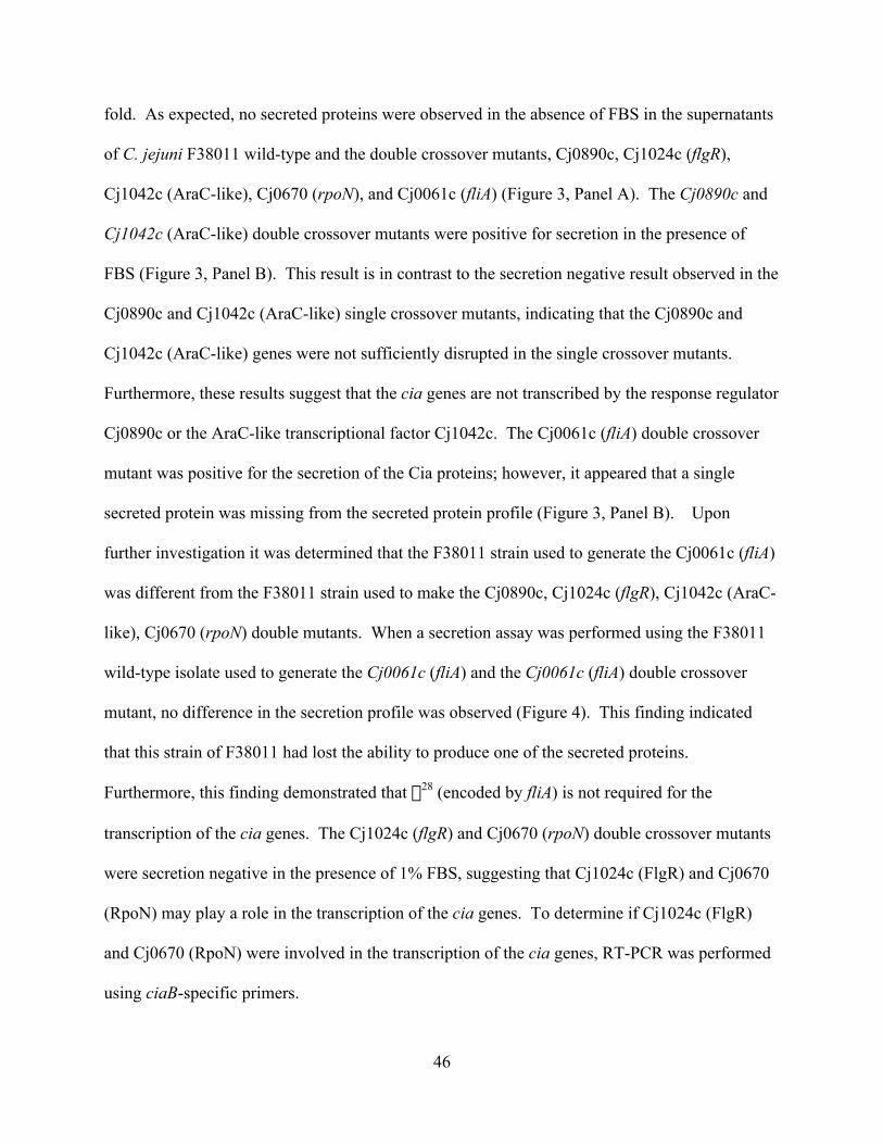
46
fold. As expected, no secreted proteins were observed in the absence of FBS in the supernatants
of C. jejuni F38011 wild-type and the double crossover mutants, Cj0890c, Cj1024c (flgR),
Cj1042c (AraC-like), Cj0670 (rpoN), and Cj0061c (fliA) (Figure 3, Panel A). The Cj0890c and
Cj1042c (AraC-like) double crossover mutants were positive for secretion in the presence of
FBS (Figure 3, Panel B). This result is in contrast to the secretion negative result observed in the
Cj0890c and Cj1042c (AraC-like) single crossover mutants, indicating that the Cj0890c and
Cj1042c (AraC-like) genes were not sufficiently disrupted in the single crossover mutants.
Furthermore, these results suggest that the cia genes are not transcribed by the response regulator
Cj0890c or the AraC-like transcriptional factor Cj1042c. The Cj0061c (fliA) double crossover
mutant was positive for the secretion of the Cia proteins; however, it appeared that a single
secreted protein was missing from the secreted protein profile (Figure 3, Panel B). Upon
further investigation it was determined that the F38011 strain used to generate the Cj0061c (fliA)
was different from the F38011 strain used to make the Cj0890c, Cj1024c (flgR), Cj1042c (AraC-
like), Cj0670 (rpoN) double mutants. When a secretion assay was performed using the F38011
wild-type isolate used to generate the Cj0061c (fliA) and the Cj0061c (fliA) double crossover
mutant, no difference in the secretion profile was observed (Figure 4). This finding indicated
that this strain of F38011 had lost the ability to produce one of the secreted proteins.
Furthermore, this finding demonstrated that s28 (encoded by fliA) is not required for the
transcription of the cia genes. The Cj1024c (flgR) and Cj0670 (rpoN) double crossover mutants
were secretion negative in the presence of 1% FBS, suggesting that Cj1024c (FlgR) and Cj0670
(RpoN) may play a role in the transcription of the cia genes. To determine if Cj1024c (FlgR)
and Cj0670 (RpoN) were involved in the transcription of the cia genes, RT-PCR was performed
using ciaB-specific primers.

47
ciaB is transcribed and translated in s54 and FlgR knockouts.
Previous results by Rivera-Amill et al. (2001) have shown using RT-PCR that ciaB is transcribed
in the presence of deoxycholate and 1% FBS. We isolated RNA from Cj0890c, Cj1024c (flgR),
Cj1042c (AraC-like), Cj0670 (rpoN), and Cj0061c (fliA) double crossover mutants grown on
MH supplemented with sodium deoxycholate (0.1% wt/vol) and used ciaB- and aspA- specific
primers to determine if ciaB gene transcription was regulated by Cj0670 (RpoN) or Cj1024c
(FlgR). The aspA gene was chosen as a control since it is constitutively expressed. Using ciaB-
specific primers, a product was amplified in the Cj0890c, Cj1042c (AraC-like), Cj0061c (fliA),
Cj1024c (flgR), and Cj0670 (rpoN) double mutants (Figure 5). This result indicates that Cj0670
(RpoN) and Cj1024c (FlgR) are not involved in the transcription of ciaB. Furthermore, these
data suggest that ciaB transcription is under the control a s70 promoter.
To validate the results above, whole cell lysates were prepared from the C. jejuni F38011 wild-
type, ciaB null mutant, and the double crossover mutants [Cj0890c, Cj1024c (flgR), Cj1042c
(AraC-like), Cj0670 (rpoN), and Cj0061c (fliA)] grown on MH supplemented with sodium
deoxycholate (0.1% wt/vol). Immunoblot analysis using an anti-C. jejuni CiaB antibody (# 407)
revealed that the CiaB protein was made in the C. jejuni F38011 wild-type, Cj0890c, Cj1024c
(flgR), Cj1042c (AraC-like), Cj0670 (rpoN), and Cj0061c (fliA) double crossover mutants (not
shown). As expected, the ciaB null mutant displayed no reactive band at 73 kDa, the estimated
size of CiaB.

48
DISCUSSION
This study was undertaken to gain a better understanding of how cia gene expression is regulated
in Campylobacter. We sought to determine if a response regulator or an AraC-like
transcriptional factor played a role in the regulation cia gene transcription. Studies by Konkel et
al. (1999) identified eight secreted proteins, designated CiaA-CiaH, ranging in size from 108-
12.8 kDa. To date only one of these proteins has been identified, CiaB (Konkel et al., 1999).
Interestingly, a ciaB null mutant contained no secreted proteins in the supernatant fluid (Konkel
et al., 1999). Studies by Rivera-Amill et al. (2001) demonstrated that bile salts (deoxycholate,
cholate, chenodeoxycholate) induce the synthesis of Cia proteins. In addition, a component of
fetal bovine serum was required for the synthesis and secretion of the Cia proteins (Rivera-Amill
et al., 2001). Konkel et al. (2004) has indicated that the secretion of the Cia proteins is
dependent upon a functional flagellar export apparatus.
Upon completion of this work, it became clear that there was phenotypic variation between the
response regulator and AraC-like transcriptional factor single crossover mutants compared to the
double crossover mutants. For Cj1024c (flgR), the single crossover mutant displayed motility,
whereas the double crossover mutant displayed a nonmotile phenotype. In the Cj0890c single
crossover mutants, 2/6 were sensitive to deoxycholate. In contrast the Cj0890c chromosomal
mutant and the Cj0890c double crossover mutant were deoxycholate resistant. The Cj0890c and
Cj1042c (AraC-like) single crossover mutants exhibited a secretion deficient phenotype in the
presence of 1% FBS, while in the Cj0890c and Cj1042c (AraC-like) double crossover mutants a
positive secretion profile was observed in the presence of 1% FBS. Given that inconsistent
phenotypes were observed between the single and double crossover mutants, it is imperative that

49
single crossover mutants, generated in genes coding for multiple domain proteins be mapped as
to where the insertion occurred. In addition, the phenotypes of the mutants must be validated by
a second independent method such as complementation of the knocked out gene, reinsertion of
chromosomal DNA containing the suicide vector from the mutant into a wild-type strain, or the
generation a of a double crossover mutant. A potential problem that can arise with single
crossover mutants is the reversion of the disrupted gene to a functional wild-type copy of the
gene by excision of the inserted suicide plasmid. Furthermore, the use of kanamycin as a
selective marker in Campylobacter suicide vectors can lead to problems because Campylobacter
can spontaneously mutate to confer kanamycin resistance, removing the selective pressure that
retains the suicide vector. In this paper, the double crossover mutants were designed so that a
large central portion of the gene being disrupted was deleted and replaced by the cat gene. This
method reduces the possibility that functional domains are maintained in the disrupted genes.
This study has shown that the Cj0670 (rpoN) and Cj1024c (flgR) are required for the production
of a functional secretion apparatus. Recent work confirms that Cj0670 (rpoN) and Cj1024c
(flgR) are required to produce a functional flagella in C. jejuni (Wösten et al., 2004; Carrillo et
al., 2004). These results strengthen the previous finding that the flagella are required for
secretion of the Cia proteins (Konkel et al., 2004). In addition, Konkel et al. (2004) has shown
that mutants in the basal body, rod, or hook prevent a functional flagella apparatus from being
assembled. Furthermore, either the FlaA or FlaB filament is required for the secretion of the Cia
proteins (Konkel et al., 2004).

50
Through this study we have shown that Cj0670 (rpoN) and Cj0061c (fliA) double crossover
mutants transcribe and translate the ciaB gene. Thus, the ciaB gene must be expressed from a
s70 promoter.
Transcriptional regulatory systems under the control of s70 have been observed in several
bacteria including Escherichia coli and Rhodobacter capsulatus. In E. coli, TyrR, a protein that
shows homology to NtrC (a s54-dependent transcriptional activator) is required for the regulation
of s70 genes encoding for aromatic amino acid biosynthesis and transport (Yang et al., 2002).
Expression of ntrC in R. capsulatus allows for the regulation of s70 transcribed genes (Xu and
Hoover, 2001). In E. coli, the response regulator, PhoB interacts with s70 to transcribe over 40
genes in phosphate depletion conditions (Blanco et al., 2002). Given that transcriptional
regulatory elements can allow for the expression of s70 regulated genes it is possible that ciaB
transcription may be under the control of a s70 transcriptional element.
While this study did not reveal if the transcription of cia genes are regulated by a two component
regulatory system, the mutants generated in this paper will allow us to identify genes transcribed
by response regulators through the use of recently developed microarray technology.
Furthermore, studies are under way to generate double crossover mutants in the remaining
response regulators [Cj0285c (cheV), Cj0355c, Cj0643, Cj1223c, Cj1227c, Cj1261 (racR),
Cj1491c, and Cj1068] to reexamine the phenotypes observed in the single crossover mutants. In
addition, double crossover mutants of all the transcriptional regulatory genes identified in the C.
jejuni NCTC 11168 genome are being generated.

51
ACKNOWLEGEMENTS
We thank Dr. Debabrata Biswas for performing the secretion assays on the single crossover
response regulator mutants and the F38011 and Cj0061c (fliA) double crossover mutant. We
thank Nicole Lindstrom for assisting with TEM. We thank Amy Keech for performing the RT-
PCR analysis. We thank Dr. John D. Klena and Dr. Brian H. Raphael for critical review of this
manuscript.
This work was supported by NIH grant DK58911 awarded to MEK.

52
REFERENCES
Blanco, A.G., Sola, M., Gomis-Ruth, F.X., Coll, M. (2002) Tandem DNA recognition by
PhoB, a two-component signal transduction transcriptional activator. Structure (Camb)
10, 701-13.
Colland, F., Rain, J.C., Gounon, P., Labigne, A., Legrain, P., De Reuse H. (2001)
Identification of the Helicobacter pylori anti-s28 factor. Mol Microbiol
41, 477-87.
Carrillo, C.D., Taboada, E., Nash, J.H.E., Lanthier, P., Kelly, J., Lau, P., Verhulp, R.,
Mykytczuk, O., Sy, J., Findlay, W.A., Amoako, K., Gomis, S., Willson, P., Austin, J.W.,
Potter, A., Babiuk, L., Allan, B., Szymanski, C.M. (2004) Genome-wide expression
analyses of Campylobacter jejuni NCTC11168 reveals coordinate regulation of motility
and virulence by flhA. J Biol Chem Feb. 25 [Epub ahead of print].
Dingle, K.E., Colles, F.M., Wareing, D.R.A., Ure, R, Fox, A.J., Bolton, F.E., Bootsma,
H.J., Willems, R.J.L., Urwin, R., Maiden, M.C.J. (2001) Multilocus sequence
typing system for Campylobacter jejuni. J Clin Microbiol 39, 14-23.
Guerry, P., Logan, S.M., Thornton, S., Trust, T.J. (1990) Genomic organization and
expression of Campylobacter flagellin genes. J Bacteriol 172, 1853-60.
Gunn, J.S. (2000) Mechanisms of bacterial resistance and response to bile. Microbes
Infect 2, 907-13.
Gupta, S., Chowdhury, R. (1997) Bile affects production of virulence factors and motility
of Vibrio cholerae. Infect Immun 65, 1131-4.

53
Hendrixson, D.R., DiRita, V.J. (2003) Transcription of s54-dependent but not s28-
dependent flagellar genes in Campylobacter jejuni is associated with formation of the
flagellar secretory apparatus. Mol Microbiol 50, 687-702.
Hoch, J.A., Silhavy, T.J. (1995) Two-component signal transduction. ASM Press,
Washington, D.C.
Jagannathan, A., Constantinidou, C., Penn, C.W. (2001) Roles of rpoN, fliA, and flgR in
expression of flagella in Campylobacter jejuni. J Bacteriol 183, 2937-42.
Kinsella, N., Guerry, P., Cooney, J., Trust, T.J. (1997) The flgE gene of Campylobacter
coli is under the control of the alternative sigma factor s54. J Bacteriol
179, 4647-53.
Konkel, M.E., Cieplak, W., Jr. (1992) Altered synthetic response of Campylobacter
jejuni to cocultivation with human epithelial cells is associated with enhanced
internalization. Infect Immun 60, 4945-9.
Konkel, M.E., Garvis, S.G., Tipton, S.L., Anderson, D.E., Jr., Cieplak, W., Jr. (1997)
Identification and molecular cloning of a gene encoding a fibronectin-binding protein
(CadF) from Campylobacter jejuni. Mol Microbiol 24, 953-63.
Konkel, M.E., Klena, J.D., Rivera-Amill, V., Monteville, M.R., Biswas, D., Raphael, B.,
Mickelson, J. (2004) Secretion of virulence proteins from Campylobacter jejuni is
dependent on a functional flagellar export apparatus. J Bacteriol 186.
Konkel, M.E., Kim, B.J., Rivera-Amill, V., Garvis, S.G. (1999) Bacterial secreted
proteins are required for the internalization of Campylobacter jejuni into cultured
mammalian cells. Mol Microbiol 32, 691-701.

54
Konkel, M.E., Tilly, K. (2000) Temperature-regulated expression of bacterial virulence
genes. Microbes Infect 2, 157-66.
Laemmli, U.K. (1970) Cleavage of structural proteins during the assembly of the head of
bacteriophage T4. Nature 227, 680-5.
Lin, J., Sahin, O., Michel, L.O., Zhang, Q. (2003) Critical role of multidrug efflux pump
CmeABC in bile resistance and in vivo colonization of Campylobacter jejuni. Infect
Immun 71, 4250-9.
Maniatis, T., Fritsch, E.F., Sambrook, J. (1982) Molecular cloning: a laboratory manual.
Cold Spring Harbor Laboratories, Cold Spring Harbor, NY. pp. 89-90.
Matz, C., van Vliet, A.H.M., Ketley, J.M., Penn, C.W. (2002) Mutational and
transcriptional analysis of the Campylobacter jejuni flagellar biosynthesis gene flhB.
Microbiol 148, 1679-85.
Maurelli, A.T., Hromockyj, A.E., Bernardini, M.L. (1992) Environmental regulation
of Shigella virulence. Curr Top Microbiol Immun 180, 95-116.
Prouty, A.M., Gunn, J.S. (2000) Salmonella enterica serovar typhimurium invasion is
repressed in the presence of bile. Infect Immun 68, 6763-9.
Rivera-Amill, V., Kim, B.J., Seshu, J., Konkel, M.E. (2001) Secretion of the virulence-
associated Campylobacter invasion antigens from Campylobacter jejuni requires a
stimulatory signal. J Infect Dis 183, 1607-16.
Schuhmacher, D.A., Klose, K.E. (1999) Environmental signals modulate ToxT-
dependent virulence factor expression in Vibrio cholerae. J Bacteriol
181, 1508-14.

55
Wösten, M.M.S.M., Wagenaar, J.A., Van Putten, J.P.M. (2004) The FlgS/FlgR two-
component signal transduction system regulates the fla regulon in Campylobacter jejuni.
J Biol Chem Feb. 11 [Epub ahead of print].
Xu, H., Hoover, T.R. (2001) Transcriptional regulation at a distance in bacteria. Curr
Opin Microbiol 4, 138-44.
Yang, J., Camakaris, H., Pittard, J. (2002) Molecular analysis of tyrosine-and
phenylalanine-mediated repression of the tyrB promoter by the TyrR protein of
Escherichia coli. Mol Microbiol 45, 1407-19.
Young, G.M., Schmiel, D.H., Miller, V.L. (1999) A new pathway for the secretion of
virulence factors by bacteria: the flagellar export apparatus functions as a protein-
secretion system. Proc Natl Acad Sci USA 96, 6456-61.

56
Table 1. Oligonucleotides used to generate the C. jejuni single crossover mutants
Primer Name Sequence (5' to 3')
Cj0285c Forward 43-62 GAGCTTGTCGATTTCCGTATCj0285c Reverse 551-571 TGCTGTCATCAAGTATAAGTGCj0355c Forward 11-33 TAGTTATAGAAGATGAGATTAGC
Cj0355c Reverse 535-556 CATTTGGAGTTACTAGCTCAGGCj0643 Forward 24-45 TGATGACAATAAAATGCTCGGC
Cj0643 Reverse 518-538 TTACTGGCATATTGACATCTGCj0890c Forward 28-49 GAAGATGATATAGATTTAAACG
Cj0890c Reverse 540-560 CTTGTAGTATCATCTTTACAACj1024c Forward 19-38 GAAGATGATATTAATATGCG
Cj1024c Reverse 523-544 TAGAATGGGTGTGAATATAACGCj1042c Forward 21-37 ACCAGAAGATTTAAAGC
Cj1042c Reverse 575-592 AATACTCAAAAAGTTGGGCj1223c Forward 20-39 TTTTAGAAGATGATTTGAGC
Cj1223c Reverse 530-550 CCCAGAGTTCTTCAAAAATTCCj1227c Forward 23-43 AAGATGATTTAGAATTAGCAG
Cj1227c Reverse 533-552 CTCACTAATAGAAGAACAATCj1261 Forward 38-55 GACTTTGCACAATTATTATCTG
Cj1261 Reverse 481-500 CCGTGTTGTTGGATAAGGTACj1491c Forward 27-48 AATATTAGTTGAAGATGAG
Cj1491c Reverse 516-535 CATGCAAAACCAAAGCTCCACj1608 Forward 25-44 GAAATTTATCTAGCGCAAAG
Cj1608 Reverse 718-739 CATTGCTATTGATATTTTTTTC

57
Table 2. Oligonucleotides used to generate the C. jejuni double crossover mutants
Primer 5' end Forward (5'to 3') Enzyme
Cj0890c TT(ggatcc)TTGCAAAAATGAAAGATGG BamHICj1042c AGATGTAAGTGAAAAACG -Cj1024c AA(ggatcc)CAAAAAGAGCAGCGATTACAG BamHICj0061c TTGGAAGACATTTTAATAGAAG -Cj0670 GGGTTAAATTTCTTGGTCTTG -cat TT(ggatcc)AGCTCTAGAGTCAACCGTGATATAG NheI
Primer 5' end Reverse (5' to 3') EnzymeCj0890c AA(ccgcgg)AAA(gctagc)TAAAGATATAACTTCATAACC SacII, NheICj1042c AA(ccgcgg)TTT(gctagc)GTTTGTGTATATTTAGC SacII, NheICj1024c AA(ccgcgg)AAA(gctagc)CTTAAAGTAGCATTTCCTGTC SacII, NheICj0061c AA(ccgcgg)AAA(gctagc)CACAAGCTCATCTTGCTCTTTC SacII, NheICj0670 AA(ccgcgg)AAAA(gctagc)GTGTTCAAAATATCCTTCCTCATTTAAG SacII, NheI
Primer 3' end Forward (5' to 3') EnzymeCj0890c TT(gctagc)GATAAAACAATCAATATAGC NheICj1042c TT(gctagc)TTGGTTTATTGAAAGATTTAAAGAG NheICj1024c TT(gctagc)TGGTAATATTAGAGAATTAATATCC NheICj0061c TT(gctagc)CACGAAGTGCTAGATGATCTTAAAG NheICj0670 TT(gctagc)GCTACTCTTTATAAAATAGGGCTTATG NheI
Primer 3' end Reverse (5' to 3') Cj0890c AA(ccgcgg)GCTGAGTATTTTTTAAAGTG SacIICj1042c AA(ccgcgg)CTACTTTGAATATTTTACC SacIICj1024c AA(ccgcgg)AACAGGCACTCTAACACAAGTTGC SacIICj0061c AA(ccgcgg)ATTTCTTTGATTTCATCTTTATC SacIICj0670 AA(ccgcgg)TAAAGCTACTAAAGCAATAGCTCCTAAG SacII
cat AA(gctagc)GTATAGAAGTGCGCCCTTTAGTTCC NheI

58
Table 3A. Phenotypes displayed by the C. jejuni response regulator and AraC-like transcriptional factorSingle crossover mutants.Isolate Transcriptional Class Motility Secretion Secretion Deoxcholate
(No FBS) (1% FBS) (resistant/sensitive)
Cj0285c (cheV) Response Regulator + - + R
Cj0355c Response Regulator - - + R
Cj0643 Response Regulator - - + R
Cj0890c Response Regulator + - - S (2/6)
Cj1024c (flgR) Response Regulator + - - R
Cj1223c Response Regulator + - + S (6/6)
Cj1227c Response Regulator + - + R
Cj1261 (racR) Response Regulator - - + R
Cj1491c Response Regulator - - + R
Cj1608 Response Regulator - - + S (6/6)
Cj0284c (cheA) Histidine Kinase + - + R
Cj1042c AraC-like + - - R
Table 3B. Phenotypes displayed by the C. jejuni double crossover mutants.
Isolate Transcriptional Class Motility Secretion Secretion Deoxcholate ciaB mRNA CiaB protein(No FBS) (1% FBS) (resistant/sensitive) Synthesis
Cj0061c (fliA) Sigma Factor + - + R + +
Cj0670 (rpoN) Sigma Factor - - - R + +
Cj0890c Response Regulator + - + R + +
Cj1024c (flgR) Response Regulator - - - R + +
Cj1042c AraC-like + - + R + +

59
Figure 1. Assessment of C. jejuni double crossover mutant motility on MH medium
supplemented with 0.4% Bacto Agar after 48 hr.

60
FIG. 1
Cj1024c (flgR)Cj0890c Cj1042c (AraC-like)
F38011 Cj0670 (rpoN)Cj0061c (fliA)

61
Figure 2. Transmission electron microscopy examination of C. jejuni wild-type F38011, fliA
double crossover mutant, and fliA complement isolates. Bar = 0.2 mm.

62
FIG. 2
F38011 Cj0061c (fliA) Cj0061c comp

63
Figure 3. C. jejuni Cia protein secretion requires Cj0670 (rpoN) and Cj1024c (flgR). C. jejuni
cells were cultured on MH-blood plates and labeled in MEM in the absence (panel A) and
presence (panel B) of FBS. Lanes: 1, C. jejuni F38011; 2, Cj0670 (rpoN) mutant; 3, Cj0061c
(fliA) mutant; 4, Cj1024c (flgR) mutant; 5, Cj0890c mutant; and 6, Cj1042c (AraC-like) mutant.
Molecular mass standards, in kDa, are indicated on the left.

64
FIG. 3

65
Figure 4. The Cj0061c (fliA) mutant secretion profile is identical to that of wild-type C. jejuni
F38011. C. jejuni cells were cultured on MH-blood plates and labeled in MEM in the absence
(lanes 1 and 2) and presence (lanes 3 and 4) of FBS. Lanes: 1, C. jejuni F38011; 2, Cj0061c
(fliA) mutant; 3, C. jejuni F38011; and 4, Cj0061c (fliA) mutant. Molecular mass standards, in
kDa, are indicated on the left.

66
FIG. 4
97.466.0
45.0
31.0
25.5
14.5
1 2 3 4

67
Figure 5. ciaB is transcribed in Cj0670 (rpoN) and Cj1024c (flgR) mutants. RT-PCR analysis
was performed with C. jejuni cultured on MH agar supplemented with 0.1% deoxycholate.
Bacterial RNA was extracted, and RT-PCR analysis was done, as described in Materials and
Methods. RT-PCR analysis of each RNA sample was performed using ciaB-specific primers,
aspA-specific primers and ciaB primers without the RT enzyme (control).

68
FIG. 5
ciaB
aspA
Con
trol
Cj0890c Cj0670 (rpoN)Cj0061c (fliA) Cj1024c (flgR)Cj1042c
ciaB
aspA
Con
trol
ciaB
aspA
Con
trol
ciaB
aspA
Con
trol
ciaB
aspA
Con
trol
1650
850650500400
1000

69
CONCLUSIONS AND FURTHER DIRECTIONS
Through this body of work we have shown that Cj0670 (rpoN) and Cj1024c (flgR) are required
in the production of a functional secretion apparatus. Furthermore, one of the secreted proteins,
CiaB, appears to be expressed by s70. However, the question still remains as to whether the
expression of the other cia genes is regulated by a two component regulatory system. Therefore,
double crossover mutants of the remaining response regulators [Cj0285c (cheV), Cj0355c,
Cj0643, Cj1118c (cheY), Cj1223c, Cj1227c, Cj1261 (racR), Cj1491c, and Cj1608] need to be
generated to validate their secretion phenotype.
Recently, the Konkel lab has sent RNA samples to Dr. Craig Parker (USDA, Albany, CA) for
microarray analysis. C. jejuni F38011 RNA was isolated from MH broth and MH broth plus
sodium deoxycholate (0.05% wt/vol). Upon microarray analysis four response regulators
[Cj0355c, Cj1118c (cheY), Cj1227c, and Cj1491c] were shown to be upregulated in the presence
of deoxycholate. Double crossover mutants of these four response regulators are currently being
generated by Dr. Brian Raphael and will be subject to secretion assays and microarray analysis to
determine possible genes under the control of these response regulators. Cj0285c (cheV),
Cj0643, Cj1118c (cheY), Cj1223c, Cj1261 (racR), Cj1491c, and Cj1608 double crossover
mutants have been generated and subjected to motility assays and deoxycholate sensitivity
assays. Cj1118c (cheY) and Cj0285c (cheV), which are involved in chemotaxis, were found to
be nonmotile. The remaining RRs [Cj0643, Cj1223c, Cj1261 (racR), Cj1491c, and Cj1608]
displayed motility. Upon, testing for deoxycholate sensitivity, Cj0643 was determined to be
extremely sensitive to growth in the presence of deoxycholate. Cj0285c (cheV), Cj1118c (cheY),
Cj1223c, Cj1261 (racR), Cj1491c, and Cj1608 double crossover mutants showed no sensitivity

70
to deoxycholate. Immunoblot analysis using an anti-C. jejuni CiaB antibody (# 407) revealed
that the CiaB protein was made in Cj0285c (cheV), Cj0643, Cj1118c (cheY), Cj1223c, Cj1261
(racR), Cj1491c, and Cj1608 double crossover mutants. Currently, Dr. Raphael is generating
double crossover mutants in Cj0355c and Cj1227c. The Cj0643 double crossover mutant was
sent to Dr. Zhang, to determine if its sensitivity to deoxycholate will hinder the colonization of
the cecum in chickens.
With the use of microarray technology the identification of the genes under the control of
response regulators may be ascertained by generating constitutively active response regulators.
Response regulators can be made constitutively active by changing the conserved aspartic acid in
the active site to a glutamic acid (Nohaile et al., 1997). This substitution mimics the
phosphorylation of the aspartic acid, thereby activating the response regulator without the need
of the phosphate being transferred from the response regulator’s histidine kinase partner (Nohaile
et al., 1997). By comparing the double crossover mutant response regulator to the constitutively
expressed response regulator we will be able to identify the genes being regulated by that
specific response regulator. By identifying these genes we may be able to determine potential
signals that control the activation of the response regulators.
71
REFERENCE
Nohaile, M., Kern, D., Wemmer, D., Stedman, K., Kustu, S. (1997) Structural and
functional analyses of activating amino acid substitutions in the receiver domain of NtrC:
evidence for an activating surface. J Mol Biol 273, 299-316.

72
APPENDIX
This project is the result of the concerted efforts of many individuals. My contribution to this
work includes: designing the oligonucleotides required to generate the single and double
crossover mutants and the generation of all the C. jejuni mutants used in this study (Table 1 and
2). Furthermore, I performed the motility and deoxycholate sensitivity assays of the single and
double crossover mutants (Table 3A and 3B; Fig. 1). I performed the secretion assay on the
double crossover mutants (Fig. 3). Nicole Lindstrom performed the TEM (Fig. 2). D. Biswas
performed the secretion assays on the single crossover mutants and the F38011 and Cj0061c
(fliA) double crossover mutant (Table 3A; Fig. 4). A. Keech performed the RT-PCR analysis on
the double crossover mutants (Fig. 5). B. Raphael performed the western blot analysis on the
double crossover mutants using the anti-CiaB antibody (Table 3B). Future work towards the
publication of this paper will be performed by M. Konkel and B. Raphael.