Barrett's esophagus treatment, barrett's esophagus ablation, barrett's esophagus and smoking

Version 7 date 2011-12-19 1
Study protocol for resectabel cancer of the esophagus and gastro-esophageal junction.
Ethic approval: EPN Stockholm 2006/738-32, 2008-403-32
EudraCTnr 2006-001785-16
Approval Medical Products Agency of Sweden Dnr: 151:2006/24273
Coordinating investigator/sponsor:Lars Lundell, Professor of Surgery, Department of Surgery, Gastrocentrum, Karolinska University Hospital, Huddinge, Stockholm Sweden
Investigator signature………………………………………………………………………….
Date……………….
Version 7 date 2011-12-19 2
Site Investigator:Gunnar Adell, Department of Oncology, Karolinska University Hospital, Södersjukhuset Stockholm Sweden
Singe Friesland, Gun Wickart-Johansson, Department of Oncology, Karolinska University Hospital, Radiumhemmet, Solna, Stockholm Sweden
Karl Erik Johansson, Ass. Professor of Surgery, Department of Surgery, University Hospital, Linköping, Sweden
Torbjörn Myrnäs, Ass. Professor of Surgery, Department of Surgery, Norrlands University Hospital, Umeå, SwedenNielsHilmer Nielsen Department of Oncology, Norrlands University Hospital, Umeå, Sweden
Ingemar Näslund, Ass. Professor of Surgery, Department of Surgery, University Hospital, Örebro, Sweden, Mattias Elmlund, Department of Oncology
Gunnar Wagenius, Simon Ekman, Michael Bergqvist, MD, PhD. Department of Oncology, Akademiska sjukhuset, Uppsala, Sweden
Erik Johnsson, Kirurgiska Kliniken, Sahlgrenska Universitetssjukhuset, Göteborg, SwedenHedda Haugen, Department of Oncology, Sahlgrenska Universitetssjukhuset, Göteborg, Sweden
Ullevål University Hospital, Oslo: Egil Johnson, kirurgisk avdelingPetra Weber Hauge, onkologisk avdelning, Gunilla Frykholm, onkologisk avdelning
St Olav University Hosptal, Trondheim Gjermund Johnsen, kirurgisk avdelningIngunn Hatlevoll, onkologisk avdeling, Gunilla Frykholm, onkologisk avdeling
Universitetssykehuset i Nord-Norge, Tromsø Jørn Kjæve, kirurgisk avdelingLise Balteskard, onkologisk avdeling
Haukeland Universitetssykehus, Bergen Asgaut Viste, Kirurgisk avdelning
Radiumhospitalet, Oslo, Stephan Stoldt, Kirurg Kliniken, Anne-Birgitte Jacobsen, onkologisk avdelning.
Mälarsjukhuset, Eskilstuna, Pehr Lind, Onkologiska kliniken

Version 7 date 2011-12-19 3
1. Introduction – Background2. Study objectives3. SCHEDULE4. STUDY DESIGN5. PRETREATMENT EVALUATION6. ELIGIBILITY CRITERIA7. STRATIFICATION AND RANDOMIZATION8. TREATMENT PLAN9. ON-STUDY ASSESSMENT AND FOLLOW-UP10. QUALITY OF LIFE ASSESSMENT11. DEFINITIONS OF OUTCOMES12. STATISTICAL CONSIDERATIONS AND METHODOLOGY13. QUALITY ASSURANCE AND MONITORING14. Time plan15. PUBLICATION16. ETHICAL ASPECTS17. Study coordination, innovative analyses18. Principal investigators19. REFERENCES

Version 7 date 2011-12-19 4
Background:On a yearly basis roughly 4-500 patients present with carcinoma of the esophagus in Sweden. With regard to cancers originating in the gastro-esophageal junction (GEJ) it is some what more difficult to estimate the incidence figures due to difficulties in classification of the site of origin of the neoplastic process. A good estimate is, however, that GEJ cancers affect 150 – 200 patients each year in this country. There are some epidemiological data which are of importance for the management of the patients. First the number of adenocarcinomas is steadily increasing both in the body of the esophagus as well as in the GEJ. Concomitant with that, the corresponding figures for squamous cell cancer are decreasing. Second already at the time of first presentation of these patients, the tumours have usually reached an advanced stage in the vast majority of the cases. It can therefore be estimated that roughly only one third of these patients are only amenable to a treatment with curative intent. These facts emphasis the importance of the urgency of development of palliative management strategies.
Even in those individuals, who are candidates for treatment with curative intent, long-term follow up have consistently demonstrated that tumour free survival can only be expected to plateau at a level of 20 %. The main mechanism behind these dismal figures resides in the preponderance for these tumours to metastasis to loco-regional lymph nodes already when penetrating through the muscularis mucosae. In fact careful investigations have demonstrated that tumours which have penetrated into the superficial third of the mucosa already in 25 % had developed local lymph node metastases.
Until quite recently surgical therapy has been considered the gold standard in the treatment of carcinoma of the esophagus and GEJ. Recent studies have been carried out to challenge this basic therapeutic concept in these tumour manifestations, but until now radio-chemotherapy alone has not been shown to offer benefits which are superior to surgery alone. Therefore surgery will remain the basic therapeutic modality in these cancers although it seems to offer a therapeutic efficacy which is far from optimal.
During the recent decade a number of studies have been carried out to determine the value of neoadjuvant or adjuvant treatment given in addition to surgery. It is clear that postoperative treatment can not be recommended. However, when it comes to neoadjuvant treatment it remains to be determined whether radio-chemotherapy offers advantages above what can be achieved by chemotherapy alone. The down side of combination neoadjuvant therapy might well be that the risks associated with surgery are enhanced.
Although the ultimate objective of the actual multi modality regimen is to improve the survival, the research methodology aspects that hereby emerge are overwhelming, in terms of recruiting patients enough into respective therapy arm. There are, however, alternative complementary methodology strategies available that relate to the relationships between the degree of histological response and the subsequent over all survival. It has repeatedly been shown that a complete eradication of tumour cells, when evaluated in the resected specimen, directly translates into a better survival of the patient. Due to the basic design of a similar study all patient will have a resection, the opportunity emerges to use a surrogate variable as the primary end point. By the definition of the rate of complete histological response, as the primary objective in a similar study, the preconditions for the power calculations, the enrolment of patients and the conduct of the trial change most favourably.
Version 7 date 2011-12-19 5
Study objectives:
Primary objective: To evaluate whether combined radiochemotherapy gives higher complete
histological response (pCR) after resection than chemotherapy alone in patients with resectable carcinoma of the esophagus and gastric cardia.
Secondary objectives: To evaluate the safety of respective neoadjuvant therapies To assess the safety profile of carrying out radical surgery after respective
neoadjuvant therapy To study the overall and disease free survival in respective groups To evaluate if pretreatment tumor characteristics predict the pCR and
incomplete pCR rates
SCHEDULE

Version 7 date 2011-12-19 6
Patient population Histologically confirmed squamous cell carcinoma or adenocarcinoma of the esophagus
and gastric cardia.
Suitable for surgery alone.
T1N1, T2N0, T2N1, T3N0, T3N1.
M1a
Stratification
Histology Arm A:Chemoradiation + SurgeryRANDOMISATION
Arm B: Chemotherapy + SurgeryT-stage
N-stage Tumor location (prox, mid or distal esophagus, gastric cardia)
Institution
3. STUDY DESIGN
3.1 The study is designed as a two-arm phase III randomized, open, multicentre, controlled trial.
3.2 The study population will consist of patients with squamous cell or adenocarcinoma of the esophagus (Siewert type I) and adenocarcinoma of the gastric cardia (Siewert type II), with stages as defined in inclusion criteria.
3.3 Patients will be randomised between induction chemotherapy or chemoradiotherapy, all followed by surgery
4 ELIGIBILITY CRITERIA

Version 7 date 2011-12-19 7
Inclusion criteria4.1 Histologically verified squamous cell carcinoma, adenocarcinoma of the esopohagus or
gastric cardia (type II)
4.2 Tumour located in cervical oesophagus, not requiring laryngo-esophagectomy.
4.3 Patients with performance status 0-1 according to WHO scale and with resectable tumours, as assessed at the prerandomisation evaluation.
4.4 Adequate haematological function, defined as having WBC > 3 x 10⁹/litre and platelets > 100 x 109/litre
4.5 Adequate renal function defined as having normal serum creatinine level and/or calculated glomerular filtration rate > 60 ml/min
4.6 Tumour stage : T1N1, T2N0, T2N1, T3N0, T3N1. M1a
4.6 Written informed consent 4.8 Age <75 years
4.9 No major illness that make chemoradiotherapy unsuitable, life expectancy at least 3 months
Exclusion criteria4.10 Pregnancy and/or lactation. Women of childbearing ages can be included provided that
adequate contraceptive methods are used
4.11 Patients with diabetes complications (e.g. retinopathy, neuropathy) as well as patients with uncontrolled cardiac disease or myocard infarction within 12 months are considered unsuitable for chemoradiotherapy.
4.12 Concomitant malignancy (< 5 years since diagnosis) that can interfere the interpretation of study results, ongoing antitumoral treatment
4.13 Patients being unable to comply with the protocol
4.14 Tumor stage T1 N0, T4 NX or TXNXM1b

Version 7 date 2011-12-19 8
5 STRATIFICATION AND RANDOMISATION
5.1 Prior to randomization, patients will be stratified by histology (squamous cell carcinoma, adenocarcinoma), location of the tumor, institution and by T and N stage
5.2 Randomisation will be carried out by use of a computer based randomization programme operational at the Regional Oncological Center Stockholm
5.3 Randomization will be processed through the following contact persons and addresses:
Telephone number: 0046 8 51772981 Fax number: 0046 851775444 Onkologiskt Centrum M80 Stockholm Gotland
6 PRETREATMENT EVALUATION
The following investigations are required prior to protocol entry.
6.1 Histological confirmation of diagnosis of invasive squamous cell carcinoma or adenocarcinoma of the esophagus.
6.2 Pre-randomisation endoscopic assessment of the primary lesion. This will include length, location, (cm from the incisors), and sampling for histopatological confirmation performed within 1 month of the randomization.
6.3 Laboratory studies: Full blood count with differential and platelet counts, creatinine, liver function tests, LD (optional) performed within 1 month of the randomization.
6.4 Respiratory (dynamic and static spirometry) and cardiac function tests (exercise ECG surveilled bicycle test) performed within 2 months of the randomization
6.5 CT of the thorax and upper abdomen to assess tumour status performed within 1 month of the randomization.
6.6 Laparoscopy is recommended for carcinomas in the distal third of the esophagus primarily for those originating in the gastric cardia (performed within 1 month of the randomization)
6.7 Bronchoscopy (optional) when indicated.
6.8 PET-CT scan performed within 1 month of the randomization ( for centres outside Stockholm region, optional)
6.9 Barium esophagogram (optional).
6.10 Endoscopic ultrasonography performed within 1 month of the randomization 6.11 Audiogram

Version 7 date 2011-12-19 9
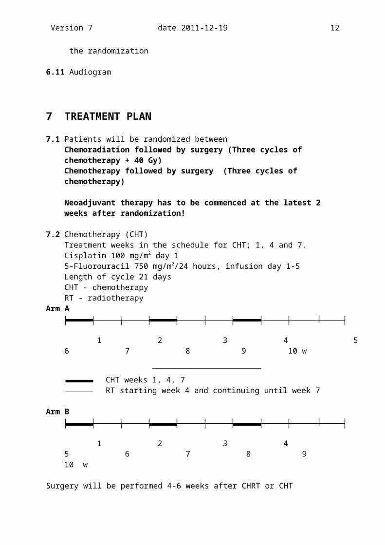
7 TREATMENT PLAN
7.1 Patients will be randomized betweenChemoradiation followed by surgery (Three cycles of chemotherapy + 40 Gy)Chemotherapy followed by surgery (Three cycles of chemotherapy)
Neoadjuvant therapy has to be commenced at the latest 2 weeks after randomization!
7.2 Chemotherapy (CHT) Treatment weeks in the schedule for CHT; 1, 4 and 7.Cisplatin 100 mg/m2 day 15-Fluorouracil 750 mg/m2/24 hours, infusion day 1-5Length of cycle 21 daysCHT - chemotherapyRT - radiotherapy
Arm A
1 2 3 4 5 6 7 8 9 10 w
CHT weeks 1, 4, 7RT starting week 4 and continuing until week 7
Arm B
1 2 3 4 5 6 7 8 9 10 w
Surgery will be performed 4-6 weeks after CHRT or CHT
Dose modificationsDay 1 Nadir Platinol 5-FU LPK TPK LPK TPK> 2.5 > 75 > 1.0 > 75 100 % 5 days
> 1.0 < 75 50 % 50 % 5 days< 1.0 xx 50 % 50 % 5 days
> 2.5 < 75 0 % 0 days< 2.5 xx 0 % 0 days
Dose adjusted according to current LPK and TPK values. Day 1 Values directly according to actual dose.xx Independently of TPK values.
Dose modifications: In case of neutropenia at day 22 and CHT has to be delayed, also the start of radiotherapy is suggested to be delayed one week. G-CSF is mandatory to be given as prophylactic treatment after each course. In case of delayed CHT, G-CSF should be considered.

Version 7 date 2011-12-19 10
(Neulasta, Neupogen etc)
If a patient suffers from moderate or severe hearing impairment and/or moderate or severe tinnitus cisplatin can be replaced. In case of an adenocarcinoma oxaliplatin, 130 mg/m2 should be used. In case of a squamous cell carcinoma carboplatin, AUC 5 should be used. Cisplatin can be replaced before starting treatment, or during the treatment course
If renal function is moderately impaired (GFR less than 50 ml/min) during treatment cisplatin should be replaced. In case of an adenocarcinoma oxaliplatin 130 mg/m2 should be used. In case of a squamous cell carcinoma carboplatin, AUC 5 should be used. If renal function is severely impaired during treatment, chemotherapy should be discontinued.
Arm A: if the second course has to be delayed, start RT as planned week 4, consider to give Neupogen 300mg/day for 5 days and give the CHT week 5 instead. If the CHT has to be delayed > 2 weeks, continue only with RT. It is mandatory to give Neulasta, Neupogen etc even after the second and third course in such case.
Arm B: if the second course has to be delayed, consider to give Neulasta, Neupogen etc 300mg/day for 5 days and give the CHT week 5 instead. It is mandatory to give Neulasta, Neupogen etc even after the second course in such a case. If the chemotherapy has to be delayed more than 2 weeks, no more CHT should be given and the patient should be referred to surgery immediately.
7.3 RadiotherapyAll dose planning shall be performed with a CT-based three-dimensional treatment planning system. All patients included in the study shall be able to receive 40 GY to the target volume without exceeding absorbed doses to the organs at risk.
Patient positioning and immobilisationThe patient should be positioned in a supine position with e.g. the head on a standard head-rest and arms preferably above the head, allowing a multiple field technique.
Patient data acquisitionMultiple CT-scans, covering the entire PTV and total lung volume. The CT investigation shall be made in the treatment position.
Target volumeThe different volumes of interest shall be defined in agreement with ICRU Report 50.
GTV Primary esophageal tumor and gross lymph node metastases. Lymph node metastases are defined from diagnostic CT and EUS examinations.
CTV GTV (gross tumour volume) + local subclinical extension. A dose of 40 Gy is meant to encompass gross tumour volume (GTV) and regional lymph nodes. For tumours located at or above the level of carina, the caudal border of CTV was 5 cm below diagnosed tumour, whereas the supraclavicular nodes defined the cranial border. For

Version 7 date 2011-12-19 11
tumours located mainly below the carina level, the cranial border of CTV included 5 cm of radiographically uninvolved esophagus, while the coeliac lymph nodes were included in the target volume, at the same time defining the caudal border, down to upper part of L1. In lateral, anterior and posterior directions CTV should encompass GTV and paraoesophageal area with a margin of 1 cm but not including anatomical barriers as pleura, pericard og bone.
PTV Appropiate margins (as small as possible) should be added to the CTV to take into account the effects of organ and patient movements and inaccuracies in beam and patient set-up in order to ensure that the prescribed dose is actually absorbed in the CTV.
Organs at riskThe spinal cord, the lung and the heart..
Simulation procedureA simulation procedure is mandatory for all fields. The position of all shielding blocks should be indicated on the simulation films.
Treatment techniqueA multiple field technique with optimal beam entry directions and beam weights in order to obtain as homogeneous a dose as possible in the PTV while at the same time minimizing the dose to the organs at risk.
Normal tissue sparingField shaping should be performed with customized blocks (individually cut) or with a multileaf collimator.
Dose to organs at riskSpinal cordThe maximum dose to the spinal cord is 40 Gy
LungNormal pulmonary tissue should be spared as much as possible. The beam geometry should be arranged in such a way that the lung tissue is spared as much as possible. The volume for the dose > 20 Gy to the lungs should be minimized as much as possible and should not exceed one third of the lung volume. Attention should be given, especially to patients with low pulmonary function, in view of risk of radiation induced pneumonitis and subsequent radiation fibrosis
HeartThe volume for the dose > 30 Gy to the heart should be minimized as much as possible
KidneyThe total dose to both kidneys should be kept as low as possible and not exceed 12 Gy and not more than 20 Gy for a single kidney
Dose computationA three-dimensional computerized dose planning with inhomogeneity correction shall be performed.
Version 7 date 2011-12-19 12
Dose specificationThe dose specification should be in terms of the ICRU Reference point (see ICRU Report 50) which should be located in the center of the PTV, and if possible on or near the central axis of the beams.
Fractionation schedulePTV: 40 Gy in 20 fractions (2.00 Gy/fraction) in a course of 4 weeks (approximately 28days), 1 fraction per day and 5 fractions per week. In case of bank holidays or machine breakdown, the treatment time should be extended. It is not allowed to give 2 fractions per day.
Treatment verification, minimum requirementsSimulator port filmsTreatment verification at least at the first, in the middle and at the end of the treatment.Measurements of entrance dose or other equivalent methods to ensure that the correct calculated dose is given to the patient.Radiation treatment charts shall be saved as well as the dose plans, portal and verification films.
AbbreviationsGTV (Gross Tumour Volume) is the gross palpable or visible/demonstrable extent and
location of malignant growth
CTV (Clinical Target volume) is a tissue volume that contains a demonstrable GTV and/or subclinical microscopic malignant disease, which has to be eliminated. This volume thus has to be treated adequately in order to achieve the aim of therapy, cure or palliation.
PTV (Planning Target Volume) is a geometrical concept, and it is defined to select appropiate beam sizes and beam arrangements, taking into consideration the net effect of all the possible geometrical variations, in order to ensure that the prescribed dose is actually absorbed in the CTV.
7.4 Surgical treatmentPreoperative investigationsPreoperative evaluation of operability is performed according to the protocol above regarding functional status. The inclusion criteria preclude that all patients resectable. If the colon is planned to be utilized as the esophageal substitute, a colonoscopy is performed before operation.Surgery has to be carried out 4-6 weeks after completion of the respective induction, neoadjuvant therapies
The operative treatment is standardized as follows. All participating centers shall have a documented experience in surgery directed towards cancers of the esophagus and gastric cardia. This constitutes a vital prerequisite to ensure high quality of the procedure and to minimize morbidity and postoperative mortality. Furthermore, all operating surgeons should be trained and familiar with the concept of two-field lymphadenectomy. Basically all operations are performed as thoracoabdominal resections with intrathoracic anastomosis for cancers in the distal third and cardia whereas three stage resection is carried out for

Version 7 date 2011-12-19 13
those in the middle and upper part of the organOperative and postoperative data will be transferred directly into the CRF from the national register for surgery for cancer of the esophagus and stomach (ENREV) and thereby made available for those involved in the follow-up process.
8. ON-STUDY ASSESSMENT, SAFETY AND FOLLOW-UP
8.1 Monitoring of treatmentPatients randomised to receive chemoradiation, should be reviewed weekly during their radiation treatment. Side effects related to therapy are to be assessed using the NCI CTCAE v 3.0 scale (see Appendix 1),nutritional support and local analgesics are to be administered if and when necessary.Patients receiving chemotherapy alone, should be monitored every 3rd week.
For all patients weekly full blood counts and serum creatinine level determinations should be made after chemotherapy until the neutrophile count is > 1.0 x 10⁹/L and platelets are > 100 x 109/L.
8.2 Safety assessmentsAll Adverse Events (AEs) or Serious Adverse Events (SAEs) are recorded in appropriate CFR forms and be reported using NCI-CTCAE v3.0 scale. All SAEs are reported on specific SAE form and sent to the ROC (Telephone number: 0046 8 51772981 Fax number: 0046 85177544). The copy of SAE should even be faxed to the coordinating investigator (Lars Lundell, fax nr 0046 8 58586450) as soon as possible but no later than 1 week after the SAE. The site investigator at respective site is responsible to that it will be done according to GCP roules.
A Serious Adverse Event (SAE) is an event which Results in death Is life-threatening Is disabling Requires hospitalization, whether initial or prolonged, and does not include planned
hospitalization Requires intervention to prevent permanent impairment/damage
The term “life-threatening” in the definition of “serious” refers to an event in which the patientwas at risk of death at the time of the event; it does not refer to an event which hypotheticallymight have caused death had it been more severe.
8.3 Follow-up assessmentFollow-up examination for symptoms and signs of local recurrence, appearance of metastases and treatment complications should be made every 3 months during the first 2 years and thereafter twice yearly.Endoscopy and other investigations are reserved for specific problems Table 1. Summary of investigations and follow-up required for this study.
1) Baseline 2) DuringCT/RT
3) PostCT/RT
4) Post surgery
5) Follow up
Physical examination

Version 7 date 2011-12-19 14
PS, Weight
Nutritional route, p.o., PEG, CF-tube
Endoscopy+ EUS
Endoscopy When
clinically indicated
Computerized Tomography of thorax & upper abdomen
When
clinically indicated
PET/CT (optional)
Spirometry, cardiac function test (exercise ECG surveilled bicycle test)
WBC, Hb, TpKCreatinine
(wkly)
Laparoscopy (Siewert type II)
Bronchoscopy (optional)
Barium oesophogram (optional)
Quality of Life assessment once yearly
6)
Audiogram
if PET/CT is performed, no additional CT should be done
1) Physical examination (including PS, weight) and blood counts 1 week before start of therapy, all other investigations within 1 month before randomization 2) weekly for patients in Arm A and every 3rd week for patients in Arm B3) 3 weeks after completion of either CHT or CHRT4) 1 months after surgery5) every 3rd month starting 1 month after surgery during first 2 years, thereafter twice yearly up to 5 years after completion of therapy6) at month 12, 24, 36, 48 and 60 after completion of therapy
9 QUALITY OF LIFE ASSESSMENT
Specific attention is to be directed towards quality of life, particularly swallowing function. To this end, simple quality of life assessment questionnaires using a self-assessment linear analogue scale to measure physical well-being, mood, pain, nausea and vomiting, appetite, swallowing and tiredness, will be administered once every 3 months and at the time of the

Version 7 date 2011-12-19 15
first relapse (see Appendix 3). This will be completed with EORTC QLQ-C30 to which is added the OES 18 module.

Version 7 date 2011-12-19 16
10 DEFINITIONS OF OUTCOMES
10 Complete Histological Response (pCR) as defined after serial sectioning of the operative specimen and tissue sections evaluated according to a predefined investigational protocol (see appendix)
11 Crude survival= Overall survival, is counted from time to randomization to death or end of follow-up
12 Time to recurrent disease. CT thorax and abdomen should be performed to document a clinical suspicion of recurrent disease. If possible recurrent disease should be verified histologically or by percutaneous puncture
13 Quality of LifeQuality of life will be measured pretreatment, after completion of neoadjuvant therapy and at yearly intervals during the follow up, using linear analogue self-assessment scales for physical well-being, mood, pain, nausea and vomiting, appetite, swallowing and tiredness (see Appendix 3). The EORTC QLQ-C30 will also be used.
13.1 SurgeryType of surgery will be recorded and also postoperative assessment. Residual disease will be recorded and also postoperative complications.
10.6 Toxicity/ChemoradiotherapyToxicity will be scored according to the NCI CTCAE v 3.0 scale (see Appendix 1).
11 STATISTICAL CONSIDERATIONS AND METHODOLOGY
11.1 Sample sizeThe sample size considerations are based on the primary endpoint CHR. The aim is to achieve an improvement in the CHR from 20 % to 35 % with a power of 80 % using a two-sided log-rank test with significance level 5 %. Then it is necessary to include 86 patients per arm: n= P1x(100-P1)+P2x(100-P2) x f(alfa,beta)
(P1-P2)2
To have 172 eligible patients for analyses, 8 more patients are included in the study, e.g. 90 patients/arm will be included, the total amount will be180 patients.
11.2 Analysis planThe main analyses will be based on the intention to treat principle. Demographic treatment and clinical data will be reported by treatment arm.
A multivariate Cox survival analysis will be carried out to study prognostic factors and to estimate the adjusted treatment difference.
Subgroup analyses are planned for stage groups since one could expect a larger treatment difference in higher stages.
Quality of life as measured by using linear analogue self-assessment scale, the
Version 7 date 2011-12-19 17
EORTC QLQ-C30 and the special form for Esophagus cancer (form Q) will be accounted for by treatment arm and time after treatment (9).
Toxicity due to chemoradiotherapy, chemotherapy and postoperative complications will be presented by treatment arm.
11.3 Interim analyses, monitoring and stopping rulesToxicity of chemotherapy and radiation therapy will be evaluated and protocol modification considered if excessive toxicity is observed. Late toxicities will be evaluated by Quality of Life. A formal analysis for safety and toxicity will be undertaken after 25 % of the projected accrual has been done.
An interim analysis will be performed after 66 % of the events have been observed (i.e. number of patients completed the preoperative treatment protocol), with significance level of 1.5% for the analysis. In order to maintain an overall significance level of 5% the final analysis will then be performed on the level 4%. If at this analysis one of the arms is found to be significantly inferior in terms of safety, the data monitoring committee shall inform the study coordinators to assemble the principal investigators and advisory boards. They decide whether to stop the trial or not.
An independent Safety and Data Monitoring Committee will review toxicity and survival data. It will also make recommendations on changes to the protocol and/or stopping the trial. The committee will include four individuals representing the participating regions of the country

Version 7 date 2011-12-19 18
12 QUALITY ASSURANCE AND MONITORING
Quality assurance and monitoring will be done according to GCP rules:
a. Treatment, main treatment according to randomization and later alternative therapy.
b. Follow-up, side effects, survival, cause of deaths.
c. Data reporting and management.Case Record Forms (CRFs) should be sent on a continuous basis to the Regional Oncology Centre in Stockholm.
d. All operative procedures and related postoperative courses are concomitantly recorded in the Swedish Quality Assurance Register
13. Time plan
First patient in Q1 2007. During Q1 2008 one patient per week will be enrolled. From Q2 2008, 2 patients will be enrolled per week. Based on these calculations the last patient in will be reached Q 4 2009.
14. PUBLICATION
Any publication based on the data from this study has to be divided into one of two categories but the following general principles will be followed. Authors of the publication are those who actively participate in processing of the study protocol, recruiting patients, compiling the results and putting together the article. However, all participating clinics will be named in a special appendix, and contacts identified. First the steering committee determines who will be actively participating in the analyses of data and writing of manuscripts of all scientific reports addressing the primary and secondary end-points, defined in the protocol from this study. The details of the decision making principles within the committee are detailed under 16. The steering committee decides who will be the 1st author of respective publications as well as the co-authors regarding publications referred to above. Co-authorship will be offered any institution where > 20 patients have been enrolled. The second principle is applicable to other subgroup studies and analyses which are referred to under 16.
15. ETHICAL ASPECTS
The investigator will ensure that this study is conducted in full conformance with principles of the”Declaration of Helsinki” (as amended in Tokyo, Venice and Hong-Kong), or with the laws and regulations of the country in which the research is conducted, whichever affords the greater protection to the individual.
16. Study coordination, innovative analyses

Version 7 date 2011-12-19 19
A steering committee is established which represents all regions of the country. In a situation where other countries (regions) will enter the study, this committee will expand so that a balanced representation will be pursued. Provided that one center is unable to enroll patients to the level which motivates a position in the committee the continuation and representation of that centre in the committee will be decided upon at these regular meetings. The committee will take decisions with simple majority as the basic principle in case a consensus based decision can otherwise not be taken. The committee contains one surgeon and one oncologist from each region. The committee will meet at least once yearly after which a study report will be distributed to each participating center. The committee will ensure that all participants are encouraged to engage themselves in substudies as well as in translational research projects originating from the basic study population. If individual trialists or centers are interested in specific subanalyses or specific subgroup studies, the committee has to be addressed through a formal application and the committee will give approval with delineation of the details of the conduct of the study as well as the publication principles. In order to speed up the processing of similar applications the committee will be able to take decisions through mailing and/or telephone conferencing. Again simple majority principles will be applied. The committee will be competent to make decisions provided that half of the delegates are present or have given their written approval. If a delegate will not answer this will be interpreted in favor of the opinion of the majority of the present members of the committee.
Steering committee:Lars Lundell (chairman) Professor of Surgery, Department of Surgery, Gastrocentrum, Karolinska University Hospital, Huddinge, Stockholm SwedenSigne Friesland ,MD, PhD Department of Oncology, Karolinska. University hospital, Radiumhemmet, Stockholm, SwedenGunnar Adell,MD PhD , Department of Oncology, Karolinska. University hospital, Södersjukhuset, Stockholm, SwedenKarl Erik Johansson, MD PhD, Department of Surgery, University Hospital, Linköping, SwedenMaria Albertsson MD PhD Department of Oncology, University Hospital, Linköping, SwedenTorbjörn Myrnäs, MD, Department of Surgery, Norrlands University Hospital, Umeå, SwedenNielsHilmer Nielsen Department of Oncology, Norrlands University Hospital, Umeå, SwedenIngemar Näslund, MD, PhD, Department of Surgery, University Hospital, Örebro, SwedenTBA Department of Oncology, University Hospital, Örebro, SwedenMagnus Sundbom MD PhD, Ass. Professor, Department of Surgery, Akademiska sjukhuset, Uppsala, SwedenGunnar Wagenius, Simon Ekman MD PhD, Department of Oncology, Akademiska sjukhuset, Uppsala, SwedenErik Johnsson, MD, PhD. Department of Surgery, Sahlgrenska University HospitalHedda Haugen MD, Department of Oncology, Sahlgrenska University Hospital, Göteborg, SwedenUllevål University Hospital, Oslo:Egil Johnson, kirurgisk avdeling, Gunilla Frykholm, onkologisk avdelingSt Olav University Hospital, Trondheim Gjermund Johnsen, kirurgisk avdelingGunilla Frykholm, onkologisk avdelingUniversitetssykehuset i Nord-Norge, Tromsø Jørn Kjæve, kirurgisk avdelingLise Balteskard, onkologisk avdelingHaukeland Universitetssykehus, Bergen Asgaut Viste, Kirurgisk avdelning

Version 7 date 2011-12-19 20
Rikshospitalet, Oslo, Stephan Stoldt, Kirurg Kliniken, Anne-Birgitte Jacobsen, onkologisk avdelning.
17. Investigators:
Coordinating investigator:Lars Lundell, Professor of Surgery, Department of Surgery, Gastrocentrum, Karolinska University Hospital, Huddinge, Stockholm SwedenInvestigators at participating sites:Gunnar Adell, MD, PhD, Department of Oncology, Södersjukhuset, Karolinska Universityhospital, Stockholm, SwedenGun Wichardt-Johansson,MD, Department of Oncology, Radiumhemmet, Karolinska.se niversityhospital, Stockholm, SwedenKarl Erik Johansson, MD PhD, Department of Surgery, University Hospital, Linköping, SwedenMaria Albertsson Department of Oncology, University Hospital, Linköping, SwedenTorbjörn Myrnäs, MD, Department of Surgery, Norrlands University Hospital, Umeå, SwedenNielsHilmer Nielsen Department of Oncology, Norrlands University Hospital, Umeå, SwedenIngemar Näslund, MD PhD, Department of Surgery, University Hospital, Örebro, SwedenMattias Elmlund, MD Department of Oncology, University Hospital, Örebro, SwedenGunnar Wagenius, Simon Ekman, Michael Bergvist MD PhD, Department of Oncology, Akademiska sjukhuset, Uppsala, SwedenErik Johnsson, MD, PhD. Department of Surgery, Sahlgrenska University HospitalHedda Haugen MD, Department of Oncology, Sahlgrenska University Hospital, Göteborg, SwedenUllevål University Hospital, Oslo:Egil Johnson, kirurgisk avdeling, Petra Weber Hauge, onkologisk avdeling, Gunilla Frykholm, onkologisk avdelingSt Olav University Hosptal, Trondheim Gjermund Johnsen, kirurgisk avdeling, Ingunn Hatlevoll, onkologisk avdeling, Gunilla Frykholm, onkologisk avdelingUniversitetssykehuset i Nord-Norge, Tromsø Jørn Kjæve, kirurgisk avdeling, Lise Balteskard, onkologisk avdelingHaukeland Universitetssykehus, Bergen Asgaut Viste, Kirurgisk avdelningRikshospitalet, Oslo, Stephan Stoldt, Kirurg Kliniken, Anne-Birgitte Jacobsen, onkologisk avdelning.
Version 7 date 2011-12-19 21
18. REFERENCES
1. Leichman L, Steiger Z, Seydel HG, Vaitkevicius VK. Combined preoperative chemotherapy and radiation therapy for cancer of the esophagus: The Wayne State University, South West Oncology Group and Radiation Therapy Oncology Group experience. Semin Oncol 11: 178-185, 1984
2. Herskovic Z, Leichman L, Lattin P, Han I, Ahmad K, Leichman CG, Rosenberg J, Steiger Z, Bendal C, White B, Seydel G, Seyedsadr M, Vaitkevicius V. Chemo/radiation with and without surgery in the thoracic esophagus: The Wayne State experience. Int J Radiat Oncol Biol Phys 15: 655-662, 1988
3. Mercke C, Albertsson M, Hambraeus G et al, Cisplatin and 5-FU combined with radiotherapy and surgery in the treatment of squamous cell carcinoma of the esophagus - Palliative effects and tumor response. Acta Oncol 30: 617-622, 1991
4. Forastiere A, Orringer MB, Perez Tamayo C, Urba A, Husted S, Takasugi BJ, Zahurak M. Concurrent chemotherapy and radiation therapy followed by transhiatal esophagectomy for local-regional cancer of the esophagus. J Clin Oncol 8: 119-127, 1990.
5. Walsh TN, Noonan N. A comparison of multimodal therapy and surgery for esophageal adenocarcinoma. The New England Journal of Medicine 335/7, 462-467, 1996
6. Ellis FH, Heatley GJ, Krasna MJ et al. Esophagogastrectomy for carcinoma of the esophagus and cardia: a comparison of findings and results after standard resection in three consecutive eight-year intervals with improved staging criteria. J Thorac Cardiovasc Surg 113: 836-848, 1997
7. Dupont WD, Plummer WD. Power and sample size calculations, a review and computer program. Controlled Clin Trials 11: 116-128, 1990
8. Geller N, Pocock S. Biometrics 43: 213-223, 1987
9. EORTC QLQ-C30 Scoring Manual on behalf of EORTC Quality of Life Study Group. 2nd Edition, February 1999, prepared by P. Fayers, N. Aaronson, K. Bjordal, D. Curran and M. Groenvold.
10. Crowley J, Green S, Liu PY and Wolf M. Data monitoring committees and early stopping guidelines: The southwest oncology group experience. Statistics in Medicine, 13, nr 13/14, 1391-1399, 1994
11. MCR oesophageal cancer working group. Surgical resection in oesophageal cancer: a randomised controlled trial. Lancet 2002; 359; 261-87.
12. Allum W et al Perioperative chemotherapy in operable gastric and lower oesophageal cancer. A randomized controlled trial (the MAGIC trial ISRCTN 93793971) Proc Am Soc Clin Oncol 2003; 21: 998 (A)
13. Hulscher JBF et al Extended transthoracic resection compared with limited transhiatal resection for adenocarcinoma of the esophagus. N Engl J Med 2002; 347: 1662-69.

Version 7 date 2011-12-19 22
14. Macdonald JS et al Chemoradiotherapy after surgery compared with suergery alone for adenocarcinoma of the stomach and gastroesophageal junction. N Engl J Med 2001; 345: 725-30
15. Nygaard K et al Pre-operative radiotherapy prolongs survival in operable esophageal carcinoma:a randomized, multicenter study of pre-operative radiotherapy and chemotherapy. The second Scandinavian trial in esophageal cancer. World J Surg 1992; 16: 1104-09.
16. Le Prise E et al. A randomised study of chemotherapy, radiotion therapy, and surgery versus surgery for localized squamous cell carcinoma of the esophagus. Cancer 1994: 73: 1779-84.
17. Walsh et al. A comparison of multimodal therapy and surgery for esophageal adenocarcinoma. N Engl J Med 1996; 335: 462-7.
18. Bosset JF et al. Chemoradiotherapy followed by surgery compared to surgery alone in sqaumous cell cancer of the esophagus. N Engl J Med 1997; 337: 161-7.
19. Urba SG et al. Randomized trial of preoperative chemoradiation versus surgery alone in patients with locoregional esophageal carcinoma. J Clin Oncol 2001; 19: 305-13.
20. Burmeister BH et al A randomized phase III trial of preoperative chemoradiation followed by surgery(CR-S) versus surgery alone (S) for localized resectable cancer of the esophagus. Proc Am Soc Clin Oncol 2002; 518 (A)
21. Kelsen DP et al Chemotherapy followed by surgery compared to surgery alone for localized esophageal cancer. N Engl J Med 1998; 339: 1979-84
22. Ancona E et al. Only pathologically complete response to neoadjuvant chemotherapy improves significantly the long term survival of patients with resectable esophageal squamous cell carcinoma: final report of a randomized controlled trial of preoperative chemotherapy versus surgery alone. Cancer 2001; 91: 2165-74
23. Law S et al Preoperative chemotherapy versus surgical therapy alone for squamous cell carcinoma of the esophagus: a prospective randomized trial. J Thorac Cardiovasc Surg 1997; 114: 210-7
24. Roth JA et al. Randomized clinical trial of preoperative and postoperative adjuvant chemotherapy with cisplatin, vindesine, and bleomycin for carcinoma of the esophagus. J Thorac Cardiovasc Surg 1988; 96: 242-8.
25. Schlag PM Randomized trial for preoperative chemotherapy for squamous cell cancer of the esophagus. Arch Surg 1992; 127: 1446-50.
26. Whooley BP et al Analysis of reduced death and complication rates after esophageal resection. Ann Surg 2001; 233: 338-44.
27. Law SY et al Pattern of recurrence after oesophageal resection for cancer: clinical

Version 7 date 2011-12-19 23
implications. Br J Surg. 1996; 83: 107-11
28. Law SY et al. Two-field dissection is enough for esophageal cancer. Dis Esophagus. 2001; 14: 98-103.
29. Bollschweiler E et al. Preoperative risk analysis in patients with adenocarcinoma or squamous cell carcinoma of the oesophagus. Br J Surg 2000; 87: 1106-10
30. Doty JR et al. Postesophagectomy morbidity, mortality, and length of hospital stay after preoperative chemoradiotion therapy. Ann Thorac Surg 2002; 74: 227-31
31. Law SY et al Improvement in treatment results and long-term survival of patients with esophageal cancer. Impact of chemoradiation and change in treatment strategy. Ann Surg 2003; 238: 339-48
32. Birkmeyer JD et al. Hospital volume and surgical mortality in the United States. N Engl J Med 2002; 346: 1128-37
33. Metzger R et al. High volume centers for esophagectomy: what is the number needed to achieve low postoperative mortality. Dis Esophagus 2004; 17: 310-14
34. Stockeld D, Tennvall J, Wagenius G, Albertsson M, Backman L, Brodin O, Cwikiel M, Granstrom L, Gustafsson G, Gustavsson S, Hambraeus G, Lewensohn R, Sjostedt S, Strander H, Aberg B, Fagerberg J. A Swedish study of chemoradiation in squamous cell carcinoma of the esophagus. Acta Oncol. 2001;40(5):566-73.