Part V Marital disease( stenosis and...
19
١ University of Mosul / College of Nursing Anatomy & physiology (1) Part V: Nursing management of patients with Cardiovascular disorders. Outlines Marital disease( stenosis and regurgitation) Congenital heart disease ( ASD, VSD, and tetralogy of fallut) Cardiac catheterization. Learning objectives At the end of this chapter, the student should be able to: 1. Define the terms 2. Differentiate between acquired and congenital diseases. 3. Define valvular disorders of the heart and describe the pathophysiology, clinical manifestations, and management of patients with mitral and aortic disorders. 4. Describe the classifications of ASD and VSD. 5. Describe the causes,pathophysiology, clinical manifestations, diagnosis, and managements of tetralogy of fallut. 6.Use the nursing process as a framework for care of patients with Use the nursing process as a framework for care of patients with ASD and VSD and tetralogy of fallut.
Transcript of Part V Marital disease( stenosis and...

١ University of Mosul / College of Nursing Anatomy & physiology (1)
Part V: Nursing management of patients with Cardiovascular
disorders.
Outlines
Marital disease( stenosis and regurgitation)
Congenital heart disease ( ASD, VSD, and tetralogy of fallut)
Cardiac catheterization.
Learning objectives
At the end of this chapter, the student should be able to:
1. Define the terms
2. Differentiate between acquired and congenital diseases.
3. Define valvular disorders of the heart and describe the
pathophysiology, clinical manifestations, and management of
patients with mitral and aortic disorders.
4. Describe the classifications of ASD and VSD.
5. Describe the causes,pathophysiology, clinical manifestations,
diagnosis, and managements of tetralogy of fallut.
6.Use the nursing process as a framework for care of patients with
Use the nursing process as a framework for care of patients with
ASD and VSD and tetralogy of fallut.
٢ University of Mosul / College of Nursing Anatomy & physiology (1)
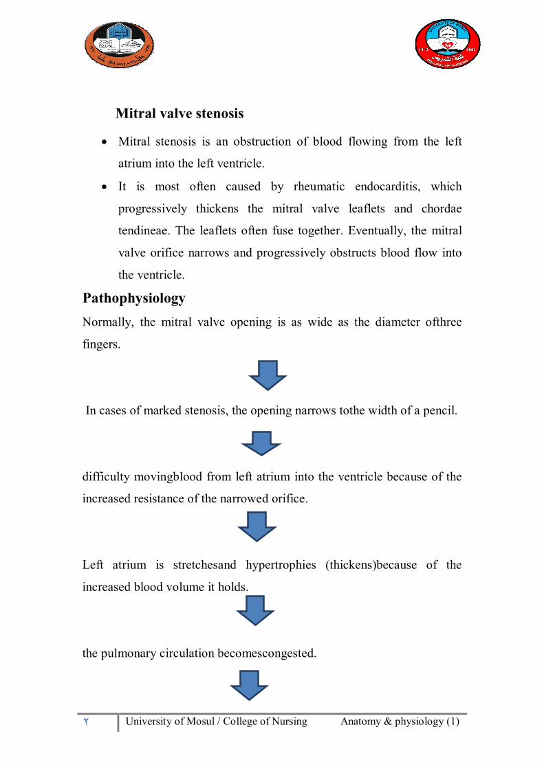
Mitral valve stenosis
Mitral stenosis is an obstruction of blood flowing from the left
atrium into the left ventricle.
It is most often caused by rheumatic endocarditis, which
progressively thickens the mitral valve leaflets and chordae
tendineae. The leaflets often fuse together. Eventually, the mitral
valve orifice narrows and progressively obstructs blood flow into
the ventricle.
Pathophysiology Normally, the mitral valve opening is as wide as the diameter ofthree
fingers.
In cases of marked stenosis, the opening narrows tothe width of a pencil.
difficulty movingblood from left atrium into the ventricle because of the
increased resistance of the narrowed orifice.
Left atrium is stretchesand hypertrophies (thickens)because of the
increased blood volume it holds.
the pulmonary circulation becomescongested.

٣ University of Mosul / College of Nursing Anatomy & physiology (1)
the right ventricle become congested with blood andeventually fails.
Clinical Manifestations
1. breathing difficulty (ie, dyspnea) on exertion as a result of
pulmonary venous hypertension.
2. Patients show progressive fatigue as a result of low cardiac output.
3. patient may expectorate blood (ie, hemoptysis), cough, and
experience repeated respiratory infections.
Assessment and Diagnostic Findings
1.Assess the vital signs: The pulse is weak and often irregular because of atrial fibrillation
(caused by the strain on the atrium).
A low-pitched, rumbling, diastolic murmur is heard at the apex.
As a result of the increased blood volume and pressure, the atrium
dilates, hypertrophies, and becomes electrically unstable, and the
patient experiences atrial dysrhythmias.
2.Echocardiography is used to diagnose mitral stenosis.
3. Electrocardiography (ECG) and cardiac catheterization with
angiography are used to determine the severity of the mitral stenosis.
Medical Management Antibiotic prophylaxis therapy is instituted to prevent recurrence of
infections.
anticoagulants to decrease the risk for developing atrial thrombus.

٤ University of Mosul / College of Nursing Anatomy & physiology (1)
treatment for anemia by anti-anemic medications.
Surgical intervention
It is consists of :
valvuloplasty, usually a commissurotomy to open or rupture the
fused commissures of the mitral valve.
Percutaneous transluminalvalvuloplasty or,
mitral valve replacement may be performed.
MITRAL REGURGITATION Mitral regurgitation involves blood flowing back from the leftventricle
into the left atrium during systole. Often, the marginsof the mitral valve
cannot close during systole.
Clinical Manifestations Chronic mitral regurgitation is often asymptomatic, but acute
mitral regurgitation (eg, that resulting from a myocardial
infarction) usually manifests as severe congestive heart failure.
Dyspnea, fatigue, and weakness are the most common symptoms.
Palpitations, shortness of breath on exertion, and,
cough from pulmonary congestion also occur.
Assessment and Diagnostic Findings

٥ University of Mosul / College of Nursing Anatomy & physiology (1)
A systolic murmur is heard as a high-pitched, blowing sound at the
apex.
The pulse may be regular and of good volume, or it may be
irregular as a result of extrasystolic beats or atrial fibrillation.
Echocardiography is used to diagnose and monitor the progression
of mitral regurgitation.
Medical Management Management of mitral regurgitation is the same as that for
congestiveheart failure.
Surgical intervention consists of:
mitral valve replacement or valvuloplasty (ie, surgical repair of the
heart valve).
Atrial Septal Defect (ASD)
An atrial septal defect (ASD) is a hole in the wall between the two upper chambers of the heart. The condition is present from birth (congenital). Smaller atrial septal defects may close on their own during infancy or early childhood.
Large and long-standing atrial septal defects can damage the heart and lungs.
ASD: Is an abnormal opening between the atria allowing blood from the
higher pressure(left atrium) to flow into the lower pressure(rightatrium).
Classification of ASD
There are 3 major types of ASDs or interatrial communications:
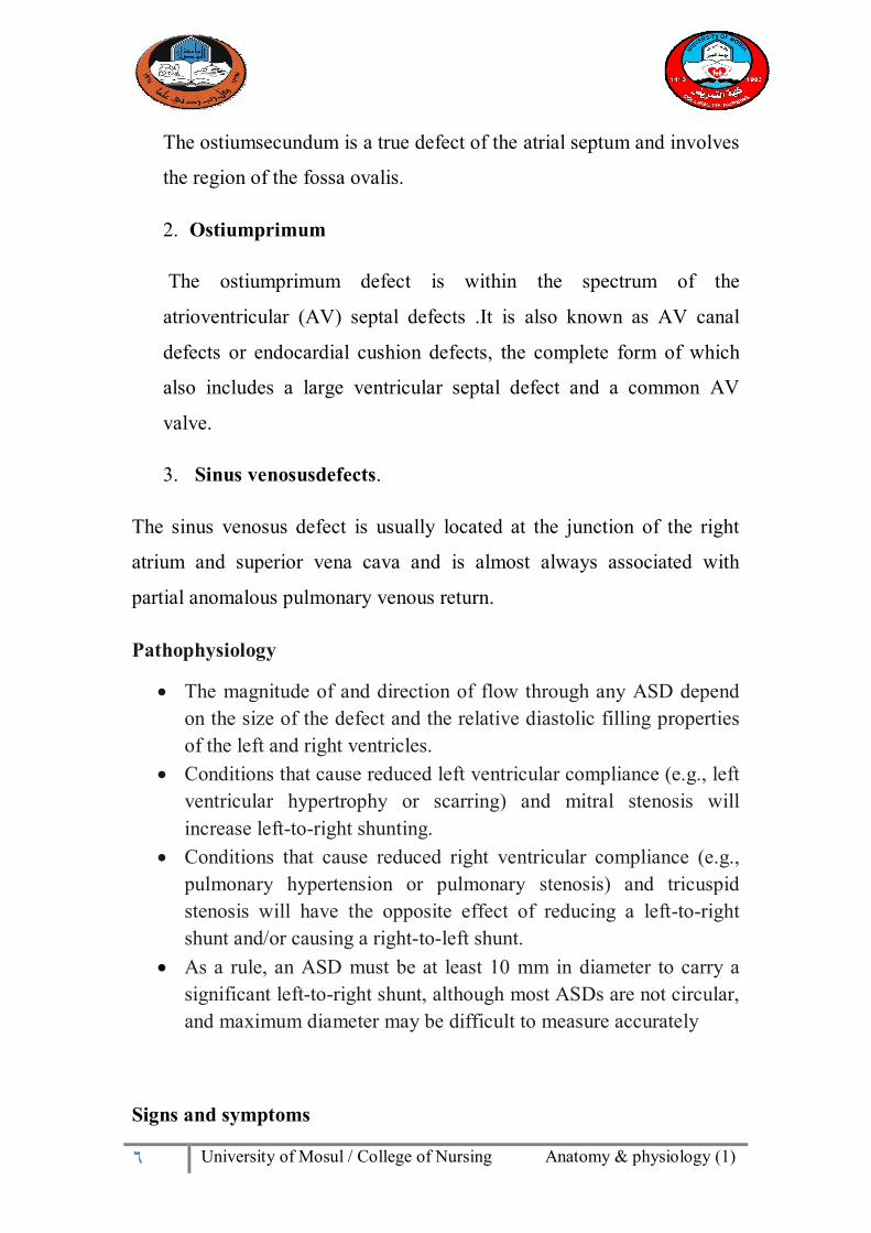
1. Ostiumsecundum
٦ University of Mosul / College of Nursing Anatomy & physiology (1)
The ostiumsecundum is a true defect of the atrial septum and involves
the region of the fossa ovalis.
2. Ostiumprimum
The ostiumprimum defect is within the spectrum of the
atrioventricular (AV) septal defects .It is also known as AV canal
defects or endocardial cushion defects, the complete form of which
also includes a large ventricular septal defect and a common AV
valve.
3. Sinus venosusdefects.
The sinus venosus defect is usually located at the junction of the right
atrium and superior vena cava and is almost always associated with
partial anomalous pulmonary venous return.
Pathophysiology
The magnitude of and direction of flow through any ASD depend on the size of the defect and the relative diastolic filling properties of the left and right ventricles.
Conditions that cause reduced left ventricular compliance (e.g., left ventricular hypertrophy or scarring) and mitral stenosis will increase left-to-right shunting.
Conditions that cause reduced right ventricular compliance (e.g., pulmonary hypertension or pulmonary stenosis) and tricuspid stenosis will have the opposite effect of reducing a left-to-right shunt and/or causing a right-to-left shunt.
As a rule, an ASD must be at least 10 mm in diameter to carry a significant left-to-right shunt, although most ASDs are not circular, and maximum diameter may be difficult to measure accurately
Signs and symptoms
٧ University of Mosul / College of Nursing Anatomy & physiology (1)
Exercise intolerance in the form of exertional dyspnea or fatigue
Heart murmur, a whooshing sound that can be heard through a stethoscope.
Shortness of breath, especially when exercising. Fatigue. Swelling of legs, feet or abdomen. Heart palpitations or skipped beats. Frequent lung infections. Stroke. Bluish skin color
Risk factors Family history. genetic problems, such as Down syndrome. Rubella infection (German measles) during pregnancy. Drug or alcohol use or exposure to certain substances. Use of
certain medications, , such as cocaine, during pregnancy can harm the developing fetus.
Diagnostic Procedures
The large and severe an ASD is based on the symptoms, physical exam, and the results of heart tests.
1.Abnormal heart sounds: A murmur may be heard only in certain body positions, and sometimes a murmur may not be heard at all. A murmur means that blood is flowing in a turbulent (not smooth) way.
2. Signs of heart failure in some adults.
3. Echocardiogram .
Other tests that may be done include:
Cardiac catheterization Coronary angiography (for patients over 35 years old) Doppler study of the heart
٨ University of Mosul / College of Nursing Anatomy & physiology (1)
ECG Heart MRI Transesophageal echocardiography (TEE)
Treatment
ASD may not need treatment if there are few or no symptoms, or if the defect is small. Surgery to close the defect is recommended if the defect causes a large amount of shunting, the heart is swollen, or symptoms occur.
A procedure has been developed to close the defect without surgery.
The procedure involves placing an ASD closure device into the heart through tubes called catheters.
The health care provider makes a tiny surgical cut in the groin, then inserts the catheters into a blood vessel and up into the heart.
The closure device is then placed across the ASD and the defect is closed.
Complications Larger defects can cause mild to life-threatening problems, including:
1. Right-sided heart failure. 2. Shortened life expectancy. 3. Increased risk of a stroke.
4. Arrhythmias, particularly atrial fibrillation 5. Heart infections (endocarditis)
Less common serious complications may include:
Pulmonary hypertension. Eisenmenger syndrome. In rare cases, pulmonary hypertension
can cause permanent lung damage, and it becomes irreversible.
This complication, called Eisenmenger syndrome, usually develops

٩ University of Mosul / College of Nursing Anatomy & physiology (1)
over many years and occurs only in a small percentage of people
with large atrial septal defects.
Ventricular Septal Defect(VSD)
A ventricular septal defect (VSD) : It is a type of congenital heart disease (CHD).
Is an abnormal opening between the right and left ventricles, may vary in size from small pin hole to absence of the septum.
Many VSD especially those in infants with small defects close spontaneously.
VSDs are of various sizes and locations, can be single or multiple, and in adults may be complicated by sub pulmonary stenosis, pulmonary hypertension (PHTN), and/or aortic regurgitation (AR).
Classifications of VSD
1. Type I: Infundibular VSD This type is result from deficiency in the septum above and
anterior to the crista supraventricularis, beneath the aortic and pulmonary valves .
2. Type II : A membranous VSD
o This defect is inferior to the crista supraventricularis and borders the septal leaflet of the tricuspid valve.
o The defect may extend into the muscular septum and is then referred to as a perimembranous (or paramembranous) VSD.
3.Type III :Inlet defects

١٠ University of Mosul / College of Nursing Anatomy & physiology (1)
This type is result from deficiency of the inlet septum located beneath both mitral and tricuspid valves .
Despite proximity to those valves, this type of defect is not associated with mitral or tricuspid regurgitation unless associated with atrioventricular canal defect.
This typically large defect is often associated with Down syndrome.
4. Type IX:Muscular defects
This type is account for 5 to 20 percent of VSD, are bordered only by muscle within the trabecular septum, away from the cardiac valves.
Muscular defects can be small or large, single or multiple, and occasionally oblique with multiple exits resembling Swiss cheese.
Pathophysiology
The pressure generated during contraction by the left ventricle is
higher than that generated by the simultaneous contraction of the
right ventricle.
Blood will thus be pushed through the VSD (also called
"shunted") from the left ventricle to the right ventricle.
The right ventricle has to do extra work to handle the additional
blood volume.
It may have trouble keeping up with the load and enlarge, affecting
its ability to pump efficiently.
In addition, the lungs receive too much blood under too much
pressure.

١١ University of Mosul / College of Nursing Anatomy & physiology (1)
The arterioles (small arteries) in the lungs thicken in response to
the excess blood under excess pressure.
If this extra pressure persists, permanent damage can be done to the
lungs.
Symptoms
If the hole is large, the baby often has symptoms related to heart failure.
The most common symptoms include:
1. Shortness of breath, fast and hard breathing. 2. Paleness. 3. Failure to gain weight. 4. Fast heart rate. 5. Sweating while feeding. 6. Frequent respiratory infections.
Diagnostic procedures
Listening with a stethoscope usually reveals a heart murmur (the sound of the blood crossing the hole). The loudness of the murmur is related to the size of the defect and amount of blood crossing the defect.
Tests may include:
1. Cardiac catheterization (rarely needed, unless there are concerns of high blood pressure in the lungs)
2. Chest x-ray -- looks to see if there is a large heart with fluid in the lungs
3. ECG -- shows signs of an enlarged left ventricle 4. Echocardiogram -- used to make a definite diagnosis 5. MRI of the heart -- used to find out how much blood is getting to
the lungs
Treatment:

١٢ University of Mosul / College of Nursing Anatomy & physiology (1)
Surgical repair of the shunt if not closed, as in ASD.
Possible Complications
1. Aortic insufficiency (leaking of the valve that separates the left ventricle from the aorta)
2. Damage to the electrical conduction system of the heart during surgery (causing an irregular heart rhythm)
3. Delayed growth and development (failure to thrive in infancy) 4. Heart failure 5. Infective endocarditis (bacterial infection of the heart) 6. Pulmonary hypertension (high blood pressure in the lungs) leading
to failure of the right side of the heart.
Tetralogy of Fallut(TOF)
Tetralogy of Fallut, which is one of the most common congenital heart
disorders.
The classic form includes 4 defects:
Comprises right ventricular (RV) outflow tract obstruction
(RVOTO) (Pulmonary stenosis).
Ventricular septal defect (VSD).
Aorta dextroposition,(Overriding aorta) and,
RV hypertrophy.
Signs and symptoms
The clinical features of tetralogy of Fallot are directly related to the severity of the anatomic defects. Infants often display the following:
Difficulty with feeding Failure to thrive

١٣ University of Mosul / College of Nursing Anatomy & physiology (1)
Episodes of bluish pale skin during crying or feeding (ie, "Tet" spells)
Exertional dyspnea, usually worsening with age
Physical findings include the following:
1. Most infants are smaller than expected for age 2. Cyanosis of the lips and nail bed is usually pronounced at birth 3. After age 3-6 months, the fingers and toes show clubbing 4. A systolic thrill is usually present anteriorly along the left sternal
border 5. A harsh systolic ejection murmur (SEM) is heard over the
pulmonic area and left sternal border 6. During cyanotic episodes, murmurs may disappear 7. In individuals with aortopulmonary collaterals, continuous
murmurs may be auscultated
Diagnosis of TOF
Hemoglobin and hematocrit values are usually elevated in proportion to the degree of cyanosis.
Patients with significant cyanosis have the following, in association with a tendency to bleed:
1. Decreased clotting factors. 2. Low platelet count. 3. Diminished coagulation factors. 4. Diminished total fibrinogen. 5. Prolonged prothrombin and coagulation times.
Arterial blood gas (ABG) results are as follows:
1. Oxygen saturation varies. 2. pH and partial pressure of carbon dioxide (pCO2) are
normal unless the patient is in extremis.
Imaging studies include the following:

١٤ University of Mosul / College of Nursing Anatomy & physiology (1)
1. Echocardiography. 2. Chest radiographs. 3. Magnetic resonance imaging (MRI).
Cardiac catheterization findings include the following:
1. Assessment of the pulmonary annulus size and pulmonary arteries.
2. Assessment of the severity of RVOTO. 3. Location of the position and size of the VSD. 4. Ruling out possible coronary artery anomalies.
Treatment
A: Medical management:
Acute treatment for hypercyanosis is as follows:
1. Place the baby on the mother's shoulder with the infant's knees tucked up underneath; this provides a calming effect, reduces systemic venous return, and increases systemic vascular resistance (SVR).
2. Oxygen is of limited value, as the primary abnormality is reduced pulmonary blood flow
3. Morphine sulfate, 0.1-0.2 mg/kg intramuscularly (IM) or subcutaneously (SC), may reduce the ventilatory drive and decrease systemic venous return
4. Phenylephrine, 0.02 mg/kg IV, is used to increase SVR. 5. Dexmedetomidine infusion has been used.
B: Medical management:
Surgery is preferably done at or about 12 months of age. Primary correction is the ideal operation and is usually performed
under cardiopulmonary bypass.

١٥ University of Mosul / College of Nursing Anatomy & physiology (1)
Palliative procedures (eg, placement of the modified Blalock-Taussig shunt) may be necessary in patients with contraindications to primary repair, which include the following:
1. The presence of an anomalous coronary artery 2. Very low birth weight 3. Small pulmonary arteries 4. Multiple VSDs 5. Multiple coexisting intracardiac malformations
Cardiac Catheterization
Cardiac catheterization involves passing a thin flexible tube
(catheter) into the right or left side of the heart, usually from the
groin or the arm.
Cardiac catheterization is a test to check the heart. This test can
include a coronary angiogram, which checks:
1. the coronary arteries.
2. blood flow in the coronary arteries.
3. blood flow and blood pressure in the chambers of the heart,
4. find out how well the heart valves work, and
5. defects in the way the wall of the heart moves.
6. In children, this test is used to check for heart problems that
have been present since birth (congenital heart defect).
A coronary angiogram is used to find coronary artery disease
(atherosclerosis), this test also can pinpoint the size and location of

١٦ University of Mosul / College of Nursing Anatomy & physiology (1)
fat and calcium deposits (plaque) that are narrowing your coronary
arteries.
Percutaneous coronary intervention (PCI) is similar to coronary
angiogram, but it is used to open up a narrowed coronary artery
with special tools. The two common types of PCI are:
1. Angioplasty with or without coronary stents.
2. Atherectomy.
Cardiac Catheterization done to diagnose or evaluate:
Cardiac amyloidosis
Causes of congestive heart failure or cardiomyopathy
Coronary artery disease
Heart defects that are present at birth (congenital)
High blood pressure in the lungs (pulmonary hypertension)
Problems with the heart valves
The following procedures may also be done using cardiac
catheterization:
Repair of certain types of heart defects
Open a narrowed (stenotic) heart valve
Open blocked arteries or grafts in the heart (angioplasty with or
without stenting)

١٧ University of Mosul / College of Nursing Anatomy & physiology (1)
Procedure
1. Premedication will be given before the test to help patient relax.
2. cleans a site on the arm, neck, or groin and inserts a line into one of
the veins. This is called an intravenous (IV) line.
3. A larger plastic thin tube called a sheath is placed into a vein or
artery in the leg or arm. Then longer plastic tubes called catheters are
carefully moved up into the heart using live x-rays as a guide.
Then the doctor can:
Collect blood samples from the heart.
Measure pressure and blood flow in the heart's chambers
and in the large arteries around the heart.
Measure the oxygen in different parts of your heart
Examine the arteries of the heart
Perform a biopsy on the heart muscle
4. If there is blockage, patient may have" angioplasty and a stent
"placed during the procedure.
5. The patient usually be asked to lie flat on the back for a few hours
after the test to avoid bleeding.
Risks
Cardiac catheterization carries a slightly higher risk than other heart tests.
However, it is very safe when performed by an experienced team.
The risks include:
Cardiac tamponade
Heart attack

١٨ University of Mosul / College of Nursing Anatomy & physiology (1)
Injury to a coronary artery
Irregular heartbeat
Low blood pressure
Reaction to the contrast dye
Stroke
Possible complications of any type of catheterization include the
following:
Bleeding, infection, and pain at the IV or sheath insertion site
Damage to the blood vessels
Blood clots
Kidney damage due to the contrast dye (more common in patients
with diabetes or kidney problems)
Medications:
Discuss the medications with physician - he may want to stop or adjust
the doses several days prior to or on the day of the procedure, especially
those listed below.
Anticoagulant Medication
Aspirin
Diabetes Medications
Nursing Managements
1. After the bandage is removed, cover the area with a small adhesive
bandage. It is normal for the catheter insertion site to be black and
blue) for a couple of days. The site may also be slightly swollen

١٩ University of Mosul / College of Nursing Anatomy & physiology (1)
and pink, and there may be a small lump (about the size of a
quarter) at the site.
2. Wash the catheter insertion site at least once daily with soap and
water. Place soapy water on the hand or wash- cloth and gently
wash the insertion site; do not rub.
3. Keep the area clean and dry . Do not use creams, lotions or
ointment on the wound site.
4. Wear loose clothes and loose underwear.
5. Do not take a bath, for one week after the procedure.
6. The patient usually feel tired and weak the day after the procedure.
Take walks around the house and plan to rest during the day.
7. Do not strain during bowel movements for the first 3 to 4 days after
the procedure to prevent bleeding from the catheter insertion site.
8. Avoid heavy lifting (more than 10 pounds) and pushing or pulling
heavy objects for the first 5 to 7 days after the procedure.
9. Do not participate in strenuous activities for 5 days after the
procedure. This includes most sports activities.
10. Patient may climb stairs if needed, but walk up and down the stairs
more slowly than usual.
11. Gradually increase the activities until reach the normal activity
level within one week after the procedure.