Part One Amino Acids as Building Blocks
82
Part One Amino Acids as Building Blocks Amino Acids, Peptides and Proteins in Organic Chemistry. Vol.3 – Building Blocks, Catalysis and Coupling Chemistry. Edited by Andrew B. Hughes Copyright Ó 2011 WILEY-VCH Verlag GmbH & Co. KGaA, Weinheim ISBN: 978-3-527-32102-5
Transcript of Part One Amino Acids as Building Blocks
Part OneAmino Acids as Building Blocks
Amino Acids, Peptides and Proteins in Organic Chemistry.Vol.3 – Building Blocks, Catalysis and Coupling Chemistry. Edited by Andrew B. HughesCopyright � 2011 WILEY-VCH Verlag GmbH & Co. KGaA, WeinheimISBN: 978-3-527-32102-5

1Amino Acid BiosynthesisEmily J. Parker and Andrew J. Pratt
1.1Introduction
The ribosomal synthesis of proteins utilizes a family of 20 a-amino acids that areuniversally coded by the translation machinery; in addition, two further a-aminoacids, selenocysteine and pyrrolysine, are now believed to be incorporated intoproteins via ribosomal synthesis in some organisms.More than 300 other amino acidresidues have been identified in proteins, but most are of restricted distribution andproduced via post-translational modification of the ubiquitous protein aminoacids [1]. The ribosomally encoded a-amino acids described here ultimately derivefrom a-keto acids by a process corresponding to reductive amination. The mostimportant biosynthetic distinction relates to whether appropriate carbon skeletonsare pre-existing in basic metabolism or whether they have to be synthesized de novoand this division underpins the structure of this chapter.
There are a small number of a-keto acids ubiquitously found in core metabolism,notably pyruvate (and a related 3-phosphoglycerate derivative from glycolysis),together with two components of the tricarboxylic acid cycle (TCA), oxaloacetateand a-ketoglutarate (a-KG). These building blocks ultimately provide the carbonskeletons for unbranched a-amino acids of three, four, and five carbons, respectively.a-Amino acids with shorter (glycine) or longer (lysine and pyrrolysine) straightchains are made by alternative pathways depending on the available raw materials.The strategic challenge for the biosynthesis of most straight-chain amino acidscenters around two issues: how is the a-amino function introduced into the carbonskeleton and what functional group manipulations are required to generate thediversity of side-chain functionality required for the protein function?
The core family of straight-chain amino acids does not provide all the functionalityrequired for proteins. a-Amino acids with branched side-chains are used for twopurposes; the primary need is related to protein structural issues. Proteins fold intowell-defined three-dimensional shapes by virtue of their amphipathic nature: asignificant fraction of the amino acid side-chains are of low polarity and thehydrophobic effect drives the formation of ordered structures in which theseside-chains are buried away from water. In contrast to the straight-chain amino
j3
Amino Acids, Peptides and Proteins in Organic Chemistry.Vol.3 – Building Blocks, Catalysis and Coupling Chemistry. Edited by Andrew B. HughesCopyright � 2011 WILEY-VCH Verlag GmbH & Co. KGaA, WeinheimISBN: 978-3-527-32102-5
acids, the hydrophobic residues have large nonpolar surface areas by virtue of theirbranched hydrocarbon side-chains. The other role of branched amino acids is toprovide twouseful functional groups: an imidazole (histidine) and a phenol (tyrosine)that exploit aromatic functional group chemistry.
This chapter provides an overview of amino acid biosynthesis from a chemicalperspective and focuses on recent developments in the field. It highlights a fewoverarching themes, including the following:
i) The chemical logic of the biosynthetic pathways that underpin amino acid bio-synthesis.Thischemical foundationiscriticalbecauseof theevolutionarymecha-nisms that have shaped these pathways. In particular, the way in which geneduplication and functional divergence (viamutation and selection) can generatenew substrate specificity and enzyme activities from existing catalysts [2].
ii) The contemporary use of modern multidisciplinary methodology, includingchemistry, enzymology, and genomics, to characterize new biosyntheticpathways.
iii) Potential practical implications of understanding the diverse metabolism ofamino acid biosynthesis, especially medicinal and agrichemical applications.
iv) The higher-level molecular architectures that control the fate of metabolites,especially the channeling of metabolites between active sites for efficientutilization of reactive intermediates.
Box 1.1: Nitrogen and Redox in Amino Acid Biosynthesis
Ammonia is toxic and the levels of ammonia available for the biosynthesis ofamino acids in most biochemical situations is low. There are a limited number ofentry points of ammonia into amino acid biosynthesis, notably related to gluta-mate and glutamine. Once incorporated into key amino acids, nitrogen istransferred betweenmetabolites either directly or via in situ liberation of ammoniaby a multifunctional complex incorporating the target biosynthetic enzyme. Themain source of in situ generated ammonia for biosynthesis is the hydrolysis ofglutamine by glutaminases. De novo biosynthesis of amino acids, like elementfixation pathways in general, is primarily reductive in nature. This may reflect theorigins of these pathways in an anaerobic world more than 3 billion years ago.
Box 1.2: The Study of Biosynthetic Enzymes and Pathways
The source of an enzyme for biochemical study has important implications. Mostcore metabolism has been elaborated by studying a small number of organismsthat were chosen for a variety of reasons, including availability, ease of manip-ulation, ethical concerns, scientific characterization, and so on. These exemplarorganisms include the bacterium Escherichia coli, the yeast Saccharomyces cerevi-siae, the plant Arabidopsis thaliana, and the rat as a typical mammal. Much of thedetailed characterization of amino acid biosynthesis commenced with studies onthese organisms. With the rise of genetic engineering techniques, biosynthetic
4j 1 Amino Acid Biosynthesis
1.2Glutamate and Glutamine: Gateways to Amino Acid Biosynthesis
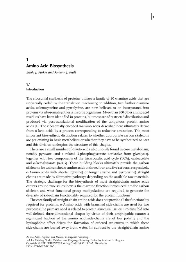
Glutamate and the corresponding amide derivative, glutamine, are critical metabo-lites in amino acid metabolism. The biochemistry of these two amino acids alsoillustrates the distinct chemistry associated with the a-amino and side-chain func-tional groups, each of which is exploited in the biosynthesis of other amino acids.These amino acids derive from ammonia and a-KG. Glutamate dehydrogenase(GDH) interconverts a-KG and glutamate (Figure 1.1) [6]. Although glutamate isformed in this way by reductive amination, this enzyme is generally not dedicated tobiosynthesis; the reverse reaction, an oxidative deamination to regenerate a-KG, is
enzymes from a wide variety of sources are available for scientific investigation,and there has been increasing emphasis on working with enzymes and pathwaysfrom alternative organisms.Metabolic diversity is greatest among prokaryotes. One fundamental change in
the underlying microbiology that has affected our understanding of pathwaydiversity has been the appreciation of the deep biochemical distinctions betweenwhat are now recognized to be two fundamental domains of prokaryotes:eubacteria and Archaea [3]. The former bacteria include those well known to beassociatedwith disease and fermentation processes; while the latter includemanymethanogens and extremophiles (prokaryotes that grow in extreme conditions,such as hyperthermophiles that grow at temperatures above 60 �C or halophilesthat grow in high ionic strength environments). Bioinformatics approaches arecomplementing conventional enzymological studies in identifying and charac-terizing interesting alternative biosynthetic pathways [4]. The greater understand-ing ofmicrobial and biosynthetic diversity is presenting exciting opportunities fornovel discoveries in biosynthesis.Much of the focus of biosynthetic enzymology now focuses on enzymes from
pathogens and hyperthermophiles. The focus on the study of enzymes frompathogens is predicated on the possibility that inhibitors of such enzymesmay beuseful as pesticides and therapeutic agents. Since humans have access to manyamino acids in their food, they have lost the ability to make �dietary essential�amino acids that typically require extended dedicated biosynthetic pathways [5].The biosynthetic enzymes of the corresponding pathways are essential for manypathogens and plants, but not for humans; hence, selective inhibitors of thesebiosynthetic enzymes are potentially nontoxic to humans, but toxic to undesirableorganisms. Enzymes from hyperthermophilic organisms, produced by geneticengineering, are scrutinized mainly because of their ease of structural charac-terization. These enzymes retain their native structures at temperatures thatdenature most other proteins, including those of the host organism. Theseproteins are of high thermal stability and simple heat treatment can be used toeffect high levels of purification of the desired protein.
1.2 Glutamate and Glutamine: Gateways to Amino Acid Biosynthesis j5
CO2O
CO2
CO2H3N
CO2NH3 + NAD(P)H + + H2O + NAD(P)+
GDH
α-ketoglutarate glutamate
Figure 1.1 Interconversion of a-KG and glutamate catalyzed by GDH.
CO2H3N
CO2
CO2H3N
CO2PO32
CO2H3N
CONH2
HOPO32NH3
GS GS
ATP ADP
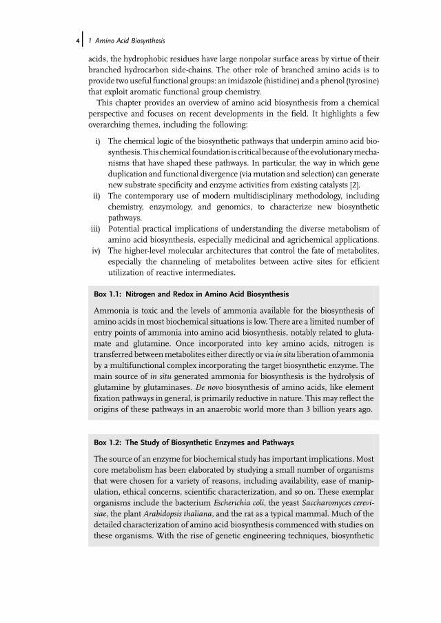
Figure 1.2 Conversion of glutamate to glutamine catalyzed by GS.
often an important in vivo role for this enzyme [7]. This deamination chemistrymightbe a factor in the relatively weak binding of ammonia (e.g., KM(NH3) is 3mM for theE. coli enzyme – above normal environmental concentrations). In many organismsthere is an additional enzyme, glutamate synthase (GOGAT), dedicated to thebiosynthesis of glutamate [8]. GOGAT utilizes ammonia generated in situ bythe hydrolysis of glutamine and this enzyme will be described after a discussionof the biosynthesis of glutamine.
The conversion of glutamate to glutamine, catalyzed by glutamine synthetase(GS), requires the activation of the side-chain carboxylate as an acyl phosphate, priorto nucleophilic substitution of the resulting good leaving group by ammonia(Figure 1.2). The use of ATP, to produce c-glutamyl phosphate, assists boththe kinetics and the thermodynamics of amide formation: by producing a morereactive carboxylic acid derivative and overturning the intrinsically favorable natureof amide hydrolysis in water.
GS fromenteric bacteria, such asE. coli andSalmonella typhimurium, is an exemplarof an amino acid biosynthetic enzyme; the overall reaction it catalyses is effectivelyirreversible in vivo (K¼ 1200). Being dedicated to biosynthesis, it has evolved tightbinding of ammonia (KM(NH3)< 200mM) which allows efficient synthesis of gluta-mine under the low ammonia conditions (much less than 1mM) found in vivo. Its invivo role as anentry point for thebiosynthesis of awide rangeofnitrogenmetabolites iseloquently communicated by the extensive feedback regulation of this enzyme by arange of nitrogen-containing metabolites, including glycine, serine, alanine, andhistidine [9–12]. Glutamine is the primary store of ammonia in many cells; the side-chain amide is chemically unreactive, but its favorable hydrolysis can be catalyzed ondemand by glutamine amidotransferase (GAT) enzymes [13].
1.2.1Case Study: GOGAT: GATs andMultifunctional Enzymes in Amino Acid Biosynthesis
In contrast to GDH, GOGAT is a dedicated biosynthetic enzyme. It is the primarysource of glutamate in plants, eubacteria and lower animals [14, 15]. These iron–sulfur flavoproteins carry out the reductive amination of a-KG to glutamate via a five-
6j 1 Amino Acid Biosynthesis
CO2H3N
CO2H3N
CO-SEnz
CO2O
CO2
CO2H3N
CO2+ H2O
NH3
GOGATGlutaminase
subunit
GOGATSynthasesubunit
+ H2O
CO2H3N
CO2
FMNH2 FMN
SH Cys1Enz
NH2
O
NH2
+ H
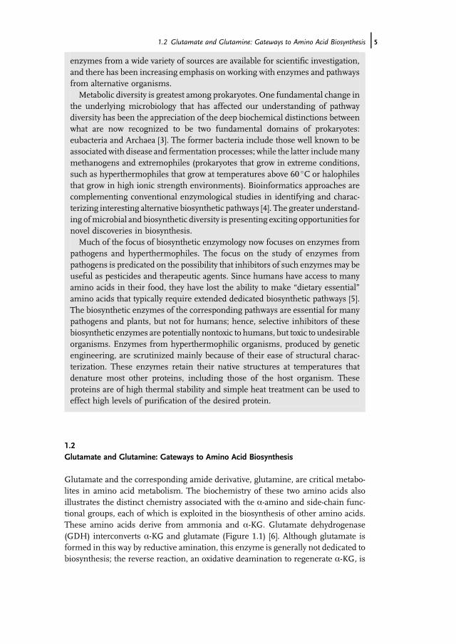
Figure 1.3 The biosynthesis of glutamate mediated by GOGAT.
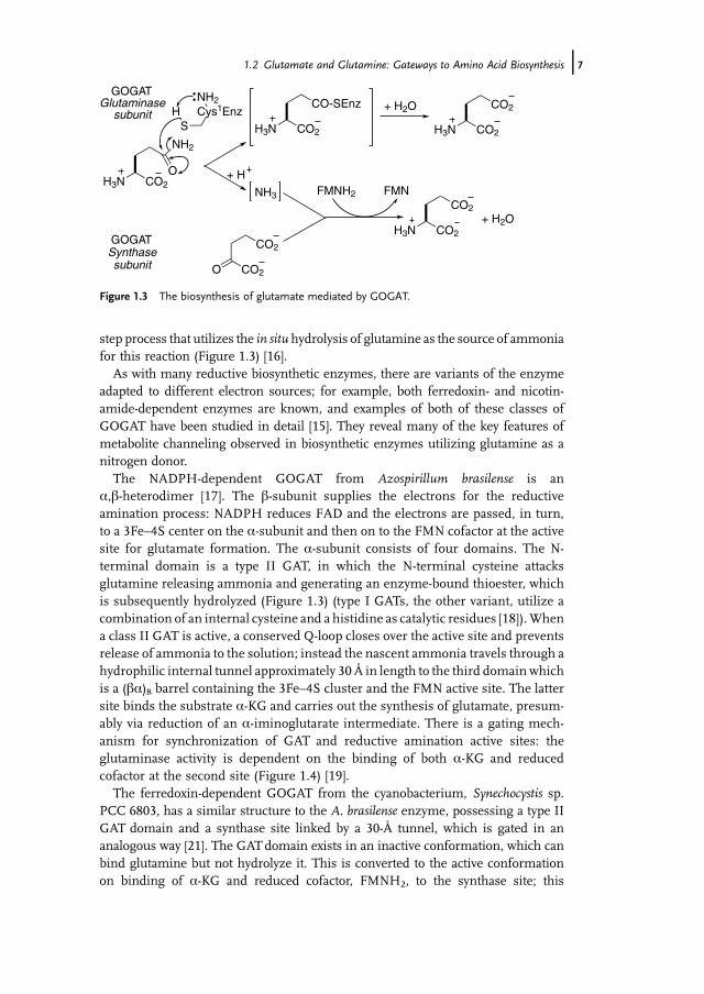
step process that utilizes the in situ hydrolysis of glutamine as the source of ammoniafor this reaction (Figure 1.3) [16].
As with many reductive biosynthetic enzymes, there are variants of the enzymeadapted to different electron sources; for example, both ferredoxin- and nicotin-amide-dependent enzymes are known, and examples of both of these classes ofGOGAT have been studied in detail [15]. They reveal many of the key features ofmetabolite channeling observed in biosynthetic enzymes utilizing glutamine as anitrogen donor.
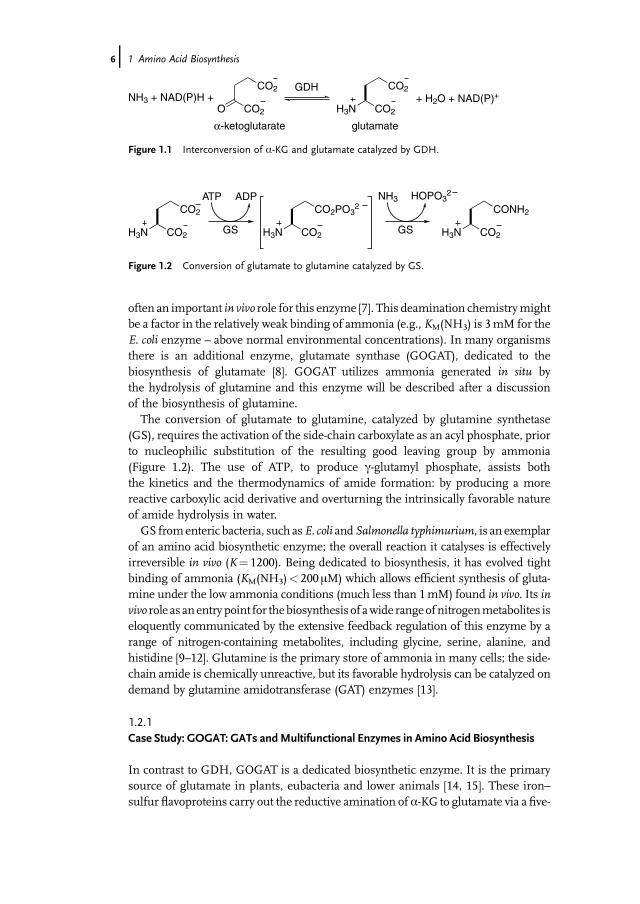
The NADPH-dependent GOGAT from Azospirillum brasilense is ana,b-heterodimer [17]. The b-subunit supplies the electrons for the reductiveamination process: NADPH reduces FAD and the electrons are passed, in turn,to a 3Fe–4S center on the a-subunit and then on to the FMN cofactor at the activesite for glutamate formation. The a-subunit consists of four domains. The N-terminal domain is a type II GAT, in which the N-terminal cysteine attacksglutamine releasing ammonia and generating an enzyme-bound thioester, whichis subsequently hydrolyzed (Figure 1.3) (type I GATs, the other variant, utilize acombination of an internal cysteine and a histidine as catalytic residues [18]).Whena class II GAT is active, a conserved Q-loop closes over the active site and preventsrelease of ammonia to the solution; instead the nascent ammonia travels through ahydrophilic internal tunnel approximately 30 Å in length to the third domain whichis a (ba)8 barrel containing the 3Fe–4S cluster and the FMN active site. The lattersite binds the substrate a-KG and carries out the synthesis of glutamate, presum-ably via reduction of an a-iminoglutarate intermediate. There is a gating mech-anism for synchronization of GAT and reductive amination active sites: theglutaminase activity is dependent on the binding of both a-KG and reducedcofactor at the second site (Figure 1.4) [19].
The ferredoxin-dependent GOGAT from the cyanobacterium, Synechocystis sp.PCC 6803, has a similar structure to the A. brasilense enzyme, possessing a type IIGAT domain and a synthase site linked by a 30-Å tunnel, which is gated in ananalogous way [21]. The GATdomain exists in an inactive conformation, which canbind glutamine but not hydrolyze it. This is converted to the active conformationon binding of a-KG and reduced cofactor, FMNH2, to the synthase site; this
1.2 Glutamate and Glutamine: Gateways to Amino Acid Biosynthesis j7

conformational switch also serves to open the entry point to the ammonia tunnel. Aconserved glutamate residue (Glu1013 in the Synechocystis enzyme), present at thetunnel constriction, has been shown to be the key residue controlling the cross-regulationmechanism. This glutamate interacts with the N-terminal amino group ofthe protein, which is the active-site base of the glutaminase, as well as affecting thegeometry of the tunnel entry point. Mutation of this residue to aspartate, asparagine,or alanine affected glutaminase activity and the sensitivity of glutaminase action tothe binding of a-KG at the synthase site [22].
GOGAT exemplifies our growing awareness of details of glutamine-dependentenzymes, in particular, and biosynthetic pathways, in general. By exploiting thehigher-level organization of multifunctional enzyme systems, metabolites can bechanneled to the next enzyme of a pathway; thereby controlling their fate. Togetherwith the potential for subtle levels of regulation, this organization ensures theefficient use of biosynthetic intermediates [20].
1.3Other Amino Acids from Ubiquitous Metabolites: Pyridoxal Phosphate-DependentRoutes to Aspartate, Alanine, and Glycine
1.3.1Pyridoxal Phosphate: A Critical Cofactor of Amino Acid Metabolism
Once glutamate is available, the a-amino function can be transferred to other a-ketoacids via amino acid aminotransferase enzymes (Figure 1.5) [23]. This family of
Figure 1.4 Structure of GOGAT showing the internal tunnel for ammonia transfer between theGAT (gold) and synthase (blue) active sites. (Picture taken from [20].)
8j 1 Amino Acid Biosynthesis
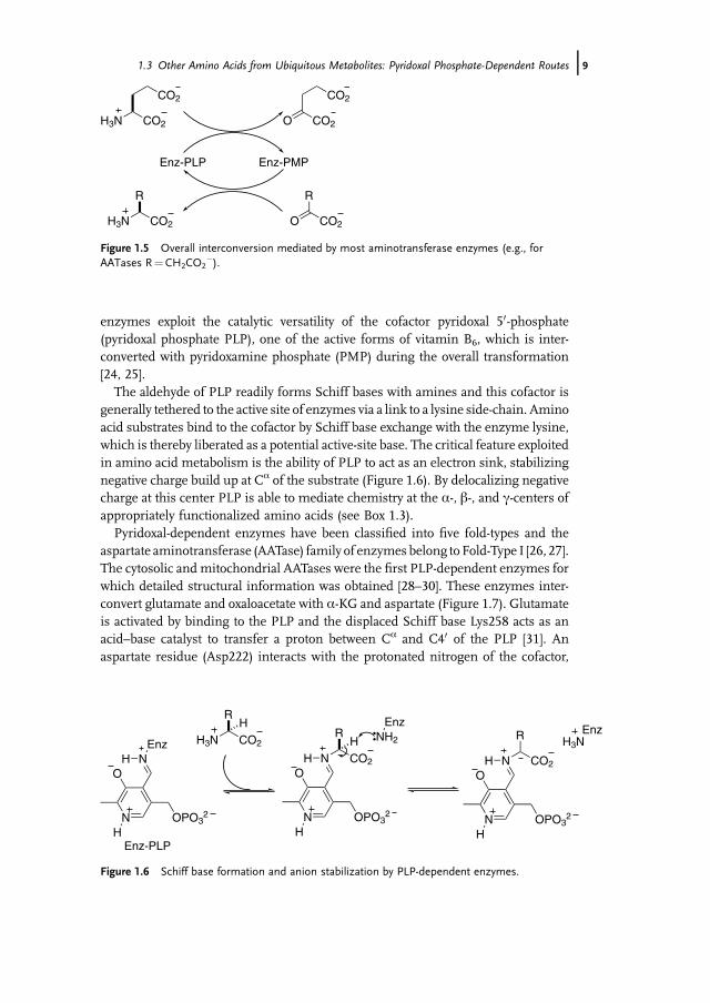
enzymes exploit the catalytic versatility of the cofactor pyridoxal 50-phosphate(pyridoxal phosphate PLP), one of the active forms of vitamin B6, which is inter-converted with pyridoxamine phosphate (PMP) during the overall transformation[24, 25].
The aldehyde of PLP readily forms Schiff bases with amines and this cofactor isgenerally tethered to the active site of enzymes via a link to a lysine side-chain. Aminoacid substrates bind to the cofactor by Schiff base exchange with the enzyme lysine,which is thereby liberated as a potential active-site base. The critical feature exploitedin amino acid metabolism is the ability of PLP to act as an electron sink, stabilizingnegative charge build up at Ca of the substrate (Figure 1.6). By delocalizing negativecharge at this center PLP is able to mediate chemistry at the a-, b-, and c-centers ofappropriately functionalized amino acids (see Box 1.3).
Pyridoxal-dependent enzymes have been classified into five fold-types and theaspartate aminotransferase (AATase) family of enzymes belong toFold-Type I [26, 27].The cytosolic and mitochondrial AATases were the first PLP-dependent enzymes forwhich detailed structural information was obtained [28–30]. These enzymes inter-convert glutamate and oxaloacetate with a-KG and aspartate (Figure 1.7). Glutamateis activated by binding to the PLP and the displaced Schiff base Lys258 acts as anacid–base catalyst to transfer a proton between Ca and C40 of the PLP [31]. Anaspartate residue (Asp222) interacts with the protonated nitrogen of the cofactor,
CO2O
CO2
CO2H3N
CO2
Enz-PLP Enz-PMP
R
CO2O
R
CO2H3N
Figure 1.5 Overall interconversion mediated by most aminotransferase enzymes (e.g., forAATases R¼CH2CO2
�).
N
O
OPO32
H
R
CO2H3N
N
H
H3NEnzR
CO2
H
N
O
OPO32
H
NH
NH2
Enz
HEnz
R
CO2
N
O
OPO32
H
NH
Enz-PLP
Figure 1.6 Schiff base formation and anion stabilization by PLP-dependent enzymes.
1.3 Other Amino Acids from Ubiquitous Metabolites: Pyridoxal Phosphate-Dependent Routes j9

stabilizing the pyridinium form and facilitating deprotonation of the substrate. Oncea proton has been transferred from Ca to C40, hydrolytic cleavage of the ketiminelinkage liberates a-KG and leaves the PMP form of the cofactor. Binding ofoxaloacetate and running the reaction in reverse leads to regeneration of the originalenzyme and production of aspartate. Aminotransferase enzymes provide a generalmechanism for interconverting a-amino acids and a-keto acids, illustrating a secondroute by which nitrogen is transferred between metabolites.
1.3.2Case Study: Dual Substrate Specificity of Families of Aminotransferase Enzymes
Aminotransferase enzymes pose an intriguing challenge for substrate specificitysince they bind two different substrates successively at the same site and must
CO2O
CO2
EnzO
O
CO2
CO2
H3NH
PLP form PMP form
R
CO2H3NH
R
CO2O
Glu α-KG
α-Keto acidα-Amino acid
+– H
N
O
OPO32
H
NHEnz
CO2
CO2
H
EnzO
O
N
O
OPO32
H
NH CO2
CO2
EnzO
O
N
O
OPO32
H
NH
EnzO
O
N
O
OPO32
H
NH2H
+– H
R
CO2H
EnzO
O
N
O
OPO32
H
NH
R
CO2
EnzO
O
N
O
OPO32
H
NH
Figure 1.7 Mechanism of aminotransferase catalysis (for AATases R¼CH2CO2�).
10j 1 Amino Acid Biosynthesis

Box 1.3: The Mechanistic Versatility of PLP: A Biochemical Electron Sink
Amino acids bind to PLP by forming a Schiff base. Once bound, the ability of PLPto stabilize a negative charge at the a-center of bound amino acids has beenharnessed by a range of amino acid biosynthetic enzymes tomediate chemistry atthe a-, b- and c-centers of suitably functionalized amino acids.
a-Center Reactivity
Cleavage of any of the three substituent bonds to the a-center can lead to acarbanionic species (Figure 1.8). Deprotonation of the a-proton, by the lysineliberated on Schiff base exchange, is used in transamination chemistry where thea-proton is relocated to the benzylic position of PLP en route to PMP as describedabove (and in some amino acid racemases). Decarboxylation provides a relatedanion, which can be protonated; this is the source of biological amines and isexploited in the biosynthesis of lysine via decarboxylation of the D-amino acidcenter of meso-diaminopimelate (DAP). Finally, when the amino acid side-chaincontains a b-hydroxyl function, retro-aldol chemistry provides a way of cleavingthis C�C bond. This is exploited in the biosynthesis of glycine, for example, by
NH
RCO2N
OH
OPO32
HN
CO2
RHN
OH
OPO32
HN
CO2HN
OH
OPO32
H O-HR1
N EnzN
OH
OPO32
H
H3NCO2
RH
NRCO2
NO
HOPO3
2
HN
RH
NO
HOPO3
2
HN
CO2
HN
OH
OPO32
H
e.g. R = (CH2)2CO2Aminotransferases
e.g. R = (CH2)3CH(NH3)CO2DAP decarboxylase
e.g. R1 = CH3
Threonine aldolase
- H -CO2+ H
N EnzN
OH
OPO32
H
H3NH
RCO2
N EnzN
OH
OPO32
H
H3NCO2
H
α-Deprotonation α-Decarboxylation α-Side chain cleavage
H2N EnzH2N Enz
R1
OH
- R1CHO
Figure 1.8 Stereoelectronic control ofa-center reactivity by PLP-dependent enzymes illustrated byenzymes involved in amino acid biosynthesis. As noted in the text, the decarboxylation example,DAP decarboxylase, utilizes a D-amino acid substrate.
1.3 Other Amino Acids from Ubiquitous Metabolites: Pyridoxal Phosphate-Dependent Routes j11
recognize these substrates but not others. AATases selectively bind glutamate andaspartate. Two active-site arginine residues (Arg292 and Arg386) bind to the twocarboxylates of these substrates, one of these, Arg292, controls the specificity formingan ionpairwith the carboxylate side-chain of each substrate (Figure 1.11).Mutation ofthis arginine to an anionic aspartate depresses the activity (kcat/KM) of the enzymewith respect to anionic substrates by a factor of more than 100 000 [34].
Other families of aminotransferases face greater challengeswith the dual substratespecificity that is a general feature of all these enzymes. Since glutamate is a commonamino donor in these systems, these enzymes must accommodate the negativelycharged c-carboxylate of glutamate while also accepting side-chains of the alternativesubstrate with different sizes, polarities, and charges. Two different strategies areemployed to deal with the issue: an �arginine switch,� whereby the key arginineundergoes a conformational shift to accommodate the new side-chain, and the use ofan extended hydrogen bond network to mediate substrate recognition, rather thanthe cationic charge of arginine (Figure 1.11) [35].
threonine aldolase. Enzymes control the identity of the bond that is cleaved byexploiting stereoelectronic factors as originally proposed by Dunathan [32]. Thecleaved bond must align with the delocalized p-orbitals of the PLP cofactor. Byspecific recognition of the a-amino acid functionalities, the enzyme can controlthe orientation of the substrate and hence its fate [33].
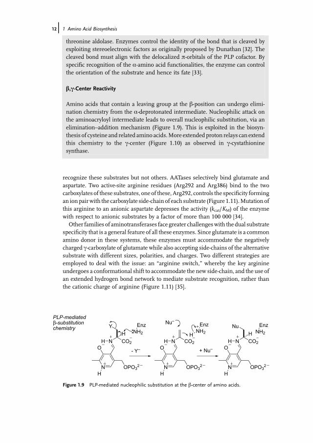
b,c-Center Reactivity
Amino acids that contain a leaving group at the b-position can undergo elimi-nation chemistry from the a-deprotonated intermediate. Nucleophilic attack onthe aminoacryloyl intermediate leads to overall nucleophilic substitution, via anelimination–addition mechanism (Figure 1.9). This is exploited in the biosyn-thesis of cysteine and related amino acids.More extendedproton relays can extendthis chemistry to the c-center (Figure 1.10) as observed in c-cystathioninesynthase.
PLP-mediated β-substitution chemistry
CO2
H
Y
N
O
OPO32
H
NH
NH2
Enz
CO2
N
O
OPO32
H
NH
NH2
Enz
CO2
H
Nu
N
O
OPO32
H
NH
+ Nu–
NH2
Enz
- Y–
Nu–
H
Figure 1.9 PLP-mediated nucleophilic substitution at the b-center of amino acids.
12j 1 Amino Acid Biosynthesis

Tyrosine aromatic amino transferases (TATases) utilize glutamate or aspartate asamino donors to produce the aromatic amino acids tyrosine, phenylalanine, andtryptophan. The TATase fromParacococcus denitrificans provides a clear example of anarginine switch [36]. The binding of a series of inhibitors to this enzyme shows thatthe active site utilizes Arg386 for specific recognition of the a-carboxylate and the
PLP-mediated γ-substitution chemistry
CO2
H
N
O
OPO32
H
NH
NH2
Enz
CO2
N
O
OPO32
H
NH
NH2
EnzY
CO2
H
N
O
OPO32
H
NH
NH2
EnzNu
CO2
N
O
OPO32
H
NH
NH2
EnzY Y
CO2
N
O
OPO32
H
NH
NH2
Enz
+ HNu
CO2
N
O
OPO32
H
NH
NH2
EnzNu
+ H
+ H
- HY
HH
H
Figure 1.10 PLP-mediated nucleophilic substitution at the c-center of amino acids.
N
O
OPO32
H
NH
O
O
OO
H
H2NNH2
NHArg292
H2N
H2NNH
Arg386
glutamate aldimine
N
O
OPO32
H
NH
O
O
H
NH2H2N
HN Arg292
H2N
H2NNH
Arg386
phenylalanine aldimine
Figure 1.11 The arginine switch in the substrate specificity of aminotransferases: in P. denitrificansTATase Arg292 forms an electrostatic attraction to the glutamate c-carboxylate; reorientation ofArg292 away from the active site allows binding of a nonpolar side-chain. (Adapted from [31].)
1.3 Other Amino Acids from Ubiquitous Metabolites: Pyridoxal Phosphate-Dependent Routes j13

surrounding region, in the vicinity of the a- and b-centers of the substrate, is rigid.However, active-site residues that bind the large hydrophobic substituent are con-formationally flexible and Arg292 moves out of the active site to accommodate bulkyuncharged substrates [37]. The arginine switch has been engineered into AATase bysite-directed mutation of six residues, thereby allowing transamination of largearomatic substrates [38]. The crystal structure of the resulting mutant provided thefirst structural evidence for the arginine switch [39].
Aspartate aminotransferase and tyrosine aminotransferase from E. coli are para-logs that share 43% sequence identity. It is likely that they evolved by gene duplicationof an ancestral AATase gene. The role of gene duplication and evolution of newsubstrate specificities is an area of general interest [40]. Directed evolution, whichmimics the action of natural selection, is a powerful strategy for tailoring proteinproperties [41]. It has been used to test these ideas. Repeated mutation of AATase,with selection for aromatic aminotransferase activity, leads to mutants with broad-ened substrate specificity [42], validating this evolutionary analysis. The first reportson the directed evolution of aminotransferases with modified substrate specificitywere of the conversion of AATases to branched-chain aminotransferases [43]. In thiscase amutant with 17 amino acid changes, remote from the active site, resulted in anarginine switch that allowed Arg292 to switch out of the active site. This changeaccommodates bulky hydrophobic side-chains (e.g., the catalytic efficiency (kcat/KM)of valine is increased by 2.1� 106) [44, 45]. It appears that the arginine switch isreadily accessible to evolution and that directed evolution strategies may provide ageneral tool for the development of new enzymes with tailored specificities.
The other mechanism for dual substrate specificity is the employment of anextended hydrogen bond network (Figure 1.12). The AATase [46] and TATase [47]from Pyrococcus horikoshii both use this strategy, as does the branched-chainaminotransferase from E. coli [48]. Binding glutamate at the active site without the
N
O
OPO32
H
NH
O
O
OO
H
glutamate aldimine
N
O
OPO32
H
NH
O
O
H
tyrosine aldimine
OMe
Thr HO
H
HO
Me
Thr
HOH
OH
Tyr
Figure 1.12 Extended hydrogen bond and p-stacking interactions in side-chain recognition ofTATase from P. horikoshii. (Adapted from [31].)
14j 1 Amino Acid Biosynthesis

presence of a cationic residue to recognize the side-chain reduces the electrostaticcomplexities for dual specificity. Interestingly, by using smaller, lessflexible, residuesthan arginine for recognition, the branched-chain aminotransferase can moreaccurately distinguish between aspartate and glutamate.
1.3.3PLP and the Biosynthesis of Alanine and Glycine
Two more of the protein amino acids, alanine and glycine, are biosynthesized bydirect exploitation of a-center PLP chemistry. Essentially any a-amino acid can becreated from the corresponding a-keto acid if an appropriate aminotransferase isavailable. Pyruvate is a ubiquitous metabolite and the corresponding amino acid,alanine, is readily available by transamination using aminotransferases of appro-priate specificity (Figure 1.13).
There are three biosynthetic routes to glycine (Figure 1.14). Some organisms, suchas the yeast S. cerevisiae, utilize all three. In organisms, such as S. cerevisiae, that haveaccess to glyoxalate, transamination provides glycine directly. In this case the aminodonor is alanine [49, 50].
The other two routes to glycine involve PLP-mediated cleavage of the proteinb-hydroxy amino acids serine and threonine by the enzymes serine hydroxy-methyltransferase (SHMT) and threonine aldolase. Enzymes of this class often haverelaxed substrate specificity and can cleave the side-chain from a number ofb-hydroxy-a-amino acids. Threonine aldolase is an important source of glycine in
Me
CO2O
Me
CO2H3NPLP
Glu α-KG
Figure 1.13 Biosynthesis of alanine from pyruvate.
H
CO2O CO2H3N
Ala pyruvate
alanine-glyoxalateaminotransferase
CO2H3N
OHH
threonine
threoninealdolase
CO2H3N
OHTHFCH2-THF
SHMTglyoxalate serineglycine
- MeCHO
Figure 1.14 Three PLP-dependent biosynthetic routes to glycine.
1.3 Other Amino Acids from Ubiquitous Metabolites: Pyridoxal Phosphate-Dependent Routes j15
S. cerevisiae [51]. Threonine forms a Schiff base with PLP which then catalyses aretro-aldol reaction to remove the side-chain as ethanal (see Box 1.3) [52, 53].
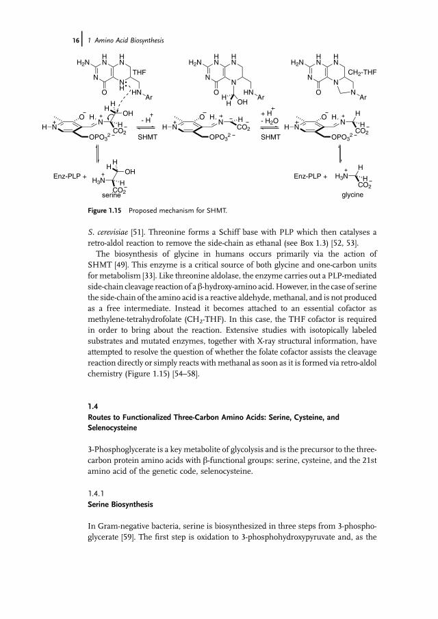
The biosynthesis of glycine in humans occurs primarily via the action ofSHMT [49]. This enzyme is a critical source of both glycine and one-carbon unitsfor metabolism [33]. Like threonine aldolase, the enzyme carries out a PLP-mediatedside-chain cleavage reaction of ab-hydroxy-amino acid.However, in the case of serinethe side-chain of the amino acid is a reactive aldehyde,methanal, and is not producedas a free intermediate. Instead it becomes attached to an essential cofactor asmethylene-tetrahydrofolate (CH2-THF). In this case, the THF cofactor is requiredin order to bring about the reaction. Extensive studies with isotopically labeledsubstrates and mutated enzymes, together with X-ray structural information, haveattempted to resolve the question of whether the folate cofactor assists the cleavagereaction directly or simply reacts withmethanal as soon as it is formed via retro-aldolchemistry (Figure 1.15) [54–58].
1.4Routes to Functionalized Three-Carbon Amino Acids: Serine, Cysteine, andSelenocysteine
3-Phosphoglycerate is a keymetabolite of glycolysis and is the precursor to the three-carbon protein amino acids with b-functional groups: serine, cysteine, and the 21stamino acid of the genetic code, selenocysteine.
1.4.1Serine Biosynthesis
In Gram-negative bacteria, serine is biosynthesized in three steps from 3-phospho-glycerate [59]. The first step is oxidation to 3-phosphohydroxypyruvate and, as the
NCO2
HNO
HOPO3
2
H OHH
H
HN
NNH
HN
O
H2N
HNAr
NCO2
HN
OH
OPO32
H
OHHH
HN
NN
HN
O
H2N
HNAr
HN
NN
HN
O
H2N
NAr
NH
CO2HN
OH
OPO32
H
H3NCO2
H
OHH
H
H3NH
CO2HEnz-PLP + Enz-PLP +
SHMT SHMT
serine glycine
THF
- H+ H- H2O
CH2-THF
Figure 1.15 Proposed mechanism for SHMT.
16j 1 Amino Acid Biosynthesis

point of commitment to the biosynthetic pathway, it is feedback regulated by the endproduct, serine [60]. The resulting a-keto acid is a substrate for transamination withglutamate acting as the amino donor. Hydrolysis of the resulting serine-b-phosphatecatalyzed by phosphoserine phosphatase (PSP) provides the free amino acid(Figure 1.16).
Systematic protein crystallography, exploiting the use of reactive intermediateanalogs, has provided a detailed series of �snapshots� of intermediates in the catalyticcycle of the PSP fromMethanococcus jannaschii, allowing the reaction to be visualizedin three-dimensional detail (Figure 1.17) [61]. A conserved aspartate residue at theend of the active-site tunnel is a nucleophilic catalyst, attacking the serine-b-phosphate to generate an acyl phosphate intermediate. Release of serine allowsthe binding of a water molecule to mediate hydrolysis of the labile aspartate-b-phosphate to regenerate the starting enzyme.
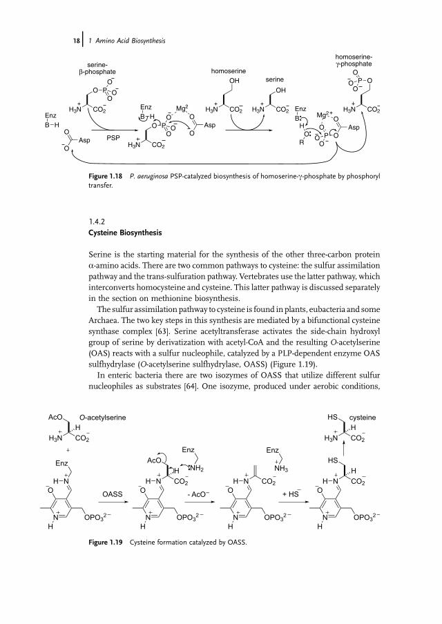
The PSP from Pseudomonas aeruginosa has evolved the ability to bind homoserinerather than water in the second half of the reaction and transfer the activatedphosphate of the aspartate-b-phosphate species to this amino acid providing accessto homoserine-c-phosphate, which is a biosynthetic precursor to threonine(Figure 1.18), as will be described later. This circumvents the need to expend ATPin phosphorylating this alcohol and is a rare example of an enzyme that transfersphosphoryl groups directly between non-nucleotide metabolites. This illustratesagain the role of changed substrate specificity in generating new enzymeactivities [62].
CO2O CO2H3N
OPO32 OPO3
2
CO2H3N
OH
CO2HO
OPO32
Glu α-KG
serine3-phosphoglycerate
PSP
3-phospho-hydroxypyruvate
PLP
Figure 1.16 Biosynthesis of serine.
CO2H3N
O Asp11
O
OP
O
OO
CO2H3N
OH
Asp11O
OPO
OO
Mg2+ H2OEnzB
EnzB
HO
H
H Mg2
Asp11
O
O
EnzB H
CO2H3N
O PO
OO
HOPO32
Figure 1.17 Catalytic details of PSP from systematic protein X-ray crystallography.
1.4 Routes to Functionalized Three-Carbon Amino Acids: Serine, Cysteine, and Selenocysteine j17
1.4.2Cysteine Biosynthesis
Serine is the starting material for the synthesis of the other three-carbon proteina-amino acids. There are two common pathways to cysteine: the sulfur assimilationpathway and the trans-sulfuration pathway. Vertebrates use the latter pathway, whichinterconverts homocysteine and cysteine. This latter pathway is discussed separatelyin the section on methionine biosynthesis.
The sulfur assimilation pathway to cysteine is found inplants, eubacteria and someArchaea. The two key steps in this synthesis are mediated by a bifunctional cysteinesynthase complex [63]. Serine acetyltransferase activates the side-chain hydroxylgroup of serine by derivatization with acetyl-CoA and the resulting O-acetylserine(OAS) reacts with a sulfur nucleophile, catalyzed by a PLP-dependent enzyme OASsulfhydrylase (O-acetylserine sulfhydrylase, OASS) (Figure 1.19).
In enteric bacteria there are two isozymes of OASS that utilize different sulfurnucleophiles as substrates [64]. One isozyme, produced under aerobic conditions,
CO2H3N
O AspO
OP
O
OO
CO2H3N
OH
AspO
OPO
OO
Mg2EnzB
EnzB
HO
R
H Mg2
AspO
O
EnzB H
CO2H3N
O PO
OO
CO2H3N
OH
CO2H3N
OPO
OO
serine-β-phosphate
homoserine-γ-phosphate
homoserineserine
PSP
Figure 1.18 P. aeruginosa PSP-catalyzed biosynthesis of homoserine-c-phosphate by phosphoryltransfer.
N
O
OPO32
H
NH
Enz
CO2H3N
H
AcO
CO2
H
AcO
N
O
OPO32
H
NH
NH2
Enz
CO2
N
O
OPO32
H
NH
NH3
Enz
CO2
H
HS
N
O
OPO32
H
NH
CO2H3N
H
HS
+ HS
O-acetylserine cysteine
OASS - AcO–
Figure 1.19 Cysteine formation catalyzed by OASS.
18j 1 Amino Acid Biosynthesis
utilizes hydrosulfide (formed by a multistep reduction of sulfate) [65]. Underanaerobic conditions a second isozyme is produced which utilizes thiosulfate andproduces S-sulfo-cysteine, which is transformed to cysteine by reaction with thiols.
The mechanism of OASS from Salmonella typhimurium has been studied indetail [65, 66]. This enzyme is a homodimer with an active-site PLP cofactor boundto Lys41. The initial stages of the reaction parallel those of aminotransferaseenzymes. The monoanion form of OAS forms a Schiff base with PLP by aminoexchange with Lys41, which is thereby liberated to act as an active-site acid–basecatalyst. In this case, deprotonation of the a-center of PLP-linked OAS by Lys41eliminates acetate and forms of a bound aminoacrylyl intermediate. After loss ofacetate, hydrosulfide binds, in the second step of this ordered Ping Pong Bi Bimechanism, and reacts with the aminoacrylate intermediate to produce cysteine.This mechanism is illustrative of a general class of PLP-dependent enzymes thatfacilitate reaction at the b-center of amino acids by facilitating the loss of a leavinggroup at that position (see Box 1.3).
1.4.3Case Study:Genome Information as a Starting Point forUncoveringNewBiosyntheticPathways
With the availability of a large number of genome sequences it is possible to identifythe likely biosynthetic pathways operating in particular organisms based on thepresence or absence of particular genes for biosynthetic enzymes. This has proveda powerful tool in expanding our understanding of the diversity and distribution ofmetabolic pathways. Genome analysis of the biosynthesis of cysteine and its incor-poration into cysteinyl-tRNA have led to the discovery of two new pathways for thebiosynthesis of this amino acid. These findings, in turn, have led to developments inour understanding of the biosynthesis of selenocysteine in humans [67]. This areapresents a nice case study in the emerging use of genome analysis to identify newvariants in biosynthetic pathways.
1.4.3.1 Cysteine Biosynthesis in Mycobacterium TuberculosisAmino acid biosynthesis in Mycobacterium tuberculosis is under active investigationbecause of the growing health threat posed by tuberculosis. Inhibitors of distinctiveessential metabolic pathways in this organism may be useful as antibiotics. Thecomplete genome sequence ofM. tuberculosis is known [68].M. tuberculosis carries outcysteine biosynthesis via the sulfur assimilation pathway and adjacent genes, cysEand cysK1, encode the serine acetyltransferase and OASS activities of the cysteinesynthase complex [69]. However, genome analysis revealed the presence of two othergenes homologous toOASS, cysK1 and cysM. Furthermore, cysMwas found clusteredwith two other genes related to sulfur metabolism. One of these genes, now calledcysO, is homologous to a family of small sulfide carrier proteins, such as ThiS, whichplay a role in thiamine pyrophosphate biosynthesis [70]. A thiocarboxylate derivativeof theC-terminal group of these proteins is the sulfide carrier. The protein is activatedby ATP, to form an acyl phosphate, and then converted to the corresponding
1.4 Routes to Functionalized Three-Carbon Amino Acids: Serine, Cysteine, and Selenocysteine j19
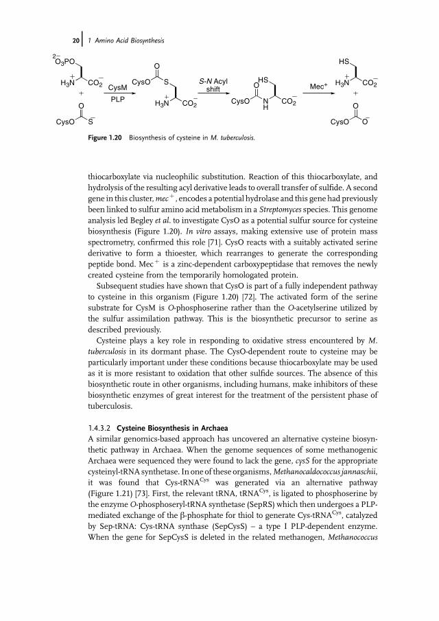
thiocarboxylate via nucleophilic substitution. Reaction of this thiocarboxylate, andhydrolysis of the resulting acyl derivative leads to overall transfer of sulfide. A secondgene in this cluster,mecþ , encodes a potential hydrolase and this gene had previouslybeen linked to sulfur amino acid metabolism in a Streptomyces species. This genomeanalysis led Begley et al. to investigate CysO as a potential sulfur source for cysteinebiosynthesis (Figure 1.20). In vitro assays, making extensive use of protein massspectrometry, confirmed this role [71]. CysO reacts with a suitably activated serinederivative to form a thioester, which rearranges to generate the correspondingpeptide bond. Mecþ is a zinc-dependent carboxypeptidase that removes the newlycreated cysteine from the temporarily homologated protein.
Subsequent studies have shown that CysO is part of a fully independent pathwayto cysteine in this organism (Figure 1.20) [72]. The activated form of the serinesubstrate for CysM is O-phosphoserine rather than the O-acetylserine utilized bythe sulfur assimilation pathway. This is the biosynthetic precursor to serine asdescribed previously.
Cysteine plays a key role in responding to oxidative stress encountered by M.tuberculosis in its dormant phase. The CysO-dependent route to cysteine may beparticularly important under these conditions because thiocarboxylate may be usedas it is more resistant to oxidation that other sulfide sources. The absence of thisbiosynthetic route in other organisms, including humans, make inhibitors of thesebiosynthetic enzymes of great interest for the treatment of the persistent phase oftuberculosis.
1.4.3.2 Cysteine Biosynthesis in ArchaeaA similar genomics-based approach has uncovered an alternative cysteine biosyn-thetic pathway in Archaea. When the genome sequences of some methanogenicArchaea were sequenced they were found to lack the gene, cysS for the appropriatecysteinyl-tRNA synthetase. In one of these organisms,Methanocaldococcus jannaschii,it was found that Cys-tRNACys was generated via an alternative pathway(Figure 1.21) [73]. First, the relevant tRNA, tRNACys, is ligated to phosphoserine bythe enzymeO-phosphoseryl-tRNA synthetase (SepRS) which then undergoes a PLP-mediated exchange of the b-phosphate for thiol to generate Cys-tRNACys, catalyzedby Sep-tRNA: Cys-tRNA synthase (SepCysS) – a type I PLP-dependent enzyme.When the gene for SepCysS is deleted in the related methanogen, Methanococcus
CO2H3N
O3PO2
CysO
O
S
CO2H3N CO2
HSCysO
O
S
CysO
O
NH
CO2H3N
HS
CysO
O
O
CysM
PLP
S-N Acylshift Mec+
Figure 1.20 Biosynthesis of cysteine in M. tuberculosis.
20j 1 Amino Acid Biosynthesis

maripaludis, the organism is a cysteine auxotroph, indicating that this is the solepathway to cysteine in this organism.
1.4.3.3 RNA-Dependent Biosynthesis of Selenocysteine and Other Amino AcidsDevelopments in cysteine biosynthesis research have underpinned our understand-ing of the biosynthesis of the 21st protein amino acid, selenocysteine. Selenocysteinehas been known to be an important residue for a range of enzymes since 1976 [74].This amino acid is incorporated into proteins by the ribosome using a tRNASec – asuppressor tRNA that corresponds to a stop codon in the genetic code [75]. Theutilization of this suppressor tRNA allows the expansion of the genetic code, butrequires an additional elongation factor for the ribosome to insert the amino acid inthe growing chain.
The biosynthesis of selenocysteine was first elucidated in E. coli and, like thearchaeal route to cysteine, it is based on modification of aminoacyl-tRNAs(Figure 1.22). The pathway starts with the ligation of serine to tRNASec, catalyzedby SerRS. The resulting ester undergoes PLP-mediated nucleophilic substitution ofthe side-chain hydroxyl group with a selenium-based nucleophile, selenophosphate,that is produced from selenide and ATP. The mechanism of selenophosphatesynthetase from E. coli has been established using positional isotope exchangemethodology [76, 77]. The reaction of Ser-tRNASec with selenophosphate is catalyzedby SelA [78]. The nucleophilic substitution reaction is assumed to follow a mech-anism analogous to that of OASS involving an initial elimination of water to form anaminoacrylyl-tRNA[Ser]Sec intermediate that reacts with the selenophosphate and the
CO2H3N
O3PO2
HO tRNACysCO2tRNACysH3N
O3PO
PLP
SepRS SepCysS
2
CO2tRNACysH3N
HSHOPO3
2HS
Figure 1.21 Biosynthesis of Cys-tRNACys in the methanogenic archaeon M. jannaschii.
CO2H3N
HO
HO tRNASec CO2tRNASecH3N
HO
SerRS
SepSecSCO2tRNASecH3N
O3PSe+ SePO3H
(PLP)
CO2tRNASecH3N
O3PO2
2
CO2tRNASecH3N
HSeSelA
Bacteria
Eukaryotes& Archaea
Figure 1.22 Biosynthesis of the tRNA adduct of selenocysteine.
1.4 Routes to Functionalized Three-Carbon Amino Acids: Serine, Cysteine, and Selenocysteine j21

resulting phosphoselenocysteyl-tRNASec undergoes hydrolysis to generate seleno-cysteyl-tRNASec.
This biosynthetic pathway was assumed to be common to all selenocysteineutilizing enzymes, but studies in eukaryotes failed to uncover the requisite biosyn-thetic enzymes. Subsequent studies have shown that selenocysteine biosynthesisoccurs by a commonpathway inArchaea and eukaryotes that is distinct, but related tothat in bacteria. A protein believed to be associated with selenocysteine synthesiscoprecipitated with the loaded selenocysteyl-tRNASec and bioinformatics analysisshowed that the enzyme was a PLP-dependent enzyme [79]. The chemical similarityof cysteine and selenocysteine provided the clue to unraveling the biosyntheticpathway to the latter amino acid in eukaryotes: Sep-tRNASec was shown to be asubstrate for the RNA-dependent biosynthesis of selenocysteine.
In the eukaryotic and archaeal version of the biosynthetic pathway, the b-hydroxylgroup of Ser-tRNASec is activated by phosphorylation to form the phosphoserinederivative, Sep-tRNASec, which then undergoes PLP-mediated nucleophilic substi-tution of the b-phosphate leaving group with selenophosphate catalyzed by seleno-cysteine synthase, SepSecS [80]. Selenocysteine synthase is homologous to OASSboth in structure [81] and sequence [82, 83] and the catalyticmechanism is analogous,involving an initial elimination of phosphate to form an aminoacrylyl-tRNA[Ser]Sec
intermediate which reacts with the selenophosphate. Despite selenocysteine beingan addition to the 20 amino acids found ubiquitously in proteins, the phylogeneticdata suggest that its biosynthesis is a primordial process and that selenocysteine hasplayed a role in metabolism since before the divergence of the ancestors to the threekingdoms of life (bacteria, Archaea and eukaryotes) more than 3 billion years ago.
The synthesis of selenocysteine on a specialized tRNA scaffold assists in distin-guishing the otherwise similar chemistry of selenocysteine and cysteine; the biosyn-thetic enzymes recognize structural features of tRNASec. Selenocysteine is not theonlyamino acid synthesized bymodification of an aminoacyl-tRNA.N-Formylmethionineis the N-terminal residue of proteins in eubacteria and eukaryotic organelles (mito-chondria and chloroplasts). It is synthesized via formylation of Met-tRNAfMet in aprocess that also depends on binding to the tRNAfMet and is specific to this aminoacyl-tRNA species [84]. Interestingly, it has also been found that many organisms produceaminoacylated tRNAs for asparagine and glutamine by amidating aspartyl andglutamyl precursors. Again, genome analysis is proving useful in identifying thepathway(s) present in particular organisms [85]. For example, whereasGln-tRNAGln issynthesized fromglutamine in the cytoplasmof eukaryotes, themajority of eubacteriaand all Archaea make it by the transamidation route [86].
1.5Other Amino Acids from Aspartate and Glutamate: Asparagine and Side ChainFunctional Group Manipulation
Glutamate and aspartate are the parents of six further amino acids that areubiquitously found in proteins: asparagine,methionine, and threonine are produced
22j 1 Amino Acid Biosynthesis
fromaspartate; and glutamine, proline, and arginine are derived fromglutamate. Theconversion of glutamate to glutamine has already been described and illustrates thestrategy by which the remaining members of this group of protein amino acids aremade. In each case the side-chain carboxylate undergoes functional group manip-ulation starting with activation to a short-lived acyl phosphate intermediate and thensubsequent nucleophilic substitution. The two nucleophiles that are utilized areammonia and hydride ion (delivered by redox cofactors), and these will be discussedin turn.
1.5.1Asparagine Biosynthesis
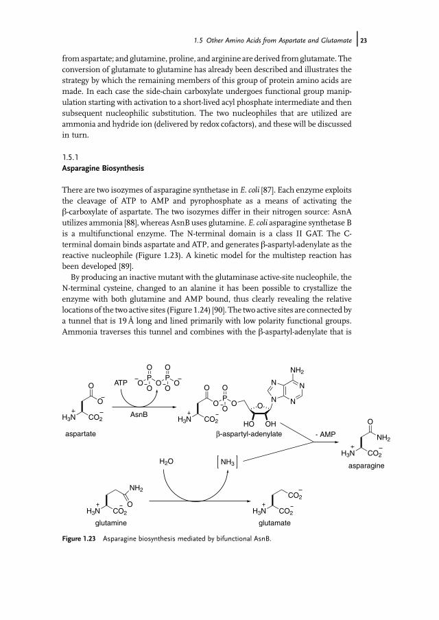
There are two isozymes of asparagine synthetase in E. coli [87]. Each enzyme exploitsthe cleavage of ATP to AMP and pyrophosphate as a means of activating theb-carboxylate of aspartate. The two isozymes differ in their nitrogen source: AsnAutilizes ammonia [88], whereas AsnBuses glutamine. E. coli asparagine synthetase Bis a multifunctional enzyme. The N-terminal domain is a class II GAT. The C-terminal domain binds aspartate and ATP, and generates b-aspartyl-adenylate as thereactive nucleophile (Figure 1.23). A kinetic model for the multistep reaction hasbeen developed [89].
By producing an inactive mutant with the glutaminase active-site nucleophile, theN-terminal cysteine, changed to an alanine it has been possible to crystallize theenzyme with both glutamine and AMP bound, thus clearly revealing the relativelocations of the two active sites (Figure 1.24) [90]. The two active sites are connected bya tunnel that is 19Å long and lined primarily with low polarity functional groups.Ammonia traverses this tunnel and combines with the b-aspartyl-adenylate that is
CO2H3N
CO2
CO2H3NO
NH2
CO2H3N
N
NN
N
NH2
O
HO OH
PO
OO
CO2H3N
O
O
O
O
OP
OP
O
O
O
O O
NH3H2OCO2H3N
O
NH2- AMP
ATP
aspartate
asparagine
glutamine glutamate
AsnB
β-aspartyl-adenylate
Figure 1.23 Asparagine biosynthesis mediated by bifunctional AsnB.
1.5 Other Amino Acids from Aspartate and Glutamate j23
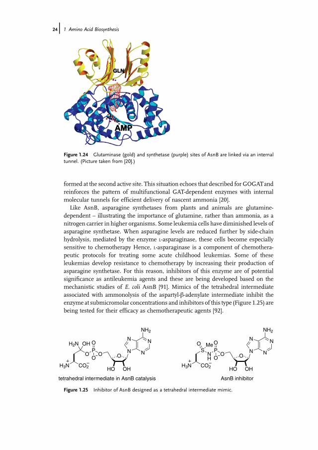
formed at the second active site. This situation echoes that described for GOGATandreinforces the pattern of multifunctional GAT-dependent enzymes with internalmolecular tunnels for efficient delivery of nascent ammonia [20].
Like AsnB, asparagine synthetases from plants and animals are glutamine-dependent – illustrating the importance of glutamine, rather than ammonia, as anitrogen carrier in higher organisms. Some leukemia cells have diminished levels ofasparagine synthetase. When asparagine levels are reduced further by side-chainhydrolysis, mediated by the enzyme L-asparaginase, these cells become especiallysensitive to chemotherapy Hence, L-asparaginase is a component of chemothera-peutic protocols for treating some acute childhood leukemias. Some of theseleukemias develop resistance to chemotherapy by increasing their production ofasparagine synthetase. For this reason, inhibitors of this enzyme are of potentialsignificance as antileukemia agents and these are being developed based on themechanistic studies of E. coli AsnB [91]. Mimics of the tetrahedral intermediateassociated with ammonolysis of the aspartyl-b-adenylate intermediate inhibit theenzyme at submicromolar concentrations and inhibitors of this type (Figure 1.25) arebeing tested for their efficacy as chemotherapeutic agents [92].
Figure 1.24 Glutaminase (gold) and synthetase (purple) sites of AsnB are linked via an internaltunnel. (Picture taken from [20].)
N
NN
N
NH2
O
HO OH
PO
OO
CO2H3N
SO
NH
Me
N
NN
N
NH2
O
HO OH
PO
OO
CO2H3N
H2N
O
OH
tetrahedral intermediate in AsnB catalysis AsnB inhibitor
Figure 1.25 Inhibitor of AsnB designed as a tetrahedral intermediate mimic.
24j 1 Amino Acid Biosynthesis

1.6Aspartate and Glutamate Families of Amino Acids
1.6.1Overview
The remaining four- and five-carbon amino acids are prepared by pathways basedaround a common chemical strategy (Figure 1.26): ATP-dependent activation of theside-chain carboxylate of the parent amino acid generates a reactive acyl phosphateintermediate that is reduced to the corresponding aldehyde by hydride transfer; theproduct amino aldehydes are also labile species and these are converted to moredurable reduced analogs in the next stage.
These pathways illustrate several common features of amino acid biosyntheticpathways. Enzymes catalyzing analogous reactions in parallel pathways are oftenhomologous in structure. Once enzymes are available that can catalyze a particularset of reactions then gene duplication and substrate specificity modification, viamutation and selection, can generate parallel pathways [93]. This evolutionarymechanism highlights the important role of the underlying chemical logic of thepathways that underpin this organization. A second general feature is that pathwaysinvolving reactive intermediates benefit from multifunctional enzyme systems thatcan efficiently channelmetabolites to the active site that catalyzes the next stage in thepathway. This not only increases the yield of the reaction, but also controls the fate ofthe metabolite when there are competing metabolic uses of the product. For thisreason there are often multiple isozymes to catalyze reactions that occur in multiplepathways and these are generally independently regulated [94]. For isozymes thatcatalyze reactions at branch-points of pathways, where a commitment to one or otherfinal product is made, the pattern of feedback regulation provides a direct confir-mation of the in vivo role of the specific form of the enzyme.
1.6.2Aspartate Family Amino Acids: Threonine and Methionine
The first three steps of the biosynthesis of threonine and methionine in plants andmicrobes are common to both pathways (Figure 1.27) [95]. Aspartokinase (AK)catalyzes the ATP-dependent b-phosphorylation of aspartate, which creates the
CO2H3N
PO
OOR'
CO2RH2N
O
O
O
Onn
CO2RH2N
O
Hn
CO2H3N
X
n
aspartate (n = 1)glutamate (n = 2)
acyl phosphateintermediate
ATPNADPH NADP+
semialdehydeintermediate
Figure 1.26 Strategy of aspartate and glutamate family amino acid biosynthesis.
1.6 Aspartate and Glutamate Families of Amino Acids j25

requisite leaving group for subsequent transformation [96]. The resulting aspartate-b-phosphate is reduced to the corresponding aldehyde by aspartate b-semialdehydedehydrogenase (ASADH). Homoserine dehydrogenase (HSDH) catalyzes the fur-ther reduction of aspartate-b-semialdehyde to homoserine – a key intermediate forthe biosynthesis of both threonine and methionine. NADPH is a hydride source forthe reduction chemistry. In E. coli and other bacteria there are independentlyregulated isozymes of AK-HSDH for threonine and methionine biosynthesis.
ASADH from several sources has been characterized [97–99]. The mechanism ofASADH (Figure 1.28) is analogous to the oxidation of glyceraldehyde-3-phosphate toglycerate-3-phosphate – one of the key oxidation steps in glycolysis [100]. Aspartate-b-phosphate undergoes initial nucleophilic substitution of phosphate with the active-site thiol of cysteine-136. The resulting thioester is then reduced by a nicotinamidecofactor, NADPH, to aspartate-b-semialdehyde ASA. The resulting aldehyde, ASA, issufficiently reactive with nucleophiles that the three-dimensional structure of a
CO2H3N
PO
OO
CO2H3N
O
O
O
O
CO2H3N
O
H
CO2H3N
OHATP ADP
AK ASADH HSDH
NADPH NADP+ NADPH NADP+
aspartate aspartate-β-phosphate
aspartate-β-semialdehyde
homoserine
Figure 1.27 Biosynthesis of homoserine.
CO2H3N
O
HS Cys136
HNNH
His277
CO2H3N
OS Cys136
HNNH
His277
OPO
O
OH
CO2H3N
OS Cys136
HNNH
His277
HOP
O
O
OHCO2H3N
OS Cys136
HNNH
His277
OPO
O
OH
CO2H3N
O
HS Cys136
HNNH
His277
HOP
O
O
OH
NADPH
NADP+
-Pi
Figure 1.28 Mechanism of ASADH.
26j 1 Amino Acid Biosynthesis
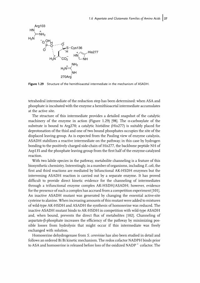
tetrahedral intermediate of the reduction step has been determined: when ASA andphosphate is incubated with the enzyme a hemithioacetal intermediate accumulatesat the active site.
The structure of this intermediate provides a detailed snapshot of the catalyticmachinery of the enzyme in action (Figure 1.29) [98]. The a-carboxylate of thesubstrate is bound to Arg270; a catalytic histidine (His277) is suitably placed fordeprotonation of the thiol and one of two bound phosphates occupies the site of thedisplaced leaving group. As is expected from the Pauling view of enzyme catalysis,ASADH stabilizes a reactive intermediate on the pathway; in this case by hydrogenbonding to the positively charged side-chain of His277, the backbone peptide NH ofAsp135 and the phosphate leaving group from the first half of the enzyme-catalyzedreaction.
With two labile species in the pathway, metabolite channeling is a feature of thisbiosynthetic chemistry. Interestingly, in a number of organisms, includingE. coli, thefirst and third reactions are mediated by bifunctional AK-HSDH enzymes but theintervening ASADH reaction is carried out by a separate enzyme. It has proveddifficult to provide direct kinetic evidence for the channeling of intermediatesthrough a trifunctional enzyme complex AK-HSDH/ASADH; however, evidencefor the presence of such a complex has accrued from a competition experiment [101].An inactive ASADH mutant was generated by changing the essential active-sitecysteine to alanine.When increasing amounts of thismutant were added tomixturesof wild-type AK-HSDH and ASADH the synthesis of homoserine was reduced. Theinactive ASADHmutant binds to AK-HSDH in competition with wild-type ASADHand, when bound, prevents the direct flux of metabolites [102]. Channeling ofaspartate-b-phosphate increases the efficiency of the pathway by minimizing pos-sible losses from hydrolysis that might occur if this intermediate was freelyexchanged with solution.
Homoserine dehydrogenase from S. cerevisiae has also been studied in detail andfollows an ordered Bi Bi kinetic mechanism. The redox cofactor NADPH binds priorto ASA and homoserine is released before loss of the oxidized NADPþ cofactor. The
CO2H3N
O
HS Cys136
HNNH
His277
HOP
O
O
OH
NH
NH2H2N
270Arg
HN
H2NNH2
Arg103
Figure 1.29 Structure of the hemithioacetal intermediate in the mechanism of ASADH.
1.6 Aspartate and Glutamate Families of Amino Acids j27

pro-(S) hydride of stereospecifically deuterated NADP[2H] is transferred and thereduction is catalyzed by carbonyl polarization by a protonated active-site lysineresidue (Lys223) [103, 104] (Figure 1.30).
1.6.2.1 Case Study: Evolution of Leaving Group Specificity in Methionine BiosynthesisFor processing to either methionine or threonine, the hydroxyl group of homoserineis converted into a good leaving group. Primary metabolism provides two mainalternatives for hydroxyl activation: polyphosphates like ATP can generate phosphateleaving groups; alternatively thioesters (notably TCA cycle metabolites acetyl-CoAand succinyl-CoA) generate carboxylate leaving groups (Figure 1.31). There arevariations in the pathway at this point depending on this choice. Phosphorylationof homoserine by homoserine kinase (HSK) to produce homoserine-c-phosphate isubiquitously used for threonine biosynthesis [105]. An interesting alternative phos-phorylation route, based on the evolution of a novel bifunctional PSP, was describedabove (Figure 1.18) [62]. Homoserine-c-phosphate is also the biosynthetic precursorto methionine biosynthesis in plants [95]. Other organisms use homoserine transa-cylases to activate homoserine for methionine biosynthesis and two different acylgroups are employed: Gram-negative bacteria make O-succinyl-homoserine, whileyeasts and many clinically important bacteria (e.g.,M. tuberculosis and P. aeruginosa)use an O-acetyl-homoserine as a precursor to methionine. Although the choice ofleaving group does not fundamentally change the chemistry, it does have implica-tions for the specificity of inhibitors and for the control of the pathways since, withdistinct building blocks, the two pathways can be controlled independently.
All homoserine transacylases have a catalytic triad of residues situated at the endof a tunnel. The a-carboxylate of the substrate is recognized by an arginine and anactive-site nucleophile (serine or cysteine) is assisted in catalysis by a histidine in
NR
HS
HR
H2NO
OHH3N
O2C
Zn2
Figure 1.30 Stereospecificity of hydride transfer in the formation of homoserine.
CO2H3N
OH
CO2H3N
ORR = PO3
2–
R = AcR = CO(CH2)2CO2CO2
H
O3PO
NH
homoserine
–
Figure 1.31 Three different activated forms of homoserine.
28j 1 Amino Acid Biosynthesis

conjunction with either an aspartate or glutamate. The structures and sequences ofhomoserine transacylases group into two families related to the active-site nucleo-phile. Homoserine transacetylase from Haemophilus influenzae is typical of oneclass [106]. A conserved serine (Ser143) is in a strained conformation and acts as areactive nucleophile to accept the acetyl group from acetyl-CoA (Figure 1.32). His337is an adjacent acid–base catalyst and, like the active site of serine proteases, there is anoxyanion hole to stabilize the tetrahedral intermediate. The residues of the tunnel arewell placed to direct homoserine to the acetylated active site and thereby assuretransesterification outcompetes hydrolysis.
It had been believed that transsuccinylases comprised the cysteine-dependentfamily of transacylases. However, when one of this family of enzymes from Bacilluscereuswas fully characterized it was found to be a transacetylase. The structure of thisenzyme illustrates both the details of the active-site architecture, with the catalytictriad of Cys142, His235, and Glu237, and the basis for the substrate specificity of theenzyme (Figure 1.33a) [107]. A glutamate residue, Glu111, protrudes into the active
Figure 1.32 Homoserine transacetylase from H. influenzae. (From [102].)
Figure 1.33 Pointmutagenesis (E111G)ofB. cereushomoserine transacylase changes its substratespecificity from that of a transacetylase (a) to a transsuccinylase (b). (From [107].)
1.6 Aspartate and Glutamate Families of Amino Acids j29
site limiting its size, allowing binding of acetyl species but excluding succinyl specieson steric and electrostatic grounds. In many other enzymes of this class thecorresponding residue is glycine, which presents no such impediments to succinylderivatives.Making a single pointmutation of this glutamate to glycinewas sufficientto convert a specific transacetylase into a specific transsuccinylase (Figure 1.33b).This shows again the power of point mutations to engineer modifications to enzymesubstrate specificity.
1.6.2.2 Threonine, Homocysteine, and PLPThe manipulations of these activated homoserine derivatives to form threonineand homocysteine (en route to methionine) both involve catalysis of the loss of theside-chain leaving group. In each case this is catalyzed by a PLP-dependentenzyme. The ability to mediate the chemistry at b-, and c-centers of amino acids,in addition to the a-center chemistry previously described, illustrates the catalyticversatility of PLP that makes it an indispensible cofactor in amino acid metabolism(see Box 1.3).
1.6.2.3 Threonine SynthaseThreonine synthase (TS) catalyzes the conversion of homoserine-c-phosphate tothreonine. Threonine synthases are PLP-dependent enzymes, of type II fold,with a complex mechanism that utilizes the full capacity of PLP to stabilizereactive intermediates [108]. Sequence alignments have identified two subfami-lies – class I and class II [109]. Class I enzymes are found in plants and somebacteria and Archaea, and are allosterically activated by S-adenosylmethionine[110]. Three-dimensional structures for both classes of enzymes are available[111–113].
Owing to its potential significance for the development of antibiotics to treattuberculosis [114], the class I TS fromM. tuberculosis has been studied in detail [115].In the resting state, the PLP cofactor forms a Schiff base with Lys69. Whenhomoserine-c-phosphate binds to PLP it displaces Lys69, which then acts as a protonrelay in a sequence of acid–base reactions that are shown in Figure 1.34, which givesan overview of the mechanism of the reaction.
Deprotonation of the a-proton of the substrate by Lys69 produces an aza-allylanion. Reprotonation at the benzylic position of the PLP, by the conjugate acid ofLys69, generates a new iminium ion. Deprotonation at the b-position by theregenerated basic Lys69 leads to elimination of phosphate and the formation ofa conjugated iminium ion. Now a proton transfer is required from the benzylicposition of the PLP and the terminus of the conjugated system to produce aa,b-unsaturated ketimine ready for reaction with water at the b-position to generatethreonine. While Lys69 is well placed to undertake the initial proton transfers, themolecular gymnastics required to also mediate the latter proton transfer is beyondits reach and there has been debate in the literature about the catalytic grouprequired for this chemistry. Recent detailed structural studies show that the mostlikely acid–base catalyst is the 50-phosphate of the PLP cofactor (Figure 1.35) [115].This phosphate moiety is less than 5 Å away from both the benzylic position of the
30j 1 Amino Acid Biosynthesis
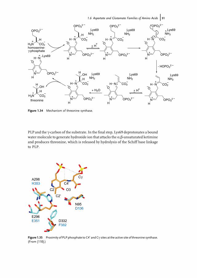
PLP and the c-carbon of the substrate. In the final step, Lys69 deprotonates a boundwatermolecule to generate hydroxide ion that attacks the a,b-unsaturated ketimineand produces threonine, which is released by hydrolysis of the Schiff base linkageto PLP.
CO2
H
N
O
OPO32
H
NH
NH2
Lys69
CO2
N
O
OPO32
H
NH
NH3
Lys69OPO3
2
CO2
H
N
O
OPO32
H
NH
NH2
Lys69
CO2
N
O
OPO32
H
NH
NH2
Lys69OPO3
2 OPO32
CO2
N
O
OPO32
H
NH
NH2
Lys69
- HOPO32
CO2
N
O
OPO32
H
NH
NH2
Lys69
+– HCO2
HH3N
OPO32
Lys69
N
O
OPO32
H
NH
OH
CO2
HH3N
OH+– H
H
+ H2O
H
homoserine-γ-phosphate
threonine
Figure 1.34 Mechanism of threonine synthase.
Figure 1.35 Proximity of PLP phosphate to C40 andCc sites at the active site of threonine synthase.(From [110].)
1.6 Aspartate and Glutamate Families of Amino Acids j31
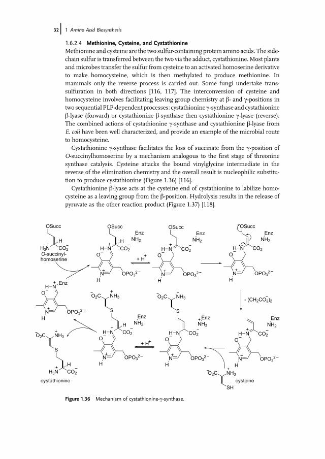
1.6.2.4 Methionine, Cysteine, and CystathionineMethionine and cysteine are the two sulfur-containing protein amino acids. The side-chain sulfur is transferred between the two via the adduct, cystathionine.Most plantsandmicrobes transfer the sulfur from cysteine to an activated homoserine derivativeto make homocysteine, which is then methylated to produce methionine. Inmammals only the reverse process is carried out. Some fungi undertake trans-sulfuration in both directions [116, 117]. The interconversion of cysteine andhomocysteine involves facilitating leaving group chemistry at b- and c-positions intwo sequential PLP-dependent processes: cystathionine c-synthase and cystathionineb-lyase (forward) or cystathionine b-synthase then cystathionine c-lyase (reverse).The combined actions of cystathionine c-synthase and cystathionine b-lyase fromE. coli have been well characterized, and provide an example of the microbial routeto homocysteine.
Cystathionine c-synthase facilitates the loss of succinate from the c-position ofO-succinylhomoserine by a mechanism analogous to the first stage of threoninesynthase catalysis. Cysteine attacks the bound vinylglycine intermediate in thereverse of the elimination chemistry and the overall result is nucleophilic substitu-tion to produce cystathionine (Figure 1.36) [116].
Cystathionine b-lyase acts at the cysteine end of cystathionine to labilize homo-cysteine as a leaving group from the b-position. Hydrolysis results in the release ofpyruvate as the other reaction product (Figure 1.37) [118].
CO2
H
N
O
OPO32
H
NH
NH2
Enz
CO2
N
O
OPO32
H
NH
OSucc
CO2
H
N
O
OPO32
H
NH
NH2
EnzS
CO2
N
O
OPO32
H
NH
NH2
EnzOSucc OSucc
CO2
N
O
OPO32
H
NH
NH2
Enz
- (CH2CO2)2
CO2
N
O
OPO32
H
NH
NH3
EnzS
+ H
+ H
O2C NH3O2C NH3
SH
O2C NH3
NH2
Enz
Enz
N
O
OPO32
H
NH
CO2
H
H3N
OSucc
CO2
H
H3N
S
O2C NH3
O-succinyl-homoserine
cysteinecystathionine
Figure 1.36 Mechanism of cystathionine-c-synthase.
32j 1 Amino Acid Biosynthesis

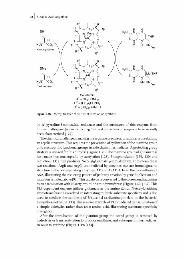
1.6.2.5 Methionine SynthaseThe biosynthesis of methionine is completed by methylation of homocysteine,catalyzed by methionine synthase (MS). The ultimate source of the methyl group isN5-methyl-THF (Me-THF) where the methyl group can originate in the cleaved side-chainofserine (asseen in thebiosynthesisofglycine).Thereare twodistinctversionsofthis enzyme depending on the immediate source of themethyl group, eitherMe-THFor methyl cobalamin. In organisms that biosynthesize vitamin B12, or obtain it fromtheir environment, this is often carried out by a multifunctional MS that uses methylcobalamin as the alkylating agent (Figure 1.38) [119]. After methionine synthesis, themethyl cobalamin is reconstituted by methyl transfer from Me-THF [120]. There aretwo MS enzymes in E. coli – one uses methyl cobalamin as methyl donor, whereasanother usesMe-THFdirectly as the alkylating agent in a reaction that is dependent ona zinc ion for catalysis [121]. It is believed that homocysteine coordinates to the zinc asthe thiolate and is thereby activated as a nucleophile to react with Me-THF.
1.6.3Glutamate Family Amino Acids: Proline and Arginine
Proline and arginine are made from glutamate by routes that utilize the samechemical strategy that is seen in methionine and threonine biosynthesis. Thisinvolves activation of the c-carboxylate and biosynthetic reduction (Figure 1.39). Abacterial pathway has been characterized: glutamate-5-kinase [122] phosphorylatesthe c-carboxylate of glutamate and NADPH-dependent reduction leads to glutamate-c-semialdehyde, which undergoes spontaneous cyclization to the correspondingimine, D1-pyrroline-5-carboxylate. In plants a bifunctional enzyme mediates both ofthese steps and ensures efficient use of the reactive acyl phosphate intermediate [123,124]. Two alternative pathways to this cyclic imine via oxidative deamination ofornithine have been reported [125, 126]. Reduction of the imine to proline is catalyzed
CO2
H
N
O
OPO32
H
NH
NH2
Lys210
CO2
N
O
OPO32
H
NH
NH2
Lys210
O2C
H
NH3
S
CO2H3N
O2C
H
NH3
S
Lys210
N
O
OPO32
H
NH
O2C
H
NH3
SH
+ H
CO2ONH3 +
cystathionine
pyruvate
homocysteine
+H2O
Figure 1.37 Mechanism of cystathionine-b-lyase.
1.6 Aspartate and Glutamate Families of Amino Acids j33
by D1-pyrroline-5-carboxylate reductase and the structures of this enzyme fromhuman pathogens (Neisseria meningitidis and Streptococcus pyogenes) have recentlybeen characterized [127].
The chemical challenge inmaking the arginine precursor, ornithine, is in retainingan acyclic structure. This requires the prevention of cyclization of the a-amino grouponto electrophilic functional groups in side-chain intermediates. A protecting groupstrategy is utilized for this purpose (Figure 1.39). The a-amino group of glutamate isfirst made non-nucleophilic by acetylation [128]. Phosphorylation [129, 130] andreduction [131] then produces N-acetylglutamate-c-semialdehyde. In bacteria thesetwo reactions (ArgB and ArgC) are mediated by enzymes that are homologous instructure to the corresponding enzymes, AK and ASADH, from the biosynthesis ofASA, illustrating the recurring pattern of pathway creation by gene duplication andmutation as noted above [93]. This aldehyde is converted to the corresponding amineby transamination withN-acetylornithine aminotransferase (Figure 1.40) [132]. ThisPLP-dependent enzyme utilizes glutamate as the amino donor. N-Acetylornithineaminotransferase has evolved an interestingmultiple-substrate specificity and is alsoused to mediate the synthesis of N-succinyl-L,L-diaminopimelate in the bacterialbiosynthesis of lysine [133]. This is a rare example of PLP-mediated transamination ofa simple aldehyde, rather than an a-amino acid, illustrating substrate specificitydivergence.
After the introduction of the c-amino group the acetyl group is removed byhydrolysis or trans-acetylation to produce ornithine, and subsequent intermediates,en route to arginine (Figure 1.39) [134].
HN
NN
HN
O
H2N
HNAr
HCO2
HH3N
SH
NN
NN
R2
R2R3
R2
Co
R1H
R1
CobalaminR1 = CH2CONH2
R2 = (CH2)2CONH2
R3 = (CH2)2CONHR
R1
CO2
HH3N
SMe
NN
NN
R2
R2R3
R2
Co
R1H
R1
Me
R1
HN
NN
HN
O
H2N
HNAr
Me
homocysteine
methionine
THF
Me-THF
Figure 1.38 Methyl transfer chemistry of methionine synthase.
34j 1 Amino Acid Biosynthesis

Although the general chemical strategy for the biosynthesis of ornithine iscommon to many organisms, contemporary studies have revealed a diversity ofdetail in the pathways, associated with different acyl-transfer chemistry [135]. Thefirst step of the biosynthesis in E. coli, which served as the initial exemplar for
NAD(P)H
NAD(P)+
CO2H3N
O2C
CO2H3N
O3PO2C
CO2H3N
OHC
- HOPO32
2
NH CO2
NH2
CO2
NAD(P)H
NAD(P)+
ATP
ADP
NAD(P)H
NAD(P)+
CO2AcHN
O2C
CO2AcHN
O3PO2C
CO2AcHN
OHC
- HOPO32
2
ATP
ADP
Glu
α-KG
AcSCoA HSCoA
CO2AcHN
H3N
CO2H3N
H3N
PLP
proline ornithine
γ-phosphatederivative
Δ1-pyrroline-5-carboxylate
glutamate-5-kinase
Δ1-pyrroline-5-carboxylate
reductase
N-acetyl glutamate
N-acetylornithine
aminotransferase
γ-semialdehydederivative
N-acetylglutamatesynthase
N-acetyl-glutamate-5-kinase
Figure 1.39 Biosynthesis of proline and ornithine.
CO2O
CO2
CO2H3N
CO2
Enz-PLP Enz-PMP
CO2OCO2H3N
CO2
NHAcH3N
CO2
NHAc
CO2O
CO2
CO2H3N
CO2
Enz-PLP Enz-PMP
CO2
HN CO2
O
CO2
HN CO2
O
O H
N-acetylornithine
N-succinyl-L,L-diaminopimelate
Figure 1.40 Multiple substrate specificity of N-acetylornithine aminotransferase.
1.6 Aspartate and Glutamate Families of Amino Acids j35
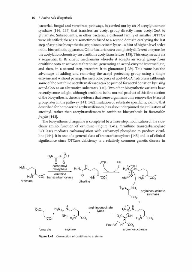
bacterial, fungal and vertebrate pathways, is carried out by an N-acetylglutamatesynthase [136, 137] that transfers an acetyl group directly from acetyl-CoA toglutamate. Subsequently, in other bacteria, a different family of smaller DITTOswere identified; these are sometimes fused to a second domain catalyzing the laststep of arginine biosynthesis, argininosuccinate lyase – a hint of higher-level orderin the biosynthetic apparatus. Other bacteria use a completely different enzyme forthe acetylation chemistry: an ornithine acetyltransferase [138]. This enzyme acts viaa sequential Bi Bi kinetic mechanism whereby it accepts an acetyl group fromornithine onto an active-site threonine, generating an acetyl-enzyme intermediate,and then, in a second step, transfers it to glutamate [139]. This route has theadvantage of adding and removing the acetyl protecting group using a singleenzyme and without paying the metabolic price of acetyl-CoA hydrolysis (althoughsome of the ornithine acetyltransferases can be primed for acetyl donation by usingacetyl-CoA as an alternative substrate) [140]. Two other biosynthetic variants haverecently come to light: although ornithine is the normal product of this first sectionof the biosynthesis, there is evidence that some organisms only remove theN-acetylgroup later in the pathway [141, 142]; mutation of substrate specificity, akin to thatdescribed for homoserine acyltransferases, has also underpinned the utilization ofsuccinyl- rather than acetyltransferases in ornithine biosynthesis in Bacteroidesfragilis [143].
The biosynthesis of arginine is completed by a three-step modification of the side-chain amino function of ornithine (Figure 1.41). Ornithine transcarbamoylase(OTCase) mediates carbamoylation with carbamoyl phosphate to produce citrul-line [144]. It is one of a general class of transcarbamoylases [145] and is of clinicalsignificance since OTCase deficiency is a relatively common genetic disease in
CO2H3N
H3N
O
OH2NP
O
O
O
CO2H3NO
HNH2N
CO2H3NO
HNH2N
AMP
O2C NH2
CO2
CO2H3N
HNH2N
O2C NH
CO2HEnz-B
CO2H3N
HNH2NO2C
NH2CO2
ATP PPi
ornithine citrulline
argininosuccinateargininefumarate
ornithinetranscarbamoylase
carbamoyl phosphate
argininosuccinatesynthase
argininosuccinatelyase
Figure 1.41 Conversion of ornithine to arginine.
36j 1 Amino Acid Biosynthesis

humans. The enzyme mechanism is now believed to involve acid–base catalysis bythe phosphate group of the substrate, carbamoyl phosphate [146].
The urea group of citrulline is converted to an amidine by the introduction of thefinal amino group. This transformation occurs in a two-step process that exem-plifies the third general mechanism of amine transfer in metabolism (after GATand PLPaction) – ligation of thea-amino group of aspartate and then elimination offumarate, which is recycled via the TCAcycle [147]. Argininosuccinate synthetase ishomologous to asparagine synthetase [148] and uses ATP to activate the urea byadenylation [149]; nucleophilic substitution by aspartate produces argininosucci-nate. Argininosuccinate lyase, the terminal enzyme of the pathway, is a member ofa family of aspartase enzymes that catalyze the elimination of fumarate (whichfeeds into the TCA cycle) from aspartate and N-aspartyl derivatives [150, 151],demonstrating the generality of this two step synthetase/lyase route for the indirecttransfer of ammonia – the third general process for transfer of nitrogen betweenmetabolites.
1.7Biosynthesis of Aliphatic Amino Acids with Modified Carbon Skeletons: Branched-Chain Amino Acids, Lysine, and Pyrrolysine
1.7.1Overview
Four of the ubiquitous aliphatic protein amino acids, valine, leucine, isoleucine, andlysine, aremade by the synthesis of new carbon skeletons. Lysine is now known to bethe precursor of the 22nd ribosomally encoded amino acid, pyrrolysine. Thebiosynthesis of these amino acids involves parallel pathways in which a chemicalstrategy is duplicated and homologous or promiscuous enzymes are used for thebiosynthesis of more than one amino acid. Valine and isoleucine are made fromprecursor a-keto acids by a common route, which also provides a key biosyntheticprecursor for leucine. The carbon skeleton of leucine is made from an a-keto acidintermediate in valine biosynthesis by a pathway homologous to the biosynthesis ofa-KG in the TCA cycle. This route, in turn is utilized for the biosynthesis of anotheramino acid, a-aminoadipic acid, which is not incorporated into proteins but is abiosynthetic precursor to lysine in one of the pathways to that amino acid. Lysine isunusual in beingmade indifferent organismsby two completely different pathways –from a-aminoadipic acid in some fungi and bacteria, and from aspartate-b-semi-aldehyde in plants and other bacteria.
1.7.2Valine and Isoleucine
Three enzymes, acetohydroxyacid synthase (AHAS) [152], acetohydroxyacid isomer-oreductase (AHIR) [153], and dihydroxyacid dehydratase [154], mediate the four key
1.7 Biosynthesis of Aliphatic Amino Acids with Modified Carbon Skeletons j37

steps common to the biosynthesis of all the branched-chain amino acids [155]. Beingspecific to branched-chain amino acid biosynthesis and not found in animals, theseenzymes are key targets for herbicide research [156]. AHAS, in particular, is the targetof a number of commercial herbicides. All three enzymes have relaxed substratespecificity and process metabolites on two parallel pathways to produce two a-ketoacid products (Figure 1.42).
Pyruvate reacts with the anion of the thiamine pyrophosphate cofactor of AHAS(Figure 1.43). Decarboxylation of the initial adduct leads to an acetyl a-anionequivalent, illustrating the role of thiamine pyrophosphate as an umpolung reagentin biochemistry. Reaction of this nucleophile with a second a-keto acid provides abranched carbon skeleton [157]. Two a-keto acids can participate as electrophiles inthe second stage of the reaction: pyruvate or a-ketobutyrate; these lead to valine andisoleucine, respectively. The a-ketobutyrate can be biosynthesized in one step fromthreonine (see below).
O
Me CO2S
N
OPP
Py
Me
HO
O
Me
OH
MeO
O
HO
Me
OH
O
O
MeH
O
CO2
Me
Me
O
Et CO2
O
Me
OH
EtO
O
HO
Me
OH
O
O
EtH
O
CO2
Me
Et
NH3
CO2
Me
MeNH3
CO2
Me
Et
O
CO2
NH3
CO2Me
Me
Me
Me
leucine valine isoleucine
threonineor pyruvate
pyruvate
pyruvate
Figure 1.42 Overview of the parallel pathways to branched-chain amino acids.
38j 1 Amino Acid Biosynthesis

The structures of AHAS with bound sulfonylurea or imidazolinone show themechanismof inhibition involves obstruction of the channel leading to the active site.The details of this inhibition provide an understanding of the basis of evolvingherbicide resistance and will be useful in the generation of alternativeherbicides [158].
Threonine deaminase (also known as threonine dehydratase) is a PLP-dependentenzyme that is the usual source of a-ketobutyrate needed for isoleucine biosynthe-sis [159, 160]. It is the point of commitment to isoleucine biosynthesis and theenzyme is subject to feedback regulation by isoleucine [161, 162]. Schiff baseformation between threonine and the active-site PLP facilitates deprotonation atthe a-center and consequent elimination of water, in a reaction characteristic of PLP-facilitated transformations of amino acids with b-leaving groups (Box 1.3). Cleavageof the link between PLP and the amino acid portion and hydrolysis chemistryproduces a-ketobutyrate and ammonium ions as the product (Figure 1.44). Analternative route to a-ketobutyrate [163] based on the homologation of pyruvate isoutlined later (Figure 1.48).
The two products of AHAS are processed by a bifunctional enzyme – AHIR [153].While the enzyme acts on two different hydroxy acids, it has an absolute requirement
SN
OPPPy
O
Me CO2
H
SN
OPPPy
O
R CO2
MeCO2
HO
SN
OPPPy
MeOH
SN
OPPPy
MeO CO2
HO
R
H
H
O
Me
OH
CO2R
- CO2
thiamine PPanion
pyruvate
R = Me or Et
Figure 1.43 Mechanism of AHAS.
CO2
HHO
N
O
OPO32
H
NH
NH2
Enz
CO2
N
O
OPO32
H
NH
NH3
Enz
- H2O
CO2
HO
H3N
Enz
N
O
OPO32
H
NH+ H2O
CO2O
NH4
Figure 1.44 Conversion of threonine into a-ketobutyrate and ammonium ions.
1.7 Biosynthesis of Aliphatic Amino Acids with Modified Carbon Skeletons j39
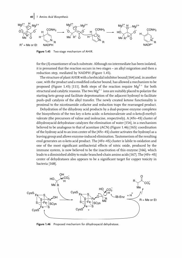
for the (S)-enantiomer of each substrate. Althoughno intermediate has been isolated,it is presumed that the reaction occurs in two stages – an alkyl migration and then areduction step, mediated by NADPH (Figure 1.45).
The structure of plantAHIRwith a herbicidal inhibitor bound [164] and, in anothercase, with the product and amodified cofactor bound, has allowed amechanism to beproposed (Figure 1.45) [111]. Both steps of the reaction require Mg2þ for bothstructural and catalytic reasons. The twoMg2þ ions are suitably placed to polarize thestarting keto group and facilitate deprotonation of the adjacent hydroxyl to facilitatepush–pull catalysis of the alkyl transfer. The newly created ketone functionality isproximal to the nicotinamide cofactor and reduction traps the rearranged product.
Dehydration of the dihydroxy acid products by a dual-purpose enzyme completesthe biosynthesis of the two key a-keto acids: a-ketoisovalerate and a-keto-b-methyl-valerate (the precursors of valine and isoleucine, respectively). A [4Fe–4S] cluster ofdihydroxyacid dehydratase catalyzes the elimination of water [154], in a mechanismbelieved to be analogous to that of aconitase (ACN) (Figure 1.46) [165]: coordinationof the hydroxy acid to an iron center of the [4Fe–4S] cluster activates the hydroxyl as aleaving group and allows enzyme-induced elimination. Tautomerismof the resultingenol generates an a-keto acid product. The [4Fe–4S] cluster is labile to oxidation andone of the most significant antibacterial effects of nitric oxide, produced by theimmune system, is now believed to be the inactivation of this enzyme [166], whichleads to a diminished ability tomake branched-chain amino acids [167]. The [4Fe–4S]center of dehydratases also appears to be a significant target for copper toxicity inbacteria [168].
O
Me
O
R1
HMg2Mg2
O
O
N
CONH2
R
H
H O
Me
OMg2
Mg2
O
O
N
CONH2
R
H
H
R1
HO
Me
HO
Mg2Mg2
O
O
N
CONH2
R
H
R1
NADPH NADP+
H
R1 = Me or Et
Figure 1.45 Two-stage mechanism of AHIR.
HO
Me
OH
HR
Fe
CO2
S Fe
Fe S
Me
HOH
R
CO2
B-Enz + H
O
CO2
Me
R
SOH
SCys
CysS
CysS
Fe
S FeS Fe
Fe SS
O
SCys
CysS
CysS
Fe
S
R = Me or Et
Figure 1.46 Proposed mechanism for dihydroxyacid dehydratase.
40j 1 Amino Acid Biosynthesis

Glutamate acts as amino donor in the transamination of a-ketoisovalerateand a-keto-b-methylvalerate catalyzed by the branched-chain aminotransferase(Figure 1.47). This produces valine and isoleucine, respectively. In addition tothis fate, a-ketoisovalerate is also homologated to a-ketoisocaproate en route toleucine.
1.7.3Homologation of a-Keto Acids, and the Biosynthesis of Leucine and a-AminoadipicAcid
The aliphatic amino acids described so far provide a wide range of chemical diversityfor proteins. However, there are two significant omissions that are rectified in theuniversal genetic code. The nonpolar amino acids, valine and isoleucine, arebranched at the b-position, which causes steric congestion when multiply incorpo-rated within a-helical structures. Homologation of valine to leucine relieves thissteric crowding by placing the branching at the c-center of the amino acid, thusincreasing the opportunity for forming stable hydrophobic cores in a-helical pro-teins. The other omission is the lack of an amino acid with a side-chain aminosubstituent. Ornithine is not incorporated into proteins, possibly because the aminoside-chain can cleave an adjacent peptide link via lactamization to form a six-membered ring. The only amine side-chain among the protein amino acids is thatof lysine. This is a homologated version of ornithine that would produce a lessfavorable seven-membered ring species on lactamization. Access to a homologationpathway for a-keto acids is used to address both these issues.
In the TCA cycle, a-KG is synthesized from oxaloacetate by a three enzymehomologation process. Citrate synthase (CS) condenses oxaloacetatewith acetyl-CoA.Hydrolysis of the thioester adduct ensures that the condensation reaction is favor-able. Hydroxylmigration occurs via a dehydration-rehydration sequence catalyzed byan iron–sulfur-dependent enzyme, ACN, which is named in honor of the interme-diate alkene, aconitate. Isocitrate dehydrogenase (IDH) catalyses the oxidation ofisocitrate to a b-keto acid that spontaneously decarboxylates, in the presence of anenzymic divalent cation, to produce a-KG. Gene duplication and functional diver-gence has generated variants of this three-step process that are used to homologatethe other two common a-keto acids of primarymetabolism (pyruvate and a-KG) anda range of other a-keto acids including a-ketobutyrate (the biosynthetic precursor to
CO2O CO2H3NPLP
Glu α-KG
R2 R2R1 R1
valine: R1 = R2 = Meisoleucine: R1 = Me; R2 = Etleucine: R1 = H; R2 = CHMe2
HH
Figure 1.47 Transamination to form branched-chain amino acids.
1.7 Biosynthesis of Aliphatic Amino Acids with Modified Carbon Skeletons j41

valine) (Figure 1.48) [163]. Transamination of a-ketoisovalerate and of a-ketoadipicacid makes leucine and a-aminoadipic acid, respectively [169], while the homolo-gation of pyruvate to a-ketobutyrate provides an alternative route to the startingmaterial for leucine biosynthesis in some Archaea and bacteria. Many of theseenzymes have not been studied comprehensively and mechanistic proposals are
CO2CO2CO2
O CO2
MeMe
O CO2
CO2
O CO2
CO2
HO CO2
MeMe
HO CO2
CO2
HO CO2
CO2
CH3COSCoA CH3COSCoA CH3COSCoACS
CO2
O
Me
CO2
HO
Me
CO2
CH3COSCoAHCS IPMS CMS
+ H– 2OACN HACN
CO2
MeMe
CO2
CO2
CO2
CO2
CO2 CO2 CO2 CO2
Me
CO2
HO HO HO HO
CO2
MeMe
CO2
CO2
CO2
CO2
CO2 CO2 CO2 CO2
Me
CO2
O O O O
NAD(P)+
NAD(P)H
NAD(P)+
NAD(P)H
NAD(P)+
NAD(P)H
NAD(P)+
NAD(P)H
IPMI
IDH HIDH
+ H2 + HO 2 + HO – 2O
- CO2 - CO2 - CO2 - CO2
CH2
MeMe
CH2
CO2
CH2
CO2
CO2 CO2 CO2 CO2
CH2
Me
O O O O
CH2
MeMe
CH2
CO2
CH2
CO2
CO2 CO2 CO2H3N H3N H3N
glutamate α-aminoadipate leucine
IPMI
Glu
α-KG
Glu
α-KG
oxaloacetate α-KG α-ketoisovalerate pyruvate
citrate homocitrate isopropylmalate citramalate
α-KG α-ketoadipate α-ketoisocaproate α-ketobutyrate
Me
CO2H3N
isoleucine
Me
IPMDH IPMDH
Figure 1.48 Homologation of a-keto acids to produce amino acids.
42j 1 Amino Acid Biosynthesis

often drawn by analogy from the action of homologous enzymes that have beenstudied in more detail.
Homocitrate synthase (HCS) [170], isopropylmalate synthase (IPMS) [171], andcitramalate synthase (CMS) [172] are homologous enzymes that catalyze the Claisencondensation of acetyl-CoA with a-KG, a-ketoisovalerate, and pyruvate, respectively.They are alsohomologous to oneof the classes ofCS found in anaerobic bacteria [173].Hydrolysis of the initially formed thioester adduct makes the otherwise reversibleClaisen condensation, favorable. The structure of IPMS has been determined [174]and that structure was used to shed light on the catalytic machinery of theseenzymes [175]. Carbonyl groups of both substrates are polarized by the enzyme:acetyl-CoA by an arginine to facilitate enol formation and a-KG by chelation to anessential Zn2þ . On the basis of modeling, kinetic studies and site-directed muta-genesis, the general base for deprotonation of acetyl-CoA is believed to be a histidine,acting in concert with a glutamate as a catalytic dyad.
A variety of homologous isomerases catalyze the next step in the homologationprocess. These include homoaconitase (HACN) and isopropylmalate isomerase(IPMI), and are all members of the ACN superfamily [176]. As an example of therelatedness of these enzymes, in P. horikoshii a single enzyme, with dual substratespecificity, acts as both an IPMI and a HACN [177]. These enzymes are not fullycharacterized, but it is assumed that they exploit the dehydration–rehydrationchemistry of a [4Fe–4S] cluster that has been characterized inACNandwas describedfor dihydroxyacid dehydratases above [165 (Figure 1.4–6)].
The final step in the homologation process is the oxidative decarboxylation of theb-hydroxy acids. In this step an oxidized nicotinamide cofactor (NAD(P)þ ) accepts ahydride from the secondary alcohol center and generates a b-keto acid. Thisintermediate spontaneously decarboxylates, in the presence of an enzyme-bounddivalent metal ion, to leave the homologated a-keto acids. This step is catalyzed by asuperfamily of homologous enzymes, ICDH [178], isopropylmalate dehydrogenase(IPMDH) [179], and homoisocitrate dehydrogenase (HIDH) [180]. Dynamic X-raycrystallography has provided detailed structural information about the catalyticmechanism of ICDH which is the best characterized enzyme of the family [181].Again, in some organisms dual substrate enzymes act in parallel pathways; forexample, in both Thermus thermophilus [182] and P. horikoshii [183] a single enzymecatalyses two oxidative decarboxylation reactions producing both a-KG and a-ketoa-dipic acid. A loop at the active site of these enzymes appears to control specificity andmutations within this loop canmodify the substrate specificity to change the pathwaythat an enzyme acts on. The conversion of E. coli ICDH to a bifunctional ICDH/IPMDHbymutagenesis has been reported by Koshland et al. Ser113 in the substraterecognition loop forms a hydrogen bond to thed-carboxylate of isocitrate.Mutation ofthis residue to glutamate, which introduces electrostatic repulsion to the normalsubstrate, together with systematic mutation of two other local residues, Asp115 andVal116, produced severalmutants with a preference for isopropylmalate as substrate.In T. thermophilus HICDH, Arg85 of the corresponding loop is involved in recog-nition of the d/c-carboxylate of isocitrate/homoisocitrate. When this residue ismutated to a large nonpolar residue, valine, the enzyme is converted to an IPMDH,
1.7 Biosynthesis of Aliphatic Amino Acids with Modified Carbon Skeletons j43
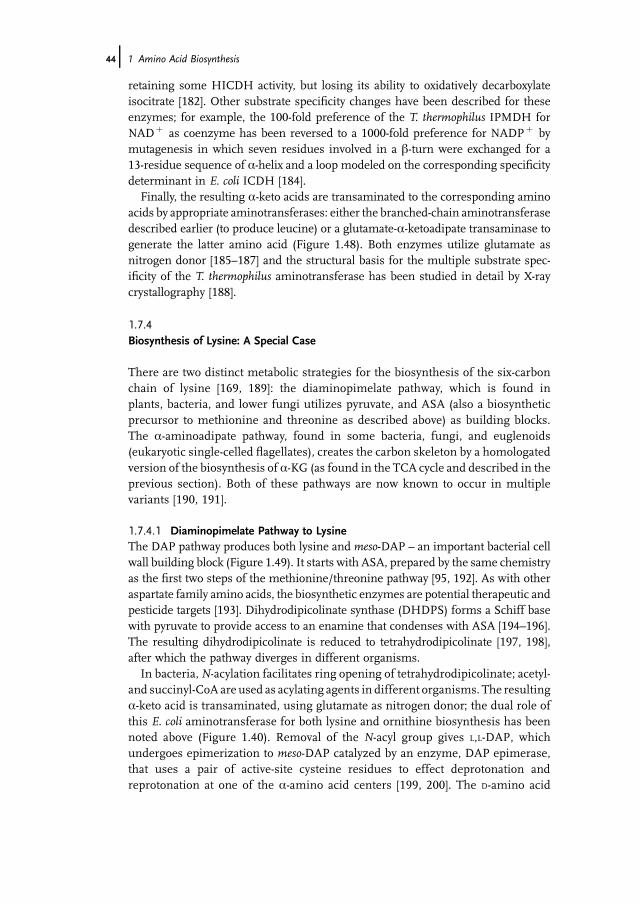
retaining some HICDH activity, but losing its ability to oxidatively decarboxylateisocitrate [182]. Other substrate specificity changes have been described for theseenzymes; for example, the 100-fold preference of the T. thermophilus IPMDH forNADþ as coenzyme has been reversed to a 1000-fold preference for NADPþ bymutagenesis in which seven residues involved in a b-turn were exchanged for a13-residue sequence of a-helix and a loop modeled on the corresponding specificitydeterminant in E. coli ICDH [184].
Finally, the resulting a-keto acids are transaminated to the corresponding aminoacids by appropriate aminotransferases: either the branched-chain aminotransferasedescribed earlier (to produce leucine) or a glutamate-a-ketoadipate transaminase togenerate the latter amino acid (Figure 1.48). Both enzymes utilize glutamate asnitrogen donor [185–187] and the structural basis for the multiple substrate spec-ificity of the T. thermophilus aminotransferase has been studied in detail by X-raycrystallography [188].
1.7.4Biosynthesis of Lysine: A Special Case
There are two distinct metabolic strategies for the biosynthesis of the six-carbonchain of lysine [169, 189]: the diaminopimelate pathway, which is found inplants, bacteria, and lower fungi utilizes pyruvate, and ASA (also a biosyntheticprecursor to methionine and threonine as described above) as building blocks.The a-aminoadipate pathway, found in some bacteria, fungi, and euglenoids(eukaryotic single-celled flagellates), creates the carbon skeleton by a homologatedversion of the biosynthesis of a-KG (as found in the TCA cycle and described in theprevious section). Both of these pathways are now known to occur in multiplevariants [190, 191].
1.7.4.1 Diaminopimelate Pathway to LysineThe DAP pathway produces both lysine and meso-DAP – an important bacterial cellwall building block (Figure 1.49). It starts with ASA, prepared by the same chemistryas the first two steps of the methionine/threonine pathway [95, 192]. As with otheraspartate family amino acids, the biosynthetic enzymes are potential therapeutic andpesticide targets [193]. Dihydrodipicolinate synthase (DHDPS) forms a Schiff basewith pyruvate to provide access to an enamine that condenses with ASA [194–196].The resulting dihydrodipicolinate is reduced to tetrahydrodipicolinate [197, 198],after which the pathway diverges in different organisms.
In bacteria, N-acylation facilitates ring opening of tetrahydrodipicolinate; acetyl-and succinyl-CoA are used as acylating agents in different organisms. The resultinga-keto acid is transaminated, using glutamate as nitrogen donor; the dual role ofthis E. coli aminotransferase for both lysine and ornithine biosynthesis has beennoted above (Figure 1.40). Removal of the N-acyl group gives L,L-DAP, whichundergoes epimerization to meso-DAP catalyzed by an enzyme, DAP epimerase,that uses a pair of active-site cysteine residues to effect deprotonation andreprotonation at one of the a-amino acid centers [199, 200]. The D-amino acid
44j 1 Amino Acid Biosynthesis
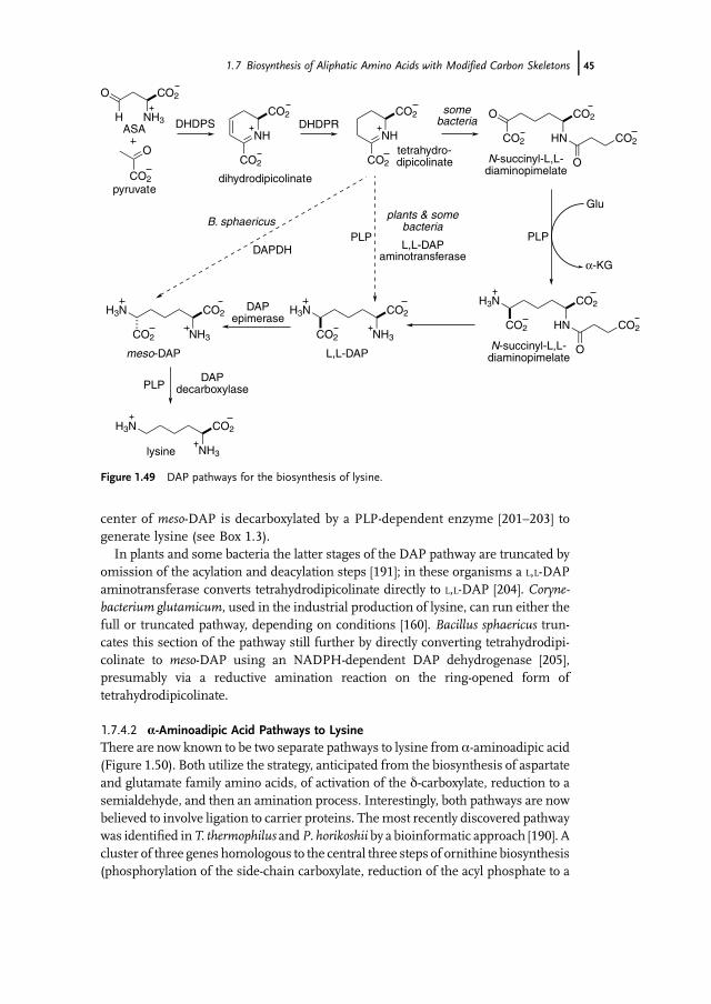
center of meso-DAP is decarboxylated by a PLP-dependent enzyme [201–203] togenerate lysine (see Box 1.3).
In plants and some bacteria the latter stages of the DAP pathway are truncated byomission of the acylation and deacylation steps [191]; in these organisms a L,L-DAPaminotransferase converts tetrahydrodipicolinate directly to L,L-DAP [204]. Coryne-bacterium glutamicum, used in the industrial production of lysine, can run either thefull or truncated pathway, depending on conditions [160]. Bacillus sphaericus trun-cates this section of the pathway still further by directly converting tetrahydrodipi-colinate to meso-DAP using an NADPH-dependent DAP dehydrogenase [205],presumably via a reductive amination reaction on the ring-opened form oftetrahydrodipicolinate.
1.7.4.2 a-Aminoadipic Acid Pathways to LysineThere are now known to be two separate pathways to lysine from a-aminoadipic acid(Figure 1.50). Both utilize the strategy, anticipated from the biosynthesis of aspartateand glutamate family amino acids, of activation of the d-carboxylate, reduction to asemialdehyde, and then an amination process. Interestingly, both pathways are nowbelieved to involve ligation to carrier proteins. Themost recently discovered pathwaywas identified inT. thermophilus andP. horikoshii by a bioinformatic approach [190]. Acluster of three genes homologous to the central three steps of ornithine biosynthesis(phosphorylation of the side-chain carboxylate, reduction of the acyl phosphate to a
H3N CO2
NH3
H3N CO2
NH3CO2
H3N CO2
NH3CO2
O CO2
HNCO2
O
CO2NH
CO2
CO2
NH
CO2
CO2
O
NH3
CO2
O
CO2
H
pyruvate
ASA
dihydrodipicolinate
tetrahydro-dipicolinate N-succinyl-L,L-
diaminopimelate
L,L-DAPmeso-DAP
lysine
DHDPS DHDPR
DAP decarboxylase
DAP epimerase
H3N CO2
HNCO2
O
CO2
N-succinyl-L,L-diaminopimelate
Glu
α-KG
PLP
PLP
PLP
plants & somebacteria
somebacteria
B. sphaericus
L,L-DAPaminotransferaseDAPDH
Figure 1.49 DAP pathways for the biosynthesis of lysine.
1.7 Biosynthesis of Aliphatic Amino Acids with Modified Carbon Skeletons j45

semialdehyde, and transamination to the amine) were identified in T. thermophilus.Genes for the first and last steps of arginine biosynthesis (for the protecting groupchemistry of N-acylation and deacylation) were absent. Disruption of these putativearginine biosynthetic genes led to organisms auxotrophic for lysine. This demon-strated a biosynthetic route to lysine that is analogous to the biosynthesis of ornithinefrom glutamate [206]. In reconstituting the a-aminoadipic acid pathway from T.thermophilus it was discovered that an alternativeN-acylation pathway operates in thisbacterium. In the first step of the pathway the amino group of a-aminoadipic acid is
NAD(P)H
NAD(P)+
CO2H3N
CO2H3N
CO2H3N
- AMP
ATP
PPi
Glu
α-KG
CO2H3N
PLP
O2C
OHC
H3N
O
OAMP
CO2H3N
O
SEnz
CO2LysWCONH
O2C
LysWCO2
CO2
NH3
CO2
CO2H3N
CO2
CO2
NH2
CO2
O
CO2
CO2LysWCONH
O
OP
O
O
O
NAD(P)H
NAD(P)+
- HOPO32
ATP
ADP
CO2LysWCONH
O
H
CO2LysWCONH
H3NNADPH
NADP+
lysine
α-aminoadipicacid
α-KG
saccharopine
α-AAA reductase
α-AAA reductase
α-AAA reductase
NAD+ NADH
Figure 1.50 Biosynthetic routes to lysine from a-aminoadipic acid.
46j 1 Amino Acid Biosynthesis

protected by acylation by the side-chain carboxylate of the C-terminal glutamate of asmall protein, LysW. The functional groupmodification of the a-aminoadipic acid ismediated on the LysW adduct which acts as a carrier protein for the biosynthesis.Following completion of the side-chain manipulation, the LysW adduct is cleaved torelease lysine [207].
Although the discovery of a bacterial route to lysine from a-aminoadipic acid is arecent discovery, this amino acid has been known for some time to have twometabolic roles in fungi – both as an alternative precursor to lysine [169], and asa key precursor in the biosynthesis of penicillin and cephalosporin antibiotics [208].For penicillin biosynthesis the d-carboxylate of a-aminoadipic acid is incorporatedinto a tripeptide by a nonribosomal peptide synthetase, d-(a-L-aminoadipyl)-L-cystei-nyl-D-valine synthetase [209]. The peptide bonds, including that to the side-chain ofa-aminoadipic acid, are made by ligating the carboxyl group of each component to amultifunctional enzyme via thioester links to 40-phosphopantetheine groups whichthen undergo successive nucleophilic substitution reactions with the amine of theadjacent component [210].
Walsh et al. have proposed that the activation and reduction of the d-carboxylate ofa-aminoadipic acid occurs in an analogous fashion to nonribosomal peptide syn-thesis (Figure 1.50). a-Aminoadipate reductase comprises two proteins – Lys2 andLys5. Lys2 is a large (155 kDa) multidomain protein. Sequence homologies withnonribosomal peptide synthetases suggest that residues 225–808 constitute a 60-kDaadenylation domain (A) that activates a-aminoadipic acid as the aminoacyl-d-AMPderivative [211]. Hydrolysis of the pyrophosphate side-product makes the reactionfavorable. Lys5 primes the PCP domain (residues 809–924) of Lys2 by adding a 40-phosphopantatheine unit to Ser880. The aminoacyl-d-AMPderivative is ligated to the40-phosphopantatheine unit as a thioester. NADPH-dependent reduction to a-ami-noadipic acid-d-semialdehyde, mediated by the reductase domain (residues925–1392), cleaves the link to the carrier protein.
The biosynthesis of lysine in this pathway is completed by an unusual two-steptransamination process that does not involve a PLP-dependent enzyme. Typicaltransamination chemistry involves the reductive amination of PLP to PMP byglutamate and subsequent reductive amination of the target carbonyl compoundby PMP. In this case, however, a saccharopine dehydrogenase reductively aminatesa-aminoadipic acid-d-semialdehyde with glutamate directly to produce saccharo-pine [212–214]. A second saccharopine dehydrogenase, structurally related to alaninedehydrogenase, mediates the NADþ -dependent oxidative cleavage of a-KG from theadduct to leave lysine [215]. Kinetic and structural studies have led to the proposal fora proton relay mechanism for this final step of the pathway [216]. NADþ abstracts ahydride to generate an imine that is converted to a carbinolamine by base-catalyzedaddition of water. Further acid–base chemistry leads to fragmentation of thecarbinolamine and formation of the two products (Figure 1.51).
1.7.4.3 PyrrolysinePyrrolysine is an uncommon amino acid found in someArchaea and eubacteria; only1% of genomes thus far sequenced contain evidence for its presence. It was first
1.7 Biosynthesis of Aliphatic Amino Acids with Modified Carbon Skeletons j47

observed as an active-site residue in the crystal structure of monomethylaminemethyltransferase from a methanogen,Methanosarcina barkeri [217], and it has nowbeen found in other methylamine methyltransferases [218] associated with themetabolism of methylamines to methane. It is believed that the electrophilic imineof pyrrolysine is the functional group that activates methylamines for transfer of amethyl group to an adjacent cobalt corrin center [219].
In contrast to selenocysteine, this amino acid addition to the genetic code isbiosynthesized from the free amino acid precursor, lysine, and then incorporated intoan amber suppressor tRNA by a specific pyrrolysyl-tRNA synthetase [220–223]. Thebiosynthetic pathway to pyrrolysine is as yet undetermined, however a gene clusterfromMethanosarcina acetivorans pylBCD encodes for the biosynthesis of this aminoacid and recombinant E. coli containing these genes, together with the pylTS genesthat encode tRNAPyl and pyrrolysyl-tRNA synthetase, respectively, biosynthesizepyrrolysine and incorporate it into reading frames containing the amber codon [224].The pylBCD genes show homology to genes that encode radical (S)-adenosylmethio-nine-dependent proteins, proteins forming amides and amino acid dehydrogenases,respectively; this combination of catalytic functionalities has led to the proposal of apossible biosynthetic route (Figure 1.52) [219]: manipulation of (3R)-3-methyl-D-glutamate, by analogy with the chemical strategy of the first stages of prolinebiosynthesis (reduction to the c-aldehyde and spontaneous cyclization), could
CO2H3N
CO2
CO2
NH
H
NR CONH2
CO2H3N
CO2
CO2
NH
NR CONH2
H
OH B Enz
CO2H3N
CO2
CO2
NH2
HOCO2
O
CO2
CO2H3N
H3N
lysine
Figure 1.51 Formation of lysine by saccharopine dehydrogenase (lysine forming).
CO2H3N
O2C
CO2H3N
OHC
NH CO2
NH
O
HN CO2
NH3
H3N CO2
NH3
3R-3-methyl-D-glutamate pyrrolysine
lysine
Figure 1.52 Possible biosynthetic route to pyrrolysine.
48j 1 Amino Acid Biosynthesis

generate the required imino acid which could then be activated and used to acylatelysine to form pyrrolysine.
1.8Biosynthesis of the Aromatic Amino Acids
1.8.1Shikimate Pathway
The biosynthesis of the aromatic amino acids tryptophan, phenylalanine, andtyrosine begins with the shikimate pathway (Figure 1.53). This pathway consists ofseven enzyme-catalyzed reactions, which convert erythrose 4-phosphate (E4P) andphosphoenol pyruvate (PEP), both originally derived from glucose, into the prearo-matic compound chorismate [225, 226]. From chorismate, the pathway branches toproduce tryptophan, or phenylalanine and tyrosine. Chorismate is also the precursorfor other important aromatic compounds.
The first step of the shikimate pathway is catalyzed by 3-deoxy-D-arabino-heptu-losonate-phosphate (DAH7P) synthase. Feedback regulation of this enzyme by
H
O
OH
OH2–O3PO
CO2–2–O3PO
CO2–
OHO CO2
–
CO2–
NH2
NH
NH2
CO2–
CO2–
NH2
OH
CO2–HO
O
OHOH
2–O3PO
CO2–
2–O3POOH
O CO2–
CO2–
2–O3POOH
OH
HO
OH
CO2–
OHO
ADP ATP
CO2–
HOOH
OH
CO2–
OHOHO
NADP+
NADPH
PEP
synthasechorismate synthase
EPSP kinaseshikimate
dehydrogenaseshikimate
dehydroquinase
synthase3-dehydroquinateDAH7P synthase
shikimate
PEP
E4PDAH7P DHQ DHS
chorismate EPSP shikimate 3-phosphate
Figure 1.53 Shikimate pathway.
1.8 Biosynthesis of the Aromatic Amino Acids j49

aromatic amino acids is an important mechanism for control of flux through theshikimate pathway. DAH7P synthase catalyses an aldol-like reaction in which thethree-carbon unit PEP condenses with the four-carbon aldehyde, E4P. It has beenshown that the C–Obond of PEP is cleaved during the reaction, requiringwater to actas a nucleophile on C2 of PEP, generating a phosphorylated hemiacetal intermediate(Figure 1.54). This intermediate then loses phosphate to produce the linear form ofDAH7P. All DAH7P synthases have been shown to require a divalent metal ion foractivity [227]. It is likely that the metal ion plays a role in activating the aldehydiccarbon to nucleophilic attack (Figure 1.54) [228].
Directed evolution experiments have been used to replace DAH7P synthase with apyruvate-utilizing aldolase, 2-keto-3-deoxy-6-phosphogalactonate (KDPGal) synthase(Figure 1.55) [229, 230]. Frost et al. showed that both the E. coli enzyme and that fromKlebsiella pneumoniae could support biosynthesis of aromaticmetabolites in a cell linelacking all isozymes of DAH7P synthase. Gene shuffling and modifications of the
2–O3PO
OOH
HO
OP
O
-OO- H
OH
2–O3PO
OHHO
CO2–
O
HO
PO
-O O- HO
2–O3PO
OHHO
CO2–
HO
O
CO2–
2+M
2–O3PO
OHHO
CO2–
O
HO
PO
-O O-
DAH7P
Figure 1.54 Proposed mechanism for DAH7P synthase.
H
O
OH
OH2–O3PO
CO2–2–O3PO
CO2–HO
O
OHOH
2–O3PO
DAH7P synthase
PEP
DAH7P
O
CO2–2–O3PO H
O
OH
O
HO CO2–
OH2–O3PO
KDPGal synthase
Figure 1.55 Directed evolution of the pyruvate-utilizing aldolase KDPGal synthase to replaceDAH7P synthase.
50j 1 Amino Acid Biosynthesis

E. coli enzyme by site directed mutagenesis produced mutant forms of the aldolasethat exhibited a 60-fold increase of kcat/Km over wild-type activity.
One of the most interesting aspects of DAH7P synthase is the variation instructures and regulation patterns that have been observed for this enzyme [231].Structures of DAH7P synthases have been determined from a variety of sourcesincluding E. coli, Pyrococcus furiosus, S. cerevisiae, Thermotoga maritima, and M.tuberculosis (Figure 1.56) [231–234]. Despite large primary sequence variation, thecore catalytic unit for each of these proteins is a standard (ba)8 barrel. Variableaccessory domains have been shown to augment this barrel structure and differentquaternary structures are observed for enzymes from different species. The variableextra-barrel elements are associated with feedback regulation. E. coli expresses threeDAH7P synthase isozymes each with a binding site for a single aromatic amino acid.On the other hand, the M. tuberculosis genome encodes a single DAH7P synthase.This enzyme shows synergistic inhibition by a combination of tryptophan andphenylalanine, with both amino acids binding simultaneously to independentallosteric sites formed extra-barrel subdomains.
3-Dehydroquinate (DHQ) synthase, which catalyzes the second step of theshikimate pathway, is responsible for production of the first carbocycle of thepathway. This enzyme has attracted a great deal of attention due to the number of
Figure 1.56 Different forms of DAH7Psynthase showing basic catalytic barrel in blue,and N-terminal and loop extensions in red andyellow, respectively, from (a) E. coli type Ia (Phe
sensitive), (b) P. furiosus type Ib (unregulated),(c) T. maritima Ib (Phe, Tyr inhibited) [235], and(d) M. tuberculosis (Phe þ Tyr inhibited) [231].
1.8 Biosynthesis of the Aromatic Amino Acids j51

different chemical steps catalyzed by a single enzyme (Figure 1.57) [236–238]. DHQsynthase uses the pyranose form of DAH7P. In the first step it undergoes temporaryoxidation at C5 to facilitate syn elimination by an E1cbmechanism to produce an enolpyranose intermediate. The C5 keto group of the product is then reduced. Theseredox steps use NADþ/NADH as a cofactor to accept and deliver the hydride.Following reduction, the enol pyranose intermediate ring opens and cyclizes via anintramolecular aldol reaction. There has been some debate about the role of theenzyme in thesefinal two steps, as the enol pyranose intermediate, released in situ viaphotolysis of an o-nitrobenzyl protected form, was able to form dehydroquinatewithout assistance from the enzyme (Figure 1.58). However, as some epimer isproduced in the nonenzymatic reaction (produced by the enol attacking the wrong
CO2–HO
O
OHOH
2–O3PO
HO
OH
CO2–
OHO
CO2–HO
O
OOH
2–O3PO
CO2–HO
O
OOH
CO2–O
O
OHOH
CO2–
O
–OOH
OH
NAD+ NADH
HPO42-
NAD+ NADH
DAH7P
DHQ
H
Figure 1.57 Reaction mechanism for dehydroquinate synthase.
CO2–O
O
OHOH
NO2
HO
OH
CO2–
OHO
DHQ
CO2–
OH
OH
OHO
1-epiDHQ (1-4%)
hυ
Figure 1.58 Nonenzymatic generation of DHQ.
52j 1 Amino Acid Biosynthesis

face of the carbonyl), the enzyme appears to be required at least as a template toensure the correct stereochemistry of the final product dehydroquinate is generated(Figure 1.58). DHQ synthase is one of a handful of enzymes that use transientoxidation to achieve its overall chemistry [239].
1.8.2Case Study: Alternative Synthesis of Dehydroquinate in Archaea
Euryarchaea have a different strategy for the biosynthesis of dehydroquinate(Figure 1.59) [240, 241]. Recent work on Methanocaldococcus jannaschii has shownthat dehydroquinate production occurs through two alternative enzyme-catalyzed
CO2–
OPO32–
2–O3POH
O
OH
OH
HO
–O2C
+H3N
aspartatesemialdehyde
2–O3PO
O
O
HOOH
6-deoxy-5-ketofructose 1-phosphate
PEP
E4P
CO2–HO
O
OHOH
2–O3PO
HO
OH
CO2–
OHO
–O2C
+H3N
OHO
OH
2–O3PO H
O
O
DAH7PPi
NAD+ NADH, NH4+
Pi
ADH
DHQ
alternativepathway
standardpathway
–O2C
OHO
OHO
Figure 1.59 Alternative routes for aromatic amino acid biosynthesis. Dehydroquinate synthase IIcatalyzes the oxidative deamination of ADH and its cyclization to form DHQ, which then feeds intothe standard shikimate pathway.
1.8 Biosynthesis of the Aromatic Amino Acids j53

steps. Thefirst of these is catalyzed by a lysine-dependent, Schiff base-utilizing, type Ialdolase. This enzyme, 2-amino-3,7-dideoxy-D-threo-hept-6-ulosonic acid (ADH)synthase, catalyses a transaldolase reaction between 6-deoxy-5-ketofructose 1-phos-phate and ASA (a biosynthetic precursor to threonine, methionine, and lysine). Thestructure of this enzyme has revealed the likely active site residues and allowed themechanism of this reaction to be predicted [242].
Following the production of ADH, the DHQ is produced in an oxidative deam-ination reaction that requiresNADþ . In contrast toDHQsynthasewhere oxidation istransient, an equivalent of NADþ is required in this transformation. Analogous tothe DHQ synthase reaction that takes place as part of the canonical shikimatepathway, stereospecific intramolecular cyclization is required to produce DHQ. Thediscovery of this alternate route to DHQ in these organisms highlights how differentstrategies can be employed and different chemistries can evolve to achieve the sameoverall solution.
The third step in the pathway towards chorismate is catalyzed by dehydroquinase.There are two distinct types of dehydroquinase that operate in shikimate pathwaymetabolism that have evolved convergently and utilize different mechanisms for thedehydration reaction [243].Whereas the type I enzyme catalyzes a reactionwith overallsyn elimination, the type II enzymes catalyze anti elimination inwhich themore acidichydrogen is lost (Figure 1.60) [244]. Type I enzymes proceed via a Schiff basemechanism [245, 246]. The covalent attachment of substrate to the enzymes enablesconformational change promoting the syn elimination to occur by increasing theacidity of the pro-(S) hydrogen. The reaction catalyzed by the type II enzyme proceedsvia a standard E1cbmechanism,with the loss of themore acidic pro-(R) hydrogen. TypeII enzymes operate in the shikimate pathway of some significant human pathogensand there has been considerable attention on the development of inhibitors. Severalinhibitors with nanomolar inhibition constants have been described [247].
The fourth step of the shikimate pathway is the NADPH-mediated reduction ofdehydroshikimate catalyzed by shikimate dehydrogenase [248, 249]. Structures ofseveral shikimate dehydrogenases have been determinedwith the ternary shikimate/NADPH/enzyme complex of the shikimate dehydrogenase from Staphylococcusepidermidis revealing clearly the interaction of substrates with the enzyme, and
HO
OH
CO2–
OHO
CO2–
OH
OHO
HR
HS
CO2–
OH
OHO
HSHR
type Idehydroquinase
type IIdehydroquinase
Figure 1.60 Opposite stereochemical pathways catalyzed by the two types of dehydroquinase.
54j 1 Amino Acid Biosynthesis
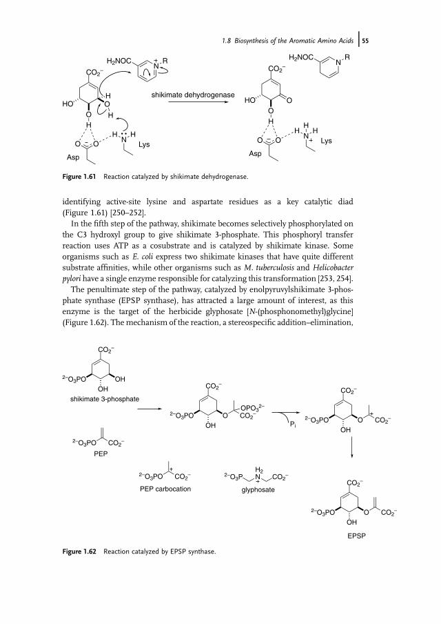
identifying active-site lysine and aspartate residues as a key catalytic diad(Figure 1.61) [250–252].
In the fifth step of the pathway, shikimate becomes selectively phosphorylated onthe C3 hydroxyl group to give shikimate 3-phosphate. This phosphoryl transferreaction uses ATP as a cosubstrate and is catalyzed by shikimate kinase. Someorganisms such as E. coli express two shikimate kinases that have quite differentsubstrate affinities, while other organisms such as M. tuberculosis and Helicobacterpylori have a single enzyme responsible for catalyzing this transformation [253, 254].
The penultimate step of the pathway, catalyzed by enolpyruvylshikimate 3-phos-phate synthase (EPSP synthase), has attracted a large amount of interest, as thisenzyme is the target of the herbicide glyphosate [N-(phosphonomethyl)glycine](Figure 1.62). Themechanism of the reaction, a stereospecific addition–elimination,
CO2–
OO
HO
H
O O
Asp
HN
H
H
H
NRH2NOC
Lys
CO2–
OO
HO
H
O O
Asp
HN
H
NRH2NOC
Lys
H
shikimate dehydrogenase
Figure 1.61 Reaction catalyzed by shikimate dehydrogenase.
CO2–
2–O3POOH
COO 2–
CO2–
2–O3POOH
OHCO2
–
2–O3POOH
COO 2–
OPO32–
CO2–
2–O3POOH
COO 2–
shikimate 3-phosphate
2–O3PO CO2–
PEP
Pi
2–O3PO CO2– 2–O3P
H2N CO2
–
PEP carbocation glyphosate
EPSP
Figure 1.62 Reaction catalyzed by EPSP synthase.
1.8 Biosynthesis of the Aromatic Amino Acids j55

has been well studied [255, 256]. The active site is found in a cleft between twodomains and during the catalytic cycle there is extensive domain movement(Figure 1.63). Glyphosate inhibits by mimicking the cationic transition state asso-ciated with the protonated PEP substrate (Figure 1.64) [257]. Transition state analogsthat include both PEP and shikimate 3-phosphate functionality are particularly
Figure 1.63 EPSP synthase in open (unliganded, PDB ID: 1EPS) and closed forms (liganded).(From [257].)
Figure 1.64 Interactions of the glyphosate and shikimate 3-phosphate with EPSP synthase.(From [257].)
56j 1 Amino Acid Biosynthesis
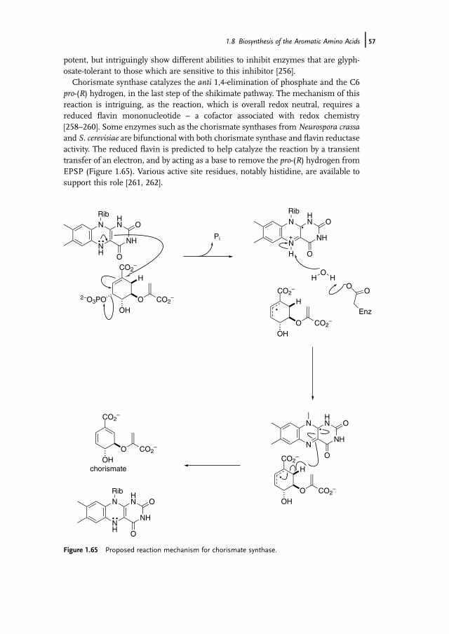
potent, but intriguingly show different abilities to inhibit enzymes that are glyph-osate-tolerant to those which are sensitive to this inhibitor [256].
Chorismate synthase catalyzes the anti 1,4-elimination of phosphate and the C6pro-(R) hydrogen, in the last step of the shikimate pathway. The mechanism of thisreaction is intriguing, as the reaction, which is overall redox neutral, requires areduced flavin mononucleotide – a cofactor associated with redox chemistry[258–260]. Some enzymes such as the chorismate synthases from Neurospora crassaand S. cerevisiae are bifunctional with both chorismate synthase and flavin reductaseactivity. The reduced flavin is predicted to help catalyze the reaction by a transienttransfer of an electron, and by acting as a base to remove the pro-(R) hydrogen fromEPSP (Figure 1.65). Various active site residues, notably histidine, are available tosupport this role [261, 262].
NH
N
NH
HN
RibO
OCO2
–
H
O2–O3POOH
CO2–
N
N
NH
HN
RibO
O
CO2–
H
OOH
CO2–
HO
H–O
O
Enz
N
N
NH
HN O
OCO2–
H
OOH
CO2–
Pi
H
CO2–
O CO2–
OHchorismate
NH
N
NH
HN
RibO
O
Figure 1.65 Proposed reaction mechanism for chorismate synthase.
1.8 Biosynthesis of the Aromatic Amino Acids j57

1.8.3Biosynthesis of Tryptophan, Phenylalanine, and Tyrosine from Chorismate
From chorismate the pathway for the synthesis of aromatic amino acids branches toproduce either anthranilate for tryptophan biosynthesis or prephenate, the precursorto both phenylalanine and tyrosine. Chorismate is also the precursor to a range ofother aromatic metabolites (Figure 1.66).
1.8.3.1 Tryptophan BiosynthesisAnthranilate synthase catalyses the first committed step in the biosynthesis oftryptophan from chorismate [263–265]. This enzyme is one of a group of chorismateutilizing enzymes that are predicted to sharemanymechanistic features and are likelyto have common evolutionary origins (Figure 1.67) [266–268]. The first step is 1,4-nucleophilic substitution by ammonia. This is followedby elimination of pyruvate. Theammonia used in this reaction is generated in situ by a glutamine amidotransferase.
CO2–
OHO CO2
–
OH
–O2C
O
CO2–
CO2–
O CO2–
CO2–
NH3+
O CO2–
CO2–
O CO2–
CO2–
NH3+
O CO2–
CO2–
NH2
CO2–
OH
CO2–
OH
OH
NH3+
prephenate
anthranilate
ADC
isochorismate
p-hydroxybenzoate
salicylate
ADC
ADIC
phenylalaninetyrosine
tryptophan
folate
enterobactin
coenzyme Q
yersiniabactin
pristinamycin I
pyocyanin
chorismate
Figure 1.66 Multiple end-products generated from chorismate.
58j 1 Amino Acid Biosynthesis

The indole ring of tryptophan is created by the next three steps of the pathway(Figure 1.68). A ribose moiety is first tethered to anthranilate, in a reaction catalyzedby phosphoribosyl anthranilate synthase [269, 270]. Following isomerization ofphosphoribosyl anthranilate involving an Amadori rearrangement (Figure 1.69),the indole ring is constructed by intramolecular electrophilic substitution. Thisreaction chemistry is analogous to that observed for histidine biosynthesis, asdescribed below [271, 272].
The final step in tryptophan production is catalyzed by the tryptophan synthasecomplex [273]. This enzyme has attracted a great deal of attention and beenextensively reviewed as the one of the first enzymes to be discovered that catalyzestwo distinct reactions at independent active sites (in the a- and b-domains) that areconnected via an internal tunnel [274]. This was thefirst well-documented example ofsubstrate channeling in amino acid biosynthesis. The reaction chemistry is relativelystraightforward; glyceraldehyde 3-phosphate is eliminated in the a-tryptophansynthase reaction, producing indole, which is channeled to the second active site(Figure 1.70). In the PLP-dependent b-reaction, serine undergoes nucleophilicdisplacement of water by indole via an elimination–addition mechanism typical ofb-substitution reactions of PLP (Box 1.3). The nett result is the generation oftryptophan (Figure 1.68).
1.8.3.2 Phenylalanine and Tyrosine BiosynthesisPrephenate is formed by the reaction catalyzed by chorimate mutase(Figure 1.71) [275]. This is the only known example of a Claisen rearrangement
CO2–
OH
O CO2–
CO2–
OH
O CO2–
CO2–
OH
O CO2–
CO2–
OH
O CO2–
NH3
H2N Lys
H2O CO2–
O CO2–
OH
isochorismate
isochorismatesynthase
aminodeoxychorismatesynthase
anthranilatesynthase
CO2–
O CO2–
NH2
CO2–
O CO2–
HN Lys
CO2–
NH2
CO2–
O CO2–
NH2
NH3
anthranilate
4-amino-4-deoxychorismate
Figure 1.67 Related enzymatic reactions using chorismate as a substrate.
1.8 Biosynthesis of the Aromatic Amino Acids j59
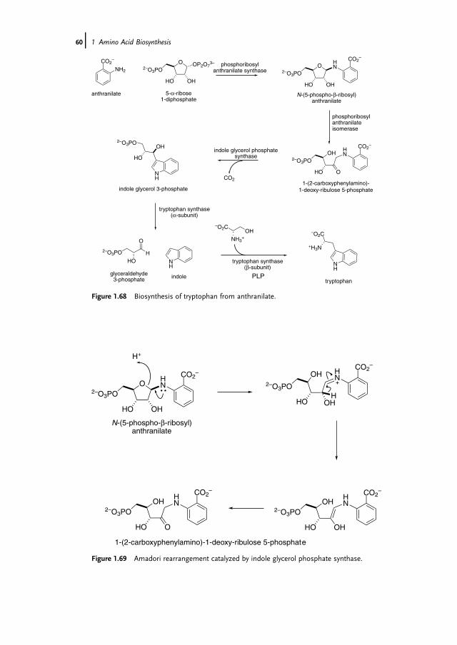
O OP2O73–
OHHO
2–O3PO
CO2–
NH2O
OHHO
2–O3PO
HN
CO2–
OH
OHO
2–O3PO
HN
CO2–
NH
OH
HO
2–O3PO
NH
O
HO
2–O3PO H
NH
–O2C
+H3N
–O2COH
NH3+
tryptophanindoleglyceraldehyde
3-phosphate
indole glycerol 3-phosphate
anthranilate 5-α-ribose 1-diphosphate
N-(5-phospho-β-ribosyl)anthranilate
phosphoribosyl anthranilate synthase
phosphoribosylanthranilateisomerase
indole glycerol phosphate synthase
1-(2-carboxyphenylamino)-1-deoxy-ribulose 5-phosphate
tryptophan synthase(α-subunit)
tryptophan synthase
PLP(β-subunit)
CO2
Figure 1.68 Biosynthesis of tryptophan from anthranilate.
O
OHHO
2–O3PO
HN
CO2–
OH
OHO
2–O3PO
HN
CO2–
N-(5-phospho-β-ribosyl)anthranilate
1-(2-carboxyphenylamino)-1-deoxy-ribulose 5-phosphate
H+
OH
OHHO
2–O3PO
HN
CO2–
H
OH
OHHO
2–O3PO
HN
CO2–
Figure 1.69 Amadori rearrangement catalyzed by indole glycerol phosphate synthase.
60j 1 Amino Acid Biosynthesis

reaction in primary metabolism and one of the few enzyme-catalyzed pericyclicreactions. Chorismate mutase can be effectively inhibited with a transition stateinhibitor, and the activity of the enzyme is often subject to inhibition by tyrosine and/or phenylalanine as part of regulation of the pathway.
From prephenate there are a number of alternative pathways to aromatic meta-bolites (Figure 1.72). Tyrosine is produced by a combination of oxidation andtransamination reactions, whereas phenylalanine is produced by dehydration andtransamination. As the order of these reactions can vary there are usually twopathways for tyrosine and phenylalanine generation [276–279].
1.8.4Histidine Biosynthesis
Histidine is synthesized in eight steps from precursors phosphoribosyl pyrophos-phate (PRPP) and ATP [280, 281]. The reaction chemistry for the formation of
N
O
HO
2–O3PO
H
H+
H
N
OH
HO
2–O3PO
H
H+
NH
O
HO
2–O3PO H
Figure 1.70 Generation of indole via a retro-aldol reaction catalyzed by thea-subunit of tryptophansynthase.
CO2–
OOH
O
–O2C
OH
CO2–
–O2C
OH
O
CO2–
CO2–
O
–O2C
OH
CO2–
chorismate prephenate
transition state analogue
Figure 1.71 Reaction catalyzed by chorismate mutase.
1.8 Biosynthesis of the Aromatic Amino Acids j61

histidine has distinct parallels with that of tryptophan biosynthesis. This relationshipis apparent from the first step of the pathway with the reaction catalyzed by ATP-PRPP transferase (Figure 1.73) being analogous to the first step in tryptophanbiosynthesis.
In many organisms the next two steps of the pathway – loss of diphosphate andhydrolysis of the adenine ring – are catalyzed by a single bifunctional protein, HisI
CO2–
O CO2–
OH
–O2C
OH
O
CO2–
O
CO2–
O
CO2–
–O2C
OH
CO2–
NH2
OH
tyrosine
CO2–
NH2
OH
phenylalanine
CO2–
NH2
arogenate
4-hydroxyphenylpyruvate
phenylpyruvate
chorismate prephenatearogenatedehydrogenase
arogenatedehydratase
phenylpyruvateaminotransferase
4-hydroxyphenylpyruvateaminotransferase
prephenatedehydratase
prephenatedehydrogenase
chorismatemutase
arogenateaminotransferase
Figure 1.72 Pathways to phenylalanine and tyrosine.
O OP2O73–
OHHO
2–O3PO
5-α-ribose 1-diphosphate
N N
NN
H2N O
HO OH
OP3O94–
N N
NN
HN O
HO OH
OP3O94–
O
OHHO
2–O3PO
ATP phosphoribosyl transferase (HisG)
Figure 1.73 First step of histidine biosynthesis.
62j 1 Amino Acid Biosynthesis

(Figure 1.74).However, there are cases inArchaea and proteobacteria where there aretwo distinct proteins responsible for these transformations. It has been speculatedthat the genes giving rise to these enzyme activities were originally separate entitiesand gene fusion was part of their evolution [282–285].
Following hydrolysis of the adenine ring, the ribofuranose ring is opened byway ofthe Amadori rearrangement (Figure 1.75), again in a step analogous to the reactioncatalyzed by phosphoribosyl anthranilate isomerase as part of tryptophan biosyn-thesis [286–288]. The overlap between these reactions of tryptophan and histidinebiosynthesis is more than just common reaction chemistry. Gene complementationstudies in Streptomyces coelicolor have shown that a single isomerase operates in bothtryptophan and histidine pathways, with dual substrate specificity.
Following the Amadori rearrangement the indole ring is formed [289, 290]. Thisstep also is particularly interesting fromamolecular evolution perspective. Ammoniais required for this transformation and this is provided by the glutaminase, HisF.This step also leads to the generation of the purine intermediate aminoimidazolecarboxamide ribonucleotide.
The final steps of histidine biosynthesis involve transformation of the glycerolphosphate tail of the intermediate imidazole glycerol 3-phosphate (Figure 1.76). Thefirst step is a dehydration reaction catalyzed by imidazole glycerol phosphatedehydratase (HisB) [291, 292]. As with other enzyme activities in this pathway, thisreaction is catalyzed by an enzyme that forms one part of a bifunctional enzyme inmany organisms. The other function of the protein is to catalyze the dephosphor-ylation reaction in a later step in the pathway. Biochemical studies indicate that thesetwo enzyme activities operate independently and therefore that the bifunctionalprotein is likely to have arisen by gene fusion [285].
The intervening step between dehydration and dephosphorylation of the glycerolphosphate side-chain is catalyzed by the PLP-dependent histidinol phosphate ami-notransferase, HisC [293–295]. The final steps for histidine biosynthesis are theoxidation of the primary alcohol functionality to the carboxylate, in two sequentialNADþ -dependent oxidations.
N N
NN
HN O
HO OH
OP3O94–
O
OHHO
2–O3PON N
NN
HN O
HO OH
OPO32-
O
OHHO
2–O3PO
HN N
N
NO
OHO
HO OPO32-
O
OHHO
2–O3PO
H2N
HisI
HisI
Figure 1.74 Two sequential reactions catalyzed by HisI.
1.8 Biosynthesis of the Aromatic Amino Acids j63
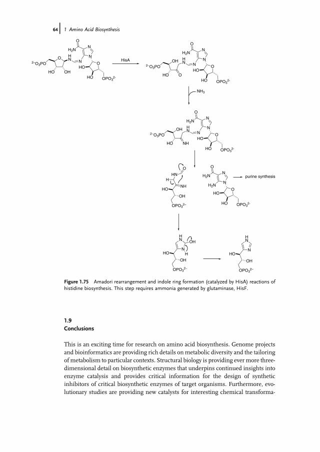
1.9Conclusions
This is an exciting time for research on amino acid biosynthesis. Genome projectsand bioinformatics are providing rich details onmetabolic diversity and the tailoringofmetabolism to particular contexts. Structural biology is providing evermore three-dimensional detail on biosynthetic enzymes that underpins continued insights intoenzyme catalysis and provides critical information for the design of syntheticinhibitors of critical biosynthetic enzymes of target organisms. Furthermore, evo-lutionary studies are providing new catalysts for interesting chemical transforma-
HN N
N
N
O
OHO
HO OPO32-
OH
OHO
2–O3PO
H2N
N
HN
OPO32–
HO
OH
N
NO
OHO
HO OPO32-
H2N
HN N
N
NO
OHO
HO OPO32-
O
OHHO
2–O3PO
H2N
purine synthesis
H2N
NH3
HN
O
NHHO
OH
OPO32–
H
N
HN
OPO32–
HO
OH
OH
H
HN N
N
N
O
OHO
HO OPO32-
OH
NHHO
2–O3PO
H2N
HisA
Figure 1.75 Amadori rearrangement and indole ring formation (catalyzed by HisA) reactions ofhistidine biosynthesis. This step requires ammonia generated by glutaminase, HisF.
64j 1 Amino Acid Biosynthesis

tions as well as new insights into the origins and development of this fascinating areaof metabolism.
N
HN
OPO32–
O
N
HN
OPO32–
NH3+
N
HN
OH
NH3+
N
HN
–O2CNH3
+
N
HN
OPO32–
HO
OH
HisB HisC
HisB
HisD
O O–
O–
O
O
O O–
O– O
NH3+
Figure 1.76 Final steps in the synthesis of histidine.
References
1 Garavelli, J.S. (2004) The RESIDdatabase of protein modifications as aresource and annotation tool. Proteomics,4, 1527–1533.
2 Copley, R.R. and Bork, P. (2000)Homology among (beta alpha)8 barrels:implications for the evolution ofmetabolic pathways. Journal of MolecularBiology, 303, 627–640.
3 Woese, C.R., Kandler, O., and Wheelis,M.L. (1990) Towards a natural system oforganisms: proposal for the domainsArchaea, Bacteria, and Eucarya.Proceedings of the National Academy ofSciences of the United States of America, 87,4576–4579.
4 Nishida, H. (2001) Evolution of aminoacid biosynthesis and enzymes withbroad substrate specificity.Bioinformatics,17, 1224–1225.
5 Shinivasan, V., Morowitz, H., and Smith,E. (2008) Essential amino acids, fromLUCA to LUCY. Complexity, 13, 8–9.
6 Hudson, R.C. and Daniel, R.M. (1993) l-Glutamate dehydrogenases –distribution, properties and mechanism.Comparative Biochemistry and PhysiologyB, 106, 767–792.
7 Smith, T.J., Peterson, P.E., Schmidt, T.,Fang, J., and Stanley, C.A. (2001)Structures of bovine glutamatedehydrogenase complexes elucidate themechanism of purine regulation. Journalof Molecular Biology, 307, 707–720.
8 Helling, R.B. (1994)Why does Escherichiacoli have two primary pathways forsynthesis of glutamate. Journal ofBacteriology, 176, 4664–4668.
9 Woods, D.R. and Reid, S.J. (1993) Recentdevelopments on the regulation andstructure of glutamine-synthetaseenzymes from selected bacterialgroups. FEMS Microbiology Reviews,11, 273–283.
10 Stadtman, E.R. (2001) The story ofglutamine synthetase regulation. The
References j65

Journal of Biological Chemistry, 276,44357–44364.
11 Reitzer, L. (2003) Nitrogen assimilationand global regulation in Escherichia coli.Annual Review of Microbiology, 57,155–176.
12 Liaw, S.H., Pan, C., and Eisenberg, D.(1993) Feedback inhibition of fullyunadenylylated glutamine-synthetasefrom Salmonella typhimurium by glycine,alanine, and serine. Proceedings of theNational Academy of Sciences of the UnitedStates of America, 90, 4996–5000.
13 Zalkin, H. and Smith, J.L. (1998)Enzymes utilizing glutamine as an amidedonor. Advances in Enzymology, 72,87–144.
14 van den Heuvel, R.H.H., Curti, B.,Vanoni, M.A., and Mattevi, A. (2004)Glutamate synthase: a fascinatingpathway from L-glutamine to L-glutamate.Cellular and Molecular Life Sciences, 61,669–681.
15 Lea, P.J. andMiflin, B.J. (2003) Glutamatesynthase and the synthesis of glutamate inplants. Plant Physiology and Biochemistry,41, 555–564.
16 Vanoni, M.A. and Curti, B. (1999)Glutamate synthase: a complexiron–sulfur flavoprotein. Cellular andMolecular Life Sciences, 55, 617–638.
17 Stabile, H., Curti, B., and Vanoni, M.A.(2000) Functional properties ofrecombinant Azospirillum brasilenseglutamate synthase, a complexiron–sulfur flavoprotein. EuropeanJournal of Biochemistry, 267, 2720–2730.
18 Massiere, F. and Badet-Denisot, M.-A.(1998) The mechanism of glutamine-dependent amidotransferases. Cellularand Molecular Life Sciences, 54, 205–222.
19 Binda, C., Bossi, R.T.,Wakatsuki, S., Arzt,S., Coda, A., Curti, B., Vanoni, M.A., andMattevi, A. (2000) Cross-talk andammonia channeling between activecenters in the unexpected domainarrangement of glutamate synthase.Structure, 8, 1299–1308.
20 Raushel, F.M., Thoden, J.B., and Holden,H.M. (2003) Enzymes with moleculartunnels.Accounts of Chemical Research, 36,539–548.
21 van den Heuvel, R.H.H., Ferrari, D.,Bossi, R.T., Ravasio, S., Curti, B., Vanoni,
M.A., Florencio, F.J., and Mattevi, A.(2002) Structural studies on thesynchronization of catalytic centers inglutamate synthase. The Journal ofBiological Chemistry, 277, 24579–24583.
22 Dossena, L., Curti, B., and Vanoni, M.A.(2007) Activation and coupling of theglutaminase and synthase reaction ofglutamate synthase is mediated by E1013of the ferredoxin-dependent enzyme,belonging to loop 4 of the synthasedomain. Biochemistry, 46, 4473–4485.
23 Christen, P. andMehta, P.K. (2001) Fromcofactor to enzymes. The molecularevolution of pyridoxal-50-phosphate-dependent enzymes. Chemical Record,1, 436–447.
24 Hayashi, H. (1995) Pyridoxal enzymes:mechanistic diversity and uniformity.Journal of Biochemistry, 118, 463–473.
25 John, R.A. (1995) Pyridoxal phosphate-dependent enzymes. Biochimica etBiophysica Acta – Protein Structure andMolecular Enzymology, 1248, 81–96.
26 Jansonius, J.N. (1998) Structure,evolution and action of vitamin B6-dependent enzymes. Current Opinion inStructural Biology, 8, 759–769.
27 Schneider, G., K€ack, H., and Lindqvist, Y.(2000) The manifold of vitamin B6dependent enzymes. Structure, 8, R1–R6.
28 Ford, G.C., Eichele, G., and Jansonius,J.N. (1980) Three-dimensional structureof a pyridoxal-phosphate-dependentenzyme, mitochondrial aspartate-aminotransferase. Proceedings of theNational Academy of Sciences of the UnitedStates of America, 77, 2559–2563.
29 Borisov, V.V., Borisova, S.N., Sosfenov,N.I., and Vainshtein, B.K. (1980) Electrondensity map of chicken heart cytosolaspartate transaminase at 3.5 Åresolution. Nature, 284, 189–190.
30 Kirsch, J.F., Eichele, G., Ford, G.C.,Vincent, M.G., Jansonius, J.N., Gehring,H., and Christen, P. (1984)Mechanism ofaction of aspartate-aminotransferaseproposed on the basis of its spatialstructure. Journal of Molecular Biology,174, 497–525.
31 Kochhar, S., Finlayson, W.L., Kirsch, J.F.,and Christen, P. (1987) The stereospecificlabilization of the C-40 pro-S hydrogen ofpyridoxamine 50-phosphate is abolished
66j 1 Amino Acid Biosynthesis
in (Lys258Ala) aspartate-aminotransferase. The Journal ofBiological Chemistry, 262, 11446–11448.
32 Dunathan, H.C. (1966) Conformationand reaction specificity in pyridoxalphosphate enzymes. Proceedings of theNational Academy of Sciences of the UnitedStates of America, 55, 712–716.
33 Matthews, R.G. and Drummond, J.T.(1990) Providing one-carbon units forbiological methylations: mechanisticstudies on serinehydroxymethyltransferase,methylenetetrahydrofolate reductase, andmethyltetrahydrofolate-homocysteinemethyltransferase. Chemical Reviews, 90,1275–1290.
34 Cronin, C.N. and Kirsch, J.F. (1988) Roleof arginine-292 in the substrate-specificityof aspartate-aminotransferase asexamined by site-directed mutagenesis.Biochemistry, 27, 4572–4579.
35 Eliot, A.C. and Kirsch, J.F. (2004)Pyridoxal phosphate enzymes:Mechanistic, structural, and evolutionaryconsiderations. Annual Review ofBiochemistry, 73, 383–415.
36 Okamoto, A., Nakai, Y., Hayashi, H.,Hirotsu, K., and Kagamiyama, H. (1998)Crystal structures of Paracoccusdenitrificans aromatic amino acidaminotransferase: a substrate recognitionsite constructed by rearrangement ofhydrogen bond network. Journal ofMolecular Biology, 280, 443–461.
37 Okamoto, A., Ishii, S., Hirotsu, K., andKagamiyama, H. (1999) The active site ofParacoccus denitrificans aromatic aminoacid aminotransferase has contraryproperties: flexibility and rigidity.Biochemistry, 38, 1176–1184.
38 Onuffer, J.J. and Kirsch, J.F. (1995)Redesign of the substrate specificity ofEscherichia coli aspartateaminotransferase to that ofEscherichia colityrosine aminotransferase by homologymodeling and site-directed mutagenesis.Protein Science, 4, 1750–1757.
39 Malashkevich, V.N., Onuffer, J.J., Kirsch,J.F., and Jansonius, J.N. (1995)Alternating arginine-modulated substratespecificity in an engineered tyrosineaminotransferase. Nature StructuralBiology, 2, 548–553.
40 Penning, T.M. and Jez, J.M. (2001)Enzyme redesign. Chemical Reviews, 101,3027–3046.
41 J€ackel, C., Kast, P., and Hilvert, D. (2008)Protein design by directed evolution.Annual Review of Biophysics, 37, 153–173.
42 Rothman, S.C. and Kirsch, J.F. (2003)How does an enzyme evolved in vitrocompare to naturally occurring homologspossessing the targeted function?Tyrosine aminotransferase fromaspartate aminotransferase. Journal ofMolecular Biology, 327, 593–608.
43 Yano, T., Oue, S., and Kagamiyama, H.(1998) Directed evolution of an aspartateaminotransferase with new substratespecificities. Proceedings of the NationalAcademy of Sciences of the United States ofAmerica, 95, 5511–5515.
44 Oue, S., Okamoto, A., Yano, T., andKagamiyama, H. (1999) Redesigning thesubstrate specificity of an enzyme bycumulative effects of the mutations ofnon-active site residues. The Journal ofBiological Chemistry, 274, 2344–2349.
45 Oue, S., Okamoto, A., Yano, T., andKagamiyama, H. (2000) Cocrystallizationof a mutant aspartate aminotransferasewith a C5-dicarboxylic substrate analog:structural comparison with theenzyme–C4-dicarboxylic analog complex.Journal of Biochemistry, 127, 337–343.
46 Ura, H., Harata, K., Matsui, I., andKuramitsu, S. (2001) Temperaturedependence of the enzyme–substraterecognition mechanism. Journal ofBiochemistry, 129, 173–178.
47 Matsui, I., Matsui, E., Sakai, Y., Kikuchi,H., Kawarabayasi, Y., Ura,H., Kawaguchi,S., Kuramitsu, S., and Harata, K. (2000)The molecular structure ofhyperthermostable aromaticaminotransferase with novel substratespecificity from Pyrococcus horikoshii. TheJournal of Biological Chemistry, 275,4871–4879.
48 Goto, M., Miyahara, I., Hayashi, H.,Kagamiyama, H., and Hirotsu, K. (2003)Crystal structures of branched-chainamino acid aminotransferase complexedwith glutamate and glutarate: truereaction intermediate and doublesubstrate recognition of the enzyme.Biochemistry, 42, 3725–3733.
References j67
49 Schl€osser, T., G€atgens, C., Weber, U., andStahmann, K.-P. (2004) Alanine:glyoxylate aminotransferase ofSaccharomyces cerevisiae-encoding geneAGX1 and metabolic significance. Yeast,21, 63–73.
50 Takada, Y. and Noguchi, T. (1985)Characteristics of alanine glyoxylateaminotransferase from Saccharomycescerevisiae, a regulatory enzyme in theglyoxylate pathway of glycine and serinebiosynthesis from tricarboxylic acid-cycleintermediates. Biochemical Journal, 231,157–163.
51 Monschau, N., Stahmann, K.-P., Sahm,H., McNeil, J.B., and Bognar, A.L. (1997)Identification of Saccharomyces cerevisiaeGLY1 as a threonine aldolase: a keyenzyme in glycine biosynthesis. FEMSMicrobiology Letters, 150, 55–60.
52 Liu, J.-Q., Nagata, S., Dairi, T., Misono,H., Shimizu, S., and Yamada, H. (1997)TheGLY1 gene of Saccharomyces cerevisiaeencodes a low-specific L-threoninealdolase that catalyzes cleavage of L-allo-threonine and L-threonine to glycine.European Journal of Biochemistry, 245,289–293.
53 Melendez-Hevia, E., de Paz-Lugo, P.,Cornish-Bowden, A., and Cardenas, M.L.(2009) A weak link in metabolism: themetabolic capacity for glycinebiosynthesis does not satisfy the need forcollagen synthesis. Journal of Biosciences,34, 853–872.
54 Renwick, S.B., Snell, K., and Baumann,U. (1998) The crystal structure of humancytosolic serinehydroxymethyltransferase: a target forcancer chemotherapy. Structure, 6,1105–1116.
55 Rajaram, V., Bhavan, B.S., Kaul, P.,Prakash, V., Rao, N.A., Savithri, H.S., andMurthyl, M.R.N. (2007) Structuredetermination and biochemical studieson Bacillus stearothermophilus E53Qserine hydroxymethyltransferase and itscomplexes provide insights on functionand enzyme memory. FEBS Journal, 274,4148–4160.
56 Bhavani, S., Trivedi, V., Jala, V.R.,Subramanya, H.S., Kaul, P., Prakash, V.,Rao, N.A., and Savithri, H.S. (2005) Roleof Lys-226 in the catalytic mechanism of
Bacillus stearothermophilus serinehydroxymethyltransferase – crystalstructure and kinetic studies.Biochemistry, 44, 6929–6937.
57 Szebenyi, D.M.E., Musayev, F.N., diSalvo, M.L., Safo, M.K., and Schirch, V.(2004) Serine hydroxymethyltransferase:role of Glu75 and evidence that serine iscleaved by a retroaldol mechanism.Biochemistry, 43, 6865–6876.
58 Trivedi, V., Gupta, A., Jala, V.R.,Saravanan, P., Rao, G.S.J., Rao, N.A.,Savithri, H.S., and Subramanya, H.S.(2002) Crystal structure of binary andternary complexes of serinehydroxymethyltransferase from Bacillusstearothermophilus – insights into thecatalytic mechanism. The Journal ofBiological Chemistry, 277, 17161–17169.
59 Stauffer, G.V. (1996) Biosynthesis ofserine, glycine, and one-carbon unit, inEscherichia coli and Salmonella: Cellularand Molecular Biology, vol. I (eds F.C.Neidhardt, R. CurtissIII, J.L. Ingraham,E.C.C. Lin, K.B. Low, B. Magasanik, W.S.Reznikoff, M. Riley, M. Schaechter, andH.E. Umbarger), 2nd edn, AmericanSociety for Microbiology, Washington,DC, pp. 506–513.
60 Burton, R.L., Chen, S., Xu, X.L., andGrant, G.A. (2009) Transient kineticanalysis of the interaction of L-serine withEscherichia coliD-3-phosphoglyceratedehydrogenase reveals the mechanism ofV-type regulation and the order of effectorbinding. Biochemistry, 48, 12242–12251.
61 Wang,W., Cho,H.S., Kim, R., Jancarik, J.,Yokota, H., Nguyen, H.H., Grigoriev, I.V.,Wemmer, D.E., and Kim, S.-H. (2002)Structural characterization of the reactionpathway in phosphoserine phosphatase:crystallographic �snapshots� ofintermediate states. Journal of MolecularBiology, 319, 421–431.
62 Singh, S.K., Yang, K., Karthikeyan, S.,Huynh, T., Zhang, X.J., Phillips, M.A.,and Zhang, H. (2004) The thrH geneproduct of Pseudomonas aeruginosa is adual activity enzyme with a novelphosphoserine: homoserinephosphotransferase activity.The Journal ofBiological Chemistry, 279, 13166–13173.
63 Feldman-Salit, A.,Wirtz, M., Hell, R., andWade,R.C. (2009)Amechanisticmodel of
68j 1 Amino Acid Biosynthesis

the cysteine synthase complex. Journal ofMolecular Biology, 386, 37–59.
64 Mino, K. and Ishikawa, K. (2003)Characterization of a novel thermostableO-acetylserine sulfhydrylase fromAeropyrum pernix K1. Journal ofBacteriology, 185, 2277–2284.
65 Rabeh, W.M. and Cook, P.F. (2004)Structure and mechanism ofO-acetylserine sulfhydrylase. TheJournal of Biological Chemistry, 279,26803–26806.
66 Tai, C.-H. and Cook, P.F. (2001) Pyridoxal50-phosphate-dependent a,b-eliminationreactions: mechanism of O-acetylserinesulfhydrylase. Accounts of ChemicalResearch, 34, 49–59.
67 Su, D., Hohn, M.J., Palioura, S., Sherrer,R.L., Yuan, J., S€oll, D., and O�Donoghue,P. (2009) How an obscure archaeal geneinspired the discovery of selenocysteinebiosynthesis in humans. IUBMB Life, 61,35–39.
68 Cole, S.T., Brosch, R., Parkhill, J., Garnier,T., Churcher, C., Harris, D., Gordon, S.V.,Eiglmeier, K., Gas, S., Barry, C.E., Tekaia,F., Badcock, K., Basham, D., Brown, D.,Chillingworth, T., Connor, R., Davies, R.,Devlin, K., Feltwell, T., Gentles, S.,Hamlin, N., Holroyd, S., Hornsby, T.,Jagels, K., Krogh, A., McLean, J., Moule,S., Murphy, L., Oliver, K., Osborne, J.,Quail, M.A., Rajandream, M.-A., Rogers,J., Rutter, S., Seeger, K., Skelton, J.,Squares, R., Squares, S., Sulston, J.E.,Taylor, K.,Whitehead, S., andBarrell, B.G.(1998) Deciphering the biology ofMycobacterium tuberculosis from thecomplete genome sequence. Nature,393 537–544.
69 Schnell, R., Oehlmann, W., Singh, M.,and Schneider, G. (2007) Structuralinsights into catalysis and inhibition ofO-acetylserine sulfhydrylase fromMycobacterium tuberculosis:crystalstructures of the enzymea-aminoacrylateintermediate and an enzyme–inhibitorcomplex. The Journal of BiologicalChemistry, 282, 23473–23481.
70 Park, J.-H., Dorrestein, P.C., Zhai, H.,Kinsland, C., McLafferty, F.W., andBegley, T.P. (2003) Biosynthesis of thethiazolemoiety of thiamin pyrophosphate
(vitamin B1). Biochemistry, 42,12430–12438.
71 Burns, K.E., Baumgart, S., Dorrestein,P.C., Zhai, H., McLafferty, F.W., andBegley, T.P. (2005) Reconstitution of anew cysteine biosynthetic pathway inMycobacterium tuberculosis. Journal of theAmerican Chemical Society, 127,11602–11603.
72 Ågren, D., Schnell, R., Oehlmann, W.,Singh, M., and Schneider, G. (2008)Cysteine synthase (CysM) ofMycobacterium tuberculosis is anO-phosphoserine sulfhydrylase:evidence for an alternative cysteinebiosynthesis pathway in Mycobacteria.The Journal of Biological Chemistry, 283,31567–31574.
73 Sauerwald, A., Zhu, W.H., Major, T.A.,Roy, H., Palioura, S., Jahn, D., Whitman,W.B., Yates, J.R., Ibba, M., and S€oll, D.(2005) RNA-dependent cysteinebiosynthesis in archaea. Science, 307,1969–1972.
74 Cone, J.E., Del Rio, M.R., Davis, J.N., andStadtman, T.C. (1976) Chemicalcharacterization of selenoproteincomponent of clostridial glycinereductase: identification of selenocysteineas the organoselenium moiety.Proceedings of the National Academy ofSciences of the United States of America, 73,2659–2663.
75 Leinfelder, W., Zehelein, E.,Mandrandberthelot, M.A., and Bock, A.(1988) Gene for a novel transfer-RNAspecies that accepts L-serine andcotranslationally inserts selenocysteine.Nature, 331, 723–725.
76 Mullins, L.S., Hong, S.-B., Gibson, G.E.,Walker, H., Stadtman, T.C., and Raushel,F.M. (1997) Identification of aphosphorylated enzyme intermediate inthe catalytic mechanism forselenophosphate synthetase. Journal of theAmerican Chemical Society, 119,6684–6685.
77 Midelfort, C.F. and Rose, I.A. (1976) Astereochemical method for detection ofATP terminal phosphate transfer inenzymatic reactions. Glutaminesynthetase. The Journal of BiologicalChemistry, 251, 5881–5887.
References j69

78 Forchhammer, K. and B€ock, A. (1991)Selenocysteine synthase from Escherichiacoli. Analysis of the reaction sequence.The Journal of Biological Chemistry, 266,6324–6328.
79 Kernebeck, T., Lohse, A.W., andGr€otzinger, J. (2001) A bioinformaticalapproach suggests the function of theautoimmune hepatitis target antigensoluble liver antigen/liver pancreas.Hepatology, 34, 230–233.
80 Ganichkin, O.M., Xu, X.-M., Carlson,B.A., Mix, H., Hatfield, D.L., Gladyshev,V.N., and Wahl, M.C. (2008) Structureand catalytic mechanism of eukaryoticselenocysteine synthase. The Journalof Biological Chemistry, 283, 5849–5865.
81 Araiso, Y., Palioura, S., Ishitani, R.,Sherrer, R.L., O�Donoghue, P., Yuan, J.,Oshikane, H., Domae, N., DeFranco, J.,S€oll, D., and Nureki, O. (2008) Structuralinsights into RNA-dependent eukaryaland archaeal selenocysteine formation.Nucleic Acids Research, 36, 1187–1199.
82 Xu, X.-M., Carlson, B.A., Mix, H., Zhang,Y., Saira, K., Glass, R.S., Berry, M.J.,Gladyshev, V.N., andHatfield, D.L. (2007)Biosynthesis of selenocysteine on itstRNA in eukaryotes. PLoS Biology, 5,96–105.
83 Yuan, J., Palioura, S., Salazar, J.C., Su, D.,O�Donoghue, P., Hohn, M.J., Cardoso,A.M., Whitman, W.B., and S€oll, D. (2006)RNA-dependent conversion ofphosphoserine forms selenocysteine ineukaryotes and archaea. Proceedings of theNational Academy of Sciences of the UnitedStates of America, 103, 18923–18927.
84 Schmitt, E., Panvert,M., Blanquet, S., andMechulam, Y. (1998) Crystal structure ofmethionyl-tRNAf
Met transformylasecomplexed with the initiatorformylmethionyl-tRNAf
Met. The EMBOJournal, 17, 6819–6826.
85 Curnow, A.W., Hong, K.-W., Yuan, R.,Kim, S.-I., Martins, O., Winkler, W.,Henkin, T.M., and S€oll, D. (1997)Glu-tRNAGln amidotransferase: a novelheterotrimeric enzyme required forcorrect decoding of glutamine codonsduring translation. Proceedings of theNational Academy of Sciences of theUnited States of America, 94,11819–11826.
86 Ibba, M. and Soll, D. (2000) Aminoacyl-tRNA synthesis. Annual Review ofBiochemistry, 69, 617–650.
87 Humbert, R. and Simoni, R.D. (1980)Genetic and biochemical studiesdemonstrating a second gene coding forasparagine synthetase in Escherichia coli.Journal of Bacteriology, 142, 212–220.
88 Cedar, H. and Schwartz, J.H. (1969)Asparagine synthetase ofEscherichia coli I.Biosynthetic role of the enzyme,purification, and characterization of thereaction products.The Journal of BiologicalChemistry, 244, 4112–4121.
89 Tesson, A.R., Soper, T.S., Ciustea, M., andRichards, N.G.J. (2003) Revisiting thesteady state kinetic mechanism ofglutamine-dependent asparaginesynthetase from Escherichia coli. Archivesof Biochemistry and Biophysics, 413, 23–31.
90 Larsen, T.M., Boehlein, S.K., Schuster,S.M., Richards, N.G.J., Thoden, J.B.,Holden, H.M., and Rayment, I. (1999)Three-dimensional structure ofEscherichia coli asparagine synthetase B: ashort journey from substrate to product.Biochemistry, 38, 16146–16157.
91 Richards, N.G.J. and Kilberg, M.S. (2006)Asparagine synthetase chemotherapy.Annual Review of Biochemistry, 75,629–654.
92 Koizumi, M., Hiratake, J., Nakatsu, T.,Kato, H., and Oda, J. (1999) A potenttransition-state analogue inhibitor ofEscherichia coli asparagine synthetase A.Journal of the American Chemical Society,121, 5799–5800.
93 Zhang, Y., Thiele, I., Weekes, D., Li, Z.,Jaroszewski, L., Ginalski, K., Deacon,A.M.,Wooley, J., Lesley, S.A.,Wilson, I.A.,Palsson, B., Osterman, A., and Godzik, A.(2009) Three-dimensional structural viewof the central metabolic network ofThermotoga maritima. Science, 325,1544–1549.
94 Curien, G., Biou, V., Mas-Droux, C.,Robert-Genthon, M., Ferrer, J.-L., andDumas, R. (2008) Amino acidbiosynthesis: new architectures inallosteric enzymes. Plant Physiology andBiochemistry, 46, 325–339.
95 Azevedo, R.A., Lancien, M., and Lea, P.J.(2006) The aspartic acid metabolicpathway, an exciting and essential
70j 1 Amino Acid Biosynthesis

pathway in plants. Amino Acids, 30,143–162.
96 Liu, X., Pavlovsky, A.G., and Viola, R.E.(2008) The structural basis for allostericinhibition of a threonine-sensitiveaspartokinase. The Journal of BiologicalChemistry, 283, 16216–16225.
97 Hadfield, A., Shammas, C., Kryger, G.,Ringe, D., Petsko, G.A., Ouyang, J., andViola, R.E. (2001) Active site analysis ofthe potential antimicrobial targetaspartate semialdehyde dehydrogenase.Biochemistry, 40, 14475–14483.
98 Blanco, J., Moore, R.A., and Viola, R.E.(2003) Capture of an intermediate in thecatalytic cycle of L-aspartate-b-semialdehyde dehydrogenase.Proceedings of the National Academy ofSciences of the United States of America,100, 12613–12617.
99 Blanco, J., Moore, R.A., Kabaleeswaran,V., and Viola, R.E. (2003) A structuralbasis for the mechanism of aspartate-b-semialdehyde dehydrogenase fromVibrio cholerae. Protein Science, 12, 27–33.
100 Holland, M.J. and Westhead, E.W. (1973)Purification and characterization ofaspartic beta-semialdehydedehydrogenase from yeast andpurification of an isoenzyme ofglyceraldehyde-3-phosphatedehydrogenase. Biochemistry, 12,2264–2270.
101 Geck,M.K. andKirsch, J.F. (1999)Anovel,definitive test for substrate channelingillustrated with the aspartateaminotransferase/malate dehydrogenasesystem. Biochemistry, 38, 8032–8037.
102 James, C.L. and Viola, R.E. (2002)Production and characterization ofbifunctional enzymes. Substratechanneling in the aspartate pathway.Biochemistry, 41, 3726–3731.
103 DeLaBarre, B., Thompson, P.R., Wright,G.D., and Berghuis, A.M. (2000) Crystalstructures of homoserine dehydrogenasesuggest a novel catalytic mechanism foroxidoreductases. Nature StructuralBiology, 7, 238–244.
104 Jacques, S.L., Ejim, L.J., and Wright, G.D.(2001) Homoserine dehydrogenase fromSaccharomyces cerevisiae: kineticmechanism and stereochemistry of
hydride transfer. Biochimica et BiophysicaActa – Protein Structure and MolecularEnzymology, 1544, 42–54.
105 Huo, X. and Viola, R.E. (1996) Substratespecificity and identification of functionalgroups of homoserine kinase fromEscherichia coli. Biochemistry, 35,16180–16185.
106 Mirza, I.A., Nazi, I., Korczynska, M.,Wright, G.D., and Berghuis, A.M. (2005)Crystal structure of homoserinetransacetylase from Haemophilusinfluenzae reveals a new family ofa/b-hydrolases. Biochemistry, 44,15768–15773.
107 Zubieta, C., Arkus, K.A.J., Cahoon, R.E.,and Jez, J.M. (2008) A single amino acidchange is responsible for evolution ofacyltransferase specificity in bacterialmethionine biosynthesis. The Journal ofBiological Chemistry, 283, 7561–7567.
108 Laber, B., Gerbling, K.-P., Harde, C., Neff,K.-H., Nordhoff, E., and Pohlenz, H.-D.(1994) Mechanisms of interaction ofEscherichia coli threonine synthase withsubstrates and inhibitors. Biochemistry,33, 3413–3423.
109 Laber, B.,Maurer,W.,Hanke,C.,Gr€afe, S.,Ehlert, S., Messerschmidt, A., andClausen, T. (1999) Characterization ofrecombinant Arabidopsis thalianathreonine synthase. European Journal ofBiochemistry, 263, 212–221.
110 Curien, G., Job, D., Douce, R., andDumas, R. (1998) Allosteric activation ofArabidopsis threonine synthase by S-adenosylmethionine. Biochemistry, 37,13212–13221.
111 Thomazeau, K., Curien, G., Dumas, R.,and Biou, V. (2001) Crystal structure ofthreonine synthase from Arabidopsisthaliana. Protein Science, 10, 638–648.
112 Omi, R., Goto, M., Miyahara, I.,Mizuguchi, H., Hayashi, H.,Kagamiyama, H., and Hirotsu, K. (2003)Crystal structures of threonine synthasefrom Thermus thermophilus HB8 –
conformational change, substraterecognition, and mechanism. The Journalof Biological Chemistry, 278, 46035–46045.
113 Garrido-Franco, M., Ehlert, S.,Messerschmidt, A., Marinkovic, S.,Huber, R., Laber, B., Bourenkov, G.P., and
References j71

Clausen, T. (2002) Structure and functionof threonine synthase from yeast. TheJournal of Biological Chemistry, 277,12396–12405.
114 Sassetti, C.M. and Rubin, E.J. (2003)Genetic requirements for mycobacterialsurvival during infection. Proceedings ofthe National Academy of Sciences of theUnited States of America, 100,12989–12994.
115 Covarrubias, A.S.,H€ogbom,M., Bergfors,T., Carroll, P., Mannerstedt, K., Oscarson,S., Parish, T., Jones, T.A., and Mowbray,S.L. (2008) Structural, biochemical, and invivo investigations of the threoninesynthase fromMycobacterium tuberculosis.Journal of Molecular Biology, 381, 622–633.
116 Clausen, T., Huber, R., Prade, L., Wahl,M.C., and Messerschmidt, A. (1998)Crystal structure of Escherichia colicystathionine c-synthase at 1.5 Åresolution. The EMBO Journal, 17,6827–6838.
117 Kong, Y., Wu, D., Bai, H., Han, C., Chen,J., Chen, L.,Hu, L., Jiang,H., and Shen, X.(2008) Enzymatic characterization andinhibitor discovery of a new cystathioninec-synthase from Helicobacter pylori.Journal of Biochemistry, 143, 59–68.
118 Clausen, T., Huber, R., Laber, B., Pohlenz,H.-D., and Messerschmidt, A. (1996)Crystal structure of the pyridoxal-50-phosphate dependent cystathionineb-lyase from Escherichia coli at 1.83 Å.Journal of Molecular Biology, 262, 202–224.
119 Goulding, C.W., Postigo, D., andMatthews, R.G. (1997) Cobalamin-dependent methionine synthase is amodular protein with distinct regions forbinding homocysteine,methyltetrahydrofolate, cobalamin, andadenosylmethionine. Biochemistry, 36,8082–8091.
120 Matthews, R.G., Koutmos, M., and Datta,S. (2008) Cobalamin-dependent andcobamide-dependent methyltransferases.Current Opinion in Structural Biology, 18,658–666.
121 Gonzalez, J.C., Peariso, K., PennerHahn,J.E., and Matthews, R.G. (1996)Cobalamin-independent methioninesynthase from Escherichia coli: a zincmetalloenzyme. Biochemistry, 35,12228–12234.
122 Marco-Marin, C., Gil-Ortiz, F., Perez-Arellano, I., Cervera, J., Fita, I., andRubio,V. (2007) A novel two-domain architecturewithin the amino acid kinase enzymefamily revealed by the crystal structure ofEscherichia coli glutamate 5-kinase.Journal of Molecular Biology, 367,1431–1446.
123 Hu, C.A., Delauney, A.J., and Verma, D.P.(1992) A bifunctional enzyme (d-1-pyrroline-5-carboxylate synthetase)catalyzes the first two steps in prolinebiosynthesis in plants. Proceedings of theNational Academy of Sciences of the UnitedStates of America, 89, 9354–9358.
124 Turchetto-Zolet, A.C., Margis-Pinheiro,M., andMargis, R. (2009) The evolution ofpyrroline-5-carboxylate synthase in plants:a key enzyme in proline synthesis.Molecular Genetics and Genomics, 281,87–97.
125 Costilow, R.N. and Laycock, L. (1971)Ornithine cyclase (deaminating) –purification of a protein that convertsornithine to proline and definition ofoptimal assay conditions. The Journal ofBiological Chemistry, 246, 6655–6660.
126 Graupner, M. and White, R.H. (2001)Methanococcus jannaschii generates L-proline by cyclization of L-ornithine.Journal of Bacteriology, 183, 5203–5205.
127 Nocek, B., Chang, C., Li, H., Lezondra, L.,Holzle, D., Collart, F., and Joachimiak, A.(2005) Crystal structures of d-1-pyrroline-5-carboxylate reductase from humanpathogens Neisseria meningitides andStreptococcus pyogenes. Journal ofMolecularBiology, 354, 91–106.
128 Caldovic, L. and Tuchman, M. (2003) N-Acetylglutamate and its changing rolethrough evolution. Biochemical Journal,372, 279–290.
129 Fernandez-Murga, M.L., Gil-Ortiz, F.,Llacer, J.L., and Rubio, V. (2004) Argininebiosynthesis in Thermotoga maritima:characterization of the arginine-sensitiveN-acetyl-L-glutamate kinase. Journal ofBacteriology, 186, 6142–6149.
130 Ramon-Maiques, S.,Marina,A.,Gil-Ortiz,F., Fita, I., and Rubio, V. (2002) Structureof acetylglutamate kinase, a key enzymefor arginine biosynthesis and a prototypefor the amino acid kinase enzyme family,during catalysis. Structure, 10, 329–342.
72j 1 Amino Acid Biosynthesis

131 Cherney, L.T., Cherney, M.M., Garen,C.R., Niu, C., Moradian, F., and James,M.N.G. (2007) Crystal structure of N-acetyl-c-glutamyl-phosphate reductasefrom Mycobacterium tuberculosis incomplex with NADPþ . Journal ofMolecular Biology, 367, 1357–1369.
132 Rajaram, V., Prasuna, P.R., Savithri, H.S.,and Murthy, M.R.N. (2008) Structure ofbiosynthetic N-acetylornithineaminotransferase from Salmonellatyphimurium: studies on substratespecificity and inhibitor binding. Proteins– Structure Function and Bioinformatics,70, 429–441.
133 Ledwidge, R. and Blanchard, J.S. (1999)The dual biosynthetic capability of N-acetylornithine aminotransferase inarginine and lysine biosynthesis.Biochemistry, 38, 3019–3024.
134 Slocum, R.D. (2005) Genes, enzymes andregulation of arginine biosynthesis inplants. Plant Physiology and Biochemistry,43, 729–745.
135 Xu, Y., Labedan, B., and Glansdorff, N.(2007) Surprising arginine biosynthesis: areappraisal of the enzymology andevolution of the pathway inmicroorganisms. Microbiology andMolecular Biology Reviews, 71, 36–47.
136 Bachmann, C., Kr€ahenb€uhl, S., andColombo, J.P. (1982) Purification andproperties of acetyl-CoA: L-glutamate N-acetyltransferase from human liver.Biochemical Journal, 205, 123–127.
137 Shi, D., Sagar, V., Jin, Z.M., Yu, X.L.,Caldovic, L., Morizono, H., Allewell,N.M., andTuchman,M. (2008) The crystalstructure ofN-acetyl-L-glutamate synthasefrom Neisseria gonorrhoeae providesinsights into mechanisms of catalysis andregulation. The Journal of BiologicalChemistry, 283, 7176–7184.
138 Alonso, E. and Rubio, V. (1989)Participation of ornithineaminotransferase in the synthesis andcatabolism of ornithine in mice. Studiesusing gabaculine and argininedeprivation. Biochemical Journal, 259,131–138.
139 Lqbal, A., Clifton, I.J., Bagonis, M.,Kershaw, N.J., Domene, C., Claridge,T.D.W., Wharton, C.W., and Schofield,C.J. (2009) Anatomy of a simple acyl
intermediate in enzyme catalysis:combined biophysical and modelingstudies on ornithine acetyl transferase.Journal of the American Chemical Society,131, 749–757.
140 Marc, F., Weigel, P., Legrain, C., Almeras,Y., Santrot, M., Glansdorff, N., andSakanyan, V. (2000) Characterization andkinetic mechanism of mono- andbifunctional ornithine acetyltransferasesfrom thermophilic microorganisms.European Journal of Biochemistry, 267,5217–5226.
141 Shi, D., Yu, X., Roth, L., Morizono, H.,Hathout, Y., Allewell, N.M., andTuchman, M. (2005) Expression,purification, crystallization andpreliminary X-ray crystallographic studiesof a novel acetylcitrulline deacetylase fromXanthomonas campestris. ActaCrystallographica F, 61, 676–679.
142 Shi, D., Morizono, H., Yu, X., Roth, L.,Caldovic, L., Allewell, N.M., Malamy,M.H., and Tuchman, M. (2005)Crystal structure of N-acetylornithinetranscarbamylase from Xanthomonascampestris: a novel enzyme in anew arginine biosynthetic pathwayfound in several eubacteria. TheJournal of Biological Chemistry, 280,14366–14369.
143 Shi, D., Morizono, H., Cabrera-Luque, J.,Yu, X.L., Roth, L.,Malamy,M.H., Allewell,N.M., and Tuchman, M. (2006) Structureand catalytic mechanism of a novel N-succinyl-L-ornithine transcarbamylase inarginine biosynthesis of Bacteroidesfragilis. The Journal of Biological Chemistry,281, 20623–20631.
144 Allewell, N.M., Shi, D.,Morizono,H., andTuchman, M. (1999) Molecularrecognition by ornithine and aspartatetranscarbamylases. Accounts of ChemicalResearch, 32, 885–894.
145 Labedan, B., Boyen, A., Baetens, M.,Charlier, D., Chen, P., Cunin, R.,Durbeco, V., Glansdorff, N., Herve, G.,Legrain, C., Liang, Z., Purcarea, C.,Roovers, M., Sanchez, R., Toong, T.-L.,Van de Casteele, M., van Vliet, F., Xu, Y.,and Zhang, Y.-F. (1999) The evolutionaryhistory of carbamoyltransferases: acomplex set of paralogous genes wasalready present in the last universal
References j73

common ancestor. Journal of MolecularEvolution, 49, 461–473.
146 Sankaranarayanan, R., Cherney, M.M.,Cherney, L.T., Garen, C.R., Moradian, F.,and James, M.N.G. (2008) The crystalstructures of ornithinecarbamoyltransferase fromMycobacterium tuberculosis and its ternarycomplex with carbamoyl phosphate and L-norvaline reveal the enzyme�s catalyticmechanism. Journal of Molecular Biology,375, 1052–1063.
147 Yu, Y., Terada, K., Nagasaki, A., Takiguchi,M., and Mori, M. (1995) Preparation ofrecombinant argininosuccinatesynthetase and argininosuccinate lyase –
expression of the enzymes in rat tissues.Journal of Biochemistry, 117, 952–957.
148 Bork, P. and Koonin, E.V. (1994) A P-loop-like motif in a widespread ATPpyrophosphatase domain: implicationsfor the evolution of sequence motifs andenzyme activity. Proteins – StructureFunction and Genetics, 20, 347–355.
149 Goto,M., Omi, R.,Miyahara, I., Sugahara,M., and Hirotsu, K. (2003) Structures ofargininosuccinate synthetase in enzyme-ATP substrates and enzyme-AMPproductforms: stereochemistry of the catalyticreaction. The Journal of BiologicalChemistry, 278, 22964–22971.
150 Tsai, M., Koo, J., and Howell, P.L. (2005)Recovery of argininosuccinate lyaseactivity in duck d1 crystallin. Biochemistry,44, 9034–9044.
151 Fujii, T., Sakai, H., Kawata, Y., andHata, Y.(2003) Crystal structure of thermostableaspartase from Bacillus sp YM55-1:structure-based exploration of functionalsites in the aspartase family. Journal ofMolecular Biology, 328, 635–654.
152 Dailey, F.E. and Cronan, J.E. (1986)Acetohydroxy acid synthase I, a requiredenzyme for isoleucine and valinebiosynthesis in Escherichia coli K-12during growth on acetate as the solecarbon source. Journal of Bacteriology, 165,453–460.
153 Dumas, R., Biou, V., Halgand, F., Douce,R., and Duggleby, R.G. (2001)Enzymology, structure, and dynamics ofacetohydroxy acid isomeroreductase.Accounts of Chemical Research, 34,399–408.
154 Flint, D.H., Emptage, M.H., Finnegan,M.G., Fu, W., and Johnson, M.K. (1993)The role and properties of the iron–sulfurcluster in Escherichia coli dihydroxy-aciddehydratase. The Journal of BiologicalChemistry, 268, 14732–14742.
155 Chipman, D., Barak, Z., and Schloss, J.V.(1998) Biosynthesis of 2-aceto-2-hydroxyacids: acetolactate synthases andacetohydroxyacid synthases. Biochimica etBiophysica Acta – Protein Structure andMolecular Enzymology, 1385, 401–419.
156 Kishore, G.M. and Shah, D.M. (1988)Amino acid biosynthesis inhibitors asherbicides. Annual Review of Biochemistry,57, 627–663.
157 Engel, S., Vyazmensky, M., Vinogradov,M., Berkovich, D., Bar-Ilan, A., Qimron,U., Rosiansky, Y., Barak,Z., andChipman,D.M. (2004) Role of a conserved argininein the mechanism of acetohydroxyacidsynthase: catalysis of condensation with aspecific ketoacid substrate. The Journal ofBiological Chemistry, 279, 24803–24812.
158 McCourt, J.A., Pang, S.S., King-Scott, J.,Guddat, L.W., and Duggleby, R.G. (2006)Herbicide-binding sites revealed in thestructure of plant acetohydroxyacidsynthase. Proceedings of the NationalAcademy of Sciences of the United States ofAmerica, 103, 569–573.
159 Scarselli, M., Padula, M.G., Bernini, A.,Spiga, O., Ciutti, A., Leoncini, R.,Vannoni, D., Marinello, E., and Niccolai,N. (2003) Structure and functioncorrelations between the rat liverthreonine deaminase andaminotransferases. Biochimica etBiophysica Acta – Proteins and Proteomics,1645, 40–48.
160 Eikmanns, B.J., Eggeling, L., and Sahm,H. (1993) Molecular aspects of lysine,threonine, and isoleucine biosynthesis inCorynebacterium glutamicum. Antonie VanLeeuwenhoek International Journal ofGeneral and Molecular Microbiology, 64,145–163.
161 Shulman, A., Zalyapin, E., Vyazmensky,M., Yifrach, O., Barak, Z., and Chipman,D.M. (2008) Allosteric regulation ofBacillus subtilis threonine deaminase, abiosynthetic threonine deaminase with asingle regulatory domain. Biochemistry,47, 11783–11792.
74j 1 Amino Acid Biosynthesis

162 Eisenstein, E. (1991) Cloning, expression,purification, and characterization ofbiosynthetic threonine deaminase fromEscherichia coli. The Journal of BiologicalChemistry, 266, 5801–5807.
163 Drevland,R.M.,Waheed,A., andGraham,D.E. (2007) Enzymology and evolution ofthe pyruvate pathway to 2-oxobutyrate inMethanocaldococcus jannaschii. Journal ofBacteriology, 189, 4391–4400.
164 Biou, V., Dumas, R., CohenAddad, C.,Douce, R., Job, D., and PebayPeyroula, E.(1997) The crystal structure of plantacetohydroxy acid isomeroreductasecomplexed with NADPH, twomagnesium ions and a herbicidaltransition state analog determined at 1.65Å resolution. The EMBO Journal, 16,3405–3415.
165 Beinert, H., Kennedy, M.C., and Stout,C.D. (1996) Aconitase as iron–sulfurprotein, enzyme, and iron-regulatoryprotein.Chemical Reviews, 96, 2335–2374.
166 Duan, X., Yang, J., Ren, B., Tan, G., andDing, H. (2009) Reactivity of nitric oxidewith the [4Fe–4S] cluster of dihydroxyaciddehydratase from Escherichia coli.Biochemical Journal, 417, 783–789.
167 Hyduke, D.R., Jarboe, L.R., Tran, L.M.,Chou, K.J.Y., and Liao, J.C. (2007)Integrated network analysis identifiesnitric oxide response networks anddihydroxyacid dehydratase as a crucialtarget in Escherichia coli. Proceedings of theNational Academy of Sciences of the UnitedStates of America, 104, 8484–8489.
168 Macomber, L. and Imlay, J.A. (2009) Theiron–sulfur clusters of dehydratases areprimary intracellular targets of coppertoxicity. Proceedings of the NationalAcademy of Sciences of the United States ofAmerica, 106, 8344–8349.
169 Xu, H., Andi, B., Qian, J., West, A.H., andCook, P.F. (2006) The a-aminoadipatepathway for lysine biosynthesis in fungi.Cell Biochemistry andBiophysics, 46, 43–64.
170 Wulandari, A.P.,Miyazaki, J., Kobashi,N.,Nishiyama,M., Hoshino, T., and Yamane,H. (2002) Characterization of bacterialhomocitrate synthase involved in lysinebiosynthesis. FEBS Letters, 522, 35–40.
171 de Carvalho, L.P.S. and Blanchard, J.S.(2006) Kinetic and chemical mechanismof alpha-isopropylmalate synthase from
Mycobacterium tuberculosis. Biochemistry,45, 8988–8999.
172 Howell, D.M., Xu, H.M., andWhite, R.H.(1999) (R)-Citramalate synthase inmethanogenic archaea. Journal ofBacteriology, 181, 331–333.
173 Li, F., Hagemeier, C.H., Seedorf, H.,Gottschalk, G., and Thauer, R.K. (2007)Re-citrate synthase from Clostridiumkluyveri is phylogenetically related tohomocitrate synthase andisopropylmalate synthase rather than toSi-citrate synthase. Journal of Bacteriology,189, 4299–4304.
174 Koon, N., Squire, C.J., and Baker, E.N.(2004) Crystal structure of LeuA fromMycobacterium tuberculosis, a key enzymein leucine biosynthesis. Proceedings of theNational Academy of Sciences of the UnitedStates of America, 101, 8295–8300.
175 Qian, J., Khandogin, J., West, A.H., andCook, P.F. (2008) Evidence for a catalyticdyad in the active site of homocitratesynthase from Saccharomyces cerevisiae.Biochemistry, 47, 6851–6858.
176 Gruer, M.J., Artymiuk, P.J., and Guest,J.R. (1997) The aconitase family: threestructural variations on a common theme.Trends in Biochemical Sciences, 22, 3–6.
177 Yasutake, Y., Yao, M., Sakai, N., Kirita, T.,and Tanaka, I. (2004) Crystal structure ofthe Pyrococcus horikoshii isopropylmalateisomerase small subunit provides insightinto the dual substrate specificity of theenzyme. Journal of Molecular Biology, 344,325–333.
178 Hurley, J.H., Thorsness, P.E.,Ramalingam, V., Helmers, N.H.,Koshland, D.E., and Stroud, R.M. (1989)Structure of a bacterial enzyme regulatedby phosphorylation, isocitratedehydrogenase. Proceedings of the NationalAcademy of Sciences of the United States ofAmerica, 86, 8635–8639.
179 Imada, K., Sato, M., Tanaka, N., Katsube,Y., Matsuura, Y., and Oshima, T. (1991) 3-Dimensional structure of a highlythermostable enzyme, 3-isopropylmalatedehydrogenase of Thermus thermophilus at2.2 Å resolution. Journal of MolecularBiology, 222, 725–738.
180 Howell, D.M., Graupner, M., Xu, H., andWhite, R.H. (2000) Identification ofenzymes homologous to isocitrate
References j75
dehydrogenase that are involved incoenzyme B and leucine biosynthesis inmethanoarchaea. Journal of Bacteriology,182, 5013–5016.
181 Bolduc, J.M., Dyer, D.H., Scott, W.G.,Singer, P., Sweet, R.M., Koshland, D.E.,and Stoddard, B.L. (1995) Mutagenesisand Laue structures of enzymeintermediates: isocitrate dehydrogenase.Science, 268, 1312–1318.
182 Miyazaki, J., Kobashi, N., Nishiyama, M.,and Yamane, H. (2003) Characterizationof homoisocitrate dehydrogenaseinvolved in lysine biosynthesis of anextremely thermophilic bacterium,Thermus thermophilus HB27, andevolutionary implication of beta-decarboxylating dehydrogenase. TheJournal of Biological Chemistry, 278,1864–1871.
183 Miyazaki, K. (2005) Bifunctionalisocitrate-homoisocitrate dehydrogenase:a missing link in the evolution ofb-decarboxylating dehydrogenase.Biochemical and Biophysical ResearchCommunications, 331, 341–346.
184 Chen,R.,Greer,A., andDean,A.M. (1996)Redesigning secondary structure to invertcoenzyme specificity in isopropylmalatedehydrogenase. Proceedings of the NationalAcademy of Sciences of the United States ofAmerica, 93, 12171–12176.
185 Miyazaki, T., Miyazaki, J., Yamane, H.,and Nishiyama, M. (2004)a-Aminoadipate aminotransferase froman extremely thermophilic bacterium,Thermus thermophilus. Microbiology, 150,2327–2334.
186 Matsuda, M. and Ogur, M. (1969)Enzymatic and physiological properties ofyeast glutamate-a-ketoadipatetransaminase. The Journal of BiologicalChemistry, 244, 5153–5158.
187 Matsuda, M. and Ogur, M. (1969)Separation and specificity of yeastglutamate-a-ketoadipate transaminase.The Journal of Biological Chemistry, 244,3352–3358.
188 Tomita, T., Miyagawa, T., Miyazaki, T.,Fushinobu, S., Kuzuyama, T., andNishiyama, M. (2009) Mechanism formultiple-substrates recognition ofa-aminoadipate aminotransferase from
Thermus thermophilus. Proteins – StructureFunction and Bioinformatics, 75, 348–359.
189 Velasco, A.M., Leguina, J.I., and Lazcano,A. (2002)Molecular evolution of the lysinebiosynthetic pathways. Journal ofMolecular Evolution, 55, 445–449.
190 Nishida, H., Nishiyama, M., Kobashi, N.,Kosuge, T., Hoshino, T., and Yamane, H.(1999) Aprokaryotic gene cluster involvedin synthesis of lysine through the aminoadipate pathway: a key to the evolution ofamino acid biosynthesis. GenomeResearch, 9, 1175–1183.
191 Hudson, A.O., Gilvarg, C., and Leustek, T.(2008) Biochemical and phylogeneticcharacterization of a noveldiaminopimelate biosynthesispathway in prokaryotes identifies adiverged form of LL-diaminopimelateaminotransferase. Journal of Bacteriology,190, 3256–3263.
192 Viola, R.E. (2001) The central enzymes ofthe aspartate family of amino acidbiosynthesis. Accounts of ChemicalResearch, 34, 339–349.
193 Coulter, C.V., Gerrard, J.A., Kraunsoe,J.A.E., and Pratt, A.J. (1999) Escherichiacoli dihydrodipicolinate synthase anddihydrodipicolinate reductase: kinetic andinhibition studies of two putativeherbicide targets. Pesticide Science, 55,887–895.
194 Dobson, R.C.J., Valegard, K., andGerrard,J.A. (2004) The crystal structure of threesite-directed mutants of Escherichia colidihydrodipicolinate synthase: furtherevidence for a catalytic triad. Journal ofMolecular Biology, 338, 329–339.
195 Devenish, S.R.A., Huisman, F.H.A.,Parker, E.J., Hadfield, A.T., and Gerrard,J.A. (2009) Cloning and characterisationof dihydrodipicolinate synthase from thepathogen Neisseria meningitidis.Biochimica et Biophysica Acta – Proteinsand Proteomics, 1794, 1168–1174.
196 Dobson, R.C.J., Griffin, M.D.W.,Devenish, S.R.A., Pearce, F.G., Hutton,C.A., Gerrard, J.A., Jameson, G.B., andPerugini, M.A. (2008) Conserved main-chain peptide distortions: a proposed rolefor Ile203 in catalysis bydihydrodipicolinate synthase. ProteinScience, 17, 2080–2090.
76j 1 Amino Acid Biosynthesis

197 Ge, X., Olson, A., Cai, S., and Sem, D.S.(2008) Binding synergy and cooperativityin dihydrodipicolinate reductase:implications for mechanism and thedesignof biligand inhibitors.Biochemistry,47, 9966–9980.
198 Pearce, F.G., Sprissler, C., and Gerrard,J.A. (2008) Characterization ofdihydrodipicolinate reductase fromThermotoga maritima reveals evolution ofsubstrate binding kinetics. Journal ofBiochemistry, 143, 617–623.
199 Pillai, B., Moorthie, V.A., van Belkum,M.J., Marcus, S.L., Cherney, M.M.,Diaper, C.M., Vederas, J.C., and James,M.N.G. (2009) Crystal structure ofdiaminopimelate epimerase fromArabidopsis thaliana, an amino acidracemase critical for L-lysine biosynthesis.Journal of Molecular Biology, 385, 580–594.
200 Usha, V., Dover, L.G., Roper, D.L., andBesra, G.S. (2008) Characterization ofMycobacterium tuberculosisdiaminopimelic acid epimerase: pairedcysteine residues are crucial forracemization. FEMS Microbiology Letters,280, 57–63.
201 Hu, T., Wu, D., Chen, J., Ding, J., Jiang,H., and Shen, X. (2008) The catalyticintermediate stabilized by a �Down� activesite loop for diaminopimelatedecarboxylase from Helicobacter pylori:enzymatic characterization with crystalstructure analysis.The Journal of BiologicalChemistry, 283, 21284–21293.
202 Lee, J., Michael, A.J., Martynowski, D.,Goldsmith, E.J., and Phillips, M.A. (2007)Phylogenetic diversity and the structuralbasis of substrate specificity in theb/a-barrel fold basic amino aciddecarboxylases. The Journal of BiologicalChemistry, 282, 27115–27125.
203 Gokulan, K., Rupp, B., Pavelka, M.S.,Jacobs, W.R., and Sacchettini, J.C. (2003)Crystal structure of Mycobacteriumtuberculosis diaminopimelatedecarboxylase, an essential enzyme inbacterial lysine biosynthesis. The Journalof Biological Chemistry, 278, 18588–18596.
204 Watanabe, N., Cherney, M.M., vanBelkum, M.J., Marcus, S.L., Flegel, M.D.,Clay, M.D., Deyholos, M.K., Vederas, J.C.,and James, M.N.G. (2007) Crystalstructure of LL-diaminopimelate
aminotransferase from Arabidopsisthaliana: a recently discovered enzyme inthe biosynthesis of L-lysine by plants andChlamydia. Journal of Molecular Biology,371, 685–702.
205 White, P.J. (1983) The essential role ofdiaminopimelate dehydrogenase in thebiosynthesis of lysine by Bacillussphaericus. Journal of General Microbiology,129, 739–749.
206 Miyazaki, J., Kobashi, N., Nishiyama, M.,and Yamane, H. (2001) Functional andevolutionary relationship betweenarginine biosynthesis and prokaryoticlysine biosynthesis through alpha-aminoadipate. Journal of Bacteriology, 183,5067–5073.
207 Horie, A., Tomita, T., Saiki, A., Kono, H.,Taka, H., Mineki, R., Fujimura, T.,Nishiyama, C., Kuzuyama, T., andNishiyama, M. (2009) Discovery ofproteinaceous N-modification in lysinebiosynthesis of Thermus thermophilus.Nature Chemical Biology, 5, 673–679.
208 Baldwin, J.E., Shiau, C.Y., Byford, M.F.,and Schofield, C.J. (1994) Substrate-specificity of L-d-(a-aminoadipoyl)-L-cysteinyl-D-valine synthetase fromCephalosporium acremonium:demonstration of the structure of severalunnatural tripeptide products.Biochemical Journal, 301, 367–372.
209 Aharonowitz, Y., Bergmeyer, J., Cantoral,J.M., Cohen, G., Demain, A.L., Fink, U.,Kinghorn, J., Kleinkauf, H., MacCabe, A.,Palissa, H., Pfeifer, E., Schwecke, T.,Vanliempt, H., Vondohren, H., Wolfe, S.,and Zhang, J.Y. (1993) d-(L-a-Aminoadipyl)-L-cysteinyl-D-valinesynthetase, the multienzyme integratingthe four primary reactions in b-lactambiosynthesis, as a model peptidesynthetase. Bio/Technology, 11, 807–810.
210 Byford, M.F., Baldwin, J.E., Shiau, C.-Y.,andSchofield, C.J. (1997) Themechanismof ACV synthetase. Chemical Reviews, 97,2631–2650.
211 An, K.-D., Nishida, H., Miura, Y., andYokota, A. (2003) Molecular evolution ofadenylating domain of aminoadipatereductase. BMC Evolutionary Biology, 3, 9.
212 Storts, D.R. and Bhattacharjee, J.K. (1987)Purification and properties ofsaccharopine dehydrogenase (glutamate
References j77
forming) in the Saccharomyces cerevisiaelysine biosynthetic pathway. Journal ofBacteriology, 169, 416–418.
213 Johansson, E., Steffens, J.J., Lindqvist, Y.,and Schneider, G. (2000) Crystal structureof saccharopine reductase fromMagnaporthe grisea, an enzyme of thealpha-aminoadipate pathway of lysinebiosynthesis. Structure, 8, 1037–1047.
214 Andi, B., Cook, P.F., and West, A.H.(2006) Crystal structure of the His-taggedsaccharopine reductase fromSaccharomyces cerevisiae at 1.7 Åresolution. Cell Biochemistry andBiophysics, 46, 17–26.
215 Burk, D.L., Hwang, J., Kwok, E., Marrone,L., Goodfellow, V., Dmitrienko, G.I., andBerghuis, A.M. (2007) Structural studiesof the final enzyme in the a-aminoadipatepathway – saccharopine dehydrogenasefrom Saccharomyces cerevisiae. Journal ofMolecular Biology, 373, 745–754.
216 Xu, H.Y., Alguindigue, S.S., West, A.H.,and Cook, P.F. (2007) A proposed protonshuttle mechanism for saccharopinedehydrogenase from Saccharomycescerevisiae. Biochemistry, 46, 871–882.
217 Hao, B., Gong, W., Ferguson, T.K., James,C.M., Krzycki, J.A., andChan,M.K. (2002)A new UAG-encoded residue in thestructure of a methanogenmethyltransferase. Science, 296,1462–1466.
218 Soares, J.A., Zhang, L., Pitsch, R.L.,Kleinholz, N.M., Jones, R.B., Wolff, J.J.,Amster, J., Green-Church, K.B., andKrzycki, J.A. (2005) The residuemass of L-pyrrolysine in three distinct methylaminemethyltransferases. The Journal ofBiological Chemistry, 280, 36962–36969.
219 Krzycki, J.A. (2004) Function ofgenetically encoded pyrrolysine incorrinoid-dependent methylaminemethyltransferases. Current Opinion inChemical Biology, 8, 484–491.
220 Blight, S.K., Larue, R.C., Mahapatra, A.,Longstaff, D.G., Chang, E., Zhao, G.,Kang, P.T., Green-Church, K.B., Chan,M.K., and Krzycki, J.A. (2004) Directcharging of tRNACUA with pyrrolysine invitro and in vivo. Nature, 431, 333–335.
221 Polycarpo, C., Ambrogelly, A., Berube, A.,Winbush, S.A.M., McCloskey, J.A., Crain,P.F., Wood, J.L., and Soll, D. (2004) An
aminoacyl-tRNA synthetase thatspecifically activates pyrrolysine.Proceedings of the National Academy ofSciences of the United States of America,101, 12450–12454.
222 Yuan, J., O�Donoghue, P., Ambrogelly, A.,Gundllapalli, S., Sherrer, R.L., Palioura,S., Simonovic, M., and S€oll, D. (2010)Distinct genetic code expansion strategiesfor selenocysteine and pyrrolysine arereflected in different aminoacyl-tRNAformation systems. FEBS Letters, 584,342–349.
223 Kavran, J.M., Gundlliapalli, S.,O�Donoghue, P., Englert, M., S€oll, D., andSteitz, T.A. (2007) Structure of pyrrolysyl-tRNA synthetase, an archaeal enzyme forgenetic code innovation. Proceedings of theNational Academy of Sciences of the UnitedStates of America, 104, 11268–11273.
224 Longstaff, D.G., Larue, R.C., Faust, J.E.,Mahapatra, A., Zhang, L., Green-Church,K.B., and Krzycki, J.A. (2007) A naturalgenetic code expansion cassette enablestransmissible biosynthesis and geneticencoding of pyrrolysine. Proceedings of theNational Academy of Sciences of the UnitedStates of America, 104, 1021–1026.
225 Abell, C. (1999) Enzymology andmolecular biology of the shikimatepathway. Comprehensive Natural ProductsChemistry, 1, 573–607.
226 Herrmann, K.M. andWeaver, L.M. (1999)The shikimate pathway. Annual Review ofPlant Physiology and Plant MolecularBiology, 50, 473–503.
227 Stephens, C.M. and Bauerle, R. (1991)Analysis of the metal requirement of 3-deoxy-D-arabino-heptulosonate-7-phosphate synthase from Escherichia coli.The Journal of Biological Chemistry, 266,20810–20817.
228 Shumilin, I.A., Bauerle, R., Wu, J.,Woodard, R.W., and Kretsinger, R.H.(2004) Crystal structure of the reactioncomplex of 3-deoxy-D-arabino-heptulosonate-7-phosphate synthasefrom Thermotoga maritima refinesthe catalytic mechanism and indicates anew mechanism of allosteric regulation.Journal of Molecular Biology, 341,455–466.
229 Ran, N., Draths, K.M., and Frost, J.W.(2004) Creation of a shikimate pathway
78j 1 Amino Acid Biosynthesis

variant. Journal of the American ChemicalSociety, 126, 6856–6857.
230 Ran, N. and Frost, J.W. (2007) Directedevolution of 2-keto-3-deoxy-6-phosphogalactonate aldolase to replace 3-deoxy-D-arabino-heptulosonic acid 7-phosphate synthase. Journal of theAmerican Chemical Society, 129,6130–6139.
231 Webby, C.J., Baker, H.M., Lott, J.S., Baker,E.N., and Parker, E.J. (2005) The structureof 3-deoxy-D-arabino-heptulosonate 7-phosphate synthase from Mycobacteriumtuberculosis reveals a common catalyticscaffold and ancestry for type I and type IIenzymes. Journal ofMolecular Biology, 354,927–939.
232 Wagner, T., Shumilin, I.A., Bauerle, R.,andKretsinger, R.H. (2000) Structure of 3-deoxy-D-arabino-heptulosonate-7-phosphate synthase from Escherichia coli:Comparison of the Mn2þ�2-phosphoglycolate and the Pb2þ�2-phosphoenolpyruvate complexes andimplications for catalysis. Journal ofMolecular Biology, 301, 389–399.
233 Schofield, L.R., Anderson, B.F., Patchett,M.L., Norris, G.E., Jameson, G.B., andParker, E.J. (2005) Substrate ambiguityand structure of Pyrococcus furiosus 3-deoxy-D-arabino-heptulosonate-7-phosphate synthase: an ancestral 3-deoxyald-2-ulosonate phosphatesynthase? Biochemistry, 44, 11950–11962.
234 K€onig, V., Pfeil, A., Braus, G.H., andSchneider, T.R. (2004) Substrate andmetal complexes of 3-deoxy-D-arabino-heptulosonate-7-phosphate synthasefrom Saccharomyces cerevisiae provide newinsights into the catalytic mechanism.Journal of Molecular Biology, 337, 675–690.
235 Wu, J., Howe, D.L., and Woodard, R.W.(2003) Thermotoga maritima 3-deoxy-D-arabino-heptulosonate 7-phosphate(DAHP) synthase: the ancestraleubacterial DAHP synthase? The Journalof Biological Chemistry, 278, 27525–31.
236 Widlanski, T., Bender, S.L., and Knowles,J.R. (1989) Dehydroquinate synthase: asheep in wolf�s clothing? Journal of theAmerican Chemical Society, 111,2299–2300.
237 Carpenter, E.P., Hawkins, A.R., Frost,J.W., and Brown, K.A. (1998) Structure of
dehydroquinate synthase reveals an activesite capable of multistep catalysis.Nature,394, 299–302.
238 Bartlett, P.A. and Satake, K. (1988) Doesdehydroquinate synthase synthesizedehydroquinate? Journal of the AmericanChemical Society, 110, 1628–1630.
239 Tanner,M.E. (2008) Transient oxidation asa mechanistic strategy in enzymaticcatalysis. Current Opinion in ChemicalBiology, 12, 532–538.
240 Porat, I., Sieprawska-Lupa, M., Teng, Q.,Bohanon, F.J., White, R.H., andWhitman, W.B. (2006) Biochemical andgenetic characterization of an early step ina novel pathway for the biosynthesis ofaromatic amino acids and p-aminobenzoic acid in the archaeonMethanococcus maripaludis. MolecularMicrobiology, 62, 1117–1131.
241 White, R.H. (2004) l-Aspartatesemialdehyde and a 6-deoxy-5-ketohexose1-phosphate are the precursors to thearomatic amino acids inMethanocaldococcus jannaschii.Biochemistry, 43, 7618–7627.
242 Morar, M., White, R.H., and Ealick, S.E.(2007) Structure of 2-amino-3,7-dideoxy-D-threo-hept-6-ulosonic acid synthase, acatalyst in the archaeal pathway for thebiosynthesis of aromatic amino acids.Biochemistry, 46, 10562–10571.
243 Gourley, D.G., Shrive, A.K., Polikarpov, I.,Krell, T., Coggins, J.R., Hawkins, A.R.,Isaacs, N.W., and Sawyer, L. (1999) Thetwo types of 3-dehydroquinase havedistinct structures but catalyze the sameoverall reaction. Nature Structural Biology,6, 521–525.
244 Shneier, A., Harris, J., Kleanthous, C.,Coggins, J.R., Hawkins, A.R., and Abell,C. (1993) Evidence for oppositestereochemical courses for the reactionscatalyzed by type I and type IIdehydroquinases. Bioorganic & MedicinalChemistry Letters, 3, 1399–402.
245 Shneier, A., Kleanthous, C., Deka, R.,Coggins, J.R., and Abell, C. (1991)Observation of an imine intermediate ondehydroquinase by electrospray massspectrometry. Journal of the AmericanChemical Society, 113, 9416–9418.
246 Kleanthous, C., Reilly, M., Cooper, A.,Kelly, S., Price, N.C., and Coggins, J.R.
References j79

(1991) Stabilization of the shikimatepathway enzyme dehydroquinase bycovalently bound ligand. The Journal ofBiological Chemistry, 266, 10893–10898.
247 Gonzalez-Bello, C. and Castedo, L. (2007)Progress in type II dehydroquinaseinhibitors: from concept to practice.Medicinal Research Reviews, 27, 177–208.
248 Chaudhuri, S. and Coggins, J.R. (1985)The purification of shikimatedehydrogenase from Escherichia coli.Biochemical Journal, 226, 217–223.
249 Chaudhuri, S., Anton, I.A., and Coggins,J.R. (1987) Shikimate dehydrogenasefrom Escherichia coli. Methods inEnzymology, 142, 315–320.
250 Han, C., Hu, T., Wu, D., Qu, S., Zhou, J.,Ding, J., Shen, X., Qu, D., and Jiang, H.(2009) X-ray crystallographic andenzymatic analyses of shikimatedehydrogenase from Staphylococcusepidermidis. FEBS Journal, 276,1125–1139.
251 Singh, S., Korolev, S., Koroleva, O.,Zarembinski, T., Collart, F., Joachimiak,A., and Christendat, D. (2005) Crystalstructure of a novel shikimatedehydrogenase from Haemophilusinfluenzae. The Journal of BiologicalChemistry, 280, 17101–17108.
252 Michel, G., Roszak, A.W., Sauve, V.,Maclean, J., Matte, A., Coggins, J.R.,Cygler, M., and Lapthorn, A.J. (2003)Structures of shikimate dehydrogenaseAroE and its paralog YdiB: a commonstructural framework for differentactivities. The Journal of BiologicalChemistry, 278, 19463–19472.
253 Segura-Cabrera, A. and Rodriguez-Perez,M.A. (2008) Structure-based prediction ofMycobacterium tuberculosis shikimatekinase inhibitors by high-throughputvirtual screening. Bioorganic & MedicinalChemistry Letters, 18, 3152–3157.
254 Pauli, I., Caceres, R.A., and Filgueira deAzevedo, W. (2008) Molecular modelingand dynamics studies of shikimate kinasefrom Bacillus anthracis. Bioorganic andMedicinal Chemistry, 16, 8098–8108.
255 Berti, P.J. and Chindemi, P. (2009)Catalytic residues and an electrostaticsandwich that promote enolpyruvylshikimate 3-phosphate synthase (AroA)catalysis. Biochemistry, 48, 3699–3707.
256 Funke, T., Healy-Fried, M.L., Han, H.,Alberg, D.G., Bartlett, P.A., andSch€onbrunn, E. (2007) Differentialinhibition of class I and class II 5-enolpyruvylshikimate-3-phosphatesynthases by tetrahedral reactionintermediate analogues. Biochemistry, 46,13344–13351.
257 Schonbrunn, E., Eschenburg, S.,Shuttleworth, W.A., Schloss, J.V.,Amrhein, N., Evans, J.N.S., and Kabsch,W. (2001) Interaction of the herbicideglyphosate with its target enzyme 5-enolpyruvylshikimate 3-phosphatesynthase in atomic detail.Proceedings of theNational Academy of Sciences of the UnitedStates of America, 98, 1376–1380.
258 Fernandes, C.L., Breda, A., Santos, D.S.,Basso, L.A., and de Souza, O.N. (2007) Astructural model for chorismate synthasefrom Mycobacterium tuberculosis incomplex with coenzyme and substrate.Computers in Biology and Medicine, 37,149–158.
259 Bornemann, S., Balasubramanian, S.,Coggins, J.R., Abell, C., Lowe, D.J., andThorneley, R.N. (1995) Escherichia colichorismate synthase: a deuteriumkinetic-isotope effect under single-turnover andsteady-state conditions shows that a flavinintermediate forms before the C-(6proR)-H bond is cleaved. Biochemical Journal,305, 707–710.
260 Balasubramanian, S., Coggins, J.R., andAbell, C. (1995) Observation of asecondary tritium isotope effect in thechorismate synthase reaction.Biochemistry, 34, 341–348.
261 Rauch, G., Ehammer, H., Bornemann, S.,andMacheroux, P. (2008) Replacement oftwo invariant serine residues inchorismate synthase provides evidencethat a proton relay system is essential forintermediate formation and catalyticactivity. FEBS Journal, 275, 1464–1473.
262 Rauch, G., Ehammer, H., Bornemann, S.,and Macheroux, P. (2007) Mutagenicanalysis of an invariant aspartate residuein chorismate synthase supports its role asan active site base. Biochemistry, 46,3768–74.
263 Lin, X., Xu, S., Yang, Y., Wu, J., Wang, H.,Shen, H., and Wang, H. (2009)Purification and characterization of
80j 1 Amino Acid Biosynthesis
anthranilate synthase component I (TrpE)from Mycobacterium tuberculosis H37Rv.Protein Expression and Purification, 64,8–15.
264 Morollo, A.A. and Eck, M.J. (2001)Structure of the cooperative allostericanthranilate synthase from Salmonellatyphimurium. Nature Structural Biology, 8,243–247.
265 Tang, X.-F., Ezaki, S., Atomi, H., andImanaka, T. (2001) Anthranilate synthasewithout an LLES motif from ahyperthermophilic archaeon is inhibitedby tryptophan.Biochemical andBiophysicalResearch Communications, 281, 858–865.
266 Bulloch, E.M.M., Jones,M.A., Parker, E.J.,Osborne, A.P., Stephens, E., Davies,G.M., Coggins, J.R., and Abell, C. (2004)Identification of 4-amino-4-deoxychorismate synthase as themolecular target for the antimicrobialaction of (6S)-6-fluoroshikimate. Journalof the American Chemical Society, 126,9912–9913.
267 He, Z., Lavoie, K.D.S., Bartlett, P.A., andToney, M.D. (2004) Conservation ofmechanism in three chorismate-utilizingenzymes. Journal of theAmericanChemicalSociety, 126, 2378–2385.
268 He, Z. and Toney, M.D. (2006) Directdetection and kinetic analysis ofcovalent intermediate formation in the 4-amino-4-deoxychorismate synthasecatalyzed reaction. Biochemistry, 45,5019–5028.
269 Kim, C., Xuong, N.-H., Edwards, S.,Madhusudan; Yee, M.-C., Spraggon, G.,andMills, S.E. (2002) The crystal structureof anthranilate phosphoribosyltransferasefrom the enterobacterium Pectobacteriumcarotovorum. FEBS Letters, 523, 239–246.
270 Schlee, S., Deuss, M., Bruning, M., Ivens,A., Schwab, T., Hellmann, N.,Mayans, O.,and Sterner, R. (2009) Activation ofanthranilate phosphoribosyltransferasefrom Sulfolobus solfataricus by removal ofmagnesium inhibition and acceleration ofproduct release. Biochemistry, 48,5199–5209.
271 Sterner, R., Merz, A., Thoma, R., andKirschner, K. (2001)Phosphoribosylanthranilate isomeraseand indoleglycerol-phosphate synthase:tryptophan biosynthetic enzymes from
Thermotoga maritima. Methods inEnzymology, 331, 270–280.
272 Sterner, R., Dahm, A., Darimont, B.,Ivens, A., Liebl, W., and Kirschner, K.(1995) (ba)8-Barrel proteins of tryptophanbiosynthesis in the hyperthermophileThermotoga maritima. The EMBO Journal,14, 4395–4402.
273 Raboni, S., Bettati, S., and Mozzarelli, A.(2009) Tryptophan synthase: a mine forenzymologists. Cellular and Molecular LifeSciences, 66, 2391–2403.
274 Leopoldseder, S., Hettwer, S., and Sterner,R. (2006) Evolution of multi-enzymecomplexes: the case of tryptophansynthase. Biochemistry, 45, 14111–14119.
275 Sasso, S., €Okvist,M., Roderer, K.,Gamper,M., Codoni, G., Krengel, U., and Kast, P.(2009) Structure and function of acomplex between chorismate mutase andDAHP synthase: efficiency boost for thejunior partner. The EMBO Journal, 28,2128–2142.
276 Song, J., Bonner, C.A., Wolinsky, M., andJensen, R.A. (2005) The TyrA family ofaromatic-pathway dehydrogenases inphylogenetic context. BMC Biology,3, 13.
277 Bonner, C.A., Jensen, R.A., Gander, J.E.,and Keyhani, N.O. (2004) A core catalyticdomain of the TyrA protein family:arogenate dehydrogenase fromSynechocystis. The Biochemical Journal,382, 2792–91.
278 Bonner, C.A., Disz, T., Hwang, K., Song,J., Vonstein, V., Overbeek, R., and Jensen,R.A. (2008) Cohesion group approach forevolutionary analysis of TyrA, a proteinfamily with wide-ranging substratespecificities. Microbiology and MolecularBiology Reviews, 72, 13–53.
279 Sun, W., Shahinas, D., Bonvin, J., Hou,W., Kimber, M.S., Turnbull, J., andChristendat, D. (2009) The crystalstructure of Aquifex aeolicus prephenatedehydrogenase reveals the mode oftyrosine inhibition. The Journal ofBiological Chemistry, 284, 13223–13232.
280 Alifano, P., Fani, R., Lio, P., Lazcano, A.,Bazzicalupo, M., Carlomagno, M.S., andBruni, C.B. (1996) Histidine biosyntheticpathway and genes: structure, regulation,and evolution.Microbiological Reviews, 60,44–69.
References j81

281 Martin, R.G., Berberich, M.A., Ames,B.N., Davis, W.W., Goldberger, R.F., andYourno, J.D. (1971) Enzymes andintermediates of histidine biosynthesis inSalmonella typhimurium. Methods inEnzymology, 17, 3–44.
282 Papaleo, M.C., Russo, E., Fondi, M.,Emiliani, G., Frandi, A., Brilli, M.,Pastorelli, R., and Fani, R. (2009)Structural, evolutionary and geneticanalysis of the histidine biosynthetic�core� in the genus Burkholderia. Gene,448, 16–28.
283 Fondi, M., Emiliani, G., Lio, P.,Gribaldo, S., and Fani, R. (2009) Theevolution of histidine biosynthesis inarchaea: insights into the his genesstructure and organization in LUCA.Journal of Molecular Evolution, 69,512–526.
284 Jung, S., Chun, J.-Y., Yim, S.-H., Cheon,C.-I., Song, E., Lee, S.-S., and Lee, M.-S.(2009) Organization and analysis of thehistidine biosynthetic genes fromCorynebacterium Glutamicum. GenesGenomics, 31, 315–323.
285 Fani, R., Brilli, M., Fondi, M., and Lio, P.(2007) The role of gene fusions in theevolution of metabolic pathways: thehistidine biosynthesis case. BMCEvolutionary Biology, 7 (Suppl. 2), S12.
286 Henn-Sax, M., Thoma, R., Schmidt, S.,Hennig,M., Kirschner, K., and Sterner, R.(2002) Two (ba)8-barrel enzymes ofhistidine and tryptophan biosynthesishave similar reaction mechanisms andcommon strategies for protecting theirlabile substrates. Biochemistry, 41,12032–12042.
287 Leopoldseder, S., Claren, J., Juergens, C.,and Sterner, R. (2004) Interconverting thecatalytic activities of (beta alpha)8-barrelenzymes from different metabolicpathways: sequence requirements andmolecular analysis. Journal of MolecularBiology, 337, 871–879.
288 Mayans, O., Ivens, A., Nissen, L.J.,Kirschner, K., and Wilmanns, M. (2002)Structural analysis of two enzymescatalysing reverse metabolic reactions
implies common ancestry. The EMBOJournal, 21, 3245–54.
289 Lipchock, J.M. and Loria, J.P. (2008) 1H,15N and 13C resonance assignment ofimidazole glycerol phosphate (IGP)synthase protein HisF from Thermotogamaritima. Biomolecular NMRAssignments,2, 219–221.
290 Chaudhuri, B.N., Lange, S.C., Myers,R.S., Davisson, V.J., and Smith, J.L. (2003)Toward understanding the mechanism ofthe complex cyclization reaction catalyzedby imidazole glycerolphosphate synthase:crystal structures of a ternary complex andthe free enzyme. Biochemistry, 42,7003–7012.
291 Glynn, S.E., Baker, P.J., Sedelnikova, S.E.,Davies, C.L., Eadsforth, T.C., Levy, C.W.,Rodgers, H.F., Blackburn, G.M., Hawkes,T.R., Viner, R., and Rice, D.W. (2005)Structure and mechanism ofimidazoleglycerol-phosphatedehydratase. Structure, 13, 1809–1817.
292 Sinha, S.C., Chaudhuri, B.N., Burgner,J.W., Yakovleva, G., Davisson, V.J., andSmith, J.L. (2004) Crystal structure ofimidazole glycerol-phosphatedehydratase: duplication of an unusualfold. The Journal of Biological Chemistry,279, 15491–15498.
293 Marineo, S., Cusimano, M.G., Limauro,D., Coticchio, G., and Puglia, A.M. (2008)The histidinol phosphate phosphataseinvolved in histidine biosynthetic pathwayis encoded by SCO5208 (hisN) inStreptomyces coelicolor A32. CurrentMicrobiology, 56, 6–13.
294 Fernandez, F.J., Vega,M.C., Lehmann, F.,Sandmeier, E., Gehring, H., Christen, P.,and Wilmanns, M. (2004) Structuralstudies of the catalytic reaction pathway ofa hyperthermophilic histidinol-phosphateaminotransferase.The Journal of BiologicalChemistry, 279, 21478–21488.
295 le Coq, D., Fillinger, S., and Aymerich, S.(1999) Histidinol phosphate phosphatase,catalyzing the penultimate step of thehistidine biosynthesis pathway, isencoded by ytvP (hisJ) in Bacillus subtilis.Journal of Bacteriology, 181, 3277–3280.
82j 1 Amino Acid Biosynthesis